Note: Descriptions are shown in the official language in which they were submitted.
CA 02965714 2017-04-25
WO 2016/070273 PCT/CA2015/051133
1
TITLE OF THE INVENTION
Method and apparatus for characterization of terahertz radiation
FIELD OF THE INVENTION
[0001] The present invention relates to terahertz radiation. More
specifically, the present invention is
concerned with a method and an apparatus for characterization of terahertz
radiation.
BACKGROUND OF THE INVENTION
[0002] Coherent terahertz (THz) detection methods, such as terahertz time-
domain spectroscopy (THz-TDS),
allow the spectroscopy of materials without assuming the Kramers-Kronig
relation [1]. Since the spectral
resolution in terahertz time-domain spectroscopy (THz-TDS) depends on the
length of the scan (2i v= VT), long
scanning times are required to achieve high spectral resolution. Various
methods, such as photoconductive
antennas [2], electro-optic (EO) sampling, air-biased-coherent-detection
(ABCD) [3] and spectral domain
interferometry (SDI) [4-5] have been demonstrated for measuring the temporal
THz electric field profile. Among
these methods, the electro-optic (EO) sampling method has become most common
due to its simplicity [6].
[0003] In electro-optic (EO) sampling, a linearly polarized femtosecond laser
pulse co-propagates with a
picosecond THz pulse in an electro-optic (EO) crystal. The THz electric field
induces birefringence in the crystal,
which changes the polarization of the linearly co-propagating laser pulse. The
change in the phase between the
two polarization components of the probe beam, which is proportional to the
THz electric field, can be measured
by using a quarter-wave plate and a Wollaston polarizer placed after the
detection crystal. In this case, the
phase change appears as a modulation in the intensity of the probe beam. The
complete THz waveform can be
reconstructed by scanning the probe pulse over the entire THz pulse.
[0004] Several improvements in electro-optic (EO) sampling have been proposed,
such as the chirped-pulse or
spectral-encoding method [7], the cross-correlation method [8], the two
dimensional THz pulse characterization
method with dual echelons [9], and the tilted wavefront detection method using
prisms [10].
[0005] To obtain high spectral resolution for spectroscopic purposes, a long
scanning time is required, which is
typically achieved by using thicker detection crystals. If a thin crystal is
used, internal reflections from the two
surfaces of the crystal interfere with the main detected THz signal, which
induces unwanted beating in the
measured spectrum.
CA 02965714 2017-04-25
WO 2016/070273 PCT/CA2015/051133
2
[0006] All the aforementioned THz detection methods based on electro-optic
(EO) sampling have used a
quarter-wave plate and a Wollaston prism to measure the THz electric field.
However, with recent advances in
high power THz generation methods, the use of thicker crystals poses a so
called "over-rotation" issue. If the
THz electric field is high enough to introduce a phase difference of more than
900, a reversal in the intensity
modulation of the detection beam occurs, leading to ambiguities in the
measured THz field [11], a situation
referred to as "over-rotation". Birefringence introduced in the electro-optic
(EO) crystal is proportional to both the
THz electric field and the thickness of the crystal. In principle thinner
crystals could be used to avoid over-
rotation, but thinner crystals cause internal reflection effects, as discussed
hereinabove. Moreover, the use of
thin crystals reduces the signal-to-noise ratio (SNR) of the measured THz
signal, due to the decrease in
interaction length.
[0007] The air-biased-coherent-detection (ABCD) method [3] does not have the
problem of over-rotation, but
the need for a high voltage supply makes it more complicated to use when
compared with the electro-optic (EO)
sampling methods, and the use of plasma for detection is intrinsically
unstable.
[0008] Therefore, a simple method is yet desirable to satisfy the requirement
for measuring intense THz
electric fields.
[0009] To allow long scans in time, with the goal to improve spectral
resolution and to avoid over-rotation for
intense THz pulses, a method based on spectral domain interferometry (SDI) has
been proposed. In this
method, change in the phase difference introduced in the probe beam due to the
THz electric field is measured
using spectral domain interferometry (SDI).
[0010] The spectral domain interferometry (SDI) method has already been used
to measure phase changes as
small as few micro-radians for various other applications [12]. The spectral
domain interferometry (SDI) method
not only has the ability to measure intense THz electric fields for
spectroscopic purposes with good spectral
resolution, but also simplifies the setup by eliminating the need for lock-in
amplifiers. It also allows the use of
thick detection crystals by solving the problem of over-rotation for high-
power THz sources.
[0011] Details on the use of spectral domain interferometry (SDI) for
measuring small phase changes have
been described in previous works [13-15]. Here only a brief overview of this
method is given for the sake of
completeness. In conventional spectral domain interferometry (SDI), a
broadband light source of
CA 02965714 2017-04-25
WO 2016/070273 PCT/CA2015/051133
3
bandwidth Ail, centered around A., is used to illuminate a reference surface
and the sample surface in a
Michelson interferometer scheme. The reflected signals from the reference and
the sample surfaces, with
intensities /R and /s respectively, are spectrally dispersed over a charged-
coupled device (CCD) camera using a
grating to yield an interference signal that can be represented by:
1(k) = 1 R(k)+ 1 s(k)+2V 1 R(k)1 s(k)cos[0,, +2 kL] (1)
where k = 277 is the wave vector, 00 is a phase constant and L is the optical
path difference (OPD) between
2
the reference signal and the sample signal.
[0012] The instantaneous phase difference between the reference surface and
the sample surface is
determined using the following relation:
(lin(im)
0= arctan _____________________________ _
Re (/(L))' (2)
where i(L) is the Fourier transform of relation (1) above.
[0013] Thus, any change in the optical path difference over time can be
tracked [15] by monitoring the phase
change given by relation (2).
[0014] A spectral domain interferometry (SDI) detection set up as proposed in
PCT patent application WO
2014/019091 is shown in FIG. 1. A beam splitter (BSI) divides a laser beam
into a probe beam and a pump
beam. The pump beam is used to generate the THz signal. A beam splitter (BS2)
divides the probe beam further
into two equal parts. The reflected part of the probe beam is sent to a 0.3 mm-
thick glass plate. The two surfaces
of the glass plate each reflect about 4% of the incident beam. Half of the
probe beam that is reflected from the
glass plate is transmitted through the beam splitter (BS2). The reflected
signal from the glass plate consists of
two pulses, a front pulse that is reflected from the front surface, and a back
pulse reflected from the back surface
of the glass plate. The front pulse and the back pulse are separated by 3 ps,
due to the refractive index of 1.5
associated with the glass plate. Using a cylindrical lens (CD), these two
pulses propagate through a hole in an
CA 02965714 2017-04-25
WO 2016/070273 PCT/CA2015/051133
4
off-axis mirror, and their line-like spatial profile is focused onto a 0.5 mm
thick ZnTe detection crystal,
overlapping the THz beam. A cylindrical lens (CL2) is used to collimate the
probe beam, which is then sent to a
spectrometer. A typical custom made spectrometer consists of a grating, with
600 grooves/mm, a cylindrical
lens, with a focal length f= 100 mm, and a 2D charged-coupled device (CCD)
camera (PixeLINK, PL-B953) with
760X 1024 pixels.
[0015] Using the spectrometer, interference fringes in the spectrum are
observed due to the interference
between the front and back pulses. In spectral domain interferometry (SDI),
the different spectral components of
the beam are separated after the diffraction grating, and thus the various
spectral components of the probe
pulse are not mode locked any more. This is why the interference pattern can
be measured over the depth range
of the spectral domain interferometry (SDI) method, as determined by the
spectrometer used. For a Gaussian
profiled spectrum, the depth range can be written as follows:
4
cl 21n2 N = (3)
R- 2A2
[0016] To measure the complete THz pulse, an optical delay line is used to
vary the delay between the THz
pulse and the optical pulse. The THz pulse is temporally matched with the
optical back probe pulse. The delay
between the front pulse and the back pulse is large enough, i.e. 3 ps, so that
the front pulse can pass through
the ZnTe crystal without seeing the THz electric field. The presence of the
THz electric field changes the
refractive index of the ZnTe crystal via the Pockels effect. The back pulse
experiences this change in the
refractive index, while the front pulse does not, thus introducing a phase
difference between the two optical
probe pulses. This phase change between the two optical probe pulses is
proportional to the THz electric field.
Therefore, the shape of the THz electric field can be reconstructed by
changing the delay between the THz and
the probe pulse. In the spectral domain interferometry (SDI) method, the
change in the phase introduced by the
change in the refractive index of the ZnTe crystal is measured, from which the
THz electric field can be
measured up to the depth range of the spectral domain interferometry (SDI)
method.
[0017] To reconstruct the THz signal, data from the camera are numerically
treated, which involves several
intermediate steps. These steps are as follows. The data from the camera,
acquired in the wavelength space,
are rescaled to the wave vector (k)-space. Then, they are Fourier transformed
to obtain the frequency
corresponding to the optical path difference between the two signals reflected
from the glass plate. The phase
difference between these two pulses reflected from the glass plate is measured
using Relation (1) above. This
CA 02965714 2017-04-25
WO 2016/070273 PCT/CA2015/051133
phase is tracked over time while changing the delay between the THz signal and
the probing signal. The phase
waveform gives the waveform of the THz electric field.
[0018] The spectral domain interferometry (SDI) method described hereinabove
has overcome several
problems that exist in other THz detection methods, most notably over-rotation
and complex setups. However,
the scan length is limited by the thickness of the glass plate, which in the
system discussed in relation to FIG. 1
was 3 ps, with a glass plate with a thickness of 0.3 mm, whereas there are
many cases when longer scans
would be necessary to resolve the fine spectrum. As for signal-to-noise ratio
(SNR), results of THz electric field
measured using spectral domain interferometry (SDI) and electro-optic (EO)
sampling show that the signal-to-
noise ratio (SNR) is lower with spectral domain interferometry (SDI) than with
electro-optic (EO) sampling. This
is partially because of vibrations in the experimental environment, which
changes the angle between the probe
beam and the glass plate, thus introducing noise to the phase. The spectral
domain interferometry (SDI) signal is
also affected by the strong background near zero optical path difference,
which significantly reduces the signal-
to-noise ratio (SNR).
[0019] It thus appears that conventional THz detection methods, such as
electro-optic (EO) sampling and the
air-biased-coherent-detection (ABCD) method for example, suffer from over-
rotation effects and/or have a
complex configuration, while the more recent spectral domain interferometry
(SDI) method discussed
hereinabove needs be improved as far as measuring long scans and signal-to-
noise ratio (SNR) are concerned.
[0020] In the spectral domain interferometry (SDI) method, the scan length can
be increased by using a thicker
glass plate, whose thickness is within the depth range of the spectral domain
interferometry (SDI) system. A
Mach¨Zehnder-type interferometer configuration can also be used to increase
the overlap between the
reference and the probe pulse. The signal-to-noise ratio (SNR) can be improved
by using a low readout noise
camera. The self-referencing method can also be used in the spectral domain
interferometry (SDI) detection,
where the optical probe beam is focused at the detection crystal in a line-
like pattern. This line can be imaged
back on to the 2D charged-coupled device (CCD) camera along the vertical
direction i.e. perpendicular to the
diffraction plane of the grating in the spectrometer. This way, the phase
change or the optical path difference
measured along the vertical direction of the charged-coupled device (CCD)
camera gives the spatial profile of
the THz signal.
[0021] Thus, as the art stands, in relation to scan length, using a thicker
glass plate would increase the scan
CA 02965714 2017-04-25
WO 2016/070273 PCT/CA2015/051133
6
length, but this would also reduce the signal-to-noise ratio (SNR), due to the
larger optical path difference
between the two interfering signals [16]. The Mach¨Zehnder-type interferometer
configurations are more
sensitive to vibrations, also resulting in larger noise in the measurement
[0022] In relation to signal-to-noise ratio (SNR), even when using both a low
readout noise camera and the
self-referencing method in spectral domain interferometry (SDI) detection it
is found that the signal-to-noise ratio
(SNR) of spectral domain interferometry (SDI) measurements are much lower
compared with those of electro-
optic (EO) sampling.
[0023] There is still a need in the art for a method and system for
characterization of terahertz radiation.
SUMMARY OF THE INVENTION
[0024] More specifically, in accordance with the present invention, there is
provided a method for
characterizing terahertz radiation using spectral domain interferometry,
comprising overlapping a pump beam
and a terahertz beam in a detecting crystal; obtaining two probe pulses by
propagating the probe beam into a
polarization maintaining single-mode optical fiber after the detecting
crystal; and measuring a change in the
optical path difference between the two probe pulses.
[0025] There is further provided a spectral domain interferometry system for
characterizing terahertz radiation,
comprising a detection crystal, where a terahertz pulse and a probe beam are
made to overlap; a polarization-
maintaining optical fiber propagating the probe beam after the detection
crystal and outputting two probe pulses;
and a spectrometer where the two probe pulses interfere.
[0026] Other objects, advantages and features of the present invention will
become more apparent upon
reading of the following non-restrictive description of specific embodiments
thereof, given by way of example
only with reference to the accompanying drawings.
BRIEF DESCRIPTION OF THE DRAWINGS
[0027] In the appended drawings:
[0028] FIG. 1 is a diagrammatical view of a system for THz detection using
spectral domain interferometry
(SDI), as known in the art;
CA 02965714 2017-04-25
WO 2016/070273 PCT/CA2015/051133
7
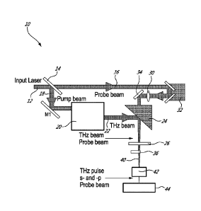
[0029] FIG. 2 is a diagrammatical view of system for THz detection according
to an embodiment of an aspect
of the present invention;
[0030] FIG. 3A shows a THz electric field trace recorded using a conventional
electro-optic sampling system
and method;
[0031] FIG. 3B shows a THz electric field trace recorded using a system and
method according to the present
invention;
[0032] FIG. 4 show spectra of THz pulses measured using a method of the
present invention (continuous line)
and a conventional electro-optic sampling system and method (dotted line);
[0033] FIG. 5 shows signal-to-noise ratio (SNR) of the THz electric field
measurements dependence on optical
path difference (OPD) between the two signals at the exit end of the optical
fiber; and
[0034] FIG. 6 shows the dependence of the THz peak electric field on the angle
between the two wire-grid
polarizers, measured using the present method (dots) and the conventional
electro-optic sampling method
(squares).
DESCRIPTION OF ILLUSTRATIVE EMBODIMENTS
[0035] The present invention is illustrated in further details by the
following non-limiting examples.
[0036] A system 10 according to an embodiment of an aspect of the present
invention is shown in FIG. 2.
[0037] A laser beam 12, such as a 800 nm laser beam for example, is split into
a pump beam arm 16 and a
probe beam arm 18 using a beam splitter 14.
[0038] The pump beam arm 18 is used to generate THz radiation in a THz source
20 using optical methods,
such as optical rectification in a nonlinear crystal such as a LiNb03 crystal
for example, or four-wave mixing in air
plasma for example. A tilted-pulse-front method in a LiNb03 crystal for
example can be used to generate THz
radiations with energies up to 0.3 0 with bandwidth in a range comprised
between 0.1 and 3 THz.
[0039] The generated few-cycle THz beam 22 is focused using an off-axis
parabolic (OAP) mirror 24 onto an
electro-optic detection crystal 26.
CA 02965714 2017-04-25
WO 2016/070273 PCT/CA2015/051133
8
[0040] The optical probe beam arm 16 is focused by a spherical piano-convex
lens 30, and then propagates
through a hole in the off-axis parabolic (OAP) mirror 24 and to the detection
crystal 26, where it overlaps with the
focused THz beam 22.
[0041] A quarter wave plate 36 is used before a single mode polarization-
maintaining optical fiber 40, such as
Thorlabs polarization maintaining 780-HP for example, to convert the linear
polarization state of the optical probe
beam 16 to circular polarization state. Then the optical probe beam 16 is
coupled into the polarization-
maintaining optical fiber 40, with its polarization direction set along the
two orthogonal birefringent axes (x and y)
of the polarization-maintaining optical fiber 40. Propagation of the optical
probe beam 16 through the
polarization-maintaining optical fiber 40 results in an intrinsic optical path
difference between the two orthogonal
polarization components of the optical probe beam 16, due to the birefringence
in the fiber. As a result, two
pulses are created at the exit of the polarization-maintaining optical fiber
40 with temporal separation between
them. A polarizer 42 is placed at the end of polarization-maintaining optical
fiber 40 with its transmission axis at
45 with respect to the axes (x and y) of the polarization-maintaining optical
fiber 40. By allowing the component
of each polarization state, i.e. fast and slow, to pass through the polarizer
42, two pulses with a temporal delay
between them, and with the same linear polarization, are generated. The
optical probe beam 16 is then sent to a
spectrometer 44 that is used to observe the interference fringes due to the
interaction between the fast and slow
pulses.
[0042] A custom-made spectrometer consisted of a diffraction grating (600
grooves/mm), a piano-convex
cylindrical lens (f=150 mm), and a two-dimensional (2D) charged-coupled device
(CCD) camera (Dalsa Inc.
480 x640 pixels). At the charged-coupled device (CCD) camera, the fast and
slow pulses interfere, thus allowing
measuring the phase difference between them.
[0043] The THz pulse is aligned in a polarization state vertical to the paper
plane and parallel to the optical
probe beam 16 polarization state at the ZnTe detection crystal 26. The
presence of the THz electric field induces
birefringence in the ZnTe detection crystal 26 via the Pockels effect. This
birefringence is detected by the optical
probe beam 16 as a change in the optical path difference (OPD) between the two
orthogonal signals generated
by the polarization-maintaining optical fiber 40.
[0044] Therefore, the change in the phase difference introduced by the THz
pulse can be measured using the
interference of the two signals. When there is no THz radiation reaching the
detection crystal 26, this measured
CA 02965714 2017-04-25
WO 2016/070273 PCT/CA2015/051133
9
phase difference between the two signals is proportional to the length of the
polarization maintaining optical fibre
40, and defines a reference phase difference. When a THz radiation reaches the
detection crystal 26, the THz
electric field induces birefringence in the electro-optic crystal 26 due to
the Pockels effect, which affects the
probe pulse, which is temporally matched with the THz pulse, and an extra
phase difference is introduced
between the two signals formed after the detecting crystal 26, compared to the
reference phase difference, and
this extra phase difference is directly proportional to the THz electric
field. By delaying the probe beam 16 using
a delay stage 32 the temporal shape of the THz pulse can thus be
reconstructed.
[0045] The detection crystal 26 may be an electro-optic crystal, such as ZnTe,
GaP and GaSe for example,
with a typical thickness less than a few mm, typically between 10 p.m to 2 mm.
[0046] In order to reconstruct the THz signal, the data from the camera of the
spectrometer 44 are numerically
treated, involving some intermediate steps, as follows. First, the data from
the camera of the spectrometer which
are acquired in the wavelength space are rescaled in the wave vector (k)-
space. These data are then Fourier
transformed to obtain the frequency corresponding to the optical path
difference (OPD) between the two s and p
components. The phase between the s and p components is determined using
relation (2) above. This phase is
tracked over time by changing the delay between the THz signal and the probe
signal. The phase waveform
gives the waveform of the THz signal.
[0047] More precisely, the resulting interference between the two signals at
the charged-coupled device (CCD)
camera can be expressed using a relation adapted from relation 1) reported
hereinabove:
1(k) = 1 F (k) + s (k) 2,11p(k) 1 s (k) o s[ õ k L] (4)
[0048] where k = 2rrilk is the wave number, IF is the fast axis signal
intensity, /s is the slow axis signal intensity,
00 is the phase constant, and L is the optical path difference between the
fast and the slow axis signals. The
interference is recorded using a CCD camera and rescaled from wavelength space
to wave-number (k) space
and Fourier transformed to obtain the corresponding fast Fourier transform
spectrum. The instantaneous phase
difference between the two signals is calculated using the relation 2)
discussed hereinabove:
0 = tan (2) /(0)-1
(2)
CA 02965714 2017-04-25
WO 2016/070273 PCT/CA2015/051133
[0049] where Im(i (L)) is the imaginary part and Re(i (L)) is the real part of
the Fourier transform of relation (4)
for an optical path difference equal to L, corresponding to the optical path
difference introduced to the fast and
slow signals by the polarization maintaining optical fiber. The change in
optical path difference (OPD) over time
can be traced by monitoring the phase change in relation (2). This phase
change is proportional to the THz
electric field. Hence the temporal shape of the THz electric field can be
reconstructed by varying the delay time
between the THz pulse and the optical probe beam pulse by using the delay
stage.
[0050] The traces of the THz electric fields measured using the present
system, with an optical fiber having
parameters as shown in Table 1 below for example on the one hand, and the
conventional electro-optic
sampling method on the other hand, are shown in FIG. 3.
Numerical Aperture 0.12
Attenuation 4 dB/km @ 850 nm
Operating Wavelength 770 - 1100 nm
Second Mode Cut-off 710 60 nm
Mode Field Diameter (1/e2 fit ¨ near field) 5.3 1.0 pm @ 850 nm
Beat Length 2.4 mm @ 850 nm
Birefringence 3.5 x 10⁻⁴
Table I
[0051] The trace in FIG. 3B is obtained using an optical fiber length of 80
cm. At that length, the two signals
have an optical path difference of 400 pm, i.e. a temporal separation of 1.33
ps, between them at the exit end of
the fiber. It can be seen that the temporal scan length has been extended by
more than ten times compared to
CA 02965714 2017-04-25
WO 2016/070273 PCT/CA2015/051133
11
the Michelson based spectral domain interferometry (SDI) method reported in
previous works (see WO
2014/019091), where the scan length of the THz signal that could be measured
was limited by the thickness of
the glass plate in use (300 pm), resulting in a scan window of 3ps. This
limitation is here overcome.
[0052] The corresponding power spectra for the THz electric field traces are
shown in Fig. 4. Using the present
fiber based spectral domain interferometry (SDI) method, a signal-to-noise
ratio (SNR) of 43,000 in the power
spectrum was measured, compared to 110,000 with the conventional electro-optic
sampling method. Compared
with the Michelson based spectral domain interferometry (SDI) method, the
present fiber based spectral domain
interferometry (SDI) method results in an enhancement of the signal-to-noise
ratio (SNR) by more than four
times.
[0053] In the search for an optimal fiber length that yields better signal-to-
noise ratio (SNR) in spectral domain
interferometry (SDI) THz detection, different fiber lengths were tried, from
60 to 240 cm, with corresponding
optical path difference (OPD) between the signals at the exit end of the fiber
between 300 and 1200 pm. FIG. 5
shows the signal-to-noise ratio (SNR) measured using the present fiber based
spectral domain interferometry
(SDI) method using various fiber lengths. It was found that the signal-to-
noise ratio (SNR) increases as the
optical path difference (OPD) between the interfering signals decreases from
1200 pm to 400 pm. However,
decreasing the optical path difference (OPD) from 400 pm to 300 pm reduced the
signal-to-noise ratio (SNR).
One could attribute this reduction in the signal-to-noise ratio (SNR) to the
fact that for the spectral domain
interferometry (SDI) method, working very close to the dc component of the
interference signal results in the
signal subject to many low-frequency noises in a laboratory environment, thus
reducing signal-to-noise ratio
(SNR) of THz detection.
[0054] Based on the experimental findings of FIG. 5, one can attribute the
enhancement in the signal-to-noise
ratio (SNR) obtained, compared with the Michelson based spectral domain
interferometry (SDI) method, to the
fact that the optical path difference (OPD) between the signals could be small
(400 pm) in the case of the fiber-
based spectral domain interferometry (SDI) method, while the optical path
difference (OPD) between the signals
in the case of the Michelson based spectral domain interferometry (SDI) method
was relatively large (900 pm).
Other possible reason for the signal-to-noise ratio (SNR) enhancement of the
fiber-based method is the
elimination of angular vibrations in the glass plate those have been
encountered in the case Michelson based
spectral domain interferometry (SDI), thus reducing the noise due to those
vibrations and accordingly yields an
overall better signal-to-noise ratio (SNR) in the case of spectral domain
interferometry (SDI) fiber based method.
CA 02965714 2017-04-25
WO 2016/070273 PCT/CA2015/051133
12
[0055] Furthermore, in order to study the capability of the present method in
measuring different THz fields, the
THz electric field has been varied from about 4 kV/cm to about 70 kV/cm by
rotating the angle between the two
wire-grid polarizers. The results are shown in FIG. 6, where the THz fields
measured using the fiber-based
spectral domain interferometry (SDI) method are compared with the THz fields
measured using the conventional
electro-optic sampling method. A good agreement between the two methods is
evident, suggesting that the
present method is a promising method for measuring lower THz electric fields
as well.
[0056] Thus a polarization-maintaining optical fibre (PMF) is used as the
active component for detecting THz
radiation, which also solves the problem of scan length and signal-to-noise
ratio (SNR). By using sufficiently long
polarization-maintaining fibres, the scan length can be extended to values
much larger than 3 ps. The optimum
length of the fibre is determined by its dispersion, which may vary depending
on the fibre type. The signal-to-
noise ratio (SNR) is also greatly improved by replacing free-space optics with
fibres, and also by shifting the
signal to be measured outside of the strong background near zero optical path
difference (OPD).
[0057] There is thus provided a method and a system for terahertz (THz)
electric field measurement based on
spectral-domain interferometry (SDI) and using a polarization maintaining
single-mode optical fiber in the optical
probe beam line. The polarization maintaining optical fiber is placed after
the detection crystal, and is used to
increase the phase difference between the two polarizations states of the
optical probe beam that is required in
the spectral-domain interferometry (SDI) method. It was shown that the signal-
to-noise ratio (SNR) van be
enhanced by more than four times compared with previously reported Michelson
based spectral domain
interferometry (SDI) method. Moreover, the scanning time of the THz pulse has
been extended to >30
picoseconds.
[0058] Furthermore, the present system and method have the potential to allow
THz measurement of modest-
intensity, oscillator-based THz sources, and not just intense THz sources.
This is because the spectral domain
interferometry (SDI) signal to be measured can be shifted outside of the noisy
background near zero optical path
difference (OPD), thus allowing smaller phase shifts to be measured with
higher signal-to-noise ratio (SNR). This
is of high commercial significance, since even though intense THz sources are
becoming more and more
accessible, most of the THz spectroscopy experiments are still performed using
oscillator-based THz systems.
The added ability to provide a new detection method for this larger THz
community may significantly increase
the interest for commercialization.
CA 02965714 2017-04-25
WO 2016/070273 PCT/CA2015/051133
13
[0059] To overcome the limitation of scan length and signal-to-noise ratio
(SNR) at the same time, a method
based on spectral domain interferometry (SDI) is provided, where a
polarization-maintaining fibre is used instead
of a glass plate to get the two pulses.
[0060] The scope of the claims should not be limited by the embodiments set
forth in the examples, but should
be given the broadest interpretation consistent with the description as a
whole.
CA 02965714 2017-04-25
WO 2016/070273 PCT/CA2015/051133
14
REFERENCES
1. B. B. Hu and M. C. Nuss, Opt. Lett. 20, 1716 (1995).
2. S. Kono, M. Tani, P. Gu and K. Sakai, Appl. Phys. Lett. 77, 4104 (2000).
3. X. Lu and X.-C. Zhang, Appl. Phys. Lett. 98, 151111 (2011).
4. Gargi Sharma, Kanwarpal Singh, lbraheem Al-Naib, Roberto Morandotti, and
Tsuneyuki Ozaki, Opt.
Lett. 37, 4338 (2012).
5. Gargi Sharma, Kanwarpal Singh, Akram Ibrahim, lbraheem Al-Naib, Roberto
Morandotti, Francois Vidal,
and Tsuneyuki Ozaki, Opt. Lett. 15, 2705 (2013).
6. Q. Wu and X.-C. Zhang, Appl. Phys. Lett. 67, 3523 (1995).
7. Z. Jiang and X.-C. Zhang, Opt. Lett. 23, 1114 (1998).
8. S. P. Jamison, J. Shen, A. M. MacLeod, W. A. Gillespie and D. A.
Jaroszynski, Opt. Lett. 28, 1710
(2003).
9. K. Y. Kim, B. Yellampalle, A. J. Taylor, G. Rodriguez and J. H. Glownia,
Opt. Lett. 32, 1968 (2007).
10. Y. Kawada, T. Yasuda, H. Takahashi and S.-I. Aoshima, Opt. Lett. 33,
180 (2008).
11. J. Fletcher, Opt. Express 10, 1425 (2002).
12. J. Zhang, B. Rao, L. Yu and Z. Chen, Opt. Lett. 34, 3442 (2009).
13. A. F. Fercher, C. K. Hitzenberger, G. Kamp and S. Y. El-Zaiat, Optics
Commun. 117, 43 (1995).
14. K. Singh, C. Dion, S. Costantino, M. Wajszilber, M. R. Lesk and T.
Ozaki, Exp. Eye Res. 91, 63 (2010).
15. K. Singh, C. Dion, M. R. Lesk, T. Ozaki and S. Costantino, Rev. Sci.
lnstrum. 82, 023706 (2011).
16. T. Bajraszewski, M. Wojtkowski, M. Szkulmowski, A. Szkulmowska, R.
Huber and A. Kowalczyk, Opt.
Express 16, 4163 (2008).