Note: Descriptions are shown in the official language in which they were submitted.
CA 02965752 2017-04-25
WO 2016/067029 PCT/GB2015/053238
1
METHOD FOR THE ENRICHMENT OF CIRCULATING TUMOR DNA
FIELD OF THE INVENTION
The invention relates to a method for the purification or enrichment of
circulating cell free
nucleosomes of tumor origin and associated circulating tumor DNA from blood,
serum or
plasma.
BACKGROUND OF THE INVENTION
Cellular DNA exists as a protein-nucleic acid complex called chromatin. The
nucleosome is
the basic unit of chromatin structure and consists of double stranded DNA
(dsDNA) wound
around a protein complex. The DNA is wound around consecutive nucleosomes in a
structure often said to resemble "beads on a string" and this forms the basic
structure of
open or euchromatin. In compacted or heterochromatin this string is coiled and
super coiled
in a closed and complex structure.
Each nucleosome in chromatin consists of a protein complex of eight highly
conserved core
histones (comprising of a pair of each of the histones H2A, H2B, H3, and H4).
Around this
complex are wrapped approximately 146 base pairs (bp) of DNA. Cell free
nucleosomes are
reported to comprise 160-200bp DNA which may be due to the presence of further
linker
DNA. Another histone, H1 or H5, acts as a linker and is involved in chromatin
compaction.
Normal cell turnover in adult humans involves the creation by cell division of
some 1011 cells
daily and the death of a similar number, mainly by apoptosis. During the
process of
apoptosis chromatin is broken down into mononucleosomes and oligonucleosomes
some of
which may be found in the circulation. Under normal conditions the level of
circulating
nucleosomes found in healthy subjects is reported to be low. Elevated levels
are found in
subjects with a variety of conditions including many cancers, auto-immune
diseases,
inflammatory conditions, stroke and myocardial infarction (Holdenreider &
Stieber, 2009).
DNA abnormalities are characteristic of all cancer diseases. The DNA of cancer
cells differs
from that of healthy cells in many ways including, but not limited to, point
mutations,
translocations, gene copy number, micro-satellite abnormalities, DNA strand
integrity and
nucleotide modifications (for example methylation of cytosine at position 5).
These tumor
associated alterations in DNA structure or sequence are investigated routinely
in cancer
cells or tissue removed at biopsy or surgery for clinical diagnostic,
prognostic and treatment
purposes. Tumor genetic and epigenetic characteristics vary between different
tumor types
and between different patients with the same tumor disease. Moreover these
characteristics
CA 02965752 2017-04-25
WO 2016/067029 PCT/GB2015/053238
2
vary over time within the same cancer of the same patient with the progression
of the
disease and in the development of acquired resistance to drug or other
therapies. Thus
serial investigation of tumor DNA in cells removed at surgery or biopsy may
help the clinician
to monitor disease progression and detect any relapse or acquired treatment
resistance at
an early stage (possibly many months earlier than radiological detection) and
allow
potentially successful changes in treatment courses.
However, tissue DNA tests have limitations as invasive biopsy procedures
cannot be
performed repeatedly on patients for monitoring purposes. For some patients
biopsy may not
be used at all. Biopsy is expensive to perform, uncomfortable for the patient,
poses patient
risk, and may lead to surgical complications. Moreover, a tumor in a patient
may consist of
multiple tumoral clones located within different areas of the same tumor or
within different
metastases (in metastatic cancer) not all of which may be sampled on biopsy. A
tissue
biopsy DNA investigation therefore provides a snap-shot of the tumor, both in
time and in
space, amongst different tumor clones located within different areas of a
tumor at a
particular moment in time.
The blood of cancer patients contains circulating tumor DNA (ctDNA) which is
thought to
originate from the release of chromatin fragments or nucleosomes into the
circulation from
dying or dead cancer cells. Investigation of matched blood and tissue samples
from cancer
patients shows that cancer associated mutations, present in a patient's tumor
(but not in
his/her healthy cells) are also present in ctDNA in blood samples taken from
the same
patient (Newman eta!, 2014). Similarly, DNA sequences that are differentially
methylated
(epigenetically altered by methylation of cytosine residues) in cancer cells
can also be
detected as methylated sequences in ctDNA in the circulation. In addition the
proportion of
cell-free circulating DNA (cfDNA) that is comprised of ctDNA is related to
tumor burden so
disease progression may be monitored both quantitatively by the proportion of
ctDNA
present and qualitatively by its genetic and/or epigenetic composition.
Analysis of ctDNA can
produce highly useful and clinically accurate data pertaining to DNA
originating from all or
many different clones within the tumor and which hence integrates the tumor
clones
spatially. Moreover, repeated sampling over time is a much more practical and
economic
option. Analysis of (ctDNA) has the potential to revolutionize the detection
and monitoring of
tumors, as well as the detection of relapse and acquired drug resistance at an
early stage for
selection of treatments for tumors through the investigation of tumor DNA
without invasive
tissue biopsy procedures. Such ctDNA tests may be used to investigate all
types of cancer
associated DNA abnormalities (e.g.; point mutations, nucleotide modification
status,
translocations, gene copy number, micro-satellite abnormalities and DNA strand
integrity)
CA 02965752 2017-04-25
WO 2016/067029 PCT/GB2015/053238
3
and would have applicability for routine cancer screening, regular and more
frequent
monitoring and regular checking of optimal treatment regimens (Zhou eta!,
2012).
Blood, plasma or serum may be used as a substrate for ctDNA assays and any DNA
analysis method may be employed including, without limitation, DNA sequencing,
epigenetic
DNA sequencing analysis (e.g., for sequences containing 5-methylcytosine),
PCR,
BEAMing, NGS (targeted or whole genome), digital PCR, cold PCR (co-
amplification at
lower denaturation temperature-PCR), MAP (MIDI-Activated Pyrophosphorolysis),
PARE
(personalized analysis of rearranged ends) and Mass Spectrometry.
As DNA abnormalities are characteristic of all cancer diseases and ctDNA has
been
observed for all cancer diseases in which it has been investigated, ctDNA
tests have
applicability in all cancer diseases. Cancers investigated include, without
limitation, cancer of
the bladder, breast, colorectal, melanoma, ovary, prostate, lung liver,
endometrial, ovarian,
lymphoma, oral, leukaemias, head and neck, and osteosarcoma (Crowley eta!,
2013; Zhou
eta!, 2012; Jung et al, 2010). The nature of ctDNA tests will now be
illustrated by outlining
three (non-limiting) example approaches.
The first example involves the detection of a cancer associated gene sequence
mutation in
ctDNA. Blood tests involving the detection of a single gene mutation in ctDNA
generally have
low clinical sensitivity. There are two reason for this. Firstly, although all
cancers have
mutations, the frequency of any particular mutation in a particular cancer
disease is usually
low. For example, although K-ras and p53 mutations are regarded as two of the
more
frequent cancer mutations and have been studied in a wide range of cancers
including
bladder, breast, colon, lung, liver, pancreas, endometrial and ovarian
cancers, they were
detected in 23%-64% and 17%-54% of cancer tissue samples respectively.
Secondly, even
if the cancer tissue of a patient does contain the mutation, the level or
concentration of
mutated ctDNA present in the blood of the patient may be low and difficult to
detect. For
example, K-ras and p53 mutations could be detected in the ctDNA of 0%-75% of K-
ras and
p53 tissue positive patients. The sum of these two effects meant that K-ras or
p53 mutations
were detected in the blood of less than 40% of cancer patients (Jung eta!,
2010).
The second example involves the detection of multiple cancer associated gene
sequence
mutations in ctDNA. Although mutations of any particular gene such as K-ras or
p53 may be
present in only a minority of cancers, all cancers contain mutations so study
of a sufficiently
large panel of mutations should in principle, facilitate the detection of most
or even all
tumors. One way to increase the clinical sensitivity of such tests is
therefore to test for a
CA 02965752 2017-04-25
WO 2016/067029 PCT/GB2015/053238
4
wide range of mutations in many genes. Newman eta! have taken this approach
for non-
small cell lung cancer (NSCLC) and investigated 521 exons and 13 intron
sequences from
139 recurrently mutated genes. The mutations studied encompassed multiple
classes of
cancer associated genetic alterations, including single nucleotide variation
(SNV) and fusion
genes. In this way the authors reported the detection of more than 95% of
stage II-IV tumors
and 50% of stage I tumors with 96% specificity in ctDNA blood tests (Newman
eta!, 2014).
The third example involves the detection of cancer associated epigenetic
alterations to
particular gene sequences in ctDNA. This approach can be applied to any DNA or
nucleotide
modification. A prime example of this approach is the detection of genes which
are
differentially methylated at cytosine residues in certain cancers. A large
number of genes
have been investigated for this purpose in a variety of cancers. A few of
these are SEPTI N-
9, APC, DAPK, GSTP1, MGMT, p16, RASSF1A, T1G1, BRCA1, ERa, PRB, TMS1, MLH1,
HLTF, CDKN2A,SOCS1, SOCS2, PAX5, PGR, PTGS2 and RARp2 investigated in bladder,
breast, colorectal, melanoma, ovarian and prostate cancers. An illustrative
example of this
approach is the detection of methylated SEPTIN-9 in ctDNA for the detection of
ColoRectal
Cancer (CRC) which was reported to detect 68% of CRC cases with a clinical
specificity of
89% (Grutzmann et al, 2008).
The tumor derived ctDNA fraction of cfDNA circulates as small DNA fragments
with a
median length of approximately 180 base pairs (bp) which is the size expected
for DNA
fragments circulating in the form of mono-nucleosomes (Newman eta!, 2014).
These 180bp
DNA fragments are thought to comprise the nucleosomal DNA plus some linker
DNA.
Cancer patients are reported to have higher cfDNA levels than healthy
subjects. Workers in
the field have reported ranges of 0-100 ng/ml (mean 30 ng/ml) cfDNA for
healthy subjects
and 0-1000 ng/ml (mean 180 ng/ml) cfDNA for subjects with cancer
(Schwarzenbach eta!,
2011). Circulating cfDNA consists of DNA molecules of various sizes up to
20,000 base
pairs in length (Zhou eta!, 2012). In agreement with the hypothesis that ctDNA
circulates
predominantly as mono-nucleosomes, measured levels of cell free nucleosomes in
the
circulation are, like DNA levels, higher in cancer patients than in healthy
subjects
(Holdenrieder eta!, 2001). However, raised levels of circulating nucleosomes
per se are not
used clinically as biomarkers of cancer as nucleosomes are a non-specific
product of cell
death and raised levels are observed for many conditions involving elevated
cell death
including acute trauma (Holdenrieder and Stieber, 2009). As a product of cell
death,
circulating nucleosome levels can rise markedly on treatment with cytotoxic
drugs or
radiotherapy. However, nucleosomes are also cleared from the circulation so
levels may
CA 02965752 2017-04-25
WO 2016/067029 PCT/GB2015/053238
spike with treatment and then fall as shown in Figures 1 and 2 reproduced from
Holdenrieder
eta!, 2001.
Although the level of circulating cell free nucleosomes per se has not been
used in clinical
practice as a blood based biomarker in cancer, the epigenetic composition of
circulating cell
free nucleosomes in terms of their histone modification, histone variant, DNA
modification
and adduct content have been investigated as blood based biomarkers in cancer
(WO
2005/019826; WO 2013/030577; WO 2013/030579; WO 2013/084002).
The biological origin of cfDNA is not well understood. Fragmentation of
chromatin to produce
mononucleosomes and oligonucleosomes is a feature of apoptotic cell death.
Necrotic cells
are thought to produce larger DNA molecules of thousands of base pairs in
length, but DNA
fragmentation may also occur in some cases of necrosis. Further, common DNA
repeat
sequences (eg; ALU or LINE1 sequences) may be released as 200-400 base pair
DNA
fragments from cells undergoing non-apoptotic or necrotic cell death
(Schwarzenbach et a!,
2011). DNA fragments may also be secreted by cells as a form of inter-cellular
communication. The origin of ctDNA is thought to be related to the death of
cancer cells.
DNA fragments may be released as nucleosomes from necrotic and/or apoptotic
tumor cells.
However, necrotic and apoptotic cells are usually phagocytosed by macrophages
or other
scavenger cells and DNA may be released by macrophages that have engulfed
necrotic or
apoptotic cells (Schwarzenbach eta!, 2011).
There are a variety of methods available for extracting cfDNA from blood,
serum or plasma
and these have been compared for yield of extracted DNA and for their
efficiency of
extraction of DNA fragments of different lengths. Phenol-chloroform and sodium
iodide
extraction methods provide the highest yield and extract small DNA fragments
of less than
200bp in length. Other methods tested (including commercially available
methods) are
reported to have lower DNA extraction yields and to fail to extract small DNA
fragments of
less than 200bp in length (Fong et al, 2009).
Extraction of cfDNA from blood, serum or plasma for analysis of ctDNA is
usually performed
using commercially available DNA extraction products. Such extraction methods
claim high
recoveries of circulating DNA (>50%) and some products (for example; the
QIAamp
Circulating Nucleic Acid Kit produced by Qiagen) are claimed to extract DNA
fragments of
small size. Typical sample volumes used are in the range 1-5mL of serum or
plasma.
CA 02965752 2017-04-25
WO 2016/067029 PCT/GB2015/053238
6
There are currently no ctDNA based tests in routine use for clinical oncology
purposes due
to a number of limitations. A major methodological limitation is a requirement
for high quality
DNA. Current ctDNA sampling methods produce poor quality ctDNA samples due to
the
nature of the sample. The main difficulty lies in the presence of large
amounts of non-tumor
cfDNA in the circulation which complicates any analysis of ctDNA. Estimates
from different
workers vary but the fraction of ctDNA present in the circulation can be too
low to detect or
above 50% of cfDNA. However, for most cancer patients the ctDNA fraction is a
small part of
cfDNA. For example, recent studies report that the ctDNA fraction increases
with tumor size
in pre-treatment lung cancer patients. The highest level found was 3.2% in a
patient with a
large tumor burden but most patients were found to have ctDNA fractions below
0.1%
(Newman eta!, 2014). This means that for many patient samples, a very low
level of ctDNA
must be analysed in the presence of a much higher level of non-tumor derived
DNA.
Moreover this DNA is from the same subject and hence of similar sequence and
will interfere
in any method for the quantification or analysis of ctDNA.
A similar problem occurs for the measurement of circulating cell free
nucleosomes and/or
the epigenetic composition of circulating nucleosomes as biomarkers for cancer
because
nucleosomes per se are a non-specific indicator of cell death and are released
as part of the
normal cell turnover process of the body as well as in conditions associated
with elevated
levels of cell death such as autoimmune diseases, stroke, sepsis, post trauma,
burns,
myocardial infarction, cerebral stroke, during graft rejection after organ
transplantation and
after severe exercise. Thus nucleosomes of tumor origin circulate together
with other non-
tumor nucleosomes of various cellular and tissue origins. These non-tumor
nucleosomes will
interfere in any method for the quantification or epigenetic analysis of
nucleosomes of tumor
origin.
There is therefore a great need for a method for the enrichment of circulating
nucleosomes
and ctDNA of tumor origin from blood, serum or plasma samples.
SUMMARY OF THE INVENTION
According to a first aspect of the invention, there is provided the use of a
histone H3.1 and/or
H3.2 and/or H3t binding agent for detecting, isolating and/or purifying cell
free nucleosomes
of tumor origin or circulating tumor DNA (ctDNA) from a biological sample.
According to a further aspect of the invention, there is provided a method for
isolating
circulating cell free nucleosomes of tumor origin from a biological sample by
affinity
purification wherein said method comprises the steps of:
CA 02965752 2017-04-25
WO 2016/067029 PCT/GB2015/053238
7
(i) contacting the sample with a histone H3.1 and/or H3.2 and/or H3t
binding
agent;
(ii) isolating bound nucleosomes from the sample; and
(iii) analysing the isolated nucleosomes and/or associated DNA.
According to a further aspect of the invention, there is provided a method for
isolating
purified circulating tumor DNA (ctDNA) from a biological sample, wherein said
method
comprises the steps of:
(i) isolating circulating cell free nucleosomes containing histone H3.1
and/or
H3.2 and/or H3t;
(ii) extracting DNA from the nucleosome sample produced in step (i); and
(iii) analysing the extracted DNA.
According to a further aspect of the invention, there is provided an
immunoassay method for
detecting an epigenetic epitope of tumor derived circulating nucleosomes in a
biological
sample, wherein said method comprises the steps of:
(i) contacting the sample with a histone H3.1 and/or H3.2 and/or H3t
binding
agent;
(ii) contacting the nucleosomes or sample with a second binding agent which
binds to said epitope;
(iii) detecting and/or quantifying the binding of said second binding agent
to said
epitope; and
(iv) using the presence or degree of such binding as a measure of the
presence
of the particular epitope of tumor derived nucleosomes in the sample.
According to a further aspect of the invention, there is provided an
immunoassay method for
detecting an epigenetic epitope of tumor derived circulating nucleosomes in a
biological
sample wherein said method comprises the steps of:
(i) contacting the sample with a first binding agent which binds to said
epitope;
(ii) contacting the nucleosomes or sample with a histone H3.1 and/or H3.2
and/or
H3t binding agent;
(iii) detecting and/or quantifying the binding of said histone H3.1 and/or
H3.2
and/or H3t binding agent to nucleosomes in the sample; and
(iv) using the presence or degree of such binding as a measure of the
presence
of the particular epitope of tumor derived nucleosomes in the sample.
CA 02965752 2017-04-25
WO 2016/067029
PCT/GB2015/053238
8
According to a further aspect of the invention, there is provided a method of
diagnosing
cancer which comprises the step of detecting circulating cell free nucleosome
associated
histone variant H3.1 and/or H3.2 and/or H3t in a biological sample obtained
from a human or
animal subject.
BRIEF DESCRIPTION OF THE FIGURES
Figure 1: Circulating levels of cell-free nucleosomes per se in a
patient with
head and neck cancer, treated with 16 daily doses of radiotherapy. Levels were
measured
with a commercial nucleosome ELISA employing an immobilised antibody directed
to bind to
a core nucleosome epitope (copied from Holdenrieder et al; 2001)
Figure 2: Circulating levels of cell-free nucleosomes per se in a
patient with
small-cell lung cancer, treated with cytotoxic chemotherapy on day 1. Levels
were measured
over more than 42 days with a commercial nucleosome ELISA employing an
immobilised
antibody directed to bind to a core nucleosome epitope (copied from
Holdenrieder et al,
2001)
Figure 3: Circulating levels of cell-free nucleosomes per se and
circulating
levels of cell-free nucleosomes containing histone variant H3.1 and/or H3.2
and/or H3t in 6
patients with CRC pre-surgery and at 6, 24, 48, 72 and 96 hours post-surgery.
Levels of cell-
free nucleosomes per se were measured with a commercial nucleosome ELISA
employing
an immobilised antibody directed to bind to a core nucleosome epitope. Levels
of circulating
cell-free nucleosomes containing histone variants were measured with a similar
assay but
employing an immobilized antibody directed to bind to histone variants H3.1,
H3.2 or H3t.
Figure 4: Circulating levels of cell-free nucleosomes per se and
circulating
levels of cell-free nucleosomes containing histone variant H3.1, H3.2 or H3t
and 5-
methylcytosine (5mc) in 6 patients with CRC pre-surgery and at 6, 24, 48, 72
and 96 hours
post-surgery. Levels of cell-free nucleosomes per se were measured with a
commercial
nucleosome ELISA employing an immobilised antibody directed to bind to a core
nucleosome epitope. Levels of circulating cell-free nucleosomes containing
histone H3
variants and 5mc were measured with an assay employing an immobilized antibody
directed
to bind to the histone variant H3.1, H3.2 or H3t and a labelled antibody
directed to bind to
5mc.
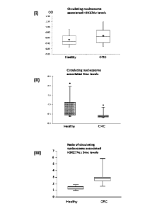
Figure 5: Circulating levels of cell-free nucleosomes containing (i)
both histone
variant H3.1, H3.2 or H3t and histone modification H3K27Ac or (ii) both
histone variant H3.1,
CA 02965752 2017-04-25
WO 2016/067029
PCT/GB2015/053238
9
H3.2 or H3t and 5-methylcytosine (5mc) in healthy patients and patients with
CRC.
Expression of the levels of circulating cell-free nucleosomes containing the
histone H3
variant as well as histone modification H3K27Ac as a ratio to those containing
5mc, leads to
improved discrimination between blood samples taken from healthy subjects and
subjects
with CRC (iii).
Figure 6:
Circulating levels of cell-free nucleosomes containing (i) both histone
variant H3.1, H3.2 or H3t and 5-methylcytosine or (ii) nucleosomes per se
containing 5-
methylcytosine (5mc) in newly diagnosed patients with prostate cancer and
healthy control
subjects of similar age. The data are also displayed as box-plots for cell-
free nucleosomes
containing (iii) a, both histone variant H3.1, H3.2 or H3t and 5-
methylcytosine or (iii) b,
nucleosomes per se containing 5-methylcytosine (5mc) patients.
DETAILED DESCRIPTION OF THE INVENTION
The structure of nucleosomes in terms of their epigenetic signal composition
may vary in
cancer cells. The use of antibodies or other binders directed to bind to
epigenetic signals
that are more common in cancer cells than in other cells may be selective for
the binding of
cell free nucleosomes of tumor origin in a biological sample taken from a
subject which
contains cell free nucleosomes with a mixture of cellular origins.
According to a first aspect of the invention, there is provided the use of a
histone H3.1 and/or
H3.2 and/or H3t binding agent for detecting, isolating and/or purifying
circulating tumor DNA
(ctDNA) from a biological sample.
In one embodiment, cell free nucleosomes of tumor origin are isolated and/or
purified.
The ctDNA in patient samples is often present in very low or undetectable
concentrations
and furthermore comprises only a small proportion of the cfDNA present. The
clinical
performance and utility of cancer tests based on ctDNA analysis would be
improved if better
quality samples were available. Similarly circulating cell free nucleosomes of
tumor origin
circulate as part of a mixture of nucleosomes with a variety of origins and
comprise only a
proportion of the cell free nucleosomes present. Surprisingly we now show that
enrichment
or isolation of nucleosomes of tumor origin, together with the nucleosome
associated ctDNA
fraction of cfDNA, from blood, serum or plasma samples may be performed using
an affinity
purification isolation method.
CA 02965752 2017-04-25
WO 2016/067029 PCT/GB2015/053238
Holdenrieder has reported that elevated circulating cell-free nucleosome
levels are
characteristic of malignant and benign tumor diseases (Holdenrieder eta!,
2001). However,
this is not useful for the detection of cancer as circulating nucleosome
levels are a non-
specific marker for cell death and are elevated in a wide variety of
conditions including
autoimmune diseases, stroke, sepsis, post trauma, burns, myocardial
infarction, cerebral
stroke, during graft rejection after organ transplantation and after severe
exercise
(Holdenrider & Stieber, 2009). Holdenrieder measured circulating nucleosomes
using an
ELISA technique in which the first immobilized antibody employed was directed
to bind to a
common nucleosome core epitope and the second labelled antibody was directed
to bind
double-stranded DNA. This ELISA method is designed to detect all nucleosomes,
or
nucleosomes per se, complete with associated dsDNA regardless of epigenetic
structure.
Circulating nucleosome levels can spike markedly 2-5 days after a sudden
increase in cell
death resulting from any number of disparate causes including trauma, stroke
or treatment
with cytotoxic drugs or radiotherapy. Levels then fall over a period of 2-3
days as shown in
Figures 1 and 2 (reproduced from Holdenrieder eta!, 2001). This effect is due
to induction of
cell death followed by clearance from the circulation (Holdenrider & Stieber,
2009).
We have performed similar experiments using the same commercially available
nucleosome
ELISA, employing the same immobilized antibody directed to bind to a common
nucleosome
core epitope and the same labelled antibody directed to bind to double-
stranded DNA, used
by Holdenrieder eta!, to detect all nucleosomes regardless of epigenetic
status. In these
experiments we measured circulating cell-free nucleosome levels in 6 patients
with CRC
pre-surgery and at 6, 24, 48, 72 and 96 hours post-surgery. In agreement with
the findings of
Holdenrieder, a post-surgery rise in the level of nucleosomes was observed in
every case as
shown in Figure 3. The detailed timing of the rise in nucleosome levels, as
well as the timing
of any subsequent fall, varied between patients.
It is clear that nucleosomes released into the circulation of patients with no
tumor disease
(including for example due to surgical trauma) cannot have a tumor origin.
These
nucleosomes will contribute to cfDNA but will not contain ctDNA. It will also
be clear to those
skilled in the art, that nucleosomes released post-surgery in our own
experiments may have
a tumor origin but may also have a non-tumor origin due to the trauma of
surgery. The
fraction of such cfDNA that has a tumor origin (i.e.; the ctDNA fraction) may
be determined
as the allelic fraction of the cfDNA which contains tumor associated
mutations. We have
developed a method whereby circulating nucleosomes of tumor origin containing
ctDNA may
be enriched or isolated from other nucleosomes and cfDNA.
CA 02965752 2017-04-25
WO 2016/067029
PCT/GB2015/053238
11
We re-assayed the samples taken from the 6 CRC patients pre-surgery and post-
surgery
using a second ELISA method. This second ELISA method employed the same
labelled
antibody directed to bind to double-stranded DNA as the commercially available
method
used by Holdenrieder, but employed an immobilized antibody directed to bind to
histone
variant H3.1, H3.2 or H3t. The results show that the response in the level of
nucleosomes
containing H3.1, H3.2 or H3t to surgery is dissimilar to that of the response
observed for
nucleosomes per se using the ELISA employing an antibody directed to bind to a
common
nucleosome core epitope. The level of circulating nucleosomes containing these
H3 variants
has different response characteristics to the level of (total) circulating
nucleosomes per se,
and is less affected by surgery (Figure 3).
A variety of epigenetically modified nucleotides have been described in the
literature and
epigenetic modification patterns in DNA and/or DNA nucleotide residues are
known to be
altered in cancer. The best described of these includes methylation of
cytosine at position 5.
DNA containing 5-methylcytosine is often referred to as methylated DNA. We
developed and
performed a third ELISA method on the same 6 CRC patients. This ELISA was a
method for
the measurement of nucleosomes containing DNA incorporating 5-methylcytosine
residues
as an example of an assay for DNA containing an epigenetically modified
nucleotide as
described in WO 2013/030577 but using an immobilized anti-H3.1, H3.2 or H3t
antibody as
capture antibody. This third ELISA method thus detected cell free nucleosomes
containing
both histone variant H3.1, H3.2, or H3t as well as 5-methylcytosine residues
and was thus
similar to the second ELISA method described above but used a different
labelled antibody
(directed to bind to 5-methylcytosine rather than dsDNA). The results for the
6 CRC patients
show that the level of H3 variant nucleosome associated 5-methylcytosine is
low and less
affected by surgery than the level of (total) circulating nucleosomes per se
(Figure 4).
In addition we have used this assay to measure concurrent histone variant
H3.1, H3.2, or
H3t with 5-methylcytosine methylated DNA levels in circulating nucleosomes in
healthy
subjects and subjects with CRC. The level of methylated DNA in cancer subjects
was lower
than in healthy subjects. This finding agrees with the published literature
finding that DNA is
globally hypo-methylated in cancer cells. The methylation of DNA in cancer
cells is
estimated to be reduced by approximately 50% compared to the DNA of healthy
cells
(Guerrero-Preston et al, 2007; Soares eta!, 1999). However, the cancer
associated increase
in the level of circulating nucleosomes is reported to be 970% on average
(Holdenrieder et
al, 2001) and the increase in cfDNA to be about 600% (Schwarzenbach et al,
2011). The
large increase in total cfDNA might be expected to lead to an overall rise in
the absolute
CA 02965752 2017-04-25
WO 2016/067029 PCT/GB2015/053238
12
levels of circulating nucleosome associated methylated DNA, despite the
(smaller) fall in the
proportion of circulating cell-free nucleosomes containing methylated DNA.
However, the
results of the present assay employing a capture antibody to histone variant
H3.1, H3.2 or
H3t show a decrease in the absolute level of circulating nucleosomes
containing methylated
DNA (and histone H3 variant) in cancer patients, indicating that a large part
of the cancer
associated observed rise in (total) circulating nucleosomes per se assay is
not of tumor cell
origin. Moreover, we have shown this H3 variant nucleosome associated 5-
methylcytosine
assay to be effective for the detection of cancer in a blood test indicating
that all or a large
proportion of the cell free nucleosomes identified by it are of tumor origin.
A box plot showing
results of this assay for cancer patients and healthy patients is shown in
Figure 5.
We have also developed and performed a fourth ELISA method for the measurement
of
circulating cell-free nucleosomes containing histone variant H3.1, H3.2 or H3t
and the
histone modification H3K27Ac (acetylated lysine 27 of histone H3). This is
similar to the
assays for nucleosomes containing an epigenetically modified histone as
described in the
literature (WO 2005/019826) but employed an immobilized anti-H3.1, H3.2 or H3t
antibody
as capture antibody. This fourth ELISA was identical to the third ELISA method
described
above except that it employed a different labelled antibody directed to bind
to H3K27Ac
(rather than 5-methylcytosine). We have similarly used this fourth assay to
measure
concurrent H3.1, H3.2 or H3t and H3K27Ac levels in the same cell free
circulating
nucleosomes in healthy subjects and subjects with CRC. The level of H3K27Ac in
cancer
subjects was higher than in healthy subjects. This finding agrees with the
published literature
finding that acetylation of H3K27 is elevated in the chromatin of cancer cells
(Karczarski et
al, 2014). Moreover, we have shown this assay to be effective for the
detection of cancer in
a blood test indicating that all or a large proportion of the cell free
nucleosomes identified by
it are of tumor origin. A box plot showing results of this assay for cancer
patients and healthy
patients is shown in Figure 5. The assay results differentiate between blood
samples taken
from healthy subjects and subjects with cancer. 1Mien the results of the third
and fourth
ELISA assays described here for epigenetically modified circulating cell free
nucleosomes
containing histone H3 variants as well as 5-methylcytosine (5mc) or H3K27Ac
respectively,
the discrimination between subjects with cancer and healthy subjects is
enhanced further.
These results show that assays for circulating cell free nucleosomes
containing both the
histone modification H3K27Ac as well as histone variant H3.1, H3.2 or H3t are
useful in
clinical oncology including, without limitation, for the detection of cancer,
as well as for
prognostic prediction, therapy selection, patient monitoring and relapse
monitoring/detection.
Assays for nucleosome associated H3K27Ac may be performed in isolation or as
part of an
assay panel comprising epigenetic and/or other tests.
CA 02965752 2017-04-25
WO 2016/067029 PCT/GB2015/053238
13
In order to further confirm that the circulating nucleosome fraction
containing the histone
variant H3.1, H3.2 or H3t is enriched for nucleosomes of tumor origin and is
useful for
oncology blood tests, we performed an ELISA method using an immobilized anti-
H3.1, H3.2
or H3t antibody as capture antibody and a labelled anti-5-methylcytosine
antibody (ie; the
third ELISA method described above) for nucleosomes containing both the
histone variant
H3.1, H3.2 or H3t as well as DNA incorporating 5-methylcytosine residues, on
samples
taken from 9 men newly diagnosed with prostate cancer and 10 healthy men of
similar age.
The men with prostate cancer were found to have lower circulating nucleosome
associated
5-methylcytosine levels than healthy men and this assay may thus be used,
either alone or
as part of a diagnostic panel, as a method to detect prostate cancer. This
ELISA was then
repeated but using an immobilized antibody directed to bind to a common
nucleosome core
epitope (a fifth ELISA design). This fifth assay showed less discrimination
for prostate
cancer. The results are shown in Figure 6.
We conclude that circulating cell-free nucleosomes containing the histone
variant H3.1, H3.2
or H3t may also contain other epigenetic signals. We also conclude that the
levels of
circulating cell-free nucleosomes containing both a histone H3 variant and
another particular
epigenetic signal may be different in cancer and healthy subjects and that
this may happen
in concordance with the levels observed in cancer cells. We further conclude
that this
remains true even though the level of the particular epigenetic signal may be
elevated or
depressed in the chromatin of cancer cells. The fact that otherwise identical
ELISA methods,
which capture exactly the same circulating nucleosome fraction and differ only
in the
epigenetic signaling structure targeted by the labelled antibody, can produce
results that rise
or fall (for the same circulating nucleosome fraction containing histone H3
variants) in
concordance with the expression of the same epigenetic signals found in the
chromatin of
cancer cells indicates a tumor origin for this nucleosome fraction.
We further conclude that circulating nucleosomes containing a histone H3
variant also have
dissimilar cellular release characteristics to other circulating nucleosomes.
Such
nucleosomes have different origins and are less a result of trauma induced
cell death. As
nucleosomes containing H3.1, H3.2 or H3t are not produced to a large extent by
trauma
induced cell death and can be used as a biomarker for cancer, it is clear that
they are
characteristic of cancer and have a predominantly tumor origin. Separation or
isolation of
nucleosomes containing histone H3.1, H3.2 or H3t produces an isolation or
enrichment of
circulating nucleosomes of tumor origin containing ctDNA from a bodily fluid
sample such as
blood, serum or plasma.
CA 02965752 2017-04-25
WO 2016/067029
PCT/GB2015/053238
14
It will be clear to those skilled in the art that the level of nucleosomes
containing a histone
H3 variant as a proportion of nucleosomes present may be used as a measure of
the
proportion of cfDNA that comprises ctDNA in a sample. Such a measure is
similar to allelic
frequency measures of cancer associated mutations in ctDNA and may be used as
a
measure tumor burden and response to therapy.
Other epigenetic marks characteristic of cancer may similarly be useful in
methods of the
invention provided that they occur more frequently in circulating nucleosomes
of tumor
origin, or ctDNA, than other circulating nucleosomes, or cfDNA. The epigenetic
signal need
not be unique to nucleosomes or DNA of tumor origin, but the increased
frequency should
be sufficient to enrich nucleosomes and DNA of tumor origin. Such epigenetic
marks may
include histone variants (or isoforms), histone modifications, DNA
modifications and
nucleosome adducts.
In a first embodiment of the invention a wholly or partially purified tumor
nucleosome
preparation is isolated from a sample of a biological fluid. The purification
method involves
the affinity isolation of cell free nucleosomes containing a histone or DNA
epigenetic signal
epitope characteristic of cancer employing a binder that binds to the said
epigenetic epitope.
The tumor nucleosome preparation and/or its associated ctDNA may then be
analysed. In a
preferred embodiment tumor nucleosome and ctDNA isolation is performed by an
immunological affinity purification method employing a binder to histone H3.1
or H3.2 or H3t.
It will be clear to persons skilled in the art that any binding agent capable
of specific binding
to a histone H3 variant (or other appropriate nucleosome epitope
characteristic of cancer)
may be used for affinity purification methods of the invention. Such binding
agents may
include, without limitation, antibodies, aptamers or binding proteins (e.g.;
nucleosome
binding proteins).
Antibodies may be raised by a variety of methods known in the art including
immunization
and library methods such as phage display. The immune response may be induced
against,
or the library may be selected for binding to, the moiety or antigen of
interest. Antibodies
directed to bind to a histone H3 variant may be raised against a variety of
such moieties
including the whole histone H3 isoform protein amino acid sequence and may
optionally
contain post-translational histone modifications. The protein may be purified
from living cells
or produced synthetically. Alternatively a peptide sequence representing a
part of the H3
isoform amino acid sequence may be used and this may also optionally contain
post-
15
translational histone modifications. Nucleosomes or other chromatin fractions
containing
histone H3 isoforms may also be used.
In the present investigations an antibody which binds to the peptide sequence
AT K AA R K
SAP AT G GVKKP H was employed. This amino acid sequence occurs in the sequence
for histone variants H3.1, H3.2 and H3t but not in other histone H3 variants.
It will be clear
to those skilled in the art that binding agents directed to bind to any or all
of histone variants
H3.1 and/or H3.2 and/or H3t and/or H3.2 and/or H3t may be employed in methods
of the
invention. We have primarily referred herein to histone H3.1 but this notation
is intended to
include histone H3.1 and/or H3.2 and/or H3t throughout.
According to a further aspect of the invention, there is provided a method for
isolating
circulating cell free nucleosomes of tumor origin from a biological sample by
affinity
purification wherein said method comprises the steps of:
(I) contacting the sample with a histone H3.1 and/or H3.2 and/or H3t
binding
agent;
(ii) isolating bound nucleosomes from the sample; and
(iii) analysing the isolated nucleosomes and/or associated DNA.
The analysis of the isolated nucleosomes of tumor origin may involve any
suitable method of
analysis of which many are known in the art. These methods include without
limitation
analysis by ELISA using a second antibody or other binder to a common
nucleosome
epitope such as dsDNA or to an epigenetic structure of interest including a
histone
modification, histone variant, DNA modification or another molecule adducted
to a
nucleosome. These methods include methods wherein a histone H3 variant binder
is
employed in place of a general anti-nucleosome epitope binder so that
circulating
nucleosomes of tumor origin (rather than nucleosomes per se) are analysed for
the
epigenetic composition. These methods also include multiplex methods for the
analysis of
multiple epitopes present in circulating nucleosomes of tumor origin.
According to one embodiment of this aspect there is provided an immunoassay
for the
analysis of a particular epigenetic epitope of tumor derived circulating
nucleosomes in terms
of any particular nucleosome associated modified histone, histone variant,
modified
nucleotide or in terms of the presence of any another molecule adducted to a
nucleosome
which comprises the steps of:
Date Recue/Date Received 2022-03-17
CA 02965752 2017-04-25
WO 2016/067029 PCT/GB2015/053238
16
(I) contacting the sample with a histone H3.1 and/or H3.2 and/or H3t
binding
agent;
(ii) contacting the nucleosomes or sample with a second binding agent which
binds to said epitope;
(iii) detecting and/or quantifying the binding of said second binding agent
to said
epitope; and
(iv) using the presence or degree of such binding as a measure of the
presence
of the particular epitope of tumor derived nucleosomes in the sample.
According to an alternative embodiment there is provided a method for
detecting and
measuring cell free nucleosomes containing a particular epigenetic epitope, or
composition
of tumor derived circulating nucleosomes, in terms of any particular
nucleosome associated
modified histone, histone variant, modified nucleotide or in terms of the
presence of another
molecule adducted to a nucleosome which comprises the steps of:
(i) contacting the sample with a first binding agent which binds to said
epitope;
(ii) contacting the nucleosomes or sample with a histone H3.1 and/or H3.2
and/or
H3t binding agent;
(iii) detecting and/or quantifying the binding of said histone H3.1 and/or
H3.2
and/or H3t binding agent to nucleosomes in the sample; and
(iv) using the presence or degree of such binding as a measure of the
presence
of the particular epitope of tumor derived nucleosomes in the sample.
The analysis of nucleosomes of tumor origin isolated by a method of the
invention may also
involve any proteomics method known in the art including without limitation
electrophoresis
methods, chromatographic methods and any method involving mass spectrometry
including
methods involving chromatography and mass spectrometry and/or stable isotope
labelled
mass spectrometry and/or methods involving protein digestion to produce
peptides for
identification and/or quantification by mass spectrometry or any combinatorial
mass
spectrometry method with any other method. In a preferred embodiment of the
invention a
circulating nucleosome preparation enriched for nucleosomes of tumor origin is
prepared by
affinity purification of circulating nucleosomes in a blood, serum or plasma
sample taken
from a cancer patient and the epigenetic composition of the nucleosome
preparation is
investigated by a method involving mass spectrometry. In one embodiment the
method
comprises the steps of:
(i) contacting the sample with a histone variant H3.1 and/or H3.2 and/or
H3t
binding agent;
(ii) isolating bound nucleosomes from the sample; and
CA 02965752 2017-04-25
WO 2016/067029 PCT/GB2015/053238
17
(iii) analyzing the nucleosomes isolated in step (ii) using a method
comprising
mass spectrometry.
In one embodiment, isolation of nucleosomes containing ctDNA is performed by
an
immunological affinity purification method comprising the steps of:
(I) contacting the sample with binding agent directed to bind to an
epigenetic
epitope more commonly occurring in tumor derived than non-tumor derived
nucleosomes;
(ii) isolating bound nucleosomes from the sample; and
(iii) optionally extracting DNA from the nucleosomes isolated in step (ii).
In a further embodiment, the binding agent directed to bind to an epigenetic
epitope
characteristic of tumor derived nucleosomes is directed to bind to histone
variant H3.1
and/or H3.2 and/or H3t in an immunological affinity purification method
comprising the steps
of:
(i) contacting the sample with a histone variant H3.1 and/or H3.2 and/or
H3t
binding agent;
(ii) isolating bound nucleosomes from the sample; and
(iii) optionally extracting DNA from the nucleosomes isolated in step (ii).
Investigation of the purified or isolated ctDNA may involve analysis of any or
all types of
cancer associated DNA abnormalities including, without limitation, epigenetic
analysis
including the methylation of DNA sequences, point mutations, translocations,
gene copy
number, micro-satellite abnormalities and DNA strand integrity. Further any
DNA analysis
method may be employed including, without limitation, DNA sequencing,
methylated DNA
sequencing analysis, PCR, BEAMing, NGS (targeted or whole genome), digital
PCR, cold
PCR (co-amplification at lower denaturation temperature-PCR), MAP (MIDI-
Activated
Pyrophosphorolysis), PARE (personalized analysis of rearranged ends) and Mass
Spectrometry.
In one embodiment, the biological sample comprises a blood, serum or plasma
sample.
According to a further aspect of the invention, there is provided a method of
diagnosing
cancer which comprises the step of detecting circulating cell free nucleosome
associated
histone variant H3.1 and/or H3.2 and/or H3t in a biological sample obtained
from a human or
animal subject.
CA 02965752 2017-04-25
WO 2016/067029 PCT/GB2015/053238
18
In one embodiment, the method of diagnosis additionally comprises detecting
one or more
histone modification, modified nucleotide, histone variant or isoform or
nucleosome adduct.
In a further embodiment, the histone modification comprises H3K27Ac and/or 5-
methylcytosine.
According to a further aspect of the invention, there is provided the use of a
kit comprising a
histone H3.1 and/or H3.2 and/or H3t binding agent in any of the methods
described herein.
The invention will now be illustrated with reference to the following non-
limiting examples.
EXAMPLE 1
An antibody directed to bind specifically to histone isoform H3.1, H3.2 or H3t
is biotinylated
and immobilized on streptavidin coated magnetic beads (Dynal) by the
recommended
method of the manufacturer. The beads are washed several times with loading
buffer using
a magnetic separation system. Serum or plasma taken from a cancer patient is
diluted in
loading buffer and added to the beads. Nucleosomes containing histone H3
variant are
adsorbed to the beads. Other serum/plasma components remain in solution and
are
removed by means of magnetic separation. The beads are washed with buffer. The
nucleosomes containing histone H3 variant are now isolated on the beads. ctDNA
associated with the isolated nucleosomes is extracted by the phenol/chloroform
method or
other standard extraction methods. The extracted DNA may be analysed for
genetic or
epigenetic features of cancer.
EXAMPLE 2
An antibody directed to bind specifically to histone isoform H3.1, H3.2 or H3t
is biotinylated
and immobilized on streptavidin coated magnetic beads (Dynal) by the
recommended
method of the manufacturer. The beads are washed several times with loading
buffer using
a magnetic separation system. Serum or plasma taken from a cancer patient is
diluted in
loading buffer and added to the beads. Nucleosomes containing histone H3
variant are
adsorbed to the beads. Other serum/plasma components remain in solution and
are
removed by magnetic separation. The beads are washed with buffer. The
nucleosomes
containing histone H3 variant are now isolated on the beads. The isolated
nucleosomes are
removed from the magnetic beads using an elution buffer and the nucleosomes
are
analysed by proteomics methods including Mass Spectroscopy.
EXAMPLE 3
CA 02965752 2017-04-25
WO 2016/067029 PCT/GB2015/053238
19
Serum samples taken from 6 patients with CRC pre-surgery and at 6, 24, 48, 72
and 96
hours post-surgery were assayed for circulating cell-free nucleosome levels
using a
commercial (total) nucleosome ELISA produced by Roche employing a common anti-
nucleosome core epitope and a labelled anti-dsDNA antibody. The samples were
then re-
assayed but using an anti-histone variant H3.1, H3.2 or H3t capture antibody.
The measured
(total) nucleosome levels increased following the trauma of surgery using the
commercial
ELISA but the response to surgery was altered and muted for nucleosomes
containing
histone H3 variant measured using the assay employing anti-histone H3.1, H3.2
or H3t
antibody. The results are shown in Figure 3.
EXAMPLE 4
Serum samples taken from patients with CRC and healthy control subjects were
assayed
using an ELISA employing an anti-histone variant H3.1, H3.2 or H3t capture
antibody and
either a labelled anti-histone modification H3K27Ac antibody or a labelled
anti-5-
methylcytosine antibody. When the labelled anti-histone modification H3K27Ac
antibody was
used the results showed that a higher level of nucleosome associated H3K27Ac
was present
in the circulation of the cancer patients than in the healthy controls.
Conversely, when the
labelled anti-5-methylcytosine antibody was used the results showed that a
lower level of
nucleosome associated 5-methylcytosine was present in the circulation of the
cancer
patients than in the healthy controls (Figure 5). Both of these findings are
consistent with the
epigenetic alterations reported for the chromatin of cancer cells and tissue
(decreased global
DNA methylation and increased global H3K27 acetylation). Moreover, when the
results are
of the two epigenetically modified nucleosome results are expressed as a ratio
the combined
data differentiate healthy from CRC subjects with high accuracy as shown in
Figure 5. These
results indicate that these ELISA methods can be used to detect cancer and
that the
nucleosomes bound to the solid phase anti-histone H3 variant antibody are
predominantly of
tumor origin.
EXAMPLE 5
Serum samples taken from 9 men newly diagnosed with prostate cancer and 10
healthy men
of similar age were assayed with an ELISA method using an immobilized anti-
H3.1, H3.2 or
H3t antibody as capture antibody and a labelled anti-5-methylcytosine antibody
for
nucleosomes containing DNA incorporating 5-methylcytosine residues. The men
with
prostate cancer were found to have lower circulating nucleosome associated 5-
methylcytosine levels than healthy men and this assay may thus be used, either
alone or as
part of a diagnostic panel, as a method to detect prostate cancer. This ELISA
was then
repeated but using an immobilized antibody directed to bind to a common
nucleosome core
CA 02965752 2017-04-25
WO 2016/067029
PCT/GB2015/053238
epitope. This assay showed less discrimination for prostate cancer. The
results are shown in
Figure 6.
REFERENCES
Crowley eta!, Nature Reviews Clinical Oncology, 10, 472-484, 2013.
Fong et al, Clinical Chemistry 55(3), 587-589, 2009.Grutzmann eta!, PLoS ONE
3(11):
e3759. doi:10.1371/journal.pone.0003759, 2008.
Guerrero-Preston eta!, Epigenetics 2(4), 223-226, 2007.
Holdenrieder eta!, Int J Cancer 95, 114-120, 2001.
Holdenrieder and Stieber, Critical Reviews in Clinical Laboratory Sciences;
46(1): 1-24,
2009.
Jung eta!, Clinica Chimica Acta, 411, 1611-1624, 2010.
Karczarski eta!, Clinical Proteomics, 11:24, 2014.
Newman et al, Nature Medicine 20(5), 548-554, 2014.
Schwarzenbach et al, Nature Reviews Cancer, 11(6), 426-437, 2011.
Soares eta!, Cancer 85(1), 112-118, 1999.
Zhou eta!, Seminars in Oncology, 39(4), 440-448, 2012.