Note: Descriptions are shown in the official language in which they were submitted.
CA 02965850 2017-04-25
WO 2016/069584
PCT/US2015/057560
SYSTEM AND METHOD FOR TARGETING FEEDBACK
BACKGROUND
1. Field of Invention
[0001] The field of the currently claimed embodiments of this invention
relate to
targeted feedback, and more particularly to targeted feedback for imaging
devices with one or
more sensors for observation and tracking of one or more tools.
2. Discussion of Related Art
[0002] In image-guided interventions, the tracking and localization of
imaging devices
and medical tools during procedures are considered the main enabling
technology in image-
guided surgery (IGS) systems.
[0003] Limitations of the current approach on both the research and
commercial sides
may be attributed to the available tracking technologies and to the
feasibility of integrating these
systems and using them in clinical environments. Thus, there remains a need
for improved
imaging devices for use in image-guided surgery.
SUMMARY
[0004] Aspects of the invention may involve systems, devices, and methods.
In one
embodiment, a system for guidance of an imaging device may be provided. The
system may
include a handheld imaging device; a multidirectional feedback device (e.g.,
an ungrounded
haptic feedback device, an audio feedback device, and/or a visual feedback
device); and a
control unit in communication with the multidirectional feedback device and
the handheld
imaging device The control unit may be configured to: receive a target
location, determine an
initial position and pose of the handheld imaging device, calculate a position
and pose deviation
relative to said initial position and pose, translate said position and pose
deviation into control
data, and transmit said control data to the multidirectional feedback device,
wherein the
multidirectional feedback device uses said control data to provide an operator
with feedback to
guide the handheld imaging device towards the target.
[0005] In another embodiment a method of guidance of a handheld imaging
device may
be provided. The method may include receiving by a control unit a target
location; determining
1
CA 02965850 2017-04-25
WO 2016/069584
PCT/US2015/057560
by the control unit an initial position and pose of the handheld imaging
device; calculating by
the control unit a position and pose deviation relative to said initial
position and pose; translating
by the control unit said position and pose deviation into feedback
instructions; and transmitting
by the control unit said feedback instructions to a multidirectional feedback
device, wherein the
multidirectional feedback device uses the feedback instructions to instruct an
operator to guide
the handheld imaging device toward the target.
BRIEF DESCRIPTION OF THE DRAWINGS
[0006] Further objectives and advantages will become apparent from a
consideration of
the description, drawings, and examples.
[0007] Figure 1 shows an example imaging component for an example imaging
system;
[0008] Figure 2 shows another example imaging system;
[0009] Figure 3 shows an example imaging component for an example imaging
system;
[0010] Figure 4 shows an example feedback device coupled to an imaging
device;
[0011] Figure 5 shows another example feedback device coupled to an imaging
device;
[0012] Figure 6 shows example components of a feedback device;
[0013] Figure 7 shows example torque actuator components on a feedback
device;
[0014] Figure 8 shows example vibrotactile components on a feedback device;
[0015] Figure 9 illustrates an example intersection of a tool and an
ultrasound beam;
[0016] Figure 10 shows an example screenshot of a tool-tip display;
[0017] Figure 11 depicts an example workflow; and
[0018] Figure 12 depicts an illustrative embodiment of a computer for
performing the
methods and building the devices and systems described herein.
DETAILED DESCRIPTION
[0019] Some embodiments of the current invention are discussed in detail
below. In
describing embodiments, specific terminology is employed for the sake of
clarity. However, the
invention is not intended to be limited to the specific terminology so
selected. A person skilled
in the relevant art will recognize that other equivalent components can be
employed and other
methods developed without departing from the broad concepts of the current
invention. All
references cited anywhere in this specification are incorporated by reference
as if each had been
individually incorporated.
[0020] Some embodiments of this invention describe IGNimage-guided
interventions)-
enabling "platform technology" going beyond the current paradigm of relatively
narrow image-
2
CA 02965850 2017-04-25
WO 2016/069584
PCT/US2015/057560
guidance and tracking. It simultaneously aims to overcome limitations of
tracking, visualization,
and guidance; specifically using and integrating techniques e.g. related to
needle identification
and tracking using 3D computer vision and structured light; and imaging device
tracking using
local sensing approaches; among others. Examples of IGI may be seen in U.S.
Patent
Application No. 13/511,101, titled "Low-cost image-guided navigation and
intervention systems
using cooperative sets of local sensors," published as U.S. Patent Application
Publication No.
2013/0016185. Furthermore U.S. Patent Application Nos. 14/092,843 and
14/092,755 depict
sample IGIs. The contents of U.S. Patent Application Nos. 13/511,101,
14/092,843, and
14/092,755 are incorporated herein by reference in their entirety.
[0021] The current invention covers a wide range of different embodiments,
sharing a
tightly integrated common core of components and methods used for general
imaging,
projection, vision, targeting, and local sensing.
[0022] Some embodiments of the current invention are directed to combining
a group of
complementary technologies to provide a local sensing approach that can
provide enabling
technology for the tracking of targets and guidance of medical devices or
tools, for example,
with the potential to significantly reduce errors and increase positive
patient outcomes. This
approach can provide a platform technology for the tracking (e.g., ultrasound
probes, the patient,
the environment, and/or other imaging devices), intervention guidance, and/or
information
visualization according to some embodiments of the current invention. By
combining
ultrasound imaging with image analysis algorithms and probe-mounted light-
sensitive devices,
feedback devices, independent optical-inertial sensors, according to some
embodiments of the
current invention, it is possible to reconstruct the position and trajectory
of surgical needles and
other tools or objects by incrementally tracking and guiding their current
motion.
[0023] Some embodiments of the current invention allow the segmentation,
tracking,
and guidance of needles, imaging devices (e.g., ultrasound probes) and other
tools, using visual,
ultrasound, and/or other imaging and localization modalities and haptic, audio
and/or visual
feedback.
[0024] Such devices can allow imaging procedures with improved sensitivity
and
specificity as compared to the current state of the art. This can open up
several possible
application scenarios that previously required harmful X-ray/CT or expensive
MRI imaging,
and/or external tracking, and/or expensive, imprecise, time-consuming, or
impractical hardware
setups, or that were simply afflicted with an inherent lack of precision and
guarantee of success,
3
CA 02965850 2017-04-25
WO 2016/069584
PCT/US2015/057560
such as: biopsies, RF/HIFU ablations etc.: can allow 2D- or 3D-ultrasound-
based needle
guidance, brachytherapy: can allow 3D-ultrasound acquisition and needle
guidance for precise
brachytherapy seed placement, other applications relying on tracked imaging
and tracked tools.
[0025] Some embodiments of the current invention may provide several
advantages over
existing technologies, such as combinations of: low-cost tracking, local,
compact, and non-
intrusive solution¨ ideal tracking system for hand-held and compact ultrasound
systems that are
primarily used in intervention and point-of-care clinical suites, but also for
general needle/tool
tracking under visual tracking in other interventional settings.
[0026] For example, some embodiments of the current invention are directed
to system
and methods to guide an imaging device and/or medical tool. This guidance may
be combined
with techniques for the tracking of imaging devices (e.g., ultrasound probes)
and/or medical
tools (e.g., needles, pointers, biopsy tools, laparoscopes, ablation devices,
surgical instruments,
or elongated tools). By combining ultrasound imaging with image analysis
algorithms and
probe-mounted light-sensitive devices it is possible to reconstruct the
position and trajectory of
tools (e.g., needles, pointers, biopsy tools, laparoscopes, ablation devices,
surgical instruments,
or elongated tools) and other objects by incrementally tracking their current
motion according to
an embodiment of the current invention. This can provide several possible
application
scenarios that previously required expensive, imprecise, or impractical
hardware setups. For
example, 3D ultrasound-based needle guidance.
[0027] Current sonographic procedures mostly use handheld 2D ultrasound
(US) probes
that return planar image slices through the scanned 3D volume (the "region of
interest" (ROI)).
For percutaneous interventions requiring tool guidance, prediction of the tool
trajectory is
currently based on tracking with sensors attached to the distal (external)
tool end and on mental
extrapolation of the trajectory, relying on the operator's experience. An
integrated system with
3D ultrasound, tool tracking, tool trajectory prediction and interactive user
guidance would be
highly beneficial.
[0028] In one embodiment, a handheld device may contain a feedback system
(e.g.,
haptic, audio, and/or visual) used to assist an operator in targeting. The
handheld device may
also enable position sensing. The feedback system may assist an operator in
positioning a tool
to or near a target. The feedback system may assist an operator in keeping an
imaging device
(e.g., an ultrasound probe) located at or above a target. For example, the
feedback system may
direct the operator to keep the imaging device at a viewing location for
positioning a medical
4
CA 02965850 2017-04-25
WO 2016/069584
PCT/US2015/057560
device (e.g., a needle) to a target. A control unit may determine an initial
position and pose for
an imaging device. The control unit may then determine if the imaging device
is moving in the
wrong direction (e.g., the operator of the imaging device is inadvertently
sliding and/or rotating
away from the region of interest) and/or if the imaging device is not or not
quickly enough
moving towards a target area. If the imaging device is not conforming to the
target area, the
control unit may transmit operator feedback to instruct the operator to move
in a particular
direction.
[0029] The feedback system may provide for spatial targeting enhancement
when using
a medical visualization/imaging system. The feedback system may include a
directional haptic
feedback device for handheld use (e.g., ungrounded haptic feedback device) and
a control unit
for calculation of targeting information and resulting feedback control data.
An embodiment
may also include directional audio or visual feedback devices. In one
embodiment a feedback
device may contain any combination of haptic, audio, and/or visual feedback to
the operator.
[0030] The haptic feedback device may include actuators for vibrotactile
feedback,
torque feedback, or both. The actuators may be designed and arranged in such a
way as to enable
transmission of directional haptic information ("haptic display") to the
operator to provide
instruction on positioning the device relative to some other object (e.g.,
external target locations
or instrument positions).
[0031] The visual feedback may include arrows or animations on a screen.
The audio
feedback may include different tones and/or varying pitch to direct the
operator to bring the
imaging device or a medical tool to a determined location.
[0032] The feedback system may include a control unit that receives a
target location
relative to the device pose, or an instrument location relative to said target
location, and
computes the resulting device pose deviation. This pose deviation may be
translated into haptic
display actuations, audio feedback, and/or visual feedback. The haptic display
actuations may
include torque impulses for rotational deviation (e.g., generated by one or
more asymmetrical-
impulse-driven flywheels oriented so as to be linearly independent),
vibrotactile impulses for
translational deviation (e.g., generated by one or more asymmetric vibration
actuators oriented
S0 as to be linearly independent), or both. Depending on the application
requirements, subsets of
the full six degrees of freedom for haptic feedback may be realized in the
device. The operator
can then use the displayed directional information to position the haptic
device in an ideal
position that minimizes the device pose deviation in a closed-loop control
fashion.
CA 02965850 2017-04-25
WO 2016/069584
PCT/US2015/057560
[0033] In one embodiment, the haptic feedback device may be mounted on a
handheld
ultrasound probe. Feedback actuations will thus result in, for example, haptic
display that is
directly calibrated to the operator's hand and the probe orientation. In
another embodiment, the
feedback device may be integrated into the handheld imaging device enclosure
during
manufacturing of the imaging device.
[0034] The haptic feedback device may contain sensing elements including
one or more
of accelerometers, gyroscopes, magnetometers, or optical sensors that provide
pose and position
information of the feedback device. In one embodiment, the haptic feedback
device may be
physically separate from the imaging device and the sensing elements may
provide relative pose
between the feedback device and the imaging device. The relative pose will
transform the
original pose deviation to feedback device coordinates, which then drives, for
example, the
haptic actuators. Such a physically separated haptic feedback device may be
housed in, for
example, a wrist-worn (e.g., a smartwatch), finger-worn, or forehead-worn
housing, or otherwise
strapped onto a body part of an operator. The sensing elements may also be
contained in a
haptic feedback device mounted on an imaging device.
[0035] In another embodiment, the feedback device can be mounted on an
instrument to
provide feedback (e.g., haptic, audio, and/or visual) to the operator. In one
embodiment, one
hand of the operator may be operating the imaging device and the other hand
may be guiding an
instrument, the feedback may assist the operator in guiding and/or placing the
instrument
relative to the imaging device or another external structure. For example, an
operator may be
operating an ultrasound probe to view images of a patient while receiving
feedback on the
placement of a needle to a target inside the patient.
[0036] In another application, the haptic feedback can be used to
continuously position
the imaging device along, for example, a time-varying path. The feedback may
instruct an
operator to move the imaging device, for example, back and forth over an area
of the patient.
Multiple images at slightly different angles will be acquired with the back
and forth motion of
the imaging device. These multiple images may be combined to produce a higher-
dimensional
image of the target area (e.g., a three-dimensional volume). Images may be
acquired to produce
a higher-dimensional image of the target area, for example, by placing the
imaging device in a
reference position, and then having the control unit issue vibrational
feedback commands to the
haptic feedback component that instruct the operator to translate the imaging
device in the
indicated direction until feedback ceases, or until feedback commands in a
different direction are
6
CA 02965850 2017-04-25
WO 2016/069584
PCT/US2015/057560
issued. In another example, the control unit may issue torque feedback
commands to the haptic
feedback component that instruct the operator to rotate the imaging device in
the indicated
direction until feedback ceases, or until feedback commands in a different
direction are issued.
Another example would perform the same functions based on visual or auditory
feedback, using
at least one of displayed directional indicators, displayed targeting
indicators, audible directional
indicators, and/or voice/speech samples. The auditory and visual feedback may
be in addition to
or replace the haptic feedback commands. Another example may estimate the
imaging device
position from the sensing elements included in the feedback device. During
translations and
rotations of the imaging device, imaging data may be collected and compounded
into a higher-
dimensional representation. One example of this technique may be found in
"Multi-DoF probe
trajectory reconstruction with local sensors for 2D-to-3D ultrasound", Stolka
et al., ISBI 2010,
incorporated herein by reference in its entirety. Instrument tracking
capabilities, such as those
described in "The Kinect as an Interventional Tracking System", Wang et al.,
SPIE 2012
(incorporated herein by reference in its entirety) may be used to dynamically
navigate through
the reconstructed three-dimensional image of the area by, for example,
reslicing along a plane
defined by the instrument.
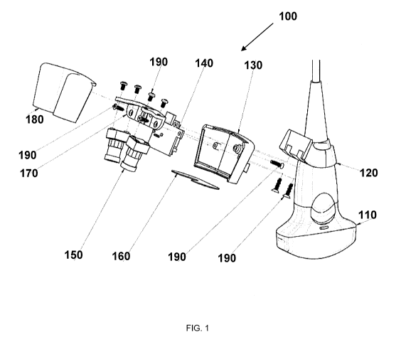
[0037] Figure 1 shows an embodiment of an imaging component 100 for an
imaging
system according to an embodiment of the current invention. Imaging component
100 includes
an imaging device 110, bracket 120 that is structured to be attachable to
imaging device 110. In
the example of Figure 1, the imaging device 110 is an ultrasound probe and
bracket 120 is
structured to be attached to a probe handle of the ultrasound probe.
Ultrasound probes may
include, for example, Ultrasonix #C5-2. However, the broad concepts of the
current invention
are not limited to only this example. The bracket 120 can be structured to be
attachable to other
handheld instruments for image-guided surgery, such as surgical orthopedic
power tools or
stand-alone handheld brackets, for example. In other embodiments, the bracket
120 can be
structured to be attachable to the C-arm of an X-ray system or an MRI system,
for example.
[0038] Imaging component 100 may include top shell 180 and bottom shell
130 that may
be coupled together to form a head shell. Top shell 180 and bottom shell 130
may be coupled
securely to stabilization assembly 170 (e.g., stabilization bar). Head shell
may house
stabilization assembly 170 and other components of imaging component 100.
Screws 190 may
be used to couple the components of imaging component 100.
7
CA 02965850 2017-04-25
WO 2016/069584
PCT/US2015/057560
[0039] In one embodiment, head shell may also include a feedback device.
The
feedback device may be a haptic feedback device including, for example, one or
more linearly
independent asymmetrical-impulse-driven flywheels and/or one or more linearly
independent
asymmetric vibration actuators. Visual feedback may be shown on display 220
and may
include, for example, arrows or on-screen animations. In another embodiment,
one or more
indicator LEDs may be used to provide visual feedback. Audio feedback may be
provided
through the use of, for example, one or more speakers. The speakers may be
located, for
example, with the control unit.
[0040] In another embodiment, the feedback device may be separate from the
head shell.
The feedback device may be coupled to bracket 120 or may be removeably coupled
to the
imaging device through a separate fastener.
[0041] Imaging component 100 may also include one or more light-sensitive
devices 150
(e.g., cameras, PSDs (position-sensitive devices), reflection-based laser
sensing, etc.) securely
attached to stabilization assembly 170. The one or more light-sensitive
devices 150 may be at
least one of a visible-light camera, an infra-red camera, a time-of-flight
camera, a PSD (position-
sensitive device), and/or a reflection-based laser sensing device in some
embodiments of the
current invention. The one or more light-sensitive devices 150 may be arranged
to observe a
surface region close to and during operation of the imaging component 100. In
Figure 1, the one
or more light-sensitive devices 150 may be arranged and configured for stereo
observation of a
region of interest.
[0042] Imaging component 100 may also include a printed circuit board 140
that may
include one or more microprocessors, one or more light sources, and a memory
device. The
light sources may include one or more LEDs, CFLs (compact fluorescent lamp),
incandescent
bulbs, and/or lasers. The light source may emit light in the visible spectrum,
infrared,
ultraviolet, and/or other spectrum. The printed circuit board may also be
connected to one or
more light-sensitive devices 150, the light source, and the memory device, and
may be securely
coupled to stabilization assembly 170.
[0043] Imaging component 100 may also include lens 160 that provides a
screen for one
or more light-sensitive devices 150. In one embodiment, lens 160 may be made
of ultra-tough
gorilla glass of .031" thickness. Lens 160 may be frosted or partially frosted
to diffuse the light
emitted from the light source.
8
CA 02965850 2017-04-25
WO 2016/069584
PCT/US2015/057560
[0044] Figure 2 shows an embodiment of imaging system 200 according to an
embodiment of the current invention. Imaging system 200 includes imaging
component 100
being controlled by a user. The user is also inserting a tool. Imaging system
200 includes image
display 210. Image display may 210 display output from imaging device 110 such
as sonogram
images. Imaging system 200 also includes augmented display 220. Augmented
display 220
may be a touch screen and allow input from the user. Augmented display 220 may
overlay
tracking information on top of output from imaging device 110. Tracking
information may
include current tracking status, current location, and/or current insertion
depth of the tool being
inserted by the user. Overlaid information may also include tool tip location,
tool tip distance to
a selected target, and feedback information to help guide imaging device 110
and/or the
insertion tool. Augmented display 220 may also provide visual feedback (e.g.,
arrows or
animation) to direct the operator to reposition imaging device 110 and/or
another medical tool.
Augmented display 220 may include one or more speakers to provide audio
feedback to the
operator to assist in device and tool guidance.
[0045] In one embodiment, imaging system 200 may include, for example, an
ultrasound
probe (e.g., imaging device 110) and one or more displays (e.g., 210 and 220).
A first display
(e.g., 210) may be configured to communicate with the ultrasound probe to
receive ultrasound
signals and display images from the ultrasound probe. Imaging component 100
may be at least
one of attached to or integral with the ultrasound probe and the imaging
device may be
configured to communicate with a second display (e.g., 220) to display images
from the imaging
component 100 and, in some embodiments, images from the ultrasound probe. The
first and
second display may be the same display. Similarly, the processing units that
provide the data to
be displayed on the one or more displays may be separate (two or more units)
or integrated (one
unit). The imaging component 100 may include stabilization assembly 170 (or
other
stabilization assembly), an imaging device assembly (e.g., 180 and 130)
physically coupled to
the stabilization assembly, a plurality of light-sensitive devices (e.g., 150)
physically coupled to
the stabilization assembly, and a memory unit (e.g., 710) physically coupled
to the imaging
device assembly (e.g., head shell). The memory unit may be configured to store
calibration
information and/or usage information for the image-guided ultrasound system.
[0046] Imaging system 200 may also include a control unit in communication
with the
feedback device. The control unit may be part of or coupled with the feedback
device or the
control unit may be separate from the feedback device. In one embodiment an
operator 250 may
9
CA 02965850 2017-04-25
WO 2016/069584
PCT/US2015/057560
select a target on, for example, display 220. The control unit may receive the
target selection.
The control unit may also determine the position and pose of the feedback
device, imaging
device (e.g., ultrasound probe 110) and/or medical tools based on sensors
connected to or
outside the feedback device, the imaging device (e.g., ultrasound probe 110),
and/or medical
tools. The control unit may calculate the position and pose of the feedback
device, imaging
device (e.g., ultrasound probe 110) and/or medical tools to the target
location. The control unit
may determine if the feedback device, imaging device (e.g., ultrasound probe
110) and/or
medical tools, for example, has moved away from the target position or is not
or not quickly
enough moving towards the target. The control unit may then calculate a
deviation to another
position and pose of the feedback device (e.g., a position and pose closer to
the target or for a
better view of the target), imaging device (e.g., ultrasound probe 110) and/or
medical tools. The
control unit may translate the deviation into control data and transmit the
control data to the
feedback device. The feedback device can then instruct the operator to guide
the feedback
device, imaging device (e.g., ultrasound probe 110) and/or medical tools into
the new position,
[0047] Imaging system 200 may include an image processing module including
one or
more integrated circuits and/or microprocessors. The image processing module
may be located
on printed circuit board 140 (or another circuit in the image processing
module) and/or may be
located externally to imaging component 100 (e.g., an external computer or
processing module).
[0048] Figure 3 shows another embodiment of an imaging component for an
imaging
system according to an embodiment of the current invention. In particular,
Figure 3 shows
bracket 120 connected to imaging device 110. Bracket 120 is also connected to
bottom shell
130. Bottom shell 130 is connected to top shell 180. Lens 160 may be secured
in place between
bottom shell 130 and top shell 180. Bottom shell 130 and top shell 180 may
house feedback
device 300.
[0049] Figure 4 shows an embodiment of a feedback device according to an
embodiment
of the current invention. Figure 4 depicts a handheld imaging device 110
(e.g., an ultrasound
probe) carries a representative feedback device 300, containing haptic
feedback components and
sensing components. The imaging device 110 and the feedback device 300 are set
up to
communicate with a control unit, which computes feedback signals based on at
least one of
current feedback device pose, instrument pose, imaging device pose, pre-
defined target location
relative to imaging device, and/or pre-defined target location relative to
instrument pose. These
feedback signals are then communicated to the operator 250 via audio feedback,
visual
CA 02965850 2017-04-25
WO 2016/069584
PCT/US2015/057560
feedback, and/or haptic feedback device. The configuration of Figure 4
provides audio feedback
(as indicated by sound icon 410 in display 220) and visual feedback via
alignment indicator 420
(bar on top-left of display 220) based on, for example, the deviation between
target location and
instrument pose
[0050] Figure 5 depicts another example of feedback device 300 and imaging
device
110. In Figure 5, feedback device wraps partially around and is removably
coupled to imaging
device 110.
[0051] Figure 6 shows an example embodiment of components for feedback
device 300.
Figure 6 illustrates one embodiment for various components of feedback device
300. Feedback
device 300 may include actuators 610 which may be arranged with respective
supporting
electronics components on a carrier unit 620 close to or around imaging device
110. The specific
spatial configuration of the actuators 610 may be dictated by requirements on
the degrees of
freedom of actuation, i.e. with directions of actuation aligned such that they
are non-collinear
with multiple degrees of freedom. One example technique to achieve directions
of actuation
aligned such that they are non-collinear would be to arrange the motor axes of
up to three, for
example, torque actuators perpendicular to each other, with each axis defining
a degree of
freedom for torque actuation. The same principle may be independently applied
to vibrotactile
actuators.
[0052] Figure 7 shows example torque actuator components on an example
feedback
device 300. Torque actuators 700 (shown as cylinders labeled "Act," with
actuated masses
"M") are arranged with their respective supporting electronics components on a
carrier unit 620
close to or around imaging device 110. The specific spatial configuration of
torque actuators 700
may be dictated by requirements on the degrees of freedom of actuation, i.e.
with directions of
actuation aligned such that they are non-collinear where multiple degrees of
freedom are needed.
One possible way to achieve this would be to arrange the motor axes of up to
three torque
actuators 700 perpendicular to each other, with each axis defining a degree of
freedom for
torque actuation. Carrier unit 620 may also include memory unit 710. Memory
unit 710 may be
configured to store calibration information and/or usage information.
[0053] Figure 8 shows example vibrotactile components on an example
feedback device
300. Vibrotactile actuators 800 (shown as cylinders labeled "Act," with
actuated masses "M")
are arranged with their respective supporting electronics components on a
carrier unit 620 close
to or around imaging device 110. The specific spatial configuration of
vibrotactile actuators 800
11
CA 02965850 2017-04-25
WO 2016/069584
PCT/US2015/057560
may be dictated by requirements on the degrees of freedom of actuation, i.e.
with directions of
actuation aligned such that they are non-collinear where multiple degrees of
freedom are needed.
One possible way to achieve this would be to arrange the motor axes of up to
three vibrotactile
actuators 800 perpendicular to each other, with each axis defining a degree of
freedom for
vibrotactile actuation.
[0054] Figure 9 illustrates the intersection of a tool 910 and ultrasound
beam 920 from
imaging device 110 as an ultrasound probe. The image processing module may
execute
instructions for tracking a medical tool 910 (e.g., a needle, a pointer, a
biopsy tool, a
laparoscope, an ablation device, a surgical instrument, or an elongated tool).
Image processing
module may first register the tool with the imaging device, where the position
of the tool 910 is
known with respect to the imaging device. A representation of tool 910 may be
presented on
display 220. The processing module may receive a selection of a target (e.g.,
a tumor, a vessel,
a suspicious lesion, or other clinically relevant sites) in the images from
the ultrasound probe, or
may receive the target selection based on other imaging data introduced into
the system (such as
pre-defined target sites in CT or MRI data, later to be registered to the
imaging device). The
selection may be received from a touchscreen displaying the ultrasound images,
for example.
The module may also track tool 910, display a representation of tool 910 in
the display as tool
910 is being tracked. The module may indicate a tool tip in the display (e.g.,
though the use of
one or more perpendicular lines, pointed line arrangements, and/or color
variations).
Additionally, the module may calculate a distance between the tool tip and the
target. A speaker
may output audio, wherein the audio changes based on the calculated distance
between the tool
tip and the target. Display 220 may show the calculated distance between the
tool tip and the
target; output visual cues as to the quality of the tool tracking; and/or
indicate a loss of tool
tracking though audio, visual, and/or haptic cues. The processing module may
further display
the tracked tool 910 as a line and may represent the quality of the tool
tracking as a function of a
length of the displayed line. In a specific example of a tracked tool 910
intersecting the
ultrasound imaging area at 930, there may be a certain segment of the tool 910
physically
contained within the volume of the ultrasound beam 920. The length of this
segment can be
computed by the processing module based on knowledge about the standard beam
shape, and
may be displayed as overlaid variations in color or length or as overlaid
markers 940 on the
displayed tool representation itself.
12
CA 02965850 2017-04-25
WO 2016/069584
PCT/US2015/057560
[0055] Figure 10 shows an example screenshot of a tool-tip display in an
embodiment
according to an embodiment of the current invention. Figure 10 includes a
screen shot from
display 220 including live ultrasound image 1010. Display 220 also shows a
representation of
the medical tool 1020 indicating tool's 910 current position (indicated by
double magenta lines),
the tip indicated by the end of the double magenta lines. A dotted blue/green
line indicates the
future trajectory 1030 of medical tool 910. A perpendicular yellow line 1040
may indicate the
intersection of tool trajectory and ultrasound image plane. Target 1050 (a
green circle) may
indicate an operator selected target location. Target alignment indicator 420
(a green status bar)
may indicate the absolute deviation between tool trajectory and target
location and may be used
for guidance towards target 1050. Sound icon 410 may indicate audio feedback.
The depicted
navigation-related representations show a projection onto the 2D live
ultrasound image 1060 as
well as a projection onto a top-down/bird's-eye view of the imaging device
environment 1070.
[0056] Although Figures 1-10 illustrate the imaging system as an ultrasound
imaging
system and that the bracket 120 is structured to be attached to an imaging
device 110 as an
ultrasound probe, the broad concepts of the current invention are not limited
to this example.
The bracket may be structured to be attachable to other imaging systems, such
as, but not limited
to, x-ray and magnetic resonance imaging systems, for example.
[0057] Figure 11 depicts an example workflow according to an embodiment of
the
current invention. In 1110, a selection of target 1050 may be received. Target
1050 may be
selected by operator 250 selecting a region of interest via, for example,
augmented display 220.
Target information (e.g., location relative to the imaging device 110, or
approximately relative
to patient anatomy in case of probe tracking allowing updates to the initial
operator-selected
target location 1050) may be transmitted to the control unit. From 1110, flow
may move to
1120.
[0058] In 1120, the control unit may determine an initial position and pose
of the
imaging device 110 (e.g., ultrasound probe) and/or feedback device. The
position and pose may
include the location and rotation of imaging device 110. From 1120, flow may
move to 1130.
[0059] In 1130, the control unit may calculate a position and pose
deviation relative to
the initial position and pose. The deviation calculation may be with respect
to an imaging
device 110 that is moving away from targeted area 1050 or not moving towards
targeted area
1050 quickly enough. In one embodiment, the control unit may calculate another
position for
the imaging device 110. From 1130, flow may move to 1140.
13
CA 02965850 2017-04-25
WO 2016/069584
PCT/US2015/057560
[0060] In 1140, the control unit may translate the position and pose
deviation into
feedback instructions. The feedback instructions providing instructions on
moving the imaging
device 110 towards target 1050 or towards a new position for the imaging
device. In one
embodiment, the control unit may constantly calculate and recalculate relative
pose. For
example, sensors may provide a relative pose between the multidirectional
feedback device and
the handheld imaging device (if separate) and the control unit may calculate a
new pose
deviation based on the pose deviation of the imaging device and the relative
pose between the
multidirectional feedback device and the imaging device. From 1140, flow may
move to 1150.
[0061] In 1150, the control unit may transmit the control instructions to
multidirectional
feedback device 300. Feedback device 300 may use the received instructions to
instruct an
operator to guide the imaging device 110 towards target 1050 or towards the
new position for
the imaging device 110. The guidance being one of directional haptic feedback,
audio feedback,
and/or video feedback. In one embodiment, the multidirectional feedback device
300 may
provide operator 250 with feedback to continuously position the handheld
imaging device 110 or
a medical tool 910 within safe operation pose boundaries that are predefined,
updated during
system operation, or a combination of predefined and updated during system
operation. From
1150, flow may end.
[0062] In an embodiment, tracking of a medical tool 910 (e.g., needle,
surgical
instrument) may be accomplished through one or more visible features on tool
910. (Basic tool
tracking has been described in previous publications by the inventors, such as
Stolka et al.
"Navigation with local sensors in handheld 3D ultrasound: initial in-vivo
experience," SPIE
Medical Imaging 2011, Lake Buena Vista, FL/USA, pp. 79681J-79681J.
International Society
for Optics and Photonics, 2011, and Wang et al. "The Kinect as an
interventional tracking
system," SPIE Medical Imaging, San Diego, CA, USA, pp. 83160U-83160U.
International
Society for Optics and Photonics, 2012, both of which are included by
reference in their
entirety.) The visible feature may include a detectable pattern, the pattern
being initially created
using a pseudo random binary sequence, or more generally a de Bruijn sequence,
wherein the
pattern is one of marked, printed, etched, or applied to tool 910. The pattern
may be used to
detect insertion depth of tool 910 into a human or animal body. Alternatively,
the visible feature
may include an attachment such as a ring attached to the tool. The ring may be
reflective and/or
cylindrical or handle shaped. The ring may include a detectable pattern used
in calculating an
insertion depth of the tip of the tool, the detectable pattern may be
initially created using a
14
CA 02965850 2017-04-25
WO 2016/069584
PCT/US2015/057560
pseudo random binary sequence. Imaging system 200 may initially calculate a
distance from the
ring to the tip of the tool and use this calculated distance to calibrate the
imaging system 200 for
tool tracking.
[0063] The displayed information to assist in medical tool 910 positioning
may include
information about the length of intersection between the medical tool 910 and
the non-
infinitesimally thin ultrasound imaging plane, by drawing markers on the
medical tool line to
denote the extent of said intersection. In other words, a line may indicate
the medical tool
trajectory, wherein a portion of the line may be shaded differently to
indicate the area where the
medical tool 910 will cross the imaging plane of the ultrasound.
[0064] Insertion depth calculation may be made based on the one or more
visible
features on tool 910. Because of the nature of the visible feature, the
insertion depth of the tip of
tool 910 may be correctly calculated even when a portion of the one or more
visible features is
not viewable by the one or more light sensitive devices. For example, when the
visible feature
includes the detectable pattern created using a pseudo random binary sequence,
the pattern is
non-periodic and unique over small segments. Therefore, even if a small
portion of the pattern
is visible, imaging system 200 may still calculate the insertion depth. Tool
tip location may be
calculated (e.g., candidate tip locations) using the one or more visible
features. The calculated
tip locations may be in a three dimensional plane and may be based on the
insertion location,
calculated insertion depth, and angle of entry of the medical tool. Insertion
depth of the tool tip
and possible tip locations may be displayed on augmented display 220. A
surgeon or other
medical personal may use the displayed information when performing an IGI, for
example.
[0065] The following describes one possible technique of localizing the
medical tool tip
in stereo images using the pattern on the medical tool shaft in an embodiment.
Given a pair of
stereo images (left and right light-sensitive device images) and light-
sensitive device calibration
(intrinsic and extrinsic light-sensitive device parameters), the first step of
tip localization is to
rectify the left and right images. Next, the medical tool is detected in these
images as straight
lines centered at the middle of the shaft. In order to localize the tip of the
medical tool in 3D, the
medical tool line is reconstructed in 3D space. This line is then sampled with
a constant delta
providing a set of 3D points. These points are then projected back into the
left and right images
resulting in two sets of 2D points for the left and right rectified images.
Then, the pixel
intensities at these points are computed using interpolation. This will
generate two intensity
vectors with regular sampling. In the next step, the two intensity vectors are
correlated against
CA 02965850 2017-04-25
WO 2016/069584
PCT/US2015/057560
all possible "sub-patterns." A sub-pattern is a minimal continuous portion of
the whole pattern
that could be uniquely identified. For each sub-pattern, the location that
maximizes correlation
and the correlation value is recorded. The sub-patterns with the highest
correlation value is
selected in the left and right vectors. Since the offset of the sub-pattern
with respect to the tip is
known, the 3D location of the tip can be estimated. Note that left and right
images provide two
almost independent estimates of the tip location. As a verification step, the
two estimated tip
locations should be closer than a threshold. The final tip location is given
as the weighted-
average of these two estimated tip positions.
[0066] In another embodiment, light waves may be filtered by the one or
more light
sensitive devices to only allow light of a specific wavelength and to restrict
light of other
wavelengths. A coating may be applied to the medical tool or other tool that
may be illuminated
based on receiving light of a specific wavelength. The coating may produce or
reflect a light of
the specific wavelength. The reflected or produced light of a specific
wavelength may be
detected by the light sensitive devices. The reflected or produced light of a
specific wavelength
may reduce the occurrence of false positives. Further, the coating may only
illuminate or
produce light of a specific wavelength to reveal the detectable pattern. The
possible tip
locations and insertion depth of the tip of the medical tool or tool may be
calculated based on
based on the displayed detectable pattern of light in a specific wavelength.
Illustrative Computer System
[0067] FIG. 12 depicts an illustrative computer system that may be used in
implementing
an illustrative embodiment of the present invention. Specifically, Figure 12
depicts an
illustrative embodiment of a computer system 1200 that may be used in
computing devices such
as, e.g., but not limited to, standalone or client or server devices. Figure
12 depicts an illustrative
embodiment of a computer system that may be used as client device, or a server
device, etc. The
present invention (or any part(s) or function(s) thereof) may be implemented
using hardware,
software, firmware, or a combination thereof and may be implemented in one or
more computer
systems or other processing systems. In fact, in one illustrative embodiment,
the invention may
be directed toward one or more computer systems capable of carrying out the
functionality
described herein. An example of a computer system 1200 is shown in Figure 12,
depicting an
illustrative embodiment of a block diagram of an illustrative computer system
useful for
implementing the present invention. Specifically, Figure 12 illustrates an
example computer
16
CA 02965850 2017-04-25
WO 2016/069584
PCT/US2015/057560
1200, which in an illustrative embodiment may be, e.g., (but not limited to) a
personal computer
(PC) system running an operating system such as, e.g., (but not limited to)
MICROSOFT
WINDOWS NT/98/2000/XP/Vista/Windows 7/Windows 8, etc. available from
MICROSOFT Corporation of Redmond, WA, U.S.A. or an Apple computer or tablet
executing MAC OS, OS X, or iOS from Apple of Cupertino, CA, U.S.A., or a
computer
running a Linux or other UNIX derivative. However, the invention is not
limited to these
platforms. Instead, the invention may be implemented on any appropriate
computer system
running any appropriate operating system. In one illustrative embodiment, the
present invention
may be implemented on a computer system operating as discussed herein. An
illustrative
computer system, computer 1200 is shown in Figure 12. Other components of the
invention,
such as, e.g., (but not limited to) a computing device, a communications
device, a telephone, a
personal digital assistant (PDA), an iPhone, an iPad, a Surface, and Android
device, a 3G/4G
wireless device, an LTE device, a wireless device, a personal computer (PC), a
handheld PC, a
laptop computer, a smart phone, a mobile device, a netbook, a handheld device,
a portable
device, an interactive television device (iTV), a digital video recorder
(DVR), client
workstations, thin clients, thick clients, fat clients, proxy servers, network
communication
servers, remote access devices, client computers, server computers, peer-to-
peer devices,
routers, web servers, data, media, audio, video, telephony or streaming
technology servers, etc.,
may also be implemented using a computer such as that shown in Figure 12. In
an illustrative
embodiment, services may be provided on demand using, e.g., an interactive
television device
(iTV), a video on demand system (VOD), via a digital video recorder (DVR),
and/or other on
demand viewing system. Computer system 1200 and/or parts of computer system
1200 may be
used to implement the network, processing device, and/or components as
described in Figures 1-
11. Such as imaging component 100, printed circuit board 140, other devices of
imaging system
200, the control unit, feedback device 300, and/or components of the feedback
device (e.g.,
haptic actuators).
[0068] The computer system 1200 may include one or more processors, such
as, e.g., but
not limited to, processor(s) 1204. The processor(s) 1204 may be connected to a
communication
infrastructure 1206 (e.g., but not limited to, a communications bus, cross-
over bar, interconnect,
or network, etc.). Processor 1204 may include any type of processor,
microprocessor, or
processing logic that may interpret and execute instructions (e.g., for
example, a field
programmable gate array (FPGA)). Processor 1204 may comprise a single device
(e.g., for
17
CA 02965850 2017-04-25
WO 2016/069584
PCT/US2015/057560
example, a single core) and/or a group of devices (e.g., multi-core). The
processor 1204 may
include logic configured to execute computer-executable instructions
configured to implement
one or more embodiments. The instructions may reside in main memory 1208 or
secondary
memory 1210. Processors 1204 may also include multiple independent cores, such
as a dual-
core processor or a multi-core processor. Processors 1204 may also include one
or more
graphics processing units (GPU) which may be in the form of a dedicated
graphics card, an
integrated graphics solution, and/or a hybrid graphics solution. Various
illustrative software
embodiments may be described in terms of this illustrative computer system.
After reading this
description, it will become apparent to a person skilled in the relevant
art(s) how to implement
the invention using other computer systems and/or architectures.
[0069] Computer system 1200 may include a display interface 1202 that may
forward,
e.g., but not limited to, graphics, text, and other data, etc., from the
communication
infrastructure 1206 (or from a frame buffer, etc., not shown) for display on
the display unit
1201. The display unit 1201 may be, for example, a television, a computer
monitor, iPad, a
mobile phone screen, display 210, display 220, etc. The output may also be
provided as sound
through, for example, a speaker.
[0070] The computer system 1200 may also include, e.g., but is not limited
to, a main
memory 1208, random access memory (RAM), and a secondary memory 1210, etc.
Main
memory 1208, random access memory (RAM), and a secondary memory 1210, etc.,
may be a
computer-readable medium that may be configured to store instructions
configured to implement
one or more embodiments and may comprise a random-access memory (RAM) that may
include
RAM devices, such as Dynamic RAM (DRAM) devices, flash memory devices, Static
RAM
(SRAM) devices, etc.
[0071] The secondary memory 1210 may include, for example, (but is not
limited to) a
hard disk drive 1212 and/or a removable storage drive 1214, representing a
floppy diskette
drive, a magnetic tape drive, an optical disk drive, a compact disk drive CD-
ROM, flash
memory, etc. The removable storage drive 1214 may, e.g., but is not limited
to, read from
and/or write to a removable storage unit 1218 in a well-known manner.
Removable storage unit
1218, also called a program storage device or a computer program product, may
represent, e.g.,
but is not limited to, a floppy disk, magnetic tape, optical disk, compact
disk, etc. which may be
read from and written to removable storage drive 1214. As will be appreciated,
the removable
18
CA 02965850 2017-04-25
WO 2016/069584
PCT/US2015/057560
storage unit 1218 may include a computer usable storage medium having stored
therein
computer software and/or data. Secondary memory 1210 may also include memory
unit 710.
[0072] In alternative illustrative embodiments, secondary memory 1210 may
include
other similar devices for allowing computer programs or other instructions to
be loaded into
computer system 1200. Such devices may include, for example, a removable
storage unit 1222
and an interface 1220. Examples of such may include a program cartridge and
cartridge
interface (such as, e.g., but not limited to, those found in video game
devices), a removable
memory chip (such as, e.g., but not limited to, an erasable programmable read
only memory
(EPROM), or programmable read only memory (PROM) and associated socket, and
other
removable storage units 1222 and interfaces 1220, which may allow software and
data to be
transferred from the removable storage unit 1222 to computer system 1200.
[0073] Computer 1200 may also include an input device 1203 which may
include any
mechanism or combination of mechanisms that may permit information to be input
into
computer system 1200 from, e.g., a user. Input device 1203 may include logic
configured to
receive information for computer system 1200 from, e.g. a user. Examples of
input device 1203
may include, e.g., but not limited to, a mouse, pen-based pointing device, or
other pointing
device such as a digitizer, a touch sensitive display device, and/or a
keyboard or other data entry
device (none of which are labeled). Other input devices 1203 may include,
e.g., but not limited
to, a biometric input device, a video source, an audio source, a microphone, a
web cam, a video
camera, a light-sensitive device, and/or other camera. Data and/or images from
imaging
component 100 (e.g., imaging device 110, light-sensitive devices 150, sensing
elements such as
accelerometers, gyroscopes, and/or magnetometers).
[0074] Computer 1200 may also include output devices 1215 which may include
any
mechanism or combination of mechanisms that may output information from
computer system
1200. Output device 1215 may include logic configured to output information
from computer
system 1200. Embodiments of output device 1215 may include, e.g., but not
limited to, display
1201, and display interface 1202, including displays, printers, speakers,
cathode ray tubes
(CRTs), plasma displays, light-emitting diode (LED) displays, liquid crystal
displays (LCDs),
printers, vacuum florescent displays (VFDs), surface-conduction electron-
emitter displays
(SEDs), field emission displays (FEDs), etc. Computer 1200 may include
input/output (I/0)
devices such as, e.g., (but not limited to) input device 1203, communications
interface 1224,
cable 1228 and communications path 1226, etc. These devices may include, e.g.,
but are not
19
CA 02965850 2017-04-25
WO 2016/069584
PCT/US2015/057560
limited to, a network interface card, and/or modems. Output device may also
include feedback
device 300 and feedback components (e.g., vibrotactile feedback actuators 800,
torque feedback
actuators 700, displays 210, 220, LEDs, speakers, etc.).
[0075] Communications interface 1224 may allow software and data to be
transferred
between computer system 1200 and external devices.
[0076] In this document, the terms "computer program medium" and "computer
readable
medium" may be used to generally refer to media such as, e.g., but not limited
to, removable
storage drive 1214, a hard disk installed in hard disk drive 1212, memory unit
710, flash
memories, removable discs, non-removable discs, etc. In addition, it should be
noted that
various electromagnetic radiation, such as wireless communication, electrical
communication
carried over an electrically conductive wire (e.g., but not limited to twisted
pair, CAT5, etc.) or
an optical medium (e.g., but not limited to, optical fiber) and the like may
be encoded to carry
computer-executable instructions and/or computer data that embodiments of the
invention on
e.g., a communication network. These computer program products may provide
software to
computer system 1200. It should be noted that a computer-readable medium that
comprises
computer-executable instructions for execution in a processor may be
configured to store
various embodiments of the present invention. References to "one embodiment,"
"an
embodiment," "example embodiment," "various embodiments," etc., may indicate
that the
embodiment(s) of the invention so described may include a particular feature,
structure, or
characteristic, but not every embodiment necessarily includes the particular
feature, structure, or
characteristic.
[0077] Further, repeated use of the phrase "in one embodiment," or "in an
illustrative
embodiment," do not necessarily refer to the same embodiment, although they
may. The various
embodiments described herein may be combined and/or features of the
embodiments may be
combined to form new embodiments.
[0078] Unless specifically stated otherwise, as apparent from the
following discussions,
it is appreciated that throughout the specification discussions utilizing
terms such as
"processing," "computing," "calculating," "determining," or the like, refer to
the action and/or
processes of a computer or computing system, or similar electronic computing
device, that
manipulate and/or transform data represented as physical, such as electronic,
quantities within
the computing system's registers and/or memories into other data similarly
represented as
CA 02965850 2017-04-25
WO 2016/069584
PCT/US2015/057560
physical quantities within the computing system's memories, registers or other
such information
storage, transmission or display devices.
[0079] In a similar manner, the term "processor" may refer to any device or
portion of a
device that processes electronic data from registers and/or memory to
transform that electronic
data into other electronic data that may be stored in registers and/or memory.
A "computing
platform" may comprise one or more processors.
[0080] Embodiments of the present invention may include apparatuses for
performing
the operations herein. An apparatus may be specially constructed for the
desired purposes, or it
may comprise a general purpose device selectively activated or reconfigured by
a program
stored in the device.
[0081] Embodiments may be embodied in many different ways as a software
component. For example, it may be a stand-alone software package, or it may be
a software
package incorporated as a "tool" in a larger software product, such as, for
example, a scientific
modeling product. It may be downloadable from a network, for example, a
website, as a stand-
alone product or as an add-in package for installation in an existing software
application. It may
also be available as a client-server software application, or as a web-enabled
software
application. It may also be part of a system for detecting network coverage
and responsiveness.
A general purpose computer may be specialized by storing programming logic
that enables one
or more processors to perform the techniques indicated herein and the steps
of, for example,
figure 11.
[0082] Embodiments of the present invention may include apparatuses for
performing
the operations herein. An apparatus may be specially constructed for the
desired purposes, or it
may comprise a general purpose device selectively activated or reconfigured by
a program
stored in the device.
[0083] Embodiments may be embodied in many different ways as a software
component. For example, it may be a stand-alone software package, or it may be
a software
package incorporated as a "tool" in a larger software product. It may be
downloadable from a
network, for example, a website, as a stand-alone product or as an add-in
package for
installation in an existing software application. It may also be available as
a client-server
software application, or as a web-enabled software application.
[0084] While various embodiments of the present invention have been
described above, it
should be understood that they have been presented by way of example only, and
not limitation.
21
CA 02965850 2017-04-25
WO 2016/069584
PCT/US2015/057560
Thus, the breadth and scope of the present invention should not be limited by
any of the above-
described illustrative embodiments, but should instead be defined only in
accordance with the
following claims and their equivalents.
22