Note: Descriptions are shown in the official language in which they were submitted.
CA 02965866 2017-04-25
WO 2015/085240 PCT/US2014/068909
SKIN PERFUSION MONITORING DEVICE
CROSS-REFERENCE
[0001] This application claims the benefit of U.S. Provisional Application No.
61/912,124, filed
December 5, 2013, which application is incorporated herein by reference in its
entirety.
FIELD OF THE INVENTION
[0002] The field of the invention generally relates to the use of an applied
force or another
external perturbation, such as temperature change in the measurement of
cutaneous blood,
including dermal capillary, displacement and reperfusion for use in the
detection of skin cancer
and other mammalian disease states.
BACKGROUND OF THE INVENTION
[0003] Assessment of skin capillary blood refill rate has been used for
determination of health
status, primarily for use as an index of whole body shock or whole body
dehydration. Typically,
such assessment involves determining the refill time of capillaries located in
the skin following
the transitory removal of blood via an applied force. Although useful as a
general assessment of
capillary health and vascular system function, non-invasive devices measuring
tissue perfusion
parameters have not been shown useful for determining or diagnosing skin
disease or other
pathological states in subjects.
SUMMARY OF THE INVENTION
[0004] The disclosures described herein generally relates to a method and
device for the
dynamic measurement of skin blood flow parameters (e.g., capillary blood flow
parameters)
useful in the determination of skin disease states such as skin cancers. An
exemplary form of a
device comprises an approximately cylindrical inner member and an
approximately cylindrical
outer member generally arranged about a common axis. Sensors incorporated
within the inner
member are used, for example, for measurements pertaining to the presence of
blood within the
region of skin against which the device is positioned, for example, by hand.
[0005] In brief, the outer member is configured to form contiguous contact
with the skin surface,
with the inner member configured to move relative to the outer member. In one
embodiment, the
device is a hand held device, wherein the outer member is held in contact with
the skin surface
by a user's hand. Movement of the inner member relative to the outer member
allows the inner
member upon movement to provide transitory pressure to a portion of the skin
surface. This
transitory pressure is intended to result in the removal and reperfusion of
blood through the skin
capillaries so affected by the transitory pressure, which may be monitored by
one or more
1
CA 02965866 2017-04-25
WO 2015/085240 PCT/US2014/068909
sensors of the device. In one embodiment, the device further comprises one or
more sensors that
may, in some instances, be contained within the inner member and may, in some
instances, be
photonic in nature, wherein the one or more sensors are configured for the
determination of
dynamic blood perfusion parameters within the skin capillary bed during one or
more aspects
associated with the process of skin blood perfusion associated with the
actions of the inner
member. In one non-limiting example, the device is configured to be held and
positioned on
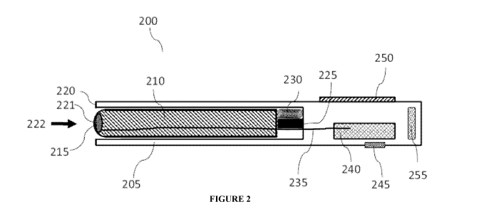
the body by hand.
[0006] In a preferred embodiment, the overall shape of the device is that of
wand or pen where
the outer member also provides a means for being held to the skin surface by a
clinician
performing the assessment. Other means, such as straps, Velcro, belts or a
layer of medical
adhesive that immobilize the device with respect to the skin surface, are also
readily conceivable.
Contained within the device, either in the outer or inner member, depending on
the overall
configuration, are necessary power sources, e.g., battery, switches,
mechanical force actuators or
springs to transiently move the inner member, and electronic circuitry and
sensors configured
for obtaining capillary blood measurements. In certain instances, one or more
functions, e.g.,
data analysis circuitry, power, data display, photonic light sources and
sensors, and other
components and devices, may be located in a separate portion of the device
connected to the
inner and outer member portion by means of electrical wires and/or fiber
optics.
[0007] Data and analysis from the device may be displayed on a small screen
located on the
outside of the outer aspect of the outer member in a preferred embodiment. In
other
embodiments, such data may be transmitted either wirelessly or via electrical
connection to
adjacent data receiving devices for display, storage and further analysis.
[0008] Provided herein, in one aspect, is a method to detect a change in blood
microcirculation,
the method comprising (a) reversibly applying an external force locally to a
skin region for a
duration of time suitable to alter blood perfusion in the skin region; (b)
using one or more
photonic excitation sources and one or more detectors to measure one or more
blood flow
parameters in response to the external force, before, during and/or after
application of said
external force; (c) analyzing and quantifying the one or more blood flow
parameters; and (d)
comparing the one or more blood flow parameters to a data set to determine the
absence or
presence of a disease state. In some embodiments, the one or more photonic
excitation sources
and one or more detectors to measure one or more blood flow parameters in
response to the
external force, are used before the application of the external force. In some
embodiments, the
one or more photonic excitation sources and one or more detectors to measure
one or more
blood flow parameters in response to the external force, are used during the
application of the
external force. In some embodiments, the one or more photonic excitation
sources and one or
2
CA 02965866 2017-04-25
WO 2015/085240 PCT/US2014/068909
more detectors to measure one or more blood flow parameters in response to the
external force,
are used after the application of the external force. In some embodiments, the
one or more
photonic excitation sources and one or more detectors to measure one or more
blood flow
parameters in response to the external force, are used before and during the
application of the
external force. In some embodiments, the one or more photonic excitation
sources and one or
more detectors to measure one or more blood flow parameters in response to the
external force,
are used before and after the application of the external force. In some
embodiments, the one or
more photonic excitation sources and one or more detectors to measure one or
more blood flow
parameters in response to the external force, are used during and after the
application of the
external force. In some embodiments, the one or more photonic excitation
sources and one or
more detectors to measure one or more blood flow parameters in response to the
external force,
are used before, during and after the application of the external force. In
one embodiment, the
data set comprises measured blood flow parameters of at least one skin region
in response to an
external force, wherein at least one skin region is a reference skin region. A
reference skin
region includes a skin region having or not having a disease state. In one
embodiment, the
disease state is cancer. An exemplary cancer is skin cancer. Skin cancer
includes stages 0, 1, 2, 3
and 4 of skin cancer. In one embodiment, the method to detect a change in
blood
microcirculation is performed on both an area of skin of an individual
suspected of having a
disease, e.g., melanoma, and an area of skin of the same individual which is
known to not have
the disease, e.g., the reference or control skin region. In another
embodiment, a reference region
is a region of skin of another individual, wherein the reference region has or
does not have a
disease state.
[0009] Provided herein, in one aspect, is a device for measuring blood
microcirculation, the
device comprising (a) a means to provide an external force to a skin region,
wherein the external
force is sufficient in pressure and duration to alter blood perfusion in the
skin region; and (b) a
sensor comprising a photonic excitation source and a photonic detector,
wherein the sensor is
configured to measure one or more blood flow parameters prior to, during,
and/or after
application of an external force to the skin region. In one embodiment, the
photonic detector
measures an applied photonic energy absorption by a component of blood. In one
embodiment,
the photonic detector is an imaging detector. In one embodiment, photonic
energy is delivered to
and collected from one or more areas of the skin region using optical fibers.
In one embodiment,
photonic energy is delivered to an area of the skin region from the photonic
excitation source. In
another embodiment, photonic energy is detected from an area of the skin
region with the
photonic detector. In one embodiment, the sensor comprises a plurality of
photonic detectors,
3
CA 02965866 2017-04-25
WO 2015/085240 PCT/US2014/068909
wherein each photonic detector is located at a different distance from the
photonic excitation
source than another photonic detector.
[0010] In one aspect, provided herein is a device for measuring blood
microcirculation, the
device comprising a means to provide an external force to a skin region,
wherein the external
force is sufficient in pressure and duration to alter blood perfusion in the
skin region; and a
sensor comprising a photonic excitation source and a photonic detector,
wherein the sensor is
configured to measure one or more blood flow parameters prior to, during,
and/or after
application of an external force to the skin region.
[0011] The photonic detector measures an applied photonic energy absorption by
a component
of blood. In one embodiment, the photonic detector is an imaging detector. A
photonic energy
can be delivered to and collected from one or more areas of the skin region
using optical fibers.
[0012] In another embodiment, the sensor comprises a plurality of photonic
detectors, wherein
each receiver for a photonic detector is located at different distances from
the emission location
of the photonic excitation source of the sensor.
[0013] In one aspect, a means to provide an external force to a skin region
comprises an inner
member configured to move relative to an outer member, allowing for the
application of variable
pressure to the skin region.
[0014] In another aspect, a device is configured to measure one or more blood
flow parameters
of an area of the skin region, wherein the area is equivalent to or greater
than 0.100 mm in
diameter.
[0015] A device can be configured to measure one or more blood flow parameters
of an area of
the skin region, wherein the area is between about 1 mm and about 5 mm in
diameter.
[0016] A device can also be configured to measure one or more blood flow
parameters of an
area of the skin region, wherein the area is between about 1 mm and about 30
mm in diameter, 5
mm and about 30 mm in diameter, between about 5 mm and about 25 mm in
diameter, between
about 5 mm and about 20 mm in diameter, between about 5 mm and about 15 mm in
diameter or
between about 5 mm and about 10 mm in diameter, between about 10 mm and about
20 mm in
diameter, between about 1 mm and about 10 mm in diameter, between about 1 mm
and about 20
mm in diameter or between about 10 mm and about 30 mm in diameter.
[0017] A photonic excitation source can emit light at wavelengths below 400
nm, between 400
nm and 450 nm, between 450 nm and 500 nm, between 500 nm and 550 nm, between
550 nm
and 600 nm, between 600 nm and 650 nm, between 650 nm and 700 nm, or above 700
nm.
[0018] In one aspect, the inner member comprises a convex, concave or non-
planar surface for
exerting pressure on the skin.
4
CA 02965866 2017-04-25
WO 2015/085240 PCT/US2014/068909
[0019] Also provided herein is a method to detect a change in blood
microcirculation,
comprising: reversibly applying an external force locally to one or more skin
regions for a
duration of time sufficient to alter blood perfusion in the skin region;
providing one or more
photonic excitation sources and one or more photonic detectors to measure one
or more blood
flow parameters in response to the external force before, during and/or after
application of said
external force; analyzing and quantifying the one or more measured blood flow
parameters from
said one or more regions of the skin; assessing said blood flow parameters to
identify blood flow;
and comparing the blood flow to one or more other assessments to determine the
presence of a
disease state. In some embodiments, the one or more photonic excitation
sources and one or
more detectors to measure one or more blood flow parameters in response to the
external force,
are used before the application of the external force. In some embodiments,
the one or more
photonic excitation sources and one or more detectors to measure one or more
blood flow
parameters in response to the external force, are used during the application
of the external force.
In some embodiments, the one or more photonic excitation sources and one or
more detectors to
measure one or more blood flow parameters in response to the external force,
are used after the
application of the external force. In some embodiments, the one or more
photonic excitation
sources and one or more detectors to measure one or more blood flow parameters
in response to
the external force, are used before and during the application of the external
force. In some
embodiments, the one or more photonic excitation sources and one or more
detectors to measure
one or more blood flow parameters in response to the external force, are used
before and after
the application of the external force. In some embodiments, the one or more
photonic excitation
sources and one or more detectors to measure one or more blood flow parameters
in response to
the external force, are used during and after the application of the external
force. In some
embodiments, the one or more photonic excitation sources and one or more
detectors to measure
one or more blood flow parameters in response to the external force, are used
before, during and
after the application of the external force.
[0020] In one aspect, the disease state is cancer. Cancer, in some instances
can be skin cancer
that is benign or malignant. In other instances, the cancer is metastatic.
[0021] In another aspect, the disease state is hypercholesterolemia, Alzheimer
disease, carpal
tunnel syndrome, schizophrenia, hypertension, renal disease, type 2 diabetes,
peripheral vascular
disease, atherosclerotic coronary artery disease, heart failure, systemic
sclerosis, obesity,
primary aging, sleep apnea, neonatal & adult sepsis, wound healing, or a
combination thereof.
[0022] In the methods described herein, the one or more other assessments can
comprise blood
flow parameters measured in response to an external force applied to a skin
region or regions.
CA 02965866 2017-04-25
WO 2015/085240 PCT/US2014/068909
In one embodiment, the skin region comprises a lesion suspicious for cancer.
In some instances,
the reference skin region does not have cancer.
[0023] In such methods, the blood flow parameters are analyzed and quantified.
[0024] Analyzing the one or more measured blood flow parameters comprises
utilizing multi
exponential decay and rise functions; and life time distributions.
[0025] Assessing blood flow parameters relative to one or more other
assessments can comprise
comparing signal lifetimes and lifetime distributions obtained from the skin
region with a
reference skin region.
[0026] Analyzing the one or more measured blood flow parameters can comprise
determining
temporal relationships and correlations between signals acquired from a
plurality of photonic
detectors, where each receiver for a photonic detector is located at a
different distance from the
emission of the photonic excitation source.
[0027] Analyzing the one or more measured blood flow parameters can comprise
determining
temporal relationships and correlations between signals acquired from a
plurality of photonic
detectors at different wavelengths emitted from the photonic excitation
source.
[0028] In such methods, the one or more blood flow parameters can provide a
pressure-induced
hemodynamic profile of the skin region, wherein pressure-induced vasodilation
is determined
from the shape of the pressure-induced hemodynamic profile, and wherein the
pressure-induced
vasodilation is indicative of the presence of the disease state.
[0029] The methods can further comprise performing hemodynamic analyses on a
plurality of
skin region locations, wherein the hemodynamic analysis of each location is
compared to
another location to determine or compare disease status.
[0030] Also provided herein is a method to detect changes in blood
microcirculation,
comprising: reversibly altering the temperature of one or more skin regions
for a duration of
time; using one or more photonic excitation sources and one or more photonic
detectors to
measure one or more blood flow parameters in response to the temperature
alteration before,
during and/or after alteration of the temperature of the one or more skin
regions; analyzing and
quantifying the one or more measured blood flow parameters from the one or
more areas of the
skin; and assessing said blood flow parameters to identify blood flow, and
comparing the blood
flow to one or more other assessments to determine the presence of a disease
state. In some
embodiments, the one or more photonic excitation sources and one or more
detectors to measure
one or more blood flow parameters in response to the external force, are used
before the
application of the external force. In some embodiments, the one or more
photonic excitation
sources and one or more detectors to measure one or more blood flow parameters
in response to
the external force, are used during the application of the external force. In
some embodiments,
6
CA 02965866 2017-04-25
WO 2015/085240 PCT/US2014/068909
the one or more photonic excitation sources and one or more detectors to
measure one or more
blood flow parameters in response to the external force, are used after the
application of the
external force. In some embodiments, the one or more photonic excitation
sources and one or
more detectors to measure one or more blood flow parameters in response to the
external force,
are used before and during the application of the external force. In some
embodiments, the one
or more photonic excitation sources and one or more detectors to measure one
or more blood
flow parameters in response to the external force, are used before and after
the application of the
external force. In some embodiments, the one or more photonic excitation
sources and one or
more detectors to measure one or more blood flow parameters in response to the
external force,
are used during and after the application of the external force. In some
embodiments, the one or
more photonic excitation sources and one or more detectors to measure one or
more blood flow
parameters in response to the external force, are used before, during and
after the application of
the external force.
[0031] Analyzing the one or more measured blood flow parameters can comprise
quantifying
amplitudes, temporal gradients and temporal shapes of hemodynamic profiles.
[0032] In one aspect, the disease state is cancer. Cancer, in some instances
can be skin cancer
that is benign or malignant. In other instances, the cancer is metastatic.
[0033] In another aspect, the disease state is hypercholesterolemia, Alzheimer
disease, carpal
tunnel syndrome, schizophrenia, hypertension, renal disease, type 2 diabetes,
peripheral vascular
disease, atherosclerotic coronary artery disease, heart failure, systemic
sclerosis, obesity,
primary aging, sleep apnea, neonatal & adult sepsis, wound healing, or a
combination thereof.
BRIEF DESCRIPTION OF THE DRAWINGS
[0034] The novel features of the invention are set forth with particularity in
the appended claims.
A better understanding of the features and advantages of the present invention
will be obtained
by reference to the following detailed description that sets forth
illustrative embodiments, in
which the principles of the invention are utilized, and the accompanying
drawings of which:
[0035] Figure 1 is illustrative of an embodiment of a skin perfusion
monitoring device.
[0036] Figure 2 is illustrative of an embodiment of a skin perfusion
monitoring device depicting
inner and outer members.
[0037] Figures 3A-B are illustrative of one configuration of an outer member
of a skin perfusion
monitoring device. Figure 3A is a cross sectional view where the disposable
component 304 is
configured to seat or guide the attachment of outer member component 301
through its bowl like
structure. Figure 3B is an end-on view; the structure of disposable component
304 has an
opening enabling the inner member 302 to traverse through the outer member
components and
7
CA 02965866 2017-04-25
WO 2015/085240 PCT/US2014/068909
thereby contact the desired skin region (not shown) in order to accomplish
blood removal and
blood flow measurements.
[0038] Figures 4A-B are illustrative of a configuration of an outer member of
a skin perfusion
monitoring device having a handle. Figure 4A: movement of slide button 403 in
direction of
arrow 420 will cause pin 414 to move down track 409 thereby causing spring 404
to move inner
member downward in direction indicated by arrow 421, a translational motion.
Distance of
movement of inner member 401 into the skin and tissue 408 may be attributable
to the strength
(force) exerted by spring 404 and the relative stiffness or resistance to
compression offered by
skin and tissue 408. The result of such translational motion is shown in
Figure 4B. To return
the inner member back to the initial state and to enable the tissue to
decompress, slide button
403 may then be moved in the direction shown by arrow 416 in Figure 4B.
[0039] Figure 5 is illustrative of one configuration of an inner member of a
skin perfusion
monitoring device.
[0040] Figure 6 exemplifies sensor elements of an inner member of a skin
perfusion monitoring
device.
[0041] Figures 7A-B are illustrative of a sensor of the inner member of a skin
perfusion
monitoring device, wherein the sensor comprises a plurality of detectors and
one light source.
An example of the convex shape of the inner member structure is shown in
Figure 7A. Figure
7B illustrates this point by presenting an array of photodetection elements
703 spaced about a
single photonic source 702.
[0042] Figure 8 is illustrative of a sensor of the inner member of a skin
perfusion monitoring
device, wherein the sensor has an imaging capability.
[0043] Figure 9 is an exemplary illustration of electronic circuitry elements
enabling operation
of a skin perfusion monitoring device.
[0044] Figure 10 exemplifies representative data obtained using a skin
perfusion monitoring
device as described herein.
[0045] Figures 11A-C exemplify representative data from (Figure 11A) normal
skin; (Figure
11B) a benign nevus, i.e., a mole; and (Figure 11C) a confirmed basal cell
carcinoma, BCC
using a skin perfusion monitoring device.
[0046] Figures 12A-D are illustrative of pressure-induced hemodynamics with
wavelength of
light emitted from a skin perfusion monitoring device. Figures 12 A-D
illustrate that signal
dynamics are dependent on the wavelength of light used. Figure 12C shows that,
in general, both
signal rise and recovery dynamics is slower at shorter interrogation
wavelength (405 nm) as
compared to dynamics at 590 nm (Figure 12A, Figure 12B) or 660 nm (Figure
12D).
8
CA 02965866 2017-04-25
WO 2015/085240 PCT/US2014/068909
[0047] Figure 13 provides results from a clinical study on human subjects
illustrating
differences in one pressure-induced hemodynamic parameter (a refill time
constant) in normal
tissue, benign moles and skin cancers measured using a skin perfusion
monitoring device.
[0048] Figure 14 is illustrative of exemplary data obtained using a skin
perfusion monitoring
device under temperature perturbation.
DETAILED DESCRIPTION OF THE INVENTION
[0049] Provided herein, in various aspects, are methods and devices for
mechanical
displacement of blood from a desired skin region followed by the reperfusion
of blood into this
region.
[0050] Skin microcirculation has been considered an accessible and potentially
representative
vascular bed to evaluate and understand the mechanisms of microvascular
function and
dysfunction. Vascular dysfunction (including impaired endothelium-dependent
vasodilation)
induced by different pathologies is evident in the cutaneous circulation. It
has been suggested
that the skin microcirculation may mirror generalized systemic vascular
dysfunction in
magnitude and underlying mechanisms. Furthermore, minimally invasive skin-
specific
methodologies using laser systems make the cutaneous circulation a useful
translational model
for investigating mechanisms of skin physiology and skin pathophysiology
induced either by
skin disease itself or by other diseases such as vascular, rheumatologic, and
pneumologic
diseases. To date, the skin has been used as a circulation model to
investigate vascular
mechanisms in a variety of diseased states, including hypercholesterolemia,
Alzheimer disease,
carpal tunnel syndrome, schizophrenia, hypertension, renal disease, type 2
diabetes, peripheral
vascular disease, atherosclerotic coronary artery disease, heart failure,
systemic sclerosis,
obesity, primary aging, sleep apnea, neonatal & adult sepsis, wound healing,
or a combination
thereof
[0051] Prior devices described suffer from the absence of adequate reference
(control) signal
making them sensitive to the type of tissue, physiological state and
environmental parameters
(e.g., temperature) which introduces significant error due to biological
variability and requires
complicated calibration and parameterization procedures. Examples include one
such device as
described by Howell (US Patent No. 3,698,382) wherein a platform system
provides varying
pneumatic pressure to a housing that is placed upon the skin. Within the
housing are optical
sensors intended to enable the determination of blood refill rates. As
pneumatic pressure is
varied, an assessment of capillary refill rate is then made using the optical
sensors present within
the device. Alternatively, Shani and Shavit (US Patent No. 6,685,635) describe
a system having
an external housing through which pressure is applied resulting in removal of
blood from the
9
CA 02965866 2017-04-25
WO 2015/085240 PCT/US2014/068909
depressed body region. As pressure is transitorily applied to the external
housing, capillary
blood refill is assessed using sensors located within the structure of
housing. They also report
the use of a temperature sensor to improve determination of skin capillary
state and overall
physiological status. A somewhat different approach is described by Messerges
and Hutchinson
(US Patent No. 8,082,017) which combines pulse oximetry with capillary refill
time assessment.
This device is designed to be placed upon the end of a patient's appendage,
e.g., a finger or a toe.
When affixed to the patient, one member of the device is located on one side
of the appendage
and a second member is located on the opposing side of the appendage. Pressure
resulting in
blood loss is then accomplished by an actuator located in one hinge resulting
in both members
compressing the intervening tissues.
[0052] None of these devices are specifically constructed as to enable a local
determination of
skin (capillary) blood perfusion enabling the definition of cancerous from non-
cancerous skin
tissue. That is, cancerous or precancerous lesions are often of the dimension
of a few millimeters
or less. Moreover, the described devices do not have a suitable shape to
enable efficient
displacement of blood from the area of interest.
[0053] The methods and devices disclosed herein overcome the shortcomings of
the prior
devices which is capable of determining with high spatial resolution of
cutaneous blood,
including relative capillary, displacement and refill rates over closely
spaced area of skin, e.g.,
within a mole or suspect cancer growth as compared to an adjacent skin surface
must be
constructed towards this aim and dimensioned accordingly.
[0054] In some embodiments, displacement of blood results from a transitory
pressure applied
to the skin by one or more solid structures of a device, such as a device
described herein,
pressing on the skin region. In one embodiment, the device comprises an inner
member and an
outer member. In another embodiment, the structure utilized for pressure
application is the inner
membrane of the device. Reperfusion of blood into the skin region results upon
the cessation of
transitory pressure, due to the release of the compressive force. In an
exemplary embodiment,
the device utilized for pressure application comprises one or more sensors.
The one or more
sensors located, in some instances, within the structure utilized for pressure
application (in some
instances, an inner member) provide measurements of skin blood flow at one or
more instances
during performance of blood perfusion events (for example, no pressure,
pressure, cessation of
pressure). Data from such measurements can be employed for the determination
of one or more
parameters of blood flow dynamics (generally referred to as hemodynamics).
Hemodynamic
parameters, in various embodiments, correlate to a disease state. In one
embodiment, the disease
state relates to the physiology of the individual at the site of measurement,
e.g., a skin cancer
lesion. In another embodiment, a hemodynamic parameter is reflective of the
health of an
CA 02965866 2017-04-25
WO 2015/085240 PCT/US2014/068909
individual as a whole, e.g., cardiovascular status. In another embodiment, a
hemodynamic
parameter is reflective of the health of an individual with respect to, for
example,
hypercholesterolemia, Alzheimer disease, carpal tunnel syndrome,
schizophrenia, hypertension,
renal disease, type 2 diabetes, peripheral vascular disease, atherosclerotic
coronary artery
disease, heart failure, systemic sclerosis, obesity, primary aging, sleep
apnea, neonatal & adult
sepsis, wound healing, or a combination thereof
[0055] Definitions
[0056] A malignant cancer is a cancer that has undergone characteristic
anaplasia with loss of
differentiation, increased rate of growth, invasion of surrounding tissue, and
is capable of
metastasis.
[0057] Metastatic cancer is a cancer at one or more sites in the body other
than the site of origin
of the original (primary) cancer from which the metastatic cancer is derived.
[0058] A tumor that does not metastasize is referred to as "benign".
[0059] There are several types of cancer that start in the skin. The most
common types are basal
cell carcinoma and squamous cell carcinoma, which are non-melanoma skin
cancers. Actinic
keratosis is a skin condition that sometimes develops into squamous cell
carcinoma.
Non-melanoma skin cancers rarely spread to other parts of the body. Melanoma
is more likely to
invade nearby tissues and spread to other parts of the body.
[0060] A melanoma is a malignant tumor of melanocytes which are found
predominantly in skin
but also in the bowel and the eye (uveal melanoma). It is one of the rarer
types of skin cancer but
causes the majority of skin cancer related deaths. Malignant melanoma is a
serious type of skin
cancer caused by uncontrolled growth of pigment cells, called melanocytes.
Melanomas also
include, but are not limited to, a choroidea melanoma, malignant melanomas,
cutaneous
melanomas and intraocular melanomas.
[0061] Melanoma may be divided into the following types: Lentigo maligna,
Lentigo maligna
melanoma, superficially spreading melanoma, acral lentiginous melanoma,
mucosal melanoma,
nodular melanoma, polypoid melanoma, desmoplastic melanoma, amelanotic
melanoma,
soft-tissue melanoma, and uveal melanoma. Melanoma stages are as follows:
[0062] Stage 0 ¨ melanoma in situ (Clark Level I).
[0063] Stage I/II ¨ invasive melanoma: Tla: less than 1.00 mm primary, without
ulceration,
Clark Level II-III; T lb: less than 1.00 mm primary, with ulceration or Clark
Level IV-V; and
T2a: 1.00-2.00 mm primary, without ulceration.
[0064] Stage II ¨ High Risk Melanoma: T2b: 1.00-2.00 mm primary, with
ulceration; T3a:
2.00-4.00 mm primary, without ulceration; T3b: 2.00-4.00 mm primary, with
ulceration; T4a:
11
CA 02965866 2017-04-25
WO 2015/085240 PCT/US2014/068909
4.00 mm or greater primary without ulceration; and T4b: 4.00 mm or greater
primary with
ulceration.
[0065] Stage III ¨ Regional Metastasis: N1: single positive lymph node; N2: 2-
3 positive lymph
nodes or regional skin/in-transit metastasis; and N3: 4 positive lymph nodes
or lymph node and
regional skin/in transit metastases.
[0066] Stage IV ¨ Distant Metastasis: Mla: Distant Skin Metastasis, Normal
LDH; M lb: Lung
Metastasis, Normal LDH; and Mlc: Other Distant Metastasis OR Any Distant
Metastasis with
Elevated LDH.
[0067] In one embodiment, the methods described herein identify a melanoma or
a likelihood,
or risk of melanoma.
[0068] Additional steps or variations in the general method, such as the use
of stepwise or
incremental pressure, series of rapid pressures and releases, series of
measurements, etc., may be
employed within the overall scope of the devices and methods disclosed herein.
Accordingly,
the scope of the present disclosure is not limited to those series of steps or
actions presented and
exemplified here.
[0069] Figure 1 presents an illustration of an exemplary blood perfusion
device, 100. As shown,
device 100 has inner member 101 enclosed substantially within a first
component 102. The first
component 102 is associated with a support component 104 and a base component
105.
Collectively, components 102, 104 and 105 comprise the outer member of device
100 and are
presented to generally indicate that an outer member of a device may be
comprised of multiple
components having a variety of functions. For example, the outer membrane
component 102 is
useful as a guide for inner member 101. As another example, outer membrane
component 105 is
useful to orient the device for positioning on a specific region of a skin
surface 107. As another
example, outer member component 104 is useful as a support, enabling the
device to house
electronics (not shown) and/or photonic sources (not shown) useful for device
operation.
[0070] Also shown in Figure 1 is wire 109 extending from outer member
component 104. Wire
109 is shown to generally illustrate the functions that may be usefully
present in such structures
in various embodiments associated by having one or more external connections
between device
and one or more additional structures, etc. For example, wire 109 may
represent an electrical
power cord enabling the supply of power to device electrical components.
Alternatively, the
wire may represent a fiber optic cable transferring photonic energies to and
from device 100 to
an external unit having photonic energy sources and/or photonic energy
receivers with
associated electronics enabling signal analysis and processing. A third
possibility is that wire
109 represents a data transference means, e.g., USB cable, between device 100
and a separate
12
CA 02965866 2017-04-25
WO 2015/085240 PCT/US2014/068909
unit, e.g., a laptop computer or cell phone, enabling data analysis, device
operational commands,
and display of processed results.
[0071] Returning to inner member 101, inner member 101 may be configured to
enable
measurements of skin blood flow through one or more sensors (not shown)
located in inner
member 101 at component end 108, wherein 108 is a point of contact with a skin
surface 107.
Also contained within inner member 101 may be additional electronics, etc. to
support the
measurement of skin properties through one or more sensors located in end 108,
and electrical
wires, photonic guides and/or other forms of contacts enabling transference of
power, data
and/or photonic information between inner member 101 and outer member
components 102 and
104.
[0072] Also shown in this general illustration is a spring 106 and a mounting
ring 103 on inner
member 101 that are presented to generally indicate the need to provide a
means of exerting a
small force on the skin surface 107 through the movement of inner member 101
towards and
against the skin surface, as indicated by arrow 110. The purpose of this small
force is to
maintain the contact of the inner ring with the skin through all phases of
measurement. Action of
the spring 106 located between and in contact with inner member 101 and with
mounting ring
103 results in a depressive force on the skin 107 through the pressure of the
contact by end 108.
The spring constant of spring 106 is chosen to be small enough so that the
depression of the skin
surface does not result in a forcing of blood from skin capillaries located in
the immediate
vicinity of this applied force.
[0073] To enable application of force to the inner member, the component 102
may contain any
means of applying the force to the inner member, i.e., an actuator or force
transducer, for
example: an electromagnet (solenoid), a linear motor, or pneumatic or
hydraulic control. To be
usefully applied, in an exemplary feature of the device, an opposing force
resulting from the
contact of inner member end 108 against the skin 107 is resisted by the
structure and positioning
of components of the outer member, collectively 105, 104 and 102. In one
embodiment, it is
desired that the applied force results in a depression of the skin surface
(and associated removal
of blood) rather than a lifting of the device or portions thereof from the
skin; accordingly, the
structures and operation of devices described herein are, in many instances,
configured to enable
this function. In this instance, device 100 may be held against skin surface
by placement of one
or more fingers on the outer top surface of outer member component 105,
thereby through the
strength of the hand enabling the depressive force exerted by inner member 102
to be
successfully accomplished. In an exemplary device, the end of the inner member
108 is not
subject to motion artifacts after the pressure to the inner member is
released. Therefore the end
of the inner member 108, containing sensing elements may be permanently
attached to the inner
13
CA 02965866 2017-04-25
WO 2015/085240 PCT/US2014/068909
member 101 or it may not be attached permanently. For example, 108 could be a
component
shown in Figure 7 comprising light sources and detectors. As another example,
108 could be a
component shown in Figure 5; in this case the inner member 101 would apply
force to feature
108 and then be retracted from 108, leaving 108 attached to the skin by means
of adhesive
forces. In the case 108 is not permanently attached to inner member 101, it
could be connected
by additional cables to enable electronics needed for feature 108 operation.
[0074] Figure 2 provides an illustration of an embodiment of a device as
provided herein. As
shown, device 200 has an outer member 205 that substantially encloses inner
member 210,
except for an end opening generally indicated by arrow 222. A blood flow
sensor head 215 is
positioned at the end of inner member 210. Positioned between outer member 205
and inner
member 210 is an actuator (e.g., a solenoid) 225 and a spring 230, enabling
controlled
piston-like movement of inner member 210 within outer member 205. Actuator 225
and spring
230 are intended to provide both extensive force (actuator 225) and retractive
force (spring 230)
to enable operation of the device, for example, inner member 210 exerts a
depressive force on a
skin region as the outer member 205 is positioned against the skin by hand. It
should be noted
that spring 230 is so configured as to enable retraction of the extended inner
member while
allowing continuous contact between the skin and the inner member.
[0075] In alternate embodiments, the outer member 220 may be affixed to skin,
e.g., with use of
a medical adhesive, or with belts, Velcro or straps to constrain its position
and orientation with
respect to skin surface. In certain instances, the structure affixing the
outer member to the skin
may itself be a portion of the device, e.g., as a separable disposable
structure having an adhesive.
[0076] In yet other alternate embodiments, a plurality of inner members 210,
sensor heads 215
and/or sensors located within sensor head 215 may be incorporated within
device 200 in order to
provide a plurality of measurements at one time.
[0077] Sensor head 215 at the end of inner member 210 is configured with one
or more sensors
(not shown) to enable measurement of skin physiological parameters when end
opening 222 of
device 200 is positioned against the skin. In correct use, device end opening
surfaces 220 and
221 are positioned to be substantially in contact with the skin in order to
enable pressure
variation to be applied to the immediate skin area and measurements of skin
blood perfusion to
be obtained while doing so. Sensor signals so obtained are conveyed between
electronics 240
and sensor head 215 by connector 235.
[0078] Also shown within device 200 are operating switch 245, battery 255 and
display 250 to
enable operation of device 200. Battery 255 may be replaceable, rechargeable,
or in certain
instances, power to device 200 may be supplied by an external power source,
e.g., electrical
outlet connected to the device.
14
CA 02965866 2017-04-25
WO 2015/085240 PCT/US2014/068909
[0079] Not shown in Figure 2 are necessary electrical and connections (e.g.,
optical connections)
between the various components of the device 200 to enable their
functionality. It will be readily
appreciated that such electrical connections as well as electronic circuitry
contained within
electronics 240 are well understood by those skilled in the art of electronic
circuitry.
[0080] It may be readily appreciated that the control of mechanical motions
and photonic signal
delivery and acquisition may be accomplished in a variety of ways and are not
constrained to the
examples and device component configurations presented here.
Device Operation
[0081] In an exemplary mode of operation, a device of the present disclosure,
for example, such
as one illustrated in Figure 2, is first positioned on a region of mammalian
skin wherein the area
to be measured is in contact with end surface 221 of inner member 210. At
least a portion of the
corresponding end surface 220 of outer member 205 thereby is also caused to
come into contact
with skin surface. In addition, inner member 210 is so constructed to aid in
the shielding of
photonic sensors contained in sensor head 215 from stray or non-intended
energy sources, e.g.,
stray light.
[0082] The operator of the device then activates the device using switch 245.
Activation results
in electrical power being supplied from battery 255 to electronics 240 and
other components,
e.g., actuator 225 and display 250, as directed by electronics 240. Upon
activation, inner
member 210 is mechanically moved in an outward direction relative to outer
member 205, e.g.,
by actuator 225. In various embodiments, the force utilized to move inner
member 210 may be
by means other than an actuator, e.g., electromagnet, electroactive polymers,
or pneumatic
pressure supplied by an external source, manual means, etc., and the scope of
the present
disclosure is not constrained to any one means of applying a translational
force to inner member
210.
[0083] The translational motion of inner member 210, while outer member 205
remains
positioned in substantial contact with the skin at surface 220, results in
mechanical force being
applied to the immediate skin surface. As a result, skin vasculature in the
immediate area is
compressed, resulting in an outflow of blood from the compressed region. It is
readily
understood that sensor head 215 is advantageously positioned at the end of
inner member 210 to
perform measurements upon local skin blood flow throughout this process.
[0084] In many implementations, it is a desired feature that the structure or
mode of operation of
the inner member may be such that blood removal from the compressed skin
region is facilitated.
This may include the shape of the surface that contacts the skin being, e.g.,
convex rather than
planar such that blood is progressively moved from the region as more of the
inner member
contacts the skin. Examples of the convex shape of the inner member structure
are shown in
CA 02965866 2017-04-25
WO 2015/085240 PCT/US2014/068909
Figure 2, Figure 5, Figure 7A and Figure 8. For example, Figure 2 shows the
rounded end 221 of
inner member 210.
[0085] In an alternative embodiment, the device or inner member may be applied
at an angle to
the general plane of the skin and then shifted to an orientation generally at
right angles to the
skin during the application of pressure. Additional means or inner membrane
shapes facilitating
blood removal are conceivable and the scope of the present disclosure is not
limited to these
examples.
[0086] As alternate applications, the structure or mode of operation of the
inner member can be
chosen to increase the amount of blood in the compressed region. This may
include having a
shape of the surface of the inner member in contact with skin that is concave
such that blood is
trapped by the edges and progressively pushed toward the center of the
compressed area where
the sensing area is located.
[0087] After transitioning a certain distance, movement by inner member 210
relative to outer
member 205 ceases. This distance may be a predetermined distance, e.g., a
predetermined
distance relative to outer member 205. The travel of inner member of this
predetermined
distance may be governed by a variety of means, e.g., through actions of
actuator 225 under the
control of one or more sensors able to discern distance travelled or
electronic timing present
within electronics 240.
[0088] In alternate embodiments, the distance traversed by inner member 210
may be governed
by one or more sensors able to discern one or more physiological parameters
associated with the
desired outcome of motion, e.g., the partial or complete removal of blood or
increase in the
amount of blood in the skin region under compression and thereby facilitate
automated operation
of the device. Examples of such sensors include pressure transducers so
positioned within device
200 or on the device 200 surface as to sense the pressure applied by inner
member 210 to skin
surface. Such sensors may be present on the inner member 210, outer member 205
or both, the
scope is not constrained to any one location. The scope of the present
disclosure is not
constrained to any one form or method of determining distance traversed.
[0089] Such distance sensors may also include those sensors utilized for
making determinations
of the blood present in the compressed region. That is, by a determination of
change in amount
of blood (e.g., blood removal), a feedback signal from said blood sensors to
electronics may then
be used to govern the means used to move inner member 210, e.g., control the
actuator 225.
[0090] In addition, pressure sensors located on the outer member in contact
with the skin surface
may be utilized to ensure that outer member 205 remains in substantial contact
with skin surface
but is not applying by itself an undesired level of pressure to the local skin
area. Readings from
16
CA 02965866 2017-04-25
WO 2015/085240 PCT/US2014/068909
such sensors may be sent to display 250 to enable the individual using the
device to more
appropriately position the device on the skin surface.
[0091] Upon ceasing movement, inner member 210 may remain stationary relative
to outer
member 205 and thereby hindering local blood flow in the compressed region for
either a short
duration, e.g., < 1 second, or longer. Longer durations may advantageously
enable the
establishment of a plurality of measurements to which either prior or
subsequent measurements
may be compared.
[0092] After the desired time period has passed, the force applied to the
inner member 210 is
turned off resulting in the inner member being rapidly retracted back into
outer member 205. In
preferred embodiments, this retraction may be the result of cessation of
actuator activation and
resulting from tissue-compression force pushing the inner member 210 back
towards original
position with a spring 230 useful in maintaining the surface of inner member
221 to remain in
effectively continuous contact with the skin. In alternate embodiments, other
methods for
retracting inner member 210 may be employed, e.g., vacuum or manual, and the
scope is not
constrained to any one means of moving inner member 210 back within outer
member 205.
[0093] In one embodiment, after returning the inner member 210 to the original
position relative
to outer member 205, blood measurements utilizing sensors located in sensor
head 215 may
continue for either a predetermined or arbitrary length of time. In another
embodiment, during
and after returning the inner member 210 to the original position relative to
outer member 205,
blood measurements utilizing sensors located in sensor head 215 may continue
for either a
predetermined or arbitrary length of time. The methods and devices provided
herein are not
constrained to any one measurement period.
[0094] It is a desired feature that the rate of travel and distances traversed
by inner member 210
relative to outer member 205, upon release of the applied force, does not
exceed the elasticity
present in the measured skin region and thereby cause an interruption in the
measured skin
signals. In such instances, wherein the rate of travel and distances are
congruent with the
rebound elasticity of the compressed skin region, sensor head 215 and its end
surface 221
remain in substantial contact with the skin surface throughout these motions.
[0095] In certain instances, the entire device may be pressed against the
skin, e.g., pressure
applied outer member 205 also resulting in local blood loss in the skin area
in substantial contact
with outer member 205. In such instances, useful data may be obtained by
examining the
relative effect of inner member 210 further locally compressing the skin
region in a reversible
fashion.
[0096] Alternatively, when an external force to apply pressure using outer
member 205 is
employed, the inner member 210 may be configured or commanded to not move in
relationship
17
CA 02965866 2017-04-25
WO 2015/085240 PCT/US2014/068909
to the outer unit. Such a result may be obtained by an electronic command
instructing the inner
member not to move.
[0097] In related embodiments, the device may be effectively constructed as a
single unit
whereby the inner member and outer member form effectively a single contiguous
structure. In
such embodiments, needed force (e.g., pressure) for blood removal may be
applied through a
separate mechanism, e.g., mechanical (by hand), hydraulic or pneumatic
mechanisms applied to
entire structure 200. In yet still other embodiments, in a device having an
effectively solid
structure, the device can be securely fixed to the skin region and the user
transiently applies
pressure by hand for sensing purposes.
[0098] In alternate or additional embodiments, a device comprises a plurality
of inner
members and at least one outer member. In such configurations, a plurality of
skin surfaces may
be measured in effectively a simultaneous fashion. Such plural forms of the
device may be
advantageously employed where a suspect lesion is measured during the same
measurement
period as a non-suspect (control) skin area is measured, without the extended
time period
required by sequential measurements.
[0099] In yet other configurations of the device, the outer member may have at
least one
element separable from the inner member.
[00100] In such embodiments, the outer member may be comprised of one or more
separate
components, e.g., an adhesive strip having one or more alignment marks to aid
in positioning of
the inner member and a separate ring or guiding structure to enable the
placement of the inner
member in a position in accordance with the adhesive strip alignment marks. In
yet other
embodiments, the outer member may have a conformable portion or separable
component, e.g.,
sponge or soft rubber, element that contacts the skin to promote both good
contact of the device
with the skin and to provide comfort to the user. Additional forms and types
of the structure of
the outer members are readily conceivable and therefore the scope of this
disclosure is not
restricted to the examples and configurations presented herein.
[00101] It will be readily appreciated that one or more measurements
concerning the presence
of blood in the measured region may be made at various points in the
measurement cycle. In
preferred embodiments, such measurements are made in an effectively continuous
fashion, e.g.,
once every 10 milliseconds, such that a contiguous data set describing local
blood removal and
reperfusion is obtained enabling detailed characterization of the blood flow
dynamics. Data from
one or more measurements may then be analyzed to ascertain the likelihood or
presence of a
disease state.
[00102] Exemplary elements of a blood perfusion device are described in
greater detail below.
18
CA 02965866 2017-04-25
WO 2015/085240 PCT/US2014/068909
Outer Member
[00103] Provided herein, in various aspects, is a blood perfusion device
comprising an outer
member and an inner member, wherein the device is configured to measure at
least one blood
flow parameter from a skin region. A primary function of the outer member is
to serve as a
guide or support to enable the proper positioning and operation of the inner
member. As such, in
one embodiment, the outer member has at least one surface region in
substantial contact with a
region of skin proximal to the skin area to be depressed by the inner member
and at least one
surface portion able to contact at least a portion of the inner member. In an
additional
embodiment, the outer member, once positioned against the skin region as a
first step in the
measurement process, is intended to be relatively stationary during the
remaining steps of the
measurement process, e.g., remain immobile against the skin, thereby aiding in
the guiding of
the inner member during its motions relative to the removal and reperfusion of
blood from the
measured region. Upon completion of the measurement process, the outer member
may be then
removed or lifted from the skin surface. This removal may also be coincident
with the removal
of the inner member, dependent on the exact configuration of the device.
[00104] In structure, an exemplary form of the outer member is one that (a) is
at least partially
conical or cylindrical in overall shape, wherein the inner member is enclosed
circumferentially,
at least in part, by the outer member and (b) has a surface that may contact
the inner member at
least at one location via one or more contact points. In many embodiments,
such contact points
enable the guiding of the inner member to a specific skin location for the
application of pressure.
In such embodiments, the outer member may have an opening through which the
inner member
may pass to cause the pressure necessary for blood removal from the skin
region.
[00105] In other embodiments, the outer member may have a shape other than
cylindrical or
conical, e.g., rectangular or C shaped, or even have a shape whereby the inner
member is not
substantially encircled by the outer member, e.g., the outer member is
configured as a linear rail
or guide that is configured to serve as a guide to the inner member during
inner member
operation.
[00106] In these and other embodiments, the outer member may be comprised of
separable
components. For example, at least a portion of one component of the outer
member may be a
ring or similar conical structure in contact with the inner member. A separate
component of the
outer member may be in the form of a transparent tape. In this embodiment, the
tape may serve
as an interface between the skin and the other components of the device, e.g.,
the other portions
of the outer member and the inner member.
[00107] In a related or additional embodiment, the outer member has a
separable component
that has both a guiding function as well as an adhesive function. Figure 3
presents an example of
19
CA 02965866 2017-04-25
WO 2015/085240 PCT/US2014/068909
one such embodiment. Figure 3 presents a section of a device 300 having an
outer member
component 301 in contact with inner member 302 and disposable outer member
component 304.
Disposable component 304 has adhesive 305 to facilitate the positioning and
adhesion of device
300 to the skin in a desired location. As shown in Figure 3A, a cross
sectional view, the
disposable component 304 is configured to seat or guide the attachment of
outer member
component 301 through its bowl like structure. As shown in Figure 3B, an end-
on view, the
structure of disposable component 304 has an opening enabling the inner member
302 to
traverse through the outer member components and thereby contact the desired
skin region (not
shown) in order to accomplish blood removal and blood flow measurements.
[00108] In such instances as those illustrated in Figure 3, a separable
component may be
constructed as a disposable component such that it may be employed on a single
use basis. A
desirable feature of such embodiments is that the disposable component may be
positioned onto
the skin in advance of the attachment to this disposable component by the
other portions of the
outer member by the operator.
[00109] One requirement for such separable components, e.g., a tape, collar or
other disposable
component, is that it be so constructed as to enable the operation of the
inner member, e.g., the
application of pressure to the skin by the inner member and/or the measurement
of blood within
the skin by one or more sensors located within the inner member. In those
instances wherein a
separable component, such as a tape, intervenes between the skin surface and a
device
component such as an inner member having motion and/or sensing capabilities,
in many
embodiments, it is desired that the separable component be relatively thin
(e.g., less than 0.2 mm
in thickness) and conformable or stretchable to the movements and applied
pressures by the
device component (e.g., inner member) as well as able to pass signals employed
in measurement
(e.g., the separable component is effectively transparent to the wavelengths
of light utilized for
photonic measurements). In one embodiment, separable component 304 is made of
a flexible
material that can easily compress to conform to a convex probe head shape of a
device
component (e.g., outer member).
[00110] In certain instances, an outer member is constructed to be affixed to
the skin and then
disposed of after use. In one embodiment, this disposable outer member may
also contain one or
more blood sensors, e.g., photonic sources and/or photonic receivers. In such
instances, the inner
member may serve as a mechanical means enabling pressure application for blood
displacement
and/or reperfusion from the measured region. For embodiments such as these,
sensors may be
fabricated or positioned within a tape or other separable component using one
or more methods
of construction, such as printed electronics, whereby the circuitry elements
and sensors are
effectively printed into the structure of the separable component, e.g., the
tape.
CA 02965866 2017-04-25
WO 2015/085240 PCT/US2014/068909
[00111] The outer member, in various embodiments, is configured to enable the
positioning of
the device by hand on the intended region of the skin of an individual for
blood perfusion
measurement. Accordingly, all or a portion of the outer member may be
constructed in the form
of a handle or similar structure enabling its manual placement and operation.
For example, a
device with the outer member in the shape of a pen or rod-like structure
generally sized between
about 7 centimeters and about 15 centimeters in length and from about 1
centimeter to about 4
centimeters in approximate diameter would enable clasping of the outer member
of the device
by hand for use in positioning and device operation. It would be understood
that alternate sizes
and holding arrangements are conceivable and the dimensions of the device are
not restricted to
those described here.
[00112] Alternatively or additionally, the outer member may be constructed
with differing
functional sections. That is, a portion of the outer member may be configured
to be held by a
hand, while a separate portion of the outer member is configured to interact
(e.g., as a guide
and/or as an anchoring point for forces to be applied) with the inner member.
For example, an
outer member having a handle extending at roughly a 90 angle to a roughly
conical portion of
the outer member, wherein the outer member interacts with the inner member, is
illustrated in
Figure 4.
[00113] As shown in Figure 4, device 400 has outer member components 402 and
405 and an
inner member 401. Not shown are sensors, electronics, power, etc. needed for
device operation.
However, one can readily envisage the ability to put such elements within this
device by one
skilled in the art of medical device construction. Outward motion of inner
member 401 relative
to outer member 402 is achieved through means of spring 404 contacting outer
member 402 at
base structure point 410 and contact notch structure 422 on inner member 401.
Governing the
outer motion is slide button 403 connecting to structure 415 that contains pin
414 in track 409
located in the outer member 402.
[00114] As shown in Figure 4A, movement of slide button 403 in direction of
arrow 420 will
cause pin 414 to move down track 409 thereby causing spring 404 to move inner
member
downward in direction indicated by arrow 421, a translational motion.
[00115] Distance of movement of inner member 401 into the skin and tissue 408
may be
attributable to the strength (force) exerted by spring 404 and the relative
stiffness or resistance to
compression offered by skin and tissue 408. The result of such translational
motion is shown in
Figure 4B. To return the inner member back to the initial state and to enable
the tissue to
decompress, slide button 403 may then be moved in the direction shown by arrow
416 in Figure
4B. Such movement will result in structure 415 moving pin 414 upwards against
spring 404,
thereby removing the downward force of spring 404 on inner member 401. The
release of this
21
CA 02965866 2017-04-25
WO 2015/085240 PCT/US2014/068909
downward force will enable the skin and tissue 408 to push back against inner
member 401 in
the direction of arrow 417.
[00116] In various instances, it may be desirable to facilitate this return
motion shown by arrow
417. In particular, such facilitation may enable various masses of inner
member 401 to be
employed without deleteriously affecting measurements, e.g., having too great
a mass of inner
member 401 to allow skin and tissue to decompress readily. To achieve this
return motion,
structures such as spring 407 may be employed. As shown, spring 407 extends
between outer
member 402 and inner member contact point 406. Upon outward motion of inner
member 401,
spring 407 compresses. If the strength of spring of 407 is selected to be
lesser than the strength
of spring 404, then net movement outward will occur, until skin and tissue
resistance stops this
outward movement. Upon removal of the downward force of spring 404, e.g., by
actions of slide
button 403 in the direction of arrow 416, spring 417 will aid the skin and
tissue 408 in the
movement of inner member back into the device 400.
[00117] In various embodiments, the point(s) or elements of contact between
the inner member
and outer member function to facilitate the movement of the inner member
relative to the outer
member. Such a function may be in addition to the point of contact serving as
a guide for the
inner member. To enable the relative motion of the inner member, the point(s)
of contact may be
configured in a variety of forms, e.g., in the form of a gear, an electrical
element within a
solenoid-type arrangement between the inner and outer member, a mechanical
spring attachment
point, a seal enabling the use of pneumatic pressures, or various combinations
of these or other
methods of power or force transference.
[00118] In many embodiments, the outer member is intended to be positioned and
held against
the skin at a desired body location. Upon device activation and through the
relatively stationary
positioning of the outer member (as held by hand or adhesive), the activation
of the translational
mechanism thereby enables the motion of the solid inner member to apply
pressure to the skin.
This pressure depresses the skin and tissues in the contact region and results
in the forced
removal of blood from the immediate skin region due to this applied pressure.
It is desirable in
most instances that the applied pressure exceeds the filling pressures
typically provided by the
blood vasculature to the skin capillary and arteriole networks such that blood
is prevented from
flowing into these capillary beds and is also forced from them into the
surrounding venous
system.
[00119] In various embodiments, the forces applied by and/or distances
traversed by the inner
member may be such that deeper capillary and vascular systems are affected,
e.g., resulting in
blood flow being prevented to or forced from these vascular structures. In
such instances,
22
CA 02965866 2017-04-25
WO 2015/085240 PCT/US2014/068909
additional measured data may be obtained from deeper tissue regions useful for
the diagnoses of
a disease state, e.g., the degree of skin tumor invasiveness into a deeper
tissue structure.
[00120] In other or additional embodiments, the force to enable the
translational motion of the
inner member relative to the outer member is supplied by hand and point(s) of
contact serve
purely to guide the translational motion of the inner member. That is, one
hand may hold the
outer member in position against the skin while a second hand applies force to
the inner member
causing the inner member to move. In such instances, the inner member or
structures associated
with the inner member may project through more than one opening in the outer
member, e.g.,
one opening for the point of contact with the skin and one opening for contact
with a hand or
other external force applying structure.
[00121] In still other embodiments, the outer and inner members are
immobilized relative to
each other such that force applied to the outer member results in pressure
being applied to the
skin by the inner member. In such instances, the outer member may be
structurally indistinct
from the inner member, i.e., the inner member is distinguished by the presence
of one or more
sensors and the outer member has one or more circuitry elements needed for
data measurement
and display, wherein both the inner and outer members are housed within the
same overall shell
or covering.
[00122] In various embodiments, the outer member and/or inner member may also
contain
components or structures enabling the transference of data, processed data,
power and/or sensor
(photonic) energy to and/or from a unit separate from the device. For example,
the outer member
or inner member may be configured for attachment to a USB cable enabling
transference of
measured data with a separate unit, e.g., a cell phone, for control/operation
instructions, signals,
additional data processing and display of results. In an alternate example,
the outer unit may
be configured for attachment to a fiber optic cable, enabling the transference
of photonic energy
to and from the outer member and then to the inner member and/or sensors. In
still another
example, the outer member or inner member may be configured as a tube enabling
the
transference of pneumatic power from an external pump to the outer member,
thereby supplying
a source of pneumatic force useful for the application of pressure by the
inner member against
the skin. In still other instances, the pneumatic pressure source may be in
the form of a cartridge
located in the outer member and pressures applied onto the inner member are
governed through
the use of various valves and seals.
[00123] In additional or other embodiments, the outer member or inner member
may comprise
one or more controls and/or display or alerts. Examples of these may include
one or more on/off
switches or buttons for initiating and/or operating the device, one or more
indicator lights
indicating the operational status of the device, one or more audible alerts
indicating the status of
23
CA 02965866 2017-04-25
WO 2015/085240 PCT/US2014/068909
the device or instructional activities to be performed, one or more small
displays configured to
display operational status, data and/or the results of data process, and any
combination thereof
[00124] The outer member may be constructed of a variety of materials, e.g.,
plastics, rubbers,
metals. The outer member may have electronic circuitry, batteries, lights,
displays, etc. The
exact composition of outer member materials is dependent on the nature of the
device
embodiment and functional needs. Creating such constructions are well known to
those skilled
in the art of medical device construction.
Inner Member
[00125] Provided herein, in various aspects, is a blood perfusion device
comprising an outer
member and an inner member, wherein the device is configured to measure at
least one blood
flow parameter from a skin region. In various embodiments, a primary function
of the inner
member is to mechanically exert pressure onto a desired skin or tissue region
resulting in the
forcing of blood from capillaries in the immediate vicinity of this applied
pressure, i.e., a
blanching of the skin caused by pressure. In exemplary embodiments, this
mechanical
transmission of pressure is accomplished by the movement of an effectively
solid or rigid
surface against the skin or tissue region, wherein the surface is associated
with at least a portion
of the inner member. In preferred embodiments, such pressures are those
sufficient to result in a
cessation of flow into the capillary bed and/or associated arterioles, and are
generally assumed to
be in the vicinity of 4 kPa, or in general, in excess of the internal pressure
associated with
capillary flow, e.g. approximately 4 kPa or 32 mm Hg. In alternate
embodiments, the applied
pressure may be greater, lesser or varying, in order to execute the desired
removal and
subsequent reperfusion of blood in the measured region. In one embodiment, the
pressure is
from about 1 kPa to about 400 kPa, from about 2 kPa to about 400 kPa, from
about 3 kPa to
about 400 kPa, from about 80 kPa to about 400 kPa, from about 5 kPa to about
400 kPa, from
about 4 kPa to about 400 kPa, from about 4 kPa to about 400 kPa, from about 1
kPa to about 350
kPa, from about 1 kPa to about 300 kPa, from about 1 kPa to about 250 kPa,
from about 1 kPa to
about 200 kPa, from about 1 kPa to about 150 kPa, from about 1 kPa to about
100 kPa, from
about 1 kPa to about 50 kPa, from about 2 kPa to about 100 kPa, from about 3
kPa to about 75
kPa, or from about 3 kPa to about 50 kPa.
[00126] In general terms, application of pressure via the motion of the inner
member against
the skin results in depression of the skin and underlying tissue. In part, the
distance or depth of
the depressed region is dependent upon the compliance or stifthess of the
tissues underlying the
immediate skin region. For example, a skin region having a bone or bone-like
structure
immediately beneath will exhibit greater resistance (after an initial
depression of a few microns
to millimeters) than a skin region overlaying soft tissues such as an
abdominal region having an
24
CA 02965866 2017-04-25
WO 2015/085240 PCT/US2014/068909
excess of subcutaneous fat. Accordingly, the present methods and devices
disclosed herein are
not restricted to any one distance of movement, and may range from fractions
of a millimeter to
several millimeters or centimeters, dependent on the device embodiment and the
skin region
examined.
[00127] In order to accomplish said desired pressures and movements of the
inner member, it is
a desirable feature of various device embodiments that the inner member be
constructed, at least
in part, in the form of a solid structure enabling the transference of applied
power and motion
from the outer member to the skin region via the inner member. Such inner
member structures
may include, but are not limited to, structures in the form of rods, closed
cylinders, spheres,
inflate-able bags (pneumatic or hydraulic), electroactive polymers configured
for translation
elongation, or structures comprised of substances capable of phase transitions
which upon
conversion, e.g., from liquid to solid, result in a change in the overall
dimensions of the
structure.
[00128] Overall, the region of skin to be compressed is desired to be of a
dimension suitable for
the measurement of cutaneous blood flow (e.g., skin capillary blood flow)
during a process
described herein (e.g., applied and/or removed pressure to the region) by the
sensor
methodology employed. Accordingly, the dimensions of the skin contact region
(and
corresponding inner member surface) are, in various embodiments, preferably
greater than that
represented by a single capillary, i.e. 6-8 microns in cross section. In many
embodiments, a
desired function of the device, in part, is the ability to distinguish between
normal capillary
networks and those associated with cancerous tissues, wherein the dimensions
of the contact
region are therefore more preferably greater than that of a single capillary.
Accordingly, in
many embodiments, the dimensions of the skin contact region and of the
corresponding surface
of the inner member are at least 0.1 mm2 in area.
[00129] In exemplary embodiments, the shape of the inner member that contacts
and
compresses the skin region (or contacts a portion of an outer member, e.g., a
tape, intervening
between the inner member and the skin region) is configured to facilitate the
movement of blood
from the compressed skin region. The present inventors have discovered that
the movement of
blood from the skin is facilitated by the use of a rounded (convex) surface to
apply pressure to
the skin, as opposed to a stiff, planar surface that tends to pool blood
within the region of
applied pressure; moreover we have demonstrated that using flat or concave
surface to apply
pressure to the skin tends to lead to the accumulation of the blood within the
region of applied
pressure. In alternate embodiments and for the diagnosis of different disease
states, it may be
advantageous to use a concave inner membrane and/or sensor surface.
CA 02965866 2017-04-25
WO 2015/085240 PCT/US2014/068909
[00130] Accordingly, in exemplary embodiments, the shape of the inner member
surface is
configured to facilitate the movement of blood from the approximate center of
the skin contact
region to the periphery as translational motion is applied. For example, the
inner member
surface has, e.g., a convex shape such as spherical or parabolic surface and
is of sufficient
rigidity to enable effective pressure to be applied. In various embodiments,
this movement of
blood from the region may be further facilitated by a slight rocking motion or
inclination of the
inner member during compression.
[00131] In an additional embodiment, the inner member serves as a support or
structure
housing one or more sensors which enable the determination of blood flow in a
measured region
of skin. In an exemplary embodiment, such sensors comprise at least one source
of photonic
energy, to be applied to the skin region, and at least one means, e.g.,
waveguide, fiber optics,
etc., of receiving one or more photonic energies from the skin region. A non-
limiting example
of such an arrangement is shown in Figure 5.
[00132] Figure 5 illustrates a section of an exemplary inner member 501,
having rounded tip
506. Within inner member 501 is fiber optic 502 providing photonic energies to
a skin tissue
region 507 located within larger skin tissue region 505. Photonic energies so
delivered may
scatter and be absorbed in region 507. A portion of these energies may in turn
encounter return
fiber optics 503, conveying these photonic signals to one or more
photodetectors located
elsewhere in the device, as shown by arrows directed upward. The controlled
movement of
inner member section 501 in the direction of arrow 504 results in the
measurement of a blood
flow parameter from tissue region 507 during the conditions associated with
tissue compression
and the forcing of blood from this region.
[00133] One may readily envisage embodiments where a plurality of photonic
energy sources
and/or photonic energy signal receivers (photodetectors) are utilized to
better inspect larger skin
regions and/or comparatively assess under the same measurement cycle various
discrete skin
areas within a larger site of measurement. For example, if a desired function
of the device is to
delineate the margins of a tumor, then by use of an array of sources and
detectors, e.g.,
employing multiple fiber optic cables with multiple sources and
photodetectors, one might
effectively image the boundary or signals associated with the transition of
capillary types
associated with cancerous tissue versus normal tissue.
[00134] In order to accomplish the desired functions of the inner member, the
inner member
may be composed of a variety of materials and components. For example,
materials such as
plastics, rubbers, metals such as stainless steel, aluminum, brass, may be
employed in various
combinations in order to configure the inner member according to the
requirements of that
embodiment of the device.
26
CA 02965866 2017-04-25
WO 2015/085240 PCT/US2014/068909
[00135] In alternate or additional embodiments of the device, the inner member
can be used
alone without the outer member. In such a case the inner member would
preferably be a short,
round or square part, for example as the part 701 depicted in Figure 7. In one
embodiment, the
surface of 701 in contact with the skin comprises an adhesive coating or an
adhesive consumable
part which would maintain the surface 701 in the contact with the skin
throughout a
measurement of a blood flow parameter. The pressure would be applied manually
by, e.g.,
pressing by the hand onto the upper surface of the 701. Such configuration is
advantageous as
it reduces contributions from motion artifacts after the force is released.
Sensors
[00136] Provided herein, in various aspects, is a blood perfusion device
configured to measure
at least one blood flow parameter from a skin region using one or more
sensors. In various
embodiments, the sensor comprises an energy source or transmitter, such as a
photonic
excitation source. In various embodiments, the sensor comprises an energy
receiver or detector,
such as a photonic energy signal receiver or photodetector. In some
embodiments, the sensor is
a component of an inner member of the device. In other or additional
embodiments, the sensor
is a component of an outer member of the device. In some embodiments, the
device comprises
a plurality of energy sources. In other or additional embodiments, the device
comprises a
plurality of energy receivers or detectors. A principal element of the device
of the present
methods and devices disclosed herein is the incorporation of at least one
sensor intended for the
measurement of a blood flow parameter, including, but not limited to, blood
volume and
perfusion rates in the measured body region, e.g., the skin capillary blood
vessels. In general,
such sensors may utilize the transference of one or more energies to and from
the body region
where such energies are chosen based upon their interaction with one or more
aspects of
biological tissues appropriate for the determination of skin capillary blood
perfusion.
[00137] Generally, such energies are preferably supplied to the immediate body
region by a
transmitter located in or on the device. Following interaction with one or
more body tissues,
structures and/or chemical components, a portion of the non-absorbed energy
may then be
radiated back from the body region to be received by a receiver on the device.
The resultant
data may then be analyzed for signals associated with one or more components
of blood, e.g.,
hemoglobin, or blood vessels, associated with capillary blood perfusion.
[00138] In preferred forms of the methods and devices disclosed herein, such
energies are
photonic in nature, e.g., signals at one or more visible wavelengths that are
absorbed, in part, by
chromophores contained within the hemoglobin of blood. In order to supply such
photonic
energies, a source such as a light emitting diode is typically employed. Such
sources
advantageously provide light centered about a single frequency, e.g., 590 nm
20 nm or 420 nm
27
CA 02965866 2017-04-25
WO 2015/085240 PCT/US2014/068909
20 nm, which may be selected for its sensitivity to one or more blood
components and/or
insensitivity to other biological structures or chemical compounds within the
skin or to enable
measurement at various depths in the tissue (see below).
[00139] Such ranges of light may be obtained by use of one or more filters
within the light path
from the light source to the detector, e.g., as a band pass filter position in
front of the
photodetector, or photonic detector, element of the device. Such positioning
may also
advantageously limit the introduction of unwanted light to the photodetector
where such light
arises from a source other than that of the device, e.g., light at other
wavelengths arising from
light sources present elsewhere, such as a room light. In addition, the use of
filters assists in
assuring that intended photonic energies are measured, e.g., filters that only
enable polarized
light to pass may be employed within various embodiments of the device.
[00140] It should be understood that a variety of chromophores are present
within biological
tissues including blood and accordingly, the scope of the present methods and
devices disclosed
herein is not restricted to any one wavelength or wavelengths for the
determination of blood
presence within the measured region. Likewise, a plurality of photonic
energies, i.e., different
wavelengths, may be employed to enable more detailed analysis of the capillary
blood perfusion.
In certain instances, such different wavelengths may be selected to enable
various depths of
measurement, i.e., certain frequencies penetrating to deeper tissue regions
than others, to enable
a three dimensional interpretation of capillary perfusion and density.
[00141] In alternate embodiments, sensing at different wavelengths can be used
to implement
ratiometric detection to facilitate separation of contributions from light
scattering by the tissue
and absorption by the blood and to suppress motion artifacts. Sensing
employing different
wavelengths may utilize common or differing structures for the delivery of
photonic signals to
the skin surface, e.g. multiple LEDs utilizing a common fiber optic for
delivery of differing
photonic signals of differing wavelengths. In addition, the various light
sources may be rapidly
turned on and off such that signals from one wavelength do not interfere with
those of another
during the course of a measurement cycle.
[00142] In addition, photonic energies responsive to blood components other
than those in the
visible wavelengths may also be employed. Such energies may include near
infrared,
mid-range infrared or ultraviolet.
[00143] In certain instances, photonic energies may interact with tissue
structures or
components not directly present within the blood, e.g., melanins within the
skin cells themselves
that may provide indirect indices of blood volume/vessel arrangement and/or
tissue structure
associated with a disease state.
28
CA 02965866 2017-04-25
WO 2015/085240 PCT/US2014/068909
[00144] To provide the necessary photonic energy, alternate means other than
light emitting
diodes are readily conceivable. Such alternate sources may include vertical
cavity
semiconductor lasers, liquid crystals, incandescent bulbs, organic light
emitting diodes or
halogen lamps. Accordingly, the present invention is not restricted to any one
form or type of
photonic source. In alternate embodiments, an ambient light may be used with a
desired
detection wavelength selected using appropriate optical filter in front of the
photonic detector.
[00145] In exemplary embodiments, photonic energy is delivered to a sensor
head located at the
end region of an inner member by one or more photonic sources. An example of
such an
embodiment is shown in Figure 6, which presents sensor elements of a device
provided herein.
As shown, the end of inner member 610 contains sensor head 611, which is a
structure
employed to mount one or more sensing elements within the inner member.
Photonic energy is
conveyed to lens 616 by fiber optic cable 635. Photonic energy may then be
received through
lens 615 and conveyed back to a photodetector or other forms of light
sensitive structures
located elsewhere through fiber optic cable 625. The structure of inner member
610 is
enclosed, in part, by outer member 605. Surfaces generally indicated by
surfaces 620 and 621
are regions intended to contact skin surface at least in part during operation
of the device. It
should be understood that within the scope of the present methods and devices
disclosed herein,
sensor head 611 may not be configured as a separable element distinct from
inner member 610.
[00146] In this embodiment, lens 616 and 615 may serve to collect and orient
photonic energies
between the device and the skin surface. Such lenses may be constructed as
separate
components or be constructed from a larger structure, e.g., by polishing the
end of an optic fiber
used to transfer photonic energy. In addition, the lens (or other photonic
lens or guide) may be
configured in or angled in relationship to the surface to optimize signal
transmission into body
tissue.
[00147] Another example may be shown by Figure 2, wherein photonic energy is
delivered to a
sensor head 215 by one or more sources located within the electronic component
240 whereby
the photonic energy is transmitted to the sensor head via one or more fiber
optic cables,
represented by connector 235. In other embodiments, the photonic source is
located within
sensor head 215 and connector 235 serves to supply electrical signals
governing the activation of
the photonic source. It should be understood that within the scope of the
present methods and
devices disclosed herein, sensor head 215 may not be configured as a separable
element distinct
from inner member 210, e.g., a single structure may comprise both
functionalities.
[00148] In this embodiment, one or more lenses may be employed to collect and
orient the
emission of photonic energy from the device into the energies between the
device and the skin
surface. Such lenses may be constructed as separate components or be
constructed from a
29
CA 02965866 2017-04-25
WO 2015/085240 PCT/US2014/068909
means for conveying the photonic energy to the surface of the sensor head,
e.g., by polishing the
end a fiber optic. In addition, the angle of the lens, fiber optic (or other
photonic lens or guide)
may be configured in, or angled in, relationship to the surface 221 to
optimize signal
transmission into body tissue.
[00149] To receive photonic energies after being transmitted into the body
tissue, one or more
detectors responsive to photonic energy are employed in preferred embodiments
of the present
methods and devices disclosed herein. Such detectors typically are comprised
of one or more
semiconductor devices, e.g., photodiodes, wherein the photonic energy is
converted to an
electrical signal. Other forms of detectors are conceivable, e.g.,
photomultipliers, and the
scope of the methods and devices disclosed herein are not constrained to any
one type of
photonic energy detector.
[00150] In preferred embodiments, one or more photodetectors are located
within device
electronic circuitry. In such instances, the received photonic energies are
transmitted through a
fiber optic cable or a fiber optic bundle and connector to the appropriate
electronic circuitry and
components. Accordingly, in some embodiments, the device may be comprised of a
plurality
of optical fibers to enable both emission and reception of photonic energy,
with various fibers
constrained to either emission or reception.
[00151] In alternative embodiments, one or more photodetectors may be
positioned within the
sensor head. In such instances, a connector may serve to convey an electrical
signal from the
sensor head to electronic circuitry.
[00152] As with the emission of photonic energy, in one embodiment, one or
more lenses may
be employed within the sensor head to orient the received photonic signal to
enable subsequent
detection and signal analysis. Such lenses may be separate components or a
portion of a
component, e.g., a polished end of an optical fiber. In addition, the angle of
the fiber optic (or
other photonic lens or guide) may be configured in relationship to the surface
to optimize signal
reception from the body tissue.
[00153] In preferred embodiments, the sensor head photonic emission source at
the surface of
the sensor head is positioned in general proximity to where a receiver of the
photonic signal is
located. In preferred embodiments, an emitter/receiver pair is located in
close proximity to
each other and effectively flush with the surface of a sensor head, as shown
in Figure 6. The
spacing between emitter and receiver is preferably such that the photonic
signal propagates in
large part through the adjacent skin and tissue and, in one embodiment, is
confined primarily to
the skin.
[00154] In preferred embodiments, the solid elements comprising the components
where light
is emitted from the sensor head and resultant signals are received, e.g., the
lens, are effectively
CA 02965866 2017-04-25
WO 2015/085240 PCT/US2014/068909
flush with the surface of the sensor head such that an effectively planar
surface over the entire
surface is achieved.
[00155] In alternate embodiments, one or both of the components may be
slightly recessed or
slightly extruded relative to the surface sensor head. Such arrangements may
lessen the
likelihood of immediate transfer of photonic signal from emitter to receiver
thereby reducing
available signal from the body or enhance blood displacement upon application
of pressure.
[00156] In other alternate embodiments, both signal emitters and receivers are
located at the
distance from the skin surface providing means to perform measurements of
photonic energy
reflected from skin; furthermore in the reflectance configuration the use of
photonic energy at
different wavelengths is desired, thereby providing means for ratiometric
determination of
time-dependent changes in an effective skin color in response to mechanical or
temperature-induced perturbation of skin surface. Skin color changes are one
of the indices
sensitive to changes in skin capillary density and blood flow.
[00157] In general an opaque or material that does not result in significant
transference of
photonic energies or fluoresce in response to photonic energies employed is
desired to comprise
the structural aspects of the sensor head, i.e., device components not
including those through
which photonic signals are intended to travel. Likewise, other aspects of the
device, e.g.,
sections of the outer member and sections of the inner member are generally
preferred not to be
constructed of materials that transmit photonic energies in the utilized
frequencies nor fluoresce
in the utilized frequencies, if these sections of these components may be
within the photonic
path or otherwise interact with the photonic signals.
[00158] In other embodiments, the photonic source(s) and photodetector(s) may
be located at or
in near proximity to a device surface that is intended to contact a skin
region for measurement.
An illustrative example of one such embodiment is shown in Figure 7. Panel A
of Figure 7
presents a cross section image of a plurality of photodetectors 703 in near
proximity to a
photonic source 702. Note that photodetectors 703 and photonic source 702 are
at the surface
of an inner member 701 and are intended to interact with skin and tissue 705.
Not shown are
electrical elements, e.g., wires, providing power and signal data between the
photodetectors and
photonic source and controlling circuitry located elsewhere.
[00159] Arrangements of photonic signal emitters and receivers may include
other forms than
pairs, e.g., a plurality of receivers to a single emitter or the converse. In
other instances,
varying numbers and arrangements of receivers and emitters may be employed in
a pairwise or
non-pairwise fashion or organization. Such arrangements may serve to increase
the sensitivity of
the device and thereby enable a reduced power of photonic energy to be
employed, which in turn
may further restrict the measured region to the skin vasculature rather than
deeper tissues.
31
CA 02965866 2017-04-25
WO 2015/085240 PCT/US2014/068909
Figure 7B illustrates this point by presenting an array of photodetection
elements 703 spaced
about a single photonic source 702. Alternatively, arrays of emitters and
receivers (or sources
and detectors) may be employed. These arrays may serve to provide a two
dimensional map of
a skin region blood flow. In such instances, various spatial combinations of
emitters and
receivers may be employed sequentially to provide insight into overall blood
vessel arrangement,
density and depth and enable simultaneous measurement-based comparison of a
lesion area and
a healthy surrounding tissue. In other embodiments, various types and spacing
of emitters and
receivers may be employed to facilitate the use of one or more wavelengths of
photonic energies,
effectively simultaneously, for enabling the examination of overall blood
vessel arrangement,
density and depth.
[00160] In yet another embodiment, multi-element photodetectors such as those
employed in
electronic cameras, e.g., charge coupled devices (CCDs), may be employed as a
component of a
photonic energy sensor. In such instances, a larger area may be simultaneously
measured
without the need for multiple fiber optic lines or multiple photodiodes. An
example of this
form of embodiment is presented in Figure 8. Figure 8 presents the portion of
an inner member
801 that contacts skin and tissue 805 at inner member surface 807. In this
instance, photonic
energy is supplied to the skin and tissue region 809 via optic fiber 802 in
the direction of the
solid arrow. Upon scattering in the tissue of the body region 809, the
transmitted light
indicated by dashed arrows 808, is collected through the surface of inner
member 807 and
relayed through lens 803 onto CCD 810. In this example, the composition of
structure 806
comprising at least a portion of inner member 801 is effectively transparent,
allowing photonic
energy 808 to transit from skin region 809 to CCD 810.
[00161] The scope of the present methods and devices disclosed herein is not
restricted to the
use of photonic energies for the determination of amount of blood, blood
capillary density,
perfusion rate or volume, and blood flow dynamics, either directly or
indirectly, in the measured
region. Examples of other such energies include, but are not limited to:
electromagnetic (radio
wave) energy in gigahertz or terahertz frequencies, or ultrasonic energies.
[00162] In still other embodiments, sensors that respond to tissue properties,
e.g., pressure,
motion or temperature, may be employed to help in the determination of blood
volume or flow
in the measured region. For example, a suitably constructed pressure sensor or
transducer may
be employed to detect minute movements of the skin associated with pulsatile
blood flow
through the measured region. Upon the application of pressure resulting in
significant loss of
blood perfusion through the region, the variation in pressure would be
anticipated to diminish
significantly.
32
CA 02965866 2017-04-25
WO 2015/085240 PCT/US2014/068909
[00163] In yet other embodiments, one or more sensors, e.g., pressure and/or
motion/distance,
may be employed to aid in the conducting of the measurement cycle. For
example, an
individual employing the device may manually apply pressure to the inner
member in order to
displace the blood in the measured skin region. A pressure sensor may then be
located on the
device in such a location that determination of the pressure of the inner
member upon the skin is
sensed. Such a sensor may then provide an alert when a predetermined pressure
of the inner
member on the skin is obtained. Such alerts may be in the form of an indicator
light positioned
on the device visible to the user or as an audible noise, e.g., a beep. The
user, in response to
the alert, may then maintain the applied pressure at that level for a desired
period of time.
[00164] In a related fashion, such a pressure sensor may be part of an
automated measurement
cycle whereby the automated depression of the inner member on the skin is
regulated in part by
the use of one or more pressure sensors positioned such that pressure of the
inner member
against the skin is detected.
[00165] In a somewhat similar fashion, a sensor sensing the relative
displacement of the inner
member relative to the outer member may be employed to aid in the measurement
process.
That is, the distance traversed by the inner member relative to the outer
member once the outer
member is positioned against the skin may serve to aid in the operation of the
device.
[00166] Signals associated with temperature may serve as additional metrics
regarding the
physiological status of the measured region. For example, it is well known
that blood flow to
the skin surface may be significantly lessened by cold temperatures due to
vasoconstriction.
Conversely, blood flow to the skin may be significantly enhanced in those
scenarios where the
body or regions of the body are attempting to shed heat, i.e. skin
vasodilation. In such
instances, the use of a temperature sensor, e.g., a thermocouple, positioned
on the sensor head to
contact the skin may provide data useful in the analysis by enabling
corrective terms to be
employed. A second temperature sensor positioned elsewhere may also be
employed to
provide additional useful temperature data, e.g., ambient air temperature,
which may be
employed in the subsequent data analysis.
[00167] In a somewhat different use of temperature, the area to be measured
may be
intentionally chilled and the recovery of blood perfusion to the region
monitored with a device
of the present methods and devices disclosed herein. In such instances, the
body's
vasoconstriction actions serve to limit blood flow to the immediate region.
Accordingly, in
such embodiments, a device of the present methods and devices disclosed herein
may simply
monitor the immediate region as the region warms up and blood re-perfuses the
region without
movement of the inner member. Alternatively, in such embodiments, the inner
member and
outer member may be constructed as a single unified structure, one in which
the inner member is
33
CA 02965866 2017-04-25
WO 2015/085240 PCT/US2014/068909
incapable of differential movement with respect to outer member. To chill the
skin region, a
component such as a Peltier thermoelectric cooler may be incorporated into the
device, in
particular into one or more areas of the device intended to contact the skin.
Alternatively,
external cooling means, e.g., an ice cube held against the skin, may be
employed.
[00168] Similarly, heating the measurement region can also provide additional
information
disease status in the region of interest. Various means, e.g., heating
elements, may be
employed to heat the skin region. Similarly, cycling the temperature in
conjunction with the
measurement can also provide additional information about disease status in
the region of
interest.
[00169] Additional sensors may be included in the device or components of the
device, e.g.,
adhesive tape having one or more sensors incorporated or added, to aid in the
operation of the
device and/or determination of skin region physiological status. These sensors
include but are
not limited to: biochemical sensors, e.g., for secreted biomolecules
indicative of a disease state,
pressure sensors, temperature sensors, pH or ionic sensors, electrical e.g.,
capacitive sensors, and
position or motion sensors, e.g., that aid in a more effective mapping of the
boundary of a
suspected skin lesion.
[00170] In yet other embodiments, video and/or audio sensors may be employed
to facilitate
device placement and correct alignment on a skin region, e.g., a suspected
lesion, or to provide
additional information regarding blood and/or disease status. For example, use
of a video
sensor, e.g., a small camera attachment, may help assist the orientation of
the device on the
patient or enable the automatic recording of the lesion image in one or more
wavelengths of
light. Such images, e.g., the overall color or heterogeneity of appearance may
assist in the
diagnoses of a disease state. Likewise the use of one or more highly sensitive
audio pickups or
microphones located on or near the device surface in contact with the body may
enable
additional information regarding blood flow to the general area being
measured.
[00171] In various embodiments of the present methods and devices disclosed
herein, one or
more sensors and sensor types may be employed within a device to provide data,
enabling the
assessment of blood within the measured region.
[00172] A number of sensors are conceivable and accordingly, the nature and
type of sensors
that may be employed within the scope of this disclosure are not limited to
those examples and
embodiments presented here.
Electronics
[00173] In various aspects, in order to enable the functions of the devices
provided herein, one
or more electronic components are utilized. In exemplary embodiments, a device
comprises an
inner member and an outer member, wherein the device is configured to measure
one or more
34
CA 02965866 2017-04-25
WO 2015/085240 PCT/US2014/068909
signals indicative of blood perfusion. In general, these electrical components
may govern the
automated movement of the inner member relative to the outer member or skin
surface, the
activation of one or more sensors useful for the determination of blood flow
at one or more time
points, the analysis and display of results, and any combination thereof.
[00174] A representative illustration of electronic circuitry elements
enabling such
functionalities is presented in Figure 9. As shown in Figure 9, contained
within electronic
circuit 901 is a processing unit 910 having memory. Also present in circuit
901 are other
components, e.g., components for digital signal acquisition 908 such as
multiplex switch, analog
to digital converter, and amplifiers; and components to generate signals 909,
e.g., power
regulators, relays and digital to analog converters. Such components are
typically employed
for supplying regulated power to one or more sensors, receiving data from such
sensors, as well
as the control of electrically operated elements.
[00175] Sensors and electrical elements that may be employed by circuitry 901
are generally
indicated by the group delineated by box 902 and include, without limitation,
photonic sensors
903, pressure and temperature sensors 904, photonic sources 906 and a force
transducer 907.
[00176] In addition to sensors and electrical elements, circuitry 901 may also
have additional
inputs and outputs, including, but not limited to, power input 913 (e.g.,
battery), on/off switch
912 governing overall operation of the device, user controls 905 enabling
staged operation of the
device, and display or alert 911 for conveying device status and/or
measurement data and results
via visual and/or audible means.
[00177] It will be readily understood that the exact nature and arrangement of
circuitry
elements will be particular to that embodiment of the device and the example
presented here is
solely to illustrate the forms and types of circuitry elements enabling the
control and operation
of a typical device embodiment. Additional components, sensors, actuators,
etc. may all
involve various permutations of the components and elements presented here and
accordingly
the scope of the present disclosure is not restricted to that presented in
this example.
[00178] Likewise, one skilled in the art of electronics will readily
appreciate that various
elements of the electronic circuitry can be located in various components of
the device in order
to better enable the overall functionality of the device. For example, certain
electronic circuitry
elements associated with the control of device operations may be located in
the outer member
whereas initial signal processing may be located in the inner member. In yet
other
embodiments, a portion of the data analysis and display may be located in a
unit in wired or
wireless contact with either the inner or outer member. The exact nature of
the placement of
electronic elements is therefore governed by the form and requirements of the
device
CA 02965866 2017-04-25
WO 2015/085240 PCT/US2014/068909
embodiment and therefore, the scope of the disclosure is not restricted to any
one method or
structure for the arrangement of electronic elements.
Data Analysis
[00179] In various aspects, data obtained by a device of the present
disclosure enables a
description of one or more parameters associated with blood flow and/or
quantity in the
measured region (e.g., skin region). Exemplary parameters include, without
limitation, the
dimensionality of vasculature, vascularization density, flow resistance,
ability of cutaneous
blood vessels to vasodilate or vasoconstrict in the measured region, and any
combination thereof.
Such parameters are useful in the determination of a disease state such as
skin cancer when the
parameters are compared to normal, non-malignant skin. Furthermore, in various
embodiments,
it is a desired feature of the present methods and devices disclosed herein
that the data obtained
by a device of the present disclosure enables a description of the
dimensionality and/or quantity
of capillary blood vessels in the measured skin region through the measurement
of blood
capillary perfusion and other related parameters. Such indices are useful in
the determination
of a disease state, such as cancerous or precancerous states, e.g., a
melanoma, as compared to
normal, non-malignant skin. That is, it is well known that skin cancers often
have a denser
capillary network or larger dimensioned vasculature relative to those present
in non-cancerous
skin or common nevi. For example it has been shown that mean vascular counts
in cutaneous
malignant melanoma are up to ¨ 324 % higher than in common acquired nevi.
Moreover a
gradual rise in vascularity with tumor progression was observed offering a
basis for early
detection and for monitoring efficacy of treatment.
[00180] Accordingly, assessment of the rate and amplitude by which blood
reperfuses a skin
region may serve as useful tool in discriminating between a cancerous,
atypical and
non-cancerous state.
[00181] Induction of angiogenesis generally provides a supply of nutrients and
oxygen for
malignant tissue growth, invasion, and metastasis. In order for a tumor cell
to survive, it cannot
be more than a few hundred micrometers from the nearest blood vessel. Blood
vessel structural
abnormalities have been shown to reveal underlying disease very early during
the onset of
disease; for example, after arrival of only 60 to 80 of tumor cells to an in
vivo host tissue it starts
to exhibit atypical changes in vasculature and that these changes extend
beyond tumor margins.
Skin cancers have a denser capillary network or larger dimensioned vasculature
relative to those
present in non-cancerous skin or common nevi.
[00182] For example, mean vascular counts (MVC) in cutaneous malignant
melanoma are up to
¨ 324 % higher than in common acquired nevi and ¨ 500% higher than in normal
skin. Similar
increases in MVC may occur in BCC and SCC tumors. Moreover a gradual rise in
vascularity
36
CA 02965866 2017-04-25
WO 2015/085240 PCT/US2014/068909
with tumor progression offers a basis for early detection, for monitoring
efficacy of treatment
and prognostic value. Neovascularization in melanoma correlates with poor
prognosis, mortality,
and elevated rate of relapse. Measurements of passive blood perfusion using
high-resolution
laser Doppler perfusion imaging exhibit significantly elevated blood flow in
primary melanoma
tumors as compared to dysplastic melanocytic nevi (2.2x) and normal skin
(3.6x); increase blood
flow occurs in BCC tumors. Thus, one useful parameter that may be determined
within the
scope of the present disclosure is a relatively higher amount of blood being
present in a suspect
region, e.g., the tumor as compared to adjacent normal skin. Therefore, blood
volume represents
a target parameter that can potentially be used for diagnostic purpose at one
or multiple
time-points.
[00183] Blood flow rate in a tumor is proportional to (a) pressure difference
between arterial
and venous side and (b) inversely proportional to viscous and geometric
resistance of a vascular
network. Pressures on the arterial side of tumor and normal tissue are equal,
however pressures
in more dominant venular vessels of the tumor are significantly lower than in
the normal tissue.
Moreover many sold tumors have highly elevated interstitial fluid pressure
(IFP) which is
attributed to leaky capillaries, increased resistance to interstitial fluid
flow, and impaired
lymphatic drainage. IFP in combination with lower venular pressure has been
implicated in
being responsible for the vessel collapse, the flow stasis and reversal in
tumor vasculature.
[00184] Vascular resistance to blood flow in cutaneous cancers is higher by
one to two orders
of magnitude than in surrounding normal tissues; this increase is due to
various factors such as
changes in diameter of blood vessels, disorder in the geometry of the vascular
network and
increased tortuosity of the vessels. Tumor tissues are known to develop
vascular networks with
major geometrical abnormalities such as heterogeneous vessel distribution, a
lack of vessel
hierarchy, increased intervessel distances, arterial to venous shunts,
excessive branching, and
blind vascular ends. Geometrical flow resistance of tumors is nonlinear
function to applied
pressure. The flow resistance is significantly higher at lower perfusion
pressure and then
asymptotically decreases to a constant value at higher pressures; such non-
linear flow
dependence in tumors is in contrast to a constant flow resistance of normal
tissues and has been
attributed to viscoelasticity of tumor vessels and to cellular pressure
exerted by the surrounding
tumor cells. Thus, relative changes in a blood flow rate and overall temporal
dynamics of blood
flow through tumor vasculature as compared to normal vasculature may represent
second set of
kinetic parameters that can be used for diagnostic purpose.
[00185] Another aspect of microcirculation relevant to this project is
pressure-induced
vasodilation (PIV). Locally applied mechanical pressure induces localized
cutaneous
vasodilation in the human skin. In contrast newly developed tumor vessels
typically lack smooth
37
CA 02965866 2017-04-25
WO 2015/085240 PCT/US2014/068909
muscle cells and are less likely to respond to physical or chemical stimuli;
i.e. their ability to
actively vasodilate is impaired. Accordingly, impaired PIV represents
additional parameter that
may be exploited for diagnostic purposes.
[00186] Additional blood flow parameters or characteristics may forthcoming
that also aid in
distinguishing between normal skin and underlying tissue and a disease state,
e.g., cancer.
Accordingly, the scope of parameters that may be determined using the method
and devices of
the present methods and devices disclosed herein are not limited to those
examples presented
here.
[00187] An example of representative data obtained using a device of the
present methods and
devices disclosed herein and derivation of measured parameters from this data
is presented in
Figure 10, taken from a normal (non-disease) skin region. As shown, the data
curve
representing the log intensity of received (transmitted) photonic energy
remains relatively flat
(non-rising) until approximately 25 seconds. At this time, pressure is applied
and the signal
rises due to the loss of absorbing chromophores, e.g., blood, from the
measured region. Note that
instead of signal rising, a decrease can also be observed if the shape of the
inner member in
contact with skin is flat or concave due to pooling of the blood in the
compressed area.
[00188] The dynamics of pressure-induced signal increase exhibits fast and
slow rise
components. One component may be attributed in part to the removal from the
immediate region
of chromophores present within the blood. These chromophores absorb, at least
in part, the
applied photonic energy. When the chromophores in blood, e.g., hemoglobin, are
absent, i.e.,
removed by the pressure, more of the applied signal is therefore transmitted
via scattering
through the skin tissue. The other component altered by compression is the
scattering path of the
photonic signal which is changed due to dimensional changes in the cells and
extracellular space
and redistribution of interstitial fluid under compression; the present
inventors have shown that
the contribution of such scattering component is significantly smaller than
that caused by blood
removal so that the signal is dominated by pressure-induced hemodynamics.
[00189] Upon release of pressure, at approximately 60 seconds, the tissue
rapidly decompresses
(typically within several seconds), resulting in an influx of blood back into
the local vessels.
This influx results in a decrease over time of measured signal to levels
approximating the initial,
pre-compression state. The relaxation of the signal back to the approximate
baseline level is
characterized by fast and slow decay components. In one possible
interpretation the
decompression may be thought of as having two major components, one being the
relatively
immediate expansion of tissue back to original dimension and the second being
the relatively
slower refilling of the local blood vessels from which the blood was removed.
In another
38
CA 02965866 2017-04-25
WO 2015/085240 PCT/US2014/068909
possible interpretation the faster and slower components are dues to refill of
larger and smaller
vessels, respectively.
[00190] In this instance, the absolute level of the signal returns to a level
lower than the initial
state then over time returns to the initial baseline level, e.g., at
approximately 110 seconds. As
shown in Figure 10, this reduction below baseline levels is attributed to
pressure induced
vasodilation (PIV) which over time, diminishes back to initial conditions.
[00191] In various instances, one or more attributes of a signal obtained from
a measured
region during the course of these manipulations may not precisely match the
pattern of the signal
described here. For example, the baseline itself may drift or change over a
period of time. In
such instances, these variations may be accounted for mathematically to enable
direct
comparison to other signals obtained elsewhere. Alternatively, these
variations may in
themselves prove of diagnostic value and therefore be employed in the analysis
for
determination of a disease state being present. The scope of the present
methods and devices
disclosed herein therefore is not restricted to data or measured values having
the precise shape,
forms and magnitudes of those data examples presented here.
[00192] A variety of mathematical tools and approaches may be employed for the
analysis of
these data. For example, the data obtained following the cessation of pressure
may be treated
as the sum of one, two or more exponential curves. Utilizing standard
mathematical
approaches, e.g., least squares fitting, to arrive at mathematical solutions
for the parameters of
the curves, values such as estimated lifetimes (tl, t2) and signal amplitudes
(A, B) may be
determined that describe the rate of signal decrease in each curve and
amplitudes of decay and
rise components such as those shown in Figure 10. In this instance, the rate
of relaxation of the
signal back to its lowest point so determined is represented by the term tl .
[00193] The absolute magnitude of the signal rise from baseline may be defined
by parameters
Ao ¨ Bo and may be considered to approximately represent the absolute volume
of
chromophores (blood) displaced from the measured region.
[00194] Alternative analysis includes using the maximum entropy method to
obtain a
distribution of lifetimes (and corresponding decay rates) without any
assumptions about the
functional form of such distribution. Such lifetime distribution provides a
more realistic model
for the blood flow analysis in tissues containing a number of blood vessels
with different
diameters and densities.
[00195] In yet another general analysis approach the amplitudes, temporal
gradients and
temporal shapes of the hemodynamic profiles for skin cancer diagnosis can be
quantified.
[00196] Other parameters, e.g., maximal signal amplitude, may be determined
using additional
mathematical techniques such as signal averaging, etc. In related embodiments,
other body
39
CA 02965866 2017-04-25
WO 2015/085240 PCT/US2014/068909
parameters such as heart rate may be determined through analysis of the signal
data, e.g.,
analysis of pulsatile repetitive patterns within data set following
reperfusion.
[00197] The analysis of temporal relationships and correlations between
signals from multiple
detectors sampling photonic energies at different distances from the light
source and at different
wavelengths can be used to determine both the lateral and vertical spatial
velocities of capillary
refill which provides another parameter to selectively characterize flow of
blood preferentially
along parallel or vertical directions to skin surface.
[00198] Once determined, one or more of these parameters may be utilized to
assess the
likelihood of a disease state in the immediate skin region. Such
determinations may be
accomplished in a variety of ways. For example, measurements from a suspected
skin area
may be compared to those of an adjacent area presumed to be healthy or in a
non-disease state.
If the measures differ by more than a specified amount, then a disease state
or probability of a
disease state being present may be assigned.
[00199] Alternatively, parameters derived from measurements of a suspected
skin area may be
compared against tabulated values or algorithms obtained through clinical
studies examining
multiple individuals and lesions. Such comparisons might be performed either
electronically or
manually. If the values differ by more than a specified amount, then a disease
state or
probability of a disease state such as melanoma within the measured skin area
may be then
arrived at.
[00200] Alternatively, the present methods and devices disclosed herein can be
used for
monitoring of suspected area of skin for treatment efficacy assessment.
[00201] The scope of the present methods and devices disclosed herein is not
constrained to
any one form or method of data analysis or determination of probability of
disease state.
[00202] Figure 11 presents data from normal skin (Figure 11A); a benign nevus,
i.e., a mole,
(Figure 11B); and a confirmed basal cell carcinoma, BCC, (Figure 11C). All
three present the
same general profile. That is, the signals generally exhibit a steady baseline
value in the
absence of pressure. However, upon the application of pressure, the signals
rise and
asymptotically approach a maximum plateau in each instance. Then, upon the
cessation of
pressure, the signal rapidly decreases back to approximately baseline levels.
Closer inspection
reveals differences between these graphs, however. For instance, it may be
noted that in Panel C,
the BCC has minimal or no reduction of signal below that of baseline values,
an observation
attributable to an inhibited or reduced PIV response in the cancerous tissue.
Through use of
various forms of mathematical analyses, the present methods and devices
disclosed herein
enables determination of disease states in skin regions.
CA 02965866 2017-04-25
WO 2015/085240 PCT/US2014/068909
[00203] In an alternate example, consider a scenario using a device as
disclosed herein whereby
blood is forced out of a skin region, then allowed to perfuse back in, all the
while being
monitored using an photonic energy, e.g., 405 nm, 590 nm or 660 nm light
source where
excitation and detection areas on the skin are within 1 mm distance.
[00204] Figures 12 A-D illustrates that signal dynamics are dependent on the
wavelength of
light used. For example Figure 12C shows that, in general, both signal rise
and recovery
dynamics is slower at shorter interrogation wavelength (405 nm) as compared to
dynamics at
590 nm (Figure 12A, Figure 12B) or 660 nm (Figure 12D). This may be
attributable in part to
significantly reduced light penetration depths at shorter wavelengths ,
thereby light at these
wavelengths probing dynamics mainly in the capillaries near skin surface. It
is a desired feature
of the methods and devices disclosed herein to use different wavelengths of
light to probe and
enable characterization of vascularity at different depths in the tissue.
[00205] In alternate embodiments, energies other than photonic energy may be
employed, e.g.,
ultrasound radiation may be used to monitor cutaneous blood dynamics. In one
possible
embodiment both ultrasound emitter and ultrasound detector are co-located in
the inner member;
and a time-gated detection system is used to selectively detect cutaneous
hemodynamic. In
alternate embodiment the light modulated at the ultrasound frequency is
absorbed by the blood
leading to the emission of ultrasonic radiation at said frequency which is
detected by an
ultrasound detector co-located next to the light source.
[00206] Data in Figure 12 also suggest that signal dynamic is dependent on the
duration and the
magnitude of applied pressure leading in some cases to inversion of signal
change after removal
of applied pressure, most apparent at 405 nm (Figure 12C); such feature is
related, in general, to
interplay between active physiological response of the tissue, light
scattering and absorption
providing novel information for tissue characterization or diagnostic of
atypical response. For
example the decrease of the signal below the initial baseline following the
release of the pressure
(Figure 12C) is due in part to pressure-induced vasodilation which is a
characteristic response of
a normal cutaneous tissue; the PIV may be partly or completely inhibited in
malignant tissues as
shown in Figure 11C, providing an additional parameter that can be used to
increase diagnostic
power of the method and device.
[00207] Results of one such form of analyses utilizing data obtained with a
device of the
present methods and devices disclosed herein employing pressure to dynamically
affect skin
blood flow are presented in Figure 13. As shown, values of a parameter
associated with the
rate of signal reduction following pressure cessation, tl, are similar, i.e.,
not significantly
different, between those of moles (or benign nevi) and those normal skin
(controls). Values
obtained from cancerous skin tissue (as subsequently determined through
histological analysis),
41
CA 02965866 2017-04-25
WO 2015/085240 PCT/US2014/068909
differed however from those of either moles or normal skin. These data clearly
demonstrate
the ability of measurements obtained with a device of the present methods and
devices disclosed
herein to usefully distinguish between various skin health states.
[00208] As an alternate form of manipulation of blood flow within a skin
region, temperature
may be employed. An example of the influence of temperature on skin blood flow
is shown in
Figure 14. In this example, the skin region was chilled by placement of an ice
cube on the
surface prior to measurements. Upon removal of the ice, the skin region was
then measured
using a device of the present methods and devices disclosed herein without the
use of applied
pressure. As shown, the signal decreased over time. This decrease is
attributable to the skin
region warming up and a cessation or relaxation of cold-induced
vasoconstriction. One can
readily conceive of device embodiments wherein the use of temperature is
employed, with or
without the use of applied pressure, to manipulate vascular status and thereby
obtain
measurements useful for the determination of disease states.
[00209] In yet other forms of the methods and devices disclosed herein,
pressure may be
employed to enhance the pooling or retention of blood within a measured
region. Such retention
or engorgement of blood may be through a variety of means, e.g., the
employment of a negative
local pressure, higher pressure on a perimeter of a measured area or the
concave shape of the
inner member as described above. Upon cessation of this applied pressure
(negative or positive),
blood flowing through one or more capillary networks may then be measured to
arrive at one or
more parameters descriptive of such networks. In form, a pooling of blood may
result in a
reversal of signals observed, e.g., upon application of pressure in this
instance, the baseline
signal would decrease further due to the increased concentration of absorbing
chromophores,
e.g., hemoglobin in blood, in the measured region. Upon release, blood volume
would decrease
through surrounding capillary networks and thereby provide useful information
regarding
capillary volume and blood flow dynamics. Accordingly, the present methods and
devices
disclosed herein also includes those forms of the methods and devices
disclosed herein wherein
the application of the device results in an accumulation of blood in the
measured region and is
not restricted to forms of the methods and devices disclosed herein causing
the removal of blood
from a measured region.
[00210] Data analysis using measurements may be performed within the
electronics of the
device itself, or may be performed in part or in whole upon transference of
some or all of the
data or mathematical transforms of the data or parameters to one or more data
processing units,
e.g., laptop computers, internet-based data storage and computing centers,
etc. In certain
embodiments, measurements can be taken one or more times, e.g., a baseline
measurement and
one or more measurements.
42
CA 02965866 2017-04-25
WO 2015/085240 PCT/US2014/068909
[00211] Such transference of data may be accomplished by wireless, e.g.,
Bluetooth or WiFi,
communication means using appropriately configured electronics within the
electronics section
140. Alternatively, wired means, e.g., direct electrical connection between
the device and an
external device such as a laptop computer, may be employed.
[00212] The device itself may present data, parameters, analysis, findings
and/or operational
status using displays or indicators. Accordingly, displays may utilize
alphanumeric characters,
simple lights, sounds or other means of conveying information to the user of
the device.
[00213] Overall, the scope of the present methods and devices disclosed herein
is not limited to
the examples presented herein. Additional forms of the methods and devices
disclosed herein are
readily conceivable as well as are forms of the methods and devices disclosed
herein involving
various combinations of the embodiments presented herein and therefore are
within the scope of
the methods and devices disclosed herein.
EXAMPLES
[00214] The application may be better understood by reference to the following
non-limiting
examples, which are provided as exemplary embodiments of the application. The
following
examples are presented in order to more fully illustrate embodiments and
should in no way be
construed, however, as limiting the broad scope of the application.
[00215] Devices of the methods and devices disclosed herein may be employed
for a variety of
uses and applications. Such applications include
Example 1: Skin Lesion Assessment
[00216] In this example, a device as disclosed herein may be employed by a
clinician to assess
a suspect skin lesion, e.g., a mole-like growth, for characteristics
associated with a cancerous
state.
[00217] The clinician would position the end of the device on a suspected skin
lesion, e.g., such
that the inner member sensor head was located at the area to be examined. The
clinician would
then activate the device while maintaining the device against the skin surface
using the outer
member. The operational cycle would then automatically occur resulting in a
local depression
of the skin followed by relief of the pressure. Measurements would be taken
automatically by
the device. The measurement would then be repeated on the normal-looking skin
in the general
area of the body where the lesion is located to obtain reference data.
[00218] The data from the measurements would be automatically computed and a
score
indicative of the probability of a cancerous state being present or possibly
occurring in the future
would then be displayed on the device.
[00219] The clinician could then utilize this information to better guide
subsequent actions
concerning the patient's health, e.g., recommend removal of the lesion by a
surgical procedure.
43
CA 02965866 2017-04-25
WO 2015/085240 PCT/US2014/068909
Example 2: Thermal Response Skin Lesion Assessment
[00220] This example is follows Example 1 above, with the exception that after
performing the
initial exam with the device, the clinician then chills the suspected area of
skin by placing a
small cube of ice against it for a short period of time, e.g., 1-2 minutes.
The clinician then
immediately places the device against the area again and monitors the recovery
of perfusion due
to thermal rise, rather than pressure-related recovery. Figure 4 shows an
example of signal
recovery after cooling measured using 590 nm light. Alternatively a sensor
with a built-in
cooling or heating element can be used to facilitate such measurement.
Example 3: Hand-Held Device for Consumer Use
[00221] In this example, a device as disclosed herein may be employed by a
consumer to assess
a suspect skin lesion, e.g., a mole-like growth, for characteristics
associated with a cancerous
state.
[00222] The consumer would position the end of the device on a suspected skin
lesion, e.g.,
such that the inner member sensor head was located at the area to be examined.
The consumer
would then activate the device while maintaining the device against the skin
surface using the
outer member. The operational cycle would then automatically occur resulting
in a local
depression of the skin followed by relief of the pressure. Measurements would
be taken
automatically by the device. The measurement would then be repeated on the
normal-looking
skin in the general area of the body where the lesion is located to obtain
reference data.
[00223] The data from the measurements would be automatically computed and a
score
indicative of the probability of a cancerous state being present or possibly
occurring in the future
would then be displayed on the device.
[00224] After performing the initial exam with the device, the consumer then
chills the
suspected area of skin by placing a small cube of ice against it for a short
period of time, e.g.,
1-2 minutes. The consumer then immediately places the device against the area
again and
monitors the recovery of perfusion due to thermal rise, rather than pressure-
related recovery.
Figure 4 shows an example of signal recovery after cooling measured using 590
nm light.
Alternatively a sensor with a built-in cooling or heating element can be used
to facilitate such
measurement.
[00225] The consumer could then utilize this information to determine if the
consumer should
contact a clinician for further assessment. The clinician can then better
guide subsequent
actions concerning the patient's health, e.g., recommend removal of the lesion
by a surgical
procedure.
44
CA 02965866 2017-04-25
WO 2015/085240 PCT/US2014/068909
[00226] Additional applications and uses of the methods and devices disclosed
herein are
conceivable and therefore the scope of possible applications is not limited to
those examples
presented above.
REFERENCES
[00227] 1 Dudewicz, E. J. & Van Der Meulen, E. C. Entropy-Based Tests of
Uniformity.
Journal of the American Statistical Association 76, 967-974,
doi:10.1080/01621459.1981.10477750 (1981).
[00228] 2 Khan, F., Patterson, D., Belch, J. J., Hirata, K. & Lang, C. C.
Relationship
between peripheral and coronary function using laser Doppler imaging and
transthoracic
echocardiography. Clin Sci (Lond) 115, 295-300, doi:10.1042/CS20070431 (2008).
[00229] 3 Cracowski, J. L., Minson, C. T., Salvat-Melis, M. & Halliwill, J.
R.
Methodological issues in the assessment of skin microvascular endothelial
function in humans.
Trends Pharmacol Sci 27, 503-508, doi:10.1016/j.tips.2006.07.008 (2006).
[00230] 4 Holowatz, L. A. & Kenney, W. L. Local ascorbate administration
augments NO-
and non-NO-dependent reflex cutaneous vasodilation in hypertensive humans. Am
J Physiol
Heart Circ Physiol 293, H1090-1096, doi:10.1152/ajpheart.00295.2007 (2007).
[00231] 5 Rousseau, P. et al. Axon-reflex cutaneous vasodilatation is
impaired in type 2
diabetic patients receiving chronic low-dose aspirin. Microvasc Res 78, 218-
223,
doi:10.1016/j.mvr.2009.06.005 (2009).
[00232] 6 Struijker-Boudier, H. A. et al. Evaluation of the
microcirculation in hypertension
and cardiovascular disease. Eur Heart J 28, 2834-2840,
doi:10.1093/eurheartj/ehm448 (2007).
[00233] 7 Turner, J., Belch, J. J. & Khan, F. Current concepts in
assessment of
microvascular endothelial function using laser Doppler imaging and
iontophoresis. Trends in
cardiovascular medicine 18, 109-116, doi:10.1016/j.tcm.2008.02.001 (2008).
[00234] 8 Gokce, N. Clinical assessment of endothelial function: ready for
prime time?
Circulation. Cardiovascular imaging 4, 348-350,
doi:10.1161/CIRCIMAGING.111.966218
(2011).
[00235] 9 Holowatz, L. A., Thompson-Torgerson, C. S. & Kenney, W. L. The
human
cutaneous circulation as a model of generalized microvascular function.
Journal of applied
physiology 105, 370-372, doi:10.1152/japplphysio1.00858.2007 (2008).
[00236] 10 Holowatz, L. A., Santhanam, L., Webb, A., Berkowitz, D. E. &
Kenney, W. L.
Oral atorvastatin therapy restores cutaneous microvascular function by
decreasing arginase
activity in hypercholesterolaemic humans. J Physiol 589, 2093-2103,
doi:10.1113/jphysio1.2010.203935 (2011).
CA 02965866 2017-04-25
WO 2015/085240 PCT/US2014/068909
[00237] 11 Khalil, Z., LoGiudice, D., Khodr, B., Maruff, P. & Masters, C.
Impaired
peripheral endothelial microvascular responsiveness in Alzheimer's disease.
Journal of
Alzheimer's disease : JAD 11, 25-32 (2007).
[00238] 12 Ming, Z., Siivola, J., Pietikainen, S., Narhi, M. & Hanninen, O.
Postoperative
relieve of abnormal vasoregulation in carpal tunnel syndrome. Clinical
neurology and
neurosurgery 109, 413-417, doi:10.1016/j.clineuro.2007.02.014 (2007).
[00239] 13 Israel, A. K. et al. Peripheral endothelial dysfunction in patients
suffering from
acute schizophrenia: a potential marker for cardiovascular morbidity?
Schizophr Res 128, 44-50,
doi:10.1016/j.schres.2011.02.007 (2011).
[00240] 14 Hogas, S. M. et al. Methods and potential biomarkers for the
evaluation of
endothelial dysfunction in chronic kidney disease: a critical approach.
Journal of the American
Society of Hypertension : JASH 4, 116-127, doi:10.1016/j jash.2010.03.008
(2010).
[00241] 15 Sokolnicki, L. A., Roberts, S. K., Wilkins, B. W., Basu, A. &
Charkoudian, N.
Contribution of nitric oxide to cutaneous microvascular dilation in
individuals with type 2
diabetes mellitus. American journal of physiology. Endocrinology and
metabolism 292,
E314-318, doi:10.1152/ajpendo.00365.2006 (2007).
[00242] 16 Rossi, M., Carpi, A., Galetta, F., Franzoni, F. & Santoro, G. The
investigation of
skin blood flowmotion: a new approach to study the microcirculatory impairment
in vascular
diseases? Biomedicine & pharmacotherapy = Biomedecine & pharmacotherapie 60,
437-442,
doi:10.1016/j.biopha.2006.07.012 (2006).
[00243] 17 Green, D. J. et al. Impaired skin blood flow response to
environmental heating in
chronic heart failure. Eur Heart J 27, 338-343, doi:10.1093/eurheartj/ehi655
(2006).
[00244] 18 Roustit, M., Simmons, G. H., Carpentier, P. & Cracowski, J. L.
Abnormal digital
neurovascular response to local heating in systemic sclerosis. Rheumatology
(Oxford) 47,
860-864, doi:10.1093/rheumatology/ken065 (2008).
[00245] 19 Rossi, M. et al. Skin vasodilator function and vasomotion in
patients with morbid
obesity: effects of gastric bypass surgery. Obes Surg 21, 87-94,
doi:10.1007/s11695-010-0286-9
(2011).
[00246] 20 Holowatz, L. A., Thompson, C. S. & Kenney, W. L. L-Arginine
supplementation
or arginase inhibition augments reflex cutaneous vasodilatation in aged human
skin. J Physiol
574, 573-581, doi:10.1113/jphysio1.2006.108993 (2006).
[00247] 21 Thompson-Torgerson, C. S., Holowatz, L. A., Flavahan, N. A. &
Kenney, W. L.
Rho kinase-mediated local cold-induced cutaneous vasoconstriction is augmented
in aged
human skin. Am J Physiol Heart Circ Physiol 293, H30-36,
doi:10.1152/ajpheart.00152.2007
(2007).
46
CA 02965866 2017-04-25
WO 2015/085240 PCT/US2014/068909
[00248] 22 Trzepizur, W. et al. Microvascular endothelial function in
obstructive sleep apnea:
Impact of continuous positive airway pressure and mandibular advancement.
Sleep medicine 10,
746-752, doi:10.1016/j.sleep.2008.06.013 (2009).
[00249] 23 Spronk, P. E., Zandstra, D. F. & Ince, C. Bench-to-bedside review:
sepsis is a
disease of the microcirculation. Crit Care 8, 462-468, doi:10.1186/cc2894
(2004).
[00250] 24 Alba-Alejandre, I., Hiedl, S. & Genzel-Boroviczeny, O.
Microcirculatory
changes in term newborns with suspected infection: an observational
prospective study.
International journal of pediatrics 2013, 768784, doi:10.1155/2013/768784
(2013).
[00251] 25 Takkin, L., Sample, R., Moore, P. & P., G. Prediction of Wound
Healing
Outcome using Skin Perfusion Pressure & Transcutaneous Oximetry. Wounds 21
(2009).
[00252] 26 Shapiro, J. & Nouvong, A. in Topics in the Prevention, Treatment
and
Complications of Type 2 Diabetes (ed Mark Zimering) (InTech, 2011).
[00253] 27 Carmeliet, P. & Jain, R. K. Angiogenesis in cancer and other
diseases. Nature
407, 249-257, doi:10.1038/35025220 (2000).
[00254] 28 Nishida, N., Yano, H., Nishida, T., Kamura, T. & Kojiro, M.
Angiogenesis in
cancer. Vascular health and risk management 2, 213-219 (2006).
[00255] 29 Folkman, J. Angiogenesis in cancer, vascular, rheumatoid and other
disease. Nat
Med 1, 27-31 (1995).
[00256] 30 Emmett, M. S., Dewing, D. & Pritchard-Jones, R. O. Angiogenesis and
melanoma - from basic science to clinical trials. American journal of cancer
research 1, 852-868
(2011).
[00257] 31 Li, C. Y. et al. Initial stages of tumor cell-induced angiogenesis:
evaluation via
skin window chambers in rodent models. Journal of the National Cancer
Institute 92, 143-147
(2000).
[00258] 32 Velasco, P. & Lange-Asschenfeldt, B. Dermatological aspects of
angiogenesis.
The British journal of dermatology 147, 841-852 (2002).
[00259] 33 Chin, C. W., Foss, A. J., Stevens, A. & Lowe, J. Differences in the
vascular
patterns of basal and squamous cell skin carcinomas explain their differences
in clinical
behaviour. The Journal of pathology 200, 308-313, doi:10.1002/path.1363
(2003).
[00260] 34 Srivastava, A., Laidler, P., Davies, R. P., Horgan, K. & Hughes, L.
E. The
prognostic significance of tumor vascularity in intermediate-thickness (0.76-
4.0 mm thick) skin
melanoma. A quantitative histologic study. Am J Pathol133, 419-423 (1988).
[00261] 35 Kashani-Sabet, M., Sagebiel, R. W., Ferreira, C. M., Nosrati, M. &
Miller, J. R.,
3rd. Tumor vascularity in the prognostic assessment of primary cutaneous
melanoma. J Clin
Oncol 20, 1826-1831 (2002).
47
CA 02965866 2017-04-25
WO 2015/085240 PCT/US2014/068909
[00262] 36 Toth-Jakatics, R., Jimi, S., Takebayashi, S. & Kawamoto, N.
Cutaneous
malignant melanoma: correlation between neovascularization and peritumor
accumulation of
mast cells overexpressing vascular endothelial growth factor. Human pathology
31, 955-960
(2000).
[00263] 37 Barnhill, R. L., Fandrey, K., Levy, M. A., Mihm, M. C., Jr. &
Hyman, B.
Angiogenesis and tumor progression of melanoma. Quantification of vascularity
in melanocytic
nevi and cutaneous malignant melanoma. Lab Invest 67, 331-337 (1992).
[00264] 38 Weidner, N. Intratumor microvessel density as a prognostic factor
in cancer. Am
J Pathol 147, 9-19 (1995).
[00265] 39 Straume, O., Salvesen, H. B. & Akslen, L. A. Angiogenesis is
prognostically
important in vertical growth phase melanomas. Int J Oncol 15, 595-599 (1999).
[00266] 40 Ria, R. et al. Angiogenesis and progression in human melanoma.
Dermatology
research and practice 2010, 185687, doi:10.1155/2010/185687 (2010).
[00267] 41 Stucker, M. et al. High-resolution laser Doppler perfusion imaging
aids in
differentiating between benign and malignant melanocytic skin tumours. Acta
dermato-venereologica 82, 25-29 (2002).
[00268] 42 Jain, R. K. Determinants of tumor blood flow: a review. Cancer Res
48,
2641-2658 (1988).
[00269] 43 Simonsen, T. G., Gaustad, J. V., Leinaas, M. N. & Rofstad, E. K.
High interstitial
fluid pressure is associated with tumor-line specific vascular abnormalities
in human melanoma
xenografts. PLoS One 7, e40006, doi:10.1371/journal.pone.0040006 (2012).
[00270] 44 Lunt, S. J., Fyles, A., Hill, R. P. & Milosevic, M. Interstitial
fluid pressure in
tumors: therapeutic barrier and biomarker of angiogenesis. Future oncology 4,
793-802,
doi:10.2217/14796694.4.6.793 (2008).
[00271] 45 Heldin, C. H., Rubin, K., Pietras, K. & Ostman, A. High
interstitial fluid pressure
- an obstacle in cancer therapy. Nat Rev Cancer 4, 806-813,
doi:10.1038/nrc1456 (2004).
[00272] 46 Less, J. R., Posner, M. C., Skalak, T. C., Wolmark, N. & Jain, R.
K. Geometric
resistance and microvascular network architecture of human colorectal
carcinoma.
Microcirculation 4, 25-33 (1997).
[00273] 47 Sevick, E. M. & Jain, R. K. Geometric resistance to blood flow in
solid tumors
perfused ex vivo: effects of tumor size and perfusion pressure. Cancer Res 49,
3506-3512
(1989).
[00274] 48 Gaustad, J. V., Simonsen, T. G., Brurberg, K. G., Huuse, E. M. &
Rofstad, E. K.
Blood supply in melanoma xenografts is governed by the morphology of the
supplying arteries.
Neoplasia 11, 277-285 (2009).
48
CA 02965866 2017-04-25
WO 2015/085240 PCT/US2014/068909
[00275] 49 Lyng, H., Skretting, A. & Rofstad, E. K. Blood flow in six human
melanoma
xenograft lines with different growth characteristics. Cancer Res 52, 584-592
(1992).
[00276] 50 Sensky, P. L., Prise, V. E., Tozer, G. M., Shaffi, K. M. & Hirst,
D. G. Resistance
to flow through tissue-isolated transplanted rat tumours located in two
different sites. British
journal of cancer 67, 1337-1341 (1993).
[00277] 51 Baish, J. W. & Jain, R. K. Fractals and cancer. Cancer Res 60, 3683-
3688 (2000).
[00278] 52 Gessner, R. C., Aylward, S. R. & Dayton, P. A. Mapping
microvasculature with
acoustic angiography yields quantifiable differences between healthy and tumor-
bearing tissue
volumes in a rodent model. Radiology 264, 733-740, doi:10.1148/radio1.12112000
(2012).
[00279] 53 Garry, A. et al. Cellular mechanisms underlying cutaneous pressure-
induced
vasodilation: in vivo involvement of potassium channels. Am J Physiol Heart
Circ Physiol 289,
H174-180, doi:10.1152/ajpheart.01020.2004 (2005).
[00280] 54 Abraham, P., Fromy, B., Merzeau, S., Jardel, A. & Saumet, J. L.
Dynamics of
local pressure-induced cutaneous vasodilation in the human hand. Microvasc Res
61, 122-129,
doi:10.1006/myre.2000.2290 (2001).
[00281] 55 Fromy, B. et al. Early decrease of skin blood flow in response to
locally applied
pressure in diabetic subjects. Diabetes 51, 1214-1217 (2002).
[00282] 56 Livesey, A. K. & Brochon, J. C. Analyzing the distribution of decay
constants in
pulse-fluorimetry using the maximum entropy method. Biophys J 52, 693-706
(1987).
[00283] 57 Jacques, S. L. Optical properties of biological tissues: a review.
Physics in
medicine and biology 58, R37-61, doi:10.1088/0031-9155/58/11/R37 (2013).
49