Note: Descriptions are shown in the official language in which they were submitted.
APPARATUSES, SYSTEMS AND METHODS FOR CONTROLLED DELIVERY OF
THERAPEUTICS AND RELATED SUBSTANCES
CROSS-REFERENCE TO RELATED APPLICATIONS
This non-provisional application claims the priority benefit of U.S.
Provisional
Application No. 62/072,365 filed on October 29, 2014, pending, and U.S.
Provisional
Application No. 62/072,373 filed October 29, 2014, pending, and U.S.
Provisional
Application No. 62/072,414, filed on October 29, 2014, pending.
FIELD OF THE INVENTION
The present invention generally relates to apparatuses, systems and methods
for
medical procedures, and especially those that require injecting a substance
into a subject's
body.
BACKGROUND
The following description includes information that may be useful in
understanding
the present invention. It is not an admission that any of the information
provided herein is
prior art or relevant to the presently claimed invention, or that any
publication specifically or
implicitly referenced is prior art.
When physicians are performing procedures on or around certain areas of the
body
such as the spinal cord, brain, and joints, very precise, controlled, and
stable manipulations
are often required to avoid patient injury and to optimize outcome. There is a
need in the art
for apparatuses, systems and methods that will improve the safety, precision,
accuracy and
efficiency of performing certain medical procedures in those areas, including
procedures
requiring the injection of one or more medically useful substances.
SUMMARY OF THE INVENTION
In various embodiments, the invention teaches a floating cannula system for
injecting
a substance into a subject, the system including: a base cannula including a
proximal end, a
distal end, and a lumen; a floating cannula including a lumen, wherein (1) the
floating
cannula is configured to be at least partially contained inside the lumen of
the base cannula,
1
Date Recue/Date Received 2022-05-11
CA 02966029 2017-04-26
WO 2016/069936 PCT/US2015/058134
(2) the floating cannula includes a proximal end and a distal end that extend
farther
proximally and distally than the proximal end and distal end of the base
cannula when
engaged therein, and (3) the floating cannula is configured to move in a
direction along a
longitudinal axis of the base cannula when engaged therein; a distal stopper
connected to the
distal end of the floating cannula, wherein the distal stopper is configured
and positioned to
prevent movement of the distal stopper in the proximal direction past the
distal end of the
base cannula, when the floating cannula is engaged in the base cannula;a
proximal stopper
connected to the proximal end of the floating cannula, wherein the proximal
stopper is
configured and positioned to prevent movement of the proximal stopper in the
distal direction
past the proximal end of the base cannula, and wherein the distance from the
proximal
stopper to the distal stopper is greater than the distance between the
proximal and distal ends
of the base cannula; a hollow needle connected to the distal end of the
floating cannula; and a
delivery tube connected to the hollow needle, wherein at least part of the
length of the
delivery tube is contained inside and/or connected to the lumen of the
floating cannula and/or
the lumen of the base cannula. In some embodiments, one or more support tabs
are
connected to the base cannula. In certain embodiments, the system further
includes a
connector removably attached to the support tabs. In some embodiments, the
system further
includes a stereotactic device including a guiding arm configured to be
lowered into a
surgical field, and the connector is removably attached to the guiding arm of
the stereotactic
device. In some embodiments, the delivery tube is connected to an external
pump and
reservoir, and the reservoir contains the substance to inject into the
subject. In certain
embodiments, the needle includes a tissue stopper. In certain embodiments, the
positions of
the distal and/or proximal stoppers on the floating cannula may be changed. In
some
embodiments, the support tabs include finger grips. In certain embodiments,
the connector
includes one or more indentations configured to closely fit an end of one or
more of the
support tabs. In some embodiments, the connector includes a tab lock that
locks one or more
of the support tabs in place in the one or more indentations.
In various embodiments, the invention teaches a method for injecting a
substance into
a subject. In some embodiments, the method includes (1) providing a floating
cannula
system including a base cannula including a proximal end, a distal end, and a
lumen; a
floating cannula including a lumen, wherein (a) the floating cannula is
configured to be at
least partially contained inside the lumen of the base cannula, (b) the
floating cannula
includes a proximal end and a distal end that extend farther proximally and
distally than the
2
CA 02966029 2017-04-26
WO 2016/069936 PCT/US2015/058134
proximal end and distal end of the base cannula when engaged therein, and (c)
the floating
cannula is configured to move in a direction along a longitudinal axis of the
base cannula
when engaged therein; a distal stopper connected to the distal end of the
floating cannula,
wherein the distal stopper is configured and positioned to prevent movement of
the distal
stopper in the proximal direction past the distal end of the base cannula,
when the floating
cannula is engaged in the base cannula; a proximal stopper connected to the
proximal end of
the floating cannula, wherein the proximal stopper is configured and
positioned to prevent
movement of the proximal stopper in the distal direction past the proximal end
of the base
cannula, and wherein the distance from the proximal stopper to the distal
stopper is greater
than the distance between the proximal and distal ends of the base cannula; a
hollow needle
connected to the distal end of the floating cannula; and a delivery tube
connected to the
hollow needle, wherein at least part of the length of the delivery tube is
contained inside the
lumen of the floating cannula and/or the lumen of the base cannula; (2)
providing the
substance to inject into the subject; and (3) utilizing the floating cannula
system to inject the
substance into the subject. In some embodiments, the floating cannula system
further
includes one or more support tabs connected to the base cannula. In certain
embodiments,
the floating cannula system further includes a connector removably attached to
the support
tabs. In certain embodiments, the floating cannula system further includes a
stereotactic
device including a guiding arm configured to be lowered into a surgical field,
and the
connector is removably attached to the guiding arm of the stereotactic device.
In some
embodiments, the delivery tube of the floating cannula system is connected to
an external
pump and reservoir, and the reservoir contains the substance injected into the
subject. In
some embodiments, the needle of the floating cannula system includes a tissue
stopper. In
certain embodiments, the positions of the distal and/or proximal stoppers on
the floating
cannula may be changed. In certain embodiments, the support tabs of the
floating cannula
system include finger grips. In certain embodiments, the connector of the
floating cannula
system includes one or more indentations configured to closely fit an end of
one or more of
the support tabs. In certain embodiments, the substance injected into the
subject includes
cells. In certain embodiments, the cells are neural progenitor cells. In
certain embodiments,
the substance including neural progenitor cells is injected into the subject's
spinal cord. In
certain embodiments, the subject has been diagnosed with a neurologic disease,
neurologic
trauma, cancer, or combinations thereof In certain embodiments, the subject
has been
3
CA 02966029 2017-04-26
WO 2016/069936 PCT/US2015/058134
diagnosed with amyotrophic lateral sclerosis (ALS). In some embodiments, the
neural
progenitor cells express glial cell line derived neurotrophic factor.
In various embodiments, the invention teaches a kit including: any of the
floating
cannula systems described above; and instructions for the use thereof to
inject a substance
into a subject.
In various embodiments, the invention teaches a syringe pump system. In some
embodiments, the system includes a motor assembly including (a) a housing,
including a first
end and a second end, (b) a motor, and (c) a rotatable drive shaft, wherein
the motor is
configured to cause the rotatable drive shaft to rotate, and the motor and
rotatable drive shaft
are at least partly contained within the housing; a carpule assembly including
(a) a first end
including an elongated inlet port, (b) a second end including an elongated
outlet port, and (c)
a chamber disposed between and in fluid communication with the elongated inlet
port and the
elongated outlet port; an elongated plunger, including (a) a receiving end,
(b) a body, and (c)
a pushing end, wherein (1) the elongated plunger is configured to nest within
the elongated
inlet port, (2) the pushing end of the plunger is configured to form a
substantially fluid-tight
seal with the chamber, and (3) the rotatable drive shaft is configured to
apply a drive force to
the receiving end of the plunger, either directly, or indirectly through an
intervening shaft,
such that the plunger can be pushed in the direction of the outlet port. In
some embodiments,
the syringe pump system further includes a coupling collar including a first
end and a second
end, wherein the first end of the coupling collar is configured to connect to
the second end of
the housing, and wherein the second end of the coupling collar is configured
to connect to the
first end of the carpule. In some embodiments, the syringe pump system further
includes a
delivery tube including a first end and a second end, wherein the first end of
the delivery tube
is connected to and in fluid communication with the second end of the carpule.
In some
embodiments, the second end of the delivery tube is connected to and in fluid
communication
with a cannula including a hollow needle. In some embodiments, the second end
of the
delivery tube is connected to and in fluid communication with a floating
cannula system
configured to inject a substance into a subject, the floating cannula system
including: a base
cannula including a proximal end, a distal end, and a lumen; a floating
cannula including a
lumen, wherein (1) the floating cannula is configured to be at least partially
contained inside
the lumen of the base cannula, (2) the floating cannula includes a proximal
end and a distal
end that extend farther proximally and distally than the proximal end and
distal end of the
base cannula when engaged therein, and (3) the floating cannula is configured
to move in a
4
CA 02966029 2017-04-26
WO 2016/069936 PCT/US2015/058134
direction along a longitudinal axis of the base cannula when engaged therein;
a distal stopper
connected to the distal end of the floating cannula, wherein the distal
stopper is configured
and positioned to prevent movement of the distal stopper in the proximal
direction past the
distal end of the base cannula, when the floating cannula is engaged in the
base cannula; a
proximal stopper connected to the proximal end of the floating cannula,
wherein the proximal
stopper is configured and positioned to prevent movement of the proximal
stopper in the
distal direction past the proximal end of the base cannula, and wherein the
distance from the
proximal stopper to the distal stopper is greater than the distance between
the proximal and
distal ends of the base cannula; a hollow needle connected to the distal end
of the floating
cannula; and wherein the second end of the delivery tube is connected to the
hollow needle,
and at least part of the length of the delivery tube is contained inside the
lumen of the floating
cannula and/or the lumen of the base cannula. In certain embodiments, a pair
of support tabs
are connected to the base cannula. In some embodiments, the syringe pump
system further
includes a connector removably attached to the support tabs. In certain
embodiments, the
syringe pump system further includes a stereotactic device including a guiding
arm
configured to be lowered into a surgical field, wherein the connector is
removably attached to
the guiding arm of the stereotactic device. In certain embodiments, the hollow
needle
includes a tissue stopper. In certain embodiments, the positions of the distal
and/or proximal
stoppers on the floating cannula may be changed. In some embodiments, the
support tabs
.. include finger grips. In some embodiments, the connector includes one or
more indentations
configured to closely fit an end of one or more of the support tabs. In some
embodiments, the
connector includes a tab lock that locks one or more of the support tabs in
place in the one or
more indentations. In some embodiments, the carpule includes a medically
useful fluid
substance. In some embodiments, the medically useful fluid substance includes
cells. In
.. some embodiments, the cells are neural progenitor cells. In some
embodiments, the neural
progenitor cells express glial cell line derived neurotrophic factor.
In various embodiments, the invention teaches a method for injecting a fluid
substance into a subject, including: providing (1) a syringe pump system,
including a motor
assembly including (a) a housing, including a first end and a second end, (b)
a motor, and (c)
a rotatable drive shaft, wherein the motor is configured to cause the
rotatable drive shaft to
rotate, and the motor and rotatable drive shaft are at least partly contained
within the housing;
a carpule assembly including (a) a first end including an elongated inlet
port, (b) a second end
including an elongated outlet port, and (c) a chamber disposed between and in
fluid
5
CA 02966029 2017-04-26
WO 2016/069936 PCT/US2015/058134
communication with the elongated inlet port and the elongated outlet port; an
elongated
plunger, including (a) a receiving end, (b) a body, and (c) a pushing end,
wherein (1) the
elongated plunger is configured to nest within the elongated inlet port, (2)
the pushing end of
the plunger is configured to form a substantially fluid-tight seal with the
chamber, and (3) the
.. rotatable drive shaft is configured to apply a drive force to the receiving
end of the plunger,
either directly, or indirectly through an intervening shaft, such that the
plunger can be pushed
in the direction of the outlet port, thereby expelling any fluid in the
chamber; a cannula
system, wherein the cannula system includes a delivery tube that includes a
first delivery tube
end and a second delivery tube end, and wherein (1) the first delivery tube
end is connected
to and in fluid communication with the second end of the carpule assembly, and
(2) the
second delivery tube end is connected to and in fluid communication with a
hollow needle;
and a medically useful fluid substance located within the chamber of the
carpule; (2) inserting
a portion of the hollow needle into the subject; and (3) pumping the medically
useful fluid
substance out of the chamber, through the delivery tube and hollow needle, and
into the
subject. In some embodiments, the hollow needle is inserted into the spinal
cord of the
subject. In certain embodiments, the cannula system includes a floating
cannula system that
includes a base cannula including a proximal end, a distal end, and a lumen; a
floating
cannula including a lumen, wherein (a) the floating cannula is configured to
be at least
partially contained inside the lumen of the base cannula, (b) the floating
cannula includes a
proximal end and a distal end that extend farther proximally and distally than
the proximal
end and distal end of the base cannula when engaged therein, and (c) the
floating cannula is
configured to move in a direction along a longitudinal axis of the base
cannula when engaged
therein; a distal stopper connected to the distal end of the floating cannula,
wherein the distal
stopper is configured and positioned to prevent movement of the distal stopper
in the
proximal direction past the distal end of the base cannula, when the floating
cannula is
engaged in the base cannula; and a proximal stopper connected to the proximal
end of the
floating cannula; wherein (1) the proximal stopper is configured and
positioned to prevent
movement of the proximal stopper in the distal direction past the proximal end
of the base
cannula; (2) the distance from the proximal stopper to the distal stopper is
greater than the
distance between the proximal and distal ends of the base cannula; (3) the
hollow needle is
connected to the distal end of the floating cannula; (4) the delivery tube is
connected to the
hollow needle, and (5) at least part of the length of the delivery tube is
contained inside the
lumen of the floating cannula and/or the lumen of the base cannula. In some
embodiments,
6
CA 02966029 2017-04-26
WO 2016/069936 PCT/US2015/058134
the floating cannula system further includes one or more support tabs
connected to the base
cannula. In certain embodiments, the floating cannula system further includes
a connector
removably attached to the support tabs. In certain embodiments, the floating
cannula system
further includes a stereotactic device including a guiding arm configured to
be lowered into a
surgical field, and wherein the connector is removably attached to the guiding
arm of the
stereotactic device. In some embodiments, the hollow needle of the floating
cannula system
includes a tissue stopper. In certain embodiments, the positions of the distal
and/or proximal
stoppers on the floating cannula may be changed. In certain embodiments, the
support tabs
of the floating cannula system include finger grips. In some embodiments, the
connector of
.. the floating cannula system includes one or more indentations configured to
closely fit an end
of one or more of the support tabs. In certain embodiments, the medically
useful fluid
substance injected into the subject's spinal cord includes cells. In certain
embodiments, the
cells are neural progenitor cells. In certain embodiments, the neural
progenitor cells express
glial cell line derived neurotrophic factor. In some embodiments, the subject
has been
diagnosed with a neurologic disease, neurologic trauma, cancer, or
combinations thereof. In
some embodiments, the subject is a human who has been diagnosed with
amyotrophic lateral
sclerosis (ALS).
In various embodiments, the invention teaches a kit that includes any of the
syringe
pump systems described above; and instructions for the use thereof to inject a
substance into
a subject.
In various embodiments, the invention teaches a system for injecting a
therapeutic
substance into a tissue site of a subject. In some embodiments, the system
includes a
stereotactic device including: a guiding arm configured to guide a medical
instrument
towards or away from the tissue site of the subject along a first axis; a
positioning arm
configured to position the guiding arm along a second axis perpendicular to
the first axis; an
attaching arm configured to attach the stereotactic device to an arm of a
tissue retractor; and a
connecting arm configured to connect the attaching arm to the positioning arm;
wherein one
or more of the guiding arm, positioning arm, and connecting arm are motorized
and
configured to be electronically controlled in order to adjust their relative
positions. In certain
embodiments, one or more of the guiding arm, positioning arm and connecting
arm include
sensors for sensing their positions relative to one another or a landmark on
the subject. In
some embodiment, the system further includes a computer configured to
wirelessly receive
input from one or more of the sensors and/or wirelessly control the position
of one or more
7
CA 02966029 2017-04-26
WO 2016/069936 PCT/US2015/058134
arms of the stereotactic device. In some embodiments, the system further
includes a cannula
system connected to the guiding arm of the stereotactic device, wherein the
cannula system
includes a hollow tube and a hollow needle connected thereto. In certain
embodiments, the
cannula system includes one or more sensors configured to sense the extent to
which the
hollow needle is inserted into the subject. In certain embodiments, the system
further
includes a syringe pump, wherein the syringe pump is attached to the
stereotactic device, and
wherein the syringe pump is connected to and in fluid communication with the
hollow tube of
the cannula system. In certain embodiments, the syringe pump includes one or
more
electronically controlled motors configured to pump the therapeutic substance
through the
hollow tube and hollow needle of the cannula system. In some embodiments, the
syringe
pump includes one or more sensors configured to sense the volume and/or flow
rate of the
therapeutic substance. In certain embodiments, the operation of the syringe
pump motor is
controlled by the computer system.
In various embodiments, the invention teaches a method for injecting a
therapeutic
substance into a tissue site of a subject, including: (1) providing a system
for injecting the
therapeutic substance into the tissue site of the subject, wherein the system
includes (a) a
stereotactic device including: (i) a guiding arm configured to guide a medical
instrument
towards or away from the tissue site of the subject along a first axis; (ii) a
positioning arm
configured to position the guiding arm along a second axis perpendicular to
the first axis; (iii)
an attaching arm configured to attach the stereotactic device to an arm of a
tissue retractor;
and (iv) a connecting arm configured to connect the attaching arm to the
positioning arm;
wherein one or more of the guiding arm, positioning arm and connecting arm are
motorized,
and configured to be electronically controlled to adjust their relative
positions; (b) a cannula
system including a hollow tube and a hollow needle connected thereto, wherein
the cannula
system is attached to the guiding arm of the stereotactic device; and (c) a
syringe pump
including a chamber which includes the therapeutic substance, wherein the
syringe pump is
connected to and in fluid communication with the hollow tube of the cannula;
operating the
stereotactic device to position the hollow needle of the cannula system into
the tissue site of
the subject; and (3) operating the syringe pump to pump the therapeutic
substance through the
hollow tube and hollow needle of the cannula system and into the tissue site
of the subject.
In some embodiments, one or more of the guiding arm, positioning arm and
connecting arm
of the stereotactic device further include sensors for sensing their positions
relative to one
another or a landmark on the subject. In some embodiments, the system further
includes a
8
CA 02966029 2017-04-26
WO 2016/069936 PCT/US2015/058134
computer configured to wirelessly receive input from one or more of the
sensors of the
stereotactic device and/or control the position of one or more arms of the
stereotactic device.
In some embodiments, the cannula system further includes one or more sensors
configured to
sense the extent to which the hollow needle is inserted into the subject. In
some
embodiments, the syringe pump further includes one or more electronically
controlled motors
configured to pump the therapeutic substance through the hollow tube and
hollow needle of
the cannula system. In some embodiments, the syringe pump further includes one
or more
sensors configured to sense the volume and/or flow rate of the therapeutic
substance. In
certain embodiments, the syringe pump and/or stereotactic device are operated
electronically.
In certain embodiments, the tissue site of the subject is the subject's spinal
cord. In certain
embodiments, the therapeutic substance includes neural progenitor cells. In
various
embodiments, the neural progenitor cells express glial cell line derived
neurotrophic factor.
In certain embodiments, the subject has been diagnosed with a neurologic
disease, neurologic
trauma, cancer, or combinations thereof. In some embodiments, the subject has
been
diagnosed with amyotrophic lateral sclerosis.
BRIEF DESCRIPTION OF THE DRAWINGS
Exemplary embodiments are illustrated in the referenced figures. It is
intended that
the embodiments and figures disclosed herein are to be considered illustrative
rather than
restrictive.
Figure 1A depicts, in accordance with an embodiment of the invention,
stereotactic
apparatus 100. Stereotactic apparatus 100 is clamped to arm 301 of tissue
retractor 300.
Cylindrical object 400 is fastened to stereotactic apparatus 100 by side clamp
6000. Figure
1B depicts stereotactic apparatus 100 without attachment to a tissue
retractor. Figure 1C
depicts stereotactic apparatus 200. Figure 1D depicts stereotactic apparatus
100 attached to
cylindrical object 400 and tissue retractor 300. Instrument 7000 is shown
attached to guiding
arm 1000 of stereotactic apparatus 100, and extending downward along the z-
axis between
the arms of tissue retractor 300.
Figure 2A depicts, in accordance with an embodiment of the invention,
stereotactic
apparatus 100. Tissue retractor 300 and cylindrical object 400 are shown.
Figure 2B depicts
an alternate view of stereotactic apparatus 100. Figure 2C depicts an
alternate view of
stereotactic apparatus 200.
9
CA 02966029 2017-04-26
WO 2016/069936 PCT/US2015/058134
Figure 3 depicts, in accordance with an embodiment of the invention, a
partially
exploded view of stereotactic apparatus 100.
Figure 4 depicts, in accordance with an embodiment of the invention, a
partially
exploded view of stereotactic apparatus 100.
Figure 5 depicts, in accordance with an embodiment of the invention, loosening
knob
114 allows for adjustment of the position of positioning arm 2000 along the x-
axis.
Figure 6 depicts, in accordance with an embodiment of the invention, loosening
screw
135 allows for adjustment of the position of positioning arm 2000 along the y-
axis.
Figure 7 depicts, in accordance with an embodiment of the invention, loosening
knob
130 allows for adjustment of the position of cylindrical object 400 along the
x-axis.
Figure 8 depicts, in accordance with an embodiment of the invention, loosening
of
knob 114 allows for rotation of positioning arm 2000 around the x-axis and
associated motion
of guiding arm 1000 along the y-z plane.
Figure 9 depicts, in accordance with an embodiment of the invention, loosening
screw
135 allows for rotation of cross clamp 132 around the y-axis, and associated
motion of
guiding arm 1000 along the x-z plane.
Figure 10 depicts, in accordance with an embodiment of the invention, rotating
dial
116 causes telescoping of inner nesting element 112 of positioning arm 2000.
Figure 10 also
shows rotating dial 101 causes motion of instrument attachment component 107
along the z-
axis.
Figure 11 depicts, in accordance with an embodiment of the invention, rotating
dial
131 causes telescoping motion of inner nesting element 119 of connecting arm
3000.
Figure 12 depicts, in accordance with an embodiment of the invention, a
partially
exploded view of connecting arm 3000. Arrows labeled "14A" indicate the cross
section
represented in Figure 14A.
Figure 13 depicts, in accordance with an embodiment of the invention, an
exploded
view of a portion of connecting arm 3000.
Figure 14A depicts, in accordance with an embodiment of the invention, a cross-
sectional view of the long axis of connecting arm 3000. Figure 14B depicts a
cross-sectional
view of the short axis of connecting arm 3000.
Figure 15 depicts, in accordance with an embodiment of the invention, a
partially
exploded view of positioning arm 2000. Arrows labeled "17A" indicate the cross
section
represented in Figure 17A.
CA 02966029 2017-04-26
WO 2016/069936 PCT/US2015/058134
Figure 16 depicts, in accordance with an embodiment of the invention, a
partially
exploded view of a portion of positioning arm 2000.
Figure 17A depicts, in accordance with an embodiment of the invention, a cross-
sectional view of the long axis of positioning arm 2000. Figure 17B depicts,
in accordance
with an embodiment of the invention, a cross sectional view of the short axis
of positioning
arm 2000.
Figure 18 depicts, in accordance with an embodiment of the invention, an
exploded
view of guiding arm 1000. Arrows labeled "19" indicate the cross section
represented in
Figure 19.
Figure 19 depicts, in accordance with an embodiment of the invention, a cross-
sectional view of the long axis of guiding arm 1000.
Figure 20 depicts, in accordance with an embodiment of the invention, an
exploded
view of side clamp 6000, and it's attachment to securing arm 4000.
Figure 21 depicts, in accordance with an embodiment of the invention, an
alternate
exploded view of securing arm 4000.
Figure 22 depicts, in accordance with an embodiment of the invention, side
clamp
6000.
Figure 23 depicts, in accordance with an embodiment of the invention, a
perspective
view of a floating cannula system 8000.
Figure 24 depicts, in accordance with an embodiment of the invention, a
perspective
view of a floating cannula system 8000 attached to connector 420.
Figure 25 depicts, in accordance with an embodiment of the invention, an
exploded
view of a floating cannula system 8000 and connector 420.
Figure 26 depicts, in accordance with an embodiment of the invention, a
perspective
and exploded view of a floating cannula system 8000 attached to connector 420.
Figure 27 depicts, in accordance with an embodiment of the invention, a side
view of
a floating cannula system 8000 attached to connector 420.
Figure 28 depicts, in accordance with an embodiment of the invention, a
perspective
view of a floating cannula system 8000 prior to attachment to a connector 420
and
stereotactic device 100.
Figure 29 depicts, in accordance with an embodiment of the invention, a
perspective
view of a floating cannula system 8000 and support tabs 402a and 402b that
have been
11
CA 02966029 2017-04-26
WO 2016/069936 PCT/US2015/058134
mounted on pins 424a and 424b (shown in Fig. 28) of connector 420 and
stereotactic device
100.
Figure 30 depicts, in accordance with an embodiment of the invention, a
perspective
view of a floating cannula system 8000 attached to a connector 420 and
stereotactic device
100 after the support tabs 402a and 402b have been rotated into spaces or
indentations 422a
and 422b.
Figure 31 depicts, in accordance with an embodiment of the invention, a
partially
exploded view of syringe pump 9000.
Figure 32 depicts, in accordance with an embodiment of the invention, a cross-
.. sectional and partially exploded view of a portion of syringe pump 9000.
Figure 33 depicts, in accordance with an embodiment of the invention, a cross-
sectional view of a portion of syringe pump 9000.
Figure 34 depicts, in accordance with an embodiment of the invention, syringe
pump
9000 can be positioned in side clamp 6000 of stereotactic device 100.
Figure 35 depicts, in accordance with an embodiment of the invention, syringe
pump
9000 engaged in side clamp 6000 of stereotactic device 100.
Figure 36 depicts, in accordance with an embodiment of the invention, syringe
pump
9000 connected to floating cannula 8000 through delivery tube 7000. The
floating cannula
8000 is shown connected to the guiding arm of stereotactic device 100.
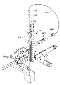
Figure 37 depicts, in accordance with an embodiment of the invention, syringe
pump
9000 can be connected to floating cannula 8000 through tube 10000. Tube 10000
terminates
in coupler/connector 10001 on one end, which couples tube 10000 to syringe
pump 9000. On
the other end, tube 10000 is connected to delivery tube 7000 through male Luer
lock fitting
10002 and female Luer lock fitting 10003. The floating cannula 8000 is shown
connected to
the guiding arm of stereotactic device 100.
Figure 38 depicts, in accordance with an embodiment of the invention, arrows
and
lines indicate locations at which various components or categories of
components (labeled
"A", "B", and "C") can be positioned on a stereotactic device and a syringe
pump attached
thereto. "A" indicates one or more components such as an electromechanical
switch, an
.. optical sensor, an electromagnetic sensor, and a capacitive sensor, as
described in greater
detail herein. "B" indicates one or more components such as a strain gauge-
based sensor,
piezo-based sensor, electromagnetic sensor, optical sensor, capacitive sensor,
and
potentiometric sensor. "C" indicates one or more components such as a video-
based motion
12
capture system, a potentiometer (linear distance sensor), a linear variable
differential
transformer (LVDT), an inductive proximity sensor, a rotary encoder, an
incremental
encoder, an absolute position encoder, a Gill sensor, and an ultrasonic
sensor.
Figure 39 depicts, in accordance with an embodiment of the invention, arrows
and
lines indicate locations at which various components or categories of
components (labeled
"A", "B", and "C") can be positioned on a floating cannula system. "A"
indicates one or
more components such as an electromechanical switch, an optical sensor, an
electromagnetic
sensor, and a capacitive sensor, as described in greater detail herein. "B"
indicates one or
more components such as a strain gauge-based sensor, piezo-based sensor,
electromagnetic
sensor, optical sensor, capacitive sensor, and potentiometric sensor. "C"
indicates one or
more components such as a video-based motion capture system, a potentiometer
(linear
distance sensor), a linear variable differential transformer (LVDT), an
inductive proximity
sensor, a rotary encoder, an incremental encoder, an absolute position
encoder, a Gill sensor,
and an ultrasonic sensor.
DETAILED DESCRIPTION OF THE INVENTION
Unless defined otherwise, technical and scientific terms used herein have the
same
meaning as commonly understood by one of ordinary skill in the art to which
this invention
belongs. Szycher's Dictionary of Medical Devices CRC Press, 1995, may provide
useful
guidance to many of the terms and phrases used herein. One skilled in the art
will recognize
many methods and materials similar or equivalent to those described herein,
which could be
used in the practice of the present invention. Indeed, the present invention
is in no way
limited to the methods and materials specifically described.
In some embodiments, properties such as dimensions, shapes, relative
positions, and
so forth, used to describe and claim certain embodiments of the invention are
to be
understood as being modified by the term "about."
The terms "patient" and "subject" are used interchangeably herein. These terms
are
intended to include all animal subjects, including mammals. Human
patients/subjects are
intended to be within the scope of the patients/subjects treated using the
various embodiments
.. of the inventive systems, apparatuses and methods described herein.
As used herein, the terms "anatomical feature" and "anatomical structure"
include any
tissue or collection of tissues found on or in a subject's body.
13
Date Recue/Date Received 2022-05-11
CA 02966029 2017-04-26
WO 2016/069936 PCT/US2015/058134
As used herein, the term "fluid," includes any fluid, including but in no way
limited to
a gas or a fluid.
As demonstrated herein, in some embodiments the invention discloses novel
stabilizing apparatuses, cannula systems and apparatuses, syringe pump systems
and
apparatuses, and methods of use thereof. In some embodiments, the invention
discloses
imaging systems and methods that can be used alone or in conjunction with the
aforementioned apparatuses, systems and methods. In some embodiments, the
invention
discloses automation of the aforementioned devices and systems through
sensors, motors,
receivers, transmitters and computers. While one of skill in the art would
readily appreciate
that there are many possible applications of the systems and apparatuses
described herein,
certain embodiments are especially useful for procedures performed on or
around the spinal
cord, including delivery of cutting edge cellular and molecular therapies
thereto.
Although numerous embodiments of stereotactic apparatuses are described
herein,
there are certain features common to all of them. First, each stereotactic
apparatus includes
one or more components that make up a "securing section" capable of stably
connecting to an
arm of a tissue retracting device, or other support system (e.g. table, lamp,
or any other solid
object which can be clamped). The second feature common to each of the
stereotactic
apparatuses described herein is a "positioning section," which includes one or
more
components capable of positioning an instrument over a desired location in a
subject's body.
The third common feature is a "connecting section," which serves to operably
connect the
positioning section and the securing section. A fourth common feature is a
"guiding section,"
which can be used to guide an instrument into or remove an instrument from a
subject's
body.
Provided below are descriptions of various components, combinations of
components,
and configurations of components relative to one another that can be used to
arrive at each of
the common sections described above. Additional features that can be added to
the
stereotactic apparatus are also described.
Securing Section
In some embodiments, the securing section of the stereotactic apparatus is
configured
to removably attach to an arm of a tissue retractor, or any device of similar
dimensions.
Removable attachment can be accomplished in any of a number of ways, using a
wide range
of components and combinations thereof. Merely by way of non-limiting
examples, the
14
CA 02966029 2017-04-26
WO 2016/069936 PCT/US2015/058134
securing section could attach to the arm of a tissue retractor by using one or
more clasps, one
or more clamps, one or more magnets, one or more screws, one or more pins, one
or more
slot and groove arrangements, one or more straps, combinations thereof and the
like.
Therefore, each of these components, and modified versions thereof, are within
the scope of
the invention. It is further contemplated that the attaching portion of the
apparatus could be
configured to attach to any of a variety of types of equipment that might be
found in a setting
in which a medical procedure is performed, including, but in no way limited to
a table, a
lamp, a brace, a tray, imaging equipment, and the like. It is also
contemplated that the device
could be configured for use in a non-surgical setting, in which it may be used
to perform any
objective that requires the use of precision guidance. It is further
understood that the device
could be scaled up or down in size appropriately for such objectives. Thus,
the device could
be configured to be an appropriate size for precision delivery of items on a
microscopic scale
(e.g., injecting a substance into a cell), or it could be configured to be an
appropriate size to
position or deliver much larger items.
In some embodiments, a clamping mechanism is incorporated on the securing arm,
and used to attach the stereotactic apparatus to the arm of a tissue
retractor. One of skill in
the art would readily appreciate that numerous types of clamping mechanisms
are suitable to
accomplish this function. One non-limiting example is depicted in Figure 3,
which shows
clamping mechanism 5000 of securing arm 4000 can be used to clamp arm 301 of
tissue
retractor 300 (partially shown). A more detailed view of the clamping
components of this
particular embodiment is shown in Figure 21, and the individual components
(and their
functions) are thoroughly described in the examples section.
Importantly, the clamping mechanism shown in Figure 21 can be used to securely
and
removably attach a stereotactic apparatus (including stereotactic apparatus
100) to the arm of
a number of different types of tissue retractors. Non-limiting examples of
retractors to which
the clamping mechanism can attach include the Mast Quadrant Retractor System
(Medtronic), the MARS Retractor System (Globus Medical), the Spyder Retractor
System
(Aesculap), the Ravine Retractor System (K2M), the Synframe Retractor System
(DePuy
Synthes), and the Luxor Retractor System (Stryker). One of skill in the art
would readily
appreciate that any retractor with one or more arms similar to those
retractors described
above could also be used in conjunction with the inventive stereotactic
apparatuses described
herein. One of skill in the art would further appreciate that the alternative
attaching
mechanisms (such as clamps, clasps, and similar mechanisms) described above
would allow
CA 02966029 2017-04-26
WO 2016/069936 PCT/US2015/058134
for the attachment of the securing section of an apparatus to one or more arms
of alternative
retractor devices that are not specifically listed above. Similarly, the
securing section of the
stereotactic apparatus can include an attaching mechanism of a suitable shape,
size and
orientation for attaching the stereotactic apparatus to a device other than a
tissue retractor,
without significantly affecting the function of the remainder of the device.
Positioning Section
In the context of medical applications, the purpose of the positioning section
is to
allow for stable positioning of an instrument over a desired anatomical
location, by
positioning a guiding arm to which the instrument is attached. There are many
possible
components and configurations thereof that could make up a positioning section
of the
stereotactic apparatus. In certain embodiments the positioning section
includes components
that allow for telescoping motion, which permits fine adjustment of the
position of the
instrument attached to the guiding arm. In some embodiments, a positioning arm
is used. In
various embodiments, the positioning arm includes two or more nested elements
that are
operably connected to one another as well as an input component (e.g., a dial)
in a manner
that allows for telescoping motion. In a non-limiting example, the telescoping
motion is
accomplished by the components depicted in Figures 15-17. The interaction
between and
operation of the specific components of Figures 15-17 are thoroughly described
in the
examples section.
There are numerous possible ways of stabilizing and controlling the
telescoping
motion of the positioning arm. Merely by way of non-limiting example, if a
mechanism with
a threaded shaft is used, as depicted in Figures 15-17, the number of
threadings on the shaft
and the pitch of the threadings can be used to dictate the degree to which the
positioning arm
telescopes in response to associated input (e.g. rotation of a dial). In
certain embodiments,
the positioning arm is stabilized through the use of components that limit its
range of motion
in all but the axis along which it is advanced or retracted. Merely by way of
non-limiting
example, Figure 16 shows the configuration of guiding set screws 176a and 176b
and
supporting elements 178a and 178b is used to apply pressure on L-shaped tracks
179a and
179b of inner nested element 112 of positioning arm 2000. Figure 16 also shows
that screw
175 is positioned on the opposite side of set screws 176a and 176b, in order
to add to the
stability of inner nested component 112, especially while it is being extended
or retracted.
16
CA 02966029 2017-04-26
WO 2016/069936 PCT/US2015/058134
All of these components can help to improve precision while utilizing the
device, which can
be particularly important in a medical setting.
There are many possible ways of attaching the positioning arm to the guiding
aiiii. As
shown in Figure 3, one way positioning arm 2000 can be connected to guiding
arm 1000 is
through the use of screw 133 that traverses the short axis of guiding arm 1000
and connects
to grooved receiving socket 134.
Connecting Section
The long axis of the connecting section of the stereotactic apparatus can be
configured
to be perpendicular to the long axis of the securing section and the
positioning section. In
some embodiments, the connecting section, like the positioning section, is a
telescoping arm
("connecting arm"). In some embodiments, the telescoping connecting arm can be
stabilized
and controlled by any of the aforementioned components that can be associated
with the
positioning section, as described above. Merely by way of non-limiting
example, telescoping
.. of the connecting arm can be accomplished through the use of the components
shown in
Figures 12-14, the interaction between which and function of which are
thoroughly described
in the examples section.
Guiding Section
The guiding section can be configured to allow for the attachment of one or
more
instruments that can be extended into and retracted from a subject's body. In
other
embodiments, the guiding section can be useful for extending towards or
retracting from
another target, including in non-medical settings, as indicated above. In some
embodiments,
the guiding section includes a guiding arm. There are many possible ways by
which an
instrument can be attached to a guiding arm. One of skill in the art would
readily appreciate
that the possible components that could be used to attach an instrument to a
guiding arm
would vary depending upon the dimensions and nature of the instrument to be
attached.
Merely by way of non-limiting examples, attachment of various instruments to
the guiding
arm can be accomplished by using one or more straps, clamps, clasps, magnets,
and
combinations thereof.
Examples of instruments that could be attached to the guiding arm include, but
are in
no way limited to a cannula (including the floating cannula system described
herein), a
biopsy needle, a needle, a tube, a cauterization device, a laser, a drill, an
endoscope, a
17
guidewire, a fiberoptic device, an electrode, a saw, an ultrasonic device, a
spectroscopic
device, a camera, an electrical sensor, a thermal sensor, a catheter, a
draining tube, an
imaging device (such as any of those listed and/or described herein) and the
like. In certain
embodiments, the instrument guided by the inventive apparatuses described
herein includes a
.. guide needle and an injection needle configured to be concentrically housed
therein. In some
embodiments, the concentric arrangement of the guide needle and the injection
needle allows
the injection needle to be advanced through the guide needle, once the guide
needle is
properly positioned in a subject during a medical procedure, so that the
injection needle can
deliver a payload of biological or chemical material to an appropriate site in
the subject. In
some embodiments, the instrument guided and/or stabilized by the inventive
apparatus is a
spinal multisegmental cell and drug delivery device, such as the device
described in U.S.
Patent Application No. 12/598,667.
One of skill the art would also readily appreciate that there are numerous
possible
ways by which the apparatus can be configured to allow for an instrument to be
extended into
and retract from a subject, or other target, while connected to the guiding
arm. Figure 18
depicts one non-limiting example of a mechanism that can be used for that
purpose. The
association between the components shown in Figure 18 and the function of
those
components are thoroughly described in the examples section.
Orientation of Individual Sections
The securing section, connecting section, positioning section and guiding
section can
be connected to one another by any of a variety of ways depending upon the
desired range of
motion of each section. In some embodiments, a perpendicular orientation of
the positioning
.. arm and connecting arm, relative to one another, is established through the
use of a
component with perpendicularly situated clamping collars. In an embodiment,
cross clamp
132 (depicted in Figure 1A) can be used. As shown in Figure 5, when cross
clamp 132 is
used to secure positioning arm 2000, knob 114 can be rotated to loosen collar
115, thereby
allowing for adjustment of the position of positioning arm 2000 along the x-
axis. As shown
in Figure 8, loosening of collar 115 by rotating knob 114 also allows for
rotation of
positioning arm 2000 along the x-axis, which translates into motion of guiding
arm 1000
along the y-z plane.
18
Date Recue/Date Received 2022-05-11
CA 02966029 2017-04-26
WO 2016/069936 PCT/US2015/058134
As shown in Figure 6, when cross clamp 132 is used to secure connecting arm
3000,
rotation of screw 135 loosens lower collar 117, which allows for adjustment of
the position of
positioning arm 2000 along the y-axis. As shown in Figure 9, loosening collar
117 also
allows for rotation of cross clamp 132 along the y-axis, which in turn
translates into motion
of guiding arm 1000 along the x-z plane.
Additional Features
The main sections of the stereotactic apparatuses described above can be
configured
to allow for incorporating additional features on the apparatuses. For
example, the
stereotactic apparatus can include clamps (or any other mechanism(s) of
attachment
described herein) situated on one or more of the main sections of the
apparatus (i.e. guiding
section, positioning section, connecting section, and attaching section) for
attaching
additional instruments or devices that are useful for a particular
application.
In certain embodiments, the stereotactic apparatus includes a side clamp
attached to
.. the securing section, which allows for attaching a useful instrument or
device. For example,
as demonstrated in Figure 3, side clamp 6000 can be used to hold cylindrical
device 400. The
components of a particular non-limiting embodiment (side clamp 6000) are
clearly shown in
Figure 22, and thoroughly described in the examples section. One of skill in
the art would
readily appreciate that a side clamp such as side clamp 6000 can be used to
attach any of a
number of devices with appropriate dimensions to the stereotactic apparatus.
Although the
particular device 400 shown in Figure 3 is cylindrical, a device of
practically virtually any
shape could be attached by appropriately modifying the shape and dimensions of
the clamp
(e.g. side clamp).
Devices that can be attached to the stereotactic apparatuses described herein
can
include, but are in no way limited to, a pump (such as the pump of the syringe
pump system
described herein), a reservoir for receiving a substance removed from a
subject's body, a
small motor, a control panel, an imaging device or portion thereof (including
any
appropriately sized imaging device described herein) and the like. In some
embodiments, the
device attached is a fiber optic camera, or portion thereof, that can be
positioned to view an
opening in a patient's body in which a tissue retractor is engaged. In some
embodiments, a
reservoir attached to the apparatus can be configured to hold any of a variety
of useful
substances, including but in no way limited to cells (including stem cells for
various
19
CA 02966029 2017-04-26
WO 2016/069936 PCT/US2015/058134
therapeutic treatments), fluids, medications, contrast agents, radioactive
materials,
combinations thereof, and the like.
An additional category of devices that could be attached to one or more
sections of
the inventive apparatuses described herein is a light source. In various
embodiments, the
inventive apparatuses may include one or more light sources configured to
project light onto
a region of interest on or in a subject's body during a medical procedure. In
some
embodiments, one or more of the light sources is attached to the guiding arm.
In some
embodiments, the light source is a laser. In some embodiments, the light
source is a
relatively high energy laser that can be used for cauterizing or cutting. In
some
embodiments, the light source is a relatively low energy laser that can be
used for visually
targeting a region on or in a subject's body for incision or other medical
intervention. In
other embodiments, the light source provides relatively low energy light for
aiding in
visualizing a region of interest. In still other embodiments, the light source
provides light of
a wavelength that causes fluorescence of a fluorophore. In various
embodiments, the
fluorophore is introduced into a subject's body directly, present in cells
residing in a subject's
body, or naturally occurring. Merely by way of non-limiting examples, the
wavelength of the
light projected by the light source can be in the visible, IR, or UV range.
Another category of devices that can be incorporated onto the stereotactic
apparatuses
described herein is an imaging modality. In some embodiments, the imaging
modality is
attached to the guiding arm. However, one of skill in the art would recognize
that all or a
portion of an imaging modality (or any other device described herein, or
similar thereto) of an
appropriate size could be attached to any arm of the apparatuses described
herein, by any
form of attachment described herein. In some embodiments, the imaging modality
includes a
device used to perform MRI, CT, or ultrasound imaging. In some embodiments, an
endoscope is attached to the guiding arm. In some embodiments, one or more
components of
a microscope or other magnifying instrument are attached to the guiding arm.
One of skill in
the art would readily appreciate that any of a number of other useful
instruments of a size
suitable for attaching to the guiding arm could be used in conjunction with
the inventive
apparatuses described herein, and attached thereto by any means for attachment
described
herein.
As indicated above, in some embodiments, the apparatus is configured so that
the
positions of the various sections described above can be manipulated manually.
However,
one of skill in the art would readily appreciate that the apparatus could also
be configured
CA 02966029 2017-04-26
WO 2016/069936 PCT/US2015/058134
with one or more motors, gears, pulleys, and electronic controls, so that one
or more sections
of the apparatus could be electronically controlled.
In some embodiments, the apparatuses described herein, or one or more portions
thereof, are made of stainless steel. In some embodiments, the apparatuses are
made of
titanium, austenitic steel, martensitic steel, brass, carbon fiber, plastic,
composites,
combinations thereof, and the like. In preferred embodiments, the material or
materials used
are biocompatible.
In some embodiments, the invention teaches a method that includes using any of
the
stereotactic apparatuses described herein for the purposes of facilitating one
or more of the
processes of (1) introducing a substance into a subject, (2) removing a
substance from a
subject, and (3) manipulating a portion of a subject's body. One of skill in
the art would
readily appreciate that the device could be used to introduce a substance into
and/or remove a
substance from any portion of subject's body, including, but in no way limited
to an organ,
joint (shoulder, hip, knee, etc.), ligament, tendon, muscle, eye, cavity, or
any other tissue. In
some embodiments, the apparatus can be used to introduce a substance into or
remove a
substance from a subject's brain. In some embodiments, the substances
introduced into the
subject's body can include but are in no way limited to biological and/or
synthetic
substances. Biological substances can include, but are in no way limited to
stem cells, neural
progenitor cells, tissues, blood, hormones, clotting factors, vectors
(including but not limited
to viral vectors, plasmids and the like), DNA, RNA, proteins, growth factors,
inhibitory
substances, matrices, combinations thereof, and the like. Synthetic substances
that can be
introduced into a subject's body can include but are in no way limited to
pharmaceutical
agents, markers (including but not limited to biomarkers or any other type of
marker that
could be visualized with or without the use of imaging equipment), implantable
medical
devices, electrical sensors, electrical stimulators (including devices for
stimulating one or
more portions of a subject's brain), glue, sutures, chemotherapeutics,
radioactive substances,
hyperpolarized substances, combinations thereof, and the like.
Substances that can be removed from a subject's body utilizing the inventive
stereotactic apparatuses and methods include, but are in no way limited to,
any of the above-
named substances that can be introduced into a subject, in addition to
tissues, organs, cancer
cells and pre-cancer cells, bone marrow, fluid, foreign bodies, combinations
thereof, and the
like.
21
CA 02966029 2017-04-26
WO 2016/069936 PCT/US2015/058134
In some embodiments, the inventive method includes using any of the inventive
apparatuses described herein to position any of the instruments described
herein such that
they can be introduced between the spreading elements of a retractor device
described herein
and then the adjacent sections of tissue associated therewith. In an
embodiment, the
inventive method includes using guiding arm 1000 of inventive apparatus 100 to
introduce a
needle associated with a cannula into any portion of a subject's spinal cord
(including the
section specifically described in the non-limiting examples herein). A payload
of neural
progenitor cells is then advanced through the cannula and needle and into the
subject's spinal
cord. In some embodiments, the neural progenitor cells express glial cell line
derived
neurotrophic factor (GDNF). In some embodiments, the subject has been
diagnosed with a
neural degenerative disease. In some embodiments, the neural degenerative
disease is
amyotrophic lateral sclerosis (ALS). In some embodiments, the subject has a
neurologic
injury. In some embodiments, one or more sections of the subject's spinal cord
is damaged,
severed, or partly severed.
In some embodiments, the invention teaches a method that includes (1)
attaching any
apparatus described herein to the arm of a retractor, (2) attaching any
instrument described
herein to the guiding arm of the apparatus (by any means described above), and
(3) advancing
the instrument through the separating elements of the retractor and into a
subject's body
through an incision in the subject's body. Figure 1D shows a non-limiting
example of how
the components of an apparatus can be situated to perform this method.
Floating Cannula Instruments & Systems
In some embodiments, a guiding arm of any of the stereotactic devices
described
herein may be attached to any of the floating cannula systems described
herein. The floating
cannula system (or one or more components thereof) attached to a guiding arm
may be
utilized to perform precision injections (including injecting any medically
useful substance,
whether described herein or otherwise). Merely by way of non-limiting example,
the cannula
system and stereotactic device may be used when injecting a substance into the
spinal cord,
thus allowing a caregiver to accurately position the cannula and needle in the
correct location.
Typically, once a needle is inserted into a subject's tissue, any movement of
the
subject with respect to the needle may damage the subject's tissue. This is
particularly
problematic for injections into sensitive areas, such as the spinal cord or
brain, as damage to a
spinal cord or brain could have severe consequences. For instance, if a
stereotactic device
22
CA 02966029 2017-04-26
WO 2016/069936 PCT/US2015/058134
lowered a needle into the spine, and the needle did not provide a stopping
mechanism, or
allow for movement along the longitudinal axis of the needle, a reflex
(including but not
limited to a cardiac or pulmonary reflex), twitch, or bucking of the patient
could cause the
needle to penetrate too far, or otherwise change directions and damage or
sever spinal cord
tissue (e.g. by shearing). This could have catastrophic consequences to a
patient.
Therefore, in some embodiments the invention teaches a floating cannula
system,
with one or more components that can be used separately from or in conjunction
with any of
the stereotactic systems described herein. In some embodiments, the floating
cannula can be
attached to the guiding arm of a stereotactic device, thereby allowing for
movement of the
cannula in response to patient movement, once the needle has been inserted
into the patient.
In some embodiments, the system includes a floating cannula interacting with a
base cannula,
where the floating cannula may move up and down with respect to the base
cannula to
accommodate movement of the patient. The base cannula may be attached to a
connector,
which is in turn attached to a stereotactic device. This configuration can
provide stability and
support derived from the connector's attachment to the stereotactic device. In
other
embodiments, the base cannula may be attached directly to the stereotactic
device. The base
cannula may include two or more support tabs, such as the support tabs 402a
and 402b
depicted in Figure 23, with holes that receive pins attached to the connector,
such as the holes
417a and 417b depicted in Figure 23. Additionally, the tabs may include finger
pads for easy
manipulation and handling of the cannula by a caregiver.
In some embodiments, the support tabs may include sockets (e.g. elements 417a
and
417b of Figure 23) for removably connecting to or mounting the support tabs
onto pins that
are supported by the connector. This will allow the tabs to hold the base
cannula in place
while allowing rotation about the pins. In some embodiments, the connector may
include a
locking member. Merely by way of non-limiting example, the connector 420 may
include a
locking member 418, as depicted in Figure 24. In some embodiments, the support
tabs may
be rotated into recesses or spaces in the connector, and then the locking
member may be
moved to block the support tabs from rotating back out. Merely by way of
example, Figure
24 demonstrates recessed portions of connector 420 in which support tabs 402a
and 402b are
engaged and secured in place by locking member 418. In some embodiments, more
than one
locking member may be utilized to block the support tabs.
In some embodiments, the locking member is a physical restraint that creates
an
interference fit by rotating a handle that blocks the support tabs from
rotating out of place.
23
CA 02966029 2017-04-26
WO 2016/069936 PCT/US2015/058134
The locking handle may be rotated into place once the tabs are mounted on the
pins, and then
rotated into one or more slots on the connector. Accordingly, the base
cannula, in some
embodiments, may be rigidly attached to the stereotactic device through the
connector. In
other embodiments, the base cannula may attach directly to the guiding arm or
other
positioning section of a stereotactic device through tabs. In other
embodiments, the
stereotactic device may include pressure cuffs that attach directly to the
round tube of the
cannula. Ultimately, a variety of methods/devices may be utilized for
attaching a base
cannula to a stereotactic device, including one or more of any suitable type
of attachment
mechanism described and/or depicted herein.
A floating cannula may extend down from the base cannula that is supported by
the
stereotactic device. In some embodiments, the floating cannula will fit inside
the lumen of
the base cannula. In other embodiments, the base cannula will fit inside the
lumen of the
floating cannula. In both embodiments, the concentric fit allows the base
cannula to contact
the floating cannula while allowing the floating cannula to slide along a
longitudinal axis of
the base cannula and with respect to the base cannula. In some embodiments,
the fit between
the floating cannula and the base cannula will prevent the floating cannula
from moving
substantially in other directions, aside from along the longitudinal axis. In
some
embodiments, the floating cannula and base cannula may be connected to the
stereotactic
device through a hinged mechanism that allows for motion in a direction
perpendicular to the
longitudinal axis of the cannulas, in order to accommodate patient movement
after the needle
is placed.
In some embodiments, the floating cannula will run along the inside lumen of
the base
cannula and the floating cannula will be of a sufficient length to protrude on
both sides of the
base cannula. Additionally, the floating cannula may include stoppers situated
such that they
are positioned beyond each of the ends of the base cannula when the floating
cannula is
engaged therein. Merely by way of example, the stoppers may be configured
according to
the arrangement demonstrated in Figure 26, in which stoppers 410a and 410b are
located near
the ends of floating cannula 404, such that they limit the range of motion of
base cannula 406
when floating cannula 404 is engaged therein. In the configuration
demonstrated in Figure
27, the top (proximal) stopper 410a is shown fixed to the top (proximal) end
of the floating
cannula, and it prevents the floating cannula from falling down and out of the
base cannula
406. Figure 27 shows that by positioning lower stopper 410b at the distal end
of the floating
cannula, a needle 416 located at the bottom of the floating cannula can be
inserted into tissue
24
CA 02966029 2017-04-26
WO 2016/069936 PCT/US2015/058134
due to resistance from the base cannula 406 pushing on the lower stopper 410b,
when a
downward force is applied to the base cannula 406. Without the bottom stopper,
or
comparable element, the floating cannula could not provide sufficient pressure
for inserting
the needle into the anatomical target (e.g. spinal cord) of a subject, when
the base cannula is
lowered toward the subject.
In some embodiments, the floating cannula will contain a tissue stopper that
is
attached to a needle. The tissue stopper may be any suitably shaped material
secured to the
needle that will limit the depth of an injection when the tissue stopper makes
contact with the
tissue at the injections site. The tissue stopper may be positioned at any
point along the
needle, depending on the depth of injection required for a particular
procedure. The tissue
stopper may be any of a number of shapes, including but in no way limited to
flat, wedge-
shaped, ball-shaped, and cup-shaped. Any suitable shape which provides a
mechanical
means to limit how far a needle injects into a tissue site (e.g. the spinal
cord) is within the
scope of the invention. Merely by way of example, the tissue stopper may be
configured
according to the embodiment of Figure 27, which shows tissue stopper 412
positioned
between needle 416 (positioned at the end of the floating cannula) and lower
stopper 410b.
In some embodiments, the floating cannula will fit inside the base cannula,
and thus
the stoppers may be positioned on the floating cannula, so that they contact
the proximal and
distal rims of the base cannula and prevent the floating cannula from moving
past certain
points with respect to the base cannula. In other embodiments, the floating
cannula may fit
on the outside of the base cannula (the base cannula would run at least
partially inside the
lumen of the floating cannula) and may have internal and/or external stoppers.
In some
embodiments, there will also be space for attaching the base cannula to the
stereotactic device
through the floating cannula. In some embodiments, the floating cannula will
fit inside the
base cannula and protrude on both sides of the base cannula. In other
embodiments, the base
cannula will fit inside the lumen of the floating cannula, and the floating
cannula will only
cover a distal portion of the base cannula. In this embodiment, other stoppers
or movement
restriction systems may be utilized to limit the travel of the floating
cannula with respect to
the base cannula. For example, the base cannula may include a slot, along
which a tab
connected to the inside lumen of the floating cannula, would ride. The tab may
contact
another tab on the inside of the lumen of the base cannula that is configured
to contact the tab
from the floating cannula.
CA 02966029 2017-04-26
WO 2016/069936 PCT/US2015/058134
In some embodiments, a delivery tube connected to a fluid reservoir will run
the
entire length of the cannula system and terminate at a hollow needle. In some
embodiments a
section of the delivery tube is located outside of the floating and base
cannulas, and a section
of the delivery tube is contained within the lumen of the floating cannula (a
portion of which
is contained within the base cannula). In some embodiments, the delivery tube
is in fluid
communication with a hollow needle located at the end thereof. Thus, in some
embodiments,
a fluid can be introduced through a first end of the delivery tube located
outside of the
cannula. The fluid could then be advanced through the entire delivery tube
(including a
portion contained within the floating cannula) until it exits a hollow needle
at the end thereof.
In some embodiments, the delivery tube is in fluid communication with a fluid
reservoir. In
some embodiments, the fluid reservoir is connected to a fluid pump (including
any suitably
sized fluid pump, such as the fluid pumps described herein). In some
embodiments there
may be an external (or internal) pump and reservoir that contain a therapeutic
or other
injectable substance for injecting into a tissue site (or other location in a
subject). The non-
limiting example of Figure 27 depicts a delivery tube 408 nested within the
floating and base
cannulas, and terminating in hollow needle 416.
In some embodiments, the invention includes a procedure for injecting a
substance
into a subject using a floating cannula system and stereotactic device
described herein. This
procedure may include attaching the floating cannula system to the guiding arm
of a
stereotactic device (through any mechanism/means described herein). Then, the
cannula will
be advanced towards an injection site by advancing the guiding arm of the
stereotactic
device. In some embodiments, the hollow needle associated with the cannula
will contact the
injection site (e.g. the spinal cord) and pressure will push the floating
cannula proximally into
the lumen of the base cannula. The floating cannula will continue to move
proximally until
the stopper on the bottom portion of the floating cannula contacts the distal
end of the base
cannula. Then, the distal end of the base cannula will apply pressure to the
stopper, which
will be transferred to the floating cannula. Thereafter, the pressure will
push the needle into
the injection site. In some embodiments, the needle will be inserted until the
tissue stopper
makes contact with the tissue at the injection site (e.g. the spinal cord).
Once the needle is
inserted into the tissue, the friction from the tissue on the needle and
potentially the negative
pressure from the injection site on the needle will hold the needle in place
such that it is
situated in the direction of the longitudinal axis of the cannulas.
26
CA 02966029 2017-04-26
WO 2016/069936 PCT/US2015/058134
In some embodiments, once the needle is fully inserted into the injection site
(as
limited by the tissue stopper), the guiding arm of the stereotactic device may
be retracted
from the injection site, so that the base cannula also moves away from the
injection site, and
upward with respect to the floating cannula and the needle. This will create
space between
the distal stopper of the floating cannula and the distal end of the base
cannula, which will
allow movement of the floating cannula along the longitudinal axis of the
cannulas. This
freedom of movement will accommodate movement of the subject in whom the
needle is
inserted (e.g. movement from respiration, heartbeat, bucking, and the like).
Accommodation
of movement is particularly important in procedures requiring injecting into
delicate areas
(e.g. the spinal cord), as a sudden force along the longitudinal axis of the
needle has the
potential to cause the needle to puncture further into the subject and cause
considerable
damage, depending on the local organs or other anatomical structures in the
needle's path.
One of skill in the art would readily appreciate that one or more components
of the
floating cannula systems described herein could be utilized independently from
the
stereotactic device described herein. Thus, various combinations of the
individual
components of the cannula systems described herein are within the scope of the
present
invention.
Syringe Pump
In various embodiments, the invention teaches a syringe pump system that can
be
used to facilitate the precision injections described herein above, as well as
for other
purposes. In certain embodiments, the syringe pump system may be configured to
attach to a
stereotactic device, including any of the stereotactic devices described
herein. In some
embodiments, the syringe pump system is configured to be secured by the side
clamp of a
stereotactic device described herein. Although the figures (for example Figure
37) depict a
syringe pump system oriented in one direction relative to the stereotactic
device, the syringe
pump systems described herein can also be oriented in the opposite direction
relative to the
stereotactic device. In addition, the syringe pump systems described herein
may be
configured to interact with and attach to (permanently or removably) any of
the cannulas and
cannula systems described herein, including the floating cannula systems
described herein,
whether for the purpose of facilitating the injection of a therapeutic
substance into a subject
(as described herein), or otherwise. Further, the syringe pump systems
described herein may
27
CA 02966029 2017-04-26
WO 2016/069936 PCT/US2015/058134
be utilized as stand-alone devices, or they may be coupled with an
appropriately sized fluid
inlet of practically any fluid delivery system, especially those used in
medical applications.
The syringe pump systems described herein all include the following central
components: (a) a carpule with an interior chamber configured to hold a
quantity of a
therapeutic substance or other medically useful substance, (b) a plunger
configured to interact
with the interior chamber of the carpule and advance therein in order to expel
a therapeutic
substance (or other medically useful substance) therefrom, and (c) a motor for
imparting a
drive force (either directly, or indirectly through one or more drive shafts,
or other
components useful for translating mechanical force) to the plunger. In some
embodiments,
the syringe pump systems described herein may be utilized to deliver
therapeutic agents, such
as stem cells (including but not limited to any type of stem cell described
herein), pain
medications, chemotherapeutic agents and/or other medications (along with any
other
medically useful substance or combination of substances described elsewhere
herein), safely,
by regulating fluid dynamics and monitoring flow pressure during injection. In
preferred
embodiments, the size of the syringe pump system may be such that it does not
significantly
encumber the surgical space for the procedure in which it is utilized. As
such, the syringe
pump system may be configured to be a small hand-held device and/or a stand-
alone pump
that may be utilized in surgical procedures that do not require a stereotactic
system.
With regard to the carpule component, in certain embodiments the carpule may
be
configured as a disposable component that is removably coupled to the syringe
pump. In
some embodiments, the carpule component may contain a therapeutic agent (or
other
medically useful substance) with a predetermined amount and/or dosage to be
injected into a
specific anatomical location and/or tissue (e.g. spinal cord, brain, tumor
tissue, etc.). A non-
limiting example of a syringe pump system 9000 including a carpule 501 is
shown in Figure
31.
In certain embodiments, the carpule is made of one or more sterilizable
materials (e.g.
glass, plastic, metal, etc). In some embodiments, the carpule has an interior
chamber with a
volume of 50u1. In some embodiments, the carpule may have an interior chamber
with a
volume of 100u1, 250u1, 500u1, or more. In various embodiments, the volume of
the chamber
may be from 20u1 to 10m1 or more. The size of the carpule and volume of its
interior
chamber may be configured to be appropriate to accommodate a volume and dosage
of a
therapeutic agent (or other medically useful substance) needed for a
particular
application/procedure. In certain embodiments, the interior chamber is
cylindrical, but other
28
CA 02966029 2017-04-26
WO 2016/069936 PCT/US2015/058134
shapes are possible and within the scope of the present invention. In certain
embodiments,
the carpule is removably coupled (directly or indirectly) to a component
including a motor
and drive shaft and/or plunger. In some embodiments, the carpule is configured
to be
prefilled with a therapeutic agent (such as any type of cellular therapeutic
composition or
other therapeutic composition described herein) prior to use. In some
embodiments, the
carpule may be removably coupled to the syringe pump, so that it may be
sterilized before
and/or after use in a medical procedure (e.g. by gamma radiation, EtO, etc.).
In some
embodiments, the syringe pump system may also include a mechanism to rotate
and/or
vibrate the carpule component, in order to reduce or avoid settling, clogging
and/or clumping
of the therapeutic agent (e.g. cells). In some embodiments, the interior of
the carpule may be
coated with a substance known to prevent cells from adhering or sticking to
the interior
surface. In some embodiments, the interior of the carpule may be coated with
heparin and/or
hydrophobic coatings. In some embodiments a plasma treatment may be used on
the interior
of the carpule. In some embodiments, the interior geometry of the carpule is
configured in
such a way as to prevent agglomeration of the cells, such as large aspect
ratio pathways,
tortuous pathways and/or a series of parallel passageways. In some
embodiments, the syringe
pump system is configured to deliver a therapeutic agent via a microfluidic
flow process.
Merely by way of non-limiting examples, the interior chamber of the carpule
may be
smooth, rigid, and/or contain grooves. In certain embodiments, the carpule may
be designed
such that it has a cone-shaped interior, in order to facilitate fluid flow out
of the carpule. By
way of non-limiting examples, the cone-shaped interior may be smooth, rigid,
and/or contain
grooves. In some embodiments, the carpule may contain markings on the interior
or exterior
surface. The markings may allow the user to determine how much volume of a
substance has
been loaded in and/or expelled from the carpule. In some embodiments, the
carpule includes
a window made of glass, plastic, or another transparent or semi-transparent
material that
allows the substance in the chamber to be viewed.
In certain embodiments, the carpule includes (a) a first end including an
elongated
inlet port, (b) a second end including an elongated outlet port, and (c) a
chamber disposed
between and in fluid communication with the elongated inlet port and the
elongated outlet
port. In some embodiments, the chamber, elongated inlet port and elongated
outlet port are
approximately the same size (i.e. diameter). In other embodiments, these
sections of the
carpule are different sizes. In some embodiments, the inlet port and/or outlet
port are not
elongated, and are instead of another shape useful for a specific fluid
delivery application.
29
CA 02966029 2017-04-26
WO 2016/069936 PCT/US2015/058134
Turning now to the plunger component, in some embodiments the plunger is
elongated and it includes a receiving plunger end, a pushing plunger end, and
an elongated
plunger body. In some embodiments, the plunger is configured to nest within
the first
elongated inlet port of the carpule. In certain embodiments, the pushing end
of the plunger is
configured to form a fluid-tight seal with the interior chamber of the
carpule. In some
embodiments, the receiving plunger end is configured to receive pressure from
a drive shaft
attached to the motor, such that the plunger is advanced along the interior
chamber of the
carpule, thereby expelling fluid contained in the chamber. In other
embodiments, the plunger
is otherwise directly or indirectly attached to the motor, and configured to
advance along the
chamber of the carpule in response to input from the motor.
With regard to the motor of the syringe pump system, in certain embodiments
the
motor (e.g. element 513 in Figure 31) is contained within a housing (e.g.
element 504 in
Figure 31). In some embodiments, the syringe pump is connected to a control
box (e.g.
element 514 in Figure 31), which electronically controls the flow of fluid
pumped by the
syringe pump (rate, duration, volume, etc.). As indicated above, the motor of
the syringe
pump may be connected to a drive shaft for imparting a drive force on the
plunger, thereby
causing the plunger to advance along the interior chamber of the carpule and
expel a
therapeutic or other medically useful substance contained therein.
Although the syringe pump can be connected to a control box (or other
controller) via
wires (as shown in Figure 31), a wireless connection to the control
box/controller is also
within the scope of the present invention, and can be accomplished utilizing
any appropriate
wireless transmitters and receivers known in the art.
With respect to its power supply, in some embodiments the syringe pump may be
battery operated, while in other embodiments the syringe pump may include a
power cord to
connect to a power source.
With respect to its shape, in certain embodiments the syringe pump may be
substantially cylindrical, such that it may be held in a side clamp of a
stereotactic device (as
described herein and shown in Figure 35) and/or easily held by a user. In
other embodiments,
the syringe pump may be configured to be a different shape, which is useful
for a particular
application/procedure.
In certain embodiments, the syringe pump system may also include a
connector/coupler component. In some embodiments, the connector/coupler
contains
threading on one end to allow for its attachment to the housing that contains
the motor of the
CA 02966029 2017-04-26
WO 2016/069936 PCT/US2015/058134
syringe pump. For example connector 503 in Figure 31 has threading that mates
with
grooves on the inside of housing 504. Although threading can be used to attach
the
connector/coupler to the housing of the syringe pump, the connector/coupler
may also be
attached by any mechanism for attachment described herein that is suitable for
that purpose.
In some embodiments, the connector/coupler is configured (with threading or
otherwise) to
attach to a syringe pump motor housing with its first end, and a carpule with
its second end,
thereby facilitating the connection between the syringe pump motor and drive
shaft and the
carpule and plunger components. A non-limiting example of a useful
configuration is shown
in Figure 31.
In certain embodiments, the syringe pump system may also include a blockage
detection device that monitors variations in flow pressure in the interior of
the carpule
component (e.g through a flow sensor). In some embodiments, the flow rate may
be
controlled by the speed of the motor and the force exerted (directly or
indirectly) onto the
plunger.
In various embodiments, the syringe pump system further includes a delivery
tube
connecting the carpule to a cannula. In certain embodiments, the syringe pump
system
further includes a cannula described herein. In some embodiments, the cannula
connected by
the delivery tube to the syringe pump is a floating cannula described herein.
In some
embodiments, the delivery tube is made of a substance that may include, but is
in no way
limited to, PTFE Teflon, Tygon, silicone, PVC, FEP, PVDF, rubber, polyethylene
and
combinations thereof. In some embodiments, the delivery tube is made of
polyethylene.
In some embodiments, the syringe pump system includes a flexible and sealable
carpule delivery tube. This component may be used in conjunction with the
carpule
component described above, and can be configured in the manner of the flexible
and sealable
delivery tube 10000 shown in Figure 37. In some embodiments, the flexible and
sealable
carpule delivery tube may attach to the carpule through a coupling component
situated on its
first end. This attachment may be accomplished, for example, through a
threaded coupling
component 10001, as shown in Figure 37. One of skill in the art would readily
appreciate
that any useful coupling component could be substituted for the threaded
coupling component
1 0 0 0 1 shown in Figure 37. The carpule delivery tube may also be configured
to
simultaneously attach to a cannula system described herein. Merely by way of
example, this
can be accomplished by incorporating complimentary Luer lock fittings, such as
those shown
as elements 10002 and 10003 in Figure 37. In some embodiments, the carpule
delivery tube
31
CA 02966029 2017-04-26
WO 2016/069936 PCT/US2015/058134
may include a valve at one or both ends that serves to allow fluid to flow
only in the direction
of the cannula, and not towards the carpule. In some embodiments, the carpule
of the syringe
pump system may be pre-loaded with a sterile saline solution (or any other
physiologically
tolerable inert solution), and the carpule delivery tube may be pre-loaded
with a solution that
includes a theraperutic. In some embodiments, the therapeutic includes cells
(including any
type of cell described herein). Thus, when the syringe pump is activated, the
plunger of the
syringe pump pushes the saline solution through the end of the carpule, which
in turn
advances the therapeutic (e.g. cells) through the carpule delivery tube, then
through the
cannula system, and finally both the therapeutic (e.g. cells) and saline flow
through a hollow
needle at the tip of the cannula and into a target site in a patient into whom
the needle has
been introduced. In some embodiments, the carpule delivery tube may prevent
settling,
clogging, or clumping of the therapeutic agent being expelled therefrom (e.g.
cells).
In some embodiments, the carpule itself is pre-loaded with a therapeutic fluid
substance (e.g. cells), and the therapeutic fluid substance is pumped from the
carpule, through
a delivery tube, then through a cannula, and finally into a target site in a
subject. The
delivery tube used for these embodiments can be any delivery tube described
herein, and the
cannula can likewise be any cannula described herein.
Automation of systems and apparatuses
Various components of the stereotactic devices, cannula devices, and syringe
pump
devices may be fitted with sensors and/or motors and/or receivers and/or
transmitters in order
to "automate" the components and to facilitate the procedures described
herein.
Stereotactic device automation
Sensors may be placed at a number of locations on the stereotactic devices
described
herein, including but in no way limited to the locations indicated by arrows
and wavy lines in
Figure 38. The sensors may allow for determining the positions of each of the
arms of the
stereotactic device, relative to one another and/or relative to one or more
landmark on a
patient and/or relative to an instrument attached to the stereotactic device.
The sensors used
for this aspect of the invention may be motion sensors, heat sensors, infrared
sensors, and the
like. Further, any component or category of component listed in the figure
description for
Figure 38 may be included as indicated by the arrows and wavy lines. When
motion sensors
are positioned on the device, they may be positioned to provide feedback to a
central control
32
CA 02966029 2017-04-26
WO 2016/069936 PCT/US2015/058134
unit, which is configured for controlling one or more component of the device,
so that the
relative position of each arm can be determined and properly adjusted during a
procedure.
One or more heat sensors may be positioned on the device such that proximity
to tissue can
be determined. Thus, merely by way of non-limiting example, one or more heat
sensors may
be positioned near the tip of the guiding arm.
The stereotactic device may alternatively or additionally include motors
configured to
adjust the position of one or more arms of the device described herein (e.g.
guiding arm,
positioning arm, etc.). In certain preferred embodiments, one or more arms of
the device may
be configured with a motor. In some embodiments, the motor may be housed
within or
partially within the dial. In some embodiments, the motor is operably
connected to the dial
through a drive shaft, or an alternative connection. In some embodiments, the
motor is
configured to cause the dial of one or more of the stereotactic arms (e.g. as
depicted in the
examples) to rotate, thereby causing the arm with which it is associated to
advance or retract
in a telescoping fashion, depending upon the direction of rotation. In some
embodiments, the
motors are brushless DC motors. In someembodiments, the motors and any
associated
cabling are completely contained within the arms of the stereotactic device,
and may be held
in an appropriate position by channels, clips, conduits or other mechanisms
known in the art.
In some embodiments, the stereotactic device is configured such that it can be
autoclaved. In
some embodiments, the motors of the stereotactic device will be powered by
batteries
completely contained within the device. The batteries associated with the
device can be any
of a number of types which would be readily appreciated by one of ordinary
skill in the art.
Merely by way of non-limiting example, the batteries may be lithium polymer,
ribbon
batteries, silver oxide, lithium, zinc-air, combinations thereof or the like.
In some embodiments, one or more motors of the stereotactic device is
configured to
communicate with a wireless receiver capable of receiving wireless input from
a computing
device (e.g. a handheld computer, desktop computer, handheld computing device,
or the like).
Any sensors located on the stereotactic device (as described herein) may be
configured to
send and/or receive wireless signals (directly or indirectly) to a computing
device (e.g as
described above) in order to communicate information about relative position,
motion, or
other data they capture. Any camera located on or associated with the device
(as described
herein) may also be configured to communicate with a computing device
wirelessly, such that
it can transmit information (images, etc.) or receive information (e.g.
instructions to zoom in,
focus, change modes etc.). Non-limiting examples of wireless transmitters and
receivers that
33
CA 02966029 2017-04-26
WO 2016/069936 PCT/US2015/058134
can be used in accordance with the description above include WiFi, RF,
Bluetooth, and the
like.
Cannula device automation
In certain embodiments, the cannulas and cannula systems described herein are
fitted
with one or more sensors that allow for tracking motion of the cannulas or
portions thereof.
The motion sensors can be configured to track relative motion between cannula
parts (e.g. the
floating and base cannulas of the cannula system) and/or between one or more
cannula parts
and the stereotactic device, and to track the depth of injection of the
cannula needle when
inserted into an anatomical target (e.g. the spinal cord). Non-limiting
examples of motion
sensors that can be used in this context include a video based motion capture
system,
potentiometer (linear distance sensor), a linear variable differential
transformer (LVDT), an
inductive proximity sensor, a rotary encoder, an incremental encoder, an
absolute position
encoder, a Gill sensor, and an ultrasonic sensor. Each of these types of
components could be
useful in any of numerous positions on the device, including but not limited
to the positions
labeled "C" in Fig 39.
In some embodiments, a pressure sensor is used in conjunction with the cannula
needle, in order to measure resistance encountered by the needle (e.g. spinal
cord or other
tissue during injection). Non-limiting examples of sensors that could be
configured on or
associated with the device include strain gauge based, piezo bases,
electromagnetic, optical,
capacitive, potentiometric, or combinations thereof. Each of these types of
sensors could be
useful in any of numerous positions on the device, including but not limited
to the positions
labeled "B" in Fig 39. The attachment component of the cannula system may
further be
fitted with a sensor to detect proper closure around the connecting tabs as
described herein.
Non-limiting examples of sensors that could be used in this setting include
one or more
electromechanical switch, one or more optical sensor, one or more
electromagnetic sensor,
and one or more capacitive sensor. In some embodiments, the cannula can be
fitted with one
or more sensors in the positions labeled "A" in Figure 39.
In some embodiments, the cannula may be fitted with a sensor on or near the
tissue
stopper in order to sense the depth of injection (e.g. optically, for an
optical sensor, or by
pressure with a pressure sensor as described above).
34
CA 02966029 2017-04-26
WO 2016/069936 PCT/US2015/058134
Syringe pump device automation
In some embodiments, the syringe pump is configured with one or more sensors
to
allow for tracking fluid flow (e.g. rate, duration, clogging) of a therapeutic
or other medically
useful substance (e.g. cells) within the syringe pump. In some embodiments, a
flow meter is
.. configured to interact with one or more components of the syringe pump. The
flow meter
can be used to regulate and track the flow of fluid into a patient (e.g.
during an injection). In
some embodiments, the flow meter is positioned between the syringe pump and
cannula. In
some embodiments, the flow meter is positioned between the syringe pump and
delivery
tube. In some embodiments, one or more portion of the flow meter is positioned
within the
.. cannula. In some embodiments, one or more portion of the flow meter is
configured within
the syringe pump. In some embodiments, a syringe pump described herein is
configured with
a receiver and or transmitter connected to its motor that allows the motor to
wirelessly
communicate with a computing device (as described above) configured to control
it. In this
way, the syringe pump may be controlled (e.g. speed and duration of pumping)
by a
computing device in response to user input (or automatically) throughout the
course of a
medical procedure described herein.
Coordination of devices
In some embodiments, two or more of the stereotactic device, syringe pump,
cannula,
or imaging components described herein are controlled and coordinated by a
single
computing system (as described above). In other embodiments, each device (and
the
components connected thereto as described herein) is controlled by a separate
computing
system (as described above).
Non-limiting examples of device coordination and automated function in the
context
of medical procedures are provided herein below.
Automation of the stereotactic device can include automation relating to
placement of
a cannula needle described herein for delivery of therapeutic agents into a
subject by means
of a preloaded syringe pump of any configuration described herein. Placement
of the cannula
needle depends on localization of vasculature on the surface of the spinal
cord. Currently, a
physician places the cannula into the spinal cord by choosing stereotactic
coordinates that
will prevent the cannula from being inserted into the spinal cord vasculature.
Automation of
the stereotactic device can include the use of various imaging techniques,
imaging software
and sensors placed on the device to determine localization of the cannula
within the spinal
CA 02966029 2017-04-26
WO 2016/069936 PCT/US2015/058134
cord and assist the physician in the positioning of the cannula. In some
embodiments,
automation includes the use of preoperative imaging and/or intraoperative
imaging combined
with visual, optical, spatial recognition and/or surface tracing software
(including any
appropriate software known in the art). Further, the stereotactic device can
be configured to
have a plurality of sensors placed at various locations on the device (e.g. at
the locations
shown by arrows in Figure 38), in order to ensure the accuracy of the
placement of the
cannula. Types of sensors that may be incorporated into the device include
optical, digital,
and heat sensors that can be used for localization. Non-
limiting examples of
sensors/components that can be used in conjunction with the devices described
herein
include: a potentiometer (linear distance sensor), a linear variable
differential transformer
(LVDT), an inductive proximity sensor, a rotary encoder, an incremental
encoder, an absolute
position encoder, a Gill sensor, and an ultrasonic sensor.
In some embodiments, the stereotactic device coordinates may be based on
and/or
determined by preoperative imaging (e.g. MRI and/or CT, and/or ultrasound, and
the like)
and/or intraoperative imaging (e.g. MRI and/or CT, and/or ultrasound, and the
like). For
example, a user may input or select coordinates and/or a region of interest on
a computing
device (configured in any manner indicated above) to determine the number
and/or placement
and/or timing of the injections. In one example, the coordinates may be based
on landmarks
determined from a pre-operative image such as an MRI and/or intraoperative
imaging. In
some embodiments, sensors and/or cameras communicate with a computer that is
used to
register and optionally re-register relative coordinates of the device and/or
vasculature
(determined by IR or visible range imaging). In some embodiments, the
coordinates are
registered continuously in order to account for any patient motion before or
during the
injection process. In some embodiments, one or more camera is mounted on or
near the
device during the automated injection process (as described herein), so that
the surgeon can
visualize the target, but still benefit from the steadiness of an automated
injection. In some
embodiments, the stereotactic device, one or more camera and a computing
system are
configured to allow a surgeon to visually select and virtually mark an
anatomical target (e.g.
spinal cord injection site) based on information received from the one or more
camera (as
described above) and/or additional intraoperative imaging. A computing device
could then
automatically adjust the position of one or more arms of the stereotactic
device (as described
above), in order to place an injection at an intended target, regardless of
patient motion.
Based on the coordinates and/or region of interest, the user may select the
number of
36
CA 02966029 2017-04-26
WO 2016/069936 PCT/US2015/058134
injections need for the procedure and/or the time needed between injections
and/or the
volume of fluid introduced by each injection.
In some embodiments, the stereotactic device may be associated with a
computing
system and monitor configured to display a range of possible injection site
coordinates based
on the surface localization of the spinal cord vasculature or other region of
interest. As such,
the computer system may prompt a user to select a range of possible
coordinates for injection,
or give an option to input exact coordinates. In some embodiments, the user
may manually
input the coordinates. In some embodiments, once the coordinates have been
selected
(manually or automatically) a physician may be required to confirm the
coordinates before
the procedure continues. In some embodiments, once the coordinates are
confirmed, they are
recorded into a surgical procedure record, (electronic medical record (EMR)).
In some
embodiments, the stereotactic device (and cannula and needle attached thereto)
will be
automatically positioned to the location of interest based on the confirmed
coordinates. Once
the arms of the stereotactic device are located in the correct position, the
cannula needle may
be positioned/inserted to an appropriate depth in the spinal cord (or other
anatomical location
in other embodiments), as determined by a pre-operative MRI or intraoperative
imaging (as
described herein). The depth of needle position may be limited by a fixed or
adjustable tissue
stopper, as described herein. The cannula may also include a sensor (e.g.
resistance, optical,
and/or digital sensor), as described above. In some embodiments, the sensor
may be located
on the flange of the needle hub (i.e. tissue stopper), in order to locate the
dorsal surface of the
spinal cord. In some embodiments, the cannula is configured such that the
sensor will
provide pressure feedback to the system and ensure the cannula needle is
positioned at the
predetermined depth. Once the cannula is positioned at the predetermined
depth, the depth of
the cannula will be recorded into a surgical procedure record, EMR, and/or
patient health
record. In some embodiments, a user will confirm the finalized position of the
cannula in
order for the procedure to continue (e.g. injection of a therapeutic agent
described herein). In
some embodiments, one or more sensor positioned on the cannula may be used to
sense
"bucking" motion of a patient during the surgical procedure. In some
embodiments, the
device is configured such that when significant patient motion is detected,
the injection is
stopped and/or the cannula is retracted out of the surgical space to prevent
damage and/or
injury to the patient.
In some embodiments, a syringe pump attached to the stereotactic device is
utilized to
inject a therapeutic agent and/or other agents (e.g. pain medication, contrast
agents, etc.) via
37
CA 02966029 2017-04-26
WO 2016/069936 PCT/US2015/058134
the cannula needle to the spinal cord (as described herein). Accordingly,
confirmation of the
position of the cannula may result in an associated computing system (as
described herein)
sending a signal to an associated syringe pump to deliver a therapeutic and/or
other agent
through the cannula needle. In some embodiments, the syringe pump may be a
syringe pump
described herein. In some embodiments, the syringe pump includes one or more
sensors (e.g.
pressure sensor) configured to detect flow, volume and rate of the fluid being
delivered/injected (as described above). In addition, or in alternative
embodiments, a sensor
in the syringe pump and/or syringe pump carpule is configured to detect the
volume of a
therapeutic agent or other substance contained therein. In some embodiments,
once the
syringe pump has dispensed the entire volume of the therapeutic agent intended
to be
injected, the pump will stop and/or turn off. In one example, a series of
injections may be
required for the surgical procedure (e.g. 2-10 or more injections). As such,
the pump may
stop dispensing therapeutic agent once the contents of the pump have expelled
a
predetermined amount of therapeutic agent (e.g. 100 microliters).
In some embodiments, a cannula may receive a signal directly from a syringe
pump
(through a wire or wirelessly) once the syringe pump has finished expelling a
therapeutic
agent. In some embodiments, the cannula will retract from the spinal cord once
a
predetermined amount of the therapeutic agent has been injected. The cannula
may retract to
a predetermined height, such that when moved, the cannula may not cause injury
to a patient.
The cannula may be retracted by appropriate positioning of an arm of the
stereotactic device
(manually or automatically) and/or by appropriate retraction of the cannula
itself (manually
or automatically). For example, the cannula may be retracted to a specific
height in order to
prepare to perform another injection, if the procedure calls for a series of
injections. In
another example, the cannula may be retracted to a height significantly above
the surgical
space if the procedure has been completed.
In various embodiments, the invention teaches a kit which comprises, consists
of, or
consists essentially of one or more systems or devices disclosed or referenced
herein, or
combinations thereof. In some embodiments, the kit may include, but is in no
way limited to,
one or more stereotactic device or system and/or cannula device and/or system
and/or syringe
pump device and/or system and/or carpule and/or delivery tube and/or
therapeutic agent
(including but not limited to any therapeutic agent or combination of
therapeutic agents
described herein) and/or therapeutic stem cells (including any type of or
particular stem cells
38
CA 02966029 2017-04-26
WO 2016/069936 PCT/US2015/058134
described herein). In some embodiments, the kit includes a combination of two
or more
items described above.
In some embodiments, the components of the kit are configured to facilitate
the
treatment of a neural degenerative disease or neurologic injury, as described
herein. In some
embodiments, the components of the kit are configured to facilitate treatment
of ALS, as
described herein. In some embodiments, the components of the kit are
configured to treat a
spinal cord injury.
In one embodiment, the kit is configured particularly for the purpose of
treating
mammalian subjects. In another embodiment, the kit is configured for the
purpose of treating
human subjects. In another embodiment, the kit is configured for treating
adolescent, child,
or infant human subjects. In further embodiments, the kit is configured for
veterinary
applications, treating subjects such as, but not limited to, farm animals,
domestic animals, and
laboratory animals.
Instructions for use may be included in any of the kits described herein.
"Instructions
for use" typically include a tangible expression describing the technique to
be employed in
using the components of the kit to effect a desired outcome, such as, but in
no way limited to,
introducing a substance into a target region of a subject's body in any manner
described or
referenced herein. Optionally, the kit also contains other useful components,
such as
materials used for surgical preparation appropriate for the particular
procedure for which the
kit is intended to be used.
The materials or components assembled in the kit are typically contained in
suitable
packaging material(s). As employed herein, the phrase "packaging material"
refers to one or
more physical structures used to house the contents of the kit, which can
include one or more
of the devices, systems, therapeutics, or combinations thereof described
herein, depending
upon the particular desired application. The packaging material is constructed
by well-
known methods, preferably to provide a sterile, contaminant-free environment.
As used
herein, the term "package" can refer to plastic, paper, foil, and the like, or
similar materials
capable of holding the individual kit components. With regard to the
therapeutic material,
any type of suitable container (e.g. glass, plastic, composite, or the like)
typically used to
house the therapeutic material may be used as packaging within the kit. The
packaging
material generally has an external label which indicates the contents and/or
purpose of the kit
and/or its components.
39
CA 02966029 2017-04-26
WO 2016/069936 PCT/US2015/058134
EXAMPLE S
Example 1
Stereotactic Apparatus with Side Clamp
Figure IA depicts exemplary stereotactic apparatus 100. Stereotactic apparatus
100
includes guiding arm 1000, which includes an elongated channel 103 situated
along its long
axis (Figure 1A). Guiding arm 1000 includes a dial 101 and an elongated
cylindrical body
102 (Figure 1A). Guiding arm 1000 also includes instrument attachment
component 107, and
clamps 105 and 110 which are tightened and loosened by screws 104 and 109,
respectively
(Figure 1A). The guiding arm 1000 further includes instrument attachment
component guide
108. Figure 18 depicts an exploded view of guiding arm 1000, in which the
assembly of
threaded shaft 148, bushing 147, curved spring washer 146, radial ring 145,
set screw 144,
and dial 101 is shown. Figure 18 also depicts the assembly of screws 153a and
153b,
instrument attachment component guide 108 (with screw receiving holes 152a and
152b),
cylindrical receiving stopper 151, and screw 133. Figure 18 shows instrument
attachment
component 107 is attached to sliding carriage 149 through hole 150. Figures 10
and 18 show
that as dial 101 is turned, intermediate components 145-148 (shown in Figure
18) cause
carriage component 149 to glide along elongated channel 103 (along the z-
axis), together
with instrument attachment component 107. It follows that any instrument
attached to
instrument attachment component 107 would also travel along the z-axis when
the position of
instrument attachment component 107 is adjusted by rotating dial 101.
Figure 3 shows an exploded view of stereotactic apparatus 100, in which the
attachment of guiding arm 1000 to positioning arm 2000 is shown to be
accomplished by
securing screw 133 of guiding arm 1000 to receiving socket 134 of positioning
arm 2000.
Figure 3 also shows that positioning arm 2000 traverses a cylindrical opening
through upper
collar 115 of cross clamp 132. Figure 15 shows a partially exploded view of
positioning arm
2000, in which the assembly of collar 174, threaded shaft 173, bushing 172,
curved spring
washer 171, radial ring 170, set screw 169, and dial 116 is shown. Figure 15
also shows
outer nested component 113 and inner nested component 112 of positioning arm
2000.
Figure 16 shows the assembly of inner 112 and outer 113 nesting components of
positioning
arm 2000. Specifically, screw 175 and set screws 176a and 176b traverse outer
nested
component 113 and inner stabilizing collar 177. The set screws 176a and 176b
then contact
supporting elements 178a and 178b, respectively, which in turn rest on the
flat portions of
CA 02966029 2017-04-26
WO 2016/069936 PCT/US2015/058134
elongated L-shaped grooves 179a and 179b, respectively. This arrangement
allows
supporting elements 178a and 178b (and screw 175) to constrain motion of inner
nesting
component 112 of positioning arm 2000, and adds to the stability and control
of its
telescoping motion. Cross-sectional views of positioning arm 2000 are depicted
in Figure
17A and B.
In addition to guiding arm 1000 and positioning arm 2000, Figure 3 also shows
connecting arm 3000 of stereotactic apparatus 100 with outer nested element
118 and inner
nested element 119. Figure 3 shows connecting arm 3000 traverses the
cylindrical opening
of lower collar 117 of cross clamp 132. Figure 3 also shows that connecting
arm 3000
traverses a cylindrical opening in clamp 121, and is fastened to end screw
136. An alternate
view of these components is demonstrated in Figure 4. Figure 4 also depicts
knob 120 and
screw 135, which can each be tightened to secure connecting arm 3000 in clamp
121 and
lower collar 117 (of cross clamp 132), respectively. Figure 13 shows the
assembly of inner
119 and outer 118 nesting components of connecting arm 3000. Screw 168 and set
screws
167a and 167b traverse outer nested component 118 and inner stabilizing collar
164. Set
screws 167a and 167b then contact supporting elements 166a and 166b,
respectively, which
in turn rest on the flat portion of elongated L-shaped grooves 165a and 165b,
respectively.
This arrangement allows supporting elements 166a and 166b (and screw 168) to
constrain
motion of inner nesting element 119, and adds to the stability and control of
its telescoping
motion. Cross-sectional views of attaching arm 3000 are depicted in Figure 14A
and B.
Figure 3 also shows a view of securing arm 4000, which includes clamp 121,
body
122, and retractor attaching clamp 5000. Retractor attaching clamp 5000 is
formed by knob
123, stabilizing screw 126 (which passes through upper lip 124 of clamp 5000),
upper
stabilizing arms 125a and 125b, and lower stabilizing arms 127a and 127b. An
exploded
view of securing arm 4000 is shown in Figure 21. In this view, incorporation
of set screw
162 and rod 161 in the context of the other components of the clamp can be
seen.
Figure 3 further shows side clamp 6000 of stereotactic apparatus 100. Side
clamp
6000 includes tray arms 128a and 128b, and hinged top 129. Hinged top 129
includes an
opening through which a portion of an object clamped by side clamp 6000 (such
as elongated
.. object 400 shown in Figure 1) can be viewed.
Turning now to the various possible adjustments and orientations of the arms
(and
components thereof) of stereotactic apparatus 100 shown in Figures 5-11.
Figure 5 shows
rotation of knob 114 loosens upper collar 115 of cross clamp 132, thereby
allowing
41
CA 02966029 2017-04-26
WO 2016/069936 PCT/US2015/058134
adjustment of the position of positioning arm 2000 along the x-axis. Figure 8
shows that
rotation of knob 114 (and associated loosing of upper collar 115 of cross
clamp 132) allows
for rotation of positioning arm 2000 along the x-axis, which translates into
motion of guiding
arm 1000 along the y-z plane. Figure 6 shows that rotation of screw 135
results in loosening
lower collar 117 of cross clamp 132, which allows for adjustment of the
position of
positioning arm 2000 along the y-axis. Figure 9 shows that rotation of screw
135 (and
associated loosening of lower collar 117 of cross clamp 132) allows for
rotation of cross
clamp 132 along the y-axis, which translates into motion of guiding arm 1000
along the x-z
plane. Figure 7 demonstrates that rotation of knob 130 (and associated
loosening of side
clamp component 129) allows for adjustment of the position of cylindrical
object 400 along
the x-axis. Figure 10 shows that rotation of dial 116 is associated with
telescoping of
positioning arm 2000 along the x-axis. Figure 10 also shows that rotation of
dial 101 is
associated with motion of instrument attachment component 107 of guiding arm
1000 along
the z-axis. Figure 11 shows that rotation of dial 131 is associated with
telescoping of
connecting arm 3000 along the y-axis.
Example 2
Stereotactic Apparatus without Side Clamp
Figures IC and 2C depict stereotactic apparatus 200, which includes the same
components as stereotactic apparatus 100, with the exception of the side clamp
128 depicted
in stercotactic apparatus 100. Stereotactic apparatus 200 also functions in
the same way as
stercotactic apparatus 100, with the exception of the functions that relate to
side clamp 128.
Example 3
Surgical Procedure
A single level laminectomy can be performed on the L4 vertebral segment.
Standard
anesthetic/preoperatory techniques are used and the patient is positioned
prone. A 4 cm
incision is made at the midline above the L4 spinous process. Cutting
electrocautery is used
to cut the fascia and extend the incision to the spinous process, as well as
achieving
hemostasis of any small hemorrhages from the incision site. At this point a
Weitlaner
retractor can be used to keep the incision open. A bilateral sub-periosteal
dissection is
performed carefully by elevating the muscles and periosteum off of the lamina.
Cutting
electrocautery is used to facilitate the dissection. The spinous process is
then removed using
a Leksell rongeur. A high-speed drill is used to thin the lamina laterally.
The lamina is then
42
CA 02966029 2017-04-26
WO 2016/069936 PCT/US2015/058134
lifted and the ligamentous attachment is cut to release the lamina. Kerrison
rongeurs are then
be used to extend the laminectomy or clean up any left over bone fragments. In
this case, the
Medtronic Mast Quadrant retractor system is used. The Weitlaner retractor is
removed, and
the Mast Quadrant retractor blades are inserted into the incision and attached
to the retractor
system flex arms. The retractor is opened rostrocaudally to achieve maximum
tissue spread.
The mediolateral retractor is used in order to keep muscle out of the field. A
¨2.5 cm dura
incision is made using an #11 blade and a dural guide to prevent spinal cord
injury. Using 4-
0 Neurolon the dura is then tacked at the four corners of the opening to be
able to visualize
the nerve roots and facilitate injections. At this point, inventive device 100
is attached to the
Mast Quadrant using clamp 5000. Coronal and saggital angles can be adjusted on
the device
depending on the spinal cord target using the adjustment mechanisms described
above. In
this case, the ventral horn is targeted, so a 90-degree (orthogonal) angle of
the surgical
instrument (needle, cannula, etc) to the spinal cord is established. The
surgical instrument
(needle, cannula) can now be attached to the device. Using the dials of the
device,
rostrocaudal and mediolateral movement can be achieved to find accurate
placement to the
target. The surgical instrument is then positioned into the spinal cord using
the ventral rostral
movement provided by dial 101 to the appropriate depth. Imaging (CT, MRI,
Ultrasound,
and the like) can be used to help position the device in all planes (coronal
and saggital angle,
rostrocaudal, mediolateral and dorsoventral positioning). When the surgical
instrument
(needle) is in position, the therapeutic agent (neural progenitor cells) can
be infused into the
spinal cord target. The surgical instrument is then returned to the starting
position and can
then be repositioned for subsequent injections. Once all of the
injections/infusions are
completed, the surgical instrument can be removed, followed by the device. The
dura tacks
can then be cut and the retractor system removed. The incision can then be
closed in four
layers. The dura is closed with a running stitch using a 4-0 neurolon. Once
it's closed, a
valsalva maneuver can be performed to ensure it's watertight and there's no
cerebrospinal
fluid leakage. The deep muscle layer is closed with a 0 Vycril suture as well
as the Muscle
fascia. The dermal layer is closed using a 3-0 vycril and finally the skin is
closed using a
locked running stitch with 2-0 nylon.
43
CA 02966029 2017-04-26
WO 2016/069936 PCT/US2015/058134
Example 4
Stereo tactic Device with Cannula
Figure 23 depicts an example of a floating cannula system 8000 that may be
attached
to the guiding arm 1000 or other portion of a stereotactic apparatus 100 as
disclosed herein..
In some embodiments, the floating cannula system 8000 will include a base
cannula 406 that
has two support tabs 402a and 402b that are securely mounted to the base
cannula 406. In
some embodiments, the support tabs 402a and 402b may be utilized to connect
the base
cannula 406 to a stereotactic apparatus 100. In some embodiments, the support
tabs 402a and
402b are spaced apart as shown in Figure 23. In alternative embodiments, the
support tabs
may be closer together. In some embodiments, it may be advantageous to space
the support
tabs 402a and 402b so that they effectively stabilize the base cannula 406, in
view of the
length of the base cannula. In some embodiments, the base cannula 406 may only
contain
one or no support tabs and instead may be connected directly to a guiding arm
1000 of a
stereotactic apparatus 100. In some embodiments, the support tabs 402a and
402b may
contain attachment sockets 417a and 417b (Figure 23) that are configured to
receive pins
from a connector or guiding arm 1000 of the stereotactic device.
The base cannula 406, in some embodiments, may have a proximal end and a
distal
end, wherein the proximal end is closer to the top portion of the cannula
system 8000. The
base cannula 406 may contain a floating cannula 404 inside the lumen of base
cannula 406.
The floating cannula 404, in some embodiments, is restrained from movement by
its
engagement with base cannula 406 except that it may slide in both directions
along the
longitudinal axis of base cannula 406.
In order to limit the distance the floating cannula 404 may travel in both
directions
along the longitudinal axis of the base cannula 406, the floating cannula 404
may include
stoppers 410a and 410b. The stoppers 410a and 410b may be attached to the
floating cannula
404 above and below the proximal and distal ends of the base cannula 406
respectively, when
the floating cannula is engaged in the base cannula, as shown in Figure 23.
The proximal
stopper 410a that is above the proximal end of the base cannula 406 will
prevent the floating
cannula 404 from falling out of the base cannula 406 (due to gravity) when
positioned so that
a portion of the floating cannula 404 extends beyond the distal end of the
base cannula 406.
The distal stopper 410b placed on the distal end of the floating cannula 404
restricts the
floating cannula 404 from being pushed too far upward with respect to the base
cannula 406,
and may provide resistance for allowing a needle 416 to puncture a patient's
tissue, once the
44
CA 02966029 2017-04-26
WO 2016/069936 PCT/US2015/058134
distal stopper 410b contacts the distal end of the base cannula 406, as the
base cannula is
lowered toward an injection site on the patient.
In order to puncture tissue and deliver a substance to a patient, the floating
cannula
404 may include a hollow needle 416 and a tissue stopper 412. The floating
cannula system
8000 may be lowered down by the guiding arm 1000 of stereotactic device 100,
until the
needle 416 contacts the tissue of a patient. Then, once the needle contacts
the patient's
tissue, the floating cannula 404 will be pushed upwards with respect to the
base cannula 406.
As indicated above, the floating cannula 404 may include a distal stopper 410b
that
eventually contacts the distal edge/end of the base cannula 406 as the base
cannula 406 is
lowered towards the patient by the guiding arm 1000. Once the distal stopper
410b contacts
the distal edge/end of the base cannula 406, the stopper will provide
resistance and the
floating cannula 404 will no longer move up with respect to the base cannula
406. Stoppers
410a and 410b may be any piece of material attached to the cannula 404 that
prevents the
base cannula 406 from sliding over or past the stoppers 410a and 410b (Figure
23). Stoppers
410a and 410b thus could be configured as a bump, donut, cylinder, tab,
square, wedge, or
otherwise shaped obstruction large enough to prevent the floating cannula 404
from moving
beyond a certain limit with respect to the base cannula 406. The stoppers 410a
and 410b may
be made of any suitable material, including plastics, rubbers, thermoplastics,
glass, metal,
wood or any others. In some embodiments, a rubber stopper may be utilized to
prevent
damaging the base cannula when it come into contact with the stopper.
Then, proceeding with the process of injection, if the guiding arm 1000 moves
the
base cannula 406 farther down towards the targeted tissue site, the needle 416
will puncture
the targeted tissue site. The needle 416 will penetrate the tissue until the
tissue stopper 412
contacts the tissue site. The tissue stopper 412 may be any suitable shape or
size to prevent
the needle 416 from entering further into the tissue. The tissue stopper 412
may be wedge
shaped, disc shaped, or any other suitable shape. The tissue stopper 412 may
be included on
only part of the circumference of the needle 416 and other suitable
arrangements. The tissue
stopper 412 may be appropriately spaced/positioned with respect to the tip of
the needle 416
to allow for the correct injection depth based on the particular procedure. In
some
embodiments, the tissue stopper may be movable with respect to the needle, in
order to allow
for different injection depths required for different procedures
Once the needle 416 enters the body of the patient, and the tissue stopper 412
contacts
the patient's tissue, the base cannula 406 may be pulled upwards. This may be
accomplished
CA 02966029 2017-04-26
WO 2016/069936 PCT/US2015/058134
by moving the guiding arm 1000 upwards, which would in turn move the attached
base
cannula 406 upwards. This would move the distal edge/end of the base cannula
406 away
from and upwards with respect to the distal stopper 410b and provide a space
or distance
between the distal edge of the base cannula 406 and the distal stopper 410b.
This will allow
the floating cannula 404 a limited range of movement along the longitudinal
axis of the
cannulas. Accordingly, if the patient moves in a direction along that axis,
the floating
cannula 404 will move with respect to the base cannula 406, without causing
damage to the
patient. The travel of the floating cannula 404 along the longitudinal axis
will be limited by
the spacing of the distal 410b and proximal 410a stoppers relative to the
length of the base
cannula 406. This system 8000 will advantageously allow the needle 416 to be
precision
injected into the tissue site, and then allow some freedom of movement along
the longitudinal
axis, once the base cannula 406 is pulled back (further away from the tissue
site).
In some embodiments, the substance to be injected into the patient will be
delivered
by a delivery tube 408 that may be connected to an external reservoir and
pump. The
reservoir will be connected to the delivery tube 408 which may then run along
the length of
the entire system 8000, within the lumens of the base cannula 406 and floating
cannula 404,
and connect to the needle 416 (or include a penetrating tip that serves as the
needle 416). In
some embodiments, the delivery tube 408 may only connect to the floating
cannula 406 and
deliver the substance to inside the lumen of the cannula 406.
Figure 24 illustrates the floating cannula system 8000 connected to a
connector 420
configured to connect the system 8000 to a stereotactic device 100. The
connector 420, in
some embodiments, includes a tab lock 418 that mounts the support tabs 402a
and 402b to
the connector 420. In some embodiments, the tab lock 418 creates an
interference fit. In the
illustrated embodiments, the sockets 417a and 417b of the support tabs 402a
and 402b are
inserted onto pins 424a and 424b that are included in the connector 420
(Figure 25). The pins
424a and 424b then provide translational restraint of the cannula system 8000
in a plane
perpendicular to the longitudinal axis of the cannulas. Then, the support tabs
402a and 402b
may be rotated into place inside a space or indentation 422a and 422b in the
connector 420 by
rotation around the pins 424a and 424b that are attached to the connector 420.
Once the
support tabs 402a and 402b have been rotated into place, a tab lock 418 may be
rotated into
place, (based on a rotation or sliding action or other suitable mechanical
means) to block the
tabs 402a and 402b from rotating back out of the spaces 422a and 422b in the
connector 420
(Figures 24 and 25). In other embodiments, the tabs 402a and 402b may be
attached to
46
CA 02966029 2017-04-26
WO 2016/069936 PCT/US2015/058134
connector 420 through other suitable mechanical devices, including buckles or
other
mechanical connections.
Figure 25 depicts an exploded view of the connector 420, along with the base
cannula
406 and tabs 402a and 402b. As depicted, the tabs 402a and 402b include
sockets 417a and
.. 417b, in which pins 424a and 424b may fit. The pins 424a and 424b may be
attached to the
connector 420 and positioned so that when the tab sockets 417a and 417b are
positioned onto
the pins 424a and 424b the tabs 402a and 402b may be rotated into the spaces
or indentations
422a and 422b in the connector 420. The spaces or indentations 422a and 422b
in the
connector may be configured to accommodate the support tab ends, so that the
support tabs
.. will be restricted in the direction parallel to the longitudinal axis of
the cannulas. In this
embodiment, the spaces or indentations 422a and 422b are illustrated to
include a square
shape, so that they may accommodate a square end of the tabs 402a and 402b
that may be
rotated into place about pins 424a and 424b and locked there with the tab lock
418.
Figure 26 illustrates an exploded view of the system 8000 with the base
cannula 406
connected to the connector 420.
Figure 27 illustrates a side view of the system 8000 with the base cannula 406
attached to the connector 420.
Figure 28 illustrates an embodiment in which the floating cannula system 8000
and
connector 420 are connected to the guiding arm 1000 of the stereotactic device
100. The
connector 420 may be attached to the guiding arm 1000 with screw 430
(alternative means of
attachment, as described herein, may be separately or additionally used).
Figure 28 illustrates
the connector 420 with the pins 424a and 424b inserted and the floating
cannula system being
moved towards the pins, so that the sockets 417a and 417b of the tabs 402a and
402b may be
mounted on the pins 424a and 424b. However, in Figure 28, the support tabs
402a and 402b
.. have not yet been rotated inside spaces or indentations 422a and 422b of
the connector 420.
This allows the support tabs 402a and 402b to slide onto pins 424a and 424b
when first
placed on the pins 424a and 424b in an orientation that is rotated
approximately 90 degrees
from the orientation they assume once secured.
Figure 29 illustrates the tabs 402a and 402b mounted onto the pins 424a and
424b
(shown in figure 28). In some embodiments, the user may grip the support tabs
402a and
402b and then move the bottom opening of each of the sockets 417a and 417b
above the pins
424a and 424b, followed by sliding the support tabs 402a and 402b down the
pins 424a and
424b. Accordingly, the pins 424a and 424b will hold the support tabs 402a and
402b in place
47
CA 02966029 2017-04-26
WO 2016/069936 PCT/US2015/058134
and only allow them to slide up and down along pins 424a and 424b, or rotate
about pins
424a and 424b.
Once the tabs 402a and 402b have been placed on the pins 424a and 424b in the
orientation shown, the tabs may be rotated 90 degrees as shown in Figure 29,
so that the
edges of the tabs reside in the spaces or indentations 422a and 422b (shown in
figure 25).
Once the support tabs 402a and 402b have been rotated into place, the top and
bottom of the
spaces or indentations 422a and 422b will restrain the tabs 402a and 402b, and
therefore
floating cannula system 8000 from moving up or down or in a direction along
the
longitudinal axis of the cannulas. Figure 29 illustrates with an arrow the
direction that the
.. support tabs 402 have been rotated.
Once the support tabs 402a and 402b are rotated into spaces or indentations
422a and
422b, the tab lock 418 may be rotated down to create an interference fit,
which prevents the
support tabs 402a and 402b from rotating back out. In this embodiment, because
support tabs
402a and 402b are securely attached to the base cannula 406, only one tab lock
418 may be
required to block rotation of one of the support tabs 402a and 402b. In other
embodiments,
both support tabs 402a and 402b may have tab locks 418 that block their
rotation out of
spaces or indentations 422a and 422b.
Figure 30 illustrates the support tabs 402a and 402b and floating cannula
system 8000
rotated into place in the spaces or indentations 422a and 422b, and the tab
lock 418 secured
.. into place. In this configuration, the floating cannula system 8000 is
securely attached to the
connector 420 and guiding arm 1000 of the stereotactic device 100. As
indicated above, the
support tabs 402a and 402b are securely held to the connector 420 by the pins
424a and 424b,
spaces 422a and 422b and the tab lock 418. As described herein, other methods
of attaching
the floating cannula system 8000 to the guiding arm 1000 may be utilized. As
described
herein, the floating cannula system 8000 may be advanced towards a tissue site
to bring the
needle 416 in closer proximity to the site by lowering the guiding arm 1000.
The floating cannula system described above may be utilized for a variety of
procedures that require a precision injection. Merely by way of non-limiting
examples,
precision injections may be performed on a patient to introduce sustained
release peptides,
cells (including stem cells), vectors for gene therapy, or any other medically
relevant
substance described herein. The injections may be made to the spinal cord
parenchyma, other
neurological structures, and other parts of the body, as described herein. In
some
embodiments, the floating cannula system is used to inject neural progenitor
cells into the
48
CA 02966029 2017-04-26
WO 2016/069936 PCT/US2015/058134
spinal cord of a subject. In some embodiments, the neural progenitor cells
express glial cell
line derived neurotrophic factor. In some embodiments, the subject is a human
who has been
diagnosed with amyotrophic lateral sclerosis (ALS).
Example 5
Syringe Pump
Figure 31 depicts a partially exploded view of a syringe pump system 9000, in
which
a carpule assembly 501, a drive shaft 502, a coupling collar 503 and a motor
assembly
housing 504 can be seen. The motor 513 of syringe pump system 9000 is
configured to cause
rotatable drive shaft 502 to rotate. As shown in Figure 32, the carpule
assembly includes an
elongated inlet port 508, an elongated outlet port 511, and a chamber 510
disposed between
and in fluid communication with elongated inlet port 508 and elongated outlet
port 511.
Figure 32 also shows an elongated plunger 509, which is configured to nest
within elongated
inlet port 508. As shown in figure 32, the pushing end of elongated plunger
509 is
configured to form a substantially fluid-tight seal with chamber 510, and
rotatable drive shaft
502 is configured to apply a drive force to the receiving end of plunger 509.
With this
configuration, plunger 509 can be pushed in the direction of outlet port 511
(Figure 33),
thereby expelling any fluid in chamber 510 through outlet port opening 512.
As shown in Figure 31, coupling collar 503 is configured to connect on one end
to
motor housing assembly 504, and on the other end to carpule assembly 501.
Figure 36 depicts cannula delivery tube 7000 connected to syringe pump system
9000
and floating cannula system 8000.
As shown in Figure 37 cannula delivery tube 7000 can be connected to carpule
delivery tube 10000 through Leur lock fittings 10003 and 10002. Figure 37 also
shows
carpule delivery tube 10000 can be connected to syringe pump system 9000
through coupling
collar 10001. As described herein, cannula delivery tube 7000 may be directly
connected to a
hollow needle on the tip of the floating cannula, by running through the
lumens of the base
and floating cannulas of the cannula system.
An inventive syringe pump system described herein can be used in conjunction
with a
floating cannula system described herein and a stereotactic device described
herein, in order
to deliver neural progenitor cells expressing glial cell line derived
neurotrophic factor into a
patient's spinal cord. For example, using the configuration shown in Figure
37, once a
laminectomy is performed and a section of the spinal cord is accessible (by
performing the
49
CA 02966029 2017-04-26
WO 2016/069936 PCT/US2015/058134
surgical method described above), the guiding arm of the stereotactic device
can be used to
advance the hollow needle of the floating cannula into the patient's spinal
cord. Once the
hollow needle is inserted into the patient's spinal cord, the base cannula can
be retracted by
retracting the guiding arm upward from the injection site, thereby allowing
for travel of the
floating cannula within the base cannula, along the longitudinal axis of the
base cannula.
Next, the syringe pump can be used to pump saline, which was preloaded in the
carpule,
through carpule delivery tube 10000, which was preloaded with neural
progenitor cells
expressing glial cell line derived neurotrophic factor, thereby advancing the
cells and saline
through cannula delivery tube 7000, and ultimately through the hollow needle
of the floating
cannula and into the patient's spinal cord. If necessary, this procedure can
be repeated at the
same injection site, or at a different injection site, by replacing the used
carpule and carpule
delivery tube with a new carpule and carpule delivery tube that have been
preloaded with
saline and cells, respectively, as described above. After one or multiple
injections are
performed, the cannula can be retracted by completely retracting the guiding
arm of the
stereotactic device from the surgical site, and the incision in the patient
can be closed
according to the surgical procedure described above.
Although the delivery of therapeutic cells to the spinal cord is specifically
described
in the example above, any fluid therapeutic substance (or imaging substance)
could be
delivered into the spinal cord, or other anatomical targets, using the
cannulas, stereotactic
.. devices, and syringe pump systems described herein.
The various methods and techniques described above provide a number of ways to
carry out the invention. Of course, it is to be understood that not
necessarily all objectives or
advantages described can be achieved in accordance with any particular
embodiment
described herein. Thus, for example, those skilled in the art will recognize
that the methods
can be performed in a manner that achieves or optimizes one advantage or group
of
advantages as taught herein without necessarily achieving other objectives or
advantages as
taught or suggested herein. A variety of alternatives are mentioned herein. It
is to be
understood that some embodiments specifically include one, another, or several
features,
while others specifically exclude one, another, or several features, while
still others mitigate a
particular feature by inclusion of one, another, or several advantageous
features.
Furthermore, the skilled artisan will recognize the applicability of various
features
from different embodiments. Similarly, the various elements, features and
steps discussed
above, as well as other known equivalents for each such element, feature or
step, can be
employed in various combinations by one of ordinary skill in this art to
perform methods in
accordance with the principles described herein. Among the various elements,
features, and
steps some will be specifically included and others specifically excluded in
diverse
embodiments.
Although the application has been disclosed in the context of certain
embodiments
and examples, it will be understood by those skilled in the art that the
embodiments of the
application extend beyond the specifically disclosed embodiments to other
alternative
embodiments and/or uses and modifications and equivalents thereof.
In some embodiments, the terms "a" and "an" and "the" and similar references
used
in the context of describing a particular embodiment of the application
(especially in the
context of certain of the following claims) can be construed to cover both the
singular and the
plural. The recitation of ranges of values herein is merely intended to serve
as a shorthand
method of referring individually to each separate value falling within the
range. Unless
otherwise indicated herein, each individual value is incorporated into the
specification as if it
were individually recited herein. All methods described herein can be
performed in any
suitable order unless otherwise indicated herein or otherwise clearly
contradicted by context.
The use of any and all examples, or exemplary language (for example, "such
as") provided
with respect to certain embodiments herein is intended merely to better
illuminate the
application and does not pose a limitation on the scope of the application
otherwise claimed.
No language in the specification should be construed as indicating any non-
claimed element
essential to the practice of the application.
Certain embodiments of this application are described herein, including the
best mode
known to the inventors for carrying out the application. Variations on those
embodiments
will become apparent to those of ordinary skill in the art upon reading the
foregoing
description. It is contemplated that skilled artisans can employ such
variations as
appropriate, and the application can be practiced otherwise than specifically
described herein.
Accordingly, many embodiments of this application include all modifications
and equivalents
of the subject matter recited in the claims appended hereto as permitted by
applicable law.
Moreover, any combination of the above-described elements in all possible
variations thereof
is encompassed by the application unless otherwise indicated herein or
otherwise clearly
contradicted by context.
In closing, it is to be understood that the embodiments of the application
disclosed
herein are illustrative of the principles of the embodiments of the
application. Other
51
Date Recue/Date Received 2022-05-11
modifications that can be employed can be within the scope of the application.
Thus, by way
of example, but not of limitation, alternative configurations of the
embodiments of the
application can be utilized in accordance with the teachings herein.
Accordingly,
embodiments of the present application are not limited to that precisely as
shown and
described.
52
Date Recue/Date Received 2022-05-11