Note: Descriptions are shown in the official language in which they were submitted.
CA 02966352 2017-04-28
WO 2015/081073 PCT/US2014/067332
COMPOSITIONS AND METHODS FOR MODULATING AN IMMUNE RESPONSE
CROSS-REFERENCE TO RELATED APPLICATION
[0001] This application claims benefit under 35 U.S.C. 119(e) of U.S.
Provisional
Application No. 61/909,229 filed November 26, 2013, the contents of which are
incorporated herein
by reference in their entirety.
GOVERNMENT RIGHTS
[0002] The invention was made with government support under Grant No.
DK53056
awarded by the National Institutes of Health. The government has certain
rights to the invention.
FIELD OF INVENTION
[0003] The present invention relates to molecular immunology and cell
biology.
Specifically, described herein are compositions for increasing production of
IL-12 by regulating the
interactions between IgG and FcRn and methods of using the composition for
treating cancer and
infectious diseases in a subject. Also described herein are compositions for
decreasing production of
IL-12 by regulating the interactions between IgG and FcRn and methods of using
the composition for
treating autoimmune diseases in a subject. Also provided herein are methods
for assessing efficacy of
treatment in a subject by monitoring the levels of various cytokines.
BACKGROUND OF THE INVENTION
[0004] All publications cited herein are incorporated by reference in
their entirety to the
same extent as if each individual publication or patent application was
specifically and individually
indicated to be incorporated by reference. The following description includes
information that may be
useful in understanding the present invention. It is not an admission that any
of the information
provided herein is prior art or relevant to the presently claimed invention,
or that any publication
specifically or implicitly referenced is prior art.
[0005] Cancers arising at mucosal barrier sites, particularly the lung,
large intestine (LI),
stomach and cervix, account for a considerable fraction of human malignancies
(Siegel et al., 2012).
One contributing factor to the colon's susceptibility to malignant
transformation is its
immunosuppressive environment (MacDonald et al., 2011) which is necessary for
tolerance towards
microbial and dietary antigens but also results in dampened anti-cancer immune
responses (Revaz and
Nardelli-Haefliger, 2005; Saleh and Trinchieri, 2011). Identifying physiologic
factors capable of
countering this inherent downside of local tolerance is critical for
understanding and manipulating
carcinogenesis at this, and possibly other, mucosal sites.
1
CA 02966352 2017-04-28
WO 2015/081073 PCT/US2014/067332
[0006] The production and handling of IgG are critical components of
mucosal immunity,
particularly in the LI where IgG accounts for a large fraction of homeostatic
mucosal immunoglobulin
secretion (Kozlowski et al., 1997). The presence of IgG in the intestinal
lumen is associated with the
actions of the bidirectional IgG transport receptor, FcRn (neonatal Fc
receptor for IgG), which is
expressed lifelong in most murine and human endothelial, epithelial and
hematopoietic cells
(Claypool et al., 2004; Zhu et al., 2001). FcRn is uniquely capable of
delivering IgG into the lumen
and also retrieving lumenal IgG and IgG containing immune complexes (IgG IC)
which are delivered
into the local immune system of the lamina propria (LP) (Claypool et al.,
2004; Yoshida et al., 2004).
FcRn within antigen presenting cells such as dendritic cells (DC) also plays a
critical role in the
processing of antigens delivered as IgG IC and actively promotes major
histocompatibility complex
(MHC) class I and class II restricted T cell responses (Baker et al., 2011;
Qiao et al., 2008) which can
alternatively promote anti-bacterial IgG-driven colitis (Kobayashi et al.,
2009) and protect from
mucosal pathogens (Qiao et al., 2008; Yoshida et al., 2006).
[0007] It is well accepted that cytotoxic CD8+ T cell-mediated responses
are critical for
efficient anti-tumor immunity (Pages et al., 2005) and FcRn has recently been
shown to enable highly
efficient cross-presentation of IgG-complexed antigens by CD8-CD11b+ DC (Baker
et al., 2011).
Given the abundance of both IgG and CD8-CD11b+ monocyte-derived DC in mucosal
tissues,
especially in the context of malignancy (Kozlowski et al., 1997; Ma et al.,
2011; MacSween and
Eastwood, 1980), the role of FcRn in homeostatic CD8+ T cell responses and as
an effector of anti-
cancer immune surveillance was examined. Described herein are findings showing
that FcRn ligation
with IgG containing immune complexes (IgG IC) is directly involved in the
production of IL-12, a
key regulator of an immune response. Production of IL-12 may thus be targeted
with agents that
increase IL-12 production via altered FcRn/IgG interactions so as to treat
cancer and/or infectious
diseases or with agents that decrease IL-12 production via altered FcRn/IgG
interactions so as to treat
autoimmune diseases, therefore meeting a need for therapeutic agents to treat
cancer, infectious
diseases and autoimmune diseases.
SUMMARY OF THE INVENTION
[0008] The following embodiments and aspects thereof are described and
illustrated in
conjunction with systems, compositions and methods which are meant to be
exemplary and
illustrative, not limiting in scope.
[0009] Provided herein are compositions and methods for increasing IL-12
production in a
subject in need thereof Also provided herein are compositions and methods for
decreasing IL-12
production in a subject in need thereof
[0010] In some aspects, described herein is a composition for increasing
IL-12 production,
the composition comprising immunoglobulin G (IgG) or a variant thereof or a
fragment thereof
2
CA 02966352 2017-04-28
WO 2015/081073 PCT/US2014/067332
[0011] In an embodiment, the composition comprising immunoglobulin G
(IgG) or a variant
thereof or a fragment thereof increases signaling mediated by interaction
between IgG and FcRn.
[0012] In an embodiment, the composition comprising immunoglobulin G
(IgG) or a variant
thereof or a fragment thereof increases an immune response against an antigen.
[0013] In an embodiment, the IgG may be any isotype of IgG including
IgGl, IgG2, IgG3
and/or IgG4.
[0014] In an embodiment, the variant IgG comprises a methionine to
leucine substitution at
position 428 and an asparagine to serine substitution at position 434.
[0015] In an embodiment, the composition comprising immunoglobulin G
(IgG) or a variant
thereof or a fragment thereof further comprises an antigen conjugated to IgG
or a variant thereof or a
fragment thereof so as to create a multimeric structure which can cross-link
FcRn.
[0016] In an embodiment, the composition comprising immunoglobulin G
(IgG) or a variant
thereof or a fragment thereof further comprises an antigen complexed to IgG or
a variant thereof or a
fragment thereof so as to create a monomeric or multimeric structure which can
cross-link FcRn.
[0017] In various embodiments, the antigen is a tumor antigen, an
endogenous antigen, a
cell-associated antigen, an apoptotic body, a microbial antigen, a viral
antigen, a parasitic antigen or a
combination thereof
[0018] In various embodiments, the antigen is a protein or a
proteomimetic thereof, a peptide
or a peptidomimetic thereof, a lipid or a combination thereof
[0019] In some embodiments, the IgG or a variant thereof or a fragment
thereof is
mammalian.
[0020] In some embodiments, the IgG or a variant thereof or a fragment
thereof is human.
[0021] In some aspects, described herein are compositions for decreasing
IL-12 production
comprising an agent that inhibits signaling mediated by interaction between
FcRn and IgG.
[0022] In some embodiments, the agent that inhibits signaling mediated by
interaction
between FcRn and IgG is any one or more of a peptide, protein, small molecule,
nucleic acid,
aptamer, oligonucleotide, antibody or a combination thereof
[0023] In some embodiments, the nucleic acid agent that inhibits
signaling mediated by
interaction between FcRn and IgG is a siRNA specific to FcRn.
[0024] In some embodiments, the antibody agent that inhibits signaling
mediated by
interaction between FcRn and IgG is selected from the group consisting of a
monoclonal antibody or a
fragment thereof, a polyclonal antibody or a fragment thereof, chimeric
antibody, humanized antibody
and single chain antibody.
[0025] In some embodiments, the agent that inhibits signaling mediated by
interaction
between FcRn and IgG is a bispecific agent comprising binding sites for IgG
and FcRn.
3
CA 02966352 2017-04-28
WO 2015/081073 PCT/US2014/067332
[0026] In some embodiments, the agent that inhibits signaling mediated by
interaction
between FcRn and IgG is a recombinant Fc portion of IgG or a biologically
active portion thereof or a
proteo-mimetic thereof
[0027] In some embodiments, the agent that inhibits signaling mediated by
interaction
between FcRn and IgG is a recombinant Fc portion of IgG or a biologically
active portion thereof or a
proteo-mimetic thereof, wherein the Fc portion of IgG or a biologically active
portion thereof is
mammalian.
[0028] In some embodiments, the agent that inhibits signaling mediated by
interaction
between FcRn and IgG is a recombinant Fc portion of IgG or a biologically
active portion thereof or a
proteo-mimetic thereof, wherein the Fc portion of IgG or a biologically active
portion thereof is
human.
[0029] Also described are methods for modulating the interaction between
FcRn and IgG.
The method comprises comprising contacting a cell with an agent that binds
FcRn and/or IgG and
modulates binding of FcRn to IgG.
[0030] In some embodiments, the agent use in the method for modulating
the interaction
between FcRn and IgG, increases signaling mediated by interaction of FcRn and
IgG.
[0031] In some embodiments, the agent for use in the method modulating
the interaction
between FcRn and IgG, decreases signaling mediated by interaction of FcRn and
IgG.
[0032] In some embodiments, the agent for use in the method modulating
the interaction
between FcRn and IgG comprises binding sites specific for IgG and FcRn.
[0033] In some embodiments, the agent for use in the method modulating
the interaction
between FcRn and IgG comprises binding sites specific for IgG and FcRn.
[0034] In some embodiments, the agent for use in the method modulating
the interaction
between FcRn and IgG comprises binding sites specific for Fc portion of IgG.
[0035] In some embodiments, the agent for use in the method modulating
the interaction
between FcRn and IgG comprises a bispecific polypeptide agent comprising
binding sites specific for
IgG and FcRn.
[0036] In some embodiments, the bispecific polypeptide agent for use in
the method
modulating the interaction between FcRn and IgG comprises an antibody or
antigen binding portion
thereof that specifically binds FcRn and an antibody or antigen binding
portion thereof that
specifically binds IgG.
[0037] Also provided herein are methods for treating, inhibiting,
preventing metastasis of or
preventing relapse of cancer in a subject in need thereof comprising. The
methods comprise
providing a composition comprising immunoglobulin G (IgG) or a variant thereof
or a fragment
thereof and administering an effective amount of the composition to the
subject so as to treat, inhibit,
prevent metastasis or prevent relapse of cancer in the subject.
4
CA 02966352 2017-04-28
WO 2015/081073 PCT/US2014/067332
[0038] In some embodiments, the composition for use in the methods for
treating, inhibiting,
preventing metastasis of or preventing relapse of cancer in a subject in need
thereof increases
signaling mediated by interaction of IgG and FcRn.
[0039] In some embodiments, the composition for use in the methods for
treating, inhibiting,
preventing metastasis of or preventing relapse of cancer in a subject in need
thereof increases an
immune response against the antigen.
[0040] In some embodiments, the composition for use in the methods for
treating, inhibiting,
preventing metastasis of or preventing relapse of cancer in a subject in need
thereof comprises a
variant IgG having a methionine to leucine substitution at position 428 and an
asparagine to serine
substitution at position 434.
[0041] In some embodiments, the composition comprising immunoglobulin G
(IgG) or a
variant thereof or a fragment thereof for treating, inhibiting, preventing
metastasis of or preventing
relapse of cancer in a subject in need thereof further comprises an antigen.
In some embodiments, the
antigen is conjugated to the IgG or a variant thereof or a fragment thereof In
some embodiments, the
antigen is complexed with the IgG or a variant thereof or a fragment thereof
[0042] Also provided herein are methods for treating, inhibiting or
reducing the severity of
infectious diseases in a subject in need thereof The methods comprise
providing a composition
comprising immunoglobulin G (IgG) or a variant thereof or a fragment thereof
and administering an
effective amount of the composition to the subject so as to treat, inhibit or
reduce the severity of
infectious diseases in the subject.
[0043] In some embodiments, the composition for use in the methods for
treating, inhibiting
or reducing the severity of infectious diseases in a subject in need thereof
increases signaling
mediated by interaction of IgG and FcRn.
[0044] In some embodiments, the composition for use in the methods for
treating, inhibiting
or reducing the severity of infectious diseases in a subject in need thereof
increases an immune
response against the antigen.
[0045] In some embodiments, the composition for use in the methods for
treating, inhibiting
or reducing the severity of infectious diseases in a subject in need thereof
comprises a variant IgG
having a methionine to leucine substitution at position 428 and an asparagine
to serine substitution at
position 434.
[0046] In some embodiments, the composition comprising immunoglobulin G
(IgG) or a
variant thereof or a fragment thereof for treating, inhibiting or reducing the
severity of infectious
diseases in a subject in need thereof further comprises an antigen. In some
embodiments, the antigen
is conjugated to the IgG or a variant thereof or a fragment thereof In some
embodiments, the antigen
is complexed with the IgG or a variant thereof or a fragment thereof
CA 02966352 2017-04-28
WO 2015/081073 PCT/US2014/067332
[0047] In various embodiments of the methods, the antigen is a tumor
antigen, an
endogenous antigen, a cell-associated antigen, an apoptotic body, a microbial
antigen, a viral antigen,
a parasitic antigen or a combination thereof
[0048] Also provided are methods for treating, inhibiting or reducing the
severity of
autoimmune diseases in a subject in need thereof The methods comprise
providing a composition
comprising an agent that inhibits signaling mediated by interaction between
FcRn and IgG and
administering an effective amount of the composition to the subject so as to
treat, inhibit or reduce the
severity of autoimmune diseases in the subject.
[0049] In some embodiments, the agent for use in the methods for
treating, inhibiting or
reducing the severity of autoimmune diseases in a subject in need thereof
reduces or inhibits
production of IL-12.
[0050] In some embodiments, the agent for use in the methods for
treating, inhibiting or
reducing the severity of autoimmune diseases in a subject in need thereof is
any one or more of a
peptide, protein, small molecule, nucleic acid, aptamer, oligonucleotide,
antibody or a combination
thereof
[0051] In some embodiments, the nucleic acid agent for use in the methods
for treating,
inhibiting or reducing the severity of autoimmune diseases in a subject in
need thereof is siRNA
specific to FcRn.
[0052] In some embodiments, the antibody agent for use in the methods for
treating,
inhibiting or reducing the severity of autoimmune diseases in a subject in
need thereof is selected
from the group consisting of a monoclonal antibody or a fragment thereof, a
polyclonal antibody or a
fragment thereof, chimeric antibody, humanized antibody and single chain
antibody.
[0053] In some embodiments, the agent for use in the methods for
treating, inhibiting or
reducing the severity of autoimmune diseases in a subject in need thereof is a
bispecific agent
comprising binding sites for IgG and FcRn.
[0054] In some embodiments, the agent for use in the methods for
treating, inhibiting or
reducing the severity of autoimmune diseases in a subject in need thereof the
agent is a recombinant
Fc portion of IgG or a biologically active portion thereof or a proteo-mimetic
thereof
[0055] Further provided herein is method for determining the efficacy of
treatment in a
subject in need thereof The method includes providing a sample from a subject,
wherein the subject
has been administered an effective amount of a composition comprising
immunoglobulin G (IgG) or a
variant thereof or a fragment thereof and assaying the levels of any one or
more of IL-12, IL-2, TNF-
a, IFN-7, GM-CSF, IL-3, granzyme B, Tbet or a combination thereof in the
sample. In one
embodiment, the treatment is efficacious if the levels of any one or more of
IL-12, IL-2, TNF-a, IFN-
7, GM-CSF, IL-3, granzyme B, Tbet or a combination thereof in the sample from
the subject is higher
relative to the levels in a reference sample. In another embodiment the
treatment is not efficacious if
the levels of any one or more of IL-12, IL-2, TNF-a, IFN-7, GM-CSF, IL-3,
granzyme B, Tbet or a
6
CA 02966352 2017-04-28
WO 2015/081073 PCT/US2014/067332
combination thereof in the sample from the subject is lower relative to the
levels in a reference
sample. In an embodiment, the subject has cancer or an infectious disease. In
some embodiments, the
sample is blood, plasma or tissue.
[0056]
Also provided herein is a method for determining the efficacy of treatment in
a
subject in need thereof The method includes providing a sample from a subject,
wherein the subject
has been administered a composition comprising an agent that inhibits
signaling mediated by
interaction between FcRn and IgG and assaying the levels of any one or more of
IL-12, IL-2, TNF-a,
GM-CSF, IL-3, granzyme B, Tbet or a combination thereof in the sample. In an
embodiment,
the treatment is efficacious if the levels of any one or more of IL-12, IL-2,
TNF-a, GM-CSF,
IL-3, granzyme B, Tbet or a combination thereof in the sample from the subject
is lower relative to
the levels in a reference sample. In another embodiment, the treatment is not
efficacious if the levels
of any one or more of IL-12, IL-2, TNF-a, GM-
CSF, IL-3, granzyme B, Tbet or a combination
thereof in the sample from the subject is higher relative to the levels in a
reference sample. In an
embodiment, the subject has an autoimmune disease. In some embodiments, the
sample is blood,
plasma or tissue.
BRIEF DESCRIPTION OF FIGURES
[0057]
This patent or application file contains at least one drawing executed in
color. Copies
of this patent or patent application publication with color drawings will be
provided by the Office
upon request and payment of the necessary fee.
[0058]
Exemplary embodiments are illustrated in the referenced figures. It is
intended that
the embodiments and figures disclosed herein are to be considered illustrative
rather than restrictive.
[0059]
FIGS. 1A-1G depict, in accordance with various embodiments of the present
invention that FcRn protects against the development of colorectal cancer
through a mechanism
independent of intestinal microbiota. (FIG. 1A) Large intestine (LI) tumor
incidence at 5 months of
age and representative tumor histology in Apc
Min/ and Apcmm/ Fcg
rt-/- mice. Scale bar = 100 [Lin.
(FIG. 1B) Tumor incidence in WT and Fcgrt-/- littermates treated with 8 doses
of azoxymethane
(AOM). (FIG. 1C) Tumor incidence in AOM/DSS-treated WT and Fcgrt-/-
littermates. (FIG. 1D)
Tumor incidence and maximum tumor diameter in WT and Fcgrt-/- littermates in
each of four
independent experiments with n > 3 mice per group per experiment. (FIG. 1E)
Percent survival of
WT and Fcgrt-/- littermates treated with AOM/DSS. Significance was assessed by
Logrank test. (FIG.
1F) Richness indices of microbiota associated with the distal LI of untreated
8-week old WT and
Fcgrt-/- littermates, as revealed by T-RFLP analysis. n= 3-5 mice per group.
(FIG. 1G) Abundance of
specific microbial species in the distal LI of untreated 7-week old WT and
Fcgrt-/- littermates as
assessed by qPCR. n = 9 mice per group. Representative results of two (1A, 1B,
1E) or four (1D)
independent experiments each with n = 4-10 mice per group. All data represent
mean s.e.m. * p <
0.05, ** p < 0.01, *** p < 0.005.
7
CA 02966352 2017-04-28
WO 2015/081073 PCT/US2014/067332
[0060] FIGS. 2A-2D depict, in accordance with various embodiments of the
present
invention that FcRn drives the activation and retention of tumor-reactive
cytotoxic CD8+ T cells
which confer tumor protection. (FIG. 2A) Frequency of CD8+ T cells in the
lamina propria
lymphocyte (LPL) fraction of tumor and adjacent LI tissue in WT and Fcgrt-/-
littermates (upper
panels) following AOM/DSS treatment. Cytotoxic potential of cells within the
CD3+CD8+ gate was
assessed by intracellular staining for granzyme B (middle panels) or surface
staining of LAMP1
(lower panels). (FIG. 2B) Mean CD8+ T cell frequency and cytotoxic potential
in WT and Fcgrt-/-
mice, as assessed by flow cytometry, in each of three independent experiments.
(FIG. 2C) Cytokine
secretion of sorted effector CD8+ CD44 CD62L- cells from the LP of tumor and
adjacent tissue of
AOM/DSS treated WT and Fcgrt-/- mice following 24h restimulation with anti-CD3
and anti-CD28.
(FIG. 2D) Tumor incidence and tumor load (sum of the diameters of all tumors)
in recipient mice
adoptively transferred with CD8+ T cells from WT or Fcgrt-/- AOM/DSS-treated
donors. Significance
was assessed by Mann-Whitney test. Representative results of three independent
experiments with n>
4 mice per group per experiment. All data represent mean s.e.m. NS = not
significant. ND = not
detected. * p < 0.05, ** p < 0.01, *** p < 0.005.
[0061] FIGS. 3A-3G depict, in accordance with various embodiments of the
present
invention that CD8-CD11b DC utilize FcRn to efficiently prime protective anti-
tumor CD8+ T cell
responses. (FIG. 3A) Tumor antigen-specific IgG in the serum or MLN and LI
homogenates of
AOM/DSS treated WT or Fcgrt-/- mice. ELISA plates coated with lysates from
tumor epithelium were
probed with dilutions of serum or tissue homogenates from tumor bearing mice.
(FIG. 3B) Transcript
profiles of sorted CD8-CD11b and CD8+CD11b- DC subsets isolated from the
indicated tissue
compartment of AOM/DSS-treated WT and Fcgrt-/- littermates. (FIG. 3C) Tumor
incidence and
survival in Fcgrt-/- recipients adoptively transferred with DC from the MLN
and LP of AOM/DSS-
treated WT or Fcgrt-/- donors. Endpoint survival was assessed using a Chi-
Squared test. (FIG. 3D)
CD8+ T cell frequency in the LI LP following transfer of WT DC to AOM/DSS-
treated Fcgrt-/-
recipients. (FIG. 3E) Tumor incidence and LI LP CD8+ T cell frequency in
Itgax'FcgrtFl/Fimice and
their littermate FcgrtFl/F1 controls upon treatment with AOM/DSS. (FIGS. 3F-
3G) Tumor incidence
(FIG. 3F) and survival (FIG. 3G) of CD8+ T cell-depleted Fcgrt-/- mice
adoptively transferred with
WT DC. CD8+ T cells were depleted by chronic i.p. administration of anti-CD8
antibody (or isotype
control). Representative results of three (FIGS. 3B-3E) or two (FIG. 3A, 3F)
independent
experiments with n = 3-6 mice per group per experiment. All data represent
mean s.e.m. NS = not
significant. * p < 0.05, ** p < 0.01, *** p < 0.005.
[0062] FIGS. 4A-4F depict, in accordance with various embodiments of the
present
invention that FcRn drives the induction of endogenous tumor-reactive CD8+ T
cells and can be
therapeutically targeted. (FIG. 4A) Incidence of pulmonary metastatic nodules
formed by i.v.
administered OVA-expressing B16 melanoma cells (OVA-B16) in WT or Fcgrt-/-
mice or Fcgrt-/-
mice pre-immunized with WT or Fcgrt-/- DC. (FIG. 4B) Frequency of endogenously
occurring OVA-
8
CA 02966352 2017-04-28
WO 2015/081073 PCT/US2014/067332
specific CD8+ T cells in WT and Fcgrt-/- metastasis-bearing mice. Left panel
demonstrates results
from individual animals in a single experiment. Right panel shows the results
of three independent
experiments each with n = 3-6 mice per group. (FIG. 4C) Frequency of pulmonary
metastases from
mice treated as in (FIG. 4A) and given either a CD8+ T cell-depleting antibody
or isotype control.
(FIG. 4D) Frequency of pulmonary metastatic nodules and OVA-specific CD8+ T
cells in the lungs of
FcgrtFl/F1 and ItgaxereFcgrtFl/F1 littermates. (FIG. 4E) Incidence of
pulmonary metastatic nodules in
WT or Fcgrt-/- mice or Fcgrt-/- mice adoptively transferred with OVA-specific
CD8+ T cells primed ex
vivo by DC loaded with OVA-containing IgG IC, FcRn non-binding IHH-IgG IC or
soluble OVA.
(FIG. 4F) Incidence of pulmonary nodules in OVA-B16-treated WT and Fcgrt-/-
mice pre-immunized
with WT DC loaded ex vivo with OVA-containing IC formed with IgG or enhanced
FcRn-binding
LS-IgG. Representative results of three (FIG. 4A, 4B, 4D) or two (FIG. 4C, 4E,
4F) independent
experiments with n = 3-6 mice per group per experiment. All data represent
mean s.e.m. NS = not
significant. (*) p = 0.09, * p < 0.05, ** p < 0.01, *** p < 0.005.
[0063] FIGS. 5A-5G depict depicts, in accordance with various embodiments
of the present
invention that FcRn within DC enables homeostatic CD8+ T cell activation and
IL-12 production in
the LI. (FIG. 5A) IgG isotype content of the serum and LI or MLN homogenates
in untreated WT and
Fcgrt-/- littermates. (FIG. 5B) CD8+ T cell frequency of the LI LPL fraction
of untreated WT and
Fcgrt-/- littermates in a single experiment (left panels) or across three
independent experimental
repeats (right panel). (FIG. 5C) Frequency of CD8+ T cells in the LPL fraction
of FcgrtFl/F1 and
Itgax'FcgrtFl/F1 littermates. (FIG. 5D) Cytokine secretion by CD8+ T cells
sorted from LI LP of
untreated WT and Fcgrt-/- mice following 24h restimulation with anti-CD3 and
anti-CD28. (FIG. 5E)
Transcript profiles of CD8+ T cells sorted from LI LP of untreated littermate
control mice. (FIG. 5F)
Cytokine secretion from 24h tissue explant cultures of the MLN and LI of
untreated WT and Fcgrt-/-
mice. (FIG. 5G) Transcript profiles of sorted CD8-CD11b DC from the MLN of
untreated
littermates. Representative results of three independent experiments with n =
3-5 mice per group per
experiment. All data represent mean s.e.m. NS = not significant. * p < 0.05,
** p < 0.01, *** p <
0.005.
[0064] FIGS. 6A-6F depict, in accordance with various embodiments of the
present
invention that IgG IC ligation of FcRn in CD8-CD11b DC induced IL-12
production via activation of
a signaling cascade. (FIG. 6A) Induction of IL-12p35 upon ex vivo stimulation
of WT CD8-CD11b
DC from the spleen or MLN with IgG IC or FcRn non-binding IHH-IgG IC for 6 h.
(FIG. 6B) IL-12
secretion after 24h IgG IC stimulation of CD8-CD11b and CD8+CD11b- DC sorted
from the MLN of
AOM/DSS-treated WT and Fcgrt-/- mice. (FIG. 6C) Phosphorylation of STAT-1 and
nuclear
translocation of IRF-1 and NF-KB p65 upon IgG IC stimulation of DC isolated
from WT or Fcgrt-/-
mice. (FIG. 6D) IL-12 transcript production by WT or Stat-1-/- CD8-CD11b DC
following
stimulation with IgG or IHH-IgG IC for 6 h. (FIG. 6E) Binding of IRF-1 and NF-
kB p65 to the
promoters of IL-12p35 and IL-12p40 upon stimulation of WT or Fcgrt-/- DC with
IgG IC or IHH-IgG
9
CA 02966352 2017-04-28
WO 2015/081073 PCT/US2014/067332
IC for 4 h. (FIG. 6F) Tumor incidence in mice adoptively transferred with WT
DC and treated with a
neutralizing anti-IL-12 antibody or isotype control. Representative results of
three (FIGS. 6A-6E) or
one (FIG. 6F) independent experiments with n = 3-7 mice per group per
experiment. All data
represent mean s.e.m. * p < 0.05, ** p < 0.01, *** p < 0.005.
[0065] FIGS. 7A-7F depict, in accordance with various embodiments of the
present
invention that FcRn expressing DC predict survival in human CRC and secrete IL-
12 upon FcRn
stimulation. (FIG. 7A) Double immunohistochemical staining of FcRn+CD11c DC
in the stroma of
CRC (upper panels) and CRC-adjacent normal LI (lower panels). FcRn = brown,
CD11c = red. Scale
bar left panels = 100 m. Scale bar right panels = 20 m. (FIG. 7B)
Colocalization of FcRn + DC and
CD8+ T cells in stroma of CRC (upper panels) and CRC-adjacent normal LI (lower
panels).
Arrowheads indicate areas of colocalization. (FIG. 7C) Kaplan Meier survival
curves of 183 patients
with high (>10 per core) and low (< 10 per core) tumor infiltration by
CD11c+FcRn+ cells. (FIG. 7D)
Incidence of tumors in chimeric mice treated with AOM/DSS. WT recipients were
reconstituted with
WT bone marrow. Fcgrt-/- recipients were reconstituted with Fcgrt-/- , WT or
hFCGRT-hB2M-inFcgrt-/-
bone marrow. Representative result of two independent experiments with n = 4-5
mice per group per
experiment. (FIG. 7E) hIL-12p35 and hIL-12p40 transcript expression in hMoDC
upon stimulation
with FcRn-binding (IgG IC) or FcRn non-binding (IHH-IgG IC) immune complexes.
(FIG. 7F)
Nuclear translocation of IRF-1 and phosphorylation of STAT-1 in hMoDC upon
stimulation with IgG
IC or IHH-IgG IC. Data in panels FIGS. 7A-7B are representative of a total of
50 matched CRC and
adjacent normal LI pairs. Data in panels FIGS. 7E-7F are representative of six
donors processed in
pairs in each of three independent experiments. All data represent mean
s.e.m. * p < 0.05, ** p <
0.01, *** p < 0.005.
[0066] FIGS. 8A-8K depict, in accordance with various embodiments of the
present
invention that loss of FcRn predisposes to the development of more severe
inflammation-and non-
inflammation-associated colorectal cancer (CRC) via a mechanism that is
independent of intestinal
microbiota. (FIG. 8A) Histological classification of adenomas present in the
large intestine (LI) of
ApcM1 and ApcM11/ Fcgrt-/- mice assessed at 5 months of age. (FIG. 8B)
Frequency of adenomas in
the small intestine (SI) of Apcmn/ and Apcmn/ Fcgrt-/- mice. n = 4-6 mice per
group in each of two
independent experiments. (FIG. 8C) Maximum tumor diameter measured in WT and
Fcgrt-/- mice
treated with eight weekly doses of azoxymethane (AOM) in one representative
experiment with n > 3
mice per group. (FIG. 8D) Details of the azoxymethane/dextran sodium sulfate
(AOM/DSS)
treatment used for the induction of inflammation-associated colorectal cancer.
Mice were treated with
a single 10 mg/kg dose of AOM via i.p. injection (Day -7). Seven days later
(Day 0), mice were given
1.5% DSS in their drinking water for a period of seven days. DSS was withdrawn
and mice were
allowed to drink regular water for 14 days (Day 7-21). The cycle of one week
on DSS (Day 21-28)
and two weeks on regular water (Day 28-42) was repeated once. Mice were
sacrificed on Day 42.
(FIG. 8E) Representative histology of the tumors present in both WT and Fcgrt-
/- mice showing
CA 02966352 2017-04-28
WO 2015/081073 PCT/US2014/067332
severe dysplasia and invasion (arrows) through the lamina propria in a lesion
from an Fcgrt-/- mouse.
Scale bar = 100 m. (FIG. 8F) Weight curves during the first 20 days of
treatment of mice
undergoing AOM/DSS regimen. Mice were weighed every 1-2 days. Data are
representative of two
independent experiments with n = 5 mice per group per experiment. (FIG. 8G)
Richness indices of
microbiota found in the feces of untreated 8-week old WT and Fcgrt-/-
littermates, as revealed by T-
RFLP analysis. n = 3-5 mice per group. (FIG. 8H) Richness indices of
microbiota found associated
with the distal LI or feces of untreated 2-week old WT and Fcgrt-/-
littermates, as revealed by T-RFLP
analysis. n = 4-9 mice per group. (FIG. 81) Multidimensional scaling (MDS)
plots demonstrating
microbial community composition in each of the indicated tissue compartments
of untreated 8-week
old and 2-week old WT and Fcgrt-/- mice as assessed by T-RFLP. ANOSIM analysis
of Bray Curtis
similarity matrices revealed no significant differences between WT and Fcgrt-/-
mice for any age or
tissue compartment. (FIG. 8J) ANOSIM results for analysis of microbial
differences between tissue
compartments within each age group, regardless of genotype. (FIG. 8K)
Abundance of specific
microbial species associated with the feces of untreated 7-week old WT and
Fcgrt-/-mice as assessed
by qPCR. Values from each species have been normalized to total bacteria
(16S). Each dot represents
an individual animal. n = 9 mice per group. All data represent mean s.e.m.
ND = none detected. * p
< 0.05.
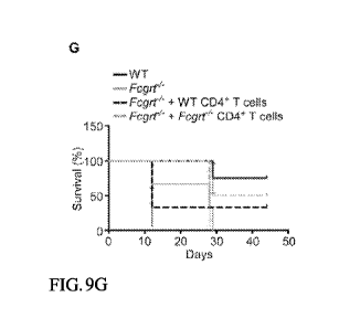
[0067] FIGS. 9A-9G depict, in accordance with various embodiments of the
present
invention that FcRn-mediated tumor immune surveillance is mediated by
selective activation and
retention of CD8+ T cells in the intestinal LP. (FIG. 9A) Flow cytometric
analysis of the frequency of
CD4+ I cells, NK cells (NK1.1) or macrophages (F4/80) in the lamina propria
lymphocyte (LPL)
fraction of tumor and adjacent LI tissue in WI and Fcgrt-/- mice following
AOM/DSS treatment.
(FIG. 9B) Absolute number of CD8+ I cells isolated from the LP tissue of the
adjacent, tumor or
untreated (baseline) LI in AOM/DSS-treated mice as assessed by flow cytometric
staining and
acquisition of fixed volumes of sample. (FIGS. 9C-9D) Flow cytometric analysis
of the frequency of
CD8+ I cells in the lamina propria (LP) fraction of tumor-adjacent LI tissue
in untreated ApcM1 and
ApcM11/ Fcgrt-/- mice (FIG. 9C) and in AOM-treated WI and Fcgrt-/- littermates
(FIG. 9D).
Representative results from one of three experiments with n = 3 mice per group
per experiment. (FIG.
9E) Flow cytometric analysis of the extent of CD8+ I cell proliferation (Ki-
67) and apoptosis
(Annexin V) in the LP fraction of tumor and adjacent LI tissue in WI and Fcgrt-
/-littermates following
AOM/DSS treatment. Plots depict cells within the CD3+CD8+ gate of cells.
Representative plots from
three independent experiments with n = 3 mice per group per experiment. (FIG.
9F) Phenotype of
CD8+ I cells in the indicated tissue compartment of WI and Fcgrt-/-
littermates treated with
AOM/DSS. Representative plots from three independent experiments with n = 3-4
mice per group per
experiment. (FIG. 9G) Survival rates of AOM/DSS-treated recipient mice
adoptively transferred with
CD4+ I cells taken from the MLN and LI LP of AOM/DSS-treated WI and Fcgrt-/-
donors.
Representative results from one of two independent experiments with n = 4 mice
per group per
11
CA 02966352 2017-04-28
WO 2015/081073 PCT/US2014/067332
experiment. Significance of survival curves was assessed by Logrank test. All
data represent mean
s.e.m. * p < 0.05.
[0068] FIGS. 10A-10L depict, in accordance with various embodiments of
the present
invention that FcRn-dependent dendritic cell (DC)-mediated tumor protection is
associated with the
presence of tumor-antigen reactive IgG in intestinal tissues and the
generation of a local Thl/Tcl
polarizing cytokine environment. (FIG. 10A) Isotype distribution of tumor
antigen-specific IgG in the
serum or LI homogenates of AOM/DSS treated WT or Fcgrt-/- mice. ELISA plates
coated with lysates
from tumor epithelium were probed with dilutions of serum or tissue
homogenates from tumor
bearing mice and developed with isotype-specific secondary antibodies. (FIG.
10B) Immunoblots
demonstrating tumor antigen-specific IgG in the serum and LI homogenates of
each of eight
AOM/DSS treated mice. IgG-depleted lysates prepared from tumor intestinal
epithelial cells (IEC) or
non-tumor control IEC were resolved under reducing conditions by SDS-PAGE and
membranes of
the transferred lysates were probed with serum or LI homogenates from tumor
bearing mice.
Representative blots from two independent experiments with n = 4 mice per
group per experiment. rd
Ab = anti-mouse IgG-HRP. (FIG. 10C) Fold increase above baseline values in
serum anti-
phosphatidylserine (a-PS) and anti-cardiolipin (a-CL) IgG content in WT and
Fcgrt-/- littermates.
(FIG. 10D) IgG isotype content of the serum, LI homogenates and MLN
homogenates in AOM/DSS-
treated WT and Fcgrt-/- mice. (FIG. 10E) Flow cytometric analysis of the
frequency of CD8+ versus
CD11b+ DC (top row, gated on CD11c+ cells) and characterization of the
CD11c+CD8-CD11b+ DC
(bottom four rows) in the mucosal tissues of AOM/DSS-treated WT and Fcgrt-/-
littermates. (FIGS.
10E-10H) Whole tissue cytokine transcript profiles of the indicated tissue
compartments from
AOM/DSS-treated WT and Fcgrt-/- littermates (FIG. 10F), untreated Apcmth/ and
Apcmm/ Fcgrt-/-
littermates (FIG. 10G) and AOM-treated WT and Fcgrt-/-littermates (FIG. 10H).
Representative
results from 2-4 independent experiments with n = 3-6 mice per group per
experiment. Data represent
mean s.e.m. (FIG. 101) Purity (top panel) and subset distribution (bottom
panel) of the DC
transferred in FIGS. 3C, 3F. (FIG. 10J) Frequency of congenic (CD45.2) WT or
Fcgrt-/- DC in the
MLN and LI LP of recipient (CD45.1) mice 3 days and 7 days after
intraperitoneal transfer. Recipient
mice injected with PBS are shown as controls. (FIG. 10K) Ex vivo antigen cross-
presentation with
DC isolated from the MLN of the indicated AOM/DSS-treated DC recipient mice
and loaded with IC
containing FcRn-binding (IgG) immune complex (IC), non-FcRn binding (IHH-IgG)
IC or soluble
antigen (OVA) and cocultured with OT-I CD8+ T cells. (FIG. 10L) Relative Fcgrt
transcript levels as
assessed by qPCR in DC (CD11c), macrophages (CD11b) and hepatocytes purified
from WT, Fcgrt-/-,
Fcg
rtFl/Flor Itgax'FcgrtFl/Fimice. Data are representative of two independent
experiments with n = 3-
mice per group per experiment. All data represent mean s.e.m. * p 5 0.05, **
p 5 0.01, *** p 5
0.005.
[0069] FIGS. 11A-11F depict, in accordance with various embodiments of
the present
invention that both CD8+ T cells and tumor-specific IgG are required for FcRn-
mediated protection
12
CA 02966352 2017-04-28
WO 2015/081073 PCT/US2014/067332
from pulmonary metastases. (FIG. 11A) CD8+ T cell frequency in the lungs of
untreated WT or
Fcgrt-/- mice. (FIGS. 11B-11C) Tumor-specific anti-OVA IgG in the lung
homogenates (FIG. 11B)
or serum (FIG. 11C) of WT and Fcgrt-/- littermates bearing lung metastases.
Lungs were harvested 2
weeks after i.v. injection of 0.5x10⁶ OVA-expressing B16 melanoma cells (OVA-
B16). Anti-OVA
IgG content was evaluated by ELISA and normalized to protein content of the
homogenates for (FIG.
11B). (FIG. 11D) Frequency of OVA-specific IgG producing B cells in the lymph
nodes (LN) or
spleens of OVA-B16 lung metastasis-bearing WT and Fcgrt-/- mice. B cells were
isolated and cultured
on OVA-coated ELISpot plates for 24 h. (FIG. 11E) Representative lobes from
immunized or non-
immunized mice injected with OVA-B16 cells. (FIG. 11F) Ex vivo antigen cross-
presentation using
DC stimulated with immune complexes (IC) formed with NIP-OVA and either FcRn-
binding (IgG
IC), non-FcRn binding (IHH-IgG IC) or enhanced FcRn-binding (LS-IgG IC)
immunoglobulin and
cocultured with OT-I CD8+ T cells. All data are representative of the results
of one of three
independent experiments with n = 3-6 mice per group per experiment. Data
represent mean s.e.m.
NS = not significant. * p < 0.05.
[0070] FIGS. 12A-12E depict, in accordance with various embodiments of
the present
invention that FcRn in DC drives homeostatic local activation of CD8+ T cells
and the Thl
polarization of CD4+ T cells within the LI LP by facilitating the production
of Tcl and Thl polarizing
cytokines. (FIG. 12A) Frequency (upper panels) and effector status (lower
panels) of adoptively
transferred congenic CD8+ I cells (CD45.1) in the LI LP and MLN of untreated
WI and Fcgrt-/-
recipient mice (CD45.2). 1x10⁶ CD8+ I cells were transferred into recipient
mice via i.v. injection and
their distribution and phenotype was assessed 10 days later. (FIG. 12B)
Frequency of adoptively
transferred congenic CD8+ I cells from WI and Fcgrt-/- donor mice (CD45.2) in
the LI LP and MLN of
untreated recipient mice (CD45.1) 7 days after transfer. (FIG. 12C) Whole
tissue transcript profiles of
the LI from untreated WI and Fcgrt-/- littermates (left panel) or FcgrtFl/F1
and Itgax"eFcgrtFl/F1
littermates (right panel), as assessed by qPCR. (FIG. 12D) Cytokine secretion
by CD4+ I cells isolated
via magnetic sorting from the LI LP of untreated WI and Fcgrt-/- mice and
restimulated for 24 h with
anti-CD3 (aCD3) and anti-CD28 (aCD28). (FIG. 12E) CD4+ I cell frequency in the
LI LP of
untreated WI and Fcgrt-/- mice (left panels) or FcgrtFl/F1 and
Itgax'FcgrtFl/F1 mice (right panels).
Representative results from three independent experiments with n = 3-5 mice
per group per
experiment. Data represent mean s.e.m. * p < 0.05, ** p < 0.01, *** p <
0.005.
[0071] FIGS. 13A-13C depict, in accordance with various embodiments of
the present
invention that FcRn-dependent induction of IL-12 is not dependent on MYD88 and
is not required for
FcRn-mediated cross-priming. (FIG. 13A) Induction of IL-12p35 upon ex vivo
stimulation of Myd88-
/- CD8-CD11b+ DC with FcRn-binding IC (IgG IC) or FcRn non-binding IC (IHH-IgG
IC) for 6 h.
(FIG. 13B) Binding of IRF-1 to the IL-12p35 promoter upon stimulation of Myd88-
/-CD8-CD11b+ DC
with IgG IC or IHH-IgG IC for 4 h. (FIG. 13C) Ex vivo antigen cross-
presentation in the presence of
an IL-12 neutralizing antibody (aIL-12) or isotype control. DC were stimulated
with IgG IC or IHH-
13
CA 02966352 2017-04-28
WO 2015/081073 PCT/US2014/067332
IgG IC and co-cultured with OT-I CD8+ T cells. Representative data shown from
one of three
independent experiments. Data represent mean s.e.m. * p < 0.05, ** p < 0.01.
[0072] FIGS. 14A-14H depict, in accordance with various embodiments of
the present
invention that human DC strongly express FcRn and localize to the stroma of
both normal and CRC-
associated LI. (FIG. 14A) Double immunohistochemical staining of FcRn+CD11c
DC in the stroma
of CRC-bearing (upper panels) and tumor-adjacent normal (lower panels) LI of
additional cases of
human CRC. See also FIG. 7A. Scale bar left panels = 100 [Lin. Scale bar right
panels = 20 [tm. (FIG.
14B) Colocalization of FcRn + DC and CD8+ T cells in the stroma of CRC-bearing
(upper panels) and
tumor-adjacent normal (lower panels) LI of additional cases of human CRC.
Arrowheads indicate
areas of colocalization. See also FIG. 7B. Data in FIGS. 14A-14B are
representative of 50 matched
normal LI and CRC assessed. (FIG. 14C) Correlation between the number of
FcRn+CD11c DC and
CD8+ T cells in the stroma of normal LI adjacent to CRC. Significance was
assessed using
Spearman's rank correlation. (FIG. 14D) Multivariable analysis of the impact
of colonic LP
CD11c+FcRn+ cells on patient survival in 183 human CRC patients. An increasing
number of
CD11+FcRn+ cells has a positive effect on patient survival (univariate
analysis p = 0.0333) and this
effect is maintained in multivariable analysis with the indicated parameters.
See also FIG. 7C. (FIG.
14E) CD8+ T cell frequency in the tumor LP of chimeric mice treated with
AOM/DSS. WT recipients
were reconstituted with bone marrow from WT donors. Fcgrt-/- recipients were
reconstituted with
bone marrow from either Fcgrt-/-, WT or hFCGRT-hB2M-inFcgrf/- donors.
Representative results
from one of two independent experiments with n = 4-5 mice per group per
experiment. (FIG. 14F)
Phenotype of human monocyte derived DC (hMoDC) as determined by flow
cytometric analysis.
Shaded curve represents isotype control. (FIG. 14G) Expression of FcRn in
hMoDC, as assessed by
the same antibody utilized for immunohistochemical staining of CRC cases.
(FIG. 14H)
Densitometric analysis of Western blots of hMoDC stimulated with IgG IC
depicted in FIG. 7F. Data
in panels FIGS. 14F-14H are representative of six donors processed in pairs in
each of three
independent experiments. All data represent mean s.e.m.
[0073] FIGS. 15A-15B depict, in accordance with various embodiments of
the present
invention that antibody mediated blockade of FcRn decreases Thl cytokine
transcript levels during
IgG-mediated colitis. (FIG. 15A) Whole colon cytokine transcript levels from
flagellin immunized
hFCGRT/hB2M/mFcgrt-/- chimeric mice treated with either DVN24 or an isotype
control before and
during the onset of DSS colitis. (FIG. 15B) Cytokine transcript levels from
CD8+ T cells isolated
from the lamina propria of hFCGRT/hB2M/mFcgrt-/- BM chimeras treated with
DVN24 or an isotype
control or mFcgrt-/- BM chimeras treated with PBS during DSS colitis. *P <
0.05. **P < 0.01.
[0074] FIG. 16 depicts, in accordance with various embodiments of the
present invention,
FcRn-mediated upregulation of IL-12p35 is dependent upon ERK and calmodulin
but not cytoskeletal
rearrangements. Primary mouse dendritic cells were stimulated with IgG
containing immune
complexes against chicken ovalbumin that were wild type and able to bind FcRn
or mutant and unable
14
CA 02966352 2017-04-28
WO 2015/081073 PCT/US2014/067332
to bind FcRn (IHH-IC) due to three mutations in the Fc domain of IgG. RNA was
isolated after 5 or
11 hours from such cells treated with inhibitors for cytoskeleton
(cytochalasin D and Latrunculin),
calmodulin (W7) or ERK. The RNA was reverse transcribed and IL-12 p35 was
quantified by qPCR.
DETAILED DESCRIPTION OF THE INVENTION
[0075] All references cited herein are incorporated by reference in their
entirety as though
fully set forth. Unless defined otherwise, technical and scientific terms used
herein have the same
meaning as commonly understood by one of ordinary skill in the art to which
this invention belongs.
Singleton et al., Dictionary of Microbiology and Molecular Biology 3rd ed., J.
Wiley & Sons (New
York, NY 2001); March, Advanced Organic Chemistry Reactions, Mechanisms and
Structure 5th ed.,
J. Wiley & Sons (New York, NY 2001); and Sambrook and Russel, Molecular
Cloning: A Laboratory
Manual 3rd ed., Cold Spring Harbor Laboratory Press (Cold Spring Harbor, NY
2001), provide one
skilled in the art with a general guide to many of the terms used in the
present application.
[0076] One skilled in the art will recognize many methods and materials
similar or
equivalent to those described herein, which could be used in the practice of
the present invention.
Indeed, the present invention is in no way limited to the methods and
materials described. For
purposes of the present invention, the following terms are defined below.
[0077] As used herein, the term "antibody" refers to an intact
immunoglobulin or to a
monoclonal or polyclonal antigen-binding fragment with the Fc (crystallizable
fragment) region or
FcRn binding fragment of the Fc region, referred to herein as the "Fc
fragment" or "Fc domain".
Antigen-binding fragments may be produced by recombinant DNA techniques or by
enzymatic or
chemical cleavage of intact antibodies. Antigen-binding fragments include,
inter alia, Fab, Fab',
F(ab')2, Fv, dAb, and complementarity determining region (CDR) fragments,
single-chain antibodies
(scFv), single domain antibodies, chimeric antibodies, diabodies, tetrabodies
and other multimerized
scFv moieties and polypeptides that contain at least a portion of an
immunoglobulin that is sufficient
to confer specific antigen binding to the polypeptide. The Fc domain includes
portions of two heavy
chains contributing to two or three classes of the antibody. The Fc domain may
be produced by
recombinant DNA techniques or by enzymatic (e.g. papain cleavage) or via
chemical cleavage of
intact antibodies. The amino acid sequence of the Fc domain can be modified to
increase its affinity
with FcRn to enable better induction of secreted proteins such as IL-12,
interferon-gamma, IL-2,
tumor necrosis factor, granulocyte macrophage colony stimulating factor, IL-3
and granzyme B or
transcription factors such as t-bet or modified to decrease its affinity for
FcRn and the induction of
these aforementioned cytokines and transcription factors.
[0078] The term "antibody fragment," as used herein, refer to a protein
fragment that
comprises only a portion of an intact antibody, generally including an antigen
binding site of the intact
antibody and thus retaining the ability to bind antigen. Examples of antibody
fragments encompassed
CA 02966352 2017-04-28
WO 2015/081073 PCT/US2014/067332
by the present definition include: (i) the Fab fragment, having VL, CL, VH and
CH1 domains; (ii) the
Fab' fragment, which is a Fab fragment having one or more cysteine residues at
the C-terminus of the
CH1 domain; (iii) the Fd fragment having VH and CH1 domains; (iv) the Fd'
fragment having VH
and CH1 domains and one or more cysteine residues at the C-terminus of the CH1
domain; (v) the Fv
fragment having the VL and VH domains of a single arm of an antibody; (vi) the
dAb fragment (Ward
et al., Nature 341, 544-546 (1989)) which consists of a VH domain; (vii)
isolated CDR regions; (viii)
F(ab')2 fragments, a bivalent fragment including two Fab' fragments linked by
a disulphide bridge at
the hinge region; (ix) single chain antibody molecules (e.g., single chain Fv;
scFv) (Bird et al.,
Science 242:423-426 (1988); and Huston et al., PNAS (USA) 85:5879-5883
(1988)); (x) "diabodies"
with two antigen binding sites, comprising a heavy chain variable domain (VH)
connected to a light
chain variable domain (VL) in the same polypeptide chain (see, e.g., EP
404,097; WO 93/11161; and
Hollinger et al., Proc. Natl. Acad. Sci. USA, 90:6444-6448 (1993)); (xi)
"linear antibodies"
comprising a pair of tandem Fd segments (VH-CH1-VH-CH1) which, together with
complementary
light chain polypeptides, form a pair of antigen binding regions (Zapata et
al. Protein Eng.
8(10):1057-1062 (1995); and U.S. Pat. No. 5,641,870).
[0079] As described herein, an "antigen" is a molecule that is bound by a
binding site on a
polypeptide agent, such as an antibody or antibody fragment thereof Typically,
antigens are bound
by antibody ligands and are capable of raising an antibody response in vivo.
An antigen can be a
polypeptide, protein, nucleic acid, lipid or other molecule. In the case of
conventional antibodies and
fragments thereof, the antibody binding site as defined by the variable loops
(L1, L2, L3 and H1, H2,
H3) is capable of binding to the antigen. The term "antigenic determinant"
refers to an epitope on the
antigen recognized by an antigen-binding molecule, and more particularly, by
the antigen-binding site
of said molecule.
[0080] An "Fv" fragment is an antibody fragment which contains a complete
antigen
recognition and binding site. This region consists of a dimer of one heavy and
one light chain variable
domain in tight association, which can be covalent in nature, for example in
scFv. It is in this
configuration that the three CDRs of each variable domain interact to define
an antigen binding site
on the surface of the VH-VL dimer. Collectively, the six CDRs or a subset
thereof confer antigen
binding specificity to the antibody. However, even a single variable domain
(or half of an Fv
comprising only three CDRs specific for an antigen) has the ability to
recognize and bind antigen,
although usually at a lower affinity than the entire binding site.
[0081] A "cancer" or "tumor" as used herein refers to an uncontrolled
growth of cells which
interferes with the normal functioning of the bodily organs and systems. A
subject that has a cancer
or a tumor is a subject having objectively measurable neoplastic cells present
in the subject's body.
Included in this definition are benign and malignant cancers, as well as
dormant tumors or
micrometastases. Cancers which migrate from their original location and seed
vital organs can
eventually lead to the death of the subject through the functional
deterioration of the affected organs.
16
CA 02966352 2017-04-28
WO 2015/081073 PCT/US2014/067332
Hematopoietic cancers, such as leukemia, are able to out-compete the normal
hemopoietic
compartments in a subject, thereby leading to hemopoietic failure (in the form
of anemia,
thrombocytopenia and neutropenia) ultimately causing death.
[0082] As used herein, "complexed" refers to the non-covalent
interactions between any two
molecules. Examples include but are not limited to complexes formed between
and an antigen and an
antibody (for example, IgG or a variant thereof or a fragment thereof) wherein
the antigen and the
antibody interact via non-covalent bonds. Examples of non-covalent
interactions include but are not
limited to electrostatic interactions (for example, ionic interactions,
hydrogen bonds, halogen bonds),
van der Waals forces (dipole-dipole, dipole-induced, London dispersion
forces), pi-effects (pi-pi
interactions, cation-pi, anion-pi, polar-pi) and/or hydrophobic interactions.
In various embodiments,
the complex between the IgG or a fragment thereof or a variant thereof and the
antigen forms
multimeric structures that can cross-link/bind with FcRn.
[0083] As used herein, "conjugated" refers to covalent interactions
between any two
molecules. Examples include but are not limited to fusion proteins comprising
an antigen and an
antibody (for example, IgG or a variant thereof or a fragment thereof) or any
other antigen-antibody
complex that may be covalently linked. In various embodiments, the conjugation
between the IgG or
a fragment thereof or a variant thereof forms and the antigen forms monomeric
or multimeric
structures that can cross-link/bind with FcRn.
[0084] As used herein, an "epitope" can be formed both from contiguous
amino acids, or
noncontiguous amino acids juxtaposed by tertiary folding of a protein.
Epitopes formed from
contiguous amino acids are typically retained on exposure to denaturing
solvents, whereas epitopes
formed by tertiary folding are typically lost on treatment with denaturing
solvents. An epitope
typically includes at least 3, and more usually, at least 5, about 9, or about
8-10 amino acids in a
unique spatial conformation. An "epitope" includes the unit of structure
conventionally bound by an
immunoglobulin VH/VL pair. Epitopes define the minimum binding site for an
antibody, and thus
represent the target of specificity of an antibody. In the case of a single
domain antibody, an epitope
represents the unit of structure bound by a variable domain in isolation. The
terms "antigenic
determinant" and "epitope" can also be used interchangeably herein. In various
embodiments, an
epitope may be protein, peptide, nucleic acid, lipid, other molecules or
combinations thereof
[0085] As used herein, an "immune response" being modulated refers to a
response by a cell
of the immune system, such as a B cell, T cell (CD4 or CD8), regulatory T
cell, antigen-presenting
cell, dendritic cell, monocyte, macrophage, NKT cell, NK cell, basophil,
eosinophil, or neutrophil, to
a stimulus. In some embodiments, the response is specific for a particular
antigen (an "antigen-
specific response"), and refers to a response by a CD4 T cell, CD8 T cell, or
B cell via their antigen-
specific receptor. In some embodiments, an immune response is a T cell
response, such as a CD4+
response or a CD8+ response. Such responses by these cells can include, for
example, cytotoxicity,
proliferation, cytokine or chemokine production, trafficking, or phagocytosis,
and can be dependent
17
CA 02966352 2017-04-28
WO 2015/081073 PCT/US2014/067332
on the nature of the immune cell undergoing the response. In some embodiments
of the compositions
and methods described herein, an immune response being modulated is T-cell
tolerance.
[0086] By "metastasis" is meant the spread of cancer from its primary
site to other places in
the body. Cancer cells can break away from a primary tumor, penetrate into
lymphatic and blood
vessels, circulate through the bloodstream, and grow in a distant focus
(metastasize) in normal tissues
elsewhere in the body. Metastasis can be local or distant. Metastasis is a
sequential process,
contingent on tumor cells breaking off from the primary tumor, traveling
through the bloodstream,
and stopping at a distant site. At the new site, the cells establish a blood
supply and can grow to form
a life-threatening mass. Both stimulatory and inhibitory molecular pathways
within the tumor cell
regulate this behavior, and interactions between the tumor cell and host cells
in the distant site are also
significant.
[0087] The term "monoclonal antibody" as used herein refers to an
antibody obtained from a
population of substantially homogeneous antibodies, i.e., the individual
antibodies comprising the
population are identical except for possible naturally occurring mutations
that can be present in minor
amounts. Monoclonal antibodies are highly specific, being directed against a
single antigen.
Furthermore, in contrast to polyclonal antibody preparations that typically
include different antibodies
directed against different determinants (epitopes), each monoclonal antibody
is directed against a
single determinant on the antigen. The modifier "monoclonal" is not to be
construed as requiring
production of the antibody by any particular method. For example, the
monoclonal antibodies to be
used in accordance with the invention can be made by the hybridoma method
first described by
Kohler et al., Nature 256:495 (1975), or can be made by recombinant DNA
methods (see, e.g., U.S.
Pat. No. 4,816,567). The "monoclonal antibodies" can also be isolated from
phage antibody libraries
using the techniques described in Clackson et al., Nature 352:624-628 (1991)
or Marks et al., J. Mol.
Biol. 222:581-597 (1991), for example.
[0088] As used herein, "selectively binds" or "specifically binds" refers
to the ability of an
antibody or antibody fragment thereof described herein to bind to a target,
such as a molecule present
on the cell-surface, with a KD 10-5 M (10000 nM) or less, e.g., 10-6 M, 10-7
M, 10-8 M, 10-9 M, 10-10
M, 10-11 M, 10-12 M, or less. Specific binding can be influenced by, for
example, the affinity and
avidity of the polypeptide agent and the concentration of polypeptide agent.
The person of ordinary
skill in the art can determine appropriate conditions under which the
polypeptide agents described
herein selectively bind the targets using any suitable methods, such as
titration of a polypeptide agent
in a suitable cell binding assay.
[0089] "Subject" or "individual" or "animal" or "patient" or "mammal," is
meant any
subject, particularly a mammalian subject, for whom diagnosis, prognosis, or
therapy is desired. In
some embodiments, the subject has cancer. In some embodiments, the subject had
cancer at some
point in the subject's lifetime. In various embodiments, the subject's cancer
is in remission, is re-
18
CA 02966352 2017-04-28
WO 2015/081073 PCT/US2014/067332
current or is non-recurrent. In some embodiments the subject has an infectious
disease. In some
embodiments, the subject has an autoimmune disease.
[0090] "Mammal" as used herein refers to any member of the class
Mammalia, including,
without limitation, humans, domestic animals, farm animals, zoo animals, sport
animals, pet animals
such as dogs, cats, guinea pigs, rabbits, rats, mice, horses, cattle, cows;
primates such as apes,
monkeys, orangutans, and chimpanzees; canids such as dogs and wolves; felids
such as cats, lions,
and tigers; equids such as horses, donkeys, and zebras; food animals such as
cows, pigs, and sheep;
ungulates such as deer and giraffes; rodents such as mice, rats, hamsters and
guinea pigs; and so on. In
certain embodiments, the mammal is a human subject. The term does not denote a
particular age or
sex. Thus, adult and newborn subjects, as well as fetuses, whether male or
female, are intended to be
included within the scope of this term
[0091] As used herein, the term "target" refers to a biological molecule
(e.g., peptide,
polypeptide, protein, lipid, carbohydrate) to which a polypeptide domain which
has a binding site can
selectively bind. The target can be, for example, an intracellular target
(e.g., an intracellular protein
target) or a cell surface target (e.g., a membrane protein, a receptor
protein). Preferably, a target is a
cell surface target, such as a cell surface protein.
[0092] As used herein, the terms "treat," "treatment" "treating," or
"amelioration" refer to
therapeutic treatments, wherein the object is to reverse, alleviate,
ameliorate, inhibit, slow down or
stop the progression or severity of a condition associated with, a disease or
disorder. The term
"treating" includes reducing or alleviating at least one adverse effect or
symptom of a condition,
disease or disorder, such as an autoimmune disease, a chronic infection or a
cancer. Treatment is
generally "effective" if one or more symptoms or clinical markers are reduced.
Alternatively,
treatment is "effective" if the progression of a disease is reduced or halted.
That is, "treatment"
includes not just the improvement of symptoms or markers, but also a cessation
of at least slowing of
progress or worsening of symptoms that would be expected in absence of
treatment. Beneficial or
desired clinical results include, but are not limited to, alleviation of one
or more symptom(s),
diminishment of extent of disease, stabilized (i.e., not worsening) state of
disease, delay or slowing of
disease progression, amelioration or palliation of the disease state, and
remission (whether partial or
total), whether detectable or undetectable. The term "treatment" of a disease
also includes providing
relief from the symptoms or side-effects of the disease (including palliative
treatment).
[0093] A number of tumor antigens have been identified that are
associated with specific
cancers. As used herein, the terms "tumor antigen" and "cancer antigen" are
used interchangeably to
refer to antigens which are differentially expressed by neoplastic cells and
can thereby be exploited in
order to target neoplastic cells. Cancer antigens are antigens which can
potentially stimulate
apparently tumor-specific immune responses. Some of these antigens are
encoded, although not
necessarily expressed, by normal cells. These antigens can be characterized as
those which are
normally silent (i.e., not expressed) in normal cells, those that are
expressed only at certain stages of
19
CA 02966352 2017-04-28
WO 2015/081073 PCT/US2014/067332
differentiation and those that are temporally expressed such as embryonic and
fetal antigens. Other
cancer antigens are encoded by mutant cellular genes, such as oncogenes (e.g.,
activated ras
oncogene), suppressor genes (e.g., mutant p53), fusion proteins resulting from
internal deletions or
chromosomal translocations. Still other cancer antigens can be encoded by
viral genes such as those
carried on RNA and DNA tumor viruses. Many tumor antigens have been defined in
terms of multiple
solid tumors: MAGE 1, 2, & 3, defined by immunity; MART-1/Melan-A, gp100,
carcinoembryonic
antigen (CEA), HER-2, mucins (i.e., MUC-1), prostate-specific antigen (PSA),
and prostatic acid
phosphatase (PAP). In addition, viral proteins such as hepatitis B (HBV),
Epstein-Barr (EBV), and
human papilloma (HPV) have been shown to be important in the development of
hepatocellular
carcinoma, lymphoma, and cervical cancer, respectively. However, due to the
immunosuppression of
patients diagnosed with cancer, the immune systems of these patients often
fail to respond to the
tumor antigens.
[0094] "Endogenous antigens" as used herein refers to antigens that have
been generated
within the body. They include xenogenic (heterologus), autologus, idiotypic or
allogenic antigens and
autoantigens.
[0095] FcRn is a neonatal Fc receptor. It is similar in structure to
major histocompatibility
complex I (MHC I). Human FcRn is very stringent regarding its specificity and
binds human Fc, but
not mouse, rat, bovine, or sheep Fc. The FcRn can bind to two sites of the IgG
(Sanchez et al., 1999;
Schuck et al., 1999; West A.P. and Bjorkman, 2000). Although mouse IgGs do not
bind efficiently to
human FcRn and therefore have a short half-life in humans (Frodin et al.,
1990), mouse IgG as an
immune complex is capable of binding human FcRn and inducing signaling. In
contrast, mouse FcRn
binds IgG from every species analyzed (Ober et al., 2001).
[0096] Most serum proteins have a short serum half-life (about 1-2 days).
However, two
types of serum proteins, namely albumin and antibodies of the IgG class, have
greatly extended serum
half-lives. For example, most subclasses of IgG have a half-life of about 10-
20 days in humans. The
Fc region of IgG is required for this extension of half-life. Thus, truncated
IgG polypeptides carrying
only the Fc region, and potentially also proteins carrying a short FcRn
binding peptide sequence
(FcBP) (Sockolosky et al. Proc Nati Acad Sci U S A 2012, 109, 16095-100), also
show such extended
serum half-life. Moreover, when the Fc region is fused with a fusion partner
(e.g., a biologically
active protein), this Fc fusion protein shows an extended serum half-life due
to its interaction with
FcRn.
[0097] The mechanism by which FcRn extends the serum half-life of IgG and
IgG Fc fusion
proteins is well established (Ghetie and Ward, 2000, 2002; Roopenian and
Akilesh, 2007). FcRn is
localized in the endosomal compartments of many cell types, including vascular
endothelium. Serum
proteins are constantly being endocytosed and directed to the early endosomal
vesicles. FcRn is
harbored primarily in this acidified vesicle. In this acidified environment,
the Fc region binds FcRn,
and the IgG/FcRn complex is then recycled either apically or basolaterally
back to the plasma
CA 02966352 2017-04-28
WO 2015/081073 PCT/US2014/067332
membrane, whereupon exposure to the neutral pH 7.2 extracellular environment
results in its release
into the circulation. In contrast, other endocytosed proteins that do not bind
FcRn are not rescued,
and thus continue through the endosomal route to catabolic elimination,
resulting in their short half-
life. The biochemical mechanism by which the Fc region of IgG binds FcRn in an
acidic environment
is well understood. The CH2-CH3-hinge region of the Fc region contains solvent
exposed histidine
residues, which when protonated, engage residues on FcRn with sufficient
affinity to permit IgG to
exploit the FcRn recycling pathway to escape catabolic elimination.
[0098] As described herein, the inventors discovered that cross-linking
FcRn on dendritic
cells with antigen/antibody immune complexes, which function as ligands for
FcRn, directly leads to
the production of IL-12 by these cells, as well as interferon-gamma, tumor
necrosis factor, granzyme
B and t-bet by CD8+ T cells. FcRn functions as a signaling molecule by
organizing the necessary
proteins, including elements of the cytoskeleton and mitogen activated protein
kinases (MAPK) to
directly promote the transcription of IL-12 through factors such as IRF-1 and
NFKB. As
demonstrated herein, FcRn-mediated upregulation of IL-12p35 is dependent upon
ERK and
calmodulin but not cytoskeletal rearrangements. The production of FcRn-
dependent IL-12 is essential
for the generation of CD8+ effector T cells and their effector function
through factors such interferon-
gamma, tumor necrosis factor, granzyme B and t-bet. Such effector T cells
mediate anti-infectious
immunity and mediate tumor immune-surveillance and eradication. If IL-12 is
neutralized, the FcRn-
mediated effects on CD8+ effector T cell functions that are associated with
tumor eradication are lost.
[0099] In a homologous manner, blockade of FcRn function is associated
with decreased
FcRn-mediated signal transduction and thereby decreased production of IL-12
and related factors
which is associated with protection from autoimmune disorders as shown in
animal models of
inflammatory bowel disease. While not wishing to be bound by any particular
mechanism or theory,
the aspects and embodiments described herein are based on the finding that
binding of FcRn to
antigen/antibody immune complexes regulates IL-12 production by dendritic
cells, which can be
manipulated for therapeutic purposes. Since IL-12 is a master regulator of
immune responses
associated with tumor and anti-infectious immunity on the one hand, and
inappropriate inflammation
on the other, it is desirable to increase IL-12 for anti-tumor immunity and
decrease IL-12 for anti-
inflammatory purposes. Accordingly, described herein are compositions and
methods for
increasing/enhancing/up-regulating IL-12 production for treating cancer and
infectious diseases and
compositions and methods for decreasing IL-12 production for treating
autoimmune disorders.
Compositions and Methods for treating cancer and/or infectious diseases
[0100] In some aspects, the compositions and methods described herein up-
regulate/increase/enhance production of IL-12 by increasing/enhancing
interaction between IgG and
FcRn (for example, in response to an antigen bound to the IgG, or due to
mutations in IgG that
increase the binding between IgG and FcRn). The interactions between IgG and
FcRn yield signals
that result in production of IL-12 by dendritic cells. The compositions
provided herein comprise IgG
21
CA 02966352 2017-04-28
WO 2015/081073 PCT/US2014/067332
or a variant thereof or a fragment thereof so as to increase IL-12 production.
As described herein, the
compositions can further comprise antigens, including but not limited to,
tumor antigens, bacterial
antigens, viral antigens, parasitic antigens or combinations thereof, as to
increase IL-12 production.
As used herein, "IgG" can refer to any isotype of IgG including IgGl, IgG2,
IgG3 and/or IgG4.
[0101] In some embodiments, the variants of IgG in the compositions and
methods described
herein are functional, non-naturally occurring variants of IgG that enhance
binding between IgG and
FcRn, so as to increase IL-12 production. "Functional variants of IgG," as
used herein, useful with
the compositions and methods described herein include molecules comprising
mutations, such as
insertions, deletions and truncations in full-length IgG,or the constant
region of an IgG molecule,
provided such molecules retain the ability to bind to FcRn and increase IL-12
production. One
example of such a variant includes, but is not limited to, an IgG comprising a
methionine to leucine
substitution at position 428 and an asparagine to serine substitution at
position 434 according to the
Kabat numbering scheme. Another example of a functional IgG variant useful
with the compositions
and methods described herein is an engineered variant of human IgG1 omprising
mutations of Met
252, Ser254, Thr256, His433, and Asn434 to Tyr252, Thr254, G1u256, Lys433, and
Phe434, as
described in "Engineering the Fc region of immunoglobulin G to modulate in
vivo antibody levels,"
Nature Biotechnology, 2005 (23):10, pp 1283-88, the contents of which are
herein incorporated by
reference in their entireties. Other functional IgG variants useful with the
compositions and methods
described herein are described in US8188231, US8802823, U52011025068,
U520060235208,
W02012106578 Al, the contents of each of which are herein incorporated by
reference in their
entireties.
[0102] For example, functional IgG variants can comprise amino acid
modifications at Fc
positions 230, 240, 244, 245, 247, 262, 263, 266, 273, 275, 299, 302, 313,
323, 325, 328, and 332,
wherein the numbering of the residues in the Fc region is that of the EU index
as in Kabat. In some
embodiments, functional IgG variants can comprise at least one amino acid
substitution in the Fc
region at a position selected from the group consisting of 221, 222, 223, 224,
225, 227, 228, 230, 231,
232, 233, 234, 235, 236, 237, 238, 239, 240, 241, 243, 244, 245, 246, 247,
249, 255, 258, 260, 262,
263, 264, 265, 266, 267, 268, 269, 270, 271, 272, 273, 274, 275, 276, 278,
280, 281, 282, 283, 284,
285, 286, 288, 290, 291, 292, 293, 294, 295, 296, 297, 298, 299, 300, 301,
302, 303, 304, 305, 313,
317, 318, 320, 322, 323, 324, 325, 326, 327, 328, 329, 330, 331, 332, 333,
334, 335, 336, and 337,
wherein the numbering of the residues in the Fc region is that of the EU index
as in Kabat.
[0103] In some embodiments, said IgG variants comprise at least one
substitution selected
from the group consisting of D221K, D221Y, K222E, K222Y, T223E, T223K, H224E,
H224Y,
T225E, T225K, T225W, P227E, P227G, P227K, P227Y, P228E, P228G, P228K, P228Y,
P230A,
P230E, P230G, P230Y, A231E, A231G, A231K, A231P, A231Y, P232E, P232G, P232K,
P232Y,
E233A, E233D, E233F, E233G, E233H, E2331, E233K, E233L, E233M, E233N, E233Q,
E233R,
E2335, E233T, E233V, E233W, E233Y, L234A, L234D, L234E, L234F, L234G, L234H,
L234I,
22
CA 02966352 2017-04-28
WO 2015/081073 PCT/US2014/067332
L234K, L234M, L234N, L234P, L234Q, L234R, L234S, L234T, L234V, L234W, L234Y,
L235A,
L235D, L235E, L235F, L235G, L235H, L235I, L235K, L235M, L235N, L235P, L235Q,
L235R,
L235S, L235T, L235V, L235W, L235Y, G236A, G236D, G236E, G236F, G236H, G236I,
G236K,
G236L, G236M, G236N, G236P, G236Q, G236R, G236S, G236T, G236V, G236W, G236Y,
G237D,
G237E, G237F, G237H, G237I, G237K, G237L, G237M, G237N, G237P, G237Q, G237R,
G2375,
G237T, G237V, G237W, G237Y, P238D, P238E, P238F, P238G, P238H, P238I, P238K,
P238L,
P238M, P238N, P238Q, P238R, P238S, P238T, P238V, P238W, P238Y, 5239D, 5239E,
5239F,
5239G, 5239H, S239I, 5239K, 5239L, 5239M, 5239N, 5239P, 5239Q, 5239R, 5239T,
5239V,
5239W, 5239Y, V240A, V240I, V240M, V240T, F241D, F241E, F241L, F241R, F2415,
F241W,
F241Y, F243E, F243H, F243L, F243Q, F243R, F243W, F243Y, P244H, P245A, K246D,
K246E,
K246H, K246Y, P247G, P247V, D249H, D249Q, D249Y, R255E, R255Y, E258H, E2585,
E258Y,
T260D, T260E, T260H, T260Y, V262A, V262E, V262F, V262I, V262T, V263A, V263I,
V263M,
V263T, V264A, V264D, V264E, V264F, V264G, V264H, V264I, V264K, V264L, V264M,
V264N,
V264P, V264A, V264R, V2645, V264T, V264W, V264Y, D265F, D265G, D265H, D265I,
D265K,
D265L, D265M, D265N, D265P, D265Q, D265R, D2655, D265T, D265V, D265W, D265Y,
V266A,
V266I, V266M, V266T, 5267D, 5267E, 5267F, 5267H, S267I, 5267K, 5267L, 5267M,
5267N,
5267P, 5267Q, 5267R, 5267T, 5267V, 5267W, 5267Y, H268D, H268E, H268F, H268G,
H268I,
H268K, H268L, H268M, H268P, H268Q, H268R, H268T, H268V, H268W, E269F, E269G,
E269H,
E2691, E269K, E269L, E269M, E269N, E269P, E269R, E2695, E269T, E269V, E269W,
E269Y,
D270F, D270G, D270H, D270I, D270L, D270M, D270P, D270Q, D270R, D2705, D270T,
D270W,
D270Y, P271A, P271D, P271E, P271F, P271G, P271H, P271I, P271K, P271L, P271M,
P271N,
P271Q, P271R, P271S, P271T, P271V, P271W, P271Y, E272D, E272F, E272G, E272H,
E2721,
E272K, E272L, E272M, E272P, E272R, E2725, E272T, E272V, E272W, E272Y, V273I,
K274D,
K274E, K274F, K274G, K274H, K274I, K274L, K274M, K274N, K274P, K274R, K274T,
K274V,
K274W, K274Y, F275L, F275W, N276D, N276E, N276F, N276G, N276H, N276I, N276L,
N276M,
N276P, N276R, N2765, N276T, N276V, N276W, N276Y, Y278D, Y278E, Y278G, Y278H,
Y278I,
Y278K, Y278L, Y278M, Y278N, Y278P, Y278Q, Y278R, Y2785, Y278T, Y278V, Y278W,
D280G,
D280K, D280L, D280P, D280W, G281 D, G281 E, G281K, G281N, G281P, G281Q, G281Y,
V282E, V282G, V282K, V282P, V282Y, E283G, E283H, E283K, E283L, E283P, E283R,
E283Y,
V284D, V284E, V284L, V284N, V284Q, V284T, V284Y, H285D, H285E, H285K, H285Q,
H285W,
H285Y, N286E, N286G, N286P, N286Y, K288D, K288E, K288Y, K290D, K290H, K290L,
K290N,
K290W, P291D, P291E, P291G, P291H, P291I, P291Q, P291T, R292D, R292E, R292T,
R292Y,
E293F, E293G, E293H, E2931, E293L, E293M, E293N, E293P, E293R, E2935, E293T,
E293V,
E293W, E293Y, E294F, E294G, E294H, E2941, E294K, E294L, E294M, E294P, E294R,
E2945,
E294T, E294V, E294W, E294Y, Q295D, Q295E, Q295F, Q295G, Q295H, Q295I, Q295M,
Q295N,
Q295P, Q295R, Q2955, Q295T, Q295V, Q295W, Q295Y, Y296A, Y296D, Y296E, Y296G,
Y296H,
Y296I, Y296K, Y296L, Y296M, Y296N, Y296Q, Y296R, Y2965, Y296T, Y296V, N297D,
N297E,
23
CA 02966352 2017-04-28
WO 2015/081073 PCT/US2014/067332
N297F, N297G, N297H, N297I, N297K, N297L, N297M, N297P, N297Q, N297R, N297S,
N297T,
N297V, N297W, N297Y, S298D, S298E, 5298F, 5298H, S298I, 5298K, 5298M, 5298N,
5298Q,
5298R, 5298T, 5298W, 5298Y, T299A, T299D, T299E, T299F, T299G, T299H, T299I,
T299K,
T299L, T299M, T299N, T299P, T299Q, T299R, T2995, T299V, T299W, T299Y, Y300A,
Y300D,
Y300E, Y300G, Y300H, Y300K, Y300M, Y300N, Y300P, Y300Q, Y300R, Y3005, Y300T,
Y300V,
Y300W, R301D, R301E, R301H, R301Y, V302I, V303D, V303E, V303Y, 5304D, 5304H,
5304L,
5304N, 5304T, V305E, V305T, V305Y, W313F, K317E, K317Q, E318H, E318L, E318Q,
E318R,
E318Y, K320D, K320F, K320G, K320H, K320I, K320L, K320N, K320P, K3205, K320T,
K320V,
K320W, K320Y, K322D, K322F, K322G, K322H, K322I, K322P, K3225, K322T, K322V,
K322W,
K322Y, V323I, 5324D, 5324F, 5324G, 5324H, S324I, 5324L, 5324M, 5324P, 5324R,
5324T,
5324V, 5324W, 5324Y, N325A, N325D, N325E, N325F, N325G, N325H, N325I, N325K,
N325L,
N325M, N325P, N325Q, N325R, N3255, N325T, N325V, N325W, N325Y, K326I, K326L,
K326P,
K326T, A327D, A327E, A327F, A327H, A327I, A327K, A327L, A327M, A327N, A327P,
A327R,
A3275, A327T, A327V, A327W, A327Y, L328A, L328D, L328E, L328F, L328G, L328H,
L328I,
L328K, L328M, L328N, L328P, L328Q, L328R, L3285, L328T, L328V, L328W, L328Y,
P329D,
P329E, P329F, P329G, P329H, P329I, P329K, P329L, P329M, P329N, P329Q, P329R,
P329S,
P329T, P329V, P329W, P329Y, A330E, A330F, A330G, A330H, A330I, A330L, A330M,
A330N,
A330P, A330R, A3305, A330T, A330V, A330W, A330Y, P331D, P331F, P331H, P331I,
P331L,
P331M, P331Q, P331R, P331T, P331V, P331W, P331Y,1332A, I332D, 1332E, I332F,
I332H, I332K,
I332L, I332M, I332N, I332P, I332Q, I332R, I332S, I332T, I332V, I332W, I332Y,
E333F, E333H,
E3331, E333L, E333M, E333P, E333T, E333Y, K334F, K334I, K334L, K334P, K334T,
T335D,
T335F, T335G, T335H, T335I, T335L, T335M, T335N, T335P, T335R, T3355, T335V,
T335W,
T335Y, 1336E, I336K, I336Y, 5337E, 5337H, and 5337N, wherein the numbering of
the residues in
the Fc region is that of the EU index as in Kabat.
[0104] In some embodiments, said IgG variants comprise any one of the
following
combinations of substitutions: S239D/A330L/1332E, S239D/A330Y/I332E/L2341,
S239D/A330Y/1332EN2661, S239D/D265F/N297D/1332E, S239D/D265H/N297D/1332E,
S239D/D2651/N297D/1332E, S239D/D265L/N297D/1332E, S239D/D265T/N297D/1332E,
S239D/D265Y/N297D/1332E, S239D/E2721/A330L/1332E, S239D/E2721/1332E,
S239D/E272K/A330L/1332E, S239D/E272K/1332E, S239D/E2725/A330L/1332E,
S239D/E2725/1332E, S239D/E272Y/A330L/1332E, S239D/E272Y/1332E,
S239D/F241S/F243HN262T/V264T/N297D/A330Y/I332E, S239D/H268D, S239D/H268E,
5239D/I332D, 5239D/I332E, 5239D/I332E/A327D, 5239D/1332E/A330I,
5239D/I332E/A330Y,
S239D/I332E/E272H, S239D/I332E/E272R, S239D/I332E/E283H, S239D/I332E/E283L,
5239D/I332E/G236A, 5239D/I332E/G2365, 5239D/I332E/H268D, 5239D/I332E/H268E,
S239D/I332E/K246H, S239D/I332E/R255Y, S239D/I332E/5267E, S239D/I332E/V2641,
5239D/1332E/V2641/A330L, 5239D/1332E/V264I/5298A, 5239D/1332E/V284D,
24
CA 02966352 2017-04-28
WO 2015/081073 PCT/US2014/067332
S239D/I332E/V284E, S239D/I332E/V284E, S239D/I332N, S239D/I332D,
S239D/K274E/A330L/1332E, S239D/K274E/1332E, S239D/K326E/A330L/1332E,
5239D/K326E/A330Y/I332E, 5239D/K326E/I332E, 5239D/K326T/A330Y/I332E,
S239D/K326T/I332E, S239D/N297D/A330Y/I332E, S239D/N297D/I332E,
S239D/N297D/K326E/1332E, S239D/5267E/A330L/1332E, S239D/5267E/1332E,
S239D/5298A/K326E/1332E, S239D/5298A/K326T/1332E, S239D/V2401/A330Y/1332E,
5239D/V264T/A330Y/I332E, 5239D/Y278T/A330L/1332E, 5239D/Y278T/I332E, 5239E,
5239E/D265G, 5239E/D265N, 5239E/D265Q, 5239E/I332D, 5239E/I332E, 5239E/I332N,
S239E/1332Q, S239E/N297D/1332E, S239E/V2641/A330Y/1332E, S239E/V2641/1332E,
5239E/V264I/5298A/A330Y/1332E, 5239F, 5239G, 5239H, S239I, 5239K, 5239L,
5239M, 5239N,
5239N/I332D, 5239N/I332E, 5239N/1332E/A330L, 5239N/I332E/A330Y, 5239N/I332N,
5239N/I332Q, 5239P, 5239Q, 5239Q/I332D, 5239Q/I332E, 5239Q/I332N, 5239Q/I332Q,
5239Q/V264I/1332E, 5239R, 5239T, 5239V, 5239W, 5239Y, V240A, V240I,
V240I/V2661, V240M,
V240T, F241D, F241E, F241E/F243Q/V262T/V264E/I332E, F241E/F243Q/V262T/V264E,
F241E/F243R/V262E/V264R/1332E, F241E/F243R/V262EN264R,
F241E/F243Y/V262T/V264R/I332E, F241E/F243Y/V262TN264R, F241L,
F241L/F243L/V2621/V2641, F241L/V2621, F241R/F243QN262T/V264R/1332E,
F241R/F243QN262T/V264R, F241W, F241W/F243W, F241W/F243W/V262A/V264A, F241Y,
F241Y/F243Y/V262T/V264T/N297D/I332E, F241Y/F243Y/V262T/V264T, F243E, F243L,
F243L/V262I/V264W, F243L/V264I, F243W, P244H, P244H/P245A/P247V, P245A, K246D,
K246E, K246H, K246Y, P247G, P247V, D249H, D249Q, D249Y, R255E, R255Y, E258H,
E2585,
E258Y, T260D, T260E, T260H, T260Y, V262E, V262F, V263A, V263I, V263M, V263T,
V264A,
V264D, V264E, V264E/N297D/I332E, V264F, V264G, V264H, V264I,
V2641/A330L/1332E,
V2641/A330Y/I332E, V264I/1332E, V264K, V264L, V264M, V264N, V264P, V264Q,
V264R,
V2645, V264T, V264W, V264Y, D265F, D265F/N297E/I332E, D265G, D265H, D265I,
D265K,
D265L, D265M, D265N, D265P, D265Q, D265R, D2655, D265T, D265V, D265W, D265Y,
D265Y/N297D/I332E, D265Y/N297D/T299L/I332E, V266A, V266I, V266M, V266T, 5267D,
5267E, 5267E, 5267E/A327D, 5267E/P331D, 5267E/53241, 5267E/V282G, 5267F,
5267H, S267I,
5267K, 5267L, 5267L/A3275, 5267M, 5267N, 5267P, 5267Q, 5267Q/A3275, 5267R,
5267T,
5267V, 5267W, 5267Y, H268D, H268E, H268F, H268G, H268I, H268K, H268L, H268M,
H268P,
H268Q, H268R, H268T, H268V, H268W, E269F, E269G, E269H, E2691, E269K, E269L,
E269M,
E269N, E269P, E269R, E2695, E269T, E269V, E269W, E269Y, D270F, D270G, D270H,
D270I,
D270L, D270M, D270P, D270Q, D270R, D2705, D270T, D270W, D270Y, P271A, P271D,
P271E,
P271F, P271G, P271H, P271I, P271K, P271L, P271M, P271N, P271Q, P271R, P271S,
P271T,
P271V, P271W, P271Y, E272D, E272F, E272G, E272H, E2721, E272K, E272L, E272M,
E272P,
E272R, E2725, E272T, E272V, E272W, E272Y, V273I, K274D, K274E, K274F, K274G,
K274H,
K274I, K274L, K274M, K274N, K274P, K274R, K274T, K274V, K274W, K274Y, F275L,
F275W,
CA 02966352 2017-04-28
WO 2015/081073 PCT/US2014/067332
N276D, N276E, N276F, N276G, N276H, N276I, N276L, N276M, N276P, N276R, N276S,
N276T,
N276V, N276W, N276Y, Y278D, Y278E, Y278G, Y278H, Y278I, Y278K, Y278L, Y278M,
Y278N,
Y278P, Y278Q, Y278R, Y278S, Y278T, Y278V, Y278W, Y278W, Y278W/E283R/V3021,
Y278W/V3021, D280G, D280K, D280L, D280P, D280W, G281D, G281D/V282G, G281E,
G281K,
G281N, G281P, G281Q, G281Y, V282E, V282G, V282G/P331D, V282K, V282P, V282Y,
E283G,
E283H, E283K, E283L, E283P, E283R, E283R/V302I/Y278W/E283R, E283Y, V284D,
V284E,
V284L, V284N, V284Q, V284T, V284Y, H285D, H285E, H285K, H285Q, H285W, H285Y,
N286E,
N286G, N286P, N286Y, K288D, K288E, K288Y, K290D, K290H, K290L, K290N, K290W,
P291D,
P291E, P291G, P291H, P291I, P291Q, P291T, R292D, R292E, R292T, R292Y, E293F,
E293G,
E293H, E2931, E293L, E293M, E293N, E293P, E293R, E293S, E293T, E293V, E293W,
E293Y,
E294F, E294G, E294H, E2941, E294K, E294L, E294M, E294P, E294R, E2945, E294T,
E294V,
E294W, E294Y, Q295D, Q295E, Q295F, Q295G, Q295H, Q295I, Q295M, Q295N, Q295P,
Q295R,
Q2955, Q295T, Q295V, Q295W, Q295Y, Y296A, Y296D, Y296E, Y296G, Y296I, Y296K,
Y296L,
Y296M, Y296N, Y296Q, Y296R, Y2965, Y296T, Y296V, N297D, N297D/I332E,
N297D/1332E/A330Y, N297D/1332E/5239D/A330L, N297D/1332E/5239D/D265V,
N297D/I332E/5298A/A330Y, N297D/I332E/T299E, N297D/I332E/T299F,
N297D/I332E/T299H,
N297D/I332E/T2991, N297D/I332E/T299L, N297D/I332E/T299V, N297D/I332E/Y296D,
N297D/I332E/Y296E, N297D/I332E/Y296H, N297D/I332E/Y296N, N297D/I332E/Y296Q,
N297D/I332E/Y296T, N297E/I332E, N297F, N297G, N297H, N297I, N297K, N297L,
N297M,
N297P, N297Q, N297R, N2975, N2975/I332E, N297T, N297V, N297W, N297Y,
5298A/I332E,
5298A/K326E, 5298A/K326E/K334L, 5298A/K334L, 5298D, 5298E, 5298F, 5298H,
S298I, 5298K,
5298M, 5298N, 5298Q, 5298R, 5298T, 5298W, 5298Y, T299A, T299D, T299E, T299F,
T299G,
T299H, T299I, T299K, T299L, T299M, T299N, T299P, T299Q, T299R, T2995, T299V,
T299W,
T299Y, Y300A, Y300D, Y300E, Y300G, Y300H, Y300K, Y300M, Y300N, Y300P, Y300Q,
Y300R,
Y3005, Y300T, Y300V, Y300W, R301D, R301E, R301H, R301Y, V3021, V303D, V303E,
V303Y,
5304D, 5304H, 5304L, 5304N, 5304T, V305E, V305T, V305Y, W313F, K317E, K317Q,
E318H,
E318L, E318Q, E318R, E318Y, K320D, K320F, K320G, K320H, K320I, K320L, K320N,
K320P,
K3205, K320T, K320V, K320W, K320Y, K322D, K322F, K322G, K322H, K322I, K322P,
K3225,
K322T, K322V, K322W, K322Y, V323I, 5324D, 5324F, 5324G, 5324H, S324I,
5324I/A327D,
5324L, 5324M, 5324P, 5324R, 5324T, 5324V, 5324W, 5324Y, N325A, N325D, N325E,
N325F,
N325G, N325H, N325I, N325K, N325L, N325M, N325P, N325Q, N325R, N3255, N325T,
N325V,
N325W, N325Y, K326I, K326L, K326P, K326T, A327D, A327E, A327F, A327H, A327I,
A327K,
A327L, A327M, A327N, A327P, A327R, A3275, A327T, A327V, A327W, A327Y, L328A,
L328D,
L328D/I332E, L328E, L328E/I332E, L328F, L328G, L328H, L328H/I332E, L328I,
L3281/I332E,
L3281/I332E, L328K, L328M, L328M/I332E, L328N, L328N/I332E, L328P, L328A,
L328Q/I332E,
L328Q/I332E, L328R, L3285, L328T, L328T/I332E, L328V, L328V/I332E, L328W,
L328Y, P329D,
P329E, P329F, P329G, P329H, P329I, P329K, P329L, P329M, P329N, P329Q, P329R,
P329S,
26
CA 02966352 2017-04-28
WO 2015/081073 PCT/US2014/067332
P329T, P329V, P329W, P329Y, A330E, A330F, A330G, A330H, A330I, A330L,
A330L/1332E,
A330M, A330N, A330P, A330R, A330S, A330T, A330V, A330W, A330Y, A330Y/I332E,
P331D,
P331F, P331H, P331I, P331L, P331M, P331Q, P331R, P331T, P331V, P331W, P331Y,
I332A,
I332D, 1332E, I332E/G281D, I332E/H268D, I332E/H268E, I332E/S239D/S298A,
1332E/5239N/5298A, 1332E/V264I/5298A, I332E/V284E, 13 32F, 13 32H, 13 32K, 13
32L, 13 32M,
I332N, I332P, I332Q, I332R, I332S, I332T, I332V, I332W, I332Y, E333F, E333H,
E3331, E333L,
E333M, E333P, E333T, E333Y, K334F, K334I, K334P, K334T, T335D, T335F, T335G,
T335H,
T335I, T335L, T335M, T335N, T335P, T335R, T3355, T335V, T335W, T335Y, 1336E,
I336K,
I336Y, 5337E, 5337H, and 5337N, wherein the numbering of the residues in the
Fc region is that of
the EU index as in Kabat.
[0105] The IgG or Fc variants described herein are defined according to
the amino acid
modifications that compose them. Thus, for example, 1332E is an Fc variant
with the substitution
1332E relative to the parent Fc polypeptide. Likewise, 5239D/A330L/1332E (also
referred to as
239D/330L/332E) defines an Fc variant with the substitutions 5239D, A330L, and
1332E (239D,
330L, and 332E) relative to the parent Fc polypeptide. It is noted that the
order in which substitutions
are provided is arbitrary, that is to say that, for example, 5239D/A330L/1332E
is the same Fc variant
as 5239D/1332E/A330L, and so on. For all positions discussed in the present
invention, numbering is
according to the EU index or EU numbering scheme (Kabat et al., 1991,
Sequences of Proteins of
Immunological Interest, 5th Ed., United States Public Health Service, National
Institutes of Health,
Bethesda, incorporated by reference). The EU index or EU index as in Kabat
refers to the numbering
of the EU antibody (Edelman et al., 1969, Proc Natl Acad Sci USA 63:78-85,
incorporated by
reference).
[0106] Any other IgG mutants/variants (such as insertions, deletions,
substitutions,
truncations or frameshift mutations) that increase the interactions between
the IgG and FcRn and
increase IL-12 production can be used in the compositions and methods
described herein. In some
embodiments, the IgG or a variant thereof or a fragment thereof is mammalian.
In some
embodiments, the IgG or a variant thereof or a fragment thereof is human. In
some embodiments, the
IgG or a variant thereof or a fragment thereof is recombinant.
[0107] In some embodiments, fragments of IgG that can be used in the
compositions
described herein include wild type Fc fragments IgG or mutant Fc fragments of
IgG, provided such
molecules retain the ability to bind to FcRn and increase IL-12 production.
[0108] In some embodiments, the composition comprising IgG or a variant
thereof or a
fragment thereof comprises the variable region of an FcRn specific antibody or
a multimeric form
(such as a triabody) of the variable region. In some embodiments, the
composition comprising IgG or
a variant thereof or a fragment thereof comprises multimerized scFv structures
such as tetrabodies.
[0109] In some embodiments, the compositions comprising IgG or a variant
thereof or a
fragment thereof further comprise an antigen that can be conjugated to or
complexed with the IgG or a
27
CA 02966352 2017-04-28
WO 2015/081073 PCT/US2014/067332
variant thereof or a fragment thereof The interaction between FcRn and the IgG
complexed with an
antigen or the IgG conjugated to an antigen results in increased production of
IL-12 in response to the
antigen. In various embodiments, the antigen is a tumor antigen, a microbial
antigen, a viral antigen,
a parasitic antigen or a combination thereof In some embodiments, the antigen
is a protein or a
proteomimetic thereof, a peptide or a peptidomimentic thereof, a lipid or a
combination thereof In
some embodiments, a composition in which the IgG or a variant thereof or a
fragment thereof is
complexed to an antigen, the antigen is non-covalently bound to the IgG or a
fragment thereof or a
variant thereof In some embodiments, a composition in which the IgG or a
variant thereof or a
fragment thereof is conjugated to an antigen, the antigen is covalently bound
to the IgG or a fragment
thereof or a variant thereof
[0110] As used herein, in various embodiments, IL-12 production is
"increased" if IL-12
levels are increased by a statistically significant amount, for example by at
least 10%, at least 20%, at
least 30%, at least 40%, at least 50%, at least 60%, at least 70%, at least
80%, at least 90%, at least
95%, at least 97%, at least 98%, or more, up to and including at least 100% or
more, at least 2-fold,
at least 3-fold, at least 4-fold, at least 5-fold, at least 6-fold, at least 7-
fold, at least 8-fold, at least 9-
fold, at least 10-fold, at least 50-fold, at least 100-fold, or more, in the
presence of an agent or
stimulus, relative to the absence of such an agent or stimulus. The agent or
stimulus can be the
binding of FcRn to the IgG or a fragment thereof or a variant thereof so as to
increase IL-12
production. In some embodiments, IgG or a fragment thereof or a variant
thereof can be complexed
or conjugated to an antigen, as described herein.
[0111] The compositions described herein are used to treat, inhibit,
prevent relapse of and/or
prevent metastasis of cancer in a subject in need thereof The methods for
treating, inhibiting,
preventing relapse of and/or preventing metastasis of cancer in the subject
comprise providing a
composition comprising IgG or a variant thereof or a fragment and
administering an effective amount
of the composition to the subject. In some embodiments, the IgG variant
comprises a methionine to
leucine substitution at position 428 and an asparagine to serine substitution
at position 434 according
to the Kabat numbering scheme. The compositions can further comprise an
antigen conjugated or
complexed with the IgG or a variant thereof or a fragment. In some
embodiments, the IgG or a
variant thereof or a fragment thereof is mammalian. In some embodiments, the
IgG or a variant
thereof or a fragment thereof is human. In some embodiments, the IgG or a
variant thereof or a
fragment thereof is recombinant. In some embodiments, the methods for
treating, inhibiting,
preventing relapse of and/or preventing metastasis of cancer in the subject
further comprise
administering to the subject a chemotherapeutic aigggent and/or radiation
therapy, concurrently or
sequentially with the composition.
[0112] Examples of cancer include but are not limited to, carcinoma,
lymphoma, blastoma,
sarcoma, and leukemia. More particular examples of such cancers include, but
are not limited to, basal
cell carcinoma, biliary tract cancer; bladder cancer; bone cancer; brain and
CNS cancer; breast cancer;
28
CA 02966352 2017-04-28
WO 2015/081073 PCT/US2014/067332
cancer of the peritoneum; cervical cancer; choriocarcinoma; colon and rectum
cancer; connective
tissue cancer; cancer of the digestive system; endometrial cancer; esophageal
cancer; eye cancer;
cancer of the head and neck; gastric cancer (including gastrointestinal
cancer); glioblastoma; hepatic
carcinoma; hepatoma; intra-epithelial neoplasm; kidney or renal cancer; larynx
cancer; leukemia; liver
cancer; lung cancer (e.g., small-cell lung cancer, non-small cell lung cancer,
adenocarcinoma of the
lung, and squamous carcinoma of the lung); lymphoma including Hodgkin's and
non-Hodgkin's
lymphoma; melanoma; myeloma; neuroblastoma; oral cavity cancer (e.g., lip,
tongue, mouth, and
pharynx); ovarian cancer; pancreatic cancer; prostate cancer; retinoblastoma;
rhabdomyosarcoma;
rectal cancer; cancer of the respiratory system; salivary gland carcinoma;
sarcoma; skin cancer;
squamous cell cancer; stomach cancer; testicular cancer; thyroid cancer;
uterine or endometrial
cancer; cancer of the urinary system; vulval cancer; as well as other
carcinomas and sarcomas; as well
as B-cell lymphoma (including low grade/follicular non-Hodgkin's lymphoma
(NHL); small
lymphocytic (SL) NHL; intermediate grade/follicular NHL; intermediate grade
diffuse NHL; high
grade immunoblastic NHL; high grade lymphoblastic NHL; high grade small non-
cleaved cell NHL;
bulky disease NHL; mantle cell lymphoma; AIDS-related lymphoma; and
Waldenstrom's
Macroglobulinemia); chronic lymphocytic leukemia (CLL); acute lymphoblastic
leukemia (ALL);
Hairy cell leukemia; chronic myeloblastic leukemia; and post-transplant
lymphoproliferative disorder
(PTLD), as well as abnormal vascular proliferation associated with
phakomatoses, edema (such as
that associated with brain tumors), and Meigs' syndrome.
[0113] In some embodiments described herein, the methods further comprise
administering a
tumor or cancer antigen to a subject being administered the composition for
increasing IgG and FcRn
interactions described herein. In exemplary embodiments, tumor-specific
antigens that can be
conjugated or complexed with IgG or a variant thereof or a fragment thereof
include, but are not
limited to, any one or more of 4-1BB, 5T4, adenocarcinoma antigen, alpha-
fetoprotein, BAFF, B-
lymphoma cell, C242 antigen, CA-125, carbonic anhydrase 9 (CA-IX), C-MET,
CCR4, CD152,
CD19, CD20, CD200, CD22, CD221, CD23 (IgE receptor), CD28, CD30 (TNFRSF8),
CD33, CD4,
CD40, CD44 v6, CD51, CD52, CD56, CD74, CD80, CEA, CNT0888, CTLA-4, DRS, EGFR,
EpCAM, CD3, FAP, fibronectin extra domain-B, folate receptor 1, GD2, GD3
ganglioside,
glycoprotein 75, GPNMB, HER2/neu, HGF, human scatter factor receptor kinase,
IGF-1 receptor,
IGF-I, IgGl, L1-CAM, IL-13, IL-6, insulin-like growth factor I receptor,
integrin a5131, integrin avi33,
MORAb-009, MS4A1, MUC1, mucin CanAg, N-glycolylneuraminic acid, NPC-1C, PDGF-R
a,
PDL192, phosphatidylserine, prostatic carcinoma cells, RANKL, RON, ROR1, SCH
900105, SDC1,
SLAMF7, TAG-72, tenascin C, TGF beta 2, TGF-I3, TRAIL-R1, TRAIL-R2, tumor
antigen
CTAA16.88, VEGF-A, VEGFR-1, VEGFR2 or vimentin. Other antigens specific for
cancer will be
apparent to those of skill in the art and can be used in connection with
alternate embodiments of the
invention.
29
CA 02966352 2017-04-28
WO 2015/081073 PCT/US2014/067332
[0114] In some embodiments of the methods described herein, the methods
further comprise
administering a chemotherapeutic agent to the subject being administered a
composition for
increasing IgG and FcRn interactions. Non-limiting examples of
chemotherapeutic agents can include
alkylating agents such as thiotepa and CYTOXAN@ cyclosphosphamide; alkyl
sulfonates such as
busulfan, improsulfan and piposulfan; aziridines such as benzodopa,
carboquone, meturedopa, and
uredopa; ethylenimines and methylamelamines including altretamine,
triethylenemelamine,
trietylenephosphoramide, triethiylenethiophosphoramide and
trimethylolomelamine; acetogenins
(especially bullatacin and bullatacinone); a camptothecin (including the
synthetic analogue
topotecan); bryostatin; callystatin; CC-1065 (including its adozelesin,
carzelesin and bizelesin
synthetic analogues); cryptophycins (particularly cryptophycin 1 and
cryptophycin 8); dolastatin;
duocarmycin (including the synthetic analogues, KW-2189 and CB1-TM1);
eleutherobin;
pancratistatin; a sarcodictyin; spongistatin; nitrogen mustards such as
chlorambucil, chlornaphazine,
cholophosphamide, estramustine, ifosfamide, mechlorethamine, mechlorethamine
oxide
hydrochloride, melphalan, novembichin, phenesterine, prednimustine,
trofosfamide, uracil mustard;
nitrosureas such as carmustine, chlorozotocin, fotemustine, lomustine,
nimustine, and ranimnustine;
antibiotics such as the enediyne antibiotics (e.g., calicheamicin, especially
calicheamicin gammal I
and calicheamicin omegaIl (see, e.g., Agnew, Chem. Intl. Ed. Engl., 33: 183-
186 (1994)); dynemicin,
including dynemicin A; bisphosphonates, such as clodronate; an esperamicin; as
well as
neocarzinostatin chromophore and related chromoprotein enediyne antiobiotic
chromophores),
aclacinomysins, actinomycin, authramycin, azaserine, bleomycins, cactinomycin,
carabicin,
caminomycin, carzinophilin, chromomycinis, dactinomycin, daunorubicin,
detorubicin, 6-diazo-5-
oxo-L-norleucine, ADRIAMYCIN@ doxorubicin (including morpholino-doxorubicin,
cyanomorpho lino -doxorubicin, 2 -pyrro lino -doxorubicin and de
oxydoxorubicin), epirubicin,
esorubicin, idarubicin, marcellomycin, mitomycins such as mitomycin C,
mycophenolic acid,
nogalamycin, olivomycins, peplomycin, potfiromycin, puromycin, quelamycin,
rodorubicin,
streptonigrin, streptozocin, tubercidin, ubenimex, zinostatin, zorubicin; anti-
metabolites such as
methotrexate and 5-fluorouracil (5-FU); folic acid analogues such as
denopterin, methotrexate,
pteropterin, trimetrexate; purine analogs such as fludarabine, 6-
mercaptopurine, thiamiprine,
thioguanine; pyrimidine analogs such as ancitabine, azacitidine, 6-azauridine,
carmofur, cytarabine,
dideoxyuridine, doxifluridine, enocitabine, floxuridine; androgens such as
calusterone,
dromostanolone propionate, epitiostanol, mepitiostane, testolactone; anti-
adrenals such as
aminoglutethimide, mitotane, trilostane; folic acid replenisher such as
frolinic acid; aceglatone;
aldophosphamide glycoside; aminolevulinic acid; eniluracil; amsacrine;
bestrabucil; bisantrene;
edatraxate; defofamine; demecolcine; diaziquone; elformithine; elliptinium
acetate; an epothilone;
etoglucid; gallium nitrate; hydroxyurea; lentinan; lonidainine; maytansinoids
such as maytansine and
ansamitocins; mitoguazone; mitoxantrone; mopidanmol; nitraerine; pentostatin;
phenamet;
pirarubicin; losoxantrone; podophyllinic acid; 2-ethylhydrazide; procarbazine;
PSK@ polysaccharide
CA 02966352 2017-04-28
WO 2015/081073 PCT/US2014/067332
complex (JHS Natural Products, Eugene, Oreg.); razoxane; rhizoxin; sizofuran;
spirogermanium;
tenuazonic acid; triaziquone; 2,2',2"-trichlorotriethylamine; trichothecenes
(especially T-2 toxin,
verracurin A, roridin A and anguidine); urethan; vindesine; dacarbazine;
mannomustine; mitobronitol;
mitolactol; pipobroman; gacytosine; arabinoside ("Ara-C"); cyclophosphamide;
thiotepa; taxoids, e.g.,
TAXOL@ paclitaxel (Bristol-Myers Squibb Oncology, Princeton, N.J.), ABRAXANE@
Cremophor-
free, albumin-engineered nanoparticle formulation of paclitaxel (American
Pharmaceutical Partners,
Schaumberg, Ill.), and TAXOTERE@ doxetaxel (Rhone-Poulenc Rorer, Antony,
France);
chloranbucil; GEMZAR@ gemcitabine; 6-thioguanine; mercaptopurine;
methotrexate; platinum
analogs such as cisplatin, oxaliplatin and carboplatin; vinblastine; platinum;
etoposide (VP-16);
ifosfamide; mitoxantrone; vincristine; NAVELBINE; vinorelbine; novantrone;
teniposide; edatrexate;
daunomycin; aminopterin; xeloda; ibandronate; irinotecan (Camptosar, CPT-11)
(including the
treatment regimen of irinotecan with 5-FU and leucovorin); topoisomerase
inhibitor RFS 2000;
difluoromethylornithine (DMF0); retinoids such as retinoic acid; capecitabine;
combretastatin;
leucovorin (LV); oxaliplatin, including the oxaliplatin treatment regimen
(FOLFOX); lapatinib
(Tykerb); inhibitors of PKC-alpha, Raf, H-Ras, EGFR (e.g., erlotinib
(Tarceva@)) and VEGF-A that
reduce cell proliferation and pharmaceutically acceptable salts, acids or
derivatives of any of the
above. In addition, the methods of treatment can further include the use of
radiation therapy.
[0115] The compositions described herein are also used to treat, inhibit
and/or reduce the
severity of infectious diseases in a subject in need thereof The methods for
treating, inhibiting and/or
reducing the severity of infectious diseases in the subject include providing
a composition comprising
IgG or a variant thereof or a fragment and administering an effective amount
of the composition to the
subject. The compositions can further comprise an antigen conjugated or
complexed with the IgG or
a variant thereof or a fragment, as described herein. In some embodiments, the
IgG or a variant
thereof or a fragment thereof is mammalian. In some embodiments, the IgG or a
variant thereof or a
fragment thereof is human. In some embodiments, the IgG or a variant thereof
or a fragment thereof
is recombinant.
[0116] In various embodiments, bacterial antigens can be any antigen
present in infectious
bacteria and that induce an immune response in a subject. Examples of
infectious bacteria include:
Helicobacterpyloris, Borelia burgdorferi, Legionella pneumophilia,
Mycobacteria sps (such as M.
tuberculosis, M. avium, M. intracellulare, M. kansaii, M. gordonae),
Staphylococcus aureus,
Neisseria gonorrhoeae, Neisseria meningitidis, Listeria monocytogenes,
Streptococcus pyogenes
(Group A Streptococcus), Streptococcus agalactiae (Group B Streptococcus),
Streptococcus (viridans
group), Streptococcus faecalis, Streptococcus bovis, Streptococcus (anaerobic
sps.), Streptococcus
pneumoniae, pathogenic Campylobacter sp., Enterococcus sp., Haemophilus
influenzae, Bacillus
anthracis, corynebacterium diphtheriae, corynebacterium sp., Erysipelothrix
rhusiopathiae,
Clostridium perfringens, Clostridium tetani, Enterobacter aerogenes,
Klebsiella pneumoniae,
Pasturella multocida, Bacteroides sp., Fusobacterium nucleatum,
Streptobacillus moniliformis,
31
CA 02966352 2017-04-28
WO 2015/081073 PCT/US2014/067332
Treponema pallidium, Treponema pertenue, Leptospira, and Actinomyces israelli.
The compositions
and methods described herein are contemplated for use in treating infections
with these bacterial
agents. Other infectious organisms (such as protists) include: Plasmodium
falciparum and
Toxoplasma gondii. The compositions and methods described herein are
contemplated for use in
treating infections with these agents.
[0117] In various embodiments, viral antigens can be any antigens present
in infectious
viruses and that induce an immune response in a subject. Examples of
infectious viruses include:
Retroviridae (for example, HIV); Picornaviridae (for example, polio viruses,
hepatitis A virus;
enteroviruses, human coxsackie viruses, rhinoviruses, echoviruses);
Calciviridae (such as strains that
cause gastroenteritis); Togaviridae (for example, equine encephalitis viruses,
rubella viruses);
Flaviridae (for example, dengue viruses, encephalitis viruses, yellow fever
viruses); Coronaviridae
(for example, coronaviruses); Rhabdoviridae (for example, vesicular stomatitis
viruses, rabies
viruses); Filoviridae (for example, ebola viruses); Paramyxoviridae (for
example, parainfluenza
viruses, mumps virus, measles virus, respiratory syncytial virus);
Orthomyxoviridae (for example,
influenza viruses); Bungaviridae (for example, Hantaan viruses, bunga viruses,
phleboviruses and
Nairo viruses); Arena viridae (hemorrhagic fever viruses); Reoviridae (e.g.,
reoviruses, orbiviurses
and rotaviruses); Birnaviridae; Hepadnaviridae (Hepatitis B virus);
Parvoviridae (parvoviruses);
Papovaviridae (papilloma viruses, polyoma viruses); Adenoviridae (most
adenoviruses);
Herpesviridae (herpes simplex virus (HSV) 1 and HSV-2, varicella zoster virus,
cytomegalovirus
(CMV), herpes viruses); Poxviridae (variola viruses, vaccinia viruses, pox
viruses); and Iridoviridae
(such as African swine fever virus); and unclassified viruses (for example,
the etiological agents of
Spongiform encephalopathies, the agent of delta hepatitis (thought to be a
defective satellite of
hepatitis B virus), the agents of non-A, non-B hepatitis (class 1=internally
transmitted; class
2=parenterally transmitted (i.e., Hepatitis C); Norwalk and related viruses,
and astroviruses). The
compositions and methods described herein are contemplated for use in treating
infections with these
viral agents.
[0118] Examples of fungal infections that can be treated with the
compositions and methods
described herein include but are not limited to: aspergillosis; thrush (caused
by Candida albicans);
cryptococcosis (caused by Oyptococcus); and histoplasmosis. Thus, examples of
infectious fungi
include, but are not limited to, Oyptococcus neofonnans, Histoplasma
capsulatum, Coccidioides
immitis, Blastomyces dermatitidis, Chlamydia trachomatis, Candida albicans.
The compositions and
methods described herein are contemplated for use in treating infections with
these fungal agents.
[0119] In some embodiments of the aspects described herein, the methods
further comprise
administering an effective amount of a viral, bacterial, fungal, or parasitic
antigen in conjunction with
the compositions comprising IgG or a variant thereof or a fragment thereof Non-
limiting examples of
suitable viral antigens include: influenza HA, NA, M, NP and NS antigens; HIV
p24, poi, gp41 and
gp120; Metapneumovirus (hMNV) F and G proteins; Hepatitis C virus (HCV) El, E2
and core
32
CA 02966352 2017-04-28
WO 2015/081073 PCT/US2014/067332
proteins; Dengue virus (DEN1-4) El, E2 and core proteins; Human Papilloma
Virus Ll protein;
Epstein Barr Virus gp220/350 and EBNA-3A peptide; Cytomegalovirus (CMV) gB
glycoprotein, gH
glycoprotein, pp65, IE1 (exon 4) and pp 150; Varicella Zoster virus (VZV) 1E62
peptide and
glycoprotein E epitopes; Herpes Simplex Virus Glycoprotein D epitopes, among
many others. The
antigenic polypeptides can correspond to polypeptides of naturally occurring
animal or human viral
isolates, or can be engineered to incorporate one or more amino acid
substitutions as compared to a
natural (pathogenic or non-pathogenic) isolate.
Compositions and Methods for treating autoimmune diseases
[0120] In some aspects, described herein are compositions for
decreasing/down-regulating
production of IL-12 by altering or inhibiting interactions between IgG and
FcRn. The compositions
comprise agents that inhibit (reduce or block) signaling mediated by
interaction between FcRn and
IgG. Such agents include, but are not limited to, antibodies ("antibodies"
includes antigen-binding
portions of antibodies such as epitope- or antigen-binding peptides,
paratopes, functional CDRs;
recombinant antibodies; chimeric antibodies; tribodies; midibodies; or antigen-
binding derivatives,
analogs, variants, portions, or fragments thereof), protein-binding agents,
small molecules,
recombinant protein, peptides, aptamers, avimers and protein-binding
derivatives, portions or
fragments thereof In some embodiments, the composition comprises agents that
inhibit FcRn-
mediated downstream signaling such as signaling mediated by interactions
between, for example,
FcRn and WAVE2, or FcRn and calmodulin.
[0121] Antisense oligonucleotides represent another class of agents that
are useful in the
compositions and methods described herein, particularly as IgG and/or FcRn
antagonists. This class of
agents and methods for preparing and using them are all well-known in the art,
as are ribozyme and
miRNA molecules. See, e.g., PCT U52007/024067 for a thorough discussion.
Alternatively, an agent
that inhibits interactions between IgG and FcRn can, in some embodiments of
the compositions and
methods described herein, include recombinant Fc or conjugates, or protein or
antibody, small
interfering RNA specific for or targeted to FcRn mRNA, and antisense RNA that
hybridizes with the
mRNA of FcRn, for example.
[0122] Other agents for use in the compositions and methods described
herein that inhibit
IgG interaction with FcRn include, for example, antibodies against FcRn,
specifically reactive or
specifically binding to FcRn. In some embodiments, the antibody is a blocking
antibody and can be
any of a monoclonal antibody or a fragment thereof, a polyclonal antibody or a
fragment thereof, a
chimeric antibody, humanized antibody or a single chain antibody. In some
embodiments, the agent is
a bispecific agent comprising binding sites for IgG and FcRn. In some
embodiments, the agent is a
recombinant Fc portion of IgG or a biologically active portion thereof or a
proteo-mimetic thereof
The Fc portion or a biologically active portion thereof can be mammalian. The
Fc portion or a
biologically active portion thereof can be human.
33
CA 02966352 2017-04-28
WO 2015/081073 PCT/US2014/067332
[0123] As used herein, a "blocking" antibody or an antibody "antagonist"
is one which
inhibits or reduces the biological activity of the antigen(s) it binds. For
example, a IgG/FcRn
bispecific blocking or antagonist antibody binds IgG and FcRn and inhibits the
ability of IgG and
FcRn to induce IL-12 production by dendritic cells. In certain embodiments,
the blocking antibodies
or antagonist antibodies or portions thereof described herein completely
inhibit the interaction
between IgG and FcRn. In certain embodiments, the blocking antibodies or
antagonist antibodies or
portions thereof described herein reduce/decrease the interaction between IgG
and FcRn. In an
embodiment, the antibody is a monoclonal antibody that specifically binds
FcRn. In an embodiment,
the monoclonal antibody that specifically binds FcRn is DVN24. In an
embodiment, DVN24 is
humanized.
[0124] Simple binding assays can be used to screen for or detect agents
that bind to FcRn or
IgG, or disrupt the interaction between a FcRn and IgG. Further, agents that
inhibit the FcRn/IgG
interaction for use in the compositions and methods described herein,
including recombinant FcRn or
IgG peptido-mimetics, can be identified by, for example, transfecting cells
with expression vectors
expressing FcRn or IgG or portions thereof; contacting the cells with an
agent; lysing the cells; and
characterizing the FcRn/IgG interaction in comparison with cells not contacted
with agent. Cells can
be characterized using, for example, co-immunoprecipitation.
[0125] Another variation of assays to determine binding of a FcRn protein
to a IgG protein is
through the use of affinity biosensor methods. Such methods can be based on
the piezoelectric effect,
electrochemistry, or optical methods, such as ellipsometry, optical wave
guidance, and surface
plasmon resonance (SPR). For example, efficacy of an siRNA on the expression
of FcRn or IgG can
be monitored using methods known in the art such as quantitative RT-PCR with
specific
oligonucleotide primers for each gene respectively, or ELISA for FcRn and/or
IgG from a sample of
peripheral blood. Alternatively, the population of blood cells can be
determined by FACS analysis
using the markers characteristic of particular populations and subpopulations
known in the art or
disclosed herein.
[0126] In some aspects, provided herein are methods for suppressing an
immune response in
vivo (for example by decreasing IL-12 production), comprising administering to
the subject a
therapeutically effective amount of an inhibitor of interaction between FcRn
and IgG, or an inhibitor
of the generation of its downstream signaling, as described herein. Reducing
or inhibiting the
interaction between IgG and FcRn is useful for specifically suppressing an
immune response in vivo,
which can be useful for the treatment of conditions related to immune function
including autoimmune
disease and transplantation (e.g., bone marrow or organs). The inhibitors that
decrease IgG/FcRn
interactions can be used alone as a primary therapy or in combination with
other therapeutics as a
combination therapy to enhance the therapeutic benefits of other medical
treatments.
[0127] In various embodiments, as used hrerein, interaction between IgG
and FcRn is
"decreased" if one or more signaling activities or downstream read-outs of
FcRn activity, such as IL-
34
CA 02966352 2017-04-28
WO 2015/081073 PCT/US2014/067332
12 production, is reduced by a statistically significant amount, for example
by at least 10%, at least
20%, at least 30%, at least 40%, at least 50%, at least 60%, at least 70%, at
least 80%, at least 90%, at
least 95%, at least 97%, at least 98%, or more, up to and including at least
100% or more, at least 2-
fold, at least 3-fold, at least 4-fold, at least 5-fold, at least 6-fold, at
least 7-fold, at least 8-fold, at least
9-fold, at least 10-fold, at least 50-fold, at least 100-fold, or more, in the
presence of an inhibitor,
relative to the absence of such an inhibitor. In various embodiments, the
inhibitor alters FcRn/IgG
mediated signaling so as to decrease IL-12 production.
[0128] In some embodiments of the methods described herein, the subject
being
administered an inhibitor for decreasing IgG/FcRn interactions is diagnosed
with, has, or suffers from
an autoimmune disease. Accordingly, provided herein, in some aspects, are
methods of treating a
subject having or diagnosed with an autoimmune disorder comprising
administering an effective
amount of an agent for decreasing FcRn/IgG interactions. Also provided herein
are methods for
inhibiting or reducing the severity of an autoimmune disorder in a subject
administering an effective
amount of an agent for decreasing FcRn/IgG interactions.
[0129] "Autoimmune disease" refers to a class of diseases in which a
subject's own
antibodies react with host tissue or in which immune effector T cells are
autoreactive to endogenous
self-peptides and cause destruction of tissue. Thus an immune response is
mounted against a subject's
own antigens, referred to as self-antigens. A "self-antigen" as used herein
refers to an antigen of a
normal host tissue. Normal host tissue does not include neoplastic cells.
[0130] Accordingly, in some embodiments, the autoimmune diseases to be
treated or
prevented using the methods described herein, include, but are not limited to:
rheumatoid arthritis,
Crohn's disease, ulcerative colitis, multiple sclerosis, primary sclerosing
cholangitis, systemic lupus
erythematosus (SLE), autoimmune encephalomyelitis, myasthenia gravis (MG),
Hashimoto's
thyroiditis, Goodpasture's syndrome, pemphigus (e.g., pemphigus vulgaris),
Grave's disease,
autoimmune hemolytic anemia, autoimmune thrombocytopenic purpura, scleroderma
with anti-
collagen antibodies, mixed connective tissue disease, polymyositis, pernicious
anemia, idiopathic
Addison's disease, autoimmune- associated infertility, Kawasaki's disease,
glomerulonephritis (e.g.,
crescentic glomerulonephritis, proliferative glomerulonephritis), bullous
pemphigoid, Sjogren's
syndrome, insulin resistance, and autoimmune diabetes mellitus (type 1
diabetes mellitus; insulin-
dependent diabetes mellitus). Autoimmune disease has been recognized also to
encompass
atherosclerosis and Alzheimer's disease. In one embodiment of the aspects
described herein, the
autoimmune disease is selected from the group consisting of multiple
sclerosis, type-I diabetes,
Hashinoto's thyroiditis, Crohn's disease, rheumatoid arthritis, systemic lupus
erythematosus, gastritis,
autoimmune hepatitis, hemolytic anemia, autoimmune hemophilia, autoimmune
lymphoproliferative
syndrome (ALPS), autoimmune uveoretinitis, glomerulonephritis, Guillain-Barre
syndrome, psoriasis
and myasthenia gravis.
CA 02966352 2017-04-28
WO 2015/081073 PCT/US2014/067332
[0131] The term "effective amount" as used herein refers to the amount of
an agent for
modulating (increasing or decreasing) the interactions between FcRn and IgG or
a fragment thereof or
a variant thereof needed to alleviate at least one or more symptom of the
disease or disorder, and
relates to a sufficient amount of pharmacological composition to provide the
desired effect, for
example increasing IL-12 production to so as to treat cancer or infectious
diseases, or decreasing IL-
12 production to treat autoimmune diseases. The term "therapeutically
effective amount" therefore
refers to an amount of an agent for modulating the interactions between FcRn
and IgG or a fragment
thereof or a variant thereof using the methods as disclosed herein, that is
sufficient to effect a
particular effect when administered to a typical subject. An effective amount
as used herein would
also include an amount sufficient to delay the development of a symptom of the
disease, alter the
course of a symptom of disease (for example but not limited to, slow the
progression of a symptom of
the disease), or reverse a symptom of disease. Thus, it is not possible to
specify the exact "effective
amount". However, for any given case, an appropriate "effective amount" can be
determined by one
of ordinary skill in the art using only routine experimentation.
[0132] Effective amounts, toxicity, and therapeutic efficacy can be
determined by standard
pharmaceutical procedures in cell cultures or experimental animals, e.g., for
determining the LD50
(the dose lethal to 50% of the population) and the ED50 (the dose
therapeutically effective in 50% of
the population). The dosage can vary depending upon the dosage form employed
and the route of
administration utilized. The dose ratio between toxic and therapeutic effects
is the therapeutic index
and can be expressed as the ratio LD50/ED50. Compositions and methods that
exhibit large
therapeutic indices are preferred. A therapeutically effective dose can be
estimated initially from cell
culture assays. Also, a dose can be formulated in animal models to achieve a
circulating plasma
concentration range that includes the IC50 (i.e., the concentration of the
agent for modulating
interactions between FcRn and IgG or a fragment thereof or a variant thereof,
which achieves a half-
maximal inhibition of symptoms) as determined in cell culture, or in an
appropriate animal model.
Levels in plasma can be measured, for example, by high performance liquid
chromatography. The
effects of any particular dosage can be monitored by a suitable bioassay. The
dosage can be
determined by a physician and adjusted, as necessary, to suit observed effects
of the treatment.
[0133] The agents useful according to the compositions and methods
described herein,
including antibodies and other polypeptides, are isolated agents, meaning that
the agents are
substantially pure and are essentially free of other substances with which
they may be found in nature
or in vivo systems to an extent practical and appropriate for their intended
use. In particular, the
agents are sufficiently pure and are sufficiently free from other biological
constituents of their host
cells so as to be useful in, for example, producing pharmaceutical
preparations. Because an isolated
agent may be admixed with a pharmaceutically acceptable carrier in a
pharmaceutical preparation, the
agents may comprise only a small percentage by weight of the preparation.
36
CA 02966352 2017-04-28
WO 2015/081073 PCT/US2014/067332
[0134] The agents described herein for modulating the interactions
between FcRn and IgG or
a fragment thereof or a variant thereof can be administered to a subject in
need thereof by any
appropriate route which results in an effective treatment in the subject. As
used herein, the terms
"administering," and "introducing" are used interchangeably and refer to the
placement of a
polypeptide agent into a subject by a method or route which results in at
least partial localization of
such agents at a desired site, such as a site of inflammation, such that a
desired effect(s) is produced.
[0135] In some embodiments, the agents described herein for modulating
the interactions
between FcRn and IgG or a fragment thereof or a variant thereof are
administered to a subject by any
mode of administration that delivers the agent systemically or to a desired
surface or target, and can
include, but is not limited to, injection, infusion, instillation, and
inhalation administration. To the
extent that polypeptide agents can be protected from inactivation in the gut,
oral administration forms
are also contemplated. "Injection" includes, without limitation, intravenous,
intramuscular,
intraarterial, intrathecal, intraventricular, intracapsular, intraorbital,
intracardiac, intradermal,
intraperitoneal, transtracheal, subcutaneous, subcuticular, intraarticular,
sub capsular, subarachnoid,
intraspinal, intracerebro spinal, and intrasternal injection and infusion. In
preferred embodiments, the
agents for modulating interactions between FcRn and IgG or a fragment thereof
or a variant thereof
for use in the methods described herein are administered by intravenous
infusion or injection.
[0136] The phrases "parenteral administration" and "administered
parenterally" as used
herein, refer to modes of administration other than enteral and topical
administration, usually by
injection. The phrases "systemic administration," "administered systemically",
"peripheral
administration" and "administered peripherally" as used herein refer to the
administration of an agent
for modulating interactions between FcRn and IgG or a fragment thereof or a
variant thereof other
than directly into a target site, tissue, or organ, such as a tumor site, such
that it enters the subject's
circulatory system and, thus, is subject to metabolism and other like
processes.
[0137] For the clinical use of the methods described herein,
administration of an agent for
modulating interactions between FcRn and IgG or a fragment thereof or a
variant thereof can include
formulation into pharmaceutical compositions or pharmaceutical formulations
for parenteral
administration, e.g., intravenous; mucosal, e.g., intranasal; ocular, or other
mode of administration. In
some embodiments, an agent for modulating interactions between FcRn and IgG or
a fragment thereof
or a variant thereof described herein can be administered along with any
pharmaceutically acceptable
carrier compound, material, or composition which results in an effective
treatment in the subject.
Thus, a pharmaceutical formulation for use in the methods described herein can
contain an agent for
modulating interactions between FcRn and IgG or a fragment thereof or a
variant thereof as described
herein in combination with one or more pharmaceutically acceptable
ingredients.
[0138] The phrase "pharmaceutically acceptable" refers to those
compounds, materials,
compositions, and/or dosage forms which are, within the scope of sound medical
judgment, suitable
for use in contact with the tissues of human beings and animals without
excessive toxicity, irritation,
37
CA 02966352 2017-04-28
WO 2015/081073 PCT/US2014/067332
allergic response, or other problem or complication, commensurate with a
reasonable benefit/risk
ratio. The phrase "pharmaceutically acceptable carrier" as used herein means a
pharmaceutically
acceptable material, composition or vehicle, such as a liquid or solid filler,
diluent, excipient, solvent,
media, encapsulating material, manufacturing aid (e.g., lubricant, talc
magnesium, calcium or zinc
stearate, or steric acid), or solvent encapsulating material, involved in
maintaining the stability,
solubility, or activity of, an agent for modulating interactions between FcRn
and IgG or a fragment
thereof or a variant thereof Each carrier must be "acceptable" in the sense of
being compatible with
the other ingredients of the formulation and not injurious to the patient.
Some examples of materials
which can serve as pharmaceutically-acceptable carriers include: (1) sugars,
such as lactose, glucose
and sucrose; (2) starches, such as corn starch and potato starch; (3)
cellulose, and its derivatives, such
as sodium carboxymethyl cellulose, methylcellulose, ethyl cellulose,
microcrystalline cellulose and
cellulose acetate; (4) powdered tragacanth; (5) malt; (6) gelatin; (7)
excipients, such as cocoa butter
and suppository waxes; (8) oils, such as peanut oil, cottonseed oil, safflower
oil, sesame oil, olive oil,
corn oil and soybean oil; (9) glycols, such as propylene glycol; (10) polyols,
such as glycerin, sorbitol,
mannitol and polyethylene glycol (PEG); (11) esters, such as ethyl oleate and
ethyl laurate; (12) agar;
(13) buffering agents, such as magnesium hydroxide and aluminum hydroxide;
(14) alginic acid; (15)
pyrogen-free water; (16) isotonic saline; (17) Ringer's solution; (19) pH
buffered solutions; (20)
polyesters, polycarbonates and/or polyanhydrides; (21) bulking agents, such as
polypeptides and
amino acids (22) serum components, such as serum albumin, HDL and LDL; (23) C2-
C12 alchols,
such as ethanol; and (24) other non-toxic compatible substances employed in
pharmaceutical
formulations. Release agents, coating agents, preservatives, and antioxidants
can also be present in
the formulation. The terms such as "excipient", "carrier", "pharmaceutically
acceptable carrier" or the
like are used interchangeably herein.
[0139] The
agents for modulating (increasing or decreasing) interactions between FcRn and
IgG or a fragment thereof or a variant thereof described herein can be
specially formulated for
administration of the compound to a subject in solid, liquid or gel form,
including those adapted for
the following: (1)
parenteral administration, for example, by subcutaneous, intramuscular,
intravenous or epidural injection as, for example, a sterile solution or
suspension, or sustained-release
formulation; (2) topical application, for example, as a cream, ointment, or a
controlled-release patch
or spray applied to the skin; (3) intravaginally or intrarectally, for
example, as a pessary, cream or
foam; (4) ocularly; (5) transdermally; (6) transmucosally; or (79) nasally.
Additionally, a bispecific
or multispecific polypeptide agent can be implanted into a patient or injected
using a drug delivery
system. See, for example, Urquhart, et al., Ann. Rev. Pharmacol. Toxicol. 24:
199-236 (1984);
Lewis, ed. "Controlled Release of Pesticides and Pharmaceuticals" (Plenum
Press, New York, 1981);
U.S. Pat. No. 3,773,919; and U.S. Pat. No. 35 3,270,960.
38
CA 02966352 2017-04-28
WO 2015/081073 PCT/US2014/067332
[0140]
Further embodiments of the formulations and modes of administration of an
agent for
modulating interactions between FcRn and IgG or a fragment thereof or a
variant thereof that can be
used in the methods described herein are illustrated below.
[0141]
Parenteral Dosage Forms. Parenteral dosage forms of an agent for modulating
(increasing or decreasing) interactions between FcRn and IgG or a fragment
thereof or a variant
thereof can also be administered to a subject by various routes, including,
but not limited to,
subcutaneous, intravenous (including bolus injection), intramuscular, and
intraarterial. Since
administration of parenteral dosage forms typically bypasses the patient's
natural defenses against
contaminants, parenteral dosage forms are preferably sterile or capable of
being sterilized prior to
administration to a patient. Examples of parenteral dosage forms include, but
are not limited to,
solutions ready for injection, dry products ready to be dissolved or suspended
in a pharmaceutically
acceptable vehicle for injection, suspensions ready for injection, controlled-
release parenteral dosage
forms, and emulsions.
[0142]
Suitable vehicles that can be used to provide parenteral dosage forms of the
disclosure are well known to those skilled in the art. Examples include,
without limitation: sterile
water; water for injection USP; saline solution; glucose solution; aqueous
vehicles such as but not
limited to, sodium chloride injection, Ringer's injection, dextrose Injection,
dextrose and sodium
chloride injection, and lactated Ringer's injection; water-miscible vehicles
such as, but not limited to,
ethyl alcohol, polyethylene glycol, and propylene glycol; and non-aqueous
vehicles such as, but not
limited to, corn oil, cottonseed oil, peanut oil, sesame oil, ethyl oleate,
isopropyl myristate, and benzyl
benzoate.
[0143]
Aerosol formulations. An agent for modulating (increasing or decreasing)
interactions
between FcRn and IgG or a fragment thereof or a variant thereof can be
packaged in a pressurized
aerosol container together with suitable propellants, for example, hydrocarbon
propellants like
propane, butane, or isobutane with conventional adjuvants. An agent for
modulating interactions
between FcRn and IgG or a fragment thereof or a variant thereof interactions
can also be administered
in a non-pressurized form such as in a nebulizer or atomizer. An agent for
modulating interactions
between FcRn and IgG or a fragment thereof or a variant thereof can also be
administered directly to
the airways in the form of a dry powder, for example, by use of an inhaler.
[0144]
Suitable powder compositions include, by way of illustration, powdered
preparations
of an agent for modulating interactions between FcRn and IgG or a fragment
thereof or a variant
thereof thoroughly intermixed with lactose, or other inert powders acceptable
for intrabronchial
administration. The powder compositions can be administered via an aerosol
dispenser or encased in
a breakable capsule which can be inserted by the subject into a device that
punctures the capsule and
blows the powder out in a steady stream suitable for inhalation. The
compositions can include
propellants, surfactants, and co-solvents and can be filled into conventional
aerosol containers that are
closed by a suitable metering valve.
39
CA 02966352 2017-04-28
WO 2015/081073 PCT/US2014/067332
[0145] Aerosols for the delivery to the respiratory tract are known in
the art. See for
example, Adjei, A. and Garren, J. Pharm. Res., 1: 565-569 (1990); Zanen, P.
and Lamm, J.-W. J. Int.
J. Pharm., 114: 111-115 (1995); Gonda, I. "Aerosols for delivery of
therapeutic an diagnostic agents
to the respiratory tract," in Critical Reviews in Therapeutic Drug Carrier
Systems, 6:273-313 (1990);
Anderson et al., Am. Rev. Respir. Dis., 140: 1317-1324 (1989)) and have
potential for the systemic
delivery of peptides and proteins as well (Patton and Platz, Advanced Drug
Delivery Reviews, 8:179-
196 (1992)); Timsina et. al., Int. J. Pharm., 101: 1-13 (1995); and Tansey, I.
P., Spray Technol.
Market, 4:26-29 (1994); French, D. L., Edwards, D. A. and Niven, R. W.,
Aerosol Sci., 27: 769-783
(1996); Visser, J., Powder Technology 58: 1-10 (1989)); Rudt, S. and R. H.
Muller, J. Controlled
Release, 22: 263-272 (1992); Tabata, Y, and Y. Ikada, Biomed. Mater. Res., 22:
837-858 (1988);
Wall, D. A., Drug Delivery, 2: 10 1-20 1995); Patton, J. and Platz, R., Adv.
Drug Del. Rev., 8: 179-
196 (1992); Bryon, P., Adv. Drug. Del. Rev., 5: 107-132 (1990); Patton, J. S.,
et al., Controlled
Release, 28: 15 79-85 (1994); Damms, B. and Bains, W., Nature Biotechnology
(1996); Niven, R. W.,
et al., Pharm. Res., 12(9); 1343-1349 (1995); and Kobayashi, S., et al.,
Pharm. Res., 13(1): 80-83
(1996), contents of all of which are herein incorporated by reference in their
entirety.
[0146] The formulations of the agents for modulating (increasing or
decreasing) interactions
between FcRn and IgG or a fragment thereof or a variant thereof described
herein further encompass
anhydrous pharmaceutical compositions and dosage forms comprising the
disclosed compounds as
active ingredients, since water can facilitate the degradation of some
compounds. For example, the
addition of water (e.g., 5%) is widely accepted in the pharmaceutical arts as
a means of simulating
long-term storage in order to determine characteristics such as shelf life or
the stability of
formulations over time. See, e.g., Jens T. Carstensen, Drug Stability:
Principles & Practice, 379-80
(2nd ed., Marcel Dekker, NY, N.Y.: 1995). Anhydrous pharmaceutical
compositions and dosage
forms of the disclosure can be prepared using anhydrous or low moisture
containing ingredients and
low moisture or low humidity conditions. Pharmaceutical compositions and
dosage forms that
comprise lactose and at least one active ingredient that comprise a primary or
secondary amine are
preferably anhydrous if substantial contact with moisture and/or humidity
during manufacturing,
packaging, and/or storage is expected. Anhydrous compositions are preferably
packaged using
materials known to prevent exposure to water such that they can be included in
suitable formulary
kits. Examples of suitable packaging include, but are not limited to,
hermetically sealed foils, plastics,
unit dose containers (e.g., vials) with or without desiccants, blister packs,
and strip packs.
[0147] Controlled and Delayed Release Dosage Forms. In some embodiments
of the
methods described herein, an agent for modulating (increasing or decreasing)
interactions between
FcRn and IgG or a fragment thereof or a variant thereof can be administered to
a subject by
controlled- or delayed-release means. Ideally, the use of an optimally
designed controlled-release
preparation in medical treatment is characterized by a minimum of drug
substance being employed to
cure or control the condition in a minimum amount of time. Advantages of
controlled-release
CA 02966352 2017-04-28
WO 2015/081073 PCT/US2014/067332
formulations include: 1) extended activity of the drug; 2) reduced dosage
frequency; 3) increased
patient compliance; 4) usage of less total drug; 5) reduction in local or
systemic side effects; 6)
minimization of drug accumulation; 7) reduction in blood level fluctuations;
8) improvement in
efficacy of treatment; 9) reduction of potentiation or loss of drug activity;
and 10) improvement in
speed of control of diseases or conditions. (Kim, Cherng-ju, Controlled
Release Dosage Form Design,
2 (Technomic Publishing, Lancaster, Pa.: 2000)). Controlled-release
formulations can be used to
control a compound'sonset of action, duration of action, plasma levels within
the therapeutic window,
and peak blood levels. In particular, controlled- or extended-release dosage
forms or formulations can
be used to ensure that the maximum effectiveness of a compound of formula (I)
is achieved while
minimizing potential adverse effects and safety concerns, which can occur both
from under-dosing a
drug (i.e., going below the minimum therapeutic levels) as well as exceeding
the toxicity level for the
drug.
[0148] A variety of known controlled- or extended-release dosage forms,
formulations, and
devices can be adapted for use with the agents for modulating interactions
between FcRn and IgG or a
fragment thereof or a variant thereof described herein. Examples include, but
are not limited to, those
described in U.S. Pat. Nos.: 3,845,770; 3,916,899; 3,536,809; 3,598,123;
4,008,719; 5674,533;
5,059,595; 5,591 ,767; 5,120,548; 5,073,543; 5,639,476; 5,354,556; 5,733,566;
and 6,365,185 B1,
each of which is incorporated herein by reference in their entireties. These
dosage forms can be used
to provide slow or controlled-release of one or more active ingredients using,
for example,
hydroxypropylmethyl cellulose, other polymer matrices, gels, permeable
membranes, osmotic systems
(such as OROSO (Alza Corporation, Mountain View, Calif USA)), multilayer
coatings,
microparticles, liposomes, or microspheres or a combination thereof to provide
the desired release
profile in varying proportions. Additionally, ion exchange materials can be
used to prepare
immobilized, adsorbed salt forms of the disclosed compounds and thus effect
controlled delivery of
the drug. Examples of specific anion exchangers include, but are not limited
to, Duolite A568 and
Duolite AP143 (Rohm&Haas, Spring House, Pa. USA).
[0149] In some embodiments, an agent for modulating (increasing or
decreasing) interactions
between FcRn and IgG or a fragment thereof or a variant thereof for use in the
methods described
herein is administered to a subject by sustained release or in pulses. Pulse
therapy is not a form of
discontinuous administration of the same amount of a composition over time,
but comprises
administration of the same dose of the composition at a reduced frequency or
administration of
reduced doses. Sustained release or pulse administrations are particularly
preferred when the disorder
occurs continuously in the subject, for example where the subject has
continuous or chronic
symptoms of a viral infection. Each pulse dose can be reduced and the total
amount of the agent for
modulating interactions between FcRn and IgG or a fragment thereof or a
variant thereof administered
over the course of treatment to the patient is minimized.
41
CA 02966352 2017-04-28
WO 2015/081073 PCT/US2014/067332
[0150] The interval between pulses, when necessary, can be determined by
one of ordinary
skill in the art. Often, the interval between pulses can be calculated by
administering another dose of
the composition when the composition or the active component of the
composition is no longer
detectable in the subject prior to delivery of the next pulse. Intervals can
also be calculated from the
in vivo half-life of the composition. Intervals can be calculated as greater
than the in vivo half-life, or
2, 3, 4, 5 and even 10 times greater the composition half-life. Various
methods and apparatus for
pulsing compositions by infusion or other forms of delivery to the patient are
disclosed in U.S. Pat.
Nos. 4,747,825; 4,723,958; 4,948,592; 4,965,251 and 5,403,590.
Assays for assessing efficacy of therapeutic agents
[0151] Provided herein are methods for determining the efficacy of a
treatment in a subject
in need thereof The method includes providing a sample from a subject,
assaying the sample for
levels of any one or more of IL-12, TNF-oi, IFN-7, GM-CSF, IL-3, IL-2,
granzyme B, Tbet or a
combination thereof and determining whether the treatment is efficacious.
[0152] In one embodiment, the subject is diagnosed with cancer or an
infectious disease and
is receiving or has received a treatment that includes a composition
comprising immunoglobulin G
(IgG) or a variant thereof or a fragment thereof In the subject receiving a
composition comprising
immunoglobulin G (IgG) or a variant thereof or a fragment thereof, the
treatment is determined to be
efficacious if the levels of any one or more of IL-12, TNF-oi, IFN-y, GM-CSF,
IL-3, IL-2, granzyme
B, Tbet or a combination thereof in the sample from the subject is higher
relative to the levels in a
reference sample. In the subject receiving a composition comprising
immunoglobulin G (IgG) or a
variant thereof or a fragment thereof, the treatment is determined to be not
efficacious if the levels of
any one or more of IL-12, TNF-oi, IFN-y, GM-CSF, IL-3, IL-2, granzyme B, Tbet
or a combination in
the sample from the subject is lower relative to the levels in a reference
sample
[0153] In one embodiment, the subject is diagnosed with an autoimmune
disease and is
receiving or has received a treatment that includes a composition comprising
an agent that inhibits
signaling mediated by interaction between FcRn and IgG. In the subject
receiving the composition
comprising an agent that inhibits signaling mediated by interaction between
FcRn and IgG, the
treatment is determined to be efficacious if the levels of any one or more of
IL-12, TNF-oi, IFN-y,
GM-CSF, IL-3, IL-2, granzyme B, Tbet or a combination in the sample from the
subject is lower
relative to the levels in a reference sample. In the subject receiving the
composition comprising an
agent that inhibits signaling mediated by interaction between FcRn and IgG,
the treatment is
determined to be not efficacious if the levels of any one or more of IL-12,
TNF-oi, IFN-y, GM-CSF,
IL-3, IL-2, granzyme B, Tbet or a combination in the sample from the subject
is higher relative to the
levels in a reference sample.
[0154] In various embodiments, the sample is blood, plasma or tissue.
[0155] In various embodiments, the methods for assaying the levels of IL-
12, TNF-oi, IFN-y,
GM-CSF, IL-3, IL-2, granzyme B, or Tbet in a sample will be apparent to a
person of skill in the art.
42
CA 02966352 2017-04-28
WO 2015/081073 PCT/US2014/067332
For example, specific antibodies may be used to detect the presence of one or
more proteins of
interest. Any suitable immunoassay method may be utilized, including those
which are commercially
available, to ascertain the presence of, and optionally quantify the amount
of, the protein of interest
present in the sample. In various embodiments, the antibody is any one or more
of a monoclonal
antibody or fragment thereof, a polyclonal antibody or a fragment thereof,
chimeric antibodies,
humanized antibodies, human antibodies, and a single chain antibody. Extensive
discussion of the
known immunoassay techniques is not required here since these are known to
those of skill in the art.
Typical suitable immunoassay techniques include Western blots, sandwich enzyme-
linked
immunoassays (ELISA), radioimmunoassays (RIA), competitive binding assays,
homogeneous
assays, heterogeneous assays, etc. Various known immunoassay methods are
reviewed, e.g., in
Methods in Enzymology, 70, pp. 30-70 and 166-198 (1980).
[0156] Further, "sandwich-type" assays may be used with the methods
described herein.
Some examples of sandwich-type assays are described in U.S. Pat. No. 4,168,146
and U.S. Pat. No.
4,366,241. Alternatively, "competitive-type" assays may be used with the
methods described herein.
In a competitive assay, the labeled probe is generally conjugated with a
molecule that is identical to,
or an analog of, the analyte. Thus, the labeled probe competes with the
analyte of interest for the
available receptive material. Examples of competitive immunoassay devices are
described in U.S.
Pat. No. 4,235,601, U.S. Pat. No. 4,442,204 and U.S. Pat. No. 5,208,535.
[0157] Techniques that may be used to assess the amount of nucleic acid
encoding any one
or more of IL-12, TNF-a, IFN-7, GM-CSF, IL-3, IL-2, granzyme B, or Tbet from a
sample obtained
from a subject include but are not limited to in situ hybridization (e.g.,
Angerer (1987) Meth. Enzymol
152: 649). Preferred hybridization-based assays include, but are not limited
to, traditional "direct
probe" methods such as Southern blots or in situ hybridization (e.g., FISH and
FISH plus SKY), and
"comparative probe" methods such as comparative genomic hybridization (CGH),
e.g., cDNA-based
or oligonucleotide-based CGH. The methods can be used in a wide variety of
formats including, but
not limited to, substrate (e.g. membrane or glass) bound methods or array-
based approaches. Probes
that may be used for nucleic acid analysis are typically labeled, e.g., with
radioisotopes or fluorescent
reporters. Preferred probes are sufficiently long so as to specifically
hybridize with the target nucleic
acid(s) under stringent conditions. The preferred size range is from about 200
bases to about 1000
bases. Hybridization protocols suitable for use with the methods of the
invention are described, e.g.,
in Albertson (1984) EMBO J. 3: 1227-1234; Pinkel (1988) Proc. Natl. Acad. Sci.
USA 85: 9138-9142;
EPO Pub. No. 430,402; Methods in Molecular Biology, Vol. 33: In situ
Hybridization Protocols,
Choo, ed., Humana Press, Totowa, N.J. (1994), Pinkel, et al. (1998) Nature
Genetics 20: 207-211,
and/or Kallioniemi (1992) Proc. Natl Acad Sci USA 89:5321-5325 (1992).
[0158] Methods of "quantitative" amplification are well known to those of
skill in the art.
For example, quantitative PCR involves simultaneously co-amplifying a known
quantity of a control
sequence using the same primers. This provides an internal standard that may
be used to calibrate the
43
CA 02966352 2017-04-28
WO 2015/081073 PCT/US2014/067332
PCR reaction. Detailed protocols for quantitative PCR are provided in Innis,
et al. (1990) PCR
Protocols, A Guide to Methods and Applications, Academic Press, Inc. N.Y.).
Measurement of DNA
copy number at microsatellite loci using quantitative PCR anlaysis is
described in Ginzonger, et al.
(2000) Cancer Research 60:5405-5409. The known nucleic acid sequence for the
genes is sufficient
to enable one of skill in the art to routinely select primers to amplify any
portion of the gene.
Fluorogenic quantitative PCR may also be used in the methods of the invention.
In fluorogenic
quantitative PCR, quantitation is based on amount of fluorescence signals,
e.g., TaqMan and sybr
green.
[0159] Other suitable amplification methods include, but are not limited
to, ligase chain
reaction (LCR) (see Wu and Wallace (1989) Genomics 4: 560, Landegren, et al.
(1988) Science
241:1077, and Barringer et al. (1990) Gene 89: 117), transcription
amplification (Kwoh, et al. (1989)
Proc. Natl. Acad. Sci. USA 86: 1173), self-sustained sequence replication
(Guatelli, et al. (1990) Proc.
Nat. Acad. Sci. USA 87: 1874), dot PCR, and linker adapter PCR, etc.
[0160] In certain embodiments, other techniques may be used to determine
expression of a
polynucleotide gene product, including microarray analysis (Han, M., et al.,
Nat Biotechnol, 19: 631-
635, 2001; Bao, P., et al., Anal Chem, 74: 1792-1797, 2002; Schena et al.,
Proc. Natl. Acad. Sci. USA
93:10614-19, 1996; and Heller et al., Proc. Natl. Acad. Sci. USA 94:2150-55,
1997) and SAGE (serial
analysis of gene expression). Like MPSS, SAGE is digital and can generate a
large number of
signature sequences. (see e.g., Velculescu, V. E., et al., Trends Genet, 16:
423-425., 2000; Tuteja R.
and Tuteja N. Bioessays. 2004 Aug; 26(8):916-22), although orders of magnitude
fewer than that are
available from techniques such as MPSS. Examples of nucleic acid microarrays
may be found in, for
example, U.S. Pat. Nos. 6,391,623, 6,383,754, 6,383,749, 6,380,377, 6,379,897,
6,376,191,
6,372,431, 6,351,712 6,344,316, 6,316,193, 6,312,906, 6,309,828, 6,309,824,
6,306,643, 6,300,063.
[0161] In various embodiments of the methods described herein, the
reference value is based
on the presence and/or amount of protein of interest including any one or more
of IL-12, TNF-a, IFN-
7, GM-CSF, IL-3, IL-2, granzyme B, or Tbet in a sample obtained from the
subject (for example, a
subject having cancer, infectious diseases or autoimmune diseases).
[0162] In some embodiments, the reference value is the mean or median
presence of the
protein of interest (or nucleic acid encoding the protein of interest) in a
population of subjects that do
not have a disease-state. For example, in subjects with cancer, the reference
value is the mean or
median presence of any one or more of IL-12, TNF-a, IFN-7, GM-CSF, IL-3, IL-2,
granzyme B, or
Tbet in a population of subjects that do not have the cancer. For example, in
subjects with an
infectious disease, the reference value is the mean or median presence of any
one or more of IL-12,
TNF-a, IFN-7, GM-CSF, IL-3, IL-2, granzyme B, or Tbet in a population of
subjects that do not have
the infectious disease. For example, in subjects with an autoimmune disease,
the reference value is
the mean or median presence of any one or more of IL-12, TNF-a, IFN-y, GM-CSF,
IL-3, IL-2,
44
CA 02966352 2017-04-28
WO 2015/081073 PCT/US2014/067332
granzyme B, or Tbet in a population of subjects that do not have the the
autoimmune disease.In
various embodiments, the levels of any one or more of IL-12, TNF-a, IFN-7, GM-
CSF, IL-3, IL-2,
granzyme B, or Tbet is altered (increased if the subject has cancer or an
infectious disease and
decreased if the subject has an autoimmune disease) relative to the reference
value by at least or about
10%, 20%, 30%, 40%, 50%, 60%, 70%, 80%, 90% or 100%. In various embodiments
the levels of
any one or more of IL-12, TNF-a, IFN-y, GM-CSF, IL-3, IL-2, granzyme B, or
Tbet is altered
(increased if the subject has cancer or an infectious disease and decreased if
the subject has an
autoimmune disease) relative to the reference value by at least or about 1-
fold, 2-fold, 3-fold, 4-fold,
5-fold, 10-fold, 15-fold, 20-fold, 25-fold, 30-fold, 35-fold, 40-fold, 45-
fold, 50-fold, 55-fold, 60-fold,
65-fold, 70-fold, 75-fold, 80-fold, 85-fold, 90-fold, 95-fold, 100-fold or a
combination thereof
[0163] While particular embodiments of various aspects disclosed herein
have been shown
and described, it will be obvious to those skilled in the art that, based upon
the teachings herein,
changes and modifications may be made without departing from this invention
and its broader aspects
and, therefore, the appended claims are to encompass within their scope all
such changes and
modifications as are within the true spirit and scope of this invention.
[0164] All patents and other publications identified are expressly
incorporated herein by
reference for the purpose of describing and disclosing, for example, the
methodologies described in
such publications that might be used in connection with the present invention.
These publications are
provided solely for their disclosure prior to the filing date of the present
application. Nothing in this
regard should be construed as an admission that the inventors are not entitled
to antedate such
disclosure by virtue of prior invention or for any other reason. All
statements as to the date or
representation as to the contents of these documents is based on the
information available to the
applicants and does not constitute any admission as to the correctness of the
dates or contents of these
documents.
[0165] Exemplary embodiments of the various aspects disclosed herein can
be
described by one or more of the following numbered paragraphs:
A. A composition for increasing IL-12 production, the composition comprising
immunoglobulin
G (IgG) or a variant thereof or a fragment thereof
B. The composition of paragraph A, wherein the composition increases signaling
mediated by
interaction between IgG and FcRn.
C. The composition of paragraph A, wherein the composition increases an immune
response
against an antigen.
D. The composition of paragraph A, wherein the variant IgG comprises a
methionine to leucine
substitution at position 428 and an asparagine to serine substitution at
position 434.
E. The composition of paragraph A, further comprising an antigen conjugated
to IgG or a variant
thereof or a fragment thereof so as to create a monomeric or a multimeric
structure which can
cross-link FcRn.
CA 02966352 2017-04-28
WO 2015/081073 PCT/US2014/067332
F. The composition of paragraph A, further comprising an antigen complexed
to IgG or a variant
thereof or a fragment thereof so as to create a multimeric structure which can
cross-link FcRn.
G. The composition of paragraphs E or F, wherein the antigen is a tumor
antigen, an endogenous
antigen, a cell-associated antigen, an apoptotic body, a microbial antigen, a
viral antigen, a
parasitic antigen or a combination thereof
H. The composition of paragraph G, wherein the antigen is a protein or a
proteomimetic thereof,
a peptide or a peptidomimetic thereof, a lipid or a combination thereof
I. The composition of paragraph A, wherein the IgG or a variant thereof or
a fragment thereof is
mammalian.
J. The composition of paragraph A, wherein the IgG or a variant thereof or
a fragment thereof is
human.
K. A composition for decreasing IL-12 production, the composition comprising
an agent that
inhibits signaling mediated by interaction between FcRn and IgG.
L. The composition of paragraph K, wherein the agent is any one or more of a
peptide, protein,
small molecule, nucleic acid, aptamer, oligonucleotide, antibody or a
combination thereof
M. The composition of paragraph L, wherein the nucleic acid is siRNA specific
to FcRn.
N. The composition of paragraph L, wherein the antibody is selected from the
group consisting
of a monoclonal antibody or a fragment thereof, a polyclonal antibody or a
fragment thereof,
chimeric antibody, humanized antibody and single chain antibody.
O. The composition of paragraph K, wherein the agent is a bispecific agent
comprising binding
sites for IgG and FcRn.
P. The composition of paragraph K, wherein the agent is a recombinant Fc
portion of IgG or a
biologically active portion thereof or a proteo-mimetic thereof
Q. The composition of paragraph P, wherein the Fc portion of IgG or a
biologically active
portion thereof is mammalian.
R. The composition of paragraph P, wherein the Fc portion of IgG or a
biologically active
portion thereof is human.
S. A method for modulating the interaction between FcRn and IgG comprising
contacting a cell
with an agent that binds FcRn and/or IgG and modulates binding of FcRn to IgG.
T. The method of paragraph S, wherein the agent increases signaling mediated
by interaction of
FcRn and IgG.
U. The method of paragraph S, wherein the agent decreases signaling mediated
by interaction of
FcRn and IgG.
V. The method of paragraph S, wherein the agent comprises binding sites
specific for IgG and
FcRn.
W. The method of paragraph S, wherein the agent comprises binding sites
specific for IgG or
FcRn.
46
CA 02966352 2017-04-28
WO 2015/081073 PCT/US2014/067332
X. The method of paragraph S, wherein the agent comprises binding sites
specific for Fc portion
of IgG.
Y. The method of paragraph S, wherein agent comprises a bispecific polypeptide
agent
comprising binding sites specific for IgG and FcRn.
Z. The method of paragraph Y, wherein the bispecific polypeptide agent
comprises an antibody
or antigen binding portion thereof that specifically binds FcRn and an
antibody or antigen
binding portion thereof that specifically binds IgG.
AA. A method for treating, inhibiting, preventing metastasis of or
preventing relapse of
cancer in a subject in need thereof comprising:
(a) providing a composition comprising immunoglobulin G (IgG) or a variant
thereof or a
fragment thereof; and
(b) administering an effective amount of the composition to the subject so
as to treat, inhibit,
prevent metastasis or prevent relapse of cancer in the subject.
BB. A method for treating, inhibiting or reducing the severity of
infectious diseases in a
subject in need thereof comprising:
(a) providing a composition comprising immunoglobulin G (IgG) or a variant
thereof or a
fragment thereof; and
(b) administering an effective amount of the composition to the subject so
as to treat, inhibit
or reduce the severity of infectious diseases in the subject.
CC. The method of paragraphs AA or BB, wherein the composition increases
signaling mediated
by interaction of IgG and FcRn.
DD. The method of paragraphs AA or BB, wherein the composition increases an
immune
response against the antigen.
EE. The method of paragraphs AA or BB, wherein the variant IgG comprises a
methionine to
leucine substitution at position 428 and an asparagine to serine substitution
at position 434.
FF. The method of paragraphs AA or BB, wherein the composition further
comprises an antigen,
wherein the antigen is conjugated to the IgG or a variant thereof or a
fragment thereof, or
wherein the antigen is complexed with the IgG or a variant thereof or a
fragment thereof
GG. The method of paragraph FF, wherein the antigen is a tumor antigen, a
microbial
antigen, a viral antigen, a parasitic antigen or a combination thereof
HH. The method of paragraph FF, wherein the antigen is a protein or a
proteomimetic
thereof, a peptide or a peptidomimetic thereof, a lipid or a combination
thereof
II. A method for treating, inhibiting or reducing the severity of autoimmune
diseases in a subject
in need thereof comprising:
(a) providing a composition comprising an agent that inhibits signaling
mediated by
interaction between FcRn and IgG; and
47
CA 02966352 2017-04-28
WO 2015/081073 PCT/US2014/067332
(b)
administering an effective amount of the composition to the subject so as to
treat, inhibit
or reduce the severity of autoimmune diseases in the subject.
JJ. The method of paragraph II, wherein the agent reduces or inhibits
production of IL-12.
KK. The method
of paragraph II, wherein the agent is any one or more of a peptide,
protein, small molecule, nucleic acid, aptamer, oligonucleotide, antibody or a
combination
thereof
LL. The method of paragraph KK, wherein the nucleic acid is siRNA specific to
FcRn.
MM. The method
of paragraph KK, wherein the antibody is selected from the group
consisting of a monoclonal antibody or a fragment thereof, a polyclonal
antibody or a
fragment thereof, chimeric antibody, humanized antibody and single chain
antibody.
NN. The method
of paragraph II, wherein the agent is a bispecific agent comprising
binding sites for IgG and FcRn.
00. The method
of paragraph KK, wherein the agent is a recombinant Fc portion of IgG
or a biologically active portion thereof or a proteo-mimetic thereof
PP. A method for downregulating expression of IL-12 in a subject comprising:
(a) providing a composition comprising an FcRn antibody; and
(b) administering an effective amount of the composition to the subject so
as to downregulate
expression of IL-12 in the subject.
QQ. A method
for determining the efficacy of treatment in a subject in need thereof
comprising:
(a) providing a sample from a subject, wherein the subject has been
administered an effective
amount of a composition comprising immunoglobulin G (IgG) or a variant thereof
or a
fragment thereof;
(b) assaying the levels of any one or more of IL-12, TNF-a, IFN-7, GM-CSF, IL-
3, IL-2,
granzyme B, Tbet or a combination thereof in the sample; and
(c) determining that the treatment is efficacious if the levels of any one or
more of IL-12, TNF-
a, IFN-7, GM-CSF, IL-3, IL-2, granzyme B, Tbet or a combination thereof in the
sample
from the subject is higher relative to the levels in a reference sample or
determining that the
treatment is not efficacious if the levels of any one or more of IL-12, TNF-a,
IFN-y, GM-
CSF, IL-3, IL-2, granzyme B, Tbet or a combination thereof in the sample from
the subject
is lower relative to the levels in a reference sample,
wherein the subject has cancer or an infectious disease.
RR.A method for determining the efficacy of treatment in a subject in need
thereof comprising:
(a) providing a sample from a subject, wherein the subject has been
administered a composition
comprising an agent that inhibits signaling mediated by interaction between
FcRn and IgG;
(b) assaying the levels of any one or more of IL-12, TNF-a, IFN-y, GM-CSF, IL-
3, IL-2,
granzyme B, Tbet or a combination thereof in the sample; and
48
CA 02966352 2017-04-28
WO 2015/081073 PCT/US2014/067332
(c) determining that the treatment is efficacious if the levels of any one or
more of IL-12, TNF-
a, IFN-7, GM-CSF, IL-3, IL-2, granzyme B, Tbet or a combination thereof in the
sample
from the subject is lower relative to the levels in a reference sample or
determining that the
treatment is not efficacious if the levels of any one or more of IL-12, TNF-a,
IFN-7, GM-
CSF, IL-3, IL-2, granzyme B, Tbet or a combination thereof in the sample from
the subject
is higher relative to the levels in a reference sample,
wherein the subject has an autoimmune disease.
SS. The method of paragraph QQ or RR, wherein the sample is blood, plasma or
tissue.
EXAMPLES
[0166] The following examples are provided to better illustrate the
claimed invention and are
not to be interpreted as limiting the scope of the invention. To the extent
that specific materials are
mentioned, it is merely for purposes of illustration and is not intended to
limit the invention. One
skilled in the art may develop equivalent means or reactants without the
exercise of inventive capacity
and without departing from the scope of the invention.
Example 1
Experimental methods
[0167] Mice and tumor models. Fcgrt-/- mice (Roopenian et al., 2003),
deficient in FcRn, on
a C57BL/6 background were originally purchased from The Jackson Laboratory.
FcgrtFl/F1 mice
were a kind gift of Dr. E. Sally Ward (University of Texas Southwestern
Medical Center) (Montoyo
et al., 2009). hFCGRT-hB2M-mFcgrt-/- mice have been described previously
(Yoshida et al., 2004).
All procedures were approved by the Harvard Medical Area Standing Committee on
Animals. AOM,
AOM/DSS, Apcmin/+ and lung metastasis tumor models were performed using
previously described
protocols (LeibundGut-Landmann et al., 2008; Meunier et al., 2009; Wirtz et
al., 2007).
Induction of colorectal cancer
[0168] Inflammation-associated CRC was induced by a single i.p. dose of
10 mg/kg
azoxymethane (AOM) (Sigma Aldrich) and the subsequent administration of two 7-
day courses of
1.5% dextran sodium sulfate (DSS) (MP Biomedicals) in drinking water, as
outlined in FIG. 8D and
described previously (Wirtz et al., 2007). Tumor burden was assessed two weeks
after withdrawal of
the final course of DSS. Tumor load was calculated as the sum of all tumor
diameters, as described
previously (Grivennikov et al., 2012). Bone marrow chimeras were generated by
lethal irradiation of
recipients (2 x 6 Gy) followed by reconstitution with 1x10⁶ bone marrow cells
from the appropriate
donor. Treatment of the chimeras with AOM/DSS was begun 8 weeks after
reconstitution. Non-
inflammation-associated CRC was induced by eight weekly i.p. injections of 10
mg/kg AOM. Tumor
burden was assessed 12 weeks after administration of the final dose of AOM.
Tumor burden in
ApcMin/+ and ApcMin/+Fcgrt-/- mice was assessed at 5-6 months of age in
untreated mice. In all
experiments, tumor evaluation 14 was carried out blindly by counting
macroscopically visible tumors
49
CA 02966352 2017-04-28
WO 2015/081073 PCT/US2014/067332
(>1 mm in diameter) and measuring the largest diameter of each lesion with
digital calipers.
Adoptive transfer experiments
[0169] CD8+ T cells or DC from the MLN and LI lamina propria were
isolated from donor
mice at day 21 of the AOM/DSS treatment course (see FIG. 8D). Isolation of
cells was carried out by
sequential collagenase digestions, as previously described (Baker et al.,
2011). CD8+ T cells were
subsequently purified using negative magnetic selection (Miltenyi Biotec). DC
were purified using
positive magnetic selection with CD11c-microbeads (Miltenyi Biotec) and
yielded predominantly
CD8-CD11b+ DC (FIG. 10I). 1x10⁶ CD8+ T cells or DC were adoptively transferred
to recipient
mice via i.p. injection, on days 0 and 21 for CD8+ T cells and days -7 and 14
for DC. Appropriate
localization of the transferred cells to the MLN and LI lamina propria of the
recipient mice was
verified in separate experiments using mice congenic for Ly5.1 expression
(FIG. 10J). For depletion
of CD8+ T cells, mice were initially treated i.p. on consecutive days with
three doses of 0.5 mg of an
anti-CD8 antibody (clone 53-6.72, BWHBRI Antibody Core Facility) (or isotype
control) beginning 3
days prior to the initial DC transfer (Kruisbeek, 1991). Thereafter, CD8+ T
cell depletion was
maintained by administration of 0.5 mg anti-CD8 antibody (or isotype) every
three days until
termination of the experiment. For IL-12 neutralization, mice were initially
treated on consecutive
days with three i.p. doses of 0.5 mg of an anti-IL-12p40 antibody (clone
C17.8) (kindly provided by
Dr. Giorgio Trinchieri, National Cancer Institute) (or isotype control)
beginning 3 days prior to the
initial DC transfer. Neutralization was maintained by administration of 0.5 mg
anti-IL-12p40
antibody (or isotype) twice per week until termination of the experiment.
Lung metastasis experiments
[0170] Lung metastases were induced by the i.v. injection of 0.5x10⁶ OVA-
transfected B16
melanoma cells (OVA-B16 cells, a generous gift of Dr. Kenneth Rock, University
of Massachusetts
Medical School) in log phase growth (Falo et al., 1995; 15 LeibundGut-Landmann
et al., 2008).
Evaluation of lung nodules was carried out following lung inflation and
fixation in 10% formalin. For
DC immunization experiments, DC were isolated by collagenase digestion from
the lung and draining
lymph nodes of metastasis-bearing donor mice and 1x10⁶ DC were injected s.c.
into the hind footpad
of recipient mice. Two weeks later, recipients were given OVA-B16 cells, as
above. For CD8+ T cell
protection experiments, OVA-specific OT-I CD8+ T cells were stimulated with DC
pulsed with IgG
IC or IHH-IgG IC, as described above, in the presence of 20 U/ml IL-2 for 5
days before purification
and i.v. injection of 1x10⁶ T cells into recipient mice having received OVA-
B16 cells 24h earlier. For
immunization with ex vivo loaded DC, LS-IgG was generated and WT DC were
loaded for 3h ex vivo
with OVA-containing IgG IC or LS-IgG IC and then washed extensively before
s.c. footpad injection.
For quantification of CD8+ T cells specific for the tumor antigen OVA, lungs
were digested with
collagenase II as previously described (Olszak et al., 2012) in order to
create a single cell suspension.
Cells were stained with anti-CD3, anti-CD8 and the SIINFEKL-H2-Kb tetramer or
a control LCMV-
H2-Kb tetramer (Beckman-Coulter). Frequency of cells positive for the SIINFEKL-
H2-Kb tetramer
CA 02966352 2017-04-28
WO 2015/081073 PCT/US2014/067332
within the CD3+CD8+ gate was assessed by flow cytometry.
Microbiota analysis
[0171] Analysis of the microbiota was conducted as using previously
published methods
(Uronis et al., 2011). Briefly, for T-RFLP analysis, samples were collected
from the feces, proximal
LI or distal LI of adult (8-week old) and pre-weaning (2-week old) littermates
and snap frozen in
liquid nitrogen. Samples were processed for T-RFLP analysis as previously
described (Uronis et al.,
2011). Analysis was conducted using Sequentix Gelquest to assign size (length
of fragment) and peak
height (abundance) to each TRF. Using PRIMER v6, TRF abundance was
standardized by total and
transformed by square root. The standardized transformed abundances were
compiled into a Bray
Curtis similarity matrix and Analysis of Similarity (ANOSIM) was used to test
for statistically
significant differences in overall community composition between genotypes.
Diversity was
measured using the Shannon diversity (H), Margalef richness (d), and Pielou
evenness (J) indices and
differences were assessed by Student's t test. For qPCR, samples were
collected from the feces,
proximal LI or distal LI of 7-week old littermates and snap frozen in liquid
nitrogen. Samples were
processed as described above. qPCR was performed using previously published
primer sets (Arthur et
al., 2012; Miyamoto et al., 2002; Periasamy and Kolenbrander, 2009; Rabizadeh
et al., 2007; Shames
et al., 1995).
Human DC and tissue experiments
[0172] Human leukopacks were obtained from the Kraft Family Blood Donor
Center of the
Dana-Farber Cancer Institute and Brigham and Women's Hospital. hMoDC were
derived as
previously described (Zeissig et al., 2010) for 5 days in 1000 U/ml hGM-CSF
and 500 U/ml hIL-4.
During the final 24h of culture, 100 U/ml IFN-7 was added. IgG and IHH-IgG
stimulations were
carried out as described above. One set of human CRC tissue micro-arrays
containing 50 samples of
matched tumor and adjacent normal tissue from the same donors were obtained
from BioMax USA. A
second TMA containing multiple punches from each of 220 patients and for which
survival data was
available has previously been described (Karamitopoulou et al., 2011). Tissue
was stained using the
EnVision G2 Doublestain System, Rabbit/Mouse (DAB+/Permanent Red) Kit from
Dako following
heat-induced epitope retrieval in 10mM citrate, 1mM EDTA, 0.05% Tween (pH
6.0). Primary
antibodies were anti-hFCGRT (HPA012122, Sigma Aldrich), anti-hCD11c
(Novocastra) and anti-
hCD8 (Dako) all of which were used at 1/50. Experiments were performed under
Brigham and
Women's Hospital Review Board approval.
Biochemical methods
[0173] Flow cytometry, RNA analysis, IgG quantification, ChIP, Western
blotting, ELISpot
and in vitro co-culture experiments were conducted as described herein and as
previously described
(Baker et al., 2011).
Flow cytometry
[0174] Cells were isolated from the spleen, MLN or colon using
collagenase digestion, as
51
CA 02966352 2017-04-28
WO 2015/081073 PCT/US2014/067332
previously described (Baker et al., 2011). All antibodies used for flow
cytometric staining were
purchased from BioLegend except the following: Ki-67 (BD Pharmingen), granzyme
B (eBioscience),
FCGR4 (Sino Biologicals). Intracellular staining for Granzyme B was carried
out using the
Cytofix/Cytoperm kit (BD Pharmingen) following a 4h restimulation by PMA (30
ng/ml) and
ionomycin (2 g/m1) (Sigma) and GolgiStop (BD Pharmingen).
RNA isolation and qPCR
[0175] RNA was isolated directly from flow cytometrically sorted cell
populations (CD8+ T
cells or DC) using an RNeasy MicroKit (Qiagen) or from snap-frozen tissue
using an RNeasy MiniKit
(Qiagen). RNA was reverse-transcribed using SuperScriptIII (Life Technologies)
and quantified by
qPCR using SYBR Green technology (Roche).
In vitro and ex vivo cultures
[0176] CD8+ or CD4+ T cell activation was assessed following 24h of
restimulation with
plate-bound anti-CD3 and anti-CD28 using a cytometric bead array (BD
Pharmingen). Immune
complexes were formed using ovalbumin conjugated to the hapten NIP (4-hydroxy-
3-iodo-5-
nitrophenylacetic acid) and NIP-specific chimeric IgG (IgG), IHH-IgG or LS-
IgG. IHH-IgG is a
mutational variant of the chimeric IgG protein which contains a NIP-specific
mouse Fab fragment and
a human IgG1 Fc fragment and which has been rendered incapable of FcRn binding
due to the
introduction of mutations in three critical amino acids in the Fc region which
are required for FcRn
ligation as previously described (Baker et al., 2011). LS-IgG was generated by
introduction of the
M428L/N4345 mutations which are known to enhance FcRn binding (Zalevsky et
al., 2010) into a
previously described chimeric antibody specific for the hapten 4-hydroxy-3-
iodo-5-nitrophenylacetic
acid (NIP) and containing a human IgG1 Fc (Ober et al., 2001) using the
QuikChange site-directed
mutagenesis kit (Strategene). In vitro cross presentation assays were carried
out by pulsing 1x10⁵
isolated DC with preformed immune complexes (0.5 [tg/ml NIP-conjugated OVA +
100 [tg/ml anti-
NIP IgG or anti-NIP IHH-IgG) for 2-3h followed by extensive washing and the
addition of 2x10⁵
purified OT-I CD8+ T cells (Baker et al., 2011). Cytokine secretion was
measured after 24h or 48h
by ELISA (BD Pharmingen). For IL-12 neutralization experiments, the indicated
concentration of IL-
12 neutralizing goat IgG (RnD Systems) or goat isotype control IgG (RnD
Systems) was added to DC
following IgG IC pulsing. DC were incubated in the presence of the
neutralizing antibody for lh
before addition of the OT-I CD8+ T cells.
IgG analysis
[0177] IgG isotypes in the serum of untreated or treated mice was
quantified using isotype
specific ELISAs (Southern Biotech). For quantification of tissue IgG, snap
frozen tissue was briefly
thawed and then homogenized in PBS containing protease inhibitors (Roche).
Insoluble material was
removed by centrifugation and protein concentration in the clarified
supernatant was assessed by BCA
assay (Thermo Scientific). Equivalent quantities of protein were used in IgG
isotype ELISAs and IgG
concentration was normalized to mg of total protein. Tumor specific IgG was
evaluated using lysates
52
CA 02966352 2017-04-28
WO 2015/081073 PCT/US2014/067332
made from purified tumor epithelial cells, isolated by dispase and collagenase
digestion (Baker et al.,
2011; Olszak et al., 2012). Tumor lysates were depleted of IgG by overnight
incubation with Protein
G Sepharose beads (GE Healthcare Life Sciences) at 4 C. For Western blotting,
10 [tg lysate from
tumor epithelium or normal intestinal epithelium was resolved by SDS-PAGE
under reducing
conditions and transferred to nitrocellulose membranes. Membrane strips were
then probed with 1/10
dilutions of serum or intestinal homogenates from individual mice and
developed with anti-mouse
IgG-HRP. Blots were developed with ECL Western Blotting Reagent (GE
Healthcare). For ELISA,
plates were coated with 1/10 dilution of tumor lysate in coating buffer before
application of serial
dilutions of serum or tissue homogenates and development with anti-mouse IgG-
HRP. OVA-specific
IgG secreting B cells from OVA-B16 metastasis-bearing mice were quantified by
ELISpot
(mAbTech) according to the manufacturer's instructions following isolation of
B cells with CD19-
microbeads (Miltenyi Biotech).
Nuclear translocation and ChIP
[0178] Nuclear tanslocation of transcription factors was assessed
following stimulation of
isolated DC with IgG IC (formed as above with FcRn-binding IgG or non-FcRn
binding IHH-IgG and
NIP-OVA) for the indicated times. Nuclei and cytoplasmic fractions were
isolated using the NE-PER
Nuclear and Cytoplasmic Extraction Kit (Thermo Scientific). Anti-NF-KB p65,
anti-IRF-1 and anti-
HDAC antibodies were all purchased from Cell Signaling Technologies. ChIP was
performed
following IC stimulation with the SimpleChIP Plus Enzymatic Chromatin IP Kit
(Magnetic Beads)
from Cell Signaling Technologies.
Statistical analyses
[0179] All data are expressed as mean s.e.m. Unless otherwise
specified, data was
analyzed using two-tailed unpaired Student's t tests. Significance of results
across independent
experiments was assessed by pairwise Student's t test. As indicated where
relevant, non-normally
distributed data was assessed using Mann-Whitney test and survival for mouse
experiments was
evaluated using Logrank test or Chi-Squared test. The human survival analysis
was performed with
the Kaplan¨Meier method and the two curves were compared with the log rank
test. Subsequently,
FcRn+CD11c+ status was entered into uni- and multivariate Cox regression
analysis. Hazard ratios
(HR) and 95% confidence intervals (CI) were used to determine the prognostic
effect of
FcRn+CD11c+ cell numbers on survival time. All analyses were carried out using
GraphPad Prism
software (GraphPad Software, Inc.).
Example 2
FcRn protects against the development of colorectal cancer
[0180] The majority of sporadic colorectal cancers (CRC) arise following
a defined series of
mutational events often involving inactivation of the adenomatous polyposis
coli (APC) gene
(Walther et al., 2009). We thus began by investigating whether FcRn could
contribute to the
53
CA 02966352 2017-04-28
WO 2015/081073 PCT/US2014/067332
development of CRC in ApcMin/+ mice which possess an abnormal copy of Apc and
spontaneously
develop large numbers of small intestinal adenomas (Saleh and Trinchieri,
2011). Typically,
ApcMin/+ mice do not develop colonic lesions in the absence of further
insults, such as the additional
loss of a tumor suppressor gene (Aoki et al., 2003; Saleh and Trinchieri,
2011). However, ApcMin/+
mice crossed with mice deficient in FcRn (Fcgrt-/-) spontaneously developed
significantly more LI
tumors than their ApcMin/+ littermates (FIG. 1A). Importantly, high grade
dysplasia and local
invasion through the LP were detected only in lesions from ApcMin/+Fcgrt-/-
but not ApcMin/+
animals (FIGS. lA and 8A). No differences were detected in the frequency of
tumors in the small
intestine (SI) (FIG. 8B), where tumor development in ApcMin/+ mice does not
depend on a second
genetic event (Aoki et al., 2003; Saleh and Trinchieri, 2011). We next
investigated the role of FcRn in
the development of CRC induced by the chronic administration of a chemical
carcinogen,
azoxymethane (AOM), which, upon repeated administration, drives the
development of colorectal
malignancies (Meunier et al., 2009). We observed that Fcgrt-/- mice subjected
to a standard regimen
of AOM administration developed significantly more abundant and larger tumors
(FIGS. 1B and 8C)
than did WT littermates. These data demonstrate the importance of FcRn in
determining susceptibility
to the development of sporadic CRC.
[0181] Knowing that inflammatory bowel disease is associated with a
heightened risk of
CRC and that inflammation plays an important role in driving even sporadic
neoplasias (Coghill et al.,
2012; Herrinton et al., 2012), we examined whether FcRn-mediated tumor
protection extended to
inflammation-associated CRC. We found that Fcgrt-/- mice treated with AOM and
dextran sodium
sulfate (AOM/DSS) (FIG. 8D) (Wirtz et al., 2007) developed significantly
larger and more abundant
colorectal adenocarcinomas than WT littermates (FIGS. 1C, 1D, 8E).
Additionally, at higher
concentrations of DSS, Fcgrt-/- mice experienced significantly poorer survival
rates compared to their
WT littermates (FIG. 1E), indicating that FcRn-mediated anti-tumor immunity is
potent enough to
influence disease outcome. The smaller initial weight loss in the Fcgrt-/-
mice compared to WT
controls (FIG. 8F) was consistent with previous findings that Fcgrt-/- mice
are protected from IgG-
induced colitis (Kobayashi et al., 2009), suggesting that tumor development in
the context of FcRn-
deficiency is not simply due to increased inflammation.
[0182] Certain intestinal microbes may play a role in promoting the
development of CRC
(Arthur and Jobin, 2011; Arthur et al., 2012). We thus profiled the intestinal
microbiota in our WT
and Fcgrt-/- littermate mice in order to determine whether FcRn was exerting
tumor protection
through the regulation of gut microbial composition. Terminal restriction
fragment length
polymorphism (T-RFLP) analysis of the overall microbial community composition
and diversity from
WT and Fcgrt-/- littermates revealed no significant differences in either post-
weaning, eight week old
mice or pre-weaning, two week old mice (FIGS. 1F and 8G, 8H) in any of three
separate intestine-
associated tissue compartments (proximal LI, distal LI and feces). However,
regardless of genotype,
the microbiota were found to differ between these three tissue sites, thereby
confirming that our
54
CA 02966352 2017-04-28
WO 2015/081073 PCT/US2014/067332
analysis had sufficient power to detect differences in microbial composition
(FIGS. 81, 8J). In order to
exclude differences in specific organisms previously associated with CRC
development (Arthur and
Jobin, 2011), we also assessed the abundance of these microbes in a separate
cohort of seven week old
mice using genus or species specific qPCR and found no significant differences
in either the distal LI
(FIG. 1G) or feces (FIG. 8K) of WT and Fcgrt-/- littermates. Together, these
data demonstrate that
FcRn does not protect against colorectal tumor development by regulating
intestinal microbial
diversity or decreasing the presence of tumor-promoting microbes.
Example 3
FcRn promotes the retention and activation of tumor protective CD8+ T cells in
the large intestine
[0183] In seeking to better understand the nature of FcRn-driven anti-
tumor immunity, we
examined the immunological composition of LP lymphocytes (LPL) in both
dissected tumors and
macroscopically tumor-free adjacent tissue. While no differences were noted in
the numbers of CD4+
T cells, natural killer (NK) cells or macrophages (FIG. 9A), significantly
greater numbers of CD8+ T
cells were consistently found both within tumor tissue and adjacent tissue of
WT AOM/DSS-treated
mice in comparison to their Fcgrt-/- littermates (FIGS. 2A, 2B, upper panels,
and FIG. 9B). This same
deficiency in CD8+ T cell infiltration into the tumor microenvironment of FcRn-
deficient mice was
also seen in ApcMin/+Fcgrt-/- animals in comparison to their ApcMin/+
littermates (FIG. 9C) as well
as in Fcgrt-/- mice treated with AOM alone (FIG. 9D). Furthermore, a greater
percentage of the CD8+
T cells from AOM/DSS treated WT animals expressed intracellular granzyme B or
surface lysosomal
associated membrane protein-1 (LAMP1) (FIGS. 2A, 2B, lower panels) than did
those from their
Fcgrt-/- littermates. We confirmed this using anti-CD3 and anti-CD28
restimulation of sorted effector
CD8+CD44+CD62L- T cells from the LI of AOM/DSS-treated mice (FIG. 2C). CD8+ T
cells from
Fcgrt-/- tumor-bearing mice secreted only small amounts of interferon-7 (IFN-
7), tumor necrosis
factor (TNF) and interleukin-10 (IL-10), the latter of which has recently been
shown to be critical for
efficient cytotoxic CD8+ T cell-mediated anti-viral and anti-tumor immunity
(Mumm et al., 2011;
Zhang and Bevan, 2011). While no differences were seen in the rates of CD8+ T
cell proliferation or
apoptosis, as assessed by Ki-67 and annexin V staining, respectively (FIG.
9E), both upregulation of
CD103, an integrin associated with T cell retention (Le Floc'h et al., 2007),
and increased expression
of activation-associated CD44 on CD62L+CD8+ T cells were observed in CD8+ T
cells infiltrating
the LP of WT but not Fcgrt-/- littermates (FIG. 9F). This was specific for the
tumor-associated tissues
as these differences were not observed in the mesenteric lymph nodes (MLN)
(FIG. 9F) and is notable
because the presence of high numbers of CD8+CD44+CD62L+ cells bearing an
effector memory
(TEM) cell phenotype has been associated with improved prognosis in human CRC
patients (Pages et
al., 2005). These data are thus most consistent with a role for FcRn in
driving anti-tumor immunity by
promoting the retention and activation of cytotoxic T cells having homed to
the LI.
[0184] We next sought to confirm that FcRn-mediated activation of CD8+ T
cells was
critical for its tumor protective function using adoptive transfer of CD8+ T
cells isolated from the
CA 02966352 2017-04-28
WO 2015/081073 PCT/US2014/067332
MLN and LP of AOM/DSS-treated WT or Fcgrt-/- mice into recipient Fcgrt-/-
AOM/DSS-treated
animals. By transferring CD8+ T cells isolated from both the MLN and total LI
LP, we aimed to
minimize the number of T cells likely to have been exposed to a tolerizing
tumor microenvironment
(Chen and Mellman, 2013). Fcgrt-/- recipient mice that received CD8+ T cells
from WT donors
developed significantly fewer tumors than non-T cell (PBS) treated control
Fcgrt-/- mice (FIG. 2D).
While the transfer of CD8+ T cells from Fcgrt-/- donors into Fcgrt-/-
recipients did exert a non-
significant decrease in tumor frequency compared to Fcgrt-/- controls, it also
led to a significant
increase in tumor size and thus of total tumor load, as measured by the total
surface of the colon
which was neoplastic (FIG. 2D) (Grivennikov et al., 2012). Similar experiments
performed with
adoptively transferred CD4+ T cells revealed that CD4+ T cells are not
sufficient for FcRn-mediated
tumor protection. Rather, our data are consistent with activation of cytotoxic
CD8+ T cells being a
primary mechanism by which FcRn-driven tumor immune surveillance operates.
Example 4
FcRn-dependent cross-priming by dendritic cells induces effective anti-tumor
CD8+ T cell responses
[0185] In light of our recent demonstration that FcRn in CD8-CD11b+ DC,
in which the
acidic endosomal and phagosomal pH favor FcRn¨IgG binding, drives the cross-
presentation of IgG
IC-delivered antigens and the resulting activation of CD8+ T cells (Baker et
al., 2011), which lack
FcRn, we sought to determine whether FcRn-dependent cross-priming by DC was
required for its
anti-tumor effects. Given that this mechanism would necessitate the presence
of tumor-reactive IgG to
form IC and that tumor-reactive IgG has previously been documented in human
CRC (Auer et al.,
1988; Kijanka et al., 2010), we first confirmed the presence of these effector
molecules in our model.
Both ELISA (FIGS. 3A and 10A) and Western blotting (FIG. 10B) assays using IgG-
depleted tumor
epithelium lysates from AOM/DSS-treated mice verified that tumor-reactive IgG
was present in the
serum as well as in MLN and LI tissue homogenates of AOM/DSS-treated WT and
Fcgrt-/-
littermates but not non-tumor bearing controls. We also noted increases of
similar magnitude in anti-
phosphatidylserine and anti-cardiolipin IgG, which could promote the formation
of IC containing
apoptotic bodies or mitochondria released by dying tumor cells (Kepp et al.,
2009), in the serum of
both WT and Fcgrt-/- littermates (FIG. 10C). Moreover, both tumor-reactive IgG
(FIGS. 3A and 10A)
and total IgG (FIG. 10D) were also present at similar levels in the MLN and
intestinal tissues of
AOM/DSS-treated WT and Fcgrt-/- mice. Thus, although FcRn is critically
important in protecting
circulating IgG from catabolism and our Fcgrt-/- mice were predictably
systemically
hypogammaglobulinemic (FIG. 10D) (Roopenian et al., 2003), this was not the
case in tissues for
either total IgG (FIG. 10D) or tumor-specific IgG (FIG. 3A) where local IgG
production by resident
plasma cells is likely sufficient to normalize tissue IgG levels. Thus, the
extreme susceptibility of
Fcgrt-/- mice to tumor development cannot simply be attributed to a local
deficiency in the IgG ligand
for FcRn.
[0186] Having confirmed the presence of IgG capable of binding tumor
antigens in both WT
56
CA 02966352 2017-04-28
WO 2015/081073 PCT/US2014/067332
and Fcgrt-/- mice, we verified that there were no differences in the
distribution of DC subsets or DC
Fc7R expression in the LI LP of WT and Fcgrt-/- littermates (FIG. 10E). This
was particularly
important given that FcRn-dependent cross-presentation requires Fc7R for the
initial IgG IC
internalization (Baker et al., 2011). Evaluation of the functional
characteristics of sorted CD8-
CD11b+ and CD8+CD11b- DC from the MLN, adjacent, and tumor tissues of AOM/DSS-
treated
mice revealed that Fcgrt-/- mice were significantly deficient in the
production of cytokines (IFN-7, IL-
12) and transcription factors (T-bet) known to drive effective cytotoxic T
cell-mediated immunity
(FIG. 3B) (Garrett et al., 2009; Gerosa et al., 1996; Trinchieri, 2003; Zhang
and Bevan, 2011). These
differences were greatest in the CD8-CD11b+ DC subset which are highly
efficient at FcRn-
dependent cross-presentation (Baker et al., 2011). Furthermore, analysis of
whole tissue transcripts
taken from the tumor and adjacent tissue of tumor-bearing mice (FIGS. 10E-10H)
clearly
demonstrated decreased transcripts at the tissue level of these pro-
cytotoxicity cytokines in FcRn-
deficient animals. These data thus indicate that FcRn within DC is required
for the establishment of a
tissue level cytokine environment within the LI that is conducive to effective
CD8+ T cell activation.
[0187] In order to demonstrate that FcRn-sufficient DC were directly
involved in driving
anti-tumor immunity, we first conducted a series of adoptive transfer
experiments. Fcgrt-/- mice
receiving CD8-CD11b+ DC (FIG. 101) from AOM/DSS treated WT donors developed
significantly
fewer tumors than control PBS-treated Fcgrt-/- mice or Fcgrt-/- mice given
Fcgrt-/- DC (FIG. 3C, left)
despite equivalent homing and persistence of donor DC from both genotypes
(FIG. 10J). Furthermore,
administration of WT DC, but not Fcgrt-/- DC, protected Fcgrt-/- recipients
from AOM/DSS-induced
mortality (FIG. 3C, right). The transfer of even a small number of FcRn-
sufficient WT CD8-CD11b+
DC was able to normalize the infiltration of CD8+ T cells into adjacent and
tumor LI tissue of Fcgrt-/-
mice (FIG. 3D), thereby confirming that no primary defect in CD8+ T cells is
operating in Fcgrt-/-
mice. Ex vivo assays on DC isolated from the MLN of Fcgrt-/- recipients 7 days
after the transfer of
WT DC further confirmed that FcRn-dependent cross-priming capacity had been
restored (FIG. 10K).
We validated these findings using mice bearing a floxed Fcgrt gene (FcgrtFVF1)
(Montoyo et al.,
2009) which were bred with Itgaxcre animals in order to specifically delete
FcRn in DC (FIG. 10L).
Treatment of ItgaxcreFcgrtFl/F1 mice with AOM/DSS induced significantly more
colorectal tumors
than were found in their FcgrtFl/F1 littermates (FIG. 3E). ItgaxcreFcgrtFl/F1
mice were also deficient
in LI LP CD8+ T cell infiltration compared (FIG. 3E, bottom), thereby further
supporting our
hypothesis that FcRn specifically within DC could orchestrate CD8+ T cell
activation within the
intestine. We validated this by performing simultaneous DC transfer and CD8+ T
cell depletion
experiments which revealed that removal of CD8+ T cells from Fcgrt-/-
recipients of WT CD8-
CD11b+ DC undergoing AOM/DSS treatment abrogated the improvement in tumor
incidence and
cancer survival conferred by the WT DC (FIG.s 3F,G). These data thus confirm
that an important
mechanism of FcRn-mediated tumor protection is DC-dependent activation of CD8+
T cells via cross-
priming of IgG IC-delivered tumor antigens and conditioning of the cytokine
environment.
57
CA 02966352 2017-04-28
WO 2015/081073 PCT/US2014/067332
Example 5
FcRn within DC enables activation of endogenous CD8+ T cells towards defined
cognate tumor
antigens
[0188] To confirm the individual components of FcRn-mediated tumor immune
surveillance
in an antigen specific system and demonstrate the effectiveness of targeting a
single defined tumor
antigen to an FcRn-enabled pathway, we used a pulmonary metastasis model using
a melanoma cell
line (B16) expressing the OVA antigen (OVA-B16) (Falo et al., 1995). Knowing
that FcRn is highly
expressed in the lung (Spiekermann et al., 2002), we first verified that the
lungs of WT mice were
enriched in CD8+ T cells in comparison to those of their Fcgrt-/- littermates
(FIG. 11A). These data
extend the range of FcRn-regulated mucosal CD8+ T cell responses to a second
site which is
frequently affected by cancer (Siegel et al., 2012) and is known to engage in
both FcRn-dependent
immune responses (Yoshida et al., 2004) and immunological crosstalk with the
intestine (Keely et al.,
2011). Subsequent to i.v. administration of OVA-B16, we observed a rise in
anti-OVA IgG in lung
homogenates and serum from both WT and Fcgrt-/- littermates (FIGS. 11B, 11C)
and detected
equivalent numbers of anti-OVA IgG-secreting B cells in the LN and spleens of
WT and Fcgrt-/- mice
(FIG. 11D), thereby confirming that there is no defect in the local production
of tumor antigen-
specific IgG in FcRn deficient animals. WT mice developed considerably fewer
pulmonary nodules
than Fcgrt-/- mice and subcutaneous vaccination at a distant site with WT DC,
but not Fcgrt-/- DC
conferred protection from pulmonary metastatic seeding to Fcgrt-/- recipients.
(FIGS. 4A and 11E).
Using SIINFEKL/H2kb tetramer staining, we established that a greater
proportion of endogenous
CD8+ T cells with OVA tumor antigen specificity arose in the lungs of WT mice
receiving OVA-B16
tumor cells than in their Fcgrt-/- littermates (FIG. 4B). In order to confirm
that DC-based, FcRn-
mediated tumor protection was dependent upon activation of CD8+ T cells, we
chronically
administered a depleting anti-CD8 antibody to Fcgrt-/- recipients immunized
with WT CD8-CD11b+
DC and given OVA-B16. Whereas the transfer of WT DC significantly decreased
the incidence of
metastatic pulmonary nodules in Fcgrt-/- recipients, this protection was
abrogated by depletion of
CD8+ T cells (FIG. 4C). We further confirmed that the main locus of FcRn-
mediated tumor immune
surveillance was the DC by showing that ItgaxcreFcgrtFl/F1 mice developed
greater numbers of
pulmonary nodules than did their FcgrtFl/F1 littermates and were less
efficient in driving the
expansion of tumor specific CD8+ T cells (FIG. 4D). These findings identify
both endogenously
arising tumor-reactive IgG and cognate endogenously derived CD8+ T cells as
important components
of the mechanism by which DC exert FcRn-dependent tumor immune surveillance.
[0189] We next sought to demonstrate that targeting FcRn-mediated cross-
presentation with
a single IgG-complexed tumor antigen could be a viable and attractive strategy
for anti-tumor
immunotherapy. In order to do so, we made use of a non-FcRn binding IHH-IgG
containing three
point mutations in the Fc domain which disable FcRn, but not Fcy receptor,
binding (Baker et al.,
2011) and an enhanced FcRn binding LS-IgG containing the 'LS' mutation
(M428L/N4345), which
58
CA 02966352 2017-04-28
WO 2015/081073 PCT/US2014/067332
increase FcRn binding while maintaining pH dependency (Claypool et al., 2004;
Zalevsky et al.,
2010). When OVA-reactive OT-I CD8+ T cells were stimulated ex vivo with WT DC
primed with
OVA-containing IgG or IHH-IgG IC and adoptively transferred to Fcgrt-/-
recipient mice 24h after
i.v. administration of OVA-B16 cells, only CD8+ T cells primed by IgG IC-
pulsed DC protected
against the development of pulmonary metastases (FIG. 4E). Furthermore,
immunizing mice with DC
loaded with OVA-containing LS-IgG IC conferred significantly greater
protection from metastasis
development than did immunization with DC loaded with IgG IC (FIG. 4F). This
is consistent with
our finding that LS-IgG IC was more potent than native IgG IC at inducing
cross-priming of low dose
antigen in vitro (FIG. 11F). Collectively, these data demonstrate that
targeting the immunostimulatory
potential of FcRn using complexes formed from a single defined tumor antigen
and IgG or FcRn-
binding-enhanced IgG is a tractable and effective anti-tumor therapeutic
approach.
Example 6
Dendritic cell FcRn enables homeostatic CD8+ T cell activation and Tcl
cytokine secretion in the LI
[0190] Our discovery of FcRn-mediated anti-tumor immunity arising in the
LI in the absence
of preexisting inflammation suggested that FcRn might be playing an active
role in intestinal immune
surveillance before the onset of cancer development and thus led us to
investigate whether FcRn
regulates CD8+ T cell activation in the LI under homeostatic conditions.
Similar to our observation in
AOM/DSS-treated mice, and despite the well-known differences in circulating
IgG concentrations
(Roopenian et al., 2003), similar quantities of IgG were present in the LI and
MLN tissue of both WT
and Fcgrt-/- littermates under steady-state conditions (FIG. 5A). These
results confirmed that the
susceptibility of Fcgrt-/- mice to tumor development could not be attributed
to homeostatic local IgG
deficiency. In spite of these comparable tissue IgG amounts, however, the LI
LP of WT mice
contained greater quantities of CD8+ T cells, but not other lymphocyte
subsets, relative to that
observed in Fcgrt-/- mice (FIG. 5B). A similar deficiency in CD8+ T cell
infiltration into the LI LP
was present in untreated ItgaxcreFcgrtFl/F1 mice compared to their littermate
controls (FIG. 5C).
Moreover, CD8+ T cells from the LI LP of WT mice secreted more IFN-7, IL-10
and TNF upon
restimulation in comparison to T cells obtained from Fcgrt-/- littermates
(FIG. 5D) and expressed
more activation and cytotoxicity associated cytokines when assessed
immediately after isolation (FIG.
5E). Adoptive transfer of congenic CD8+ T cells into WT and Fcgrt-/-
recipients indicated that not
only did a greater number of transferred T cells accumulate in the LI LP of WT
mice 10 days after
transfer but that these also upregulated significantly more of the activation
marker CD44 (FIG. 12A),
consistent with our findings of deficient CD8+CD44+CD62L+ T cells in Fcgrt-/-
tumor-bearing mice
(FIG. 9F). In contrast, CD8+ T cells from WT or Fcgrt-/- donors transferred to
congenic WT
recipients homed equally well to the LI LP (FIG. 12B), thereby confirming that
the effect of FcRn is
within the local tissue microenvironment rather than being intrinsic to the T
cells.
[0191] Given that efficient CD8+ T cell activation requires an
appropriate cytokine
environment, we next examined the local tissue cytokine milieu of Fcgrt-/-
mice under homeostatic
59
CA 02966352 2017-04-28
WO 2015/081073 PCT/US2014/067332
conditions (Gerosa et al., 1996; Zhang and Bevan, 2011). Tissue explant
cultures (FIG. 5F) and
analysis of tissues RNA transcripts (FIG. 12C) indicated that in the absence
of FcRn, the MLN and LI
were deficient in their ability to produce cytotoxicity-promoting IL-12 and
TNF. By examining the
cytokine profiles of sorted DC from the MLN of untreated WT and Fcgrt-/-
littermates, we observed a
similar dependence for FcRn on the expression of IFN-7, IL-12p35, T-bet and
TNF, but not IL-23p19,
in CD8-CD11b+ DC (FIG. 5G). This suggests that FcRn within the CD8-CD11b+
subset of tissue-
associated DC is responsible for establishing a cytokine milieu conducive to
CD8+ T cell activation
and thus promoting tumor immune surveillance in the LI. Similarly, CD4+ T
cells from the LI of
untreated Fcgrt-/- mice were deficient in secretion of Thl cell-associated
cytokines upon anti-CD3
and anti-CD28 re-stimulation despite being present in equal amounts in WT and
Fcgrt-/- mice (FIGS.
12D, 12E). These data suggest that IgG IC ligation of FcRn in DC contributes
to the establishment of
homeostatic Thl and T cytotoxic-1 (Tc1) cell polarization as well as CD8+ T
cell function in the LI.
Example 7
Multimeric IgG IC ligation of FcRn in DC induces the production of IL-12
[0192] Knowing that IL-12 is a potent enhancer of CD8+ T cell-mediated
immunity (Gerosa
et al., 1996; Trinchieri, 2003) and having consistently observed greater
quantities of IL-12 in DC,
particularly the CD8-CD11b+ subset, from the mucosal tissues of WT compared to
Fcgrt-/-
littermates, we next investigated the effects of ligation of FcRn by IgG IC or
IgG IC incapable of
binding FcRn (IHH-IgG IC) on IL-12 secretion. Incubation of WT CD8-CD11b+ DC
from the MLN
and spleen of untreated WT mice with FcRn binding IgG IC, but not FcRn-non-
binding IHH-IgG IC,
led to the direct induction of IL-12p35 transcripts (FIG. 6A). Similarly, IgG
IC stimulation of WT but
not Fcgrt-/- CD8-CD11b+ DC isolated from the MLN of AOM/DSS treated mice
resulted in
increased IL-12 secretion (FIG. 6B). We observed that stimulation of Fcgrt-/-
DC with IgG IC led to
considerably less phosphorylation of the Thl cell-associated transcription
factor STAT-1 (Antonios et
al., 2010) than was seen in WT DC (FIG. 6C). We confirmed that STAT-1
activation was an
important component of FcRn-induced IL-12 production by treating CD8-CD11b+ DC
isolated from
Statl -/- mice with FcRn-binding IgG IC and FcRn-non-binding IHH-IgG IC and
observing that IgG
IC failed to induce IL-12p35 in the absence of STAT-1 (FIG. 6D). We further
showed that (Liu et al.,
2003; Murphy et al., 1995) stimulation of WT CD8-CD11b+ DC with IgG IC led to
significantly
greater nuclear translocation and IL-12p35 promoter binding of both interferon
regulatory factor-1
(IRF-1) and NF-KB p65 than was observed in Fcgrt-/- DC or upon stimulation
with IHH-IgG IC
(FIGS. 6C, 6E) and confirmed that this was MYD88 independent (FIGS. 13A, 13B).
IgG IC ligation
of FcRn in DC is thus able to directly induce the production of the potent Thl
and Tcl cell-associated
cytokine IL-12.
[0193] In order to demonstrate that FcRn-mediated induction of IL-12 by
DC contributes to
the ability of this receptor to promote anti-tumor immune surveillance, we
next performed an IL-12
neutralization experiment in which Fcgrt-/- mice adoptively transferred with
WT DC were subjected
CA 02966352 2017-04-28
WO 2015/081073 PCT/US2014/067332
to AOM/DSS treatment in the presence of a neutralizing anti-IL-12 antibody or
isotype control
(Wysocka et al., 1995). Whereas WT DC were able to decrease the incidence of
colorectal tumors in
Fcgrt-/- recipients down to the numbers seen for WT control mice, this
protection was completely
abrogated when mice were treated with anti-IL-12 (FIG. 6F). However, in vitro
co-culture assays
demonstrated that FcRn-dependent CD8+ T cell priming was independent of IL-12
production (FIG.
13C) indicating that the cross-presenting and cytokine inducing functions of
FcRn are independent of
each other. Collectively, these data indicate that FcRn-driven DC-mediated
tumor protection results
from a dual ability to promote cross-presentation of IgG IC-delivered antigen
to CD8+ T cells as well
as to induce secretion of IL-12 which, notably, can augment the cytotoxic
capacity of the primed T
cells.
Example 8
FcRn expressing DC predict survival in human CRC and secrete FcRn-dependent IL-
12
[0194] To definitively establish that our observations in mice were
relevant to the
development of human CRC, we evaluated the presence of FcRn-expressing DC in
50 cases of human
CRC and matched adjacent normal tissue utilizing immunohistochemical staining
for FcRn and
CD11c. As shown in FIGS. 7A and 14A, FcRn+CD11c+ cells were clearly present in
the stroma of
both tumor LI (upper panels) and adjacent normal LI (lower panels) of CRC
patients. Furthermore, a
direct interaction of FcRn+ stromal cells with CD8+ T cells was observed in
both tumor LI (upper
panels) and CRC-adjacent normal LI (lower panels) (FIGS. 7B and 14B).
Importantly, the frequency
of FcRn+CD11c+ DC correlated positively with the presence of CD8+ T cells in
the CRC-adjacent
normal stroma (FIG. 14C). In order to determine if the presence of FcRn+CD11c+
cells in the tumor
microenvironment had an impact on patient survival, we stained a well
characterized tissue
microarray of 183 human CRC cases for these cells and analyzed their impact on
disease outcome.
Kaplan-Meier survival curves indicated that patients with > 10 FcRn+CD11c+
cells per punch had
significantly longer survival times over a 70 month follow up than did those
with < 10 FcRn+CD11c+
cells (FIG. 7C). Furthermore, increasing numbers of FcRn+CD11c+ cells were
found to have a
positive effect on patient survival in univariate proportional hazard analysis
(p = 0.0333), an effect
which was maintained in a multivariable analysis (p = 0.0388) when adjusting
for the indicated
clinical parameters (FIG. 14D). Collectively, these studies demonstrate that
FcRn-expressing DC
localize to both the CRC and CRC-associated adjacent microenvironment,
correlate with the
infiltration of CD8+ T cells into the tumor tissue and predict improved
prognosis for CRC patients.
[0195] In order to demonstrate a direct causative link between human FcRn
and anti-tumor
immune surveillance, we generated chimeric mice in which irradiated Fcgrt-/-
recipients were
reconstituted with bone marrow from donors that were either WT, Fcgrt-/- or
expressed human FcRn
and 132-microglobulin (I32M) on an Fcgrt-/- background (hFCGRT-hB2M-mFcgrt-/-)
and thus possess
only the human form of the receptor (Roopenian et al., 2003). When the
chimeras were subjected to
AOM/DSS treatment, Fcgrt-/- mice reconstituted with Fcgrt-/- bone marrow
developed far greater
61
CA 02966352 2017-04-28
WO 2015/081073 PCT/US2014/067332
numbers of colorectal tumors than did Fcgrt-/- mice reconstituted with either
WT or hFCGRT-hB2M-
mFcgrt-/- bone marrow (FIG. 7D). Furthermore, CD8+ T cell infiltration in the
LI LP of tumor-
bearing mice was restored to WT levels in Fcgrt-/- mice possessing human-FcRn
expressing
hematopoietic cells (FIG. 14E). Thus, human FcRn is equally as capable of
orchestrating anti-tumor
immune surveillance as is its murine ortholog.
[0196] We lastly sought to confirm that the intracellular mechanisms we
had demonstrated in
mouse DC were also operative in their human equivalents using human monocyte-
derived DC
(hMoDC), which phenotypically akin to the murine CD8-CD11b+ DC subset which
engage in
efficient FcRn-dependent cross-priming (FIGS. 14F, 14G) (Baker et al., 2011;
Collin et al., 2011). As
shown in FIG. 7E, stimulation of hMoDC by IgG IC leads to greater production
of both IL-12p35 and
IL-12p40 than stimulation with non-FcRn-binding IHH-IgG IC. Furthermore, IgG
IC induced greater
phosphorylation of STAT-1 and nuclear translocation of IRF-1 after both lh and
3h than did IHH-IgG
IC (FIGS. 7F and 14H). Together, they support the importance of human FcRn
function in DC in
enabling anti-tumor immunity through its ability to regulate IL-12 production
and CD8+ T cell
activation.
Example 9
[0197] As shown in FIG. 15, blockade with the FcRn specific monoclonal
antibody DVN24
decreases the levels of Thl cytokines.
[0198] The findings presented here identify a key physiological role for
FcRn-mediated
cross-priming in driving homeostatic activation of CD8+ T cells and in CD8+ T
cell-mediated anti-
tumor immune surveillance in the LI and lung. We have clearly established in
multiple tumor models
that genetic deletion of FcRn increases susceptibility to carcinogenesis at
these mucosal sites.
Transfer of CD8+ T cells primed under FcRn-sufficient conditions or of WT FcRn-
bearing DC was
capable of rescuing FcRn-deficient animals from both a high tumor burden and
tumor-induced
mortality. This depended upon IgG IC-mediated ligation of FcRn within mucosal
DC which enabled
the priming of tumor antigen-specific endogenous CD8+ T cells via its dual
induction of antigen
cross-presentation and the production of immune-enhancing cytokines such as IL-
12. We have also
demonstrated the human relevance of our findings by demonstrating that FcRn-
expressing human DC
respond to IgG IC stimulation with the production of cytotoxicity-promoting IL-
12, that FcRn+ DC
localize to the CRC microenvironment where their ability to induce anti-tumor
immunity contributes
to improved patient survival and that human FcRn in hematopoietic cells can
substitute for its mouse
ortholog in protecting against the development of colorectal carcinoma. Our
findings are furthermore
consistent with a model in which IgG IC binding to FcRn within mucosal DC
directs not only the
intracellular trafficking of IgG IC but also the previously unrecognized
organization of a signaling
cascade which enhances the secretion of cytotoxicity-promoting cytokines.
These features of FcRn
biology uniquely enable potent anti-tumor immune surveillance requiring only
small amounts of
antigen and capable of overcoming the immunoregulatory environment
characteristic of intestinal
62
CA 02966352 2017-04-28
WO 2015/081073 PCT/US2014/067332
and, potentially, other mucosal tissues (MacDonald et al., 2011). Moreover,
our demonstration that
FcRn-deficiency does not result in decreased levels of tissue-associated IgG
but rather diminished
levels of CD8+ T cells and decreased CD8+ T cell function under homeostatic
conditions further
suggests that a major function of FcRn in tissues is in the regulation of cell
mediated immunity rather
than solely the protection of IgG from catabolism which is observed
systemically.
[0199] An important prerequisite for FcRn-mediated tumor protection is
the presence of IgG
capable of recognizing a tumor antigen and initiating a cascade of FcRn-
dependent anti-tumor
responses which feed in to the recently described "Cancer-Immunity Cycle"
(Brichory et al., 2001;
Chen and Mellman, 2013; Desmetz et al., 2011). The presence of appreciable
quantities of
endogenous IgG autoantibodies reactive or cross-reactive towards altered or
abnormally expressed
tumor antigens is well documented and these are likely to serve as the
initiators for FcRn-mediated
tumor protection. Specifically, the accelerated release of tumor-associated
antigens either alone or as
part of cellular debris or apoptotic bodies caused by increased rates of tumor
cell death (Kepp et al.,
2009), will promote the formation of immune complexes with endogenous tumor-
reactive or
phospholipid-specific autoantibodies. Subsequently, the concomitant induction
of IL-12 production
resulting from FcRn ligation by IgG IC can be expected to amplify local FcRn-
dependent anti-tumor
immune responses even further since IL-12 itself is a potent inducer of
humoral immunity (Metzger,
2010). A key physiological role for FcRn within DC therefore appears to be the
integration of
humoral and cellular adaptive immune responses capable of targeting mucosal
malignancies and,
undoubtedly, a host of intracellular microbial infections.
[0200] Several intriguing aspects of FcRn biology emerge from our work.
The first of these
is that FcRn-dependent immune regulation is operative under homeostatic
conditions and is critical
for establishing baseline colonic CD8+ T cell activation and function. We
predict that such
homeostatic responses are largely directed at microbial antigens given that
IgG with antibacterial
specificities are abundantly present in the intestine (Macpherson et al.,
1996) and that the pathways
described here may also play a critical role in immune surveillance against
acute and chronic
microbial infections (Yoshida et al., 2006) Secondly, our work highlights the
differential role played
by FcRn in different body compartments. Whereas FcRn is critical for
maintaining IgG persistence
within the circulatory system (Roopenian et al., 2003), our observations
indicate that within tissues,
FcRn is predominantly involved in the regulation of local immune responses. In
addition to the
inadequate immune activation seen in the intestines of Fcgrt-/- mice, this
idea is supported by our
findings that Fcgrt-/- mice were only minimally deficient in IgG quantities
within tissues where equal
amounts of IgG producing cells were able to compensate for the lack of FcRn-
mediated IgG
protection. Finally, we have demonstrated the feasibility and effectiveness of
targeting FcRn-mediated
anti-cancer immunosurveillance pathways using a single defined tumor antigen
in complex with
native IgG or IgG engineered for enhanced FcRn binding. In addition to
enabling antigen specific
CD8+ T cell mediated immunity after tumor onset, such therapies also have the
potential to promote
63
CA 02966352 2017-04-28
WO 2015/081073 PCT/US2014/067332
tumor immune surveillance in healthy high risk individuals by enhancing the
baseline cytotoxic
potential of the intestine. While a large body of knowledge exists pertaining
to the dynamics of FcRn¨
IgG interaction (Vaughn et al., 1997) and the ability to engineer IgG with
increased affinity for FcRn
is currently available (Mi et al., 2008; Zalevsky et al., 2010), targeting of
FcRn has yet to be exploited
by current DC-based vaccination strategies (Tacken et al., 2007) despite the
growing interest in DC
antibody vaccines (Palucka and Banchereau, 2013).
[0201] The pleiotropic nature of FcRn and its wide-reaching influence on
normal physiology
remain poorly understood. Whereas the main function of FcRn systemically is
the protection of
monomeric IgG from catabolism (Roopenian et al., 2003), a major role in
tissues, particularly
mucosal tissues replete with IgG, appears to be one of immunological
activation upon ligation by
multimeric IgG IC. To this effect, FcRn participates in the organization not
only of an antigen
presentation cascade but also of a signaling cascade that is associated with
innate effector immune
function. As shown here, a major consequence of this role for FcRn is the
efficient induction of anti-
tumor immunity. These studies show that FcRn functions in anti-tumor immunity
through the
induction (via IL-12) and instruction (via cross-presentation) of CD8+ T
cells. Developing a greater
understanding of the nuances of FcRn-modulated immune activation, particularly
at the tissue level
where FcRn in dendritic cells promotes the immunogenic catabolism of IgG-
complexed antigens,
holds considerable promise for the development of new therapies against
mucosal diseases.
REFERENCES
1. Antonios, D., Rousseau, P., Larange, A., Kerdine-Romer, S., and Pallardy,
M. (2010).
Mechanisms of IL-12 Synthesis by Human Dendritic Cells Treated with the
Chemical Sensitizer
NiSO4. J. Immunol. 185, 89-98.
2. Aoki, K., Tamai, Y., Horiike, S., Oshima, M., and Taketo, M.M. (2003).
Colonic polyposis
caused by mTOR-mediated chromosomal instability in Apc+/delta716 Cdx2+/-
compound
mutant mice. Nat. Genet. 35, 323-330.
3. Arthur, J.C., and Jobin, C. (2011). The struggle within: Microbial
influences on colorectal
cancer. Inflamm. Bowel Dis. /7, 396-409.
4. Arthur, J.C., Perez-Chanona, E., Muhlbauer, M., Tomkovich, S., Uronis,
J.M., Fan, T.J.,
Campbell, B.J., Abujamel, T., Dogan, B., Rogers, A.B., et al. (2012).
Intestinal inflammation
targets cancer-inducing activity of the microbiota. Science 338, 120-123.
5. Auer, I.O., Grosch, L., Hardorfer, C., and Roder, A. (1988). Ulcerative
colitis specific cytotoxic
IgG-autoantibodies against colonic epithelial cancer cells. Gut 29, 1639-1647.
6. Baker, K., Qiao, S.-W., Kuo, T.T., Aveson, V.G., Platzer, B., Andersen,
J.-T., Sandlie, I., Chen,
Z., de Haar, C., Lencer, W.I., et al. (2011). Neonatal Fc receptor for IgG
(FcRn) regulates cross-
64
CA 02966352 2017-04-28
WO 2015/081073 PCT/US2014/067332
presentation of IgG immune complexes by CD8-CD11b+ dendritic cells. Proc.
Natl. Acad. Sci.
U.S.A. 108, 9927-9932.
7. Brichory, F., Beer, D., LeNaour, F., Giordano, T., and Hanash, S.
(2001). Proteomics-based
Identification of Protein Gene Product 9.5 as a Tumor Antigen That Induces a
Humoral Immune
Response in Lung Cancer. Cancer Res. 61, 7908-7912.
8. Chen, Daniel S., and Mellman, I. (2013). Oncology Meets Immunology: The
Cancer-Immunity
Cycle. Immunity 39,1-10.
9. Claypool, S.M., Dickinson, B.L., Wagner, J.S., Johansen, F.E., Venu, N.,
Borawski, J.A.,
Lencer, W.I., and Blumberg, R.S. (2004). Bidirectional transepithelial IgG
transport by a
strongly polarized basolateral membrane Fcgamma-receptor. Mol. Biol. Cell 15,
1746-1759.
10. Coghill, A.E., Newcomb, P.A., Poole, E.M., Hutter, C.M., Makar, K.W.,
Duggan, D., Potter,
J.D., and Ulrich, C.M. (2012). Genetic Variation in Inflammatory Pathways Is
Related to
Colorectal Cancer Survival. Clin. Cancer Res. /7, 7139-7147.
11. Collin, M., Bigley, V., Haniffa, M., and Hambleton, S. (2011). Human
dendritic cell deficiency:
the missing ID? Nat. Rev. Immunol. 11, 575-583.
12. Desmetz, C., Mange, A., Maudelonde, T., and Solassol, J. (2011).
Autoantibody signatures:
progress and perspectives for early cancer detection. J. Cell Mol. Med. 15,
2013-2024.
13. Falo, L.D., Kovacsovics-Bankowski, M., Thompson, K., and Rock, K.L.
(1995). Targeting
antigen into the phagocytic pathway in vivo induces protective tumour
immunity. Nat. Med. /,
649-653.
14. Garrett, W.S., Punit, S., Gallini, C.A., Michaud, M., Zhang, D., Sigrist,
K.S., Lord, G.M.,
Glickman, J.N., and Glimcher, L.H. (2009). Colitis-Associated Colorectal
Cancer Driven by T-
bet Deficiency in Dendritic Cells. Cancer Cell 16, 208-219.
15. Gerosa, F., Paganin, C., Peritt, D., Paiola, F., Scupoli, M.T., Aste-
Amezaga, M., Frank, I., and
Trinchieri, G. (1996). Interleukin-12 primes human CD4 and CD8 T cell clones
for high
production of both interferon-gamma and interleukin-10. J. Exp. Med. 183, 2559-
2569.
16. Grivennikov, S.I., Wang, K., Mucida, D., Stewart, C.A., Schnabl, B.,
Jauch, D., Taniguchi, K.,
Yu, G.Y., Osterreicher, C.H., Hung, K.E., et al. (2012). Adenoma-linked
barrier defects and
microbial products drive IL-23/IL-17-mediated tumour growth. Nature 491, 254-
258.
17. Herrinton, L.J., Liu, L., Levin, T.R., Allison, J.E., Lewis, J.D., and
Velayos, F. (2012). Incidence
and mortality of colorectal adenocarcinoma in persons with inflammatory bowel
disease from
1998 to 2010. Gastroenterology 143, 382-389.
18. Karamitopoulou, E., Zlobec, I., Panayiotides, I., Patsouris, E.S., Peros,
G., Rallis, G., Lapas, C.,
Karakitsos, P., Terracciano, L.M., and Lugli, A. (2011). Systematic analysis
of proteins from
different signaling pathways in the tumor center and the invasive front of
colorectal cancer.
Hum. Pathol. 42, 1888-1896.
CA 02966352 2017-04-28
WO 2015/081073 PCT/US2014/067332
19. Keely, S., Talley, N.J., and Hansbro, P.M. (2011). Pulmonary-intestinal
cross-talk in mucosal
inflammatory disease. Mucosal Immunol. 5, 7-18.
20. Kepp, O., Tesniere, A., Zitvogel, L., and Kroemer, G. (2009). The
immunogenicity of tumor cell
death. Curr. Opin. Oncol. 21, 71-76.
21. Kijanka, G., Hector, S., Kay, E.W., Murray, F., Cummins, R., Murphy, D.,
MacCraith, B.D.,
Prehn, J.H.M., and Kenny, D. (2010). Human IgG antibody profiles differentiate
between
symptomatic patients with and without colorectal cancer. Gut 59, 69-78.
22. Kobayashi, K., Qiao, S.W., Yoshida, M., Baker, K., Lencer, W.I., and
Blumberg, R.S. (2009).
An FcRn-dependent role for anti-flagellin immunoglobulin G in pathogenesis of
colitis in mice.
Gastroenterology /37, 1746-1756 el 741.
23. Kozlowski, P.A., Cu-Uvin, S., Neutra, M.R., and Flanigan, T.P. (1997).
Comparison of the oral,
rectal, and vaginal immunization routes for induction of antibodies in rectal
and genital tract
secretions of women. Infect. Immun. 65, 1387-1394.
24. Le Floc'h, A., Jalil, A., Vergnon, I., Chansac, B.L.M., Lazar, V.,
Bismuth, G., Chouaib, S., and
Mami-Chouaib, F. (2007). otE137 integrin interaction with E-cadherin promotes
antitumor CTL
activity by triggering lytic granule polarization and exocytosis. J. Exp. Med.
204, 559-570.
25. LeibundGut-Landmann, S., Osorio, F., Brown, G.D., and Reis e Sousa, C.
(2008). Stimulation of
dendritic cells via the dectin-1/Syk pathway allows priming of cytotoxic T-
cell responses. Blood
112, 4971-4980.
26. Liu, J., Cao, S., Herman, L.M., and Ma, X. (2003). Differential
Regulation of Interleukin (IL)-12
p35 and p40 Gene Expression and Interferon (IFN)-gamma primed IL-12 Production
by IFN
Regulatory Factor 1. J. Exp. Med. 198, 1265-1276.
27. Ma, Y., Aymeric, L., Locher, C., Kroemer, G., and Zitvogel, L. (2011). The
dendritic cell-tumor
cross-talk in cancer. Curr. Opin. Immunol. 23, 146-152.
28. MacDonald, T.T., Monteleone, I., Fantini, M.C., and Monteleone, G. (2011).
Regulation of
Homeostasis and Inflammation in the Intestine. Gastroenterology 140, 1768-
1775.
29. Macpherson, A., Khoo, U.Y., Forgacs, I., Philpott-Howard, J., and
Bjarnason, I. (1996). Mucosal
antibodies in inflammatory bowel disease are directed against intestinal
bacteria. Gut 38, 365-
375.
30. MacSween, J.M., and Eastwood, S.L. (1980). Immunoglobulins associated with
human tumours
in vivo: igg concentrations in eluates of colonic carcinomas. Br. J. Cancer
42, 503-509.
31. Metzger, D.W. (2010). Interleukin-12 as an adjuvant for induction of
protective antibody
responses. Cytokine 52, 102-107.
32. Meunier, C., Cai, J., Fortin, A., Kwan, T., Marquis, J.F., Turbide, C.,
Van Der Kraak, L., Jothy,
S., Beauchemin, N., and Gros, P. (2009). Characterization of a major colon
cancer susceptibility
locus (Ccs3) on mouse chromosome 3. Oncogene 29, 647-661.
66
CA 02966352 2017-04-28
WO 2015/081073 PCT/US2014/067332
33. Mi, W., Wanjie, S., Lo, S.T., Gan, Z., Pickl-Herk, B., Ober, R.J., and
Ward, E.S. (2008).
Targeting the neonatal fc receptor for antigen delivery using engineered fc
fragments. J.
Immunol. 181, 7550-7561.
34. Montoyo, H.P., Vaccaro, C., Hafner, M., Ober, R.J., Mueller, W., and Ward,
E.S. (2009).
Conditional deletion of the MHC class I-related receptor FcRn reveals the
sites of IgG
homeostasis in mice. Proc. Natl. Acad. Sci. U.S.A. 106, 2788-2793.
35. Mumm, John B., Emmerich, J., Zhang, X., Chan, I., Wu, L., Mauze, S.,
Blaisdell, S., Basham,
B., Dai, J., Grein, J., et al. (2011). IL-10 Elicits IFN-gamma-Dependent Tumor
Immune
Surveillance. Cancer Cell 20, 781-796.
36. Murphy, T.L., Cleveland, M.G., Kulesza, P., Magram, J., and Murphy, K.M.
(1995). Regulation
of interleukin 12 p40 expression through an NF-kappa B half-site. Mol. Cell
Biol. 15, 5258-
5267.
37. Pages, F., Berger, A., Camus, M., Sanchez-Cabo, F., Costes, A., Molidor,
R., Mlecnik, B.,
Kirilovsky, A., Nilsson, M., Damotte, D., et al. (2005). Effector Memory T
Cells, Early
Metastasis, and Survival in Colorectal Cancer. N. Engl. J. Med. 353, 2654-
2666.
38. Palucka, K., and Banchereau, J. (2013). Dendritic-Cell-Based Therapeutic
Cancer Vaccines.
Immunity 39, 38-48.
39. Qiao, S.W., Kobayashi, K., Johansen, F.E., Sollid, L.M., Andersen, J.T.,
Milford, E., Roopenian,
D.C., Lencer, W.I., and Blumberg, R.S. (2008). Dependence of antibody-mediated
presentation
of antigen on FcRn. Proc. Natl. Acad. Sci. U.S.A. 105, 9337-9342.
40. Revaz, V., and Nardelli-Haefliger, D. (2005). The importance of mucosal
immunity in defense
against epithelial cancers. Curr. Opin. Immunol. /7, 175-179.
41. Roopenian, D.C., Christianson, G.J., Sproule, T.J., Brown, A.C., Akilesh,
S., Jung, N., Petkova,
S., Avanessian, L., Choi, E.Y., Shaffer, D.J., et al. (2003). The MHC class I-
like IgG receptor
controls perinatal IgG transport, IgG homeostasis, and fate of IgG-Fc-coupled
drugs. J. Immunol.
170, 3528-3533.
42. Saleh, M., and Trinchieri, G. (2011). Innate immune mechanisms of
colitis and colitis-associated
colorectal cancer. Nat. Rev. Immunol. 11, 9-20.
43. Siegel, R., Naishadham, D., and Jemal, A. (2012). Cancer statistics, 2012.
CA. Cancer J. Clin.
62, 10-29.
44. Spiekermann, G.M., Finn, P.W., Ward, E.S., Dumont, J., Dickinson, B.L.,
Blumberg, R.S., and
Lencer, W.I. (2002). Receptor-mediated immunoglobulin G transport across
mucosal barriers in
adult life: functional expression of FcRn in the mammalian lung. J. Exp. Med.
196, 303-310.
45. Tacken, P.J., de Vries, I.J.M., Torensma, R., and Figdor, C.G. (2007).
Dendritic-cell
immunotherapy: from ex vivo loading to in vivo targeting. Nat. Rev. Immunol.
7, 790-802.
46. Trinchieri, G. (2003). Interleukin-12 and the regulation of innate
resistance and adaptive
immunity. Nat. Rev. Immunol. 3, 133.
67
CA 02966352 2017-04-28
WO 2015/081073 PCT/US2014/067332
47. Uronis, J.M., Arthur, J.C., Keku, T., Fodor, A., Carroll, I.M., Cruz,
M.L., Appleyard, C.B., and
Jobin, C. (2011). Gut microbial diversity is reduced by the probiotic VSL#3
and correlates with
decreased TNBS-induced colitis. Inflamm. Bowel Dis. /7, 289-297.
48. Vaughn, D.E., Milburn, C.M., Penny, D.M., Martin, W.L., Johnson, J.L., and
Bjorkman, P.J.
(1997). Identification of critical IgG binding epitopes on the neonatal Fc
receptor. J. Mol. Biol.
274, 597-607.
49. Walther, A., Johnstone, E., Swanton, C., Midgley, R., Tomlinson, I., and
Kerr, D. (2009).
Genetic prognostic and predictive markers in colorectal cancer. Nat. Rev.
Cancer 9, 489-499.
50. Wirtz, S., Neufert, C., Weigmann, B., and Neurath, M.F. (2007). Chemically
induced mouse
models of intestinal inflammation. Nat. Protoc. 2, 541-546.
51. Wysocka, M., Kubin, M., Vieira, L.Q., Ozmen, L., Garotta, G., Scott, P.,
and Trinchieri, G.
(1995). Interleukin-12 is required for interferon-7 production and lethality
in lipopolysaccharide-
induced shock in mice. Eur. J. Immunol. 25, 672-676.
52. Yoshida, M., Claypool, S.M., Wagner, J.S., Mizoguchi, E., Mizoguchi, A.,
Roopenian, D.C.,
Lencer, W.I., and Blumberg, R.S. (2004). Human neonatal Fc receptor mediates
transport of IgG
into luminal secretions for delivery of antigens to mucosal dendritic cells.
Immunity 20, 769-
783.
53. Yoshida, M., Kobayashi, K., Kuo, T.T., Bry, L., Glickman, J.N., Claypool,
S.M., Kaser, A.,
Nagaishi, T., Higgins, D.E., Mizoguchi, E., et al. (2006). Neonatal Fc
receptor for IgG regulates
mucosal immune responses to luminal bacteria. J. Clin. Invest. 116, 2142-2151.
54. Zalevsky, J., Chamberlain, A.K., Horton, H.M., Karki, S., Leung, I.W.L.,
Sproule, T.J., Lazar,
G.A., Roopenian, D.C., and Desjarlais, J.R. (2010). Enhanced antibody half-
life improves in
vivo activity. Nat. Biotechnol. 28, 157-159.
55. Zeissig, S., Dougan, S.K., Barral, D.C., Junker, Y., Chen, Z., Kaser, A.,
Ho, M., Mandel, H.,
McIntyre, A., Kennedy, S.M., et al. (2010). Primary deficiency of microsomal
triglyceride
transfer protein in human abetalipoproteinemia is associated with loss of CD1
function. J. Clin.
Invest. 120, 2889-2899.
56. Zhang, N., and Bevan, Michael J. (2011). CD8+ T Cells: Foot Soldiers of
the Immune System.
Immunity 35, 161-168.
57. Zhu, X., Meng, G., Dickinson, B.L., Li, X., Mizoguchi, E., Miao, L., Wang,
Y., Robert, C., Wu,
B., Smith, P.D., et al. (2001). MHC class I-related neonatal Fc receptor for
IgG is functionally
expressed in monocytes, intestinal macrophages, and dendritic cells. J.
Immunol. 166, 3266-
3276.
58. Arthur, J.C., Perez-Chanona, E., Muhlbauer, M., Tomkovich, S., Uronis,
J.M., Fan, T.J.,
Campbell, B.J., Abujamel, T., Dogan, B., Rogers, A.B., et al. (2012).
Intestinal inflammation
targets cancer-inducing activity of the microbiota. Science 338, 120-123.
68
CA 02966352 2017-04-28
WO 2015/081073 PCT/US2014/067332
59. Baker, K., Qiao, S.-W., Kuo, T.T., Aveson, V.G., Platzer, B., Andersen, J.-
T., Sandlie, I., Chen,
Z., de Haar, C., Lencer, W.I., et al. (2011). Neonatal Fc receptor for IgG
(FcRn) regulates
crosspresentation of IgG immune complexes by CD8-CD11b+ dendritic cells. Proc.
Natl. Acad.
Sci. U.S.A. 108, 9927-9932.
60. Falo, L.D., Kovacsovics-Bankowski, M., Thompson, K., and Rock, K.L.
(1995). Targeting
antigen into the phagocytic pathway in vivo induces protective tumour
immunity. Nat. Med. /,
649-653.
61. Grivennikov, S.I., Wang, K., Mucida, D., Stewart, C.A., Schnabl, B.,
Jauch, D., Taniguchi, K.,
Yu, G.Y., Osterreicher, C.H., Hung, K.E., et al. (2012). Adenoma-linked
barrier defects and
microbial products drive IL-23/IL-17-mediated tumour growth. Nature 491, 254-
258.
62. Kruisbeek, A.M. (1991). In Vivo Depletion of CD4- and CD8-Specific T
Cells. Curr. Prot.
Immunol. 1, 4.1.1-4.1.5.
63. LeibundGut-Landmann, S., Osorio, F., Brown, G.D., and Reis e Sousa, C.
(2008). Stimulation of
dendritic cells via the dectin-1/Syk pathway allows priming of cytotoxic T-
cell responses. Blood
112, 4971-4980.
64. Miyamoto, Y., Watanabe, K., Tanaka, R., and Itoh, K. (2002). Distribution
Analysis of Six
Predominant Bacteroides Species in Normal Human Feces Using 16S rDNA-Targeted
Species-
Specific Primers. Microbial Ecology in Health and Disease 14, 133-136.
65. Montoyo, H.P., Vaccaro, C., Hafner, M., Ober, R.J., Mueller, W., and Ward,
E.S. (2009).
Conditional deletion of the MHC class I-related receptor FcRn reveals the
sites of IgG
homeostasis in mice. Proc. Natl. Acad. Sci. U.S.A. 106, 2788-2793.
66. Ober, R.J., Radu, C.G., Ghetie, V., and Ward, E.S. (2001). Differences in
promiscuity for
antibody-FcRn interactions across species: implications for therapeutic
antibodies. Int. Immunol.
/3, 1551-1559.
67. Olszak, T., An, D., Zeissig, S., Vera, M.P., Richter, J., Franke, A.,
Glickman, J.N., Siebert, R.,
Baron, R.M., Kasper, D.L., and Blumberg, R.S. (2012). Microbial exposure
during early life has
persistent effects on natural killer T cell function. Science 336, 489-493.
68. Periasamy, S., and Kolenbrander, P.E. (2009). Aggregatibacter
actinomycetemcomitans Builds
Mutualistic Biofilm Communities with Fusobacterium nucleatum and Veillonella
Species in
Saliva. Infect. Immun. 77, 3542-3551.
69. Rabizadeh, S., Rhee, K.-J., Wu, S., Huso, D., Gan, C.M., Golub, J.E., Wu,
X., Zhang, M., and
Sears, C.L. (2007). Enterotoxigenic Bacteroides fragilis: A potential
instigator of colitis.
Inflamm. Bowel Dis. /3, 1475-1483.
70. Roopenian, D.C., Christianson, G.J., Sproule, T.J., Brown, A.C., Akilesh,
S., Jung, N., Petkova,
S., Avanessian, L., Choi, E.Y., Shaffer, D.J., et al. (2003). The MHC class I-
like IgG receptor
controls perinatal IgG transport, IgG homeostasis, and fate of IgG-Fc-coupled
drugs. J. Immunol.
170, 3528-3533.
69
CA 02966352 2017-04-28
WO 2015/081073 PCT/US2014/067332
71. Shames, B., Fox, J.G., Dewhirst, F., Yan, L., Shen, Z., and Taylor, N.S.
(1995). Identification of
widespread Helicobacter hepaticus infection in feces in commercial mouse
colonies by culture
and PCR assay. J. Clin. Microbiol. 33, 2968-2972.
72. Uronis, J.M., Arthur, J.C., Keku, T., Fodor, A., Carroll, I.M., Cruz,
M.L., Appleyard, C.B., and
Jobin, C. (2011). Gut microbial diversity is reduced by the probiotic VSL#3
and correlates with
decreased TNBS-induced colitis. Inflamm. Bowel Dis. /7, 289-297.
73. Wirtz, S., Neufert, C., Weigmann, B., and Neurath, M.F. (2007). Chemically
induced mouse
models of intestinal inflammation. Nat. Protoc. 2, 541-546.
74. Yoshida, M., Claypool, S.M., Wagner, J.S., Mizoguchi, E., Mizoguchi, A.,
Roopenian, D.C.,
Lencer, W.I., and Blumberg, R.S. (2004). Human neonatal Fc receptor mediates
transport of IgG
into luminal secretions for delivery of antigens to mucosal dendritic cells.
Immunity 20, 769-
783.
75. Zalevsky, J., Chamberlain, A.K., Horton, H.M., Karki, S., Leung, I.W.L.,
Sproule, T.J., Lazar,
G.A., Roopenian, D.C., and Desjarlais, J.R. (2010). Enhanced antibody half-
life improves in
vivo activity. Nat. Biotechnol. 28, 157-159.
[0202] The various methods and techniques described above provide a
number of ways to
carry out the application. Of course, it is to be understood that not
necessarily all objectives or
advantages described can be achieved in accordance with any particular
embodiment described
herein. Thus, for example, those skilled in the art will recognize that the
methods can be performed in
a manner that achieves or optimizes one advantage or group of advantages as
taught herein without
necessarily achieving other objectives or advantages as taught or suggested
herein. A variety of
alternatives are mentioned herein. It is to be understood that some preferred
embodiments specifically
include one, another, or several features, while others specifically exclude
one, another, or several
features, while still others mitigate a particular feature by inclusion of
one, another, or several
advantageous features.
[0203] Furthermore, the a person of ordinary skill in the art will
recognize the applicability
of various features from different embodiments. Similarly, the various
elements, features and steps
discussed above, as well as other known equivalents for each such element,
feature or step, can be
employed in various combinations by one of ordinary skill in this art to
perform methods in
accordance with the principles described herein. Among the various elements,
features, and steps
some will be specifically included and others specifically excluded in diverse
embodiments.
[0204] Although the application has been disclosed in the context of
certain embodiments
and examples, it will be understood by those skilled in the art that the
embodiments of the application
extend beyond the specifically disclosed embodiments to other alternative
embodiments and/or uses
and modifications and equivalents thereof
CA 02966352 2017-04-28
WO 2015/081073 PCT/US2014/067332
[0205] In some embodiments, the terms "a" and "an" and "the" and similar
references used
in the context of describing a particular embodiment of the application
(especially in the context of
certain of the following claims) can be construed to cover both the singular
and the plural. The
recitation of ranges of values herein is merely intended to serve as a
shorthand method of referring
individually to each separate value falling within the range. Unless otherwise
indicated herein, each
individual value is incorporated into the specification as if it were
individually recited herein. All
methods described herein can be performed in any suitable order unless
otherwise indicated herein or
otherwise clearly contradicted by context. The use of any and all examples, or
exemplary language
(for example, "such as") provided with respect to certain embodiments herein
is intended merely to
better illuminate the application and does not pose a limitation on the scope
of the application
otherwise claimed. No language in the specification should be construed as
indicating any non-
claimed element essential to the practice of the application.
[0206] Preferred embodiments of this application are described herein,
including the best
mode known to the inventors for carrying out the application. Variations on
those preferred
embodiments will become apparent to those of ordinary skill in the art upon
reading the foregoing
description. It is contemplated that skilled artisans can employ such
variations as appropriate, and the
application can be practiced otherwise than specifically described herein.
Accordingly, many
embodiments of this application include all modifications and equivalents of
the subject matter recited
in the claims appended hereto as permitted by applicable law. Moreover, any
combination of the
above-described elements in all possible variations thereof is encompassed by
the application unless
otherwise indicated herein or otherwise clearly contradicted by context.
[0207] All patents, patent applications, publications of patent
applications, and other
material, such as articles, books, specifications, publications, documents,
things, and/or the like,
referenced herein are hereby incorporated herein by this reference in their
entirety for all purposes,
excepting any prosecution file history associated with same, any of same that
is inconsistent with or in
conflict with the present document, or any of same that may have a limiting
affect as to the broadest
scope of the claims now or later associated with the present document. By way
of example, should
there be any inconsistency or conflict between the description, definition,
and/or the use of a term
associated with any of the incorporated material and that associated with the
present document, the
description, definition, and/or the use of the term in the present document
shall prevail.
[0208] It is to be understood that the embodiments of the application
disclosed herein are
illustrative of the principles of the embodiments of the application. Other
modifications that can be
employed can be within the scope of the application. Thus, by way of example,
but not of limitation,
alternative configurations of the embodiments of the application can be
utilized in accordance with
the teachings herein. Accordingly, embodiments of the present application are
not limited to that
71
CA 02966352 2017-04-28
WO 2015/081073
PCT/US2014/067332
precisely as shown and described.
72