Note: Descriptions are shown in the official language in which they were submitted.
CA 2966657 2017-05-10
84002829
1
IMPLANTABLE PROSTHESIS
This is a divisional of Canadian Patent Application Serial No. 2,739,279 filed
on October 2009.
Field of Invention
The present invention relates to an implantable prosthesis and, more
particularly, to a prosthesis for soft tissue or muscle defects.
Discussion of Related Art
Various prosthetic materials are used to repair and/or reinforce anatomical
defects, such as tissue and muscle wall hernias. For example, ventral and
inguinal
hernias are commonly repaired using a sheet of biocompatible fabric, such as a
knitted
polypropylene mesh (BARD MESH). Tissue integration with the fabric, such as by
tissue ingrowth into the fabric, eventually completes the repair.
In certain procedures, the prosthetic fabric may come into contact with tissue
or organs potentially leading to undesirable postoperative adhesions and
undesirable
tissue attachment between the mesh and the tissue or organs. To avoid such
adhesions, a prosthesis May be covered with an adhesion resistant barrier.
Examples
of such prostheses are described in U.S. Patent Nos, 5,593,441; 5,725,577 and
6,120,539, each of which is assigned to C.R. Bard, Inc.
For some procedures, a prosthesis may be provided with a support member to
facilitate placement and/or support of the prosthetic fabric at a defect site.
Examples
of various configurations of such prostheses are described in U.S. Patent Nos.
5,634,931; 5,695,525; 6,669,735 and 6,790,213, each of which is also assigned
to C.
R. Bard, Inc.
Summary of the Invention
The present invention relates to an implantable prosthesis for repairing.an
anatomical defect, such as a tissue or muscle wall defect.
In one embodiment, an implantable prosthesis comprises a. first layer of
material, and a support assembly attached to the layer of material. The
support
assembly includes a stiffening member and a sleeve of material surrounding the
stiffening member:
In another embodiment, an implantable prosthesis comprises a first layer of
mesh, a second layer of mesh attached to the first layer of mesh with at least
one
pocket therebetween, and a support assembly located between the first and
second
mesh layers. The support assembly includes a stiffening member that is
surrounded
84002829
2
by material located between the stiffening member and the first and second
layers of mesh.
In a further embodiment, an implantable prosthesis comprises a first layer of
mesh fabric, a second layer of mesh fabric attached to the first layer of mesh
fabric with at
least one pocket therebetween, a support assembly located between the first
and second layers
of mesh fabric, and a barrier layer that inhibits the formation of adhesions
thereto. The barrier
layer is attached to at least one of the first layer, the second layer and the
support assembly.
The support assembly includes a resorbable stiffening member surrounded with a
sleeve of
mesh fabric.
In another embodiment, an implantable prosthesis comprising: a first layer of
material; and a support assembly integrated with the layer of material, the
support assembly
including a stiffening member surrounded by a sleeve of material that
separates the stiffening
member from the first layer.
In another embodiment, an implantable prosthesis comprising: a first layer of
mesh; a second layer of mesh attached to the first layer of mesh; and a
support assembly
located between the first and second mesh layers, the support assembly
including a stiffening
member and a sleeve of material that separates the stiffening member from the
first and
second layers of mesh, the stiffening member located entirely within and
surrounded by the
sleeve of material.
In another embodiment, an implantable prosthesis comprising: a first layer of
mesh fabric; a second layer of mesh fabric attached to the first layer of mesh
fabric with at
least one pocket therebetween; a support assembly located between the first
and second layers
of mesh fabric, the support assembly including an absorbable stiffening member
located
entirely within and surrounded by a sleeve of mesh fabric; and a barrier layer
that inhibits the
formation of adhesions thereto, the barrier layer being attached to at least
one of the first
layer, the second layer and the support assembly.
CA 2966657 2018-09-13
84002829
2a
Various embodiments of the present invention provide certain advantages and
overcome certain drawbacks of prior prostheses. Embodiments of the invention
may not
share the same advantages, and those that do may not share them under all
circumstances.
This being said, the present invention provides numerous advantages including
ease of
implantation and promotion of desired tissue or muscle growth.
Brief Description of the Drawings
Various embodiments of the invention will now be described, by way of
example, with reference to the accompanying drawings, in which:
CA 2966657 2018-09-13
CA 2966657 2017-05-10
84002829
3
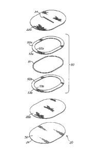
FIG. 1 is a top plan view of an implantable prosthesis according to one
illustrative embodiment of the present invention;
FIG. 2 is a bottom plan view of the prosthesis of FIG. 1;
FIG. 3 is an exploded perspective view of the prosthesis of FIG. 1;
FIG. 4 is a top plan view of one illustrative embodiment of a support assembly
for the prosthesis of FIG. 1 with the ends of a sleeve pulled back to join end
portions of the
stiffening member;
FIG. 5 is an enlarged cross-sectional view of the stiffening member taken
along
section line 5-5 of FIG. 4 illustrating the joint between the end portions of
the stiffening
member;
FIG. 6 is a top plan view of the support assembly of FIG. 4 with the
stiffening
member completely covered with a sleeve;
FIG. 7 is an enlarged cross-sectional view of the support assembly taken along
section line 7-7 of FIG. 6;
FIG. 8 is a top plan view of an implantable prosthesis employing multiple
support assemblies according to another illustrative embodiment of the present
invention;
FIG. 9 is an exploded schematic view of a procedure for manufacturing the
prosthesis of FIG. 1 according to one illustrative embodiment.
FIG. 9A is a schematic perspective view of the assembled support assembly of
FIG. 9.
CA 2966657 2017-05-10
84002829
3a
Description of Illustrative Embodiments
An implantable prosthesis is provided for repairing an anatomical defect, such
as a tissue or muscle defect, that promotes tissue of muscle ingrowth into the
prosthesis and
subsequently strengthens the area of the defect. The prosthesis is easy to
manipulate and may
be designed to minimize the incidence of postoperative adhesions between a
portion of the
prosthesis and surrounding tissue or organs. In addition, the prosthesis
strikes a balance
between being sufficiently rigid to aid in manipulation and deployment in the
area of desired
coverage and sufficiently flexible to be acceptable to both the surgeon and
the patient.
Further, the prosthesis may be constructed to allow it to be provisionally
held in place at
desired locations until sufficient tissue ingrowth occurs.
Embodiments of the prosthesis may be particularly suited for the repair of
various soft tissue or muscle wall defects, including, but not limited to,
inguinal and ventral
hernias, chest or abdominal wall reconstruction or large defects, such as
those that may occur
in obese patients. The prosthesis may include one or more features, each
independently or in
combination, contributing to such attributes.
The prosthesis may include one or more layers of biologically compatible
material that is suitable for repairing a defect. The prosthesis may include a
support assembly
that is attached to or integrated with the layer of material to facilitate
manipulation and
deployment of the prosthesis. The support assembly may include a stiffening
member that is
surrounded by material that separates the stiffening member from the layer of
material. The
stiffening member may be located in a sleeve of material. The stiffening
member may be
formed from a resorbable material. The sleeve may include interstices or
openings that allow
tissue or muscle ingrowth and/or facilitate resorption of the stiffening
member.
FIGS. 1-3 illustrate an embodiment of an implantable prosthesis for repairing
soft tissue or muscle defects. The prosthesis 20 includes an ingrowth layer 22
of tissue
infiltratable material. The ingrowth layer 22 includes at least one layer of
material that
permits or is otherwise susceptible to tissue or muscle adhesions. In one
embodiment the
ingrowth layer includes first and second layers 22a, 22b joined together. Each
layer 22a, 22b
is formed of a biologically compatible, flexible material
=
CA 2966657 2017-05-10
R4002829
4
that includes a plurality of interstices or openings which allow sufficient
tissue or
muscle ingrowth to secure the prosthesis to host tissue or muscle after
implantation.
In one embodiment, each layer 22a, 22b is formed of a knitted polypropylene
monofilament mesh fabric, such as BARI5 MESH available from C.R. Bard, Inc.
When implanted, the polypropylene mesh promotes rapid tissue or muscle
ingrowth
into and around the mesh structure. Alternatively, other materials which are
suitable
for tissue and muscle reinforcement and defect correction may be utilized,
including
SOFT TISSUE PATCH (microporous ePTFE ¨ available from W.L. Gore &
Associates, Inc.); SURGIPRO (available from US Surgical, Inc.); TRELEX
(available
from Meadox Medical); PROLENE and MERSILENE (available from Ethicon, Inc.);
and other mesh materials (e.g., available from Atrium Medical Corporation).
Absorbable materials, including polyglactin (VICRYL--available from Ethicon,
Inc.)
and polyglycolic acid (DEXON--available from US Surgical, Inc.), may be
suitable
for certain applications. Collagen materials, such as COLLAMEND from C.R.
Bard,
Inc. or SURGISIS available from Cook Biomedical, Inc., may also be used. It
also is
contemplated that the mesh fabric may be formed from multifilament yarns and
that
any suitable method, such as knitting, weaving, braiding, molding and the
like, may
be employed to form the prosthetic mesh material.
To ensure that adequate tissue or muscle ingrowth occurs, the two layers of
material may be attached in a way that would permit tissue to grow into the
interstices
or pores of each layer 22a, 22b and provide a strong bond between the
surrounding =
muscle or tissue and layer 22b. In one embodiment, the first and second layers
22a,
22b are connected with stitches 30.
In one embodiment, he first and second layers 22a and 22b are attached only
. at discrete locations. In this manner, tissue or muscle is able to
grow through the first
layer 22a and into the second layer 22b. Although a single stitch line 30 may
adequately secure the ingrowth layers together, it may be desirable to use
additional
stitch lines to limit the amount of billowing of the ingrowth layers 22a and
22b. In
addition, although the attachment is shown to include concentric patterns, any
suitable
pattern may be employed so as to minimize separation of the layers.
It should be appreciated that the invention is not limited to any particular
attachment method, as the first and second layers may be attached using other
suitable
techniques. For example, the layers may be bonded together by melting the
layers at
specific locations or in a specified pattern; sonic, induction, vibration, or
infrared/laser
welding the layers; or using a suitable bonding agent. The point or points of
CA 2966657 2017-05-10
84002829
attachment may comprise any suitable pattern, such as a spiral pattern, a
serpentine
pattern or a grid-like pattern of dots or beads, that maintains a' sufficient
quantity of
open or non-impregnated interstices for tissue or muscle infiltration.
To aid in positioning and/or provisionally attaching the prosthesis, the
prosthesis may include at least one pocket 32. In this manner, a surgeon may
use the
pocket to position the prosthesis in the desired area. Thereafter, the surgeon
may
suture or staple one of the layers of material to the surrounding in-growth
tissue,
muscle or peritoneum layer. For example, the surgeon may enter the pocket and
suture or 'staple the upper layer of the pocket to the tissue, muscle or
peritoneum layer. "
As such, the prosthesis may be provisionally held in place at least until
sufficient
tissue or muscle ingrowth occurs. In one embodiment, the first and second
layers 22a,
22b are attached in a manner to form the pocket 32 therebetween. However, it
should
be appreciated that the invention is not limited in this respect and that a
pocket 32
need not be employed or that other suitable pockets formed in other suitable
manners
may be employed. For example, a pocket may be formed from an additional layer
of
material or portion thereof attached to the first layer 22a.
To gain access to the interior of the pocket, the prosthesis may include at
least
one opening to the pocket 32. In one embodiment, the opening includes an
elongated
cut or slit 34 formed in the first layer 22a. However, it is to be appreciated
that the
prosthesis may include any suitable opening that allows access to the pocket
as would
be apparent to one of skill in the art.
To position the prosthesis, the surgeon may insert one or more fingers (or a
suitable surgical instrument) into the pocket and manipulate the prosthesis
into place.
In one embodiment, the pocket 32 is sized to accept several fingers of the
surgeon's
hand, although other suitably sized pockets may be employed, as the present
invention
is not limited in this respect. Further, the pocket 32 may be formed of
multiple
pockets with multiple openings so that one or more fingers may be inserted
into
individual finger sections.
In certain procedures, such as in the repair of ventral hernias or in the
reconstruction of chest or abdominal walls, the ingrowth layer may come into
contact
with tissue, muscle or organs, which is not intended to grow into the ingrowth
layer.
Such contact could potentially lead to undesirable postoperative adhesions
between
the ingrowth layer and the surrounding tissue, muscle or organs. To minimize
or
eliminate the incidence of postoperative adhesions to selected portions of the
prosthesis, the prosthesis may include a tissue, muscle or organ adhesion
resistant
CA 2966657 2017-05-10
84002829
6
barrier layer 36 overlying at least a portion, and preferably all, of one side
of the
ingrowth layer 22.
In one embodiment, the barrier layer 36 is attached to the prosthesis on the
side adjacent to the second layer 22b. The prosthesis 20 may be positioned in
a
patient such that the barrier layer 36 faces the region of potential undesired
adhesion,
such as the abdominal viscera (e.g., intestines) or the thoracic viscera
(e.g., heart or
lungs). As will be discussed in more detail below, the barrier layer 36 is
formed of a
material and/or with a structure that does not substantially stimulate and in
fact resists
tissue, muscle or organ ingrowth and adhesion formation when implanted,
thereby
limiting or completely eliminating the incidence of undesired postoperative
adhesions
between the ingrowth layer and adjacent tissue, muscle or organs.
In one embodiment, the barrier layer 36 is formed from a sheet of expanded
polytetrafluoroethylene (ePTFE) having fibril lengths¨also referred to as pore
size or
intemodal distance¨that will not permit significardtissue ingrowth. In one
embodiment, the fibril lengths of the ePTFE are less than 5 microns. In
another
embodiment, the fibril lengths of the ePTFE are less than 1 micron and in
still another
embodiment, the fibril lengths are less than 0.5 microns. Examples of other
suitable
materials for forming the barrier layer 36 include FLUORO-TEX Pericardial and
Peritoneum Surgical Membrane and FLUORO-TEX Dura Substitute available from
C. R. Bard, and PRECLUDE Pericardial Membrane, PRECLUDE Peritoneal
Membrane and PRECLUDE Dura Substitute membrane available from W. L. Gore &
Associates, Inc.
A representative and non-limiting sampling of other suitable micro to non-
porous materials includes silicone elastomer, such as SILASTIC Rx Medical
Grade
Sheeting (Platinum Cured) distributed by Dow Coming Corporation, and
microporous
polypropylene sheeting (available from Celgard, Inc.) and film. Autogenous,
heterogenous and xenogeneic tissue also are contemplated including, for
example,
pericardium and small intestine submucosa. Absorbable materials, such as
SEPRAFILM available from Genzyme Corporation and oxidized, regenerated
cellulose (Intercede (TC7)) may be employed for some applications. It is to be
appreciated that other suitable bioeompatible adhesion resistant materials
also may be
used.
The prosthesis 20 may be particularly useful in repairing tissue defects Where
conventional tissue approximation is not feasible, for example, the repair of
a large
defect, such as a large incisional hernia, particularly one which occurs in
tissue or
=
CA 2966657 2017-05-10
84002829
7
muscle weakened by previous- stu-gery or in tissue or muscle of obese
patients. For
this purpose, theprosthesis 20 bridges the defect and supparts the surrounding
tissue
or muscle as the tissue or muscle grows into the ingrowth layer and after such
ingrowth occurs. In one embodiment, to support stresses induced by the patient
(e.g.,
by patient movements), thereby limiting recurrent defects, it is desirable
that the tissue
or muscle be able to grow into the layer of ingrowth material that is best
suited for
supporting suCh stresses. Since the Exit layer 22a includes at least one
opening 34, it
is relatively less able to support the'required stres.s. On the other hand,
the second
layer 22b includes no sizable openings, or other large discontinuities, and is
generally
uniform and is therefore more able to support the required load. Therefore, in
the
embodiment described herein, the load bearing layer is the second layer 22b.
It should be appreciated that the present invention is not limited in this
respect
and that the prosthesis 20 may be fanned with suitably sized and shaped
openings or
discontinuities in the second layer 22b, provided such openings or
discontinuities do
not reduce the load bearing ability of the second layer beyond a tolerable
amount. For
example, a relatively smaller prosthesis may employ such openings or
discontinuities.
These openings or discontinuities may be used to help at least provisionally
anchor
the prosthesis and promote tissue ingrowth. Examples of prostheses employing
such
openings and discontinuities are described in U.S. Pat. Nos. 6,290,708 and
6,224,616,.
=
To permit and facilitate tissue or muscle growth into the second layer 22b,
the
barrier layer 36 preferably may be attached to the second layer 22b in u way
that
permits tissue to grow into the pores of the second layer 22b and provide a
strong
bond between the surrounding muscle or tissue And the second layer 22b.
In one embodiment, the first and second layers 22a, 22b are attached together
at discrete attachment lines, using stitches which allow sufficient tissue
infiltration to
the ingrowth layer, and in particular, the second layer 22b, while providing a
= connection between the-first and second layers 22a and 22b. In addition,
these same. '
stitches (e.g., stitches 38) may be used to secure the second layer 22b to the
barrier
layer 36. Although stitch lines 38.may adequately secure the barrier layer 36
to the .
ingrowth layer 22, it may be desirable to use additional stitch lines, such as
a center
stitch line 39, to limit the amount of billowing of the barrier layer away
from the
ingrowth layer. Although the attachment is shown to include concentric
patterns, any
CA 2966657 2017-05-10
84002829
8
suitable pattern may be employed so as to minimize separation of the ingrowth
layer
and the barrier layer.
If desired, different sets of stitches may be used to secure the first and
second
layers 22a and 22b together as compared to stitches used to secure the second
layer
22b to the barrier layer 36. For example, not all the stitch lines 30 are
required to pass
through the barrier layer 36. Rather, only the stitch lines 38 pass through
the barrier
layer 36. It is preferred that as few stitches as necessary are employed to
secure the
barrier layer 36 to the second layer 22b so that tissue or muscle adhesion on
the
barrier layer side of the prosthesis is minimized. Also, in the embodiment
shown, the
center stitch line 39 passes only through the second layer 2.2b and the
barrier layer 36,
as the first layer 22a includes the access opening 32 at that location.
Although, in one embodiment, the barrier layer 36 is attached to the ingrowth
layer 22b with stitches, it should be appreciated that the invention is not
limited in this
= respect, as the barrier layer may be attached using other suitable
techniques. For
example, the barrier layer may be bonded to the ingrowth layer by heating the
layers,
welding the layers, or using a suitable bonding agent. In either case, a
suitable
pattern, such as a spiral pattern, a serpentine pattern or a grid-like pattern
of dots or
beads may be used, provided a sufficient quantity of open or non-impregnated
interstices is maintained in at least the second layer 22b for tissue or
muscle
infiltration. =
When stitches are employed to attach the ingrowth layer 22b to the barrier
layer 36, to further minimize adhesions, the stitches may be formed from a non-
porous, adhesion resistant material. For example, the stitches may be formed
with a
suitable polytetrafiuoroethylene (PTFE) monofilament. PTFE stitches may
provide a
softer, more flexible prosthesis that is easier to manipulate as compared to a
prosthesis
using other stitch materials, such as polypropylene monofilament. PTFE
monofilament also facilitates the manufacturing process due to the low
friction
characteristics of the material. Nevertheless, it should be understood that
any suitable
material, such as polypropylene monofilament, may be employed for the
stitches. For
example, because some of the stitch lines do not pass through the barrier
layer, or
where no barrier layer is employed, materials other than an adhesion resistant
material
may be employed. For ease of manufacturing, however, all stitches may be
formed of
the same material, although the invention is not limited in this respect.
The layers may be stitched using a typical sewing stitch formed by a sewing
machine using a bobbin and sewing thread. Preferably, the barrier layer is
positioned
CA 2966657 2017-05-10
84002829
9
on the ingrowth layer to face the sewing needle so that the locking portion of
each
stitch (i.e. the bobbin) is formed on the ingrowth side of the prosthesis
rather than on
the barrier side to reduce the incidence of localized adhesions with tissue,
muscle or
organs. The stitches may be formed using a #10 ball-tipped needle to reduce
the
potential incidence of ingrowth through the stitch holes. The sheets of
ingrowth
material with or without the barrier layer may be held by a frame during the
sewing
procedure on a computer controlled table that has been programmed with the
desired
stitch pattern.
While the barrier layer 36 preferably covers the entire surface of one side of
the ingrowth layer 22, it is to be understood that the barrier layer 36 may be
configured to cover only selected portions of one side of the prosthesis to
enhance
ingrowth from both sides in those portions free of the barrier layer.
Similarly, the
prosthesis may be configured such that the barrier layer covers the entire
surface on
one side of the prosthesis and covers one or more portions of the other side
of the
prosthesis.
In some instances, it may be desirable to isolate the outer peripheral edge of
the prosthesis 20 from adjacent tissue, muscle or organs. In one embodiment, a
peripheral barrier 40 extends completely about the outer peripheral edge 24 of
the
prosthesis 20 to inhibit adhesions thereto. It is to be understood, however,
that the
peripheral barrier 40 may be configured to cover only those selected portions
of the
outer peripheral edge of the prosthesis where protection from the formation of
postoperative adhesions is desired.
The peripheral barrier 40 may be formed integrally with either the ingrowth
layer 22 or the barrier lager 36. Alternatively, the peripheral barrier 40 may
be
formed by a separate component that is attached to or incorporated into the
outer
peripheral edge of the prosthesis. In one illustrative embodiment, the
peripheral
barrier 40 is formed from a portion of the ingrowth layer 22. In particular,
the
ingrowth layer 22 may be altered so as th substantially eliminate the tissue
infiltratable interstices or openings along its outer margin, thereby creating
a
peripheral barrier 40.
In one embodiment, the peripheral edge 24 of layers 22 is melted to seal the
material and form an outer peripheral barrier 40. The barrier layer 36 may be
configured, such as with submicronal sized pores, so that a portion of the
melted
material of layer 22 becomes fused to the barrier layer 36. The peripheral
edge 24
may be melted using any suitable process. In one embodiment, the peripheral
edge 24
CA 2966657 2017-05-10
84002829
may be melted by heat sealing the layer. In the exemplary embodiment, the
peripheral barrier 40 is formed by melting a ring of polypropylene mesh fabric
to.the
ePTFE barrier layer 36 in a shape that approximates the desired configuration
of the
prosthesis. This may be accomplished by overlying oversized sheets of the mesh
'
= fabric and ePTFE material in a fixture and heat sealing the layers using
a heated die
= configured with the desired shape of the prosthesis. The melted ring may
be formed
by applying heat to the fabric at a temperature range of approximately 320 F.
to 400
F. for a period of approximately 3 to 5 seconds. The temperature chosen
typically
should be below the sintering temperature of the ePTFE barrier layer. Other
sealing
techniques may be used, such as ultrasonic, induction, vibration,
infrared/laser
welding and the like, as the present invention is not limited in this respect.
Once
fused, the ingrowth layer is stitched to the barrier layer, as described
above, and
subsequently die cut flush along a portion of the ring to complete the
prosthesis with a
peripheral barrier.
Other suitable techniques for creating a peripheral barrier may be employed,
as the present invention is not limited in this respect. Examples. of such
other
techniques are described in U.S. patent No. 7,404,819.
Although some embodiments described above include a barrier layer, the
present invention is not limited in this reipect. Thus, other embodiments may
or may
not include the barrier layer or the peripheral barrier.
In some instances, such as (but not limited to) the correction of relatively
large
defects, it may be desirable to employ a prosthesis that is sufficiently rigid
so that it
= can be easily and effectively manipulated and positioned in the desired
area yet
sufficiently flexible so that the prosthesis is adequately tolerated by both
the physician
implanting the prosthesis and the patient receiving the prosthesis. The
prosthesis
should conform to the shape of the area being covered and should be
sufficiently rigid
such that the edges do not excessively curl. This attribute may be
particularly useful
with a large=prosthesis sized for use with large defects in obese patients.
Thus,
= =
according to one aspect of the invention, to balance the stiffness and
flexibility, the
prosthesis 20 includes a support assembly 50. The support assembly may be
coupled
to the ingrowth layer in any suitable manner.
The support assembly contributes to the stability of the prosthesis; allowing
it
to remain in a desired shape during the implantation procedure, subject to
proper
CA 2966657 2017-05-10
84002829
11
fixation techniques. This stability facilitates deployment and placement of
the
prosthesis by making it easy to handle. For example, the support assembly aids
in
allowing the prosthesis to remain substantially planar during implantation.
During
implantation of the prosthesis, sutures may be passed around the support
assembly to
maintain the prosthesis in generally the desired configuration and location.
In one illustrative embodiment shown in FIG. 3, the support assembly 50
includes a support or stiffening member 51 that is surrounded with-material
that
separates the stiffening member from the ingrowth layer 22. The stiffening
member
may be resilient so that the support assembly can be collapsed or deformed
from an
expanded configuration to facilitate delivery of the prosthesis to a surgical
site and
then return to its expanded configuration to facilitate handling and support
of the
prosthesis at the surgical site. In one embodiment, the stiffening member may
be
located in a sleeve of material 52. However, it is to be understood that the
support
assembly may employ other suitable arrangements apparent to one of skill in
the art to
surround the stiffening member with material that separates the stiffening
member 51
from the ingrowth layer 22.
In one embodiment, the stiffening member 51 is formed of a resorbable
material. The resorbable stiffening member facilitates initial handling and
deployment of the prosthesis. Thereafter, the stiffening member will gradually
degrade until it is completely resorbed by the body. Such an arrangement may
be
advantageous-in that the stiffening member is eventually resorbed by the body
after it
is no longer needed to facilitate the handling and deployment of the
prosthesis.
In one embodiment, the stiffening member 51 is formed from a polydioxonane
(PDO) monofilament having a diameter of approximately 0.038 inches. However,
it
is contemplated that the stiffening member may be formed of any biocompatible,
resorbable or non-resorbable material, including monofilaments, multifilaments
or
molded shapes, provided suitable stiffness and handling properties are
maintained. It
should be appreciated that the stiffening member (or the individual filaments
or bands
collectively forming the stiffening member) may have any suitable cross-
sectional
size and shape, such as circular, square, rectangular, triangular, elliptical,
etc.
In one illustrative embodiment, the prosthesis employs a stiffening member 51
that is configured in the shape of a ring. However, the stiffening member may
be
configured in any pattern, such as a spiral pattern, a square pattern, an
elliptical
pattern, a circular Pattern or the like. In one embodiment as shown, the
support
assembly 50 employs a continuous, uninterrupted ring. The ring may be formed-
by
CA 2966657 2017-05-10
84002829
12
joining the end portions of a length of material, such as a monofilament.
However, it
should be appreciated that the stiffening member may be formed of one or more
discrete, discontinuous segments, arranged in any configuration that may
impart
suitable stiffness and handling to the prosthesis.
The sleeve 52 may be formed of a porous material that allows passage or
infiltration of fluid and/or tissue to promote degradation and/or resorption
of the
stiffening member 51. In one embodiment, the material includes interstices or
pores
having a size from approximately 0.00035 in2 to approximately 0.00085 in2. It
may
be desirable to employ a sleeve having an interstice or pore size of
approximately
0.00085 in2 when the support assembly is used with an ingrowth layer 22 formed
of
material having a similar pore or interstice size of approximately 0.00085
in2. A
sleeve having a smaller interstice or pore size, such as approximately 0.00035
in2,
may be desired when the support assembly is used with an ingrowth layer 22
formed
of material having a larger pore or interstice size, such as greater than
0.00085 in2.
However, it is to be understood that the sleeve may employ material having
other
suitable interstice or pore sizes as would be apparent to one of skill in the
art.
In one embodiment, the sleeve 52 is formed from a mesh fabric that inehides
interstices or pores that allow tissue infiltration or ingrowth into the
support assembly
to eventually surround and resorb the stiffening member 51. In one embodiment,
the
sleeve is formed from a knitted polypropylene mesh. The mesh may be knitted
with
monofilament having a diameter of approximately 0.006 inches. The mesh may
employ any suitable fabric pattern that provides desired properties. It is to
be
understood that the sleeve may be formed of any suitable mesh material
including, but
not limited to, the material used for the ingrowth layer or other
biocompatible
materials having suitable properties. It also is contemplated that the sleeve
may be
formed from multifilament yarns and that any suitable method, such as
knitting;
weaving, braiding, molding and the like, may be employed to form the sleeve.
In one illustrative embodiment shown in FIG. 3, the sleeve may be formed
with two rings of mesh 52a, 52b that are attached to form the sleeve. Each
mesh ring
may have a width of approximately 0.25 to 0.38 inches. As shown, the
stiffening
rnember 51 is sandwiched between the mesh rings 52a, 52b which are attached to
.
each along the inner and outer sides of the stiffening member to surround the
stiffening,member in a sleeve of material.
If desired, the sleeve may be configured as a unitary member that is formed
with a single piece of material. In one illustrative embodiment shown in FIGS.
4-7,
CA 2966657 2017-05-10
84002829
13
the support assembly 50 may employ a sleeve 52 configured as a tubular or sock-
like
member that receives the stiffening member 51 therein. In one embodiment, the
sleeve is a tubular mesh fabric material.
As shown in FIG. 4, a length of material for the stiffening member 51 is
inserted through the sleeve 52 and looped with the ends of the material being
joined
together to form a continuous ring configuration. As shown in FIG. 5, the end
portions 54 of the stiffening member 51 may overlie arid be joined together at
a joint
55 using any suitable technique, such as welding, bonding and the like. After
joining
the ends 54 of the stiffening member, the ends 56a, 56b of the sleeve material
are
pulled together completely over the stiffening member 51 and joined to form a
continuous sleeve over the stiffening member. As shown in FIGS. 6-7, the ends
56a,
56b of the sleeve 52 may overlap to completely cover the stiffening member.
One
end 56a of the sleeve material may be flared to fit over the opposite end 56b
of the
sleeve to ensure complete coverage of the stiffening member and facilitate
joining of
the sleeve ends.
Although several illustrative embodiments have been provided for the support
assembly, it is to be understood that the support assembly may employ other
structural
arrangements apparent to one of skill in the art.
The support assembly 50 may be configured to surround the outer area 26 of
the prosthesis and reinforce at least the outer area 26. In the embodiment
shown in
the figures, the support assembly 50 is not disposed at the peripheral edge
24. Rather,
the support assembly 50 is spaced inwardly of the peripheral edge 24. However,
it
should be appreciated that the present invention is not limited in this
respect, as the
support assembly 50 may be disposed at the peripheral edge 24.
The support assembly 50 may be disposed on the prosthesis in any suitable
manner as the present invention is not limited in this respect. In one
embodiment, the
support assembly 50 is sandwiched between the first and second layers 22a, 22b
of
ingrowth material and may or may not be physically attached thereto. A stitch
line 30
formed by sewing threads may be stitched at least along or through the outside
or
inside edge 53a, 53b (FIG. 3) of the support assembly 50 to keep it. from
moving with
respect to layers 22a and 22b. Because of the rigidity of the support assembly
50, one
stitch line along or through one side of the support assembly 50 may be
enough.
HOwever, preferably, two stitch lines, one on each edge of the assembly,
secure the
support assembly in place. Preferably, these stitches extend through both of
the first
and second layers 22a and 22b, but not through the barrier layer 36, if it is
present.
=
CA 2966657 2017-05-10
= 84002829
14
Another advantage is that the support assembly 50, if stitched or bonded to
the barrier
layer 36 or to the first and second layers 22a and 22b, holds the layers 22a,
22b and/or
layer 36 together in a manner to prevent billowing of layer 36 with respect to
layer 22
or layers 22a and 22b with respect to each other.
Alternatively, the support assembly 50 may overlie or underlie the ingrowth
layer 22 and may be attached, regardless of location, with stitches or a
bonding agent,
or fused by ultrasonic, induction, vibration, infrared/laser welding and the
like. In
instances where a barrier layer is employed, it may be desirable that the
support
assembly 50 is not positioned under the barrier layer 36 or protrude
therethrough, as
doing so may result in undesirable adhesions forming on the support assembly.
Although the stiffening member 51 is described as being formed of a
monofilament, it is to be understood that other suitable constructions may be
employed. For example, the stiffening member may be one or more molded
elements
that are subsequently attached to, or molded onto, the prosthesis.
In another embodiment shown in FIG. 8, a second support assembly 60 may
be disposed inwardly of the first support assembly 50 and may be employed to
reinforce the inner area 28 of the prosthesis. As shown, the second, inner
support
assembly 60 may be concentric or generally concentric with the first, outer
support
assembly 50. However, it should be appreciated that the invention is not
limited in
this respect as other suitable arrangements may be employed.
In one embodiment, the prosthesis 20 is relatively flat and sufficiently
pliable
to allow a surgeon to manipulate the prosthesis to insert the prosthesis and
conform
the prosthesis to the anatomical site of interest, allow the prosthesis to be
sutured,
stapled or otherwise anchored. The prosthesis 20 may be configured to have any
suitable shape or size that is conducive to facilitating the correction of a
particular
defect. In the embodiments illustrated in the figures, the prosthesis 20 has a
generally
flat, oval shape. Examples of other shapes include, but are not limited to,
circular,
square, rectangular and irregular shapes.
In an exemplary embodiment, each of the first and second layers 22a and 22b
is formed of an approximately 0.027 inch thick sheet of BARD MESH knitted from
polypropylene monofilament with a diameter of approximately 0.006 inches. The
barrier layer 36 is formed from an approximately 0.006 to 0.008 inch thick
sheet of
ePTFE. The barrier 36 is attached to layers 22a and 22b using approximately 3
nun to
4 mat long stitches formed of a 0.008 inch to 0.012 inch diameter PTFE
monofilament.
CA 2966657 2017-05-10
84002829
In one embodiment, the prosthesis 20 has a generally oval shape that may have
any desired size. For example, the prosthesis, as measured generally along the
major and
minor axes of the oval, may be approximately sized as follows: 5 inches by 7
inches; 7 inches
by 9 inches; 8 inches by 10 inches; or 10 inches by 13 inches. The prosthesis
may also be
sized to cover an area greater than 50 square cm. In one embodiment, the
prosthesis covers an
area of approximately 68 square cm; in another embodiment, approximately 119
square cm; in
yet another embodiment, approximately 152 square cm; and in still another
embodiment, (e.g.,
for an obese patient) approximately 246 square cm. It should be understood,
however, that
the materials and dimensions described are merely exemplary and that any
suitable sizes and
shapes may be employed for the prosthesis.
In one embodiment, the prosthesis is sized such that the prosthesis overlaps
the
edges of the defect by at least 3 cm and, in some embodiments, by at least 4
cm and in still
other embodiments, by at least 5 cm. Although the prosthesis has been
described above as
correcting a single defect, it is contemplated that a suitable sized and
shaped prosthesis may
be used to correct more than one defect.
One example of a procedure to manufacture the prosthesis will now be
described. The support assembly 50 is made by capturing the stiffening member
51 between
two rings of die cut material 52a, 52b. FIG. 9 shows an exploded view of
material blanks
used to form the prosthesis 20. To form the support assembly 50, a mesh layer
152b is
positioned flat on a multi-pin alignment fixture such that alignment pins pass
through
alignment holes 156 provided in each blank. Next, the stiffening member 51 is
positioned on
the mesh layer 152b. Thereafter, a mesh layer 152a is placed on top of the
stiffening member
51, with the fixture alignment pins passing through corresponding alignment
holes. A row of
stitches is then placed on each side of the stiffening member 51 to attach the
mesh layers
152a, 152b together and capture the stiffening member therebetween.
The assembled layers with the captured stiffening member are then die cut, to
produce the support assembly 152 (as shown in FIG. 9A). As shown, the
resulting assembly
152 includes a series of radially extending alignment tabs 155.
CA 2966657 2017-05-10
84002829
15a
The support assembly 152 is positioned, using the alignment tabs 155, between
the first and second layers 122a and 122b. The barrier layer 136 is positioned
adjacent to the
second layer 122b opposite the support assembly 152. The assembly is stitched
together,
using stitch patterns shown in FIGS 1-2. The radial tabs 155 may then be
removed and the
assembly may be placed in a heated die to fuse
CA 2966657 2017-05-10
84002829
16
portions of the first and second layers. 122a, 122b to the barrier layer 136.
=A final die =
cut is then made to achieve the desired shape of prosthesis 20 with a
resulting
peripheral edge formed by the fused layers.
The illustrative procedure for manufacturing the prosthesis has been described
in conjunction with using a support assembly 50 that is formed with multiple
layers of
material. As one of skill in the art would understand, a prosthesis using a
support
assembly that includes a tubular sleeve, as described above in connection with
FIGS.
4-7, may be manufactured in a similar manner by positioning the tubular sleeve
support assembly between the first and second layers 122; 122b of material.
It should be understood that the foregoing description of various embodiments
of the invention are intended merely to be illustrative thereof and that other
embodiments, modifications, and equivalents of the invention are within the
scope of
the invention recited in the claims appended hereto. Further, the prosthesis
described
above includes various features that may be employed singularly or in any
suitable
combination.