Note: Descriptions are shown in the official language in which they were submitted.
APOAEQUORIN-CONTAINING COMPOSITIONS AND
METHODS OF USING SAME TO TREAT NEURONAL INFLAMMATION
CROSS-REFERENCE TO RELATED APPLICATIONS
[0001] This
application claims the benefit of U.S. Provisional application
62/078,099, filed November 11, 2014.
FIELD OF THE INVENTION
[0002] This
invention relates generally to compositions useful for the treatment of
neuronal inflammation. More
specifically, the present invention is directed to
apoaequorin-containing compositions and methods of using those compositions to
treat
neuronal inflammation.
BACKGROUND OF THE INVENTION
[0003] In 2009,
stroke accounted for about one of every 19 deaths in the United
States, making it the third leading cause of death behind only heart disease
and cancer.
As a result, finding ways to ameliorate injury following stroke is imperative.
Much
attention has been placed on the role of calcium in ischemia and possible
neuroprotection
by blocking its toxic effects post-ischemia.
[0004] Calcium
(Ca2) plays a pivotal role in various neuronal processes, including
neurotransmitter release and synaptic plasticity. Neurons arc continuously
subjected to
fluctuations in intracellular Ca2 as a result of ongoing activity, however
excess or
sustained increases in intracellular Ca2 can be toxic to neurons. Thus,
neuronal
intracellular Ca2+ is very tightly regulated, and several mechanisms exist
which enable
neurons to limit or control cytosolic Ca2 + levels. In particular, calcium
binding proteins
(CaBPs; such as calbindin, parvalbumin, and calretinin) are important for
binding and
buffering cytosolic Ca2'.
[0005] Studies in
the hippocampus have shown that the presence of CaBPs confers
some protection against excitotoxic insults that normally result in cell
death.
CA 2966891 2019-08-02
CA 02966891 2017-05-04
WO 2016/077437
PCT/US2015/060116
Interestingly, decreased levels of CaBPs are observed with advancing age, and
in
neurodegenerative disorders, including Alzheimer's disease, and Parkinson's
disease.
Treatments aimed at minimizing Ca2 toxicity during ischemia by administering
CaBPs
before an ischemic insult have also had positive results. For example, Yenari
et al.
treated animals with calbindin prior to inducing ischemia and found that over-
expression
of calbindin was neuroprotective. In addition, Fan et al. treated rats with
calbindin prior
to ischemia and demonstrated a smaller infarct volume, better behavioral
recovery, and
decreased apoptosis in the calbindin-treated animals. Indeed, much research
has focused
on understanding the deleterious effects of stroke. Interestingly, a major
risk factor for
stroke is aging, and one prominent hypothesis of brain aging is the Ca2'
hypothesis of
aging. This hypothesis argues that an aging-related change in the ability to
regulate
calcium and calcium-dependent processes is a critical contributor to an
increase in
susceptibility to cognitive decline and neurodegenerative disorders. Given
these aging-
related changes in Ca2+, and the critical role of Ca2t in ischemic cell death,
much research
= has focused on Ca.2' dysregulation in both neurons and glia.
100061
Excessive intracellular Ca2' accumulation following ischemia is known to
potentiate cell death through excitotoxicity.
Following an ischemic insult, Ca2+
accumulates within the cell through voltage-gated Ca21 channels (VGCCs),
through
NMDA receptors, and through release from intracellular organelles. Numerous
studies
have shown that blocking Ca2+ entry through NMDA receptors, VGCCs, or both in
combination can be neuroprotectivc against ischemia. Interestingly, when NMDA
receptor blockers were brought to clinical trials, they failed to provide
neuroprotection
and they produced undesirable side effects, such as hallucinations and coma.
While it is
uncertain why NMDA receptor blockers failed in clinical trials, it is clear
that there is a
need for continued research focused on ameliorating the devastating effects of
ischemic
stroke.
[00071
Despite advances, there is still a need for new and alternative therapeutics
which treat neuronal inflammation. In particular, pharmaceutical or
nutraceutical
compositions which have reduced side effects as compared to prior agents arc
desired
and, if discovered, would meet a long felt need in the medical and nutritional
health
communities.
2
CA 02966891 2017-05-04
WO 2016/077437 PCT/US2015/060116
SUMMARY OF THE INVENTION
[0008] The present invention is based in part on the inventors' recent
research on
apoaequorin, a calcium binding protein, and the unexpected finding that
apoaequorin
possesses novel neuroprotective abilities. In particular, apoaequorin has been
found to be
useful in preconditioning neurons in a subject to reduce subsequent neuronal
inflammation. Accordingly, the present invention provides apoaquoring-
containing
compositions and methods of use which offer substantial advantageous in
neuroprotective
applications.
100091 In a first aspect, the present invention is directed to methods of
preconditioning neurons to reduce neuronal inflammation in a subject. Such
methods
include the step of administering apoaequorin to a subject, wherein the
subject's neurons
arc preconditioned to reduce neuronal inflammation.
[0010] In one embodiment, administering to the subject is by injection. In
an
alternative embodiment, administering to the subject is by oral delivery, for
example, by
apoaequorin formulated in a unit dosage form selected from a tablet or
capsule. In certain
embodiments, apoaequorin is administered to a subject in the form of a
nutraceutical
composition.
[0011] As can be appreciated, the present invention encompasses apoaequorin
for
preconditioning neurons to reduce neuronal inflammation in a subject, as well
as the use
of apoaequorin for the manufacture of a composition for preconditioning
neurons to
reduce neuronal inflammation in a subject.
100121 In another aspect, the present invention is directed to methods of
reducing
Tumor Necrosis Factor a (TNFa) protein level in a subject. Such methods
include the
step of administering apoaequorin to a subject, wherein the subject's level of
TNFa
protein is reduced.
[0013] In certain embodiments, administering to the subject is by
injection. In
alternative embodiments,' administering to the subject is by oral delivery,
for example, by
apoaequorin formulated in a unit dosage form selected from a tablet or
capsule. In certain
embodiments, apoaequorin is administered to a subject in the form of a
nutraceutical
composition.
3
CA 02966891 2017-05-04
WO 2016/077437
PCT/1JS2015/060116
[00141 As can be
appreciated, the present invention encompasses apoaequorin for
reducing TNFot protein level in a subject, as well as the use of apoaequorin
for the
manufacture of a composition for reducing TNFoc protein level in a subject.
[0015] The present
invention provides various advantages over prior compositions
and methods in that it provides for the general improvement of a subject's
mental and
physical health through its neuroprotective functions.
[0016] Other objects,
features and advantages of the present invention will become
apparent after review of the specification and claims
BRIEF DESCRIPTION OF THE DRAWINGS
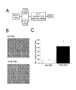
[0017] Fig. 1A-C
depicts effects of oxygen-glucose deprivation on cell death in acute
hippocampal brain slices. A) Diagram of experimental design. Corona(
hippocampal
slices were incubated for 1 hr in artificial cerebral spinal fluid (aCSF).
Half of the slices
were transferred to the ischemic condition for 5 min of oxygen-glucose
deprivation
(OGD) while the other half remained normoxic (no OGD). All of the slices were
then
transferred to aCSF for a 30 min reperfusion and trypan blue staining. The
slices were
then fixed in 10% neutral-buffered formalin. B) Representative images of
trypan blue
staining in area CA1 of the hippocampus in a slice that remained normoxic (no
OGD) and
in a slice subjected to 5 min OGD. Notice that there is less staining in the
normoxic slice
compared to the OGD slice. C) There was a significant increase in the number
of trypan
blue-stained neurons in area CAI of the hippocampus from slices that underwent
5 min
OGD compared to slices that remained normoxic (*,p < .01).
[0018] Fig. 2A-C
depicts dose-dependent effects of apoaequorin on ischemic cell
death. A) Diagram of experimental design. Rats that were cannulated
bilaterally in the
dorsal hippocampus were given an infusion of 0, 0.4, 1, or 4% apoaequorin (AQ)
in one
hemisphere and vehicle (0% AQ) in the other hemisphere. One day following the
infusion, coronal hippocampal slices were cut and incubated in artificial
cerebral spinal
fluid (aCSF) for 1 hr. All slices were transferred to the ischemic condition
for 5 min of
oxygen-glucose deprivation (OGD). Slices were then transferred to aCSF for a
30 min
reperfusion and trypan blue staining. The slices were then fixed in 10%
neutral-buffered
formalin. B) Representative images of trypan blue staining in area CAI of the
4
CA 02966891 2017-05-04
WO 2016/077437
PCT/US2015/060116
hippocampus following ischemia in a vehicle-treated slice or a 4% AQ-treated
slice.
Notice that there is less staining in the AQ-treated slice compared to the
vehicle-treated
slice. C) Graph shows neuroprotection (percent of cells rescued) as a function
of the dose
of apoaequorin. There was significant neuroprotection in the rats treated with
I or 4%
AQ (but not 0.4% AQ) compared to the 0% AQ (vehicle; *, p < .01).
[0019] Fig. 3A-C depicts time-dependent effects of apoaequorin
on isehemic cell
=
death. A) Diagram of experimental design. Rats that were cannulated
bilaterally in the
dorsal hippocampus were given an infusion 4% apoaequorin (AQ) in one
hemisphere and
vehicle (0% AQ) in the other hemisphere. Coronal hippocampal slices were cut I
hr, 1
day. 2 days, 3 days, or 5 days post-infusion, and slices were incubated for 1
hr in artificial
cerebral spinal fluid (aCSF). All slices were transferred to the ischemic
condition for a 5
min oxygen-glucose deprivation (OGD). Slices were then transferred to aCSF for
a 30
min reperfusion and trypan blue staining. The slices were then fixed in 10%
neutral-
buffered formalin. A second set of rats was given bilateral infusion of 4% AQ
and the
brains were removed at 1 hr, I day, 2 days, or 3 days post-infusion to be used
for Western
blotting. B) An infusion of 4% AQ 1 or 2 days prior to ischemia resulted in
significant
neuroprotection, but the neuroprotective effect was no longer evident at 3 or
5 days post-
infusion. Notice that AQ is also not neuroprotective when infused just 1 hr
prior to
ischcmia (p = .78). C). Western blot analysis of the AQ protein at 22 kD. AQ
is present
in the dorsal hippocampus (AQ-dhpc) at 1 hr and 1 day, but is no longer
present at 3 days
post-infusion. At 2 days post-infusion, a band is present in only 29% of the
rats. Notice
that there is never a band in the ventral hippocampus (AQ-vhpc), regardless of
the
infusion time. Analysis of I3-actin (45 kD) revealed no effect of protein
loading at any
time point in either dorsal (actin-dhpc) or ventral (actin-vhpc) hippocampus.
p < .01
[0020] Fig. 4A-B depicts effects of apoaequorin on interleukin-
10 mRNA expression.
A) Interlcukin-10 (IL-10) mRNA expression is significantly increased 1 hour
after 4%
AQ was infused into the dorsal hippocampus. This statistically significant
increase was
transient as IL-10 mRNA expression returned to near baseline levels within 1
to 2 days,
although a biologically relevant 2- to 3-fold increase was still observed. B)
8-actin
mRNA expression did not significantly differ between 4% AQ and the vehicle-
treated
CA 02966891 2017-05-04
WO 2016/077437 PC1
/US2015/960116
hemisphere (p = .52). For both graphs, data arc expressed as fold-change from
the
vehicle-treated control hemisphere.
[0021] Fig. 5 illustrates the experimental methodology Utilized in Example
2.
[0022] Fig. 6A-B depicts data showing intrahippocampal infusion of AQ is
neuroprotective.
[0023] Fig. 7A-D depicts cytokine expression after AQ infusion.
[0024] Fig. 8A-C illustrates data demonstrating oral administration of AQ
is
neuroprotective.
[0025] Fig. 9A-C depicts data showing AQ infusion alters IL-10 and TNF-n.
protein expression.
[0026] Fig. 10A-C illustrates AQ infusion and trace fear conditioning in
aging rats.
[0027] Fig. 11A-C depicts oral administration of AQ is time- & dose-
dependent.
[0028] Fig. 12 depicts the experimental methodology utilized in Example 4.
[0029] Fig. 13A-C depicts oral administration of AQ is neuroprotective.
[0030] Fig. 14A-D shows data demonstrating oral administration of AQ alters
cytokinc protein expression.
[0031] Fig. 15A-B depicts shows data demonstrating intrahippocampal
infusion of
AQ alters cytokinc protein expression.
[0032] Fig. 16 illustrates data showing IL-10 nAb reverses AQ's
neuroprotective
effect.
DETAILED DESCRIPTION OF THE INVENTION
I. IN GENERAL
[0033] Before the present materials and methods are described, it is
understood
that this invention is not limited to the particular methodology, and
materials described,
as these may vary. It is also to be understood that the terminology used
herein is for the
purpose of describing particular embodiments only, and is not intended to
limit the scope
of the present invention which will be limited only by the appended claims.
[0034] It must be noted that as used herein and in the appended claims, the
singular forms "a", "an", and "the" include plural reference unless the
context clearly
dictates otherwise. As well, the terms "a" (or "an"), "one or more" and "at
least one" can
6
be used interchangeably herein. It is also to be noted that the terms
"comprising",
"including", and "having" can be used interchangeably.
[0035] Unless defined otherwise, all technical and scientific terms
used herein
have the same meanings as commonly understood by one of ordinary skill in the
art to
which this invention belongs.
[0036] Animals. 92 male F344 adult rats were used. Rats were kept on a
14/10-hr
day/night cycle with access to food and water ad libitum. Weight for each
animal was
recorded two times per week, as to account for significant weight increases
and/or
decreases.
[0037] Drugs. Apoaequorin (AQ; Quincy Bioscience) was prepared in
double
deionized water at a concentration of 7.4%. Experimental groups in the dose
dependent
experiments (n ¨ 18) received 0 (n = 4), 3.6 (n = 5), 48 (n = 4), 240 (n = 3),
or 480 mg/kg
of AQ mixed into their daily PB. For the remainder of the studies, rats (n 73)
received
48 mg/kg of AQ mixed into their daily PB. Animals were assigned to one of five
groups;
No AQ (n = 12), 1 hour AQ (n = 17), 1 day AQ (n = 15), 2 days AQ (n = 15), and
7 days
AQ (n = 14. Rats received 1/4 teaspoon of PB placed in a petri dish in the
cage every day
at a designated time. Petri dishes were not removed until all PB was consumed.
Animals
were weighed twice per week, as to maintain proper AQ dosage.
[0038] AQ for infusion studies was prepared as previously described (Detert et
at., PLOS ONE,
8(11): 1-10 November 1,2013). IL-10 neutralizing antibody (nAb) and its IgG
control were
prepared in sterile PBS. 0.5 ug was infused at a rate of I ul/min through 1 ul
Hamilton Syringes.
[0039] avgen-Glucose Deprivation. On the last day of administration,
rats were
allowed 1 hour after PB consumption for digestion, deeply anesthetized with
isoflurane,
and coronal slices (400 um) of dorsal hippocampus (dhpc; Bregma -3.14 - -4.16;
Paxinos
& Watson, 1998) were prepared using standard procedures (Moyer & Brown, 2007).
Following 1 hr slice recovery in aCSF, one hemisphere of each brain
(counterbalanced)
was subjected to in vitro ischemia by transferring slices to an oxygen-glucose
deprivation
chamber (glucose replaced with fructose and bubbled with 95% N2- 5% CO2
instead of a
95% 02 ¨ 5% CO2) for 5 min, while the other hemisphere remained in recovery.
All slices
were then placed into oxygenated aCSF containing 0.2% trypan blue for a 30 min
reperfiision period. Trypan blue stains dead cells while leaving living cells
unstained
7
CA 2966891 2019-08-02
(DeRenzis & Schechtman, 1973). The slices were rinsed twice in oxygenated,
room
temperature aCSF then fixed in 10% neutral buffered formalin overnight in the
refrigerator. Slices were then cryoprotected in 30% sucrose, sectioned on a
cryostat (40
um), and mounted onto subbed slides for cell counts.
[00401 Cell Counts. Slices were examined under an Olympus microscope
(equipped
with a digital camera) at 10X, and pictures were taken (CellSens). Trypan blue
stained
neurons within CA1 (about an 800 pm section) were counted by an experimenter
blind to
experimental conditions. Statistical analyses were performed using SPSS (v
21Ø0; IBM
Corporation; Armonk, NY). An ANOVA was used to evaluate a drug effect, and
Fisher's
LSD post-hoc evaluations were used to evaluate group interactions. Asterisk
(*) indicates
p < .05.
[0041] Western Blots. Animals were deeply anesthetized with isoflurane,
brains
rapidly removed, frozen, and stored at -80 C. Upon time of dissection, samples
were
dissected from dhpc (Bregma -3.14 - -4.16mm). Samples were homogenized,
centrifuged
at 4000 RPM for 20 mm, supernatant was removed, and protein was measured using
a
Bradford protein assay kit (Bio-Rad). Protein samples were normalized and
loaded for
SDS-PAGE (12%). Proteins (3014) were transferred onto PVDF membranes using the
Turbo Transfer System (Bio-Rad). Membranes were incubated in blocking buffer
(2 hr),
primary antibody (overnight at 4 C; 1:1000 mouse anti-aequorin [Chemicon] or
1:1000
rabbit anti-13-actin [Cell Signaling Technology], and secondary antibody (90
min;
1:20,000 goat anti-mouse [Santa Cruz Biotechnology] or 1:40,000 goat anti-
rabbit
[Millipore]). Membranes were then washed, placed in a chemiluminescence
solution
(Thermo Scientific), and imaged with a Syngene GBox. Images were taken with
GeneSys
software (v 1.2.4.0; Synoptics camera 4.2MP), and fluorescence for each band
was
evaluated with GeneTools software (v 4.02; Cambridge, England). Values are
expressed
as a percentage of control animals. Statistics were performed with SPSS (v.
21).
100421 Although any methods and materials similar or equivalent to those
described
herein can be used in the practice or testing of the present invention, the
preferred
methods and materials are now described.
8
CA 2966891 2019-08-02
All
references cited in this specification are to be taken as indicative of the
level of skill in
the art. Nothing herein is to be construed as an admission that the invention
is not
entitled to antedate such disclosure by virtue of prior invention.
THE INVENTION
[0043] Ischemic
stroke affects ¨795,000 people each year in the U.S., which results in
an estimated annual cost of $73.7 billion. Calcium is pivotal in a variety of
neuronal
signaling cascades, however, during ischemia, excess calcium influx can
trigger
excitotoxic cell death. Calcium
binding proteins help neurons regulate/buffer
intracellular calcium levels during ischemia. Aequorin is a calcium binding
protein
isolated from the jellyfish Aequorea victoria, and has been used for years as
a calcium
indicator, but little is known about its neuroprotective properties. The
present study used
an in vitro rat brain slice preparation to test the hypothesis that an intra-
hippocampal
infusion of apoaequorin (the calcium binding component of aequorin) protects
neurons
from ischemic cell death. Bilaterally cannulated rats received an apoaequorin
infusion in
one hemisphere and vehicle control in the other. Hippocampal slices were then
prepared
and subjected to 5 minutes of oxygen-glucose deprivation (OGD), and cell death
was
assayed by trypan blue exclusion. Apoaequorin dose-dependently protected
neurons from
OGD ¨ doses of 1% and 4% (but not 0.4%) significantly decreased the number of
trypan
blue-labeled neurons. This effect was also time dependent, lasting up to 48
hours. This
time dependent effect was paralleled by changes in cytokine and chemokine
expression,
indicating that apoaequorin may protect neurons via a neuroimmunomodulatory
mechanism. These data support the hypothesis that pretreatment with
apoaequorin
protects neurons against ischemic cell death, and may be an effective
neurotherapeutic.
[00441 Aequorin is
a photo-protein originally isolated from luminescent jellyfish and
other marine organisms. The aequorin complex comprises a 22,285-dalton
apoaequorin
protein, molecular oxygen and the luminophore coelenterazine. When three Ca2+
ions
bind to this complex, coelenterazine is oxidized to coelenterrnide, with a
concomitant
release of carbon dioxide and blue light. Acquorin is not exported or secreted
by cells,
nor is it compartmentalized or sequestered within cells. Accordingly, aequorin
9
CA 2966891 2019-08-02
CA 02966891 2017-05-04
WO 2016/077437
PCT/US2015/060116
measurements have been used to detect Ca2 changes that occur over relatively
long
periods. In several experimental systems, aequorin's luminescence was
detectable many
hours to days after cell loading. It is further known that aequorin also does
not disrupt
cell functions or embryo development.
[0045] Because of its Ca2I-dependent luminescence, the aequorin complex has
been
extensively used as an intracellular Ca2' indicator. Aequorea victoria
aequorin has been
specifically used to: (1) analyze the secretion response of single adrenal
chromaffin cells
to nicotinic cholinergic agonists; (2) clarify the role of Ca2+ release in
heart muscle
damage; (3) demonstrate the massive release of Ca?' during fertilization; (4)
study the
regulation of the sarcoplasmic reticulum Ca2' pump expression in developing
chick
myoblasts; and (5) calibrate micropipets with injection volumes of as little
as three
picolitcrs.
100461 Apoaequorin has an approximate molecular weight of 22 kDa.
Apoaequorin
can be used to regenerate aequorin by reducing the disulfide bond in
apoaequorin. The
calcium-loaded apoaequorin retains the same compact scaffold and overall
folding pattern
as unreacted photoproteins containing a bound substrate.
[0047] Conventional purification of aequorin from the jellyfish Aequorea
victoria
requires laborious extraction procedures and sometimes yields preparations
that are
substantially heterogeneous or that are toxic to the organisms under study.
Two tons of
jellyfish typically yield approximately 125mg of the purified photoprotcin. In
contrast,
recombinant aequorin is preferably produced by purifying apoaequorin from
genetically
engineered Escherichia coli, followed by reconstitution of the aequorin
complex in vitro
with pure coelenterazine. Apoaequorin useful in the present invention has been
described
and is commercially-obtainable through purification schemes and/or syntheses
known to
those of skill in the art. S. Inouye, S. Zenno, Y. Sakaki, and F. Tsuji. High
level
expression and purification of apoaequorin. (1991) Protein Expression and
Purification 2,
122-126.
[0048] Aequorin is a CaBP isolated from the coelenterate Aequorea victoria.
Aequorin belongs to the EF-hand family of CaBPs, with EF-hand loops that are
closely
related to CaBPs in mammals. In addition, aequorin has been used for years as
an
indicator of Ca2` and has been shown to be safe and well tolerated by cells.
IIowever, to
CA 02966891 2017-05-04
WO 2016/077437
PCT/US2015/060116
date, no studies have investigated its therapeutic potential. Aequorin is made
up of two
components ¨ the calcium binding component apoaequorin (AQ) and the
chemiluminescent molecule coelenterazine. Since the AQ portion of this protein
contains
the calcium binding domains, AQ was used in the present studies.
[0049] For the current experiments, we used an in vitro model
of global ischemia in
acute hippocampal brain slices. In acute hippocampal slices, OGD-induced
damage is
most evident in area CAI of the hippocampus, similar to that seen in vivo.
Acute
hippocampal slices offer many advantages over use of cell cultures and in vivo
models,
including that the tissue morphology is relatively unchanged from the intact
animal,
changes in extracellular ion concentration and release of neurotransmitters
are similar to
that reported in vivo, and there is no vascular or other systemic responses
that cannot be
controlled in vivo. Neuronal damage following OGD in acute slices is seen
within the
first 30 minutes of reperfusion, however, due to the short life of slices,
only early changes
in ischemia are able to be analyzed. Because hippocampal neurons are
vulnerable to cell
death following ischemia, we tested the hypothesis that an infusion of AQ
directly into
the hippocampus will be neuroprotective when administered prior to an ischemic
insult.
[0050] The present invention is directed to the administration
of apoaequorin-
containing compositions to a subject in order to, in general, correct or
maintain the
calcium balance in that subject. The maintenance of ionic calcium
concentrations in
plasma and body fluids is understood to be critical to a wide variety of
bodily functions,
including, but not limited to neuronal excitability, muscle contraction,
membrane
permeability, cell division, hormone secretion, bone mineralization, or the
prevention of
= cell death following ischemia. Disruption in calcium homeostasis, i.e., a
calcium
imbalance, is understood to cause and/or correlate with many diseases,
syndromes and
conditions. Exemplary diseases, syndromes and conditions include those
associated with
sleep quality, energy quality, mood quality, memory quality and pain
perception. The
study of CaBPs has led to their recognition as protective factors acting in
the maintenance
of proper ionic calcium levels.
[0051] In certain embodiments, the methods of the present
invention comprise
administering apoaequorin as the sole active ingredient for providing
neuroprotection, for
delaying the progression of neuronal inflammation, for preventing the onset of
neuronal
CA 02966891 2017-05-04
WO 2016/077437
PCT/1JS2015/060116
inflammation, and for preventing and/or treating the recurrence of neuronal
inflammation.
In certain embodiments, the invention provides methods which comprise
administering
apoaequorin in combination with one or more additional agents having known
therapeutic
or nutraceutical value.
[0052] As used herein, the term "treating" includes
preventative as well as disorder
remittent treatment. As used herein, the terms "reducing", "alleviating",
"suppressing" and
="inhibiting" have their commonly understood meaning of lessening or
decreasing. As
used herein, the term "progression" means increasing in scope or severity,
advancing,
growing or becoming worse. As used herein, the term "recurrence" means the
return of a
disease after a remission.
[0053] As used herein, the term "administering" refers to
bringing a patient, tissue,
organ or cell in contact with apoaequorin. As used herein, administration can
be
accomplished in vitro, i.e., in a test tube, or in vivo, i.e., in cells or
tissues of living
organisms, for example, humans. In preferred embodiments, the present
invention
encompasses administering the compositions useful in the present invention to
a patient
or subject. A "patient" or "subject", used equivalently herein, refers to a
mammal,
preferably a human, that either: (1) has neuronal inflammation remediable or
treatable by
administration of apoacquorin; or (2) is susceptible to a neuronal
inflammation that is
preventable by administering apoacquorin.
100541 As used herein, the terms "effective amount" and
"therapeutically effective
amount" refer to the quantity of active agents sufficient to yield a desired
therapeutic
response without undue, adverse side effects such as toxicity, irritation, or
allergic
response. The specific "effective amount" will, obviously, vary with such
factors as the
particular condition being treated, the physical condition of the patient, the
type of animal
being treated, the duration of the treatment, the nature of concurrent therapy
(if any), and
the specific formulations employed and the structure of the compounds or its
derivatives.
In this case, an amount would be deemed therapeutically effective if it
resulted in one or
more of the following: (1) the prevention of neuronal inflammation; and (2)
the reversal
or stabilization of a neuronal inflammation. The optimum effective amounts can
be
readily determined by one of ordinary skill in the art using routine
experimentation.
12
CA 02966891 2017-05-04
WO 2016/077437
PCT/US2015/0611116
[00551 In certain preferred compositions for oral administration to
subjects,
apoaequorin is formulated with at least one acceptable carrier at a dosage of
approximately 10 mg/dose, a dose preferably in capsule form, with recommended
dosage
for a subject approximately 10 mg/day (i.e., one capsule per day).
[0056] Compositions according to the present invention include liquids or
lyophilized or otherwise dried formulations and include diluents of various
buffer content
(e.g., Tris-HC1, acetate, phosphate), pH and ionic strength, additives such as
albumin or
gelatin to prevent absorption to surfaces, detergents (e.g., Tween 20, Tween
80, Pluronic
F68, bile acid salts), solubilizing agents (e.g., glycerol, polyethylene
glycerol), anti-
oxidants (e.g., ascorbic acid, sodium metabisulfite), preservatives (e.g.,
Thimerosal,
benzyl alcohol, parabens), bulking substances or tonicity modifiers (e.g.,
lactose,
mannitol), covalent attachment of polymers such as polyethylene glycol to the
protein,
complexation with metal ions, or incorporation of the material into or onto
particulate
preparations of polymeric compounds such as polylactic acid, polyglycolic
acid, or
hydrogels, or onto liposomes, microemulsions, micelles, lamellar or
multilamellar
vesicles, erythrocyte ghosts or spheroplasts. Such compositions will influence
the
physical state, solubility, stability, rate of in vivo release, and rate of in
vivo clearance.
Controlled or sustained release compositions include formulation in lipophilic
depots
(e.g., fatty acids, waxes, oils).
[0057] Also encompassed by the invention are methods of administering
particulate
compositions coated with polymers (e.g., poloxamers or poloxamines). Other
embodiments of the compositions incorporate particulate forms protective
coatings,
protease inhibitors or permeation enhancers for various routes of
administration,
including parenteral, pulmonary, nasal and oral. In certain embodiments, the
composition
is administered parenterally, paracancerally, transmucosally, intramuscularly,
intravenously, intradcrmally, subcutaneously, intraperitonealy,
intraventricularly,
intracranially or intratumorally.
[0058] Further, as used herein, "pharmaceutically acceptable carriers" are
well
known to those skilled in the art and include, but are not limited to, 0.01-
0.1M and
preferably 0.05M phosphate buffer or 0.9% saline. Additionally, such
pharmaceutically
acceptable carriers may be aqueous or non-aqueous solutions, suspensions and
emulsions.
13
=
CA 02966891 2017-05-04
WO 2016/077437 PCT/US2015/060116
Examples of non-aqueous solvents arc propylene glycol, polyethylene glycol,
vegetable
oils such as olive oil and injectable organic esters such as ethyl olcatc.
Aqueous carriers
include water, alcoholic/aqueous solutions, emulsions or suspensions,
including saline
and buffered media.
[0059] Parenteral vehicles include sodium chloride solution, Ringer's
dextrose,
dextrose and sodium chloride, lactated Ringer's and fixed oils. Intravenous
vehicles
include fluid and nutrient replenishers, electrolyte replenishers such as
those based on
Ringer's dextrose, and the like. Preservatives and other additives may also be
present,
such as, for example, antimicrobials, antioxidants, collating agents, inert
gases and the
like.
[0060] Apoaequorin-containing compositions of the present invention are
particularly
useful when formulated in the form of a pharmaceutical injectable dosage,
including a
apoaequorin in combination with an injectable carrier system. As used herein,
injectable
and infusion dosage forms (i.e., parenteral dosage forms) include, but are not
limited to,
liposomal injectables or a lipid bilayer vesicle having phospholipids that
encapsulate an
active drug substance. Injection includes a sterile preparation intended for
parenteral use.
[0061] Five distinct classes of injections exist as defined by the USP:
emulsions,
lipids, powders, solutions and suspensions. Emulsion injection includes an
emulsion
comprising a sterile, pyrogen-free preparation intended to be administered
parcnterally.
Lipid complex and powder for solution injection are sterile preparations
intended for
reconstitution to form a solution for parenteral use. Powder for suspension
injection is a
sterile preparation intended for reconstitution to form a suspension for
parenteral use.
Powder lyophilized for liposomal suspension injection is a sterile freeze
dried preparation
intended for reconstitution for parenteral use that is formulated in a manner
allowing
incorporation of liposomes, such as a lipid bilayer vesicle having
phospholipids used to
encapsulate an active drug substance within a lipid bilayer or in an aqueous
space,
whereby the formulation may be formed upon reconstitution. Powder lyophilized
for
solution injection is a dosage form intended for the solution prepared by
lyophilization
("freeze drying"), whereby the process involves removing water from products
in a frozen
state at extremely low pressures, and whereby subsequent addition of liquid
creates a
solution that conforms in all respects to the requirements for injections.
Powder
14
CA 02966891 2017-05-04
WO 2016/077437
PCT/US2015/060116
lyophilized for suspension injection is a liquid preparation intended for
parenteral use that
contains solids suspended in a suitable fluid medium, and it conforms in all
respects to the
requirements for Sterile Suspensions, whereby the medicinal agents intended
for the
suspension are prepared by lyophilization.
Solution injection involves a liquid
preparation containing one or more drug substances dissolved in a suitable
solvent or
mixture of mutually miscible solvents that is suitable for injection. Solution
concentrate
injection involves a sterile preparation for parenteral use that, upon
addition of suitable
solvents, yields a solution conforming in all respects to the requirements for
injections.
Suspension injection involves a liquid preparation (suitable for injection)
containing solid
particles dispersed throughout a liquid phase, whereby the particles are
insoluble, and
whereby an oil phase is dispersed throughout an aqueous phase or vice-versa.
Suspension
= liposomal injection is a liquid preparation (suitable for injection)
having an oil phase
dispersed throughout an aqueous phase in such a manner that liposomes (a lipid
bilayer
vesicle usually containing phospholipids used to encapsulate an active drug
substance
either within a lipid bilayer or in an aqueous space) are formed. Suspension
sonicated
injection is a liquid preparation (suitable for injection) containing solid
particles dispersed
throughout a liquid phase, whereby the particles are insoluble. In addition,
the product
may be sonicatcd as a gas is bubbled through the suspension resulting in the
formation of
microsphcres by the solid particles.
100621 The parenteral carrier system includes one or more
pharmaceutically suitable
excipients, such as solvents and co-solvents, solubilizing agents, wetting
agents,
suspending agents, thickening agents, emulsifying agents, chelating agents,
buffers, pH
adjusters, antioxidants, reducing agents, antimicrobial preservatives, bulking
agents,
protectants, tonicity adjusters, and special additives.
[0063] Controlled of sustained release compositions
administrable according to the
invention include formulation in lipophilic depots (e.g., fatty acids, waxes,
oils). Also
comprehended by the invention are particulate compositions coated with
polymers (e.g.,
poloxamers or poloxamines) and the compound coupled to antibodies directed
against
tissue-specific receptors, ligands or antigens or coupled to ligands of tissue-
specific
receptors.
CA 02966891 2017-05-04
WO 20161977437 PCT/US2015/060116
100641 Other embodiments of the compositions administered according to the
invention incorporate particulate forms, protective coatings, protease
inhibitors or
permeation enhancers for various routes of administration, including
parenteral,
pulmonary, nasal, ophthalmic and oral.
[09651 Chemical entities modified by the covalent attachment of water-
soluble
polymers such as polyethylene glycol, copolymers of polyethylene glycol and
polypropylene glycol, carboxymethyl cellulose, dextran, polyvinyl alcohol,
polyvinylpyrrolidone or polyproline are known to exhibit substantially longer
half-lives in
blood following intravenous injection than do the corresponding unmodified
compounds.
Such modifications may also increase the chemical entities solubility in
aqueous solution,
eliminate aggregation, enhance the physical and chemical stability of the
compound, and
greatly reduce the immUnogenicity and reactivity of the compound. As a result,
the
desired in vivo biological activity may be achieved by the administration of
such
polymer-entity abducts less frequently or in lower doses than with the
unmodified entity.
[0066] In yet another method according to the invention, the composition
can be
delivered in a controlled release system. For example, the agent may be
administered
using intravenous infusion, an implantable osmotic pump, a transdermal patch,
liposomes,
or other modes of administration. In one embodiment, a pump may be used. In
another
embodiment, polymeric materials can be used. In yet another embodiment, a
controlled
release system can be placed in proximity to the therapeutic target, i.e., the
brain, thus
requiring only a fraction of the systemic dose.
[0067] The composition can comprise apoaequorin alone, or can further
include a
pharmaceutically acceptable carrier, and can be in solid or liquid form such
as tablets,
powders, capsules, pellets, solutions, suspensions, elixirs, syrups,
beverages, emulsions,
gels, creams, ophthalmic formulations, or suppositories, including rectal and
urethral
suppositories. Pharmaceutically acceptable carriers also include gums,
starches, sugars,
cellulosic materials, and mixtures thereof. The composition containing
apoaequorin can
be administered to a patient by, for example, subcutaneous implantation of a
pellet. In a
further embodiment, a pellet provides for controlled release of apoaequorin
over a period
of time. The composition can also be administered by intravenous, intra-
arterial,
intramuscular injection of a liquid, oral administration of a liquid or solid,
or by topical
16
CA 02966891 2017-05-04
WO 2016/077437 PCT/US2015/060116
application. Administration can also be accomplished by use of a rectal
suppository or a
urethral suppository.
[0068] The compositions administrable by the invention can be prepared by
known
dissolving, mixing, granulating, or tablet-forming processes. For oral
administration,
apoaequorin or its physiologically-tolerated derivates such as salts, esters,
N-oxides, and
the like are mixed with additives customary for this purpose, such as
vehicles, stabilizers,
or inert diluents, and converted by customary methods into suitable forms for
administration, such as tablets, coated tablets, hard or soft gelatin
capsules, aqueous,
alcoholic or oily solutions.
[0069] Examples of suitable inert vehicles are conventional tablet bases
such as
lactose, sucrose, or cornstarch in combination with binders such as acacia,
cornstarch,
gelatin, with disintegrating agents such as cornstarch, potato starch, alginic
acid, or with a
lubricant such as stearic acid or magnesium stearate.
[0070] Examples of suitable oily vehicles or solvents are vegetable or
animal oils
such as sunflower oil or fish-liver oil. Compositions can be effected both as
dry and as
wet granules. For parenteral administration (subcutaneous, intravenous,
intraarterial, or
intramuscular injection), the chemical entity or its physiologically tolerated
derivatives
such as salts, esters, N-oxides, and the like are converted into a solution,
suspension, or
expulsion, if desired with the substances customary and suitable for this
purpose, for
example, solubilizers or other auxiliaries.
100711 Examples are sterile liquids such as water and oils, with or without
the
addition of a surfactant and other pharmaceutically acceptable adjuvants.
Illustrative oils
are those of petroleum, animal, vegetable, or synthetic origin, for example,
peanut oil,
soybean oil or mineral oil. In general, water, saline, aqueous dextrose and
related sugar
solutions, and glycols such as propylene glycols or polyethylene glycol are
preferred
liquid carriers, particularly for injectable solutions.
[0072] The preparation of compositions which contain an active component is
well
understood in the art. Such compositions may be prepared as aerosols delivered
to the
nasopharynx or as injectables, either as a liquid solutions or suspensions;
however, solid
forms suitable for solution in, or suspension in, liquid prior to injection
can also be
prepared. The composition can also be emulsified. The active therapeutic
ingredient is
17
CA 02966891 2017-05-04
WO 2016/077437
PCT/LeS2015/060116
often mixed with excipients which arc pharmaceutically acceptable and
compatible with
the active ingredient. Suitable excipients include, for example, water,
saline, dextrose,
glycerol, ethanol, or the like or any combination thereof. In addition, the
composition can
contain minor amounts of auxiliary substances such as wetting or emulsifying
agents, pH
buffering agents which enhance the effectiveness of the active ingredient.
[0073] An active component can be formulated into the composition as
neutralized
pharmaceutically acceptable salt forms. Pharmaceutically acceptable salts
include the acid
addition salts, which are formed with inorganic acids such as, for example,
hydrochloric,
or phosphoric acids, or such organic acids as acetic, tartaric, mandelic, and
the like. Salts
formed from the free carboxyl groups can also be derived from inorganic bases
such as,
for example, sodium, potassium, ammonium, calcium, or ferric hydroxides, and
such
organic bases as isopropylamine, trimethylamine, 2-ethylamino ethanol,
histidinc,
procaine, and the like.
[0074] For topical administration to body surfaces using, for example,
creams, gels,
drops, and the like apoaequorin or its physiologically-tolerated derivates are
prepared and
applied as solutions, suspensions, or emulsions in a physiologically
acceptable diluent
with or without a pharmaceutical carrier.
[00751 In another method according to the invention, the active component
can be
delivered in a vesicle, in particular, a liposome (sec Langer, Science
249:1527-1533
(1990); Treat et al., Liposomes in the Therapy of Infectious Disease and
Cancer, Lopez-
Berestein and Fidler (eds.), Liss, N.Y., pp.353-365 (1989).
100761 Salts of apoaequorin are preferably pharmaceutically acceptable
salts. Other
salts may, however, be useful in the preparation of the compositions according
to the
invention or of their pharmaceutically acceptable salts. Suitable
pharmaceutically
acceptable salts include acid addition salts which may, for example, be formed
by mixing
a solution of apoacquorin with a solution of a pharmaceutically acceptable
acid such as
hydrochloric acid, sulphuric acid, methanesulphonic acid, fumaric acid, maleic
acid,
succinic acid, acetic acid, benzoic acid, oxalic acid, citric acid, tartaric
acid, carbonic acid
or phosphoric acid.
[0077] In addition, apoacquorin-containing compositions described herein
may be
provided in the form of nutraccutical compositions where apoacquorin prevents
the onset
18
CA 02966891 2017-05-04
WO 2016/077437 PCT/US2015/060116
of or reduces or stabilizes various deleterious effects of neuronal
inflammation. The term
"nutraceutical" or "nutraceutical composition", for the purpose of this
specification, refers
to a food item, or a part of a food item, that offers medical health benefits,
including
prevention and/or treatment of disease. A nutraceutical composition according
to the
present invention may contain only apoaequorin as an active ingredient, or
alternatively,
may further comprise, in admixture with dietary supplements including
vitamins, co-
enzymes, minerals, herbs, amino acids and the like which supplement the diet
by
increasing the total intake of that substance.
100781 Therefore, the present invention provides methods of providing
nutraceutical
benefits to a patient comprising the step of administering to the patient a
nutraceutical
composition containing apoaequorin . Such compositions generally include a
"nutraceutically-acceptable carrier" which, as referred to herein, is any
carrier suitable for
oral delivery including aforementioned pharmaceutically-acceptable carriers
suitable for
the oral route. In certain embodiments, nutraceutical compositions according
to the
invention comprise dietary supplements which, defined on a functional basis,
include
immune boosting agents, anti-inflammatory agents, anti-oxidant agents, anti-
viral agents,
or mixtures thereof.
[0079] Immune boosters and/or anti-viral agents arc useful for accelerating
wound-
healing and improved immune function; and they include extracts from the
coneflowers,
or herbs of the genus Echinacea, extracts from herbs of the genus Sambuca, and
Goldenseal extracts. Herbs of the genus Astragalus are also effective immune
boosters in
either their natural or processed forms. Astragalus stimulates development of
stem cells
in the marrow and lymph tissue active immune cells. Zinc and its bioactive
salts, such as
zinc gluconate and zinc acetate, also act as immune boosters in the treatment
of the
common cold.
[00801 Antioxidants include the natural, sulfur-containing amino acid
allicin, which
acts to increase the level of antioxidant enzymes in the blood. Herbs or
herbal extracts,
such as garlic, which contain allicin, are also effective antioxidants. The
catechins, and
the extracts of herbs such as green tea containing catechins, arc also
effective
antioxidants. Extracts of the genus Astragalus also show antioxidant activity.
The
bioflavonoids, such as quereetin, hesperidin, rutin, and mixtures thereof, are
also effective
19
CA 02966891 2017-05-04
WO 2016/077437
PCT/1JS2015/060116
as antioxidants. The primary beneficial role of the bioflavonoids may be in
protecting
vitamin C from oxidation in the body. This makes more vitamin C, or ascorbic
acid,
available for use by the body.
100811 Bioflavonoids such as quercetin are also effective anti-inflammatory
agents,
and may be used as such in the inventive compositions. Anti-inflammatory
herbal
supplements and anti-inflammatory compounds derived from plants or herbs may
also be
used as anti-inflammatory agents in the inventive composition. These include
bromolain,
a proteolytic enzyme found in pineapple; teas and extracts of stinging nettle;
turmeric,
extracts of turmeric, or curcumin, a yellow pigment isolated from turmeric.
[0082] Another supplement which may be used in the present invention is
ginger,
derived from herbs of the genus Zingiber. This has been found to possess
cardiotonic
activity due to compounds such as gingerol and the related compound shogaol as
well as
providing benefits in the treatment of dizziness, and vestibular disorders.
Ginger is also
effective in the treatment of nausea and other stomach disorders.
100831 Supplements which assist in rebuilding soft tissue structures,
particularly in
rebuilding cartilage, arc useful in compositions for treating the pain of
arthritis and other
joint disorders. Glucosamine, glucosamine sulfate, chondroitin may be derived
from a
variety of sources such as Elk Velvet Antler. Marine lipid complexes, omega 3
fatty acid
complexes, and fish oil are also known to be useful in treating pain
associated with
arthritis.
[0084] Supplements useful in treating migraine headaches include feverfew
and
Gingko biloba. The main active ingredient in feverfew is the sesquiterpene
lactone
parthenolide, which inhibits the secretions of prostaglandins which in turn
cause pain
through vasospastic activity in the blood vessels. Feverfew also exhibits anti-
inflammatory properties. Fish oil, owing to its platelet-stabilizing and
antivasospastic
actions, may also be useful in treating migraine headaches. The herb Gingko
biloba also
assists in treatment of migraines by stabilizing arteries and improving blood
circulation.
[0085] The invention will be more fully understood upon consideration of
the
following non-limiting Examples.
CA 02966891 2017-05-04
WO 2016/077437
PCT/US2015/060116
EXAMPLES
100861 Example 1. Pretreatment with Apoaequorin Protects Hippocampal CA1
Neurons from Oxygen-Glucose Deprivation.
Materials and Methods
SuNects
100871 Subjects were
142 adult male F344 rats (mean age 4.0 0.1 mo.; Harlan).
Subjects were maintained in an Association for Assessment and Accreditation of
Laboratory Animal Care (AAALAC) accredited facility on a 14 hr light-10 hr
dark cycle
and housed individually with free access to food and water.
Surgery
100881 Rats were given ibuprofen water (15 mg/kg/day) for at least one day
before
and two days after surgery. On the day of surgery, rats were anesthetized with
isoflurane
and mounted on a stereotaxic apparatus. Under aseptic conditions, bilateral 26-
gauge
stainless steel guide cannulac were implanted in the dorsal hippocampus
(relative to
bregma: AP -3.5 mm, L 2.6 mm, V -3.0 mm). Cannulae were secured to the skull
with
stainless steel screws and acrylic cement. Stainless steel caps were placed in
the guide
cannulae to prevent occlusion, and rats were allowed to recover at least 7
days prior to
infusion.
Intruhippocampal Infilsions
[0089] The aequorin protein is made up of two components, apoaequorin and
coelenterazinc. The apoaequorin component (AQ) contains the EF-hands that bind
Ca2'
[51] and thus was the component used in the current studies. Rats were given
an infusion
of AQ in zero Ca2 artificial cerebral spinal fluid (aCSF; in mM: 124.00 NaCl,
2.80 KC1,
2.00 MgSO4, 1.25 NaH2PO4, 26.00 NatICO, 10.00 D-glucose, and 0.40 Na-
ascorbate),
which also contained 6% DMSO to facilitate neuronal uptake of AQ. Rats
received
bilateral infusions (0.5 l/hemisphere) over 60s, and the infusion cannulae
remained in
place for an additional 2 min to ensure diffusion away from the tip. The 33-
gauge
infusion cannulac were cut to extend 0.5 mm beyond the guide cannulae. To
determine
the dosage-dependent ncuroprotection of AQ, animals were infused with 0.4, 1,
or 4%
AQ (w/v; Quincy Bioscience, Madison, WI) in one hemisphere (counterbalanced),
and
the other was infused with vehicle. In addition, a subset of rats was infused
with vehicle
21
CA 02966891 2017-05-04
WO 20161077437 PCPUS2015/060116
(0% AQ) in both hemispheres to serve as a control ¨ 11 for each group).
Slice Preparation
100901 To determine the neuroprotective effect of AQ on an acute brain
slice model
of ischemia, 94 male F344 rats were used (mean age 4.4 0.2 mo.). Brain
slices were
prepared as previously described from control rats (0% AQ, n = 10) or from
rats infused
with AQ at one of the following time points after infusion: 1 hr (n = 10), 1
day (n = 10), 2
days (n = 10), 3 days (n = 10), or 5 days (n = 5). Briefly, rats were deeply
anesthetized
with isoflurane, perfused through the ascending aorta with ice-cold,
oxygenated (95% 02/
5% CO2) sucrose-CSF (in mM: 206.00 sucrose, 2.80 KC1, 2.00 MgSO4, 1.25
NaH2F04,
1.00 CaCl2, 1.00 MgCl2, 26.00 NaHCO3, 10.00 D-glucose, and 0.40 Na-ascorbate)
and
the brain rapidly removed and placed in ice-cold, oxygenated sucrose-CSF. The
brain
was blocked near the site of the cannula, and 400 um thick coronal slices were
cut on a
temperature-controlled Vibratome as described previously. Only the first 5
slices
immediately posterior to the cannula placement (and devoid of any visible
cannula track)
were collected and used in the experiments described below. Slices were
incubated on a
mesh net submerged in oxygenated (95% 02 / 5% CO2), aCSF (composition in mM:
124.00 NaCI, 2.80 KCl, 2.00 MgSO4, 1.25 NaH2PO4, 2.00 CaCl2, 26.00 NaHCO3,
10.00
D-glucose, and 0.40 Na-ascorbate) at 35 'C. Following a 1 hr recovery, slices
were
subjected to 5-mM oxygen-glucose deprivation (OGD) to induce ischemia. OGD was
induced by transferring the slices to a 35 'C solution of fructose-CSF (in
which an
equimolar concentration of fructose was substituted for glucose), which was
bubbled with
95% N2 / 5% CO2 (in which N2 replaced 02). Following the OGD, slices were
transferred
to a 35 0C solution containing oxygenated aCSF plus 0.2% trypan blue (Sigma-
Aldrich,
St. Louis, MO) for 30 min reperfusion. Trypa.n blue penetrates dead and dying
cells and
stains them blue while leaving living cells unstained. The slices were then
briefly rinsed
in room temperature, oxygenated aCSF and immediately fixed in 10% neutral
buffered
formalin overnight in the refrigerator. Slices were cryoprotected with 30%
sucrose for a
minimum of I day, after which they were subseetioned on a cryostat at 40 jtm,
mounted
onto gelatin-coated slides, dehydrated in increasing steps of alcohol, and
coverslipped
with Permount.
22
CA 02966891 2017-05-04
WO 2016/077437 PCT/US2015/060116
Cell Counts
[0091] The slices were examined under an upright microscope (Olympus BX51)
equipped with a digital camera (Olympus DP70) and a 10X objective. Within each
40-
jtm subsection, a photograph was taken of the CAI cell body layer (at the tip
of the upper
blade of the dentate gyms). To avoid excessive staining due to neuronal damage
as a
result of the initial hippocampal slice preparation, only interior subsections
were
photographed for analysis. An individual blind to treatment condition then
counted the
number of trypan blue stained neurons located throughout the entire image.
Data were
counted from only one subsection. Percent neuroprotection was assessed for
each animal
by normalizing the data from the AQ-treated hemisphere to the vehicle-treated
hemisphere.
Western Blot Analysis
[0092] To determine how long AQ remained in the dorsal hippocampus
following an
infusion, 24 adult male F344 rats (mean age 4.2 0.1 mo.) were infused with
4% AQ in
both hemispheres. Rats were anesthetized with an overdose of isoflurane at 1 h
(n = 4), 1
d (n = 7), 2 d (n = 7), or* 3 d (n = 6) after infusion, and the brain was
removed, rapidly
frozen on dry ice, and stored at -80 C. From each rat, two bilateral brain
regions (dorsal
hippocampus and ventral hippocampus; dhpc and vhpc, respectively) were
dissected out
and homogenized separately. Samples were centrifuged at 4000 rpm, and the
supernatant
removed and measured using a Bradford protein assay kit (Bio-Rad, Hercules,
CA).
Protein samples were normalized (50 or 150 jig/lane) and loaded for SDS-PAGE
(10%).
Proteins were transferred onto PVDF membranes using a semidry transfer
apparatus (Bio-
Rad, Hercules, CA). Membranes were then incubated for 2 hours in blocking
buffer (3%
nonfat dry milk) after which they were incubated in primary antibody
(overnight at 4 'V;
1:5000 mouse anti-aequorin [Millipore, Billerica, MA] or 1:1000 rabbit anti-13
-actin [Cell
Signaling Technology, Boston, MA]) followed by secondary antibody (90 min;
1:5000
goat anti-mouse [Santa Cruz Biotechnology, Santa Cruz, CA] or 1:5000 goat anti-
rabbit
[Millipore]). Membranes were then washed (0.05% Tween-20 in tris-buffered
saline),
placed in a chemiluminescence solution (Santa Cruz Biotechnology), and exposed
to
autoradiographic film (Hyperfilm MP). Images were taken and densitometry was
performed using NIH Image J Software. A band was considered positive if the
optical
23
CA 02966891 2017-05-04
WO 2016/077437 PCT/US2015/060116
density value of the band (minus the background for each lane) was greater
than 2
standard deviations above the mean of the ventral hippocampus bands. From this
quantification, a positive band was observed in 100% of the 1 hour bands, 83%
of the 1
day bands, 29% of the 2 day bands, 0% of the 3 day bands, and 0% of the vhpc
lanes.
Comparison was made to the ventral hippocampus because this is an adjacent
brain
structure that should not contain AQ, and was thus used as a negative control
structure.
Quantitative RT-PCR
[0093] Twelve male rats
(each at 3.8 mo.) received unilateral infusions of 4% AQ as
described above, and tissue was collected at 1 hour, 1 day, or 2 days post-
infusion (n = 4
per group). Hippocampi were excised and immediately placed into TRIzol reagent
(Life
Technologies Corp., Carlsbad, California). Tissues were homogenized using a 25-
gauge
needle and syringe, and samples were stored at -80 C until RNA isolation. RNA
isolation
from all tissues was performed at the same time using the TRIzol method (Life
Technologies Corp, Carlsbad, CA), according to manufacturer's instructions.
Isolated
RNA was dissolved in 50 jt1 RNasc free H20, and RNA purity was calculated
based on
the absorbance ratio of 260 nm and 280 nm. An absorbance reading between 1.8
and 2.1
was considered sufficiently pure to proceed with reverse transcription.
Samples
presenting with a ratio less than 1.8 were further purified utilizing the
Qiagen RNeasy
MinElute Cleanup Kit (Qiagen, Valencia, CA) according to manufacturer's
instructions,
and purified RNA was resuspended in 50 pA of RNase free H20. Total RNA from
all
samples was reverse transcribed to cDNA using the Qiagen RT2 HT First Strand
Kit-96
(Qiagen). Samples were amplified in triplicate in 96-well plates utilizing
primers specific
for rat IL-10 and B-actin (RT2 qPRC Primer Assay: Qiagen) and RT2 SYBR Green
qPCR
mastermix (Qiagen) on a StepOne Real Time PCR system and software (Life
Technologies Corp., Carlsbad, California). Changes in IL-10 gene expression
with AQ
treatment relative to vehicle treatment were calculated using the Pfaffl
equation,
normalizing to B-actin expression in corresponding samples at each time-point
and
compared to vehicle-treated hippocampi isolated from each rat. Primer
efficiency was
calculated based on dilution curves of two randomly selected samples for IL-10
and
actin. 13-Actin expression was not altered by infusion of AQ when compared to
tissue
24
CA 02966891 2017-05-04
WO 2016/077437 PCT/US2015/060116
infused with aCSF, indicating AQ infusion did not generally or nonspecifically
affect
gene transcription.
Gene expression arrays
[0094] cDNA was taken from the rats used for RT-PCR (see Methods). PCR
analyses focused on overall genetic markers of inflammatory cytokines and
receptors,
with Qiagen's RT2 Profiler Arrays conducted as per manufacturer protocol.
Briefly, 2X
RT2 SYBR Green Mastermix, cDNA (see above), and RNase-free water were
combined,
and 25 Ill of this mix was added to each well of the 96-well PCR Profiler
Array well
plate. Samples were run using StepOne Real Time PCR system and software, and
those
samples with multiple melt curves were eliminated from analysis (n = 2
excluded). One
animal from the study had to be eliminated altogether, due to general
variability in gene
expression over two standard deviations from the mean. Gene expression changes
were
calculated using Qiagen's Web-Based RT2 Profiler PCR Array Analysis Software
v3.5.
Data analysis and statistics
[0095] Statistical analyses were performed using Statview (v 5.0; SAS
Institute, Inc.,
Cary, NC). An ANOVA was used to evaluate treatment effects. Fisher's PLSD was
used
for post hoc comparisons: Data are reported as the mean standard error of
the mean.
Results
Oxygen-Glucose Deprivation Results in Significant Cell Death
100961 Acute hippocampal slices were prepared, exposed to 5 min oxygen-
glucose
deprivation (OGD), and stained by transferring them to an oxygenated-aCSF that
contained trypan blue (see methods). As can be seen in Figure 1, OGD resulted
in
significantly more cell death compared with control slices that did not
undergo OGD. An
ANOVA analyzing the average number of trypan blue-stained cells in ischemic or
non-
ischemic conditions demonstrated a statistically significant effect of
ischemia, F(1, 12) =-
9.65, p < .01. These findings are consistent with prior studies indicating
that OGD results
in significant cell death in CA I region of the hippocampus [52].
Decreased Cell Death with Apoaequorin Treatment
[00971 To examine the potential neuroprotective effects of an intra-
hippocampal
infusion of apoaequorin (AQ) prior to OGD, rats were infused with 0, 0.4, 1,
or 4% AQ
24 hr prior to OGD (see Figure 2A). AQ was neuroprotective in a dose-dependent
CA 02966891 2017-05-04
WO 2016/077437
PCT/US2015/060116
manner such that intra-hippocampal infusions with either 1% or 4% AQ prior to
ischemia
resulted in a significant increase in neuroprotection compared to vehicle (0%
AQ)
infusion, F(3, 40) = 3.61, p < .05 (Figure 2B&C). Post hoc analysis revealed
that
infusions of 1 or 4% AQ significantly enhanced neuroprotection relative to the
0% AQ
group, p < .01, and that infusion of 0.4% AQ was not statistically different
from any of
the other groups. It was also worth noting that that amount of neuroprotection
was not
different between the 1% and 4% AQ treatment groups.
100981 To evaluate the time course over which AQ is
neuroprotective, rats were
infused with 4% AQ at various times (1 h, 1 d, 2 d, 3 d, or 5 d) prior to OGD
(Figure 3A).
One-way ANOVA indicated there was a significant effect of time on the ability
of an
intra-hippocampal infusion of AQ to protect neurons from a subsequent OGD,
F(5, 49) =
3.35, p < .05. Post-hoc tests revealed that the neuroprotective effects of AQ
required at
least 1 day to emerge and that they lasted at least 2 days (p < .05 for each
time point). No
statistically significant neuroprotection was observed when slices were
subjected to OGD
3 or 5 days following AQ infusion (p = .10 and p = .47, respectively).
Western Blot Analysis of Apoaequorin
100991 To determine how long AQ remains within the dorsal
hippocampus
following an intra-hippocampal infusion, AQ protein levels were measured using
Western
blot analysis at different times (1 h, 1 d, 2 d, or 3 d) following bilateral
infusion of 4%
AQ into the dorsal hippocampus. Figure 3C illustrates that within dorsal
hippocampus
AQ is present at I h and 1 d, barely visible at 2 d and no longer present by 3
d post-
infusion. Thus, positive bands were observed in 100% of the 1 h, 83% of the 1
d, 29% of
the 2 d, and 0% of the 3d lanes. As expected, AQ was not detected in the
ventral
hippocampus (vhpc), which was used as a negative control stnicture given its
distance
from the injection site (sec Figure 3C). To ensure that enough protein was
loaded into the
= gels to enable visualization of extremely faint bands, Western blots were
repeated in a
subset of animals, but the gels were loaded with 150 ng of protein per lane
(instead of the
normal 50 jig per lane). In these blots, additional bands came through in the
2- and 3-day
lanes such that 57% of the 2 d and 25% of the 3 d lanes had positive bands
suggesting
that AQ is detectable within dorsal hippocampus for up to 3 days following
dorsal
hippocampal infusions. Importantly, no time-dependent changes were observed
when
26
CA 02966891 2017-05-04
WO 2016/077437 PCT/US2015/060116
samples were stained for I3-actin, suggesting that these differences reflected
time-
dependent changes in the presence of AQ and not a general change in protein
content (see
Figure 3C).
C:ytokine and Chetnokine Expression Following AQ-infitsion
[00100] That an intra-
hippocampal infusion of AQ resulted in significant
neuroprotection at time points when very little protein was present suggests
that AQ may
trigger some cascade of events that ultimately protect neurons from an
ischemie insult.
One possibility is that AQ induces a pre-conditioning-like effect, resulting
in reduced cell
death at later time points. Ischemic pre-conditioning is a phenomenon whereby
a brief
ischemic episode attenuates damage caused by a subsequent more severe ischcmic
insult.
Recent evidence has shown that multiple cytokines and chemokines are
associated with
ischemic preconditioning. Given the link between ischemic pre-conditioning and
alterations in cytokine production, we tested the hypothesis that an infusion
of AQ may
lead to an increase in cytokinc or chcmokinc expression, which may ultimately
impact the
ability of neurons to tolerate a later ischemic insult. RT-PCR was used to
investigate
mRNA changes in the anti-inflammatory cytokine interleukin-10 (IL-10), and PCR
arrays
were used to look at multiple gene expression changes following AQ infusion.
Adult rats
received an infusion of 4% AQ in one hemisphere and vehicle in the other as
described
(see Methods). At different times following the AQ infusion (1 h, 1 d, or 2 d
later), the
hippocampi were removed and quantitative RT-PCR was performed to evaluate time-
and
treatment-dependent changes. One-way ANOVA indicated a significant difference
between the four treatment groups, F(3, 19) ¨ 9.55,p < .0005. Post hoc
analyses revealed
that IL-10 mRNA was significantly increased 1 h after infusion in the AQ-
relative to the
vehicle-treated hemisphere (p < .001; see Figure 4A). Moreover, this AQ-
induced
enhancement of IL-1I) expression at I h was significantly larger than the
enhancement at
1 d (p < .001) or 2 d (p < .001). Although IL-10 expression was increased 2-3
fold at the
later time points, these were not statistically significantly different from
vehicle-treated
hemispheres, suggesting that the significant increase in 1L-10 observed at I
hour may be
due to an acute response to AQ infusion.
[00101] To investigate
whether the AQ-related change in cytokinc expression was
restricted to 1L-10 rather than being part of a more global change in mRNA
expression
27
CA 02966891 2017-05-04
WO 2016/077437
PCT/I:S2015/060116
patterns, PCR arrays were performed. Eighty-two total genes related to
cytokine and
chemokine responses were surveyed. Among these, 80 genes were present to
varying
extents in the control hemisphere and 2 genes (CCR8, chemokine receptor 8; and
CRP, C-
reactive protein) were not detected. Of the 80 genes that were detectable,
only 16 were
significantly different between AQ- and vehicle-treated hemispheres (see Table
1, data
organized by response time). The majority of genes were increased at 1-hour
post-AQ
infusion, and thereafter decreased to or near baseline levels by 1 day. Of the
8 that were
significantly upregulated at 1 hour, only one remained elevated through the 2-
day post-
infusion time point, Chemokine ligand 10 (CXCL10). Six genes were not
significantly
upregulated at 1 hour but were upregulated at 1-day post-AQ infusion. Of these
six, only
two did not remain elevated at 2 days ¨ Chemokine ligand 11 (CXCL11) and
Interleukin-
1 receptor type II (IL-IrII). Only two genes were significantly upregulated
exclusively at
the 2 days post-AQ infusion time point ¨ Chemokine receptor 1 (XCR1) and
Complement
component 3 (C3). These results indicate that an infusion of AQ into the
dorsal
hippocampus has a dramatic effect on cytokine and chemokine mRNA expression at
both
=
short- and long-term time points.
Discussion
[00102]
The current study demonstrates that the calcium binding protein apoaequorin
(AQ) is neuroprotective in a time- and dose-dependent manner when administered
prior
to ischemic injury. Intra-hippocampal infusion of either 1% or 4% AQ resulted
in
significantly fewer dead or dying neurons as compared to animals infused with
control
(see Figure 2). This neuroprotection was time-dependent, in that it took up to
1 or 2 days
to develop and it subsided by 3 to 5 days. Neuroprotection may involve a pre-
conditioning like effect, whereby an AQ infusion modulates cytokine and
chemokine
expression, which subsequently protects neurons from oxygen-glucose
deprivation
(OGD).
1001031
Previous studies have suggested a neuroprotective role for CaBPs. For
example, neurons that contain the CaBP calbindin are more resistant to
excitotoxic and
ischemia-related injuries than neurons that lack calbindin. In addition, some
studies have
noted that calbindin expression increases following traumatic brain injury and
ischemia,
indicating that calbindin may be increased to maintain Ca2f homeostasis and
protect
28
CA 02966891 2017-05-04
WO 2016/077437
PCT/US20115/060116
against excitotoxicity. Likewise, using either gene therapy or protein
transduction,
overexpression of CaBPs prior to ischemia has also been found to be
neuroprotective. In
contrast, that calbindin is present in both the dentate gyms (an area
resistant to ischemic
cell death) and CAI (an area vulnerable to cell death) has been used as an
argument
against a role for calbindin in neuroprotection. Finally, others have reported
that recovery
from ischemia is enhanced in calbindin knockout mice. Since these were not
inducible
knockouts, it is possible that other compensatory mechanisms played a role in
the
observed neuroprotection.
[001041 Studies examining the effect of artificial calcium
chelators (e.g., BAPTA-
AM, EGTA, etc...) on excitotoxicity have had mixed results, with some studies
finding
neuroprotection and others finding enhanced vulnerability to cell death.
Nikonenko et al.
demonstrated neuroprotection in rat organotypic hippocampal slice cultures
following
OGD in slices treated with EGTA, BAPTA, Mibcfradil, Kurtoxin, Nickel, Zinc,
and
Pimozide. On the contrary, Abdel-Hamid and Baimbridgc loaded cultured
hippocampal
neurons with the calcium chelator BAPTA-AM and found enhanced glutamate
excitotoxicity in those neurons. The authors' suggest that the presence of
artificial
calcium chelators interferes with normal Ca2'-dependent mechanisms that
prevent Ca24
influx into the cell. These opposing results may be due to a number of
factors, including
the mode of inducing excitotoxicity, the type of Ca21- chclator used, or the
use of cultured
=
neurons as compared to acute brain slices.
[00105] Interestingly, when the AQ protein was most readily
detected in the dorsal
hippocampus, at 1-hour post-infusion, neuroprotection was not observed (see
Figure 3).
Although it is unknown how or whether AQ enters the cell, the current study
used DMSO
with AQ for infusions, which is used to transport drugs across membranes.
Thus, it is
likely that AQ had the opportunity to enter cells. Moreover, the
centrifugation process for
the Western blot samples was designed to isolate intracellular components of
the cell (by
=
centrifuging at a low speed), and AQ's presence in these samples strongly
suggests its
presence within the cells. Although significant neuroprotection was evident at
1 and 2
days post-infusion, much less AQ was evident in dorsal hippocampus (Figure
3C),
suggesting that neuroprotection did not merely result from immediate effect of
AQ
binding Ca.21-. Rather, the data suggest that neuroprotection results from a
cascade of
29
CA 02966891 2017-05-04
WO 201(9/077437
PC1711S2015/060116
events caused by the AQ infusion. Since the neuroprotective effects were
observed at 1
and 2 days post-infusion when the protein was barely present or not detected
(but not at 1
hour when AQ expression was at its highest), this cascade is likely to be due
to other AQ-
triggered mechanisms, including a pre-conditioning-like effect post-infusion.
This type
of an effect would take time to develop, and would explain why neuroprotection
was not
immediately observed (e.g., 1 hr post-infusion). Preconditioning may also
explain why
robust neuroprotection was observed at 1 or 2 days post-infusion, despite
lower detection
of the protein at these time points. While the exact mechanisms are currently
unknown,
studies have implicated cytokines and chemokines in preconditioning.
1001061 To investigate whether the observed neuroprotection following AQ
infusion
is due to a preconditioning-like effect, we measured changes in IL-10 mRNA, an
anti-
inflammatory cytokinc known to be involved in preconditioning. A statistically
significant increase in 1L-10 mRNA was observed 1 hour after infusion.
Although not
statistically significant, a biologically significant (>2-fold) increase in IL-
10 mRNA
continued to be observed for up to 2 days following AQ infusion (see Figure
4A). Anti-
inflammatory cytokines can act by recruiting cell populations that are
protective through
cytokine secretion, which in turn prevent or down-regulation the induction of
a damaging
pro-inflammatory immune response, actively protecting against future insult.
The
increased IL-10 expression at 1-hour post-AQ infusion may be serving as a
protective
primer for the upcoming OGD insult such that 1-2 days later, the brain is
fully primed and
better able to withstand an ischcmic insult. This effect is short-lived such
that by 3-5
days post-AQ infusion, little to no neuroprotection is evident.
1001071 Given that an increase in IL-10 mRNA at 1 hour post-AQ infusion
suggests
a preconditioning-like effect, multi-gene PCR arrays were used to evaluate the
effects of
AQ on the expression of a wide variety of cytokines and chemokines (sec Table
1).
These studies revealed that AQ infusion differentially regulates, in a time-
dependent
manner, expression of a number of cytokines and cytokinc receptor genes
compared to
the vehicle-treated hemisphere. Of the 82 total genes examined in the array,
16 were
significantly upregulatcd following infusion of AQ. Within these 16, a time-
dependent
effect was evident, such that 8 were rapidly upregulated immediately following
AQ
infusion whereas the remaining 8 were upregulated only after a 1- or 2-day
delay.
CA 02966891 2017-05-04
WO 2016/077437 PCIA S2015/060116
Table 1. Fold change in genes following 4% AQ infusion, grouped by
response time
Time From AQ Infusion
Fast Responders (within 1 hour) 1 Hour 1 Day 2 Days
Chemokine ligand 1 (CXCL1) 19.36f 1.77 -1.81
Chemokine ligand 3 (CCL3) 20.07f 8.85* 1.15
Chemokine ligand 4 (CCL4) 33.89f 6.20* 1.68
Chemokine ligand 10 (CXCLIO) 9.59* 6.27 8.45*
Interleukin 1 alpha (IL-1a) 36.631 1.23 1.25
Interleukin 1 beta (IL-113) 32.461 8.94* 1.43
Interleukin-10 (IL-10) 5.26* 4.27 3.14
Tumor necrosis factor (TNF-a) 23.15f 4.64 -1.29
Slower Responders (within 1 day)
CD40 ligand 1.62 17.91f
Chcmokinc ligand 9 (CXCL9) 1.27 26.46-1- 13.47*
Chemokine ligand 11 (CXCL11) 4.47 15.22-1. 3.84
Chemokine receptor 3 (CXCR3) 1.17 35.51f 11.66 1-
Interleukin I receptor, type II (IL-1r1I) 2.09 6.90* -1.16
Interleukin 2 receptor, beta (IL-2113) 1.74 11.92 6.70*
Slowest Responders (within 2 days)
Chemokine receptor 1 (XCR1) -2.04 3.19 8.81*
Complement component 3 (C3) 2.07 3.93 10.11f
Numbers represent fold change from vehicle-infused hemisphere (* p <
.05; f p< .01).
31
,
CA 02966891 2017-05-04
WO 2016/077437 PCT/US2015/060116
[00108] Of the
cytokines that were upregulated post-AQ infusion, effects of
preconditioning have been examined in only four: (1) interleukin-13 (IL-1B),
(2) IL-10,
(3) tumor necrosis factor-a (TNF-a), and (4) complement component 3 (C3). All
four of
these cytokines have been shown to be increased following preconditioning. IL-
1B, a
pro-inflammatory cytokinc. has been shown to increase within 6 hours after
preconditioning after which it rcturns to baseline within 3-4 days. This is
consistent with
the present study, which demonstrates a rapid increase in IL-1B mRNA followed
by a
return to baseline levels by 2 days post-AQ (Table 1). While IL-1B is a pro-
inflammatory
cytokine, moderate increases can be neuroprotective. Likewise, IL-10 has also
been
shown to rapidly increase following preconditioning, with a fairly quick
return to
baseline. Here we show using both quantitative RT-PCR (Figure 4) and PCR
arrays
(Table 1) that IL-10 is significantly upregulated at 1 lir post-AQ infusion.
IL-10 has been
shown to decrease the release of TNF-a and reduce brain injury following focal
ischemia
in rats. Following preconditioning, TNF-a is rapidly upregulated, persists for
up to 2
days, and is no longer detected after 3-4 days. The current experiments
demonstrate an
increase in TNF-a gene expression at 1 hour, but not at I or 2 days post-AQ
infusion. C3
was significantly upregulated 24 hours following lipopolysaccharide (LPS)
preconditioning. Here, a significant increase in C3 gene expression was
observed at 2
days after AQ-infusion. Activation of the complement host defense system,
including
C3, has been shown to have both damaging and protective effects. Taken
together, these
data indicate that the increase in IL-1B, IL-10, TNF-a, and C3 in the current
experiment
may be one reason for the neuroprotective effects of AQ-infusion.
[00109] While only four
of the upregulated cytokines have been examined in
preconditioning, almost all of the 16 have been examined following cerebral
ischemia.
Only chemokine ligand-9 (CXCL9), chetnokine ligand-11 (CXCL11), and chemokinc
receptor-1 (XCR1) have not been, to our knowledge, previously examined with
their
relations to cerebral ischemia. Of the other cytokines, all have been shown to
increase
following ischemia, except Interleukin-2 receptor, beta (IL-2rB). Under
normal
conditions, IL-2rB is found within the cell membrane of hippocampal CA I
pyramidal
neurons. Following ischemia, IL-2rB not only decreases within CAI, but it also
32
CA 02966891 2017-05-04
WO 2016/077437
PCT/US2015/060116
translocates from the cell membrane to the cytoplasm and nucleus. How some
cytokines
function following ischemia likely depends upon their expression patterns,
which may
influence when and whether they are neuroprotective or not. For example, CD40
ligand
plays a role in inflammation and tissue injury, and it is upregulated
following focal
ischemia. However, CD40 ligand also protects neurons from neuronal stress and
deficiency in CD40 ligand results in neuronal dysfunction, indicating that
CD40 ligand is
important for general neuronal function. The present data indicate a
significant increase
in CD40 ligand at both 1 and 2 days post-AQ infusion. This sustained increase
in CD40
ligand may contribute to the time course of our observed neuroprotection.
Although
beyond the scope of the present study, it will be important (and the data
suggest
worthwhile) to further assess the neuroprotective effects of AQ over a longer
time frame
using an in vivo model of isehemia.
[00110] In conclusion the
current experiments support the hypothesis that AQ protects
neurons against ischemia when administered directly to the brain prior to an
ischemic
insult. These effects are both dose- and time-dependent such that a single
intra-
hippocampal infusion of AQ protects neurons from OGD for up to 2 days.
Moreover, AQ
infusions activated cytokine and chemokine gene expression in a manner similar
to those
seen with ischemic preconditioning. Thus, pretreatment with AQ may be an
effective
way to protect neurons against ischemic stroke by acting as a chemical
preconditioning
agent.
[00111] Example 2. Effect of
intrahippocampal infusion of Apoacquorin on cytokine
protein expression.
[00112] In previous
experiments, our lab has shown that a single intrahippocampal
infusion of AQ 24 and 48 hours prior in vitro ischemic insult significantly
reduces cell
death (Detert et al., 2013). It has also been found that there arc concurrent
changes in
cytokine mRNA after AQ infusion, including interleukin-10 (IL-10) and tumor
necrosis
factor-alpha (TNF-u; Deiert et at., 2013). These data indicate that AQ's
neuroprotective
mechanism may involve modulation of certain anti- and pro-inflammatory
molecules,
possibly involving a preconditioning-like effect. The current study was
designed to
further investigate whether cytokine protein expression also changes in a time-
dependent
manner after an intrahippocampal infusion of AQ. By focusing on possible
changes in
33
CA 02966891 2017-05-04
WO 2016/077437
PCT/11S2015/060116
protein levels, we hope to gain a better understanding of the extent to which
AQ
modulates various eytokines and ultimately understand the mechanism by which
AQ
protects neurons from oxygen-glucose deprivation.
[00113] Our lab has previously shown that an infusion of
apoaequorin (AQ) into
the CA1 region of hippocampus is neuroprotective in a time- and dose-dependent
manner
(Detert et at., 2013).
1001141 Significant neuroprotection was observed at 1 and 2
days, but not 1 hour
after AQ was infused. 'This was paralleled by altered cytokine mRNA
expression,
suggesting that this ischemic neuroprotection may involve a
neuroimmunomodulatory
response (Detcrt et al., 2013).
[00115] Induction of a mild stress stimulus can trigger
ischemic preconditioning
via modulation of inflammatory cytokine expression (Gidday, 2006).
[00116] IL-10 protects neurons from ischemic damage both in
vitro and in vivo
(Grilli et al., 2000).
= [00117] IL-10 inhibits the upregulation of TNF-a, a proinflamatory
cytokine,
which is involved in the pathologic mechanisms of hemorrhagic stroke (Ewen et
al.,
2013).
[001181 The present example demonstrates intrahippocampal
infusion of AQ
initiates a neuroimmunomodulatory response that triggers changes in IL-10 and
TNF-a
protein expression. Data supporting this conclusion is provided in Figs. 5, 6A-
B, 7A-D
and 8A-C.
1001191 Example 3. The Neurotherapeutic Effects of the Calcium
Binding Protein
Apoaequorin.
Calcium-binding proteins (CaBPs) mitigate ischemic cell death.
[00120] Data from our lab show that the CaBP apoacquorin (AQ)
is
neuroprotective when infused into the dorsal hippocampus prior to in vitro
ischemia, and
leads to a time-specific elevation of IL-10 and TNF-a mRNA, suggesting a role
for AQ in
preconditioning (Detert et at., 2013).
34
CA 02966891 2017-05-04
WO 2016/077437
PCT/US2015/060116
[00121] The present example demonstrates that single
hippocampal infusion of AQ
will differentially modulate IL-10 and INF-a protein expression.
[00122] Calcium toxicity is evident in normal aging. According
to the calcium
hypothesis of aging, dysregulation of calcium homeostasis contributes to
cognitive
decline in normal aging (Khachaturian, 1987).
[00123] There is an age-related reduction in CaBPs (DeJong et
at., 1996; Bu et at.,
2003; Moyer et al., 2011), and findings from our lab demonstrate reduced CaBP
expression in the hippocampus, a structure important for trace fear learning
(McEchron et
al., 1998). Trace fear conditioning is impaired in normal aging (Villarreal et
al., 2004;
= McEchron et al., 2004; Moyer et al., 2006). Mitigating excess calcium
leads to improved
cognitive function in aging animals (Deyo et al., 1989; Veng et al., 2003).
[00124] This example further demonstrates that single
hippocampal infusion of AQ
will mitigate aging-related deficits in acquisition of trace fear
conditioning.
[00125] Oral administration was used as a delivery method.
Using the hazelnut
spread Nutellalz) or peanut butter as a vehicle, delivery of compounds to rats
can be
accomplished orally (lsaksson et at., 2011; Cundell et al., 2003).
1001261 Recent data from our lab demonstrate AQ is
neuroprotective when
administered orally at a single dose prior to in vitro ischcmia (Adams et al.,
SIN 2013).
This example further demonstrates neuroprotective effects of AQ oral
administration are
dose- and time-dependent.
[00127] Figs. 9A-C, I0A-C and 11A-C support the following
conclusions. Direct
infusion of AQ results in altered IL-10 and TNF-a protein expression relative
to vehicle.
Both 1L-10 and TNF-a show differential expression patterns following AQ
infusion,
indicating AQ's neuroprotective effects may be mediated by an immunomodulatory
response.
[00128] AQ infusion did not rescue trace fear learning
impairments in aging
animals, and it did not interfere with learning of this task in adults. Aging
rats
demonstrate decreased freezing to the tone 24 h following conditioning
relative to adults,
but AQ administration did not lead to increased freezing in aging rats as was
predicted.
CA 02966891 2017-05-04
WO 2016/077437 PCT/US2015/060116
[00129] Oral administration of AQ results in neuroprotection that is time-
and
dose-dependent. A dose of 48 mg/kg of AQ, and 7 days of oral administration
led to a
significant reduction in cell death following ischemia.
Example 4. Oral administration of AQ is neuroprotective in an acute slice
model.
[00130] Our lab has recently demonstrated that apoaequorin (AQ) is
neuroprotective in an acute brain slice model of ischemic stroke called oxygen
glucose
deprivation (OGD). Rats that received a 4% AQ infusion demonstrated decreased
cell
death following OGD (Detert et al., 2013).
1001311 This example demonstrates decrease in cell death is due to an
immunomodulatory mechanism, involving time-dependent changes in cytokine mRNA.
[00132] Oral administration of compounds to rats was accomplished by using
the
hazelnut spread Nutella (Isaksson et al., 2011) or peanut butter (Cundell, et
al., 2003) as
a vehicle. Recently, AQ has been shown to be non-toxic when administered via
gavage
to rats (Moran, et al., 2013). Oral administration of AQ delivered in a
vehicle, such as
peanut butter, is less invasive than other methods (such as viral delivery,
direct infusion
or gavage), and an oral delivery system could generalize to human studies.
[00133] This example demonstrates that oral administration of AQ protects
neurons from oxygen glucose deprivation-induced cell death.
Methods
[00134] Animals. 92 male F344 adult rats were used. Rats were kept on a
14/10-hr
day/night cycle with access to food and water ad libitum. Weight for each
animal was
recorded two times per week, as to account for significant weight increases
and/or
decreases.
[00135] Drugs. Apoaequorin (AQ; Quincy Bioscience) was prepared in double
&ionized water at a concentration of 7.4%. Experimental groups in the dose
dependent
experiments (n = 18) received 0 (n = 4), 3.6 (n = 5), 48 (n = 4), 240 (n = 3),
or 480 mg/kg
of AQ mixed into their daily PB. For the remainder of the studies, rats (n =
73) received
48 tug/kg of AQ mixed into their daily PB. Animals were assigned to one of
five groups;
No AQ (n = 12), 1 hour AQ (n = 17), 1 day AQ (n = 15), 2 days AQ (n = 15), and
7 days
AQ (n = 14. Rats received Vt teaspoon of PB placed in a petri dish in the cage
every day
36
CA 02966891 2017-05-04
WO 2016/077437
PCT/US2015/060116
at a designated time. Petri dishes were not removed until all PB was consumed.
Animals
were weighed twice per week, as to maintain proper AQ dosage.
= [00136] AQ for infusion studies was prepared as previously
described (Detert et
al., 2013). IL-I0 neutralizing antibody (nAb) and its IgG control were
prepared in sterile
PBS. 0.5 ug was infused at a rate of 1 ul/min through 1 ul Hamilton Syringes.
[00137] Oxygen-Glucose Deprivation. On the last day of
administration, rats were
allowed 1 hour after PB consumption for digestion, deeply anesthetized with
isoflurane,
and coronal slices (400 pm) of dorsal hippocampus (dhpc; Bregma -3.14 - -4.16;
Paxinos
& Watson, 1998) were prepared using standard procedures (Moyer & Brown, 2007).
Following 1 hr slice recovery in aCSF, one hemisphere of each brain
(counterbalanced)
was subjected to in vitro ischemia by transferring slices to an oxygen-glucose
deprivation
chamber (glucose replaced with fructose and bubbled with 95% N2- 5% CO2
instead of a
95% 02 ¨ 5% CO2) for 5 min, while the other hemisphere remained in recovery.
All slices
were then placed into oxygenated aCSF containing 0.2% trypan blue for a 30 min
reperfusion period. Trypan blue stains dead cells while leaving living cells
unstained
= (DeRenzis & Schcchtman, 1973). 'The slices were rinsed twice in
oxygenated, room
temperature aCSF then fixed in 10% neutral buffered formalin overnight in the
refrigerator. Slices were then cryoprotected in 30% sucrose, sectioned on a
cryostat (40
um), and mounted onto subbed slides for cell counts.
[00138] Cell Counts. Slices were examined under an Olympus
microscope
(equipped with a digital camera) at 10X, and pictures were taken (CellSens).
Trypan blue
stained neurons within CA1 (about an 800 pm section) were counted by an
experimenter
blind to experimental conditions. Statistical analyses were performed using
SPSS (v
21Ø0; IBM Corporation; Armonk, NY). An ANOVA was used to evaluate a drug
effect,
and Fisher's LSD post-hoc evaluations were used to evaluate group
interactions. Asterisk
(*) indicates p < .05.
[00139] Western Blots. Animals were deeply anesthetized with
isoflurane, brains
rapidly removed, frozen, and stored at -80 C. Upon time of dissection, samples
were
dissected from dhpc (Bregma -3.14 - -4.16mm). Samples were homogenized,
centrifuged
at 4000 RPM for 20 min, supernatant was removed, and protein was measured
using a
Bradford protein assay kit (Bio-Rad). Protein samples were normalized and
loaded For
37
CA 02966891 2017-05-04
WO 2016/077437 PCT/US2015/060116
SUS-PAGE (12%). Protoins (30p.g) were transferred onto PVDF membranes using
the
Turbo Transfer System (Bio-Rad). Membranes were incubated in blocking buffer
(2 hr),
primary antibody (overnight at 4 C; 1:1000 mouse anti-aequorin [Chemicon] or
1:1000
rabbit anti-I3-actin [Cell Signaling Technology], and secondary antibody (90
min;
1:20,000 goat anti-mouse [Santa Cruz Biotechnology] or 1:40,000 goat anti-
rabbit
[Millipore]). Membranes were then washed, placed in a chemilurninescence
solution
(Thermo Scientific), and imaged with a Syngene GBox. Images were taken with
GeneSys
software (v 1.2.4.0; Synoptics camera 4.2MP), and fluorescence for each band
was
evaluated with GeneTools software (v 4.02; Cambridge, England). Values arc
expressed
as a percentage of control animals. Statistics were performed with SPSS (v.
21).
Summary
[00140] Figs. 12, 13A-C, 14A-D, 15A-B and 16 support the following
conclusions:
Apoaequorin's neuroprotective effect is dose-dependent. When administered
orally, AQ
protects from OGD-induced cell death at a dose of 48 mg/kg.
1001411 Apoaequorin has a long-lasting neuroprotective effect. Brain slices
from
rats that received 1 hour, 1 day, 2 days, or 7 days oral administration of AQ
exhibited
neuroprotection.
[00142] Apoaequorin administration alters cytokinc protein expression.
[001431 TNF-a protein expression increases after 2 days oral administration
of AQ,
whereas IL-10 protein expression remains the same.
[00144] When infused, IL-10 protein expression increases at I hour as
compared to
vehicle infused hemisphere. Moreover, TNF-a increases at 1 day and thereafter
protein
levels dip below baseline. 4. Apoaequorin's neuroprotective effect is reversed
by 1L-10
neutralizing antibody.
[00145] When infused 1 day prior to in vitro OGD, AQ's neuroprotective
effect is
abolished when paired with an IL-10 nAb.
[00146] The present data suggest that AQ's neuroprotective effect involves
IL-10;
whether it be via neutralization of 11-10, or downstream cascades.
[00147] It is understood that the examples and embodiments described herein
arc for
illustrative purposes only and that various modifications or changes in light
thereof will
be suggested to persons skilled in the art and are to be included within the
spirit and
38
purview of this application and scope of the appended claims.
39
CA 2966891 2019-08-02
CA 02966891 2017-05-04
WO 2016/077437
PCT/US2015/060116
REFERENCES
1. Go AS, Mozaffarian D, Roger VL, Benjamin El, Berry JD, et al. (2013)
Executive summary: heart disease and stroke statistics--2013 update: a report
from the
American Heart Association. Circulation 127: 143-152.
2. Jones TA, Allred RP, Adkins DL, Hsu JE, O'Bryant A, et al. (2009)
Remodeling the brain with behavioral experience after stroke. Stroke 40: S136-
138.
3. Ginsberg MD (2009) Current status of neuroprotection for cerebral ischemia:
synoptic overview. Stroke 40: S111-114.
4. Lin RC, Scheller Rh (2000) Mechanisms of synaptic vesicle exocytosis. Ann
Rev Cell Dev Biol 16: 19-49.
5. Berridge MJ (1998) Neuronal calcium signaling. Neuron 21: 13-26.
6. Bano D, Young KW, Guerin CJ, Lefeuvre R, Rothwell NJ, et al. (2005)
Cleavage of the plasma membrane Na+/Ca2+ exchanger in excitotoxicity. Cell
120: 275-
285.
7. Choi DW (1992) Excitotoxic cell death. J Neurobiol 23: 1261-1276.
8. Lee JM, Zipfel GJ, Choi DW (1999) The changing landscape of ischaemic
brain injury mechanisms. Nature 399: A7-14.
9. Kristian T, Siesjo BK (1998) Calcium in ischemic cell death. Stroke 29: 705-
718.
10. Baimbridge KG, Celio MR, Rogers JH (1992) Calcium-binding proteins in the
nervous system. Trends Neurosci 15: 303-308.
11. Chard PS, Jordan J, Marcuccilli CJ, Miller RJ, Leiden JM, et al. (1995)
Regulation of excitatory transmission at hippocampal synapses by calbindin
D28k. Proc
Natl Acad Sci U S A 92: 5144-5148.
12. Lledo PM, Somasundaram B, Morton AJ, Emson PC, Mason WT (1992)
Stable transfection of calbindin-D28k into the GH3 cell line alters calcium
currents and
intracellular calcium homeostasis. Neuron 9: 943-954.
13. Celio MR, PauIs TL, Schwaller B (1996) Introduction to EE-hand calcium-
binding proteins. In: Cclio MR, editor. Guidebook to the Calcium-binding
Proteins. New
York: Oxford University Press. pp. 15-20.
CA 02966891 2017-05-04
WO 2016/077437
PCT/U S 2015/060116
14. Freund TF, Buzsaki G, Leon A, Baimbridge KG, Somogyi P (1990)
Relationship of neuronal vulnerability and calcium binding protein
immunoreactivity in
ischemia. Exp Brain Res ,83: 55-66.
15. Gary DS, Sooy K, Chan SL, Christakos S, Mattson MP (2000) Concentration-
and cell type-specific effects of calbindin D28k on vulnerability of
hippocampal neurons
to seizure-induced injury. Brain Res Mol Brain Res 75: 89-95.
16. Iacopino AM, Christakos S, German D, Sonsalla PK, Altar CA (1992)
Calbindin-D28K-containing neurons in animal models of neurodegeneration:
possible
=
protection from excitotoxicity. Brain Res Mol Brain Res 13: 251-261.
17. Rami A, Rabic A, Thomasset M, Krieglstein J (1992) Calbindin-D28K and
ischemic damage of pyramidal cells in rat hippocampus. J Neurosci Res 31: 89-
95.
18. Choi JH, Lee CH, Yoo KY, Hwang IK, Lee IS, et al. (2010) Age-related
changes in calbindin-D28k, parvalbumin, and calretinin immunoreactivity in the
dog
main olfactory bulb. Cell Mol Neurobiol 30: 1-12.
19. De Jong GI, Naber PA, Van der Zee EA, Thompson LT, Disterhoft JF, et al.
(1996) Age-related loss of calcium binding proteins in rabbit hippocampus.
Neurobiol
Aging 17: 459-465.
20. Moyer JR, Jr., Furtak SC, McGann JP, Brown TH (2011) Aging-related
changes in calcium-binding proteins in rat perirhinal cortex. Neurobiol Aging
32: 1693-
1706.
21. Bu J, Sathyendra V, Nagykery N, Geula C (2003) Age-related changes in
calbindin-D28k, calretinin, and parvalbumin-immunoreactive neurons in the
human
cerebral cortex. Exp Neurol 182: 220-231.
22. Krzywkowski P, Potier B, Billard JM, Dutar P, Lamour Y (1996) Synaptic
mechanisms and calcium binding proteins in the aged rat brain. Life Sci 59:
421-428.
23. Villa A, Podini P, Panzeri MC, Racchetti G, Meldolesi J (1994) Cytosolic
Ca'
binding proteins during rat brain ageing: loss of calbindin and calretinin in
the
hippocampus, with no change in the cerebellum. Eur J Neurosci 6: 1491-1499.
24. Mattson MP, Magnus T (2006) Ageing and neuronal vulnerability. Nat Rev
Neurosci 7: 278-294.
41
CA 02966891 2017-05-04
WO 2016/077437 PCT/IS2015/060116
25. Iacopino AM, Christakos S (1990) Specific reduction of calcium-binding
protein (28-kilodalton calbindin-D) gene expression in aging and
neurodegenerative
diseases. Proc Natl Acad Sci U S A 87: 4078-4082.
26. Thibault 0, Gant JC, Landfield PW (2007) Expansion of the calcium
hypothesis of brain aging and Alzheimer's disease: minding the store. Aging
Cell 6: 307-
317.
27. Hof PR, Morrison JH (1991) Neocortical neuronal subpopulations labeled by
a monoclonal antibody to calbindin exhibit differential vulnerability in
Alzheimer's
disease. Exp Neurol 111:293-301.
28. Mikkonen M, Alafuzoff I, Tapiola T, Soininen H, Niettinen R (1999)
Subfield- and layer-specific changes in parvalbumin, calretinin and calbindin-
D28K
immunoreactivity in the entorhinal cortex in Alzheimer's disease. Neuroscience
92: 515-
532.
29. Sutherland MK, Wong L, Somerville MJ, Yoong LKK, Bergeron C, et al.
(1993) Reduction of calbindin-28k mRNA levels in Alzheimer as compared to
Huntington hippocampus. Brain Res Mo1 Brain Res 18: 32-42.
30. Yenari MA, Minami M, Sun GH, Meier TJ, Kunis DM, et al. (2001)
Calbindin d28k overexpression protects striatal neurons from transient focal
cerebral
ischemia. Stroke 32: 1028-1035.
31. Fan Y, Shi L, Gu Y, Zhao Y, Xie J, et al. (2007) Pretreatment with PTD-
calbindin D 28k alleviates rat brain injury induced by ischemia and
reperfusion. J Cereb
Blood Flow Metab 27: 719-728.
32. Urra X, Chamorro A (2013) Emerging issues in acute ischemie stroke. J
Neurol 260: 1687-1692.
33. Khachaturian ZS (1984) Towards theories of brain aging. In: Kay DWK,
Burrows (ID, editors. Handbook of Studies on Psychiatry and Old Age. New York:
Elsevier. pp. 7-30.
34. Khachaturian ZS (1989) The role of calcium regulation in brain aging:
reexamination of a hypothesis. Aging 1: 17-34.
35. Landfield PW (1987) 'Increased calcium-current' hypothesis of brain aging.
Neurobiol Aging 8: 346-347.
42
CA 02966891 2017-05-04
WO 2016/077437 PC TAI
S2015/060116
36. Wu WW, Oh MM, Disterhoft JF (2002) Age-related biophysical alterations of
hippocampal pyramidal neurons: implications for learning and memory. Ageing
Res Rev
1: 181-207.
37. Disterhoft JF, Oh MM (2007) Alterations in intrinsic neuronal excitability
during normal aging. Aging Cell 6: 327-336.
38. Brait VH, Arumugam TV, Drummond GR, Sobey CG (2012) Importance of T
lymphocytes in brain injury, immunodeficiency, and recovery after cerebral
ischcmia. J
Ccreb Blood Flow Mctab 32: 598-611.
39. Mahesh VB, Dhandapani KM, Brann DW (2006) Role of astrocytcs in
reproduction and neuroprotection. Mol Cell Endocrinol 246: 1-9.
40. Sama DM, Norris CM (2013) Calcium dysregulation and neuroinflammation:
Discrete and integrated mechanisms for age-related synaptic dysfunction.
Ageing Res
Rev.
41. Simon RP, Swan JH, Griffiths T, Meldrum BS (1984) Blockade of N-methyl-
D-aspartate receptors may protect against ischcmic damage in the brain.
Science 226:
850-852.
42. Choi DW (1985) Glutamate neurotoxicity in cortical cell culture is calcium
dependent. Neurosci Lett 58: 293-297.
43. Lysko PG, Cox JA, Vigano MA, Henneberry RC (1989) Excitatory amino
acid neurotoxicity at the N-methyl-D-aspartatc receptor in cultured neurons:
pharmacological characterization. Brain Res 499: 258-266.
44. Park CK, Nehls DG, Graham DI, Teasdalc GM, McCulloch J (1988) The
glutamate antagonist MK-801 reduces focal ischemic brain damage in the rat.
Ann
Neurol 24: 543-551.
45. Nikonenko I, Bancila M, Bloc A. Muller D, Bijlenga P (2005) Inhibition of
T-
type calcium channels protects neurons from delayed ischcmia-induced damage.
Mol
Pharmacol 68: 84-89.
46. Hadley MN, Zabramski JM, Spetzler RF, Rigamonti D, Fi field MS, et al.
(1989) The efficacy of intravenous nimodipinc in the treatment of focal
cerebral ischemia
in a primate model. Neurosurgery 25: 63-70.
43
CA 02966891 2017-05-04
WO 2016/077437
PCT/ETS2015/060116
47. Uematsu D, Araki N, Greenberg JH, Sladky J, Reivich M (1991) Combined
therapy with MK-801 and nimodipine for protection of ischetnic brain damage.
Neurology 41: 88-94.
48. Lipton SA (2004) Failures and successes of NMDA receptor antagonists:
molecular basis for the use of open-channel blockers like memantine in the
treatment of
acute and chronic neurologic insults. NeuroRx 1: 101-110.
49. Toma S, Chong KT, Nakagawa A, Teranishi K, Inouye S, et al. (2005) The
crystal structures of semi-synthetic aequorins. Protein Sci 14: 409-416.
50. Cobbold PH, Lee JAC (1991) Aequorin measurements of cytoplasmic free
calcium. In: McCormack JG, Cobbold Ptl, editors. Cellular calcium: a practical
approach. New York: Oxford University Press. pp. 55-81.
51. Shimomura 0, Inouye S (1996) Titration of recombinant aequorin with
calcium chloride. Biochein Biophys Res Commun 221: 77-81.
52. Raley-Susman KM, Lipton P (1990) In vitro ischemia and protein synthesis
in
the rat hippocampal slice: the role of calcium and NMDA receptor activation.
Brain Res
515: 27-38.
53. Newman GC, Hospod FE, Wu P (1989) Glucose utilization of ischemic
hippocampal slices. J Neurosci Methods 28: 23-34.
54. Taylor CP, Burke SP, Weber ML (1995) Hippocampal slices: glutamate
overflow and cellular damage from ischemia arc reduced by sodium-channel
blockade. J
Neurosci Methods 59: 121-128.
55. Whittingham IS, Lust WD, Passonneau JV (1984) An in vitro model of
ischemia: metabolic and electrical alterations in the hippocampal slice. J
Neurosci 4: 793-
802.
56. Pohorecki R, Becker GL, Reilly PJ, Landers DF (1990) Ischemie brain injury
in vitro: protective effects of NMDA receptor antagonists and calmidazolium.
Brain Res
528: 133-137.
57. Lipton P (1999) Ischemic cell death in brain neurons. Physiol Rev 79: 1431-
1568.
58. Kirino T, Sano K (1984) Selective vulnerability in the gerbil hippocampus
following transient ischemia. Acta Neuropathol 62: 201-208.
44
CA 02966891 2017-05-04
WO 2016/977437
PCT/US2915/060116
59. Moran DL, Marone PA, Bauter MR, Soni MG (2013) Safety assessment of
Apoaequorin, a protein preparation: Subchronic toxicity study in rats. Food
Chem
Toxicol 57C: 1-10.
60. Moyer JR, Jr., Brown TM (2007) Visually-guided patch-clamp recordings in
brain slices. In: Walz W, editor. Advanced Techniques for Patch-Clamp
Analysis. 3rd ed.
Totowa, NJ: Humana Press. pp. 169-227.
61. Moyer JR, Jr., Brown TH (1998) Methods for whole-cell recording from
visually preselected neurons of perirhinal cortex in brain slices from young
and aging
rats. J Neurosci Methods 86: 35-54.
62. DeRenzis FA, Schechtman A (1973) Staining by neutral red and trypan blue
in sequence for assaying vital and nonvital cultured cells. Stain Technol 48:
135-136.
63. Pfaffl MW (2001) A new mathematical model for relative quantification in
= real-time RT-PCR. Nucleic Acids Res 29: e45.
64. Simon RP, Niiro M, Gwinn R (1993) Prior ischemic stress protects against
experimental stroke. Neurosci Lett 163: 135-137.
65. Xu GP, Dave KR, Vivero R, Schmidt-Kastner R, Sick Ti, et al. (2002)
Improvement in neuronal survival after ischemic preconditioning in hippocampal
slice
cultures. Brain Res 952: 153-158.
66. Oh MM, Oliveira FA, Disterhoft JF (2010) Learning and aging related
changes in intrinsic neuronal excitability. Front Aging Neurosci 2: 2.
67. Pera J, Zawadzka M, Kaminska B, Szczudlik A (2004) Influence of chemical
and ischemic preconditioning on cytokine expression after focal brain
ischemia. J
Neurosci Res 78: 132-140.
68. Mattson MP, Rychlik B, Chu C, Christakos S (1991) Evidence for calcium-
reducing and excito-protective roles for the calcium-binding protein calbindin-
D28k in
cultured hippocampal neurons. Neuron 6: 41-51.
69. Lowenstein DH, Gwinn RP, Seren MS, Simon RP, McIntosh TK (1994)
Increased expression of mRNA encoding calbindin-D28K, the glucose-regulated
proteins, or the 72 kDa heat-shock protein in three models of acute CNS
injury. Brain
Res Mot Brain Res 22: 299-308.
=
CA 02966891 2017-05-04
WO 2016/077437
PCT/US2015/060116
70. Mattson MP, Cheng B, Baldwin SA, Smith-Swintosky VL, Keller J, et al.
(1995) Brain injury and tumor necrosis factors induce calbindin D-28k in
astrocytes:
evidence for a cytoprotective response. J Neurosci Res 42: 357-370.
71. Hwang IK, Kang TC, Lee JC, Park SK, An SJ, et al. (2003) Chronological
alterations of calbindin D-28k immunorcactivity in the gerbil main olfactory
bulb after
ischemic insult. Brain Res 971: 250-254.
72. Freund TF, Ylinen A, Miettinen R, Pitkanen A, Lahtinen H, et al. (1992)
Pattern of neuronal death in the rat hippocampus after status epilepticus.
Relationship to
calcium binding protein content and ischemic vulnerability. Brain Res Bull 28:
27-38.
73. Klapstein GJ, Vietla S, Lieberman DN, Gray PA, Airaksinen MS, etal. (1998)
Calbindin-D28k fails to, protect hippocampal neurons against ischemia in spite
of its
cytoplasmic calcium buffering properties: evidence from calbindin-D28k
knockout mice.
Neuroscience 85: 361-373.
74. Tymianski M, Wallace MC, Spigelman I, Uno M, Carlen PL, et al. (1993)
Cell-permeant Ca2+ chelators reduce early excitotoxic and ischemic neuronal
injury in
vitro and in vivo. Neuron 11:221-235.
75. Tymianski M, Spigclman I, Zhang L, Carlen PL, Tator CH, et al. (1994)
Mechanism of action and persistence of neuroprotection by eell-permeant Ca2+
chelators. J Cereb Blood Flow NI-dab 14: 911-923.
76. Abdel-Hamid KM, Baimbridge KG (1997) The effects of artificial calcium
buffers on calcium responses and glutamate-mediated excitotoxicity in cultured
hippocampal neurons. Neuroscience 81: 673-687.
77. Cronberg T, Rytter A, Wieloch T (2005) Chelation of intracellular calcium
reduces cell death after hyperglycemic in vitro ischemia in murine hippocampal
slice
cultures. Brain Res 1049; 120-127.
78. Dubinsky JM (1993) Effects of calcium chelators on intracellular calcium
and
excitotoxicity. Neurosci Lett 150: 129-132.
79. Barone FC, White RF, Spera PA, Ellison J, Currie RW, et al. (1998)
Ischemic
preconditioning and brain tolerance: temporal histological and functional
outcomes,
protein synthesis requirement, and interleukin-1 receptor antagonist and early
gene
expression. Stroke 29: 1937-1950; discussion 1950-1931.
46
CA 02966891 2017-05-04
WO 2016/077437
PCT/US2015/060116
80. Kirino T (2002) Tschemic tolerance. J Cereb Blood Flow Metab 22: 1283-
1296.
81. Jung M, Sola A, Hughes J, Kluth DC, Vinuesa E, et al. (2012) Infusion of
IL-
10-expressing cells protects against renal ischcmia through induction of
lipocalin-2.
Kidney Int 81: 969-982.
82. Levin SG, Godukhin OV (2011) Anti-inflammatory cytokines. TGF-betal and
IL-10, exert anti-hypoxic action and abolish posthypoxic hyperexcitability in
hippocampal slice neurons: comparative aspects. Exp Neurol 232: 329-332.
83. Lin RC, Scheller RH (2000) Mechanisms of synaptic vesicle cxocytosis. Annu
Rev Cell Dev Biol 16: 19-49.
84. Hasko G, Szabo C, Nemeth ZH, Lendvai B, Vizi ES (1998) Modulation by
dantrolenc of endotoxin-induced interleukin-10, tumour necrosis factor-alpha
and nitric
oxide production in vivo and in vitro. Br J Pharmacol 124: 1099-1106.
85. Wang X, Li X, Erhardt JA, Barone FC, Feuerstein GZ (2000) Detection of
tumor necrosis factor-alpha mRNA induction in ischemic brain tolerance by
means of
real-time polymerase chain reaction. J Cereb Blood Flow Metab 20: 15-20.
86. Wang X, Li X, Currie RW, Willette RN, Barone FC, et al. (2000) Application
of real-time polymerase ,chain reaction to quantitate induced expression of
interleukin-
lbeta mRNA in ischemic brain tolerance. J Neurosci Res 59: 238-246.
87. Ohtsuki T, Ruetzler CA, Tasaki K, Hallenbeck JM (1996) Interleukin-1
mediates induction of tolerance to global ischemia in gerbil hippocampal CAI
neurons. J
Cereb Blood Flow Metab 16: 1137-1142.
88. Mallard C, Hagberg H (2007) Inflammation-induced preconditioning in the
immature brain. Semin Fetal Neonatal Med 12: 280-286.
89. Gerard C, Bruyns C. Marchant A, Abramowicz D, Vandenabeele P, et al.
(1993) Interleukin 10 reduces the release of tumor necrosis factor and
prevents lethality
in experimental endotoxemia. J Exp Mcd 177: 547-550.
90. Spera PA, Ellison JA, Feuerstein GZ, Barone FC (1998) IL-10 reduces rat
brain injury following focal stroke. Neurosci Lett 251: 189-192.
47
CA 02966891 2017-05-04
WO 2016/077437
PCT/CS2015/060116
91. Yang J, Ahn I-1N, Chang M, Narasimhan P, Chan PH, et al. (2013)
Complement component 3 inhibition by an antioxidant is neuroprotective after
cerebral
ischemia and reperfusion in mice. J Neurochem 124: 523-535.
92. Hwang IK, Yoo KY, Kim DW, Lee RI, Kang MY, et at. (2006) Transient
ischemia-induced changes of interleukin-2 and its receptor beta
immunoreactivity and
=
levels in the gerbil hippocampal CA1 region. Brain Res 1106: 197-204.
93. Ishikawa M, Vowinkel T, Stokes KY, Arumugam TV, Yilmaz G, et al. (2005)
CD40/CD40 ligand signaling in mouse cerebral microvasculature after focal
ischemia/reperfusion. Circulation III: 1690-1696.
94. Tan J, Town I, Mori T, Obregon D, Wu Y, et al. (2002) CD40 is expressed
and functional on neuronal cells. EMBO J 21: 643-652.
48