Note: Descriptions are shown in the official language in which they were submitted.
CA 02966958 2017-05-05
WO 2016/075180 PCT/EP2015/076275
1
System and methods for distributed dosimetry on a single light guide
Field of the invention
The invention relates to systems and methods to perform distributed radiation
dosimetry using spatial encoding of a light signal generated by radiation and
captured by a
probe along a single light guide, so that the position and the intensity of
the signal along the
light guide can be determined.
Background of the invention
Light guides, and more specifically optical fibers, can conveniently be used
for remote
sensing. Currently, there are a number of applications where optical fibers
are used for single
point or distributed sensing, in order to monitor temperature and/or pressure
in a given
environment, using for instance Fiber Bragg Grating (FBG) technology.
In particular, in the medical field, optical fibers are also used since years,
at least at an
investigational level, for in-vivo radiation monitoring, in different fields,
such as radiation
therapy and nuclear medicine. In these domains, the fibers are used, possibly
in combination
with a radiation detector, in order to guide the light generated by the
exposure to ionizing
radiation, to an external reader.
As a specific example of medical dose monitoring, the fields of interventional
procedures and of brachytherapy are discussed hereinafter.
Interventional procedures in cardiology are widely used, as a minimally
invasive
alternative to surgical interventions. The entire procedure is based on the
(intensive) use of
fluoroscopic imaging in order to follow the patient's anatomy in real-time and
to visualize the
position of catheters and other tools during insertion. However, imaging with
X-rays does
also imply exposure of patients (and staff) to ionizing radiation.
Particularly in complex
procedures, the doses absorbed by different organs can be quite high.
In a recent study performed by E. Vano (Implications for medical imaging of
the new
ICRP thresholds for tissue reactions, presented at the International
Commission on
Radiological Protection symposium, 22-10-2013, Abu Dhabi), where 4128 patients
were
included, it is shown that in a period included between 2010 and 2011, 16% to
27% of the
patients undergoing a cardiac interventional procedure at the San Carlos
university hospital
in Madrid, were exposed to a cardiac radiation dose of at least 500 mGy and to
a lung dose of
at least 1000 mGy. As a comparison, 0.1 mGy is the dose associated with an X-
ray lung
CA 02966958 2017-05-05
WO 2016/075180 PCT/EP2015/076275
2
radiography and 2000 mGy is the typical dose imparted to a tumour daily, in
radiation
therapy.
In recent studies it has been shown that exposure of the heart to ionizing
radiation,
severely increases the risk of heart failure and that there are a number of
cardiac pathologies
that are strictly linked to radiation exposure (S.W. Yusuf et al., Radiation-
Induced Heart
Disease: A Clinical Update, Cardiology Research and Practice, 2011). Because
of the societal
impact of this problem, these results were also commented by the Economist in
an article of
July 13, 2013 entitled How can radiation therapy cause heart disease?
Shortly, systematic dose monitoring will give the following advantages:
- Provide an
accurate measurement of the radiation dose delivered to the patient
during cardiology interventional procedures.
- Empowerment of patients by giving them real, measured radiation doses,
instead of
estimations. Therefore potential risks can be correctly understood and
anticipated.
- Quality label for cath labs: lower dose for the same interventions will
be associated
with higher healthcare quality standards.
- Operator awareness: a dose measurement device will be an additional tool
for
cardiologists to monitor their performances.
On the therapy side, High Dose Rate, HDR, brachytherapy is used as an
effective
treatment in a select group of breast cancer patients. Whereas in "classical"
radiation
therapy, patients are irradiated over 6 to 7 weeks, 5 days a week, in HDR
brachytherapy, a
higher dose is typically imparted per fraction (5 Gy instead of 2 Gy) so that
a total of 10
fractions over 5 days (patients are irradiated twice a day) completes the
entire treatment.
In breast HDR brachytherapy, the dose is delivered through the insertion in
the
tumour of a number of radioactive Ir-192 sources. The kinematic of these
sources (i.e. their
position as a function of time), along with their activity, define completely
the final dose
distribution in the tumour and in the surrounding healthy breast tissue.
The expected dose is calculated by a software (treatment planning system) that
makes a number of approximations on the actual patient anatomy.
Nowadays there is no commercially available system allowing an in-vivo, real-
time
dose measurement at several points, in a convenient, user-friendly and
possibly economical
way, so that it can be easily and systematically integrated within a clinical
workflow. It would
CA 02966958 2017-05-05
WO 2016/075180 PCT/EP2015/076275
3
be an advantage to have a system available that would allow measuring the
imparted dose,
at different locations, in a minimally invasive way.
In W02012159201A1, systems and methods are described to perform dosimetric
measurements using a plurality of scintillating elements, on one optical
fiber. These elements
may be contiguous or located at a certain distance from each other. Different
kind of
scintillating materials can be used. Alternatively, the same material can be
used for all
elements. In this case band-pass filters have to be glued, between each pair
of scintillating
elements.
The response of the different scintillators is obtained by deconvolving the
total
spectrum, using signal processing tools. The actual number of scintillators
admitted on the
fiber is limited by the fact that their spectra have to differ, at least in
part, in order to be able
to separate the contribution coming from each of them. In other words, the
actual spatial
resolution that they can achieve, is limited by the dimensions of the
scintillators used and by
their number (the latter being limited by the fact that the spectra of the
different scintillators
should differ and by the overall mechanical robustness of the system). When
using the same
material for all scintillating elements on the fiber, spatial resolution can
be lost by the fact
that a band-pass filter is needed in between the different scintillators.
Furthermore, the
overall mechanical stability of the fiber may be compromised when gluing
(contiguously or
non-contiguously) many scintillators and/or filters on it.
In US6782289, system and methods are presented to perform dose measurement in
a body's blood vessels, after having injected a radioactive marker. This
marker will eventually
accumulate on plaques in the arteries and their radioactivity is measured by
the system
presented. More specifically the system disclosed in US6782289 can only
measure
scintillation coming from one region (the plaque loaded with radioactive
tracer). In this case,
a single detector is fixed at one position on the fiber.
In US20020087079A1 a system is described wherein a scintillator is integrated
in a
catheter, and optical guides are used to bring the light produced from the
scintillator to the
reader. This system is only capable of measuring a dose at one location, in
correspondence
with the scintillating element.
In U55811814A, yet another system is illustrated, wherein a single
scintillating
element, along with an optical fiber, are incorporated in an intravascular
catheter. Also this
system, as some of the ones previously discussed, is able to measure a dose
only at one
location.
CA 02966958 2017-05-05
WO 2016/075180 PCT/EP2015/076275
4
In US20060153341A1 and US20090236510A1 systems are presented, allowing multi-
point radiation detection. However, according to both descriptions, the use of
a plurality of
light guides is needed to collect the light produced at the different points.
Summary of the invention
It is an object of embodiments of the present invention to provide a good
dosimetric
system and method, allowing to measure a radiation does received by a patient,
in real-time.
A dosimetric system according to embodiments of the present invention includes
an
innovative smart dosimetric probe, including a light guide for distributed
dose measurement
along the light guide. The light signal is emitted by one or more luminescent
materials coating
the light guide over a given length.
Although the methods and systems disclosed in the prior art provide useful
solutions
in certain situations for performing in-vivo dosimetry, to our knowledge,
there exists no
commercial system that allows a spatially distributed measurement of radiation
dose, using a
single light guide.
The above objective is accomplished by embodiments of the present invention.
In a first aspect, the present invention provides a system for measuring a
radiation
dose received by a patient, in real-time, in-vivo and at multiple locations
along part of the
length of a light guide. The system comprises
a) a light guide, preferably a minimally invasive light guide, which under the
influence of
ionizing radiation undergoes measurable and quantifiable physical changes. The
light guide is
coated, over at least part of its length, with a coating comprising a first
component, e.g. a first
(internal) coating, acting as a place dependent spectral filter, and a second
component, e.g.
second (external) coating, including at least one luminescent material,
dispersed in a
transparent matrix. Alternatively, rather than being two separate layers, the
first and the
second components may be mixed in a single layer;
b) a detector system which allows the recording and quantification of the
signal emitted by
the light guide. The detector system may measure the spectral content of the
signal carried
by the light guide. The total signal may then be mathematically decomposed in
location
specific components, each giving the dose at that specific location along the
light guide; and
c) a control unit which is adapted for calculating a dose of ionizing
radiation previously or
simultaneously received by the light guide, on basis of said response signal.
The control unit
CA 02966958 2017-05-05
WO 2016/075180 PCT/EP2015/076275
can reconstruct doses at different locations along the light guide, starting
from the total
signal carried by the light guide.
It is an advantage of embodiments of the present invention that efficient
systems and
methods are provided for the design of an integrated disposable smart
dosimetric probe
5 based on a light guide which allows a spatial distributed measurement, in-
vivo and in real-
time, of the dose absorbed by a patient exposed to radiation.
In embodiments of the present invention, the light guide may be an optical
fiber.
In a system according to embodiments of the present invention, the light guide
may
be coated with the coating comprising the first and second components, e.g.
with a double
layer coating, at a discrete number of locations along its length, for example
at two locations.
In embodiments of the present invention, the luminescent material integrated
in the
coating, for example in the second (external) coating, may be selected among
A1203, BaF2,
Nal, CaF2 and BG0 (barium germanate). Any of these materials can be used in
doped or un-
doped form.
In a system according to embodiments of the present invention, the first
component,
e.g. the first (internal) coating, acting a s filter is adapted for operating
in the visible part of
the electromagnetic spectrum (i.e. from 400 nm to 700 nm). Alternatively, the
first
component, e.g. the first (internal) coating, is adapted for operating outside
the visible part
of the electromagnetic spectrum, i.e. below 400 nm or above 700 nm. In
particular
embodiments of the present invention, the first component, e.g. the first
(internal) coating, is
adapted for operating from the ultraviolet to the infrared part of the
electromagnetic
spectrum.
In a system according to embodiments of the present invention, the light guide
(for
instance an optical fiber) may be shaped in such a way that a 2D surface may
be sampled. In
this way a 2D dose measurement can be obtained using a single light guide. The
light guide
can be integrated in a flexible pad, e.g. in the form of an adhesive patch to
be put on the skin.
In a system according to embodiments of the present invention, the first and
second
components may be merged into one single layer. Alternatively, the first and
the second
components may each be provided in a separate coating layer, both coating
layers together
forming a double coating.
In a system according to embodiments of the present invention, the control
unit may
comprise a data management module, for instance for storage of data, and a
communication
module, for instance for communication with a hospital network to share or
retrieve data
CA 02966958 2017-05-05
WO 2016/075180 PCT/EP2015/076275
6
related to a patient and/or a current procedure or treatment, or for
communication to a data
storage device. Both modules may possibly be integrated in a single module.
In a system for measuring a radiation dose received by a patient, according to
embodiments of the present invention, the detector system may be adapted for
measuring
the spectral content of the signal carried by the light guide. The total
signal obtained may be
mathematically decomposed in location specific components, each giving the
dose at that
specific location along the light guide. The location specific components are
mathematically
defined through characteristic functions (base functions) H(x), where x
defines a particular
location on the light guide. The characteristic functions may be obtained in
standard
calibration conditions.
The total signal D carried by the light guide may be represented as a weighted
combination of the characteristic functions:
D =Iw,H(x),
1=1
The dose at the different locations (w,) may be calculated by the control unit
either by
algebraic inversion of the expression given above, or by using an optimization
approach, such
as, for instance, least square optimization. In the last case the optimization
will for instance
minimize the following distance:
wil-1(x)/ II
In a second aspect, the present invention provides the use of a system
according to
embodiments of the present invention for determining an amount of radiation
received by a
defined part of the body, in real-time. It is an advantage that this way, the
radiation dose can
be determined in a minimally invasive way.
Particular and preferred aspects of the invention are set out in the
accompanying
independent and dependent claims. Features from the dependent claims may be
combined
with features of the independent claims and with features of other dependent
claims as
appropriate and not merely as explicitly set out in the claims.
For purposes of summarizing the invention and the advantages achieved over the
prior art, certain objects and advantages of the invention have been described
herein above.
Of course, it is to be understood that not necessarily all such objects or
advantages may be
achieved in accordance with any particular embodiment of the invention. Thus,
for example,
those skilled in the art will recognize that the invention may be embodied or
carried out in a
manner that achieves or optimizes one advantage or group of advantages as
taught herein
CA 02966958 2017-05-05
WO 2016/075180 PCT/EP2015/076275
7
without necessarily achieving other objects or advantages as may be taught or
suggested
herein.
The above and other aspects of the invention will be apparent from and
elucidated
with reference to the embodiment(s) described hereinafter.
Brief description of the drawings
The invention will now be described further, by way of example, with reference
to
the accompanying drawings, in which:
FIG. 1 illustrates a light guide coated over part of its length, in a
continuous region,
with a double layer comprising a first coating acting as a place dependent
spectral filter and a
second coating including at least one luminescent material, for use in a
measurement system
according to embodiments of the present invention.
FIG. 2 illustrates a light guide coated over part of its length, in a
plurality of distinct
zones, with a double layer comprising a first coating acting as a place
dependent spectral
filter and a second coating including at least one luminescent material, for
use in a
measurement system according to embodiments of the present invention.
FIG. 3 illustrates a light guide coated in two distinct zones with a double
layer
comprising a first coating acting as a place dependent spectral filter and a
second coating
including at least one luminescent material, for use in a measurement system
according to
embodiments of the present invention.
FIG. 4 illustrates measurement signals detected by a coated light guide as in
FIG. 3 for
measurement at two distinct zones.
FIG. 5 illustrates measured signals detected by a coated light guide as in
FIG. 3 and
the fitted total measured response starting from the individual channels.
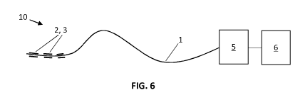
FIG. 6 schematically illustrates a system for measuring a radiation dose, in
accordance
with embodiments of the present invention.
The drawings are only schematic and are non-limiting. In the drawings, the
size of
some of the elements may be exaggerated and not drawn on scale for
illustrative purposes.
The dimensions and the relative dimensions do not necessarily correspond to
actual
reductions to practice of the invention.
Any reference signs in the claims shall not be construed as limiting the
scope.
In the different drawings, the same reference signs refer to the same or
analogous
elements.
CA 02966958 2017-05-05
WO 2016/075180 PCT/EP2015/076275
8
Detailed description of illustrative embodiments
The present invention will be described with respect to particular embodiments
and
with reference to certain drawings but the invention is not limited thereto
but only by the
claims.
The terms first, second and the like in the description and in the claims, are
used for
distinguishing between similar elements and not necessarily for describing a
sequence, either
temporally, spatially, in ranking or in any other manner. It is to be
understood that the terms
so used are interchangeable under appropriate circumstances and that the
embodiments of
the invention described herein are capable of operation in other sequences
than described or
illustrated herein.
Moreover, directional terminology such as top, bottom, front, back, leading,
trailing,
under, over and the like in the description and the claims is used for
descriptive purposes
with reference to the orientation of the drawings being described, and not
necessarily for
describing relative positions. Because components of embodiments of the
present invention
can be positioned in a number of different orientations, the directional
terminology is used
for purposes of illustration only, and is in no way intended to be limiting,
unless otherwise
indicated. It is, hence, to be understood that the terms so used are
interchangeable under
appropriate circumstances and that the embodiments of the invention described
herein are
capable of operation in other orientations than described or illustrated
herein.
It is to be noticed that the term "comprising", used in the claims, should not
be
interpreted as being restricted to the means listed thereafter; it does not
exclude other
elements or steps. It is thus to be interpreted as specifying the presence of
the stated
features, integers, steps or components as referred to, but does not preclude
the presence or
addition of one or more other features, integers, steps or components, or
groups thereof.
Thus, the scope of the expression "a device comprising means A and B" should
not be limited
to devices consisting only of components A and B. It means that with respect
to the present
invention, the only relevant components of the device are A and B.
Reference throughout this specification to "one embodiment" or "an embodiment"
means that a particular feature, structure or characteristic described in
connection with the
embodiment is included in at least one embodiment of the present invention.
Thus,
appearances of the phrases "in one embodiment" or "in an embodiment" in
various places
throughout this specification are not necessarily all referring to the same
embodiment, but
may. Furthermore, the particular features, structures or characteristics may
be combined in
CA 02966958 2017-05-05
WO 2016/075180 PCT/EP2015/076275
9
any suitable manner, as would be apparent to one of ordinary skill in the art
from this
disclosure, in one or more embodiments.
Similarly it should be appreciated that in the description of exemplary
embodiments
of the invention, various features of the invention are sometimes grouped
together in a
single embodiment, figure, or description thereof for the purpose of
streamlining the
disclosure and aiding in the understanding of one or more of the various
inventive aspects.
This method of disclosure, however, is not to be interpreted as reflecting an
intention that
the claimed invention requires more features than are expressly recited in
each claim. Rather,
as the following claims reflect, inventive aspects lie in less than all
features of a single
foregoing disclosed embodiment. Thus, the claims following the detailed
description are
hereby expressly incorporated into this detailed description, with each claim
standing on its
own as a separate embodiment of this invention.
Furthermore, while some embodiments described herein include some but not
other
features included in other embodiments, combinations of features of different
embodiments
are meant to be within the scope of the invention, and form different
embodiments, as
would be understood by those in the art. For example, in the following claims,
any of the
claimed embodiments can be used in any combination.
It should be noted that the use of particular terminology when describing
certain
features or aspects of the invention should not be taken to imply that the
terminology is
being re-defined herein to be restricted to include any specific
characteristics of the features
or aspects of the invention with which that terminology is associated.
In the description provided herein, numerous specific details are set forth.
However,
it is understood that embodiments of the invention may be practiced without
these specific
details. In other instances, well-known methods, structures and techniques
have not been
shown in detail in order not to obscure an understanding of this description.
DEFINITIONS
With "radiation dose" in the context of the present invention is meant a dose
of
ionizing irradiation, received by a pre-determined body part, e.g. a pre-
determined part of
the body, such as a body of a human or animal subject, during a radiotherapy
treatment, e.g.
during radiotherapy. For example the pre-determined body part may comprise a
tissue
volume corresponding to an irradiation target volume in an irradiation
treatment plan
specifically drawn up for the human or animal subject.
CA 02966958 2017-05-05
WO 2016/075180 PCT/EP2015/076275
"Luminescent materials" are materials that absorb energy from an external
source
different from a heat source, e.g. impinging ionizing radiation, and as a
consequence thereof
emit light.
With "dosimetric probe" in the context of the present invention is meant a
light
5 guide, such as for instance an optical fibre, coated over at least part
of its length with a
coating comprising a first component acting as a place dependent spectral
filter and a second
component including at least one luminescent material. The first and the
second components
may be integrated in a single coating layer, or they may be provided in
separate coating
layers, both layers together forming a double coating layer. The light guide
forms the core of
10 the dosimetric probe.
In a first embodiment, as illustrated in FIG. 6, the present invention
provides a system
10 for measuring a radiation dose received by a patient, in real-time, in-vivo
and at different
locations along the length of a light guide. The system 10 comprises:
a) a light guide 1, which under the influence of ionizing radiation undergoes
measurable and
quantifiable physical changes; wherein the light guide 1 is coated, over at
least part of its
length, with a coating comprising a first component 2 acting as a place
dependent spectral
filter and a second component 3 including at least one luminescent material,
dispersed in a
transparent matrix;
b) a detector system 5 which allows the recording and quantification of the
luminescent
signal emitted or transported by the light guide 1; and
c) a control unit 6 which is adapted for calculating a dose of ionizing
radiation previously or
simultaneously received by the light guide 6 on basis of said response signal.
The calculation
of the doze of ionizing radiation may be performed on-line (i.e. during
radiation) or off-line
(i.e. after a radiation step has been performed).
In embodiments of the present invention, the light guide consists of an
optical fiber 1
as illustrated in FIG. 1, coated over a given portion of its length with a
double layer
comprising a first coating 2 (internal layer) acting as a place dependent
filter and a second
coating 3 (external layer) including at least one luminescent material,
dispersed in a
transparent matrix. In alternative embodiments, the optical fiber 1 is coated
with a first
component acting as a place dependent filter and a second component including
at least one
luminescent material, dispersed in a transparent matrix, wherein the first and
the second
CA 02966958 2017-05-05
WO 2016/075180 PCT/EP2015/076275
11
components are integrated in a single coating layer provided over a
predetermined portion of
the length of the optical fibre 1.
In an alternative embodiment of the present invention, as illustrated in FIG.
2, the
optical fibre 1 is not coated with the first and second components, for
example in the form of
the double layer 2, 3, or intermixed in a single coating layer, over a
continuous portion of its
length, but rather over a plurality of discrete portions.
When exposed to radiation, the second component, e.g. the luminescent layer 3,
be it
a continuous layer or a layer comprising a plurality of discrete fields, will
emit light with a
spectrum depending on the chosen material(s). The amount of light is
proportional to the
locally imparted dose. The thus generated light will then be filtered, in a
location specific way,
by the first component, e.g. the inner coating 2, of optical fiber 1.
The location specific filtering by the first component, e.g. first coating 2,
is
mathematically defined through a characteristic function H(x), where x defines
a particular
segment on the optical fiber 1. These characteristic functions are defined
during standard
calibration conditions.
If the total dose of ionizing radiation measured by a system 10 according to
embodiments of the present invention is called D, this total measured dose D
will be a
superposition of the doses measured at each segment of the coated light guide
1. This can be
expressed as a weighted sum of these different contributions, so that the
following is
obtained:
D =lw,H(x),
1=1
In the previous equation, where assuming that the coating comprising the first
and second
components, e.g. the double coating 2, 3, on the light guide 1 is partitioned
into N segments, i
represents a specific segment of the light guide, H(x), is a matrix that
represents the
characteristic filtering responses at segments i (each row of the matrix is
related to a
particular segment i, whereas the columns correspond to the different
frequencies in the
electromagnetic spectrum) and w, is the dose recorded at that segment. The
latter (w,) is a
vector that can be calculated either by inverting the previous equation (in
case matrix H is
invertible) or by using an optimization approach. In the mathematical case
where the number
of segments N becomes very high (infinite to the limit), a spatially
continuous dose
measurement is obtained along the length of the light guide 1 provided with
coating
comprising the first and second components, e.g. the double coating 2, 3.
CA 02966958 2017-05-05
WO 2016/075180 PCT/EP2015/076275
12
In a preferred embodiment, as illustrated in FIG. 3, the coating comprising
the first
and second components, e.g. the double coating 2, 3, is placed at only two
locations so that
(point) measurements in two organs can be obtained simultaneously. This
configuration can
for instance be used in interventional procedures to measure at the same time
radiation dose
in the heart and in the lungs.
In a specific embodiment, the light guide 1 (for instance an optical fiber) is
shaped in
such a way that a 2D surface may be sampled. In this way a 2D dose measurement
can be
obtained using a single light guide 1. The light guide 1 can be integrated in
a flexible pad (not
illustrated), e.g. in the form of an adhesive patch for being put on the skin.
In particular embodiments, the two components are merged in one single coating
layer where the luminescent material (second component) and the filter
material (first
component) are dispersed in one single coating matrix.
In embodiments of the present invention, the luminescent material integrated
in the
coating is selected among (but not limited to): A1203, BaF2, Nal, CaF2, BG0
(barium
germanate) and alike. These materials can be used in doped or un-doped form.
The luminescent materials may be composed of nano or microparticles, dispersed
in
a non-absorbing matrix. This matrix is needed to coat the particles onto the
optical light guide
1. The matrix needs therefore to be transparent (i.e. non-absorbing) with
respect to the light
emitted by the luminescent material.
The filter can be composed of a color pigment in a matrix coated onto the
optical
light guide 1. One example of matrix wherein the pigments can be dispersed are
polyvinyl
alcohol (PVA) or acrylate based polymer in general, although this is in no way
!imitative for
the present invention.
Colored filters can be obtained by adding specific pigments to the matrix,
such as
(without being !imitative): cerium yellow, cobalt red, copper blue or copper
green. Different
colors will then be used at different locations, to allow spatial encoding of
the signal. The
absorption spectra of the filters may partially overlap.
In a preferred embodiment, the first component, e.g. inner coating 2, (filter)
may
operate in the visible part of the electromagnetic spectrum (i.e. from 400nm
to 700nm). In
another embodiment, the first component, e.g. inner coating 2, (filter) may
operate below
(<400nm) or above (>700nm) the visible part of the electromagnetic spectrum.
In yet another
embodiment, the first component, e.g. inner coating 2, (filter) may operate
from the
ultraviolet to the infrared part of the electromagnetic spectrum.
CA 02966958 2017-05-05
WO 2016/075180 PCT/EP2015/076275
13
In a specific embodiment, the smart dosimetric probe according to embodiments
of
the present invention, i.e. the light guide 1 coated with the first and second
components, for
instance intermixed in a single coating layer or under the form of a double
layer 2, 3, over at
least part of its length, can be used along with a microtube, such as a needle
or a catheter, to
guide the dosimetric probe to or in the neighborhood of a piece of tissue to
be irradiated by
ionizing radiation. This microtube can be used for in-vivo real-time dose
monitoring in
interventional procedures or in radiation oncology (in brachytherapy for
instance). By "real-
time" is meant that the dose is measured on a timescale such that this
information can be
used to adapt the ongoing procedure.
The control unit 6 may be equipped for calculating a dose of ionizing
radiation
received at the regions of the probe, which have been coated with luminescent
materials.
The control unit 6 will capture the response signal (filtered luminescence
signal) received or
generated by the light guide 1 and transform it into a numerical or graphical
dataset which
reflects the dose of irradiation received at different locations at the probe,
e.g. at different
locations within the catheter. To this end, each coated region will send its
light signal along
the fiber 1. The signals are collected in the detector system 5, for instance
by photo-sensors
that produce a global light spectrum, where light intensity is measured as a
function of
wavelength. An algorithm such as any of the algorithms discussed previously,
then allows
calculation of a dose of ionizing radiation in the detector system 6, for
example by converting
the spectrum in a line dose distribution.
In an advantageous embodiment, the detector system 5 or the control unit 6
includes
components selected to analyze and quantify optical signals, in spectral or in
time domain.
In a preferred embodiment, the control unit 6 also has a data storage and/or a
communication module. Through such modules, the system can interact with the
hospital
network and retrieve data related to the current procedure. These data, along
with the
dosimetric data recorded by the system, can be sent to an external server or
cloud.
The invention is further supported by the following examples which are
intended to
illustrate and not to limit the scope thereof:
Example 1: breast brachytherapy
In this first example, a patient is considered undergoing a brachytherapy
treatment
for breast cancer. More specifically, high dose rate (HDR) brachytherapy is
considered. In this
kind of treatment, guiding needles are first inserted in the breast, across
the tumor.
CA 02966958 2017-05-05
WO 2016/075180 PCT/EP2015/076275
14
Subsequently, radioactive Ir-192 sources are inserted in the guiding needles,
for a given time
and at a given position, so that the expected (i.e. calculated) dose
distribution in and around
the tumor is obtained. This procedure is repeated twice a day, for 5 days.
Typically, a total
dose of 50Gy is delivered to the tumor over the entire procedure.
Based on the above description of the treatment, it is clear that the
kinematic of each
Ir-192 source (i.e. its position as a function of the time), along with its
activity (defined as the
number of disintegrations per second), is extremely important in order to
obtain the
expected dose distribution.
In order to monitor on-line and in real-time the dose distribution along a
given line
crossing the tumor, and to compare it with the dose calculated by the
treatment plan, one of
the guiding needles was used to insert a measurement system according to
embodiments of
the present invention. The probe (coated light guide) was built in such a way
that a dose
could be measured at five contiguous locations. The filtering coating was
composed of a PVA
film wherein five different pigments were dispersed, in correspondence with
the five
measurement locations. The second coating (dosimetric coating) was based on
CsI(TI)
microparticles, also dispersed in a PVA film. CsI(TI) emits light from about
400nm up to about
700nm.
The total scintillation light recorded by the probe was sent to the reader
that would
calculate one dose per each dosimetric segment on the fiber, using the method
illustrated
previously.
The dose measured in real-time by the system according to embodiments of the
present invention was then compared with the dose calculated by the treatment
plan and
corrections in the sources kinematic could be implemented in almost real-time,
where
needed, so that an optimized, even optimal, dose distribution was obtained.
This
optimization resulted in a maximal dose to the tumor with a minimal dose (as
low as possible)
to the surrounding healthy breast tissues.
Example 2: interventional cardiology
In this second example, a patient is considered having a partial vascular
occlusion and
undergoing therefore a percutaneous transluminal coronary angioplasty (PTCA or
PCI or
simply angioplasty).
The procedure started with the insertion of a guiding wire. This was used to
assist the
insertion of a guiding catheter, through which the procedure was performed.
Also, a second
CA 02966958 2017-05-05
WO 2016/075180 PCT/EP2015/076275
(venous) catheter (a Swan-Ganz catheter) was inserted. This catheter was such
that, at the
same time, the distal part of one lumen was located in the pulmonary artery,
while the distal
part of a second lumen was in the heart. In this catheter, a measurement
system according to
embodiments of the present invention was inserted. The probe was built in a
way that only
5 two
dosimetric segments were present: one segment to monitor the dose to the lung
during
the procedure and the second one to monitor the dose in the heart. It has in
fact been
proven that these organs receive respectively 50% and 25% of the total
entrance dose, and
can, as such suffer complications on the longer term.
The two measurement locations were at a distance of 30cm from each other. The
10
filtering coating was composed of a PVA film wherein two different pigments
were dispersed
(one at each location). The luminescent coating was based on a dispersion of
A1203
microparcticles.
When the imaging beam was on, the luminescent material coating the catheter at
the
two different segments, started emitting light which was guided, through the
optical fiber, to
15 the
detector system. The detector system recorded the light spectrum received by
the fiber
and eventually the control unit calculated the dose at the two segments, using
the method
illustrated previously. This gave the interventional cardiologist a real-time
way of monitoring
the dose at the critical organs, in a minimally invasive way. On the longer
term, this real-time
monitoring is intended to lead to an optimization of the procedure and, more
specifically, of
the use of ionizing radiation, as strongly suggested by the ALARA principle
(irradiation should
be kept As Low As Reasonably Achievable). This will result in a minimization
of the dose for
the patient as well as for the practitioner.
Example 3: detection of light from an external light source
In this experiment, a two point detectors fiber was built. Each detection
segment had
a length of about 2cm. A filter coating was brought onto the fiber by painting
it with two
different tonalities of blue paint, at the two locations. The light blue
tonality was first diluted
with PVA polymer mixed with methyl alcohol. These paints were purchased at a
local shop.
In order to perform the experiment, the fiber was fixed on a table with tape.
The
lamp, used as source of photons, was also positioned at a fixed distance, with
respect to the
fiber.
The response signal of the channels was then measured, independently, using a
CCD
based spectrometer connected to the fiber through an SMA connector. This was
achieved by
CA 02966958 2017-05-05
WO 2016/075180 PCT/EP2015/076275
16
covering with a thick black felt the channel that had to be switched off. The
procedure was
repeated for both channels. FIG. 4 gives the signals generated per channel.
The channel
responses are given by the black and gray lines for the two segments
(channels) respectively.
The total response of the fiber was obtained by exposing both channels
simultaneously to light.
A fitting algorithm based on least square optimization was used to reconstruct
the
total measured signal, starting from the individual channels responses.
The first step of the fitting algorithm consisted in data normalization. Also,
a noise
correction was first applied to both individual channel data. The spectrometer
gave in fact a
constant noise level for each measurement. When combining both channels
linearly, noise
would therefore result double as much as the actual value:
SA = HA * I + 0
SB = HB * I + 0
where SA and SB represent respectively the signals at segments A and B, HA and
HB represent
the characteristic functions of channels A and B, / represents the light
intensity as sent by the
lamp and 0 is the offset (constant noise) produced by the spectrometer.
Therefore, when
linearly combining both channels, as done in the fit procedure, the following
is obtained:
Stotal = SA + SB = HA * I + HB * I + 2 * 0
In order to correct for this offest (noise) first half the mean noise value
(averaged over the
first 100 bins (arbitrary choice) of both channels) needs to be subtracted
from the individual
responses, before proceeding further to the fitting.
Finally, a least square normalization algorithm was applied to fit the total
measured
response, starting from the individual channels. The coefficients calculated
represent the
light intensity measured at each segment. FIG. 5 shows the result of the
fitting: the measured
total signal is represented by graph 50 and the fitted curve is represented by
graph 51.