Note: Descriptions are shown in the official language in which they were submitted.
CA 02967066 2017-05-05
WO 2016/081365 PCT/US2015/060878
1
TITLE: COMPOSITIONS AND METHODS FOR TREATING LYSOSOMAL
DISORDERS
RELATED APPLICATIONS
[0100] The present patent application claims the benefit of the filing
date of
U.S. Provisional Patent Application No. 62/081,696, filed November 19, 2014,
the contents of which is hereby incorporated by reference.
TECHNICAL FIELD
[0101] The present invention generally relates to compositions and methods
for treating lysosomal storage disorders.
BACKGROUND
[0102] Lysosomes are membrane bound organelles containing a host of
hydrolytic enzymes that are highly active in acidic milieu (1-3). Classically
identified as the waste management organelle, lysosomes have been shown to
be involved in major cellular processes including degradation developmental,
programmed cell death, and nutritional responses (2, 4-8). The diverse roles
and responses of the lysosome to different stimuli suggest a coordinated
regulation of expression of lysosomal genes (9, 10). Recent studies provide
modest information about the regulation of lysosomal genes (11,12) but pattern
discovery analysis for the lysosomal genes revealed the presence of a
Coordinated Lysosomal Expression and Regulation (CLEAR) element, which is
a potential binding site for Transcription Factor EB (TFEB), a member of the
microphthalmia¨transcription factor E (MiT/TFE) subfamily of bHLH (basic
helix-loop-helix) factors. The study reports a potential link between TFEB and
lysosomal biogenesis (9, 10, 12).
[0103] The regulation of Tfeb appears to be complex and dependent on cell
type and stimuli. In differentiated osteoclasts, a RAN KL-dependent signaling
pathway induces TFEB activation induced lysosomal biogenesis (13).
Starvation or stress conditions may also activate TFEB, which otherwise is
CA 02967066 2017-05-05
WO 2016/081365 PCT/US2015/060878
2
maintained in an inactivated state by mTORC1 (14, 15). One study also
showed starvation induced TFEB activity can play a vital role in lipid
metabolism and that activated TFEB can also autoregulate its own gene
expression (16).
[0104] Lysosomal storage diseases (LSDs) are a group of approximately 50
rare inherited metabolic disorders that result from defects in lysosomal
function.
The symptoms of LSD vary, depending on the particular disorder and other
variables like the age of onset, and can be mild to severe. They can include
developmental delay, movement disorders, seizures, dementia, deafness
and/or blindness. Some people with LSD have enlarged livers (hepatomegaly)
and enlarged spleens (splenomegaly), pulmonary and cardiac problems, and
bones that grow abnormally.
SUMMARY OF THE PREFERRED EMBODIMENTS
[0105] One aspect of the present invention provides a method for
treatment
of a LSD. The method may include administering to a subject in need of such
treatment a composition including a therapeutically effective amount of an
agent that mediates upregulation of Transcription Factor EB (TFEB).
[0106] In one embodiment, the agent is a statin. For example, the statin
may be atorvastatin, fluvastatin, lovastatin, pitavastatin, pravastatin,
rosuvastatin, or simvastatin. The agent may also be, for example, a lipid-
lowering drug, such as a fibrate. In some embodiments, the fibrate is
gemfibrozil or fenofibrate. In other embodiments, the agent is an analgesic,
an
antipyretic, aspirin, a cinnamon metabolite, cinnamic acid, sodium
phenylbutyrate or sodium benzoate. In yet other embodiments, the
composition may include a combination of at least two of the above agents.
[0107] The composition may also include all-trans retinoic acid or
vitamin A.
For example, the composition may include a statin or a fibrate and all-trans
retinoic acid or vitamin A. This combination of the agent(s) with all-trans
retinoic acid or vitamin A may provide a greater therapeutic effect in the
subject
CA 02967066 2017-05-05
WO 2016/081365 PCT/US2015/060878
3
than administration of the all-trans retinoic acid, vitamin A or the fibrate
alone.
The combination may be a synergistic combination. TFEB may also be
upregulated by increasing Transcription Factor EB mRNA levels increasing
Transcription Factor EB protein levels or activating a PPARa-RXRa
heterodimer.
[0108] The LSD may be a neurodegenerative disorder, for example,
neuronal ceroid lipofuscinosis, Alzheimer's disease, Huntington's disease,
Amyotrophic lateral sclerosis (ALS), Parkinson's disease, including
Parkinson's
plus diseases such as multiple system atrophy (MSA), progressive
supranuclear palsy (PSP), corticobasal degeneration (CBD) or dementia with
Lewy bodies (DLB). In another embodiment, the LSD is a disorder of the
autophagy pathway and wherein the agent increases lysosomal biogensis.
[0109] In other embodiments, the LSD is Tay-Sach's disease, Fabry
disease,
Niemann-Pick disease, Gaucher disease, Hunter Syndrome, Alpha-
mannosidosis, Aspartylglucosaminuria, Cholesteryl ester storage disease,
Chronic Hexosaminidase A Deficiency, Cystinosis, Danon disease, Farber
disease, Fucosidosis, Galactosialidosis, or Batten disease including late
infantile Batten disease and Juvenile Batten disease.
[0110] Another aspect of the present invention provides a method for
treatment of a LSD including administering to a subject in need of such
treatment a composition including a therapeutically effective amount of an
agent that mediates upregulation of the Tfeb gene. Yet another aspect
provides a method for treatment of a LSD including administering to a subject
in
need of such treatment a composition including a therapeutically effective
amount of an agent, wherein the agent restores TFEB activity.
BRIEF DESCRIPTION OF THE DRAWINGS
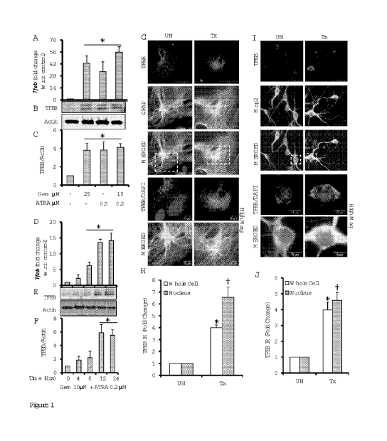
[0111] Figure 1 shows Gemfibrozil and Retinoic Acid upregulating TFEB
mRNA and protein levels in brain cells. (A, B) Mouse primary astrocytes were
treated with different concentrations of gemfibrozil and all-trans retinoic
acid
CA 02967066 2017-05-05
WO 2016/081365 PCT/US2015/060878
4
(ATRA) under serum free DMEM/F-12 medium for 12hrs followed by monitoring
mRNA levels of Tfeb by qRT-PCR (A) and TFEB protein levels by immunoblot
(B). (C) Densitometric analysis of the immunoblot for TFEB (relative to [3-
Actin).
(D, E), Mouse primary astrocytes were treated with a combination of
gemfibrozil
(10pM) and ATRA (0.2pM) for 4, 6, 12 and 24 hrs under similar culture
conditions followed by monitoring of mRNA levels of TFEB by qRT-PCR (D)
and protein levels by immunoblot (E). (F), denistometric analysis for the
immunoblot for TFEB. All results are representative of or mean SEM of at
least three independent set of experiments. (G, I) Mouse primary astrocytes
(G)
and mouse primary neurons (I) were treated with combination of gemfibrozil
and retinoic acid under serum free condition for 24hrs and were double labeled
for TFEB (red) - GFAP (green) and TFEB (red) - Map2 (green), respectively.
DAPI was used to stain nuclei. Scale bar = 20pm (for G), scale bar = 5pm for
High Magnification Images (for G); Scale bar = 50pm (for l), scale bar = 10pm
for High Magnification Images (for l). (H,J) Quantification of TFEB
immunoreactivity (TFEB IR) in whole cell and nucleus for mouse primary
astrocytes (H) and mouse primary neurons (J) calculated as fold over control.
At least 25 separate images per condition from three independent set of
experiments are quantified using ImageJ. pt < 0.05 vs untreated control.
[0112] Figure 2 shows involvement of PPARa and RXRa in fibrate drug-
mediated upregulation of TFEB mRNA and protein: (A, B) Mouse primary
astrocytes isolated from PPARa-/- and PPAR[3-/- and wild type mouse were
treated with combination of gemfibrozil (10pM) and retinoic acid(0.2pM) in
serum free DMEM/F12 for 24hrs followed by monitoring the mRNA expression
of Tfeb by real-time FOR (A) and protein level of TFEB by immunoblot (B). (C)
Densitometric analysis of TFEB levels (relative to [3-Actin) in PPARa-/- and
PPAR[3-/- and wild type astrocytes. pa<0.05 vs WT control; pb<0.05 vs PPAR[3-
/- control; ns- not significant w.r.t PPARa-/- control. (D) Mouse primary
astrocytes isolated from WT mice were pre-treated with GW9662 for 30 min
followed by treatment with gemfibrozil and retinoic acid under similar culture
CA 02967066 2017-05-05
WO 2016/081365 PCT/US2015/060878
conditions followed by monitoring the levels of TFEB protein expression by
immunoblot. (E) Densitometric analysis of immunoblot for TFEB (relative to [3-
Actin) pt<0.05 vs control; ns- not significant w.r.t control. (F) Mouse
primary
astrocytes isolated from PPARa-/- and PPAR[3-/- and WT mice were treated
with 10pM gemfibrozil and 0.2pM retinoic acid in serum free DMEM/F12 for
24hrs and double-labeled for TFEB (red) and GFAP (green). DAPI was used to
stain nuclei. UN- No treatment. Scale bar = 20pM. (G) Quantification of TFEB
immunoreactivity (TFEB IR) for mouse primary astrocytes calculated as fold
over control. At least 25 separate images per condition from three independent
set of experiments are quantified using ImageJ. pa<0.05 vs WT control;
pb<0.05 vs PPAR[3-/- control; ns- not significant w.r.t PPARa-/- control. (H,
I, J)
Mouse primary astrocytes were untransfected, transfected with scrambled
siRNA (1.0pg) or RXRa siRNA (1.0pg) for 36hrs followed by treatment with RA
(0.2pM) and gemfibrozil (10p M) in combination for 24hrs serum free
DMEM/F12 medium followed by RI-FOR for RXRa to check the level of gene
silencing (H) and quantitative real time PCR for TFEB (J)and immunoblot for
TFEB (J). (K) Denistometric analysis of immunoblot for TFEB (relative to 13
actin). p* < 0.05 vs untransfected control; p** < 0.05 vs scrambled siRNA
transfected control; ns- not significant w.r.t. RXR-a siRNA transfected
control.
All results are representative of or mean SEM of at least three independent
set of experiments.
[0113] Figure 3 shows PPARa transcriptionally regulating TFEB expression
under treatment condition: (A) Map of wild-type and mutated PPRE site of
TFEB-Luciferase promoter constructs. (B) BV2 cells were transfected with
pTFEB(WT)-Luc for 24hrs followed by treatment with different concentrations of
gemfibrozil and retinoic acid alone and in combination and subjected to
luciferase assay. p* < 0.05 vs untreated control. (C) BV2 cells were
transfected
with pTFEB(WT)-Luc for 24hrs followed by pretreatment with PPARa-, PPAR[3-,
PPARy-antagonists followed by treatment with gemfibrozil and retinoic acid and
subjected to luciferase assay. p* < 0.05 vs untreated control. p# < 0.05 vs
CA 02967066 2017-05-05
WO 2016/081365 PCT/US2015/060878
6
treatment. (D) Mouse primary astrocytes isolated from PPARa-/- (center) and
PPAR[3-/- (right) and wild type (left) mouse were transfected with pTFEB(WT)-
Luc for 24hrs followed by treatment with gemfibrozil and retinoic acid and
subjected to luciferase assay. p* < 0.05 vs untreated WT control. . p# - not
significant vs untreated PPARa-/- control. . pt < 0.05 vs untreated PPAR[3-/-
control. (E, F) BV2 cells (E) and mouse primary astrocytes (F) were
transfected
with pTFEB(WT)-Luc and pTFEB(Mu)-Luc for 24hrs followed by treatment with
gemfibrozil and retinoic acid and subjected to luciferase assay. p* < 0.05 vs
untreated pTFEB(WT)-Luc transfected control. ns- not significant w.r.t.
untreated pTFEB(Mu)-Luc transfected control. All results are mean SEM of at
least six sets of independent experiments.
[0114] Figure 4 shows trancriptional activation of TFEB by PPARa-RXRa-
PGC1a complex (A) Map of PPRE on TFEB promoter with core sequence and
amplicon length for ChIP. (B,C) Mouse astrocytes were treated with the
combination of gemfibrozil (10pM) and RA (0.2pM) for 30, 60, 120 and 240
mins and recruitment of PPARa (far left), RXRa (center left), PGC1a (center
right) and RNA Polymerase (far right) on the PPRE binding site of Tfeb
promoter was monitored by ChIP followed by RT-PCR (B) and qRT-PCR (C).
Normal IgG was used as control. p* < 0.05 vs untreated control. All results
are
representative of or mean SEM at least there independent sets of
experiments. (D) Schematic representation of induction of lysosomal
biogenesis by activating peroxisomal proliferators.
[0115] Figure 5 shows PPARa dependant upregulation of TFEB inducing
lysosomal biogenesis: (A, B) Mouse primary astrocytes isolated from PPARa-/-
and PPAR[3-/- and wild type mouse were treated with combination of
gemfibrozil (10pM) and retinoic acid (0.2pM) in serum free DMEM/F12 for 24hrs
followed by monitoring the mRNA expression of lysosomal genes (Lamp2 (left),
Limp2 (center), Npc1(right)) by real-time PCR (A) and protein level of LAMP2
by immunoblot (B). (C) Densitometric analysis of LAMP2 levels (relative to [3-
Actin) in PPARa-/- and PPAR[3-/- and wild type astrocytes. pa<0.05 vs WT
CA 02967066 2017-05-05
WO 2016/081365 PCT/US2015/060878
7
control; pb<0.05 vs PPAR[3-/- control; ns- not significant w.r.t PPARa-/-
control.
All results are representative of or mean SEM at least there independent
sets
of experiments. (D) Mouse primary astrocytes isolated from PPARa-/- and
PPAR[3-/- and WT mice were treated with 10pM gemfibrozil and 0.2pM retinoic
acid in serum free DMEM/F12 for 24hrs and double-labeled for LAMP2 (red)
and GFAP (green). DAR was used to stain nuclei. (E) Quantification of LAMP2
immunoreactivity (Lamp2 IR) for mouse primary astrocytes calculated as fold
over control. pa<0.05 vs WT control; pb<0.05 vs PPAR[3-/- control; ns- not
significant w.r.t PPARa-/- control. (F) Mouse primary neurons isolated from WT
mice were treated with 10pM gemfibrozil and 0.2pM retinoic acid in serum free
DMEM/F12 for 24hrs and double-labeled for LAMP2 (red) and Map2 (green).
DAR was used to stain nuclei. (G) Quantification of LAMP2 immunoreactivity
(Lamp2 IR) for mouse primary neurons calculated as fold over control. p*<0.05
vs untreated control. (H) Mouse primary astrocytes isolated from PPARa-/- and
PPAR[3-/- and WT mice were treated with 10pM gemfibrozil and 0.2pM retinoic
acid in serum free DMEM/F12 for 24hrs and double-labeled for LysoTracker
Red (red) and GFAP (green). (I) Quantification of LAMP2 immunoreactivity
(Lamp2 IR) for mouse primary astrocytes calculated as fold over control.
pa<0.05 vs WT control; pb<0.05 vs PPAR[3-/- control; ns- not significant w.r.t
PPARa-/- control. UN- No treatment. Scale bar = 20pm (for D, E & F), scale bar
= 10pm for High Magnification Images (for E). Al least 25 images per condition
from three different sets of experiments were analyzed for all image
quantification data using ImageJ.
[0116] Figure 6 shows that oral administration of gemfibrozil upregulates
TFEB in vivo in the cortex of WT and PPAR[3-/-, but not PPARa-/- mice: (A, D,
G) WT, PPARa-/- and PPAR[3-/- mice (n=6 in each group) were treated with
7.5mg/kg body wt/day gemfibrozil and 0.1mg/kg body weight of All-trans
retinoic acid (dissolved in 0.1% methylcellulose) or vehicle (0.1%
methylcellulose) via gavage. After 60d of treatment, mice were killed and
cortical sections were double labeled for TFEB (red) and NeuN (green). DAPI
CA 02967066 2017-05-05
WO 2016/081365 PCT/US2015/060878
8
was used to visualize nucleus (C, F, I) Higher magnification images showing
localization of TFEB and NeuN in the cortical neuron of mice from the
treatment
group (WT, PPARa-/- and PPAR[3-/-). (B, E, H) Quantification of TFEB
Immunoreactivity (TFEB IR) in untreated and treated samples from each group
(WT, PPARa-/- and PPAR[3-/-) expressed as percentage of area. pa<0.05 vs
WT control; pb<0.05 vs PPAR[3-/- control; ns- not significant w.r.t PPARa-/-
control. At least 12 sections from each group (2 sections per animal) were
quantified using ImageJ. Scale bar = 50pM and 10pm (for higher magnification
images).
[0117] Figure 7 shows that oral administration of gemfibrozil upregulates
LAMP2 in vivo in the cortex of WT and PPAR[3-/-, but not PPARa-/- mice: (A, D,
G) WT, PPARa-/- and PPAR[3-/- mice (n=6 in each group) were treated with
7.5mg/kg body wt/day gemfibrozil and 0.1mg/kg body weight of All-trans
retinoic acid (dissolved in 0.1% methylcellulose) or vehicle (0.1%
methylcellulose) via gavage. After 60d of treatment, mice were killed and
cortical sections were double labeled for LAMP2 (red) and NeuN (green). DAR
was used to visualize nucleus (C, F, I) Higher magnification images showing
localization of LAMP2 and NeuN in the cortical neuron of mice from the
treatment group (WT, PPARa-/- and PPAR[3-/-). (B, E, I) Quantification of
LAMP2 Immunoreactivity (LAMP2 IR) in untreated and treated samples from
each group (WT, PPARa-/- and PPAR[3-/-) expressed as percentage of area.
pa<0.05 vs WT control; pb<0.05 vs PPAR[3-/- control; ns- not significant w.r.t
PPARa-/- control. At least 12 sections from each group (2 sections per animal)
were quantified using ImageJ. Scale bar = 50pM and 10pm (for higher
magnification images).
[0118] Figure 8 shows that upregulation of TFEB induces lysosomal
biogenesis in both normal and LINCL patient fibroblasts: Fibroblasts from
healthy individuals (WT#1-3) and LINCL patients (NCL#1-5) and carrier of
LINCL (NCL/C) were treated with gemfibrozil (10pM) and retinoic acid (0.2pM)
in reduced serum (2%) DMEM medium for 24hrs followed by staining with
CA 02967066 2017-05-05
WO 2016/081365 PCT/US2015/060878
9
LysoTracker Red (red). Brightfield microscopy used for detecting cell
morphology. Scale bar = 20pM. Corresponding box plots represent fold change
in the LysoTracker positive signals in treated group vs control in each cell
type.
p* < 0.05 vs untreated control. ROI ¨ white dotted lines, represent area of
the
cell. Fold change calculated as Lysol +ve signal per unit area per cell in
treatment vs control. At least 25 individual images per condition per cell
type
were quantified using ImageJ.
[0119] Figures 9(A) ¨ 9(D) illustrate the upregulation of TFEB mRNA
expression in mouse astrocytes by cholesterol-lowering drugs (simvastatin and
pravastatin), aspirin (anasgesic and anti-pyretic), cinnamic acid (metabolite
of
cinnamon), and drugs for urea cycle disorders (sodium phenylbutyrate and
sodium benzoate).
[0120] Figure 10 illustrates an increase in lysosomal biogenesis by
aspirin in
primary mouse astrocytes. Cells were treated with different concentrations of
aspirin for 24 h under serum-free condition followed by Lyso-tracker staining.
Results represent three independent experiments.
[0121] Figure 11(A)-(D) shows the upregulation of LAMP2 expression by
aspirin in primary mouse astrocytes. 11(A) Cells were treated with 5 pM
aspirin
for different time periods under serum-free condition followed by monitoring
the
mRNA expression of LAMP2 by real-time PCR. Results are mean + SD of
three different experiments. ap < 0.05 vs control; bp < 0.001 vs control.
11(B)
Cells were treated with different concentrations of aspirin for 24 h under
serum-
free condition followed by Western blot for LAMP2. 11(C) Cells were treated
with 5 pM aspirin for different time periods under serum-free condition
followed
by Western blot for LAMP2. 11(D) After 24 h of aspirin treatment, cells were
double-labeled for LAMP2 and GFAP. Results represent three independent
experiments.
[0122] Figure 12(A)-(C) shows an increase in TPP1 by aspirin in primary
mouse astrocytes. 12(A) Cells were treated with different concentrations of
CA 02967066 2017-05-05
WO 2016/081365 PCT/US2015/060878
aspirin for 24 h under serum-free condition followed by Western blot for TPP1.
12(B) Cells were treated with 5 pM aspirin for different time periods under
serum-free condition followed by Western blot for TPP1. Actin was run as a
house keeping molecule. 12(C) Cells were treated with different concentrations
of aspirin for 24 h under serum-free condition followed by TPP1 activity assay
using cell extract containing 5 pg of total protein and Ala-Ala-Phe 7-amido-4-
methylcoumarin as substrate. Results represent three independent
experiments.
[0123] Figure 13(A)-(C) illustrates the upregulation of TFEB by aspirin
in
primary mouse astrocytes. 13(A) Cells were treated with different
concentrations of aspirin for 12 h under serum-free condition followed by
Western blot for TFEB. Actin was run as a house keeping molecule. 13(B)
Cells were treated with 5 pM aspirin for 12 h under serum-free condition
followed by double-labeling with GFAP and TFEB. These results are mean of
two independent experiments. 13(C) Cells were transfected with p(WT)Tfeb-
Luc plasmid and after 24 h of transfection, cells were stimulated with
different
doses of aspirin. After 4 h, firefly luciferase activity was measured in total
cell
extracts. Results are mean + SD of three different experiments. ap < 0.001 vs
control.
[0124] Figure 14(A)-(B) illustrates activation of PPARa by aspirin in
primary
mouse astrocytes. 14(A) Cells were treated with 5 pM aspirin for different min
intervals followed by isolation of nuclear extracts and electrophoretic
mobility
shift assay for monitoring DNA-binding activity of PPARa using PPARa-binding
site of the Tfeb promoter as a probe. 14(B) Astrocytes isolated from wild
type,
PPARa (-/-) and PPAR[3 (-/-) mice were transfected with PPAR luciferase
reporter (PPRE-x3-TK-luc) plasmid and after 24 h of transfection, cells were
stimulated with different doses of aspirin. After 4 h, firefly luciferase
activity was
measured in total cell extracts. Results are mean + SD of three different
experiments. ap < 0.001 vs control.
CA 02967066 2017-05-05
WO 2016/081365 PCT/US2015/060878
11
[0125] Figure 15(A)-(C) illustrates that aspirin increases the level of
TFEB in
astrocytes via PPARa. Astrocytes isolated from WT 15(A), PPARa (-/-) 15(B)
and PPAR[3 (-/-) 15(0) mice were treated with 5 pM aspirin for 12 h under
serum-free condition followed by double-labeling for TFEB and GFAP. Results
represent three independent experiments.
[0126] Figure 16(A)-(B) illustrates that aspirin increases the level of
LAMP2
in astrocytes via PPARa. Astrocytes isolated from WT, PPARa (-/-) and
PPAR[3 (-/-) mice were treated with different concentrations of aspirin for 24
h
under serum-free condition followed by Western blot analysis for LAMP2 16(A).
Actin was run as a house keeping molecule. Bands were scanned and
expressed as relative to control 16(B). Results are mean + SD of three
different experiments. ap < 0.05 vs control; bP < 0.001 vs control. ns, not
significant.
[0127] Figure 17 (A)-(C) illustrate that aspirin increases lysosomal
biogenesis in astrocytes via PPARa. Astrocytes isolated from WT 17(A),
PPARa (-/-) 17(B) and PPAR[3 (-/-) 17(0) mice were treated with 5 pM aspirin
for 24 h under serum-free condition followed by Lyso-tracker staining. Results
represent three independent experiments.
DETAILED DESCRIPTION OF THE PREFERRED EMBODIMENTS
Definitions
[0128] Unless otherwise defined, all technical and scientific terms
used
herein have the same meaning as commonly understood by one of ordinary
skill in the art to which this invention pertains. In case of conflict, the
present
document, including definitions, will control. Preferred methods and materials
are described below, although methods and materials similar or equivalent to
those described herein can be used in the practice or testing of the present
invention.
CA 02967066 2017-05-05
WO 2016/081365 PCT/US2015/060878
12
[0129] The uses of the terms "a" and "an" and "the" and similar
references in the context of describing the invention (especially in the
context of
the following claims) are to be construed to cover both the singular and the
plural, unless otherwise indicated herein or clearly contradicted by context.
Recitation of ranges of values herein are merely intended to serve as a
shorthand method of referring individually to each separate value falling
within
the range, unless otherwise indicated herein, and each separate value is
incorporated into the specification as if it were individually recited herein.
All
methods described herein can be performed in any suitable order unless
otherwise indicated herein or otherwise clearly contradicted by context. The
use of any and all examples, or exemplary language (e.g., "such as", "for
example") provided herein, is intended merely to better illuminate the
invention
and does not pose a limitation on the scope of the invention unless otherwise
claimed. No language in the specification should be construed as indicating
any non-claimed element as essential to the practice of the invention.
[0130] As used herein, the term subject refers to a human or veterinary
subject. The term "therapeutic effect" as used herein means an effect which
induces, ameliorates or otherwise causes an improvement in the pathological
symptoms, disease progression or physiological conditions associated with or
resistance to succumbing to a disorder, for example a LSD, of a subject. The
term "therapeutically effective amount" as used with respect to a drug means
an amount of the drug which imparts a therapeutic effect to the subject.
[0131] The terms "synergy", "synergism" or "synergistic" mean more than the
expected additive effect of a combination. A synergistic effect may be
attained
when the active ingredients are: (1) co-formulated and administered or
delivered simultaneously in a combined, unit dosage formulation; (2) delivered
by alternation or in parallel as separate formulations; or (3) by some other
regimen.
CA 02967066 2017-05-05
WO 2016/081365 PCT/US2015/060878
13
Compositions and Methods for Treating Lysosomal Storage Disorders
[0132] For the purpose of promoting an understanding of the principles
of
the invention, reference will now be made to embodiments, some of which are
illustrated in the drawings, and specific language will be used to describe
the
same. It will nevertheless be understood that no limitation of the scope of
the
invention is thereby intended. Any alterations and further modifications in
the
described embodiments, and any further applications of the principles of the
invention as described herein are contemplated as would normally occur to one
skilled in the art to which the invention relates.
[0133] One aspect of the present invention relates to methods of
treatment of a lysosomal storage disorder (LSD). The LSD may be, for
example, Tay-Sach's disease, Fabry disease, Niemann-Pick disease, Gaucher
disease, Hunter Syndrome, Alpha-mannosidosis, Aspartylglucosaminuria,
Cholesteryl ester storage disease, Chronic Hexosaminidase A Deficiency,
Cystinosis, Danon disease, Farber disease, Fucosidosis, Galactosialidosis or
Batten disease including late infantile Batten disease and Juvenile Batten
disease. The LSD may also be a neurodegenerative disease involving the
autophagy-lysosome pathway, for example, neuronal ceroid lipofuscinosis,
Alzheimer's disease, Huntington's disease, Amyotrophic lateral sclerosis
(ALS),
Parkinson's disease, including Parkinson's plus diseases such as multiple
system atrophy (MSA), progressive supranuclear palsy (PSP), corticobasal
degeneration (CBD) or dementia with Lewy bodies (DLB). The
neurodegenerative disorder may be characterized by defective autophage.
Such disorders include Alzheimer's, Parkinson's disease, and Huntington's
disease.
[0134] One embodiment includes administering to a subject suffering
from a LSD an agent that upregulates or enhances expression from the Tfeb
gene. Upregulation may include increasing mRNA levels for Tfeb. The
methods of the present invention also include administering to a subject
suffering from a LSD an agent that upregulates TFEB or restores TFEB activity.
CA 02967066 2017-05-05
WO 2016/081365 PCT/US2015/060878
14
Upregulation may include increasing TFEB mRNA levels, increasing TFEB
protein levels, or increasing TFEB activity. Activating a PPARa/RXRa
heterodimer results in upregulation of TFEB. The inventor has also
surprisingly
shown that TFEB is upregulated through the activity or involvement of PPARa,
but not PPAR[3 or PPARy.
[0135] The agent may be a lipid-lowering drug such as a fibrate. The fibrate
may be gemfibrozil, fenofibrate, or clofibrate. The agent may be all-trans
retinoic acid or vitamin A. Surprisingly and unexpectedly, administration of
the
fibrate in combination with all-trans retinoic acid or vitamin A to the
subject may
upregulate TFEB more than administration of the fibrate or all-trans retinoic
acid or vitamin A alone. The fibrate and all-trans retinoic acid or vitamin A,
when administered together to the subject, may cooperatively enhance
upregulation of TFEB to synergistically upregulate TFEB. A lower dose of the
fibrate may be needed in the presence of all-trans retinoic acid or vitamin A
to
achieve the same degree of TFEB upregulation as occurs when only a higher
dose of the fibrate is administered to the subject. The combination of the
fibrate and all-trans retinoic acid or vitamin A may be a synergistic
combination.
[0136] In other embodiments, the lipid lowering drug is a statin. For
example
the statin may be atorvastatin, fluvastatin, lovastatin, pitavastatin,
pravastatin,
rosuvastatin, simvastatin or a combination of at least two of these drugs. The
statin or statins may be used alone or in combination with a fibrate and or
all-
trans retinoic acid or vitamin A. In yet other embodiments, the agent may be
an
analgesic or antipyretic, for example aspirin; phenylbutyrate; sodium
benzoate;
or a cinnamon metabolite, for example cinnamic acid. Again, such agents may
be used in combination with all-trans retinoic acid or vitamin A and may be
administered together to the subject to cooperatively enhance upregulation of
TFEB to synergistically upregulate TFEB.
[0137] The lipid lowering drugs may be drugs that reduce the level
oftriglycerides circulating in the blood of the subject. Additionally, lipid-
lowering
drugs may be drugs that decrease the risk of hyperlipidemia. The fibrate may
CA 02967066 2017-05-05
WO 2016/081365
PCT/US2015/060878
mediate upregulation of TFEB via PPARa, but not PPAR[3 and PPARy. During
upregulation of TFEB, PPARa forms a heterodimer with RXR-a and the
RXRa/PPAR-a heterodimer is recruited to the promoter of the Tfeb gene via a
RXR binding site.
[0138] The
upregulation of TFEB may also be mediated by all-trans
retinoic acid. All-trans retinoic acid may also be known as ATRA, retinoic
acid,
tretinoin, and vitamin A acid. All-trans retinoic acid may mediate
upregulation
of TFEB via the retinoid X receptor-a (RXR-a). During upregulation of TFEB,
RXR-a forms a heterodimer with peroxisome proliferator-activated receptor-a
(PPAR-a) and the RXR-a/PPAR-a heterodimer is recruited to the promoter of
the Tfeb gene via a RXR binding site.
[0139] The composition mediating upregulation of TFEB may include a
combination of the agent, for example, a lipid lowering drug, and all-trans
retinoic acid or vitamin A. Such a combination may cooperatively mediate or
enhance upregulation of TFEB as compared to administration of the agent or
all-trans retinoic acid or vitamin A alone. The combination may cooperatively
enhance upregulation of TFEB about 2-fold, about 3-fold, about 4-fold, about 5-
fold, or about 10-fold as compared to administration of the lipid-lowering
drug or
all-trans retinoic acid or vitamin A alone. Particularly, the combination may
cooperatively enhance upregulation of TFEB about 3-fold as compared to
administration of the lipid-lowering drug or all-trans retinoic acid or
vitamin A
alone.
[0140] Another aspect of the present invention provides pharmaceutical
compositions including at least one of the agents disclosed above. The
pharmaceutical composition may also include all-trans retinoic acid or vitamin
A. For example, the pharmaceutical composition may include gemfibrozil or a
combination of gemfibrozil and all-trans retinoic acid or vitamin A or a
combination of aspirin and all-trans retinoic acid or vitamin A.
CA 02967066 2017-05-05
WO 2016/081365 PCT/US2015/060878
16
[0141] The pharmaceutical compositions can be in the form of, for example,
tablets, pills, dragees, hard and soft gel capsules, granules, pellets,
aqueous,
lipid, oily or other solutions, emulsions such as oil-in-water emulsions,
liposomes, aqueous or oily suspensions, syrups, alixiers, solid emulsions,
solid
dispersions or dispersible powders. In pharmaceutical compositions for oral
administration, the agent may be admixed with commonly known and used
adjuvants and excipients, for example, gum arabic, talcum, starch, sugars
(such as, e.g., mannitose, methyl cellulose, lactose), gelatin, surface-active
agents, magnesium stearate, aqueous or non-aqueous solvents, paraffin
derivatives, cross-linking agents, dispersants, emulsifiers, lubricants,
conserving agents, flavoring agents (e.g., ethereal oils), solubility
enhancers
(e.g., benzyl benzoate or benzyl alcohol) or bioavailability enhancers (e.g.
GELUCIRE). In the pharmaceutical composition, the agent may also be
dispersed in a microparticle, e.g. a nanoparticulate, composition.
[0142] For parenteral administration, the agent or pharmaceutical
compositions of the agent can be dissolved or suspended in a physiologically
acceptable diluent, such as, e.g., water, buffer, oils with or without
solubilizers,
surface-active agents, dispersants or emulsifiers. As oils for example and
without limitation, olive oil, peanut oil, cottonseed oil, soybean oil, castor
oil and
sesame oil may be used. More generally, for parenteral administration the
agent or pharmaceutical compositions of the agent can be in the form of an
aqueous, lipid, oily or other kind of solution or suspension or even
administered
in the form of liposomes or nano-suspensions.
Modes of Administration
[0143] The agents disclosed above or pharmaceutical compositions
including these agents can be administered by any method that allows for the
delivery of a therapeutic effective amount of the agent to the subject. Modes
of
administration can include, but are not limited to, oral, topical, transdermal
and
parenteral routes, as well as direct injection into a tissue, and delivery by
a
CA 02967066 2017-05-05
WO 2016/081365 PCT/US2015/060878
17
catheter. Parenteral routes can include, but are not limited to subcutaneous,
intradermal, intra-articular, intravenous, intraperitoneal and intramuscular
routes. In one embodiment, the route of administration is by topical or
transdermal administration, such as by a lotion, cream, a patch, an injection,
an
implanted device, a graft or other controlled release carrier. Routes of
administration include any route which directly delivers the composition to
the
systemic circulation (e.g., by injection), including any parenteral route.
[0144] One embodiment of the method of the present invention
comprises administering at least one agent, for example gemfibrozil or a
combination of gemfibrozil and ATRA, in a dose, concentration and for a time
sufficient to prevent the development of, or to lessen the extent of, a LSD.
Certain embodiments include administering systemically at least one agent in a
dose between about 0.1 micrograms and about 100 milligrams per kilogram
body weight of the subject, between about 0.1 micrograms and about 10
milligrams per kilogram body weight of the subject, between about 0.1
micrograms and about 1 milligram per kilogram body weight of the subject. In
practicing this method, the agent or therapeutic composition containing the
agent can be administered in a single daily dose or in multiple doses per day.
This treatment method may require administration over extended periods of
time. The amount per administered dose or the total amount administered will
be determined by the physician and will depend on such factors as the mass of
the patient, the age and general health of the patient and the tolerance of
the
patient to the compound.
[0145] Embodiments of the invention will be further described in the
following
examples, which do not limit the scope of the invention described in the
claims.
Examples
Example 1 ¨ Materials and Methods
CA 02967066 2017-05-05
WO 2016/081365 PCT/US2015/060878
18
[0146] Reagents: DMEM/F-12 50/50 lx, Hank's balanced salt solution
(HBSS) and 0.05% trypsin were purchased from Mediatech (Washington, DC).
Fetal bovine serum (FBS) was obtained from Atlas Biologicals (Fort Collins,
CO). Antibioticantimycotic, gemfibrozil and all trans retinoic acid (ATRA)
were
obtained from Sigma-Aldrich (St. Louis, MO).
[0147] Isolation of Primary Mouse Astroglia: Astroglia were isolated from
mixed glial cultures as described (24, 25) according to the procedure of
Giulian
and Baker (26). Briefly, on day 9, the mixed glial cultures were washed three
times with Dulbecco's modified Eagle's medium/F-12 and subjected to shaking
at 240 rpm for 2 h at 37 C on a rotary shaker to remove microglia. After 2
days, the shaking was repeated for 24 h for the removal of oligodendroglia and
to ensure the complete removal of all nonastroglial cells. The attached cells
were seeded onto new plates for further studies.
[0148] Isolation of Primary Mouse Neurons: Fetal (E18-E16) mouse
neurons were prepared as previously described (27) with modifications. Whole
brains were removed and the cells were washed by centrifugation three times
at 1200 rpm for 10 min, the pellet dissociated and the cells plated at 10%
confluence in 8-well chamber slides pre-treated for >2 hr with Poly-D-Lysine
(Sigma, St. Louis, MO). After 4 min, the non-adherent cell suspension was
aspirated and 500m1 complete Neurobasal media (Invitrogen) supplemented
with 2% B27 was added to each well. The cells were incubated for 4 days prior
to experimentation. Double-label immunofluorescence with [3-tubulin and either
GFAP or CD11b revealed that neurons were more than 98% pure (data not
shown). The cells were stimulated with gemfibrozil in Neurobasal media
supplemented with 2% B27 minus antioxidants (Invitrogen) for 24 hr prior to
methanol fixation and immunostaining.
[0149] Semi-Quantitative Reverse Transcriptase- Coupled Polymerase
Chain Reaction (RT-PCR): Total RNA was isolated from mouse primary
astrocytes and human primary astrocytes using RNA-Easy Qiagen (Valencia,
CA) kit following manufactures protocol. Semi-quantitative RTPCR was carried
CA 02967066 2017-05-05
WO 2016/081365 PCT/US2015/060878
19
out as described earlier (28) using oligo (dT) 12-18 as primer and moloney
murine leukemia virus reverse transcriptase (MMLV-RT, Invitrogen) in a 20p1
reaction mixture. The resulting cDNA was appropriately amplified using
Promega Master Mix (Madison, WI) and the following primers (Invitrogen) for
murine genes:
[0150] Mouse Tfeb: Sense, 5'-AAC AAA GGC ACC ATC CTC AA-3" (SEQ
ID NO.: 1); Antisense, 5"-CAG CTC GGC CAT ATT CAC AC-3" (SEQ ID NO.:
2); Mouse Lamp2: Sense, 5"-GGT GOT GGT OTT TCA GGC TTG ATT -3"
(SEQ ID NO.: 3); Antisense, 5"-ACC ACC CAA TOT AAG AGO AGG ACT-3
(SEQ ID NO.: 4)"; Mouse Limp2: Sense, 5"- TGT TGA AAC GGG AGA CAT
CA-3" (SEQ ID NO.: 5); Antisense, 5"-TGG TGA CAA CCA AAG TOG TG-3"
(SEQ ID NO.: 6); Mouse Npc1: Sense, 5"-GGG ATG CCC GTG CCT GCA AT-
3"(SEQ ID NO.: 7); Antisense, 5"-CTG GCA GOT ACA TGG CCC CG-3" (SEQ
ID NO.: 8); Mouse Gapdh: Sense, 5"- GCA CAG TCA AGG CCG AGA AT-
3"(SEQ ID NO.: 9); Antisense, 5'-GOO TTC TOO ATG GTG GTG AA-3"(SEQ
ID NO.: 10).
[0151] Amplified products were electrophoresed on 2% agarose (Invitrogen)
gels and visualized by ethidium bromide (Invitrogen) staining. Glyceraldehyde-
3-phosphate dehydrogenase (Gapdh) mRNA was used as a loading control to
ascertain that an equivalent amount of cDNA was synthesized from each
sample.
[0152] Quantitative Real-Time PCR: The mRNA quantification was
performed using the ABIPrism7700 sequence detection system (Applied
Biosystems, Foster City, CA) using SYBR Select master mix (Applied
Biosystems). The mRNA expression of the targeted genes was normalized to
the level of Gapdh mRNA and data was processed by the ABI Sequence
Detection System 1.6 software.
[0153] Immunostaining of Cells: lmmunocytochemistry was performed as
described earlier (29). Briefly, 8 well chamber slides containing mouse
primary
CA 02967066 2017-05-05
WO 2016/081365 PCT/US2015/060878
astrocytes, mouse neurons were cultured to 70-80% confluence were fixed with
chilled Methanol (Fisher Scientific, Waltham, MA) overnight, followed by two
brief rinses with filtered PBS. Samples were blocked with 2% BSA (Fisher
Scientific) in PBS containing Tween 20 (Sigma) and Triton X-100 (Sigma) for
min and incubated at room temperature under shaking conditions for 2 hr in
PBS containing the following anti-mouse primary antibodies: TFEB (1:1000;
Abcam), GFAP, (1:1000; DAKO), LAMP2 (1:500, Abcam), NeuN (1:500,
Millipore), and MAP2 (1:200, Millipore). After four 15 min washes in filtered
PBS, the slides were further incubated with Cy2 or Cy5-labeled secondary
antibodies (all 1:200; Jackson ImmunoResearch, West Grove, PA) for 1 hr
under similar shaking conditions. Following four 15 minute washes with
filtered
PBS, cells were incubated for 4-5 min with 4', 6-diamidino-2-phenylindole
(DAPI, 1:10,000; Sigma). The samples were run in an Et0H and Xylene
(Fisher) gradient, mounted, and observed under Olympus BX41 fluorescence
microscope.
[0154] Immunostaining of Tissue Sections: After 60 days of treatment,
mice were sacrificed and their brains fixed, embedded, and processed.
Sections were made from different brain regions and for immunofluorescence
staining on fresh frozen sections, anti-mouse TFEB (1:500), anti-mouse LAMP2
(1:200) and anti-mouse NeuN (1:500) were used. The samples were mounted
and observed under Olympus BX41 fluorescence microscope (30).
[0155] LysoTracker Staining: Fibroblasts cultured to 70- 80% confluence
were subjected to different stimuli under reduced serum (2%) DMEM medium
followed by incubation with 75nM LysoTracker Red DND99 (Invitrogen) for
45mins. Cells were then washed thoroughly with filtered PBS and mounted on
glass slides and viewed under BX41 fluorescence microscope
[0156] Immunoblotting: Western blotting was conducted as described
earlier (31, 32) with modifications. Briefly, cells were scraped in lx RIPA
buffer, protein was measured using Bradford reagent and sodium dodecyl
sulfate (SDS) buffer was added and electrophoresed on NuPAGE Novex 4-
CA 02967066 2017-05-05
WO 2016/081365 PCT/US2015/060878
21
12% Bis-Tris gels (Invitrogen) and proteins transferred onto a nitrocellulose
membrane (Bio-Rad) using the Thermo-Pierce Fast Semi-Dry Blotter.
[0157] The membrane was then washed for 15 min in TBS plus Tween 20
(TBST) and blocked for 1 hr in TBST containing BSA. Next, membranes were
incubated overnight at 4 C under shaking conditions with the following 1
antibodies; TFEB (1:1000, Abcam), LAMP2 (1:500, Abcam) and 13-actin (1:800;
Abcam, Cambridge, MA). The next day, membranes were washed in TBST for
1 hr, incubated in 2 antibodies against 1 antibody hosts (all 1:10,000;
Jackson
ImmunoResearch) for 1 hr at room temperature, washed for one more hour and
visualized under the Odyssey Infrared Imaging System (Li-COR, Lincoln, NE).
[0158] Construction of Mouse Tfeb Promoter-driven Reporter
Construct: Mouse genomic DNA isolated from primary mouse astrocytes was
used as the template during PCR. The 5' flanking sequence of the mouse TFEB
(-916/+61) gene was isolated by PCR. Primers were designed from gene bank
sequences. Tfeb: sense: 5'- acgcgt CCA GGA GCC AGG GAC GGG GTA CAT
CTC -3' (SEQ ID NO.: 11); antisense: 5'- agatct AAG GAG AAA CTG AGT
CCG GGC AGA AGG -3' (SEQ ID NO.: 12). The sense primer was tagged with
an Mlu1 restriction enzyme site while the antisense primer was tagged with Bgl
II. The PCR was performed using an Advantage-2 PCR kit (Clontech)
according to the manufacturer's instruction. The resulting fragments were gel
purified and ligated into the PGEM-TEasy vector (Promega). These fragments
were further subcloned into the PGL-3 Enhancer vector after digestion with
corresponding restriction enzymes and verification by sequencing ACGT Inc.
DNA Sequencing Services.
[0159] Cloning of Tfeb Promoter and Site-Directed Mutagenesis: Site-
directed mutagenesis was done by using the site directed mutagenesis kit
(Stratagene, USA). Two primers in opposite orientation were used to amplify
the mutated plasmid in a single PCR reaction. The primer sequence for
mutated promoter site were: Mutated: Sense: 5'-GCA ACA GCA AGT GCG
GAT TTG AGG GGG GGG GAC GGT GGG-3' (SEQ ID NO.: 13); Antisense
CA 02967066 2017-05-05
WO 2016/081365 PCT/US2015/060878
22
:5'-CCC ACC GTC CCC CCC CCT CAA ATC CGC ACT TGC TGT TGC-3'
(SEQ ID NO.: 14). The PCR product was precipitated with ethanol and then
phosphorylated by T4 kinase. The phosphorylated fragment was self-ligated by
T4 DNA ligase and digested with restriction enzyme Dpnl to eliminate the non-
mutated template. The mutated plasmid was cloned and amplified in
Escherichia coli (DH5-a strain) competent cells.
[0160] Assay of Tfeb Promoter-driven Reporter Activity: Cells plated at
50-60% confluence in 12-well plates were cotransfected with 0.25 pg of
pTFEB(WT)-Luc, pTFEB(Mu)-Luc and using Lipofectamine Plus (Invitrogen).
After 24 h of transfection, cells were stimulated with different agents under
serum free conditions for 6 h. Firefly luciferase activities were analyzed in
cell
extracts using the Luciferase Assay System kit (Promega) in a TD-20/20
Luminometer (Turner Designs) as described earlier (33, 34).
[0161] Chromatin Immunoprecipitation Assay: ChIP assays were
performed using method described by Nelson et al (35), with certain
modifications. Briefly, mouse primary astrocytes were stimulated by 10pM
gemfibrozil and 0.5pM RA together for 6hrs followed by fixing with
formaldehyde (1.42% final volume) and quenching with 125mM Glycine. The
cells were pelleted and lysed in IP buffer containing 150 mM NaCI, 50 mM Tris-
HCI (pH 7.5), 5 mM EDTA, NP-40 (0.5% vol/vol), Triton X-100 (1.0% vol/vol).
For 500 ml, add 4.383 g NaCI, 25 ml of 100 mM EDTA (pH 8.0), 25 ml of 1 M
Tris-HCI (pH 7.5), 25 ml of 10% (vol/vol) NP-40 and 50 ml of 10% (vol/vol)
Triton X-100 containing the following inhibitors; 10 pg/ml leupeptin, 0.5 mM
phenylmethlysulfonyl fluoride (PMSF), 30 mM p-nitrophenyl phosphate, 10 mM
NaF, 0.1 mM Na3VO4, 0.1 mM Na2Mo04 and 10 mM [3-glycerophosphate.
[0162] After one wash with 1.0 ml IP buffer the pellet was resuspended in 1
ml IP buffer (containing all inhibitors) and sonicated and sheared chromatin
was
split into two fractions (one to be used as Input). The remaining fraction was
incubated overnight under rotation at 4 C with 5-7pg of anti- PPARa or anti-
RXRa Abs or anti-PGC1a or RNA Pol or normal IgG (Santa Cruz) followed by
CA 02967066 2017-05-05
WO 2016/081365 PCT/US2015/060878
23
incubation with Protein G-Agarose (Santa Cruz) for 2hrs at 4oC under rotation.
Beads were then washed five times with cold IF buffer and a total of 100 pl of
10% Chelex (10 g/100 ml H20) was added directly to the washed protein G
beads and vortexed. After 10 min boiling, the Chelex/protein G bead
suspension was allowed to cool to room temperature. Proteinase K (100 pg/ml)
was then added and beads were incubated for 30 min at 55 C while shaking,
followed by another round of boiling for 10 min. The suspension was
centrifuged and supernatant collected. The Chelex/protein G beads fraction
was vortexed with another 100 pl water, centrifuged again, and the first and
the
second supernatants were combined. Eluate was used directly as a template in
PCR.
[0163] The following primers were used to amplify fragments flanking RXR
binding element in the mouse Tfeb promoter: Set1: sense: 5'- GAA CAT TCC
AGG TGG AGG CA-3' (SEQ ID NO.: 15), antisense: 5'- CCC CCA ACA CAT
GCT TCT CT -3' (SEQ ID NO.: 16); Set2: sense: 5'- GAG TCT CTC GGA GGA
GGT GA -3' (SEQ ID NO.: 17), antisense: 5'- ACT CCA GGC ATG CTT TCT
CC -3'(SEQ ID NO.: 18). The PCRs were repeated by using varying cycle
numbers and different amounts of templates to ensure that results were in the
linear range of PCR. The qRT-PCR was performed using the same primers and
SYBR select mastermix. Data were normalized to input and non-specific IgG
and fold increase vs control was calculated.
[0164] Densitometric Analysis: Protein blots were analyzed using ImageJ
(N IH, Bethesda, MD) and bands were normalized to their respective [3-actin
loading controls. Data are representative of the average fold change with
respect to control for at least 25 different images per condition from three
independent set of experiments.
[0165] Statistics: Values are expressed as means SEM of at least three
independent experiments. Statistical analyses for differences were performed
via Student's T-test. This criterion for statistical significance was p <
0.05.
CA 02967066 2017-05-05
WO 2016/081365 PCT/US2015/060878
24
[0166] Example 2 ¨ Activation of PPARa and RXRa induces expression of
TFEB in mouse primary brain cells
[0167] PPAR activators, like the FDA-approved drug gemfibrozil, where
examined to determine if they could upregulate the expression of TFEB in
mouse brain cells. Since it has been known that PPARa and RXRa forms a
transcriptionally active complex (21, 36, 37), we used both gemfibrozil and
ATRA, which activates RXRa , to check if there is any additive effect due to
dual treatment. Mouse primary astrocytes (MPA) were treated in serum free
media with single doses of gemfibrozil and ATRA and also in combination.
Quantitative realtime FOR data showed increased expression of Tfeb in all
three groups with the increase being marginally higher in combinatorial
treatment (but not statistically significant w.r.t individual treatments)
(Fig. 1A).
When a combination of both gemfibrozil and ATRA was used, we could achieve
similar level expression of Tfeb at much lower doses of both the compounds
(10pM and 0.2pM respectively) compared to 25pM of gemfibrozil and 0.5pM of
ATRA. The time point analysis with the combinatorial treatment showed that
the Tfeb expression could be induced as early as 6 hrs. up to 24hrs. (Fig.
1D).
The qRT-PCR data for both dose and time were validated by western blots,
which showed a similar pattern of increase in TFEB levels (Figs 1B, 10, lE &
1F).
[0168] Furthermore, we used mouse primary astrocytes and primary neurons
and treated them with the combination of gemfibrozil and ATRA in serum free
media for 24hrs and performed immunocytochemistry. The data showed a
distinct increase in the levels of TFEB in both astrocytes and neurons as well
as localization of TFEB in and around the nucleus (Fig 1G & 11). The TFEB
immunoreactivity was quantified using ImageJ and we observed ¨4-fold
increase in the overall levels of TFEB and ¨5-6- fold increase of TFEB
localization of TFEB in the nucleus upon treatment (Fig. 1H & 1J) It has been
previously shown that starvation and nutrient deficiency leads to activation
of
TFEB, so in this study all the untreated cells were maintained in serum free
CA 02967066 2017-05-05
WO 2016/081365 PCT/US2015/060878
conditions for the whole duration of the treatment as well, so that the
baseline
change in the levels of TFEB would remain the same between the groups.
[0169] Example 3 - PPARa and RXRa are involved in the drug mediated
upregulation of TFEB
[0170] The hypothesis that PPARa in conjunction with RXRa could be
involved in the drug mediated upregulation of TFEB was tested by using mouse
primary astrocytes from PPARa (-/-) animals and knocking down RXRa in WT
mouse primary astrocytes. MPA obtained from WT, PPARa (-/-) and PPAR[3 (-
/-) animals were treated under similar conditions as above and checked for the
mRNA and protein expression of TFEB. Both real-time PCR and western blots
for TFEB showed that TFEB could be upregulated in WT and PPAR[3 (-/-)
astrocytes but not at the same level in PPARa (-/-) astrocytes (Fig. 2A, 2B &
20). The findings were further confirmed by immunocytochemistry where we
observed almost 3-4-fold increase in TFEB levels in WT and PPAR[3 (-/-) but
not in PPARa (-/-) (Fig. 2F & 2G).
[0171] It was reported that PPARy coactivator-la (PGC1A) could be
involved
in transcriptionally activating Tfeb, so we tested whether PPARy is involved
in
this particular drug mediated expression of TFEB by using PPARy inhibitors.
Western blot for TFEB using pretreatment with PPARy specific inhibitors prior
to treatment with the drugs indicate that gemfibrozil and ATRA may not be
using the PPARy mediated pathway for the upregulation of TFEB (Fig. 2D &
2E). PPARa and RXRa have been known to form a transcriptional complex
and our data showed marginal increase of TFEB in presence of ATRA. We
wanted to see whether ATRA exerts its effects via RXRa. WT MPAs were
treated with RXRa specific siRNA followed by treatment with the combination of
gemfibrozil and ATRA and both mRNA and protein analyses were performed.
The data showed a successful knockdown of RXRa gene and consequently the
effect of drugs were partially abrogated in absence of RXRa, which was evident
from the levels of Tfeb mRNA after RXRa silencing (Fig. 2H & 21). The western
CA 02967066 2017-05-05
WO 2016/081365 PCT/US2015/060878
26
blot also showed similar results with the TEFB levels being significantly less
in
RXRa silenced cells compared to scrambled siRNA after treatment (Fig. 2J &
2K). Taken together these data indicate that PPARa and RXRa could be
involved in the upregulation of TFEB by gemfibrozil and ATRA.
[0172] Example 4 - PPARa/RXRa heterodimer transcriptionally regulate
TFEB expression under treatment condition
[0173] PPARa and RXRa together form a transcriptional complex, so having
determined that those receptors appear to upregulate Tfeb, we tested whether
the receptors transcriptionally regulate Tfeb expression. After analysis of
the
promoter site of Tfeb, we found the presence of a Peroxisomal Proliferator
Response Element (PPRE) about 480bp upstream to the transcription start site
(TSS) of Tfeb. The Tfeb promoter (pTFEB(WT)) containing the PERO was
cloned into the pGL3 Enhancer vector. We also mutated the core sequence of
the PPRE and the mutated promoter construct (pTFEB(Mu)) was also cloned
into PGL3 vector. The Wild type luciferase construct, when transfected into
BV2 cells showed marked increase in the luciferase activity (Fig. 3B). When
the
cells containing the pTFEB(WT) luciferase construct were treated with PPARa-
antagonist (GW6471; 250nM), PPAR[3 -antagonist (GSK0660; 250nM), PPARy-
antagonist (GW9662; 5nM) we observed the luciferase activity was similar to
the untreated cells in PPARa antagonist treated cells, but not in the PPAR[3-
or
PPARy-antagonist treated cells (Fig. 30).
[0174] In mouse primary astrocytes isolated from WT, PPARa (-/-) and
PPAR[3 (-/-) animals, we observed increased luciferase activity in WT and
PPAR[3 (-/-) cells but not in PPARa (-/-) (Fig. 3D). Furthermore, when the
construct with mutated PPRE site (pTFEB(Mu)-Luc) was transfected into BV2
and mouse primary astrocytes we found a dramatic decrease in the luciferase
activity in cells containing the mutant construct (Fig. 3E & 3F). Taken
together,
these data indicates that the activation of PPARa plays an important role in
the
induction of Tfeb upon treatment with gemfibrozil and retinoic acid.
CA 02967066 2017-05-05
WO 2016/081365 PCT/US2015/060878
27
[0175] Finally, we decided to investigate the actual DNA binding role of
PPARa on the Tfeb promoter in this context. It has been shown that upon
activation PPARa, RXRa and PGC1a forms a complex which initiates
transcriptional activation of many genes (38-42); we investigated whether that
is the case here. Mouse primary astrocytes treated with gemfibrozil and
retinoic acid for different time points from 30mins to 240mins were subjected
to
ChIP analysis by immunoprecipitating the chromatin fragments with anti-
PPARa, -RXRa and -PGC1a antibodies and anti-RNA Pol and normal IgG were
kept as controls. Both the semi-quantitative FOR and quantitative RI-FOR
showed an increased enrichment of the amplicon over time with the pulldown
by the specific antibodies (Fig. 4B & 40). Immunoprecipation followed by FOR
with normal IgG showed almost undetectable bands in RI-FOR and FOR with
total fragmented DNA showed uniform signal in RI FOR, showing the
uniformity and specificity of the results. In realtime FOR, the Ct values were
normalized to % input and further normalized with IgG signal to get a signal
over noise value, to verify the specificity of the results. The experiments
were
repeated at least three times under same condition and cycles and dilution of
FOR products were adjusted to ensure that the data were in the linear range of
the FOR. All these findings so far indicate that activation of the PPARa and
RXRa receptor directly can in fact transcriptionally regulate the expression
Tfeb.
[0176] Example 5 - Upregulation of TFEB leads to increased lysosomal
biogenesis:
[0177] TFEB is the master regulator of lysosomal gene expression and
biogenesis (9,12,16) so we expected an increase in the biogenesis and
lysosomal markers with the upregulation of TFEB. The MPAs treated under the
same condition were subjected to mRNA analysis for some lysosomal markers
like Lamp2, Limp2 and Npcl . As expected the data showed elevated levels of
those genes under treatment conditions in WT and PPAR[3 (-/-) cells, but not
in
PPARa (-/-) cells. (Fig. 5A) Western blot analysis and immunocytochemistry for
CA 02967066 2017-05-05
WO 2016/081365 PCT/US2015/060878
28
LAMP2 in WT and K.O. cells showed a similar protein expression pattern as
well (Fig. 5B, 50 & 5D). T he increase in levels of LAMP2 was also observed in
mouse primary neurons (Fig. 5E). Furthermore, when cells were stained with
LysoTracker Red we observed increased lysosome content per cell in the case
of drug treated WT and PPAR[3 (-/-) cells but not PPARa (-/-) (Fig 5F) which
is
consistent with our previous findings for TFEB and other lysosomal markers.
These data suggest that gemfibrozil and ATRA can induce TFEB expression
via PPARa/RXRa pathway which eventually leads to increased lysosomal
biogenesis.
[0178] Example 6 - Agonists of PPARa and RXRa induce lysosomal
biogenesis in vivo in the CNS of WT and PPAR[3/-, but not in PPARa-/-, mice:
[0179] Once the involvement of PPARa was confirmed in the fibrate
mediated upregulation of TFEB, we further checked whether the same results
could be replicated in in vivo settings. WT, PPARa (-/-) and PPAR[3 (-/-) mice
from same background were treated orally for 60 days with 7.5 mg/kg body
wt/day gemfibrozil and 0.1 mg/kg body weight of ATRA dissolved in 0.1%
methylcellulose, which was also used as vehicle. At the end of the treatment,
the mice were sacrificed and cerebral cortex was sectioned, and
immunofluorescence was performed for the presence of TFEB. This in vivo
immunohistochemistry data validated our cell culture findings as we did not
observe any remarkable elevation in the levels of TFEB in the cortex of PPARa
(-/-) treated animals compared to vehicle controls, but a considerable
response
was observed in WT and PPARP (-/-) animals (Fig 6A, 6D & 6G).
[0180] We further quantified the TFEB positive signals in at least twelve
sections per group and the values were represented as percentage of total
area. The quantitative analysis confirmed a significant increase in TFEB
positive signals in WT and PPAR[3 (-/-) animals, but not PPARa (-/-) animals
(Fig 6B, 6E &6H). Other sections of the cortex from the same animals were
subjected to immunohistochemistry for the presence of LAMP2. The results
indicate increased LAMP2 immunoreactivity in WT and PPAR[3 (-/-) animals,
CA 02967066 2017-05-05
WO 2016/081365 PCT/US2015/060878
29
but not PPARa (-/-) animals (Fig. 7A, 7D & 7G). The quantitative data also
suggested a significant increase in LAMP2 positive signals in WT and PPAR[3 (-
/-), but not PPARa (-/-), animals (Fig 7B, 7E & 7H).
[0181] Example 7 - Gemfibrozil and ATRA induced lysosomal biogenesis
in fibroblasts of LINCL patients
[0182] In order to test whether similar results could be replicated in
patient cells, we obtained skin fibroblasts from normal and LINCL affected
patients and treated the cells with similar concentrations of gemfibrozil and
ATRA in reduced serum media (2% serum). To account for any change
resulting due to serum starvation the untreated controls were kept in similar
serum condition for the length of the treatment (24hrs). After that the
fibroblasts were stained with LysoTracker Red and we observed similar pattern
of increased lysosome accumulation in the cells across the board. Normal
fibroblasts (WT#1 through WT#3) and fibroblasts from LINCL patients carrying
C1n2 mutations (NCL#1 through NCL#5) as well as LINCL carrier (NCL/C)
fibroblasts showed similar increase in lysosome per cell (Fig. 8). To
normalize
for the number and size of cells in the images, we calculated the LysoTracker
+ve signals per unit area per cell and then performed a fold over control
analysis. At least 25 fields per group were analyzed for LysoTracker positive
signals and the data suggested a significant increase in all fibroblasts
irrespective of the disease status, although the basal level of lysosomes in
the
cell and level of increase varied from cell to cell. This data suggest that
the
effect of the treatment is independent of the disease condition for LINCL
patients.
[0183] Example 8 - Discussion of Examples 2 to 7
[0184] Lysosomes are one of the major organelles in cells that not only
act
as the waste management machinery of the cell but also play significant roles
in other biological processes like antigen presentation, regulation of certain
hormones, bone remodeling, necrotic cell death, cell surface repair, and
CA 02967066 2017-05-05
WO 2016/081365 PCT/US2015/060878
developmental and other signaling pathways (2, 43-47). In order to carry out
these varied functions the biogenesis and activity of lysosomes needs to be
tightly regulated. According to recent findings, TFEB is a master regulator of
lysosomal biogenesis (9, 12, 15). Over the years different groups have
underscored the role of lysosome in different disease scenarios (48-53).
Lysosome-related genes are reported to be closely regulated in the orbital fat
of
patients suffering from Graves' ophthalmopathy whereas down-regulation of
lysosomal processing improved pegylated lipopolyplex-mediated gene
transfection (53, 54). The increase in lysosomal biogenesis may not
necessarily prove to be beneficial in all disease and cell types, but in some
cases induction of the autophagy-lysosomal pathway could be helpful for
cellular clearance of toxic wastes (55, 56).
[0185] Over the past few years TFEB has emerged as a potential
therapeutic target for some lysosome-related diseases. Taiji Tsunemi et al
reported that by activating transcription factor EB (TFEB) via PGC1a could
result in increased htt turnover and the elimination of protein aggregates
(57,58). There are reports suggesting a link between a-synuclein toxcitiy and
impaired function of TFEB and identified TFEB as a target for neuroprotective
therapy in PD (59). TFEB activation has been shown to enhance the folding,
trafficking and activity of a destabilized glucocerebrosidase (GC) variant in
Gaucher Disease. In case of another LSD, Tay¨Sachs disease, TFEB was
shown to rescue the activity of a [3-hexosaminidase mutant. The findings
describe TFEB as specific regulator of lysosomal proteostasis and a
therapeutic target to rescue enzyme homeostasis in LSDs (60,61).
[0186] Also it was reported that induction of lysosomal exocytosis by TFEB
overexpression can rescue pathologic storage and restore normal cellular
morphology LSDs (62). Apart from LSDs, TFEB has been shown to induce
lipid catabolism and clearance and could rescue obesity and metabolic
syndrome in mice (15, 16). Overall, in recent years TFEB has become a
potentially important transcription factor for its role in not only lysosomal
CA 02967066 2017-05-05
WO 2016/081365 PCT/US2015/060878
31
biogenesis but also due to its implications as a therapeutic target in disease
conditions. Not many therapeutically viable compounds targeting TFEB activity
have been identified although recently it was shown that 2-hydroxypropyl-13-
cyclodextrin (HP[3CD) is an FDA-approved excipient promotes TFEB-mediated
clearance of proteolipid aggregates in cells from patients suffering from
LINCL
(56). Also another study revealed induction of TFEB levels and activity as
well
as lysosomal biogenesis by Genistein (5,7-dihydroxy-3-(4-hydroxyphenyI)-4H-
1-benzopyran-4-one), a potential drug for the use in substrate reduction
therapy
(SRI) for mucopolysaccharidoses (MPSs) (63).
[0187] Recent studies have linked TFEB, lysosomal biogenesis and
autophagy with lipid metabolism (14-16, 55, 64, 65). The potential interplay
between TFEB and lipid metabolism led us to investigate the role of
gemfibrozil
and ATRA which are potential activators of PPARa and RXRa, two important
factors in lipid metabolism. Gemfibrozil, marketed as topid', is FDAapproved
drugs prescribed for hyperlipidemia (17, 19), but it has been shown to have
multiple beneficial effects (22). The ability of gemfibrozil to cross blood-
brain-
barrier (BBB) has already been documented (20). We have previously reported
that gemfibrozil in conjunction with ATRA could induce the levels of C1n2 gene
in brain cells (66). We further investigated to see whether TFEB, the master
regulator of lysosomal biogenesis could be affected by the drugs. Our data
indicates that gemfibrozil alone or in conjunction with ATRA could effectively
induce the expression of TFEB in brain cells.
[0188] Further investigation suggested the possible role of PPARa in the
process. PPARa has been shown to play significant role in different regulatory
and modulatory pathways (67-71). Certain polyunsaturated fatty acids and
oxidized derivatives and by lipid-modifying drugs of the fibrate family,
including
fenofibrate and gemfibrozil has been known to activate PPARa. Fibrate drugs
replace the HSP90 repressor complex which sequesters PPARa in the cytosol
and help to rescue the transcriptional activity of PPARa (21). Therefore, we
assessed the role of the PPAR group of receptors for this phenomenon. Our
CA 02967066 2017-05-05
WO 2016/081365 PCT/US2015/060878
32
data clearly indicate the involvement of PPARa, but not PPAR[3 and PPARy, in
the process of upregulation of TFEB by gemfibrozil. The in vitro studies were
further validated by in vivo studies, in which we used the knockout mice for
PPARa and PPAR[3. Our in vivo results also supported the cell culture data.
[0189] An analysis of the promoter region of the Tfeb gene was performed to
delineate the mechanism of upregulation of TFEB. A PERO site was found in
the mouse Tfeb promoter as well as an RXR binding site. According to
previous studies, PPAR/RXR heterodimer has shown DNA binding activity.
(70). Together, the PPAR/RXR heterodimer regulates the transcription of genes
for which products are involved in lipid homeostasis, cell growth, and
differentiation (69, 72). The pathway of Tfeb upregulation was observed to
require a co-operative effect of both PPAR and RXR. Furthermore, the effect of
both gemfibrozil and RA were abrogated in the absence of either RXRa or
PPARa. The luciferase assay results using both WT and mutant luciferase
construct of the PERO on the Tfeb promoter showed increased Tfeb promoter
dependant activity in the WT construct upon stimulation. But PPARa (-/-) cells
when transfected with pTFEB(WT)-Luc construct and also WT cells when
transfected with pTFEB(Mu)-Luc construct did not show any significant
increase in the luciferase activity. Finally, the ChIP data indicated the
recruitment of the PPARa and RXRa along with PGCla and RNA Pol on the
PERO site of the TFEB promoter. The experimental data was critically analyzed
along with incorporation of proper controls to ensure the specificity of the
findings.
[0190] Collectively, these data outline a unique mechanism where
gemfibrozil, an activator of PPARa, and ATRA, an agonist of RXRa, together
upregulate TFEB in brain cells via PPARa/RXRaheterodimer. Although one
study reported that PPARy-null trophoblast stem (TS) cells have lower levels
of
TFEB on Day 4 of differentiation, but a study using GW9662, a potent and
known PPARy antagonist in brain cells did not reveal any substantial
involvement of PPARy (73). This may be due to variation in cell types, i.e.
CA 02967066 2017-05-05
WO 2016/081365 PCT/US2015/060878
33
differentiating IS cells vs matured primary brain astrocytes/neuron or
differential level of potential for activation of PPARa. In one comprehensive
study by Settembre et al., the authors reported that PPARa and PGC1a are
targets of TFEB under starvation induced stress and that TFEB is
autoregulated in case of starvation stress, but another study by Tsunemi et
al.
places PGC1a upstream to TFEB in Huntington's disease scenario. It is quite
possible that TFEB regulates lipid metabolism via PPARa and PGC1a, both of
which have very significant role in regulating lipid metabolism. But on the
other
hand the present data indicate that a direct stimulation of PPARa can induce
the recruitment of PPARa-RXRa-PGC1a complex on TFEB promoter and
thereby influencing lysosomal biogenesis.
[0191] While stress response directly regulates TFEB function, the present
finding suggests that activation of PPARa as well as RXRa by external stimuli
can also regulate TFEB, which may in turn control the expression of PPARa or
other genes responsible for lipid metabolism. However, more detailed studies
are necessary to decipher the presence of any such feed forward regulatory
mechanism and the apparent bi-directional interplay between lipid metabolism
and lysosomal biogenesis.
[0192] In summary, this study tests a novel hypothesis that lipid
lowering
drugs like gemfibrozil can upregulate lysosomal biogenesis in brain cells via
PPARa mediated activation of TFEB. Considering the important roles played
by TFEB in certain disease scenarios there is a growing interest in
identifying
TFEB as a therapeutic target. The outcome of this investigation highlights
undiscovered properties of PPARa, describe a new treatment option for LSDs,
and reveal a more dynamic regulation of TFEB and fuel interest in
understanding the link between the lipid metabolism pathway and lysosomal
biogenesis.
[0193] Example 9 - Upregulation of TFEB mRNA expression in mouse
astrocytes by cholesterol-lowering drugs
CA 02967066 2017-05-05
WO 2016/081365 PCT/US2015/060878
34
[0194] Figure 9 illustrates the upregulation of TFEB mRNA expression in
mouse astrocytes by cholesterol-lowering drugs (simvastatin and pravastatin),
aspirin (anasgesic and anti-pyretic), cinnamic acid (metabolite of cinnamon),
and drugs for urea cycle disorders (sodium phenylbutyrate and sodium
benzoate). Mouse primary astrocytes were incubated with different
concentrations (see Figure 9) of simvastatin and pravastatin (A), (B),
cinnamic
acid (C), and sodium phenylbutyrate and sodium benzoate (D) for 5 hr. under
serum-free condition followed by monitoring the mRNA expression of TFEB by
semi-quantitative RI-FOR. Sodium formate (D) was used as a negative control
for sodium phenylbutyrate and sodium benzoate.
[0195] Example 10 - Aspirin induces lysosomal biogenesis in primary mouse
astrocytes
[0196] We examined whether aspirin, one of the most widely used
medications in the world, could upregulate lysosomal biogenesis in mouse
brain cells. Astrocytes were treated in serum-free media with different doses
of
aspirin followed by lyso-tracker staining. Aspirin at doses of 2 and 5 pM
markedly increased lysosomal biogenesis in astrocytes as evident from
increased lyso-tracker staining (Fig. 10). However, at a dose of 10 pM,
aspirin
was not very potent in increasing lysosomal biogenesis (Fig. 10).
[0197] Example 11 - Aspirin increases the expression of LAMP2 in primary
mouse astrocytes
[0198] LAMP2 is an important lysosomal membrane protein, which plays a
key role in the formation of new lysosomes. We observed time-dependent
increase in LAMP2 mRNA (Fig. 11A) and protein (Fig. 110) expression by
aspirin in astrocytes. Again dose-dependent experiment showed increase in
LAMP2 protein expression at doses of 2 and 5 pM of aspirin (Fig. 11B).
LAMP2 increase by aspirin was further confirmed by immunostaining (Fig.
11D).
CA 02967066 2017-05-05
WO 2016/081365 PCT/US2015/060878
[0199] Example 12 - Aspirin upregulates the expression and activity of
TPP1
in primary mouse astrocytes
[0200] Tripeptidylpeptidase 1 (TPP1) is the target molecule in late
infantile
Batten disease as the activity of this protein is deficient in this disease.
We
examined if aspirin could upregulate TPP1 in astrocytes. Again, dose-
dependent studies showed increase in TPP1 protein at lower doses (2 and 5
pM) of aspirin (Fig. 12A). Increase in TPP1 protein in response to 5 pM
aspirin
was evident as early as 2 h, which was maximum at 12 h of incubation (Fig.
12B). Again, we observed increase in TPP1 activity at 2 and 5 pM, but not 10
pM, aspirin (Fig. 120).
[0201] Example 13 - Aspirin upregulates the expression of TFEB in primary
mouse astrocytes
[0202] According to recent findings, TFEB is a master regulator of lysosomal
biogenesis (9, 12, 25). Over the years, different groups have underscored the
role of lysosome in different disease scenarios (48, 49, 52, 53). Lysosome-
related genes are reported to be closely regulated in the orbital fat of
patients
suffering from Graves' ophthalmopathy whereas down-regulation of lysosomal
processing improved pegylated lipopolyplex-mediated gene transfection (53,
54). The increase in lysosomal biogenesis may not necessarily prove to be
beneficial in all disease and cell types, but in some cases induction of the
autophagy-lysosomal pathway could be helpful for cellular clearance of toxic
wastes (55, 56). Over the past few years, TFEB has emerged as a potential
therapeutic target for some lysosome-related diseases. According to Tsunemi
et al (57), activation of transcription factor EB (TFEB) via PGC1a may result
in
increased htt turnover and the elimination of protein aggregates. There are
reports suggesting a link between a-synuclein toxcitiy and impaired function
of
TFEB and identified TFEB as a target for neuroprotective therapy in PD (59).
TFEB activation has also been shown to enhance the folding, trafficking and
activity of a destabilized glucocerebrosidase (GC) variant in Gaucher Disease.
In case of Tay¨Sachs disease, another LSD, TFEB has been shown to rescue
CA 02967066 2017-05-05
WO 2016/081365 PCT/US2015/060878
36
the activity of a [3-hexosaminidase mutant. These findings describe TFEB as
specific regulator of lysosomal proteostasis and a therapeutic target to
rescue
enzyme homeostasis in LSDs (60, 61).
[0203] Therefore, we examined if aspirin could upregulate TFEB in
astrocytes. Dose-dependent studies showed that aspirin was able to increase
the protein level of TFEB at doses of 2 and 5 pM aspirin (Fig. 13A). TFEB
upregulation by aspirin was further confirmed by immunofluorescence analysis
(Fig. 13B). We also cloned the Tfeb promoter into the pGL3 Enhancer vector
and examined reporter activity driven by the Tfeb promoter. Interestingly,
aspirin induced Tfeb promoter driven luciferase activity in astrocytes (Fig.
130),
suggesting that aspirin increases the transcription of the Tfeb gene.
[0204] Example 14 - Aspirin induces the activation of PPARa in primary
mouse astrocytes.
[0205] Recently we have discovered that PPARa plays a key role in the
transcription of Tfeb. Therefore, here, we examined if aspirin could induce
the
activation of PPARa in astrocytes. First, we employed electrophoretic mobility
shift assay (EMSA) to monitor the DNA-binding activity of PPARa and found
time-dependent increase in PPARa activation by aspirin (Fig. 14A). To confirm
these results, we isolated astrocytes from WT, PPARa (-/-) and PPAR[3 (-/-)
mice and monitored the transcriptional activity of PPAR. Interestingly,
aspirin
increased PPRE-driven luciferase activity in astrocytes isolated from WT and
PPAR[3 (-/-), but not PPARa (-/-), mice (Fig. 14B), suggesting that aspirin is
capable of inducing the activation of PPARa, but not PPAR[3.
[0206] Example 15 - Aspirin upregulates TFEB in primary mouse astrocytes
via PPARa
[0207] Astrocytes isolated from WT, PPARa (-/-) and PPAR[3 (-/-) mice were
treated with 5 pM aspirin followed by immunofluorescence analysis of TFEB.
Fig. 15 shows that aspirin induced the expression of TFEB in astrocytes
isolated from WT and PPAR[3 (-/-), but not PPARa (-/-), mice. These results
CA 02967066 2017-05-05
WO 2016/081365 PCT/US2015/060878
37
indicate that aspirin requires PPARa, but not PPAR[3, for increasing TFEB in
astrocytes.
[0208] Example 16 - Aspirin increases lysosomal biogenesis in primary
mouse astrocytes via PPARa
[0209] Because TFEB is the master regulator of lysosomal gene expression
and biogenesis (9, 12, 16), we examined the effect of aspirin on lysosomal
biogenesis. Astrocytes isolated from WT, PPARa (-/-) and PPAR[3 (-/-) mice
were treated with different doses aspirin followed by monitoring the level of
LAMP2. As evident from Figure 16A-B, aspirin dose-dependently increased the
level of LAMP2 in astrocytes isolated from WT and PPAR[3 (-/-), but not PPARa
(-/-), mice. We also examined the status of lysosomes by lyso-tracker
staining.
Similar to LAMP2 results, aspirin increased lysosomal biogenesis in astrocytes
isolated from WT and PPAR[3 (-/-), but not PPARa (-/-), mice (Fig. 17).
[0210] Example 17 - Discussion of Examples 10 to 16
[0211] There are several advantages of aspirin over other available
therapies for lysosomal storage disorders. In one hand, aspirin has been
widely used as an analgesic throughout the world for decades. On the other, it
can be taken orally, the least painful route. Although aspirin has been
reported
to exhibit some toxic effects (gastric ulcers, stomach bleeding, and tinnitus
etc.)
at high doses (74), in our study, aspirin is boosting lysosomal biogenesis at
low
doses (2 and 5 pM) and at low doses, aspirin may not be toxic. However, if
aspirin exhibits any toxic effects even at lower doses (e.g. gastric ulcer),
there
are ways to reduce its side effects. For example, enteric-coated aspirin is
available for oral use and to avoid the degradation in the stomach. In an open
randomized trial, low dose of aspirin in slow-releasing formulation showed
efficacy as an anti-platelet agent (75) without much noticeable side effect.
One
research study (76) used S-adenosyl-methionine (SAM), an amino acid
naturally formed in the body and found that a dose of 500 mg SAM given
CA 02967066 2017-05-05
WO 2016/081365
PCT/US2015/060878
38
together with a large dose of aspirin (1300 mg) reduced the amount of stomach
damage by 90 percent.
[0212] In
summary, we have demonstrated that aspirin, a widely-used
analgesic, increases lysosomal biogenesis in astrocytes via PPARa-mediated
upregulation of TFEB. These results highlight that this drug may be used for
therapeutic intervention in late infantile Batten disease and other LSDs as
primary or adjunct therapy.
[0213] References
1. de Duve, C. (1959) Lysosomes, a new group of cytoplasmic particles.
Subcellular particles 60, 128-159
2. De Duve, C., and Wattiaux, R. (1966) Functions of lysosomes. Annu Rev
Physiol 28, 435-492
3. Saftig, P. (2006) Physiology of the lysosome.
4. Perez-Sala, D., Boya, P., Ramos, I., Herrera, M., and Stamatakis, K. (2009)
The C-terminal sequence of RhoB directs protein degradation through an endo-
lysosomal pathway. PLoS One 4, e8117
5. Fuster, J. J., Gonzalez, J. M., Edo, M. D., Viana, R., Boya, P., Cervera,
J.,
Verges, M., Rivera, J., and Andres, V. (2010) Tumor suppressor p27(Kip1)
undergoes endolysosomal degradation through its interaction with sorting nexin
6. FASEB J 24, 2998-3009
6. Korolchuk, V. I., Saiki, S., Lichtenberg, M., Siddiqi, F. H., Roberts, E.
A.,
Imarisio, S., Jahreiss, L., Sarkar, S., Futter, M., Menzies, F. M., O'Kane, C.
J.,
Deretic, V., and Rubinsztein, D. C. (2011) Lysosomal positioning coordinates
cellular nutrient responses. Nat Cell Biol 13, 453-460
7. Boya, P., and Kroemer, G. (2008) Lysosomal membrane permeabilization in
cell death. Oncogene 27, 6434-6451
8. Martina, J. A., Diab, H. I., Lishu, L., Jeong, A. L., Patange, S., Raben,
N.,
and Puertollano, R. (2014) The nutrient-responsive transcription factor TFE3
CA 02967066 2017-05-05
WO 2016/081365 PCT/US2015/060878
39
promotes autophagy, lysosomal biogenesis, and clearance of cellular debris.
Sci Signal 7, ra9
9. Palmieri, M., Impey, S., Kang, H., di Ronza, A., Pelz, C., Sardiello, M.,
and
Ballabio, A. (2011) Characterization of the CLEAR network reveals an
integrated control of cellular clearance pathways. Hum Mol Genet 20, 3852-
3866
10. Sardiello, M., Palmieri, M., di Ronza, A., Medina, D. L., Valenza, M.,
Gennarino, V. A., Di Malta, C., Donaudy, F., Embrione, V., Polishchuk, R. S.,
Banfi, S., Parenti, G., Cattaneo, E., and Ballabio, A. (2009) A gene network
regulating lysosomal biogenesis and function. Science 325, 473-477
11. Marschner, K., Kollmann, K., Schweizer, M., Braulke, T., and Pohl, S.
(2011) A key enzyme in the biogenesis of lysosomes is a protease that
regulates cholesterol metabolism. Science 333, 87-90
12. Settembre, C., Di Malta, C., Polito, V. A., Garcia Arencibia, M., Vetrini,
F.,
Erdin, S., Erdin, S. U., Huynh, T., Medina, D., ColeIla, P., Sardiello, M.,
Rubinsztein, D. C., and Ballabio, A. (2011) TFEB links autophagy to lysosomal
biogenesis. Science 332, 1429-1433
13. Ferron, M., Settembre, C., Shimazu, J., Lacombe, J., Kato, S., Rawlings,
D.
J., Ballabio, A., and Karsenty, G. (2013) A RANKL-PKCbeta-TFEB signaling
cascade is necessary for lysosomal biogenesis in osteoclasts. Genes Dev 27,
955-969
14. Settembre, C., Zoncu, R., Medina, D. L., Vetrini, F., Erdin, S., Huynh,
T.,
Ferron, M., Karsenty, G., Vellard, M. C., Facchinetti, V., Sabatini, D. M.,
and
Ballabio, A. (2012) A lysosome-to-nucleus signalling mechanism senses and
regulates the lysosome via mTOR and TFEB. EMBO J 31, 1095-1108
15. Settembre, C., Fraldi, A., Medina, D. L., and Ballabio, A. (2013) Signals
from the lysosome: a control centre for cellular clearance and energy
metabolism. Nat Rev Mol Cell Biol 14, 283-296
16. Settembre, C., De Cegli, R., Mansueto, G., Saha, P. K., Vetrini, F.,
Visvikis,
0., Huynh, T., Carissimo, A., Palmer, D., Klisch, T. J., Wollenberg, A. C., Di
CA 02967066 2017-05-05
WO 2016/081365 PCT/US2015/060878
Bernardo, D., Chan, L., Irazoqui, J. E., and Ballabio, A. (2013) TFEB controls
cellular lipid metabolism through a starvation-induced autoregulatory loop.
Nat
Cell Biol 15, 647-658
17. Robins, S. J., Collins, D., Wittes, J. T., Papademetriou, V., Deedwania,
P.
C., Schaefer, E. J., McNamara, J. R., Kashyap, M. L., Hershman, J. M., Wexler,
L. F., and Rubins, H. B. (2001) Relation of gemfibrozil treatment and lipid
levels
with major coronary events: VA-HIT: a randomized controlled trial. JAMA 285,
1585-1591
18. Rubins, H. B., and Robins, S. J. (1992) Effect of reduction of plasma
triglycerides with gemfibrozil on high-density-lipoprotein-cholesterol
concentrations. J Intern Med 231, 421-426
19. Rubins, H. B., Robins, S. J., Collins, D., Fye, C. L., Anderson, J. W.,
Elam,
M. B., Faas, F. H., Linares, E., Schaefer, E. J., Schectman, G., Wilt, T. J.,
and
Wittes, J. (1999) Gemfibrozil for the secondary prevention of coronary heart
disease in men with low levels of high-density lipoprotein cholesterol.
Veterans
Affairs High-Density Lipoprotein Cholesterol Intervention Trial Study Group. N
Engl J Med 341, 410-418
20. Dasgupta, S., Roy, A., Jana, M., Hartley, D. M., and Pahan, K. (2007)
Gemfibrozil ameliorates relapsing-remitting experimental autoimmune
encephalomyelitis independent of peroxisome proliferator-activated receptor-
alpha. Mol Pharmacol 72, 934-946
21. Pahan, K., Jana, M., Liu, X., Taylor, B. S., Wood, C., and Fischer, S. M.
(2002) Gemfibrozil, a lipidlowering drug, inhibits the induction of nitric-
oxide
synthase in human astrocytes. J Biol Chem 277, 45984-45991
22. Roy, A., and Pahan, K. (2009) Gemfibrozil, stretching arms beyond lipid
lowering. Immunopharmacol Immunotoxicol 31, 339-351
23. Corbett, G. T., Roy, A., and Pahan, K. (2012) Gemfibrozil, a lipid-
lowering
drug, upregulates IL-1 receptor antagonist in mouse cortical neurons:
implications for neuronal self-defense. J Immunol 189, 1002-1013
CA 02967066 2017-05-05
WO 2016/081365 PCT/US2015/060878
41
24. Brahmachari, S., and Pahan, K. (2007) Sodium benzoate, a food additive
and a metabolite of cinnamon, modifies T cells at multiple steps and inhibits
adoptive transfer of experimental allergic encephalomyelitis. J Immunol 179,
275-283
25. Saha, R. N., and Pahan, K. (2007) Differential regulation of Mn-superoxide
dismutase in neurons and astroglia by HIV-1 gp120: Implications for HIV-
associated dementia. Free Radic Biol Med 42, 1866-1878
26. Giulian, D., and Baker, T. J. (1986) Characterization of ameboid microglia
isolated from developing mammalian brain. J Neurosci 6, 2163-2178
27. Jana, M., and Pahan, K. (2005) Redox regulation of cytokine-mediated
inhibition of myelin gene expression in human primary oligodendrocytes. Free
Radic Biol Med 39, 823-831
28. Khasnavis, S., Jana, A., Roy, A., Wood, T., Ghosh, S., Watson, R., and
Pahan, K. Suppression of nuclear factor-kappa B activation and inflammation in
microglia by a physically-modified saline. J Biol Chem
29. Khasnavis, S., and Pahan, K. Sodium benzoate, a metabolite of cinnamon
and a food additive, upregulates neuroprotective Parkinson disease protein DJ-
1 in astrocytes and neurons. J Neuroimmune Pharmacol 7, 424-435
30. Dasgupta, S., Jana, M., Zhou, Y., Fung, Y. K., Ghosh, S., and Pahan, K.
(2004) Antineuroinflammatory effect of NF-kappaB essential modifier-binding
domain peptides in the adoptive transfer model of experimental allergic
encephalomyelitis. J Immunol 173, 1344-1354
31. Corbett, G. T., Roy, A., and Pahan, K. Gemfibrozil, a Lipid-Lowering Drug,
Upregulates IL-1 Receptor Antagonist in Mouse Cortical Neurons: Implications
for Neuronal Self-Defense. J Immunol 189, 1002-1013
32. Saha, R. N., Liu, X., and Pahan, K. (2006) Up-regulation of BDNF in
astrocytes by TNF-alpha: a case for the neuroprotective role of cytokine. J
Neuroimmune Pharmacol 1, 212-222
CA 02967066 2017-05-05
WO 2016/081365 PCT/US2015/060878
42
33. Jana, M., Jana, A., Liu, X., Ghosh, S., and Pahan, K. (2007) Involvement
of
phosphatidylinositol 3- kinase-mediated up-regulation of I kappa B alpha in
anti-
inflammatory effect of gemfibrozil in microglia. J Immunol 179, 4142-4152
34. Jana, M., and Pahan, K. Gemfibrozil, a lipid lowering drug, inhibits the
activation of primary human microglia via peroxisome proliferator-activated
receptor beta. Neurochem Res 37, 1718-1729
35. Nelson, J. D., Denisenko, 0., and Bomsztyk, K. (2006) Protocol for the
fast
chromatin immunoprecipitation (ChIP) method. Nat Protoc 1, 179-185
36. Cullingford, T. E., Bhakoo, K., Peuchen, S., Dolphin, C. T., Patel, R.,
and
Clark, J. B. (1998) Distribution of mRNAs encoding the peroxisome proliferator-
activated receptor alpha, beta, and gamma and the retinoid X receptor alpha,
beta, and gamma in rat central nervous system. J Neurochem 70, 1366-1375
37. Nishizawa, H., Manabe, N., Morita, M., Sugimoto, M., Imanishi, S., and
Miyamoto, H. (2003) Effects of in utero exposure to bisphenol A on expression
of RARalpha and RXRalpha mRNAs in murine embryos. J Reprod Dev 49,
539-545
38. Chinetti, G., Griglio, S., Antonucci, M., Torra, I. P., Delerive, P.,
Majd, Z.,
Fruchart, J. C., Chapman, J., Najib, J., and Staels, B. (1998) Activation of
proliferator-activated receptors alpha and gamma induces apoptosis of human
monocyte-derived macrophages. J Biol Chem 273, 25573-25580
39. Brun, S., Carmona, M. C., Mampel, T., Vinas, 0., Giralt, M., Iglesias, R.,
and Villarroya, F. (1999) Activators of peroxisome proliferator-activated
receptor-alpha induce the expression of the uncoupling protein-3 gene in
skeletal muscle: a potential mechanism for the lipid intakedependent
activation
of uncoupling protein-3 gene expression at birth. Diabetes 48, 1217-1222
40. Chinetti, G., Lestavel, S., Bocher, V., Remaley, A. T., Neve, B., Torra,
I. P.,
Teissier, E., Minnich, A., Jaye, M., Duverger, N., Brewer, H. B., Fruchart, J.
C.,
Clayey, V., and Staels, B. (2001) PPAR-alpha and PPAR-gamma activators
induce cholesterol removal from human macrophage foam cells through
stimulation of the ABCA1 pathway. Nat Med 7, 53-58
CA 02967066 2017-05-05
WO 2016/081365 PCT/US2015/060878
43
41. Kelly, D. P. (2001) The pleiotropic nature of the vascular PPAR gene
regulatory pathway. Circ Res 89, 935-937
42. Boitier, E., Gautier, J. C., and Roberts, R. (2003) Advances in
understanding the regulation of apoptosis and mitosis by peroxisome-
proliferator activated receptors in pre-clinical models: relevance for human
health and disease. Comp Hepatol 2, 3
43. Pshezhetsky, A. V., and Ashmarina, M. (2001) Lysosomal multienzyme
complex: biochemistry, genetics, and molecular pathophysiology. Frog Nucleic
Acid Res Mol Biol 69, 81-114
44. Karageorgos, L. E., Isaac, E. L., Brooks, D. A., Ravenscroft, E. M.,
Davey,
R., Hopwood, J. J., and Meikle, P. J. (1997) Lysosomal biogenesis in lysosomal
storage disorders. Exp Cell Res 234, 85-97
45. Weissmann, G. (1967) The role of lysosomes in inflammation and disease.
Annu Rev Med 18, 97-112
46. Eskelinen, E. L., Tanaka, Y., and Saftig, P. (2003) At the acidic edge:
emerging functions for lysosomal membrane proteins. Trends Cell Biol 13, 137-
145
47. Brignull, L. M., Czimmerer, Z., Saidi, H., Daniel, B., Villela, I.,
Bartlett, N.
W., Johnston, S. L., Meira, L. B., Nagy, L., and Nohturfft, A. (2013)
Reprogramming of lysosomal gene expression by interleukin-4 and Stat6. BMC
Genomics 14, 853
48. Neufeld, E. F. (1991) Lysosomal storage diseases. Annu Rev Biochem 60,
257-280
49. Gieselmann, V. (1995) Lysosomal storage diseases. Biochim Biophys Acta
1270, 103-136
50. Khatiwada, B., and Pokharel, A. (2009) Lysosomal storage disease. JNMA
J Nepal Med Assoc 48, 242-245
51. Jolly, R. D. (1978) LYSOSOMAL STORAGE DISEASES. Neuropathology
and Applied Neurobiology 4, 419-427
CA 02967066 2017-05-05
WO 2016/081365 PCT/US2015/060878
44
52. Appelqvist, H., Waster, P., Kagedal, K., and 01linger, K. (2013) The
lysosome: from waste bag to potential therapeutic target. J Mol Cell Biol 5,
214-
226
53. Bai, J., Liu, Y., Sun, W., Chen, J., Miller, A. D., and Xu, Y. (2013) Down-
regulated lysosomal processing improved pegylated lipopolyplex-mediated
gene transfection. J Gene Med 15, 182-192
54. Chen, M. H., Liao, S. L., Tsou, P. L., Shih, M. J., Chang, T. C., and
Chuang,
L. M. (2008) Lysosomerelated genes are regulated in the orbital fat of
patients
with graves' ophthalmopathy. Invest Ophthalmol Vis Sci 49, 4760-4764
55. Sarkar, S., Carroll, B., Buganim, Y., Maetzel, D., Ng, A. H., Cassady, J.
P.,
Cohen, M. A., Chakraborty, S., Wang, H., Spooner, E., Ploegh, H., Gsponer, J.,
Korolchuk, V. I., and Jaenisch, R. (2013) Impaired autophagy in the lipid-
storage disorder Niemann-Pick type Cl disease. Cell Rep 5, 1302-1315
56. Song, W., Wang, F., Lotfi, P., Sardiello, M., and Segatori, L. (2014) 2-
Hydroxypropyl-betacyclodextrin promotes transcription factor EB-mediated
activation of autophagy: implications for therapy. J Biol Chem 289, 10211-
10222
57. Tsunemi, T., Ashe, T. D., Morrison, B. E., Soriano, K. R., Au, J., Roque,
R.
A., Lazarowski, E. R., Damian, V. A., Masliah, E., and La Spada, A. R. (2012)
PGC-1alpha rescues Huntington's disease proteotoxicity by preventing
oxidative stress and promoting TFEB function. Sci Trans! Med 4, 142ra197
58. La Spada, A. R. (2012) PPARGC1A/PGC-1alpha, TFEB and enhanced
proteostasis in Huntington disease: defining regulatory linkages between
energy production and protein-organelle quality control. Autophagy 8, 1845-
1847
59. Decressac, M., Mattsson, B., Weikop, P., Lundblad, M., Jakobsson, J., and
Bjorklund, A. (2013) TFEB-mediated autophagy rescues midbrain dopamine
neurons from alpha-synuclein toxicity. Proc Natl Acad Sci U S A 110, E1817-
1826
CA 02967066 2017-05-05
WO 2016/081365 PCT/US2015/060878
60. Wang, F., and Segatori, L. (2013) Remodeling the proteostasis network to
rescue glucocerebrosidase variants by inhibiting ER-associated degradation
and enhancing ER folding. PLoS One 8, e61418
61. Song, W., Wang, F., Savini, M., Ake, A., di Ronza, A., Sardiello, M., and
Segatori, L. (2013) TFEB regulates lysosomal proteostasis. Hum Mol Genet 22,
1994-2009
62. Medina, D. L., Fraldi, A., Bouche, V., Annunziata, F., Mansueto, G.,
Spampanato, C., Puri, C., Pignata, A., Martina, J. A., Sardiello, M.,
Palmieri, M.,
Polishchuk, R., Puertollano, R., and Ballabio, A. (2011) Transcriptional
activation of lysosomal exocytosis promotes cellular clearance. Dev Cell 21,
421-430
63. Moskot, M., Montefusco, S., Jakobkiewicz-Banecka, J., Mozolewski, P.,
Wegrzyn, A., Di Bernardo, D., Wegrzyn, G., Medina, D. L., Ballabio, A., and
Gabig-Ciminska, M. (2014) The phytoestrogen genistein modulates lysosomal
metabolism and Transcription Factor EB (TFEB) activation. J Biol Chem
64. Xu, X., Grijalva, A., Skowronski, A., van Eijk, M., Serlie, M. J., and
Ferrante,
A. W., Jr. (2013) Obesity activates a program of lysosomal-dependent lipid
metabolism in adipose tissue macrophages independently of classic activation.
Cell Metab 18, 816-830
65. Singh, R., and Cuervo, A. M. (2012) Lipophagy: connecting autophagy and
lipid metabolism. Int J Cell Biol 2012, 282041
66. Ghosh, A., Corbett, G. T., Gonzalez, F. J., and Pahan, K. (2012)
Gemfibrozil and fenofibrate, Food and Drug Administration-approved lipid-
lowering drugs, up-regulate tripeptidyl-peptidase 1 in brain cells via
peroxisome
proliferator-activated receptor alpha: implications for late infantile Batten
disease therapy. J Biol Chem 287, 38922-38935
67. Xu, J., Racke, M. K., and Drew, P. D. (2007) Peroxisome proliferator-
activated receptor-alpha agonist fenofibrate regulates IL-12 family cytokine
expression in the CNS: relevance to multiple sclerosis. J Neurochem 103,
1801-1810
CA 02967066 2017-05-05
WO 2016/081365 PCT/US2015/060878
46
68. Xu, J., Chavis, J. A., Racke, M. K., and Drew, P. D. (2006) Peroxisome
proliferator-activated receptor-alpha and retinoid X receptor agonists inhibit
inflammatory responses of astrocytes. J Neuroimmunol 176, 95-105
69. Krey, G., Mahfoudi, A., and Wahli, W. (1995) Functional interactions of
peroxisome proliferatoractivated receptor, retinoid-X receptor, and Sp1 in the
transcriptional regulation of the acylcoenzyme- A oxidase promoter. Mol
Endocrinol 9, 219-231
70. Juge-Aubry, C. E., Gorla-Bajszczak, A., Pernin, A., Lemberger, T., Wahli,
W., Burger, A. G., and Meier, C. A. (1995) Peroxisome proliferator-activated
receptor mediates cross-talk with thyroid hormone receptor by competition for
retinoid X receptor. Possible role of a leucine zipper-like heptad repeat. J
Biol
Chem 270, 18117-18122
71. Roy, A., Jana, M., Corbett, G. T., Ramaswamy, S., Kordower, J. H.,
Gonzalez, F. J., and Pahan, K. (2013) Regulation of cyclic AMP response
element binding and hippocampal plasticity-related genes by peroxisome
proliferator-activated receptor alpha. Cell Rep 4, 724-737
72. Marcus, S. L., Miyata, K. S., Rachubinski, R. A., and Capone, J. P. (1995)
Transactivation by PPAR/RXR heterodimers in yeast is potentiated by
exogenous fatty acid via a pathway requiring intact peroxisomes. Gene Expr 4,
227-239
73. Parast, M. M., Yu, H., Ciric, A., Salata, M. W., Davis, V., and Milstone,
D. S.
(2009) PPARgamma regulates trophoblast proliferation and promotes
labyrinthine trilineage differentiation. PLoS One 4, e8055
74. Leung, F. W. (2008) Risk factors for gastrointestinal complications in
aspirin
users: review of clinical and experimental data. Dig Dis Sci 53, 2604-2615
75. Budd, J. S., Allen, K., Walsh, A., and Bell, P. R. (1993) The
effectiveness of
low dose slow release aspirin as an antiplatelet agent. J R Soc Med 86, 261-
263
CA 02967066 2017-05-05
WO 2016/081365 PCT/US2015/060878
47
76. Laudanno, 0. M. (1987) Cytoprotective effect of S-adenosylmethionine
compared with that of misoprostol against ethanol-, aspirin-, and stress-
induced
gastric damage. Am J Med 83, 43-47
[0214] Although the invention has been described and illustrated with
reference to specific illustrative embodiments thereof, it is not intended
that the
invention be limited to those illustrative embodiments. Those skilled in the
art
will recognize that variations and modifications can be made without departing
from the true scope and spirit of the invention as defined by the claims that
follow. It is therefore intended to include within the invention all such
variations
and modifications as fall within the scope of the appended claims and
equivalents thereof.