Note: Descriptions are shown in the official language in which they were submitted.
CA 2967234 2017-05-15
- 1 -
DRY PROSTHETIC HEART VALVE PACKAGING SYSTEM
Field of the Invention
[0001] The present invention generally relates to packaging for prosthetic
heart valves
and, more particularly, to an assembly for, stei;ile storage of dry prosthetic
heart valves.
Background of the Invention
[0002] Heart valve disease continues to be a significant cause of morbidity
and
mortality, resulting from a number of ailments including rheumatic fever and
birth defects.
Currently, the primary treatment of aortic valve disease is valve replacement.
Worldwide,
approximately 300,000 heart valve replacement surgeries are performed
annually, and about
one-half of these patients received mechanical heart valves, which are
composed of rigid,
synthetic materials. The remaining patients received bioprosthetic heart valve
replacements,
which utilize biologically derived tissues for flexible fluid occluding
leaflets.
[0003] The most successful bioprosthetic materials for flexible leaflets are
whole
porcine valves and separate leaflets made from bovine pericardium stitched
together to form
a tri-leaflet valve. However, flexible leaflets formed of polymeric, fiber-
reinforced, and other
synthetic materials have also been proposed. The most common flexible leaflet
valve
construction includes three leaflets mounted to commissure posts around a
peripheral non-
expandable support structure with free edges that project toward an outflow
direction and
meet or coapt in the middle of the flowstream. A suture-permeable sewing ring
is provided
around the inflow end.
[0004] Bioprosthetic heart valves are conventionally packaged in jars filled
with
preserving solution for shipping and storage prior to use in the operating
theater. To
minimize the possibility of damage to the relatively delicate bioprosthetic
heart valves, they
are stabilized with bracketing structure to prevent them from striking the
inside of the jar.
Prior to implantation in a patient, the valve is removed from the jar and then
rinsed in a
shower or immersed and agitated in a bath. Prosthetic valves typically have a
valve holder
centrally located and sutured thereto, and the holders used for both are
attached to the
proximal end ¨ to the inflow sewing ring for mitral valves and to the outflow
commissure tips
for aortic valves ¨ so that an attached surgical delivery handle extends
proximally out of the
implant site.
CA 2967234 2017-05-15
- 2 -
[0005] Glutaraldehyde is widely used as a storage solution due to its
sterilant
properties but is known to contribute to calcification. Strategies to minimize
glutaraldehyde
content in the final product have been demonstrated to mitigate in vivo
calcification.
[0006] One such strategy is to dehydrate the bioprosthetic tissue in a
glycerol/ethanol
mixture, sterilize with ethylene oxide, and package the final product "dry."
This process
circumvents the potential toxicity and calcificaation effects of
glutaraldehyde as a sterilant and
storage solution. There have been several methods proposed to use glycerine,
alcohols, and
combinations thereof as post-glutaraldehyde processing methods so that the
resulting tissue is
in a "dry" state rather than a wet state with excess glutaraldehyde. These
approaches avoid
the use of aqueous liquid aldehyde, or liquid sterilant as storage solutions
for tissue and
devices. Glycerol-based methods can be used for such storage, such as
described in Parker et
al. (Thorax 1978 33:638). Also, U.S. Pat. No. 6,534,004 (Chen et al.)
describes the storage of
bioprosthetic tissue in polyhydric alcohols such as glycerol.
[0007] In processes where the tissue is dehydrated in an ethanol/glycerol
solution, the
tissue may be sterilized by ethylene oxide, gamma irradiation, or electron
beam irradiation.
Ethylene oxide sterilization requires exposing the tissue to increased
temperatures and water
vapor which may generate oxidative damage in the tissue (Olde Damink, L H. et
al. J Biomed
Mater Res 1995 29:149). Gamma irradiation is known to generate significant
reactive
oxygen species in collagenous substrates which causes backbone scission and
breakage of
collagen fibrils (Ohan, M P et.al. J Biomed Mater Res A 2003 67:1188). This
damage will
lead to decreased mechanical and bioch'emiµcal functionality in the tissue.
Electron beam
irradiation will also cleave the collagen backbone and lead to deterioration
of the tissue
structure and reactivity (Grant, R A et al. J Cell Sci 1970 7:387). Damage
from oxidation
during sterilization and/or storage may contribute to valve deterioration and
structural failure.
[0008] U.S. Patent Publication No. 2009/0164005 to Dove, et al. presents
solutions
for certain detrimental changes within dehydrated tissue that can occur as a
result of
oxidation either from sterilization, atmospheric exposure during storage and
handling, or
from in vivo oxidation. Dove, et al. propose permanent capping of the aldehyde
groups in the
tissue (reductive amination) to help prevent significant oxidation of the
tissue and lead to
longer service lifetimes of the material. The process involves chemical
capping of aldehydes
(and other species) or otherwise neutralizing of the dehydrated tissue to
prevent oxidation.
Dove, et al. also describe the addition of chemicals (e.g. antioxidants) to
the dehydration
,
CA 2967234 2017-05-15
- 3 -
solution (e.g., ethanol/glycerol) to prevent oxidation of the tissue during
sterilization
(ethylene oxide, gamma irradiation, electron beam irradiation, etc.) and
storage.
[0009] In view of the development of dry tissue heart valves, opportunities
for
alternative packaging for such valves arise that will save money and
facilitate deployment in
the operating field.
Summary of the Invention
[0010] The present application discloses sterile packaging for dry
bioprosthetic heart
valves. New tissue treatment technology allows for packaging the tissue valves
without
liquid glutaraldehyde in a dry package. A double sterile barrier package
disclosed herein
contains, protects and preserves the dry bioprosthesis during ETO
sterilization, transit and
storage.
[0011] The present application provides packaging for prosthetic heart valves
including an assembly for stabilizing dry prosthetic tissue implants such as
heart valves
during storage. The packaging assembly includes a double sterile barrier that
permits gas
sterilization of the tissue implant, and prevents oxidation of the implant
during long-term
storage. Tissue heart valves may be suspended within a cavity of an inner
rigid tray and a
cap may be placed over the cavity to limit movement of the valve therein. The
inner tray is
placed and sealed within an outer sterile barrier, such as another rigid tray
or a flexible pouch.
The outer sterile barrier may include a double seal so that a first gas-
permeable seal can be
closed and the contents gas sterilized, after., which a second gas-impermeable
seal can be
closed to seal out any further atmospheric contact with the tissue implant.
This keeps the
implant from being oxidized. In one embodiment two nesting trays are used for
redundant
sterile barriers, and a gas-impermeable (e.g., foil) label is placed over the
outer tray to
provide the gas-impermeable seal.
[0012] In accordance with one method for packaging a dry tissue implant
disclosed
herein, a tray is provided having an upper surface and a cavity surrounded by
an upper rim
and descending downward therefrom. A technician places a dry tissue implant in
the tray
cavity and secures it from excessive movement therein. The technician engages
a cap with
the tray rim and over the cavity, the cap constraining the tissue implant in
the cavity while
providing gas flow passages for gas flow in and out of the cavity. The tray is
then sealed by
covering the tray upper surface with a gas-permeable lid, and the sealed tray
and tissue
implant therein are placed into a secondary container having a gas-permeable
seal to form a
4 .4
CA 2967234 2017-05-15
- 4 -
dual barrier assembly. The dual barrier assembly is subjected to gas-based
sterilization; and
the secondary container is sealed with a gas-impermeable barrier to prevent
gas transfer with
the surrounding atmosphere. One way to seal the secondary container from the
surrounding
atmosphere comprises placing the secondary container within a gas-impermeable
tertiary
container such as a pouch having a gas-impermeable seal.
[0013] Another method disclosed herein is for packaging a dry tissue heart
valve, and
comprises the steps of:
providing a primary container having a gas-permeable seal;
placing a dry tissue heart valve and implant holder therefore in the primary
container;
limiting movement of the heart valve in the primary container while providing
gas flow passages around the heart valve;
sealing the primary container with the gas-permeable seal;
placing the sealed primary container and tissue implant therein into a
secondary container and sealing the secondary container with a gas-permeable
seal to
form a dual barrier assembly;
subjecting the dual barrier assembly to gas-based sterilization; and
sealing the secondary container with a gas-impermeable barrier to prevent gas
transfer with the surrounding atmosphere.
[0014] Another method disclosed herein for packaging a dry aortic tissue heart
valve
includes first providing a tray having an upper surface and a cavity
surrounded by an upper
rim and descending downward therefrom. A technician secures a dry aortic
tissue heart valve
and implant holder therefore to a folding clamshell. The heart valve secured
to the clamshell
is placed in the tray cavity. The clamshell is sized and shaped to engage the
tray rim over the
cavity and limit vertical movement of the heart valve in the cavity while
providing gas flow
passages for gas flow in and out of the cavity. The tray is then sealed by
covering the tray
upper surface with a gas-permeable lid, and placed into a secondary container
having a gas-
permeable seal to form a dual barrier assembly. A technician subjects the dual
barrier
assembly to gas-based sterilization, and then seals the secondary container
with a gas-
impermeable barrier to prevent gas transfer with the surrounding atmosphere.
[0015] In any of the aforementioned methods, the secondary container may be a
second tray having an upper surface and a cavity surrounded by an upper rim
and descending
=
CA 2967234 2017-05-15
- 5 -
downward therefrom. The second tray may be made of gas-impermeable material
and the
cavity is sized to receive the first tray, and the gas-impermeable seal may be
a gas-
impermeable label sealed to the upper rim of the second tray. In one
embodiment, the second
tray comprises a double flanged upper rim, and further includes a gas-
permeable lid sealed to
an inner flange and the gas-impermeable label sealed to an outer flange. Or,
the secondary
container may be a pouch of gas-impermeable material including a gas-
impermeable seal,
and the pouch may also include a gas-permeable seal outside of the gas-
impermeable seal.
Still further, the secondary container may be placed within a further gas-
impermeable pouch
of gas-impermeable material having a gas-impermeable seal.
[0016] A further understanding of the nature and advantages of the present
invention
are set forth in the following description and claims, particularly when
considered in
conjunction with the accompanying drawings in which like parts bear like
reference
numerals.
Brief Description of the Drawings
[0017] The invention will now be explained and other advantages and features
will
appear with reference to the accompanying schematic drawings wherein:
[0018] Figure 1 is an exploded perspective view of an exemplary dry aortic
tissue
heart valve and a holder therefore, and Figure 2 is an assembled perspective
of the heart valve
and holder;
[0019] Figure 3 is a perspective view of a subassembly of the heart valve and
holder
coupled to a disc-shaped storage clip;
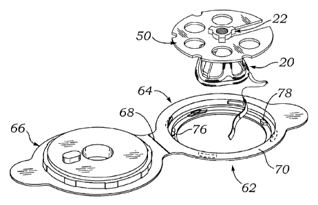
[0020] Figures 4 and 5 are expl9ded, and assembled perspective views of the
heart
valve/holder and clip subassembly positioned within a lower half of a
clamshell member used
to stabilize the heart valve during storage;
[0021] Figures 6A-6D are orthogonal views of the clam shell member;
[0022] Figure 7 is a perspective view of the heart valve/holder and clip
subassembly
positioned in the clam shell member with an upper half folded closed over the
lower half;
[0023] Figure 8 illustrates the assembly of Figure 7 placed within a cavity of
a storage
tray, and a gas-permeable lid for sealing over an upper surface of the tray;
[0024] Figures 9A-9C are orthogonal views of the storage tray;
[0025] Figure 10 is a plan view of the underside of a gas-permeable lid for
sealing
over an upper surface of the storage tray;
CA 2967234 2017-05-15
- 6 -
[0026] Figure 11 is a plan view of an upper surface of a pressure sensitive
foil label
sized to cover storage trays disclosed herein and provide a gas-impermeable
ban-ier for long-
term storage of heart valves;
[0027] Figure 12 is an exploded perspective view of the aforementioned storage
tray
and clamshell member on either side of an exemplary dry mitral tissue heart
valve
subassembly including a holder and protective cage;
[0028] Figure 13 shows the mitral tissue heart valve subassembly seated within
the
cavity of the storage tray with the clamshell member positioned thereover to
limit vertical
movement of the subassembly in the cavity;
[0029] Figure 14 shows an alterbatiVe disc-shaped insert prior to coupling to
the
mitral tissue heart valve subassembly;
[0030] Figure 15 shows the combination of the disc-shaped insert and mitral
tissue
heart valve subassembly seated within the cavity of the storage tray;
[0031] Figures 16A and 16B shows a gas-permeable lid positioned over and
sealed to
the storage tray having the mitral heart valve subassembly therein;
[0032] Figures 17A-17C are orthogonal views of a secondary storage tray sized
to
receive the first storage tray;
[0033] Figure 18 is a plan view of an alternative secondary storage tray sized
to
receive the first storage tray and having double flanges;
[0034] Figures 19A-19C show several potential configurations of the relative
heights
of the double flanges in the tray of Figure 18;
[0035] Figure 20 is a plan view of an exemplary secondary storage pouch sized
to
receive the first storage tray;
[0036] Figure 21 is a perspective view of the first storage tray positioned
within the
=
secondary storage pouch, shown transparent;
[0037] Figure 22 is a perspective view of the first storage tray positioned
within an
alternative secondary storage pouch, shown transparent; and
[0038] Figure 23 is a perspective view of the assembly of Figure 22 positioned
within
a tertiary storage container in the form of a pouch, shown transparent.
Detailed Description of the Preferred Embodiments
e
CA 2967234 2017-05-15
- 7 -
[0039] The present invention provides an improved double barrier packaging
system
for dry prosthetic heart valves that effectively stabilizes the valve within a
storage container
without the need for a liquid preservative, provides an efficient vehicle for
gas sterilization,
and prevents oxidation of the valve during long-term storage.
[0040] Figure 1 is an exploded perspective view of an exemplary aortic tissue
heart
valve 20 and a holder 22 therefore. The present application describes
packaging systems that
are particularly suitable for storing dry prosthetic tissue heart valves, and
as such do not
require liquid containment. The exemplary aortic tissue heart valve 20
includes a sewing ring
30 around an inflow end, a plurality of upstanding commissure posts 32
circumferentially
distributed around the valve and projecting in an outflow direction, and a
plurality of flexible
leaflets 34 that provide fluid occluding surfaces for the one-way valve.
Although not shown,
additional components of the heart valve 20 typically include an inner stent
and/or wire form
support structure that provide a structural skeleton surrounding an inflow
orifice and
extending up the commissure posts 32. The inner components of the heart valve
20 may be
made of suitable metal or plastic. An identification tag 35 secured to the
sewing ring 30 with
a length of suture provides a serial number representative of information
regarding the type of
heart valve 20 and other particularities about its manufacture, such as the
date.
[0041] In the illustrated embodiment, the structural components of the heart
valve 20
support each flexible leaflet 34 along a cusp edge and along two commissure
edges. A free
edge 40 of each leaflet 34 extends inward toward a central flow orifice and
coapts, or mates,
with the free edges of the other leaflets, as shown. The most common
configuration of
prosthetic aortic tissue heart valve has three flexible leaflets 34 supported
by three upstanding
commissure posts 32, although different configurations are conceivable.
[0042] Flexible leaflets 34 may be made from a variety of materials, though
bioprosthetic tissue is considered to be most effective. The most common
bioprosthetic
tissue is bovine pericardium, where the individual leaflets 34 are cut from
pericardial sac of a
cow. An exemplary dry tissue heart valve that may be stored without need for
liquid
preservatives in the packaging systems described herein may be obtained from
Edwards
Lifesciences of Irvine, CA. One preferred tissue treatment process includes
applying a
calcification mitigant such as a capping agent or an antioxidant to the tissue
to specifically
inhibit oxidation in dehydrated tissue and reduce in vivo calcification. In
one method, tissue
leaflets in assembled bioprosthetic heart valves are pretreated with an
aldehyde capping agent
CA 2967234 2017-05-15
- 8 -
prior to dehydration and sterilization. Exemplary processes are described in
U.S. Patent No.
8,357,387 to Dove, et al., filed June 25, 2009.
[0043] With reference still to Figure 1, the exemplary holder 22 includes a
central hub
structure 42 having a bore with internal threads 44, and a plurality of
outwardly and
downwardly angled legs 46. A narrow neck region 48 separates the hub structure
42 and the
upper end of the legs 46. The legs 46 are arranged to contact and engage the
valve sewing
ring 30 intermediate each pair of adjacent commissure posts 32, as seen in the
assembled
perspective of Figure 2. That is, the legs 46 contact the cusp regions of the
heart valve 20.
Although not shown, one configuration for connecting the legs 46 to the sewing
ring 30
includes attachment sutures that loop through the suture-permeable material of
the sewing
ring 30 and tie off on the holder 22, such as on one of the legs 46. During
implant, the
surgeon manipulates a handle (not shown) screwed into the threaded bore 44 and
advances
the aortic heart valve 20 into implant position at the aortic annulus. Once in
position, and
typically after anchoring sutures have been deployed between the sewing ring
30 and the
surrounding native annulus, the surgeon severs the attachment sutures coupling
the holder 22
to the valve 20, and removes the holder and handle.
[0044] Figure 3 is a perspective view of a subassembly of the aortic heart
valve 20
and holder 22 coupled to a disc-shaped storage clip 50. The clip 50 is
desirably planar and
has a substantially circular outer periphery 52 interrupted by a plurality of
semi-circular
notches 54 and a radial slot 56. The clip 50 further includes a plurality of
circular through
holes 58. The radial slot 56 terminates in a central circular aperture (not
shown) sized
approximately the same as the narrow neck region 48 of the holder 22. The
width of the
radial slot 56 is slightly smaller than the neck region 48, such that the
holder 22 may be
pushed inward along the slot and snapped into the central aperture, with the
hub structure 42
above the clip 50. As will be seen below, the clip 50 caps a cavity of a
storage tray in which
the heart valve is stored to stabilize the valve therein.
[0045] Figures 4 and 5 illustrate 'a clamshell member 62 used to stabilize the
heart
valve 20 during storage. The subassembly of the valve 20, holder 22, and clip
50 is shown in
Figure 4 exploded above a lower half 64 of the clamshell member 62, and
positioned within
the lower half in Figure 5. The clamshell member 62 is desirably constructed
of a transparent
molded material, such as a polyethylene terephthalate copolymer (PETG).
[0046] The clamshell member 62 includes the lower half 64 hinged to an upper
half
66. As seen also in Figures 6A-6C, clamshell member 62 is desirably molded
from clear
CA 2967234 2017-05-15
- 9 -
plastic and the two halves connect at a living hinge 68. The lower half 64
includes an annular
rim 70 above and surrounding a circular aperture defined by a lower ledge 72
and having a
finger tab 74 extending away from the hinge 68. A plurality of separate molded
features
project inward from the annular rim 70 above the lower ledge 72, including
four clip supports
76 and an anti-rotation projection 78. As seen in Figure 5, the generally
circular clip 50 is
sized to fit within the annular rim 70 and rest on the clip supports 76. The
circumferential
width of the anti-rotation projection 78 permits it to fit closely within the
radial slot 56 of the
clip 50, thus preventing rotation of the clip in the clamshell member 62.
[0047] The clamshell member upper half 66 has an outer ledge 80 including a
finger
tab 82 extending away from the hinge 68. An inner generally cylindrical boss
84 fits within
and mates with the inner surface features of the lower half annular rim 70. In
particular, a
series of projections 86 on the cylindrical boss 84 frictionally engage the
inner surface of the
lower half annular rim 70. The engagenrientaof the projections 86 with the
inside of the rim
70 desirably provides an audible and tactile click or snap upon closing the
halves of the
clamshell member 62. Prior to closing the clamshell member 62, the
identification tag 35
may be positioned on the circular clip 50 with the serial number facing upward
for greater
visibility and to prevent the tag from contacting and potentially damaging the
heart valve 20
during storage. The final assembly of the valve/holder/clip in the closed
clamshell member
62 is seen in Figure 7. As an additional locking feature, a downward
projection 89 on the
upper half 66 fits closely into the mid-portion of the radial slot 56 of the
clip 50, thus further
limiting movement of the clip in the clamshell member 62.
[0048] Figure 8 then illustrates the assembly of Figure 7 placed within a
cavity 90 of
a storage tray 92, whereupon a gas-permeable lid 94 having an outer band of
adhesive 95
seals over an upper surface 96 of the tray 92. Figures 9A-9C are orthogonal
views of the
storage tray 92 illustrating a flat, horizontal outer rim 98 defining the tray
upper surface 96,
and surrounding the cavity 90. The cavity 90 is formed by the inner contours
of a container
portion 100 extending downwardly from the outer rim 98. The container portion
100
includes a stepped ledge 102 on an upper end and a lower trough 104. When the
assembly of
Figure 7 is placed within the cavity 90, the clamshell member 62 rests on the
stepped ledge
102 and the heart valve 20 extends downward within the lower trough 104. Note
in Figure
6C, external features 105 on the lower half 64 of the clamshell member 62
which frictionally
engage the internal features 106 on the stepped ledge 102 of the storage tray
92. Engagement
between the features 105, 106 nominally retains the clamshell member 62 in the
storage tray
CA 2967234 2017-05-15
a
¨ 0 -
92, and prevents the clamshell member from falling out if the tray is inverted
but presents
minimal difficulty to a user removing the clamshell member using the thumb
tabs.
Preferably, the features 105, 106 engage with a snap or tactile feedback.
Because the
clamshell member 62 secures the circular clip 50, which in turn secures the
valve/holder
combination, the heart valve 20 is stably suspended within the cavity 90
without touching the
sides of the tray 92.
[0049] Figure 10 shows the gas-permeable lid 94 that seals over the upper
surface 96
of the storage tray 92. More specifically, the outer rim 98 forms a flange to
which the band
of adhesive 95 on the lid 94 may be adhered. Preferably, the lid 94 is closely
dimensioned to
the perimeter of the outer rim 98, and the band of adhesive 95 is a pressure-
seal or a heat seal
adhesive to facilitate sealing under pressure and/or temperature. The material
of the lid 94 is
breathable, or gas-permeable, to provide for gas sterilization of the contents
sealed within the
tray 92, in particular the dry tissue heart valve 20. One suitable gas-
permeable material is a
sheet of high-density polyethylene fibers: which is difficult to tear but can
easily be cut with
scissors. The material is highly breathable and water vapor and gasses can
pass through the
fibers, but not liquid water. For instance, various Tyvek materials from
DuPont may be used.
Also, exemplary hot-melt adhesives used to secure the lid 94 to the tray 92
may be obtained
from Perfecseal or Oliver-Tolas, for example. Such a material permits
sterilization of the tray
contents using Ethylene Oxide (ETO), which gradually passes through the lid 94
to the
interior tray. The lid 94 presents a sterile barrier and prevents ingress of
microorganisms.
The tray 92 is desirably a molded material, such as a polyethylene
terephthalate copolymer
(PETG), that provides rigidity and protection from jostling and external
pressures. Various
medical storage materials and packaging suitable for assembly of components of
the present
application are available from companies such as Dupont, Perfecseal, Oliver-
Tolas, and
Mangar.
[0050] Ethylene oxide (ETO), also called oxirane, is the organic compound with
the
formula C2H40. It is commonly handled and shipped as a refrigerated liquid.
ETO is often
used as sterilant because it kills bacteria (andµtheir endospores), mold, and
fungi. It is used to
sterilize substances that would be damaged by high temperature techniques such
as
pasteurization or autoclaving. Ethylene oxide is widely used to sterilize the
majority of
medical supplies such as bandages, sutures, and surgical implements in a
traditional chamber
sterilization method, where a chamber has most of the oxygen removed (to
prevent an
CA 2967234 2017-05-15
- 1 1 -
explosion) and then is flooded with a mixture of ethylene oxide and other
gases that are later
aerated.
[0051] Certain features of the clamshell member 62 and storage tray 92
facilitate gas
sterilization, such as with ETO. Specifically, the clamshell member 62
provides a cap that
limits vertical movement of the heart valve 20 in the tray cavity 90 while
providing gas flow
passages for gas flow in and out of the cavity. Good flow of sterilization gas
in and out of the
cavity 90 facilitates complete and rapid sterilization of the tissue heart
valve 20. First of all,
the clamshell member 62 sits on the stepped ledge 102, and a pair of
diametrically opposed
gas flow channels 108 provide openings between the two elements for passage of
gas into the
cavity 90. In addition, the engagement between the lower and upper halves 64,
66 of the
clamshell member 62 permits gas to flow therethrough, around the upper end of
the valve 20.
More specifically, the circular clip 50 is supported by the four clip supports
76 above the
lower ledge 72, allowing gas to flow around the clip 50. Furthermore, the clip
50 includes
large circular through holes 58 for direct gas flow therethrough. In short,
the stable yet
discontinuous engagement of the packaging 'elements permits good gas flow in
and around
the tissue heart valve 20.
[0052] Figures 12-16 illustrates an alternative packaging system for mitral
heart
valves. Figure 12 is an exploded perspective view of the same storage tray 92
and closed
clamshell member 62 for aortic valve storage on either side of an exemplary
mitral tissue
heart valve subassembly 110, including a holder 112 and a protective cage 114.
In contrast
with aortic valves, the holder 112 for mitral valves attaches to the inflow
end of the valve,
typically to the sewing ring. Although not shown, the holder 112 includes
engagement
structure, such as attachment sutures, for removably attaching to the sewing
ring of the mitral
heart valve.
[0053] The holder 112 may take a number of forms, but typically includes an
upper
bore 116 having internal threads for attaching a delivery handle. One
exemplary holder 112
that may be used is available as the TRICENTRIXO holder system for use with
the
Carpentier-Edwards PERIMOUNT Plus mitral pericardial valve from Edwards
Lifesciences of Irvine, CA. A shaft 118 of the holder 112 fits closely within
a radial slot 120
in a clip member 122 attached to the upper end of the protective cage 114. An
identification
tag 124 attached to the heart valve sewing ring with a suture passes upward
through the radial
slot 120. The holder 112 stabilizes the mitral heart valve in a fixed position
with the
protective cage 114, which in turn prevents the outflow end of the heart valve
from advert
CA 2967234 2017-05-15
- 12 -
contact with the inner walls of tray 92, .and later contact with external
surfaces and
instruments in the operating room when the heart valve is removed for
implantation.
[005+1 Figure 13 shows the mitral tissue heart valve subassembly 110 seated
within
the cavity of the storage tray 92 with the clamshell member 62 positioned
thereover. When
pressed down into the cavity of the storage tray 92, the clamshell member 62
acts as a cap on
the cavity to limit vertical movement of the heart valve subassembly 110
therein. As before,
frictional engagement between the external features 105 (Figures 6C and 6D) on
the lower
half 64 of the clamshell member 62 and internal features 106 on the stepped
ledge 102 of the
storage tray 92 retains the clamshell member as a cap over the heart valve
subassembly 110.
[0055] As an alternative to the clamshell member 62, a disc-shaped insert 130
may be
used to provide a cap over the cavity storage tray 92, as seen in Figure 14.
The insert 130
defines a flat, generally planar disc having four outward protections 132 and
a radial slot 134
open to an outer periphery 136. The insert 130 is desirably formed of a
suitable molded
plastic, such as a high-density polyethylene (HDPE). The slot 134 fits closely
around a non-
circular portion of the holder shaft 118 and includes a narrowed region 140
that retains the
shaft 118 at a closed central end of the slot 134. Once the insert 130 has
been snapped onto
the heart valve subassembly 110, the combination may be lowered into the
cavity of the
storage tray 92, as seen in Figure 15. The outward protections 132 snap under
the internal
features 106 on the stepped ledge 102 of the storage tray 92 such that the
insert 130 caps the
cavity over the heart valve subassembly 110. Flow passages 142 align with the
flow channels
108 provided in the storage tray 92 and facilitate sterilizing gas flow
between the insert 130
and tray. As before, the identification tag 124 of the mitral heart valve may
be positioned
over the top of the insert 130 so that the serial number is visible from above
without
removing the heart valve subassembly 110 from the tray 92. Also, it should be
noted that the
insert 130 engages the tray 92 in a non-rotating manner, as does the insert
slot 134 around the
non-circular holder shaft 118, which means that the valve holder 112 is held
stationary in the
tray while a user couples a threaded handle thereto.
[0056] Once the mitral heart valve subassembly 110 has been positioned within
the
cavity of the storage tray 92, as in Figure 15, and a cap such as the
clamshell member 62 is
snapped thereover, as in Figure 16A, the sawermeable lid 94 described above
seals over an
upper surface 96 of the tray 92, as in Figure 16B. Figure 15 shows the
identification tag 124
which is visible through the clear plastic of the clamshell member 62 in
Figure 16A. At this
CA 2967234 2017-05-15
- 13 -
stage, the assembly, and in particular the mitral heart valve therein, can be
subjected to gas
sterilization, such as with ETO. e
[0057] The clamshell member 62 (or insert 130 for mitral valves) restricts
rotation of
the aortic or mitral valve holders, and therefore provides an efficient way of
attaching a
threaded handle to the holder while still in the packaging.
[0058] One advantage of the packaging solutions described herein is a double
sterile
barrier, wherein the inner and outer sterile containers allow for gas
sterilization, such as with
ETO, and with a second seal the outer sterile container also provides a
barrier between the
product and the surrounding atmosphere (e.g., oxygen) after sterilization. The
inner sterile
container has been described above, and for both aortic and mitral heart
valves results in the
sealed storage tray 92 shown in Figure 16B. The sealed storage tray 92 is
received within a
secondary or outer container and the dual barrier assembly is then sterilized,
so that there are
redundant sterile barriers. Subsequently, the dual barrier assembly is sealed
to prevent the
outside air from reaching the heart valve, thus preventing oxygenation and
potentially
reducing calcification after implant. In the exemplary packaging sequence, the
inner and
outer containers are first assembled together and each closed with a gas-
permeable barrier to
form a dual barrier assembly which is gasµ-steµrilized. Subsequently, the
atmospheric barrier is
added, such as by converting the outer container from being gas-permeable to
being gas-
impermeable. However, if the entire process is done in sterile conditions,
such as in a clean
room environment, the inner container may be closed and sterilized before
being placed
within the outer container, which is then closed and sterilized. In other
words, there may be
one or two sterilization steps prior to sealing the entire assembly against
air ingress.
[0059] The present application describes two different secondary barriers -
one a
storage tray similar to that described earlier, and the other a flexible
pouch. The secondary
barrier protects and preserves the primary sterile barrier package in a
sterile environment, and
prevents oxygen from reaching the heart valve within. A further outer shelf
box may be used
to facilitate temperature monitoring during distribution and storage, and
protect the delicate
implant from distribution hazards such as shock, impact and extreme
temperatures.
[0060] Figures 17A-17C are orthogonal views of a secondary or outer storage
tray
150 sized to receive the primary or inner storage tray 92. The secondary
storage tray 150
desirably mimics the shape of the primary storage tray 92 such that the latter
can be easily
nest within a cavity 152 therein. As such, the storage tray 150 comprises an
upper surface
including a peripheral flange 154, and a container portion 156 extending
downwardly
CA 2967234 2017-05-15
- 14 -
therefrom having a stepped ledge 158 on an upper end and a lower trough 160.
The inner
walls of the container portion 156 define the cavity 152, and closely receive
the inner storage
tray 92. ,
[0061] The outer storage tray 150 provides a rigid secondary sterile barrier
that
protects and preserves the inner sterile barrier formed by the inner storage
tray 92 and lid 94.
Desirably, the outer storage tray 150 is constructed of a molded material,
such as a
polyethylene terephthalate copolymer (PETG). PETG is nominally gas-
impermeable, though
not entirely for the long-term storage needs described herein, perhaps years.
The tray 150
instead may also be formed of a molded material that is gas-impermeable for
the required
time frame, though such materials may be somewhat more expensive than PETG.
Once the
sealed inner tray 92 is placed within the outer storage tray 150, a gas-
permeable lid (not
shown, but similar to lid 94 of the inner tray 92) seals against the flange
154 and permits
sterilization gas (e.g., ETO) to reach the spaces within both trays.
[0062] With reference back to Figure 11, a gas-impermeable label 162 sized to
cover
the secondary storage tray 150 is shown. The label 162 is applied over the
sterilized tray 150,
and sealed on top of the lid. Once pressure adhered or heat sealed against the
lid, the foil
label 162 provides a complete barrier to gas transfer. The label 162
preferably includes a
layer of metal foil laminated to a layer of a agas-permeable material such as
DuPont 1073B
Tyvek, or more preferably is a single layer of foil. The label 162 may have
information
printed thereon about the contents of the packaging, such as implant type,
model,
manufacturer, serial number, date of packaging, etc. A layer of pressure
sensitive adhesive is
provided to seal on top of the previously attached lid.
[0063] In an alternative configuration, as seen in Figure 18, an outer storage
tray 180
features a cavity 182 for receiving an inner tray surrounded by a double
flange with an outer
flange 184 offset from an inner flange 186. The inner flange 186 may first be
sealed with a
die-cut and heat seal adhesive coated gas-permeable lid (e.g., Tyvek) after
placement of the
inner sterile barrier package, enabling subsequent ETO sterilization of the
entire package, and
in particular the space between the two sterile barriers. A gas-impermeable
label such as a
single layer of foil is then sealed to the outer flange 184.
[0064] Figures 19A-19C show several potential configurations of the relative
heights
of the double flanges 184, 186 in the tray 180 of Figure 18. In a preferred
embodiment, both
lids/labels applied to the flanges 184, 186 are attached with heat sealed
adhesive for better
4
long-term integrity of the bond. Heat sealing is typically accomplished by
pressing down on
CA 2967234 2017-05-15
- 15 -
the label with a heated surface such as a flat platen. However, heat and
pressure should be
applied only once to each flange seal to avoid affecting the seal integrity
after formation, and
a flat platen may require modification. There are several ways to manage this.
[0065] In a first embodiment of figtre 19A, the flanges 184, 186 are at the
same
elevation. The gas-permeable lid or label is applied to the inner flange 186
using a heated
press shaped the same as the flange. Alternatively, an insert shaped like the
flange 186 may
be introduced between a flat heated platen and the tray. After ETO
sterilization, the foil label
is applied to the outer flange 184 using a heated press shaped the same as the
flange, or an
insert shaped like the outer flange between a flat heated platen and the tray.
[0066] In Figure 19B, the inner flange 186' elevates about the outer flange
184. In
this configuration, a flat heated platen may be used to apply heat to an
adhesive-coated label
for the inner seal, while the outer seal is formed using a heated press shaped
the same as the
outer flange, or an insert shaped like the outer flange between a flat heated
platen and the
tray.
[0067] In Figure 19C, the outer flange 184' elevates about the inner flange
186. In
this configuration, the inner seal is first formed using a heated press shaped
the same as the
inner flange, or an insert shaped like the inner flange between a flat heated
platen and the
tray. Subsequently, a flat heated platen may be used to apply heat to an
adhesive-coated foil
,
label for the outer seal. The ability to use a flat heated platen for at least
one of the seals
simplifies the assembly apparatus and procedure.
[0068] Figure 20 is a plan view of an exemplary secondary storage pouch 190
sized to
receive the first storage tray 92, or inner sterile packaging. The storage
pouch 190 includes a
first gas-permeable portion 192 adjacent an open end (to the left), and a
second, larger gas-
impermeable portion 194 that is closed on the right end. The entire pouch 190
may be made
of the gas-impermeable portion 194, except for a strip of the first portion
192 on the upper
layer, or the first portion 192 may form both the upper and lower layers of
the pouch adjacent
the open end. A first seal 196 extends across the width of the open mouth of
the pouch 190
in the area of the first gas-permeable portion 192. The second seal 198 also
extends across
the width of the pouch 190 but fully within the second gas-impermeable portion
194. During
packaging, the first storage tray 92 is placed within the pouch 190 and the
first seal 196
closed, at which time the entire contents are gas-sterilized. After the
assembly is sterile, the
second seal 198 is closed to prevent any further contact between the interior
of the pouch 190
and the surrounding atmosphere. 4 =
CA 2967234 2017-05-15
- 16 -
[0069] Figure 21 is a perspective view of the first storage tray 92 sealed
with the lid
94 and positioned within the secondary storage pouch 190. The two seals 196,
198 enable
gas sterilization of the contents of the pouch 190 prior to full sealing. More
particularly, the
first seal 196 may be closed at which time the package may be subject to ETO
sterilization.
Because the first seal 196 extends across the` gas-permeable first portion
192, sterilizing gas
can enter the interior of the pouch 190. After sterilization, second seal 198
is closed to
prevent any further gas, in particular oxygen, from entering the interior of
the pouch 190.
[0070] The storage pouch 190 provides a flexible secondary sterile barrier,
and may
be constructed of various materials or laminates having at least one gas-
impermeable layer,
with a foil/polyethylene fiber laminate being preferred. An inner layer of the
foil material,
such as available from Amcor, may feature a laminate of Low Density
Polyethylene (LDPE)
to facilitate seal under pressure and temperature. A tear notch on the pouch
190 may be
provided for easy opening. With the second seal 198 closed, the foil pouch 190
provides an
oxygen and moisture barrier after ETO sterilization.
[0071] In an alternative configuration seen in Figure 22, the secondary
storage pouch
190 that receives the first storage tray 92 only includes a first gas-
permeable seal 200. In use,
the first storage tray 92 is placed within the secondary storage pouch 190 and
the seal 196
closed, at which time the entire contents are gas-sterilized. After the
assembly is sterile, the
secondary storage pouch 190 and contents within are placed within a gas-
impermeable
tertiary container, such as pouch 204 in Figure 23, to prevent any further
contact between the
interior of the pouch 190 and the surrounding atmosphere. The pouch 204 is
desirably
formed of gas-impermeable material and has a gas-impermeable seal 206.
[0072] In general, therefore, a preferred method includes stabilizing a dry
prosthetic
heart valve within a first gas-permeable container that provides some rigidity
or protection
from external damage. The first gas-permeable container and contents are then
placed in a
secondary gas-permeable container, and the entire assembly subjected to gas-
based
sterilization. Finally, the secondary container is sealed with a gas-
impermeable barrier, such
as by placing it within a gas-impermeable tertiary container to prevent gas
transfer with the
surrounding atmosphere.
[0073] In addition to the various embodiments of the double sterile packaging
described above, the final packaging will typically include a shelf box,
printed or unprinted,
constructed of paperboard with a tamper-evident carton label as an indicator
of the integrity
{
CA 2967234 2017-05-15
- 17 -
of the package and placed in a foam box for insulation. Also, a temperature
indicator for
monitoring temperature during distribution and storage is attached to the
shelf box.
[0074] The packaging solutions disclosed herein facilitate access to tissue
implants, in
particular prosthetic heart valves at the time of implantation. The process
for removing the
aortic valve 20 of Figure 1 from its packaging will be described, though
similar steps can be
used to remove the mitral heart valve of Figu,res 12-16. The first step is
removal of the outer
or secondary sterile barrier, two embodiments of which have been described.
One or both
sealed labels over the outer tray 150, 180 are first detached, and the inner
tray 92 sealed by
the sterile lid 94 (seen in Figure 16B) removed therefrom (alternatively, the
technician tears
open the pouch 190 of Figure 21). At this stage, the inner sterile packaging
may be
transported to the immediate vicinity of the operation site without undue
concern for the
integrity of the package because of the relatively rigid inner tray 92 and
sterile seal 94.
[0075] Subsequently, the technician detaches the lid 94, exposing the assembly
seen
in Figure 8. The upper half 66 of the clamshell member 62 is lifted up from
the lower half 64
to expose the generally circular clip 50 and valve holder 22, as seen in
Figure 5. A delivery
handle (not shown) can then be threaded onto the holder, and the assembly of
the valve 20,
holder 22, and clip 50 removed from the clamshell member 62. Recall that the
anti-rotation
projection 78 of the clamshell member 62 engages the radial slot 56 of the
clip 50 to prevent
rotation of the clip in the clamshell member 62. This facilitates threading
the handle onto the
holder 22, such that the operation can be done with two hands. Finally, the
clip 50 can easily
be detached from the holder 22 by pulling it`off laterally, leaving the valve
20 on the end of
the delivery handle ready form implant.
[0076] The packaging assemblies herein provide a number of distinctive
advantages
to manufacturers of dry prosthetic valves, which advantages may also be
transferred to the
storage of other tissue implants that can be stored dry, such as dental
implants, ligaments,
vessel grafts, tissue patches or scaffolds, etc. Indeed, certain aspects of
the present
application can be utilized by makers of implants in general that are required
to be stored in
double sterile containers and which can be sterilized using a gas such as ETO.
One
advantage of the packaging described herein is that it contains and stabilizes
the prosthetic
heart valve. Movement of the heart valve within the storage container is
detrimental as
delicate tissue structures may be damaged if permitted to contact the sides of
the packaging.
[0077] Due to presence of a gas-permeable sterile barrier such as a Tyvek
Header
(breathable vent) the product can easily be ETO sterilized and aerated for
acceptable levels of
CA 2967234 2017-05-15
- 18 -
residuals. After appropriate aeration time, the outer container, or second
barrier, can be
sealed (e.g., foil to foil) to prevent long term oxidation of the dry tissue
valve.
[0078] The ETO sterilization obviates traditional oven sterilization,
therefore
reducing the amount of energy spent in heating the packaged product in an oven
for multiple
days. Similarly, elimination of autoclaving of the jars and closures before
packaging will
reduce the energy consumption required in the sterilization process.
[0079] As mentioned, the double' stel-ile barrier allows for gas
sterilization, such as
with ETO, but also provides an oxygen barrier to the product after
sterilization.
Consequently, the entire assembly can be reliably stored in oxygen-free
conditions for
extended periods of time, even years, yet the outer sterile container can be
removed at the
time of use without exposing the contents of the inner sterile container to
contaminants. The
double layer of packaging enables sterile transfer of the inner package to the
sterile operating
field, and the inner package can even be temporarily stored for significant
periods before the
product is used. The new package design will be lighter in weight due to the
choice of
materials (PETG/Tyvek and air vs. Polypropylene with glutaraldehyde), which
will reduce
the shipping costs for single unit shipments.
[0080] Indeed, the biggest advantage over existing "wet" heart valve package
designs
is the elimination of storage and handling of liquid glutaraldehyde during the
packaging and
storage process, as well as the absence of glutaraldehyde at the time of use.
This reduces
hazards to the health of employees, customers, and patients, as well as the
environment.
Additionally, disposal of glutaraldehyde is bio-hazardous and therefore OSHA
requires
neutralization of the chemical before disposal or placement of appropriate
controls for
disposal. Due to decreased handling and critical storage requirements
described herein, the
packaging process is rendered less complex. The elimination of glutaraldehyde
will not
require an increased level of insulation from higher temperatures as the dry
tissue valve
already has the capability to withstand temperatures as high as 55 C.
Therefore this will
likely reduce the bulkiness of the design by reducing the size and insulation
used for shipping
the valve during summers and winters.
[0081] Current tissue valves available from Edwards Lifesciences are packaged
in a
3.8 oz polypropylene jar/closure system with liquid glutaraldehyde. The
presence of liquid
glutaraldehyde requires the package design to maintain a state of temperature
that will not
overheat or freeze the tissue valve. Therefore the current package is bulky
and heavier due to
presence of EPS (Expanded Polystyrene) foam end caps outside the secondary
package (shelf
CA 2967234 2017-05-15
- 19 -
carton) which insulates from extreme temperature conditions. The polypropylene
3.8 oz
jar/closure system with liquid glutaraldehyde, secondary package and foam
insulation make
the package design bulky and heavy resulting in increased space for storage
and increased
costs for shipping. The current single unit summer pack weighs approximately
0.85 lbs where
as the current single unit winter pack weighs approximately 1.85 lbs. The
packages disclosed
herein are significantly lighter.
[0082] While the invention has been described in its preferred embodiments, it
is to
be understood that the words which haVe seen used are words of description and
not of
limitation. Therefore, changes may be made within the appended claims without
departing
from the true scope of the invention.
a 4
e =