Note: Descriptions are shown in the official language in which they were submitted.
CA 02967337 2017-05-10
WO 2016/077438 PCT/US2015/060117
PULSE SEQUENCES FOR LOW FIELD MAGNETIC RESONANCE
BACKGROUND
[1] Magnetic resonance imaging (MRI) provides an important imaging modality
for numerous applications and is widely utilized in clinical and research
settings to produce
images of the inside of the human body. As a generality, MRI is based on
detecting magnetic
resonance (MR) signals, which are electromagnetic waves emitted by atoms in
response to
state changes resulting from applied electromagnetic fields. For example,
nuclear magnetic
resonance (NMR) techniques involve detecting MR signals emitted from the
nuclei of excited
atoms upon the re-alignment or relaxation of the nuclear spin of atoms in an
object being
imaged (e.g., atoms in the tissue of the human body). Detected MR signals may
be processed
to produce images, which in the context of medical applications, allows for
the investigation
of internal structures and/or biological processes within the body for
diagnostic, therapeutic
and/or research purposes.
[2] MRI provides an attractive imaging modality for biological imaging due
to the
ability to produce non-invasive images having relatively high resolution and
contrast without
the safety concerns of other modalities (e.g., without needing to expose the
subject being
imaged to ionizing radiation, such as x-rays. or introducing radioactive
material to the body).
Additionally, MRI is capable of capturing information about structures and/or
biological
processes that other modalities are not well suited to acquire or are
incapable of acquiring.
For example, MRI is particularly well suited to provide contrast among soft
tissues.
However, there are a number of drawbacks to conventional MRI techniques that,
for a given
imaging application, may include the relatively high cost of the equipment,
limited
availability and/or difficulty in gaining access to clinical MRI scanners, the
length of the
image acquisition process, etc.
[3] The trend in clinical MRI has been to increase the field strength of
MRI
scanners to improve one or more of scan time, image resolution, and image
contrast, which in
turn drives up costs of MRI imaging. The vast majority of installed MRI
scanners operate
using at least at 1.5 or 3 tesla (T), which refers to the field strength of
the main magnetic field
Bo of the scanner. A rough cost estimate for a clinical MRI scanner is on the
order of one
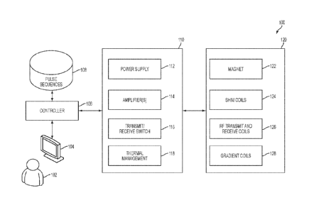
million dollars per tesla, which does not even factor in the substantial
operation, service, and
maintenance costs involved in operating such MRI scanners.
1
CA 02967337 2017-05-10
WO 2016/077438
PCT/US2015/060117
[4] Additionally, conventional high-field MRI systems typically require
large
superconducting magnets and associated electronics to generate a strong
uniform static
magnetic field (B0) in which a subject (e.g., a patient) is imaged. The size
of such systems is
considerable with a typical MRI installment including multiple rooms for the
magnet,
electronics, thermal management system, and control console areas. The size
and expense of
MRI systems generally limits their usage to facilities, such as hospitals and
academic
research centers, which have sufficient space and resources to purchase and
maintain them.
The high cost and substantial space requirements of high-field MRI systems
results in limited
availability of MRI scanners. As such, there are frequently clinical
situations in which an
MRI scan would be beneficial, but is impractical or impossible due to the
above-described
limitations and as discussed in further detail below.
SUMMARY
[5] The inventors have appreciated that performing low-field magnetic
resonance
imaging can be facilitated via the use of pulse sequences developed by the
inventors to
operate in the low-field context.
[6] Some embodiments provide for a low-field magnetic resonance imaging
(MRI) system, comprising a plurality of magnetics components comprising at
least one first
magnetics component configured to produce a low-field main magnetic field Bo
and at least
one second magnetics component configured to acquire magnetic resonance data
when
operated; and at least one controller configured to operate one or more of the
plurality of
magnetics components in accordance with at least one low-field zero echo time
(LF-ZTE)
pulse sequence.
[] Some
embodiments provide for a method for operating a low-field magnetic
resonance imaging system, the system comprising a plurality of magnetics
components, the
plurality of magnetics components comprising at least one first magnetics
component
configured to produce a low-field main magnetic field Bo and at least one
second magnetics
component configured to acquire magnetic resonance data when operated. The
method
comprises using the at least one first magnetics component to produce the low-
field main
magnetic field Bo; and controlling, using at least one controller, one or more
of the plurality
of magnetics components in accordance with at least one low-field zero echo
time (LF-ZTE)
pulse sequence.
2
CA 02967337 2017-05-10
WO 2016/077438
PCT/US2015/060117
[8] Some embodiments provide for at least one non-transitory computer
readable
storage medium storing processor executable instructions that, when executed
by a low-field
MRI system comprising a plurality of magnetics components, the plurality of
magnetics
components comprising at least one first magnetics component configured to
produce a low-
field main magnetic field Bo and at least one second magnetics component
configured to
acquire magnetic resonance data when operated, allow the low-field MRI system
to: use the
at least one first magnetics component to produce the low-field main magnetic
field Bo; and
operate one or more of the plurality of magnetics components in accordance
with at least one
low-field zero echo time (LF-ZTE) pulse sequence.
[9] Some embodiments provide for a low-field magnetic resonance imaging
(MRI) system, comprising: a plurality of magnetics components comprising at
least one first
magnetics component configured to produce a low-field main magnetic field Bo
and at least
one second magnetics component configured to acquire magnetic resonance data
when
operated; and at least one controller configured to operate one or more of the
plurality of
magnetics components in accordance with at least one low-field refocusing
(LFR) pulse
sequence, wherein the RF excitation pulses in the at least one LFR pulse
sequence are
associated with a flip angle that reduces effect of Bo inhomogeneities on net
transverse
magnetization.
[10] Some embodiments provide for a method for operating a low-field
magnetic
resonance imaging system, the system comprising a plurality of magnetics
components, the
plurality of magnetics components comprising at least one first magnetics
component
configured to produce a low-field main magnetic field Bo and at least one
second magnetics
component configured to acquire magnetic resonance data when operated. The
method
comprises operating the at least one first magnetics component to produce the
low-field main
magnetic field Bo; and controlling, using at least one controller, one or more
of the plurality
of magnetics components in accordance with at least one low-field refocusing
(LFR) pulse
sequence, wherein the RF excitation pulses in the at least one LFR pulse
sequence are
associated with a flip angle that reduces effect of Bo inhomogeneities on net
transverse
magnetization.
[1 1 ] Some
embodiments provide for at least one non-transitory computer readable
storage medium storing processor executable instructions that, when executed
by a low-field
MRI system comprising a plurality of magnetics components, the plurality of
magnetics
components comprising at least one first magnetics component configured to
produce a low-
field main magnetic field Bo and at least one second magnetics component
configured to
3
CA 02967337 2017-05-10
WO 2016/077438
PCT/US2015/060117
acquire magnetic resonance data when operated, allow the low-field MRI system
to: operate
the at least one first magnetics component to produce the low-field main
magnetic field Bo;
and operate one or more of the plurality of magnetics components in accordance
with at least
one low-field refocusing (LFR) pulse sequence, wherein the RF excitation
pulses in the at
least one LFR pulse sequence are associated with a flip angle that reduces
effect of Bo
inhomogeneities on net transverse magnetization.
[12] Some embodiments provide for a low-field magnetic resonance imaging
(MRI) system, comprising: a plurality of magnetics components configured to
produce a
plurality of magnetic fields including a low-field main magnetic field Bo, the
plurality of
magnetics components comprising at least one first magnetics component
configured to
produce the low-field main magnetic field Bo and at least one second magnetics
component
configured to acquire magnetic resonance data when operated; and at least one
controller
configured to operate one or more of the plurality of magnetics components in
accordance
with a pulse sequence designed to compensate for inhomogeneity in one or more
of the
plurality of magnetic fields at least in part by causing one or more of the
plurality of
magnetics components to apply a series of RF pulses having at least one
parameter that varies
during a respective series of pulse repetition periods of the pulse sequence.
[13] Some embodiments provide for a method for operating a low-field
magnetic
resonance imaging system, the system comprising a plurality of magnetics
components
configured to produce a plurality of magnetic fields including a low-field
main magnetic field
Bo, the plurality of magnetics components comprising at least one first
magnetics component
configured to produce the low-field main magnetic field Bo and at least one
second magnetics
component configured to acquire magnetic resonance data when operated. The
method
comprises operating the at least one first magnetics component to produce the
low-field main
magnetic field Bo; and controlling, using at least one controller, one or more
of the plurality
of magnetics components in accordance with a pulse sequence designed to
compensate for
inhomo2eneity in one or more of the plurality of magnetic fields at least in
part by causing
the plurality of magnetics components to apply a series of RF pulses having at
least one
parameter that varies during a respective series of pulse repetition periods
of the pulse
sequence.
Some embodiments provide for at least one non-transitory computer readable
storage
medium storing processor executable instructions that, when executed by a low-
field MRI
system having a plurality of magnetics components configured to produce a
plurality of
magnetic fields including a low-field main magnetic field Bo, the plurality of
magnetics
4
CA 02967337 2017-05-10
WO 2016/077438 PCT/US2015/060117
components comprising at least one first magnetics component configured to
produce the
low-field main magnetic field Bo and at least one second magnetics component
configured to
acquire magnetic resonance data when operated generate at least one magnetic
field, allow
the low-field MRI system to: operate the at least one first magnetics
component to produce
the low-field main magnetic field Bo; and operate one or more of the plurality
of magnetics
components in accordance with a pulse sequence designed to compensate for
inhomogeneity
in one or more of the plurality of magnetic fields at least in part by causing
the plurality of
magnetics components to apply a series of RF pulses having at least one
parameter that varies
during a respective series of pulse repetition periods of the pulse sequence.
BRIEF DESCRIPTION OF THE DRAWINGS
[14] Various aspects and embodiments of the disclosed technology will be
described with reference to the following figures. It should be appreciated
that the figures are
not necessarily drawn to scale. Items appearing in multiple figures are
indicated by the same
reference number in all the figures in which they appear.
[15] FIG. 1 is a block diagram of exemplary components of a low-field MRI
system, in accordance with some embodiments of the technology described
herein.
[16] FIG. 2A is a diagram illustrating one pulse repetition period of a low
field zero
echo time pulse sequence, in accordance with some embodiments of the
technology described
herein.
[17] FIG. 2B is a diagram illustrating two consecutive pulse repetition
periods of
an LF-ZTE pulse sequence, in accordance with some embodiments of the
technology
described herein.
[18] FIG. 2C is a diagram illustrating an LF-ZTE pulse sequence comprising
one or
more contrast preparation portions, in accordance with some embodiments of the
technology
described herein.
[19] FIG. 2D is a diagram illustrating a portion of an LF-ZTE pulse
sequence
comprising a T1 contrast preparation portion, in accordance with some
embodiments of the
technology described herein.
[20] FIG. 2E is a diagram illustrating a portion of an LF-ZTE pulse
sequence
comprising an electron paramagnetic resonance (EPR) pulse sequence, in
accordance with
some embodiments of the technology described herein.
CA 02967337 2017-05-10
WO 2016/077438 PCT/US2015/060117
[21] FIG. 2F is a diagram illustrating a portion of an LF-ZTE pulse
sequence
comprising a navigation pulse sequence, in accordance with some embodiments of
the
technology described herein.
[22] FIG. 2G is a diagram illustrating a portion of an LF-ZTE pulse
sequence
comprising a water/fat separation contrast preparation sequence, in accordance
with some
embodiments of the technology described herein.
[23] FIG. 3 is a flowchart of an illustrative process for performing MR
imaging in a
low-field MR system using a low-field zero echo time pulse sequence.
[24] FIG. 4 is a diagram illustrating one pulse repetition period of a low-
field
balanced steady-state free precession (LF-bSSFP) sequence, in accordance with
some
embodiments of the technology described herein.
[25] FIG. 5 is a diagram illustrating the effect of magnetic field
inhomogeneity on
the strength of transverse magnetization for different flip angles, in
accordance with some
embodiments of the technology described herein.
[26] FIG. 6 is a flowchart of an illustrative process for performing MR
imaging in a
low-field MR system using an LF-bSSFP sequence, in accordance with some
embodiments
of the technology described herein.
[27] FIG. 7 is a schematic of a low-field radio frequency (RF) coil, in
accordance
with some embodiments of the technology described herein.
[28] FIG. 8 illustrates, in the time domain, input current to the low-field
RF coil
and corresponding output from the low-field RF coil without pre-emphasis
applied, in
accordance with some embodiments of the technology described herein.
[29] FIG. 9 illustrates, in the frequency domain, how input current to the
low-field
LF coil is attenuated by the low-field RF coil and how pre-emphasis may be
used to
counteract such attenuation, in accordance with some embodiments of the
technology
described herein.
[30] FIG. 10 illustrates, in the frequency domain, a pre-emphasis waveform,
in
accordance with some embodiments of the technology of the technology described
herein.
[31] FIG. 11 illustrates, in the time domain, input current to the low-
field RF coil
and corresponding output from the low-field RF coil with pre-emphasis applied,
in
accordance with some embodiments of the technology described herein.
[32] FIGS. 12A and 12B illustrate a hi-planar magnet configuration, in
accordance
with some embodiments.
6
CA 02967337 2017-05-10
WO 2016/077438 PCT/US2015/060117
[33] FIG. 13 illustrates an exemplary seated bi-planar low-field MRI system
for use
in conjunction with one or more other modalities.
[34] FIGS. 14A and 14B illustrate exemplary reclining bi-planar low-field
MRI
systems for use in conjunction with one or more other modalities.
[35] FIGS. 15A and 15B illustrate a transportable low-field MRI system, in
accordance with some embodiments.
DETAILED DESCRIPTION
[36] The MRI scanner market is overwhelmingly dominated by high-field
systems,
and is exclusively so for medical or clinical MRI applications. As used
herein, "high-field"
refers generally to MRI systems presently in use in a clinical setting and,
more particularly, to
MRI systems operating with a main magnetic field (i.e., a BO field) at or
above 1.5T, though
clinical systems operating between .5T and 1.5T are typically also considered
"high-field."
By contrast, "low-field" refers generally to MRI systems operating with a BO
field of less
than or equal to approximately 0.2T.
[37] The appeal of high-field MRI systems includes improved resolution
and/or
reduced scan times relative to lower field systems, motivating the push for
higher and higher
field strengths for use in clinical and medical MRI applications. As discussed
above,
however, increasing the field strength of MRI systems increases the cost and
complexity of
MRI scanners, thus limiting their availability and preventing their use as a
general-purpose
and/or generally-available imaging solution.
[38] Low-field MR has been explored in limited contexts for non-imaging
research
purposes and narrow and specific contrast-enhanced imaging applications, but
is
conventionally regarded as being unsuitable for producing clinically useful
images. For
example, the resolution, contrast, and/or image acquisition time is generally
not regarded as
being suitable for clinical purposes including, but not limited to, tissue
differentiation, blood
flow or perfusion imaging, diffusion-weighted (DW) or diffusion tensor (DT)
imaging,
functional MRI (fMRI), etc. At least some of the difficulty in obtaining
clinically useful
images using low-field MRI relates to the fact that, generally speaking, pulse
sequences
designed for high-field MRI are unsuitable in a low-field environment for
reasons discussed
in further detail below.
[39] Briefly, MRI involves placing a subject to be imaged (e.g., all or a
portion of a
patient) in a static, homogenous magnetic field Bo to align a subject's atomic
net
7
CA 02967337 2017-05-10
WO 2016/077438
PCT/US2015/060117
magnetization (often represented by a net magnetization vector) in the
direction of the Bo
field. One or more transmit coils are then used to generate a pulsed magnetic
field B1 having
a frequency related to the rate of precession of atomic spins of the atoms in
the magnetic field
Bo to cause the net magnetization of the atoms to develop a component in a
direction
transverse to the direction of the Bo field. After the B1 field is turned off,
the transverse
component of the net magnetization vector precesses, its magnitude decaying
over time until
the net magnetization re-aligns with the direction of the Bo field. This
process produces MR
signals that can be detected by voltages induced in one or more receive coils
of the MRI
system.
[40] In addition, MRI involves using gradient coils to induce gradients in
the main
magnetic field Bo so that the MR signal emanating from particular spatial
locations within the
subject may be identified (i.e., gradient coils are used to spatially encode
detected MR
signals). An MR image is formed in part by pulsing the transmit coil(s) and/or
the gradient
coils in a particular sequence, referred to as a "pulse sequence," and using
the receive coil(s)
to sense MR signals induced by the pulse sequence. The detected MR signals may
then be
processed (e.g., "reconstructed") to form an image. A pulse sequence generally
describes the
order and timing in which transmit/receive coils and gradient coils operate to
prepare the
magnetization of the subject and acquire resulting MR data. For example, a
pulse sequence
may indicate an order of transmit pulses, gradient pulses, and acquisition
times during which
the receive coils acquire MR data.
[41] While a number of pulse sequences have been developed for high-field
MRI,
pulse sequences defined for high-field MRI are unsuitable for application in a
low-field
environment. The significant differences in the operating parameters of high-
field and low-
field MRI and, in particular, the substantial reduction in signal-to-noise
ratio (SNR) requires
a different approach to the design of pulse sequences suitable for low-field
MRI. The
inventors have developed pulse sequences designed specifically for low-field
MRI that
address various drawbacks of the low-field environment and that take advantage
of others to
reduce acquisition time and improve the quality of low-field MRI. The
significant differences
in the operating parameters of conventional high-field MRI pulse sequences and
low-field
MRI pulse sequences developed by the inventors are illustrated in Tables I and
2 below. In
addition, the inventors have developed pulse sequences for low-field MRI for
different
contrast types such as Ti-weighted and T2-weighted imaging, diffusion-weighted
imaging,
arterial spin labeling (perfusion imaging), Overhauser imaging, etc., each of
which have a
particular set of considerations in the low-field context.
8
CA 02967337 2017-05-10
WO 2016/077438 PCT/US2015/060117
[42] The signal to noise ratio of the MR signal is related to the strength
of the main
magnetic field Bo, and is one of the primary factors driving clinical systems
to operate in the
high-field regime. As such, the MR signal strength in low-field is small
relatively speaking,
making the design of pulse sequences critical. The inventors have developed
pulse sequences
that increase the SNR and/or decrease the time for MR data acquisition to
facilitate improved
low-field MRI (e.g., by improving resolution, enabling satisfactory
acquisition times, etc.), as
discussed in further detail below.
[43] As discussed above, the small SNR of low-field MRI is a significant
challenge in performing low-field MRI. A technique for addressing the low SNR
is to repeat
MR data acquisition for a particular spatial encoding multiple times (e.g., by
repeating a
pulse sequence with the same or similar operating parameters) and averaging
the obtained
MR signal that results. However, while averaging improves SNR, the repeat
acquisitions
increase total acquisition times. To address this issue, the inventors have
developed a number
of "rapid averaging" pulse sequences that employ averaging to increase the
signal to noise
ratio of the acquired MR signal, but allow for such averaging to be performed
rapidly thereby
reducing the overall amount of time to acquire an image. Such rapid averaging
pulse
sequences result in improved MR imaging in low-SNR (e.g., low-field)
environments. The
term -average" is used herein to describe any type of scheme for combining the
signals,
including absolute average (e.g., mean), weighted average, or any other
technique that can be
used to increase the SNR by combining MR data from multiple acquisitions.
[44] The inventors have recognized that a suitable class of rapid averaging
pulse
sequences includes zero echo time pulse sequences. The inventors have
developed pulse
sequences, referred to herein as low-field zero echo time (LF-ZTE) pulse
sequences, that are
specifically designed for use and/or optimal performance in the low-field
context. LF-ZTE
pulse sequences may comprise RF pulses that induce relatively small flip
angles (e.g., flip
angles between fifteen and fifty degrees) which allows for faster averaging of
multiple
acquisitions by virtue of the corresponding shorter relaxation times and,
therefore, less time
between successive acquisitions. In turn, quicker individual acquisitions
allow for multiple
acquisitions to be averaged rapidly. In addition, as described in more detail
below, LF-ZTE
pulse sequences allow the receive coil(s) to operate and receive MR signals
for longer periods
within the pulse sequence to increase the amount of signal obtained to
increase the SNR of
the acquisition. Consequently, fewer repetitions over which the MR signal is
averaged are
needed to attain a desired SNR. Accordingly, in some embodiments, a low-field
MRI system
may comprise one or more components (e.g., one or more transmit coils, one or
more receive
9
CA 02967337 2017-05-10
WO 2016/077438
PCT/US2015/060117
coils, one or more gradient coils, etc.) configured to operate in accordance
with one or more
LF-ZTE pulse sequences, as discussed in further detail below.
[45] Another type of rapid averaging pulse sequence developed by the
inventors
and specifically designed for use and/or optimal performance in the low-field
context is a
low-field refocusing (LFR) pulse sequence. Refocusing pulse sequences are
characterized by
having a portion of the pulse sequence configured to refocus the magnetization
to a known
state. For example, an LFR pulse sequence may comprise at least one RF pulse
that induces
a large flip angle of the net magnetization vector (e.g., a flip angles larger
than 30 degrees,
and more preferably approximately 70 degrees or more) and a refocusing phase,
after a
period of relaxation during which acquisition occurs, that drives the net
magnetization vector
toward that same large flip angle. A refocusing stage may comprise applying
gradient fields
having strengths and polarities such that the sum of the field strengths of
each gradient field
across the duration of a pulse repetition period is substantially zero (or
intended to be near
zero). For example, gradient fields applied during the refocusing phase may be
equal and
opposite to the gradient fields applied during an encoding phase of a pulse
repetition period.
Such sequences are referred to as "balanced."
[46] LFR pulse sequences do not require waiting for the net magnetization
to
realign with the Bo field between successive MR data acquisitions (e.g.,
successive
acquisitions may be obtained without needing to wait for the transverse
magnetization vector
to decrease to 0). In this way, successive acquisitions may be performed more
rapidly which,
in turn, allows for rapid averaging of multiple acquisitions to the extent
that such averaging is
performed. Some embodiments include balanced pulse sequences developed by the
inventors
for use in the low field context, referred to herein as low-field balanced
steady state free
precession (LF-bSSFP) pulse sequences, some examples of which are described in
more
detail below. Accordingly, in some embodiments, a low-field MRI system may
comprise one
or more components (e.g., a control component configured to drive one or more
transmit
coils, one or more receive coils, one or more gradient coils, etc.) configured
to operate in
accordance with one or more LFR (e.g., LF-bSFFP) pulse sequences, as discussed
in further
detail below.
[47] Generally, in the application of a pulse sequence, there is a time
delay between
the time the transmit coil(s) stop transmitting an RF excitation pulse and the
time that the
receive coil(s) are capable of accurately detecting MR signals from the
subject. This time
delay is due significantly to the so-called "ringing" of the transmit coil,
whereby the coil
absorbs energy from the transmitted RF pulse and subsequently "rings" due to
coil coupling
CA 02967337 2017-05-10
WO 2016/077438 PCT/US2015/060117
(e.g., as absorbed energy dissipates at the coil's resonant frequency). Until
coil ringing
sufficiently attenuates (this period may be termed the "ring-down" period),
the receive coil
(which, in some embodiments, may be the same coil as the receive coil) cannot
be used to
detect the MR signal.
[48] The inventors have recognized that the RF pulses emitted by the
transmit coil
may be designed to reduce the coil ringing effect by shortening the ring-down
period, thereby
increasing the acquisition time in a pulse sequence used (e.g., an LF-ZTE
pulse sequence)
which, in turn, increases the SNR of the MR signal. Accordingly, in some
embodiments, a
low-field MRI system may be configured to operate using RF pulses designed to
reduce the
length of the ring-down period. For example, the RF pulses may be shaped to
counteract the
attenuation induced to the RF pulse by the transmit coil by pre-emphasizing
the RF pulse in
proportion to the inverse of the transmit coil's transfer function. This is
described in more
detail below with reference to FIGs. 7-11.
[49] Additionally or alternatively, the ring-down period may be shortened
by
introducing a damping circuit designed to dampen the energy absorbed by the
transmit coil
from the transmitted RF pulse in series or in parallel with the transmit coil.
The damping
circuit may be switched on for a period of time after the transmit coil
finishes transmitting in
order to perform the damping and, subsequently, may be switched off before the
transmit coil
begins to transmit again. The damping circuit may be designed in a variety of
ways. In some
embodiments, for example, the damping circuit may include an n-channel metal
oxide
semiconductor field-effect transistor (nMOS FET) having its source terminal
tied to the gnd
terminal, the drain terminal tied to the signal after the tuner from the
transmit coil, and the
gate terminal tied to a fast digital input/output line. In some instances, the
damping circuit
may also include a low value resistor in series with the drain and signal
line. Such a damping
circuit can be used to short out the ring down by dumping its energy quickly
into the nMOS
FET and/or resistor.
[50] Conventional high-field MRI systems generate an oscillating Bi field
using RF
pulses where the carrier frequency of each RF pulse is designed to be constant
over its
duration. The inventors have recognized that an improved low-field MRI system
may be
obtained by generating an oscillating B1 field using frequency-modulated RF
pulses where
the carrier frequency of each RF pulse changes in time over its duration.
Examples of
frequency-modulated RF pulses include linear frequency modulated pulses and
adiabatic RF
pulses. The carrier frequency of an adiabatic pulse may vary (e.g., in
response being
modulated) in accordance with a quadratic or a geometric function. An MRI
system that uses
11
CA 02967337 2017-05-10
WO 2016/077438 PCT/US2015/060117
frequency-modulated RF pulses to generate a Bi field is less sensitive to
inhomogeneities in
the main magnetic field Bo and in the B1 field than an MRI system that uses RF
pulses having
a constant carrier frequency. However, frequency-modulated RF pulses are not
used in
conventional high-field clinical MRI systems because they are longer in
duration and have
higher power than constant frequency pulses such that the use of frequency-
modulated pulses
would result in impermissible heating of tissue of a subject (i.e., typically
results in exceeding
the specific absorption rate (SAR) allowed by regulations).
[51] The inventors have recognized that frequency-modulated low-field
pulses may
be used for low-field MRI because, at low-field, the power levels of such
pulses can be
reduced to remain below acceptable or required SAR limits. Accordingly, in
some
embodiments, a low-field MRI system may be configured to generate an
oscillating B1 field
using frequency-modulated RF pulses which may reduce the sensitivity of the
low-field MRI
system to inhomogeneities in the main magnetic Bo field and in the B1 field.
In this way, the
quality of images obtained by a low-field MRI system may be improved because
of the
increased insensitivity to Bo field inhomogeneity.
[52] The inventors have further appreciated that LF-ZTE sequences may be
suitable in the context of Overhauser-enhanced MRI (OMRI). According to some
embodiments, a low-field MRI system may be configured to operate using an LF-
ZTE pulse
sequence having one or more contrast preparation portions. For example, in
some
embodiments, a low-field MRI system may use an LF-ZTE pulse sequence
comprising one or
more electron paramagnetic resonance (EPR) pulses to generate OMRI images,
which
provides a mechanism for imaging free radicals to provide, for example,
detection of brain
trauma. As another example, in some embodiments, a low-field MRI system may
use an LF-
ZTE pulse sequence may comprise one or more portions to prepare the subject
for water/fat
contrast imaging. In yet other embodiments, a low-field MRI system may use an
LF-ZTE
pulse sequence comprising one or more Ti contrast preparation portions, one or
more T2
contrast preparation portions, one or more arterial spin labelling contrast
preparation portions
and/or one or more diffusion weighted contrast preparation portions.
[53] As discussed above, a benefit of low-field MRI is that it facilitates
deployment
of a relatively low cost MRI system that can be installed and maintained at
virtually any
location and/or may be designed to be portable/cartable to increase the
availability of the
systems, from both a cost and accessibility standpoint. As a result, such low-
field MRI
systems may operate in less regulated environments from a noise perspective
and/or may
operate in changing environments for portable/cartable systems. The inventors
have
12
CA 02967337 2017-05-10
WO 2016/077438 PCT/US2015/060117
recognized the benefit of "environmentally-informed" or adaptive pulse
sequences
configured to dynamically change based on the environment in which a given low-
field MRI
system is operating. For example, one or more parameters of a pulse sequence
may be
dynamically varied based on one or more measurements obtained from the
environment (e.g.,
measurements by one or more field sensors), measurements of the MRI system
(e.g.,
measurements of the magnetic fields generated, temperature measurements, etc.)
and/or
measurements of the subject being scanned (e.g., patient motion, etc.). It
should be
appreciated that any of the low-field pulse sequences described herein may be
configured to
be environmentally informed by allowing one or more parameters of the pulse
sequence to
vary based on one or more measurements of the environment and/or system.
[54] According to some embodiments, a low field MRI system (e.g., a
portable
low-field MRI system) may be employed in a "noisy" environment (e.g., in an
environment
with interference, such as electro-magnetic interference, that would at least
partially interfere
with operation of the low field MRI system) and a pulse sequence may be
selected and/or
adapted (e.g., parameters of the pulse sequence may be modified) based on the
nature of the
noise in the environment. As another example, a low field MRI system may be
employed to
image a subject that is moving during the course of image acquisition, and a
pulse sequence
may be selected and/or adapted to reduce the impact of the subject's motion
during
acquisition (e.g., by using a pulse sequence that has as short an acquisition
period as
possible). As another example, one or more components of a low field MRI
system may
move relative to the subject during acquisition, and a pulse sequence may be
selected and/or
adapted to reduce the impact of the motion of the MRI system component(s).
[55] It should be appreciated that the embodiments described herein may be
implemented in any of numerous ways. Examples of specific implementations are
provided
below for illustrative purposes only. It should be appreciated that the
embodiments and the
features/capabilities provided may be used individually, all together. or in
any combination of
two or more, as aspects of the technology described herein are not limited in
this respect.
[56] FIG. 1 is a block diagram of exemplary components of a MRI system 100.
In
the illustrative example of FIG. 1, MRI system 100 comprises computing device
104,
controller 106, pulse sequences store 108, power management system 110, and
magnetics
components 120. It should be appreciated that system 100 is illustrative and
that a low-field
MRI system may have one or more other components of any suitable type in
addition to or
instead of the components illustrated in FIG. 1.
13
[57] As illustrated in FIG. 1, magnetics components 120 comprises Bo
magnet 122,
shim coils 124, RF transmit and receive coils 126, and gradient coils 128. Bo
magnet 122 may
be used to generate, at least in part, the main magnetic field Bo. Bo magnet
122 may be any
suitable type of magnet that can generate a main magnetic field (e.g., a low-
field strength of
approximately 0.2T or less), and may include one or more Bo coils, correction
coils, etc.
Shim coils 124 may be used to contribute magnetic field(s) to improve the
homogeneity of
the Bo field generated by magnet 122. Gradient coils 128 may be arranged to
provide gradient
fields and, for example, may be arranged to generate gradients in the magnetic
field in three
substantially orthogonal directions (X, Y, Z) to localize where MR signals are
induced.
RF transmit and receive coils 126 may comprise one or more transmit coils
that may be used to generate RF pulses to induce a magnetic field Bi. The
transmit/receive
coil(s) may be configured to generate any suitable type of RF pulses
configured to excite an
MR response in a subject and detect the resulting MR signals emitted. RF
transmit and
receive coils 126 may include one or multiple transmit coils and one or
multiple receive coils.
The configuration of the transmit/receive coils varies with implementation and
may include a
single coil for both transmitting and receiving, separate coils for
transmitting and receiving,
multiple coils for transmitting and/or receiving, or any combination to
achieve single channel
or parallel MRI systems. Thus, the transmit/receive magnetics component is
often referred to
as Tx/Rx or Tx/Rx coils to generically refer to the various configurations for
the transmit and
receive component of an MRI system. Each of magnetics components 120 may be
constructed in any suitable way. For example, in some embodiments, one or more
of
magnetics components 120 may be any of the components described in U.S.
Application No.
U.S. Patent Application No. 14/845652 ('652 application), filed September 4,
2015 and titled
"Low Field Magnetic Resonance Imaging Methods and Apparatus".
[59] The transmit coil(s) may be configured to generate any suitable
types of RF
pulses. For example, the transmit coil(s) may be configured to generate one or
more RF
pulses each having a constant carrier frequency over its duration. As another
example, the
transmit coil(s) may be configured to generate one or more frequency-modulated
RF pulses
(e.g., linear frequency modulated RF pulses, adiabatic RF pulses, etc.)
whereby the carrier
frequency of a frequency modulated pulse changes over the course of its
duration. As yet
another example, the transmit coil(s) may be configured to generate one or
more electron
paramagnetic resonance pulses. As yet another example, the transmit coil(s)
may be used to
generate RF pulses designed to reduce the effect of coil ringing.
14
CA 2967337 2018-11-01
CA 02967337 2017-05-10
WO 2016/077438 PCT/US2015/060117
[60] Power management system110 includes electronics to provide operating
power
to one or more components of the low-field MRI system 100. For example, as
discussed in
more detail below, power management system 110 may include one or more power
supplies,
gradient power amplifiers, transmit coil amplifiers, and/or any other suitable
power
electronics needed to provide suitable operating power to energize and operate
components of
the low-field MRI system 100.
[61] As illustrated in FIG. 1, power management system 110 comprises power
supply 112, amplifier(s) 114, transmit/receive switch 116, and thermal
management
components 118. Power supply 112 includes electronics to provide operating
power to
magnetics components 120 of the low-field MRI system 100. For example, power
supply 112
may include electronics to provide operating power to one or more Bo coils
(e.g., Bo magnet
122) to produce the main magnetic field for the low-field MRI system. In some
embodiments,
power supply 112 is a unipolar, continuous wave (CW) power supply, however,
any suitable
power supply may be used. Transmit/receive switch 116 may be used to select
whether RF
transmit coils or RF receive coils are being operated.
[62] Amplifier(s) 114 may include one or more RF receive (Rx) pre-
amplifiers that
amplify MR signals detected by one or more RF receive coils (e.g., coils 124),
one or more
RF transmit (Tx) amplifiers configured to provide power to one or more RF
transmit coils
(e.g., coils 126), one or more gradient power amplifiers configured to provide
power to one
or more gradient coils (e.g., gradient coils 128), shim amplifiers configured
to provide power
to one or more shim coils (e.g., shim coils 124).
[63] Thermal management components 118 provide cooling for components of
low-field MRI system 100 and may be configured to do so by facilitating the
transfer of
thermal energy generated by one or more components of the low-field MRI system
100 away
from those components. Thermal management components 118 may include, without
limitation, components to perform water-based or air-based cooling, which may
be integrated
with or arranged in close proximity to MRI components that generate heat
including, but not
limited to, Bo coils, gradient coils, shim coils, and/or transmit/receive
coils. Thermal
management components 118 may include any suitable heat transfer medium
including, but
not limited to, air and water, to transfer heat away from components of the
low-field MRI
system 100.
[64] As illustrated in FIG. 1. low-field MRI system 100 includes controller
106
(sometimes referred to as a console in the MRI context) configured to send
instructions to
and receive information from power management system 110. Controller 106 may
be
CA 02967337 2017-05-10
WO 2016/077438 PCT/US2015/060117
configured to implement one or more pulse sequences, which are used to
determine the
instructions sent to power management system 110 to operate the magnetics
components 120
in a desired sequence, for example, by operating the transmit coil(s) and/or
the gradient coils
in the particular sequence defined by the pulse sequence. A pulse sequence
generally
describes the order and timing in which transmit/receive coils and gradient
coils operate to
prepare the magnetization of the subject and acquire resulting MR data. For
example, a pulse
sequence may indicate an order of transmit pulses, gradient pulses, and
acquisition times
during which the receive coils acquire MR data, as discussed in further detail
below.
[65] Controller 106 may be configured to control power management system
110
to operate the magnetics components 120 in accordance with an LF-ZTE pulse
sequence, a
low-field balance steady-state free precession (LF-bSSFP) pulse sequence, a
low-field
gradient echo pulse sequence, a low-field spin echo pulse sequence, a low-
field inversion
recovery pulse sequence, arterial spin labeling, diffusion weighted imaging
(DWI), and/or
any other suitable pulse sequence. Pulse sequences for low-field MRI may be
applied for
different contrast types such as Ti-weighted and T2-weighted imaging,
diffusion-weighted
imaging, arterial spin labeling (perfusion imaging), Overhauser imaging, etc.,
each of which
have a particular set of considerations in the low-field context. Controller
106 may be
implemented as hardware, software, or any suitable combination of hardware and
software,
as aspects of the disclosure provided herein are not limited in this respect.
[66] In some embodiments, controller 106 may be configured to implement a
pulse
sequence by obtaining information about the pulse sequence from pulse
sequences repository
108, which stores information for each of one or more pulse sequences.
Information stored by
pulse sequences repository 108 for a particular pulse sequence may be any
suitable
information that allows controller 106 to implement the particular pulse
sequence. For
example, information stored in pulse sequences repository 108 for a pulse
sequence may
include one or more parameters for operating magnetics components 120 in
accordance with
the pulse sequence (e.g., parameters for operating the RF transmit and receive
coils 126,
parameters for operating gradient coils 128, etc.), one or more parameters for
operating
power management system 110 in accordance with the pulse sequence, one or more
programs
comprising instructions that, when executed by controller 106, cause
controller 106 to control
system 100 to operate in accordance with the pulse sequence, and/or any other
suitable
information. Information stored in pulse sequences repository 108 may be
stored on one or
more non-transitory storage media.
16
CA 02967337 2017-05-10
WO 2016/077438 PCT/US2015/060117
[67] As illustrated in FIG. 1. controller 106 also interacts with computing
device
104 programmed to process received MR data. For example, computing device 104
may
process received MR data to generate one or more MR images using any suitable
image
reconstruction process(es). Controller 106 may provide information about one
or more pulse
sequences to computing device 104 for the processing of data by the computing
device. For
example, controller 106 may provide information about one or more pulse
sequences to
computing device 104 and the computing device may perform an image
reconstruction
process based, at least in part, on the provided information.
[68] Computing device 104 may be any electronic device, and typically
includes
one or more processors configured (e.g., programmed) to process acquired MR
data and
generate one or more images of the subject being imaged. In some embodiments,
computing
device 104 may be a fixed electronic device such as a desktop computer, a
server, a rack-
mounted computer, or any other suitable fixed electronic device that may be
configured to
process MR data and generate one or more images of the subject being imaged.
Alternatively,
computing device 104 may be a portable device such as a smart phone. a
personal digital
assistant, a laptop computer, a tablet computer, or any other portable device
that may be
configured to process MR data and generate one or images of the subject being
imaged.
[69] It should be appreciated that controller 106 may be a single
integrated
controller or may comprise separate controllers to perform functions of system
100. In some
embodiments, computing device 104 may comprise multiple computing devices of
any
suitable type, as the aspects are not limited in this respect. A user 102 may
interact with
computing device 104 (e.g., a workstation) to control aspects of the low-field
MR system 100
(e.g., program system 100 to operate in accordance with a particular pulse
sequence, adjust
one or more parameters of the system 100, etc.) and/or view images obtained by
the low-field
MR system 100. According to some embodiments, computing device 104 and
controller 106
form a single controller, while in other embodiments, computing device 104 and
controller
106 each comprise one or more controllers. It should be appreciated that the
functionality
performed by computing device 104 and controller 106 may be distributed in any
way over
any combination of one or more controllers, as the aspects are not limited for
use with any
particular implementation or architecture. Controller 106 and computing device
104 typically
comprise one or more processors capable of executing instructions embodied in
computer
code, such as software programs, firmware instructions, etc. to perform one or
more functions
in connection with the operation of system 100.
17
CA 02967337 2017-05-10
WO 2016/077438 PCT/US2015/060117
[70] As described above, the inventors have recognized that it may be
advantageous to operate a low-field MRI system, such as the low-field MRI
system 100
described above, in accordance with an LF-ZTE pulse sequence. Aspects of LF-
ZTE pulse
sequences in accordance with some embodiments are described in more detail
below with
reference to FIGs. 2A-2G and 3.
[71] FIG. 2A is a diagram illustrating one pulse repetition period 200
having
duration TR of an LF-ZTE pulse sequence, in accordance with some embodiments
of the
technology described herein. Initially. an RF pulse 202 of duration TF is
applied at the same
time as the gradient coils are generating gradient fields Gõ, Gy, and Gz at
respective operating
strengths of 204a, 204b, and 204c. The gradient fields Gx, Gy, and G, are
applied in
substantially orthogonal directions. Next, after a delay 206 of duration AT/R
that allows the
system to switch from transmit mode to receive mode, the receiving coils are
operated to
acquire the MR data during acquisition interval 208 of duration TAcQuiRE.
During the
subsequent interval 210 of duration TG (e.g., towards or at the tail end of
the interval 210), the
strength of one or more gradient fields is changed to one or more other
values. As illustrated
in FIG. 2A, the strengths of fields Gx and Gy are changed during interval 210,
but the strength
of the field Gz is unchanged during interval 210. The duration of an LF-ZTE
pulse repetition
may be 1-25 milliseconds, in some embodiments.
[72] Although in the embodiment illustrated in FIG. 2A the gradient field
strengths
204a, 204b, and 204c are shown as being constant through the pulse repetition
period 200
(with the exception of the tail end of interval 210 when the strengths are
shown to be
changing to another constant value), in other embodiments, the strengths of
the gradient
fields Gx, G. and G, may vary during the pulse repetition period. For example,
the strengths
of one or more of the gradient fields may be modulated within a pulse
repetition period to
compensate for the presence of time-varying eddy current fields. As another
example, the
strengths of one or more of the gradient fields may be modulated within a
pulse repetition
period to improve spatial encoding efficiency. As yet another example,
lowering the strengths
of one or more of the gradient fields during the transmission of the RF pulse
allows a lower-
bandwidth pulse to be used in order to excite MR signal over the same area of
the target (e.g.,
slice). Accordingly, in some embodiments, the strengths of one or more of the
gradient fields
may be reduced during transmission of the RF pulse 202.
[73] As can be seen from FIG. 2A, the gradient coils are operating
throughout the
entire duration of pulse repetition period 200, without being turned on and
off, as the case
may be with other sequences. Incrementally changing the strength of the
gradient fields
18
CA 02967337 2017-05-10
WO 2016/077438
PCT/US2015/060117
produced by the gradient coils may be less taxing on various components of an
MRI system
(e.g., a low-field MRI system) such as power amplifiers, for example, which do
not have to
drive rapid and large current changes. It can also be observed that the pulse
repetition time TR
is the sum of the duration of the RF pulse TF, the duration AT/R of
transmit/receive delay 206,
the duration TAcQuiRE of the acquisition period 208, and the duration TG of
the gradient
switching interval. That is,
TR = TF + A DR TACQUIRL TG.
[74] The RF pulse 202 may induce a flip-angle of any suitable degree. For
example, RF pulse 202 may induce a flip angle between 15 and 50 degrees and,
in some
instances, a flip angle of 90 degrees or less. In some embodiments, RF pulse
202 may induce
a small flip angle so as to minimize the time required for relaxation before
another RF pulse
may be applied in the next pulse repetition period. For example, an RF pulse
may be used to
induce a flip angle smaller than 60 degrees. As another example, an RF pulse
may be used to
induce a flip angle smaller than 40 degrees. As yet another example, an RF
pulse may be
used to induce a flip angle smaller than 20 degrees. As yet another example,
an RF pulse may
be used to induce a flip angle smaller than 15 degrees. As discussed above,
using a low flip
angle may be advantageous in low-SNR environments because it allows for
efficient
averaging of multiple acquisitions: lower flip angles result in faster
relaxation times and,
consequently, faster averaging of multiple acquisitions.
[75] The RF pulse 202 may be any suitable type of RF pulse. For example, RF
pulse 202 may be a pulse having a constant carrier frequency over its
duration. As another
example. RF pulse 202 may have a changing carrier frequency over its duration.
As yet
another example, the RF pulse 202 may be designed to reduce the duration
length of the
delay interval 206, which as discussed above is significantly due to the coil
ringing effect.
For example, pulse 202 may be shaped such that it suppresses or attenuates
coil ringing. As
one non-limiting example, pulse 202 may be pre-emphasized in the frequency
domain or in
the time domain based on the transfer function of the transmit coil and/or any
other suitable
model of how the transmit coil attenuates frequency and phase of the input
signal. Pre-
emphasizing RF pulses is described in more detail below with reference to
FIGs. 7-11. It
should be appreciated that these examples are illustrative and RF pulse 202
may be any
suitable type of RF pulse, as aspects of the technology described herein are
not limited in this
respect.
19
CA 02967337 2017-05-10
WO 2016/077438 PCT/US2015/060117
[76] FIG. 2B illustrates two periods of an illustrative LF-ZTE sequence
including
pulse repetition period 200 shown in FIG. 2A and a subsequent pulse repetition
period 220.
As shown in FIG. 2B, gradient switching interval 210 ends and repetition
period 220 begins
when the gradient fields strengths have changed to their respective next
values. In this
example, at the end of gradient switching interval 210, the field Gx achieved
a strength of
224a (different from its previous strength 204a), the field Gy achieved a
strength of 224b
(different from its previous strength 204b), and the field Gz stayed at the
same strength
(204c). While the three gradient fields coils Gx, Gy, and Gz are being applied
at respective
fields at 224a, 224b, and 204c, another RF pulse 222 of duration TF is
generated. RF pulse
222 may be any suitable type of pulse, examples of which are provided herein,
and may be a
same type of pulse as RF pulse 202 or may be a different type of pulse. Next,
after a delay
226 of duration ADR that allows the system to switch from transmit mode to
receive mode,
the receiving coils are operated to acquire the MR data during acquisition
interval 228 of
duration TAcQ. During, the subsequent interval 230 of duration TG, the
strength of one or
more gradient fields is changed to one or more other values.
[77] As may be appreciated from FIGs. 2A and 2B, each period of an LF-ZTE
pulse sequence comprises transmitting an RF pulse, a delay period until
receive coil(s) can
acquire data, and acquiring the MR signal while the gradient fields are set to
a particular
combination of strength values (which values may be time-varying, in some
embodiments).
An LF-ZTE sequence may comprise multiple such periods, one for each particular
combination of gradient field strengths, in order to obtain data from which an
image of the
subject may be reconstructed. Acquiring data for a particular combination of
gradient field
strengths corresponds to measuring a trajectory in the 3D Fourier transform of
the image of
the subject. Thus, the number of repetition periods in an LF-ZTE sequence
depends on the
number of 3D Fourier "measurements" that are to be obtained in order to
generate an image
of the subject.
[78] In some embodiments, an LF-ZTE pulse sequence may comprise one or more
contrast preparation sequences. For example, as shown in FIG. 2C, an LF-ZTE
sequence may
comprise contrast preparation pulse sequence 240, followed by one more LF-ZTE
pulse
repetition periods 242 (e.g., one or more LF-ZTE pulse repetition periods
shown in FIG. 2A),
followed by another contrast preparation pulse sequence 244, followed by one
or more LF-
ZTE pulse repetition periods 246 (e.g., one or more of the LF-ZTE pulse
repetition periods
shown in FIG. 2A), and so on. Each contrast pulse preparation sequence may
comprise one or
more RF pulses and/or one or more gradient field pulses. Examples of contrast
preparation
CA 02967337 2017-05-10
WO 2016/077438 PCT/US2015/060117
sequences are provided below, though it should be appreciated that contrast
preparation pulse
sequences (e.g., sequences 240 and 244) may be of any suitable type such that
any suitable
type of contrast preparation may be used as a part of an LF-ZTE sequence to
obtain
corresponding contrast weighted images.
[79] For example, Ti contrast preparation may be used with an LF-ZTE
sequence
by interleaving one or more T1 contrast preparation sequences with one or more
pulse
repetition periods of an LF-ZTE sequence (e.g., one or more pulse repetition
periods 200
described with reference to FIG. 2A). As shown in FIG. 2D, applying a 11
contrast
preparation sequence may comprise applying an RF pulse associated with a 180
degree flip
angle so that the RF pulse causes the net magnetization of the atoms being
imaged to rotate
180 degrees, and waiting for a delay interval 252 of duration TDELAy before
applying an LF-
ZTE pulse repetition period 254 (e.g., pulse repetition period 200 described
with reference to
FIG. 2A). As another example, an arterial spin labelling contrast preparation
may be used
with an LF-ZTE sequence by interleaving one or more arterial spin labelling
contrast
preparation sequences with the LF-ZTE sequence, whereby each arterial spin
labelling
preparation sequence comprises an RF pulse associated with a 180 flip angle
and the transmit
pulse and acquisition periods are timed to detect MR signals as a function of
blood flow
and/or perfusion.
[80] As another example, an LF-ZTE pulse sequence may be modified to allow
for
acquisition of data used to generate Overhauser-enhanced MR images. To this
end, an LF-
ZTE pulse sequence may be interleaved with one or more EPR pulse sequences. As
shown in
FIG. 2E, applying an EPR pulse sequence may comprise applying an EPR pulse 260
and
waiting for a delay interval 262 of duration TDELAy before applying an LF-ZTE
pulse
repetition period 264 (e.g., pulse repetition period 200 described with
reference to FIG. 2A).
[81] As yet another example, an LF-ZTE pulse sequence may be modified to
allow
for acquisition of data that can be used to compensate for the movement of the
subject during
imaging. To this end, an LF-ZTE pulse sequence may be interleaved with one or
more
-navigation" pulse sequences that may be used to collect data that can be
compared over time
to identify motion in the subject being imaged and correct for that motion
during the image
generation process. As shown in FIG. 2F, applying a navigation pulse sequence
may
comprise applying a low flip angle RF pulse 270 and waiting for a delay
interval 272 of
duration TDELAY before applying an LF-ZTE pulse repetition period with a
particular set of
gradient field values. The sequence of MR signals obtained after applying the
same low flip
angle pulse followed by an LF-ZTE pulse repetition period with the same set of
gradient field
21
CA 02967337 2017-05-10
WO 2016/077438
PCT/US2015/060117
values can be used to detect and/or track motion of the subject and/or may be
used to
compensate for such motion during image reconstruction.
[82] As yet another example, an LF-ZTE pulse sequence may be interleaved
with a
pulse sequence for mapping the main magnetic Bo field. For example, an LF-ZTE
pulse
sequence may be interleaved with a sequence of acquisitions used to map the
inhomogeneity
in the Bo field. Any of a variety of acquisitions designed to measure the
strength of the Bo
field off-resonance may be used to map the inhomogeneity in the Bo field such
as, for
example, multiple echo time gradient echo acquisitions. The sequence of such
acquisitions
may be termed a Bo off-resonance field mapping sequence. The resulting map of
the
inhomogeneity in the Bo field may be used, subsequently, during image
reconstruction to
compensate for any artifacts resulting from Bo inhomogeneity resulting in
improved MR
images.
[83] As yet another example, a water/fat separation contrast preparation
may be
used with an LF-ZTE pulse sequence by interleaving one or more water/fat
separation
contrast pulse sequences with one or more pulse repetition periods of an LF-
ZTE sequence
(e.g., one or more pulse repetition periods 200 described with reference to
FIG. 2A).
Applying a water/fat separation contrast preparation sequence may comprise
applying a
series of RF pulses associated with different flip angles and polarities
before applying one or
more LF-ZTE pulse repetition periods. For example, as shown in FIG. 2G,
applying a
water/fat separation contrast preparation sequence comprises applying RF pulse
280
associated with a 90 degree flip angle, sequentially applying four RF pulses
282, 284, 286,
and 288 each associated with a 180 degree flip angle, and applying RF pulse
290 associated
with a 90 flip angle and having an opposite polarity from RF pulse 280. After
these RF pulses
are applied one or more pulse repetition periods of an LF-ZTE sequence may be
performed ¨
as shown in FIG. 2G, pulse repetition period 292 is applied after RF pulse
290. Different
types of water/fat separation contrast preparations may be achieved via
different delays
between and strengths of RF pulses 280-290.
[84] It should be appreciated that the above LF-ZTE pulse sequences are
merely
exemplary and that the pulse sequences may be modified in different ways,
including the
addition of further preparation components to facilitate MR data acquisition
according to any
number of different protocols and/or contrast types, as LF-ZTE pulses
sequences are not
limited to the examples described herein. It should be further appreciated
that each pulse
repetition period of an LF-ZTE sequence may be repeated multiple times (e.g..
between 2 and
repetitions) with the same or similar parameters (e.g., same or similar RF
pulse, same or
22
CA 02967337 2017-05-10
WO 2016/077438 PCT/US2015/060117
similar gradient field strengths, etc.) such that the signals acquired across
the repeated
acquisition periods may be averaged. The number of repetitions over which MR
signals are
averaged may be selected depending on the resolution and/or image acquisition
time desired.
[85] FIG. 3 is a flowchart of an illustrative process 300 for performing
low-field
MR imaging using a low-field zero echo time pulse sequence with contrast
preparation.
Process 300 may be performed by any suitable low-field MRI system and, for
example, may
be performed by using low-field MRI system 100 described with reference to
FIG. 1.
[86] Process 300 begins at act 302, where a contrast preparation pulse
sequence is
applied to the subject being imaged. The contrast preparation pulse sequence
may comprise
one or multiple RF pulses and/or one or multiple gradient field pulses. When a
contrast
preparation pulse sequence comprises multiple pulses, the pulses may be
applied in
accordance with any suitable timing scheme (e.g., simultaneously, at least
partially
overlapping, sequentially, etc.). Any of numerous types of contrast
preparation pulse
sequences may be applied including, but not limited to, the examples of
contrast preparation
pulse sequences described above such as a Ti contrast preparation pulse
sequence, an arterial
spin labelling contrast preparation pulse sequence, an EPR pulse sequence, a
navigation pulse
sequence, and a water/fat contrast preparation pulse sequence.
[87] Next, process 300 proceeds to act 304 where the gradient fields G,,
Gy, and G,
are set to desired strengths (e.g., strengths 204a, 204b, and 204c as shown in
FIG. 2A). Once
the gradient fields are set to desired strengths, and while the gradient
fields are at the desired
strengths, an RF pulse is emitted at act 306. Any suitable type of RF pulse
may be emitted at
act 306, examples of which are provided herein. After the RF pulse is emitted
at act 306, the
gradient fields remain set to their respective strengths, and the low-field
MRI system
executing process 300 switches from transmit to receive mode at act 308. The
switch may
take place over a period of any suitable duration and, for example, over a
period long enough
for the coil ringing effect to subside sufficiently enough for the receive
coils to acquire the
MR signal.
[88] After the system has switched to receive mode at act 308, the receive
coils are
used to acquire the MR signal at act 310. The gradient coils continue to
operate, during act
310, so that the acquisition occurs while the gradient fields have strengths
to which they were
set at act 304. The MR signal obtained at act 310 may be stored for subsequent
use in
generating an MR image of the subject.
[89] After the MR signal has been acquired, at act 310, process 300
proceeds to
decision block 312, where it is determined whether another MR signal should be
acquired for
23
CA 02967337 2017-05-10
WO 2016/077438 PCT/US2015/060117
another combination of gradient field values. This determination may be made
in any suitable
way. As described above, acquiring an MR signal for a particular combination
of strengths of
the gradient fields G,, G),, and G, corresponds to measuring a trajectory in
the 3D Fourier
transform of the image of the subject. Thus, in some embodiments, the
determination of
whether another MR signal should be acquired for another combination of
gradient field
values may be made based on whether at least one or more points of the 3D
Fourier transform
should be measured. The number of points (and hence iterations of acts 304-310
of process
300) may therefore depend on the desired MR image resolution, with higher
resolution
generally requiring more iterations.
[90] When it is determined, at decision block 312, that another MR signal
is to be
acquired for another combination of gradient field strength values, process
300 returns, via
the "YES" branch, to act 304, where the gradient field strength values are set
to another set of
values. The gradient fields may be set to a combination of strengths in
dependence on the
trajectory in the 3D Fourier transform of the image for which a measurement is
desired,
which in turn may depend at least in part on the pattern through which 3D
Fourier space
(sometimes termed "k-space") is explored. Any suitable pattern of sampling of
k-space (i.e.,
the order of points in k-space for which MR signals are acquired) may be used,
as aspects of
the technology described herein are not limited in this respect. After the
gradient field
strength values are set at act 304, acts 306-310 and decision block 312 are
repeated.
[91] On the other hand, when it is determined at decision block 312, that
another
MR signal is not to be acquired, process 300 proceeds, via the "NO" branch to
act 314, where
an MR image of the subject is generated using the acquired MR signals (e.g.,
using one or
more of the MR signals obtained at act 310 of process 300). This may be done
in any suitable
way and, for example may be done by (optionally) pre-processing the acquired
signals,
applying a Fourier transform (e.g.. a 3D Fourier transform) to the pre-
processed signals to
obtain an initial image, and (optionally) processing the initial image to
obtain a final image.
Pre-processing the acquired signals may comprise demodulating the acquired
signals,
downsampling the acquired signals (e.g., after demodulating the acquired
signals), correcting
for motion of the subject, and/or correcting for any other types of artifacts.
Processing the
initial image may comprise correcting for gridding effects, correcting for RF
inhomogeneities, and performing any other suitable image processing.
[92] It should be appreciated that process 300 is illustrative and that
there are
variations of process 300. For example, although in the embodiment of FIG. 3 a
contrast
preparation pulse sequence is applied only initially, in other embodiments a
contrast
24
CA 02967337 2017-05-10
WO 2016/077438 PCT/US2015/060117
preparation pulse sequence may be applied multiple times. For example, in some
embodiments, when it is determined, at decision block 312, that another MR
signal is to be
acquired for another combination of gradient field strength values, process
300 returns, via
the "YES" branch, to act 302 (rather than act 304 as shown in FIG. 3), where a
contrast
preparation pulse sequence may be applied.
[93] As described above, the inventors have appreciated that low-field
refocusing
(LFR) pulse sequences are another class of pulse sequences particularly
suitable to the low-
field MRI setting due, at least in part, to the speed with which they can be
performed. One
non-limiting example of LFR pulse sequences designed by the inventors includes
low-field
balanced steady state free precession (LF-bSSFP) pulse sequences. The
inventors have also
recognized that, while balanced steady state free precession pulse sequences
may be
unsuitable for high-field MRI due to strict constraints on Bo field
homogeneity and/or
specific absorption rate, the LF-bSSFP pulse sequences described in further
detail below
provide an attractive solution due in part to the generally superior
homogeneity that can be
achieved at low-field strengths.
[94] As described above, LF-bSSFP pulse sequences are only one example of
the
more general class of LFR pulse sequences, which contains other low-field
refocusing pulse
sequences. For example, the general class of LFR pulse sequences includes
pulse sequences
obtained by modifying a low field pulse sequence (e.g., a low-field gradient
echo pulse
sequence, a low-field spin echo pulse sequence, etc.) through the introduction
of a refocusing
stage toward the end (e.g., at the end) of one or more (e.g., all) repetition
periods of the low-
field pulse sequence. The introduction of a refocusing stage into a repetition
period serves to
reverse or undo the magnetic dephasing resulting from the application of the
gradient fields
prior to application of the refocusing stage. For example, a refocusing stage
may be
introduced into a pulse repetition period such that the sum of the field
strengths of each
gradient field across the duration of the pulse repetition period is zero.
This is explained in
more detail below with respect to LF-bSSFP pulse sequences. As a result, LFR
pulse
sequences may be used as a framework to support other pulse sequences (e.2.,
spin echo,
gradient echo, echo-planar, etc.) to facilitate implementing such sequences in
the low field
context.
[95] FIG. 4 is a diagram illustrating one pulse repetition period 400
having duration
TR of an exemplary LF-bSSFP sequence, in accordance with some embodiments of
the
technology described herein. Initially, an RF pulse 402 associated with flip
angle a is applied.
In some embodiments, a large flip angle (e.g., flip angles between 50-90
degrees) may be
CA 02967337 2017-05-10
WO 2016/077438 PCT/US2015/060117
used. Though, in other embodiments, any suitable flip angle (e.g., a flip
angle in the range of
30-150 degrees) may be used, as aspects of the technology described herein are
not limited in
this respect. Flip angle a shown above pulse 402 indicates that, in some
embodiments, the
polarity of the RF pulse may be flipped for each pulse repetition period
(i.e., in one pulse
repetition period an RF pulse associated with flip angle a is applied, an RF
pulse associated
with flip angle -a is applied during the subsequent pulse repetition period,
an RF pulse
associated with flip angle a is applied during the subsequent pulse repetition
period, and so
on). Any suitable type of RF pulse may be applied including, but not limited
to, frequency-
modulated pulses, constant-frequency pulses, pulses designed to reduce the
coil ring down
period, and phase-modulated pulses.
[96] As shown in FIG. 4, no gradient fields are applied during application
of the RF
pulse 402. In other embodiments, however, a gradient field may be applied
during application
of the RF pulse 402 so that the RF pulse 402 may be designed to include a
range of one or
more frequencies to excite a desired portion (e.g., a desired slice or slab)
of the subject being
imaged).
[97] After RF pulse 402 is applied, gradient fields Gx, Gy, and G, are
applied at
respective operating strengths of 404a, 404b, and 404c during the so-called
"pre-phase" stage
of the LF-bSSFP sequence. Next, during acquisition stage 407 having duration
TACQURE, the
receiving coils acquire the MR signal while two of the gradient fields are
turned off and the
polarity of one of the gradient fields is reversed. For example, as
illustrated in FIG. 4, the
gradient fields Gy and G, are turned off (their strengths are set to 0) and
the polarity of the
gradient field Gx is reversed, with the strength of the gradient field G, set
to 406c. After the
acquisition stage 407, gradient fields Gx, Gy, and G, are applied at
respective operating
strengths of 408a, 408b, and 408c during the so-called "refocusing" stage of
the LF-bSSFP
sequence such that the dephasing resulting from the application of the
gradient fields during
the pre-phase and acquisition stages is reversed or undone. The strengths and
polarities of the
gradient fields are chosen such that the sum of the field strengths of each
field across the
duration TR of pulse repetition period 400 is zero (which is why this sequence
is termed
"balanced"). Thus, in some embodiments, the strengths and polarities of the
gradient fields
are chosen such that the following conditions are satisfied:
1TR
0 Gxclt =0,
TR
G dt =0 , and
.10
26
CA 02967337 2017-05-10
WO 2016/077438
PCT/US2015/060117
ITR
G = 0 .
0
[98] Conventionally, when a b-SFFP sequence is applied, the flip angle a
associated with the RF pulses is chosen to maximize the net transverse
magnetization. In
particular, the flip angle a may be set as a function of Ti and T2 relaxation
times according to:
T I T, ¨]
cos(a) = .
T, IT, +1
At low-field, the T1 and T-) relaxation times are approximately equal so that
using the above
formula results in RF pulses associated with a flip angle of 90 degrees.
However, the
inventors have appreciated in the low-field context. RF pulses associated with
a flip angle of
90 degrees may not be the optimal choice. Based on this insight, the inventors
have
recognized that rather than selecting a flip angle to maximizing the net
transverse
magnetization assuming homogeneity of Bo, a flip angle may be chosen to
instead reduce the
effect of Bo inhomogeneities on the net transverse magnetization. FIG. 5
shows, for each of
multiple flip angles, the relationship between the ratio of net transverse
magnetization (MT)
to longitudinal magnetization (Mo) and deviation (in degrees) from homogeneity
of the main
magnetic field Bo. This relationship is shown for flip angles of 90 degrees,
70 degrees, and 45
degrees by curves 502, 504, and 506, respectively. As may be appreciated from
FIG. 5, flip
angles of less than 90 degrees (e.g., 70 degrees) provide a higher net
magnetization (on
average) across a wider range of Bo inhomogeneity.
[99] Accordingly, in some embodiments, a low-field MRI system may be
configured to use an LF-bSSFP sequence whereby the RF excitation pulses in the
LF-bSSFP
sequence are associated with a flip angle that reduces the effect of Bo
inhomogeneities on the
net transverse magnetization. For example, a flip angle less than 90 degrees
may be used in
some embodiments. As another example, a flip angle in the 60-80 degree range
may be used.
As yet another example, a flip angle in the 65-75 degree range may be used,
and as yet
another example a flip angle of approximately 70 degrees is used. It should be
appreciated
that the above described LF-bSSFP pulse sequences are merely exemplary and may
be
modified in a number of ways, for example, to include various preparation
components (e.g.,
preparation pulses or pulse sequence) for various contrast types including,
but not limited to
OMRI-enhanced imaging, Ti and T2-weighted imaging, DW imaging, arterial spin
labeling,
etc., as the aspects are not limited in this respect.
[100] As may be appreciated from the embodiment illustrated in FIG. 4, the
flip
angle induced by the RF pulse may be varied across pulse repetition periods of
a pulse
27
CA 02967337 2017-05-10
WO 2016/077438
PCT/US2015/060117
sequence. Varying the flip angles across pulse repetition periods and
averaging the MR signal
so obtained may eliminate or reduce the effect that inhomogeneity in the Bi
field has on
images generated from the MR signal. Additionally or alternatively, varying
the flip angles
across pulse repetition periods allows for the acquisition of a single image,
formed from MR
signals obtained in response to excitation by RF pulses associated with
different flip angles,
having different contrasts at different flip angles. The sequence of flip
angles may be a
sequence of alternating flip angles (e.g., a as shown in FIG. 4) or any
other suitable
sequence of flip angles (e.g., any suitable sequence of flip angles in the
range of 30-150
degrees). For example, in some embodiments, the sequence of flip angles used
may be
generated according to a signal model (e.g., a parametric signal model), and
the signal model
may be subsequently employed in performing image reconstruction from the
obtained MR
signal. The sequence of flip angles is not limited to having only two
different flip angle
values (e.g., a) and, for example, may include 2-20 different flip angle
values, as a pulse
sequence is not limited by the number of flip angles that RF pulses in the
sequence are
designed to produce.
[101] The inventors have also recognized that pulse sequences can be
designed to
identify and/or compensate for inhomogeneity in the main magnetic Bo field to
reduce or
eliminate the effect of the inhomogeneity on resulting images. The inventors
have developed
pulse sequences that may be used to identify and/or compensate for Bo
inhomogeneity in the
low-field as well as the high-field settings. Such pulse sequences may be used
to generate
higher-quality images in the presence of main magnetic field inhomogeneity
than would be
possible when using conventional pulse sequences. Conventional approaches to
generating a
more homogeneous Bo field rely on additional and expensive hardware components
(e.g.,
additional magnetics components). On the other hand, using pulse sequences to
compensate
for main magnetic field inhomogeneity, as described herein, provides for a
lower cost
solution because such pulse sequences can be used to generate medically-
relevant MR
imagery despite relatively high levels of main magnetic field inhomogeneity.
[102] Accordingly, in some embodiments, one or more parameters of RF pulses
across pulse repetition periods of a pulse sequence may be varied in order to
identify and/or
compensate for inhomogeneity in the Bo field so as to reduce or eliminate the
effect of the
inhomogeneity on generated images. For example, the frequencies and/or phases
of the RF
pulses across pulse repetition periods may be varied to compensate for Bo
inhomogeneity.
The parameter(s) of RF pulses may be varied in any type of MR pulse sequence
including
high-field and low-field MR pulse sequences (e.g., low-field zero echo time
(LF-ZTE) pulse
28
CA 02967337 2017-05-10
WO 2016/077438
PCT/US2015/060117
sequences and low-field refocusing (LFR) pulse sequences, such as low-field
balanced steady
state free precession (LF-bSSFP) pulse sequences).
[103] In some embodiments, the frequency of the RF pulses used in a pulse
sequence may be varied across a small range of frequencies. For example, the
center
frequency of RF pulses in a series of RF pulses corresponding to a respective
series of pulse
repetition periods of the pulse sequence may be varied over a range of 10-
25Hz, 10-100Hz,
100-200Hz, or 10-200Hz, such that the maximum difference between center
frequencies of
any two RF pulses in the series falls in the range. The center frequency of RF
pulses in the
series of RF pulses may vary linearly during the respective series of pulse
repetition periods.
For example, in some embodiments, the center frequencies of RF pulses in a
series of RF
pulses may change in accordance with a step-wise sweep across a range of
frequencies (e.g.,
from the lowest to the highest frequency in the range or vice-versa) using a
fixed step size
such that the center frequency of the RF pulses may change by a fixed amount
corresponding
to the step size between each pair of successive pulses in the series of RF
pulses.
[104] As another example, in a LF-bSSFP sequence, the center frequencies of
RF
pulses in a pulse sequence may be varied over a range of frequencies, which is
determined
based on the duration of a single pulse repetition period of the pulse
sequence. For example,
the center frequencies of RF pulses in a pulse sequence may be varied within
1/TR of a
center frequency selected for a particular flip angle, where TR is the
duration of a pulse
repetition period of the pulse sequence. As a specific non-limiting example,
in pulse
sequences in which the duration of a pulse repetition period is in the range
of 3 to 50
milliseconds. the frequency of the RF pulses may be varied by tens of Hertz
(e.g., the
frequency may vary by 10Hz from a center frequency, which may be selected
for a
particular flip angle) or low hundreds of Hertz (e.g., the frequency may vary
by 100Hz
from a center frequency, which may be selected for a particular flip angle).
Varying the
frequency in this way allows for compensating the obtained MR signal for
variations that
result from Bo inhomogeneities. In addition, varying the RF pulse frequency in
this way
allows for the generation of a map of Bo inhomogeneities, which may be
subsequently used to
unwarp images in which the inhomogeneity caused distortion of the encoding
gradient fields.
The unwarping of an image may be performed in any suitable way and, for
example, may be
performed by applying a distortion correction to the image on a pixel-by-pixel
basis with the
values of the distortion correction calculated based on the map of Bo
inhomogeneity and the
gradient field values.
29
CA 02967337 2017-05-10
WO 2016/077438 PCT/US2015/060117
[105] In some embodiments, the frequency of the RF pulses used in a pulse
sequence may be varied across a wider range of frequencies. For example, the
center
frequency of RF pulses in a series of RF pulses corresponding to a respective
series of pulse
repetition periods of the pulse sequence may be varied over a range of 200Hz-
lkHz, 500Hz-
10kHz, or 10kHz-100kHz, such that the maximum difference between center
frequencies of
any two RF pulses in the series falls in the range. As described above, the
center frequencies
may be varied linearly and, in some embodiments, by sweeping out the range in
fixed step
sizes. Varying the RF pulse frequency in this way may be used to cover the
whole Bo range
(when there are inhomogeneities in the Bo field) over multiple acquisitions in
a situation
where the bandwidth of an RF pulse at a single frequency is too low to cover
the entire Bo
range. As in the case of varying the RF pulse frequency across a small range,
varying the RF
pulse frequency over a wider range also allows for compensation for (even
greater)
inhomogeneity in the received MR signal and for generation of a Bo
inhomogeneity map,
which among other things may be used to unwarp images.
[106] In some embodiments, a Bo inhomogeneity map may be generated from a
set
of images obtained using a pulse sequence (or multiple pulse sequences) in
which the
frequency of the RF pulses varies across pulse repetition periods of the
sequence. The set of
images may be used to estimate the Bo inhomogeneity map on a voxel-by-voxel
basis. For
example, in some embodiments, the magnitudes and phases of a particular voxel
across the
set of images may be used estimate the strength of the Bo field at the
particular voxel.
However, the Bo inhomogeneity map may be estimated from data obtained using a
pulse
sequence having a varying RF pulse frequency in any other suitable way, as
aspects of the
technology described herein are not limited in this respect.
[107] In some embodiments, the phases of the RF transmit pulses may be
varied
across pulse repetition periods of a pulse sequence. Varying the phases of RF
pulses increases
the signal/noise ratio (SNR) of the received MR signal because, when the MR
signals
obtained through the use of pulses with varying phases are averaged, sensed
signals that are
not coherent with the varied transmit RF phase are canceled. In addition,
varying the phases
of RF pulses to simulate a change in frequency may be used to implement
frequency shifts, in
some embodiments.
[108] As may be appreciated from the foregoing, various characteristics of
the RF
pulses may be varied over the duration of a pulse repetition sequence. These
characteristics
include, but are not limited to, the flip angles induced by the RF pulses, the
frequencies of the
pulses, and the phases of the RF pulses. One or more of these characteristics
may be varied at
CA 02967337 2017-05-10
WO 2016/077438 PCT/US2015/060117
the same time. Such variation provides numerous benefits including:
compensating for
inhomogeneities in the Bo and B1 fields, mapping the inhomogeneity in the BO
field and using
the generated map to remove artifacts (e.g., unwarp) in generated images, and
to increase the
signal to noise ratio of the obtained MR signal.
[109] The inventors have also recognized that varying characteristics of RF
pulses
over the duration of a pulse repetition sequence may help to correct the
obtained MR signal
for frequency drift of the main magnetic field, which may occur during
operation of the MR
system (e.g., due to the heating of the MR system during operation).
Accordingly, in some
embodiments, the frequency drift of the main magnetic field may be measured
during a pulse
sequence and the center frequency of the RF pulses in the pulse sequence may
be adjusted
based on the measured frequency drift. In this way, the pulse sequence may
adapt to the
frequency drift of the main magnetic field. The frequency drift of the main
magnetic field
may be measured by using a temperature probe, a voltage sensor, and/or in any
other suitable
way.
[110] FIG. 6 is a flowchart of an illustrative process 600 for performing
MR imaging
in a low-field MR system using an LF-bSSFP sequence, in accordance with some
embodiments of the technology described herein. Process 600 may be performed
by any
suitable low-field MR1 system and, for example, may be performed by using low-
field MRI
system 100 described with reference to FIG. I.
[111] Process 600 begins at act 602, where an RF pulse is emitted. Examples
of RF
pulses that may be used are provided herein. In some embodiments, the RF pulse
may be
associated with a flip angle a selected to reduce the effect of Bo
inhomogeneities on the net
transverse magnetization. In other embodiments, the RF pulse may be associated
with a flip
angle selected to maximize net transverse magnetization assuming a homogenous
Bo field.
[112] Next, process 600 proceeds to act 604 (the "pre-phasing" stage),
where
gradient fields having a first combination of respective strengths (e.g.,
strengths 404a, 404b,
and 404c) are applied to encode the MR signal. Next, process 600 proceeds to
act 606 (the
-acquisition" stage), where the receiving coils acquire the MR signal while
two of the three
gradient fields (e.g., the phase and frequency encoding fields) are turned off
and the polarity
of one of the magnetic fields (e.g., the slice selection field) is reversed.
It should be noted that
one of the gradient fields remains turned on during the acquisition stage.
Next. process 600
proceeds to act 608 (the "refocusing" stage), where gradient fields are
applied with strengths
and polarities selected such that the average strength of each of the magnetic
fields is 0 across
the duration TR of a pulse repetition period of the pulse sequence.
31
CA 02967337 2017-05-10
WO 2016/077438
PCT/US2015/060117
[113] Next, process 600 proceeds to decision block 610, where it is
determined
whether another MR signal should be acquired for another combination of
gradient field
values. This determination may be made in any suitable way. As described
above, acquiring
an MR signal for a particular combination of strengths of the gradient fields
Gx, Gy, and G,
corresponds to measuring a point in the 3D Fourier transform of the image of
the subject.
Thus, in some embodiments, the determination of whether another MR signal
should be
acquired for another combination of gradient field values may be made based on
whether at
least one more point of the 3D Fourier transform should be measured. The
number of points
(and hence iterations of acts 602-608 of process 600) may therefore depend on
the desired
MR image resolution, with higher resolution generally requiring more
iterations.
[114] When it is determined, at decision block 610, that another MR signal
is to be
acquired for another combination of gradient field strength values, process
600 returns, via
the "YES" branch, to act 602, where another RF pulse is emitted. As discussed
above, in
some embodiments, the RF pulse emitted may be associated with a flip angle (-
a) having an
opposite sign of the flip angle (a) associated with the RF pulse emitted
during the
immediately preceding pulse period. Acts 604-608 are then repeated, during
which repetition
another acquisition of the MR signal is performed and one or more of the
gradient field
strength values are set to different value(s).
[115] On the other hand, when it is determined at decision block 610, that
another
MR signal is not to be acquired. process 600 proceeds, via the "NO" branch, to
act 612 where
an MR image of the subject is generated using the acquired MR signals (e.g.,
using one or
more of the MR signals obtained at act 606 of process 600). This may be done
in any suitable
way and, for example may be done in any of the ways described with reference
to act 314 of
process 300.
[116] It should be appreciated that process 600 is illustrative and that
there are
variations of process 600. For example, although the LF-bSSI,I) sequence
illustrated in FIG. 6
does not include a contrast preparation pulse sequence, in some embodiments an
LF-bSSFP
sequence may be interleaved with one or more contrast preparation pulse
sequences, for
example, to provide a support framework for implementing such pulse sequences
in the low
field context.
[117] As described above, conventional pulse sequences developed for high-
field
MRI are generally unsuitable for application in a low field environment at
least in part due to
the significant differences in the operating parameters of high- and low-field
MRI. Some of
these differences are illustrated in Tables 1 and 2 below. Table 1 shows a
side-by-side
32
CA 02967337 2017-05-10
WO 2016/077438
PCT/US2015/060117
comparison of operating parameters for a conventional high-field ZTE pulse
sequence and a
low-field ZTE (LF-ZTE) pulse sequence developed by the inventors. Table 2
shows a side-
by-side comparison of operating parameters for a conventional high-field bSSFP
pulse
sequence and a low-field bSSFP (LF-bSSFP) pulse sequence developed by the
inventors.
ZTE Pulse Sequence Low-Field MRI (IF ZTE) High
Field MRI (ZTE)
BO Strength 25microT-0.21, 0.11 .21-10T
BO Homogeneity .001ppm-10Kppm, 10-50ppm .1-15ppm
NNNNNNNN
Gx - range of strengths 1-40 milliT/meter 40-
80milliT/meter
Gy - range of strengths 1-40 milliT/meter 40-
80milliT/meter
Gz - range of strengths 1-40 milliT/meter 40-
80milliT/meter
\\NN
RF Pulse Amplitude 10-1000 microT 5-15
microT
60-300MHz, 64MHz (for 1.51
RF Pulse Center Frequency 2KHz-10MHz, 4MHz (for 0.11 BO) BO),
127MHz (for 3.01 BO)
Hard pulse, pre-emphasized pulse,
frequency-modulated pulse,
RF Pulse Shape (envelope) amplitude-modulated pulse Hard pulse
Induced Flip Angle 15-50 degrees 0-6
degrees, 2-4 degrees
Maximum Transverse
Magnetization (e.g., MVMo) 15-100% 0-15%
Duration RF pulse 1 microsecond - 1 millisecond 4 -40
microseconds
Duration T/R switch period 4-100 microseconds 4-100
microseconds
Duration Acquisition period 0.5
millisecond - 20 milliseconds 0.5 - 20 milliseconds
Duration gradient ramp up
period 10 microseconds ¨ 1 millisecond 1-10 milliseconds
Duration of Repetition Period
(TR) 1-50 milliseconds 1-25
milliseconds
Number of TRs to average for
single measurement to boost
SNR 2-8 0.5 to 3
Table 1
Low-Field MRI (IF bSSFP) High Field MRI (bSSFP)
BO Strength 25micr01-0.2T, 0.1T 0.2T-10T
BO Homogeneity .001ppm-10Kppm, 10-50ppm 0.01-15ppm
1
Gx - range of strengths 1-20 milliT/meter 40-
80milliT/meter
Gy - range of strengths 1-20 milliT/meter 40-
80milliT/meter
Gz - range of strengths 1-20 milliT/meter 40-
80milliT/meter
33
CA 02967337 2017-05-10
WO 2016/077438
PCT/US2015/060117
\
RF Pulse Amplitude 10-50 microT 5-15
microT
60-300MHz, 64MHz (for 1.51 BO),
RF Pulse Center Frequency 2KHz-10MHz, 4MHz (for 0.1T
BO) 127MHz (for 3.01 BO)
Hard pulse, frequency selective Hard pulse, frequency selective
RF Pulse Shape pulse pulse
Induced Flip Angle 30-120 degrees 30-90 degrees
Maximum Transverse
Magnetization (e.g., MVMo) 50% 50%
\
\ \ \ \ \ \ \
Duration RF pulse 10 microseconds - 3
milliseconds 0.5-3 milliseconds
Duration Encoding Period 0.5-10 milliseconds 0.5-3 milliseconds
Duration Acquisition period 1-20 milliseconds 1-10 milliseconds
Duration Refocusing Period 0.5-10 milliseconds 0.5-3 milliseconds
Duration of Repetition Period
(TR) 3-50 milliseconds 3-20 milliseconds
Number of TRs to average for
single measurement to boost
SNR 2-8 0.5 to 2
Table 2
[118] As described above, in some embodiments, an RF pulse may be pre-
emphasized to counteract the attenuation induced to the RF pulse by an RF
transmit coil
(which may also be the receive coil), which in turn serves to reduce the above-
described coil
ringing effect. As described in more detail below, an RF pulse may be pre-
emphasized based
on the transfer function of the RF transmit coil (e.g., by modulating the RF
pulse with the
inverse of the coil transfer function). This is explained with reference to
FIGs. 7-11 below.
Pre-emphasis may be applied to any input RF signal for the purpose of
synthesizing an ideal
or wider-bandwidth output from the transmit RF coil.
[119] FIGs. 7, 8, and 9 illustrate how a RF transmit coil (e.g., an RF coil
in a low-
field MRI system) modifies input current based on the coil's transfer
function. FIG. 7 is a
schematic diagram of an illustrative low-field RF transmit coil. FIG. 8
illustrates input
current (top view) for a 60 microsecond pulse having a center frequency of
868KHz and
corresponding output current measured in the coil circuit shown in FIG. 7
across Li (bottom
view). As shown in FIG. 8, the output current measured across Li is
significantly different
from the input current to the RF coil. In particular, the output current
measured across Li is a
delayed and band-limited version of the input current. FIG. 9 further
illustrates, in the
34
CA 02967337 2017-05-10
WO 2016/077438
PCT/US2015/060117
frequency domain, the attenuation induced to the input RF pulse by the RF coil
of FIG. 7. In
particular, FIG. 9 shows the spectrum of the input current (solid line), the
transfer function of
the RF coil (dotted line), and the spectrum of the output current (dash dot
line). It can be seen
that the RF coil passes the center frequency of the input current, but
attenuates its higher and
lower frequency sidebands. These sidebands provide the quick rising and
falling edge of the
pulse modulation of the input waveform. Attenuating these sidebands will
introduce delays or
lags in the time domain. As a result, the duration of output signal is longer
than that of the
input signal and the resulting RF pulse takes longer to transmit than it would
have if the input
current were not attenuated by the transmit coil.
[120] In some embodiments, attenuation (e.g., of the sidebands or other
frequency
components of the waveform before or after its center frequency) induced to
the input current
by the RF coil may be counteracted by pre-emphasizing the input current by an
appropriate
pre-emphasis function. For example, input current may be pre-emphasized using
the inverse
of the RF coil transfer function as the pre-emphasis function. The pre-
emphasis may be
performed in time domain (e.g., using convolution), in the frequency domain
(e.g., using
discrete Fourier transforms). or in any other suitable way.
[121] As an example, FIG. 9 illustrates how the attenuation of induced to
input
current (shown in the top panel of FIG. 8) by the RF coil may be counteracted
through pre-
emphasis. FIG. 9 shows the spectrum (dashed line) of an input waveform pre-
emphasized
(both in phase and frequency) using the inverse of the coil transfer function.
FIG. 10 shows
an example of such a pre-emphasis function in the frequency domain. The top
panel of FIG.
shows how the amplitude of the pre-emphasis function depends on frequency and
the
bottom panel of FIG. 10 shows how the phase of the pre-emphasis function is a
function of
frequency. As may be seen in FIG. 9, the sidelobes of the input signal are
emphasized such
that their subsequent attenuation by the coil transfer function causes the
input current to
substantially match the output current, as shown in the top panel (input
current) and bottom
panel (output current) of FIG. 11. In essence, the pre-emphasis using the pre-
emphasis
function shown in FIG. 10 increased the amplitude of the sidebands in the
frequency domain
so they are passed at the needed amplitude after the RF coil circuit.
[122] The inventors have developed a number of system configurations on
which the
pulse sequence techniques described herein can be used to perform low-field
MRI. FIGS.
12A and 12B illustrate bi-planar magnetic configurations that may be used in a
low-field
MRI system in conjunction with the pulse sequence techniques described herein.
FIG. 12A
schematically illustrates a bi-planar magnet configured to produce, at least
in part, a portion
CA 02967337 2017-05-10
WO 2016/077438
PCT/US2015/060117
of a Bo field suitable for low-field MRI. Bi-planar magnet 1200 comprises two
outer coils
1210a and 1210b and two inner coils 1212a and 1212b. When appropriate current
is applied
to the coils, a magnetic field is generated in the direction indicated by the
arrow to produce a
Bo field having a field of view between the coils that, when designed and
constructed
appropriately, may be suitable for low-field MRI. The term "coil" is used
herein to refer to
any conductor or combination of conductors of any geometry having at least one
"turn" that
conducts current to produce a magnetic field, thereby forming an
electromagnet.
[123] It should be appreciated that the bi-planar geometry illustrated in
FIG. 12A is
generally unsuitable for high-field MRI due to the difficulty in obtaining a
Bo field of
sufficient homogeneity at high-field strengths. High-field MRI systems
typically utilize
solenoid geometries (and superconducting wires) to achieve the high field
strengths of
sufficient homogeneity for high-field MRI. The bi-planar Bo magnet illustrated
in FIG. 12A
provides a generally open geometry, facilitating its use in many circumstances
where high-
field MRI systems cannot. For example, generally open geometries provide
improved access
to patients to facilitate combining low-field MRI with one or more other
modalities,
techniques and/or surgical procedures, including those that are difficult or
impossible using
conventional high-field closed bore configurations. Also, open geometries can
be used with
patients who suffer from claustrophobia and may refuse to be imaged with
conventional high-
field solenoid coil geometries. Furthermore, the bi-planar design may
facilitate use with
larger patients as a result of its open design and, in some instances, a
generally larger field of
view possible at low-field strengths and homogeneity requirements. Moreover,
the generally
open design facilitates access to the patient being imaged and may improve the
ability to
position a patient within the field of view, for example, an unconscious,
sedated or
anesthetized patient.
[124] The inventors have further recognized that open geometries allow
access to
the patient, facilitating the use of MRI during other clinical procedures such
as during a
surgery or other procedures where some measure of access to the patient is
desired or
required. In general, combining MRI with other modalities and/or clinical
procedures is not
possible using conventional MRI due to the closed configuration and/or the
high field-
strengths involved. The bi-planar geometry in FIG. 12A is merely exemplary,
and other
configurations may be used. For example, according to some embodiments, a "one-
sided"
geometry is used wherein the Bo magnet essentially consists of single side, in
contrast to the
pair of opposing sides in the bi-planar geometry illustrated.
36
CA 02967337 2017-05-10
WO 2016/077438
PCT/US2015/060117
[125] FIG. 12B illustrates a hybrid bi-planar magnet using laminate
techniques to
fabricate a Bo magnet or portion thereof and/or to fabricate one or more other
magnetics
components for use in low-field MRI. For example, in the exemplary bi-planar
magnet 1200'
illustrated in FIG. 12B, laminate panels 1220a and 1220b replace inner coils
1212a and
1212b to produce a hybrid magnet. Laminate panels 1220a and 1220b may include
any
number of laminate layers having fabricated thereon one or more Bo coils,
gradient coils,
correction coils and/or shim coils, etc. or portions thereof to facilitate
production of the
magnetic fields used in low-field MRI. Suitable hybrid magnets using laminate
techniques
are described in the '652 application. In other embodiments, laminate
techniques can be used
to implement the Bo magnet in its entirety (e.g., replacing coils 1210a and
1210b).
[126] Exemplary laminate panels 1220a and 1220b may, additionally or
alternatively, have fabricated thereon one or more gradient coils, or portions
thereof, to
encode the spatial location of received MR signals as a function of frequency
or phase.
According to some embodiments, a laminate panel comprises at least one
conductive layer
patterned to form one or more gradient coils, or a portion of one or more
gradient coils,
capable of producing or contributing to magnetic fields suitable for providing
spatial
encoding of detected MR signals when operated in a low-field MRI system. For
example,
laminate panel 1220a and/or laminate panel 1220b may comprise a first gradient
coil
configured to selectively vary the Bo field in a first (X) direction to
perform frequency
encoding in that direction, a second gradient coil configured to selectively
vary the Bo field in
a second (Y) direction substantially orthogonal to the first direction to
perform phase
encoding, and/or a third gradient coil configured to selectively vary the Bo
field in a third (Z)
direction substantially orthogonal to the first and second directions to
enable slice selection
for volumetric imaging applications.
[127] Exemplary laminate panels 1220a and 1220b may. additionally or
alternatively, include additional magnetics components such as one or more
correction or
shim coils arranged to generate magnetic fields in support of the system to,
for example,
increase the strength and/or improve the homogeneity of the Bo field,
counteract deleterious
field effects such as those created by operation of the gradient coils,
loading effects of the
object being imaged, other equipment in proximity or being used in
conjunction, or to
otherwise support the magnetics of the low field MRI system. The bi-planar
magnet
illustrated in FIGS. 12A and 12B, may be produced using conventional coils,
laminate
37
CA 02967337 2017-05-10
WO 2016/077438
PCT/US2015/060117
techniques, or a combination of both, and may be used to provide magnetics
components for
a low-field MRI system, as discussed in further detail below.
[128] FIG. 13 illustrates system 1300 showing a patient 1385 seated within
the field
of view of bi-planar magnets 1315A and 1315B comprising magnetics components
configured to perform low-field MRI with an outer covering or housing, which
may further
comprise other components such as internal shielding, electrical connections,
power and
control electronics, etc, and which may generally provide a measure of
environmental
protection for low-field magnetics components (e.g., BO magnet, gradient
coils,
transmit/receive coils, etc.).
[129] FIGS. 14A and 14B show a system 1400 having a reclining configuration
in
which the magnetics components 1410A and 1410B are arranged within an frame
comprising
a seating portion 1435 adjustably oriented at an angle to accommodate a
patient being placed
between the magnetics components in a reclined position. The reclining portion
of the
system may be adjustable to facilitate a desired positioning of the patient
between the
magnetics components so that the desired portion of the patient is located
within the field of
view of the magnet. Additionally or alternatively, the magnetics components
may be
adjustable within enclosure 1415 to provide additional flexibility in
positioning the magnetics
relative to the patient. Magnetics components 1410A and 1410B may be connected
via one or
more suitable cables to power electronics, which may be mounted on a rack or
housed with
another suitable transportable structure to facilitate the portability of the
MRI system. These
example systems are generally open and thereof may have the advantages
discussed above.
[130] FIGS. 15A-15B illustrate a portable or cartable low-field MRI system
1500
suitable for use in performing techniques described herein, in accordance with
some
embodiments. System 1500 may include magnetic and power components, and
potentially
other components (e.g., thermal management, console, etc.), arranged together
on a single
generally transportable and transformable structure. System 1500 may be
designed to have at
least two configurations; a configuration adapted for transport and storage,
and a
configuration adapted for operation. FIG. 15A shows system 1500 when secured
for
transport and/or storage and FIG. 15B shows system 1500 when transformed for
operation.
System 1500 comprises a portion 1590A that can be slid into and retracted from
a portion
1590B when transforming the system from its transport configuration to its
operation
configuration, as indicated by the arrows shown in FIG. 15B. Portion 1590A may
house
power electronics, console (which may comprise an interface device such as a
touch panel
38
CA 02967337 2017-05-10
WO 2016/077438 PCT/US2015/060117
display) and thermal management. Portion 1590A may also include other
components used
to operate system 1500 as needed. The transportable system includes castors or
wheels 1572
to allow the system to be rolled to a desired location and a brake 1574 (see
FIG. 15B) to fix
the system when the desired location is reached.
[131] Portion 1590B comprises magnetics components of low-field MRI system
1500. When transformed to the configuration adapted for operating the system
to perform
MRI (as shown in FIG. 15B), supporting surfaces of portions 1590A and 1590B
provide a
surface on which the patient can lie. A slide-able bed or surface 1584 may be
provided to
facilitate sliding the patient into position so that a portion of the patient
to be imaged is within
the field of view of the low-field MRI magnetics components. System 1500
provides for a
portable compact configuration of a low-field MRI system that facilitates
access to the device
in circumstances where it conventionally is not available.
[132] FIGS. 15A-15B illustrate an example of a convertible low field MRI
system
that utilizes a bi-planar magnet forming an imaging region between housings
1586A and
1586B. Housings 1586A and 1586B house magnetics components for the convertible
system
1500. According to some embodiments, the magnetics components may be produced,
manufactured and arranged using exclusively laminate techniques, exclusively
traditional
techniques, or using a combination of both (e.g., using hybrid techniques).
The convertible
low-field MRI system 1500 allows the system to be brought to the patient to
facilitate
operation in a wide variety of circumstances.
[133] Having thus described several aspects and embodiments of the
technology set
forth in the disclosure, it is to be appreciated that various alterations,
modifications, and
improvements will readily occur to those skilled in the art. Such alterations,
modifications,
and improvements are intended to be within the spirit and scope of the
technology described
herein. For example, those of ordinary skill in the art will readily envision
a variety of other
means and/or structures for performing the function and/or obtaining the
results and/or one or
more of the advantages described herein, and each of such variations and/or
modifications is
deemed to be within the scope of the embodiments described herein. Those
skilled in the art
will recognize, or be able to ascertain using no more than routine
experimentation, many
equivalents to the specific embodiments described herein. It is, therefore, to
be understood
that the foregoing embodiments are presented by way of example only and that,
within the
scope of the appended claims and equivalents thereto, inventive embodiments
may be
practiced otherwise than as specifically described. In addition, any
combination of two or
39
CA 02967337 2017-05-10
WO 2016/077438 PCT/US2015/060117
more features, systems, articles, materials, kits, and/or methods described
herein, if such
features, systems, articles, materials, kits, and/or methods are not mutually
inconsistent, is
included within the scope of the present disclosure.
[134] The above-described embodiments can be implemented in any of numerous
ways. One or more aspects and embodiments of the present disclosure involving
the
performance of processes or methods may utilize program instructions
executable by a device
(e.g., a computer, a processor, or other device) to perform, or control
performance of, the
processes or methods. In this respect, various inventive concepts may be
embodied as a non-
transitory computer readable storage medium (or multiple non-transitory
computer readable
storage media) (e.g., a computer memory, one or more floppy discs, compact
discs, optical
discs, magnetic tapes, flash memories, circuit configurations in Field
Programmable Gate
Arrays or other semiconductor devices, or other tangible computer storage
medium) encoded
with one or more programs that, when executed on one or more computers or
other
processors, perform methods that implement one or more of the various
embodiments
described above. The computer readable medium or media can be transportable,
such that the
program or programs stored thereon can be loaded onto one or more different
computers or
other processors to implement various ones of the aspects described above. In
some
embodiments, computer readable media may be non-transitory media.
[135] The terms "program" or "software" are used herein in a generic sense
to refer
to any type of computer code or set of computer-executable instructions that
can be employed
to program a computer or other processor to implement various aspects as
described above.
Additionally, it should be appreciated that according to one aspect, one or
more computer
programs that when executed perform methods of the present disclosure need not
reside on a
single computer or processor, but may be distributed in a modular fashion
among a number of
different computers or processors to implement various aspects of the present
disclosure.
[136] Computer-executable instructions may be in many forms, such as
program
modules, executed by one or more computers or other devices. Generally,
program modules
include routines, programs, objects, components, data structures, etc. that
perform particular
tasks or implement particular abstract data types. Typically the functionality
of the program
modules may be combined or distributed as desired in various embodiments.
[137] Also, data structures may be stored in computer-readable media in any
suitable
form. For simplicity of illustration, data structures may be shown to have
fields that are
related through location in the data structure. Such relationships may
likewise be achieved
by assigning storage for the fields with locations in a computer-readable
medium that convey
relationship between the fields. however, any suitable mechanism may be used
to establish a
relationship between information in fields of a data structure, including
through the use of
pointers, tags or other mechanisms that establish relationship between data
elements.
[138] When implemented in software, the software code can be executed on
any
suitable processor or collection of processors, whether provided in a single
computer or
distributed among multiple computers.
[139] Further, it should be appreciated that a computer may be embodied in
any of a
number of forms, such as a rack-mounted computer, a desktop computer, a laptop
computer,
or a tablet computer, as non-limiting examples. Additionally, a computer may
be embedded
in a device not generally regarded as a computer but with suitable processing
capabilities,
including a Personal Digital Assistant (PDA), a smartphone or any other
suitable portable or
fixed electronic device.
[140] Also, a computer may have one or more input and output devices. These
devices can be used, among other things, to present a user interface. Examples
of output
devices that can be used to provide a user interface include printers or
display screens for
visual presentation of output and speakers or other sound generating devices
for audible
presentation of output. Examples of input devices that can be used for a user
interface
include keyboards, and pointing devices, such as mice, touch pads, and
digitizing tablets. As
another example, a computer may receive input information through speech
recognition or in
other audible formats.
[141] Such computers may be interconnected by one or more networks in any
suitable form, including a local area network or a wide area network, such as
an enterprise
network, and intelligent network (1N) or the Internet. Such networks may be
based on any
suitable technology and may operate according to any suitable protocol and may
include
wireless networks, wired networks or fiber optic networks.
[142] Also, as described, some aspects may be embodied as one or more
methods.
The acts performed as part of the method may be ordered in any suitable way.
Accordingly,
embodiments may be constructed in which acts are performed in an order
different than
illustrated, which may include performing some acts simultaneously, even
though shown as
sequential acts in illustrative embodiments.
[143] All definitions, as defined and used herein, should be understood to
control
over dictionary definitions, definitions in documents referred to herein,
and/or ordinary
meanings of the defined terms.
41
CA 2967337 2018-11-01
CA 02967337 2017-05-10
WO 2016/077438
PCT/US2015/060117
[144] The indefinite articles "a" and "an," as used herein in the
specification and in
the claims, unless clearly indicated to the contrary, should be understood to
mean "at least
one.,,
[145] The phrase "and/or," as used herein in the specification and in the
claims,
should be understood to mean "either or both" of the elements so conjoined,
i.e., elements
that are conjunctively present in some cases and disjunctively present in
other cases.
Multiple elements listed with "and/or" should be construed in the same
fashion, i.e., "one or
more" of the elements so conjoined. Other elements may optionally be present
other than the
elements specifically identified by the "and/or" clause, whether related or
unrelated to those
elements specifically identified. Thus, as a non-limiting example, a reference
to "A and/or
B", when used in conjunction with open-ended language such as "comprising" can
refer, in
one embodiment, to A only (optionally including elements other than B); in
another
embodiment, to B only (optionally including elements other than A); in yet
another
embodiment, to both A and B (optionally including other elements); etc.
[146] As used herein in the specification and in the claims, the phrase "at
least one,"
in reference to a list of one or more elements, should be understood to mean
at least one
element selected from any one or more of the elements in the list of elements,
but not
necessarily including at least one of each and every element specifically
listed within the list
of elements and not excluding any combinations of elements in the list of
elements. This
definition also allows that elements may optionally be present other than the
elements
specifically identified within the list of elements to which the phrase "at
least one" refers,
whether related or unrelated to those elements specifically identified. Thus,
as a non-limiting
example, "at least one of A and B" (or, equivalently, "at least one of A or
B," or, equivalently
"at least one of A and/or B") can refer, in one embodiment, to at least one,
optionally
including more than one, A, with no B present (and optionally including
elements other than
B); in another embodiment, to at least one, optionally including more than
one, B, with no A
present (and optionally including elements other than A); in yet another
embodiment, to at
least one, optionally including more than one, A, and at least one, optionally
including more
than one, B (and optionally including other elements); etc.
[147] Also, the phraseology and terminology used herein is for the purpose
of
description and should not be regarded as limiting. The use of "including,"
"comprising," or
"having," "containing," "involving," and variations thereof herein, is meant
to encompass
the items listed thereafter and equivalents thereof as well as additional
items.
42
CA 02967337 2017-05-10
WO 2016/077438
PCT/US2015/060117
[148] In the claims, as well as in the specification above, all
transitional phrases
such as "comprising," "including," "carrying," "having," "containing,"
"involving,"
"holding," "composed of," and the like are to be understood to be open-ended,
i.e., to mean
including but not limited to. Only the transitional phrases "consisting of'
and "consisting
essentially of' shall be closed or semi-closed transitional phrases,
respectively.
43