Note: Descriptions are shown in the official language in which they were submitted.
CA 02967481 2017-05-11
WO 2016/074085
PCT/CA2015/051173
SYSTEM AND METHOD FOR DEVICE TRACKING VIA MAGNETIC RESONANCE
IMAGING WITH LIGHT-MODULATED MAGNETIC SUSCEPTIBILITY MARKERS
CROSS-REFERENCE TO RELATED APPLICATIONS
[0001] This
application claims the benefit of U.S. Provisional Patent Application
Serial No. 62/078,794, filed on November 12, 2014, and entitled "SYSTEM AND
METHOD FOR DEVICE TRACKING VIA MAGNETIC RESONANCE IMAGING WITH LIGHT-
MODULATED MAGNETIC SUSCEPTIBILITY MARKERS."
BACKGROUND OF THE INVENTION
[0002] The
field of the invention is systems and methods for magnetic resonance
imaging ("MRI"). More particularly, the invention relates to systems and
methods for
tracking an interventional device that can be actuated to induce variable
magnetic
susceptibility effects.
[0003] The
placement of interventional devices, such as guide wires and stents,
using MRI guidance is a promising and evolving field with great clinical
potential. One
particular challenge of this field, however, has been how to develop safe and
reliable
methods for tracking such devices as they are moved and manipulated within
vessels or
organs.
[0004] One
effective method for making devices conspicuous in MRI images is to
incorporate a marker or set of markers on the device, where the markers are
made of a
material with a sufficiently large magnetic susceptibility. Examples of such
markers
include small beads of ferromagnetic material. Examples of MR-visible
interventional
instruments of this kind are described in U.S. Patents No. 5,728,079 and
6,430,129.
[0005]
Magnetic materials have been utilized on the interventional tools such as
needles, or catheters, as markers for generating contrast in MR images. These
magnetic
materials have been used to produce negative or positive contrast in their
vicinity
-1-
CA 02967481 2017-05-11
WO 2016/074085
PCT/CA2015/051173
compare to surrounding tissues. Differences in volume susceptibility values
with their
surrounding will cause field inhomogeneities which results in signal losses in
their
vicinity. Volume susceptibility of ferromagnetic materials is substantially
large that
even small concentration of these material will create substantial signal
losses.
[0006]
However, the ability to track the device as it is manipulated is only
present in tomographic slices containing the device (and the markers). If the
particular
slice containing the device is not known, it is difficult and time-consuming
to find the
device using these approaches.
SUMMARY OF THE INVENTION
[0007] The
present invention overcomes the aforementioned drawbacks by
providing a tracking device for tracking a medical device using a magnetic
resonance
imaging ("MRI") system. The tracking device includes a marker containing a
magnetic
material, an optical source, and an optical fiber coupling the optical source
to the
marker. Light generated by the optical source is communicated to the marker
via the
optical fiber to alter a magnetic susceptibility of the magnetic material in
the marker.
[0008] The
foregoing and other aspects and advantages of the invention will
appear from the following description. In the description, reference is made
to the
accompanying drawings that form a part hereof, and in which there is shown by
way of
illustration a preferred embodiment of the invention. Such embodiment does not
necessarily represent the full scope of the invention, however, and reference
is made
therefore to the claims and herein for interpreting the scope of the
invention.
BRIEF DESCRIPTION OF THE DRAWINGS
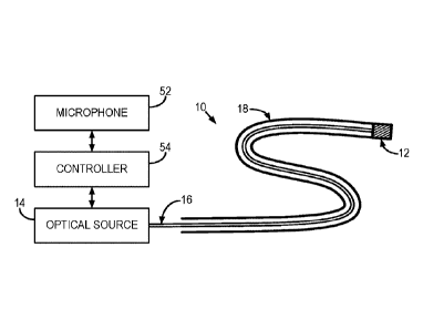
[0009] FIG. 1
is a block diagram illustrating an example of a tracking system in
accordance with some embodiments of the present invention;
-2-
CA 02967481 2017-05-11
WO 2016/074085
PCT/CA2015/051173
[0010] FIG. 2 is a block diagram illustrating an example of a tracking
marker that
forms a part of the tracking system illustrated in FIG. 1;
[0011] FIG. 3 is a block diagram illustrating another example of a tracking
marker that forms a part of a tracking system, such as the tracking system
illustrated in
FIG. 1;
[0012] FIG. 4 is a microscopic image of an example tracking marker
constructed
by coupling nickel particles to the tip of a fiber optic;
[0013] FIG. 5 is an example of a tracking marker incorporated into a biopsy
or
other medical needle;
[0014] FIG. 6 is an example of a tracking marker that includes a thermal
coupling
that is heated in response to light from an optical source and transfers this
heat to the
magnetic material in the tracking marker;
[0015] FIG. 7 is an example of a pulse sequence acquisition synchronized
with
laser pulses;
[0016] FIG. 8 is an example magnetic resonance image depicting an example
tracking marker;
[0017] FIGS. 9A-9B illustrate a correlation between laser power output and
image signal changes;
[0018] FIGS. 10A-10D depict examples of cross covariance maps of an example
tracking marker in both a laser on and a laser off state;
[0019] FIGS. 11A-11B depict examples of difference images produced by
subtracting images of a tracking marker in a laser-on state and a laser-off
state;
[0020] FIG. 12 is a flowchart setting forth the steps of an example method
for
determining a position of an example tracking marker using MRI; and
[0021] FIG. 13 is a block diagram of an example of an MRI system.
-3-
CA 02967481 2017-05-11
WO 2016/074085
PCT/CA2015/051173
DETAILED DESCRIPTION OF THE INVENTION
[0022]
Described here are systems and methods for rapid measurement of an
interventional device marker location by providing a susceptibility effect
that can be
pulsed (e.g., temporally modulated), which gives the ability to separate the
susceptibility effect of the marker from background signals by digital signal
processing
(e.g., filtering). This background separation enables projection-mode (e.g.,
volumetric)
coverage of large volumes of tissue, which is important for rapid and robust
device
position measurement.
[0023] More
particularly, the systems and methods described here utilize a laser-
induced demagnetization phenomenon to generate variable susceptibility effects
that
can be imaged with MRI. As one example, laser power is delivered to nickel
("Ni")
particles using fiber optics. Other examples will be described below. In this
example, if
the laser is off, the susceptibility effect of the Ni particles is similar to
that of normal
inside the MRI system's magnetic field. However, if the laser is on, the Ni
particles will
be demagnetized, which results in reduced susceptibility artifacts. This
effect can be
used in rapid tracking of devices by subtracting the two images acquired when
the laser
is off and on.
[0024] Certain
magnetic materials, such as nickel and cobalt, exhibit magneto-
optical effects at room temperature, such as demagnetization after irradiation
with a
pulsed laser. The optical effects are governed by the penetration depth of the
light into
the material, and have therefore been mainly studied using thin films of metal
that are
subjected to a magnetic field and laser light. When a sufficient optical
fluence is present
in the metal (e.g., 2.5 mj/cm2 ) a rapid change in magnetic susceptibility is
induced in
the metal.
-4-
CA 02967481 2017-05-11
WO 2016/074085
PCT/CA2015/051173
[0025] Laser-
induced demagnetization of thin films and particles of metal, such
as nickel and gadolinium, has been a research topic for read-write processes
in
computer technology. There are two main types of effect on the metals once
they are
placed in an external magnetic field and are exposed to a laser pulse. The
first effects are
the optical effects, in which the laser photons directly interact with the
electrons in the
metal's electron system and cause a change in magnetization within a
picosecond time
scale.
[0026] The
second effects are the thermal effects. If the temperature of any
magnetic material is increased, the magnetization of the material in an
external
magnetic field is reduced. If the temperature is increased to the Curie
temperature, a
ferromagnetic material will become paramagnetic, typically with sharply
reduced
magnetization. These thermal effects occur when the absorbed laser photons
increases
the bulk temperature of the metal, and are maximized when the temperature is
raised
to the Curie temperature and beyond.
[0027] As will
be described below in more detail, the systems and methods
described here implement this effect for tracking or otherwise following or
measuring
the position of interventional and surgical devices. Examples of
interventional and
surgical devices that can be tracked in the manner include needles; catheters;
applicators, such as ultrasonic and radio frequency ("RF") applicators; and
any other
device that may be used in connection with MRI-guided procedures.
[0028]
Referring now to FIG. 1, a tracking system 10 generally includes a tracking
marker 12 containing a magnetic material. The tracking marker 12 is coupled to
an
optical source 14 via an optical fiber 16. As described above, the tracking
system 10 can
be coupled to an interventional device 18, which may include a catheter.
-5-
CA 02967481 2017-05-11
WO 2016/074085
PCT/CA2015/051173
[0029] In some
embodiments, the optical source 14 includes a laser. As one
example, the laser can include a continuous-wave fiber-coupled photodiode
laser
machine (such as the laser manufactured as model number S1FC808, (Thorlabs
Inc.;
Newton, New Jersey, USA) with the maximum power of 24.54 mW. The wavelength of
this laser is 808 nm, which is in the near-infrared ("NIR") spectrum.
[0030] In one
preferred embodiment, the optical source 14 includes a
continuous-wave laser having the functionality to temporally modulate the
laser output
using an analog trigger signal provided by a controller that is synchronized
with the
MRI data acquisition. As one example, the continuous-wave laser can have 1 W
maximum output power and 808 nm wavelength. Having 1 W power output provides
sufficient fluence to affect all the magnetic particles within the
susceptibility marker,
and the temporal modulation of the power reduces the duty cycle of the laser
to
mitigate any bulk heating of the device.
[0031] By way
of example, the optical fiber 16 can include a 5 nm fiber optic with
125 nm cladding in a protective KevlarC) layer. In some embodiments, the
optical fiber
16 can be coupled to the optical source 14 using a FC/PC connector.
[0032]
Referring now to FIG. 2, an example of the tracking marker 12 is
illustrated. In this example, magnetic particles 20 are dispersed throughout a
substrate
22 that is coupled to the distal end of the optical fiber 16. In some
embodiments, the
substrate 22 is a translucent or otherwise transparent material, such as clear
or
otherwise non-opaque epoxy, a non-opaque plastic, or glass. The magnetic
particles 20
can, in some embodiments, include magnetic nanoparticles. As an example, the
magnetic particles 20 can be composed of nickel particles, cobalt particles,
combinations thereof, or any other suitable magnetic particle or combinations
thereof.
With the relative volume of magnetic particles 20 in an appropriate range,
such as one
-6-
CA 02967481 2017-05-11
WO 2016/074085
PCT/CA2015/051173
percent, the magnetic particles 20 and the substrate 22 together will have a
sufficiently
large magnetic susceptibility to act as a susceptibility marker for tracking,
but will still
be translucent so that light can reach all of the magnetic particles 20. In
some
embodiments, the outer surface of the substrate 22 will be coated with a
reflective layer,
with magnetic susceptibility close to tissue, such as a copper coating, in
order to reflect
light back towards the magnetic particles 20 and to contain the light within
the tracking
marker 12.
[0033] One
specific, and non-limiting example, is illustrated in FIG. 3. In this
example, the magnetic particles 20 are dispersed in a substrate 22 composed of
glass
and coupled to the distal end of an optical fiber 16. The optical fiber 16 is
composed of a
central optical fiber core 30 surrounded by a fiber cladding 32 and fiber
buffer 34. The
substrate 22 is coated, at least partially, in a metal layer 36 that reflects
light back
towards the magnetic particles 20 and to contain the light within the tracking
marker
12.
[0034] In one
preferred embodiment, the change in magnetization of the
magnetic particles 20 is maximized by ensuring that all of the magnetic
particles 20 are
bathed in a sufficient fluence of laser light (as described above). The change
in
magnetization of the magnetic particles 20 can also be maximized,
significantly, by
minimizing the susceptibility effects that are not due to the particles
affected by the
laser light. For example, the reflective coating mentioned above, as well as
any other
components and coatings used in fabricating the device, can be selected to
match the
susceptibility of tissue as closely as possible.
[0035] As one
example, the distal end of the optical fiber 16 can be stripped to
expose the cladding layer. Nickel nano-powder particles with an average size
smaller
-7-
CA 02967481 2017-05-11
WO 2016/074085
PCT/CA2015/051173
than 100 nm (such as those manufactured by Sigma-Aldrich Co.; St Louis, MO,
USA) can
be dispersed in the substrate 22.
[0036] In some
embodiments, the proximal end of the tracking marker 12 can
include a diffuser 24 that is coupled on its distal end to the substrate 22
and on its
proximal end to the optical fiber 16. The diffuser 24 can include any suitable
diffuser for
spreading out or otherwise scattering light incident from the optical fiber 16
into the
substrate 22. As one example, the diffuser 24 may be composed of a polymer.
[0037] In some
embodiments, the substrate 22 can simply include a glue, such as
a cyanoacrylate glue manufactured by Loctite (Westlake, Ohio, USA). FIG. 4
shows a
microscopic image of a tracking marker 12 constructed in this manner.
[0038] In some
other embodiments, such as those illustrated in FIG. 1, the
substrate 22 containing the particles 20 is machined, molded, or 3D-printed
from a
suitable optically translucent or transparent material into a small hollow
cylinder that
can be incorporated into a catheter with a lumen. As illustrated in FIG. 5, in
some other
embodiments, the substrate 22 containing the particles 20 is molded into a
small
tracking marker 12, which may be rectangular in shape or may be shaped in
other
geometries, that can be incorporated into a biopsy needle 24. In some
embodiments, the
substrate 22 material is selected based on its thermal conductivity, thermal
coupling to
the magnetic particles 20, specific heat capacity, or combinations thereof.
For instance,
the substrate 22 material can be selected based on these parameters such that
thermal
energy deposited in the magnetic particles 20 is adequately dissipated in the
substrate
22 and such that the bulk temperature increase in the tracking marker 12 is
minimized.
[0039] In some
other embodiments, a heat sink structure 50 can be incorporated
into the structure surrounding the tracking marker 12 in order to mitigate any
bulk
heating of the device due to absorption of the laser light and to shorten the
time
-8-
CA 02967481 2017-05-11
WO 2016/074085
PCT/CA2015/051173
required for the magnetic particles 20 to cool and re-magnetize, so that the
duration of
the pulsing of the magnetic effect of the marker 12 on and off can be
sufficiently short.
Thus, a heat sink 50 can be thermally coupled to the tracking marker 12 to
provide
cooling of the tracking marker 12, which reduces the cooling time constant of
the
tracking marker 12 and, in turn, allows for more rapid modulation of the
magnetization
state of the magnetic material in the tracking marker 12.
[0040] In
still other embodiments, such as the one illustrated in FIG. 6, the
tracking system 10 can include a thermal coupling 60 positioned between the
optical
source 14 and the tracking marker 12. The thermal coupling 60 is coupled to
the optical
source 14 and is thermally coupled to the tracking marker 12. The thermal
coupling 60
receives light from optical source 14, which increases the thermal energy of
the thermal
coupling 60. When the temperature of the thermal coupling 60 is raised, the
heat is
transferred to the magnetic material 62 in the tracking marker 12, thereby
raising the
temperature of the magnetic material 62. As described above, by heating the
magnetic
material 62 its magnetization is reduced, which provides a change in magnetic
susceptibility that can be imaged with magnetic resonance imaging. As
described above,
the magnetic material 62 can include a substrate in which magnetic particles
are
distributed, but can also include a piece of magnetic material, such as a
metal. The
thermal coupling 60 can include, for example, a layer of thermally conductive
material.
[0041] When
the tracking marker 12 is exposed to laser light delivered through
the optical fiber 16, a demagnetization of the magnetic particles 20 in the
tracking
maker 12 is induced by thermal effects, non-thermal effects, or both, so that
the
susceptibility effects of the magnetic particles 20 in the tracking marker 12
are
transiently reduced in MR images.
-9-
CA 02967481 2017-05-11
WO 2016/074085
PCT/CA2015/051173
[0042] In some
embodiments, the pulses of laser light are applied with sufficient
duration (e.g., 5 milliseconds) to affect the magnetic susceptibility of the
tracking
marker 12 for the duration of an MRI data acquisition window (see FIG. 7).
[0043] In some
other embodiments, the pulses of laser light are applied in a
periodic manner (e.g., every 50 milliseconds) so that a distinctive effect is
caused in MRI
images. As one example, the distinctive effect can include ghost artifacts.
These ghost
artifacts will appear at specific spatial offsets from the true location of
the device. These
spatial offsets can be calculated exactly based on the ratio of the period of
the laser
pulses and the repetition time ("TR") of the MRI data acquisition pulse
sequence. For
example, if the period of the laser pulses is chosen such that the light is on
for every
other data acquisition, as in FIG. 7, then the ghost artifact will appear at
an offset of
FOV/2 (where FOV is the field-of-view) from the actual location of the device.
In one
preferred embodiment, the laser light is only turned on during the data
acquisition (or
every other data acquisition as mentioned above) so that the duty cycle of the
laser
pulses is kept as low as possible, thereby minimizing heating of the marker.
[0044] In one
preferred embodiment, the synchronization of the laser pulses
with the pulse sequence is accomplished by incorporating a small microphone 52
and
controller 54 in the actuator for the optical source 14. The controller 54 may
include, for
example, a microcontroller. The microphone 52 records the distinct noise
emitted by
the gradient coils in the MRI system, which can provide the trigger signal for
turning the
optical source on or off. For instance, the microphone 52 can detect when the
gradient
coils are operating, and the signals provided by the microphone to the
controller 54 can
be processed to generate a control signal for the optical source 14 such that
the optical
source 14 is operated in synchrony with the gradient coils.
-10-
CA 02967481 2017-05-11
WO 2016/074085
PCT/CA2015/051173
[0045] In
another embodiment, the timing of the laser pulses and the repetitions
of the pulse sequence are asynchronous, but with both having a stable
frequency, so
that a pre-calibration procedure can be used to determine the location of the
resulting
ghost artifact in magnetic resonance images with the chosen TR. The advantage
of this
particular embodiment is that no synchronization signal is needed and the
design of the
actuator for the laser 14 is simpler.
[0046] In some
other embodiments, it is useful to rapidly locate the position of
the device based on projection images. This can be accomplished by making the
FOV
sufficiently large that the ghost artifact is outside the body or object being
imaged.
Projection images can be acquired in axial, sagittal, or coronal orientations.
In some
embodiments, it is useful that the measurement of the location of the ghost
artifacts is
performed automatically by the operator workstation of the MRI system. In yet
another
embodiment, the automatically located device position is used to update the
slice
position of a slice-selective scan, which is automatically started after the
device position
has been calculated. In another embodiment, the process of creating the ghost
artifact in
one or more projection images, automatically computing the device location,
updating
the slice position, and starting a slice-selective scan is initiated by
pressing a button on
the scan room interface of the MRI system.
[0047] One
preferred MRI acquisition protocol that can be used to measure and
track the position of the tracking marker is now described. Imaging can be
performed
using a conventional MRI system, such as the one described below. A balanced
steady-
state-free-precession ("bSSFP") sequence with the following parameters can be
used to
continuously acquire MR images in rapid succession: TR = 5 seconds, TE= 2.5
seconds,
matrix = 256 x 256, flip angle = 60 degrees, slice thickness = 300 mm
(projection
through large volume), FOV = 40 cm (substantially larger than the body being
imaged,
-11-
CA 02967481 2017-05-11
WO 2016/074085
PCT/CA2015/051173
to give room for the ghost artifact in the background). The laser light can be
toggled on
and off every other TR as shown in FIG. 7, creating a ghost artifact at FOV/2
from the
true location of the marker. The orientation of the scan plane can be toggled
between
axial and sagittal orientations to enable computation of all three co-
ordinates of the
marker. Alternatively, the plane containing the marker can be computed from
just one
of the orientations. For example, the coronal plane containing the marker can
be
computed from a single axial projection image showing the ghost artifact of
the marker
at FOV/2 from the true device location (with the FOV/2 offset in the phase-
encode
direction). In this example, the ghost artifact position gives the position in
both the
sagittal and coronal directions, which can be used to automatically display
the coronal
slice (or sagittal plane) containing the marker. In a preferred embodiment,
the console
software can also display a dashed line or other appropriate marker on the
image
display, indicating the last computed sagittal position of the device, to aid
the user in
quickly visualizing the device.
[0048] Another
example of an MRI acquisition protocol that can be implemented
to track the tracking marker is now described. Imaging can be performed using
a
conventional MRI system, such as a 1.5 T scanner. A multiphase fast gradient-
recalled
echo (Fast GRE) sequence with following parameters can be used to sequentially
acquire a series of 36 MR images: matrix size = 128 x 128, flip angle = 40,
bandwidth =
31.3 kHz, FOV = 13 cm, slice thickness = 5 mm, TR/TE = 5.6/2.6 ms, 5 second
delay
between images, and NEX = 1, 10.
[0049] FIG. 8
shows an example of FGRE images with susceptibility artifacts of Ni
particles. The laser output power was changed with a trend shown in FIGS. 9A-
9B. The
same imaging protocol was repeated when the laser was off throughout
acquisition of
all 36 images.
-12-
CA 02967481 2017-05-11
WO 2016/074085
PCT/CA2015/051173
[0050] Complex signal of each voxel through the acquired images can be
correlated to the laser output power trend, as follows:
Cci (0) = (n + 0) Li, j (n) (1).
n=1
[0051] Eqn. (1) can be used to calculate the cross covariance, and an
image can
be generated based on the absolute values of Cci for each voxel at zero lag
between
signal of the voxel and laser output power trend. In Eqn. (1), S (n + 0) is
the real
signal trend of voxel (i, j) at zero lag and Li, (n) is the laser output
power.
[0052] FIGS. 9A-9B also show an example of the magnitude of signal of a
voxel
that shows high positive correlation with the laser output power. The changes
in the
signal amplitude is a direct result of the changes in susceptibility value of
the nickel
particles that are being excited by the laser photons. As a result, the
susceptibility
artifact changes, thus the magnitude of the voxel signal increases or
decreases.
[0053] This time-series of images can be analyzed in various ways to
compute
the position of the marker. Computation of the cross-covariance of the signal
time-
course of each pixel vs. the time-course of the laser power can be used to
detect the
device position. FIGS. 10A-10D show examples of cross covariance maps. In
particular,
FIGS. 10A and 10B show examples of the cross covariance maps for NEX equal to
10 and
1 respectively. The highly correlated voxels were located where there was
susceptibility
artifact from the nickel particles. FIGS. 10C and 10D illustrate examples of
the cross
covariance maps for NEX equal to 10 and 1, respectively, in which the laser
was off for
all acquired images. The results in FIGS. 10C and 10D show that the highly
correlated
voxels, shown in FIGS. 10A and 10D, were caused by the laser. In some
embodiments,
the time-course of the laser power can be chosen to augment detection by the
cross-
-13-
CA 02967481 2017-05-11
WO 2016/074085
PCT/CA2015/051173
covariance or cross-correlation analysis mentioned above. For example, the
laser power
can be temporally modulated according to a Barker code, such as the Barker-7
code, (1
1 1 0 0 1 0), which will minimize background correlation that interferes with
marker
detection in low SNR images.
[0054] In some
embodiments, to visualize the changes in susceptibility artifact of
the magnetic particles images acquired with the laser on and images acquired
with the
laser off can be subtracted from each other. FIGS. 11A-11B show examples of
the
magnitude of subtraction images for NEX equal to 10 and 1, respectively. The
subtraction images show differences in signal intensity when the laser was on.
[0055] In some
embodiments, temporal filtering of sequentially acquired images
can be used to visualize the changes in the susceptibility artifact
surrounding the
marker. For example, spiral-bSSFP images (TR = 10 ms, TE= minimum, 128x128
matrix,
100 ms per frame, 2 mm spatial resolution, 10 mm slice thickness) could be
acquired
sequentially in a continuous fashion, with the laser power toggled on for
every third
data acquisition (300 ms apart). Temporal filtering of the images with a
bandpass at
3.33 Hz can the be used to detect the marker position. In some embodiments,
the
temporal on-off pattern of the laser pulses can be a code, such as a binary
Golay code,
with the detection of the device's effect in the resulting images involving
the
appropriate inverse transformation.
[0056]
Referring now to FIG. 12, a flowchart is illustrated as setting forth the
steps for an example method for tracking the position of the tracking device
described
above. The method includes providing the tracking device to a field-of-view,
as
indicated at step 1202. As one example, this step can include providing the
tracking
device via an interventional device, such as a catheter, to a region in a
subject's body
that will be imaged by an MRI system. Images of the field-of-view are then
acquired with
-14-
CA 02967481 2017-05-11
WO 2016/074085
PCT/CA2015/051173
an MRI system while the magnetic susceptibility of the magnetic material in
the tracking
device is altered, as indicated at step 1204. For instance, the magnetic
susceptibility of
the magnetic material in the tracking device is altered according to a
temporal pattern
of modulation defined by turning the optical source on and off, as described
above. As
one example, a controller in communication with the optical source provides a
control
signal that operates the optical source in accordance with the temporal
pattern of
modulation. The acquired images are then processed to determine the location
of the
tracking device in the field-of-view, as indicated at step 1206.
[0057] As one
example, the images are processed to decode the magnetic
resonance signals depicted in the images, wherein the decoding of the magnetic
resonance signals is performed based on the temporal pattern of modulation. As
another example, the images are processed to identify ghost artifacts in the
images and
to relate the location of the ghost artifacts to the location of the tracking
device. In some
embodiments, the images can be processed by applying a bandpass filter to the
images.
In these instances, the bandpass filter is preferably designed to have a
center frequency
defined by the frequency of the temporal pattern of modulation. Other examples
of how
the location of the tracking device can be determined from these images are
described
above.
[0058] Thus,
systems and methods for laser-induced demagnetization of
magnetic particles for passive tracking of a medical device have been
described.
Experimental results suggested that laser photons interact with magnetic
particles, such
as nickel particles, through the demagnetization process, thus changing the
susceptibility values of the particles. Signals of the voxels around the
magnetic particles
are highly correlated with the laser output power trend.
[0059]
Referring particularly now to FIG. 13, an example of a magnetic resonance
-15-
CA 02967481 2017-05-11
WO 2016/074085
PCT/CA2015/051173
imaging ("MRI") system 100 is illustrated. The MRI system 100 includes an
operator
workstation 102, which will typically include a display 104; one or more input
devices
106, such as a keyboard and mouse; and a processor 108. The processor 108 may
include a commercially available programmable machine running a commercially
available operating system. The operator workstation 102 provides the operator
interface that enables scan prescriptions to be entered into the MRI system
100. In
general, the operator workstation 102 may be coupled to four servers: a pulse
sequence
server 110; a data acquisition server 112; a data processing server 114; and a
data store
server 116. The operator workstation 102 and each server 110, 112, 114, and
116 are
connected to communicate with each other. For example, the servers 110, 112,
114, and
116 may be connected via a communication system 140, which may include any
suitable
network connection, whether wired, wireless, or a combination of both. As an
example,
the communication system 140 may include both proprietary or dedicated
networks, as
well as open networks, such as the internet.
[0060] The
pulse sequence server 110 functions in response to instructions
downloaded from the operator workstation 102 to operate a gradient system 118
and a
radiofrequency ("RF") system 120. Gradient waveforms necessary to perform the
prescribed scan are produced and applied to the gradient system 118, which
excites
gradient coils in an assembly 122 to produce the magnetic field gradients Y
, and
Gz used for position encoding magnetic resonance signals. The gradient coil
assembly
122 forms part of a magnet assembly 124 that includes a polarizing magnet 126
and a
whole-body RF coil 128.
[0061] RF
waveforms are applied by the RF system 120 to the RF coil 128, or a
separate local coil (not shown in FIG. 13), in order to perform the prescribed
magnetic
-16-
CA 02967481 2017-05-11
WO 2016/074085
PCT/CA2015/051173
resonance pulse sequence. Responsive magnetic resonance signals detected by
the RF
coil 128, or a separate local coil (not shown in FIG. 13), are received by the
RF system
120, where they are amplified, demodulated, filtered, and digitized under
direction of
commands produced by the pulse sequence server 110. The RF system 120 includes
an
RF transmitter for producing a wide variety of RF pulses used in MRI pulse
sequences.
The RF transmitter is responsive to the scan prescription and direction from
the pulse
sequence server 110 to produce RF pulses of the desired frequency, phase, and
pulse
amplitude waveform. The generated RF pulses may be applied to the whole-body
RF
coil 128 or to one or more local coils or coil arrays (not shown in FIG. 13).
[0062] The RF system 120 also includes one or more RF receiver channels.
Each
RF receiver channel includes an RF preamplifier that amplifies the magnetic
resonance
signal received by the coil 128 to which it is connected, and a detector that
detects and
digitizes the / and Q quadrature components of the received magnetic resonance
signal. The magnitude of the received magnetic resonance signal may,
therefore, be
determined at any sampled point by the square root of the sum of the squares
of the /
and Q components:
M = V/2 + Q2 (2);
[0063] and the phase of the received magnetic resonance signal may also be
determined according to the following relationship:
( Q
V= tan' ¨ (3).
\.. I)
[0064] The pulse sequence server 110 also optionally receives patient data
from
a physiological acquisition controller 130. By way of example, the
physiological
acquisition controller 130 may receive signals from a number of different
sensors
-17-
CA 02967481 2017-05-11
WO 2016/074085
PCT/CA2015/051173
connected to the patient, such as electrocardiograph ("ECG") signals from
electrodes, or
respiratory signals from a respiratory bellows or other respiratory monitoring
device.
Such signals are typically used by the pulse sequence server 110 to
synchronize, or
µ`gate," the performance of the scan with the subject's heart beat or
respiration.
[0065] The
pulse sequence server 110 also connects to a scan room interface
circuit 132 that receives signals from various sensors associated with the
condition of
the patient and the magnet system. It is also through the scan room interface
circuit
132 that a patient positioning system 134 receives commands to move the
patient to
desired positions during the scan.
[0066] The
digitized magnetic resonance signal samples produced by the RF
system 120 are received by the data acquisition server 112. The data
acquisition server
112 operates in response to instructions downloaded from the operator
workstation
102 to receive the real-time magnetic resonance data and provide buffer
storage, such
that no data is lost by data overrun. In some scans, the data acquisition
server 112 does
little more than pass the acquired magnetic resonance data to the data
processor server
114. However, in scans that require information derived from acquired magnetic
resonance data to control the further performance of the scan, the data
acquisition
server 112 is programmed to produce such information and convey it to the
pulse
sequence server 110. For example, during prescans, magnetic resonance data is
acquired and used to calibrate the pulse sequence performed by the pulse
sequence
server 110. As another example, navigator signals may be acquired and used to
adjust
the operating parameters of the RF system 120 or the gradient system 118, or
to control
the view order in which k-space is sampled. In still another example, the data
acquisition server 112 may also be employed to process magnetic resonance
signals
used to detect the arrival of a contrast agent in a magnetic resonance
angiography
-18-
CA 02967481 2017-05-11
WO 2016/074085
PCT/CA2015/051173
("MBA") scan. By way of example, the data acquisition server 112 acquires
magnetic
resonance data and processes it in real-time to produce information that is
used to
control the scan.
[0067] The
data processing server 114 receives magnetic resonance data from
the data acquisition server 112 and processes it in accordance with
instructions
downloaded from the operator workstation 102. Such processing may, for
example,
include one or more of the following: reconstructing two-dimensional or three-
dimensional images by performing a Fourier transformation of raw k-space data;
performing other image reconstruction algorithms, such as iterative or
backprojection
reconstruction algorithms; applying filters to raw k-space data or to
reconstructed
images; generating functional magnetic resonance images; calculating motion or
flow
images; and so on.
[0068] Images
reconstructed by the data processing server 114 are conveyed
back to the operator workstation 102 where they are stored. Real-time images
are
stored in a data base memory cache (not shown in FIG. 13), from which they may
be
output to operator display 112 or a display 136 that is located near the
magnet
assembly 124 for use by attending physicians. Batch mode images or selected
real time
images are stored in a host database on disc storage 138. When such images
have been
reconstructed and transferred to storage, the data processing server 114
notifies the
data store server 116 on the operator workstation 102. The operator
workstation 102
may be used by an operator to archive the images, produce films, or send the
images via
a network to other facilities.
[0069] The MRI
system 100 may also include one or more networked
workstations 142. By way of example, a networked workstation 142 may include a
display 144; one or more input devices 146, such as a keyboard and mouse; and
a
-19-
CA 02967481 2017-05-11
WO 2016/074085
PCT/CA2015/051173
processor 148. The networked workstation 142 may be located within the same
facility
as the operator workstation 102, or in a different facility, such as a
different healthcare
institution or clinic.
[0070] The networked workstation 142, whether within the same facility or
in a
different facility as the operator workstation 102, may gain remote access to
the data
processing server 114 or data store server 116 via the communication system
140.
Accordingly, multiple networked workstations 142 may have access to the data
processing server 114 and the data store server 116. In this manner, magnetic
resonance data, reconstructed images, or other data may be exchanged between
the
data processing server 114 or the data store server 116 and the networked
workstations 142, such that the data or images may be remotely processed by a
networked workstation 142. This data may be exchanged in any suitable format,
such
as in accordance with the transmission control protocol ("TCP"), the internet
protocol
or other known or suitable protocols.
[0071] In accordance with the present invention, the operator workstation
102
may include software and hardware components associated with triggering the
laser
pulses that are synchronized with the pulse sequence, as shown in FIG. 7. For
example,
the executable code that generates the RF and gradient pulses could be
configured to
create a TTL pulse that coincides with the start of each readout interval, and
this could
be connected via a dedicated BNC cable to the laser driver electronics such
that the laser
power can toggled on and off in synchrony with the beginning and end of the
MRI data
readout windows.
[0072] In addition, the operator workstation 102 may execute software
components or plug-ins associated with the processing of the image data
acquired
during operation of the present tracking system and the automatic update of
scan
-20-
CA 02967481 2017-05-11
WO 2016/074085
PCT/CA2015/051173
prescription information, such as slice position. These software components
can be
integral to one or more preferred embodiments of the present invention. For
example,
the operator workstation may execute a sequence of operations whereby a pulse
sequence that is synchronized with the laser pulses is played out, the image
data is
processed to automatically measure of the location of the ghost artifacts and
to derive
the device position, and this device position is used to update the slice
position of a real-
time slice-selective scan, which is automatically started after the device
position has
been calculated. In one preferred embodiment, this sequence of operations can
be
initiated by pressing a button on the scan room interface 132, and the
resulting slice-
selective scan containing the device can be displayed on a MRI-compatible
monitor
within the scan room.
[0073] The
present invention has been described in terms of one or more
preferred embodiments, and it should be appreciated that many equivalents,
alternatives, variations, and modifications, aside from those expressly
stated, are
possible and within the scope of the invention.
-21-