Note: Descriptions are shown in the official language in which they were submitted.
CA 02967636 2017-05-11
WO 2016/077759
PCT/US2015/060685
DEVICES AND METHODS FOR ABLATION OF THE SKIN
Field of the Invention
The field of the present invention relates to treatments for skin and proximal
tissue layers (e.g.,
skin tightening, treating diseases, disorders, and conditions of the skin,
skin restoration, skin lifting, skin
repositioning, and tattoo removal).
Background of the Invention
Many human health issues arise from the damage or loss of tissue due to
disease, advanced
age, and/or injury. In aesthetic medicine, elimination of excess tissue and/or
skin laxity is an important
concern that affects more than 25% of the U.S. population. Conventional
surgical therapies (e.g., a face
lift, brow lift, or breast lift) can be effective but are often invasive,
inconvenient, and expensive, while
scarring limits the applicability of surgery to certain treatment sites.
Although minimally invasive methods are available, such methods are generally
less effective
than surgical methods. Methods using energy sources (e.g., laser, non-coherent
light, radiofrequency,
and ultrasound) can be effective at improving the architecture and texture of
the skin but are much less
effective at tightening the skin or reducing skin laxity. Neurotoxins, such as
botulinum toxin, reduce the
formation of dynamic wrinkles by paralysis of the injected muscles, but such
toxins have minimal or no
direct effect on skin tightness or laxity. Finally, dermal fillers, such as
hyaluronic acid, can be injected in
the dermal layer to smooth out wrinkles and improve contours, but such fillers
do not directly tighten or
reduce laxity of the skin. Thus, surgical therapies remain the gold standard
for lifting and/or tightening
skin, as compared to energy-based techniques (e.g., laser, radiofrequency, and
ultrasound) and injection-
based techniques (e.g., botulinum toxin and fillers such as hyaluronic acid-
and collagen-based fillers).
Tissue ablative methods such as ablative fractional laser treatment create
micro-ablations with
photo-thermal energy. The use of such energy generates a coagulation zone in
tissue that interferes with
closure of the ablation zones thereby inhibiting tissue tightening. These
methods also require longer
patient healing times due to the biological reparative response to coagulated
and dead tissue during the
remodeling process. Laser ablation depth is typically limited by the depth of
the laser beam focus.
Ablation of deeper tissue layers than is possible with available laser systems
is desirable for the treatment
of scars, for example.
Accordingly, there is a need for improved methods and devices that provide
increased
effectiveness over currently available minimally-invasive techniques while
maintaining convenience,
affordability, and accessibility to patients desiring tissue restoration.
Summary of the Invention
This invention relates to apparatuses, systems, kits, and methods for non-
thermal tissue ablation.
The invention features a device for non-thermal tissue ablation including a
skin-penetrating component
and a mechanism for removing ablated tissue.
In one aspect, the invention features an apparatus for non-thermal tissue
ablation having a main
body configured for handheld operation, a tip (e.g., in the form of a
detachable cartridge) including a skin-
1
CA 02967636 2017-05-11
WO 2016/077759
PCT/US2015/060685
penetrating component with one or more ablation members (e.g., needles (e.g.,
hollow coring needles),
drill bits, abrading elements, punches, and/or blades) configured for
penetration into and retraction from
skin, and, optionally, a pressure generating source. The ablation members may
be configured to
penetrate into the skin to a depth in the range of about 0.01 mm to about 15
mm and/or to produce an
ablated tissue portion that results in the removal of an area or volumetric
fraction of tissue (e.g., skin) in
the range of about 5% to about 70%. If the pressure generating source is
present, the ablation members
are configured to be in fluid communication therewith (e.g., the ablation
members can be connected, e.g.,
via one or more connectors, such as a tube, to the pressure generating
source). The tip may be
detachably attached to the main body. The pressure generating source, if
present, may be a source of
high or low pressure and may, for example, be disposed within the main body of
the apparatus. For
example, the pressure generating source may produce vacuum or suction to
convey one or more ablated
tissue portions produced by the one or more ablation members (e.g., needles,
such as hollow coring
needles) through the ablation members and away from the skin or proximal
tissue layer or it may produce
a force that injects a fluid (e.g., including one or more of a therapeutic
agent, saline, a filler, and other
material) into the skin or proximal tissue layers. The pressure generating
source, if present, may remove
waste materials (e.g., tissue, blood, and/or interstitial fluids) from one or
more ablation members to
prevent clogging, facilitate detachment of ablated tissue portions from
surrounding tissue in a treatment
area, and/or remove waste materials from a treatment area. In some
embodiments, the apparatus may
additionally include a reservoir for collecting waste materials. The reservoir
may be disposed within the
tip or main body of the apparatus or it may be separate from the apparatus.
The reservoir may also be
configured to be in fluid communication with the ablation members of the tip.
In an embodiment, the
pressure generating source is configured to exert force that conveys one or
more ablated tissue portions
produced by the one or more ablation members through the ablation members and
into the reservoir.
In a second aspect, the invention features an apparatus for non-thermal tissue
ablation having a
main body configured for handheld operation, a tip (e.g., in the form of a
detachable cartridge) including a
skin-penetrating component with one or more ablation members (e.g., needles
(e.g., hollow coring
needles), drill bits, abrading elements, punches, and/or blades) configured
for penetration into and
retraction from skin, and a reservoir for collecting waste materials (e.g.,
tissue, blood, and/or interstitial
fluids), in which the needles are configured to be in fluid communication with
the reservoir. The ablation
members may be configured to penetrate into the skin to a depth in the range
of about 0.01 mm to about
15 mm and/or to produce an ablated tissue portion that results in the removal
of an area or volumetric
fraction of tissue (e.g., skin) in the range of about 5% to about 70%. The tip
may be detachably attached
to the main body. The reservoir may be disposed within the tip or main body of
the apparatus or it may
be separate from the apparatus. The apparatus may further include a pressure
generating source that is
a source of high or low pressure and may be disposed within the main body of
the apparatus. For
example, the pressure generating source may produce vacuum or suction to
convey one or more ablated
tissue portions produced by the one or more ablation members (e.g., needles,
such as hollow coring
needles) through the ablation members and into the reservoir or it may produce
a force that injects a fluid
(e.g., including one or more of a therapeutic agent, saline, a filler, and
other material) into the skin or
proximal tissue layers. The pressure generating source may remove waste
materials from one or more
2
CA 02967636 2017-05-11
WO 2016/077759
PCT/US2015/060685
ablation members to prevent clogging, facilitate detachment of ablated tissue
portions from surrounding
tissue in a treatment area, and/or remove waste materials from a treatment
area.
In a third aspect, the invention features an apparatus for non-thermal tissue
ablation having a
main body configured for handheld operation and a tip (e.g., in the form of a
detachable cartridge)
including a skin-penetrating component with one or more ablation members
(e.g., needles (e.g., hollow
coring needles), drill bits, abrading elements, punches, and/or blades)
configured for penetration into and
retraction from skin, in which the tip is detachably attached to the main
body. The ablation members may
be configured to penetrate into the skin to a depth in the range of about 0.01
mm to about 15 mm and/or
to produce an ablated tissue portion that results in the removal of an area or
volumetric fraction of tissue
(e.g., skin) in the range of about 5% to about 70%. The ablation members may
further be configured to
be in fluid communication with a pressure generating source (e.g., the
ablation members can be
connected, e.g., via one or more connectors, such as a tube, to the pressure
generating source). For
example, the pressure generating source may produce vacuum or suction to
convey one or more ablated
tissue portions produced by the one or more ablation members (e.g., needles,
such as hollow coring
needles) through the ablation members and away from the skin surface or it may
produce a force that
injects a fluid (e.g., including one or more of a therapeutic agent, saline, a
filler, and other material) into
the skin or proximal tissue layers. The pressure generating source may remove
waste materials (e.g.,
tissue, blood, and/or interstitial fluids) from one or more ablation members
to prevent clogging, facilitate
detachment of ablated tissue portions from surrounding tissue in a treatment
area, and/or remove waste
materials from a treatment area.
In a fourth aspect, the invention features a system for non-thermal tissue
ablation including an
apparatus of the invention (e.g., an apparatus of the first, second, and third
aspects, and any apparatus
described herein) and a reservoir for collecting waste materials (e.g.,
tissue, blood, and/or interstitial
fluids) that is in fluid communication with the apparatus. The system may
further have a base unit (e.g., a
dock, computer, control center, and/or charging station) and/or a pressure
generating source. The
reservoir may be disposed within the tip, the main body, the base unit (if
present), or a separate module,
or it may be external to these components. A pressure generating source may be
a source of high or low
pressure (e.g., a vacuum pump or fluid jet), and may be disposed within the
main body or the base unit (if
present), or it may be separate from the system. For example, the ablation
members (e.g., needles, such
as hollow coring needles), reservoir, and pressure generating source may be in
fluid communication such
that generation of vacuum by the pressure generating source draws ablated
tissue portions produced by
the one or more ablation members through the ablation members and into the
reservoir.
In a fifth aspect, the invention features a kit for non-thermal tissue
ablation including an apparatus
or system of the invention (e.g., an apparatus of the first, second, third,
and fourth aspects, and any
apparatus described herein) having a main body configured for handheld
operation and a tip (e.g., a
cartridge) including a skin-penetrating component with one or more ablation
members (e.g., needles (e.g.,
hollow coring needles), drill bits, abrading elements, punches, and/or
blades), in which the tip is
detachably attached to the main body and the ablation members are configured
to be in fluid
communication with a pressure generating source. The kit may further include a
reservoir for collecting
waste materials (e.g., tissue, blood, and/or interstitial fluids) that is
configured to be in fluid
3
CA 02967636 2017-05-11
WO 2016/077759
PCT/US2015/060685
communication with the ablation members. For example, a pressure generating
source may provide
vacuum or suction to draw one or more ablated tissue portions produced by one
or more ablation
members (e.g., needles, such as hollow coring needles) through the ablation
members and into the
reservoir. The kit may include the pressure generating source that may be a
source of high or low
pressure (e.g., a vacuum pump or fluid jet). The kit may also feature a base
unit (e.g., a dock, computer,
control center, and/or charging station). The reservoir may be disposed within
the tip, the main body, the
base unit (if present), or a separate module, or it may be external to these
components. The pressure
generating source may be disposed within the main body or the base unit (if
present), or it may be
external to these components.
In some embodiments, the main body and/or base unit may further include one or
more user
interfaces (e.g., one or more buttons, toggles, spin-wheels, dials, cursors,
screws, keys, screens, touch
screens, computers, displays, and/or switches) that may include indicators of
device configurations,
powered status, and/or other information including operation mode and needle
number and arrangement.
The user interface(s) of the main body and/or base unit may allow for control
of device parameters,
operation mode, and other features.
In some embodiments, the base unit of a system or kit includes a power source
(e.g., one or
more alternators, generators, power cords, connections to mains electricity,
and/or battery charging
stations). In some embodiments, the base unit is electrically coupled to the
apparatus. The base unit
may be coupled to the apparatus via a cable that provides power, information,
fluid flow, and/or vacuum
or suction. In other embodiments, the base unit may be wirelessly coupled to
the apparatus.
In some embodiments, systems and kits of the invention additionally include a
positioning
apparatus for positioning skin (e.g., tensioning rods, adhesives, vacuum
grippers, and needle or hook
grippers).
In some embodiments, the skin-penetrating component of the apparatus includes
1-100 ablation
members (e.g., needles (e.g., hollow coring needles), drill bits, abrading
elements, punches, and/or
blades) (e.g., 1-10, 1-20, 1-30, 1-40, 1-50, 1-60, 1-70, 1-80, 1-90, 1-100, 3-
10, 3-20, 3-30, 3-40, 3-50, 3-
60, 3-70, 3-80, 3-90, 3-100, 5-10, 5-20, 5-30, 5-40, 5-50, 5-60, 5-70, 5-80, 5-
90, 5-100, 10-20, 10-40, 10-
60, 10-80, 10-100, 20-40, 20-60, 20-80, 20-100, 40-60, 40-80, 40-100, 60-80,
60-100, and 80-100
needles). In some embodiments, 3-50 ablation members may be present. The
ablation members may
be arranged in al-or 2-dimensional array. In some embodiments, the minimum
distance between
ablation members may be between about 0.1 mm to about 50 mm (e.g., from 0.1 mm
to 0.2 mm, 0.1 mm
to 0.5 mm, 0.1 mm to 1 mm, 0.1 mm to 2 mm, 0.1 mm to 5 mm, 0.1 mm to 10 mm,
0.1 mm to 15 mm, 0.1
mm to 20 mm, 0.1 mm to 30 mm, 0.1 mm to 40 mm, 0.1 mm to 50 mm, 0.2 mm to 0.5
mm, 0.2 mm to 1
mm, 0.2 mm to 2 mm, 0.2 mm to 5 mm, 0.2 mm to 10 mm, 0.2 mm to 15 mm, 0.2 mm
to 20 mm, 0.2 mm
to 30 mm, 0.2 mm to 40 mm, 0.2 mm to 50 mm, 0.5 mm to 1 mm, 0.5 mm to 2 mm,
0.5 mm to 5 mm, 0.5
mm to 10 mm, 0.5 mm to 15 mm, 0.5 mm to 20 mm, 0.5 mm to 30 mm, 0.5 mm to 40
mm, 0.5 mm to 50
mm, 1 mm to 2 mm, 1 mm to 5 mm, 1 mm to 10 mm, 1 mm to 15 mm, 1 mm to 20 mm, 1
mm to 30 mm, 1
mm to 40 mm, 1 mm to 50 mm, 2 mm to 5 mm, 2 mm to 10 mm, 2 mm to 15 mm, 2 mm
to 20 mm, 2 mm
to 30 mm, 2 mm to 40 mm, 2 mm to 50 mm, 5 mm to 10 mm, 5 mm to 15 mm, 5 mm to
20 mm, 5 mm to
30 mm, 5 mm to 40 mm, 5 mm to 50 mm, 10 mm to 15 mm, 10 mm to 20 mm, 10 mm to
30 mm, 10 mm
4
CA 02967636 2017-05-11
WO 2016/077759
PCT/US2015/060685
to 40 mm, 10 mm to 50 mm, 15 mm to 20 mm, 15 mm to 30 mm, 15 mm to 40 mm, 15
mm to 50 mm, 20
mm to 30 mm, 20 mm to 40 mm, 20 mm to 50 mm, 30 mm to 40 mm, 30 mm to 50 mm,
and 40 mm to 50
mm). In some embodiments, the minimum distance between ablation members is
about 0.5 mm to about
2 mm apart. The minimum distance between ablation members may correspond to
the minimal size of
the array of a plurality of ablation members. For example, an array including
10 ablation members each
spaced about 1 mm apart may form a 1-dimensional array that is about 10 mm
long or a 2-dimensional
array arranged as 2 ablation members by 5 ablation members that is about 2 mm
wide and about 5 mm
long. The size of an array may correspond to the size of a skin region (e.g.,
a treatment area). For
example, a 2 mm by 5 mm array may be used on a 2 mm by 5 mm treatment area.
The skin-penetrating
component may be applied more than one time to treat a larger region of skin.
For example, a skin-
penetrating component including a 2 mm by 5 mm array of ablation members may
be applied three times
to treat a 6 mm by 5 mm skin region.
In some embodiments, one or more (e.g., all of) of the ablation members may be
hollow needles
(e.g., hollow coring needles). One or more of the needles may have one or more
holes (e.g., at one or
both ends or along the shaft of the needle). The needles may be made of metal
or plastic and/or may be
sharpened at one end. In some embodiments, the needles may be of any gauge
between 19 and 26
(e.g., 19, 20, 21, 22, 23, 24, 25, and 26 gauge). In some embodiments, the
needles may be 22 or 24
gauge needles.
The apparatus, system, or kit may be used to produce one or more tissue
portions. For example,
penetration into tissue by the ablation members (e.g., needles, drill bits,
abrading elements, punches,
and/or blades) of the apparatus may produce one or more tissue portions that
are separated from the
surrounding tissue. Retraction of the ablation members from tissue may
facilitate the separation of the
tissue portions from the surrounding tissue, and/or may allow treatment of
another area of tissue. The
number of tissue portions produced may correspond to the number of ablation
members used. For
instance, penetration into and retraction from tissue by a single ablation
member (e.g., a hollow coring
needle) may produce a single tissue portion, while penetration into and
retraction from tissue by ten
ablation members may produce ten tissue portions. Similarly, a single ablation
member used ten times
may produce ten tissue portions. A tissue portion produced by the apparatus
may have specific
dimensions. For example, the depth of penetration by the ablation members
(e.g., hollow coring needles)
may correspond to the depth or length of a tissue portion produced. In some
embodiments, a tissue
portion has at least one dimension in a range of about 10 pm to about 2 mm
(e.g., about 10 pm to 500
pm, about 10 pm to 100 pm, 10 pm to 250 pm, 10 pm to 500 pm, 10 pm to 750 pm,
10 pm to 1 mm, 10
pm to 1.5 mm, 10 pm to 2 mm, about 50 pm to 100 pm, 50 pm to 250 pm, 50 pm to
500 pm, 50 pm to
750 pm, 50 pm to 1 mm, 50 pm to 1.5 mm, 50 pm to 2 mm, 100 pm to 250 pm, 100
pm to 500 pm, 100
pm to 750 pm, 100 pm to 1 mm, 100 pm to 1.5 mm, 100 pm to 2 mm, 250 pm to 500
pm, 250 pm to 750
pm, 250 pm to 1 mm, 250 pm to 1.5 mm, 250 pm to 2 mm, 500 pm to 750 pm, 500 pm
to 1 mm, 500 pm
to 1.5 mm, 500 pm to 2 mm, 750 pm to 1 mm, 750 pm to 1.5 mm, and 750 pm to 2
mm); between about
0.1 mm to about 0.8 mm (e.g., 0.1 mm to 0.8 mm, 0.1 mm to 0.6 mm, 0.1 mm to
0.4 mm, 0.1 mm to 0.2
mm, 0.2 mm to 0.8 mm, 0.2 mm to 0.6 mm, 0.2 mm to 0.4 mm, 0.2 mm to 0.3 mm,
0.3 mm to 0.8 mm, 0.3
mm to 0.6 mm, 0.3 mm to 0.4 mm, 0.4 mm to 0.8 mm, 0.4 mm to 0.6 mm, 0.4 mm to
0.5 mm, 0.5 mm to
5
CA 02967636 2017-05-11
WO 2016/077759
PCT/US2015/060685
0.8 mm, 0.5 mm to 0.6 mm, 0.6 mm to 0.8 mm, 0.6 mm to 0.7 mm, and 0.7 mm to
0.8 mm); between
about 0.9 mm to about 20 mm (e.g., 0.9 mm to 20 mm, 0.9 mm to 17 mm, 0.9 mm to
14 mm, 0.9 mm to
11 mm, 0.9 mm to 8 mm, 0.9 mm to 5 mm, 0.9 mm to 3 mm, 3 mm to 20 mm, 3 mm to
17 mm, 3 mm to
14 mm, 3 mm to 11 mm, 3 mm to 8 mm, 3 mm to 5 mm, 5 mm to 20 mm, 5 mm to 17
mm, 5 mm to 14
mm, 5 mm toll mm, 5 mm to 8 mm, 8 mm to 20 mm, 8 mm to 17 mm, 8 mm to 14 mm, 8
mm toll mm,
11 mm to 20 mm, 11 mm to 17 mm, 11 mm to 14 mm, 14 mm to 20 mm, 14 mm to 17
mm, and 17 mm to
20 mm); between about 0.01 mm to 0.25 mm (e.g., 0.01 mm to 0.25 mm, 0.02 mm to
0.25 mm, 0.03 mm
to 0.25 mm, 0.05 mm to 0.25 mm, 0.075 mm to 0.25 mm, 0.1 mm to 0.25 mm, 0.15
mm to 0.25 mm, 0.2
mm to 0.25 mm, 0.01 mm to 0.2 mm, 0.02 mm to 0.2 mm, 0.03 mm to 0.2 mm, 0.05
mm to 0.2 mm, 0.075
mm to 0.2 mm, 0.1 mm to 0.2 mm, 0.15 mm to 0.2 mm, 0.01 mm to 0.15 mm, 0.02 mm
to 0.15 mm, 0.03
mm to 0.15 mm, 0.05 mm to 0.15 mm, 0.075 mm to 0.15 mm, 0.1 mm to 0.15 mm,
0.01 mm to 0.1 mm,
0.02 mm to 0.1 mm, 0.03 mm to 0.1 mm, 0.05 mm to 0.1 mm, 0.075 mm to 0.1 mm,
0.01 mm to 0.075
mm, 0.02 mm to 0.075 mm, 0.03 mm to 0.075 mm, 0.05 mm to 0.075 mm, 0.01 mm to
0.05 mm, 0.02 mm
to 0.05 mm, 0.03 mm to 0.05 mm, 0.01 mm to 0.03 mm, 0.02 mm to 0.03 mm, 0.03
mm to 0.03 mm, 0.01
mm to 0.03 mm, 0.02 mm to 0.03 mm, and 0.01 mm to 0.02 mm); between about 0.01
mm to about 20
mm (e.g., 0.01 mm to 1 mm, 0.01 mm to 2 mm, 0.01 mm to 5 mm, 0.01 mm to 10 mm,
0.01 mm to 15
mm, 0.05 mm to 1 mm, 0.05 mm to 2 mm, 0.05 mm to 5 mm, 0.05 mm to 10 mm, 0.05
mm to 15 mm,
0.05 mm to 20 mm, 0.1 mm to 1 mm, 0.1 mm to 2 mm, 0.1 mm to 5 mm, 0.1 mm to 10
mm, 0.1 mm to 15
mm, 0.1 mm to 20 mm, 0.5 mm to 1 mm, 0.5 mm to 2 mm, 0.5 mm to 5 mm, 0.5 mm to
10 mm, 0.5 mm to
15 mm, 0.5 mm to 20 mm, 1 mm to 2 mm, 1 mm to 5 mm, 1 mm to 10 mm, 1 mm to 15
mm, 1 mm to 20
mm, 2 mm to 5 mm, 2 mm to 10 mm, 2 mm to 15 mm, 2 mm to 20 mm, 5 mm to 10 mm,
5 mm to 15 mm,
and 5 mm to 20 mm); or between about 0.01 mm to about 2 mm (e.g., 0.01 mm to
0.1 mm, 0.01 mm to
0.5 mm, 0.01 mm to 1 mm, 0.01 mm to 1.5 mm, 0.01 mm to 1.75 mm, 0.05 mm to 0.1
mm, 0.05 mm to
0.5 mm, 0.05 mm to 1 mm, 0.05 mm to 1.5 mm, 0.05 mm to 1.75 mm, 0.05 mm to 2
mm, 0.1 mm to 0.5
mm, 0.1 mm to 1 mm, 0.1 mm to 1.5 mm, 0.1 mm to 1.75 mm, 0.1 mm to 2 mm, 0.3
mm to 0.5 mm, 0.3
mm to 1 mm, 0.3 mm to 1.5 mm, 0.3 mm to 1.75 mm, 0.3 mm to 2 mm, 0.5 mm to 1
mm, 0.5 mm to 1.5
mm, 0.5 mm to 1.75 mm, 0.5 mm to 2 mm, 0.7 mm to 1 mm, 0.7 mm to 1.5 mm, 0.7
mm to 1.75 mm, 0.7
mm to 2 mm, 1 mm to 1.5 mm, 1 mm to 1.75 mm, 1 mm to 2 mm, 1.5 mm to 1.75 mm,
1.5 mm to 2 mm,
and 1.75 mm to 2 mm).
In some embodiments, a tissue portion produced by an ablation member (e.g.,
needle, drill bit,
abrading element, punch, and blade) of the apparatus has an area dimension
less than about 2 mm2
and/or a volumetric dimension that is less than about 6 mm3. A tissue portion
may have an area
dimension in a range of about 0.001 mm2 to about 2 mm2 (e.g., 0.001 mm2 to
0.005 mm2, 0.001 mm2 to
0.01 mm2, 0.001 mm2 to 0.05 mm2, 0.001 mm2 to 0.1 mm2, 0.001 mm2 to 0.5 mm2,
0.001 mm2 to 1 mm2,
0.001 mm2 to 1.5 mm2, 0.001 mm2 to 2 mm2, 0.005 mm2 to 0.01 mm2, 0.005 mm2 to
0.05 mm2, 0.005
mm2 to 0.1 mm2, 0.005 mm2 to 0.5 mm2, 0.005 mm2 to 1 mm2, 0.005 mm2 to 1.5
mm2, 0.005 mm2 to 2
mm2, 0.01 mm2 to 0.02 mm2, 0.01 mm2 to 0.05 mm2, 0.01 mm2 to 0.1 mm2, 0.01 mm2
to 0.5 mm2, 0.01
mm2 to 1 mm2, 0.01 mm2 to 1.5 mm2, 0.01 mm2 to 2 mm2, 0.05 mm2 to 0.1 mm2,
0.05 mm2 to 0.5 mm2,
0.05 mm2 to 1 mm2, 0.05 mm2 to 1.5 mm2, 0.05 mm2 to 2 mm2, 0.1 mm2 to 0.2 mm2,
0.1 mm2 to 0.5 mm2,
0.1 mm2 to 1 mm2, 0.1 mm2 to 1.5 mm2, 0.1 mm2 to 2 mm2, 0.5 mm2 to 1 mm2, 0.5
mm2 to 1.5 mm2, 0.5
6
CA 02967636 2017-05-11
WO 2016/077759
PCT/US2015/060685
mm2 to 2 mm2, 1 mm2 to 1.5 mm2, 1 mm2 to 2 mm2, and 1.5 mm2 to 2 mm2).
In some embodiments, the volume of a tissue portion formed by use of the
apparatus is between
about 0.001 mm3 and about 6 mm3 (e.g., 0.001 mm3 to 0.01 mm3, 0.001 mm3 to 0.1
mm3, 0.001 mm3 to
0.5 mm3, 0.001 mm3 to 1 mm3, 0.001 mm3 to 2 mm3, 0.001 mm3 to 3 mm3, 0.001 mm3
to 4 mm3, 0.001
mm3 to 5 mm3, 0.001 mm3 to 6 mm3, 0.005 mm3 to 0.01 mm3, 0.005 mm3 to 0.1 mm3,
0.005 mm3 to 0.5
mm3, 0.005 mm3 to 1 mm3, 0.005 mm3 to 2 mm3, 0.005 mm3 to 3 mm3, 0.005 mm3 to
4 mm3, 0.005 mm3
to 5 mm3, 0.005 mm3 to 6 mm3, 0.01 mm3 to 0.1 mm3, 0.01 mm3 to 0.5 mm3, 0.01
mm3 to 1 mm3, 0.01
mm3 to 2 mm3, 0.01 mm3 to 3 mm3, 0.01 mm3 to 4 mm3, 0.01 mm3 to 5 mm3, 0.01
mm3 to 6 mm3, 0.1 mm3
to 0.5 mm3, 0.1 mm3 to 1 mm3, 0.1 mm3 to 2 mm3, 0.1 mm3 to 3 mm3, 0.1 mm3 to 4
mm3, 0.1 mm3 to 5
mm3, 0.1 mm3 to 6 mm3, 0.5 mm3 to 1 mm3, 0.5 mm3 to 2 mm3, 0.5 mm3 to 3 mm3,
0.5 mm3 to 4 mm3, 0.5
mm3 to 5 mm3, 0.5 mm3 to 6 mm3, 1 mm3 to 2 mm3, 1 mm3 to 3 mm3, 1 mm3 to 4
mm3, 1 mm3 to 5 mm3, 1
mm3 to 6 mm3, 2 mm3 to 3 mm3, 2 mm3 to 4 mm3, 2 mm3 to 5 mm3, 2 mm3 to 6 mm3,
3 mm3 to 4 mm3, 3
mm3 to 5 mm3, 3 mm3 to 6 mm3, 4 mm3 to 5 mm3, 4 mm3 to 6 mm3, and 5 mm3 to 6
mm3).
In some embodiments, the dimensions, geometry, number, and other
characteristics of a tissue
portion may correspond to the dimensions, geometry, number, and other
characteristics of an ablation
member (e.g., needle, drill bit, abrading element, punch, and blade) of the
skin penetrating component of
the apparatus of the invention. For example, the use of an apparatus of the
invention may form one or
more holes in a region of skin and/or proximal tissue layers (e.g., a
treatment area) by producing one or
more tissue portions with the dimensions, geometry, and other characteristics
of the holes. The diameter
and/or width of a tissue portion may be between about 0.01 mm to about 2 mm
(e.g., as described
herein). The diameter and/or width of a tissue portion generally correspond to
the diameter and/or width
of an ablation member of the invention used to produce the tissue portion. The
diameter and/or width of
an ablation member of an apparatus of the invention at its widest points may
be about 0.01 mm to about
2 mm (e.g., as described herein). For example, an apparatus including hollow
coring needles with inner
(lumen) diameters in the range of about 0.01 mm to about 2.0 mm can be used to
provide tissue portions
having a corresponding diameter in the range of about 0.01 mm to about 2.0 mm,
respectively.
An apparatus of the invention may be configured to provide one or more tissue
portions having a
change in width as a function of depth (e.g., length). For example, the outer
structure and/or inner
structure (e.g., for a hollow ablation member) of one or more ablation members
(e.g., needles, such as
hollow coring needles) of the apparatus may be tapered, having a narrower
width at either end, and/or
may vary regularly or irregularly along their lengths and so may produce one
or more tissue portions
having a narrower width at one end and/or regularly or irregularly varying
widths along their lengths. The
change in width of a tissue portion may be between about 100 pm to about 500
pm as a function of depth
(e.g., 100 pm to 200 pm, 100 pm to 300 pm, 100 pm to 400 pm, 100 pm to 500 pm,
200 pm to 300 pm,
200 pm to 400 pm, 200 pm to 500 pm, 300 pm to 400 pm, 300 pm to 500 pm, and
400 pm to 500 pm).
The width to depth ratio of a tissue portion may be between about 1:0.3 to
about 1:75. For example, the
width to depth radio of a tissue portion may be between about 1:0.3 to about
1:1 (e.g., 1:0.3 to 1:1, 1:0.35
to 1:1, 1:0.4 to 1:1, 1:0.45 to 1:1, 1:0.5 to 1:1, 1:0.55 to 1:1, 1:0.6 to
1:1, 1:0.65 to 1:1, 1:0.7 to 1:1, 1:0.75
to 1:1, 1:0.8 to 1:1, 1:0.85 to 1:1, 1:0.9 to 1:1, 1:0.95 to 1:1, 1:0.3 to
1:0.95, 1:0.35 to 1:0.95, 1:0.4 to
1:0.95, 1:0.45 to 1:0.95, 1:0.5 to 1:0.95, 1:0.55 to 1:0.95, 1:0.6 to 1:0.95,
1:0.65 to 1:0.95, 1:0.7 to 1:0.95,
7
CA 02967636 2017-05-11
WO 2016/077759
PCT/US2015/060685
1:0.75 to 1:0.95, 1:0.8 to 1:0.95, 1:0.85 to 1:0.95, 1:0.9 to 1:0.95, 1:0.3 to
1:0.9, 1:0.35 to 1:0.9, 1:0.4 to
1:0.9, 1:0.45 to 1:0.9, 1:0.5 to 1:0.9, 1:0.55 to 1:0.9, 1:0.6 to 1:0.9,
1:0.65 to 1:0.9, 1:0.7 to 1:0.9, 1:0.75 to
1:0.9, 1:0.8 to 1:0.9, 1:0.85 to 1:0.9, 1:0.3 to 1:0.85, 1:0.35 to 1:0.85,
1:0.4 to 1:0.85, 1:0.45 to 1:0.85,
1:0.5 to 1:0.85, 1:0.55 to 1:0.85, 1:0.6 to 1:0.85, 1:0.65 to 1:0.85, 1:0.7 to
1:0.85, 1:0.75 to 1:0.85, 1:0.8 to
1:0.85, 1:0.3 to 1:0.8, 1:0.35 to 1:0.8, 1:0.4 to 1:0.8, 1:0.45 to 1:0.8,
1:0.5 to 1:0.8, 1:0.55 to 1:0.8, 1:0.6 to
1:0.8, 1:0.65 to 1:0.8, 1:0.7 to 1:0.8, 1:0.75 to 1:0.8, 1:0.3 to 1:0.75,
1:0.35 to 1:0.75, 1:0.4 to 1:0.75,
1:0.45 to 1:0.75, 1:0.5 to 1:0.75, 1:0.55 to 1:0.75, 1:0.6 to 1:0.75, 1:0.65
to 1:0.75, 1:0.7 to 1:0.75, 1:0.3 to
1:0.65, 1:0.35 to 1:0.65, 1:0.4 to 1:0.65, 1:0.45 to 1:0.65, 1:0.5 to 1:0.65,
1:0.55 to 1:0.65, 1:0.6 to 1:0.65,
1:0.3 to 1:0.65, 1:0.35 to 1:0.65, 1:0.4 to 1:0.65, 1:0.45 to 1:0.65, 1:0.5 to
1:0.65, 1:0.55 to 1:0.65, 1:0.6 to
1:0.65, 1:0.3 to 1:0.6, 1:0.35 to 1:0.6, 1:0.4 to 1:0.6, 1:0.45 to 1:0.6,
1:0.5 to 1:0.6, 1:0.55 to 1:0.6, 1:0.3 to
1:0.55, 1:0.35 to 1:0.55, 1:0.4 to 1:0.55, 1:0.45 to 1:0.55, 1:0.5 to 1:0.55,
1:0.3 to 1:0.5, 1:0.35 to 1:0.5,
1:0.4 to 1:0.5, 1:0.45 to 1:0.5, 1:0.5 to 1:0.5, 1:0.3 to 1:0.45, 1:0.35 to
1:0.45, 1:0.4 to 1:0.45, 1:0.3 to
1:0.4, 1:0.35 to 1:0.4, and 1:0.3 to 1:0.35); between about 1:1 to about 1:20
(e.g., 1:1 to 1:2, 1:1 to 1:3,
1:1 to 1:4,1:1 to 1:5, 1:1 to 1:6, 1:1 to 1:7,1:1 to 1:8, 1:1 to 1:9, 1:1 to
1:10, 1:1 to 1:11, 1:1 to 1:12,1:1 to
1:13,1:1 to 1:14,1:1 to 1:15,1:1 to 1:16,1:1 to 1:17,1:1 to 1:18,1:1 to
1:19,1:1 to 1:20, 1:2 to 1:3, 1:2 to
1:4, 1:2 to 1:5, 1:2 to 1:6, 1:2 to 1:7, 1:2 to 1:8, 1:2 to 1:9, 1:2 to 1:10,
1:2 to 1:11, 1:2 to 1:12, 1:2 to 1:13,
1:2 to 1:14, 1:2 to 1:15, 1:2 to 1:16, 1:2 to 1:17, 1:2 to 1:18, 1:2 to 1:19,
1:2 to 1:20, 1:3 to 1:4, 1:3 to 1:5,
1:3 to 1:6, 1:3 to 1:7, 1:3 to 1:8, 1:3 to 1:9, 1:3 to 1:10, 1:3 to 1:11, 1:3
to 1:12, 1:3 to 1:13, 1:3 to 1:14,1:3
to 1:15, 1:3 to 1:16, 1:3 to 1:17, 1:3 to 1:18, 1:3 to 1:19, 1:3 to 1:20, 1:4
to 1:5, 1:4 to 1:6, 1:4 to 1:7, 1:4 to
1:8, 1:4 to 1:9, 1:4 to 1:10, 1:4 to 1:11, 1:4 to 1:12, 1:4 to 1:13, 1:4 to
1:14, 1:4 to 1:15, 1:4 to 1:16, 1:4 to
1:17, 1:4 to 1:18, 1:4 to 1:19, 1:4 to 1:20, 1:5 to 1:6, 1:5 to 1:7, 1:5 to
1:8, 1:5 to 1:9, 1:5 to 1:10, 1:5 to
1:11, 1:5 to 1:12, 1:5 to 1:13, 1:5 to 1:14, 1:5 to 1:15, 1:5 to 1:16, 1:5 to
1:17, 1:5 to 1:18, 1:5 to 1:19, 1:5
to 1:20, 1:6 to 1:7, 1:6 to 1:8, 1:6 to 1:9, 1:6 to 1:10, 1:6 to 1:11, 1:6 to
1:12, 1:6 to 1:13, 1:6 to 1:14, 1:6 to
1:15, 1:6 to 1:16, 1:6 to 1:17, 1:6 to 1:18, 1:6 to 1:19, 1:6 to 1:20, 1:7 to
1:8, 1:7 to 1:9, 1:7 to 1:10, 1:7 to
1:11, 1:7 to 1:12, 1:7 to 1:13, 1:7 to 1:14, 1:7 to 1:15, 1:7 to 1:16, 1:7 to
1:17, 1:7 to 1:18, 1:7 to 1:19, 1:7
to 1:20, 1:8 to 1:9, 1:8 to 1:10, 1:8 to 1:11, 1:8 to 1:12, 1:8 to 1:13, 1:8
to 1:14, 1:8 to 1:15, 1:8 to 1:16, 1:8
to 1:17, 1:8 to 1:18, 1:8 to 1:19, 1:8 to 1:20, 1:9 to 1:10, 1:9 to 1:11, 1:9
to 1:12, 1:9 to 1:13, 1:9 to 1:14,
1:9 to 1:15, 1:9 to 1:16, 1:9 to 1:17, 1:9 to 1:18, 1:9 to 1:19, 1:9 to 1:20,
1:10 to 1:11, 1:10 to 1:12, :10 to
1:13, 1:10 to 1:14, 1:10 to 1:15, 1:10 to 1:16, 1:10 to 1:17, 1:10 to 1:18,
1:10 to 1:19, 1:10 to 1:20, :11 to
1:12,1:11 to 1:13,1:11 to 1:14,1:11 to 1:15,1:11 to 1:16, 1:11 to 1:17,1:11 to
1:18,1:11 to 1:19, :11 to
1:20, 1:12 to 1:13, 1:12 to 1:14, 1:12 to 1:15, 1:12 to 1:16, 1:12 to 1:17,
1:12 to 1:18, 1:12 to 1:19, :12 to
1:20, 1:13 to 1:14, 1:13 to 1:15, 1:13 to 1:16, 1:13 to 1:17, 1:13 to 1:18,
1:13 to 1:19, 1:13 to 1:20, :14 to
1:15, 1:14 to 1:16, 1:14 to 1:17, 1:14 to 1:18, 1:14 to 1:19, 1:14 to 1:20,
1:15 to 1:16, 1:15 to 1:17, :15 to
1:18, 1:15 to 1:19, 1:15 to 1:20, 1:17 to 1:18, 1:17 to 1:19, and 1:17 to
1:20); between about 1:1 to about
1:75 (e.g., 1:1 to 1:2,1:1 to 1:5, 1:1 to 1:10, 1:1 to 1:20, 1:1 to 1:30, 1:1
to 1:40,1:1 to 1:50, 1:1 to 1:60,
1:1 to 1:75, 1:2 to 1:5, 1:2 to 1:10, 1:2 to 1:20, 1:2 to 1:30, 1:2 to
1:40,1:2 to 1:50, 1:2 to 1:60, 1:2 to 1:75,
1:5 to 1:10, 1:5 to 1:20, 1:5 to 1:30, 1:5 to 1:40, 1:5 to 1:50, 1:5 to 1:60,
1:5 to 1:75, 1:10 to 1:20, 1:10 to
1:30, 1:10 to 1:40, 1:10 to 1:50, 1:10 to 1:60, 1:10 to 1:75, 1:20 to 1:30,
1:20 to 1:40, 1:20 to 1:50, 1:20 to
1:60, 1:20 to 1:75, 1:30 to 1:40, 1:30 to 1:50, 1:30 to 1:60, 1:30 to 1:75,
1:40 to 1:50, 1:40 to 1:60, 1:40 to
1:75, 1:50 to 1:60, 1:50 to 1:75, and 1:60 to 1:75); between about 1:25 to
about 1:75 (e.g., 1:25 to 1:75,
8
CA 02967636 2017-05-11
WO 2016/077759
PCT/US2015/060685
1:30 to 1:75, 1:35 to 1:75, 1:40 to 1:75, 1:45 to 1:75, 1:50 to 1:75, 1:55 to
1:75, 1:60 to 1:75, 1:65 to 1:75,
1:70 to 1:75, 1:25 to 1:70, 1:30 to 1:70, 1:35 to 1:70, 1:40 to 1:70, 1:45 to
1:70, 1:50 to 1:70, 1:55 to 1:70,
1:60 to 1:70, 1:65 to 1:70, 1:25 to 1:65, 1:30 to 1:65, 1:35 to 1:65, 1:40 to
1:65, 1:45 to 1:65, 1:50 to 1:65,
1:55 to 1:65, 1:60 to 1:65, 1:25 to 1:60, 1:30 to 1:60, 1:35 to 1:60, 1:40 to
1:60, 1:45 to 1:60, 1:50 to 1:60,
1:55 to 1:60, 1:25 to 1:55, 1:30 to 1:55, 1:35 to 1:55, 1:40 to 1:55, 1:45 to
1:55, 1:50 to 1:55, 1:25 to 1:50,
1:30 to 1:50, 1:35 to 1:50, 1:40 to 1:50, 1:45 to 1:50, 1:25 to 1:45, 1:30 to
1:45, 1:35 to 1:45, 1:40 to 1:45,
1:25 to 1:40, 1:30 to 1:40, 1:35 to 1:40, 1:25 to 1:35, 1:30 to 1:35, and 1:25
to 1:30); or between about
1:03 to about 1:75 (e.g., 1:0.3 to 1:0.5, 1:0.3 to 1:1, 1:0.3 to 1:2, 1:0.3 to
1:5, 1:0.3 to 1:10, 1:0.3 to 1:20,
1:0.3 to 1:30, 1:0.3 to 1:40, 1:0.3 to 1:50, 1:0.3 to 1:60, 1:0.3 to 1:75,
1:0.5 to 1:1, 1:0.5 to 1:2, 1:0.5 to
1:5, 1:0.5 to 1:10, 1:0.5 to 1:20, 1:0.5 to 1:30, 1:0.5 to 1:40, 1:0.5 to
1:50, 1:0.5 to 1:60, and 1:0.5 to 1:75).
In all aspects of the invention, the apparatus may be configured to provide
from about 10 to about
10000 tissue portions per cm2 area (e.g., 10 to 50, 10 to 100, 10 to 200, 10
to 300, 10 to 400, 10 to 500,
10 to 600, 10 to 700, 10 to 800, 10 to 900, 10 to 1000, 10 to 2000, 10 to
4000, 10 to 6000, 10 to 8000,10
to 10000, 50 to 100, 50 to 200, 50 to 300, 50 to 400, 50 to 500, 50 to 600, 50
to 700, 50 to 800, 50 to 900,
50 to 1000, 50 to 2000, 50 to 4000, 510 to 6000, 50 to 8000, 50 to 10000, 100
to 200, 100 to 300, 100 to
400, 100 to 500, 100 to 600, 100 to 700, 100 to 800, 100 to 900, 100 to 1000,
100 to 2000, 100 to 4000,
100 to 6000, 100 to 8000, 100 to 10000, 200 to 300, 200 to 400, 200 to 500,
200 to 600, 200 to 700, 200
to 800, 200 to 900, 200 to 1000, 200 to 2000, 200 to 4000, 200 to 6000, 200 to
8000, 200 to 10000, 300
to 400, 300 to 500, 300 to 600, 300 to 700, 300 to 800, 300 to 900, 300 to
1000, 300 to 2000, 300 to
4000, 300 to 6000, 300 to 8000, 300 to 10000, 400 to 500, 400 to 600, 400 to
700, 400 to 800, 400 to
900, 400 to 1000, 400 to 2000, 400 to 4000, 400 to 6000, 400 to 8000, 400 to
10000, 500 to 600, 500 to
700, 500 to 800, 500 to 900, 500 to 1000, 500 to 2000, 500 to 4000, 500 to
6000, 500 to 8000, 500 to
10000, 600 to 700, 600 to 800, 600 to 900, 600 to 1000, 600 to 2000, 600 to
4000, 600 to 6000, 600 to
8000, 600 to 10000, 700 to 800, 700 to 900, 700 to 1000, 700 to 2000, 700 to
4000, 700 to 6000, 700 to
8000, 700 to 10000, 800 to 900, 800 to 1000, 800 to 2000, 800 to 4000, 800 to
6000, 800 to 8000, 800 to
10000, 900 to 1000, 900 to 2000, 900 to 4000, 900 to 6000, 900 to 8000, 900 to
10000, 1000 to 2000,
1000 to 4000, 1000 to 6000, 1000 to 8000, 1000 to 10000, 2000 to 4000, 2000 to
6000, 2000 to 8000,
2000 to 10000, 4000 to 6000, 4000 to 8000, 4000 to 10000, 6000 to 8000, 6000
to 10000, and 8000 to
10000 tissue portions per cm2 area) of a skin region to which the apparatus is
applied (e.g., a treatment
area). The invention features an apparatus configured to remove about 5%-70%
(e.g., 5%, 10%, 15%,
20%, 25%, 30%, 35%, 40%, 45%, 50%, 55%, 60%, 65%, and 70%) of tissue within a
treatment area. In
some embodiments, about 10% of tissue within a treatment area is removed by
the apparatus. In an
embodiment, the apparatus may be configured to remove about 10% of the tissue
within a treatment area
using 24 gauge needles. For example, penetration into and retraction from
tissue within a treatment area
by an array of 24 gauge hollow coring needles may result in the removal of
about 10% of the tissue within
the treatment area.
Any of the apparatuses, systems, and kits of the invention may further include
an actuation
mechanism for driving penetration into the skin by the ablation members (e.g.,
needles (e.g., hollow
coring needles), drill bits, abrading elements, punches, and blades) of the
skin-penetrating component.
The actuation mechanism may be mechanically or electrically coupled to the
ablation members. In some
9
CA 02967636 2017-05-11
WO 2016/077759
PCT/US2015/060685
embodiments, the actuation mechanism is configured to allow penetration into
the skin by the ablation
members to a depth of about 0.1 mm to about 15 mm (e.g., 0.1 mm to 0.2 mm, 0.1
mm to 0.5 mm, 0.1
mm to 1 mm, 0.1 mm to 2 mm, 0.1 mm to 5 mm, 0.1 mm to 10 mm, 0.1 mm to 15 mm,
0.2 mm to 0.5 mm,
0.2 mm to 1 mm, 0.2 mm to 2 mm, 0.2 mm to 5 mm, 0.2 mm to 10 mm, 0.2 mm to 15
mm, 0.5 mm to 1
mm, 0.5 mm to 2 mm, 0.5 mm to 5 mm, 0.5 mm to 10 mm, 0.5 mm to 15 mm, 1 mm to
2 mm, 1 mm to 5
mm, 1 mm to 10 mm, 1 mm to 15 mm, 2 mm to 5 mm, 2 mm to 10 mm, 2 mm to 15 mm,
5 mm to 10 mm,
5 mm to 15 mm, and 10 mm to 15 mm). In some embodiments, the actuation
mechanism is configured to
allow penetration into the skin by the ablation members to a depth of about 10
mm to about 15 mm. In
other embodiments, the actuation mechanism is configured to allow penetration
into the skin by the
ablation members to a depth of about 2 mm to about 5 mm. The actuation
mechanism may be selected
from the group consisting of a pneumatic actuator, an electromagnetic
actuator, a motor with a cam, a
piezoelectric actuator, and a motor with a lead screw (e.g., a stepper motor).
The actuation mechanism
may drive penetration of the needles into the skin with a force of about 0.5 N
to about 20 N per needle
(e.g., 0.5 N to 0. 75 N, 0.5 N to 1 N, 0.5 N to 1.25 N, 0.5 N to 1.5 N, 0.5 N
to 2 N, 0.5 N to 5 N, 0.5 N to 10
N, 0.5 N to 12 N, 0.5 N to 15 N, 0.5 N to 20 N, 0.75 N to 1 N, 0.75 N to 1.25
N, 0.75 N to 1.5 N, 0.75 N to
2 N, 0.75 N to 5 N, 0.75 N to 10 N, 0.75 N to 12 N, 0.75 N to 15 N, 0.75 N to
20 N, 1 N to 1.25 N, 1 N to
1.5 N, 1 N to 2 N, 1 N to 5 N, 1 N to 10 N, 1 N to 12 N, 1 N to 15 N, 1 N to
20 N, 1.25 N to 1.5 N, 1.25 N
to 2 N, 1.25 N to 5 N, 1.25 N to 10 N, 1.25 N to 12 N, 1.25 N to 15 N, 1.25 N
to 20 N, 1.5 N to 2 N, 1.5 N
to 5 N, 1.5 N to 10 N, 1.5 N to 12 N, 1.5 N to 15 N, 1.5 N to 20 N, 2 N to 5
N, 2 N to 10 N, 2 N to 12 N, 2
N to 15 N, 2 N to 20 N, 5 N to 10 N, 5 N to 12 N, 5 N to 15 N, 5 N to 20 N, 10
N to 12 N, 10 N to 15 N, 10
N to 20 N, 12 N to 15 N, 12 N to 20 N, and 15 N to 20 N). The actuation
mechanism may also drive
retraction of the needles from the skin.
Any of the apparatuses, systems, and kits of the invention may further have an
actuation or
translation mechanism for driving the ablation members (e.g., needles (e.g.,
hollow coring needles), drill
bits, abrading elements, punches, and blades) across the skin. A translation
mechanism may include
wheels (e.g., coupled to the main body and/or tip of the apparatus to permit
wheels to translate across a
skin surface). An actuation mechanism may be mechanically or electrically
coupled to one or more
ablation members. The actuation mechanism may be selected from the group
consisting of a pneumatic
actuator, an electromagnetic actuator, a motor with a cam, a piezoelectric
actuator, and a motor with a
lead screw (e.g., a stepper motor).
In some embodiments, the apparatuses, systems, and kits of the invention may
further include a
position detection mechanism (e.g., an optical tracking mechanism to guide
manual translation of the
apparatus across a skin surface). In apparatuses, systems, and kits having one
or more actuation,
translation, and/or position detection mechanisms, one or more activation
mechanisms may activate the
components. These activation mechanisms may include toggles, spin-wheels,
buttons, screws, switches,
cursors, dials, and/or keys. Actuation, translation, position detection,
and/or activation mechanisms may
be disposed on or within the main body (e.g., on the user interface) or the
tip of the apparatus or on or
within a base unit (e.g., on the user interface), if present.
In some embodiments, the apparatus has a release mechanism for detaching the
tip. In another
embodiment, the tip is designed for a single use. Tips may have varying
numbers of ablation members
CA 02967636 2017-05-11
WO 2016/077759
PCT/US2015/060685
(e.g., needles, drill bits, abrading elements, punches, and blades) and
ablation member configurations,
and tips of varying ablation members and ablation member configurations may be
detachably attachable
to the main body of the apparatus.
In some embodiments, the apparatus is battery powered or is powered by a cord
that can be
plugged into an outlet (e.g., an outlet providing a standard mains power).
When battery powered, the
main body of the apparatus may have a release mechanism for gaining access to
the battery (e.g., to
replace a depleted battery and/or remove a battery for charging).
Alternatively, the apparatus may have a
battery that is built into the main body that is not designed to be
replaceable.
The invention also features methods of treating a skin condition, which
include a) forming a
plurality of tissue portions by contacting the ablation members (e.g., needles
(e.g., hollow coring needles),
drill bits, abrading elements, punches, and blades) of any of the apparatuses
or systems of the first-fourth
aspects to the skin of a subject, and b) removing the resultant plurality of
tissue portions from the skin. In
an embodiment of the invention, penetration into the skin by the ablation
members forms the plurality of
tissue portions. The tissue portions may be removed from the ablation members
and/or skin by the use
of a pressure source (e.g., a vacuum applied, e.g., through the ablation
members).
In some embodiments, the method of the invention may involve treatment of the
derm is and/or
epidermis. The method may involve treatment of the skin and/or proximal tissue
layers. In some
embodiments, the method of the invention may be used to treat one or more
diseases, disorders, or
conditions in underlying skin layers, such as fat, muscle, and facial SMAS
(superficial muscular
aponeurotic system). In such embodiments, the apparatus of the invention may
include a skin-
penetrating component configured to provide a tissue portion having an
appropriate depth (e.g., 0.1-15
mm) to reach the targeted underlying skin layers (e.g., fat, muscle, and
facial SMAS).
In any embodiment described herein, the apparatuses, systems, kits, and
methods may be used
to eliminate tissue volume or area of the skin and/or proximal tissue layers,
promoting one or more of the
following effects: tissue growth, skin tightening, skin rejuvenation, improved
skin texture or appearance,
decreased skin laxity, lifting of skin, skin repositioning, tattoo removal,
and/or an expansion of tissue
volume or area. In some embodiments, the devices, apparatuses, and methods are
useful for treating
one or more diseases, disorders, or conditions of the skin to improve skin
appearance, to rejuvenate skin,
and/or to tighten skin. Diseases, disorders, or conditions may include removal
of pigment, veins (e.g.,
spider veins or reticular veins), glands (e.g., sebaceous glands or sweat
glands), hair follicles, and/or
vessels in the skin, as well as treatment of acne, allodynia, blemishes,
ectopic dermatitis,
hyperpigmentation, hyperplasia (e.g., lentigo or keratosis), loss of
translucency, loss of elasticity,
melasma (e.g., epidermal, dermal, or mixed subtypes), photodamage, rashes
(e.g., erythematous,
macular, papular, and/or bullous conditions), psoriasis, rhytides (or
wrinkles, e.g., lateral canthal lines
("crow's feet"), age-related rhytides, sun-related rhytides, or heredity-
related rhytides), sallow color, scar
contracture (e.g., relaxation of scar tissue), scarring (e.g., due to acne,
surgery, or other trauma), skin
aging, skin contraction (e.g., excessive tension in the skin), skin
irritation/sensitivity, skin laxity (e.g., loose
or sagging skin or other skin irregularities), striae (or stretch marks),
vascular lesions (e.g., angioma,
erythema, hemangioma, papule, port wine stain, rosacea, reticular vein, or
telangiectasia), or any other
unwanted skin irregularities (e.g., areas of fibrosis and /or necrosis).
11
CA 02967636 2017-05-11
WO 2016/077759
PCT/US2015/060685
In other embodiments, the apparatuses, systems, kits, and methods described
herein allow for
treatment of uneven surfaces (e.g., the face). In particular, large area
ablation techniques can be difficult
to apply in a conformal or uniform manner to uneven skin surfaces. Thus, the
apparatus is configured
such that it can conform to the skin surface, even if the surface is uneven.
In some embodiments, a compressive force may be applied to the treatment area
prior to
treatment. The compressive force may be applied with the hands or with a
positioning apparatus, which
can be integrated into the main body of the apparatus or used as a standalone
device.
In other embodiments, the apparatuses, systems, kits, and methods described
herein allow for
immediate assessment of the expected or approximate outcome of the treatment.
In contrast to energy-
based methods, the expected or approximate outcome of the treatment performed
with the apparatus of
the present invention can be immediately visible. For instance, treatment with
conventional energy-based
devices activates remodeling of the tissue and the end-result is only visible
weeks to months after
treatment. The outcome of treatment with the apparatus of the present
invention may be assessed within
minutes to hours to days after treatment as the treatment involves surgical
removal of a portion of the
skin.
In other embodiments, the apparatuses, systems, kits, and methods described
herein allow for
rapid healing. For instance, compared to surgery, the treatment can be much
less invasive and the
healing can be, therefore, much faster. In some embodiments, a non-compressive
bandage may be
applied to the skin after the removal of tissue portions to promote healing.
In other embodiments, a
bandage may be applied to promote healing in a preferred direction.
In some embodiments, the treatment results in a reduction of skin surface
area. In particular, the
reduction in skin surface area may occur in a direction orthogonal to Langer
lines.
In some embodiments, the treatment may not leave lasting changes in the
architecture of the
skin such that the same skin region may be treated multiple times. Treatment
of the same area multiple
times may permit sequential tissue area and/or volume reduction without any
adverse changes in skin
architecture, function, or appearance. In contrast, treatment with energy-
based methods results in
changes at the ultrastructural level which are likely to be additive with each
subsequent treatment,
potentially limiting the number of times in which such a treatment can be
applied.
Definitions
By "tissue portion" is meant that portion of skin and/or proximal tissue
layers (e.g., fat, muscle,
and/or facial superficial muscular aponeurotic system) that is ablated, cut,
abraded, damaged, and/or
removed (e.g., as a plug) by an ablation member (e.g., needle) of the
apparatus. A tissue portion may
have particular dimensions, geometry, and other characteristics that
correspond to the particular
dimensions, geometry, and other characteristics of an ablation member of the
skin penetrating
component of the invention.
By "about" is meant +/- 10% of the recited value.
By "non-thermal tissue ablation" is meant a tissue ablation (e.g., destruction
or removal)
technique that does not transfer substantial thermal energy to the surrounding
non-ablated tissue (e.g., as
opposed to thermal and photo-thermal tissue ablation techniques, such as laser
ablation techniques). For
example, non-thermal tissue ablation does not produce a coagulation zone in
tissue, or produces a
12
CA 02967636 2017-05-11
WO 2016/077759
PCT/US2015/060685
substantially reduced (e.g., >90% reduction, as compared to thermal ablation
techniques) coagulation
zone in tissue, which can prevent and/or slow closure and/or healing of an
ablated zone (e.g., a hole).
By "non-thermal ablation apparatus" is meant a device capable of performing
non-thermal tissue
ablation.
By "skin-penetrating component" is meant an element that includes one or more
ablation
members (e.g., needles, drill bits, abrading elements, punches, blades, fluid
jets, and/or probes) that are
capable of puncturing the skin. The skin-penetrating component may alone be
capable of creating a
tissue portion or, when combined with a pressure generating source, may be
capable of producing a
tissue portion.
By "subject" is meant a mammal (e.g., a human or non-human mammal).
By "treating" a disease, disorder, or condition in a subject is meant reducing
at least one symptom
of the disease, disorder, or condition, e.g., a skin condition, such as
treatment of acne, allodynia,
blemishes, ectopic dermatitis, hyperpigmentation, hyperplasia (e.g., lentigo
or keratosis), loss of
translucency, loss of elasticity, melasma (e.g., epidermal, dermal, or mixed
subtypes), photodamage,
rashes (e.g., erythematous, macular, papular, and/or bullous conditions),
psoriasis, rhytides (or wrinkles,
e.g., lateral canthal lines ("crow's feet"), age-related rhytides, sun-related
rhytides, or heredity-related
rhytides), sallow color, scar contracture (e.g., relaxation of scar tissue),
scarring (e.g., due to acne,
surgery, or other trauma), skin aging, skin contraction (e.g., excessive
tension in the skin), skin
irritation/sensitivity, skin laxity (e.g., loose or sagging skin or other skin
irregularities), striae (or stretch
marks), vascular lesions (e.g., angioma, erythema, hemangioma, papule, port
wine stain, rosacea,
reticular vein, or telangiectasia), irregular veins (e.g., spider veins or
reticular veins), or any other
unwanted skin irregularities (e.g., areas of fibrosis and /or necrosis,
undesirable pigmentation,
undesirable glands (e.g., sebaceous glands or sweat glands), hair follicles,
and undesirable vessels).
Other features and advantages of the invention will be apparent from the
following Detailed
Description and the claims.
Brief Description of the Drawings
Figures 1A and 1B show schematic views of two handheld apparatuses 10 of the
invention.
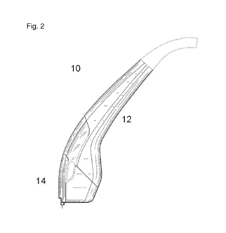
Figure 2 is an illustration showing an apparatus 10 of the invention including
main body 12 and tip
14.
Figure 3A and 3B are illustrations showing perspective and side views,
respectively, of an
apparatus of the invention including user interface 16. Figure 3C is an
illustration showing user
interaction with user interface 16.
Figure 4 shows an apparatus of the invention in which tip 14 is detachable
from main body 12 by
quick-release mechanism 18. The main body also includes user interface 16.
Figures 5A and 5B are illustrations showing two perspective views of an
apparatus of the
invention in which tip 14 is detachable from main body 12 by quick-release
mechanism 18. The arrow in
Figure 5B is for illustration purposes only.
Figure 6 shows tip 14 of an apparatus of the invention with skin penetrating
components 20. Also
shown are tips having a variable number and configuration of ablation members.
13
CA 02967636 2017-05-11
WO 2016/077759
PCT/US2015/060685
Figure 7 is an illustration showing apparatus 10 of the invention featuring
user interface 16 to
activate penetration into the tissue with skin penetrating component 20. The
inset shows the ablation
members of skin penetrating component 20 in extended and retracted
configurations.
Figure 8 is an illustration showing a system of the invention that includes
base unit 30 with user
interface 32 coupled to a handheld apparatus by cable 34. The cable carries
one or more of power,
information, and suction to and/or from the handheld apparatus.
Figures 9A and 9B are illustrations of two base units 30 of the invention
including user interfaces
32 and cables 34.
Figure 10 shows a skin positioning apparatus 40 of the invention that includes
skin tensioning
rods 42. The skin positioning apparatus may position skin for treatment of
tissue layers such as dermis
52, subcutaneous fat 54, and muscle 56, e.g., using an apparatus of the
invention.
Figure 11 shows a system of the invention that includes a tip 14 with an
integrated reservoir for
waste collection 60, a handheld main body 12 with a user interface 16, a cable
34 for carrying one or
more of power, information, and suction, and a base unit 30 that includes a
user interface 32.
Figure 12 shows a system of the invention that includes a tip 14, a handheld
main body 12 with a
user interface 16; one or more cables 34 for carrying one or more of power,
information, and suction, and
a base unit 30 that includes a user interface 32 and a reservoir for waste
collection 60.
Figure 13 shows a system of the invention that includes a tip 14, a module
including a reservoir
for waste collection 60, a handheld main body 12 with a user interface 16 and
a miniature vacuum source
70, a cable 34 for carrying one or more of power, information, and suction,
and a base unit 30 that
includes a user interface 32.
Figure 14 shows a system of the invention includes a tip 14, a module
including a reservoir for
waste collection 60, a handheld main body 12 with a user interface 16 and a
miniature vacuum source 70,
a battery unit 36, and a base unit 30 that includes a user interface 32.
Figure 15 shows possible needle tip configurations for the ablation members of
the tip of the
apparatus of the invention.
Figure 16 is a schematic depicting reduction of tissue in a treated area by
closure of ablations in
a preferred direction.
Figure 17 is a schematic depicting the architecture of a "stamping" device.
The "stamping" device
of the invention includes z-actuator 84, x-actuator 82, control electronics
38, array gripper 22, skin-
penetrating component 20, tubing 24, reservoir for waste collection 60, vacuum
source 70, and skin
positioning apparatus 40.
Figures 18A, 18B, and 18C illustrate operation of a device with a "stamping"
architecture (18A)
and automatic (18B) and manual (18C) operation of a device with a "brushing"
architecture.
Figure 19 is a schematic depicting the architecture of a "brushing" device.
The "brushing" device
of the invention includes z-actuator 84, translating mechanism 86, position
detection mechanism 88,
control electronics 38, array gripper 22, skin-penetrating component 20,
tubing 24, reservoir for waste
collection 60, vacuum source 70, and skin positioning apparatus 40.
14
CA 02967636 2017-05-11
WO 2016/077759
PCT/US2015/060685
Detailed Description
This invention relates to apparatuses, systems, kits, and methods for treating
skin (e.g.,
eliminating tissue volume, tightening skin, lifting skin, and/or reducing skin
laxity) by ablating tissue
without substantial thermal energy being imparted to the surrounding (e.g.,
non-ablated) tissue. In
particular, the invention relates to apparatuses, systems, kits, and methods
that include skin-penetrating
components with ablation members (e.g., needles, drill bits, abrading
elements, punches, blades, fluid
jets, and/or probes) capable of mechanical fractional ablation of the
epidermal, dermal, and proximal
tissue layers (e.g., fat, muscle, and SMAS (superficial muscular aponeurotic
system)).
In particular embodiments, the present invention provides one or more of the
following
advantages. First, the non-thermal fractional ablation apparatuses, systems,
kits, and methods herein
allow for skin tightening, skin lifting, and/or reduction of skin laxity
without inducing coagulation in the
surrounding tissue. In contrast, thermal ablation techniques prevent and/or
inhibit skin tightening by
allowing coagulation of the tissue and formation of rigid tissue cores that
cannot be compressed.
Second, the handheld, compact, modular, and versatile apparatuses and systems
herein facilitate ease of
use and sterilization and permit treatment of varied skin regions and
conditions with a single instrument.
For example, a tip of an apparatus having an array with a particular number
and configuration of ablation
members (e.g., needles) can be used to treat a particular skin region and/or
condition and, if desired, the
tip may be exchanged during the treatment for a different tip having a
different number and configuration
of ablation members (e.g., needles) for treatment of a different skin region
and/or condition. This
adaptability may allow for treatment of multiple skin regions and/or
conditions within a single treatment
session. Third, the apparatuses, systems, and kits include micro-sized
features, which can be beneficial
for controlling the extent of skin treatment and for ease of handling the
apparatus. Fourth, the
apparatuses, systems, kits, and methods described herein may require less
skill than that of a surgeon to
operate and/or perform. For example, treatment of patients can occur in an
outpatient setting, rather than
in an inpatient, surgical setting. Fifth, the apparatuses, systems, kits, and
methods herein constitute
minimally invasive techniques that can provide more predictable results and/or
minimize risk factors to a
greater degree than that for more invasive techniques (e.g., plastic surgery)
or non-invasive energy-
based techniques (e.g., laser, radiofrequency, and ultrasound). Sixth, the
apparatuses, systems, kits,
and methods herein can allow for rapid closing of holes or slits after
treating the skin (e.g., within a few
seconds or minutes after treating skin, such as within about ten to about
sixty seconds), thereby
minimizing the extent of bleeding and/or clotting within the holes or slits
and/or scar formation. Seventh,
the apparatuses, systems, kits, and methods herein can be useful for
maximizing the tightening effect
while minimizing healing time by optimizing tightening (e.g., by controlling
the extent of skin pleating, such
as by increasing the extent of skin pleating for some applications or skin
regions and by decreasing the
extent of skin pleating for other applications or skin regions, as described
herein). Eighth, the
apparatuses, systems, kits, and methods for tissue removal described herein
provide efficient clearance
of partially ablated tissue and debris from ablated tissue portions, thus
reducing the time for healing and
improving the skin tightening treatment. Ninth, the apparatuses, systems,
kits, and methods herein allow
visualization of results in real time during the course of the treatment. For
example, the operator can ask
the patient for feedback in real time during the treatment and can adjust the
treatment course according
CA 02967636 2017-05-11
WO 2016/077759
PCT/US2015/060685
to the patient's preference. These and other advantages are facilitated by the
handheld, compact,
versatile, easy to use, and easy to sterilize apparatuses of the invention.
In some embodiments, apparatuses, systems, kits, and methods of the invention
allow for the
treatment of skin with varied thickness. Skin regions vary in thickness
depending on the location on the
body. For example, Kakasheva-Mazenkovska et al., (Contributions, Soc. Biol.
Med. Sci., MASA, XXXII,
2, p. 119-128 (2011), incorporated by reference herein in its entirety)
describes thin skin regions for 23-53
year old adults as including the anterior lower leg (average skin thickness of
1.7 mm) and the cheeks
(average skin thickness of 2.1 mm) and thick skin regions as the anterior leg
(average skin thickness of
4.9 mm, e.g., in the anterior upper leg) and the gluteus (average skin
thickness of 5.2 mm).
In addition to variations in skin thickness, different regions of the body
present issues of
accessibility with known treatments. The versatility of the apparatuses of the
present invention, which
can be configured to treat skin of varying thicknesses at various locations on
a subject, is therefore
desirable.
Ablation Apparatus
The invention features an apparatus including a main body for handheld
operation and a tip (e.g.,
configured as a detachable cartridge) that can be attached and detached (e.g.,
by a quick release
mechanism) to the main body. The tip includes a skin-penetrating component
with one or more ablation
members (e.g., needles (e.g., hollow coring needles), drill bits, abrading
elements, punches, and/or
blades) configured for penetration into and retraction from skin that may also
be configured to be in fluid
communication with a pressure generating source (e.g., a vacuum pump, suction
source, or fluid injection
component (e.g., a high pressure fluid jet)). Such an apparatus provides many
benefits including ease of
use, ease of clean up and sterilization, disposability of components (e.g.,
the tip), rapid treatment of the
skin, lower skill level required for use, and the potential for outpatient
treatment with rapid healing times.
Main body
Figures 1A, 1B, and 2 show schematics of apparatuses 10 of the invention each
including main
body 12 and tip 14. Main body 12 is configured for handheld operation, which
facilitates ease of use.
Main body 12 may feature a contoured design to permit comfortable, ergonomic
operation. Such a
design may also permit treatment of multiple areas of a subject without
forcing the subject to move, in
contrast to other, larger medical treatment systems. Main body 12 may be
readily cleaned and sterilized
(e.g., by steam sterilization).
Main body 12 of apparatus 10 may include additional components, such as a
reservoir for
collecting waste materials (e.g., tissue, blood, and/or interstitial fluids),
a pressure generating source
(e.g., a vacuum pump, suction source, or high pressure fluid jet), tubing
and/or cables to couple various
components, device control electronics and actuation mechanisms, activation
mechanisms, a power
supply (e.g., an alternator and/or battery component), and/or a user
interface. The components of the
apparatus may be provided to an operator (e.g., a doctor or surgeon) in
sterile condition prior to use on a
patient and many, if not all, of the components can be re-sterilized or
replaced with sterile components
prior to a subsequent use. For example, tubing components may be readily
removable from the device
16
CA 02967636 2017-05-11
WO 2016/077759
PCT/US2015/060685
for sterilization or replacement after use of the apparatus.
Figures 3A and 3B are illustrations of different views of an apparatus of the
invention with user
interface 16, while Figure 3C demonstrates user interaction with user
interface 16. User interface 16 of
main body 12 may include indicators that the tip is properly coupled to main
body 12, that the device is
charged or otherwise powered (e.g., the amount of battery life remaining),
that the ablation members
(e.g., needles) are in an extended or retracted position, that a pressure
generating source is coupled to
the device, the fill level of a reservoir for collecting waste materials,
and/or other useful information. User
interface 16 may include information about the apparatus, such as the number
of ablation members of the
apparatus, the arrangement of the ablation members, the potential depth of
tissue penetration by the
ablation members, the mechanism or mode of operation, and/or other useful
information. User interface
16 may include buttons, keys, switches, toggles, spin-wheels, LED displays,
and/or touch screens that
allow the user to observe and change various parameters or configurations
during operation of the
apparatus, to activate the high or low pressure generating source, and/or to
initiate penetration into the
skin by the ablation members. User interface 16 may be configured and disposed
to allow a user to
access buttons, keys, switches, toggles, spin-wheels, LED displays, and/or
touch screens with the hand
holding the apparatus or with the free hand (Figure 3C). For example, a button
for activating the high or
low pressure generating source may be disposed on the one side of the main
body such that it is can be
depressed by one or more fingers of the user during operation. User interface
16 may also be configured
to transmit and/or receive information from another unit, such as a computer
or base unit (see Figures 8
and 9).
Main body 12 may feature additional buttons, keys, switches, toggles, spin-
wheels, and/or touch
screens to initiate penetration into the skin by the needles and/or
translation of the device across the skin.
These features may be components of user interface 16.
Main body 12 is configured to couple with a tip including a skin-penetrating
component with
ablation members (e.g., needles). Main body 12 may have a locking mechanism to
secure the tip in
place during operation. The locking mechanism may allow mechanical and/or
electrical connection of
additional components (e.g., one or more actuators that can be used to operate
the components of the
tip). In some embodiments, locking main body 12 and tip 14 may be used to
establish fluidic connection
between, e.g., the ablation members, a reservoir, and/or a pressure generating
source. The main body-
tip locking mechanism may be engaged and disengaged repeatably. The main body-
tip locking
mechanism may include one or more of adhesive, magnetic, electrical, and/or
mechanical components
(e.g., one or more gaskets, o-rings, septa, springs, clasps, and other
engagement members). In some
embodiments, the main body may include a groove or depression for placement of
an o-ring (e.g., a viton
o-ring, a nitrile rubber o-ring, and a thermoplastic polyurethane o-ring) that
will allow for a seal to form
between main body 12 and tip 14. The portion of tip 14 engineered to engage
with main body 12 may
include a corresponding groove or depression. In other embodiments, a locking
mechanism may involve
mated pieces made of molded plastic. Figures 5A and 5B show two views of main
body 12 and mated tip
14. As an example, the body of tip 14 may be formed to fit as a sheath over a
rim of main body 12, or
vice versa, such that one component may form a seal by sliding partway into
the other component. In the
instance that tip 14 fits over the edge of main body 12, the inner surface of
the housing of tip 14 may
17
CA 02967636 2017-05-11
WO 2016/077759
PCT/US2015/060685
include a ridge formed as a stop to facilitate the seal. Main body 12 and tip
14 may also include
interlocking ridges (e.g., made of plastic, rubber, or other material) to
enhance or form a seal between the
components. Main body 12 may also feature a mechanism to activate detachment
of the tip from the
main body. This mechanism may include one or more of a button, key, switch,
toggle, spin-wheel, touch
screen, and/or sliding lock. The detachment mechanism may be a quick-release
mechanism. Figure 4
shows an apparatus of the invention with quick-release mechanism 18 to
separate tip 14 from main body
12. In some embodiments, one component (e.g., main body 12) includes a
depressible portion that
engages a seal when the other component (e.g., tip 14) is slid around the rim
of the other. Depression of
the portion may be disengaged by activation of a sliding lock, eliminating the
seal between the
components to allow their separation and, e.g., removal and replacement of tip
14.
Main body 12 may also include a power supply. For example, main body 12 may
have a housing
for batteries that power operation of the device or may be configured to
receive an element including
batteries (see Figure 14). The housing may be configured to charge the
batteries (e.g., when depleted)
with a paired charging station, without requiring removal of the batteries, or
the batteries may be removed
from the device for replacement or charging. In another embodiment, main body
12 may include
electronics and components (e.g., a power cord) that allow it to be powered
from an external power
supply, such as a direct or alternating current supply or a generator.
Tip
Tip 14 (e.g., configured as a detachable cartridge; see e.g., Figures 1-5) of
apparatus 10 of the
invention includes a skin-penetrating component (e.g., one or more needles,
such as one or more hollow
coring needles) and may be detachably attached to main body 12. The
detachability of tip 14 provides an
advantage in that the component that interacts with the skin can be easily
removed from apparatus 10,
thereby minimizing the cleaning and sterilization of apparatus 10. In some
embodiments, tip 14 is
designed for a single use. For example, tip 14 may be disposable.
Alternatively, tip 14 may be cleaned
and sterilized for reuse.
The detachability of tip 14 also facilitates the design and use of tips having
varying numbers and
configurations of ablation members (e.g., coring needles) and provides for
quick interchangeability of
apparatus architectures and applications. Different tip geometries may be
useful for treatment of different
regions of the skin. For example, a small tip may be useful for treatment of a
limited surface area (e.g.,
the pen-oral area) while a large tip may be useful for treatment of a large
surface area (e.g., the
abdomen). A small tip may have a small number of ablation members (e.g., as
few as 1) that may be
arranged in a 1-dimensional array (e.g., a linear array), while a large tip
may have many ablation
members (e.g., up to or more than 100) that may be arranged in a 2-dimensional
array (e.g., a
rectangular array). Figure 6 shows several different tips 14 with skin-
penetrating components 20 of
different geometries.
Tip 14 may further include elements for coupling the ablation members
therewith. Such an
element may have magnetic, adhesive, electrical, and/or mechanical components.
For example, the
coupling element may include one or more plastic connectors configured to
couple to one or more
ablation members. The ablation members may be joined to a coupling element by
a molded plastic
18
CA 02967636 2017-05-11
WO 2016/077759
PCT/US2015/060685
connection. Tip 14 may further feature an element coupling the ablation
members fluidly to other
components of the system such as a reservoir for collecting waste materials
and/or a pressure generating
source. This element may be a tube or series of tubes. In one embodiment, a
seal formed by
mechanically mating main body 12 and tip 14 may also be the seal establishing
fluid connectivity with
other components. In other embodiments, one or more tubes coupled to skin-
penetrating component 20
of tip 14 must be mated (e.g., via one or more o-ring, gasket, KF, LF, OF,
quick coupling, Swagelok, and
other sealing mechanisms) to establish fluid connection between components of
tip 14 and other
components of the system.
Tip 14 may further couple with a detachable cover piece to cover the ablation
members when the
device is not in use in order to keep the components clean and/or sterile.
In some embodiments, tip 14 may include a reservoir for collecting waste
materials (e.g., tissue,
blood, and/or interstitial fluids) that is in fluid communication with the
ablation members. The reservoir
may further be in fluid communication with a pressure generating source (e.g.,
a vacuum pump). Tip 14
may have a filter, membrane, or other physical element that maintains
separation between materials that
enter the tip, such as collected waste materials, and other components of the
system.
Reservoir
The apparatus may include or be otherwise coupled to a reservoir for
collecting tissue, fluids
(e.g., blood and/or interstitial fluids), and other waste. The collection of
tissue and fluid allows skin
tightening, minimizes the risk of infection, and maintains a clear treatment
field for the operator of the
apparatus.
The reservoir may be in fluid communication with the ablation members of the
tip. The reservoir
may be disposed within the tip or the main body of the apparatus, or it may be
external to these
components. Alternatively, a separate module of the apparatus may contain the
reservoir. This module
may be disposed between the tip and the main body, such that the module
containing the reservoir is
coupled to both components. The coupling elements may include mechanical and
other components as
described above. The module and/or its components may be designed for a single
use; for example, the
reservoir may be disposable. Alternatively, the reservoir may be easily
removed from the system for
cleaning (e.g., sterilization) and reuse.
The reservoir may be detachably attached to the tip and/or main body of the
apparatus. The
reservoir may be readily removable from the system (e.g., for ease of
sterilization or disposability). The
reservoir may be made of materials that are chemically and/or thermally
resistant, and may feature
chemically and/or thermally resistant coatings.
Sterilizing chemicals may be stored within the reservoir during, prior to, or
after use of the
apparatus. Sterilizing chemicals may include ethylene oxide, chlorine bleach,
formaldehyde, hydrogen
peroxide, peracetic acid, or other chemicals.
The reservoir may further be in fluid communication with a pressure generating
source. For
example, the reservoir may be in fluid communication with a vacuum pump.
Transfer of ablated tissue
and other materials may be achieved by applying a differential pressure across
the circuit including the
needles and reservoir. A filter, membrane, or other physical element may
prevent suction of materials out
19
CA 02967636 2017-05-11
WO 2016/077759
PCT/US2015/060685
of the reservoir toward the vacuum pump. Such a filter, membrane, or physical
element may be disposed
within the reservoir. A filter, membrane, or physical element may also be
detachable from the reservoir,
pressure generating source, and/or their coupling elements for sterilization
and/or disposal.
Pressure Generating Source
The apparatus may further include or be otherwise coupled to a pressure
generating source. In
some embodiments, a separate tissue removal apparatus may include the pressure
generating source.
The tissue removal apparatus could additionally include a reservoir for
collecting waste materials and a
component to prevent material from the reservoir from contaminating the
pressure generating source. In
other embodiments, the pressure generating source may be configured to be in
fluid communication with
the ablation members of the tip and/or with a reservoir for collecting waste
materials. Materials may be
separated from the pressure generating source with one or more filters,
membranes, and/or other
physical elements known in the art.
The pressure generating source may be a low pressure generating source. For
example, the
pressure generating source may be capable of providing vacuum and/or suction.
Vacuum sources may
include one or more rotary pumps, momentum transfer pumps, diffusion pumps,
scroll pumps, and/or
diaphragm pumps. In some embodiments, a low pressure generating source may
include a house or
central vacuum system. In other embodiments, a suction source may include a
wall or portable suction
device. In some embodiments, a vacuum source provides an absolute pressure
less than about 6.3 kPa
(e.g., from about 0.1 kPa to about 6 kPa, such as from 0.1 kPa to 6 kPa, 0.1
kPa to 5 kPa, 0.1 kPa to 4
kPa, 0.1 kPa to 3 kPa, 0.1 kPa to 2 kPa, 0.1 kPa to 1 kPa, 0.5 kPa to 6 kPa,
0.5 kPa to 5 kPa, 0.5 kPa to
4 kPa, 0.5 kPa to 3 kPa, 0.5 kPa to 2 kPa, 0.5 kPa to 1 kPa, 1 kPa to 6 kPa, 1
kPa to 5 kPa, 1 kPa to 4
kPa, 1 kPa to 3 kPa, 1 kPa to 2 kPa, 1.5 kPa to 6 kPa, 1.5 kPa to 5 kPa, 1.5
kPa to 4 kPa, 1.5 kPa to 3
kPa, and 1.5 kPa to 2 kPa).
A low pressure generating source may be configured to remove tissue portions
and other waste
materials formed by penetration into tissue by ablation members. For example,
suction and/or vacuum
may be applied to remove waste materials from the ablation members (e.g., from
the cores of coring
needles) to prevent clogging during operation, to facilitate the separation of
ablated tissue portions from
surrounding tissue in a treatment area, and/or to remove waste materials from
the treatment area.
Suction and/or vacuum may be applied via the ablation members (e.g., needles)
of the apparatus. The
ablation members and low pressure generating source may be configured to
remove tissue portions and
other waste materials by providing suction and/or vacuum after penetration
into the skin by but before
removal of the needles. For example, a pressure generating source, such as a
vacuum pump, may be
coupled to needles that include holes as well as a reservoir for waste
collection. Following penetration
into the tissue by the ablation members, vacuum may be applied to draw waste
materials from a treated
skin area through holes in the ablation members and though tubing coupling the
ablation members to the
reservoir. A filter may prevent waste materials from leaving the reservoir and
possibly aspirating within
the pressure generating source (e.g., vacuum pump). In other embodiments, the
pressure generating
source (e.g., vacuum pump) may be activated after the ablation members (e.g.,
hollow coring needles)
have been removed from the skin to clear any waste materials from the hollows
of the ablation members
CA 02967636 2017-05-11
WO 2016/077759
PCT/US2015/060685
and prevent clogging to allow for effective continued treatment.
Alternatively, the pressure generating
source (e.g., vacuum pump) may be integrated with a separate tissue removal
apparatus. Such an
apparatus may be configured with an array of small access ports along the
bottom of a chamber which
may be applied to a skin region. The access ports that contact a treated skin
area may be configured to
form a seal with the tissue such that, upon separation of the tissue removal
apparatus from the skin
region, ablated tissue portions and other waste materials may also be removed.
In an alternative configuration, the pressure generating source (e.g., vacuum
pump) may be
configured to directly ablate and/or facilitate ablation of the skin. For
example, the ablation members
(e.g., hollow needles) may be configured to apply a high level of vacuum
(e.g., a vacuum with an absolute
pressure less than about 6.3 kPa) to the skin, thereby directing tissue
removal via either a suctioning
mechanism or through conveyance of damage to the tissue that is targeted for
removal or destruction.
The size of an ablated tissue portion may be controlled by the level of
vacuum, the duration of exposure,
and the dimensional size (e.g., area or volume) over which the vacuum is
applied. In one embodiment,
vacuum may be used to ablate tissue by causing local boiling off or
vaporization of tissue at ambient
temperatures. In another embodiment, vacuum may ablate tissue by causing
desiccation or freeze-drying
of tissue.
The pressure generating source may alternatively facilitate exposure of a
treatment area to fluid
or gas and/or injection of fluid or gas into a treatment area. For example,
the pressure generating source
may be a fluid injection component (e.g., a high pressure fluid jet or an
array of high pressure fluid jets).
In some embodiments, a fluid jet or an array of fluid jets may be configured
to ablate tissue non-thermally.
For example, a jet with fluid pressure greater than about 200 psi may be
positioned external to the skin
surface, such that interaction between the fluid jet and the skin produces a
hole in the skin. The size of
the hole may be determined by the fluid jet size and length of exposure. For
example, to provide an
ablated skin portion with a shallower depth, the fluid jet may be applied for
a shorter time. Alternatively,
to provide an ablated skin portion with a greater depth or diameter, the fluid
jet may be applied to the skin
region for a longer time. A high pressure fluid jet for tissue ablation is a
non-thermal ablative mechanism
and does not generate a thermal injury to the surrounding tissue. In other
embodiments, fluid jets may be
used to clear clogs in ablation members. Alternatively, fluid jets may be
configured to facilitate the
removal of waste materials from a treatment area (e.g., by rinsing and/or
otherwise dislodging waste
materials). In other embodiments, one or more fluid jets may be used to expose
the treatment area to
one or more chemicals (e.g., medicaments, botulinum toxin, and fillers, such
as hyaluronic acid- and
collagen-based fillers). For example, fluid jets may be used to flush a
treatment area with a collagen-
based filler following ablation of the skin by the ablation members (e.g.,
needles) of the apparatus.
Non-limiting possible pressures for a fluid injection component (e.g., a fluid
jet) include from about
200 psi to about 100000 psi (e.g., from 200 psi to 1000 psi, 200 psi to 5000
psi, 200 psi to 10000 psi, 200
psi to 50000 psi, 200 psi to 100000 psi, 500 psi to 1000 psi, 500 psi to 5000
psi, 500 psi to 10000 psi, 500
psi to 50000 psi, 500 psi to 100000 psi, 750 psi to 1000 psi, 750 psi to 5000
psi, 750 psi to 10000 psi, 750
psi to 50000 psi, 750 psi to 100000 psi, 1000 psi to 5000 psi, 1000 psi to
10000 psi, 1000 psi to 50000
psi, 1000 psi to 100000 psi, 1500 psi to 5000 psi, 1500 psi to 10000 psi, 1500
psi to 50000 psi, 1500 psi
to 100000 psi, 2000 psi to 5000 psi, 2000 psi to 10000 psi, 2000 psi to 50000
psi, 2000 psi to 100000 psi,
21
CA 02967636 2017-05-11
WO 2016/077759
PCT/US2015/060685
2500 psi to 5000 psi, 2500 psi to 10000 psi, 2500 psi to 50000 psi, 2500 psi
to 100000 psi, 4000 psi to
5000 psi, 4000 psi to 10000 psi, 4000 psi to 50000 psi, 4000 psi to 100000
psi, 5000 psi to 10000 psi,
5000 psi to 50000 psi, 5000 psi to 100000 psi, 7500 psi to 10000 psi, 7500 psi
to 50000 psi, 7500 psi to
100000 psi, 10000 psi to 50000 psi, 10000 psi to 100000 psi, 50000 psi to
100000 psi, and 75000 psi to
100000 psi) and from about 15 psi to about 200 psi (e.g., 15 psi to 20 psi, 15
psi to 50 psi, 15 psi to 75
psi, 15 psi to 100 psi, 15 psi to 125 psi, 15 psi to 150 psi, 15 psi to 175
psi, 15 psi to 200 psi, 20 psi to 50
psi, 20 psi to 75 psi, 20 psi to 100 psi, 20 psi to 125 psi, 20 psi to 150
psi, 20 psi to 175 psi, 20 psi to 200
psi, 50 psi to 75 psi, 50 psi to 100 psi, 50 psi to 125 psi, 50 psi to 150
psi, 50 psi to 175 psi, 50 psi to 200
psi, 75 psi to 100 psi, 75 psi to 125 psi, 75 psi to 150 psi, 75 psi to 175
psi, 75 psi to 200 psi, 100 psi to
125 psi, 100 psi to 150 psi, 100 psi to 175 psi, 100 psi to 200 psi, 125 psi
to 150 psi, 125 psi to 175 psi,
125 psi to 200 psi, 150 psi to 175 psi, 150 psi to 200 psi, and 175 psi to 200
psi).
In one embodiment, an apparatus containing one or more fluid jets may be
configured for
insertion into the fatty layer or under the derm is or epidermis. The array of
fluid jets may be configured to
emit fluid at very high pressure to ablate the tissue above. A low pressure
out-flow tube may be
positioned on the surface of the skin for removal of fluid and debris. In
another embodiment, a fluid jet or
an array of fluid jets may be configured for discontinuous fluid flow to allow
removal of fluid and debris
before reactivating the jet. In another embodiment, a fluid jet or an array of
fluid jets may be configured to
move (e.g., in a circular fashion) in relation to the skin, e.g., to produce
an array of cylindrical ablations.
Fluid jets of the invention may be continuous or discontinuous fluid streams,
and may feature
turbulent and/or laminar flow. One or more nozzles may be configured to form a
fluid jet. For example, a
convergent nozzle may be used which reduces the diameter of the outlet, thus
increasing the velocity of
the fluid jet. In some embodiments, the ablation members of the tip (e.g.,
hollow needles) may be
conduits for fluid streams.
Fluid jets may include one or more fluids. Non-limiting examples of fluids for
use in a fluid jet or
fluid jet array include aqueous and non aqueous solutions, such as isotonic
and non isotonic buffers, and
saline solutions. Fluid jets may include additional ingredients that have a
desirable medical or aesthetic
activity or utility (e.g., therapeutic agents, such as heparin, fibrin,
antibiotics, lidocaine, and other
analgesics, and/or botulinum toxin, and fillers, such as hyaluronic acid- and
collagen-based fillers).
Alternatively, a fluid jet may be a gas jet such as an air jet. In some
embodiments, a fluid jet or
an array of fluid jets may be configured to remove tissue, fluids, and/or
other debris generated during
ablation of the skin. For example, a pressurized air stream may be applied to
the skin following ablation
via the ablation members (e.g., needles).
A pressure generating source may include a venturi-effect element at an end of
an ablation
member (e.g., a hollow needle). The venturi-effect element may convert a high
pressure air stream into a
vacuum. This conversion would push ablated tissue and other waste materials
into a collection reservoir
after exiting the end of the ablation member.
Actuation, Translation, and Position Detection Mechanisms
The apparatus may further include actuation mechanisms to drive ablation
members (e.g.,
needles, such as hollow coring needles) into or across skin. A "z" actuator
may drive penetration into the
22
CA 02967636 2017-05-11
WO 2016/077759
PCT/US2015/060685
skin by the ablation members and/or retraction of the ablation members after
insertion. The apparatus
may include a feature or setting that has the ability to control or change the
depth of penetration of the
ablation members into the skin. For example, a scroll wheel on a user
interface of a main body may
adjust the allowed depth of penetration by the ablation members by physically
retracting the ablation
members and/or providing an electrical signal to a z-actuator. Alternatively,
digital controls on the user
interface of the base unit may control the depth and/or timing of penetration
into and retraction out of the
skin by the ablation members (e.g., needles). For example, an operator may
program a computer
component of the base unit to require a certain displacement of the ablation
members (e.g., needles) into
the skin based upon the area being treated. The z-actuator may be programmed
or otherwise set to
displace the ablation members (e.g., needles) up to about 15 mm into thick
skin (e.g., on a patient's back)
or about 2 mm into thin skin (e.g., on a patient's cheeks), for instance. The
z-actuator may also be
capable of operating at a high speed to minimize treatment time and deflection
of the skin during the
penetration of the ablation members and penetration force. The z-actuator may
further be capable of
operating with relatively high force. Preferably, a force of about 0.5 N to
about 20 N (e.g., 0.5 N to 0. 75
N, 0.5 N to 1 N, 0.5 N to 1.25 N, 0.5 N to 1.5 N, 0.5N to 2 N, 0.5 N to 5 N,
0.5N to 10 N, 0.5 N to 12 N,
0.5 N to 15 N, 0.5 N to 20 N, 0.75 N to 1 N, 0.75 N to 1.25 N, 0.75 N to 1.5
N, 0.75 N to 2 N, 0.75 N to 5
N, 0.75 N to 10 N, 0.75 N to 12 N, 0.75 N to 15 N, 0.75 N to 20 N, 1 N to 1.25
N, 1 N to 1.5 N, 1 N to 2 N,
1 N to 5 N, 1 N to 10 N, 1 N to 12 N, 1 N to 15 N, 1 N to 20 N, 1.25 N to 1.5
N, 1.25 N to 2 N, 1.25 N to 5
N, 1.25 N to 10 N, 1.25 N to 12 N, 1.25 N to 15 N, 1.25 N to 20 N, 1.5 N to 2
N, 1.5 N to 5 N, 1.5 N to 10
N, 1.5 N to 12 N, 1.5 N to 15 N, 1.5 N to 20 N, 2 N to 5 N, 2 N to 10 N, 2 N
to 12 N, 2 N to 15 N, 2 N to 20
N, 5 N to 10 N, 5 N to 12 N, 5 N to 15 N, 5 N to 20 N, 10 N to 12 N, 10 N to
15 N, 10 N to 20 N, 12 N to
15 N, 12 N to 20 N, and 15 N to 20 N) per ablation member (e.g., needle) can
be applied to ensure
insertion of the ablation member into the skin. Actuator types having these
characteristics include
pneumatic actuators, electromagnetic actuators, motors with cams, motors with
lead screws (e.g., stepper
motors), and piezoelectric actuators. The insertion force may be inversely
correlated with needle gauge.
For example, a 24 gauge needle may be operated with an insertion force of 12
N, while a 20 gauge
needle may be operated with a higher insertion force.
The apparatus may include an "x" and/or "y" actuator for driving the ablation
members (e.g.,
needles) across the skin. The x/y-actuator may be used to establish the
treatment coverage by defining
the distance between two applications of an array of ablation members. The x/y-
actuator may be
characterized by a relatively large displacement range (e.g., up to about 30
mm). The x/y-actuator may
also operate at a relatively high speed to minimize treatment time. Actuator
types having these
characteristics include pneumatic actuators, electromagnetic actuators, motors
with cams, piezoelectric
actuators, and motors with lead screws (e.g., stepper motors).
Actuation components may be disposed in the main body of the apparatus or
external to the main
body. The z-, x-, and y- actuators may be activated independently or together
by one or more buttons,
keys, toggles, switches, screws, dials, cursors, spin-wheels, or other
components. In an embodiment,
each of the z-, x-, and y- actuators can be separately controlled (e.g., using
separate activation
components, such as a button, or by using separate controls in a user
interface). Figure 7 is an
illustration of an apparatus of the invention with the ablation members (e.g.,
needles) of the skin-
23
CA 02967636 2017-05-11
WO 2016/077759
PCT/US2015/060685
penetrating component 20 in both retracted and extended positions. Z-actuation
may be activated by a
trigger button element disposed on the main body of the apparatus that is
accessible to the index finger of
the operator. Alternatively, digital controls on user interface 32 of base
unit 30 or user interface 16 of
main body 12 may control the depth and/or timing of penetration into and
retraction out of the skin by the
ablation members (e.g., needles) and/or translation of the apparatus across
the skin surface.
The apparatus may further include a translation mechanism to drive ablation
members across the
skin (e.g., x- and y- translation). A translation mechanism may include, e.g.,
driving wheels or rods. A
translation mechanism may permit automatic or manual translation of the
apparatus across the skin.
Translating components (e.g., wheels) may be disposed in or on the main body,
be detachably attachable
to the tip, or be disposed external to the main body. The translating
mechanism may be activated by an
activator, such as a button, key, toggle, switch, screw, cursor, dial, spin-
wheel, or other component,
and/or may be digitally controlled at user interface 32 of base unit 30 or
user interface 16 of main body
12.
The apparatus may also include a position detection mechanism, such as an
optical tracking
mechanism. A position detection mechanism (e.g., a camera, infrared sensor,
photodiode, and LED and
detector) may assist in tracking movement of the apparatus in relation to a
patient or a treatment area.
The optical tracking mechanism may also facilitate placement of the skin-
penetrating component on the
skin surface in the instance of manual translation of the device across the
skin. Control electronics for a
position detection mechanism may be disposed in the main body of the apparatus
or external to the main
body (e.g., in a base unit or separate computer). For example, the position
detection mechanism may
monitor the distance between the previous needle insertion and the current
device position and send a
signal to the control electronics to actuate the skin penetration mechanism
when the device has reached
the desired position (e.g., a position a defined distance from the position
where the needles were last
inserted). Desired distances and/or positions may be controlled at user
interface 32 or user interface 16.
Materials
The apparatuses, systems, kits, and methods of the invention can include any
useful materials.
For example, the main body, tip, and other components may include and/or be
formed from any
useful polymer or plastic. Such materials may include alginate, benzyl
hyaluronate,
carboxymethylcellulose, cellulose acetate, chitosan, collagen, dextran, epoxy,
gelatin, hyaluronic acid,
hydrocolloids, nylon (e.g., nylon 6 or PA6), pectin, poly (3-hydroxyl butyrate-
co- poly (3-hydroxyl valerate),
polyalkanes, polyalkene, polyalkynes, polyacrylate (PA), polyacrylonitrile
(PAN), polybenzimidazole (PBI),
polycarbonate (PC), polycaprolactone (PCL), polyester (PE), polyethylene
glycol (PEG), polyethylene
oxide (PEO), PEO/polycarbonate/polyurethane (PEO/PC/PU), poly(ethylene-co-
vinyl acetate) (PEVA),
PEVA/polylactic acid (PEVA/PLA), polyethylene, polypropylene, poly (ethylene
terephthalate) (PET),
PET/poly (ethylene naphthalate) (PET/PEN) polyglactin, polyglycolic acid
(PGA), polyglycolic
acid/polylactic acid (PGA/PLA), polyimide (PI), polylactic acid (PLA), poly-L-
lactide (PLLA),
PLLA/PC/polyvinylcarbazole (PLLA/PC/PVCB), poly (6-malic acid)-copolymers (PM
LA), polymethacrylate
(PMA), poly (methyl methacrylate) (PMMA), polystyrene (PS), polyurethane (PU),
poly (vinyl alcohol)
(PVA), polyvinylcarbazole (PVCB), polyvinyl chloride (PVC),
polyvinylidenedifluoride (PVDF),
24
CA 02967636 2017-05-11
WO 2016/077759
PCT/US2015/060685
polyvinylpyrrolidone (PVP), silicone, rayon, polytetrafluoroethylene (PTFE),
polyether ether ketone
(PEEK), or combinations thereof. Polymers and/or plastics of the invention may
be composite materials
in which additives to the polymers and/or plastics, such as ceramics or
particles, alter the mechanical
properties.
Elements of the invention (e.g., all or a portion of the apparatus, such as
all or a portion of the
main body, tip, or other components) may also include and/or be formed from
any useful metal or metal
alloy. For example, in some embodiments, the ablation members may be metallic
needles. Metals and
alloys featured in the invention include stainless steel; titanium; a nickel-
titanium (NiTi) alloy; a nickel-
titanium-niobium (NiTiNb) alloy; a nickel-iron-gallium (NiFeGa) alloy; a
nickel-manganese-gallium
(NiMnGa) alloy; a copper-aluminum-nickel (CuAlNi) allow; a copper-zinc (CuZn)
alloy; a copper-tin
(CuSn) alloy; a copper-zinc-aluminum (CuZnAl) alloy; a copper-zinc-silicon
(CuZnSi) alloy; a copper-zinc-
tin (CuZnSn) alloy; a copper-manganese alloy; a gold-cadmium (AuCd) alloy; a
silver-cadmium (AgCd)
alloy; an iron-platinum (FePt) alloy; an iron-manganese-silicon (FeMnSi)
alloy; a cobalt-nickel-aluminum
(CoNiAl) alloy; a cobalt-nickel-gallium (CoNiGa) alloy; or a titanium-
palladium (TiPd) alloy. Elements of
the invention may also include and/or be formed from glass. For example, an
apparatus of the invention
may include glass needles.
Apparatuses, systems, kits, and methods of the invention may use one or more
adhesives. An
adhesive may be located on a surface, between elements, or otherwise adhered
to an element of the
invention. Useful adhesives include a biocompatible matrix (e.g., those
including at least one of collagen
(e.g., a collagen sponge), low melting agarose (LMA), polylactic acid (PLA),
and/or hyaluronic acid (e.g.,
hyaluranon); a photosensitizer (e.g., Rose Bengal, riboflavin-5-phosphate (R-5-
P), methylene blue (MB),
N-hydroxypyridine-2-(1H)-thione (N-HTP), a porphyrin, or a chlorin, as well as
precursors thereof); a
photochemical agent (e.g., 1,8 naphthalimide); a synthetic glue (e.g., a
cyanoacrylate adhesive, a
polyethylene glycol adhesive, or a gelatin-resorcinol-formaldehyde adhesive);
a biologic sealant (e.g., a
mixture of riboflavin-5-phosphate and fibrinogen, a fibrin-based sealant, an
albumin-based sealant, or a
starch-based sealant); or a hook or loop and eye system (e.g., as used for
Velcro ). In particular
embodiments, adhesives are biodegradable.
Adhesives may be pressure-sensitive adhesives (PSAs). The properties of
pressure sensitive
adhesives are governed by three parameters: tack (initial adhesion), peel
strength (adhesion), and shear
strength (cohesion). Pressure-sensitive adhesives can be synthesized in
several ways, including solvent-
borne, water-borne, and hot-melt methods. Tack is the initial adhesion under
slight pressure and short
dwell time and depends on the adhesive's ability to wet the contact surface.
Peel strength is the force
required to remove the PSA from the contact surface. The peel adhesion depends
on many factors,
including the tack, bonding history (e.g. force, dwell time), and adhesive
composition. Shear strength is a
measure of the adhesive's resistance to continuous stress. The shear strength
is influenced by several
parameters, including internal adhesion, cross-linking, and viscoelastic
properties of the adhesive.
Permanent adhesives are generally resistant to debonding and possess very high
peel and shear
strength. Pressure-sensitive adhesives may include natural rubber, synthetic
rubber (e.g., styrene-
butadiene and styrene-ethylene copolymers), polyvinyl ether, polyurethane,
acrylic, silicones, and
ethylene-vinyl acetate copolymers. A copolymer's adhesive properties can be
altered by varying the
CA 02967636 2017-05-11
WO 2016/077759
PCT/US2015/060685
composition (via monomer components) changing the glass transition temperature
(Tg) or degree of
cross-linking. In general, a copolymer with a lower Tg is less rigid and a
copolymer with a higher Tg is
more rigid. The tack of PSAs can be altered by the addition of components to
alter the viscosity or
mechanical properties. Pressure sensitive adhesives are further described in
Czech et al., "Pressure-
Sensitive Adhesives for Medical Applications," in Wide Spectra of Quality
Control, Dr. lsin Akyar (Ed.,
published by InTech), Chapter 17 (2011), which is hereby incorporated by
reference in its entirety.
The apparatuses, systems, kits, and methods of the invention may include one
or more useful
therapeutic agents. For example, the ablation members (e.g., needles) of the
apparatus of the invention
may be configured to administer one or more therapeutic agents to the skin.
Examples of such agents
include one or more growth factors (e.g., vascular endothelial growth factor
(VEGF), platelet-derived
growth factor (PDGF), transforming growth factor beta (TGF-6), fibroblast
growth factor (FGF), epidermal
growth factor (EGF), and keratinocyte growth factor); one or more stem cells
(e.g., adipose tissue-derived
stem cells and/or bone marrow-derived mesenchymal stem cells); one or more
skin whitening agents
(e.g., hydroquinone); one or more vitamin A derivatives (e.g., tretinoin), one
or more analgesics (e.g.,
paracetamol/acetaminophen, aspirin, a non-steroidal antiinflammatory drug, as
described herein, a
cyclooxygenase-2-specific inhibitor, as described herein, dextropropoxyphene,
co-codamol, an opioid
(e.g., morphine, codeine, oxycodone, hydrocodone, dihydromorphine, pethidine,
buprenorphine,
tramadol, or methadone), fentanyl, procaine, lidocaine, tetracaine, dibucaine,
benzocaine, p-
butylaminobenzoic acid 2-(diethylamino) ethyl ester HCI, mepivacaine,
piperocaine, dyclonine, or
venlafaxine); one or more antibiotics (e.g., cephalosporin, bactitracin,
polymyxin B sulfate, neomycin,
bismuth tribromophenate, or polysporin); one or more antifungals (e.g.,
nystatin); one or more
antiinflammatory agents (e.g., a non-steroidal antiinflammatory drug (NSAID,
e.g., ibuprofen, ketoprofen,
flurbiprofen, piroxicam, indomethacin, diclofenac, sulindac, naproxen,
aspirin, ketorolac, or tacrolimus), a
cyclooxygenase-2-specific inhibitor (COX-2 inhibitor, e.g., rofecoxib
(Vioxx()), etoricoxib, and celecoxib
(Celebrex )), a glucocorticoid agent, a specific cytokine directed at T
lymphocyte function), a steroid
(e.g., a corticosteroid, such as a glucocorticoid (e.g., aldosterone,
beclometasone, betamethasone,
cortisone, deoxycorticosterone acetate, dexamethasone, fludrocortisone
acetate, hydrocortisone,
methylprednisolone, prednisone, prednisolone, or triamcinolone) or a
mineralocorticoid agent (e.g.,
aldosterone, corticosterone, or deoxycorticosterone)), or an immune selective
antiinflammatory derivative
(e.g., phenylalanine-glutamine-glycine (FEG) and its D-isomeric form (feG)));
one or more antimicrobials
(e.g., chlorhexidine gluconate, iodine (e.g., tincture of iodine, povidone-
iodine, or Lugol's iodine), or silver,
such as silver nitrate (e.g., as a 0.5% solution), silver sulfadiazine (e.g.,
as a cream), or Ag+ in one or
more useful carriers (e.g., an alginate, such as Acticoat including
nanocrystalline silver coating in high
density polyethylene, available from Smith & Nephew, London, U.K., or
Silvercel including a mixture of
alginate, carboxymethylcellulose, and silver coated nylon fibers, available
from Systagenix, Gatwick, U.K.;
a foam (e.g., Contreet Foam including a soft hydrophilic polyurethane foam
and silver, available from
Coloplast NS, Humleba3k, Denmark); a hydrocolloid (e.g., Aquacel Ag including
ionic silver and a
hydrocolloid, available from Conva Tec Inc., Skillman, NJ); or a hydrogel
(e.g., Silvasorb including ionic
silver, available from Medline Industries Inc., Mansfield, MA)); one or more
antiseptics (e.g., an alcohol,
such as ethanol (e.g., 60-90%), 1-propanol (e.g., 60-70%), as well as mixtures
of 2-propanol/isopropanol;
26
CA 02967636 2017-05-11
WO 2016/077759
PCT/US2015/060685
boric acid; calcium hypochlorite; hydrogen peroxide; manuka honey and/or
methylglyoxal; a phenol
(carbolic acid) compound, e.g., sodium 3,5-dibromo-4-hydroxybenzene sulfonate,
trichlorophenylmethyl
iodosalicyl, or triclosan; a polyhexanide compound, e.g., polyhexamethylene
biguanide (PHMB); a
quaternary ammonium compound, such as benzalkonium chloride (BAG),
benzethonium chloride (BZT),
cetyl trimethylammonium bromide (CTMB), cetylpyridinium chloride (CPC),
chlorhexidine (e.g.,
chlorhexidine gluconate), or octenidine (e.g., octenidine dihydrochloride);
sodium bicarbonate; sodium
chloride; sodium hypochlorite (e.g., optionally in combination with boric acid
in Dakin's solution); or a
triarylmethane dye (e.g., Brilliant Green)); one or more antiproliferative
agents (e.g., sirolimus, tacrolimus,
zotarolimus, biolimus, or paclitaxel); one or more emollients; one or more
hemostatic agents (e.g.,
collagen, such as microfibrillar collagen, chitosan, calcium-loaded zeolite,
cellulose, anhydrous aluminum
sulfate, silver nitrate, potassium alum, titanium oxide, fibrinogen,
epinephrine, calcium alginate, poly-N-
acetyl glucosamine, thrombin, coagulation factor(s) (e.g., II, V, VII, VIII,
IX, X, XI, XIII, or Von Willebrand
factor, as well as activated forms thereof), a procoagulant (e.g., propyl
gallate), an anti-fibrinolytic agent
(e.g., epsilon aminocaproic acid or tranexamic acid), and the like); one or
more procoagulative agents
(e.g., any hemostatic agent described herein, desmopressin, coagulation
factor(s) (e.g., II, V, VII, VIII, IX,
X, XI, XIII, or Von Willebrand factor, as well as activated forms thereof),
procoagulants (e.g., propyl
gallate), antifibrinolytics (e.g., epsilon aminocaproic acid), and the like);
one or more anticoagulative
agents (e.g., heparin or derivatives thereof, such as low molecular weight
heparin, fondaparinux, or
idraparinux; an anti-platelet agent, such as aspirin, dipyridamole,
ticlopidine, clopidogrel, or prasugrel; a
factor Xa inhibitor, such as a direct factor Xa inhibitor, e.g., apixaban or
rivaroxaban; a thrombin inhibitor,
such as a direct thrombin inhibitor, e.g., argatroban, bivalirudin,
dabigatran, hirudin, lepirudin, or
ximelagatran; or a coumarin derivative or vitamin K antagonist, such as
warfarin (coumadin),
acenocoumarol, atromentin, phenindione, or phenprocoumon); one or more immune
modulators,
including corticosteroids and non-steroidal immune modulators (e.g., NSAIDS,
such as any described
herein); one or more proteins; and/or one or more vitamins (e.g., vitamin A,
C, and/or E). One or more of
botulinum toxin, fat (e.g. autologous), hyaluronic acid, a collagen-based
filler, or other filler may also be
administered to the skin.
A therapeutic agent may include anticoagulative and/or procoagulative agents.
For instance, by
controlling the extent of bleeding and/or clotting in treated skin regions, a
skin tightening effect may be
more effectively controlled. Thus, in some embodiments, the methods and
devices herein include or can
be used to administer one or more anticoagulative agents, one or more
procoagulative agents, one or
more hemostatic agents, one or more fillers, or combinations thereof. In
particular embodiments, the
therapeutic agent controls the extent of bleeding and/or clotting in the
treated skin region, including the
use one or more anticoagulative agents (e.g., to inhibit clot formation prior
to skin healing or slit/hole
closure) and/or one or more hemostatic or procoagulative agents.
Ablation System
Any of the apparatuses of the invention described herein may be components of
a system for
non-thermal tissue ablation. In addition to the main body and tip of the
apparatus, a system for non-
thermal tissue ablation may include additional elements, such as a reservoir
for collecting waste materials
27
CA 02967636 2017-05-11
WO 2016/077759
PCT/US2015/060685
(e.g., tissue, blood, and/or interstitial fluids), a pressure generating
source (e.g., a vacuum pump),
mechanisms for actuation (e.g., pneumatic and/or electromagnetic actuators),
translation (e.g., driving
wheels), and position detection (e.g., a camera), a base unit, and a skin
positioning apparatus. Any or all
of the components may be readily sterilized prior to and/or after treatment of
a patient or, if desired,
replaced with sterile components.
Base Unit
A system for non-thermal tissue ablation may have a base unit that may
include, e.g., a user
interface, a power supply, control electronics, mechanisms to drive operation
of the apparatus, and other
components. The base unit may feature a computer, which may be programmed to
operate and/or
control any or all aspects of an apparatus of the invention.
A user interface of a base unit may include buttons, keys, switches, toggles,
spin-wheels,
screens, touch screens, keyboards, cursors, dials, indicators, displays,
and/or other components. The
user interface may be configured to indicate proper coupling of the tip and/or
reservoir module to the main
body, charged and/or powered status of the apparatus, the mode and/or position
of ablation members
(e.g., needles), coupling of a pressure generating source (e.g., a vacuum
pump) to the apparatus, the
application of low or high pressure, the fill level of a waste-collecting
reservoir, actuation of system
components, and/or other useful indicia. The user interface may be configured
to provide information
about the number and kind of ablation members of the apparatus, the treatment
area, the treatment
coverage (e.g., percentage of skin surface area ablated) the arrangement of
the ablation members, the
potential depth of penetration by the ablation members (if relevant), the
mechanism or mode of operation,
use count of the tip and/or reservoir, and other useful information. The user
interface may allow
adjustment of parameters and/or operation mode, application of high or low
pressure, and/or activation of
penetration into the skin by the ablation members. The user interface may also
be configured to transmit
and/or receive information from another unit. For example, user actions at a
user interface on the main
body of the apparatus may be reflected by a user interface of the base unit,
or vice versa.
The base unit may include buttons, keys, switches, toggles, spin-wheels,
and/or other activation
mechanisms to allow adjustment of parameters and/or operation mode,
application of high or low
pressure, penetration into the skin by the ablation members, and/or powering
on or off of the base unit,
pressure generating source, apparatus, and/or other system components. These
components may be
integrated into the user interface.
The base unit may further include electronics to control operation of the
apparatus, pressure
generating source, and/or other components of the system. For example, the
base unit may include one
or more microcontrollers, programmable logic, discrete elements, and/or other
components. The base
unit may further have one or more power supplies. Power supplies may include
batteries, alternators,
generators, and/or other components. The base unit may be configured to allow
conversion of main
power to DC for system operation, for example. In some embodiments, the base
unit has a battery
charging station for use with a battery-powered apparatus.
The base unit may include a reservoir for collecting waste materials, a
pressure generating
source, mechanisms to drive ablation members into or across the skin, a
position detection mechanism,
28
CA 02967636 2017-05-11
WO 2016/077759
PCT/US2015/060685
and other components, as provided above.
One or more cables may couple the base unit to the main body. The cable or
cables may carry
power and/or electrical signals to permit operation of the apparatus and its
components. The cable or
cables may be capable of carrying high pressure, vacuum, and/or suction.
Multiple cables may be joined
together. For example, tubing or wrapping material may be placed around
multiple cables to effectively
create a single cable linking the base unit and the apparatus. Figure 8 shows
a system of the invention
that includes a main body coupled to base unit 30 by cable 34 capable of
carrying one or more of power,
information, and suction. The base unit shown also includes user interface 32.
In addition to or in place
of coupling via a cable, the base unit and the main body may be wirelessly
coupled. The base unit may
also have a power cord that can be plugged into a wall, floor, or ceiling
power source and/or a tube for
connection to an external pressure generating source (e.g., a house or medical
suction system). Figures
9A and 9B illustrate examples of base unit configurations. For example, the
base unit of Figure 9A is a
small docking station, while the base unit of Figure 9B is a larger, portable
station.
Skin Positioning Apparatus
A non-thermal tissue ablation system may further include a skin positioning
apparatus. A skin
positioning apparatus should be configured to allow for efficient and
effective positioning of skin prior to,
during, and after ablation and/or tissue removal. Positioning the skin
provides control to the direction of
skin-tightening subsequent to treatment and ensures that ablation occurs in
the desired location and with
the desired dimensions.
An apparatus capable of gripping and/or lifting the skin provides numerous
advantages: it holds
the skin in place during the introduction of the ablation members (e.g.,
needles), minimizes deflection of
the skin when embedding more than one needle at a time ("needle-bed effect");
reduces the risk of the
user moving the apparatus during treatment, which could result in
unpredictable treatment coverage;
allows lifting of the skin (reducing the risk of ablation members damaging
underlying structures, such as
blood vessels, nerves, muscle, and bone); and allows tensioning of the skin to
permit generation of non-
circular ablations. A skin gripping and/or lifting apparatus should have high
gripping force to sustain the
insertion force of the ablation members, permit gripping of wet skin that may
be covered with blood and/or
interstitial fluids, minimize damage to the skin, and permit fast gripping and
release to minimize treatment
duration. Skin positioning mechanisms include, e.g., penetrating needle
grippers, rollers pinching the
skin, adhesives, freezing grippers, and vacuum grippers (including Coanda and
Bernouilli grippers).
Figure 10 shows skin positioning apparatus 40 of the invention that includes
tensioning rods 42.
Tensioning rods 42 are used to apply force to the skin surface by moving the
rods toward each other,
thus pinching the skin to elevate the dermis 52 and subcutaneous fat 54 away
from the underlying
structures (e.g., sub-dermal muscle layer 56, blood vessels, and nerve
fibers). Additional examples and
details of skin positioning apparatuses are provided in PCT/US14/50426,
"Methods and Apparatuses for
Skin Treatment Using Non-Thermal Tissue Ablation," which is herein
incorporated by reference in its
entirety.
29
CA 02967636 2017-05-11
WO 2016/077759
PCT/US2015/060685
Tissue Removal
A system or kit of the invention may further include components to aid in the
removal of tissue and/or
fluids, such as blood and interstitial fluids. Tissue removal components may
include a low or high
pressure generating source as described above. In addition to or instead of
these components, tissue
removal components may include adhesive materials, temperature controllers,
and/or other elements.
For example, a heating element coupled to the needles of the skin-penetrating
component may be
actuated which causes the needles to heat up to facilitate separation of
ablated tissue portions from the
skin. A vacuum source may then be applied to remove the heated ablated tissue
portions and fluids.
Additional examples and details of tissue removal components and apparatuses
are provided in
PCT/US14/50426.
Additional Components
A system of the invention may include additional components, such as a camera
and/or viewing
station. A camera may be used to image a treatment area before, during, or
after treatment. In some
embodiments, a camera may be disposed in or on the apparatus. The camera may
transmit signal to a
viewing station, such as a computer, that may be disposed in the line of sight
of the device operator. The
image or images transmitted by the camera may assist the operator in treating
the skin.
A system may further include a fluid system coupled to the ablation members to
facilitate removal
of tissue portions or to irrigate the skin portion, e.g., with saline or a
phosphate buffered solution; a heat
source (e.g., a resistive heater or current) in communication with one or more
ablation members to
promote cauterization of ablation of tissue portions; and/or an optical
element (e.g., a lens, a prism, a
reflector, etc.) to facilitate viewing of the skin.
Configurations
Systems of the invention may include a variety of components in different
configurations. For
example, systems may include a reservoir for collecting waste materials (e.g.,
tissue, blood, and/or
interstitial fluids) as well as a base unit. The reservoir may be disposed in
the base unit, in the main
body, in the tip, or in a separate module disposed between the tip and the
main body or external to the
apparatus and base unit components. Similarly, a pressure generating source
(e.g., a vacuum pump)
may be disposed external to other components or may be integrated into the
main body or the base unit.
Mechanisms for actuation, translation, and/or position detection; control
electronics; and/or user
interface(s) may be included in the main body and/or the base unit. These
configurations facilitate the
sterilization of the apparatus and/or system components as needed for patient
treatment.
Figure 11 is a schematic illustrating a possible configuration of a system
including reservoir 60,
main body 12 with user interface 16, base unit 30 with user interface 32, and
cable 34. In this system, the
reservoir is integrated into detachably attachable tip 14, which is designed
for a single use. The needles
included in tip 14 are hollow and include one or more holes that are in fluid
communication with reservoir
60. Reservoir 60 is further in fluid communication with a vacuum pump, such as
an oil-free scroll pump,
disposed in the base unit via tubing (e.g., nylon or Teflon tubing). A filter,
such as a stainless steel
sterilizing grade filter membrane (Mott Corporation), can be used to prevent
materials from exiting the
CA 02967636 2017-05-11
WO 2016/077759
PCT/US2015/060685
reservoir and aspirating in the vacuum pump. The base unit may include control
electronics, a power
supply, and a user interface that permit powering of the base unit and
apparatus; activation of actuators
disposed in the main body that cause translation of the skin-penetrating
component across the skin
and/or penetration into the skin by the needles; control of the displacement
and speed of translation
across the skin and the depth of penetration into the skin by the needles; the
application of vacuum; and
other parameters. The cable coupling the base unit and the apparatus is
capable of carrying power,
information, and vacuum, and facilitates interaction between the user
interfaces of the main body and the
base unit. Activators, such as buttons and scroll wheels on the handheld main
body, can be used to
activate the device by the operator with his or her hand(s) to allow easy and
controlled operation. The
actuators may also be digitally controlled (e.g., at a user interface). As
such, operation of the system may
be entirely or almost entirely controllable by features of the apparatus.
Treatment of a region of skin of a patient may proceed by supplying power to
the vacuum pump,
if present, and other components of the system, preparing the skin region for
treatment (e.g., sterilizing
and/or positioning the skin), placing the skin-penetrating component of the
apparatus upon the skin in the
treatment region, and activating the mechanism that drives penetration of the
ablation members (e.g.,
needles, such as hollow coring needles) of the skin-penetrating component into
the skin. The operator
may activate the vacuum source, if present, to remove waste materials (e.g.,
tissue, blood, and/or
interstitial fluids) from the treatment area and/or ablation members with an
activator, such as a button,
e.g., disposed on the main body. Alternatively, the activation of the vacuum
source, if present, may be
automatically trigged by the apparatus when the ablation members are inserted
into or retracted from the
skin. Removal of waste materials may proceed by suctioning the waste materials
into the reservoir via
holes in the ablation members (e.g., through the hollow lumen of a coring
needle). Application of vacuum
may be ceased prior to translation of the skin-penetrating component to an
adjacent skin region for further
treatment. The process may be repeated until the entire skin region of
interest has been treated, at which
point the tip can be detached from the main body via a quick-release
mechanism, the tip disposed of, and
the other components of the system sterilized as needed. Such treatment may
provide a plurality of
tissue portions with dimensions, geometries, and other characteristics
corresponding to the dimensions,
geometries, and other characteristics of the ablation members. For example,
hollow coring needles
inserted about 2 mm into the skin may provide tissue portions having a depth
or length of about 2 mm.
A system of the invention with a similar configuration might, alternatively,
integrate a miniature
vacuum pump into the main body of the apparatus. In this instance, a cable
coupling the main body and
the base unit might be used to carry power and information but not suction. A
miniature vacuum pump
may have lower power requirements than a larger vacuum pump. In another
related embodiment,
actuation and/or translation mechanisms may be disposed in or on the main body
instead of in or on the
base unit.
In another embodiment, the reservoir may be a component of the base unit. This
configuration of
the system permits collection of a larger volume of waste. Figure 12 is a
schematic of such a system that
includes main body 12 with user interface 16 that couples to base unit 30 with
user interface 32 and
reservoir 60 via one or more cables 34 (note that here, as in all other
figures, components may not be
drawn to scale). Main body 12 further couples to detachably attachable tip 14.
31
CA 02967636 2017-05-11
WO 2016/077759
PCT/US2015/060685
In this system, one or more cables capable of providing power, information,
and/or suction or
vacuum act as conduits for waste between the handheld apparatus and the base
unit. The cable or
tubing for waste extends through the main body and is configured to be in
fluid communication with the
ablation members of the skin-penetrating component of the tip, which may be
hollow needles having one
or more holes (e.g., a central, longitudinal hole along the axis of the
needle). The vacuum source may be
disposed in the base unit or external to the system; for example, the vacuum
source may be a medical or
house vacuum source. Alternatively, the vacuum source may be a pump, such as a
scroll, momentum
transfer, rotary, diffusion, or diaphragm pump disposed within the base unit.
The base unit of a system,
such as that shown in Figure 12, may further include a power supply, control
electronics, and/or
actuation, translation, and/or position detection mechanisms. Actuation,
translation, and/or position
detection mechanisms may, alternatively, be disposed within main body 12. User
interfaces 16 and 32
may interact and/or reflect changes made at the other user interface. User
action at user interface 16,
including depression or activation of buttons, key switches, toggles, touch
screens, scroll wheels, and/or
other components may be performed with the hands.
In an alternative embodiment, a module coupling to the tip and/or handheld
device may include
the reservoir. This module may be an element of the tip. A system of the
invention having reservoir 60
disposed in such a module is schematically depicted in Figure 13. In the
system shown, the module is
detachably attached to both tip 14 and main body 12 via, e.g., a quick-release
mechanism to allow for
easy sterilization and/or disposal of both the tip and the reservoir. The
reservoir may be an element or
the entirety of the module. A miniature vacuum source 70 may be disposed in
the main body of the
apparatus. The ablation members of the skin-penetrating component, the
reservoir, and the vacuum
source are all in fluid communication; a filter may be disposed in the
reservoir, module, and/or main body
to block waste materials, such as tissue, from aspirating into the vacuum
source. The base unit may
include a user interface as well as control electronics and actuation,
translation, and/or position detection
mechanisms. Cable 34 couples main body 12 and base unit 30 and carries power
and information
therebetween. In some embodiments, cable 34 is not present and the apparatus
is powered by batteries
that may be disposed in main body 12. Figure 14 schematically depicts a system
of the invention that
includes battery pack 36 that may insert into main body 12. The apparatus may
be charged by either
removing the batteries from their housing, e.g., to be charged in a battery
charging unit, or by placing the
device in a battery charging station of the system. The apparatus and/or base
unit 30 may also include
components that allow for wireless communication therebetween.
Ablation Members
The invention features a tip and/or cartridge having one or more ablation
members (e.g., needles
(e.g., hollow coring needles), drill bits, abrading elements, punches, blades,
and/or fluid jets) configured
for penetration into and retraction from skin. These ablation members may be
of varying number and
characteristics and may be arranged in various configurations.
Needles
Ablation members of the invention are preferably needles. Needles of the
invention may include
32
CA 02967636 2017-05-11
WO 2016/077759
PCT/US2015/060685
and/or be formed of a variety of materials (e.g., any described herein). For
example, the needles may be
made of molded plastic, metal, or glass. The needles may also have coatings
including chemical
coatings. Such coatings may include therapeutic agents as described above.
Needles may be of varying sizes and geometries. For example, needles may be
hollow coring
needles. Needles may be of any gauge, including gauges between 19 and 26
(e.g., 19, 20, 21, 22, 23,
24, 25, and 26 gauge). In a preferred embodiment, the needles are 24 gauge
needles. In another
preferred embodiment, the needles are 22 gauge needles. The outer and/or inner
diameter of the
needles may vary across their lengths, such that the diameter of one region of
a needle may be different
from the outer and/or inner diameter of another region of said same needle.
The change in a diameter
across the needles may or may not be continuous. The outer and/or inner
diameter of the needles at
their widest point may be between about 0.01 mm to about 2 mm (e.g., 0.01 mm
to 0.1 mm, 0.01 mm to
0.5 mm, 0.01 mm to 1 mm, 0.01 mm to 1.5 mm, 0.01 mm to 1.75 mm, 0.05 mm to 0.1
mm, 0.05 mm to
0.5 mm, 0.05 mm to 1 mm, 0.05 mm to 1.5 mm, 0.05 mm to 1.75 mm, 0.05 mm to 2
mm, 0.1 mm to 0.5
mm, 0.1 mm to 1 mm, 0.1 mm to 1.5 mm, 0.1 mm to 1.75 mm, 0.1 mm to 2 mm, 0.3
mm to 0.5 mm, 0.3
mm to 1 mm, 0.3 mm to 1.5 mm, 0.3 mm to 1.75 mm, 0.3 mm to 2 mm, 0.5 mm to 1
mm, 0.5 mm to 1.5
mm, 0.5 mm to 1.75 mm, 0.5 mm to 2 mm, 0.7 mm to 1 mm, 0.7 mm to 1.5 mm, 0.7
mm to 1.75 mm, 0.7
mm to 2 mm, 1 mm to 1.5 mm, 1 mm to 1.75 mm, 1 mm to 2 mm, 1.5 mm to 1.75 mm,
1.5 mm to 2 mm,
and 1.75 mm to 2 mm). The needles may or may not be entirely partially
cylindrical. For example, one or
more needles may be rectangular, serrated, scalloped, and/or irregular in one
or more dimension and
along some or all of their lengths. In some embodiments, the inner lumen
diameter may vary along the
length of a needle. For example, the inner diameter may be wider at the distal
end of the needle (e.g.,
away from the tip that penetrates the skin). This may facilitate the removal
of tissue from the treatment
area and/or the needles themselves and may limit the need for clearing of the
ablation member using a
pressure generating source (e.g., a vacuum source).
The needles may be configured to provide tissue portions. For example,
penetration into and/or
retraction from tissue by the needles may result in ablated tissue portions.
The dimensions, geometry,
number, and other characteristics of a tissue portion should correspond to the
dimensions, geometry,
number, and other characteristics of the skin penetrating component of the
invention (e.g., the needle or
array of needles). For example, a tissue portion created by penetration into
the skin with a cylindrical,
coring needle may have a cylindrical geometry, while a tissue portion created
by penetration into the skin
with a serrated ablation member may have a serrated or irregular geometry.
Needles of the invention may be configured to provide tissue portions having a
change in width
as a function of depth. For example, the part of an ablated tissue portion
that originates from deeper
tissue may be narrower than that part that originates from tissue closer to
the skin surface. This change
in width may be between about 100 m to about 500 m as a function of depth
(e.g., 100 m to 200 m,
100 m to 300 m, 100 m to 400 m, 100 m to 500 m, 200 m to 300 m, 200 m
to 400 m, 200 m
to 500 m, 300 m to 400 m, 300 m to 500 m, and 400 m to 500 m). The
needles may be
configured to provide ablated tissue portions having a width to depth ratio
between about 1:0.3 to about
1:75. For example, the width to depth radio of a tissue portion may be between
about 1:0.3 to about 1:1
(e.g., 1:0.3 to 1:1, 1:0.35 to 1:1, 1:0.4 to 1:1, 1:0.45 to 1:1, 1:0.5 to 1:1,
1:0.55 to 1:1, 1:0.6 to 1:1, 1:0.65
33
CA 02967636 2017-05-11
WO 2016/077759
PCT/US2015/060685
to 1:1, 1:0.7 to 1:1, 1:0.75 to 1:1, 1:0.8 to 1:1, 1:0.85 to 1:1, 1:0.9 to
1:1, 1:0.95 to 1:1, 1:0.3 to 1:0.95,
1:0.35 to 1:0.95, 1:0.4 to 1:0.95, 1:0.45 to 1:0.95, 1:0.5 to 1:0.95, 1:0.55
to 1:0.95, 1:0.6 to 1:0.95, 1:0.65
to 1:0.95, 1:0.7 to 1:0.95, 1:0.75 to 1:0.95, 1:0.8 to 1:0.95, 1:0.85 to
1:0.95, 1:0.9 to 1:0.95, 1:0.3 to 1:0.9,
1:0.35 to 1:0.9, 1:0.4 to 1:0.9, 1:0.45 to 1:0.9, 1:0.5 to 1:0.9, 1:0.55 to
1:0.9, 1:0.6 to 1:0.9, 1:0.65 to 1:0.9,
1:0.7 to 1:0.9, 1:0.75 to 1:0.9, 1:0.8 to 1:0.9, 1:0.85 to 1:0.9, 1:0.3 to
1:0.85, 1:0.35 to 1:0.85, 1:0.4 to
1:0.85, 1:0.45 to 1:0.85, 1:0.5 to 1:0.85, 1:0.55 to 1:0.85, 1:0.6 to 1:0.85,
1:0.65 to 1:0.85, 1:0.7 to 1:0.85,
1:0.75 to 1:0.85, 1:0.8 to 1:0.85, 1:0.3 to 1:0.8, 1:0.35 to 1:0.8, 1:0.4 to
1:0.8, 1:0.45 to 1:0.8, 1:0.5 to
1:0.8, 1:0.55 to 1:0.8, 1:0.6 to 1:0.8, 1:0.65 to 1:0.8, 1:0.7 to 1:0.8,
1:0.75 to 1:0.8, 1:0.3 to 1:0.75, 1:0.35
to 1:0.75, 1:0.4 to 1:0.75, 1:0.45 to 1:0.75, 1:0.5 to 1:0.75, 1:0.55 to
1:0.75, 1:0.6 to 1:0.75, 1:0.65 to
1:0.75, 1:0.7 to 1:0.75, 1:0.3 to 1:0.65, 1:0.35 to 1:0.65, 1:0.4 to 1:0.65,
1:0.45 to 1:0.65, 1:0.5 to 1:0.65,
1:0.55 to 1:0.65, 1:0.6 to 1:0.65, 1:0.3 to 1:0.65, 1:0.35 to 1:0.65, 1:0.4 to
1:0.65, 1:0.45 to 1:0.65, 1:0.5 to
1:0.65, 1:0.55 to 1:0.65, 1:0.6 to 1:0.65, 1:0.3 to 1:0.6, 1:0.35 to 1:0.6,
1:0.4 to 1:0.6, 1:0.45 to 1:0.6, 1:0.5
to 1:0.6, 1:0.55 to 1:0.6, 1:0.3 to 1:0.55, 1:0.35 to 1:0.55, 1:0.4 to 1:0.55,
1:0.45 to 1:0.55, 1:0.5 to 1:0.55,
1:0.3 to 1:0.5, 1:0.35 to 1:0.5, 1:0.4 to 1:0.5, 1:0.45 to 1:0.5, 1:0.5 to
1:0.5, 1:0.3 to 1:0.45, 1:0.35 to
1:0.45, 1:0.4 to 1:0.45, 1:0.3 to 1:0.4, 1:0.35 to 1:0.4, and 1:0.3 to
1:0.35); between about 1:1 to about
1:20 (e.g., 1:1 to 1:2, 1:1 to 1:3, 1:1 to 1:4, 1:1 to 1:5, 1:1 to 1:6, 1:1 to
1:7, 1:1 to 1:8, 1:1 to 1:9, 1:1 to
1:10, 1:1 to 1:11, 1:1 to 1:12, 1:1 to 1:13, 1:1 to 1:14, 1:1 to 1:15, 1:1 to
1:16, 1:1 to 1:17, 1:1 to 1:18, 1:1
to 1:19, 1:1 to 1:20, 1:2 to 1:3,1:2 to 1:4,1:2 to 1:5, 1:2 to 1:6, 1:2 to
1:7,1:2 to 1:8, 1:2 to 1:9, 1:2 to
1:10, 1:2 to 1:11, 1:2 to 1:12, 1:2 to 1:13, 1:2 to 1:14, 1:2 to 1:15, 1:2 to
1:16, 1:2 to 1:17, 1:2 to 1:18, 1:2
to 1:19, 1:2 to 1:20, 1:3 to 1:4, 1:3 to 1:5, 1:3 to 1:6, 1:3 to 1:7, 1:3 to
1:8, :3 to 1:9, 1:3 to 1:10, 1:3 to
1:11, 1:3 to 1:12, 1:3 to 1:13, 1:3 to 1:14, 1:3 to 1:15, 1:3 to 1:16, 1:3 to
1:17, 1:3 to 1:18, 1:3 to 1:19, 1:3
to 1:20, 1:4 to 1:5, 1:4 to 1:6, 1:4 to 1:7, 1:4 to 1:8, 1:4 to 1:9, 1:4 to
1:10, :4 to 1:11, 1:4 to 1:12, 1:4 to
1:13, 1:4 to 1:14, 1:4 to 1:15, 1:4 to 1:16, 1:4 to 1:17, 1:4 to 1:18, 1:4 to
1:19, 1:4 to 1:20, 1:5 to 1:6, 1:5 to
1:7, 1:5 to 1:8, 1:5 to 1:9, 1:5 to 1:10, 1:5 to 1:11, 1:5 to 1:12, 1:5 to
1:13, :5 to 1:14, 1:5 to 1:15, 1:5 to
1:16, 1:5 to 1:17, 1:5 to 1:18, 1:5 to 1:19, 1:5 to 1:20, 1:6 to 1:7, 1:6 to
1:8, 1:6 to 1:9, 1:6 to 1:10, 1:6 to
1:11, 1:6 to 1:12, 1:6 to 1:13, 1:6 to 1:14, 1:6 to 1:15, 1:6 to 1:16, 1:6 to
1:17, 1:6 to 1:18, 1:6 to 1:19, 1:6
to 1:20, 1:7 to 1:8, 1:7 to 1:9, 1:7 to 1:10, 1:7 to 1:11, 1:7 to 1:12, 1:7 to
1:13, 1:7 to 1:14, 1:7 to 1:15, 1:7
to 1:16, 1:7 to 1:17, 1:7 to 1:18, 1:7 to 1:19, 1:7 to 1:20, 1:8 to 1:9, 1:8
to 1:10, 1:8 to 1:11, 1:8 to 1:12,1:8
to 1:13, 1:8 to 1:14, 1:8 to 1:15, 1:8 to 1:16, 1:8 to 1:17, 1:8 to 1:18, 1:8
to 1:19, 1:8 to 1:20, 1:9 to 1:10,
1:9 to 1:11, 1:9 to 1:12, 1:9 to 1:13, 1:9 to 1:14, 1:9 to 1:15, 1:9 to 1:16,
1:9 to 1:17, 1:9 to 1:18, 1:9 to
1:19, 1:9 to 1:20, 1:10 to 1:11, 1:10 to 1:12, 1:10 to 1:13, 1:10 to 1:14,
1:10 to 1:15, 1:10 to 1:16, 1:10 to
1:17, 1:10 to 1:18, 1:10 to 1:19, 1:10 to 1:20,1:11 to 1:12,1:11 to 1:13,1:11
to 1:14,1:11 to 1:15, 1:11 to
1:16, 1:11 to 1:17,1:11 to 1:18, 1:11 to 1:19,1:11 to 1:20, 1:12 to 1:13, 1:12
to 1:14, 1:12 to 1:15, 1:12 to
1:16, 1:12 to 1:17, 1:12 to 1:18, 1:12 to 1:19, 1:12 to 1:20, 1:13 to 1:14,
1:13 to 1:15, 1:13 to 1:16, 1:13 to
1:17, 1:13 to 1:18, 1:13 to 1:19, 1:13 to 1:20, 1:14 to 1:15, 1:14 to 1:16,
1:14 to 1:17, 1:14 to 1:18, 1:14 to
1:19, 1:14 to 1:20, 1:15 to 1:16, 1:15 to 1:17, 1:15 to 1:18, 1:15 to 1:19,
1:15 to 1:20, 1:17 to 1:18, 1:17 to
1:19, and 1:17 to 1:20); between about 1:1 to about 1:75 (e.g., 1:1 to 1:2,1:1
to 1:5, 1:1 to 1:10,1:1 to
1:20, 1:1 to 1:30, 1:1 to 1:40,1:1 to 1:50, 1:1 to 1:60, 1:1 to 1:75, 1:2 to
1:5, 1:2 to 1:10, 1:2 to 1:20, 1:2 to
1:30, 1:2 to 1:40, 1:2 to 1:50, 1:2 to 1:60, 1:2 to 1:75, 1:5 to 1:10, 1:5 to
1:20, 1:5 to 1:30, 1:5 to 1:40,1:5
to 1:50, 1:5 to 1:60, 1:5 to 1:75, 1:10 to 1:20, 1:10 to 1:30, 1:10 to 1:40,
1:10 to 1:50, 1:10 to 1:60, 1:10 to
34
CA 02967636 2017-05-11
WO 2016/077759
PCT/US2015/060685
1:75, 1:20 to 1:30, 1:20 to 1:40, 1:20 to 1:50, 1:20 to 1:60, 1:20 to 1:75,
1:30 to 1:40, 1:30 to 1:50, 1:30 to
1:60, 1:30 to 1:75, 1:40 to 1:50, 1:40 to 1:60, 1:40 to 1:75, 1:50 to 1:60,
1:50 to 1:75, and 1:60 to 1:75);
between about 1:25 to about 1:75 (e.g., 1:25 to 1:75, 1:30 to 1:75, 1:35 to
1:75, 1:40 to 1:75, 1:45 to 1:75,
1:50 to 1:75, 1:55 to 1:75, 1:60 to 1:75, 1:65 to 1:75, 1:70 to 1:75, 1:25 to
1:70, 1:30 to 1:70, 1:35 to 1:70,
1:40 to 1:70, 1:45 to 1:70, 1:50 to 1:70, 1:55 to 1:70, 1:60 to 1:70, 1:65 to
1:70, 1:25 to 1:65, 1:30 to 1:65,
1:35 to 1:65, 1:40 to 1:65, 1:45 to 1:65, 1:50 to 1:65, 1:55 to 1:65, 1:60 to
1:65, 1:25 to 1:60, 1:30 to 1:60,
1:35 to 1:60, 1:40 to 1:60, 1:45 to 1:60, 1:50 to 1:60, 1:55 to 1:60, 1:25 to
1:55, 1:30 to 1:55, 1:35 to 1:55,
1:40 to 1:55, 1:45 to 1:55, 1:50 to 1:55, 1:25 to 1:50, 1:30 to 1:50, 1:35 to
1:50, 1:40 to 1:50, 1:45 to 1:50,
1:25 to 1:45, 1:30 to 1:45, 1:35 to 1:45, 1:40 to 1:45, 1:25 to 1:40, 1:30 to
1:40, 1:35 to 1:40, 1:25 to 1:35,
1:30 to 1:35, and 1:25 to 1:30); or between about 1:03 to about 1:75 (e.g.,
1:0.3 to 1:0.5, 1:0.3 to 1:1,
1:0.3 to 1:2, 1:0.3 to 1:5, 1:0.3 to 1:10, 1:0.3 to 1:20, 1:0.3 to 1:30, 1:0.3
to 1:40, 1:0.3 to 1:50, 1:0.3 to
1:60, 1:0.3 to 1:75, 1:0.5 to 1:1, 1:0.5 to 1:2, 1:0.5 to 1:5, 1:0.5 to 1:10,
1:0.5 to 1:20, 1:0.5 to 1:30, 1:0.5
to 1:40, 1:0.5 to 1:50, 1:0.5 to 1:60, and 1:0.5 to 1:75).
Needles may be of varying lengths and may have varying active lengths (i.e.,
the length of needle
configured to penetrate the skin). Active lengths may vary between about 0.1
mm to about 15 mm (e.g.,
0.1 mm to 0.2 mm, 0.1 mm to 0.5 mm, 0.1 mm to 1 mm, 0.1 mm to 2 mm, 0.1 mm to
5 mm, 0.1 mm to 10
mm, 0.1 mm to 15 mm, 0.2 mm to 0.5 mm, 0.2 mm to 1 mm, 0.2 mm to 2 mm, 0.2 mm
to 5 mm, 0.2 mm
to 10 mm, 0.2 mm to 15 mm, 0.5 mm to 1 mm, 0.5 mm to 2 mm, 0.5 mm to 5 mm, 0.5
mm to 10 mm, 0.5
mm to 15 mm, 1 mm to 2 mm, 1 mm to 5 mm, 1 mm to 10 mm, 1 mm to 15 mm, 2 mm to
5 mm, 2 mm to
10 mm, 2 mm to 15 mm, 5 mm to 10 mm, 5 mm to 15 mm, and 10 mm to 15 mm) and
may be selectable
with manual or automatic controls (e.g., a scroll wheel or an actuation
mechanism such as an
electromagnetic actuator). Needle parameters may be selected based on the area
of skin and the
condition to be treated. For example, treatment of thin, lax skin on the
cheeks may benefit from coring
needles having active lengths of about 2 mm and medium gauge (e.g., 22 gauge),
while treatment of thick
skin on the back may benefit from coring needles having lengths closer to 15
mm and thicker gauges
(e.g., 26 gauge).
The needles of the invention may or may not be hollow. Hollow needles may have
a plurality of
holes. For example, needles may have holes at either end and/or along their
lengths. The needles may
include and/or be coated with chemical or biological materials to treat skin.
In some embodiments, holes
in the needles may facilitate the injection of chemical or bioactive agents
into tissue. Such agents may be
injected at multiple depths or at specific areas along the needles or in
specific patterns. The size of the
needle holes may control the amount of chemical or bioactive agents delivered
to particular locations. In
some embodiments, chemical or bioactive agents may be used to destroy or
ablate skin tissue. Typical
chemical or bioactive agents used include trichloroacetic acid, alpha hydroxy
acids, beta hydroxy acids,
liquid nitrogen, hypoosmotic fluids, hyperosmotic fluids, and bioactive
proteins (e.g., one or more
hormones, antibodies, and/or enzymes, such as enzymes that liquefy tissue,
such as one or more
proteases, DNases, hyaluronidase, and collagenases, or combinations thereof).
Chemicals or bioactive
agents may be used to create an injury, ablated tissue portion, and/or
stimulate new tissue formation.
Chemicals or bioactive agents may also include fillers, such as collagen-based
fillers.
Needles may include one or more barbs on either their outer or inner surfaces.
The ends (tips) of
CA 02967636 2017-05-11
WO 2016/077759
PCT/US2015/060685
the needles configured to penetrate the skin may be sharpened to a fine point
or otherwise configured.
Two possible needle tip configurations are shown in Figure 15.
The needles may be coupled to other components of an apparatus, system, or kit
such as a
reservoir for collecting waste materials and/or a pressure generating source.
Coring needles may be in
fluid communication with such components to facilitate the removal of ablated
tissue, for example. The
needles may also be coupled to a substrate disposed in the tip. The substrate
may enforce the needle
array configuration and sufficiently bind the needles to prevent the needles
from becoming stuck or left
behind in the skin upon penetration. A substrate may include adhesive and/or
mechanical coupling
components and materials such as glues or plastic overmoldings. The needles
may further be electrically
and/or mechanically coupled to actuation mechanisms to drive the needles
across and into the skin
surface. A coupling mechanism may include an array gripper.
Arrays
When a tip has more than one ablation member (e.g., needle), the ablation
members may be
configured to form a one- or two-dimensional array (including linear, radial,
rectangular, and irregular
arrays). The size and geometry of an array may be selected based on the area
of skin and condition
being treated. For example, a small array may be selected for treatment of the
pen-oral area, while a
large array may be suitable for treatment of the abdomen. Arrays of the same
size may feature different
numbers and/or arrangements of ablation members (e.g., needles). For example,
one linear array may
include five needles spaced about 2 mm apart while another linear array may
include ten needles spaced
about 1 mm apart. The main body may be configured for detachable attachment to
a variety of tips
having different numbers and configurations of ablation members. Also, the tip
housing and/or structure
may be configured for inclusion of arrays of varying sizes and geometries.
The tip may have as few as 1 or as many as hundreds of ablation members (e.g.,
needles). In
some embodiments, 1-100 ablation members may be present (e.g., 1-10, 1-20, 1-
30, 1-40, 1-50, 1-60, 1-
70, 1-80, 1-90, 1-100, 3-10, 3-20, 3-30, 3-40, 3-50, 3-60, 3-70, 3-80, 3-90, 3-
100, 5-10, 5-20, 5-30, 5-40,
5-50, 5-60, 5-70, 5-80, 5-90, 5-100, 10-20, 10-40, 10-60, 10-80, 10-100, 20-
40, 20-60, 20-80, 20-100, 40-
60, 40-80, 40-100, 60-80, 60-100, or 80-100 ablation members). In preferred
embodiments, the tip may
have 3-50 ablation members (e.g., needles). The use of an array of multiple
ablation members may
facilitate skin treatment over larger areas and in less time.
The minimum distance between two ablation members (e.g., needles) in an array
may be
between about 0.1 mm to about 50 mm (e.g., from 0.1 mm to 0.2 mm, 0.1 mm to
0.5 mm, 0.1 mm to 1
mm, 0.1 mm to 2 mm, 0.1 mm to 5 mm, 0.1 mm to 10 mm, 0.1 mm to 15 mm, 0.1 mm
to 20 mm, 0.1 mm
to 30 mm, 0.1 mm to 40 mm, 0.1 mm to 50 mm, 0.2 mm to 0.5 mm, 0.2 mm to 1 mm,
0.2 mm to 2 mm,
0.2 mm to 5 mm, 0.2 mm to 10 mm, 0.2 mm to 15 mm, 0.2 mm to 20 mm, 0.2 mm to
30 mm, 0.2 mm to
mm, 0.2 mm to 50 mm, 0.5 mm to 1 mm, 0.5 mm to 2 mm, 0.5 mm to 5 mm, 0.5 mm to
10 mm, 0.5
mm to 15 mm, 0.5 mm to 20 mm, 0.5 mm to 30 mm, 0.5 mm to 40 mm, 0.5 mm to 50
mm, 1 mm to 2 mm,
1 mm to 5 mm, 1 mm to 10 mm, 1 mm to 15 mm, 1 mm to 20 mm, 1 mm to 30 mm, 1 mm
to 40 mm, 1
mm to 50 mm, 2 mm to 5 mm, 2 mm to 10 mm, 2 mm to 15 mm, 2 mm to 20 mm, 2 mm
to 30 mm, 2 mm
40 to 40 mm, 2 mm to 50 mm, 5 mm to 10 mm, 5 mm to 15 mm, 5 mm to 20 mm, 5
mm to 30 mm, 5 mm to
36
CA 02967636 2017-05-11
WO 2016/077759
PCT/US2015/060685
40 mm, 5 mm to 50 mm, 10 mm to 15 mm, 10 mm to 20 mm, 10 mm to 30 mm, 10 mm to
40 mm, 10 mm
to 50 mm, 15 mm to 20 mm, 15 mm to 30 mm, 15 mm to 40 mm, 15 mm to 50 mm, 20
mm to 30 mm, 20
mm to 40 mm, 20 mm to 50 mm, 30 mm to 40 mm, 30 mm to 50 mm, and 40 mm to 50
mm). The
minimum distance may correspond to the minimal size of an array, while the
maximum distance may
correspond to the maximum size of an array.
Arrays of different sizes and geometries may be selected based on the area of
treatment and the
skin condition being treated. Arrays may also be selected for compatibility
with actuation mechanisms
and control electronics of a given apparatus, system, or kit. Alternatively,
actuation mechanisms and
control electronics of an apparatus, system, or kit may be selected for
compatibility with a desired array
size and/or geometry. For example, a long, linear array may be used in
combination with a translating
mechanism with driving wheels, while a large, rectangular array may be used in
combination with an x-
actuator to drive the ablation members (e.g., needles) across the skin.
In any of the apparatuses, systems, kits, and methods herein, the tip may be
configured to
provide from about 10 to about 10000 ablated tissue portions per cm2 area
(e.g., 10 to 50, 10 to 100, 10
to 200, 10 to 300, 10 to 400, 10 to 500, 10 to 600, 10 to 700, 10 to 800, 10
to 900, 10 to 1000, 10 to 2000,
10 to 4000, 10 to 6000, 10 to 8000, 10 to 10000, 50 to 100, 50 to 200, 50 to
300, 50 to 400, 50 to 500, 50
to 600, 50 to 700, 50 to 800, 50 to 900, 50 to 1000, 50 to 2000, 50 to 4000,
510 to 6000, 50 to 8000, 50 to
10000, 100 to 200, 100 to 300, 100 to 400, 100 to 500, 100 to 600, 100 to 700,
100 to 800, 100 to 900,
100 to 1000, 100 to 2000, 100 to 4000, 100 to 6000, 100 to 8000, 100 to 10000,
200 to 300, 200 to 400,
200 to 500, 200 to 600, 200 to 700, 200 to 800, 200 to 900, 200 to 1000, 200
to 2000, 200 to 4000, 200 to
6000, 200 to 8000, 200 to 10000, 300 to 400, 300 to 500, 300 to 600, 300 to
700, 300 to 800, 300 to 900,
300 to 1000, 300 to 2000, 300 to 4000, 300 to 6000, 300 to 8000, 300 to 10000,
400 to 500, 400 to 600,
400 to 700, 400 to 800, 400 to 900, 400 to 1000, 400 to 2000, 400 to 4000, 400
to 6000, 400 to 8000, 400
to 10000, 500 to 600, 500 to 700, 500 to 800, 500 to 900, 500 to 1000, 500 to
2000, 500 to 4000, 500 to
6000, 500 to 8000, 500 to 10000, 600 to 700, 600 to 800, 600 to 900, 600 to
1000, 600 to 2000, 600 to
4000, 600 to 6000, 600 to 8000, 600 to 10000, 700 to 800, 700 to 900, 700 to
1000, 700 to 2000, 700 to
4000, 700 to 6000, 700 to 8000, 700 to 10000, 800 to 900, 800 to 1000, 800 to
2000, 800 to 4000, 800 to
6000, 800 to 8000, 800 to 10000, 900 to 1000, 900 to 2000, 900 to 4000, 900 to
6000, 900 to 8000, 900
to 10000, 1000 to 2000, 1000 to 4000, 1000 to 6000, 1000 to 8000, 1000 to
10000, 2000 to 4000, 2000 to
6000, 2000 to 8000, 2000 to 10000, 4000 to 6000, 4000 to 8000, 4000 to 10000,
6000 to 8000, 6000 to
10000, and 8000 to 10000 tissue portions per cm2 area) of the skin region to
which the apparatus is
applied (e.g., treatment area). The tip may be configured to remove about 5%-
70% (e.g., 5%, 10%, 15%,
20%, 25%, 30%, 35%, 40%, 45%, 50%, 55%, 60%, 65%, and 70%) of tissue within a
treatment area. In
a preferred embodiment, about 10% of tissue within a treatment area is
removed.
In any of the apparatuses, systems, kits, and methods herein, one or more
components of the
device may be selected or designed to secure the ablation member(s) (e.g., one
or more needles) and/or
prevent or minimize angular movement (e.g., wobbling) of the ablation
member(s). The needle(s) may be
secured to a substrate so as to minimize or reduce angular movement of the
needle(s) during insertion to
less than 5 degrees, e.g., less than 4, 3, or 2 degrees. An angular movement
of the needle(s) during
insertion of -1-1.5 degrees is within nominal tolerances, whereas an angular
movement of the needle(s)
37
CA 02967636 2017-05-11
WO 2016/077759
PCT/US2015/060685
during insertion of -4-5 degrees or more is to be avoided, if possible. For
example, components that join
ablation member(s) to other components (e.g., a substrate) may be designed
with low mechanical
tolerances to firmly secure the ablation member(s). This may reduce the
prevalence of or lower the risk
of destabilization and/or reduction in the structural integrity of ablation
member(s) that may result from
repeated use. For example, firmly securing the needle(s) may prevent and/or
minimize dulling, bending,
and curling of needle tip(s) that could reduce the effectiveness of the
needle(s). Firmly securing the
needle(s) may also reduce the risk of over-striking (e.g., striking a hole
produced by a needle more than
once).
Ablated tissue portions
The present invention features apparatuses, systems, kits, and methods for
generating ablated
tissue portions having various geometric dimensions. The apparatuses, systems,
kits, and methods of
the invention can be configured to produce tissue portions by producing holes
in the skin (e.g., by
penetration with ablation members, such as hollow needles). The apparatuses,
systems, kits, and
methods of the invention can further be configured to provide tissue portions
with specific dimensions,
geometries, and other characteristics. Characteristics (e.g., dimensions,
geometries, and other
characteristics) of tissue portions may reflect the characteristics of holes
formed in the skin. For example,
an apparatus may be configured to produce a hole having a change in width or
diameter as a function of
depth (e.g., by use of ablation members, such as needles, having changes in
width or diameter along
their lengths), such that a corresponding tissue portion may also have a
change in width or diameter as a
function of depth. Certain width or depth ratios of one or more ablation
members may allow for
improvement of skin tightening (e.g., forming a hole having a larger diameter
at the skin surface than at
the skin depth may facilitate hole closing via mechanical hole closure or,
alternatively, forming a hole
having a smaller diameter at the skin surface than at the skin depth may
accelerate closure of the
epidermal layer (e.g., reepithelialization) and therefore minimize the risk of
adverse events, such as
infections, and minimize healing time), skin rejuvenation (e.g., skin texture,
color, and/or architecture),
treatment of thin skin regions (e.g., lower anterior leg and cheeks), and/or
treatment of thick skin (e.g.,
anterior leg and gluteus). Using apparatuses, systems, kits, and methods with
certain width to depth
ratios may further minimize the risk of scarring while maximizing skin
tightening. Such benefits may
minimize healing time, improve treatment to abnormal skin areas (e.g.,
irregularly shaped and/or small
treatment areas), and/or increase the ability to tune hole depth and diameter
to the treatment objective. A
provided tissue portion may have a width to depth ratio of between about 1:0.3
to about 1:75 (e.g., as
described herein) and/or have a change in width as a function of depth between
about 100 pm to about
500 pm (e.g., as described herein).
In some embodiments, the ablated tissue portions provided by apparatuses,
systems, kits, and
methods of the invention may have at least one dimension between about 10 gm
and about 2 mm (e.g.,
about 10 pm to 500 pm, about 10 pm to 100 pm, 10 pm to 250 pm, 10 pm to 500
pm, 10 pm to 750 pm,
10 pm to 1 mm, 10 pm to 1.5 mm, 10 pm to 2 mm, about 50 pm to 100 pm, 50 pm to
250 pm, 50 pm to
500 pm, 50 pm to 750 pm, 50 pm to 1 mm, 50 pm to 1.5 mm, 50 pm to 2 mm, 100 pm
to 250 pm, 100 pm
to 500 pm, 100 pm to 750 pm, 100 pm to 1 mm, 100 pm to 1.5 mm, 100 pm to 2 mm,
250 pm to 500 pm,
38
CA 02967636 2017-05-11
WO 2016/077759
PCT/US2015/060685
250 pm to 750 pm, 250 pm to 1 mm, 250 pm to 1.5 mm, 250 pm to 2 mm, 500 pm to
750 pm, 500 pm to
1 mm, 500 pm to 1.5 mm, 500 pm to 2 mm, 750 pm to 1 mm, 750 pm to 1.5 mm, and
750 pm to 2 mm);
between about 0.1 mm to about 0.8 mm (e.g., 0.1 mm to 0.8 mm, 0.1 mm to 0.6
mm, 0.1 mm to 0.4 mm,
0.1 mm to 0.2 mm, 0.2 mm to 0.8 mm, 0.2 mm to 0.6 mm, 0.2 mm to 0.4 mm, 0.2 mm
to 0.3 mm, 0.3 mm
to 0.8 mm, 0.3 mm to 0.6 mm, 0.3 mm to 0.4 mm, 0.4 mm to 0.8 mm, 0.4 mm to 0.6
mm, 0.4 mm to 0.5
mm, 0.5 mm to 0.8 mm, 0.5 mm to 0.6 mm, 0.6 mm to 0.8 mm, 0.6 mm to 0.7 mm,
and 0.7 mm to 0.8
mm); between about 0.9 mm to about 20 mm (e.g., 0.9 mm to 20 mm, 0.9 mm to 17
mm, 0.9 mm to 14
mm, 0.9 mm to 11 mm, 0.9 mm to 8 mm, 0.9 mm to 5 mm, 0.9 mm to 3 mm, 3 mm to
20 mm, 3 mm to 17
mm, 3 mm to 14 mm, 3 mm to 11 mm, 3 mm to 8 mm, 3 mm to 5 mm, 5 mm to 20 mm, 5
mm to 17 mm, 5
mm to 14 mm, 5 mm to 11 mm, 5 mm to 8 mm, 8 mm to 20 mm, 8 mm to 17 mm, 8 mm
to 14 mm, 8 mm
to 11 mm, 11 mm to 20 mm, 11 mm to 17 mm, 11 mm to 14 mm, 14 mm to 20 mm, 14
mm to 17 mm, and
17 mm to 20 mm); between about 0.01 mm to 0.25 mm (e.g., 0.01 mm to 0.25 mm,
0.02 mm to 0.25 mm,
0.03 mm to 0.25 mm, 0.05 mm to 0.25 mm, 0.075 mm to 0.25 mm, 0.1 mm to 0.25
mm, 0.15 mm to 0.25
mm, 0.2 mm to 0.25 mm, 0.01 mm to 0.2 mm, 0.02 mm to 0.2 mm, 0.03 mm to 0.2
mm, 0.05 mm to 0.2
mm, 0.075 mm to 0.2 mm, 0.1 mm to 0.2 mm, 0.15 mm to 0.2 mm, 0.01 mm to 0.15
mm, 0.02 mm to 0.15
mm, 0.03 mm to 0.15 mm, 0.05 mm to 0.15 mm, 0.075 mm to 0.15 mm, 0.1 mm to
0.15 mm, 0.01 mm to
0.1 mm, 0.02 mm to 0.1 mm, 0.03 mm to 0.1 mm, 0.05 mm to 0.1 mm, 0.075 mm to
0.1 mm, 0.01 mm to
0.075 mm, 0.02 mm to 0.075 mm, 0.03 mm to 0.075 mm, 0.05 mm to 0.075 mm, 0.01
mm to 0.05 mm,
0.02 mm to 0.05 mm, 0.03 mm to 0.05 mm, 0.01 mm to 0.03 mm, 0.02 mm to 0.03
mm, 0.03 mm to 0.03
mm, 0.01 mm to 0.03 mm, 0.02 mm to 0.03 mm, and 0.01 mm to 0.02 mm); between
about 0.01 mm to
about 20 mm (e.g., 0.01 mm to 1 mm, 0.01 mm to 2 mm, 0.01 mm to 5 mm, 0.01 mm
to 10 mm, 0.01 mm
to 15 mm, 0.05 mm to 1 mm, 0.05 mm to 2 mm, 0.05 mm to 5 mm, 0.05 mm to 10 mm,
0.05 mm to 15
mm, 0.05 mm to 20 mm, 0.1 mm to 1 mm, 0.1 mm to 2 mm, 0.1 mm to 5 mm, 0.1 mm
to 10 mm, 0.1 mm
to 15 mm, 0.1 mm to 20 mm, 0.5 mm to 1 mm, 0.5 mm to 2 mm, 0.5 mm to 5 mm, 0.5
mm to 10 mm, 0.5
mm to 15 mm, 0.5 mm to 20 mm, 1 mm to 2 mm, 1 mm to 5 mm, 1 mm to 10 mm, 1 mm
to 15 mm, 1 mm
to 20 mm, 2 mm to 5 mm, 2 mm to 10 mm, 2 mm to 15 mm, 2 mm to 20 mm, 5 mm to
10 mm, 5 mm to 15
mm, and 5 mm to 20 mm); or between about 0.01 mm to about 2 mm (e.g., 0.01 mm
to 0.1 mm, 0.01 mm
to 0.5 mm, 0.01 mm to 1 mm, 0.01 mm to 1.5 mm, 0.01 mm to 1.75 mm, 0.05 mm to
0.1 mm, 0.05 mm to
0.5 mm, 0.05 mm to 1 mm, 0.05 mm to 1.5 mm, 0.05 mm to 1.75 mm, 0.05 mm to 2
mm, 0.1 mm to 0.5
mm, 0.1 mm to 1 mm, 0.1 mm to 1.5 mm, 0.1 mm to 1.75 mm, 0.1 mm to 2 mm, 0.3
mm to 0.5 mm, 0.3
mm to 1 mm, 0.3 mm to 1.5 mm, 0.3 mm to 1.75 mm, 0.3 mm to 2 mm, 0.5 mm to 1
mm, 0.5 mm to 1.5
mm, 0.5 mm to 1.75 mm, 0.5 mm to 2 mm, 0.7 mm to 1 mm, 0.7 mm to 1.5 mm, 0.7
mm to 1.75 mm, 0.7
mm to 2 mm, 1 mm to 1.5 mm, 1 mm to 1.75 mm, 1 mm to 2 mm, 1.5 mm to 1.75 mm,
1.5 mm to 2 mm,
and 1.75 mm to 2 mm). For instance, the diameter or width of a tissue portion
may be between about
0.01 mm and about 2 mm at its widest point (e.g., as described herein). For
example, penetration into
tissue by about 1 mm with a needle having a diameter of about 2 mm may produce
a tissue portion
having a depth or length of about 1 mm and a diameter of about 2 mm.
A tissue portion may have an area dimension in a range of about 0.001 mm2 to
about 2 mm2
(e.g., 0.001 mm2 to 0.005 mm2, 0.001 mm2 to 0.01 mm2, 0.001 mm2 to 0.05 mm2,
0.001 mm2 to 0.1 mm2,
0.001 mm2 to 0.5 mm2, 0.001 mm2 to 1 mm2, 0.001 mm2 to 1.5 mm2, 0.001 mm2 to 2
mm2, 0.005 mm2 to
39
CA 02967636 2017-05-11
WO 2016/077759
PCT/US2015/060685
0.01 mm2, 0.005 mm2 to 0.05 mm2, 0.005 mm2 to 0.1 mm2, 0.005 mm2 to 0.5 mm2,
0.005 mm2 to 1 mm2,
0.005 mm2 to 1.5 mm2, 0.005 mm2 to 2 mm2, 0.01 mm2 to 0.02 mm2, 0.01 mm2 to
0.05 mm2, 0.01 mm2 to
0.1 mm2, 0.01 mm2 to 0.5 mm2, 0.01 mm2 to 1 mm2, 0.01 mm2 to 1.5 mm2, 0.01 mm2
to 2 mm2, 0.05 mm2
to 0.1 mm2, 0.05 mm2 to 0.5 mm2, 0.05 mm2 to 1 mm2, 0.05 mm2 to 1.5 mm2, 0.05
mm2 to 2 mm2, 0.1
mm2 to 0.2 mm2, 0.1 mm2 to 0.5 mm2, 0.1 mm2 to 1 mm2, 0.1 mm2 to 1.5 mm2, 0.1
mm2 to 2 mm2, 0.5
mm2 to 1 mm2, 0.5 mm2 to 1.5 mm2, 0.5 mm2 to 2 mm2, 1 mm2 to 1.5 mm2, 1 mm2 to
2 mm2, and 1.5 mm2
to 2 mm2) and/or a volume between about 0.001 mm3 and about 6 mm3 (e.g., 0.001
mm3 to 0.01 mm3,
0.001 mm3 to 0.1 mm3, 0.001 mm3 to 0.5 mm3, 0.001 mm3 to 1 mm3, 0.001 mm3 to 2
mm3, 0.001 mm3 to
3 mm3, 0.001 mm3 to 4 mm3, 0.001 mm3 to 5 mm3, 0.001 mm3 to 6 mm3, 0.005 mm3
to 0.01 mm3, 0.005
mm3 to 0.1 mm3, 0.005 mm3 to 0.5 mm3, 0.005 mm3 to 1 mm3, 0.005 mm3 to 2 mm3,
0.005 mm3 to 3 mm3,
0.005 mm3 to 4 mm3, 0.005 mm3 to 5 mm3, 0.005 mm3 to 6 mm3, 0.01 mm3 to 0.1
mm3, 0.01 mm3 to 0.5
mm3, 0.01 mm3 to 1 mm3, 0.01 mm3 to 2 mm3, 0.01 mm3 to 3 mm3, 0.01 mm3 to 4
mm3, 0.01 mm3 to 5
mm3, 0.01 mm3 to 6 mm3, 0.1 mm3 to 0.5 mm3, 0.1 mm3 to 1 mm3, 0.1 mm3 to 2
mm3, 0.1 mm3 to 3 mm3,
0.1 mm3 to 4 mm3, 0.1 mm3 to 5 mm3, 0.1 mm3 to 6 mm3, 0.5 mm3 to 1 mm3, 0.5
mm3 to 2 mm3, 0.5 mm3
to 3 mm3, 0.5 mm3 to 4 mm3, 0.5 mm3 to 5 mm3, 0.5 mm3 to 6 mm3, 1 mm3 to 2
mm3, 1 mm3 to 3 mm3, 1
mm3 to 4 mm3, 1 mm3 to 5 mm3, 1 mm3 to 6 mm3, 2 mm3 to 3 mm3, 2 mm3 to 4 mm3,
2 mm3 to 5 mm3, 2
mm3 to 6 mm3, 3 mm3 to 4 mm3, 3 mm3 to 5 mm3, 3 mm3 to 6 mm3, 4 mm3 to 5 mm3,
4 mm3 to 6 mm3,
and 5 mm3 to 6 mm3).
The ablated tissue portion can have any combination of the dimensions
described herein. For
instance, in some non-limiting embodiments, the ablated tissue portion has at
least one dimension that is
less than about 2 mm and an area dimension that is less than about 2 mm2. In
other embodiments, the
ablated tissue portion has at least one dimension that is less than about 2 mm
and a volumetric
dimension that is less than about 6 mm3. In yet other embodiments, the ablated
tissue portion has at
least one dimension that is less than about 2 mm and an area dimension that is
less than about 2 mm2
and a volumetric dimension that is less than about 6 mm3. In some embodiments,
the ablated tissue
portion has an aerial dimension that is less than about 2 mm2 and a volumetric
dimension that is less than
about 6 mm3.
Ablation Kit
The invention also features kits for skin tightening and/or for treating
diseases, disorders, and
conditions that would benefit from skin restoration or tightening. Kits may
include one or more tips and or
cartridges including skin-penetrating components with one or more ablation
members (e.g., coring
needles) configured for penetration into and retraction from skin as well as a
main body of the apparatus
configured for handheld operation. As described above, tips in a kit may be
configured to be detachably
attached to the main body. The ablation members of a tip may be configured to
be in fluid communication
with a pressure generating source (e.g., a vacuum pump), such as when a tip is
attached to a main body.
Kits of the invention may include additional components, such as a reservoir
for collecting waste
materials (e.g., tissue, blood, and/or interstitial fluids); a pressure
generating source; mechanisms for
actuation, translation, and position detection (e.g., one or more pneumatic,
electromagnetic, and/or
CA 02967636 2017-05-11
WO 2016/077759
PCT/US2015/060685
piezoelectric actuators; driving wheels; and/or a camera); a base unit; and a
skin positioning apparatus
(e.g., tensioning rods). In addition, kits of the invention may include any
other useful components, such
as instructions on how to use the device(s), an air blower, a heating element
(e.g., a heat gun or heating
pad), one or more therapeutic agents (e.g., any described herein, such as an
anticoagulative and/or
procoagulative agent, and optionally in combination with a useful dispenser
for applying the therapeutic
agent, such as a brush, spray, film, ointment, cream, lotion, or gel), one or
more wound cleansers (e.g.,
including any antibiotic, antimicrobial, or antiseptic, such as those
described herein, in any useful form,
such as a brush, spray, film, ointment, cream, lotion, or gel), one or more
compression dressings, one or
more closures (e.g., bandage, hemostats, sutures, or adhesives), one or more
debriding agents, one or
more adhesives (e.g., any described herein), one or more cosmetics (e.g., as
described herein), and/or
other suitable or useful materials.
Kits of the invention may also feature one or more replacement tips (e.g., one
or more tips of a
single configuration or of different configurations). Kits may be packaged
with the tip in sterile form and
with instructions for applying the tip to the main body of an apparatus of the
invention.
Kits of the invention may include any of the components provided herein (e.g.,
tips, reservoir
containing modules, and cables) in any number. Kits may also have or be
designed to have any of the
configurations described herein.
Ablation Method and Treatment
Any of the apparatuses, systems, kits, and methods of the invention may be
used for non-thermal
tissue ablation. The apparatuses, systems, kits, and methods of the invention
can be applied to treat one
or more skin regions. In particular embodiments, these regions are treated
with one or more procedures
to improve skin appearance. Accordingly, the apparatuses, systems, kits, and
methods herein can be
useful for skin rejuvenation (e.g., removal of pigment, veins (e.g., spider
veins or reticular veins), glands
(e.g., sebaceous glands or sweat glands), hair follicles, and/or vessels in
the skin) or for treating acne,
allodynia, blemishes, ectopic dermatitis, hyperpigmentation, hyperplasia
(e.g., lentigo or keratosis), loss
of translucency, loss of elasticity, melasma (e.g., epidermal, dermal, or
mixed subtypes), photodamage,
rashes (e.g., erythematous, macular, papular, and/or bullous conditions),
psoriasis, rhytides (or wrinkles,
e.g., lateral canthal lines ("crow's feet"), age-related rhytides, sun-related
rhytides, or heredity-related
rhytides), sallow color, scar contracture (e.g., relaxation of scar tissue),
scarring (e.g., due to acne,
surgery, or other trauma), skin aging, skin contraction (e.g., excessive
tension in the skin), skin
irritation/sensitivity, skin laxity (e.g., loose or sagging skin or other skin
irregularities), striae (or stretch
marks), tattoo removal, vascular lesions (e.g., angioma, erythema, hemangioma,
papule, port wine stain,
rosacea, reticular vein, or telangiectasia), or any other unwanted skin
irregularities.
Such treatments may be applied to any part or parts of the body, including the
face (e.g., eyelid,
cheeks, chin, forehead, lips, or nose), neck, chest (e.g., as in a breast
lift), arms, hands, legs, abdomen,
and/or back. Accordingly, the apparatuses, systems, kits, and methods of the
invention can be
configured to be useful for treatment of regions of the body with different
sizes and geometries. For
example, tips having ablation member arrays of different sizes, geometries,
and arrangements may be
included in a kit of the invention to allow for treatment of both facial
(e.g., with tips having small arrays of
41
CA 02967636 2017-05-11
WO 2016/077759
PCT/US2015/060685
regular or irregular geometries) and abdominal regions (e.g., with tips having
large arrays of regular
geometries). Such arrangements and configurations can include any useful shape
(e.g., linear, curved, or
stellate), size, geometry, depth, and/or other characteristics.
Treatment methods may involve forming a plurality of ablated tissue portions
by contacting the
ablation members (e.g., needles, such as hollow coring needles) of the tip to
the skin of a subject and
removing the ablated tissue portions from the skin. Penetration into the skin
by the ablation members
may create small wounds (e.g., microwounds) and/or holes and so effectively
reduce tissue volume
and/or improve tissue quality upon healing. For example, forming a series of
ablated tissue portions (e.g.,
ablation of about 10% of the total skin area) and corresponding holes in a
high laxity skin region and
subsequent compression of the skin region to close the holes may promote the
growth of new skin (e.g.,
improved tissue). Healing of the tissue under compression allows for the
existing tissue to span the gap
introduced by the removal of an ablated tissue portion, thereby reducing the
skin volume and area (e.g.,
by tightening the skin).
Prior to contacting the skin with the ablation members, the skin may be
gripped, lifted, and/or
positioned to facilitate treatment. For example, tensioning rods may be used
to apply a compressive
force to the skin as provided in Figure 10. Such a force may be applied
throughout the treatment.
Any beneficial area or volumetric fraction of the skin region can be removed.
For example,
between about 5% to about 70% of tissue may be removed (e.g., as described
herein). In some
preferred embodiments, about 10% of the treatment area is removed.
Tissue can be removed from the treatment region with various hole density
(i.e., the number of
holes per unit area) corresponding to the number and geometry of ablation
members included in the tip or
tips used and the number of applications of the tip or tips to the treatment
region. Different hole densities
may be desirable for different regions of skin and for different conditions
and may be achieved using
different tips. For example, 15 holes corresponding to the size of a 19 gauge
needle and their
corresponding ablated tissue portions may be created in a given treatment area
by actuation of a single
19 gauge needle 15 times, or by actuating an array having five 19 gauge
needles three times. Spacing
the same number of holes further apart will result in a lower hole density per
unit area. For example, 15
holes may be created within a 0.5 mm by 0.3 mm region or within a 5 mm by 3 mm
region. In particular
embodiments, apparatuses, systems, kits, and methods of the invention (e.g.,
any described herein) are
configured to provide from about 10 to about 10000 ablated tissue portions per
cm2 area of the skin
region (e.g., as described herein). The array of holes created by ablation of
the skin may be created in
any beneficial pattern within the skin region. For example, a higher density
and/or smaller spacing of
tissue portions and corresponding holes can be ablated in the skin in the
center of a pattern or in thicker
portions of the skin. A pattern may be random or include one or more of
staggered rows and/or blocks,
parallel rows and/or blocks, a circular pattern, a spiral pattern, a square or
rectangular pattern, a
triangular pattern, a hexagonal pattern, a radial distribution, or a
combination of one or more such
patterns. The pattern may arise from the use of one or more tips with one or
more configurations and
numbers of ablation members applied in any ordered or disordered manner.
Modifications to the average
length, width, shapes, and/or other characteristics of one or more ablation
members used to treat a skin
region may also result in a specific pattern of holes in the skin. Such
patterns may be optimized to
42
CA 02967636 2017-05-11
WO 2016/077759
PCT/US2015/060685
promote unidirectional, non-directional, or multidirectional contraction or
expansion of skin (e.g., in the x-
direction, y-direction, x-direction, x-y plane, y-z plane, x-z plane, and/or
xyz-plane), such as by modifying
the average length, depth, width, density, orientation, and/or spacing between
ablations.
Any useful portion of the skin and/or underlying structures (e.g., SMAS) can
be ablated. Tissue
portions created by penetration into the skin with the ablation members of a
tip may include epidermal
tissue, dermal tissue, and/or cells or tissue proximal to the dermal/fatty
layer boundary (e.g., stem cells).
In some embodiments, a tissue portion may have a length that corresponds to a
typical total depth of the
skin layer (e.g., epidermal and dermal layers). The total depth of the
epidermal and dermal layers may
vary based on the region and age of the body being treated. In some instances,
the depth of the
epidermal layer is between about 0.01 mm to 0.2 mm, and/or the depth of the
dermal layer is between
about 0.3 mm to 6.0 mm. The total depth of the skin layer (e.g., epidermal and
dermal layers) may be
between about 0.3 mm and 6.2 mm, corresponding to a possible tissue portion
having a length between
about 0.3 mm and 6.2 mm (e.g., between about 0.3 mm and 0.6 mm, 0.3 mm and 0.9
mm, 0.3 mm and
1.5 mm, 0.3 mm and 2.0 mm, 0.3 mm and 2.5 mm, 0.3 mm and 3.0 mm, 0.3 mm and
3.5 mm, 0.3 mm
and 4.0 mm, 0.3 mm and 4.5 mm, 0.3 mm and 5.0 mm, 0.3 mm and 5.5 mm, 0.3 mm
and 6.0 mm, 0.3
mm and 6.2 mm, 0.6 mm and 0.9 mm, 0.6 mm and 1.5 mm, 0.6 mm and 2.0 mm, 0.6 mm
and 2.5 mm,
0.6 mm and 3.0 mm, 0.6 mm and 3.5 mm, 0.6 mm and 4.0 mm, 0.6 mm and 4.5 mm,
0.6 mm and 5.0
mm, 0.6 mm and 5.5 mm, 0.6 mm and 6.0 mm, 0.6 mm and 6.2 mm, 0.9 mm and 1.5
mm, 0.9 mm and
2.0 mm, 0.9 mm and 2.5 mm, 0.9 mm and 3.0 mm, 0.9 mm and 3.5 mm, 0.9 mm and
4.0 mm, 0.9 mm
and 4.5 mm, 0.9 mm and 5.0 mm, 0.9 mm and 5.5 mm, 0.9 mm and 6.0 mm, 0.9 mm
and 6.2 mm, 1.5
mm and 2.0 mm, 1.5 mm and 2.5 mm, 1.5 mm and 3.0 mm, 1.5 mm and 3.5 mm, 1.5 mm
and 4.0 mm,
1.5 mm and 4.5 mm, 1.5 mm and 5.0 mm, 1.5 mm and 5.5 mm, 1.5 mm and 6.0 mm,
1.5 mm and 6.2
mm, 2.0 mm and 2.5 mm, 2.0 mm and 3.0 mm, 2.0 mm and 3.5 mm, 2.0 mm and 4.0
mm, 2.0 mm and
4.5 mm, 2.0 mm and 5.0 mm, 2.0 mm and 5.5 mm, 2.0 and 6.0 mm, 2.0 mm and 6.2
mm, 2.5 mm and 3.0
mm, 2.5 mm and 3.5 mm, 2.5 mm and 4.0 mm, 2.5 mm and 4.5 mm, 2.5 mm and 5.0
mm, 2.5 mm and
5.5 mm, 2.5 mm and 6.0 mm, 2.5 mm and 6.2 mm, 3.0 mm and 3.5 mm, 3.0 mm and
4.0 mm, 3.0 mm
and 4.5 mm, 3.0 mm and 5.0 mm, 3.0 mm and 5.5 mm, 3.0 and 6.0 mm, 3.0 mm and
6.2 mm, 3.5 mm
and 4.0 mm, 3.5 mm and 4.5 mm, 3.5 mm and 5.0 mm, 3.5 mm and 5.5 mm, 3.5 and
6.0 mm, 3.5 mm
and 6.2 mm, 4.0 mm and 4.5 mm, 4.0 mm and 5.0 mm, 4.0 mm and 5.5 mm, 4.0 and
6.0 mm, 4.0 mm
and 6.2 mm, 4.5 mm and 5.0 mm, 4.5 mm and 5.5 mm, 4.5 and 6.0 mm, 4.5 mm and
6.2 mm, 5.0 mm
and 5.5 mm, 5.0 mm and 6.0 mm, 5.0 mm and 6.2 mm, 5.5 mm and 6.0 mm, 5.5 mm
and 6.2 mm, or 6.0
mm and 6.2 mm). In some instances, the average total depth of the skin layer
(e.g., epidermal and
dermal layers) may be about 1.5 mm, about 3 mm, or about 6 mm.
In some instances, it may be desirable to configure apparatuses, systems,
kits, and methods of
the invention to provide one or more tissue portions that do not include
significant amounts of
subcutaneous tissue, or, in other instances, to provide tissue portions that
do include significant amounts
of subcutaneous tissue. Electronic and/or physical mechanisms may be used to
control the depth of an
ablation (i.e., the penetration into the skin by the ablation members) and the
corresponding size of an
ablated tissue portion and hole. For example, an apparatus may include one or
more stop arrangements
(e.g., one or more collars and/or sleeves); one or more scroll wheels,
buttons, dials, toggles, or other
43
CA 02967636 2017-05-11
WO 2016/077759
PCT/US2015/060685
components to physically retract the skin-penetrating component; a vibrating
arrangement (e.g., a
piezoelectric element, a solenoid, a pneumatic element, or a hydraulic
element) that mechanically
couples to at least one ablation member (e.g., to promote insertion of one or
more ablation members into
the skin region, such as by providing an amplitude of vibration in the range
of about 50-500 pm or by
providing a frequency of the induced vibrations to be between about 10 Hz and
about 10 kHz); a z-
actuation mechanism (e.g., a pneumatic, electromagnetic, or piezoelectric
actuator or a motor with a
cam); and/or one or more sensors (e.g., force sensors, optical sensors, laser
fibers, photodetectors,
and/or position sensors) in communication with one or more needles, pins,
actuators, valves, pressure
generating sources, and/or user interfaces to detect the position of ablation
members and/or the position
of the apparatus relative to the treated skin portion.
Healing of Skin Regions After Removal of Ablated Tissue Portions
A compressive wound dressing may be applied after ablation to promote skin
tightening. A hole
created by penetration into the skin with the ablation members of the tip may
be closed with a suture,
staple, dressing, tunable dressing, glue, sealant, and/or other compression
retaining devices. Such
dressings may be applied in the proximity of the treatment zone or at a
distant site provided that it
conveys the appropriate mechanical force on the treatment site (e.g., by
gluing the surrounding area into
a compressed state, which then confers compression to the treated area). Wound
dressings may be
applied in a preferred direction to promote healing in a particular direction
or along particular axes (Figure
16). For example, healing may be engineered to occur along Langer lines. In
some embodiments, a
photochemical agent may be applied to the tissue and the tissue then
irradiated with visible light to
produce a seal.
Examples
Example 1: Stamping Mechanism
In one example, a system of the invention includes the apparatus, a reservoir
for collecting waste
materials (e.g., tissue, blood, and/or interstitial fluids), a low pressure
generating source, a skin gripper
and/or lifter, a base unit, and a cable coupling the apparatus and the base
unit. Figure 17 shows a
schematic representation of the components of this system. The handheld
apparatus has a separate
module disposed between the tip and the main body that includes reservoir 60
for waste collection. This
module is detachably attachable to both the tip and the main body via a quick-
release mechanism. Both
the body of the tip and the waste module are made of plastic materials and are
meant to be disposed of
after a single use (e.g., after treatment of a distinct region of the skin of
a subject) either as a single unit
or as two separated components.
Skin-penetrating component 20 of the tip includes an array of hollow,
cylindrical, metallic coring
needles that are sharpened and open at their tips. The needles are coupled to
a substrate and are
further coupled to plastic tubing 24 that establishes fluid communication
between the needles, reservoir,
and low pressure generating source 70 of the system. This low pressure
generating source is a scroll-
type oil-free vacuum pump disposed in the base unit and separated from the
reservoir and needles by a
series of filters (e.g., stainless steel 0.2 pm membrane filters) to prevent
aspiration of waste materials into
44
CA 02967636 2017-05-11
WO 2016/077759
PCT/US2015/060685
the device. A solenoid valve disposed in the main body of the apparatus allows
for separation of the
treatment site from the vacuum source without ceasing power supply to the
vacuum pump. A user
interface disposed on the main body of the apparatus permits a user to
activate actuation mechanisms to
drive needles into and across the skin as well as to activate the solenoid
valve and thus provide suction to
the treatment site. The user interface also indicates the number and
configuration of needles in the array
of the tip, the level of vacuum being supplied to the system and the powered
status of the vacuum, the
powered status of the apparatus, the mode of operation, and other useful
information. The user interface
of the main body receives signal from a user interface of the base unit via a
cable coupling the apparatus
and the base unit, and changes made in one user interface are reflected in the
other. The user interface
of the base unit includes a touch screen as well as various buttons to provide
power to the vacuum
source; actuation, translation, and position detection mechanisms of the
apparatus; and control
electronics 38. The base unit receives electrical power from a wall unit and
transmits power to
components of the system via the cable coupling the base unit and the
apparatus.
The main body includes the actuation mechanisms to drive needle action.
Electromagnetic z-
actuator 84 controls the timing and depth of needle penetration as well as the
withdrawal of the needles,
while electromagnetic x-actuator 82 controls the movement of the apparatus
across the treatment
surface. Separate buttons disposed on the main body operate the z- and x-
actuators. The actuation
mechanisms may be configured by the user interface of the base unit (e.g., the
depth of penetration into
the skin by the needles may be selected from a range of options; controlling
the penetration depth may
involve supplying electrical signals with different amplitudes to the
actuator). Array gripper 22 provides a
mechanical connection between the needle array and the actuation mechanisms.
Prior to treatment, the skin region may be sterilized, plucked, shaved,
massaged, heated, cooled,
treated with chemicals and/or bioactive agents, and/or otherwise prepared. The
region of skin is
positioned using skin positioning apparatus 40 that utilizes tensioning rods
to apply a compressive force.
System components are supplied with power, and the desired operating
parameters are selected on
either user interface (e.g., the depth of penetration by the needles). The
skin-penetrating component is
placed upon the surface of the skin and the z-actuator is activated to cause
the needles to penetrate into
the skin. Before the needles are retracted, the solenoid valve is activated
and vacuum applied to the
treatment area, removing tissue (e.g., ablated tissue portions) from within
the needle and waste materials
from the area and depositing tissue, blood, interstitial fluids, and/or any
other debris within the reservoir.
After the valve is closed, activation of the z-actuator causes the needles to
withdraw from the skin.
Subsequent activation of the x-actuator moves the apparatus to an adjacent
treatment area, where the
actions may be repeated. The amount of movement caused by activation of the x-
actuator depends on
the size of the area but may be selected to be as far as 50 mm. The tip and/or
reservoir may be replaced
at any point during the treatment, though preferably after the treatment of a
given region is complete. The
system may be configured to remove between about 5% and about 70% of tissue
from the treatment
area. For example, the system may be configured to remove about 10% of tissue
from the treatment
area. Accordingly, the system may be configured to produce a particular
arrangement, density, and
geometry of ablated tissue portions.
CA 02967636 2017-05-11
WO 2016/077759
PCT/US2015/060685
Figure 18A demonstrates this treatment method schematically. In the
illustration, adjacent areas
are treated with a system including a 2-dimensional needle array. This
treatment method may be thought
of as a "stamping" mechanism.
Example 2: Brushing Mechanism
In a second example, a system of the invention may have substantially the same
components
and configuration as a system designed to operate with a "stamping mechanism"
but may include a
translation mechanism in place of an x-actuator. Such a system is
schematically represented in Figure
19. In this system, translation mechanism 86 features driving wheels which can
be detachably coupled to
the tip. The translation mechanism may be nearly continuously activated by
means of a button disposed
on the main body such that action of the device resembles a "brushing" motion
(Figure 18B). The system
operator may alternatively select to manually translate the device across the
skin surface. Manual
translation may be particularly useful in skin regions with small or irregular
geometries such as on the
face. The system further includes a position detection mechanism (i.e., an
optical tracking mechanism) to
assist the operator in providing even treatment across a skin surface (Figure
18C). For example, the
position detection mechanism may facilitate automatic operation of the
apparatus by detecting the
distance between the previous needle insertion and the current device position
and activating penetration
into the skin by the needles when the device has reached the desired position.
The system may also
include a camera configured to transmit images to a viewing station such as a
computer of the base unit.
The camera is disposed on the main body of the apparatus and assists the
operator in selecting regions
for treatment and translating the apparatus across the region.
Example 3: Treatment of skin laxity and/or rhytides in the face
An apparatus or system of the invention may be used to administer treatment to
the skin of a
subject. Treatment may be performed outside of an operating room environment,
thereby minimizing the
cost of treatment.
The system used for treatment of the subject may be any of those described
herein. For
example, the system may be that of Example 1. For treatment of skin laxity in
the face, a tip with a
rectangular array of, e.g., 50 metallic coring needles may be selected for
application to a treatment area
of about 4 mm by about 9 mm. The selected needles may be 24 gauge needles and
may be affixed to
the tip structure by plastic molded around one end of each needle. The other
needle ends may be, e.g.,
sharpened to fine points. The minimum distance between any two needles may be
about 0.9 mm. With
this tip, about 10% of the area of skin may be ablated upon activation of the
device. The needles may be
configured to penetrate about 2 mm into the skin. Thus, with this tip, ablated
tissue portions may have
volumes of about 0.2 mm3.
As described above, the skin area may first be sterilized, treated with
chemicals, and/or otherwise
prepared for treatment. The tensioning rods of the optional skin positioning
apparatus may subsequently
be applied to the skin to position the skin and facilitate ease and
effectiveness of device operation.
Treatment may proceed with the driving of the needles into the skin by
activation of the z-actuator,
removal of waste materials by activation of suction, removal of the needles
from the skin, and translation
46
CA 02967636 2017-05-11
WO 2016/077759
PCT/US2015/060685
of the device to an adjacent region for treatment. When sufficient tissue area
has been treated, the
device components may be powered off, any residual fluids and/or debris are
removed, the skin surface
and/or holes are cleaned and/or flushed with fluid, and a compressive wound
dressing applied to the skin
to cause the holes to close in a preferred direction. The tip and the separate
reservoir module may be
disposed of, and other components of the system may be sterilized.
The treatment may be rapid (e.g., less than 30 minutes), minimizing patient
downtime and
allowing treatment to be carried out as an outpatient procedure. Within days,
a reduction in skin laxity
and/or rhytides in the treatment area may be observed. The treatment should be
more effective at
reducing skin laxity, inducing skin tightening, and/or rejuvenating skin
(i.e., improving skin architecture,
reducing wrinkles) than energy-based skin treatment methods, such as laser,
ultrasound, and radio
frequency methods, while requiring similar or reduced patient downtime and
environmental/training
requirements. In certain applications and configurations, the treatment may
also allow deeper tissue
ablation than is possible with lasers, for example, to permit the treatment of
scars and the removal of sub-
dermal tissue layers.
Other Embodiments
All publications, patent applications, and patents mentioned in this
specification are herein
incorporated by reference.
Various modifications and variations of the described apparatuses, systems,
kits, and methods of
the invention will be apparent to those skilled in the art without departing
from the scope and spirit of the
invention. Although the invention has been described in connection with
specific desired embodiments, it
should be understood that the invention as claimed should not be unduly
limited to such specific
embodiments. Indeed, various modifications of the described modes for carrying
out the invention are
intended to be within the scope of the invention.
47