Note: Descriptions are shown in the official language in which they were submitted.
CA 02967665 2017-05-11
WO 2016/118469
PCT/US2016/013835
PROCEDURE FOR THE RAPID DETERMINATION OF BACTERIAL
SUSCEPTIBILITY TO ANTIBIOTICS THAT INHIBIT PROTEIN SYNTHESIS
TECHNICAL FIELD
The present invention relates generally to the field of microbiology and the
healthcare industry and more particularly relates to a methodology for the
rapid
evaluation of bacterial susceptibility or non-susceptibility to antibiotics
that inhibit
protein synthesis.
BACKGROUND
Pathogens resistant to multiple antibiotics present a continually increasing
health
risk, particularly in clinical settings. Patients may acquire infections
through intrusive,
but necessary, medical means, such as infections in the respiratory pathway
during
mechanical ventilation, in the urinary tract or blood vessels via catheters or
even
through skin wounds, such as incisions required for any number of medical
procedures.
Immunocompromised patients and patients located in Intensive Care Units (ICUs)
are at
increased risk of acquiring nosocomial infectious diseases which may be
resistant to one
or more antibiotics. For a variety of reasons, such infections may be
associated with a
high mortality rate. Previously, the European Center for Disease Control
(ECDC)
reported 25,000 annual deaths due to multi-resistant pathogens.
Well-selected, early antibiotic treatments provide the best defense against
such
multi-resistant pathogens. Given the high prevalence of resistances, current
procedures
require a bacterial culture for identification of the microorganism followed
by an
antibiogram, which routinely requires 2-3 days of bacterial growth. The step
of
culturing bacteria to construct an antibiogram alone generally requires about
one day of
incubation, or about a minimum of 18 hours.
Given the relative long time necessary to perform standard antibiogram,
antibiotics are usually empirically provided at the onset This first line of
defense often
1
CA 02967665 2017-05-11
WO 2016/118469
PCT/US2016/013835
relies on antibiotics generally known to be effective based on the likely
pathogen
involved. However, such treatments may be ineffective in 20-40% of cases, and
a
change of antibiotics later may have a reduced probability of success. Even
educated
assumptions may contribute to antibiotic misuse or overuse resulting in
increasingly
resistant strains of bacteria while the results of an antibiogram are pending.
The antibiogram results from clinical testing of isolated bacteria strains in
vitro
for bacterial susceptibility to antibiotics. A common methodology for
constructing an
antibiogram based on diffusion is the Kirby-Bauer method (Bauer A 147, Kirby
WMM,
,Sherris JC, Turck M. Antibiotic susceptibility testing by a standardized
single disc
method. Am J Clin Pathol 1966;45:493-496). In the semi-quantitative Kirby-
Bauer
method, several discs containing different antibiotics are placed in different
zones of
nutrient rich bacteria culture. Because the antibiotic diffuses into the agar
away from
the disc, the diameter around the disc in which bacteria does not grow is
suggestive of
the minimum inhibitory concentration (MIC) of that antibiotic to the cultured
strain of
bacteria.
A quantitative method may rely on dilution in a series of broths or agar
solutions
having progressively lower concentrations of the antibiotic in question. The
lowest
concentration of antibiotic in which the bacteria cannot grow provides the
minimum
inhibitory concentration of that antibiotic to the tested strain of bacteria.
This
quantitative method may be routinely employed in the hospitals, usually using
commercial panels of antibiotics and semi-automated systems of incubation and
software for data interpretation like the MicroScan WalkAwayTm (Siemens),
PhoenixTm
(Becton Dickinson), or VitekTm 2 (bioMerieux). With such growth-dependent
automated systems, results of susceptibility or resistance to antimicrobians
from a
specific microorganism may be obtained in around 6-9 hours.
Each of the diffusion and the dilution methods rely on the principal of
inhibiting
bacterial proliferation in a nutrient rich medium and this requires sufficient
time for
many reproductive cycles of bacteria. As such, both methodologies may require
a
2
CA 02967665 2017-05-11
WO 2016/118469
PCT/US2016/013835
minimum of between 18 hours and 24 hours. It can be understood, conventional
testing
such as antibiograms fails to address the problems described above.
Additionally, a number of experimental approaches have been attempted with
the goal of achieving faster susceptibility-resistance determinations.
However, those
experimental approaches failed to supplant the conventional, time consuming
antibiogram. Accordingly, a need still exists for susceptibility testing
capable of rapidly
determining an antibiotic treatment enabling the rapid, effective
administration of
effective antibiotic treatments and reducing the misuse or overuse of
antibiotics.
SUMMARY OF INVENTION
One embodiment of the invention relates to a method of rapidly evaluating the
susceptibility of bacterial strains to a protein synthesis inhibiting
antibiotic. The method
may include the step of incubating a first portion of the strain of bacteria
with a protein
synthesis inhibiting antibiotic and adding an agent selected to induce a
bacterial
response which depends on or is influenced by protein synthesis. A second
portion of
the strain of bacteria may also be incubated with the agent selected to induce
a bacterial
response which depends on, or is influenced by, protein synthesis. The
bacterial
response in the first portion and the second portion of the strain of bacteria
may then be
evaluated and the strain of bacteria may be classified as susceptible or not
susceptible to
the protein synthesis inhibiting antibiotic based on the evaluation of the
first and second
portions of the strain of bacteria.
BRIEF DESCRIPTION OF DRAWINGS
FIGS. IA-F depict images of nucleoids from Escherichia coil obtained with a
Micromax assay in Example 1.
FIGS. 2A-F depict images of nucleoids from Eseherichia coil obtained with a
Micromax assay in Example 2.
3
CA 02967665 2017-05-11
WO 2016/118469
PCT/US2016/013835
FIGS. 3A-F depict images of nucleoids from Staphylococcus aureus obtained
with a Micromax assay in Example 3.
FIGS. 4A-F depict images of nucleoids from Pseudomonas aeruginosa obtained
with a Micromax assay in Example 5.
FIGS. 5A-F depict images of nucleoids from Enterococcus faecalis obtained
with a variant Micromax assay in Example 6.
FIGS. 6A-F depict images of Pseudomonas aeruginosa obtained with a variant
Micromax assay in Example 7.
While the present invention may be embodied with various modifications and
alternative forms, specific embodiments are illustrated in the figures and
described
herein by way of illustrative examples. It should be understood the figures
and detailed
description are not intended to limit the scope of the invention to the
particular form
disclosed, but that all modifications, alternatives, and equivalents falling
within the
spirit and scope of the claims are intended to be covered.
DETAILED DESCRIPTION OF INVENTION
Certain embodiments of the present invention allow for the rapid determination
of bacterial susceptibility or resistance to antibiotics, particularly
antibiotics which
inhibit bacterial protein synthesis. As a non-limiting example, a number of
antibiotics
that inhibit bacterial protein synthesis are described in Lambert T,
Antibiotics that affect
the ribosome. Rev Sci Tech Off Int Epiz 2012 31: 57-64. Such antibiotics may
be tested
rapidly in accordance with embodiments described herein. These antibiotics
affect
bacterial cell wall by interacting with ribosomes, the organules where
proteins are
synthesized. The bacterial ribosomes are ribonucleoprotein complexes assembled
in two
big subunits, 30S and 505.
Examples of antibiotics families which inhibit protein synthesizing include
4
CA 02967665 2017-05-11
WO 2016/118469
PCT/US2016/013835
Oxazolinidones which prevent the formation of the initiation complex.
Oxazolinidones
seem to bind to the 23S rRNA V domain of 50S ribosomal subunit and to occupy
the
Aminoacyl (A) site of the 50S ribosomal subunit, inducing a conformational
change that
prevents tRNA from entering the site and forcing tRNA to separate from the
ribosome.
Oxazolinidones which may be tested in accordance with certain embodiments of
the
present invention include: eperezolid, linezolid, posizolid, radezolid,
ranbezolid,
sutezolid, tedizolid, and others.
Additional protein synthesis inhibiting antibiotics include tetracyclines and
glycylcyclines (tigecycline), which bind to the 30S ribosomal subunit,
preventing the
entry of the aminoacyl transfer (t) RNAs to the Aminoacyl (A) site of the
ribosome
which is blocked by the antibiotic. A non-exhaustive list of tetracyclines
which may be
tested in accordance with certain embodiments of the present invention
include:
doxycycline, chlortetracycline, clomocycline, demeclocycline, lymecycline,
meclocycline, metacycline, minocycline, oxytetracycline, penimepicycline,
rolitetracycline, tetracycline and others.
Yet another family of protein synthesis inhibiting antibiotics includes
aminoglycosides such as: tobramycin, strepromycin, dihydrostreptomycin,
gentamicin,
kanamycin, amikacin, arbekacin, bekanamycin, dibekacin, neomycin, framycetin,
paromomycin, ribstamycin, netilmicin, sisomucin, isepamicin, verdamcin,
astromicin,
hygromycin B and others. Aminoglycosides affecting initiation, elongation and
termination of protein synthesis increasing the error rate with premature
termination of
the peptidyl chain and also affect ribosomal translocation. They bind to the
30S subunit,
specifically to the 16S ribosomal(r)RNA and the decoding A-site for the 4,6-
substituted
2-deoxystreptamine (2-DOS).
Still more families of protein synthesis inhibiting antibiotics include
macrolides,
lincosamides, phenicols (chloramphenicol), streptogramins, and pleuromutilins
and
quinupristin/dalfopristin which block the peptidyl transfer step of peptide
elongation on
the 50S subunit. They bind to the 23S rRNA component of the SOS ribosome close
to
5
CA 02967665 2017-05-11
WO 2016/118469
PCT/US2016/013835
the peptidyl transferase centre (i.e domain V of the 23S rRNA), blocking the
elongation
of the peptide chain causing premature termination and leading to premature
dissociation of the peptidyl-tRNA from the ribosome. Pleuromutilins bind to
the
peptidyl transferase centre, as well as phenicols. These latter bind
specifically to the
nucleotides within the central loop of domain V of the 23S rRNA. Ribosomal
proteins
L16 and the peptidyl (P)-site also participate in the binding. Orthosomycins
also inhibit
translation by binding to the 50S ribosomal subunit. Macrolides which may be
tested in
accordance with various embodiments of the present invention include:
azithromycin,
clarithromycin, dirithromycin, erythromycin, flurithromycin, josamycin,
midecamycin,
miocamycin, oleandomycin, rokitamycin, roxithromycin, spiramycin,
troleandomycin,
tylosin, ketolides, telithromycin, cethromycin, solithromycin and others.
Lincosamides
which may be tested in accordance with various embodiments of the present
invention
include: clindamycin, lincomycin, pirlimycin, and others. Streptogramins which
may be
tested in accordance with various embodiments of the present invention
include:
pristinamycin, quinupristin/dalfopristin, virginamicin, and others.
Pleuromutilins which
may be tested in accordance with various embodiments of the present invention
include:
retapamulin, tiamulin, valnemulin, and others. The amphenicol family of
antibiotics
including chloramphenicol, azidamfenicol, thiamphenicol, florfenicol and
others may
also be tested with certain embodiments of the present invention.
Additionally, Fusidic acid prohibits protein synthesis by preventing the
turnover
of elongation factor G (EF-G) in the ribosome.
Retapamulin and mupirocin may also inhibit protein synthesis, but their
precise
mechanism is unknown.
Additional antibiotics that inhibit protein synthesis envisioned for use with
certain embodiments described herein include those antibiotics which inhibit
the peptide
deformylase.
Bacterial resistant to antibiotics that inhibit bacterial protein synthesis
manifests
by a number of mechanisms Lambert T Antibiotics that affect the ribosome. Rev
Sci
6
CA 02967665 2017-05-11
WO 2016/118469
PCT/US2016/013835
kch Off Int Epiz 2012 31: 57-64.
One mechanism by which bacteria may exhibit a resistance to protein synthesis
inhibiting bacteria is by enzymatic inactivation. Detoxification enzymes,
mainly
encoded by genes from plasmids or transposons, may metabolize antibiotics like
aminoglycosides, erythromycin, lincosamides, chloramphenicol and
streptogramins,
thereby limiting their antibacterial efficacy.
Another bacterial mechanism which exhibits a resistance to protein synthesis
inhibiting bacteria is target alteration. Mutations may affect the rRNAs (e.g.
16S rRNA
or 23S rRNA) or ribosomal proteins (e.g. S12 to streptomycin, L4 and L22 to
.. macrolides) involved in antibiotic binding. Moreover, methyltransferases
may also
affect targets. For example, the 23S rRNA may be methylated in adenine 2058 by
Erm
enzymes, constitutive or inducible, leading to resistance to macrolides. They
are mostly
borne by mobile elements, representing a potential risk of dissemination.
Monomethylation results in low level resistance to erythromycin whereas
dimethylation
.. confers high resistance.
Additionally, Ribosomal protection proteins, homologues to the elongation
factors, confer resistance to tetracyclines possibly preventing protein
synthesis
inhibiting antibacterial activity.
Yet another mechanism by which bacteria may resist protein synthesis
inhibiting
antibiotics is by impaired uptake. Impermeable or energy dependent efflux
systems
reduce the intracellular concentration of the antibiotic and may produce a
moderate
resistance to aminoglycosides, and specifically to tetracyclines in gram-
negative
bacteria.
Certain embodiments described herein provide a means for rapidly determining
whether bacteria is susceptible or non-susceptible in relation to antibiotics
being tested.
The term "susceptible" should be understood to correspond to the CLSI
definition, for
example a susceptible microorganism exhibits a level of antimicrobial activity
associated with a high likelihood of therapeutic success. As used herein, the
term "non-
7
CA 02967665 2017-05-11
WO 2016/118469
PCT/US2016/013835
susceptible" refers to those microorganisms which are not determined to be
susceptible.
In practice, this definition encompasses the CLSI indications of both
resistant and
intermediate microorganisms.
Whereas, some previous rapid susceptibility detection methods relied on the
rapid growth in the individual cell size of small numbers of bacteria, certain
protein
synthesis inhibiting antibiotics may prevent this growth which may present a
false
positive for resistant bacteria under certain conditions.
In accordance with certain embodiments of the present invention, a bacterial
response is induced which depends on or is influenced by protein synthesis.
Bacterial
resistance to protein synthesis inhibiting antibiotics can be evaluated
through the
antibiotics ability to supress the bacterial response. For the purposes of
this disclosure a
"bacterial response" should be understood to be a chemical, biological,
genetic, or
physical change to bacteria at the DNA level, a cellular component level, or
at the
cellular level. In some embodiments, the bacterial response is directly, or
indirectly
discernible, such as through the use of assays, microfluidic devices, or by
other
measurement and/or testing protocols known to those of skill in the art.
Examples of bacterial responses at the DNA level include DNA damage and
DNA fragmentation. In particular, DNA damage or fragmentation which is at
least
partially dependent on protein synthesis may indicate of the effectiveness of
protein
synthesis inhibiting antibiotics. As non-limiting examples, this DNA
fragmentation,
which is at least partially dependant on protein synthesis may be induced in
some
bacteria by exposure to quinolones (e.g. norofloxin, ciproflaxin,
moxifloxacin, and
others). Since all quinolones produce a similar response in gram negative
bacteria,
namely DNA fragmentation, quinolones are expected to work well as an agent for
inducing a bacterial response. While this is the case for at least most gram
negative
bacteria, resistance to quinolones is possible, and in such strains a
susceptibility
determination can't be made with respect to the protein synthesis inhibiting
antibiotic.
8
A bacterial response in the form of DNA fragmentation may be induced with
mitomycin
C. Mitomycin C presents a robust agent for inducing bacterial response,
because at
present, there does not appear to be any natural bacterial resistances to
mitomycin C.
Mitomycin C appears to produce protein synthesis dependant responses in at
least E.
coi, K. pneumonia, P aeruginosa, and A. baumannii and is expected to produce a
bacterial response in most gram negative bacteria.
In other embodiments, DNA fragmentation which depends on protein synthesis
may be indirectly produced, such as by lysostaphin which partially digests
bacterial cell
walls after a short exposure causing the release significant quantities of
deoxyribonuclease (DNase). DNase is an enzyme which, when released from the
bacterial cell wall results in DNA fragmentation in a manner which depends on
protein
synthesis. As another example, DNA fragmentation can be induced by incubating
bacteria with surfactants and/or enzymes that effect bacteria cell walls in a
manner that
depends on protein synthesis and which promote autolysis. Autolysis in certain
bacteria
may be induced by incubation with bile, deoxycholate, Triton X-100TM, as well
as
peptidoglycan digesting enzyme lysozyme and antibiotic inhibitors of
peptidoglycan
synthesis.
In certain further embodiments, the bacterial response of DNA damage may be
induced by alkykating agents. A non-exhaustive list of alkykating agents may
include:
nitrogen mustards, such as cyclophosphamide, mechlorethamide, uramustine,
melphalan, chlorambucil, ifosfamide,
bendamustine; diepoxybutane;
carzinophilin/azinomycin B; sandramycin, luzopeptins, and isochrysohermidin;
biselezin, pyrrolobenzodiazepine dimers; dinuclear cis-DDP analogues;
psoralens;
cyclophosphamide, pyrrolizide alkaloids; and others. Perhaps even "alkylating-
like"
agents, such as platinums, or platinum analogues, may be employed to induce a
bacterial response. Alkylating-like agents may include cisplatin, carboplatin,
nedaplatin, oxalipatin, satraplatin, or triplatin tetranitrate.
Examples of bacterial responses at the cellular component level may include
cell
wall damage, which may be caused by agents that inhibit peptidoglycan
synthesis or
9
Date Recue/Date Received 2021-01-13
CA 02967665 2017-05-11
WO 2016/118469
PCT/US2016/013835
even cause peptidoglycan digestion. The beta-lactams family of antibiotics may
be
employed for inducing cell wall damage in a manner which depends on protein
synthesis. As non-limiting examples, beta-lactams contemplated for possible
use
include penicillins (penems), cephalosporins (cephems), carbapenems. Beta-
lactams
have demonstrated effectiveness in inducing bacterial response in a number of
bacterial
species, gram negative strains of bacteria being the most widely tested.
However, beta-
lactams can only be effective as an agent for inducing a bacterial response in
strains of
bacteria which are not themselves resistant to beta-lactams.
Additional families of antibiotics to induce this bacterial response may
include
cycloserine, fosfomycin, bacitracin, and glycopeptides. Cell wall
lysis, or
peptidoglycan digestion may be induced with lysozyme. Because most bacteria
have
peptidoglycan, Lysozyme is expected to provide a useful agent for inducing a
bacterial
response in both gram negative and gram positive bacteria.
Examples of bacterial responses at the cellular level includes changes in cell
appearance, such as cell size, or cell enlargement. In particular, various
antibiotics and
DNA damaging or toxic agents may induce cell enlargement in a manner which
depends
on, or is influenced by, protein synthesis.
It may be appreciated that bacterial responses can occur on multiple levels
simultaneously, sequentially, or in overlapping intervals. Additionally,
certain agents
described may be capable of inducing bacterial responses on multiple levels.
In the
examples which follow, the bacterial response may be described in terms of the
bacterial
response which is monitored by assays or other means. In some instances, the
concentration of the agent employed can affect the type of bacterial response
which is
induced.
In certain embodiments, a bacterial response is induced in bacteria, such as
in a
strain of bacteria or in a sample of bacteria. As a non-limiting example, a
bacteria
sample may be generated in a clinical setting by known culturing methods for
isolating
and identifying bacteria. The agent inducing this bacterial response may be
induced in
CA 02967665 2017-05-11
WO 2016/118469
PCT/US2016/013835
two separated portions of the bacteria, such as a first portion and a second
portion. The
portions may be separated spatially, such as within the same petri dish or
other
incubation container, or they may be physically separated, such as in
separated petri
dishes or incubation containers. Regardless of the manner in which the first
portion and
the second portion are separated, it may be appreciated that the designation
of a "first"
and a -second" portion may be considered arbitrary with respect to location or
spacing,
except to the extent the portions are separated sufficiently for incubation
under differing
conditions. Additionally, the designation of a "first" and a "second" portion
may be
arbitrary with respect to the timing of any incubation. The first and second
portions
may be incubated with their respective treatments simultaneously, one after
the other, or
in a staggered manner. As a non-limiting example, the start of multiple
treatments may
be staggered in a manner which causes those treatments to be completed at or
around
the same time.
In one embodiment, both portions are subjected to an agent which induces a
bacterial response. In particular, the induced bacterial response is one that
either
depends on the synthesis of proteins or which is influenced by the protein
synthesis.
One of the first and second portions are exposed to the protein synthesis
inhibiting
antibiotic and the other is not. In some embodiments, one of the portions of
the
bacterial strain are exposed to the protein synthesis inhibiting antibiotic
prior to
incubation with the agent for inducing a bacterial response. In some
embodiments, the
protein synthesis inhibiting antibiotic is introduced in dosages which are
recognized as
susceptibility and/or resistance break points. For example, International
organizations
like the Clinical and Laboratory Standards Institute (CLSI) establish the
breakpoint
concentration of susceptibility or resistance for each antibiotic and
microorganism.
Susceptibility of a bacteria to an antibiotic may be understood in some cases
with
reference to a minimum inhibitory concentration (MIC), i.e. the lowest dose of
the
antibiotic which significantly inhibits bacterial cell growth.
In some embodiments, an additional treatment is applied to a portion of the
bacterial strain, which may be separated from the first and second portions.
This
portion may be arbitrarily considered a third portion and may be incubated
with the
11
CA 02967665 2017-05-11
WO 2016/118469
PCT/US2016/013835
protein synthesis inhibiting protein, but without inducing the bacterial
response. In
some cases, this additional treatment may serve as a control for comparison
with the
treatments of the first and the second portions. This control allows for a
determination
as to whether the protein synthesis inhibiting antibiotic itself induces any
cell changes
making the bacterial response more difficult to determine.
In a further embodiment, an additional treatment may be applied to another
portion of the bacterial strain which may be separated spatially and/or
physically from
the other portions. This portion may remain free from the agent which induces
the
bacterial response and free from the protein synthesis inhibiting antibiotic
and may
serve as a control. This portion may be arbitrarily considered a fourth
portion which
may be employed in conjunction with the first and second portions or in
conjunction
with the first, second and third portions. As one example, the fourth portion
may serve
as a control providing baseline information regarding DNA fragmentation or
perhaps
even cell size. The fourth portion may provide a baseline for comparison with
the
portion subjected to only the bacterial response inducing agent. In the event
there is no
significant differences, the bacteria may be resistant to the agent selected
to induce the
bacterial response.
Rapid determination of susceptibility or non-susceptibility to antibiotics
that
inhibit protein synthesis - evaluating responses at the DNA level.
Example 1
As an exemplification of the principals previously described, two strains of
Escherichia colt exponentially growing in Mueller-Hinton broth were assayed
(FIG 1).
The first strain of Escherichia colt was a TG1 strain susceptible to both the
aminoglycoside tobramycin (an inhibitor of protein synthesis) and the
quinolone
ciprofloxacin (an agent which induces DNA fragmentation by trapping of
topoisomerases in DNA) (FIGS. 1A-C). The second strain was a clinical isolate
resistant
to tobramycin and susceptible to ciprofloxacin (FIGS. 1D-F). Four treatments
were
applied to each strain to rapidly distinguish the susceptible and the
resistant strain to
tobramycin.
12
CA 02967665 2017-05-11
WO 2016/118469
PCT/US2016/013835
One portion of both bacterial strains was incubated with tobramycin at 4 g/ml
for 40
minutes (FIGS. 1 A and D), a dosage indicated by the CLSI as the breakpoint of
susceptibility to tobramycin in the standard antibiogram based on
microdilution.
Another portion of both strains was incubated with ciprofloxacin at 1 uginal
for 30 min
(FIGS. 1 B and E). Still another portion of both strains was incubated with
tobramycin
at 4 ug/m1 for 10 min followed by ciprofloxacin at 1 jig/m1 for 30 min,
without
removing the tobramycin (FIG 1 C, F). A final portion was left with no
antibiotic.
After each incubation cells were processed using the variant of the Micromax
technology to visualize the nucleoids, i.e. bacterial chromosomal DNA, in all
the cells
of the population. Cells from each culture were immersed in an agarose
microgel on a
slide and incubated with a specific lysing solution to remove the cell wall in
all the cells
and release in the microgel the nucleoids contained inside the bacteria. These
are dried,
stained with a high sensitive fluorochrome for DNA like SYBR Gold and
visualized
under fluorescence microscopy. FIG 1 depicts representative images captured
under
each of the conditions described below.
As can be seen in FIGS. lA and D, those cells incubated with tobramycin, the
antibiotic that inhibits the synthesis of proteins, did not result in any
modifications of
the nucleoids. These results were similar in appearance to those from the
cultures
without any antibiotics in both susceptible and resistant strains to
tobramycin.
Those cells incubated with ciprofloxacin alone demonstrated nucleoids with
extensive fragmented DNA, as expected, since both strains are susceptible to
the
quinolone, seen in FIGS. 1B and E.
Those cells incubated with tobramycin followed by ciprofloxacin demonstrated
nucleoids with reduced level of DNA fragmentation in the strain susceptible to
tobramycin, FIG 1C. In the resistant strain of bacteria, the DNA was
extensively
fragmented, similarly to those from the culture incubated with ciprofloxacin
only, in the
strain resistant to tobramycin, FIG 1F.
13
CA 02967665 2017-05-11
WO 2016/118469
PCT/US2016/013835
FIGS. 1A-F illustrate that a tobramycin pre-incubation significantly reduces
the
DNA fragmentation level caused by ciprofloxacin in the tobramycin-susceptible
strain
FIG 1C, whereas this decreasing effect is not evident in the tobramycin-
resistant strain.
Like FIG 1E, a large extent of DNA fragmentation is seen in FIG 1F.
The above described methodology was employed to rapidly determine the
susceptibility of 12 strains of E. coil to tobramycin. One portion of each
bacterial strain
was incubated with tobramycin at the susceptibility breakpoint dosage, 4
ug/ml, for 40
min. A portion of each strain was incubated with ciprofloxacin at 1 Rg/m1 for
30 min.
Still another portion of each strain was incubated with tobramycin at 4 mg/m1
for 10 min
followed by ciprofloxacin at 1 ug/m1 for 30 min, without removing the
tobramycin. A
final portion of each strain was left with no antibiotic. E. coil strains were
identified as
susceptible or not based on the levels of DNA fragmentation in the portion of
each
strain incubated with ciprofloxacin and the portion incubated with tobramycin
and
ciprofloxacin.
By comparing assayed strains for the amount of DNA fragmentation, this
methodology identified nine of the twelve strains as susceptible to tobramycin
and three
of the twelve strains as non-susceptible. A standard antibiogram obtained by
microdilution was performed on the same strains and a comparison between the
results
indicated the same nine strains as susceptible and the same three strains as
non-
susceptible (resistant) according to the MIC-CLSI criterion (breakpoint of
susceptibility
<4 ttg/ml) were successfully identified by the rapid test.
It can be understood, DNA fragmentation by ciprofloxacin is at least partially
dependent on protein synthesis. If protein synthesis is successfully inhibited
by
tobramycin (i.e. in the strain susceptible to tobramycin), the DNA
fragmentation by
ciprofloxacin is decreased. If the protein synthesis is not successfully
inhibited by
tobramycin (i.e. in the strain resistant (non-susceptible) to tobramycin), the
DNA
fragmentation by ciprofloxacin remains massive, unchanged. This distinction
provides
14
CA 02967665 2017-05-11
WO 2016/118469
PCT/US2016/013835
a means for determining susceptible and non-susceptible strains. Importantly,
this
distinction can be rapidly discriminated in an assay.
The principals exemplified in Example 1 were confirmed with the
aminoglycoside amikacin. Briefly, a susceptible and resistant strain of E.
colt were
exposed to similar conditions to those described in Example 1, except that
amikacin was
utilized as the protein synthesis inhibiting antibiotic. Specifically, each
was strain
subjected to four treatments. One portion received no antibiotics, and another
portion
received only a quinolone, such as ciprofloxacin or norfloxacin to induce DNA
fragmentation. Still another portion received the protein synthesis inhibiting
antibiotic
aminoglycoside amikacin, followed by the quinolone. Another portion received
only
amikacin. A final portion received neither antibiotic. The susceptibility of
E. colt to
amikacin was successfully determined through the above described methodology.
The principals exemplified in Example 1 were further confirmed with the
aminoglycoside gentamicin. Briefly, the rapid test was performed on 15
isolated E. colt
strains according to the methodology described above, except that gentamicinin
was
utilized as the protein synthesis inhibiting antibiotic. Suppressed DNA
fragmentation
were used to characterize bacterial susceptibility and those characterizations
were
verified with the standard antibiogram for gentamicin by microdilution. The
results
correlated perfectly and the 10 susceptible strains and the 5 resistant (non-
susceptible)
strains to gentamicin were all unambiguously identified with the rapid test.
Similar
results were additionally achieved with chloramphenicol as inhibitor of
protein
synthesis and the quinolone ciprofloxacin.
Example 2
DNA damage or DNA fragmentation induced by mitomycin C has been found
partially dependent on protein synthesis. Mitomycin C is an alkylating agent
that reacts
with the guanine nucleoside sequence 5'-CpG-3'. It inhibits DNA replication by
covalently reacting with DNA, forming crosslinks between complementary strands
of
DNA. Bacterial DNA fragmentation may occur secondarily as a consequence of DNA
CA 02967665 2017-05-11
WO 2016/118469
PCT/US2016/013835
repair and the activation of the SOS response or during the cell death
process. Bacterial
susceptibility to protein synthesis inhibiting antibiotics may be determined
utilizing
mitomycin C as an agent which induces DNA damage or DNA fragmentation.
Mitomycin C may present a robust agent for inducing DNA fragmentation or
DNA damage in bacteria because no significant resistances to mitomycin C are
expected, unlike antibiotics such as quinolones or inhibitors of cell wall
synthesis
described previously. For this
reason, mitomycin C may have a more expanded
application to many bacterial species and strains.
Bacteria may be incubated with an antibiotic that inhibits protein synthesis
prior
to the addition of mitomycin C. If the bacterial strain is susceptible to the
antibiotic that
affects protein synthesis, the level of DNA fragmentation of the bacterial
chromosome
by the mitomycin C is reduced in comparison to that produced by incubation
with
mitomycin C alone. If the bacteria are resistant to the antibiotic that
affects protein
synthesis, the level of chromosomal DNA fragmentation by mitomycin C remains
practically unchanged. The antibiotic cannot act, so protein synthesis is
effective and the
DNA is fragmented by mitomycin C as usual.
As an illustrative example. two strains of Escherichia colt exponentially
growing in Mueller-Hinton broth were incubated under different treatments and
then
assayed, as seen in FIG 2. FIGS. 2 A-C depict a TG1 strain susceptible to the
aminoglycoside tobramycin, an inhibitor of protein synthesis, and mitomycin C,
an
agent which induces DNA damage. The other strain illustrated in FIGS. 2 D-F
was a
clinical isolate resistant to tobramycin.
One portion of both bacterial strains was incubated with tobramycin at 4
1.1g/m1
for 90 min (FIGS. 2 A and D), a dosage indicated by the CLSI as the breakpoint
of
susceptibility to tobramycin in the standard antibiogram based on
microdilution.
Another portion of both strains was incubated with mitomycin C at 50 mg/m1 for
60 min
(FIGS. 2B and E). Still another portion of both strains was incubated with
tobramycin
16
CA 02967665 2017-05-11
WO 2016/118469
PCT/US2016/013835
at 4 glint for 30 min followed by mitomycin C at 50 jig/m1 for 60 min,
without
removing the tobramycin (Fig. 2C and F). A final portion was left with no
antibiotic.
After incubation, cells were processed using a variant of the Micromax
technology to visualize nucleoids, i.e. bacterial chromosomal DNA, in all the
cells of
the population. Samples
from cells from the culture are immersed in an agarose
microgel on a slide and incubated with a specific lysing solution to remove
the cell wall
in all the cells and release in the microgel the nucleoids contained inside
the bacteria.
These are dried, stained with a highly sensitive fluorochrome for DNA like
SYBR Gold
and visualized under fluorescence microscopy, Nucleoids from E. coil obtained
using
the Micromax assay are represented in FIG 2, FIGS. 2A-C correspond to a strain
susceptible to tobramycin (TG1) and FIGS. 2D-E to a strain resistant to
tobramycin.
As can be seen in FIGS. 2A and D, incubation with tobramycin, the antibiotic
that inhibits the synthesis of proteins, does not result in modifications of
the nucleoids
which are similar in appearance to those from the cultures without
antibiotics, in both
susceptible and resistant strains to tobramycin.
FIGS. 2B and E, demonstrate incubation with mitomycin C results in nucleoids
with fragmented DNA in both strains, as expected. The DNA fragmentation level
can
be variable in the different nucleoids and in the different strains.
The assay depicted in FIG 2C illustrates that incubation with tobramycin
followed by mitomycin C resulted in nucleoids without observable DNA
fragmentation
in the strain susceptible to tobramycin. In contrast, FIG 2F illustrates that
in bacteria
resistant to the protein inhibiting antibiotic (tobramycin in this case) the
DNA remained
fragmented, similarly to those from the culture incubated with mitomycin C
only (FIG
2E).
From these results, it can be understood that DNA fragmentation by mitomycin
C is partially dependent on protein synthesis and that if protein synthesis is
successfully
inhibited by tobramycin (i.e. in the strain susceptible to tobramycin), the
DNA
17
CA 02967665 2017-05-11
WO 2016/118469
PCT/US2016/013835
fragmentation by mitomycin C is decreased or suppressed. If on the other hand,
protein
synthesis is not successfully inhibited by tobramycin (i.e. in a non-
susceptible strain),
the DNA fragmentation by mitomycin C remains largely unchanged. The
susceptible
and the non-susceptible strains can be rapidly discriminated with the assay.
Other agents
that induce DNA damage could be used instead of mitomycin C. The list is
numerous
and includes mainly alkylating agents, many of them used in chemotherapy of
cancer.
Example 3
In a further example, it has been found DNA damage or DNA fragmentation
induced by deoxyribonuclease (DNase) released by cell wall lysis of
Staphylococcus
aureus is dependent on protein synthesis. S. aureus is a Gram positive
bacterium which
synthesizes and secretes DNase, which is stored at the cell wall. When the
cell wall is
partially digested by a short treatment with lysostaphin, the DNase is
released resulting
in DNA fragmentation. The DNA fragmentation may be visualized using the
Micromax
assay (Tamayo M Santis R, Gosalvez J Bou Fernandez MC, Fernandez JL. Cell
wall active antibiotics reduce chromosomal DNA fragmentation by peptidoglycan
hydrolysis in Staphylococcus aureus. Arch Microbiol 2012; 194: 967-975). In
one
embodiment, bacteria are incubated with an antibiotic that affects protein
synthesis prior
to the addition of lysostaphin. If the bacterial strain is susceptible to the
antibiotic that
affects protein synthesis, the level of DNA fragmentation of the bacterial
chromosome
by the DNase is reduced or suppressed in comparison to that produced by
incubation
with the lysostaphin alone. DNase is an enzyme of protein nature, being
synthesized at
the ribosomes of S. aureus. Possibly, the amount of DNase synthesized is
reduced by the
antibiotic that inhibits protein synthesis, so the amount of DNase stored at
the cell wall
is decreased in comparison with the control cells untreated with the
antibiotic that
inhibits protein synthesis. in which case, lysostaphin releases a lower amount
of
DNase, so the DNA appears less fragmented. Otherwise, if the bacteria are non-
susceptible with respect to the antibiotic that affects protein synthesis, the
level of
chromosomal DNA fragmentation by the DNase remains practically unchanged. The
antibiotic cannot act, so protein synthesis is effective and the DNase
production is not
modified and the nucleoids are fragmented as usual.
18
CA 02967665 2017-05-11
WO 2016/118469
PCT/US2016/013835
In an exemplification of this principal, two strains of S. aureus growing in
Mueller-Hinton agar with 5% sheep blood were assayed. One strain was
susceptible to
the macrolide azithromycin (an inhibitor of protein synthesis) (FIGS. 3 A-C)
and the
other strain was a clinical isolate resistant to azithromycin (FIGS. 3 D-F).
The purpose
was to rapidly distinguish susceptible and non-susceptible strains to
azithromycin.
Cultures were processed direct from standard growing agar plates 18-24 h and
it was not
necessary the cells being exponentially growing previously to the assay.
Each of these strain was subjected to four treatments. One portion of both
strains
was incubated with azithromycin at 2 p..g/m1 for 120 min: 2 pg/m1 being
indicated by the
CLSI as the breakpoint of susceptibility to azithromycin. The cells were then
lysed to
release nucleoids and images of FIGS. 3A and 3D were generated. Another
portion of
each strain was incubated with lysostaphin at 10 jig/m1 for 1 min. FIGS. 3B
and E
depict the nucleoids released in each strain for this treatment. Still another
portion was
incubated with Azithromycin at 2 p..g/m1 for 120 min followed by lysostaphin
at 10
pg/m1 for 1 min, without removing the azithromycin. The assays for these
treatments
can be seen at FIGS. 3C and F. A final portion was left without either
antibiotic.
After the incubation, the cells were processed using the variant of a Micromax
technology to lyse cells with affected cell walls. As indicated previously,
samples from
cells from the culture are immersed in an agarose microgel on a slide and
incubated
with a specific lysing solution to remove the cell wall in those cells
affected by
lysostaphin and release in the microgel the nucleoids contained inside the
bacteria.
These are dried, stained with a highly sensitive fluorochrome for DNA like
SYBR Gold
and visualized under fluorescence microscopy.
As can be seen in FIGS. 3A and D, incubation with azithromycin alone, the
antibiotic that inhibits the synthesis of proteins, did not result in
modification of the
appearance of the bacteria. Each strain appears similar to those from the
cultures
without antibiotics, in both susceptible and resistant strains to
azithromycin.
19
CA 02967665 2017-05-11
WO 2016/118469
PCT/US2016/013835
As can be seen in FIGS. 3B and E, incubation with lysostaphin results in
release
of nucleoids with a large extent of DNA fragmentation as expected, due to the
liberation
of DNase.
Incubation with azithromycin followed by lysostaphin resulted in nucleoids
with
strongly reduced level of DNA fragmentation or even suppression of DNA
fragmentation in the strain susceptible to azithromycin, seen in FIG 3C. In
contrast, a
large extend of DNA fragmentation, similar to those from the culture incubated
with
lysostaphin only (FIG 3E) is seen in the strain resistant to azithromycin (FIG
3F).
As such, DNA fragmentation by DNase released in S. aureus through digestion
of the cell wall with lysostaphin is modulated by protein synthesis. If
protein synthesis
is successfully inhibited by azithromycin (i.e. in the strain susceptible to
azithromycin),
the DNase level stored at the cell wall is decreased, being liberated a lower
level of
DNase after cell wall digestion with lysostaphin, so the DNA fragmentation is
decreased. But if the protein synthesis is not successfully inhibited by
azithromycin (i.e.
in the non-susceptible strain), the DNA fragmentation by DNase remains
largely,
unchanged. In this manner the susceptible and the non-susceptible strains can
be rapidly
discriminated with the assay.
Example 4
In another example, it has been determined that the DNA damage or
fragmentation induced in the autolytic response of Streptococcus pneumoniae is
dependent on protein synthesis. When the Gram
positive S. pneumoniae
(pneumococcus) is incubated with surfactants and/or enzymes that affect the
cell wall,
an enzymatic response is activated resulting in autolysis and DNA
fragmentation. This
is a response previously utilized as a test to identify S. pneumoniae. A
bacterial response
may be triggered by incubation with detergents like bile, deoxycholate or
Triton X-100,
as well as the peptidoglycan digesting enzyme lysozyme and antibiotics
inhibitors of
peptidoglycan synthesis, among others. The major autolysin activated is the N-
acetylmuramoyl-L-alanine amidase (LytA) (Mellroth p, Daniels R, Eberhardt A,
CA 02967665 2017-05-11
WO 2016/118469
PCT/US2016/013835
ROnnlund D, Blom 11, Widen gren J, Normark S Henriques-Normark B. Lyt A, major
autolysin of Streptococcus pneumoniae, requires access to nascent
peptidoglycan. J Blot
Chem 2012, 287: 11018-11029).
As a non-limiting example, the agent for inducing the autolytic response
showing DNA fragmentation may include 0.05% Triton X-100, 2 mg/m1 lysozyme, 25
mM EDTA, and may have an incubation time of about 5 min. EDTA may optionally
be
used in combination with the other agents for the purpose of improving the
quality of
the images. In one embodiment, the antibiotic that inhibits protein synthesis
is provided
prior to the addition of the Triton-lysozyme-EDTA treatment. If the bacteria
are
susceptible to the antibiotic that affects protein synthesis, protein
synthesis is not
effective and the frequency of cells lysed and showing DNA fragmentation is
potently
reduced in comparison to that produced by incubation with Triton-lysozyme-EDTA
alone. On the other hand, if the bacteria are non-susceptible with respect to
the
antibiotic that affects protein synthesis, the proportion of cells lysed and
showing
chromosomal DNA fragmentation may be unchanged or only slightly reduced. The
antibiotic cannot act, so protein synthesis is effective and the DNA is
fragmented after
the autolytic-induced treatment, as usual.
In an exemplification of this principal, two strains of S. pneumoniae growing
in
Mueller-Hinton II broth complemented with cations and 2-5% lysed horse blood,
at
37 C with 5% CO2, were assayed. One strain having an MIC of 0.25 g/m1 was
susceptible and the other strain having an MIC >32 ugiml was resistant to the
macrolide
azithromycin (inhibitor of protein synthesis). The purpose of the assay was to
distinguish rapidly the susceptible and the resistant (i.e. non-susceptible)
strain to
azithromycin.
Each of these two strains were subjected to four treatments. A first portion
of
both strains was incubated with azithromycin at a concentration of 0.5 p.g/m1
for 60
mM. The dose was that indicated by the CLSI as the breakpoint of
susceptibility to
azithromycin in the standard antibiogram based on microdilution. Another
portion of
both strains was incubated with 0.05% Triton X-100, 2 mg/ml lysozyme, 25 mM
EDTA,
21
CA 02967665 2017-05-11
WO 2016/118469
PCT/US2016/013835
min. Still another portion of each strain was incubated with azithromycin at
0.5 1.1.g/m1
for 60 min followed by 0.05% Triton X-100, 2 mg/m1 lysozyme, 25 mM EDTA, 5 mm,
without removing the azithromycin. A final portion of each strain remained
without any
antibiotics.
5
After incubation cells were processed using the variant of the Micromax
technology to visualize the nucleoids. In the same manner indicated
previously, samples
from cells from the culture are immersed in an agarose microgel on a slide and
incubated with a specific lysing solution to remove the cell wall in all the
cells and
release in the microgel the nucleoids contained inside the bacteria. Whereas
incubation
is generally five minutes. in this case two minutes at room temperature proved
enough.
The microgels are dried, stained with a high sensitive fluorochrome for DNA
like SYBR
Gold and visualized under fluorescence microscopy.
Incubation with azithromycin, the antibiotic that inhibits the synthesis of
proteins, did not result in modification of the bacteria which are similar in
appearance to
those from the cultures without antibiotics, in both susceptible and resistant
strains to
azithromycin.
Incubation with Triton-lysozyme-EDTA results in cell lysed showing nucleoids
with fragmented DNA, as expected. The proportion of cells with DNA
fragmentation
can be variable in the different strains. In this case, the bacterial response
comprises
84% and 93% DNA fragmentation in the susceptible and in the resistant strain,
respectively.
Incubation with azithromycin followed by Triton-lysozyme-EDTA resulted in a
strong decrease in the percentage of cells lysed and with fragmented DNA in
the strain
susceptible to azithromycin. In particular, the fragmented DNA was at about
40%,
demonstrating 44% less fragmentation than incubation with Triton-lysozyme-EDTA
alone. In contrast, the proportion of cells lysed and with fragmented DNA was
86% in
the strain resistant to azithromycin, only 7% less.
22
CA 02967665 2017-05-11
WO 2016/118469
PCT/US2016/013835
Accordingly, it can be understood lysis and DNA fragmentation induced by
Triton-lysozyme-EDTA is partially dependent on protein synthesis. If protein
synthesis
is successfully inhibited by azithromycin (i.e. in the strain susceptible to
azithromycin),
the frequency of cell lysed and showing DNA fragmentation induced by Triton-
lysozyme-EDTA is strongly decreased. But, if the protein synthesis is not
successfully
inhibited by azithromycin (i.e. in the strain resistant to azithromycin), the
proportion of
cells lysed and with DNA fragmentation induced by Triton-lysozyme-EDTA remains
unchanged or very less reduced. In this manner susceptible and non-susceptible
strains
can be rapidly discriminated with the assay. Similar responses were obtained
using
other antibiotic inhibitors of protein synthesis like the macrolide
erythromycin and the
tetracycline doxycycline.
Rapid determination of susceptibility or non-susceptibility to antibiotics
that
inhibit protein synthesis - evaluating responses at the cell wall level.
Example 5
It has also been determined that the response of bacteria to the inhibitors of
peptidoglycan synthesis; i.e. cell wall damage, is also influenced by
ribosomal protein
synthesis. The scaffold of the bacterial cell wall is composed of the
peptidoglycan or
murein. This is a linear chain constituted by altemant N-acetylglucosamine
(NAG) and
N-acetylmuramic acid (NAM). A tetrapeptide is attached to NAM, forming an
interpeptidic bond with the tetrapeptide of the closest chain, stabilizing and
strengthening the cell wall.
The main family of antibiotics that inhibit cell wall synthesis corresponds to
the
0-lactams. These are bactericidal agents that interfere with the formation of
the
interpeptidic bond through the inhibition of the Penicillin Binding Proteins
(PBPs),
serine proteases or transpeptidases, after an irreversible reaction.
Secondarily, a build-up
of peptidoglycan precursors triggers murein hydrolases or autolysins,
degrading the
peptidoglycan and resulting in cell death (Kitano K, Tomasz A. Triggering of
aniolylic
23
CA 02967665 2017-05-11
WO 2016/118469
PCT/US2016/013835
cell wall degradation in Escherichia coli by beta-lactam antibiotics.
Antimicrob Agents
Chemother 1979, 16: 838-848).
The degradation of the peptidoglycan by agents that inhibit peptidoglycan
synthesis is influenced by protein synthesis. Bacteria are first incubated
with an
antibiotic that affects protein synthesis followed by incubation with an
antibiotic that
inhibits peptidoglycan synthesis. If the bacteria are susceptible to the
antibiotic that
affects protein synthesis, the alteration of the peptidoglycan of the
bacterial cell wall is
reduced in comparison to that produced by incubation with the antibiotic that
inhibits
peptidoglycan synthesis alone. If, however, the bacteria are non-susceptible
with
respect to the antibiotic that affects protein synthesis, the level of
affectation of the
peptidoglycan by the antibiotic that inhibits peptidoglycan synthesis remains
practically
unchanged. In this case, the antibiotic that inhibits protein synthesis cannot
act, so
protein synthesis is effective and the peptidoglycan is degraded by the
antibiotic that
inhibits peptidoglycan synthesis. It may be apparent, this procedure may only
be applied
when the bacterial strain of interest is susceptible to the antibiotic that
inhibits
peptidoglycan synthesis.
As an exemplification of this principal, FIG 4 illustrates assays of two
strains of
Pseudomonas aerugmosa exponentially growing in Mueller-Hinton broth. Both
strains
were both susceptible to the 13-lactam meropenem, which inhibits peptidoglycan
synthesis. One strain was susceptible (FIGS. 4A-C) and the other resistant
(FIGS. 4D-F)
to the aminoglycoside tobramycin (inhibitor of protein synthesis).
For the purpose of rapidly distinguishing susceptible and resistant (i.e. non-
susceptable) strains to tobramycin each strain was subjected to four
treatments. A
portion of each strain was incubated with tobramycin at 4 ps/m1 for 75 min
(FIGS. 4 A,
D). The dose utilized, was indicated by the CLSI as the breakpoint of
susceptibility to
tobramycin. FIGS. 4B and E illustrate an assay from another portion of each
strain,
which was incubated with Meropenem at 0.2 jig/m1 for 60 mm. FIGS. 4C and F,
illustrate assays from still another portion of both strains which was
incubated with
tobramycin at 4 p.g/ml for 15 min followed by meropenem at 0.2 lag/m1 for 60
min,
24
CA 02967665 2017-05-11
WO 2016/118469
PCT/US2016/013835
without removing the tobramycin. Still another portion of both strains was
maintained
without the addition of either antibiotic.
After incubation cells were processed using a variant Micromax technology to
visualize the affectation or not of the cell wall based on the release or not
of the
nucleoids contained inside the bacteria. In the same manner described
previously,
samples from cells from the culture are immersed in an agarose microgel on a
slide and
incubated with a specific lysing solution that removes the cell wall only in
those
bacteria whose peptidoglycan was affected, thus releasing in the microgel the
nucleoids
contained inside. The bacteria whose cell wall is intact are not affected by
the lysing
solution and do not release the nucleoid, thus remaining with their standard
shape. The
processed bacteria in microgel are dried, stained with a high sensitive
fluorochrome for
DNA like SYBR Gold and visualized under fluorescence microscopy.
FIGS. 4A and D illustrate that incubation with tobramycin, the antibiotic that
inhibits the synthesis of proteins, does not result in cell wall lysis and no
modifications
of the bacterial shape, which is similar in appearance to that from the
cultures without
antibiotics, in both susceptible and resistant strains to tobramycin.
FIGS 4B and E illustrate incubation with meropenem results in releasing of
nucleoids due to affectation of the cell wall. Moreover, a background of DNA
fragments
is evident. This result was observed in both strains since they are
susceptible to
meropenem.
FIG 4C illustrates an assay of the susceptible strain incubated with
tobramycin
followed by meropenem resulting in unchanged, non-lysed bacteria. In contrast,
the
nucleoids were released and a background of DNA fragments was evident in FIG
4F
depicting an assay taken of the resistant culture under the same conditions.
It can be
seen FIG 4F resembles FIGS. 4B and E, the assays incubated with meropenem
only.
Accordingly, peptidoglycan degradation by meropenem is dependent on protein
synthesis. If protein synthesis is successfully inhibited by tobramycin (i.e.
in the strain
CA 02967665 2017-05-11
WO 2016/118469
PCT/US2016/013835
susceptible to tobramycin), the peptidoglycan affectation by meropenem is
prevented.
But if the protein synthesis is not successfully inhibited by tobramycin (i.e.
in the strain
resistant to tobramycin), the peptidoglycan affectation by meropenem remains
unchanged. In this manner susceptible and non-susceptible strains can be
rapidly
discriminated with the assay.
Further exemplifying the concept described above twelve strains of Klebsiella
pneumoniae, 7 susceptible and 5 resistant to tobramycin, were processed using
the same
rationale and procedure. Specifically, one portion of each was incubated with
tobramycin at 4 g/ml for 75 min. Another portion was incubated with Meropenem
at 1
mg/m1 for 60 min. Still another portion was incubated with tobramycin at 4
1.1g/m1 for
minutes followed by meropenem at 1 jig/ml for 60 min, without removing the
tobramycin. A final portion remained without any antibiotics.
15 In an evaluation of the assayed strains all the susceptible and the
resistant (i.e.
non-susceptible) strains to tobramycin were correctly identified with the
rapid test. In
the susceptible strains, successful inhibition of protein synthesis by
tobramycin resulted
in no appearance of cell wall lysis by meropenem when using the Micromax
assay.
Otherwise, in the tobramycin resistant strains, protein synthesis was not
inhibited by the
aminoglycoside, so the affectation of the cell wall by meropenem was not
suppressed.
Example 6
Another cellular response affected by protein synthesis is cell lysis by
peptidoglycan digestion which can be induced with cell wall lytic enzymes like
lysozyme. When a bacteria like Enterococcus, gram positive, is incubated with
lysozyme, it catalyzes the hydrolysis of beta 1,4-glycosidic linkages between
N-
acetylmuramic acid and N-acetylglucosamine of cell wall peptidoglycan. Later
treatment with a lysing solution results in most of the cells being slightly
lysed, thereby
spreading the cytoplasm content, including fibers from the internal DNA-
nucleoid. In
certain embodiments of the present invention it has been demonstrated that
when
bacterial ribosomal protein synthesis is inhibited, the number of cells lysed
by the
26
CA 02967665 2017-05-11
WO 2016/118469
PCT/US2016/013835
lysozyme-lysing solution is greatly decreased. Nevertheless, when the
bacterium is not
susceptible to the dose of erythromycin or chloramphenicol, the percentage of
lysed
cells practically does not change with respect to the control culture only
incubated with
lysozyme but without the antibiotic.
As an example, two strains of Enterococcus faecalis exponentially growing in
Mueller-Hinton broth were incubated under four different conditions and then
assayed.
One strain was susceptible to the macrolide erythromycin (inhibitor of protein
synthesis) (MIC: 0.125 jig/m1) and the other resistant (MIC >128 jig/m1). Each
strain
was subjected to four treatments for the purpose of rapidly distinguishing the
susceptible and the resistant strain to erythromycin.
A portion of each strain was incubated with erythromycin at 0.5 ug/m1 for 75
minutes. The dose was that indicated by the CLSI as the breakpoint of
susceptibility to
erythromycin in the standard antibiogram based on microdilution. Another
portion of
both strains was incubated with lysozyme 1 mg/ml, 10 min at 37 C. Still
another
portion of both strains was incubated with Erythromycin at 0.5 g/ml for 75
min
followed by lysozyme 1 mg/m1 during last 10 min, at 37 C.
After incubation with lysozyme, the cells were processed using a variant of
the
Micromax technology to visualize the nucleoids, i.e. spreading of the
bacterial
chromosomal DNA, in all the cells of the population. Samples from the cultures
were
immersed in an agarose microgel on a slide and incubated with a specific
lysing solution
to remove the cell wall in all the cells and release in the microgel the
nucleoids
contained inside the bacteria. These are dried, stained with a highly
sensitive
fluorochrome for DNA like SYBR Gold and visualized under fluorescence
microscopy.
As can best be seen in FIG 5B, the assayed erythromycin-susceptible strain
contained 92.13% lysed cells releasing nucleoid fibers after incubation with
lysozyme
and the lvsing solution. As depicted in FIG 5C, the percentage of lysed cells
dropped to
3.87% in the strain incubated with erythromycin prior to the introduction of
lysozyme.
As seen in FIG 5E, the assayed erythromycin-resistant strain, after incubation
with
27
CA 02967665 2017-05-11
WO 2016/118469
PCT/US2016/013835
lysozyme and lysing solution, contained 97.36% partially lysed cells with
spreading
nucleoid fibers. Prior incubation with erythromycin did not modify this
percentage in
the resistant strain. FIG 5F depicts the assayed resistant strain having
96.77% lysed
cells. To generalize these findings, a similar response pattern has been
reproduced in
four more erythromycin-susceptible and in thirteen more erythromycin-resistant
Enterococcus strains. This inhibitory response has not been obtained after
incubation
with quinolones like ciprofloxacin, so the effect appears more specific of the
inhibition
of bacterial ribosomal protein synthesis.
Accordingly, cell lysis by cell wall lytic enzymes like lysozyme appears
adversely affected by inhibition of protein synthesis in Enterococcus, and
possibly in
other species as well. If protein synthesis is successfully inhibited by
erythromycin (i.e.
in the strain susceptible to erythromycin), cell lysis by lysozyme and
Micromax assay is
decreased and most cells appear with their close standard shape. But if the
protein
synthesis is not successfully inhibited by erythromycin (i.e. in the strain
resistant to
erythromycin), cell lysis by lysozyme and Micromax assay is not suppressed or
is much
less suppressed and most of the bacteria remain lysed, thus releasing the
nucleoids. In
this manner susceptible and non-susceptible strains can be rapidly
discriminated with
the assay.
Rapid determination of susceptibility or non-susceptibility to antibiotics
that
inhibit protein synthesis - evaluating responses at the morphological level.
In further embodiments, it has been found that protein synthesis affects cell
appearance modification, as cell enlargement induced by agents like
antibiotics, DNA
damaging or toxic agents.
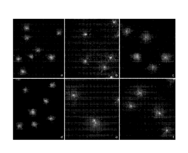
Example 7
It has previously been demonstrated that bacteria susceptible to antibiotics
that
inhibit protein synthesis like the ft-lactam, for example cephalosporines like
ceftazidime, or carbapenems like meropenem, may induce cell enlargement in the
susceptible strains at specific doses of the antibiotic. It has now been
determined that
28
CA 02967665 2017-05-11
WO 2016/118469
PCT/US2016/013835
this enlargement is dependent also on protein synthesis, so this effect can be
suppressed
in the bacterial strains susceptible to an antibiotic that inhibits protein
synthesis whereas
the effect remains in non-susceptible strains.
As an example, two strains of Pseudomonas aeruginosa exponentially growing
in Mueller-Hinton broth were assayed. These strains were both susceptible to
the 13-
lactam ceftazidime, which inhibits peptidoglycan synthesis. One strain was
susceptible
and the other resistant to the aminoglycoside tobramycin (an inhibitor of
protein
synthesis). For the purpose of rapidly distinguishing the susceptible and the
resistant
strain to tobramycin each strain was subjected to four treatments.
A portion of each strain was incubated with tobramycin at 4 jig/m1 (the CLSI
breakpoint of susceptibility) for 75 minutes. An assay of this portion is
depicted at
FIGS. 6A and D. Another portion of each strain was incubated with ceftazidime
at 0.5
jig/ml for 60 minutes. An assay of this portion is depicted at FIGS. 6B and D.
Still
another portion of both strains was incubated with tobramycin at 4 jig/ml for
15 minutes
followed by ceftazidime at 0.5 us/m1 for 60 minutes, without removing the
tobramycin.
Assays of these strains can be seen at FIGS 6C and F, respectively. A final
portion of
each strain was not treated with either antibiotic.
After incubation with the antibiotics, cells were processed using a variant
Micromax technology to visualize the enlargement or not of the bacteria.
Samples from
each culture were immersed in an agarose microgel on a slide, dried, stained
with a high
sensitive fluorochrome for DNA like SYBR Gold and visualized under
fluorescence
microscopy to visualize cell shape and size.
FIGS. 6A and D, illustrate that incubation with tobramycin, the antibiotic
that
inhibits the synthesis of proteins, does not affect the bacterial shape and
size in either
the susceptible or resistant strains. Both strains are similar in appearance
to cultures
without any antibiotics.
29
CA 02967665 2017-05-11
WO 2016/118469
PCT/US2016/013835
FIGS. 6B and E, depict assays of bacteria incubated with ceftazidime resulting
in significant cell enlargement. Similar results are seen in both strains
since both are
susceptible to ceftazidime.
FIG 6C illustrates an assay of the susceptible bacteria incubated with
tobramycin followed by ceftazidime resulting in bacteria with similar size to
those
incubated with tobramycin alone (FIG 6A) or untreated bacteria. In contrast,
FIG 6F
depicts an assay of the resistant strain incubated under the same conditions
in which
bacteria appeared enlarged similarly to those from the culture incubated with
ceftazidime only (FIG 6E).
Accordingly, enlargement by ceftazidime is dependent on protein synthesis and
as such, the suppressing effects of protein synthesis inhibiting proteins can
be employed
for the rapid determination of susceptibility to protein synthesis inhibiting
antibiotics. If
protein synthesis is successfully inhibited by tobramycin (as it was in the
strain
susceptible to tobramycin), cell enlargement by meropenem is suppressed. But
if the
protein synthesis is not successfully inhibited by tobramycin (as it was in
the strain
resistant to tobramycin), cell enlargement by meropenem is not suppressed and
the
bacteria appear with higher length. In this manner susceptible and non-
susceptible
strains can be rapidly discriminated with the assay.
Example 8
It has further been discovered that relatively low doses of mitomycin C, an
alkylating agent that induces DNA damage (see Example 3) can also affect the
morphological appearance of bacteria. In particular, reduced dosages of
mitomycin C
may result in bacterial enlargement or alterations in size. It has further
been discovered
that this cell shape modification depends on protein synthesis. As such, this
modification may be suppressed or reduced in the strains susceptible to the
antibiotic
that inhibits protein synthesis. In contrast, the cell shape modification
remains in non-
susceptible strains.
CA 02967665 2017-05-11
WO 2016/118469
PCT/US2016/013835
As an example, two strains of Escherichia coil exponentially growing in
Mueller-Hinton broth were incubated under four different conditions and then
assayed.
One strain was susceptible and the other resistant to the aminoglycoside
tobramycin (an
inhibitor of protein synthesis). For the purpose of rapidly distinguishing
susceptible and
non-susceptible strains to tobramycin each strain was incubated under four
conditions.
A portion of each strain was incubated with tobramycin at 4 [IOW for 90
minutes. As described previously, this dose is indicated by the CLSI as the
breakpoint of
susceptibility to tobramycin in the standard antibiogram based on
microdilution.
Another portion of both strains was incubated in Mitomycin C at 0.5 jig/ml for
60
minutes. Still another portion of each strain was incubated with tobramycin at
4 )tg/m1
for 30 minutes followed by mitomycin C at 0.5 )tg/m1 for 60 minutes, without
removing
the tobramycin. A final portion of both strains was not incubated with either
antibiotic.
After the incubation, cells were processed using a variant Micromax technology
to visualize any enlargement or not of the bacteria. Samples from cells from
the culture
were immersed in an agarose microgel on a slide, dried, stained with a highly
sensitive
fluorochrome for DNA like SYBR Gold and visualized under fluorescence
microscopy
to visualize cell shape and size. Images were similar those correspondent to
the previous
figure.
The assayed bacteria revealed that incubation with tobramycin, the antibiotic
that inhibits the synthesis of proteins, does not affect bacterial shape and
size, which is
similar in appearance to that from the cultures without antibiotics, in both
susceptible
and resistant strains to tobramycin.
Incubation with mitomycin C resulted in significant cell enlargement in both
the
susceptible and resistant strains of E. coll.
In the susceptible strain of bacteria, incubation with tobramycin followed by
mitomycin C resulted in bacteria that were similar in size to those incubated
with
tobramycin alone or untreated bacteria. In contrast, the resistant bacteria
incubated with
31
CA 02967665 2017-05-11
WO 2016/118469
PCT/US2016/013835
tobramycin followed by mitomycin C appeared enlarged similarly to those from
the
culture incubated with mitomycin C alone.
Accordingly, it can be understood that cell enlargement induced by mitomycin C
is dependent on protein synthesis. If protein synthesis is successfully
inhibited by
tobramycin, cell enlargement induced by mitomycin C is reduced or suppressed.
But if
the protein synthesis is not successfully inhibited by tobramycin, cell
enlargement by
mitomycin C is not suppressed and the bacteria appear with an enlarged length.
This
distinction provides a means for distinguishing susceptible and non-
susceptible strains
rapidly.
As demonstrated by Example 8, the evaluation of the suppression or not of cell
enlargement by antibiotic inhibitors of protein synthesis can only be assessed
in strains
susceptible to the cell enlargement-inducing antibiotic. Nevertheless, the
variant
incorporating mitomycin C as the agent for inducing a bacterial response may
have a
more expanded application to many bacterial species and strains because no
significant
resistances to mitomycin C are expected.
This fact has been further exemplified, by using mitomycin C in the successful
rapid detection of susceptibility-resistance to tobramycin in Pseudomonas
aeruginosa
and Klebsiella pneumoniae, as well as susceptibility-resistance to
azithromycin in
Haemophilus influenzae.
In one exemplification, two strains of H. influenzae exponentially growing in
Mueller-Hinton broth were incubated under four differing treatments and then
assayed.
One strain was susceptible and the other resistant to the macrolide
azithromycin (an
inhibitor of protein synthesis). For the purpose of rapidly distinguishing the
susceptible
and the resistant strain to azithromycin each strain was incubated under four
sets of
conditions.
A portion of each strain was incubated with azithromycin at 4 us/m1 for 150
minutes. The dose azithromycin corresponded to the dose indicated by the CLSI
as the
32
CA 02967665 2017-05-11
WO 2016/118469
PCT/US2016/013835
breakpoint of susceptibility to azithromycin in the standard antibiograms
based on
microdilution. Another portion of each strain was incubated with Mitomycin C
at 0.5
1,1g/m1 for 120 minutes. Still another portion was incubated with azithromycin
at 4
jig/m1 for 30 min followed by mitomycin C at 0.5 ug/m1 for 120 mm, without
removing
the azithromycin. A final portion of each strain was not incubated with any
antibiotics.
In the susceptible strain incubation with azithromycin followed by mitomycin C
resulted in bacteria with similar size to those incubated with azithromycin or
the
untreated control. Otherwise, the bacteria appeared enlarged similarly to
those from the
culture incubated with mitomycin C only, in the strain resistant to
azithromycin.
Other agents that induce DNA damage or cell toxicity resulting in cell shape
modification could be used instead of antibiotics or mitomycin C. Anon-
exhaustive list
or potential agents for inducing a bacterial response in the form of cell
shape
modifications includes alkylating agents, such as those often used in
chemotherapy of
cancer.
As can be easily understood from the foregoing, the basic concepts of the
present invention may be embodied in a variety of ways. The invention involves
numerous and varied embodiments including, but not limited to, the best mode
of the
invention.
As such, the particular embodiments or elements of the invention disclosed by
the description or shown in the figures or tables accompanying this
application are not
intended to be limiting, but rather examples of the numerous and varied
embodiments
generically encompassed by the invention or equivalents encompassed with
respect to
any particular element thereof. In addition, the specific description of a
single
embodiment or element of the invention may not explicitly describe all
embodiments or
elements possible; many alternatives are implicitly disclosed by the
description and
figures.
33
Moreover, for the purposes of the present description and claims, the term "a"
or
"an" entity refers to one or more of that entity; for example, "an antibiotic"
refers to one
or more antibiotics. As such, the terms "a" or "an", "one or more" and "at
least one"
should be understood as interchangeable as used herein.
All numeric values herein are assumed to be modified by the term "about",
whether or not explicitly indicated. For the purposes of the present
invention, ranges
may be expressed as from "about" one particular value to "about" another
particular
value. When such a range is expressed, another embodiment includes from the
one
particular value to the other particular value. The recitation of numerical
ranges by
endpoints includes all the numeric values subsumed within that range. A
numerical
range of one to five includes for example the numeric values 1, 1.5, 2, 2.75,
3, 3.80, 4,
5, and so forth. It will be further understood that the endpoints of each of
the ranges are
significant both in relation to the other endpoint, and independently of the
other
endpoint. When a value is expressed as an approximation by use of the
antecedent
"about," it will be understood that the particular value forms another
embodiment.
In addition, as to each term used it should be understood that unless its
utilization in this application is inconsistent with such interpretation,
common
dictionary definitions should be understood to be included in the description
for each
term as contained in the Random House Webster's Unabridged Dictionary, Second
edition.
The background section of this patent application provides a statement of the
field of endeavour to which the invention pertains. This section may also
contain
paraphrasing of certain United States patents, patent applications,
publications, or
subject matter of the claimed invention useful in relating information,
problems, or
concerns about the state of technology to which the invention is drawn toward.
It is not
intended that any United States patent, patent application, publication,
statement or
other information cited herein be interpreted, construed or deemed to be
admitted as
prior art with respect to the invention.
34
Date Recue/Date Received 2021-01-13
The claims set forth in this specification are part of this description of the
invention, and the applicant expressly reserves the right to use all of or a
portion of such
content of such claims as additional description to support any of or all of
the claims or
any element or component thereof, and the applicant further expressly reserves
the right
to move any portion of or all of the content of such claims or any element or
component
thereof from the description into the claims or vice versa as necessary to
define the
matter for which protection is sought by this application or by any subsequent
application or continuation, division, or continuation-in-part application
thereof, or to
obtain any benefit of, reduction in fees pursuant to, or to comply with the
patent laws,
rules, or regulations of any country or treaty, shall survive during the
entire pendency of
this application including any subsequent continuation, division, or
continuation-in-part
application thereof or any reissue or extension thereon.
Date Recue/Date Received 2021-01-13