Note: Descriptions are shown in the official language in which they were submitted.
CA 02967979 2017-05-15
WO 2016/122772
PCT/US2015/063479
SURGICAL HAND PIECE WITH INTEGRATED PRESSURE SENSOR
BACKGROUND OF THE INVENTION
This invention relates generally to the field of ophthalmic surgery and
more particularly to ultrasonic hand pieces for phacoemulsification.
The human eye in its simplest terms functions to provide vision by
transmitting light through a clear outer portion called the cornea, and
focusing
the image by way of the lens onto the retina. The quality of the focused
image depends on many factors including the size and shape of the eye, and
the transparency of the cornea and lens.
When age or disease causes the lens to become less transparent,
vision deteriorates because of the diminished light which can be transmitted
to
the retina. This deficiency in the lens of the eye is medically known as a
cataract. An accepted treatment for this condition is surgical removal of the
lens and replacement of the lens function by an artificial lens (I0L).
In the United States, the majority of cataractous lenses are removed by
a surgical technique called phacoemulsification. During this procedure, a thin
phacoemulsification cutting needle is inserted into the diseased lens and
vibrated ultrasonically. The vibrating cutting needle liquefies or emulsifies
the
lens so that the lens may be aspirated out of the eye. The diseased lens,
once removed, is replaced by an artificial lens.
A typical ultrasonic surgical device suitable for ophthalmic procedures
consists of an ultrasonically driven hand piece, an attached cutting needle,
an
irrigating sleeve, and an electronic control console. The hand piece assembly
is attached to the control console by an electric cable and flexible tubing.
Through the electric cable, the console varies the power level transmitted by
the hand piece to the attached cutting needle and the flexible tubing supply
irrigation fluid to and draw aspiration fluid from the eye through the hand
piece
assembly.
1
CA 02967979 2017-05-15
WO 2016/122772
PCT/US2015/063479
The operative part of the hand piece is a centrally located, hollow
resonating bar or horn directly attached to a set of piezoelectric crystals.
The
crystals supply the required ultrasonic vibration needed to drive both the
horn
and the attached cutting needle during phacoemulsification and are controlled
by the console. The crystal/horn assembly is suspended within the hollow
body or shell of the hand piece by flexible mountings. The hand piece body
terminates in a reduced diameter portion or nosecone at the body's distal end.
The nosecone is externally threaded to accept the irrigation sleeve. Likewise,
the horn bore is internally threaded at its distal end to receive the external
threads of the cutting needle. The irrigation sleeve also has an internally
threaded bore that is screwed onto the external threads of the nosecone. The
cutting needle is adjusted so that the needle projects only a predetermined
amount past the open end of the irrigating sleeve.
In use, the ends of the cutting needle and irrigating sleeve are inserted
into a small incision of predetermined width in the cornea or sclera. The
cutting needle is ultrasonically vibrated along its longitudinal axis within
the
irrigating sleeve by the crystal-driven ultrasonic horn, thereby emulsifying
the
selected tissue in situ. The hollow bore of the cutting needle communicates
with the bore in the horn that in turn communicates with the aspiration line
from the hand piece to the console. A reduced pressure or vacuum source in
the console draws or aspirates the emulsified tissue from the eye through the
open end of the cutting needle, the cutting needle and horn bores and the
aspiration line and into a collection device. The aspiration of emulsified
tissue
is aided by a saline solution or irrigating solution that is injected into the
surgical site through the small annular gap between the inside surface of the
irrigating sleeve and the cutting needle.
During surgery, the console controls irrigation and/or aspiration which
in turn determines the pressure in the eye. Aspirating too much fluid from the
eye can result in low pressure and a shallowing or collapse of the anterior
chamber. Typically, the pressure in the eye is monitored by measuring the
pressure in the irrigation and/or aspiration lines. One or more noninvasive
2
CA 02967979 2017-05-15
WO 2016/122772
PCT/US2015/063479
pressure sensors are located in a fluidics cassette to which the irrigation
and/or aspiration lines are connected. The location of these pressure sensors,
in a cassette that is remotely located from the eye, leads to some delay in
monitoring pressure in the eye. It would be desirable to locate a pressure
sensor close to the eye to minimize the delay in monitoring eye pressure.
3
CA 02967979 2017-05-15
WO 2016/122772
PCT/US2015/063479
SUMMARY OF THE INVENTION
In one example of the present invention, a surgical hand piece
comprises a shell, a channel, and a sensor housing. The channel has
proximal and distal ends. The channel is coupled to the shell such that the
proximal end of the channel is located at the proximal end of the shell and
the
distal end of the channel is located near the distal end of the shell. The
channel has an irrigation conduit passing through it. A sensor housing has an
irrigation path extending through it and a seal interface on one end of the
sensor housing. The seal interface end of the sensor housing is coupled to
the proximal end of the channel such that the irrigation conduit of the
channel
is fluidly coupled to the irrigation path of the sensor housing. The sensor
housing further comprises a cavity for receiving a pressure sensor. A
pressure sensor assembly is located in the cavity and fluidly seals the
cavity.
The pressure sensor measures fluid pressure in the irrigation path of the
sensor housing. The pressure sensor assembly further comprises: a flex
circuit; circuitry coupled to the flex circuit; and wire terminations coupled
to the
flex circuit. The pressure sensor is coupled to the flex circuit. A plug bolt
weldment is coupled to the sensor housing and the proximal end of the shell.
The plug bolt weldment has a hollow interior. The pressure sensor assembly
is at least partially located in the hollow interior of the plug bolt
weldment. The
sensor housing may also comprise an irrigation connector. A seal is located
at the seal interface of the sensor housing.
In another example of the present invention, a surgical hand piece
comprises a shell, a channel, and a sensor housing. The channel has
proximal and distal ends. The channel is coupled to the shell such that the
proximal end of the channel is located at the proximal end of the shell and
the
distal end of the channel is located near the distal end of the shell. The
channel has an irrigation conduit passing through it. A sensor housing has an
irrigation path extending through it and a seal interface on one end of the
sensor housing. The seal interface end of the sensor housing is coupled to
the proximal end of the channel such that the irrigation conduit of the
channel
is fluidly coupled to the irrigation path of the sensor housing. A pressure
4
CA 02967979 2017-05-15
WO 2016/122772
PCT/US2015/063479
sensor assembly comprises a flex circuit and a pressure sensor coupled to
the flex circuit. The pressure sensor is located in the cavity of the sensor
housing and measures fluid pressure in the irrigation path. The pressure
sensor fluidly seals the cavity. The pressure sensor assembly further
comprises: circuitry coupled to the flex circuit; and wire terminations
coupled
to the flex circuit. A plug bolt weldment with a hollow interior is coupled to
the
sensor housing and the proximal end of the shell. The pressure sensor
assembly is at least partially located in the hollow interior of the plug bolt
weldment. The sensor housing further comprises an irrigation connector. A
seal is located at the seal interface of the sensor housing. An end cap is
coupled to the plug bolt weldment and seals the hollow interior of the plug
bolt
weldment. A nose cone is located at the distal end of the shell and the
channel terminates at the nose cone.
It is to be understood that both the foregoing general description and
the following detailed description are exemplary and explanatory only and are
intended to provide further explanation of the invention as claimed. The
following description, as well as the practice of the invention, set forth and
suggest additional advantages and purposes of the invention.
BRIEF DESCRIPTION OF THE DRAWINGS
The accompanying drawings, which are incorporated in and constitute
a part of this specification, illustrate several embodiments of the invention
and
together with the description, serve to explain the principles of the
invention.
Figure 1 is a block diagram of a surgical hand piece system.
Figure 2 is a perspective view of a surgical hand piece.
Figure 3 is a side view of a portion of a surgical hand piece.
Figure 4 is a perspective view of a channel portion of a surgical hand
piece.
5
CA 02967979 2017-05-15
WO 2016/122772
PCT/US2015/063479
Figure 5 is an exploded view of a surgical hand piece.
Figure 6 is a perspective view of a sensor housing for a surgical hand
piece.
Figure 7 is a cross section view of a sensor housing for a surgical hand
piece.
Figure 8 is a perspective view of a sensor assembly for a surgical hand
piece.
DETAILED DESCRIPTION
Reference is now made in detail to the exemplary embodiments of the
invention, examples of which are illustrated in the accompanying drawings.
Wherever possible, the same reference numbers are used throughout the
drawings to refer to the same or like parts.
Figure 1 depicts an ultrasonic hand piece system. In Figure 1, hand
piece 100 is coupled to console 140. Console 140 is coupled to foot switch
150. Hand piece 100 has a cutting needle 110, a horn 120, a set of
piezoelectric crystals 130, and a nut 135 that secures the piezoelectric
crystals 130 to the horn 120. A needle interface 115 connects cutting needle
110 to a reduced diameter portion 125 of horn 120.
Needle 110 is typically a thin needle made of titanium or stainless steel
that is designed to emulsify a lens when vibrated ultrasonically. Needle 110
is
typically cylindrical in shape, has a small diameter of about 20 ¨ 30 gauge,
and has a length suitable for removal of a lens when inserted into the
anterior
chamber of the eye.
Horn 120 is typically made of a rigid material suitable for medical use
(such as a titanium alloy). Horn 120 has a reduced diameter section 125 that
is connected to a needle interface 115. Needle interface 115 typically has a
threaded connection that accepts needle 110. In this manner needle 110 is
6
CA 02967979 2017-05-15
WO 2016/122772
PCT/US2015/063479
screwed onto horn 120 at needle interface 115. This provides a rigid
connection between needle 110 and horn 120 so that vibration can be
transmitted from horn 120 to needle 110.
Piezoelectric crystals 130 supply ultrasonic vibrations that drive both
the horn 120 and the attached cutting needle 110 during phacoemulsification.
Piezoelectric crystals 130 are secured against horn 120 by nut 135.
Piezoelectric crystals 130 are typically constructed from a plurality of
crystal
segments. When excited by a signal from console 140, piezoelectric crystals
130 resonate, producing vibration in horn 120.
Console 140 includes a signal generator that produces a signal to drive
piezoelectric crystals 130. Console 140 has a suitable microprocessor, micro-
controller, computer, or digital logic controller to control the signal
generator.
In operation, console 140 produces a signal that drives piezoelectric crystals
130. Piezoelectric crystals 130, when excited, cause horn 120 to vibrate.
Needle 110, connected to horn 120, also vibrates. When needle 110 is
inserted into the anterior chamber of the eye and vibrated, it acts to
emulsify a
cataractous lens.
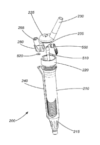
Figure 2 is a perspective view of a surgical hand piece. In the example
of Figure 2, hand piece 200 has a shell 210 with proximal and distal ends. A
nose cone 215 occupies the distal end of shell 210. A plug bolt weldment 220
is coupled to the proximal end of shell 210. An end cap 225 is coupled to the
plug bolt weldment 220. Control cable connector 230 is coupled to end cap
225. An aspiration connector 235 protrudes through end cap 225. A channel
240 is coupled to shell 210. Channel 240 has distal and proximal ends. A
sensor housing 250 is coupled to the proximal end of channel 240. The distal
end of channel 240 is coupled to shell 210 at or near nose cone 215. An
irrigation connector 255 is coupled to sensor housing 250.
Figure 3 is a side view of a portion of a surgical hand piece. In the
example of Figure 3, channel 240 is separated from shell 210. Shell 210
forms the outer portion of the hand piece and is held by a surgeon during
surgery. Shell 210 is ergonomic and may include features to facilitate easy
7
CA 02967979 2017-05-15
WO 2016/122772
PCT/US2015/063479
handling and manipulation of the hand piece. Shell 210 typically encloses
other parts of the hand piece including the horn 120, reduced diameter
section of the horn 125, piezoelectric crystals 130 and nut 135. Shell may be
made of any durable material such as stainless steel. In such a case, channel
140 is typically welded to shell 210. Channel 140 forms part of an irrigation
pathway that carries irrigation fluid to the eye during surgery. Channel 140
has an internal irrigation conduit that carries irrigation fluid. In use,
irrigation
fluid travels through channel 240 from its proximal end to its distal end.
Irrigation fluid exits the distal end of channel 240 through an opening. A
corresponding opening in shell 210 is coextensive with the opening on distal
end of channel 240. In this manner, irrigation fluid travels through channel
240 and into shell 210. From there, the irrigation fluid travels through a
passage in shell 210 (typically between an interior surface of shell 210 and
the distal end of horn 120 or reduced diameter section 125 of horn 120).
Irrigation fluid then exits the distal end of shell 210 and is carried to the
eye
via a sleeve that surrounds needle 110.
Figure 4 is a perspective view of a channel portion of a surgical hand
piece. In Figure 4, an irrigation conduit 410 is shown on proximal end of
channel 240. Irrigation conduit 410 extends through the length of channel 240
and terminates at an opening on or near the distal end of channel 240.
Figure 5 is an exploded view of a surgical hand piece. In the example
of Figure 5, hand piece 200 has a shell 210 with proximal and distal ends. A
nose cone 215 occupies the distal end of shell 210. A plug bolt weldment 220
is coupled to the proximal end of shell 210. Plug bolt weldment 220 has a
hollow interior. An aspiration conduit 510 extends from plug bolt weldment
220. An end cap 225 is coupled to the plug bolt weldment 220. Control cable
connector 230 is coupled to end cap 225. An aspiration connector 235
protrudes through end cap 225. A channel 240 is coupled to shell 210.
Channel 240 has distal and proximal ends. Irrigation conduit 410 extends
from the proximal end of channel 240 to its distal end. A sensor housing 250
is coupled to the proximal end of channel 240. A seal 520 is located between
the proximal end of channel 240 and the sensor housing 250. The distal end
8
CA 02967979 2017-05-15
WO 2016/122772
PCT/US2015/063479
of channel 240 is coupled to shell 210 at or near nose cone 215. An irrigation
connector 255 is coupled to sensor housing 250. A sensor assembly 550 fits
into sensor housing 250.
In Figure 5, sensor housing 250 provides a secure location for sensor
assembly 550. Sensor housing 250 is securely coupled to plug bolt weldment
220. Sensor assembly 550 is located in sensor housing 550. When
assembled, end cap 225 is secured to plug bolt weldment 220. Plug bolt
weldment 220 is secured to proximal end of shell 210. Seal 520 provides a
liquid tight seal between sensor housing 250 and channel 240. Plug bolt
weldment 220 has a hollow interior that provides a space for wire connections
to sensor assembly 550. Portions of the sensor assembly 550 may be
located in the hollow interior of plug bolt weldment 220.
Sensor assembly 550 measures the pressure of the irrigation fluid
traveling through sensor housing 250. Irrigation fluid travels from an
irrigation
source (typically a bottle or a bag) through flexible tubing to hand piece
200.
One end of the flexible tubing is coupled to the irrigation source, and the
other
end of the flexible tubing is coupled to hand piece 200 at irrigation
connector
255. In this case, irrigation connector 255 is a luer lock connector, but
numerous other types of connectors may be employed. Irrigation fluid enters
hand piece 200 at irrigation connector 255 and travels through a passage in
sensor housing 250. The irrigation fluid then travels through irrigation
conduit
410 in channel 240 and into shell 210 at or near nose cone 215. The
irrigation fluid continues through a passage in nose cone 215 and exits shell
210 at the end of nose cone 215. The irrigation fluid is then carried to the
eye
through a sleeve (not shown) that is coupled to the end of nose cone 215. In
this manner, a continuous path is provided for the introduction of irrigation
fluid into the eye during surgery. This continuous fluid path passes through
the length of hand piece 200. Because sensor assembly 550 is located along
the irrigation fluid path at a point that is very close to the eye, sensor
assembly 550 more accurately measures the pressure in the eye. Typically,
hand piece 210 is about four to six inches long. Accordingly, pressure sensor
9
CA 02967979 2017-05-15
WO 2016/122772
PCT/US2015/063479
assembly 550 measures the pressure of fluid about four to six inches from the
eye.
In currently available surgical systems, pressure sensors are located a
much greater distance from the eye. For example, in typical cataract
systems, an irrigation pressure sensor would be located on a surgical
console. A long length of flexible tubing connects the console to the hand
piece and carries irrigation fluid. Moreover, this flexible tubing is
typically
made of a polymer with a certain degree of compliance. In this manner, the
pressure sensor is located at one end of the flexible tubing. The other end of
the flexible tubing is connected to the hand piece. Because of the length of
flexible tubing located between the pressure sensor and the eye, the pressure
sensor does not accurately measure the pressure in the eye. As can be
appreciated, a more accurate reading of the pressure in the eye results in
better control of fluidics during surgery. Locating sensor assembly 550 in
hand piece 200 provides for a more accurate reading of eye pressure.
Turing again to the example of Figure 5, plug bolt weldment provides
space to house a portion of sensor housing 250 and sensor assembly 550.
As will be better appreciated with reference to Figure 8, wiring can be
located
in plug bolt weldment 220 to provide a pressure reading from sensor
assembly 550 to a cable at control cable connector 230.
Figure 6 is a perspective view of a sensor housing for a surgical hand
piece. In the example of Figure 6, sensor housing 250 includes an irrigation
connector 255, a seal interface 610, and a groove 620. Irrigation connector
255 receives one end of a length of flexible tubing that carries irrigation
fluid.
Seal interface has a recess that accepts seal 520. In this case, seal 520, in
its simplest form, is a washer that provides a fluid tight seal between sensor
housing 250 and channel 240. Sensor housing 250 is coupled to plug bolt
weldment 220 at groove 620. In this example, sensor housing 250 slides into
a notch in plug bolt weldment 220. The groove 620 engages the slot in plug
bolt weldment 220.
CA 02967979 2017-05-15
WO 2016/122772
PCT/US2015/063479
Figure 7 is a cross section view of a sensor housing for a surgical hand
piece. In the example of Figure 7, sensor housing 250 includes an irrigation
connector 255, a seal interface 610, and a cavity 710 for receiving pressure
sensor assembly 550 (and more particularly, pressure sensor 810). An
irrigation fluid path 720 is shown by the dashed line in Figure 7. The
irrigation
fluid path 720 extends through sensor housing 250 from the end with the
irrigation connector 255 to the end with the seal interface. In this manner,
irrigation fluid path 720 provides a continuous path through which irrigation
fluid can pass through sensor housing 250. The cavity 710 is in fluid
communication with irrigation fluid path 720. In this manner, a pressure
sensor located in cavity 710 can measure the pressure of fluid in irrigation
fluid path 720.
Figure 8 is a perspective view of a sensor assembly for a surgical hand
piece. In the example of Figure 8, sensor assembly 550 includes a pressure
sensor 810, a flex circuit 820, circuitry 830, and wire terminations 840.
Pressure sensor 810, circuitry 830, and wire terminations 840 are all mounted
on flex circuit 820. Pressure sensor 810 is sized and shaped to fit into
cavity
710 of sensor housing 250. In this manner, the cavity 710 is sized and
shaped to accommodate pressure sensor 810. Pressure sensor 810 fits
within cavity 710 and provides a fluid tight seal. Pressure sensor 810 may be
enclosed in a polymer to provide this fluid tight seal. When assembled, the
flex circuit 820 is located in plug bolt weldment 220. Wires coupled to wire
terminations 840 are also located in plug bolt weldment 220 and can extend to
control cable connector 230.
From the above, it may be appreciated that the present invention
provides an improved surgical hand piece for cataract surgery. The present
invention provides a hand piece with an integrated pressure sensor for
improved pressure measurement during surgery. The present invention is
illustrated herein by example, and various modifications may be made by a
person of ordinary skill in the art. Other embodiments of the invention will
be
apparent to those skilled in the art from consideration of the specification
and
practice of the invention disclosed herein. It is intended that the
specification
11
CA 02967979 2017-05-15
WO 2016/122772
PCT/US2015/063479
and examples be considered as exemplary only, with a true scope and spirit
of the invention being indicated by the following claims.
12