Note: Descriptions are shown in the official language in which they were submitted.
1
METHODS AND APPARATUSES FOR GENE PURIFICATION AND IMAGING
CROSS-REFERENCE TO RELATED APPLICATIONS
[0001] This Application claims priority under 35 U.S.C. 119 to U.S.
Provisional Application
Serial Number 62/083,681, filed November 24, 2014.
SEQUENCE LISTING
[0002] The instant application contains a Sequence Listing which has been
submitted in ASCII
format via EFS-Web. Said ASCII copy, created on November 23, 2015, is named
NATE-
023 5T25.ba and is 815 bytes in size.
FIELD OF THE INVENTION
[0003] The present innovations generally address molecular sample digital
counting, and more
particularly, include methods and apparatuses for nucleic acid and protein
sample purification
and imaging of associated molecular barcodes.
[0004] However, in order to develop a reader's understanding of the
innovations, disclosures
have been compiled into a single description to illustrate and clarify how
aspects of these
innovations operate independently, interoperate as between individual
innovations, and/or
cooperate collectively. The application goes on to further describe the
interrelations and
synergies as between the various innovations; all of which is to further
compliance with 35
U. S . C . 112.
BACKGROUND OF THE INVENTION
[0005] Scientists use a plurality of methods to purify and detect molecules.
Conventional
systems have fluidics and imaging of molecular barcodes in separate
instruments, have one
illumination channel per emission channel, and have inefficient methods of
moving the sample
through the purification and imaging machine.
Date Recue/Date Received 2022-03-01
CA 02968519 2017-05-19
WO 2016/085841 PCT/US2015/062109
2
SUMMARY OF THE INVENTION
[0006] The present invention provides systems, devices and methods for nucleic
acid or protein
purification and imaging.
[0007] An aspect of the present invention provides a cartridge configured for
purifying a
hybridized target molecule sample and imaging the hybridized target molecule.
The cartridge
comprises a sample input area, a first binding chamber, a first elution
channel, a second binding
chamber, a second elution channel, and a binding area.
[0008] In this aspect, the sample input area may be configured to hold a
target molecule sample,
e.g., comprising a plurality of hybridized complexes (which include a
plurality of target
molecules each hybridized with a first probe and/or a second probe), a
plurality of non-
hybridized first probes, and a plurality of non-hybridized second probes. The
first binding
chamber may be configured to receive and/or contain a first affinity matrix
and/or to receive the
sample. The first affinity matrix may be functionalized with first molecules
configured to bind
with the non-hybridized first probes and/or hybridized complexes of the sample
during a first
period of time. The first binding chamber may be additionally configured to
receive a first buffer
to remove non-hybridized second probes from the sample after the non-
hybridized first probes
and/or hybridized complexes of the sample bind with the first affinity matrix.
The first elution
channel may be configured to receive the first affinity matrix after the first
period of time and/or
configured for heating the first affinity matrix to elute a first eluted
sample comprising the
plurality of hybridized complexes and/or plurality of non-hybridized first
probes. The second
binding chamber may be configured to receive and/or contain a second affinity
matrix and/or to
receive the first eluted sample. The second affinity matrix may be
functionalized with second
molecules configured to bind with the hybridized complexes during a second
period of time. The
second binding chamber may be additionally configured to receive a second
buffer to remove at
least non-hybridized first probes. The second elution channel may be
configured to receive the
second affinity matrix after the second period of time and/or configured for
heating the second
affinity matrix to elute a second eluted sample comprising the plurality of
hybridized complexes.
The binding area may have an active binding surface configured to receive the
second eluted
sample and/or bind with the hybridized complexes.
CA 02968519 2017-05-19
WO 2016/085841 PCT/US2015/062109
3
[0009] In embodiments of this aspect, the target molecule may be a nucleic
acid or a protein. In
embodiments, the first affinity matrix and/or the second affinity matrix,
respectively, correspond
to a first set of magnetic beads (e.g., oligonucleotide-coupled magnetic
beads, e.g., F magnetic
beads) and/or a second set of magnetic beads (e.g., oligonucleotide-coupled
magnetic beads, e.g.,
G magnetic beads). The cartridge may further comprise a plurality of buffer
input areas, a
plurality of first binding chambers, a plurality of waste output areas, and/or
a plurality of bead
pads. The bubble vent may be configured to separate the sample input and/or
the first binding
chamber and/or to eliminate air bubbles. In embodiments, the active binding
surface may
comprise streptavidin, an avidin (e.g., NeutravidinTm), or oligonucleotides.
In embodiments, the
first probes include capture probes. In embodiments, the second probes include
reporter probes.
In embodiments, the cartridge may be operatively coupled to a plurality of off-
card buffer input
valves operatively coupled to a fluidic manifold and/or a plurality of waste
valves. In
embodiments, the cartridge further comprises a plurality of on-card buffer
input valves
operatively coupled to a fluidic manifold. The plurality of off-card buffer
input valves may be
configured to receive the first buffer and/or the second buffer from the
fluidic manifold and/or
provide the buffer to the cartridge. In embodiments, the flow of the second
eluted sample onto
the binding area may be done in small steps that may be re-ordered based on
pressure profiles. In
embodiments, the binding area may be further configured to receive a solution
(e.g., comprising
includes G-hooks, anti-fade media, and/or fiducials) formulated to immobilize
the second eluted
sample on the active binding surface after stretching with flow.
[0010] Another aspect of the present invention provides a cartridge configured
for purifying a
hybridized target molecule sample and imaging the hybridized target molecule.
The cartridge
comprises a buffer input area, a bubble vent, a first binding chamber, a first
elution channel, a
second binding chamber, a second elution channel, and a binding area.
[0011] In this aspect, the buffer input area may be configured to hold a
target molecule sample,
e.g., comprising a plurality of hybridized complexes (which include a
plurality of target
molecules, reporter probes, and/or capture probes), a plurality of non-
hybridized reporter probes,
and/or a plurality of non-hybridized capture probes. The bubble vent may be
configured to
separate the sample input and the first binding chamber and/or to eliminate
air bubbles. The first
binding chamber may be configured to receive and/or contain F magnetic beads
and/or to receive
the sample. The first binding chamber may be additionally configured to
receive a first buffer to
CA 02968519 2017-05-19
WO 2016/085841 PCT/US2015/062109
4
remove non-hybridized reporter probes from the sample after the non-hybridized
reporter probes
and/or hybridized complexes of the sample bind with the F magnetic beads,
which may be
functionalized with first molecules configured to bind with the non-hybridized
reporter probes
and/or hybridized complexes of the sample during a first period of time. The
first elution channel
may be configured to receive the F magnetic beads after the first period of
time and/or
configured for heating the F magnetic beads to elute a first eluted sample
comprising the
plurality of hybridized complexes and/or plurality of non-hybridized reporter
probes. The second
binding chamber may be configured to receive and/or contain G magnetic beads
and/or to
receive the first eluted sample. The second binding chamber may be
additionally configured to
receive a second buffer to remove at least non-hybridized capture probes. The
G magnetic beads
may be fitnctionalized with second molecules configured to bind with the
hybridized complexes
during a second period of time. The second elution channel may be configured
to receive the G
magnetic beads after the second period of time and/or configured for heating
the G magnetic
beads to elute a second eluted sample comprising the plurality of hybridized
complexes. The
binding area may have an active binding surface configured to receive the
second eluted sample
and/or bind with the hybridized complexes. The cartridge may be operatively
coupled to a
plurality of off-card buffer input valves operatively coupled to a fluidic
manifold. The plurality
of off-card buffer input valves may be configured to receive the first buffer
and/or the second
buffer from the fluidic manifold and/or provide the first and/or buffer to the
cartridge. The
plurality of waste valves may be configured to collect the first and/or second
buffer from the
cartridge.
[0012] In embodiments of this aspect, the target molecule may be a nucleic
acid or a protein. In
embodiments, the active binding surface may comprise streptavidin, an avidin
(e.g.,
NeutravidinTm), or oligonucleotides.
[0013] Yet another aspect of the present invention provides a system for
imaging a plurality of
hybridized complexes. The system comprises a cartridge of any of the herein
described aspects
or embodiments, a cartridge tray operatively coupled to the system and
configured to hold the
cartridge, a first heater operatively coupled to the cartridge, a second
heater operatively coupled
to the cartridge, a magnet operatively coupled to the imaging device below the
cartridge tray, a
fluidic manifold operatively coupled to the system above the cartridge tray
and configured to
hold and/or control the flow of a plurality of buffers, a plurality of off-
card buffer input valves
CA 02968519 2017-05-19
WO 2016/085841 PCT/US2015/062109
operatively coupled to the fluidic manifold and the cartridge; a plurality of
waste valves
operatively coupled to the system above the cartridge tray, and an imaging
reference surface
operatively coupled to the imaging device above the cartridge tray.
[0014] In embodiments of this aspect, the first heater may be configured to
heat the first elution
channel. In embodiments, the second heater may be configured to heat the
second elution
channel. In embodiments, the magnet may be configured to move the first
magnetic beads and/or
the second magnetic beads within the first and/or second binding chambers
and/or the first and/or
second elution channels. In embodiments, the magnet may be configured to move
parallel to the
cartridge tray. In embodiments, the plurality of off-card buffer input valves
may be configured to
receive the plurality of buffers from the fluidic manifold and/or provide the
plurality of buffers to
the cartridge. In embodiments, the plurality of waste valves may be configured
to collect the
plurality of buffers from the cartridge. In embodiments, the system further
comprises a cam
contact pad operatively coupled to the imaging device and configured to allow
preloading
against at least one contact pad, at least one adjustable contact between a
moving clamp and a
base of the imaging device, the at least one adjustable contact configured to
allow for datum A
adjustment, and a clamp motor operatively coupled to the imaging device and
configured to
move the moving clamp. In embodiments, at least one of the plurality of off-
card buffer input
valves and the plurality of waste valves operatively coupled to the system
above the cartridge
tray may be pneumatically controlled.
[0015] Another aspect of the present invention provides a method for purifying
a hybridized
target molecule sample and imaging the hybridized target molecule. The method
comprises steps
of:
(a) receiving a hybridized sample, the sample comprising a plurality of
hybridized,
complexes comprising target molecules hybridized with first probes and second
probes, a
plurality of non-hybridized first probes, and a plurality of non-hybridized
second probes
(b) binding the non-hybridized first probes and hybridized complexes of the
sample to a
first affinity matrix during a first period of time to produce a first
mixture,
(c) flowing a first buffer through the first mixture to remove non-hybridized
second
probes from the first mixture after the non-hybridized first probes and
hybridized
complexes of the sample bind with the first affinity matrix,
CA 02968519 2017-05-19
WO 2016/085841 PCT/US2015/062109
6
(d) heating the first mixture to free the non-hybridized first probes and
hybridized
complexes from the first affinity matrix and elute a first eluted sample
comprising the
plurality of hybridized complexes and plurality of non-hybridized first
probes,
(e) binding the hybridized complexes of the first eluted sample to a second
affinity matrix
during a second period of time to produce a second mixture,
(f) flowing a second buffer through the second mixture to remove the non-
hybridized first
probes from the first eluted sample after the hybridized complexes bind with
the second
affinity matrix,
(g) heating the second mixture to free the hybridized complexes from the
second affinity
matrix to elute a second eluted sample comprising the plurality of hybridized
complexes,
and
(h) binding the hybridized complexes to an active binding surface for imaging
thereof.
[0016] In embodiments of this aspect, the target molecule may be a nucleic
acid or a protein. In
embodiments, the first affinity matrix and the second affinity matrix,
respectively, correspond to
a first set of magnetic beads and a second set of magnetic beads,
respectively. In embodiments,
the active binding surface may surface may comprise streptavidin, an avidin
(e.g.,
NeutravidinTm), or oligonucleotides.
[0017] A further aspect of the present invention provides a method for
purifying a hybridized
target molecule sample and imaging the hybridized target molecule. The method
comprising
steps of:
(a)providing the cartridge of any of the herein described aspects or
embodiments,
(b) receiving a hybridized sample, the sample comprising a plurality of
hybridized
complexes comprising target molecules hybridized with first probes and second
probes, a
plurality of non-hybridized first probes, and a plurality of non-hybridized
second probes,
(c)binding the non-hybridized first probes and hybridized complexes of the
sample to a
first affinity matrix in a first binding chamber during a first period of
time,
(d) flowing a first buffer into the first binding chamber to remove non-
hybridized second
probes from the sample after the non-hybridized first probes and hybridized
complexes of
the sample bind with the first affinity matrix,
(c) directing the first affinity matrix into a first elution channel,
(0 heating the first affinity matrix to elute a first eluted sample comprising
the plurality
CA 02968519 2017-05-19
WO 2016/085841 PCT/US2015/062109
7
of hybridized complexes and plurality of non-hybridized first probes,
(g) binding the hybridized complexes of the first eluted sample to a second
affinity
matrix in a second binding chamber during a second period of time,
(h) flowing a second buffer into the second binding chamber to remove the non-
hybridized first probes from the first eluted sample after the hybridized
complexes bind
with the second affinity matrix,
(i) heating the second affinity matrix to elute a second eluted sample
comprising the
plurality of hybridized complexes, and
(j) binding the hybridized complexes to an active binding surface for imaging
thereof
[0018] In embodiments of this aspect, the target molecule may be a nucleic
acid or a protein. In
embodiments, the first affinity matrix and the second affinity matrix,
respectively, correspond to
a first set of magnetic beads (e.g., F magnetic beads) and a second set of
magnetic beads (e.g., G
magnetic beads). In embodiments, the active binding surface may surface may
comprise
streptavidin, an avidin (e.g., NeutravidinTm), or oligonucleotides. In
embodiments, the first
probes include reporter probes. In embodiments, the second probes include
capture probes. In
embodiments, the first binding chamber may be an F binding chamber. In
embodiments, the first
period of time may be a period of about 8 minutes. In embodiments, the first
magnetic beads
may be heated to about 47 C for about 7 minutes. In embodiments, the second
binding chamber
may be a G binding chamber. In embodiments, the second buffer may be F-elution
fluid. In
embodiments, the second period of time may be a period of about 7 minutes. In
embodiments,
the second buffer may be added to the second binding chamber in increments of
21u1_, forward
and 14 backward. In embodiments, the second buffer may be added in increments
of about
+2.8gL, +2 L, +24, +1.54, and 74. In embodiments, the second magnetic
beads may be heated to about 47 C for about 7 minutes. In embodiments, the
method further
comprises a step of moving a quantity of the first eluted sample across an
affinity matrix pad in a
first direction and a second direction. In embodiments, the first buffer may
be pumped to move
sample-bead mixture through the first bead pad in a first direction and a
second direction. In
embodiments, the first buffer may be added in increments of approximately
+15j1L, and -
15 L.Any of the above aspects and embodiments can be combined with any other
aspect or
embodiment.
8
[0019] Unless otherwise defined, all technical and scientific terms used
herein have the same
meaning as commonly understood by one of ordinary skill in the art to which
this invention
belongs. In the Specification, the singular forms also include the plural
unless the context clearly
dictates otherwise; as examples, the terms "a," "an," and "the" are understood
to be singular or
plural and the term "or" is understood to be inclusive. By way of example, "an
element" means
one or more element. Throughout the specification the word "comprising," or
variations such as
"comprises" or "comprising," will be understood to imply the inclusion of a
stated element,
integer or step, or group of elements, integers or steps, but not the
exclusion of any other
element, integer or step, or group of elements, integers or steps. About can
be understood as
within 10%, 9%, 8%, 7%, 6%, 5%, 4%, 3%, 2%, 1%, 0.5%, 0.1%, 0.05%, or 0.01% of
the stated
value. Unless otherwise clear from the context, all numerical values provided
herein are
modified by the term "about."
[0020] Although methods and materials similar or equivalent to those described
herein can be
used in the practice or testing of the present invention, suitable methods and
materials are
described below. The references cited herein are not admitted to be prior art
to the claimed
invention. In the case of conflict, the present Specification, including
definitions, will control.
In addition, the materials, methods, and examples are illustrative only and
are not intended to be
limiting. Other features and advantages of the invention will be apparent from
the following
detailed description and claim.
BRIEF DESCRIPTION OF THE DRAWINGS
[0022] The accompanying appendices and/or drawings illustrate various non-
limiting, example,
innovative aspects in accordance with the present descriptions:
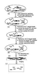
[0023] FIGURE lA shows block diagrams of the major processes according to some
embodiments.
Date Recue/Date Received 2022-03-01
CA 02968519 2017-05-19
WO 2016/085841 PCT/US2015/062109
9
[0024] FIGURES 1B-E show block diagrams illustrating the basic underlying
chemistry
according to some embodiments.
[0025] FIGURE 2 shows a detailed logic flow diagram illustrating a
purification and imaging
process according to some embodiments.
[0026] FIGURES 3A shows a labeled picture of the fluidic cartridge.
[0027] FIGURE 3B shows the fluidic layer where the two purifications occur.
[0028] FIGURE 4 shows a pictorial diagram illustrating the layer stack-up to
build the cartridge
according to some embodiments.
[0029] FIGURE 5 shows a pictorial diagram illustrating a machine that carries
out both fluidics
and imaging functions.
[0030] FIGURE 6A shows pictorial diagrams illustrating where sample is input
into the cartridge
according to some embodiments.
[0031] FIGURE 6B shows where tape is applied after sample input and where is
removed from
the buffer ports according (to prevent cross contamination) to some
embodiments.
[0032] FIGURES 7-9B show pictorial diagrams illustrating an instrument
according to some
embodiments.
[0033] FIGURE 10 shows a pictorial diagram illustrating an imaging cartridge
according to
some embodiments.
[0034] FIGURES 11-12 show pictorial diagrams illustrating purifying a sample
according to
some embodiments.
[0035] FIGURES 13A-14 show pictorial diagrams illustrating purifying a sample
according to
some embodiments.
[0036] FIGURES 15-17 show pictorial diagrams illustrating purifying a sample
according to
some embodiments.
[0037] FIGURE 18 show pictorial diagrams illustrating moving a purified sample
to an imaging
surface according to some embodiments.
[0038] FIGURES 19A-B show graphs illustrating adding the sample to the imaging
surface
according to some embodiments.
[0039] FIGURES 20-21 show pictorial diagrams illustrating binding a purified
sample to an
imaging surface according to some embodiments.
CA 02968519 2017-05-19
WO 2016/085841 PCT/US2015/062109
[0040] FIGURE 22 shows graphs comparing results obtained with the nCounter
Analysis
System and results obtained according to some embodiments of the present
invention for three
nCounter PanCancer Panels.
[0041] FIGURE 23 shows a graph illustrating differential gene expression data
obtained
according to some embodiments of the present invention.
[0042] FIGURE 24 shows a graph illustrating detection of total RNA or raw cell
lysates.
[0043] FIGURE 25 shows a graph illustrating detection of total gene expression
from fresh-
frozen tissue or from Formalin-Fixed Paraffin-Embedded (FFPE) tissues.
[0044] FIGURE 26 shows a graph comparing results obtained with the nCounter
Analysis
System and results obtained according to some embodiments of the present
invention for the
PanCancer Progression Panel.
[0045] FIGURE 27 shows a graph comparing results obtained with the nCounter
Analysis
System and results obtained according to some embodiments of the present
invention for the
Human Immunology Panel
[0046] FIGURE 28 shows a graph comparing results obtained with the
nCounter(13) Analysis
System and results obtained according to some embodiments of the present
invention in a copy
number variation (CNV) assay.
[0047] FIGURE 29 shows a graph comparing results obtained with the nCounter
Analysis
System and results obtained according to some embodiments of the present
invention in an a
miRNA analysis.
[0048] FIGURE 30 shows a graph comparing results obtained with the nCounter
Analysis
System and results obtained according to some embodiments of the present
invention for RNA-
Protein Profiling data.
[0049] The leading number of each reference number within the drawings
indicates the figure in
which that reference number is introduced and/or detailed. As such, a detailed
discussion of
reference number 101 would be found and/or introduced in Figure 1. Reference
number 201 is
introduced in Figure 2, etc.
DETAILED DESCRIPTION
CA 02968519 2017-05-19
WO 2016/085841 PCT/US2015/062109
11
[0050] Before some embodiments of the present disclosure are described in
detail, it is to be
understood that such embodiments are not limited to particular variations set
forth and may, of
course, vary. Various changes may be made to embodiments described and
equivalents may be
substituted without departing from the true spirit and scope of inventions
disclosed herein. In
addition, many modifications may be made to adapt a particular situation,
material, composition
of matter, process, process act(s) or step(s), to the objective(s), spirit or
scope of the present
disclosure. All such modifications are intended to be within the scope of any
and all claims
supported by the present disclosure.
[0051] Methods recited herein may be carried out in any order of the recited
events which is
logically possible, as well as the recited order of events. Furthermore, where
a range of values is
provided, it is understood that every intervening value, between the upper and
lower limit of that
range and any other stated or intervening value in that stated range is
encompassed within
embodiments of the disclosure. Also, it is contemplated that any optional
feature of one and/or
another of the disclosed embodiments described herein may be set forth and
claimed
independently, or in combination with any one or more of the features
described herein.
[0052] Reference to a singular item, includes the possibility that there are
plural of the same
items present. More specifically, as used herein and in the appended claims,
the singular forms
"a," "and," "said" and "the" include plural referents unless the context
clearly dictates otherwise.
It is further noted that the claims may be drafted to exclude any optional
element. As such, this
statement is intended to serve as antecedent basis for use of such exclusive
terminology as
"solely," "only" and the like in connection with the recitation of claim
elements, or use of a
"negative" limitation. Unless defined otherwise herein, all technical and
scientific terms used
herein have the same meaning as commonly understood by one of ordinary skill
in the art to
which this invention belongs.
[0053] In some embodiments, a user reading a sample (e.g., a nucleic acid
sample) may wish to
use a single device to both purify and detect the sample. In some embodiments,
using a single
device for both purposes may reduce processing time, likelihood of
contamination, cost of
performing imaging analysis, reduce the overall system cost, reduce hands on
time/steps, and/or
the like.
[0054] FIGURES 1A-E show block diagrams illustrating a purification and
imaging process
according to some embodiments. For example, a user of the instrument may
hybridize and/or
CA 02968519 2017-05-19
WO 2016/085841 PCT/US2015/062109
12
otherwise prepare a sample for processing. Referring to FIGURE 1B, in some
examples, the user
may wish to hybridize a target nucleic acid 114, e.g., using probes configured
to bind to the
target nucleic acid 114, and including affinity tags configured to bind to
magnetic beads. Target
nucleic acids 114 may include all forms of nucleic acids (e.g., RNA, DNA,
microRNA, and/or
the like). Proteins, and/or any other molecules which can be attached to the
capture and/or
reporter probes (e.g., which may be detected through a nucleic acid
intermediate) may also be
hybridized for analysis. For example, hybridization may involve mixing target
nucleic acids with
capture probes 116 and reporter probes 120. A capture probe 116 may include a
biotin moiety
used to bind the complex to an imaging surface, and/or an F tag configured to
bind to F magnetic
beads. A reporter probe 120 may include a fluorescent barcode used in the
imaging process,
and/or a G tag configured to bind to G magnetic beads. When the target nucleic
acid 114 binds to
a reporter and capture probe, it may create a hybridized tripartite complex
124 which may then
be purified for imaging and/or like processes.
[0055] As shown in FIGURE 1B, two approximately 50 base pair probes hybridize
directly to
each target molecule in solution to form a hybridized tripartite complex. The
reporter probe
carries a specific fluorescent barcode, and the capture probe contains a
biotin moiety that later
binds the tripartite complex to the imaging surface. Both probes contain
affinity tags (called "F"
or "G") that are required for magnetic bead-based purification and
immobilization
[0056] Referring to FIGURE 1A, the user may pipet 102 a sample (e.g., a
hybridized nucleic
acid sample and/or a like hybridized biological sample) into a sample
cartridge configured to be
placed in a cartridge tray of the instrument (which may be handled
automatically via
aspects/embodiments of the present disclosure). The hybridized biological
sample may also
include non-hybridized probes which may not have bound to the genes (e.g.,
excess probes). The
cartridge may also have pads (e.g., glass fiber pads) configured to hold
magnetic beads.
Magnetic beads can be of a plurality of varieties, such as F beads, G beads,
and/or the like. In
some implementations, F beads are magnetic beads coupled to DNA
oligonucleotides which are
the reverse complement of repeated sequences found on the capture probe, and
arc used as an
affinity matrix to separate hybridized complexes and free capture and/or
reporter probes during
purification. The magnetic beads may be dried down with buffer and a sugar
(e.g., trehalosc) to
stabilize the beads and to prepare them for suspension in a sample. The
cartridge may also be
configured with on-card buffer input valves configured to receive buffer from
off-card buffer
13
valves (e.g., see 902 of FIGURE 9A), and pneumatic valves configured to
control flow between
binding chambers, elution chambers, and/or like areas used for purification
processes and waste
containers configured to hold used elution fluids.
[0057] The cartridge may be a multi-layer cartridge (e.g., see 402 in FIGURE
4) comprising the
following components:
Layers Materials
1 250 gm Melinex
2 250 prnACA
3 250 prnPDMS
4 120 gm PDMS
250 prnACA
6 3 mm PMMA
[0058] Referring to FIGURE 1A, the sample may be introduced to dry magnetic
beads (e.g. F
magnetic beads (e.g., F beads, anti-F magnetic beads) which are coupled to a
15-mer DNA
oligonucleotide, 5'-GCT GTG ATG ATA GAC-3' (SEQ ID NO: 1), complementary to
the
repeats on the capture probe) configured to remove excess of at least one type
of probe (e.g., the
reporter probes) from the hybridized sample (e.g., see 104 of FIGURE 1A). The
F beads may be
dried down in 5X SSPE and 40% trehalose on pads (e.g., see 309 of FIGURE 3A;
bead pad is
partially hidden under bubble vent). In some embodiments, the F beads and the
sample may be
combined in a binding chamber (e.g., see 308 of FIGURES 3A & 3B) configured to
facilitate the
binding of the F beads to the hybridized tripartite complex molecules (e.g.,
see 126 of FIGURE
1C), and to allow for the beads to be washed such that at least some of the
unhybridized probes
(e.g., reporter probes) are washed from the sample (e.g., see 128 of FIGURE
1C). The binding
chamber may be configured with an elution channel (e.g., see 310 of FIGURES 3A
& 3B) which
Date Recue/Date Received 2022-03-01
CA 02968519 2017-05-19
WO 2016/085841 PCT/US2015/062109
14
may allow for the beads be heated (e.g., to 47 C) and the sample to be eluted
(e.g., see 130 of
FIGURE 1C).
[0059] The sample may then be passed to a second magnetic bead binding chamber
(e.g., see
106 of FIGURE lA & 314 of FIGURES 3A & 3B) which may be configured to hold
another set
of magnetic beads (e.g., G magnetic beads, also known as G beads and/or anti-G
magnetic beads,
which are coupled to a 15-mer DNA oligonucleotide, 5'-GGT CTG TGT GAT GTT -3'
(SEQ ID
NO: 2), complementary to the repeats on the reporter probe) which may be able
to bind to the
reporter probes of the hybridized tripartite complex molecules (e.g., see 132
of FIGURE 1C).
The second magnetic bead binding chamber (e.g., see 314 of FIGURES 3A & 3B)
may also
allow for washing the sample to remove excess molecules of another type of
probe (e.g., capture
probes) from the hybridized sample (e.g., see 134 of FIGURE 1C). The G beads
may be dried
down on a pad in 20X SSPE and 40% trehalose (e.g., see 312 of FIGURE 3A). The
G-bead
binding chamber may also be configured with an elution channel (e.g., see 315
of FIGURES 3A
& 3B) which may also facilitate elution (e.g., at 47 C) of the hybridized
tripartite complex
molecules from the beads (e.g., see 136 of FIGURE 1C).
[0060] As shown in FIGURE 1C, after benchtop hybridization, samples are
transferred to the
nCounter instrument. Excess probes are removed through two rounds of magnetic
bead-based
purification. First anti-F magnetic beads bind to tripartite complexes as well
as to unbound
capture probes. Unbound reporter probes are washed away, and the remaining
components are
eluted. Second, anti-G magnetic beads bind to the reporter probes. At this
state, all remaining
reporter probes are hybridized to their respective target nucleic acids.
Unbound capture probes
are washed away. A final elution step leaves only purified tripartite
complexes.
[0061] Another example uses a porous polymer matrix instead of magnetic beads.
The surface
can be activated by attaching oligonucleotides. These porous polymer materials
are very
inexpensive substrates and offer significant cost reduction compared to
magnetic beads. One
effective porous polymer matrixes is high density polyethylene with pore sizes
of 25, 75 and 125
mm nominal.
[0062] Referring to FIGURE 1A, the sample (which may now be purified of the
excess probe
molecules) may then be passed to an imaging surface 108 (e.g., a streptavidin
surface) to be
stretched and immobilized for imaging 110. For example, referring to FIGURE
1D, the biotin
moieties in the capture probes within the tripartite complexes may bind to the
imaging surface
CA 02968519 2017-05-19
WO 2016/085841 PCT/US2015/062109
138. The instrument may then flow buffer and/or like fluids on the imaging
surface of the
microfluidic cartridge 140 (e.g., see 316 of FIGURE 3A) to elongate and align
the complexes on
the surface. In some implementations the buffer and/or like fluid may also
contain a molecule
(e.g., biotinylated anti-G oligonucleotides and/or like molecules) which may
facilitate binding of
reporter probes in the tripartite complexes to the imaging surface 142. The
instrument may then
detect the complexes in the sample in order to generate a resulting graphic
and/or numerical
representation of the detected molecules 112. For example, the instrument may
include an
epifluorescence microscope configured to count the fluorescent barcodes of the
reporter probes
in the tripartite complexes, and to match the count to corresponding molecular
targets in order to
identify the molecule in the sample (e.g., see Figure 1E).
[0063] As shown in FIGURE 1D, after purification, samples move to the imaging
surface, which
is coated with streptavidin. Biotin moieties on each capture probe bind to the
imaging surface.
Flow within the microfluidic cartridge then elongates and aligns the
tripartite complexes. The
immobilization buffer contains biotinylated anti-G oligonucleotides that
anchor the reporter
probes to the imaging surface.
[0064] As shown in FIGURE 1E, samples are imaged by an epifluorescence
microscope with the
nCounter instrument. Barcodes are counted and matched with their
corresponding targets.
Coutns for each target are exported in a comma-separated value file.
[0065] FIGURE 2 shows a logic flow diagram illustrating a purification and
imaging process
according some embodiments. For example, the user may hybridize a sample
(e.g., a biological
sample; see 114 in FIGURE 1B) with excess capture and reporter probes 202 at
approximately
65 C. The user may then place the hybridized sample (e.g., via pipetting a
portion of the
hybridized sample) into a sample input area 204 (e.g., also see 302 of FIGURE
3A, 602 of
FIGURE 6A) configured to hold the biological sample. The sample input ports in
the sample
input area may be coned to allow for easier pipetting. The user may use a
single-channel or
multi-channel pipet to transfer the sample. In some embodiments, the user may
seal the sample
inputs (e.g., with transparent tape as shown at 604 of FIGURE 6B) and may
remove a seal (e.g.,
opaque tape as shown at 606 of FIGURE 6B) from a buffer input area (e.g., see
304 of FIGURE
3A) configured to receive buffer from the instrument.
[0066] The user may load the cartridge onto a cartridge tray (e.g., tray 702
in FIGURE 7) in the
instrument 206. FIGURES 8-9B illustrate a clamp motor and/or other mechanisms
for holding,
CA 02968519 2017-05-19
WO 2016/085841 PCT/US2015/062109
16
moving, and heating the cartridge, as well as imaging the sample on the
cartridge and
transferring fluids to and from the cartridge. For example, the cartridge tray
may be loaded into
an instrument nest as the tray moves inside the device, and may connect to a
fluidic manifold and
imaging reference surfaces operatively connected above the cartridge, with
heaters and bottom
contact points positioned below the cartridge and configured to push the
cartridge up against the
fluidic manifold, and imaging reference points with cam mechanism and springs
(e.g., see
FIGURE 8 and FIGURE 9B). Additionally, the instrument may include a fluidic
manifold 906
operatively coupled to off-card buffer input valves 902 and waste valves 904,
which may
connect to the cartridge and provide fluids to the cartridge, or remove used
fluids from the
cartridge, respectively.
[0067] The device may automatically determine whether the cartridge has been
correctly loaded,
and may also make sure that reagent (e.g., buffer) and waste bottles (e.g.,
502 in FIGURE 5) are
correctly connected to the cartridge, and that the reagent bottles have
sufficient levels of buffer
and/or similar fluids. In some embodiments, the user may be prompted to
replace reagent bottles
if more fluid is required (e.g., via screen 504 in FIGURE 5), and/or the like.
[0068] The instrument may then move the sample from the sample input area via
a flow (e.g.,
304), through a bubble vent and/or trap (e.g., a hydrophobic membrane; see 306
in FIGURE
3A) configured with an air bubble to separate the sample from the buffer. This
bubble prevents
sample dispersion by separating the two liquids. In some implementations
binding chambers
(e.g., such as the F binding chamber and the G binding chamber) can hold
magnetic beads and/or
other molecule-binding apparatus for purification of a sample. The F binding
chamber may hold
dry F magnetic beads which may be configured to bind to excess probe molecules
(e.g., excess
reporter probe molecules) in the hybridized sample (e.g., see 126 of FIGURE
1C). The
instrument may move the sample & beads (e.g., 151aL back and forth repeatedly)
with a pump
over the porous bead pads 210 in order to better facilitate resuspension and
binding of the F
beads to the excess capture probes and to the sample molecules (e.g., see
FIGURE 11). The F
beads may settle out of the solution; therefore the movement may be necessary
in order to keep
them suspended in the solution. The bubble vent (e.g., sec 1002 in FIGURE 10)
may be
physically positioned between the sample input area (e.g., see 1004 in FIGURE
10) and F-
binding chamber (e.g., see 308 in FIGURES 3A & 3B), and may be configured to
eliminate
bubbles, especially the large bubble between the sample and buffer after
mixing and binding has
CA 02968519 2017-05-19
WO 2016/085841 PCT/US2015/062109
17
finished. However, it is important the bubble between the sample and buffer
does not pass the
bubble vent during this mixing process in order to maintain sufficient
backpressure to pull the
sample back instead of pulling air through the bubble vent.
[0069] In some embodiments, moving a magnet pair back and forth across the
chamber may be
done instead of moving the sample back and forth with flow. Magnets may be
used in pairs to
generate a complex magnetic field suitable for mixing. The dead spot above and
between the
magnets may be critical for good mixing. The magnet speed may be related to
chamber size and
bead amounts (for example).
[0070] The binding process may, in some embodiments, last at least 8 minutes
(for example).
The bubble from the bubble vent may then be removed 212 during the mixing of
the hybridized
sample to the F beads. A magnet (e.g., an F magnet; see 1202 in FIGURE 12) in
the instrument
configured to move parallel to the cartridge, may be moved under the F binding
chamber in order
to collect the F beads 214, and to hold them in place as they are washed with
an elution buffer
216 added from the buffer input area. The elution buffer may facilitate
removal of at least one
type of non-hybridized probes (e.g., non-hybridized reporter or capture
probes; (e.g., see 128 of
FIGURE 1C)) from the F beads. During the multistage wash step, beads may be
moved around
in the F binding chamber by moving the magnet (e.g., see 1302 in FIGURE 13A).
This
movement and spreading out of the beads may allow for better washing of the
captured beads.
[0071] The beads, via the magnet, may then be pushed into an F elution channel
218 (e.g., also
see 310 of FIGURES 3A and 3B), which may be connected to a heater (e.g., an F
heater; see
1304 in FIGURE 13B) which may be configured to heat the F beads 220 (e.g., to
47 C for four
minutes) in order to elute the sample molecules from the F beads (e.g., see
130 of FIGURE 1C).
The F beads, after the heating process, may be returned to the F binding
chamber 222 (e.g., see
FIGURE 14) as the eluted sample is moved into a second binding chamber 224
(e.g., a G binding
chamber; see 314 in FIGURES 3A or 3B), configured to facilitate the binding of
a second set of
dry magnetic beads (e.g., G magnetic beads) to the hybridized tripartite
complex molecules.
[0072] A stepwise fluid introduction and flow mixing process may be utilized
with the G
magnetic beads and the eluted sample in order to achieve proper bead re-
suspension and sample
binding 226. The G magnetic beads may bind to reporter probes and/or other
probes which may
still be attached to the sample molecules (e.g., see 132 of FIGURE 1C). For
example,
introduction of the F-eluted sample into the G binding chamber may be
performed in repeated
CA 02968519 2017-05-19
WO 2016/085841 PCT/US2015/062109
18
small steps of 24 forward followed by 1 luL backward. The back and forth of
fluid may help re-
suspend the G beads and make the systems insensitive to small differences in
bead pad/bead
pocket size. This back and forth mixing can occur through the porous pad. In
some
embodiments, an exact flow profile may be +54, +5 L,
+51iL. Binding may occur
for a specified period of time, before another 7iuL is introduced. In some
embodiments (e.g., see
FIGURE 15), elutions may be flowed one lane at a time 1502 until all elutions
have been flowed
into lanes 1504 (e.g., the process may take 7 minutes).
[0073] An imaging chamber, meanwhile, may be washed 228 in order to remove
molecules (e.g.,
trehalose and free avidin) while the G beads are bound to the eluted sample. A
magnet (e.g., a G
magnet; see 1204 in FIGURE 12) may be moved under the G binding chamber 230 in
order to
collect all of the G beads and hold them as a heater (e.g., the F heater)
heats the F binding &
elution chamber 232 (e.g., to 35 C). Meanwhile an elution buffer (e.g., warmed
by the heater)
may be washed over the G beads in order to facilitate removal of excess non-
hybridized capture
probes from the G beads (e.g., see 134 of FIGURE 1C). The beads may then be
moved to a G
elution chamber (e.g., see 315 in FIGURES 3A and 3B and 1602 and 1604 in
FIGURE 16) via
the magnet 234, such that a second heater (e.g., a G heater; see 1306 in
FIGURE 13B and 1702
in FIGURE 17) configured to release the purified sample (e.g. tripartite
complexes) from the G
beads, may be initiated 236.
[0074] The G heater may be run at 47 C for 4 minutes to release the sample
from the beads (e.g.,
see 136 of FIGURE 1C). The magnet may then move the G beads back to the
binding chamber
238, and the purified sample may be moved 240 (e.g., see FIGURE 18) into a
binding area of an
SA surface (e.g., see 316 in FIGURES 3A and 3B). Flow rate into the chamber
may be
performed in steps that are about half the volume of the chamber or less
(e.g., approximately
0.254 every 78 seconds). The small elution volume and the controlled flow
(using a syringe
pump instead of gravity), allows for faster and more efficient binding.
[0075] In some embodiments, dynamic sequencing of twelve lanes may be
performed in order to
equalize flow volume of eluted samples binding to SA surface. For example,
approximately
0.254 may be flowed every 76 seconds on each lane. The stepping may be done in
sequence:
0.254 steps every 6.3 seconds per lane in a sequence (e.g., lanes one through
twelve), which
may result in 12*6.3=75.6 seconds of wait time on every lane for every 0.254
step. Because
twelve valves may be opened and closed in sequence for twelve separate lanes
and move only a
CA 02968519 2017-05-19
WO 2016/085841 PCT/US2015/062109
19
small volume, the displacement volume of each individual valve may affect the
lane to lane
reporter count variability. The variation in displacement volume from the pump
by itself may not
affect variability; however the difference in displacement volumes due to the
valves may have a
big effect on variability. The displacement of each valve may be estimated
(e.g., see 1902 in
FIGURE 19A) by using the pressure reading difference of closed vs open state
of the valve.
Minimization of lane to lane variability may require minimization of
displacement volume
variability by re-ordering the lanes. To minimize the variability, the
instrument may start pushing
from the valve that has highest displacement volume then the second highest
and so forth
finishing with the smallest. The transition from smallest to highest may have
the most negative
effect; to correct that, the instrument may be configured to push an extra
0.083 tL on that single
lane (e.g., 0.3330_, instead of 0.2504 see 1904 of FIGURE 19B).
[0076] In some embodiments (e.g., in FIGURE 20), the SA surface may be a
surface coated with
streptavidin, and may bind to the hybridized probes in the sample 2002 (e.g.,
see also 138 in
FIGURE 1D). There, the molecules in the purified, second-eluted sample may be
stretched and
immobilized 242 on the SA surface (e.g., also see 2102 in FIGURE 21 and 140 &
142 of Figure
1D), e.g., via a single solution comprising at least G-hooks (biotinylated
anti-G 15mer oligo),
mounting media (anti-fade), and fiducials (multispectral biotinylated
fluorescent 100nm beads,
i.e., diffraction limited), and being added to the SA surface at a particular
flow rate (e.g., see
2104 in FIGURE 21, which is a graph indicating flow rates that may be
utilized). The G hooks
are used to immobilize down the second end of the reporter during stretching.
The mounting
media is present to prevent photo bleaching and photo-destruction of the DNA
(due to light
interaction with dyes that create free-radicals that can break the DNA
backbone). The fiducials
produce a fluorescent signal (in all channels) that is used to align images. G-
hooks, mounting
media, fiducials, and the stretching buffer were required to be in separate
solutions in previously-
described electrostretching-based immobilization processes. In some
embodiments, the process
may replace four buffers and may eliminate the need for electrodes and a power
supply. It may
also eliminate a G-hook contamination problem, which is caused by G-hooks
slicking to the
electrodes in previous designs and carrying over into subsequent sample runs.
The instrument
may then detect the stretched molecules 244 from the SA surface on the
cartridge and produce an
output (e.g. an image, a report, and/or the like) for the user (e.g., see 144
of FIGURE 1E). In
20
some embodiments, the instrument may have a low cost optics subcomponent that
uses a three
LED illumination system.
[0077] The instrument may also perform binding gradient and area optimization
based on
density. The binding to the Streptavidin surface may generate a reporter
binding gradient over
the channel - higher density at one end (inlet) with gradual decrease toward
the other end
(outlet). Based on the reporter density determined by an initial scanning
survey, the location of
imaging area may be selected for the optimal data collection. Selecting the
final scan area in the
high density side (close to the inlet end) may generally collect more data.
However, in the case
of too high binding density, the scan may start from a less dense area by
moving the scan area
farther from the inlet end. This scheme increases the dynamic range of sample
concentration.
[0078] During imaging, mounting media may be exchanged to minimize photo-
destruction and
minimize non-specific binding of residual non-functional reporters by
eliminating free reporters.
If any free reporters are left floating free in the imaging chamber, the flow
of imaging buffer
wash them out, thus preventing binding via G-hooks in solution.
[0079] Additional teaching relevant to the present invention are described in
one or more of the
following: U.S. 2011/0086774, U.S. 2011/0145176, U.S. 2011/0201515, U.S.
2011/0229888,
U.S. 2013/0004482, U.S. 2013/0017971, U.S. 2013/0178372, U.S. 2013/0230851,
U.S.
2013/0337444, U.S. 2013/0345161, U.S. 2014/0005067, U.S. 2014/0017688, U.S.
2014/0037620, U.S. 2014/0087959, U.S. 2014/0154681, U.S. 2014/0162251, U.S.
2014/0371088, U.S. 2015/0072021, U.S. 2015/0252440, U.S. 7,473,767, U.S.
7,919,237, U.S.
7,941,279, U.S. 8,148,512, U.S. 8,415,102, U.S. 8,492,094, U.S. 8,519,115,
U.S. 8,986,926, U.S.
9,066,963, and U.S. 9,181,588.
[0080] The following example is offered by way of illustration and not by way
of limitation.
EXAMPLE
[0081] Embodiments of the present disclosure provide superior detection and
quantification of
gene expression and protein synthesis.
[0082] As shown in FIGURE 22, embodiments effectively detect targets from
three nCounter
PanCancer Panels: the PanCancer Pathways panel, the PanCancer Progression
panel, and the
Date Recue/Date Received 2022-03-01
21
PanCancer Immune Profiling panel. Here, data obtained from embodiments
(identified in as
"nCounter SPRINT Profiler") are correlated with data obtained from the
nCounter Analysis
System. As shown in FIGURE 23, embodiments enable identification of
differentially expressed
genes in lymphoma samples. As shown in FIGURE 24, embodiments can detect gene
expression
in purified total RNA or in raw cell lysates; it is notable that assays
require limited sample
preparation. As shown in FIGURE 25, embodiments provide reliable, trustworthy
detection of
gene expression with various sample types, including, but not limited to,
fresh-frozen tissue and
Formalin-Fixed Paraffin-Embedded (FFPE) tissues. As shown in FIGURE 26,
embodiments
effectively detect gene expression for the PanCancer Progression Panel and
with highly
correlated results with respect to results obtained with the nCounter
Analysis System. It is
noteworthy that embodiments of the present invention (identified as "SPRINT")
used half as
much sample input (by weight) as used with the nCounter Analysis System
(identified as
"Analysis System"). As shown in FIGURE 27, embodiments effectively detect
targets in the
Human Immunology Panel. Here, data obtained from embodiments (identified as
"nCounter
SPRINT Profiler") are correlated with data obtained from the nCounter
Analysis System. As
shown in FIGURE 28, embodiments effectively quantify targets in a copy number
variation
(CNV) assay. Similar copy number data were obtained for DNA samples run on
embodiments of
the present invention (identified as "nCounter SPRINT Profiler") and the
nCounter Analysis
System. Here, copy number data for each gene is directly above a tick mark on
the X-axis.
Thus, a vertically-related pair comprising a square (data from embodiments of
the present
invention) and circle (data from the nCounter Analysis System) represent data
for a particular
gene. As shown in FIGURE 29, embodiments effectively detect miRNA targets.
Here, data
obtained from embodiments are correlated with data obtained from the nCounter
Analysis
System. As shown in FIGURE 30, embodiments effectively detect RNA and protein
targets.
Here, data obtained from embodiments (identified as "nCounter SPRINT
Profiler") are correlated
with data obtained from the nCounter Analysis System.
[0083] The referenced items are provided solely for their disclosure prior to
the filing date of the
present application. Nothing herein is to be construed as an admission that
any invention
disclosed herein is not entitled to antedate such material by virtue of prior
invention.
Date Recue/Date Received 2022-03-01
22
[0084] Although example embodiments of the devices, systems and methods have
been
described herein, other modifications are possible. As noted elsewhere, these
embodiments have
been described for illustrative purposes only and are not limiting. Other
embodiments are
possible and are covered by the disclosure, which will be apparent from the
teachings contained
herein. Thus, the breadth and scope of the disclosure should not be limited by
any of the above-
described embodiments but should be defined only in accordance with claims
supported by the
present disclosure and their equivalents. In addition, any logic flow depicted
in the above
disclosure and/or accompanying figures may not require the particular order
shown, or sequential
order, to achieve desirable results. Moreover, embodiments of the subject
disclosure may include
methods, systems and devices which may further include any and all elements
from any other
disclosed methods, systems, and devices, including any and all elements
corresponding to gene
purification and imaging. In other words, elements from one and/or another
disclosed
embodiment may be interchangeable with elements from other disclosed
embodiments. In
addition, one or more features/elements of disclosed embodiments may be
removed and still
result in patentable subject matter (and thus, resulting in yet more
embodiments of the subject
disclosure). In addition, some embodiments of the present disclosure are
distinguishable from the
prior art for expressly not requiring one and/or an other features disclosed
in the prior art (e.g.,
some embodiments may include negative limitations). Some of the embodiments
disclosed
herein are within the scope of at least some of the following claims of the
numerous claims
which are supported by the present disclosure which may be presented.
Date Recue/Date Received 2022-03-01