Note: Descriptions are shown in the official language in which they were submitted.
- 1 -
TITLE: SYSTEM AND METHOD FOR ENHANCED MASS
SPECTROMETRY IMAGING
[0001] BLANK
FIELD
[0002] The various embodiments described herein generally relate to a
system and method for enhanced mass spectrometry imaging, in particular
intraoperative mass spectrometry imaging using exogenous agents.
BACKGROUND
[0003] When operating on patients, surgeons often need to accurately
identify a region of interest, such as a disease region or a tumor, as well as
the boundary of that region of interest. In some cases, such as when excising
a tumor, a surrounding margin of healthy tissue around the tumor will be
removed to ensure that no diseased tissue remains. When the boundary of
the disease region cannot be robustly identified, excess healthy tissue may be
removed unnecessarily, potentially leading to disability for the patient.
[0004] While intraoperative pathology methods exist to reveal
some
disease regions, there can be significant difficulties in distinguishing the
disease regions from surrounding tissues, such as fatty breast tissues for
example. Intraoperative pathology methods may also take upwards of 30
minutes to identify healthy and diseased tissue regions, leading to delays
during surgery. In some cases, these delays and insufficiently robust
identification of the disease region can lead to patients having to undergo
7467102
Date Recue/Date Received 2022-05-05
CA 02969251 2017-05-30
WO 2016/090471
PCT/CA2015/051282
- 2 -
subsequent surgeries to ensure that the entire disease region has been
removed.
[0005] Imaging Mass Spectrometry (IMS) is one technique to map the
chemical content of biological tissues in a spatially resolved manner. Recent
developments in IMS techniques have opened up the prospect of
intraoperative molecular imaging to identify disease states of tissues for
effective diagnosis. These methods identify the tissue disease states on the
basis of spatially mapping endogenous disease markers.
[0006] Desorption by Electrospray Ionization (DESI) MS imaging has
been used to identify and grade tumor regions into their respective subclasses
on the basis of endogenous lipid profiles unique to each tumor class. The
endogenous lipid profiles typically require strong cross-validation with
conventional pathology methods. Accordingly, the success of DESI-MS
imaging and other current ambient I MS technologies in identifying a diseased
region is heavily tied to the availability of validated molecular markers for
the
disease in question.
SUMMARY OF VARIOUS EMBODIMENTS
[0007] In a broad aspect, at least one embodiment described herein
provides a method of identifying a region of interest in tissue, the method
comprising administering an exogenous agent to the tissue, the exogenous
agent being capable of forming a by-product in the tissue; analyzing a sample
based on the tissue using a high sensitivity platform comprising a mass
spectrometer; determining a distribution, optionally quantitative, of the by-
product of the exogenous agent within the tissue based on the analysis of the
sample; and identifying the region of interest within the tissue based on the
determined distribution of the by-product of the exogenous agent relative to
tissue surrounding the region of interest.
CA 02969251 2017-05-30
WO 2016/090471
PCT/CA2015/051282
- 3 -
[0008] In at least one embodiment, the determining act further
comprises determining a distribution of the exogenous agent in addition to the
by-product of the exogenous agent.
[0009] In at least one embodiment, prior to the analyzing act the
method comprises acquiring the sample from the tissue after administration of
the exogenous agent to the tissue; and transporting the sample to the mass
spectrometer using a transfer line by applying a positive pressure on the
transfer line at a first end proximate to the tissue.
[0010] In at least one embodiment, prior to the analyzing act the
method comprises obtaining the sample from the tissue using an ex vivo
sampling technique.
[0011] In at least one embodiment, the method further comprises
selecting the agent from chelated metal containing agents and tumour specific
metalloporphyrins, chelated metal containing agents being Gadolinium based,
ion oxide based, iron-platinum based, manganese based, or chromium based.
[0012] In at least one embodiment, the method further comprises
selecting the exogenous agent from at least one of a metallic element, a
heavy atom, and an isotopic variant that is not endogenous to the tissue, a
metabolic precursor, an isotopic variant of a metabolic precursor, a moiety of
a metabolic precursor or a plurality of exogenous sub-agents.
[0013] In at least one embodiment, the method further comprises
identifying a boundary of the region of interest based on the distribution of
at
least one of the exogenous agent, and the by-product of the exogenous agent
relative to tissue surrounding the region of interest; and displaying an image
of the tissue with the boundary marked.
[0014] In another broad aspect, at least one embodiment described
herein provides a method of identifying a region of interest in tissue using
mass spectrometry. The method includes administering an exogenous agent
to the tissue, acquiring a sample based on the tissue after administration of
the exogenous agent to the tissue, transporting the sample to a high
CA 02969251 2017-05-30
WO 2016/090471
PCT/CA2015/051282
- 4 -
sensitivity platform, analyzing the sample using the high sensitivity
platform,
determining a distribution of at least one of the exogenous agent and a by-
product of the exogenous agent within the tissue based on the analysis of the
sample, and identifying the region of interest based on the determined
distribution.
[0015] In some embodiments, the high sensitivity platform can include
a mass analyzer.
[0016] In some embodiments the high sensitivity platform can include
at least one of an optical detection platform, a fluorescence detection
platform
and a Raman detector.
[0017] In some embodiments, the high sensitivity platform may include
the at least one of an optical detection platform, a fluorescence detection
platform and a Raman detector in tandem with a mass analyzer.
[0018] In some embodiments, the exogenous agent administered can
include at least one of a metallic element, a heavy atom, and an isotopic
variant. In some cases, the at least one isotopic variant can be an isotopic
variant of an endogenous metabolic precursor. The method can include
determining the distribution of a by-product of the isotopic variant of the
endogenous metabolic precursor. In some embodiments, the isotopic variant
is not endogenous.
[0019] In some embodiments, the exogenous agent can include at least
one of a metabolic precursor and a moiety of a metabolic precursor. The
method can include determining the distribution of a by-product of the at
least
one of the metabolic precursor and the moiety of a metabolic precursor.
[0020] In some embodiments, the exogenous agent can include a
plurality of exogenous sub-agents. In some embodiments, the exogenous
agent can be administered encapsulated in a lipidic structure such as a
liposome.
[0021] In some embodiments, acquiring the sample can include
desorbing the sample from the tissue. In some cases, the desorption can be
CA 02969251 2017-05-30
WO 2016/090471
PCT/CA2015/051282
- 5 -
performed using laser ablation vaporization, desorption electrospray
ionization, or radio frequency ablation.
[0022] In some embodiments, the method includes ionizing the sample
prior to analyzing the sample using the high sensitivity platform. In some
cases, the sample can be ionized using plasma. In some cases, the sample
can be ionized using inductively-coupled plasma. In some cases, the sample
can be ionized using rapid evaporative ionization, or electrospray ionization.
[0023] In some embodiments, the high sensitivity platform can be
configured to determine a distribution of the at least one of the exogenous
agent and a by-product of the exogenous agent.
[0024] In some embodiments, the sample can be transported to the
mass analyzer using a transfer line. In some cases, the method can further
include applying a positive pressure on the transfer line at a first end
proximate the tissue.
[0025] In some embodiments, the method further includes identifying a
boundary of the region of interest based on the distribution of at least one
of
the exogenous agent, the isotopic variant, and the by-product of the
exogenous agent. The method can further include displaying an image of the
tissue with the boundary marked.
[0026] In some embodiments, the exogenous agent administered is
selected such that the at least one of the exogenous and the by-product of the
exogenous agent has at least one of a mass to charge ratio peak and an
elemental mass peak that is not endogenous.
[0027] In some embodiments, the sample can be a tissue sample
acquired from the tissue, an ablated tissue sample, an ablation plume, a
liquefied tissue sample, an extraction of the exogenous agent, or an
extraction
of the by-product of the exogenous agent from the tissue. In some cases, the
tissue sample can be an ex-vivo tissue sample.
[0028] In another broad aspect, at least one embodiment described
herein provides a system for identifying a region of interest in tissue, the
CA 02969251 2017-05-30
WO 2016/090471
PCT/CA2015/051282
- 6 -
system comprising: a sampling unit configured to acquire a sample based on
the tissue after administration of an exogenous agent to the tissue, the
exogenous agent being capable of forming a by-product in the tissue; and a
high sensitivity platform comprising a mass spectrometer, the high sensitivity
platform being coupled to the sampling unit to analyze the sample, the high
sensitivity platform being configured to determine a distribution, optionally
quantitative, of a by-product of the exogenous agent within the tissue based
on a spectral analysis of the sample to determine spectral peaks due to the
by-product of the exogenous agent that are not endogenous to the tissue and
to identify the region of interest based on the determined distribution of the
by-
product of the exogenous agent relative to tissue surrounding the region of
interest.
[0029] In at least one
embodiment, the high sensitivity platform is
configured to determine a distribution of the exogenous agent in addition to
the by-product of the exogenous agent based on a spectral analysis of the
sample to determine spectral peaks due to the exogenous agent that are not
endogenous to the tissue, and the exogenous agent and the by-product of the
exogenous agent having at least one of a mass to charge ratio peak and an
elemental mass peak that is not endogenous to the tissue.
[0030] In at least one
embodiment, the system further comprises an
agent administration component configured to administer an exogenous agent
to the tissue prior to analysis by the mass spectrometer.
[0031] In at least one
embodiment, the exogenous agent comprises a
chelated metal containing agent or a tumour specific metalloporphyrins, the
chelated metal containing agents being Gadolinium based, ion oxide based,
iron-platinum based, manganese based, or chromium based.
[0032] In at least one
embodiment, the exogenous agent used with the
system is at least one of a metallic element, a heavy atom, and an isotopic
variant that is not endogenous to the tissue, a metabolic precursor, an
isotopic
variant of a metabolic precursor, a moiety of a metabolic precursor or a
plurality of exogenous sub-agents.
CA 02969251 2017-05-30
WO 2016/090471
PCT/CA2015/051282
- 7 -
[0033] In at least one embodiment, the at least one isotopic variant
comprises an isotopic variant of an endogenous metabolic precursor, and the
high sensitivity platform is configured to determine the distribution of a by-
product of the isotopic variant of the endogenous metabolic precursor.
[0034] In another broad aspect, at least one embodiment described
herein provides a system for identifying a region of interest in tissue using
mass spectrometry. The system can include an agent administration
component, a sampling unit and a high sensitivity platform. The agent
administration component can be configured to administer an exogenous
agent to the tissue. The sampling unit can be configured to acquire a sample
based on the tissue after administration of the exogenous agent to the tissue.
The high sensitivity platform can be configured to analyze the sample,
determine a distribution of at least one of the exogenous agent and a by-
product of the exogenous agent within the tissue based on the analysis and
identify the region of interest based on the distribution of the at least one
of
the exogenous agent and the by-product of the exogenous agent.
[0035] In some embodiments, the high sensitivity platform includes a
mass analyzer.
[0036] In some embodiments, the high sensitivity platform includes at
least one of an optical detection platform, a fluorescence detection platform
and a Raman detector. The high sensitivity platform can include the least one
of an optical detection platform, a fluorescence detection platform and a
Raman detector in tandem with the mass analyzer.
[0037] In some embodiments, the agent administration component can
be configured to administer an exogenous agent comprising at least one of a
metallic element, a heavy atom, and an isotopic variant. In some cases, the
isotopic variant comprises an isotopic variant of an endogenous metabolic
precursor, and the high sensitivity platform can be configured to determine
the
distribution of the by-product of the isotopic variant of the endogenous
metabolic precursor within the tissue. In some embodiments, the isotopic
variant is not endogenous.
CA 02969251 2017-05-30
WO 2016/090471
PCT/CA2015/051282
- 8 -
[0038] In some embodiments, the agent administration component can
be configured to administer an exogenous agent comprising at least one of a
metabolic precursor and a moiety of a metabolic precursor. The high
sensitivity platform can be configured to determine the distribution of the by-
product of the at least one of the metabolic precursor and the moiety of a
metabolic precursor within the tissue.
[0039] In some embodiments, the agent administration component can
be configured to administer an exogenous agent comprising a plurality of
exogenous sub-agents. The agent administration component can be
configured to administer the exogenous agent encapsulated in a lipidic
structure, such as a liposome.
[0040] In some embodiments, the sampling unit includes at least one of
a desorption component configured to desorb the sample from the tissue and
a vaporization component configured to vaporize the sample from the tissue.
The desorption component can be a laser ablation device.
[0041] In some embodiments, the sampling unit includes an ionization
device configured to ionize the sample from the tissue. The ionization device
can be a plasma ionization device such as an inductively-coupled plasma
ionization device.
[0042] In some embodiments, the agent administration component can
be configured to administer an exogenous agent including at least one
metallic element. The high sensitivity platform can be a mass analyzer unit
configured to determine a distribution of the at least one of the exogenous
agent and the by-product of the exogenous agent in the tissue.
[0043] In some embodiments, the exogenous agent comprises at least
one isotopic variant of the at least one metallic element and the mass
analyzer unit can be further configured to determine a distribution of the
isotopic variant in the tissue, and identify the region of interest based on
the
distribution of the isotopic variant.
CA 02969251 2017-05-30
WO 2016/090471
PCT/CA2015/051282
- 9 -
[0044] In some embodiments, the system may also include a
transportation unit including a transfer line that can couple the sampling
unit
and the mass analyzer unit. The transfer line can be configured to transport
the acquired sample to the mass analyzer unit. In some cases, the transfer
line can house either the desorption component or the vaporization
component.
[0045] In some embodiments, at least one of the sampling unit and the
transfer line comprises trackable markings. The high sensitivity platform can
be configured to track the trackable markings to identify a location of the
tissue where the sample was acquired.
[0046] In some embodiments, the transportation unit can be configured
to apply a positive pressure on the transfer line at a first end of the
transfer
line, the first end proximate to the tissue region from which the sample is
acquired.
[0047] In some embodiments, the system can also include a display
device. The high sensitivity platform can be configured to identify a boundary
of the region of interest based on the distribution of at least one of the
exogenous agent, an isotopic variant, and the by-product of the exogenous
agent, and the display device can display an image of the tissue with the
boundary marked.
[0048] In some embodiments, the sample comprises a tissue sample
from the tissue, an ablated tissue sample, an ablation plume, a liquefied
tissue sample, an extraction of the exogenous agent, or an extraction of the
by-product of the exogenous agent from the tissue. In some cases, the tissue
sample is an ex-vivo tissue sample.
[0049] In some embodiments, the exogenous agent administered is
selected such that the at least one of the exogenous and the by-product of the
exogenous agent has at least one of a mass to charge ratio peak and an
elemental mass peak that is not endogenous.
CA 02969251 2017-05-30
WO 2016/090471
PCT/CA2015/051282
- 10 -
[0050] In another broad aspect, at least one embodiment described
herein provides a use of a system for identifying a region of interest in
tissue
using mass spectrometry, wherein the system is defined herein.
BRIEF DESCRIPTION OF THE DRAWINGS
[0051] For a better understanding of the various embodiments
described herein, and to show more clearly how these various embodiments
may be carried into effect, reference will be made, by way of example, to the
accompanying drawings which show at least one example embodiment, and
which are now briefly described.
[0052] FIG. 1 is a block diagram of an example embodiment of a
system that can be used for identifying a region of interest in tissue using
mass spectrometry imaging.
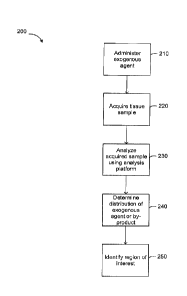
[0053] FIG. 2 is a flowchart of an example embodiment of a method
that can be used to identify a region of interest in tissue using mass
spectrometry.
[0054] FIG. 3A is a diagram illustrating the results of magnetic
resonance imaging of an exogenous agent passively targeted to breast
cancer tumors in live mice.
[0055] FIG. 3B is another diagram illustrating the results of magnetic
resonance imaging of an exogenous agent passively targeted to breast
cancer tumors in live mice.
[0056] FIG. 4 is a diagram illustrating a plot of the mass spectrum of
an
exogenous agent absorbed on the surface of a glass slide and inside mouse
kidneys using DESI-MS.
[0057] FIG. 5A is a diagram illustrating a plot of the mass spectrum
of a
breast cancer tumor from a mouse and the mass spectrum of the breast
cancer tumor after administration of an exogenous agent to the mouse.
CA 02969251 2017-05-30
WO 2016/090471
PCT/CA2015/051282
- 11 -
[0058] FIG. 5B is a diagram illustrating the spatial distribution
pattern of
various elements of the exogenous agent inside the breast cancer tumor
whose mass spectrum is shown in FIG. 5A.
[0059] FIG. 5C is a diagram illustrating magnetic resonance images of
the breast cancer tumor in a live mouse prior to administration of an
exogenous agent and at time delays after administration of the exogenous
agent.
[0060] FIG. 6A is a diagram illustrating a plot of the mass spectrum
of
the kidneys of a mouse and the mass spectrum of the kidneys after
administering an exogenous agent to the mouse.
[0061] FIG. 6B is a diagram illustrating the spatial distribution
pattern of
the exogenous agent within the mouse kidney of FIG. 6A.
[0062] FIG. 6C is a diagram illustrating magnetic resonance images of
kidneys of a live mouse prior to administration of an exogenous agent and at
time delays after administration.
[0063] FIG. 7A is a diagram illustrating a localization pattern of an
exogenous agent administered to a breast cancer tumor in a mouse.
[0064] FIG. 7B is a diagram illustrating a localization pattern of an
exogenous agent administered to the kidneys of the mouse of FIG. 7A.
[0065] FIG. 8A is a diagram illustrating a CD31 immunostained image
of a human breast cancer tumor excised from a mouse.
[0066] FIG. 8B is a diagram illustrating an overlay of an MS-image on
the immunostained image of FIG. 8A showing the localization of an
exogenous agent administered to the excised breast cancer tumors.
[0067] FIG. 80 is a diagram illustrating a zoomed in view of the
immunostained image of FIG. 8A.
[0068] FIG. 9A is another diagram illustrating the boundary of a human
breast cancer tumor.
CA 02969251 2017-05-30
WO 2016/090471
PCT/CA2015/051282
- 12 -
[0069] FIG. 9B is a diagram illustrating an overlay of a DESI-MS image
on PCK immunocytochemistry staining of the epithelial cells of the tissue
shown in FIG. 9A.
[0070] FIG. 9C is a diagram illustrating an overlay of a DESI-MS image
on Hematoxylin and Eosin (H&E) staining of the tissue shown in FIG. 9A.
[0071] FIG. 10A is a diagram illustrating the distribution of an
exogenous agent in 3 areas of a breast cancer tumor.
[0072] FIG. 10B is a diagram illustrating a quantified distribution of the
exogenous agent shown in FIG. 10A.
[0073] FIG. 11A is a diagram illustrating a plot of the expected
abundance of an exogenous marker in three tissue regions.
[0074] FIG. 11B is a diagram illustrating a plot of the measured
abundance of an exogenous marker in three tissue regions using quantitative
MS.
[0075] FIG. 11C is a diagram illustrating a plot of the measured
abundance of an exogenous marker in three tissue regions using qualitative
MS.
[0076] FIG. 12A is a diagram illustrating an example tissue region
including a region of healthy tissue and a region of diseased tissue.
[0077] FIG. 12B is an example plot illustrating the peak ratios of an
administered exogenous agent.
[0078] FIG. 120 is an example plot illustrating the peak ratios
associated with the administered exogenous agent detected in the healthy
region of FIG. 12A.
[0079] FIG. 12D is an example plot illustrating the peak ratios
associated with the administered exogenous agent detected in the diseased
region of FIG. 12A.
CA 02969251 2017-05-30
WO 2016/090471
PCT/CA2015/051282
- 13 -
[0080] Further aspects and features of the embodiments described
herein will appear from the following description taken together with the
accompanying drawings.
DETAILED DESCRIPTION OF THE EMBODIMENTS
[0081] Various apparatuses or methods will be described below to
provide an example of an embodiment of the claimed subject matter. No
embodiment described below limits any claimed subject matter and any
claimed subject matter may cover methods or apparatuses that differ from
those described below. The claimed subject matter is not limited to
apparatuses or methods having all of the features of any one apparatus or
methods described below or to features common to multiple or all of the
apparatuses or methods described below. It is possible that an apparatus or
methods described below is not an embodiment that is recited in any claimed
subject matter. Any subject matter disclosed in an apparatus or methods
described below that is not claimed in this document may be the subject
matter of another protective instrument, for example, a continuing patent
application, and the applicants, inventors or owners do not intend to abandon,
disclaim or dedicate to the public any such invention by its disclosure in
this
document.
[0082] Furthermore, it will be appreciated that for simplicity and
clarity
of illustration, where considered appropriate, reference numerals may be
repeated among the figures to indicate corresponding or analogous elements.
In addition, numerous specific details are set forth in order to provide a
thorough understanding of the embodiments described herein. However, it will
be understood by those of ordinary skill in the art that the embodiments
described herein may be practiced without these specific details. In other
instances, well-known methods, procedures and components have not been
described in detail so as not to obscure the embodiments described herein.
Also, the description is not to be considered as limiting the scope of the
embodiments described herein.
CA 02969251 2017-05-30
WO 2016/090471
PCT/CA2015/051282
- 14 -
[0083] It should also be noted that the terms "coupled" or 'coupling"
as
used herein can have several different meanings depending in the context in
which these terms are used. For example, the terms coupled or coupling can
have a mechanical, electrical or communicative connotation. For example, as
used herein, the terms coupled or coupling can indicate that two elements or
devices can be directly connected to one another or connected to one another
through one or more intermediate elements or devices via an electrical
element, electrical signal or a mechanical element depending on the particular
context. Furthermore, the term "communicative coupling" indicates that an
element or device can electrically, optically, or wirelessly send data to
another
element or device as well as receive data from another element or device.
[0084] It should also be noted that, as used herein, the wording
"and/or" is intended to represent an inclusive-or. That is, "X and/or Y" is
intended to mean X or Y or both, for example. As a further example, "X, Y,
and/or Z" is intended to mean X or Y or Z or any combination thereof.
[0085] It should be noted that terms of degree such as
"substantially",
"about" and "approximately" as used herein mean a reasonable amount of
deviation of the modified term such that the end result is not significantly
changed. These terms of degree may also be construed as including a
deviation of the modified term if this deviation would not negate the meaning
of the term it modifies.
[0086] It should be noted that the term "exogenous agent" as used
herein refers to any compound, chemical, drug, etc., such as, but not limited
to, a contrast agent, which is capable of forming a by-product in the region
of
interest in the tissue and is identifiable from a sample based on the tissue
using mass spectroscopy. The sample being based on the tissue means that
the sample can be taken from the tissue, or it can be an altered version of
the
tissue due to the sampling process. Alternatively, the exogenous agent may
be processed and metabolized in different regions of the tissue. The
exogenous agent is typically not found naturally occurring in the body or is
CA 02969251 2017-05-30
WO 2016/090471
PCT/CA2015/051282
- 15 -
naturally occurring in the body at much smaller concentrations than when the
exogenous agent is introduced to the body.
[0087] It should also be noted that the term "by-product" as used
herein
refers to a product or adduct formed in situ due to the degradation,
ionization,
complexation or otherwise transformation of the exogenous agent, and which
is identifiable from the tissue or in the tissue using mass spectroscopy. For
example, the exogenous agent may be ionized in situ to form an adduct such
as, but not limited to, a salt, for example.
[0088] Furthermore, the recitation of numerical ranges by endpoints
herein includes all numbers and fractions subsumed within that range (e.g. 1
to 5 includes 1, 1.5, 2, 2.75, 3, 3.90, 4, and 5). It is also to be understood
that
all numbers and fractions thereof are presumed to be modified by the term
"about" which means a variation of up to a certain amount of the number to
which reference is being made if the end result is not significantly changed.
[0089] Conventionally identifying a diseased region is heavily tied to the
availability of validated molecular markers for the disease in question. This
has motivated many ex vivo studies to identify, validate and catalogue small
molecule disease markers with utility in intraoperative IMS. While these IMS
techniques provide promising avenues for exploration, wide adoption in the
medical domain of small molecule mass spectrometry for identifying diseased
regions has stalled mainly due to the strict requirement for biomarker
knowledge.
[0090] Described herein are various example embodiments of a system
and method that can be used for identifying a region of interest in a
patient's
tissue using mass spectrometry. The term patient as used herein is to be
understood to refer to both human patients as well as animal patients.
Although the systems and methods described herein may be used primarily
with humans, they can also be applied to identify regions of interest in
animal
tissues as well. Examples of regions of interest include regions with
different
metabolic states, disease states, lesions, micro environments, regions of
CA 02969251 2017-05-30
WO 2016/090471
PCT/CA2015/051282
- 16 -
tissue heterogeneity such as stroma in a tumor, areas of necrosis or hypoxia,
regions of inflammation and boundaries of tumors among others.
[0091] In particular, the systems and methods described herein can
identify a region of interest in tissue based on the distribution of an
exogenous
agent within the tissue. In some embodiments, the region of interest may be
identified based on the distribution of a by-product of the exogenous agent.
For example, the by-product may be caused as a result of the processing and
metabolism of the exogenous agent in different regions of the tissue, such as
an adduct. In other embodiments, the region of interest may be identified
based on the distribution of the by-product of the exogenous agent in addition
to the exogenous agent. In other embodiments, the region of interest may be
identified based on the distribution of the exogenous agent, such as in laser-
based applications.
[0092] Embodiments of the systems and methods described herein
may provide more accurate mapping of regions of interests compared to
conventional techniques. Further, at least some of the embodiments
described herein may allow regions of interests, such as tumors, to be
mapped even without knowledge of endogenous markers. The size of such
detected tumors are larger than the resolution of the sampling process (e.g.
the laser focus, the DESI solvent spray focus, the size of the electrocautery
probe, etc.) and the minimum sampling resolution (and therefore minimum
detectable tumor size) ranges from about 20-50 microns to about one
millimeter. In some cases, regions of interest can be identified by detecting
regions of tissue having greater abundance of an administered exogenous
agent, or showing an altered metabolism, absorption, processing,
degradation, ionization, complexation or otherwise transformation of the
exogenous agent compared to healthy tissue. Thus, regions of interest can be
identified using a known administered exogenous agent even when an
endogenous marker, such as a lipidic signature, is unknown for that particular
region of interest.
CA 02969251 2017-05-30
WO 2016/090471
PCT/CA2015/051282
- 17 -
[0093] Previous approaches to tumor type identification and tumor
subclass grading rely heavily on the availability of validated molecular
markers (endogenous markers) for the diseases being mapped. One such
approach is described in detail in the patent application "System and method
for identification of biological tissues" (WO 2010136887 Al) which identifies
tissue samples based on "tissue-related" data that are endogenous to the
tissue. This approach differs from the approach taken in the embodiments
described herein as it identifies tissues based on endogenous markers, not
exogenous agents, and is therefore severely hindered in adoption for
widespread clinical use in instances where no validated markers exist for the
disease in question. Here, the inventors have identified that the act of
merely
mapping a region of interest such as a tumor does not require knowledge of
validated endogenous markers.
[0094] Some embodiments of the systems and methods described in
accordance with the teachings herein are able to overcome the limitations of
previous approaches using high sensitivity imaging of one or more exogenous
agents introduced to a patient's tissue. For example, in accordance with the
teachings herein, exogenous magnetic resonance (MR) contrast agents
passively targeted to disease sites can be used with Ambient Desorption
Electrospray Ionization Mass Spectrometry (DESI-MS) imaging to reveal
disease sites without knowledge of endogenous markers for the disease. As
discussed herein, these embodiments have been shown to reveal cancer
regions in a mouse model of human breast tumors in test cases without
invoking knowledge of lipidic markers for this disease. Other types of cancer
regions that may be detected include brain cancer, bone cancer, prostate
cancer, stomach cancer, kidney cancer, and lung cancer since these different
cancers can be identified (e.g. can "light up") when using contrast agents (Xu
GZ et al., "Comparison of FDG whole-body PET/CT and gadolinium-
enhanced whole-body MRI for distant malignancies in patients with malignant
tumors: a meta-analysis." Evidence-based Medical Center, The First Affiliated
Hospital of Guangxi Medical University, Graduate School of Guangxi Medical
University, Nanning, China; Ann Oncol. 2013; Jan. 24(1): pp. 96-101.), and
CA 02969251 2017-05-30
WO 2016/090471
PCT/CA2015/051282
- 18 -
therefore the presence of such agents with cancer regions can be detected
using mass spectrometry. Accordingly, any cancer that may be visualized
using contrast enhanced medical imaging can be detected using the
teachings herein.
[0095] The systems and methods described herein are not limited to
using MR contrast agents as an exogenous agent, but can generally include
all contrast agents used for medical imaging such as, but not limited to,
small
molecule exogenous agents, nanoparticle exogenous agents, as well as
metabolic precursors, moieties of metabolic precursors (such as bifunctional
molecules that may contain a metabolizable moiety and a tag for easy
detection) and heavy atom contrast agents, for example. Similarly, the
systems and methods described herein are not limited to using DESI-MS, and
can use other forms of mass spectrometry technology such as, but not limited
to, inductively-coupled plasma (ICP) mass spectrometry and matrix-assisted
laser desorption ionization (MALDI) mass spectrometry imaging, for example.
[0096] In at least one embodiment, the exogenous agent is any agent
capable of forming a by-product, wherein the by-product is identifiable using
mass spectroscopy. In one embodiment, the exogenous agent is ionizable,
thus forming an ionized by-product which is capable of forming a salt adduct
with other ions present in situ. In a further embodiment, the exogenous agent
is any agent containing a heavy atom, such as a transition metal, actinide,
lanthanide, or other atom in periods 5, 6, or 7, such as iodine. In a further
embodiment, the heavy atom is ionizable. For example, the exogenous agent
is any metal-based agent, wherein the metal does not exist in the body, tissue
and/or region of interest, or if the metal is present in the body, it is
present at a
significantly different concentration when the metal-based agent is
administered. In another embodiment, the metal-based exogenous agent is a
chelated metal agent (for example, a chelated gadolinium metal agent), an
iron oxide containing agent, an iron-platinum containing agent, a manganese
containing agent, a chromium containing agent, or tumour specific
metalloporphyrins.
CA 02969251 2017-05-30
WO 2016/090471
PCT/CA2015/051282
- 19 -
[0097] In one
embodiment, the exogenous agent contains a heavy
atom and is an MRI contrast agent, such as a metallic MRI contrast agent, or
a CT (computerized tomography) contrast agent, such as an iodine-based CT
contrast agent. In one embodiment, the MRI agent is gadoterate (Dotarem@),
gadodiamide (Omniscan0), gadobenate (Multi Hance ), gadopentetate
(Magnevist@, Magnegita0, Gado-MRTO ratiopharm), gadoteridol
(ProHance0), gadoversetamide (OptiMARK0), gadoxetate (Primovist0),
gadobutrol (Gadovist0), gadoterate (Dotarem0), gadodiamide (Omniscan@),
gadobenate (MultiHance@), gadopentetate (Magnevist@), gadoteridol
(ProHance@), gadofosveset (Ablavar0), gadoversetamide (OptiMARK@),
gadoxetate (Eovist0), gadobutrol (Gadavist0), Feridex I.V. (also known as
Endorem@ and ferumoxides), Resovist (also known as Cliavist@), Sinerem@
(Combidex@), or Lumirem0 (Gastromark0). In one embodiment, the iodine-
based CT agent is diatrizoate, metrizoate, ioxaglate, iopamidol, or iohexol.
[0098] In one
embodiment, the exogenous agent is gadoteridol. In
another embodiment, the exogenous agent is gadoteridol which is ionizable in
situ, and is capable of forming salt adduct based on the presence of various
cations in the region of interest in the tissue. For example, the salt adduct
is
gadoteridol-Na + or gadoteridol-K+.
[0099] In some
embodiments, the exogenous agent can be in an
ionized state on their own. In other embodiments, the exogenous agent can
become ionized in the body after administration. In other embodiments, the
exogenous agent can be ionized by using an ionization source before entry
into a mass spectrometer. This latter ionization can take place either on the
tissue surface during sampling for the mass spectrometer or after transport of
a tissue plume to the entrance of the mass spectrometer (i.e. the ionization
source can be placed anywhere along a transfer line between the tissue
surface and the entrance of the mass spectrometer).
[00100] The
embodiments described herein may be used to provide a
new platform for accelerated intraoperative identification of tumor sites and
other regions of interest in the absence of known markers. Furthermore, the
CA 02969251 2017-05-30
WO 2016/090471
PCT/CA2015/051282
- 20 -
highly multiplexed nature of mass spectrometry imaging may further simplify
the disease identification process using a simple peak ratio test between
metabolic or catabolic byproducts of exogenous agents differentially absorbed
to, or metabolized by, the diseased tissues. In some cases, the peak ratio
test
can be used to determine a relative abundance of one or more exogenous
sub-agents or by-products of the exogenous agent in a sample acquired from
the tissue.
[00101] At least some of the embodiments of the systems and methods
described herein may also provide the capability to perform quantitative
mapping of regions of interest in tissue. In these cases, quantitative mass
spectrometry can be used to determine a quantitative (or absolute)
abundance of an administered exogenous agent or by-product of the
administered exogenous agent. This may provide more robust identification of
regions of interest and the boundaries of the regions of interest. This
approach may also provide more accurate boundary assessment with
exogenous agents that do not possess 100% targeting specificity, and are
thus present in the tissues surrounding the region of interest, albeit to a
lesser
extent.
[00102] In one embodiment, the exogenous agents are administered
intravenously as a solution, or alternatively, the agents are encapsulated
inside liposomes or porphysomes and subsequently administered
intravenously.
[00103] Administering exogenous agents that contain metallic elements
along with the detection of these agents by Inductively Coupled Plasma Mass
Spectrometry (ICP-MS), particularly in a spatially resolved manner, may
provide a more robust assessment of the boundaries of regions of interest.
The high tolerance to matrix effects of ICP-MS and quantitative ionization
methods, such as photo ionization, may also provide more robust assessment
of boundary regions. In some mass spectrometry methods, matrix effects
arise from the influence of tissue constituent on the acquisition (including
the
desorption and the ionization steps) of exogenous agents. These matrix
CA 02969251 2017-05-30
WO 2016/090471
PCT/CA2015/051282
- 21 -
effects can impact the activity coefficients of the exogenous agent, leading
to
difficulties with non-quantitative detection of the exogenous agent using
conventional mass spectrometry platforms. Non-quantitative imaging may
have difficulty interpreting the intensity of some administered exogenous
agents, such as contrast agents, because the intensity is affected by the
properties of the tissue. As a result, a contrast agent may accumulate in a
particular region, that is not the region of interest, but that non-
quantitative
imaging indicates is the region of interest. The robustness of quantitative
MS,
such as ICP-MS or gentle desorption as neutrals followed by quantitative
ionization mass spectrometry in the face of these matrix effects enables the
more robust identification and mapping of a region of interest. In ICP-MS,
matrix material is obliterated by the high energy density of the plasma used
to
ionize the acquired sample thereby giving rise to a heavy atom signal
associated with the administered exogenous agent in a matric independent
manner. Nevertheless, many of the MS methods that are non-quantitative but
which reliably produce signals associated with relative ion intensity can have
adequate performance for identifying regions of interest.
[00104] Some embodiments in accordance with the teachings herein,
such as those employing ICP-MS, may also use contrast agents in an
injection dose far below what is required for conventional imaging modalities.
As such, embodiments described herein may extend the benefit of
intraoperative contrast-enhanced imaging of tumor boundaries to patients who
may be currently excluded due to health concerns. These embodiments may
also make the practice of tumor imaging with passive targeting of contrast
agents safer for all patients since lower doses of those agents may be used to
obtain acceptable imaging results.
[00105] The metallic elements contained in various exogenous agents
used in the systems and methods herein are likely to survive the forces of
laser ablation or electrocautery (which is used in the most commonly used
scalpels in the operating rooms that offer cutting and restores homeostatis at
the same time) and persist in the plume of electrocauterized tumors being
CA 02969251 2017-05-30
WO 2016/090471
PCT/CA2015/051282
- 22 -
resected. Accordingly, at least some embodiments of the systems and
methods described in accordance with the teachings herein provide an
intraoperative platform that may facilitate tumor resections on the basis of
mass spectrometry imaging of exogenous agents (e.g. small molecule
medical imaging contrast agents) or by-products in the exogenous agents
present in the laser ablation (or electrocautery) plume of targeted tumors
being resected
[00106] Various ablation devices can be used with the systems and
methods described herein, and the ablation devices can be operated in a
variety of modes and with different parameters. For example, laser ablation
can be performed using either a cauterizing or non-cauterizing laser operating
in a variety of modes, wavelengths, pulse durations and average power
values. In some cases, a laser ablation device that allows ablation in the
absence of significant damage to the tissue can be used. Other ablation
devices such as ultrasonic ablation devices, RF ablation devices and
electrocautery devices can also be used.
[00107] In one particular embodiment, a platform is provided that
comprises a transfer line (e.g. a suction-tube) coupling a high sensitivity
analysis unit (e.g. MS or ICP-MS) with any of a variety of surgical laser
scalpels or electrocautery blades, to provide intraoperative detection of
exogenous agents that allows near real time assessment of tumor boundaries
and is therefore much faster than intraoperative histology assessment. In
some cases, the transfer line may be a heated tube that can be either rigid or
flexible depending on the embodiment. In some cases, the transfer line itself
may house the illumination point of a surgical laser scalpel or a desorbing
laser fiber.
[00108] In another particular embodiment, there is provided a system
including a high sensitivity analysis unit (e.g. MS or ICP-MS) on standby in
an
operating room along with an appropriate interface to sample tissue material
that allows the analysis unit to measure, or to quantitatively measure (in
preferred embodiment), the amount of an exogenous agent that is present in
CA 02969251 2017-05-30
WO 2016/090471
PCT/CA2015/051282
- 23 -
a conventional biopsy or nanobiopsy volume excised from a tumor being
resected. This embodiment may allow the distribution and abundance
(qualitative or quantitative) of an administered exogenous agent or by-product
of the administered exogenous agent in an acquired sample to be determined
within seconds, which is much faster than is currently possible with
intraoperative histology.
[00109] Generally, embodiments of the system described herein may
include an agent administration component that is configured to administer an
exogenous agent to the tissue. The agent administration component can be
configured to administer the exogenous agent in a variety of ways such as
topically, enterally or parenterally.
[00110] In some cases, the agent administration component can be
configured to administer the exogenous agent directly to the tissue being
analyzed prior to acquisition of a sample from the tissue. For example, the
exogenous agent can be sprayed or deposited onto the tissue in areas where
samples are to be acquired. Alternatively, the agent administration component
can be configured to administer an exogenous agent to a patient such that the
exogenous agent diffuses to the region of the tissue being sampled. For
example, the agent administration component may administer an exogenous
agent to a patient orally, by injection (including any of intravenous,
intramuscular, and subcutaneous injection), and/or by inhalation.
[00111] A wide variety of exogenous agents can be used with
embodiments of the agent administration components described herein.
Generally, the exogenous agent may be any chemical agent, drug, structure
or functionality that is not a normal component of human or animal tissue, or
one that contains an isotopic variant of an endogenous molecule. This can
include, but is not limited to, various types of medical imaging contrast
agents,
drug molecules, metabolic precursors, moieties of metabolic precursors (such
as bifunctional molecules that contain a metabolizable moiety and a tag for
easy detection) functionalized molecules containing chelated metallic
elements, nanoparticles or custom cocktails of mass tags.
CA 02969251 2017-05-30
WO 2016/090471
PCT/CA2015/051282
- 24 -
[00112] In some cases, an exogenous agent may be administered that
includes a plurality of exogenous sub-agents (i.e. a cocktail of exogenous
agents) that undergo differential metabolism/absorption or processing at
disease sites and other regions of interest so that these regions of interest
can be identified by analyzing the distribution of the exogenous sub-agents
that were administered. The term exogenous sub-agent is used herein to refer
to the constituent components of an administered exogenous agent and may
include an exogenous agent.
[00113] Embodiments of the system may further include a sampling unit
configured to acquire a sample from the tissue. In different embodiments, the
sampling unit can include various sub-units configured to acquire a sample of
tissue for analysis. The sampling unit can be configured to acquire a sample
in various forms, such as direct acquisition of a tissue sample, by collecting
a
plume of ablated tissue material from an electrocautery knife or a laser
scalpel, and extraction of the exogenous agent or byproduct thereof from the
tissue for example. In some cases, a plume of tissue material can be directly
analyzed by the analysis platform while in other cases the plume of tissue
material may be condensed prior to analysis. As a skilled reader will
appreciate, in some cases tissue samples may require further processing
such as freezing or sectioning for example, depending on the particular
analysis platform implemented.
[00114] In some embodiments, the sampling unit may include a
desorption component configured to desorb the sample from the tissue. For
example, the desorption component may be a device such as, but not limited
to, a laser ablation device, a liquid extraction device, an electrocautery
device
such as an electrocautery knife, an electrospraying device, an ultrasonic
tissue atomizer, and a radio frequency ablation device among others.
[00115] Further, in at least some embodiments the sampling unit may
include an ionization component configured to ionize the sample prior to
analysis with the high sensitivity platform. For example, the ionization
component may be a device such as, but not limited to, an electrospray
CA 02969251 2017-05-30
WO 2016/090471
PCT/CA2015/051282
- 25 -
ionization device, a laser ionization device, a matrix-assisted ionization
device, a matrix-assisted laser desorption ionization device, an atmospheric
pressure chemical ionization device, a photo ionization device, a laser based
plasma source and an inductively-coupled plasma ionization device. In some
cases, the sampling unit may not require an ionization component, for
example where the administered exogenous agent includes charged particles,
or becomes ionized as an adduct (i.e. by-product of the administered
exogenous agent) in biological milieu.
[00116] In some
cases, the sampling unit may also include an ex-vivo
sampling unit that is able to acquire and secure an ex-vivo tissue sample from
a patient, such as a biopsy or nano-biopsy sample of tissue that can be
labelled by an exogenous agent. The ex-vivo sampling unit may include a
platform for securing the tissue sample in place for analysis by a high
sensitivity platform.
[00117] The system can
also include a high sensitivity platform
configured to analyze an acquired sample. In some embodiments, the high
sensitivity (analysis) platform can include various types of mass analyzers
used in mass spectrometry to determine a mass spectrum of the acquired
sample. The high sensitivity platform may also include additional components,
such as a processor, a display device and user controls depending on the
particular embodiment. These additional components may further assist users
in interacting with the system.
[00118] Generally,
the high sensitivity platform can be any analysis
platform that allows a more sensitive detection of the administered exogenous
agent compared to conventional modality imagers currently used to image
contrast agents or other exogenous contaminants (i.e. agents). The high
sensitivity platform can be configured determine at least one of a mass to
charge ratio and an elemental mass of at least one (expected) element in an
acquired sample from the tissue. In some cases, the high sensitivity platform
can be tuned to identify at least one of the presence (and also, therefore the
absences) and the abundance of at least one of a mass to charge ratio
CA 02969251 2017-05-30
WO 2016/090471
PCT/CA2015/051282
- 26 -
associated with the administered exogenous agent (or a by-product of the
administered exogenous agent) and an elemental mass associated with the
administered exogenous agent (or a by-product of the administered
exogenous agent). The high sensitivity platform can also be configured to
detect an administered exogenous agent at per tissue pixel amounts, lower
than, or consistent with the level required for conventional imaging
modalities.
[00119] In some cases, the high sensitivity platform can include one or
more of optical detection, fluorescence detection, Raman detection,
stimulated or unstimulated Raman spectroscopy of exogenous agents
targeted to tissues of expected regions of interest, Raman spectroscopy of
desorbed exogenous material in a gas phase. In some cases, these detection
methods can be used solely, while in preferred embodiments they may be
used in tandem with mass spectrometry platforms.
[00120] In some cases, using additional analysis platforms in tandem
may facilitate faster identification of regions of interest. For example,
using a
fluorescence detection platform, a fluorescent marker may reveal a potential
region of interest to guide more detailed MS analysis of the region of
interest.
This may allow the high sensitivity analysis platform to be quickly targeted
to a
potential region of interest and thus enable rapid identification of the
region of
interest and the boundary of the region of interest.
[00121] The high sensitivity analysis platform may be configured to
determine a distribution of an exogenous agent within the tissue based on the
analysis of the sample acquired from the tissue. The high sensitivity platform
can be configured to detect a smaller dose per pixel unit of the administered
exogenous agent (or a by-product thereof) compared to conventional imaging
modalities. The analysis platform may then identify a region of interest in
the
tissue based on the distribution of an exogenous agent, or a metabolic by-
product of the exogenous agent. Regions of interest such as, but not limited
to, tumors and other disease sites can be identified and mapped, even when
endogenous markers are not yet known or validated.
CA 02969251 2017-05-30
WO 2016/090471
PCT/CA2015/051282
- 27 -
[00122] Various exogenous agents can be used in the embodiments of
systems and methods for identifying a region of interest in a sample as
described herein. Typically, the exogenous agents will be agents that include
chemical elements not found in the human body or chemical elements in
ratios unnatural to those in the human body. In some embodiments, the
exogenous agents can include chemical elements that are passively or
actively targeted to, or absorbed, presented or metabolized differentially in
diseased tissues versus healthy tissues.
[00123] In at least some cases, the exogenous agents used with the
systems and methods described herein may also be limited to dose levels that
are not considered toxic or harmful in humans. In at least some cases, the
exogenous agents may be contrast agents developed for other imaging
modalities. The use of a high sensitivity analysis platform may allow such
contrast agents to be administered in doses that are lower than what is
required in conventional imaging modalities. This may also allow contrast
agents considered too toxic for use with other imaging modalities to be used
safely at lower doses. Accordingly, contrast agents that identify regions of
interest more effectively but may be considered too toxic at doses required
for
conventional imaging modalities may be used at lower, safe levels in
embodiments of the systems and methods described herein.
[00124] In some cases, the exogenous agents may include at least one
metallic element. This may allow regions of interest to be identified based on
the distribution of the metallic element in the tissue. In some cases, the
exogenous agents may include isotopic variants (e.g. variants of metallic
isotopes or metabolic precursor isotopes). The distribution of isotope ratios
of
different isotopes can be used to identify regions of interest and the
boundaries of regions of interest.
[00125] The use of exogenous agents may also allow regions of interest
to be identified in a quantitative manner. At least some embodiments of the
high sensitivity platform can be configured to determine a quantitative
distribution of the exogenous agent or a by-product of the exogenous agent
CA 02969251 2017-05-30
WO 2016/090471
PCT/CA2015/051282
- 28 -
(or elements/isotopes thereof). For example, an inductively-coupled plasma
mass spectrometry (ICP-MS) system may be used to determine a quantitative
distribution of the exogenous agent. This may allow for a more precise and
clear identification of regions of interest and the boundaries of the regions
of
interest. Such embodiments may provide more accurate boundary
assessment with exogenous agents that do not possess 100% targeting
specificity, and are also present in the tissues surrounding the regions of
interest, albeit to a lesser extent.
[00126] The example embodiments of the systems and methods
described in accordance with the teachings herein may be implemented as a
combination of hardware or software. In some cases, the example
embodiments described herein may be implemented, at least in part, by using
one or more computer programs, executing on one or more programmable
devices comprising at least one processing element, and at least one data
storage element (including volatile and non-volatile memory and/or storage
elements). These devices may also have at least one input device (e.g. a
keyboard, mouse, a touchscreen, and the like), and at least one output device
(e.g. a display screen, a printer, a wireless radio, and the like) depending
on
the nature of the device.
[00127] It should also be noted that there may be some elements that
are used to implement at least part of one of the embodiments described
herein that may be implemented via software that is written in a high-level
procedural language such as object oriented programming. Accordingly, the
program code may be written in C, C++ or any other suitable programming
language and may comprise modules or classes, as is known to those skilled
in object oriented programming. Alternatively, or in addition thereto, some of
these elements implemented via software may be written in assembly
language, machine language or firmware as needed. In either case, the
language may be a compiled or interpreted language.
[00128] At least some of these software programs may be stored on a
storage media (e.g. a computer readable medium such as, but not limited to,
CA 02969251 2017-05-30
WO 2016/090471
PCT/CA2015/051282
- 29 -
ROM, magnetic disk, optical disc) or a device that is readable by a general or
special purpose programmable device. The software program code, when
read by the programmable device, configures the programmable device to
operate in a new, specific and predefined manner in order to perform at least
one of the methods described herein.
[00129] Furthermore, at least some of the programs associated with the
systems and methods of the embodiments described herein may be capable
of being distributed in a computer program product comprising a computer
readable medium that bears computer usable instructions for one or more
processors. The medium may be provided in various forms, including non-
transitory forms such as, but not limited to, one or more diskettes, compact
disks, tapes, chips, and magnetic and electronic storage. In alternative
embodiments, the medium may be transitory in nature such as, but not limited
to, wire-line transmissions, satellite transmissions, internet transmissions
(e.g.
downloads), media, digital and analog signals, and the like. The computer
useable instructions may also be in various formats, including compiled and
non-compiled code.
[00130] Referring now to FIG. 1, shown therein is a block diagram of an
example embodiment of a system 10 that can be used to identify a region of
interest in a patient's tissue. The system 10 includes an operator unit 12, a
high sensitivity analysis platform 40, a sampling interface 42, an ionization
device 44 and a sampling device 46. The system 10 is provided as an
example and there can be other embodiments of the system 10 with different
components or a different configuration of the components described herein.
For example, the system 10 can also include an agent administration
component. The system 10 further includes several power supplies (not all
shown) connected to various components of the system 10 for providing
power thereto as is commonly known to those skilled in the art.
[00131] In general, a user may interact with the operator unit 12 and
the
sampling device 46 to acquire samples from the tissue, such as ex-vivo tissue
samples from a patient or a plume of ablated tissue material, and then
CA 02969251 2017-05-30
WO 2016/090471
PCT/CA2015/051282
- 30 -
perform analysis and further data analysis (such as mass spectrometry) to
identify a region of interest in the tissue. In some cases, the operator unit
12
and the analysis platform 40 may be combined as a high sensitivity analysis
platform that is able to perform mass spectrometry analysis and further data
analysis on a sample acquired from the tissue.
[00132] The operator unit 12 comprises a processing unit 14, a display
16, a user interface 18, an interface unit 20, Input/Output (I/O) hardware 22,
a
wireless unit 24, a power unit 26 and a memory unit 28. The memory unit 28
comprises software code for implementing an operating system 30, various
programs 32, a data analysis module 34, and one or more databases 36.
Many components of the operator unit 12 can be implemented using a
desktop computer, a laptop, a mobile device, a tablet, and the like.
[00133] The processing unit 14 controls the operation of the operator
unit 12 and can be any suitable processor, controller or digital signal
processor that can provide sufficient processing power depending on the
configuration, purposes and requirements of the system 10 as is known by
those skilled in the art. For example, the processing unit 14 may be a high
performance general processor. In alternative embodiments, the processing
unit 14 may include more than one processor with each processor being
configured to perform different dedicated tasks. In alternative embodiments,
specialized hardware can be used to provide some of the functions provided
by the processing unit 14.
[00134] The display 16 can be any suitable display that provides visual
information depending on the configuration of the operator unit 12. For
instance, the display 16 can be a cathode ray tube, a flat-screen monitor and
the like if the operator unit 12 is a desktop computer. In other cases, the
display 16 can be a display suitable for a laptop, tablet or handheld device
such as an LCD-based display and the like.
[00135] The user interface 18 can include at least one of a mouse, a
keyboard, a touch screen, a thumbwheel, a track-pad, a track-ball, a card-
reader, voice recognition software and the like again depending on the
CA 02969251 2017-05-30
WO 2016/090471
PCT/CA2015/051282
- 31 -
particular implementation of the operator unit 12. In some cases, some of
these components can be integrated with one another.
[00136] The interface unit 20
can be any interface that allows the
operator unit 12 to communicate with other devices or computers. In some
cases, the interface unit 20 can include at least one of a serial port, a
parallel
port or a USB port that provides USB connectivity. The interface unit 20 can
also include at least one of an Internet, Local Area Network (LAN), Ethernet,
Firewire, modem or digital subscriber line connection. Various combinations of
these elements can be incorporated within the interface unit 20.
[00137] The I/O hardware 22 is
optional and can include, but is not
limited to, at least one of a microphone, a speaker, a display device and a
printer, for example.
[00138] The wireless unit 24 is
optional and can be a radio that
communicates utilizing CDMA, GSM, GPRS or Bluetooth protocol according
to standards such as IEEE 802.11a, 802.11b, 802.11g, or 802.11n. The
wireless unit 24 can be used by the operator unit 12 to communicate with
other devices or computers.
[00139] The power unit 26 can
be any suitable power source that
provides power to the operator unit 12 such as a power adaptor or a
rechargeable battery pack depending on the implementation of the operator
unit 12 as is known by those skilled in the art.
[00140] The memory unit 28 can
include RAM, ROM, one or more hard
drives, one or more flash drives or some other suitable data storage elements
such as disk drives, etc. The memory unit 28 may be used to store an
operating system 30 and programs 32 as is commonly known by those skilled
in the art. For instance, the operating system 30 provides various basic
operational processes for the operator unit 12. The programs 32 include
various user programs so that a user can interact with the operator unit 12 to
perform various functions such as, but not limited to, acquiring data such as
mass spectrometry data from the analysis platform 40, viewing and
CA 02969251 2017-05-30
WO 2016/090471
PCT/CA2015/051282
- 32 -
manipulating data, adjusting parameters related to data analysis as well as
sending messages as the case may be.
[00141] In some cases, the acquired data may be preprocessed by the
analysis platform 40 and transferred to the operator unit 12 through interface
unit 20. The preprocessing may include standard signal processing
techniques such as, but not limited to, at least one of amplification,
filtering
and de-noising (e.g. averaging) using parameters that depend on the
particular signals of interest that are acquired. The interface unit 20 may be
a
multichannel data interface coupling the analysis platform 40 to the operator
unit 12.
[00142] The data analysis module 34 processes the data that is
recorded by the analysis platform 40 in order to identify the location of a
region of interest in at least one image of the tissue region from which the
sample(s) being processed were acquired. The data analysis module 34 is
typically implemented using software, but there may be instances in which
they are implemented using FPGA or application specific circuitry.
[00143] The databases 36 can be used to store data for the system 10
such as system settings, parameter values, and calibration data. The
databases 36 can also store other information required for the operation of
the
programs 32 or the operating system 30 such as dynamically linked libraries
and the like.
[00144] The operator unit 12 comprises at least one interface that the
processing unit 14 communicates with in order to receive or send information.
This interface can be the user interface 18, the interface unit 20 or the
wireless unit 24. For instance, the exogenous agent whose distribution is
being analyzed in a particular implementation of the system 10 may be
inputted by a user through the user interface 18 or this information may be
received through the interface unit 20 from a computing device. The
processing unit 14 can communicate with either one of these interfaces as
well as the display 16 or the I/O hardware 22 in order to output information
related to the distribution of exogenous agent, identification of tumor
locations
CA 02969251 2017-05-30
WO 2016/090471
PCT/CA2015/051282
- 33 -
and tumor boundary mapping. In addition, users of the operator unit 12 can
communicate information across a network connection to a remote system for
storage and/or further analysis in some embodiments. This communication
may also include email communication.
[00145] The user can also use the operator unit 12 to input information
needed for system parameters that are needed for proper operation of the
system 10 such as calibration information and other system operating
parameters as is known by those skilled in the art. Data that are obtained
from
tests, as well as parameters used for operation of the system 10, may be
stored in the memory unit 28. The stored data may include raw acquired data,
preprocessed acquired data as well as processed tumor location and tumor
mapping data.
[00146] The analysis platform 40 comprises hardware and circuitry that
is used to determine the distribution of an exogenous agent or a by-product of
the exogenous agent in an acquired sample from the tissue. For example, the
analysis platform 40 may be a mass analyzer that is configured to determine
the mass-to-charge ratio and abundance of gas-phase ions in the acquired
sample. In various embodiments, the analysis platform 40 may be
implemented using a mass analyzer, a wide range scanner, or an application
specific compact footprint limited mass range analyzer. In some cases, the
analysis platform may also include at least one of an optical detection
platform, a fluorescence detection platform and a Raman detector.
[00147] The sampling device 46 comprises hardware and circuitry that
can be used to acquire a sample from a patient's tissue. In various
embodiments, the sampling device 46 may be any one of a plurality of
devices that can be used for desorbing samples from a patient's tissue, such
as during a tumor rescission. For example, the sampling device 46 may be
one of a laser ablation device, a liquid extraction device, an electrocautery
device such as an electrocautery knife, an electrospraying device, an
ultrasonic tissue atomizer, a radio frequency ablation device, and a plasma
knife among others. The sampling device 46 may also include various types
CA 02969251 2017-05-30
WO 2016/090471
PCT/CA2015/051282
- 34 -
of sampling tools and equipment used to acquire and secure a biopsy or
nano-biopsy sample for analysis by the analysis platform 40. The sampling
device 46 may also include a small needle that can be used to acquire tissue
samples for analysis in an ex-vivo analysis platform.
[00148] The ionization device 44 comprises hardware and circuitry that
can be used to ionize an acquired sample prior to analysis by the analysis
platform 40. In various embodiments, the ionization device 44 can be any one
of a variety of ionization devices used in mass spectrometry applications. For
example, the ionization device 44 can be one of an electrospray ionization
device, a laser ionization device, a matrix-assisted ionization device, a
matrix-
assisted laser desorption ionization device, an atmospheric pressure chemical
ionization device, a photo ionization device, rapid evaporative ionization
device, and an inductively-coupled plasma or laser plasma ionization device
among others.
[00149] In some cases, the ionization device 44 may be co-located with
the sampling device 46 and may ionize the acquired sample at the source. In
some cases, the sampling device 46 and the ionization device 44 may be
combined, for example using desorption electrospray ionization. In other
cases, the ionization device 44 may be located closer to the analysis platform
40 and may ionize an acquired sample after transportation to (or near to) the
analysis platform 40. Alternatively, in some cases, the system 10 may not
include an ionization device 44, for example where the exogenous agent
being administered is charged, or becomes charged by virtue of adducts
formed with endogenous tissue material.
[00150] The sampling interface unit 42 can be used to transport an
acquired sample to the high sensitivity platform and/or support a sample for
analysis with the high sensitivity platform. In some cases the sampling
interface unit 42 can be a transportation unit that may include a transfer
line
coupling the sampling device 46 (and optionally the ionization device 44) to
the analysis platform 40. The transfer line can be configured to transport an
acquired sample to the high sensitivity platform.
CA 02969251 2017-05-30
WO 2016/090471
PCT/CA2015/051282
- 35 -
[00151] For example, the transfer line may use the suction provided by
the analysis platform 40 to transport a sample acquired by the sampling
device 46 to the analysis platform 40. Alternatively, in some cases, the
transfer line may use an additional pump to provide suction by introducing
negative pressure at the analysis platform side of the transfer line to pull
the
sample from the sampling device 46 to the analysis platform 40.
[00152] In some cases, the transfer line may be a heated tube. In
various embodiments the transfer line can be rigid or flexible. In some cases,
the transfer line may also house the illumination point of the ionization
device
44. For example, the transfer line can house a laser fiber that can be used to
desorb a sample from the tissue. In some cases, an ionization device 44 can
be located at a first end of the transfer line proximate the tissue, while in
other
cases the ionization device can be located at a second end of the transfer
line, closer to the analysis unit 40.
[00153] In various embodiments, using an ICP-MS system the ionization
device 44 may use inductively-coupled plasma to ionize an acquired sample.
In one such embodiment, the sampling interface unit 42 may also include a
transfer line coupling the sampling device 46 and the mass analyzer of the
ICP-MS system. The sampling interface unit 42 can be configured to apply a
positive pressure on the transfer line at a first end of the transfer line,
the first
end being proximate to the tissue being sampled. This may allow the acquired
sample to be transported to the mass analyzer more rapidly in embodiments
employing the suction of the mass analyzer unit.
[00154] In some cases, the transfer line may also include tracking
markers. The analysis platform 40 can be configured to identify a location of
the tracking markers to identify the location of the first end of the transfer
line
proximate the location of the sample being acquired. This can be used to
track the location from which the sample was acquired, to be used when
determining a distribution of the exogenous agent or by-product and
identifying the region of interest and the boundary thereof.
CA 02969251 2017-05-30
WO 2016/090471
PCT/CA2015/051282
- 36 -
[00155] Referring now to FIG. 2, shown therein is a flowchart 200 of an
example embodiment of a method for identifying a region of interest in tissue
using mass spectrometry.
[00156] At 210, an exogenous agent is administered to the patient. The
exogenous agent can be administered in various ways, such as direct
administration (e.g. depositing or spraying), oral ingestion, injection, and
inhalation of the exogenous agent, for example. In some cases, the
exogenous agent can be administered directly to an area of tissue expected
to include the region of interest. In other cases the exogenous agent may be
administered to the patient elsewhere and allowed to diffuse to the tissue
that
will be sampled, for example when administered orally, by injection or by
inhalation. The exogenous agent can also be administered to the tissue in
various ways including labelling by spraying or depositing (in vivo or ex
vivo).
[00157] Generally, the exogenous agent administered may be any
chemical agent, drug, structure or functionality that is not a normal
component
of human or animal tissue, or one that contains an isotopic variant of an
endogenous molecule. In some embodiments, the exogenous agent can be
any contrast agent already used and approved for use. In some cases, the
administered exogenous agent can also include at least one metallic element,
a metabolic precursor, an isotopic variant, or at least one heavy atom for
example. In some cases, the exogenous agent can be administered in a
known clinically relevant dose, while in some cases the dose may be lower
than compared to what is required in a conventional imaging modality. A lower
dose can be used in some cases due to the high sensitivity analysis platform
40 used to analyze the samples acquired from the tissue.
[00158] In some cases, a custom designed exogenous agent can be
used that includes a plurality of exogenous sub-agents. Using a plurality of
exogenous sub-agents can enable a region of interest to be identified based
on a distribution of each the plurality of exogenous sub-agents (or by-
products
thereof). This may provide more than one identifying mass to charge ratio
peak or elemental mass peak during analysis by the high sensitivity platform
CA 02969251 2017-05-30
WO 2016/090471
PCT/CA2015/051282
- 37 -
40. Areas where the peaks are coincident for the exogenous sub-agents (or
by-products) can then be used to identify the region of interest.
[00159] The plurality of exogenous sub-agents can be administered in a
variety of ways. For example, each of the exogenous sub-agents may be
administered simultaneously or encapsulated in a carrier vehicle. In some
cases, the carrier vehicle can be a lipidic structure such as a liposome for
example for these exogenous sub-agents and the exogenous agents
described herein, as well as existing imaging contrast agents described herein
and any combination thereof, and all agents being developed for use with
MRI, CT or Fluorescence imaging. This can allow a plurality of exogenous
sub-agents to be administered as an exogenous agent in a mixture that
facilitates efficient detection with a high sensitivity analysis platform such
as
mass spectrometry.
[00160] At 220, a sample from the tissue is acquired, for example using
the sampling device 46. In some cases, the sample may be acquired prior to
administering an exogenous agent, for example when the exogenous agent is
administered to a tissue sample ex-vivo. In other cases the sample can be
acquired after administration of an exogenous agent, using the administration
methods described herein such as in vivo labelling, oral ingestion or
intravenous injection.
[00161] The sample can be acquired in a variety of ways, such as by
direct desorption of in vivo tissue or from ex vivo biopsied samples. Examples
of sampling techniques for in vivo tissues include, but are not limited to,
laser
ablation vaporization, liquid extraction, capture of electrocautery plumes,
radio
frequency ablation, ultrasonic ablation, and DESI etc. Further, the sample can
be acquired in a variety of forms, such as a sample of the tissue itself, as a
plume from an electrocautery knife or a laser ablation device, or by
extracting
the exogenous agent or a by-product of the exogenous agent from the tissue
(without acquiring a sample of the tissue itself) such as by using liquid
extraction for example.
CA 02969251 2017-05-30
WO 2016/090471
PCT/CA2015/051282
- 38 -
[00162] In some cases, the sample may also be ionized when the
sample is acquired or after the sample is acquired, but before analysis. For
example, the sample may be ionized using a plasma source such as
inductively-coupled plasma. When a transfer line is used, the sample can be
ionized prior to being transported towards the analysis platform 40 or after
transportation by the transfer line, but prior to analysis by the analysis
platform 40. Other ionization methods may also be used depending on the
particular embodiment of the ionization device 44 that is implemented. In
some cases, ionization of an acquired sample may not be necessary, for
example where the administered exogenous agent already comprises ions or
has by-products that become ionized in the tissue.
[00163] At 230, the acquired sample is analyzed using a high
sensitivity
analysis platform. The analysis platform 40 may identify a mass spectrum of
the elements in the acquired sample. The analysis platform 40 can identify the
mass-to-charge ratio (and/or elemental mass) and abundance of gas-phase
ions in the acquired sample. In some cases, two-dimensional spectrometry
imaging may be used to determine the presence and/or abundance of
elements having different mass-to-charge ratios. In some cases, the analysis
platform 40 may be configured to determine a relative abundance of the ions
present in the acquired sample. In other cases, the analysis platform can be
configured to determine an absolute (i.e. quantitative) abundance of the ions
present in the acquired sample.
[00164] Various types of high sensitivity analysis platforms can be
used
here, such as, but not limited to, mass analyzers, wide range scanners and
application specific compact footprint limited mass range analyzers. In some
cases, one or more of an optical detection platform, a fluorescence detection
platform, a Raman detection platform, a stimulated or unstimulated Raman
spectroscopy platform for spectroscopy of exogenous agents targeted to
tissue types expected to be in a region of interest, and a Raman spectroscopy
platform for spectroscopy of desorbed exogenous material in a gas phase. In
some cases, these further detection platforms can be used solely, while in
CA 02969251 2017-05-30
WO 2016/090471
PCT/CA2015/051282
- 39 -
preferred embodiments they may be used in tandem with mass spectrometry
analysis platforms mentioned above.
[00165] In some cases, the
analysis platform 40 may identify a signal
intensity of one or more mass to charge ratio peaks and/or elemental mass
peaks. For example, the analysis platform 40 may identify the signal intensity
of one or more mass to charge ratio peaks and/or elemental mass peaks
associated with the administered exogenous agent or a by-product of the
administered exogenous agent. In some cases, the signal intensity may
reflect a relative abundance of ions, whereas in other cases the signal
intensity may reflect a quantitative abundance of ions in the sample. In some
cases, the analysis platform 40 can be tuned so that it only provides as an
output the peaks for associated with the exogenous agent or its by-product.
[00166] In some cases, the
acquired sample may be transported to the
analysis platform 40 directly. For example, where the acquired sample is
vaporized, desorbed and/or ionized in vivo the sample can be directly
transported to the analysis platform 40 by a transfer line in the sampling
interface 42. In some embodiments, the transfer line may transport the
acquired sample using suction or a carrier gas. In some cases, the sampling
interface 42 may also apply a positive pressure to the transfer line at the
sample source to accelerate the transportation of the sample to the analysis
platform.
[00167] In some cases, the
acquired sample may be a tissue sample
such as an ex vivo tissue sample or a small biopsy sample. In such cases, the
acquired sample may be labelled and then provided to the analysis platform
40 for analysis. In other cases, the acquired sample can be captured liquefied
tissue material or a condensed plume of ablated tissue material that can be
provided to the analysis platform 40. In further cases, acquiring the sample
may comprise extracting the exogenous agent or by-product of the exogenous
agent from the tissue and providing it to the analysis platform 40 for
analysis.
[00168] In some cases sample
processing may also be performed in
combination with analysis by the high sensitivity platform. For example, this
CA 02969251 2017-05-30
WO 2016/090471
PCT/CA2015/051282
- 40 -
may include the addition of a matrix, processing under ambient or vacuum
conditions, taking a subsection of a tissue sample, freezing a tissue sample,
and slicing a tissue sample.
[00169] In some cases, the analysis platform 40 can be selectively
tuned
to identify elements or isotope variants of the exogenous agent that was
administered at 210. One example embodiment of a selectively tuned analysis
platform 40 can be implemented using an ICP-MS tuned to the metal
elements contained in a contrast agent that is used as the exogenous agent
that is administered to the tissue.
[00170] At 240, the distribution of the exogenous agent or a by-product
of the exogenous agent is determined. The analysis platform 40 or the data
analysis module 34 can be configured to determine the distribution of the
exogenous agents (or by-products) based on the analysis of the acquired
sample performed at 230. The distribution may be represented as a spatially
resolved molecular map of the tissue. The molecular map can be stored after
being processed with software platforms to convert the spectra acquired
during the analysis of the sample at 230 to spatial 2-D coordinate positions
within the surface of the tissue. Each acquired sample may also have
associated therewith a location marker indicating a position on the surface of
the tissue where that sample was acquired.
[00171] For example, the distribution may represent different regions
where elements having a mass-to-charge ratio corresponding to the
exogenous agent (or by-product) are identified as being present. In some
cases, the distribution of the exogenous agent may be determined
quantitatively, for example by using an ICP-MS system. Quantitative analysis
of the distribution of the exogenous agent may allow the relative abundance of
the exogenous agent to be determined for each portion of the tissue from
which samples were acquired and analyzed. In some cases, the distribution
may be a distribution of the signal intensity of the one or more mass to
charge
ratio peaks and/or elemental mass peaks identified at 230. This distribution
can be displayed by display 16 as a heat map and can be used to identify a
CA 02969251 2017-05-30
WO 2016/090471
PCT/CA2015/051282
- 41 -
boundary of the region of interest. This can further be used to indicate the
region of interest to a surgeon to guide surgery to eliminate a disease
region.
[00172] In some cases, the exogenous agent may comprise a plurality of
isotopic variants. In such embodiments, the analysis platform 40 or the data
analysis module 34 can be configured to determine the distribution and
relative abundance of the different isotopes (e.g. isotopic variants of a
metallic
element or a metabolic precursor) or by-products of those isotopes in the
acquired sample(s). That is, the presence of elements having the mass to
charge ratio associated with each isotopic variant can be identified, and in
some cases their absolute abundance in different tissue regions can be
determined (e.g. using ICP-MS).
[00173] Determining the presence and/or abundance of one or more
isotopic variants in tissue regions can be used to determine a distribution of
the one or more isotopic variants in the sample(s) being analyzed. In some
cases, regions indicating a higher abundance of one or more isotopic variants
may indicate a region of interest where the one or more isotopes are targeted
or differentially absorbed or metabolized by the region of interest. The
distribution can also be used to identify boundaries of regions of interest,
for
example by identifying regions having relatively different abundance levels of
a particular isotopic variant.
[00174] In some cases, the administered exogenous agent can include
an isotopic variant of an endogenous metabolic precursor (i.e. the isotopic
variant of the metabolic precursor in the exogenous agent is not endogenous).
Using isotopic variants of metabolic precursors allows the exogenous agents
to be distinguished from endogenous tissue metabolites. The distribution of
the exogenous agent may indicate regions of differential uptake, metabolism,
absorption and processing of the exogenous agent. In particular, such regions
may be indicative of the region of the interest. Using isotopic variants that
are
not endogenous to the patient body may facilitate detection and unambiguous
identification of the exogenous agent or by-product of the exogenous agent
(as compared with endogenous molecules that constitute normal tissue
CA 02969251 2017-05-30
WO 2016/090471
PCT/CA2015/051282
- 42 -
chemistry) thereby facilitating a more accurate and robust identification of
the
region of interest.
[00175] In some
cases, the distribution of the exogenous agent or by-
product may be shown as a level of signal intensity detected in a particular
region. This may be in the form of a mass spectrum, such as the mass
spectrum plots shown in FIG. 5A. The distribution may also be shown in two-
dimensions, for example with the regions where the exogenous agent was
detected highlighted. This can be shown to a surgeon using the display unit
16. Distribution profiles can be overlaid with images showing the structure of
the tissue being analyzed, such as the images shown in FIG. 7. In some
cases, regions having a greater abundance of the exogenous agent may be
identified in output images with brighter highlighting or using a color map.
This
may be used by a surgeon to quickly identify the boundaries of region of
interest, for example when removing a tumor.
[00176] In some cases,
an exogenous agent comprising a plurality of
exogenous sub-agents can be administered to the tissue. The plurality of
exogenous sub-agents can be identified in the sample and a distribution of
each of the exogenous sub-agents can be determined using the systems and
methods described herein. In some cases, an overlapping distribution can be
determined indicating regions where the distribution of each of the exogenous
sub-agents (or by-products of the exogenous sub-agents) is coincident.
Identifying regions where the exogenous sub-agents (or by-products) are
coincident may provide a more robust marker for a region of interest and the
boundary of the region of interest.
[00177] At 250, a region of
interest is identified based on the distribution
of the exogenous agent or a by-product of the exogenous agent determined
at 240. Identifying the region of interest based on the distribution of an
exogenous agent may allow disease sites and other regions of interest to be
identified even without validated markers for the region of the interest. In
some cases, standard techniques can be used to identify the boundaries of a
CA 02969251 2017-05-30
WO 2016/090471
PCT/CA2015/051282
- 43 -
region of interest, such as principal component analysis and other statistical
methods for example.
[00178] In at least some cases, a peak ratio test can be used to
identify
a region of interest and the boundaries of regions of interest. This may
involve
qualitative and/or quantitative comparison of the m/z (mass-to-charge-ratio)
peaks of the exogenous agent administered to the tissues. The ratio of peaks
may identify regions of interest where the administered exogenous agent is
differentially absorbed or metabolized in tissues in the region of interest as
compared to the surrounding tissue (e.g. healthy tissue vs. diseased tissue).
In some cases, the exogenous agent administered can be chosen to optimize
the differential absorption or metabolism of the exogenous agent (or a
plurality
of exogenous sub-agents). For example, the exogenous agent can be chosen
so that the peaks corresponding to the exogenous agent and/or by-product
are likely to be easily distinguishable from background tissue peaks.
[00179] Referring now to FIG. 12A, shown therein is an illustration 1200
of a section of tissue having a healthy tissue region 1205 and a diseased
tissue region 1210. In this case, the systems and methods described herein
will typically be employed to identify the diseased tissue region 1210 as the
region of interest. To do so, an exogenous agent can be administered to the
tissue at administrating location 1215. The exogenous agent can be
administered in various ways, as described above, including either direct
administration to the tissue (e.g. spraying or depositing) or allowed to
diffuse
to the tissue regions after being administered indirectly (e.g. orally, by
injection etc.).
[00180] Referring now to FIG. 12B, shown therein is a plot 1220 of the
signal intensity of the peaks associated with exogenous sub-agents included
in exogenous agent administered at 1215. The administered exogenous
agent includes a first sub-agent having a mass to charge ratio m/z1 and a
second sub-agent having a mass to charge ratio m/z2. As shown in plot 1220,
the exogenous agent administered at 1215 includes a mixture of the first sub-
agent and the second sub-agent at amounts that give rise to the signal
CA 02969251 2017-05-30
WO 2016/090471
PCT/CA2015/051282
- 44 -
intensity peaks shown in plot 1220. The first sub-agent peak 1225 and the
second sub-agent peak 1230 indicate that the relative abundance of the first
sub-agent and the second sub-agent are substantially the same in the
exogenous agent administered.
[00181] Referring now to FIG. 12C, shown therein is a plot 1240 of the
signal intensity peaks detected for the first sub-agent (peak 1245) and the
second sub-agent (peak 1250) respectively in the healthy tissue region 1205.
As plot 1240 shows the first sub-agent peak 1245 indicates a greater relative
abundance of the first sub-agent in the healthy region 1205 as compared with
the second sub-agent peak 1250, indicating a lower relative abundance of the
second sub-agent.
[00182] Referring now to FIG. 12D, shown therein in a plot 1260 of the
signal intensity peaks detected for the first sub-agent (peak 1265) and the
second sub-agent (peak 1270) respectively in the diseased tissue region
1210. As plot 1260 shows, the first sub-agent peak 1265 indicates a lower
relative abundance of the first sub-agent in the diseased region 1210 as
compared with the second sub-agent peak 1270, indicating a lower relative
abundance of the second sub-agent.
[00183] As shown in FIGS. 120 and 12D, the first sub-agent and the
second sub-agent are metabolized differently within the healthy tissue region
1205 and the diseased tissue region 1210. The exogenous agent
administered at 1215 can be selected such that the metabolism of the
exogenous sub-agents administered can be known before detection.
Accordingly, analysis of the ratio between the signal intensity peaks for the
first sub-agent and the second sub-agent can be used to identify regions of
the interest.
[00184] In some cases, depending of the high sensitivity platform used,
the signal intensity peaks may reflect the relative abundance of each
exogenous sub-agent in the samples, while in other cases the signal intensity
peaks may reflect the absolute abundance of each exogenous sub-agent. In
CA 02969251 2017-05-30
WO 2016/090471
PCT/CA2015/051282
- 45 -
either case, a peak ratio test can be used to determine a relative abundance
ratio between the two exogenous-sub agents.
[00185] In some cases, a more robust assessment of the boundaries of
a region of interest can be determined using a quantitative distribution of
the
exogenous agent. For example, using ICP-MS to determine the quantitative
abundance of the administered exogenous agent and/or isotopic variants of
the exogenous agent may allow the boundaries of the region of the interest to
be more clearly delineated as compared with qualitative detection of an
exogenous agent. This may be particularly useful with exogenous agents that
are not 100% targeted to the regions being mapped as the exogenous agents
may be present in the tissue areas surrounding the region of interest, albeit
less abundantly.
[00186] Some qualitative methods of determining the distribution of an
exogenous agent may only indicate whether the exogenous agent is present.
Thus, the boundaries of the regions of interest may be less well-defined as
some tissues surrounding the region of interest may also be identified as
having exogenous agent present. Using quantitative analysis methods, the
relative abundance of an exogenous agent in different regions of a tissue can
more clearly indicate the boundaries of a region of interest. For example, a
tumor region may have a much higher abundance of the exogenous agent
than the surrounding tissues, which will have exogenous present, but in much
smaller quantities.
[00187] Furthermore, the analysis platform 40 can be configured to
track
the position on the surface of the tissue where each sample is acquired. In
some cases, this can be done using trackable markings provided on the
sampling device 46 or the transfer line.
[00188] By tracking the position of the acquired sample on the surface
of
the tissue, the distribution of the exogenous agent or by-product can be
rapidly determined and used to identify the region of interest. The analysis
platform 40 can dynamically average and scale/smooth the analysis results
(e.g. the signal intensity of the mass to charge ratio peak or the elemental
CA 02969251 2017-05-30
WO 2016/090471
PCT/CA2015/051282
- 46 -
mass peak per pixel) from the analysis of each acquired sample to rapidly
update and display the distribution of the exogenous agent on display 14. A
boundary of the region of interest can thus be identified more rapidly and a
surgeon can re-plan a resection more rapidly. This can reduce the need to
take extremely wide margins that risk profound disability by removing too
much of the healthy tissue. As discussed below with reference to FIGS. 11A-
11C, quantitative detection of the distribution of the exogenous agent can
provide a more accurate and robust indication of the boundaries of regions of
interest and can further facilitate the re-planning of a resection.
[00189] Referring now to FIG. 11A, shown therein is a plot 1100
illustrating the abundance of an exogenous agent that would be expected in
two tissue regions (H and D). The plot 1100 shows the expected signal
intensity corresponding to a quantity of exogenous agent expected in each
region.
[00190] .. In the example shown in FIG. 11A, tissue region H corresponds
to a region of healthy tissue, whereas tissue region D corresponds to a region
of diseased tissue. Accordingly, the exogenous agent is expected to be more
abundant in the region of diseased tissue. Thus, detection of a region having
a greater abundance of exogenous agent may indicate a region of interest.
Thus, as shown in FIG. 11A, an expected healthy region abundance 1102 is
much lower than the expected disease region abundance 1104.
[00191] As can be seen from FIG. 11B, the first region abundance 1102,
second region abundance 1104 and third region abundance 1106 are almost
identical to the expected abundances shown in FIG. 11A.
[00192] Referring now to FIG. 11B, shown therein is a plot 1110
illustrating the detected abundance of the exogenous agent administered in
the tissue regions H and D of FIG. 11A measured using a quantitative mass
spectrometry method, such as quantitative ICP-MS. As can be seen from FIG.
116, the detected healthy region abundance 1112, and the detected disease
region abundance 1114 correspond to the expected abundances shown in
FIG. 11A.
CA 02969251 2017-05-30
WO 2016/090471
PCT/CA2015/051282
- 47 -
[00193] Referring now to FIG. 11C, shown therein is a plot 1120
illustrating the detected abundance of the exogenous agent administered in
the tissue regions H and D of FIG. 11A measured using a qualitative analysis
method. As can be seen from FIG. 11C, although the detected healthy region
abundance 1122 corresponds well to the expected abundance for the healthy
region 1102, the detected diseased region abundance 1124 does not
correspond to the expected abundance for the disease region 1104.
Furthermore, the detected diseased region abundance 1124 is similar to the
detected healthy region abundance 1122. This may cause inaccurate
identification of the boundary between the disease and healthy regions.
[00194] In some cases, exogenous agents will not possess 100%
specificity for a region of interest that is desired to be identified or
mapped.
Accordingly, in such cases there will be some quantity of the exogenous
agent present in the tissues surrounding the region of interest. For example,
where the region of interest is a tumor, then in these cases some quantity of
exogenous agent may also be present in healthy tissue surrounding the
tumor. In such cases, a method of detecting the exogenous agent that is not
quantitative may not provide as reliable mapping of the region of interest and
the boundary of the region of interest.
[00195] For example, a detection method that only provides binary
presence/absence information about the distribution of an exogenous agent
(such as the example shown in FIG. 11C), may not provide as accurate an
assessment of the region of interest. Accordingly, a quantitative, matrix-
independent method, such as those described herein (e.g. embodiments
employing ICP-MS) may provide more robust assessment of tumor
boundaries. Nonetheless, qualitative detection of the distribution of an
administered exogenous agent may still provide an indication of regions of
interest, albeit with potentially less specificity.
CA 02969251 2017-05-30
WO 2016/090471
PCT/CA2015/051282
- 48 -
Experimental Results
[00196] Referring now to FIGS. 3A and 3B, shown therein are the results
of kinetic DCE magnetic resonance imaging of an exogenous contrast agent
(in this example Gadoteridol) that was passively targeted to breast cancer
tumors in live mice. In both cases, Gadoteridol was intravenously injected
into
the tail vein of mice under anesthesia with isofluorane.
[00197] A first MR image 305 was taken at the time of injection. The
tumor site 302 does not yet show the contrast agent in the first MR image
305. A second MR image 310 was taken at 5 minutes post injection. In the
second MR image 310, the tumor site 302 exhibits maximal contrast
enhancement. A zoomed image 312 shows the contrast enhancement of the
tumor site 302 after being administered with the exogenous agent.
[00198] Plot 320 shows the change in the signal intensity 325 seen from
the tumor site 302 over time 330 following injection. As indicated by legend
322, the maximum signal intensity appears to reach its peak in plot 320 after
5
minutes. The exogenous agent was seen to penetrate and diffuse into the
tumor core from the peripheries of the tumor. A third MR image 315 taken at
10 minutes post injection shows similar contrast enhancement of the tumor
site 302 as the second MR image 310.
[00199] Referring now to FIG. 3B, here again a first MR image 340 was
taken at the time of injection and the tumor site 352 does not yet show
enhancement by the contrast agent. A second MR image 345 was taken at 5
minutes post injection. In the second MR image 345, the tumor site 352
exhibited maximal contrast enhancement as shown in plot 355.
[00200] Plot 355 shows the change in signal intensity 360 over time 365
of the MR images taken of the breast cancer tumors. As can be seen from
plot 355 and the corresponding legend 362, the signal intensity reaches its
peak just before 5 minutes post-injection with the exogenous agent.
[00201] A zoomed image 347 shows the contrast enhancement of the
tumor site 352 at 5 minutes after injection with the Gadoteridol. A third MR
CA 02969251 2017-05-30
WO 2016/090471
PCT/CA2015/051282
- 49 -
image 350 taken at 10 minutes post injection shows similar contrast
enhancement of the tumor site 352 as the second MR image 345.
[00202] To obtain the mass spectrum plots shown in FIG. 4 below, a
stock solution of the contrast agent ProHance (Gadoteridol, 279.3 mg/mL)
from Bracco Imaging containing 500 mM Gadoteridol was used to optimize
the spray solvent, and to tune the instrument parameters. A volume of 1 pL
was spotted on a glass slide, allowed to dry out for 10 minutes, and then
analyzed by DES I-MS and DESI-MS/MS.
[00203] All the DESI-MS experiments described herein were performed
using a Thermo Fisher Scientific LTQ mass spectrometer (San Jose, CA,
USA). Data was acquired and processed using QualBrowser Xcalibur 2.0
(Thermo Fisher Scientific). Mass spectra were acquired as full scans, in the
positive ion mode, over the mass to charge ratio range from m/z 500 to 900.
Typical instrumental parameters used were 4.5 kV capillary voltage and 275
C capillary temperature. An H20-Me0H (1:1) solution was used as the spray
solvent and delivered at the flow rate of 1.5pL min-1. Methanol (Me0H) and
ultra-pure water (H20), both HPLC-MS grade, were purchased from Sigma
Aldrich (Oakville, ON, Canada). The sprayer-to-surface distance was 1.0 mm,
the sprayer to inlet distance was 6-8 mm, an incident spray was set at 54 ,
and a collection angle of 100 was used. Tandem mass spectrometry (MS/MS),
using collision-induced dissociation with collision energy of 15-25%
(manufacturer's unit) was performed to confirm contrast agent in the tissues,
and compared to MS/MS characterization of the Gadoteridol standard in
Prohance0. Depending on various factors such as the type of mass
spectrometer used, the analysis interface used, the tissue type being sampled
and the exogenous agent being used, the specific analysis parameters
described above may be adjusted to optimize the analysis, as will be
appreciated by one skilled in the art.
[00204] In order to acquire DESI-MS images from the control tissue
sample and contrast agent containing tissue samples discussed below with
reference to FIGS. 5A and 6A, the tissues were scanned using a 2D moving
CA 02969251 2017-05-30
WO 2016/090471
PCT/CA2015/051282
- 50 -
stage in horizontal rows separated by 150pm vertical steps until the entire
sample was imaged. The lines were scanned at a constant velocity in the
range of about 248 to 414 m/s and the scan time was set in the range of
about 0.43 to 0.56 S. A lateral spatial resolution (pixel size) in the range
of
about 150pm may be achieved under these conditions.
[00205] The MS spectra were processed using QualBrowser Xcalibur
2Ø The software platform ImageCreator version 3.0 was used to convert the
Xcalibur 2.0 mass spectra files (.raw) into a format compatible with BioMap
(freeware, http://www.maldi-msi.org/), which was used to process the mass
spectral data and to generate 2D spatially resolved ion images. Tentative
assignments of lipids seen in the positive ion mode in kidney and tumor
samples were made by comparing with published ESI results, which are
shown in Table 1.
[00206] Referring now to FIG. 4, shown therein is a first plot 400 of
the
mass spectrum of an exogenous agent, in this case Gadoteridol, absorbed on
a glass slide. FIG. 4 also shows a second plot 450 of the mass spectrum of
Gadoteridol inside mouse kidneys at 5 minutes post intravenous injection. The
mass spectrum plots 400 and 450 were obtained using DESI-MS imaging.
The MS/MS fragmentation pattern of the major Gadoteridol adducts
([Gadoteridol+Naj+ (m/z 582.1) and [Gadoteridol+K]+ (m/z 598.1)) can be
seen in both plots 400 and 450 as well as the resulting characteristic losses
of
water (564.1 m/z), CO2 (m/z 538.1) and 2CO2 (m/z 494.1) molecules.
[00207] The insets 410 and 460 highlight the mass-to-charge signal of
Gadoteridol in the mass spectrum from the surface of the glass slide and the
mouse kidneys respectively. In the kidney inset 460, the ratio between
[Gadoteridol+Na]+ and [Gadoteridol+N+ is different from the ratio seen in
inset 410 of the standard compound under identical spray, collection and
instrument tuning conditions. However, consistent fragmentation patterns in
the ex vivo tissue-borne Gadoteridol (plot 450) and the standard compound in
vitro (plot 400) corroborate the presence of Gadoteridol inside mouse kidneys
at 5 minutes post intravenous injection.
CA 02969251 2017-05-30
WO 2016/090471
PCT/CA2015/051282
- 51 -
[00208] Referring now to Table 1, shown therein is a representative
mass spectrum of endogenous lipid signatures characteristic to mouse
kidneys and breast cancer tumors. The m/z values are derived from previous
studies published in the literature (see for example, Milne, S. et al. Methods
2006, 39, 92; Janfelt, C. et al. Journal of mass spectrometry: JMS 2013, 48,
361). These signatures represent tentative assignment of lipids seen in the
positive ion mode images of mouse kidneys and breast cancer tumors.
[00209] To obtain the images shown below in FIGS. 5A-5C and FIGS.
6A-6C, SCID mice (from Harlan Laboratories) were inoculated in the lower
mammary fat pad with 3X10⁶ human MDA-MB-231 triple negative metastatic
breast cancer cells and housed for 3 weeks to grow tumors up to 1 cm in
diameter (caliper measurements). ProHance was administered
intravenously into the tail vein of the tumor bearing SCID mice using a 29 to
31 gauge needle attached to either a syringe or a catheter. Up to 100pL/25g
of body weight of Prohance0 was administered to each animal. MR imaging
was conducted at the University Health Network's (UHN) Spatio-Temporal
Targeting and Amplification of Radiation Response Program (STTARR), and
animals were induced using a 5% of isofluorane/oxygen or air mixture and
then transferred onto the imaging stage using a 2% isofluorane/oxygen or air
mixture. Throughout the imaging sessions, the animals' breathing was
monitored using a respiratory pad and a respiratory tracking system (when
imaged on the 1T Aspect or 7T Bruker MRI system, - 30 min per imaging
session).
[00210] After a complete washout of the Gadoteridol signal took place
(as verified by MR imaging), the mice were injected again with a second dose
of Gadoteridol as described above, sacrificed with an overdose of isofluorane
and subjected to the surgical removal of kidneys and tumors. Extracted
tissues were subsequently frozen using liquid N2 vapor and stored at -80 C
until they were sectioned using a cryotome (a CM 1950 from Leica
Biosystems with a thickness of 20pm. The tissue sections were thaw mounted
onto glass slides. The glass slides containing two 20pm consecutive slices
CA 02969251 2017-05-30
WO 2016/090471 PCT/CA2015/051282
- 52 -
were mounted on a lab-built 2D moving stage using tape and subjected to 2
dimensional DESI-MS and DESI-MS/MS analysis. The MS parameters were
tuned, and MS/MS verification of the contrast agent was obtained using the
first slice and DESI imaging was performed using the second tissue slice
present on the same glass slide without altering the collection geometry. For
each tissue slice subjected to DESI imaging, a consecutive 5pm thin slice was
taken for standard staining and pathology assessments.
TABLE 1
m/z Tentative assignment from literature
703.5 [SM 16:0 + Hl+
725.5 [SM 16:0 + Na]+
741.5
746.5 [PCp (34:0)+H]+ a/o [PCe (34:1)+H]+
756.6 [PC (32:0) + Na]+
772.3 _ PC (32:0) + Kl+
780.6 [PC (36:5) + H]+ and[PC (34:2) + Na]+
782.6 [PC (36:4) + H]+ and/or [PC (34:1) + Na]+
798.5 [PC (34:1) + KJ+
802.6 [PC (36:5) + Nal+
804.6 [PC (38:7)+H]+ a/o[PC (36:4) + Nal+
820.6 [PCp (40:5)+H]+ a/o [PCe (40:6)+H]+
824.6 [PCp ( 40:3)+H]+ a/o [PCe (40:4)+H]+
828.6 [PCp (40:1) a/oPCe (40:2) + H]+ a/o[PC (38:6) + Na]+
814.6 [PCp (38:5) a/oPCe (38:6) + H]+ a/o [PC (38:2) + Na]+
830.6 [PC(38:5)+ Na]+
832.6 [PC (38:4) + Nal+
844.5 [PC (38:6) + K]+
846.6 [PC (40:0)+H]+
856.6 [PC (40:6) + Na]+
872.6 [PC (40:6)+ K]+
[00211] Referring now to FIG. 5A, shown therein is a first plot 505
illustrating the mass spectra of control human breast cancer tumors in a
mouse contrasted with a second plot 510 illustrating the mass spectra of
human breast cancer tumors in a mouse labelled using an exogenous agent,
CA 02969251 2017-05-30
WO 2016/090471
PCT/CA2015/051282
- 53 -
which in this case is Gadoteridol. The mouse was intravenously injected with
the contrast agent and sacrificed at 5 minutes post injection.
[00212] The mass spectrum plots 505 and 510 were obtained by
performing DESI-MS on the samples from the control breast cancer tumor
and the labelled breast cancer tumor. The inset 515 shows the distribution
pattern of the major species populating the m/z profile of Gadoteridol,
[Gadoteridol+N+ of m/z 598.1, [Gadoteridol+Na]+ of m/z 582.1 as well as
[2Gadoteridol+Na+N++ of m/z 590.1. Similar to the kidney results shown in
FIG. 6 below, m/z signals characteristic to both Gadoteridol as well as some
endogenous tumor lipids from Table 1 were observed.
[00213] Referring now to FIG. 5B, shown therein are images of slices of
the breast cancer tumor excised from the mouse that were labelled using the
exogenous agent Gadoteridol. MS images 520, 525, and 530 illustrate the
spatial distribution of the major species of Gadoteridol within the breast
cancer tumor. The MS images 520, 525, and 530 were obtained using two-
dimensional DESI-MS imaging with 150um resolution at different m/z ratios.
[00214] An H&E stained image 535 of the tumor is also shown. Zoomed
inset 540 shows the boundary 542 between the tumor 570 and the adjacent
muscle tissue. Zoomed inset 545 shows evidence of necrosis in a region of
the tumor located distally from the muscle tissue.
[00215] Similar to FIG. 3, the exogenous agent in MS images 520, 525,
and 530 has the highest signal at the outer edge of the tumor. Accordingly,
the exogenous agent appears to penetrate the tumor structure from the
periphery. In MS image 525, [Gadoteridol+K]+ of m/z 598.1 was seen
throughout the tumor except in regions revealed by H&E to be necrotic (see
zoomed image 545), while the [Gadoteridol+Na]+ of m/z 582.1 and
[2Gadoteridol+Na+N++ of m/z 590.1 adducts were seen at the periphery of
the tumor at 5 minutes post injection (MS images 520 and 530 respectively).
[00216] Comparing the MS images 520, 525, and 530 to the H&E
stained images 535, 540, and 545 the necrotic region of the breast cancer
CA 02969251 2017-05-30
WO 2016/090471
PCT/CA2015/051282
- 54 -
tumor is seen to possess no Gadoteridol signal. This observation is
corroborated by the knowledge that contrast enhancement of necrotic tumors
by Gadolinium-based contrast agents is only possible after special
parameterization of the MR signal. The lack of contrast enhancement in the
necrotic regions may be due to the absence of the contrast agent in these
regions, as opposed to aberrant mechanisms affecting relaxation modulation
during contrast enhancement in MR imaging. As such, the use of mass
spectrometry to probe the localization of contrast agents within biological
tissues may shed further light on how to optimally utilize passive targeting
of
exogenous agents for medical imaging. This mode for targeting exogenous
agents to tumor sites is widely used for signal enhancement in many other
ubiquitously used clinical imaging modalities.
[00217] Referring now to FIG. 5C, shown therein are MR images of
contrast enhancement in a live mouse showing the flux of the exogenous
agent through the breast cancer tumor that was subsequently extracted and
subject to DESI-MS, the results of which are shown in FIGS. 5A and 5B. A
fast spin echo image 550 of the mouse prior to injection with the exogenous
agent illustrates the mouse anatomy. Subsequent MR images 555, 560, and
565 were taken at the time of injection, 2.5 minutes post-injection and 5
minutes post-injection respectively. Tumor 570 indicates the breast cancer
tumors that were harvested and shown in FIG. 5B.
[00218] Referring now to FIG. 6A, shown therein is a first plot 605
illustrating the mass spectra of control mouse kidneys contrasted with a
second plot 610 illustrating the mass spectra of mouse kidney labelled using
Gadoteridol. The mouse was intravenously injected with the contrast agent
and sacrificed at 5 minutes post injection.
[00219] The mass spectrum plots 605 and 610 were obtained by
performing DESI-MS imaging of mouse kidneys with 150 um resolution. The
mass spectrum plot 610 of the mouse kidney injected with Gadoteridol shows
both the endogenous lipid signatures characteristic to the kidney (see Table
CA 02969251 2017-05-30
WO 2016/090471
PCT/CA2015/051282
- 55 -
1) as well as m/z peaks typical to Gadoteridol, as discussed above with
reference to FIG. 4.
[00220] The inset 615 highlights the distribution pattern of the major
species populating the m/z profile of Gadoteridol, [Gadoteridol+N+ of m/z
598.1, [Gadoteridol+Na]+ of m/z 582.1 and [2Gadoteridol+Na+N++ of m/z
590.1. These distribution patterns were corroborated with H&E images of the
consecutive slice shown in FIG. 6B.
[00221] Referring now to FIG. 6B, shown therein are images of slices of
a mouse kidney labelled using Gadoteridol. MS images 620, 625 and 630
illustrate the spatial distribution of Gadoteridol within the mouse kidney. MS
images 620, 625 and 630 were obtained using two-dimensional DESI-MS
imaging with 150 pm resolution and are shown for different m/z ratios.
[00222] The slice 635 of the mouse kidney immediately consecutive to
the slice that was mass spectrometry image was H&E stained. In the kidney,
[Gadoteridol+Na]+ of m/z 582.1, [Gadoteridol-N+ of m/z 598.1 and
[2Gadoteridol+Na+K]++ of m/z 590.1 all localize to the medulla region 642 as
evident from the H&E staining. The zoomed in view 640 of the boundary 644
of the region with maximal Gadoteridol m/z localization on the H&E indicates
localization of the agent to the medulla region 642 of the kidney.
[00223] Referring now to FIG. 60, shown therein are Dynamic Contrast
Enhanced (DCE) MR images of contrast enhancement in a live mouse
showing the flux of Gadoteridol through the same kidney that was
subsequently extracted and subjected to DESI mass spectrometry post
injection in FIGS. 6A and 6B. A fast spin echo image 650 of the mouse prior
to intravenous injection with Gadoteridol is shown to illustrate the mouse
anatomy. Subsequent MR images 655, 660 and 665 were taken at the time of
injection, 2.5 minute post-injection and 5 minutes post-injection
respectively.
The image 670 is of a slice of the kidneys before they were harvested and
FIG. 6B shows a slice of the harvested kidneys for different m/z ratios.
CA 02969251 2017-05-30
WO 2016/090471
PCT/CA2015/051282
- 56 -
[00224] After a complete washout of the Gadoteridol signal from the
primary injection delivered for the purpose of kinetic MR imaging took place,
a
secondary intravenous injection into the tail vein was performed, and the
kidneys were harvested at 5 minutes post injection for mass spectrometry
imaging.
[00225] Referring now to FIG. 9A, shown therein is an image 900 of
pancytokeratin (PCK) immunocytochemistry staining of the epithelial cells of a
mouse injected with a human breast cancer tumor cell line 902. The breast
cancer tumor cell line 902 has a darker shade resulting from the PCK
immunocytochemistry staining. The staining also reveals the boundary 905 of
the tumor region.
[00226] Referring now to FIG. 9B, shown therein is an overlay image
920 of a DESI-MS molecular image of [Gadoteridol+K]+ (m/z 598.1) overlaid
on the PCK immunostained image 900 of the breast cancer tumor 902 shown
in FIG. 9A. As shown in FIG. 9B, the exogenous agent 910 localizes to the
tumor region 902 of the epithelial cells.
[00227] Referring now to FIG. 9C, shown therein is an overlay image
940 of the DESI-MS molecular image of [Gadoteridol+N+ (m/z 598.1) shown
in FIG. 9B overlaid on an H&E stained slice of the epithelial cells. H&E
staining is a principal stain (often referred to as the gold standard) used in
histology to examine biopsies of suspected cancers. Overlay image 940
indicates that the exogenous agent 910 is excluded from the muscle tissue
915 at the boundary 905.
[00228] As the exogenous agent 910 is excluded from the muscle tissue
915 at the tumor boundary 905, and is localized well with areas of epithelial
origin (via PCK immunohistochemistry) that are cancerous 902, this is
indicative of the utility of exogenous agent mapping in both revealing regions
of interest such as tumors and marking the boundary of the region of interest.
[00229] The localization of the exogenous agent ([Gadoteridol+Nal+ of
m/z 582.1 in FIG. 9) to the tumor periphery, if shown to be a widespread
CA 02969251 2017-05-30
WO 2016/090471
PCT/CA2015/051282
- 57 -
signature of tumor vasculature, may be used (with intraoperative MS imaging
systems such as those discussed herein) to map regions of tumor major
vasculature that may be eliminated in a resection to cut out the blood supply
to the rest of the tumor.
[00230] Referring to FIG. 10A, shown therein is an MS image 1000
showing the relative abundance of an exogenous agent in a breast cancer
tumor. The MS image 1000 illustrates the ion abundances of Gadolinium (Gd)
in breast cancer tumor determined using DESI-MS imaging.
[00231] Referring now to FIG. 10B, shown therein are the results of
quantitative analysis of the distribution of an exogenous agent in a section
of
a breast cancer tumor. Here, ICP-MS was used to quantify the amount of
Gadolinium (Gd) element in a 200 urn thick section of the breast cancer tumor
shown in FIG. 10A. The distribution of all three contributing Gadoteridol
adducts (discussed above with reference to FIG. 4) are shown overlaid on an
image of the tumor section.
[00232] To validate the ion abundances shown in the DESI-MS image
1000, a 200pm think-slice of tumor was sectioned into three areas (1025,
1030 and 1040). The total tissue weight in each section was determined using
an analytical balance and Gadoteridol was acid extracted for ICP-MS
quantification. Gadoteridol was extracted using excess volume (e.g., 3.5mIs
for every 1 mg tissue material in two rounds) of 10% perchloric acid through
mixing and vortexing followed by a 30 min centrifugation at 21,000G taking
the supernatant that was then diluted 5X with double distilled water. For ICP-
MS analysis both the standard Gadoteridol solutions and the extracted
samples were taken up in excess 2% nitric acid (1000 fold dilution) and were
subjected to ICP-MS quantification using a Nexlon 350 ICP-MS (Perkin-
Elmer).
[00233] The absolute values of the Gd elements determined in the ICP-
MS quantification process (54pg in the first area 1025, 12.8pg in the second
area 1030, and 27.5pg in the third area 1040) shown in the ICP-MS image
1020 is in qualitative agreement with the combined ion abundances seen in
CA 02969251 2017-05-30
WO 2016/090471
PCT/CA2015/051282
- 58 -
the DESI-MS image 1000 from all contributing adducts. This observation
further validates the relative abundance of the tumor-borne Gadoteridol signal
seen in DES I-MS images of contrast-enhanced breast cancer tumor.
[00234] As shown in FIG. 10B,
using exogenous agents with
quantitative, matrix independent ICP-MS detection may offer a better and
more robust assessment of tumor boundaries than previous imaging
modalities. Furthermore, laser ablation mass spectrometry that offers fixed
tissue volume desorption per pulse adds more precision to the quantitative
picture offered by plasma ionization such as ICP ionization.
[00235] In some embodiments,
laser desorption using nanosecond or
picosecond infra-red sources, for example a picosecond infra-red laser (PIRL)
may be combined with administration of exogenous agents to offer molecular
guidance during surgery using ICP mass spectrometry of exogenous agents.
Such embodiments may allow the transfer of gaseous particles to the mass
analyzer to offer precise guidance for minimally invasive surgery with the
absence of significant mechanical and thermal damage to tissues that
surround the cut site.
[00236] Such embodiments may
also provide an opportunity to
miniaturize mass spectrometry devices that may be tuned to a selected range
of m/z ratio for the metallic elements within the exogenous agents, which may
be detected as a proxy for the contrast agent molecules that bear them. This
may further facilitate adoption for more widespread intraoperative use where a
mass analyzer having a compact footprint is a key attribute.
[00237] The sensitivity of
mass spectrometry, in particular ICP-MS,
allows for a much smaller amount of the exogenous agent to be used for
imaging, resulting in potentially safer use of experimental, passively
targeted
agents that offer good disease site absorption yet are currently deemed too
toxic to be used in clinical imaging using conventional modalities because
they need to be used in higher amounts. This may also render the current
practice of invasive intraoperative contrast enhanced imaging safer for all
patients by enabling injection doses that are far below current requirements.
CA 02969251 2017-05-30
WO 2016/090471
PCT/CA2015/051282
- 59 -
[00238] Referring now to FIGS. 7A and 7B, shown therein are the results
of DESI-MS imaging of the distribution of an example exogenous agent
([Gadoteridol+KI+ of m/z 598.1) in mouse tissues. The DESI-MS images
shown in FIGS. 7A and 7B are shown overlaid on an H&E image of the tissue
.. slice immediately consecutive to the tumor slice imaged with DESI MS.
[00239] FIG. 7A shows a molecular image 700 of the distribution 705 for
a m/z of 598.1 in a breast cancer tumor determined using DESI-MS overlaid
on an H&E image of the breast cancer tumor from the tissue slice consecutive
to the slice imaged with DESI-MS. FIG. 7B shows a molecular image 710 of
the distribution 715 for a m/z of 598.1 in a mouse kidney overlaid on an H&E
image from the tissue slice consecutive to the mouse kidney slice imaged with
DESI-MS. There is dissimilarity between the Gadoteridol localization pattern
in the kidney MS image 710 and what is seen within the breast cancer tumor
MS image 700.
[00240] In the breast cancer tumor MS image 700, the [Gadoteridol+K]+
of m/z 598.1 was seen throughout the tumor except in regions revealed by
H&E to be necrotic. The [Gadoteridol+Na]+ of m/z 582.1 and
[2Gadoteridol+Na+K]++ of m/z 590.1 adducts were seen at the periphery of
the tumor MS image 700 at 5 minutes post injection.
[00241] Referring now to FIG. 8A, shown therein is an image 800 of a
breast cancer tumor 805 that has been immunostained using an anti-CD31
antibody. The CD31 immunostain reveals the location of blood vessels in the
breast cancer tumor 805. The breast cancer tumor 805 corresponds to the
breast cancer tumor shown in FIGS. 5A-C, discussed above. The
immunostaining reveals that the regions 850 in the periphery of the tumor
contain the majority of the tumor blood vessels.
[00242] Referring now to FIG. 8B, shown therein is an overlay image
820 showing the DES I-MS distribution of [Gadoteridol+Na]+ at a m/z of 582.1
overlaid with an image of the breast cancer tumor 805. The sodiated adduct of
Gadoteridol 815 localizes to the regions 850 in the periphery of the tumor
where the blood vessels of the tumor 805 are located. Analysis of the rest of
CA 02969251 2017-05-30
WO 2016/090471
PCT/CA2015/051282
- 60 -
the image shows similar colocalization with blood vessels throughout the
tumor 805. This suggests that the [Gadoteridol+Na]+ of m/z 582.1 adduct
colocalizes well with major vasculature within the tumor structure.
[00243] Referring now to FIG. 8C, shown therein is a zoomed-in image
810 of the boxed area 810 shown in FIG. 8A. The regions 850 of the major
tumor vessels can also be seen here.
[00244] As discussed above, human breast cancer tumors grown in
mice were subjected to ambient two-dimensional DESI-MS imaging to reveal
cancer regions from the spatial distribution pattern of the MR contrast agent
Gadoteridol. The adduct [Gadoteridol+Na], after transvascularization, was
seen to localize to the periphery of the tumor where the majority of the tumor
vasculature exists, while the [Gadoteridol+Kr adduct localizes uniformly
throughout the tumor core.
[00245] Unequivocal identification was achieved through MS/MS
analysis of the tissue-borne Gadoteridol, and ion abundances seen in DESI
were corroborated with ICP-MS, after extraction. The systems and methods
described herein for mapping regions of interest such as tumors using
exogenous agent may extend the intraoperative utility of DESI-MS imaging
and other high sensitivity imaging modalities to all tumor cases and regions
of
interest that could be passively targeted with contrast agents. This may allow
many tumors for which there are currently no known markers to be mapped.
Prophetic Example #1: Gadoterate
[00246] Gadoterate is administered as an exogenous contrast agent by
intravenous injection into the tail vein of mice having a tumor under
anesthesia with isofluorane. The equipment used to obtain mass
spectrometer images and other types of images is the same as that described
for the Gadoteridol example. A first MR image is taken at the time of
injection
and is expected that the tumor site will not yet show the contrast agent in
the
first MR image. A second MR image is taken at 5 minutes post injection and it
CA 02969251 2017-05-30
WO 2016/090471
PCT/CA2015/051282
- 61 -
is expected to show increased contrast enhancement due to the
administration of the exogenous agent. Mass spectrum plots are obtained
using DESI-MS on samples from a control breast cancer tumor and a labelled
breast cancer tumor. The exogenous agent in the MS images is expected to
have the highest signal at the outer edge of the tumor and the exogenous
agent appears to penetrate the tumor structure from the periphery.
Prophetic Example #2: lohexol
[00247] lohexol is
administered as an exogenous contrast agent by
intravenous injection into the tail vein of mice having a tumor under
anesthesia with isofluorane. The equipment used to obtain mass
spectrometer images and other types of images is generally the same as that
described for the Gadoteridol example except that in this case a CT scanner
is used instead of an MRI scanner. Accordingly, a first CT image is taken at
the time of injection and is expected that the tumor site will not yet show
the
contrast agent in the first CT image. A second CT image is taken at a certain
time post injection depending on the pharmacokinetics of lohexol, as is known
in the art, and it is expected to show increased contrast enhancement due to
the administration of the exogenous agent. Mass spectrum plots are obtained
using DESI-MS on samples from a control breast cancer tumor and a labelled
breast cancer tumor. The exogenous agent in the MS images is expected to
have the highest signal at the outer edge of the tumor and the exogenous
agent appears to penetrate the tumor structure from the periphery.
[00248] The systems and
results described herein illustrate the
versatility of intraoperative DESI-MS as a general platform to identify
regions
of interest such as cancerous regions, or other regions identified earlier in
the
description, in combination with the administration of a safe, clinically
approved, contrast agent. The systems and methods described herein may
CA 02969251 2017-05-30
WO 2016/090471
PCT/CA2015/051282
- 62 -
also provide a platform to glean insights into how passively targeted contrast
agents localize within tumor structures.
[00249] While the applicant's teachings described herein are in
conjunction with various embodiments for illustrative purposes, it is not
intended that the applicant's teachings be limited to such embodiments. On
the contrary, the applicant's teachings described and illustrated herein
encompass various alternatives, modifications, and equivalents, without
generally departing from the embodiments described herein.