Note: Descriptions are shown in the official language in which they were submitted.
84014988
HYDROGEL DRUG DELIVERY IMPLANTS
CROSS REFERENCE TO RELATED APPLICATIONS
This patent application claims priority to U.S. Provisional Application No.
62/089,994
filed December 10, 2014.
TECHNICAL FIELD
The technical field is related to compositions for treating the body, and
includes
pharmaceutically acceptable implant systems comprising a collection of
pharmaceutically
acceptable, covalently-crosslinked hydrogel particles having therapeutic
agents that are
disposed in a surrounding hydrogel.
BACKGROUND
Implants that deliver drugs over time in a therapeutically effective dosage
are useful in
many fields. The science of controlled drug release is diverse from a
standpoint of both range
of scientific disciplines it encompasses and the range of its applications.
BRIEF DESCRIPTION OF THE DRAWINGS
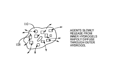
Fig. 1 is a schematic of a process of making a hydrogel encapsulating hydrogel
particles
that contain a therapeutic agent;
Fig. 2 is a schematic showing release of the therapeutic agent from the
embodiment of
Fig. 1;
Fig. 3 depicts an eye with hydrogels such as the hydrogels of Fig. 1 in place
in an eye;
Fig. 4 is a plot of data of experimental results; and
Fig. 5 depicts two curves showing a delay in release caused by a coating.
DETAILED DESCRIPTION
Hydrogel particles are used for controlled release of therapeutic agents to
deliver them
over time. In general, the particles may be placed at a site where delivery of
the agent is desired
and the agent is released as the hydrogel reacts with the physiological fluids
at the site. In areas
such as an eye, a large concentration of the agent in the hydrogel is
generally desirable since
space in, on, or inside the eye is limited. Even in sites where space is not
as limited, keeping
the volume of the treatment close to its minimum necessary volume is to be
expected to
1
Date Recue/Date Received 2022-06-08
CA 02969716 2017-06-02
WO 2016/094646
PCT/US2015/064975
optimize the delivery system. But the inventors have found that it is helpful
to add a certain
amount of extra hydrogel to these systems; the hydrogel is preferably free of
the agent that is
to be delivered.
By way of example, referring to Figs. 1-2, hydrogels 100 or organogels 100'
are foimed
by crosslinking precursors 102 around a therapeutic agent 104. Hydrogels 100
or organogels
100' may be formed as particles or as a larger hydrogel/organogel that is
processed into
particulates. Hydrogels 100 or organogels 100' may be used directly, made into
xerogels, or
otherwise processed to form particles 108, which are hydrogels or xerogels. A
hydrogel 110
(or an organogel) is formed from precursors 102 around particles 108. Hydrogel
110 (or the
organogel) can be made into a xerogel that is later rehydrated. Agents in
hydrogel particles
108 are released when the hydrogel is in aqueous solution, with any xerogels
becoming
hydrogels when exposed to the aqueous solution. The hydrogel particles provide
for diffusion
of therapeutic agents 104 outwards into hydrogel 110. Hydrogel 110 does not
change the rate
of release, or provides a minimal change in the rate of release of the agent.
Briefly, in use,
hydrogels 108 are formed ex vivo and hydrogel 110 is formed ex vivo or may be
formed in
situ. The term in situ means at the site of intended use wherein the hydrogel
is to be used, e.g.,
on a tissue of the patient. The hydrogels interact with physiological fluids
in the body and
release the agents over time.
Fig. 5 depicts two sets of controlled release profiles. The coated samples,
indicated in
dashed lines, delay release of the agent. The delay is the time between the
release profiles for
coated versus non-coated samples. The percentage delay may be calculated at a
given
cumulative release percentage by measuring the delay at that point divided by
the time required
for the cumulative release from the non-coated sample. These are hypothetical
curves. Actual
data can be expected to show variations in the profiles; artisans, however,
can readily generate
an amount of data to accurately compare coated and non-coated samples for
determining an
accurate measurement.
There are various ways to quantify the similarity between coated versus non-
coated
release profiles. In general, it may be helpful to look at the profile across
a limited range of the
cumulative release percentage since release at the earliest and latest parts
of the curves can
involve only a small portion of the total released amount. Accordingly,
options include
assessing release rate at a given cumulative percentage of released agent,
e.g., at 50%.
Alternatives could be some other point, e.g., between 10 and 90 percent;
artisans will
immediately appreciate that all ranges and values within this range are
contemplated and
supported, e.g., 20%, 25%, 33%, 60%, 67%, and so forth. Another option is to
measure the
2
CA 02969716 2017-06-02
WO 2016/094646
PCT/US2015/064975
delay (maximum delay, average delay, mean delay) across an entire range, e.g.,
from 10% to
90%; artisans will immediately appreciate that all ranges and values within
this range are
contemplated and supported, e.g., from 20% to 60%, from 10% to 50%, from 33%
to 67%,
from 15% to 95%.
In further embodiments, a first material comprising a hydrogel or a xerogel
that will
become a hydrogel is coated with a second material that is a hydrogel, a
xerogel, or precursors
that will become a hydrogel by crosslinking with each other upon exposure to
physiological or
other aqueous solution. The coating of precursors may be dry, deposited as a
powder, a melt,
or mixed with binders or other excipients, e.g., plasticizers, salts,
lubricants, and so forth.
Precursor materials
The hydrogels are made from precursors. Precursors are chosen in consideration
of the
properties that are desired for the resultant hydrogel. There are various
suitable precursors for
use in making the hydrogels and/or the organogels. The term precursor refers
to those
molecules crosslinked to form the hydrogel or organogel matrix. While other
materials might
be present in the hydrogel or organogel, such as therapeutic agents or
fillers, they are not
precursors. The term matrix is applicable for hydrogels, organogels, and
xerogels. Such
matrices include matrices with a solvent content of more than about 20% w/w;
artisans will
immediately appreciate that all the ranges and values within the explicitly
stated range is
contemplated, including 20% to 99%, 80% to 95%, at least 50%, and so forth,
with the
percentages being w/w and the solvent being water for hydrogels and the liquid
organic for
organogels.
Precursors may be dissolved in an organic solvent to make an organogel. An
organogel
is a non-crystalline, non-glassy solid material composed of a liquid organic
phase entrapped in
a three-dimensionally cross-linked network. The liquid can be, for example, an
organic
solvent, mineral oil, or vegetable oil. The solubility and dimensions of the
solvent are
important characteristics for the elastic properties and firmness of the
organogel. Alternatively,
the precursor molecules may themselves be capable of forming their own organic
matrix,
eliminating the need for a tertiary organic solvent. Removal of the solvent
(if used) from the
organogel provides a xerogel, a dried gel. The xerogels are formed by, for
example, freeze
drying, may have a high porosity (at least about 20%, a large surface area,
and a small pore
size. Xerogels made with hydrophilic materials form hydrogels when exposed to
aqueous
solutions. High porosity xerogels hydrate more quickly than more dense
xerogels. Hydrogels
are materials that do not dissolve in water and retain a significant fraction
(more than 20%) of
3
84014988
water within their structure. In fact, water contents in excess of 90% are
often known.
Hydrogels may be formed by crosslinking water soluble molecules to form
networks of
essentially infinite molecular weight. Hydrogels with high water contents are
typically soft,
pliable materials. Hydrogels and drug delivery systems as described in U.S.
Publication Nos.
2009/0017097, 2011/0142936 and 2012/0071865 may he adapted for use with the
materials
and methods herein by following the guidance provided herein.
Organogels and hydrogels may be formed from natural, synthetic, or
biosynthetic
polymers. Natural polymers may include glycosminoglycans, polysaccharides, and
proteins.
Some examples of glycosaminoglycans include dermatan sulfate, hyaluronic acid,
the
chondroitin sulfates, chitin, heparin, keratan sulfate, keratosulfate, and
derivatives thereof. In
general, the glycosaminoglycans are extracted from a natural source and
purified and
derivatized. However, they also may be synthetically produced or synthesized
by modified
microorganisms such as bacteria. These materials may be modified synthetically
from a
naturally soluble state to a partially soluble or water swellable or hydrogel
state. This
modification may be accomplished by various well-known techniques, such as by
conjugation
or replacement of ionizable or hydrogen bondable functional groups such as
carboxyl and/or
hydroxyl or amine groups with other more hydrophobic groups.
For example, carboxyl groups on hyaluronic acid may be esterified by alcohols
to
decrease the solubility of the hyaluronic acid. Such processes are used by
various
manufacturers of hyaluronic acid products (such as Genzyme Corp., Cambridge,
MA) to create
hyaluronic acid based sheets, fibers, and fabrics that form hydrogels. Other
natural
polysaccharides, such as carboxymethyl cellulose or oxidized regenerated
cellulose, natural
gum, agar, agrose, sodium alginate, carrageenan, fucoidan, furcellaran,
laminaran, hypnea,
eucheuma, gum arabic, gum ghatti, gum karaya, gum tragacanth, locust beam gum,
arbinoglactan, pectin, amylopectin, gelatin, hydrophilic colloids such as
carboxymethyl
cellulose gum or alginate gum crosslinked with a polyol such as propylene
glycol, and the like,
also form hydrogels upon contact with aqueous surroundings.
Synthetic organogels or hydrogels may be biostable or biodegradable. Examples
of
biostable hydrophilic polymeric materials are poly(hydroxyalkyl methacrylate),
poly(electrolyte complexes), poly(vinylacetate) cross-linked with hydrolysable
or otherwise
degradable bonds, and water-swellable N-vinyl lactams. Other hydrogels include
hydrophilic
hydrogels known as CARBOPOL , an acidic carboxy polymer (Carbomer resins are
high
4
Date Recue/Date Received 2022-06-08
84014988
molecular weight, allylpentaerythritol-crosslinked, acrylic acid-based
polymers, modified with
C10-C30 alkyl acrylates), polyacrylamides, polyacrylic acid, starch graft
copolymers, acrylate
polymer, ester cross-linked polyglucan. Such hydrogels are described, for
example, in U.S.
Patent No. 3,640,741 to Etes, U.S. Patent No. 3,865,108 to Hartop, U.S. Patent
No. 3,992,562
to Denzinger et al., U.S. Patent Nn. 4,002,173 to Manning et al., U.S. Patent
No. 4,014,335 to
Arnold and U.S. Patent No. 4,207,893 to Michaels.
Hydrogels and organogels may be made from precursors. The precursors are
crosslinked with each other. Crosslinks can be formed by covalent bonds or
physical bonds.
Examples of physical bonds are ionic bonds, hydrophobic association of
precursor molecule
segments, and crystallization of precursor molecule segments. The precursors
can be triggered
to react to form a crosslinked hydrogel. The precursors can be polymerizable
and include
crosslinkers that are often, but not always, polymerizable precursors.
Polymerizable precursors
are thus precursors that have functional groups that react with each other to
form matrices
and/or polymers made of repeating units. Precursors may be polymers.
Some precursors thus react by chain-growth polymerization, also referred to as
addition
polymerization, and involve the linking together of monomers incorporating
double or triple
chemical bonds. These unsaturated monomers have extra internal bonds which are
able to
break and link up with other monomers to form the repeating chain. Monomers
are
polymerizable molecules with at least one group that reacts with other groups
to form a
polymer. A macromonomer (or macromer) is a polymer or oligomer that has at
least one
reactive group, often at the end, which enables it to act as a monomer; each
macromonomer
molecule is attached to the polymer by reaction the reactive group. Thus
macromonomers with
two or more monomers or other functional groups tend to form covalent
crosslinks. Addition
polymerization is involved in the manufacture of, e.g., polypropylene or
polyvinyl chloride.
One type of addition polymerization is living polymerization.
Some precursors thus react by condensation polymerization that occurs when
monomers bond together through condensation reactions. Typically these
reactions can be
achieved through reacting molecules incorporating alcohol, amine or carboxylic
acid (or other
carboxyl derivative) functional groups. When an amine reacts with a carboxylic
acid an amide
or peptide bond is formed, with the release of water. Some condensation
reactions follow a
nucleophilic acyl substitution, e.g., as in U.S. Patent No. 6,958,212.
Some precursors react by a chain growth mechanism. Chain
5
Date Recue/Date Received 2022-06-08
84014988
growth polymers are defined as polymers formed by the reaction of monomers or
macromonomers with a reactive center. A reactive center is a particular
location within a
chemical compound that is the initiator of a reaction in which the chemical is
involved. In
chain-growth polymer chemistry, this is also the point of propagation for a
growing chain. The
reactive center is commonly radical, anionic, or cationic in nature, hut can
also take other
forms. Chain growth systems include free radical polymerization, which
involves a process of
initiation, propagation and termination. Initiation is the creation of free
radicals necessary for
propagation, as created from radical initiators, e.g., organic peroxide
molecules. Termination
occurs when a radical reacts in a way that prevents further propagation. The
most common
method of termination is by coupling where two radical species react with each
other forming
a single molecule. Some precursors react by a step growth mechanism, and are
polymers
formed by the stepwise reaction between functional groups of monomers. Most
step growth
polymers are also classified as condensation polymers, but not all step growth
polymers release
condensates. Monomers may be polymers or small molecules. A polymer is a high
molecular
weight molecule formed by combining many smaller molecules (monomers) in a
regular
pattern. Molecular weights for polymers refer to weight average molecular
weights unless
otherwise specified. Oligomers are polymers having less than about 20
monomeric repeat
units. A small molecule generally refers to a molecule that is less than about
2000 Daltons.
The precursors may thus be small molecules, such as acrylic acid or vinyl
caprolaciam, larger
molecules containing polymerizable groups, such as acrylate-capped
polyethylene glycol
(PEG-diacrylate), or other polymers containing ethylenically-unsaturated
groups, such as those
of U.S. Patent No. 4,938,763 to Dunn et al, U.S. Patent Nos. 5,100,992 and
4,826,945 to Cohn
et al, or U.S. Patent Nos. 4,741,872 and 5,160,745 to DeLuca et al.
To form covalently crosslinked hydrogels, the precursors must be covalently
crosslinked together. In general, polymeric precursors are polymers that will
be joined to other
polymeric precursors at two or more points, with each point being a linkage to
the same or
different polymers. Precursors with at least two reactive centers (for
example, in free radical
polymerization) can serve as crosslinkers since each reactive group can
participate in the
formation of a different growing polymer chain. In the case of functional
groups without a
.. reactive center, among others, crosslinking requires three or more such
functional groups on at
least one of the precursor types. For instance, many electrophilic-
nucleophilic reactions
consume the electrophilic and nucleophilic functional groups so that a third
functional group
6
Date Recue/Date Received 2022-06-08
CA 02969716 2017-06-02
WO 2016/094646
PCT/US2015/064975
is needed for the precursor to form a crosslink. Such precursors thus may have
three or more
functional groups and may be crosslinked by precursors with two or more
functional groups.
A crosslinked molecule may be crosslinked via an ionic or covalent bond, a
physical force, or
other attraction. A covalent crosslink, however, will typically offer
stability and predictability
.. in reactant product architecture.
In some embodiments, each precursor is multifunctional, meaning that it
comprises two
or more electrophilic or nucleophilic functional groups, such that a
nucleophilic functional
group on one precursor may react with an electrophilic functional group on
another precursor
to form a covalent bond. At least one of the precursors comprises more than
two functional
groups, so that, as a result of electrophilic-nucleophilic reactions, the
precursors combine to
form crosslinked polymeric products.
The precursors may have biologically inert and hydrophilic portions, e.g., a
core. In
the case of a branched polymer, a core refers to a contiguous portion of a
molecule joined to
arms that extend from the core, with the arms having a functional group, which
is often at the
.. terminus of the branch. A hydrophilic molecule, e.g., a precursor or
precursor portion, has a
solubility of at least 1 g/100 mL in an aqueous solution. A hydrophilic
portion may be, for
instance, a polyether, for example, polyalkylene oxides such as polyethylene
glycol (PEG),
polyethylene oxide (PEO), polyethylene oxide-co-polypropylene oxide (PPO), co-
polyethylene oxide block or random copolymers, and polyvinyl alcohol (PVA),
poly (vinyl
.. pyrrolidinone) (PVP), poly (amino acids, dextran, or a protein. The
precursors may have a
polyalkylene glycol portion and may be polyethylene glycol based, with at
least about 80% or
90% by weight of the polymer comprising polyethylene oxide repeats. The
polyethers and
more particularly poly (oxyalkylenes) or poly (ethylene glycol) or
polyethylene glycol are
generally hydrophilic. As is customary in these arts, the teini PEG is used to
refer to PEO with
or without hydroxyl end groups.
A precursor may also be a macromolecule (or macromer), which is a molecule
having
a molecular weight in the range of a thousand to many millions. The hydrogel
or organogel
however, may be made with at least one of the precursors as a small molecule
of about 1000
Da or less (alternatively: 2000 Da or less). The macromolecule, when reacted
in combination
with a small molecule (of about 1000 Da or less / 200 Da or less), is
preferably at least five to
fifty times greater in molecular weight than the small molecule and is
preferably less than about
60,000 Da; artisans will immediately appreciate that all the ranges and values
within the
explicitly stated ranges are contemplated. A more preferred range is a
macromolecule that is
about seven to about thirty times greater in molecular weight than the
crosslinker and a most
7
84014988
preferred range is about ten to twenty times difference in weight. Further, a
macromolecular
molecular weight of 5,000 to 50,000 is useful, as is a molecular weight of
7,000 to 40,000 or a
molecular weight of 10,000 to 20,000. There are certain advantage to having a
small molecule,
such as diffusivity for completion of reactions.
Certain macromeric precursors are the crosslinkable, biodegradable, water-
soluble
macromers described in U.S. Patent No. 5,410,016 to Hubbell et al. These
macromers are characterized by having at least two polymerizable groups,
separated by at
least one degradable region.
Synthetic precursors may he used. Synthetic refers to a molecule not found in
nature
or not normally found in a human. Some synthetic precursors are free of amino
acids or free
of amino acid sequences that occur in nature. Some synthetic precursors are
polypeptides that
are not found in nature or are not normally found in a human body, e.g., di-,
tri-, or tetra-lysine.
Some synthetic molecules have amino acid residues but only have one, two, or
three that are
contiguous, with the amino acids or clusters thereof being separated by non-
natural polymers
or groups. Polysaccharides or their derivatives are thus not synthetic.
Alternatively, natural proteins or polysaccharides may be adapted for use with
these
methods, e.g., collagens, fibrin(ogen)s, albumins, alginates, hyaluronic acid,
and heparins.
These natural molecules may further include chemical derivitization, e.g.,
synthetic polymer
decorations. The natural molecule may be crosslinked via its native
nucleophiles or after it is
derivatized with functional groups, e.g., as in U.S. Patent Nos. 5,304,595,
5,324,775,
6,371,975, and 7,129,210. Natural refers to a molecule found in
nature. Natural polymers, for example proteins or glycosaminoglycans, e.g.,
collagen,
fibrinogen, albumin, and fibrin, may be crosslinked using reactive precursor
species with
electrophilic functional groups. Natural polymers normally found in the body
are
proteolytically degraded by proteases present in the body. Such polymers may
be reacted via
functional groups such as amines, thiols, or carboxyls on their amino acids or
derivatized to
have activatable functional groups. While natural polymers may be used in
hydrogels, their
time to gelation and ultimate mechanical properties must be controlled by
appropriate
introduction of additional functional groups and selection of suitable
reaction conditions, e.g.,
pH.
Precursors may be made with a hydrophobic portion provided that the resultant
hydrogel retains the requisite amount of water, e.g., at least about 20%. In
some cases, the
8
Date Recue/Date Received 2022-06-08
84014988
precursor is nonetheless soluble in water because it also has a hydrophilic
portion. In other
cases, the precursor makes dispersion in the water (a suspension) but is
nonetheless reactable
to from a crosslinked material. Some hydrophobic portions may include a
plurality of alkyls,
polypropylenes, alkyl chains, or other groups. Some precursors with
hydrophobic portions are
sold under the trade names PLURONICTM F68, JEFFAMINETm, or TECTRONICTm. A
hydrophobic molecule or a hydrophobic portion of a copolymer or the like is
one that is
sufficiently hydrophobic to cause the molecule (e.g., polymer or copolymer) to
aggregate to
form micelles or microphases involving the hydrophobic domains in an aqueous
continuous phase
or one that, when tested by itself, is sufficiently hydrophobic to precipitate
from, or otherwise
change phase while within, an aqueous solution of water at pH from about 7 to
about 7.5 at
temperatures from about 30 to about 50 degrees Centigrade.
Precursors may have, e.g., 2-100 arms, with each arm having a terminus,
bearing in
mind that some precursors may be dendrimers or other highly branched
materials. An arm on
a hydrogel precursor refers to a linear chain of chemical groups that connect
a crosslinkable
functional group to a polymer core. Some embodiments are precursors with
between 3 and
300 arms; artisans will immediately appreciate that all the ranges and values
within the
explicitly stated ranges are contemplated, e.g., 4 to 16, 8 to 100, or at
least 6 arms.
Thus hydrogels can be made, e.g., from a multi-armed precursor with a first
set of
functional groups and a low molecular-weight precursor having a second set of
functional
groups. For example, a six-armed or eight-armed precursor may have hydrophilic
arms, e.g.,
polyethylene glycol, terminated with primary amines, with the molecular weight
of the arms
being about 1,000 to about 40,000; artisans will immediately appreciate that
all ranges and
values within the explicitly stated bounds are contemplated. Such precursors
may be mixed
with relatively smaller precursors, for example, molecules with a molecular
weight of between
about 100 and about 5000, or no more than about 800, 1000, 2000, or 5000
having at least
about three functional groups, or between about 3 to about 16 functional
groups; ordinary
artisans will appreciate that all ranges and values between these explicitly
articulated values
are contemplated. Such small molecules may be polymers or non-polymers and
natural or
synthetic.
Precursors that are not dendrimers may be used. Dendritic molecules are highly
branched radially symmetrical polymers in which the atoms are arranged in many
arms and
subarms radiating out from a central core. Dendrimers are characterized by
their degree of
structural perfection as based on the evaluation of both symmetry and
polydispersity and
require particular chemical processes to synthesize. Accordingly, an artisan
can readily
9
Date Recue/Date Received 2022-06-08
84014988
distinguish dendrimer precursors from non-dendrimer precursors. Dendrimers
have a shape
that is typically dependent on the solubility of its component polymers in a
given environment,
and can change substantially according to the solvent or solutes around it,
e.g., changes in
temperature, pH, or ion content.
Precursors may he dendrimers, e.g., as in U.S. Publication Nos. 2004/0086479
and
2004/0131582 and PCT Publication Nos. W007005249, W007001926 and W006031358,
or
the U.S. counterparts thereof; dendrimers may also be useful as
multifunctional precursors,
e.g., as in U.S. Publication Nos. 2004/0131582 and 2004/0086479 and PCT
Publication Nos.
W006031388 and W006031388. Dendrimers are highly ordered possess high surface
area to
volume ratios, and exhibit numerous end groups for potential
functionalization. Embodiments
include multifunctional precursors that are not dendrimers.
Some embodiments include a precursor that consists essentially of an
oligopeptide
sequence of no more than five residues, e.g., amino acids comprising at least
one amine, thiol,
carboxyl, or hydroxyl side chain. A residue is an amino acid, either as
occurring in nature or
derivatized thereof. The backbone of such an oligopeptide may be natural or
synthetic. In
some embodiments, peptides of two or more amino acids are combined with a
synthetic
backbone to make a precursor; certain embodiments of such precursors have a
molecular
weight in the range of about 100 to about 10,000 or about 300 to about 500
Artisans will
immediately appreciate that all ranges and values between these explicitly
articulated bounds
are contemplated.
Precursors may be prepared to be free of amino acid sequences cleavable by
enzymes
present at the site of introduction, including free of sequences susceptible
to attach by
metalloproteinases and/or collagenases. Further, precursors may be made to be
free of all
amino acids, or free of amino acid sequences of more than about 50, 30, 20,
10, 9, 8, 7, 6, 5, 4,
3, 2, or 1 amino acids. Precursors may be non-proteins, meaning that they are
not a naturally
occurring protein and cannot be made by cleaving a naturally occurring protein
and cannot bc
made by adding synthetic materials to a protein. Precursors may be non-
collagen, non-fibrin,
non-fibrinogen, and non-albumin, meaning that they are not one of these
proteins and are not
chemical derivatives of one of these proteins. The use of non-protein
precursors and limited
use of amino acid sequences can be helpful for avoiding immune reactions,
avoiding unwanted
cell recognition, and avoiding the hazards associated with using proteins
derived from natural
sources. Precursors can also be non-saccharides (free of saccharides) or
essentially non-
Date Recue/Date Received 2022-06-08
CA 02969716 2017-06-02
WO 2016/094646
PCT/US2015/064975
saccharides (free of more than about 5% saccharides by w/w of the precursor
molecular weight.
Thus a precursor may, for example, exclude hyaluronic acid, heparin, or
gellan. Precursors can
also be both non-proteins and non-saccharides. The term protein, as used
herein, is a broad
term referring to a polypeptide; the term protein fragment may be used to
refer to a less than
complete sequence of a wild-type protein: precursors or therapeutic agents may
be protein
fragments.
Peptides may be used as precursors. In general, peptides with less than about
10
residues are preferred, although larger sequences (e.g., proteins) may be
used. Artisans will
immediately appreciate that every range and value within these explicit bounds
is included,
e.g., 1-10, 2-9, 3-10, 1, 2, 3, 4, 5, 6, or 7. Some amino acids have
nucleophilic groups (e.g.,
primary amines or thiols) or groups that can be derivatized as needed to
incorporate
nucleophilic groups or electrophilic groups (e.g., carboxyls or hydroxyls).
Polyamino acid
polymers generated synthetically are normally considered to be synthetic if
they are not found
in nature and are engineered not to be identical to naturally occurring
biomolecules.
Some organogels and hydrogels are made with a polyethylene glycol-containing
precursor. Polyethylene glycol (PEG, also referred to as polyethylene oxide
when occurring
in a high molecular weight) refers to a polymer with a repeat group
(CH2CH20)., with n being
at least 3. A polymeric precursor having a polyethylene glycol thus has at
least three of these
repeat groups connected to each other in a linear series. The polyethylene
glycol content of a
polymer or arm is calculated by adding up all of the polyethylene glycol
groups on the polymer
or arm, even if they are interrupted by other groups. Thus, an arm having at
least 1000 MW
polyethylene glycol has enough CH2CH20 groups to total at least 1000 MW. As is
customary
tetininology in these arts, a polyethylene glycol polymer does not necessarily
refer to a
molecule that terminates in a hydroxyl group. Molecular weights are
abbreviated in thousands
using the symbol k, e.g., with 15K meaning 15,000 molecular weight, i.e.,
15,000 Dalions.
NH2 refers to an amine termination. SG refers to succinimidyl glutarate. SS
refers to
succinimidyl succinate. SAP refers to succinimidyl adipate. SAZ refers to
succinimidyl
azelate. SS, SG, SAP and SAZ are succinimidyl esters that have an ester group
that degrades
by hydrolysis in water. Hydrolytically degradable or water-degradable thus
refers to a material
that would spontaneously degrade in vitro in an excess of water without any
enzymes or cells
present to mediate the degradation. A time for degradation refers to effective
disappearance of
the material as judged by the naked eye. Trilysine (also abbreviated LLL) is a
synthetic
tripeptide. PEG and/or hydrogels, as well as compositions that comprise the
same, may be
11
CA 02969716 2017-06-02
WO 2016/094646
PCT/US2015/064975
provided in a form that is pharmaceutically acceptable, meaning that it is
highly purified and
free of contaminants, e.g., pyrogens.
Hydro gel Structures
The hydrogel's structure and the material composition of the hydrogel's
precursors
determine its properties. Precursor factors include properties such as
biocompatibility, water
solubility, hydrophilicity, molecular weight, arm length, number of arms,
functional groups,
distance between crosslinks, degradability, and the like. The choice of
reaction conditions also
effects the hydrogel's structure and properties, including choices of
solvents, reaction schemes,
reactant concentrations, solids content, and the like. There can be a variety
of ways to achieve
certain properties, or combination of properties. On the other hand some
properties are in
tension with each other, for instance brittleness may increase as a distance
between crosslinks
or solids content increases. Strength may be increased by increasing the
number of crosslinks
but swelling may thereby be reduced. Artisans will appreciate that the same
materials may be
used to make matrices with a great range of structures that will have highly
distinct mechanical
properties and performance, such that the achievement of a particular property
should not be
merely assumed based on the general types of precursors that are involved.
The spacing between molecular strands of the hydrogel (the matrix) affects
several
hydrogel properties, including a rate of diffusion of molecules. The
crosslinking density can
be controlled by the choice of the overall molecular weight of the
precursor(s) used as
crosslinker(s) and other precursor(s) and the number of functional groups
available per
precursor molecule. A lower molecular weight between crosslinks such as 200
will give much
higher crosslinking density as compared to a higher molecular weight between
crosslinks such
as 500,000; artisans will immediately appreciate that all ranges and values
within this range are
contemplated and supported, e.g., 200 to 250,000, 500 to 400,000, and so
forth. The
crosslinking density also may be controlled by the overall percent solids of
the crosslinker and
functional polymer solutions. Yet another method to control crosslink density
is by adjusting
the stoichiometry of nucleophilic functional groups to electrophilic
functional groups. A one
to one ratio leads to the highest crosslink density. Precursors with longer
distances between
crosslinkable sites form gels that are generally softer, more compliant, and
more elastic. Thus
an increased length of a water-soluble segment, such as a polyethylene glycol,
tends to enhance
elasticity to produce desirable physical properties. Thus certain embodiments
are directed to
precursors with water soluble segments having molecular weights in the range
of 2,000 to
100,000; artisans will immediately appreciate that all the ranges and values
within the explicitly
12
CA 02969716 2017-06-02
WO 2016/094646
PCT/US2015/064975
stated ranges are contemplated, e.g., 5,000 to 35,000. Thus embodiments
include materials
(organogels, hydrogels, xerogels, a (first) material that is placed within an
envelope or coating
of a second material, or the (second) material used for an envelope) with a
molecular weight
between crosslinks of at least 2000, at least 4000, or from 2000-250,000;
Artisans will
immediately appreciate that all ranges and values between the explicitly
stated bounds are
contemplated, with, e.g., any of the following being available as an upper or
lower limit: 3000,
5000, 10,000,50,000, 100,000. The solids content of the hydrogel (or the
xerogel or organogel
that gives rise to a hydrogel) can affect its mechanical properties and
biocompatibility and
reflects a balance between competing requirements. A relatively low solids
content is useful,
e.g., between about 2.5% to about 20%, artisans will immediately appreciate
that this range is
including all ranges and values there between, e.g., about 2.5% to about 10%,
about 5% to
about 15%, or less than about 15%. Solids content and distance between
crosslinks is measured
at the equilibrium water content of the material in water. Thus embodiments
include materials
(organogels, hydrogels, xerogels, a (first) material that is placed within an
envelope or coating
of a second material, or the (second) material used for an envelope) with a
solids content from
about 2.5% to about 20%, Artisans will immediately appreciate that all ranges
and values
between the explicitly stated bounds are contemplated. Solids content
percentages are w/w
measured at equilibrium water content.
One way to construct the materials so that the delay is controlled or
minimized is to
.. design the hydrogels with different rates of diffusion for the agent. Often
the molecular weight
(MW) of the agent is a controlling variable. There are a number of approaches
for relating
hydrogel properties to diffusion. These include the free volume theory, the
hydrodynamic
theory, the obstruction theory, combination theories, and parameters such as
mesh size, sieving
terms, distributions of openings between chains, and so forth (Amsden,
Macromolecules
(1998) 31:8382-8395). In practice, however, hydrogels can be made with various
distances
between their crosslinks and tested for a particular molecule to create a
hydrogel that provides
a desired diffusion rate. In general, a distance between crosslinks that is
large compared to the
molecule's size provides for a high rate of diffusion, a distance between
crosslinks that is small
compared to the molecule's size provides for a slow diffusion, and a distance
between
crosslinks that is smaller than the molecule provides for essentially no
diffusion. A molecule's
molecular weight is generally a useful measure of it size. There are other
factors that can be
important and these can be accounted for when creating the hydrogel: for
instance, interactions
between the molecule and the hydrogel, such as affinity or charge-charge, and
solvent effects
such as hydrophobicity of the molecule.
13
CA 02969716 2017-06-02
WO 2016/094646
PCT/US2015/064975
Accordingly, embodiments include a biomedical sustained release system for use
in a
patient comprising a collection of particles that comprise a first
biodegradable material that is
a hydrogel or a xerogel and a therapeutic agent with the first material,
before biodegradation,
having a first rate of release for the therapeutic agent as measured in
physiological solution,
and a second material that is a hydrogel or xerogel that (before
biodegradation) delays release
by a predetermined amount and, optionally, is free of the therapeutic agent
until such time as
the agent diffuses from the particles into the second hydrogel. The
predetermined amount of
release can be described with reference to a controlled release profile as
already described, e.g.,
as in Fig. 5. The rate of release from a hydrogel is measured in vitro in a
great excess of
physiological solution, enough so that the solution is very large relative to
the hydrogel so that
the agent does not accumulate in the solution and reduce the effective rate of
release. The
solution, for testing, is phosphate buffer solution at pH 7.4 osmotically
balanced for
physiological conditions, as is customary in these arts. Also, pH 7.2 is often
used for
specifically simulating the ocular tissue environment.
Various therapeutic agents are described herein; they may be incorporated into
these
systems. Their sizes are well known or easily determined. Their release rates
can be readily
established. The agent can be one as set forth specifically herein or can be
an agent having a
molecular weight of less than about 450 kDa or in a range from 200 Da to about
450kDa;
artisans will immediately appreciate that all ranges and values within this
range are
contemplated and supported, e.g., about 500 Da to about 250kDa, about 10kDa to
about
180kDa, no more than about 205kDa, about 100kDa to about 255kDa, and so forth.
Release
rates reflect the condition of the system at the time of implantation. The
systems can be
biodegradable and the relative rates may change over time as degradation takes
place. Since
the release rate through the second material is very high, however, it is
permissive to passage
of the molecule and not essential to the control of the delivery process. The
particles inside the
envelope of hydrogel and the condition of the agents in the particles are
controlling for release
of the agent. The envelope can affect the rate of release but only
incidentally.
In fact, the second material, which, in vivo, is a hydrogel that at least
partially coats the
first hydrogel, has a role in biocompatibility. It was observed that the
hydrogel particles, when
loaded with agents, specifically proteins not native to the animal model of
the in vivo test,
elicited some unwanted biological effects that indicated a lack of
biocompatibility. But the
same materials, when coated with a hydrogel, were more biocompatible. Rabbit
eyes, which
are a highly sensitive model, were used to establish these effects. Without
being bound to a
particular theory, it is theorized that macrophages or other immune system
cells were more
14
CA 02969716 2017-06-02
WO 2016/094646
PCT/US2015/064975
responsive to particles than the monolithic coating of a second material. The
particles have a
higher surface area and also have more resemblance to cell, virus, or tissue
surfaces as
compared to the sheet-like coating. Further, or alternatively, the particles
contained the agents
and might not have coated all of the agent molecules in their entirety, so
that the immune system
cells could interact with them before the hydrogel degraded. The outer
enveloping hydrogel is
sized to keep all cells out but to allow the agents from the particles to
rapidly and freely diffuse.
The term encapsulating, when used, refers to placing a coating over all of the
particles that are
injected or otherwise placed into a patient. As is evident, embodiments
include choosing the
second material to have, relative to the first material (such as particles), a
lower value for one
or more of: molecular weight, solids content, distance between crosslinks, and
persistence in
vivo. The first material (hydrogel etc.) may be in particulate form, and be a
collection of
particles with a particular size range or distribution as described elsewhere
herein. Particles
are useful for drug delivery, however, other objects may be coated, e.g.,
medical implants,
implantable materials, rods, rods with a dimension of at least 1 mm, punctal
plugs, etc.
Example 1 shows a comparison of an in situ formed hydrogel coating on release
kinetics
of a therapeutic agent. In this study, =fast degrading hydrogel particles were
used; these
conveniently provide a high rate of therapeutic agent release so that the
effects of encapsulating
the particles in a hydrogel coating intended to be permissive to passage of
the agent was tested.
A small but manageable difference in release rate was observed, with both
formulations
releasing fully in less than one week. This indicates that, for a particulate
hydrogel-based
protein delivery system designed for slow release, a relatively high-release
hydrogel can be
overlayed to make a combined system. In Example 2, it was observed that the
encapsulating
hydrogel of Example 1 had an important effect on improving biocompatibility.
Hydrogel
particles coated with an encapsulating hydrogel showed a markedly lower
inflammation as
compared to OTX-14 around the retina (Table 2).
Functional Groups
The precursors for covalent crosslinking have functional groups that react
with each
other to foim the material via covalent bonds, either outside a patient, or in
situ. The functional
groups generally are polymerizable, a broad category that encompasses free
radical, addition,
and condensation polymerization and also groups for electrophile-nucleophile
reactions.
Various aspects of polymerization reactions are discussed in the precursors
section herein.
Thus in some embodiments, precursors have a polymerizable group that is
activated by
photoinitiation or redox systems as used in the polymerization arts, or
electrophilic functional
84014988
groups, for instance: carbodiimidazole, sulfonyl chloride, chlorocarbonates, n-
hydroxysuccinimidyl ester, succinimidyl ester or sulfasuccinimidyl esters, or
as in U.S. Patent
Nos. 5,410,016 or 6,149,931. The nucleophilic functional groups may be, for
example, amine,
hydroxyl, carboxyl, and thiol. Another class of electrophiles are acyls, e.g.,
as in U.S. Patent
No. 6,958,212, which describes, among other things, Michael addition schemes
for
reacting polymers.
Certain functional groups, such as alcohols or carboxylic acids, do not
normally react
with other functional groups, such as amines, under physiological conditions
(e.g., pH 7.2-11.0,
37 C). However, such functional groups can be made more reactive by using an
activating
group such as N-hydroxysuccinimide. Certain activating groups include
carbonyldiimidazole,
sulfonyl chloride, aryl halides, sulfosuccinimidyl esters, N-
hydroxysuccinimidyl ester,
succinimidyl ester, epoxide, aldehyde, maleimides, imidoesters and the like.
The N-
hydroxysuccinimide esters or N-hydroxysulfosuccinimide (NHS) groups are useful
groups for
crosslinking of proteins or amine-containing polymers, e.g., amino terminated
polyethylene
glycol. An advantage of an NHS-amine reaction is that the reaction kinetics
are favorable, but
the gelation rate may be adjusted through pH or concentration. The NHS-amine
crosslinking
reaction leads to formation of N-hydroxysuccinimide as a side product.
Sulfonated or
ethoxylated forms of N-hydroxysuccinimide have a relatively increased
solubility in water and
hence their rapid clearance from the body. An NHS-amine crosslinking reaction
may be carried
out in aqueous solutions and in the presence of buffers, e.g., phosphate
buffer (pH 5.0-7.5),
triethanolamine buffer (pH 7.5-9.0), or borate buffer (pH 9.0-12), or sodium
bicarbonate buffer
(pH 9.0-10.0). Aqueous solutions of NHS based crosslinkers and functional
polymers
preferably are made just before the crosslinking reaction due to reaction of
NHS groups with
water. The reaction rate of these groups may be delayed by keeping these
solutions at lower
pH (pH 4-7). Buffers may also be included in the hydrogels introduced into a
body.
In some embodiments, each precursor comprises only nucleophilic or only
electrophilic
functional groups, so long as both nucleophilic and electrophilic precursors
are used in the
crosslinking reaction. Thus, for example, if a crosslinker has nucleophilic
functional groups
such as amines, the functional polymer may have electrophilic functional
groups such as N-
hydroxysuccinimides. On the other hand, if a crosslinker has electrophilic
functional groups
such as sulfosuccinimides, then the functional polymer may have nucleophilic
functional
groups such as amines or thiols. Thus, functional polymers such as proteins,
poly(ally1 amine),
or antine-tertninated di-or multifunctional puly(ethylene glycol) can be used.
16
Date Recue/Date Received 2022-06-08
84014988
One embodiment has reactive precursor species with 2 to 16 nucleophilic
functional
groups each and reactive precursor species with 2 to 16 electrophilic
functional groups each;
artisans will immediately appreciate that all the ranges and values within the
explicitly stated
ranges are contemplated, for instance 2, 3, 4, 5, 6, 7, 8, 9, 10, 11, 12, 13,
14, 15, or 16 groups.
The functional groups may he, e.g., electrophiles reactable with nucleophiles,
groups
reactable with specific nucleophiles, e.g., primary amines, groups that form
amide bonds with
materials in the biological fluids, groups that form amide bonds with
carboxyls, activated-acid
functional groups, or a combination of the same. The functional groups may be,
e.g., a strong
electrophilic functional group, meaning an electrophilic functional group that
effectively forms
a covalent bond with a primary amine in aqueous solution at pH 9.0 at room
temperature and
pressure and/or an electrophilic group that reacts by a of Michael-type
reaction. The strong
electrophile may be of a type that does not participate in a Michaels-type
reaction or of a type
that participates in a Michaels-type reaction.
A Michael-type reaction refers to the 1, 4 addition reaction of a nucleophile
on a
conjugate unsaturated system. The addition mechanism could be purely polar, or
proceed
through a radical-like intermediate state(s); Lewis acids or appropriately
designed hydrogen
bonding species can act as catalysts. The term conjugation can refer both to
alternation of
carbon-carbon, carbon-heteroatom or heteroatom-heteroatom multiple bonds with
single
bonds, or to the linking of a functional group to a macromolecule, such as a
synthetic polymer
or a protein. Michael-type reactions are discussed in detail in U.S. Patent
No. 6,958,212.
Examples of strong electrophiles that do not participate in a Michaels-type
reaction are:
succinimides, succinimidyl esters, or NHS-esters. Examples of Michael-type
electrophiles are
acrylates, methacrylates, methylmethacrylates, and other unsaturated
polymerizable groups.
Initiating Systems
Some precursors react using initiators. An initiator group is a chemical group
capable
of initiating a free radical polymerization reaction. For instance, it may be
present as a separate
component, or as a pendent group on a precursor. Initiator groups include
thermal initiators,
photoactivatable initiators, and oxidation-reduction (redox) systems. Long
wave UV and
visible light photoactivatable initiators include, for example, ethyl eosin
groups, 2, 2-
dimethoxy-2-phenyl acetophenone groups, other acetophenone derivatives,
thioxanthone
groups, benzophenone groups, and camphorquinone groups. Examples of thermally
reactive
17
Date Recue/Date Received 2022-06-08
CA 02969716 2017-06-02
WO 2016/094646
PCT/US2015/064975
initiators include 4, 4' azobis (4-cyanopentanoic acid) groups, and analogs of
benzoyl peroxide
groups. Several commercially available low temperature free radical
initiators, such as V-044,
available from Wako Chemicals USA, Inc., Richmond, Va., may be used to
initiate free radical
crosslinking reactions at body temperatures to form hydrogel coatings with the
aforementioned
monomers.
Metal ions may be used either as an oxidizer or a reductant in redox
initiating systems.
For example, ferrous ions may be used in combination with a peroxide or
hydroperoxide to
initiate polymerization, or as parts of a polymerization system. In this case,
the ferrous ions
would serve as a reductant. Alternatively, metal ions may serve as an oxidant.
For example,
the eerie ion (4+ valence state of cerium) interacts with various organic
groups, including
carboxylic acids and urethanes, to remove an electron to the metal ion, and
leave an initiating
radical behind on the organic group. In such a system, the metal ion acts as
an oxidizer.
Potentially suitable metal ions for either role are any of the transition
metal ions, lanthanides
and actinides, which have at least two readily accessible oxidation states.
Particularly useful
metal ions have at least two states separated by only one difference in
charge. Of these, the
most commonly used are ferric/ferrous; cupric/cuprous; ceric/cerous ; cob
altic/cob altou s ;
vanadate V vs. IV; permanganate; and manganic/manganous. Peroxygen containing
compounds, such as peroxides and hydroperoxides, including hydrogen peroxide,
t-butyl
hydroperoxide, t-butyl peroxide, benzoyl peroxide, cumyl peroxide may be used.
An example of an initiating system is the combination of a peroxygen compound
in one
solution, and a reactive ion, such as a transition metal, in another. In this
case, no external
initiators of polymerization are needed and polymerization proceeds
spontaneously and
without application of external energy or use of an external energy source
when two
complementary reactive functional groups containing moieties interact at the
application site.
Precursors as coatings
Embodiments include a medical device having at least a partial coating of
precursors
that form a hydrogel in situ upon exposure to aqueous solution. The term
medical device is
broad and encompasses drug delivery devices, drug depots for delivery of a
drug, an intraocular
drug depot, an implantable, a prosthesis, and objects made to contact a
physiological fluid. An
example is a punctal plug, an intraocular drug depot, or a fiber used for a
medical device,
wherein the plug or the fiber is completely or partially coated with the
precursors. A method
of applying a coating comprises dipping the device or the portion to be coated
into a melt of
polymer or polymers (precursors) that form the coating. Polymers that melt at
a temperature
18
CA 02969716 2017-06-02
WO 2016/094646
PCT/US2015/064975
of no more than about, e.g., 100 degrees C are melted, in the absence of
solvents. The plug, or
portion thereof, is dipped into the melt. The melt is allowed to cool to a
solid, and remains a
solid at 37 degrees C. Instead of dipping the plug into the melt, the melts
may be otherwise
applied, e.g., brushing, rolling, dropping melt onto the plug, and so forth.
The term melt, in the
context of a polymer, refers to a polymer that is in a liquid state but is not
dissolved in a solvent,
or the polymer acts as its own solvent. Some other materials may be present in
the melt, but
they are not solvents for the melt. It is recognized that some small amount of
a solvent can be
present in a concentration that is not effective to dissolve a substantial
portion of the polymers
in the melt, e.g., no more than 10%, weight per total weight; Artisans will
immediately
appreciate that all ranges and values between the explicitly stated bounds are
contemplated,
with any of the following being available as an upper or lower limit: 0.1,
0.2., 0.5, 1, 2, 3, 4, 5,
6, 7, 8, 9, or 10, referring to % weight/total weight. Agents may be present
in the melt that
assist in adjusting its melting point. For instance, addition of agents that
reduce the forces of
association between polymers may be added to reduce a melting point; such
agents may be
non-solvents or solvent. Such agents may be added at, e.g., no more than 10%,
weight per total
weight; Artisans will immediately appreciate that all ranges and values
between the explicitly
stated bounds are contemplated, with any of the following being available as
an upper or lower
limit: 0.1, 0.2., 0.5, 1, 2, 3, 4, 5, 6, 7, 8, 9, or 10, referring to %
weight/total weight. Also, the
use of branched polymers may be used to adjust melting temperatures.
An example of polymers that melt at a temperature that is reasonable for
dipping the
punctal plug or other device without damage includes PEGs, with the melting
point being
related to the MW of the PEG. A PEG of about 8,000 MW has been tested and is
useful. Other
MWs for PEGs are, for instance, from about 2,000 to about 100,000 (MWs for
polymers refer
to a weight average molecular weight unless otherwise specified). In general,
the polymer or
mixture of polymers is chosen to set the desired melt temperature and the
target dissolving
time.
A method of applying a precursor coating to a punctal plug or other device
comprises
exposing a punctal plug or other device to a solution comprising the
polymer(s) that will form
the coating, with the polymer(s) being in solution in a solvent that is not a
solvent for the
punctal plug. The solvent, in general, is nonaqueos and is an organic solvent.
Examples of
organic solvents are dimethlycarbonate, dimethylformamide dimethyl sulfoxide,
n-methyl
pyrrolidinone, dimethyl sulfoxide, ethyl lactate, N-dicyclohexylcarbodiimide.
Other solvents
that may be used are alcohols: ethanol, isopropanol, 1, 2-propane diol, 1, 4-
butane diol.
19
CA 02969716 2017-06-02
WO 2016/094646
PCT/US2015/064975
The precursor coatings may be made using water to dissolve coating materials
to make
a solution that is sprayed onto a plug to make a water soluble coating, a
process referred to as
a fluidized bed. An alternative configuration could use a coating material
that dissolves in a
non-aqueous solvent to form a non-aqueous solution.
The precursor coating does not have to be a melt. The precursors may be
disposed in a
suitable solvent and applied to the plug or other device, or applied in a dry
form. The coatings
may comprise excipients, e.g., binding agents, nonreactive materials or
nonreactive polymers,
plasticizers, buffer agents, visualization agents, dyes, or salts.
Visualization agents
A visualization agent may be used as a powder in a xerogel/hydrogel; it
reflects or emits
light at a wavelength detectable to a human eye so that a user applying the
hydrogel could
observe the object when it contains an effective amount of the agent. Agents
that require a
machine aid for imaging are referred to as imaging agents herein, and examples
include:
radioopaque contrast agents and ultrasound contrast agents. Some biocompatible
visualization
agents are FD&C BLUE #1, FD&C BLUE #2, and methylene blue. These agents are
preferably
present in the final electrophilic-nucleophilic reactive precursor species mix
at a concentration
of more than 0.05 mg/ml and preferably in a concentration range of at least
0.1 to about 12
mg/ml, and more preferably in the range of 0.1 to 4.0 mg/ml, although greater
concentrations
may potentially be used, up to the limit of solubility of the visualization
agent. Visualization
agents may be covalently linked to the molecular network of the
xerogel/hydrogel, thus
preserving visualization after application to a patient until the hydrogel
hydrolyzes to
dissolution. Visualization agents may be selected from among any of the
various non-toxic
colored substances suitable for use in medical implantable medical devices,
such as FD&C
BLUE dyes 3 and 6, eosin, methylene blue, indocyanine green, or colored dyes
normally found
in synthetic surgical sutures. Reactive visualization agents such as NHS-
fluorescein can be
used to incorporate the visualization agent into the molecular network of the
xerogel/hydrogel.
The visualization agent may be present with either reactive precursor species,
e.g., a crosslinker
or functional polymer solution. The preferred colored substance may or may not
become
chemically bound to the hydrogel.
Biodegradation
An organogel and/or xerogel and/or hydrogel may be formed so that, upon
hydration in
physiological solution, a hydrogel is formed that is water-degradable, as
measurable by the
CA 02969716 2017-06-02
WO 2016/094646
PCT/US2015/064975
hydrogel losing its mechanical strength and eventually dissipating in vitro in
an excess of water
by hydrolytic degradation of water-degradable groups. This test is predictive
of hydrolytically-
driven dissolution in vivo, a process that is in contrast to cell or protease-
driven degradation.
Significantly, however, polyanhydrides or other conventionally-used degradable
materials that
degrade to acidic components tend to cause inflammation in tissues. The
hydrogels, however,
may exclude such materials, and may be free of polyanhydrides, anhydride
bonds, or precursors
that degrade into acid or diacids. The term degradation by solvation in water,
also referred to
as dissolving in water, refers to a process of a matrix gradually going into
solution in, which is
a process that cannot take place for a covalently crosslinked material and
materials insoluble
in water.
For example, electrophilic groups such as SG (N-hydroxysuccinimidyl
glutarate), SS
(N-hydroxysuccinimidyl succinate), SC (N-hydroxysuccinimidyl carbonate), SAP
(N-
hydroxysuccinimidyl adipate) or SAZ (N-hydroxysuccinimidyl azelate) may be
used and have
esteric linkages that are hydrolytically labile. More linear hydrophobic
linkages such as
pimelate, suberate, azelate or sebacate linkages may also be used, with these
linkages being
less degradable than succinate, glutarate or adipate linkages. Branched,
cyclic or other
hydrophobic linkages may also be used. Polyethylene glycols and other
precursors may be
prepared with these groups. The crosslinked hydrogel degradation may proceed
by the water-
driven hydrolysis of the biodegradable segment when water-degradable materials
are used.
Polymers that include ester linkages may also be included to provide a desired
degradation rate,
with groups being added or subtracted near the esters to increase or decrease
the rate of
degradation. Thus it is possible to construct a hydrogel with a desired
degradation profile, from
a few days to many months, using a degradable segment. If polyglycolate is
used as the
biodegradable segment, for instance, a crosslinked polymer could be made to
degrade in about
1 to about 30 days depending on the crosslinking density of the network.
Similarly, a
polycaprolactone based crosslinked network can be made to degrade in about 1
to about 8
months. The degradation time generally varies according to the type of
degradable segment
used, in the following order: polyglycolate < polylactate < polytrimethylene
carbonate <
polycaprolactone. Thus it is possible to construct a hydrogel with a desired
degradation profile,
from a few days to many months, using a degradable segment. Some embodiments
include
precursors that are free of adjacent ester groups and/or have no more than one
ester group per
arm on one or more of the precursors: control of the number and position of
the esters can assist
in uniform degradation of the hydrogel.
21
CA 02969716 2017-06-02
WO 2016/094646
PCT/US2015/064975
A biodegradable linkage in the organogel and/or xerogel and/or hydrogel and/or
precursor may be water-degradable or enzymatically degradable. Illustrative
water-degradable
biodegradable linkages include polymers, copolymers and oligomers of
glycolide, dl-lactide,
1-lactide, dioxanone, esters, carbonates, and trimethylene carbonate.
Illustrative enzymatically
.. biodegradable linkages include peptidic linkages cleavable by
metalloproteinases and
collagenases. Examples of biodegradable linkages include polymers and
copolymers of
poly(hydroxy acid)s, poly(orthocarbonate)s,
poly(anhydride)s, poly(lactone)s,
poly(aminoacid)s, poly(carbonate)s, and poly(phosphonate)s.
If it is desired that a biocompatible crosslinked matrix be biodegradable or
absorbable,
one or more precursors having biodegradable linkages (or just one
biodegradable linkage, for
example an ester) present in between the functional groups may be used. The
biodegradable
linkage optionally also may serve as the water soluble core of one or more of
the precursors
used to make the matrix. For each approach, biodegradable linkages may be
chosen such that
the resulting biodegradable biocompatible crosslinked polymer will degrade or
be absorbed in
a desired period of time.
Hydrogel/Xerogel/Organogel Loading with Agents; Preparation as Particles
One approach for making a hydrogel or organogel with a therapeutic agent is to
form it
around the agent. For instance, a first precursor is added to a solvent-
protein mixture, followed
by a second precursor that is reactive with the first precursor to form
crosslinks. After
formation of the matrix in the solvent, the solvent may be removed to form a
xerogel. Potential
processes include, e.g., precipitation with non-solvent, nitrogen sweep
drying, vacuum drying,
freeze-drying, a combination of heat and vacuum, and lyophilization. If molten
precursors are
used in the absence of a tertiary solvent, there is no need to employ any
solvent removal
process. Upon cooling the material forms a rubbery solid (if above Tg), a
semirigid
semicrystalline material (if below Tm and above Tg) or a rigid glassy solid
(if below Tg).
These materials are more dense than xerogels formed from organic solvents.
When filled with
particles of other materials, e.g., therapeutic agents, buffer salts,
visualization agents, they can
be highly porous, since the solid particles create and fill the pores.
In some embodiments, the agent or agents are present in a separate phase when
precursors are reacted. The separate phase could be oil (oil-in water
emulsion), or an
immiscible solvent, a liposome, a micelle, a biodegradable vehicle, and the
like. Biodegradable
vehicles in which the active agent may be present include: encapsulation
vehicles, such as
microparticles, microspheres, microbeads, micropellets, where the active agent
is encapsulated
22
CA 02969716 2017-06-02
WO 2016/094646
PCT/US2015/064975
in a bioerodable or biodegradable polymers such as polymers and copolymers of:
poly(anhydride), poly(hydroxy acid)s, poly(lactone)s, poly(trimethylene
carbonate),
poly(glycolic acid), poly(lactic acid), poly(glycolic acid)-co-poly(glycolic
acid),
poly(orthocarbonate), poly(caprolactone), crosslinked biodegradable hydrogel
networks like
fibrin glue or fibrin sealant, caging and entrapping molecules, like
cyclodextrin, molecular
sieves and the like. Microspheres made from polymers and copolymers of
poly(lactone)s and
poly(hydroxy acid) are particularly preferred as biodegradable encapsulation
vehicles. The
therapeutic agent or encapsulated therapeutic agent may be present in solution
or suspended
form. Some agents are highly soluble while others are effectively insoluble in
aqueous solution
and can form their own phase when exposed to aqueous solvent.
Therapeutic agents can be in solid particulate form within the
hydrogel/organogel/xerogel, e.g., as a powder. For instance, water soluble
biologics (e.g.,
proteins) in solid phase can be ground or otherwise formed into a fine powder
that is added to
the precursors when a matrix is formed. The protein or other water soluble
biologic in the
xerogel may all be in a solid phase, may be all crystalline, partially
crystalline, or essentially
free of crystals (meaning more than 90% free of crystals w/w; artisans will
immediately
appreciate that all the ranges and values within the explicitly stated ranges
are contemplated).
A powder of a protein refers to a powder made from one or more proteins.
Similarly, powders
of water soluble biologics are powders having particles made of one or more
water soluble
biologics. The powders and/or xerogels and/or organogels and/or hydrogels that
contain them
may be free of encapsulating materials and be free of one or more of a
liposome, micelle, or
nanocapsule. Further, a protein particle or a water soluble biologic particle
may be made that
is free of one or more of: binders, non-peptidic polymers, surfactants, oils,
fats, waxes,
hydrophobic polymers, polymers comprising alkyl chains longer than 4 CH2
groups,
phospholipids, micelle-forming polymers, micelle-forming compositions,
amphiphiles,
polysaccharides, polysaccharides of three or more sugars, fatty acids, and
lipids. Lyophilized,
spray dried or otherwise processed proteins are often formulated with sugars
such as trehalose
to stabilize the protein through the lyophilization or other processes used to
prepare the
proteins. These sugars may be allowed to persist in the particle
throughout the
organogel/xerogel process. The particles may be made to comprise between about
20% and
about 100% (dry w/w) protein; artisans will immediately appreciate that all
the ranges and
values within the explicitly stated ranges are contemplated, e.g., about 50%
to about 80% or at
least 90% or at least about 99%. A number of factors can be controlled that
contribute to
processing and delivery of a protein without denaturation. The protein may be
prepared as a
23
CA 02969716 2017-06-02
WO 2016/094646
PCT/US2015/064975
powder, with the powder particle size being chosen in light of the size of the
ultimate
hydrogel/organogel/xerogel particle. Organic solvents for the proteins may be
chosen so that
the proteins are not solvated by the organic solvents and are compatible with
the protein.
Another factor is oxygen, and elimination of oxygen is helpful in processing
to avoid
denaturation. Another factor is chemical reactions. These may be avoided by
keeping the
protein in a solid phase and free of solvents that dissolve the protein until
such time as the
protein is implanted.
An organogel or hydrogel may be formed and then reduced to particles that are
subsequently treated to remove the organic or aqueous solvent or solvents to
form a xerogel.
For an injectable form, the organogel or hydrogel can be macerated,
homogenized, extruded,
screened, chopped, diced, or otherwise reduced to a particulate faun.
Alternatively, the
organogel or hydrogel can be formed as a droplet or a molded article
containing the suspended
protein particles. One process for making such particles involves creation of
a material that is
broken up to make the particles. One technique involves preparing the
organogel or hydrogel
with protein particles and grinding it, e.g., in a ball mill or with a mortar
and pestle. The matrix
may be chopped or diced with knives or wires. Or the matrix may be cut-up in a
blender or
homogenizer. Another process involves forcing the organogel through a mesh,
collecting the
fragments, and passing them through the same mesh or another mesh until a
desired size is
reached.
The particles of biologics or the particles of organogels or the particles of
the xerogels
may be separated into collections with a desired size range and distribution
of sizes by a variety
of methods. Very fine control of sizing is available, with sizes ranging from
less than 1 micron
to several mm, and with a mean and range of particles sizes being controllable
with a narrow
distribution. Artisans will immediately appreciate that all the ranges and
values within the
explicitly stated ranges are contemplated, e.g., from about 0.1 to about 10 pm
or from about 1
to about 30 pm. About 1 to about 500 microns is another such range that is
useful, with sizes
falling throughout the range and having a mean sizing at one value within the
range, and a
standard deviation centered around the mean value, e.g., from about 1% to
about 100%. A
simple method for sizing particles involves using custom-made or standardized
sieve mesh
sizes. Another method to measure particle size is with a laser diffraction
particle size analyzer,
such as the Coulter LS 200, which analyzes particles when suspended in a
liquid such as saline.
The term particle is broad and includes spheres, discs, and irregularly shaped
particles. A
spheroidal particle refers to a particle wherein the longest central axis (a
straight line passing
through the particle's geometric center) is no more than about twice the
length of other central
24
CA 02969716 2017-06-02
WO 2016/094646
PCT/US2015/064975
axes, with the particle being a literally spherical or having an irregular
shape. A rod-shaped
particle refers to a particle with a longitudinal central axis more than about
twice the length of
the shortest central axis. Embodiments include making a plurality of
collections of particles,
with the collections having different rates of degradation in vivo, and mixing
collections to
make a biomaterial having a degradation performance as desired.
Particles may be prepared as collections having a certain average volume,
average mean
volume, or distribution of sizes that falls within a certain range of volumes
(meaning at least
95% w/w of the particles are distributed within the range). An embodiment is a
collection of
particles having one or more of: an average volume, an average mean volume, or
a distribution
of sizes from about 0.02 vim3 to about 2 mm3; artisans will immediately
appreciate that all
ranges and values within this range are contemplated and supported, e.g., from
0.025 virri3 to 1
mm3, 0.03 pm3 to 1.5 mm3, and so forth. Further, the total volume of the
collection of particles
and/or the total volume of all of the hydrogels in the systems for human
injection may have a
value from about 0.005 to about 2.5 milliliters (m1); artisans will
immediately appreciate that
all ranges and values within this range are contemplated and supported, e.g.,
0.005 to 1 ml, 0.1
ml to 1.5 ml, and so forth. The particles may have a diameter (referring to a
longest dimension
if not symmetrical) from 0.01 microns to 2 trim; Artisans will immediately
appreciate that all
ranges and values between the explicitly stated bounds are contemplated, with,
e.g., any of the
following being available as an upper or lower limit: 1, 5, 10, 20, 50, or 100
nanometers, 0.1,
0.2, 0.5, 1, 10, 20, 30 , 40, 50, 100, 200, 300, 500, 1000 microns, 1, 1.5, or
2 mm. In particular
tissues, such as in the various structures of the eye that have been targeted
for drug delivery,
the volume available for occupation by a drug delivery depot is limited. For
example, the
suprachoroidal space could contain a depot of up to about 100 1, or 200 p1 if
it is conformal to
the shape of the space. Likewise, up to 1000, or 200 pl can be injected into
the vitreous humor
.. as long as the depot does not infringe on the visual axis. Other sites, for
instance
subconjunctival delivery, could accommodate larger depots, since the tissue
can expand to
accommodate.
Alternatively, instead of particles, the hydrogel/organogel/xerogels may be
formed as,
or as part of, a medical device or medical implant. A device may be used in or
on the body.
An implant is at least partially implanted inside the body, or may be
implanted entirely within
the body. Examples of devices are punctal plugs, objects with a rod shape,
drug delivery
devices, patches, and drug depots that are intravascular or extravascular but
in contact with at
least a portion of a blood vessel or associated structure (e.g., adventitia).
The
hydrogel/organogel/xerogels may be formed ex vivo for use with the device,
e.g., formed and
CA 02969716 2017-06-02
WO 2016/094646
PCT/US2015/064975
lyophilized to make a xerogel, or formed in situ, e.g., by providing at least
a partial coating of
precursors that form a hydrogel in vivo upon exposure to aqueous solution.
Administration
An embodiment is a hydrogel formed by in situ polymerization around hydrogel
and/or
xerogel particles, with the particles containing a therapeutic agent. The in
situ formed hydrogel
envelopes the particles and may encapsulate them. When the particles are well
mixed with the
enveloping hydrogel, they will all take-on a coating of the same and will thus
be encapsulated.
In other cases the particles are placed in the patent and then the hydrogel is
applied with the
result that there may be only a partial coating of the particles since.
In use, the hydrogel particles are mixed with precursors and injected into the
site of
intended use in the patient. The precursors react with each other to form the
enveloping
hydrogel. The particles can be made with a first diffusivity for an agent and
the enveloping
hydrogel can be made with a second diffusivity. A needle, cannula, trocar,
sprayer, or other
applicator may be used. Administration of the hydrogels and/or xerogels may
also involve
hydration in advance, at about the time of use, or at the point of use. Or
xerogels may be
implanted without hydration and allowed to hydrate in situ.
The materials described herein may be used to deliver drugs or other
therapeutic agents
(e.g., imaging agents or markers). One mode of application is to apply a
mixture of
xerogel/hydrogel particles and other materials (e.g., therapeutic agent,
buffer, accelerator,
initiator) through a needle, microneedle, cannula, catheter, or hollow wire to
a site. The mixture
may be delivered, for instance, using a manually controlled syringe or
mechanically controlled
syringe, e.g., a syringe pump. Alternatively, a dual syringe or multiple-
barreled syringe or
multi-lumen system may be used to mix the xerogel/hydrogel particles at or
near the site with
a hydrating fluid and/or other agents. Some sites require a careful
administration process, e.g.,
in an eye. Fine needles may be used and/or needles with a limited length. The
work may be
performed, if helpful, under magnification, with a stereoscope, with guided
imaging, or with
robots (for instance as described by Eindhoven University of Technology).
Precursor solutions
and particle collections may be made with sizes and lubricity for manual
injection through a
small gauge needle. Hydrophilic hydrogels crushed into spheroidal particles
about 40 to about
100 microns diameter are small enough to be manually injected through a 30
gauge needle. A
solvent with a high osmolarity and/or an osmolar agent to increase osmolarity
may be used to
ease passage of the particles/solutions through a needle.
26
CA 02969716 2017-06-02
WO 2016/094646
PCT/US2015/064975
Administration of a hydrogel and/or organogel and/or xerogel may be performed
directly into the site of interest. Embodiments of the invention include
administration at or
near an eye. The structure of the mammalian eye can be divided into three main
layers or
tunics: the fibrous tunic, the vascular tunic, and the nervous tunic. The
fibrous tunic, also
.. known as the tunica fibrosa oculi, is the outer layer of the eyeball
consisting of the cornea and
sclera. The sclera extends from the cornea (the clear front section of the
eye) to the optic nerve
at the back of the eye. The sclera is a fibrous, elastic and protective
tissue, composed of tightly
packed collagen fibrils, containing about 70% water. Overlaying the fibrous
tunic is the
conjunctiva. The conjunctiva is a membrane that covers the sclera (white part
of the eye) and
.. lines the inside of the eyelids. The conjunctiva is typically divided into
three parts: (a)
Palpebral or tarsal conjunctivam which is the conjunctiva lining the eyelids;
the palpebral
conjunctiva is reflected at the superior fornix and the inferior fornix to
become the bulbar
conjunctiva; (b) Fornix conjunctiva: the conjunctiva where the inner part of
the eyelids and the
eyeball meet; and (c) Bulbar or ocular conjunctiva: The conjunctiva covering
the eyeball, over
.. the sclera. This region of the conjunctiva is bound tightly and moves with
the eyeball
movements. The conjunctiva effectively surrounds, covers, and adheres to the
sclera. It is has
cellular and connective tissue, is somewhat elastic, and can be removed,
teased away, or
otherwise taken down to expose a surface area of the sclera.
The vascular tunic, also known as the tunica vasculosa oculi, is the middle
vascularized
.. layer which includes the iris, ciliary body, and choroid. The choroid lies
between the retina
and sclera. The choroid contains blood vessels that supply the retinal cells
with oxygen and
remove the waste products of respiration. The choroid connects with the
ciliary body toward
the front of the eye and is attached to edges of the optic nerve at the back
of the eye. The
nervous tunic, also known as the tunica nervosa oculi, is the inner sensory
which includes the
retina. The retina contains the photosensitive rod and cone cells and
associated neurons. The
retina is a relatively smooth (but curved) layer. It does have two points at
which it is different;
the fovea and optic disc. The fovea is a dip in the retina directly opposite
the lens, which is
densely packed with cone cells. The fovea is part of the macula. The optic
disc is a point on
the retina where the optic nerve pierces the retina to connect to the nerve
cells on its inside.
The mammalian eye can also be divided into two main segments: the anterior
segment and the
posterior segment. The anterior segment consists of an anterior and posterior
chamber.
The cornea and lens help to converge light rays to focus onto the retina. The
lens,
behind the iris, is a convex, springy disk which focuses light, through the
second humour, onto
the retina. It is attached to the ciliary body via a ring of suspensory
ligaments known as the
27
CA 02969716 2017-06-02
WO 2016/094646
PCT/US2015/064975
Zonule of Zinn. The iris, between the lens and the first humour, is a
pigmented ring of
fibrovascular tissue and muscle fibers. Light must first pass though the
center of the iris, the
pupil. Light enters the eye, passes through the cornea, and into the first of
two humors, the
aqueous humour. Approximately two-thirds of the total eyes refractive power
comes from the
cornea which has a fixed curvature. The aqueous humor is a clear mass which
connects the
cornea with the lens of the eye, helps maintain the convex shape of the cornea
(necessary to
the convergence of light at the lens) and provides the corneal endothelium
with nutrients. The
posterior segment is posterior to the crystalline lens and in front of the
retina. It includes the
anterior hyaloid membrane and the structures behind it, including the vitreous
humor, retina,
and optic nerve.
Figure 3 is a cross-section of eye 300 and depicts cornea 302 that is
optically clear and
allows light to pass iris 304 and penetrate lens 306. Anterior chamber 308
underlies cornea
302 and posterior chamber 310 lies between iris 304 and lens 306. Ciliary body
312 is
connected to lens 306. Conjunctiva 312 which overlies sclera 314. The vitreous
body 316
comprises the jelly-like vitreous humor, with hyaloid canal 318 being in the
same. Fovea 320
is in the macula and retina 322 overlies choroid 324. Various points of
delivery at eye 300 are
depicted. One area is topically at 350. Another area is intravitreally as
indicated at 352, 354,
and 356; sites 352, 356 are out of the lens focal area, 356 is in contact with
the inner edge of
the eye, at the edge of the retina. Site 358 is outside of the eye and on the
sclera. In use, for
.. example a syringe, catheter (not shown) or other device is used to deliver
the hydrogels 108,
110 or a hydrogel 108 and precursors 102. When precursors are delivered, they
are chosen so
they form hydrogel 110 in situ at the site of intended use. The therapeutic
agents are released
from the hydrogels.
Other sites may be chosen. Sites where drug delivery depots may be foimed
include
the anterior chamber, posterior chamber, the vitreous, episcleral,
subconjunctival, on a surface
of a cornea or a conjunctiva, on a sclera, in a sclera, beneath a sclera, or
between a sclera and
subconjunctiva in a site under and contacting the conjunctiva, on or under the
palpebral or
tarsal conjunctivam, in an eyelid, superior fornix, inferior fornix, bulbar
conjunctiva, and fornix
conjunctiva. Further sites are in the choroid, between the choroid and sclera,
between the retina
and choroid, or a combination of the same.
The hydrogel may be placed at a site that is suited to deliver the agent for
the pathology
that is being treated. The choice of dose, size of implant, and position is
affected by factors
such as a time between repeat administrations, patient comfort or compliance,
and dosage
received at a target tissue. In general, back of the eye diseases can be
treated with drugs
28
CA 02969716 2017-06-02
WO 2016/094646
PCT/US2015/064975
utilizing, e.g., topical, systemic, intraocular and subconjunctiv al delivery
routes. Systemic and
topical (referring to eye drops and non-adherent materials) delivery
modalities fall short in
delivering therapeutic drug levels to treat posterior segment diseases: these
methods of drug
delivery encounter diffusion and drug dilution issues due to the inherent
anatomical barriers of
the intraocular and systemic systems, causing significant patient side effects
(due to multiple
daily dosing), poor bioavailability and compliance issues. Pericular drug
delivery of an
ophthalmic hydrogel implant using subconjunctival, scleral, suprachoroidal,
retrobulbar or sub-
Tenon' s placement has the potential to offer a safer and enhanced drug
delivery system to the
retina compared to topical and systemic routes. For example; steroids like
dexamethasone and
triamicinolone acetonide may be mixed with the hydrogel precursor to fain) a
sustained-release
drug implant. The liquid hydrogel could then be injected in-situ into the sub-
Tenon' s capsule
where it could deliver a constant or tunable release profile of the drug over
a three to four
month time period. The minimally invasive procedure could be performed in a
doctor's office,
or after a cataract operation under topical anesthesia, to treat chronic back
of the eye diseases.
In some embodiments, a retractor is used to hold back eyelids, the user
creatse a small
buttonhole in the conjunctiva about 5-6 mm from the inferior/nasal limbus and
dissect the
conjunctiva down through Tenon' s capsule, to the bare sclera. Next, a 23-
gauge blunt cannula
86 (e.g., 15 mm in length) is inserted through the opening and the liquid drug
implant is injected
at the intended site of use. The cannula is then removed and the conjunctive
is closed with a
cauterization device. One advantage of a hydrogel implant having three
dimensional integrity
is that it will tend to resist cellular infiltration and be able to prevent
the locally administered
drug from being phagocytosed and cleared prematurely from the site. Instead,
it stays in place
until delivered. By way of contrast a microparticle, liposome, or pegylated
protein tends to be
rapidly cleared from the body by the reticuloendothelial system before being
bioeffective.
Delivery of therapeutic amounts of a drug to the retina in posterior segment
eye diseases
remains a challenge. Although intravitreal injections into the vitreous cavity
of anti-VEG F
agents have shown promise to arrest and in some cases reverse chronic age-
related diseases
like macular degeneration, these techniques and procedures are not without
risks and side
effects. Intravitreal administration of therapeutic agents into the vitreous
cavity can cause
cataracts, endophthalmitis and retinal detachments. This form of therapy
requires many
patients to receive monthly intraocular injections of an anti-VEGF drug over a
12 month time
period thus increasing the risk of infection, vitreous wicks and retinal
detachments.
Embodiments directed to an in situ hydrogel biodegradable drug implant that
contains hydrogel
particles will provide an effective alternative treatment for back of the eye
diseases, and are
29
CA 02969716 2017-06-02
WO 2016/094646
PCT/US2015/064975
expected to reduce the common side-effects associated with repeated
intravitreal injections.
For intravitreal implacement, for example, a hydrogel precursors and hydrogel
particles are
injected intravitrealy about 2.5 mm posterior to the limbus through a pars
plana incision using
a sub-retinal cannula, which may be made following dissecting-away or
otherwise clearing the
conjunctiva, as needed. A 25, 27 or 30 gauge sub-retinal cannula 94 (or other
appropriate
cannula) is then inserted and positioned intraocularly to the desired target
site where the
flowable precursors are introduced to form a hydrogel in-situ. The precursors
then folms into
an absorbable gel, adhering to the desired target site.
A drug depot of the in-situ hydrogel drug delivery implant may be designed for
controlled, long term drug release ranging from, e.g., about one to about
three months; and may
optionally be directed to treatment of diseases of the posterior segment
including, for example,
age-related macular degeneration, diabetic retinopathy, diabetic macular
edema, and the
cystoid macular. The device can carry a drug payload of various types of
therapeutic agents
for various conditions, of which some include, for example, steroids,
antibiotics, NSAIDS
and/or antiangiogenic agents, or combinations thereof. The in-situ implant
embodiments can
improve the efficacy and pharmacokinetics of potent therapeutic agents in the
treatment of
chronic back of the eye diseases and minimize patient side effects in several
ways. First, the
implant can be placed in the vitreous cavity at a specific disease site,
bypassing the topical or
systemic routes and thereby increasing drug bioavailability. Secondly, the
implant maintains
local therapeutic concentrations at the specific target tissue site over an
extended period of
time. Thirdly, as compared to various conventional systems, the number of
intravitreal
injections would be substantially reduced, thereby reducing patient risk of
infection, retinal
detachment and transient visual acuity disturbances (white specks floating in
the vitreous) that
can occur until the drug in the vitreous migrates down toward the inferior
wall of the eye and
away from the portion of the central vitreous or macula. A bolus of
conventionally-injected
drugs forms in the vitreous body and displaces the vitreous humor until
dispersed. Dispersion
typically takes a significant amount of time since the vitreous humor is quite
viscous. The
bolus thus interferes with vision, particularly when it is moved around the
eye in response to
sudden accelerations, e.g., as the patient stands up or quickly turns the
head.
The hydrogels may be formed in, on, or under scleral tissue either with or
without the
presence of the conjunctiva. The hydrogel may be adhesive to the sclera or
other tissue where
it is placed to promote drug diffusion through the intended tissue or to
provide a stable depot
to direct the therapeutic agents as required. In some embodiments, the
conjunctiva of the eye
may be removed, macerated, dissected away, or teased-free so that the tissue
can be lifted away
CA 02969716 2017-06-02
WO 2016/094646
PCT/US2015/064975
from the sclera to access a specific region of the sclera for implantation or
injection of the
hydrogel. In other embodiments, the hydrogel is injected in or on the choroid.
A hydrogel is
formed in situ that makes a layer on, and adheres, to the target site. In some
embodiments the
hydrogel is comprised of at least 50%, 75%, 80%, 90%, or 99% w/w water-soluble
precursors
(calculated by measuring the weight of the hydrophilic precursors and dividing
by the weight
of all precursors, so that the weight of water or solvents or non-hydrogel
components is
ignored) to enhance the non-adhesive properties of the hydrogel. In some
embodiments, such
hydrophilic precursors substantially comprise polyethylene oxides. In some
embodiments,
drugs to reduce tissue adherence mediated by biological mechanisms including
cell mitosis,
cell migration, or macrophage migration or activation, are included, e.g.,
anti-inflammatories,
anti-mitotics, antibiotics, PACLITAXEL, MITOMYCIN, or taxols.
In some embodiments, the conjunctiva may be punctured or penetrated with a
needle
or catheter or trocar and precursors introduced into a space between the
sclera and conjunctiva
or other spaces in the eye. In some cases the conjunctiva may be punctured to
access a natural
potential space between the tissues that is filled by the precursors, for
instance a
supracchoroidal potential space. In other cases, a potential or actual space
is created
mechanically with a needle, trocar, spreader, or the like, that breaks the
adherence between the
tissue layers so that precursors may be introduced. The conjunctiva has enough
elasticity to
allow useful amounts of precursors to be introduced or forced into such
natural or created
spaces. Similarly, in the case of intravitreal hydrogel formation, relatively
large volumes may
also be used. Accordingly, in some cases, the amount is between about 0.001 to
about 5 ml;
artisans will immediately appreciate that all the ranges and values within the
explicitly stated
ranges are contemplated, e.g., about 1 ml, about 0.005, 0.01, 0.025, or 0.05
ml, or from 0.002
ml to about 1 or 2.5 ml.
Moreover, removal of a hydrogel, whether present intraocularly or
periocularly, is also
readily achieved using either a vitrectomy cutter if the implant is located in
the vitreous cavity
or a manual I/A syringe and cannula if the implant is located on the scleral
surface or
irrigation/aspiration handpiece. This contrasts with major surgical procedures
needed for the
removal of some conventional non-absorbable implants.
In some aspects, in-situ formation of the hydrogel lets the hydrogel gel or
crosslink in
place, so that it does not flow back out through the tract of the needle and
diffuse extraocularly
through the incision site upon the removal of the needle or cannula. A shape-
stable hydrogel
thus formed can effectively deliver the drug and advantageously can have well-
controlled size,
shape, and surface area. A small needle may be used to inject the materials
since soluble or
31
84014988
flowable precursors may be used instead of an already-formed material. By way
of contrast,
alternative materials that do not cross-link quickly and firmly upon
introduction tend to flow
back out of the incision. And materials that do not covalently cross-link are
subject to creep or
weeping as the material continually reorganizes and some or all of the
material flows out.
Drugs or other therapeutic agents for delivery
Therapeutic agents include, for example, agents for treating conditions that
may result
from inflammatory or abnormal vascular conditions, retinal vein occlusion,
geographic
atrophy, retinitis pigmentosa, retinoblastoma, etc.
Therapeutic agents may be those that are, e.g., antiangiogenic, anti-VEGF,
blocks
VEGFR1, blocks VEGFR2, blocks VEGFR3, anti-PDGF, anti-angiogenesis, Sunitinib,
E7080,
Takeda-6d, Tivozanib, Regorafenib, Sorafenib, Pazopanib, Axitinib, Nintedanib,
Cediranib,
Vatalanib, Motesanib, macrolides, sirolimus, everolimus, tyrosine lcinase
inhibitors (TKIs),
Imatinib (GLEEVACTM) gefinitib (MESS A TM), toceranib (PALLADIATm ), Erlotinib
(TARCEVATm), Lapatinib (TYKERBTm) Nilotinib, Bosutinib Neratinib, lapatinib,
Vatalanib,
dasatinib, erlotinib, gefitinib, imatinib, lapatinib, lestaurtinib, nilotinib,
semaxanib,
toceranib, vandetanib.
The therapeutic agent may comprise a macromolecule, for example an antibody or
antibody fragment. The therapeutic macromolecule may comprise a VEGF
inhibitor, for
example ranibizumab, the active ingredient in the commercially available
LucentisTM. The
VEGF (Vascular Endothelial Growth Factor) inhibitor can cause regression of
the abnormal
blood vessels and improvement of vision when released into the vitreous humor
of the eye.
Examples of VEGF inhibitors include LucentisTm (ranibizumab), EyleaTm
(aflibercept or
VEGF Trap), AvastinTM (bevacizumab), MacugenTM (pegaptanib). Platelet derived
growth
factor (PDGF) inhibitors may also be delivered, e.g., FovistaTM, an anti-PGDF
aptamer.
The therapeutic agent may comprise small molecules such as of a corticosteroid
and
analogues thereof. For example, the therapeutic corticosteroid may comprise
one or more of
trimacinalone, trimacinalone acetonide, dexamethasone, dexamethasone acetate,
fluocinolone,
fluocinolone acetate, or analogues thereof. Alternatively or in combination,
the small
molecules of therapeutic agent may comprise a tyrosine kinase inhibitor
comprising one or
more of axitinib, bosutinib, cediranib, dasatinib, erlotinib, gefitinib,
imatinib, lapatinib,
lestaurtinib, nilotinib, semaxanib, sunitinib, toceranib, vandetanib, or
vatalanib, for example.
The therapeutic agent may comprise an anti-VEGF therapeutic agent. Anti-VEGF
therapies and agents can be used in the treatment of certain cancers and in
age-related macular
degeneration. Examples of anti-VEGF therapeutic agents suitable for use in
accordance with
32
Date Recue/Date Received 2022-06-08
CA 02969716 2017-06-02
WO 2016/094646
PCT/US2015/064975
the embodiments described herein include one or more of monoclonal antibodies
such as
bevacizumab (AvastinTM) or antibody derivatives such as ranibizumab
(LucentisTm), or small
molecules that inhibit the tyrosine kinases stimulated by VEGF such as
lapatinib (TykerbTm),
sunitinib (SutentTm), sorafenib (NexavarTm), axitinib, or pazopanib.
The therapeutic agent may comprise a therapeutic agent suitable for treatment
of dry
AMD such as one or more of SirolimusTM (Rapamycin), CopaxoneTM (Glatiramer
Acetate),
OtheraTM, Complement C5aR blocker, Ciliary Neurotrophic Factor, Fenretinide or
Rheopheresis.
The therapeutic agent may comprise a therapeutic agent suitable for treatment
of wet
AMD such as one or more of REDD14NP (Quark), SirolimusTM (Rapamycin), ATG003;
RcgcncronTM (VEGF Trap) or complement inhibitor (POT-4).
The therapeutic agent may comprise a kinase inhibitor such as one or more of
bevacizumab (monoclonal antibody), BIBW 2992 (small molecule targeting
EGFR/Erb2),
cetuximab (monoclonal antibody), imatinib (small molecule), trastuzumab
(monoclonal
antibody), gefitinib (small molecule), ranibizumab (monoclonal antibody),
pegaptanib (small
molecule), sorafenib (small molecule), dasatinib (small molecule), sunitinib
(small molecule),
erlotinib (small molecule), nilotinib (small molecule), lapatinib (small
molecule),
panitumumab (monoclonal antibody), vandetanib (small molecule) or E7080
(targeting
VEGFR2/VEGFR2, small molecule commercially available from Esai, Co.)
Therapeutic agents may include various classes of drugs. Drugs include, for
instance,
steroids, non-steroidal anti-inflammatory drugs (NSAIDS), anti-cancer drugs,
antibiotics, an
anti-inflammatory (e.g., Diclofenac), a pain reliever (e.g., Bupivacaine), a
Calcium channel
blocker (e.g., Nifedipine), an Antibiotic (e.g., Ciprofloxacin), a Cell cycle
inhibitor (e.g.,
Simvastatin), a protein (e.g., Insulin). Therapeutic agents include classes of
drugs including
steroids, NSAIDS, antibiotics, pain relievers, inhibitors of vascular
endothelial growth factor
(VEGF), chemotherapeutics, anti-viral drugs, for instance. Examples of NSAIDS
are
Ibuprofen, Meclofenamate sodium, mefanamic acid, salsalate, sulindac, tolmetin
sodium,
ketoprofen, diflunisal, piroxicam, naproxen, etodolac, flurbiprofen,
fenoprofen calcium,
Indomethacin, celoxib, ketrolac, and nepafenac. The drugs themselves may be
small
molecules, proteins, RNA fragments, proteins, glycosaminoglycans,
carbohydrates, nucleic
acid, inorganic and organic biologically active compounds where specific
biologically active
agents include but are not limited to: enzymes, antibiotics, antineoplastic
agents, local
anesthetics, hormones, angiogenic agents, anti-angiogenic agents, growth
factors, antibodies,
33
84014988
neurotransmitters, psychoactive drugs, anticancer drugs, chemotherapeutic
drugs, drugs
affecting reproductive organs, genes, and oligonucleotides, or other
configurations.
Therapeutic agents may include a protein or other water soluble biologics.
These
include peptides and proteins. The term protein, as used herein, refers to
peptides of at least
about 5000 Daltons. The term peptide, as used herein, refers to peptides of
any size. The term
oligopeptide refers to peptides having a mass of up to about 5000 Daltons.
Peptides include
therapeutic proteins and peptides, antibodies, antibody fragments, short chain
variable
fragments (scFv), growth factors, angiogenic factors, and insulin. Other water
soluble
biologics are carbohydrates, polysaccharides, nucleic acids, antisense nucleic
acids, RNA,
DNA, small interfering RNA (siRNA), and aptamers.
The therapeutic agents may be used as part of a method of treating the
indicated
condition or making a composition for treating the indicated condition. For
example, AZOPTI'm
(a brinzolamide opthalmic suspension) may be used for treatment of elevated
intraocular
pressure in patients with ocular hypertension or open-angle glaucoma.
BETADINErm in a
Povidone-iodine ophthalmic solution may be used for prepping of the periocular
region and
irrigation of the ocular surface. BETOPTICTm(betaxolol HC1) may be used to
lower intraocular
pressure, or for chronic open-angle glaucoma and/or ocular hypertension.
CILOXANTM
(Ciprotioxacin HC1 opthalmic solution) may be used to treat infections caused
by susceptible
strains of microorganisms. NATACYNTm (Natamycin opthalmic suspension) may be
used for
treatment of fungal blepharitis, conjunctivitis, and keratitis. NEVANACTM
(Nepanfenac
opthalmic suspension) may be used for treatment of pain and inflammation
associated with
cataract surgery. TRAVATANTM (Travoprost ophthalmic solution) may be used for
reduction of
elevated intraocular pressure - open-angle glaucoma or ocular hypertension.
FML FORTETm
(Fluorometholone ophthalmic suspension) may be used for treatment of
corticosteroid-
responsive inflammation of the palperbral and bulbar conjunctiva, cornea and
anterior segment
of the globe. LUMIGANTm(Bimatoprost ophthalmic solution) may be used for
reduction of
elevated intraocular pressure - open-angle glaucoma or ocular hypertension.
PRED FORTE'm
(Prednisolone acetate) may be used for treatment of steroid-responsive
inflammation of the
palpebral and bulbar conjunctiva, cornea and anterior segment of the globe.
PROPINETM
(Dipivefrin hydrochloride) may be used for control of intraocular pressure in
chronic open-
angle glaucoma. RESTASISTm (Cyclosporine ophthalmic emulsion) may be used to
increases
tear production in patients, e.g., those with ocular inflammation associated
with
keratoconjunctivitis sicca. ALREXTM (Loteprednol etabonate ophthalmic
suspension) may be
used for temporary relief of seasonal allergic conjunctivitis. LOTEMAXTm
(Loteprednol
34
Date Recue/Date Received 2022-06-08
84014988
etabonate ophthalmic suspension) may be used for treatment of steroid-
responsive
inflammation of the palpebral and bulbar conjunctiva, cornea and anterior
segment of the globe.
MACUGEN TM (Pegaptanib sodium injection) may be used for Treatment of
neovascular (wet)
age-related macular degeneration. OPTIVARTm (Azelastine hydrochloride) may be
used for
TM
treatment of itching of the eye associated with allergic conjunctivitis.
XALATAN (Latanoprost
ophthalmic solution) may be used to reduce elevated intraocular pressure in
patients, e.g., with
open-angle glaucoma or ocular hypertension. BETIMOLTm (Timolol opthalmic
solution) may
be used for treatment of elevated intraocular pressure in patients with ocular
hypertension or
open-angle glaucoma. Latanoprost is the pro-drug of the free acid form, which
is a prostanoid
.. selective FP receptor agonist. Latanoprost reduces intraocular pressure in
glaucoma patients
with few side effects. Latanoprost has a relatively low solubility in aqueous
solutions, but is
readily soluble in organic solvents typically employed for fabrication of
microspheres using
solvent evaporation.
Further embodiments of therapeutic agents for delivery include those that
specifically
bind a target peptide in vivo to prevent the interaction of the target peptide
with its natural
receptor or other ligands. AVASTIN, for instance, is an antibody that binds
VEGF. And
AFLIBERCEPTTm is a fusion protein that includes portions of a VEGF receptor to
trap VEGF.
An IL-1 trap that makes use of the extracellular domains of IL-1 receptors is
also known; the
trap blocks IL-1 from binding and activating receptors on the surface of
cells. Embodiments
of agents for delivery include nucleic acids, e.g., aptamers. Pegaptanib
(MACUGEN), for
example, is a pegylated anti-VEGF aptamer. An advantage of the particle-and-
hydrogel
delivery process is that the aptamers are protected from the in vivo
environment until they are
released. Further embodiments of agents for delivery include macromolecular
drugs, a term
that refers to drugs that are significantly larger than classical small
molecule drugs, i.e., drugs
such as oligonucleotides (aptamers, antisense, RNAi), ribozymes, gene therapy
nucleic acids,
recombinant peptides, and antibodies.
One embodiment comprises extended release of a medication for allergic
conjunctivitis.
For instance, ketotifen, an antihistamine and mast cell stabilizer, may be
provided in particles
and released to the eye as described herein in effective amounts to treat
allergic conjunctivitis.
Seasonal Allergic Conjunctivitis (SAC) and Perennial Allergic Conjunctivitis
(PAC) are
allergic conjunctival disorders. Symptoms include itching and pink to reddish
eyes. These two
eye conditions are mediated by mast cells. Non-specific measures to ameliorate
symptoms
conventionally include: cold compresses, eyewashes with tear substitutes, and
avoidance of
allergens. Treatment conventionally consists of antihistamine mast cell
stabilizers, dual
Date Recue/Date Received 2022-06-08
84014988
mechanism anti-allergen agents, or topical antihistamines. Corticosteroids
might be effective
but, because of side effects, are reserved for more severe forms of allergic
conjunctivitis such
as vernal keratoconjunctivitis (VKC) and atopic keratoconjunctivitis (AKC).
Oxifloxacin is the active ingredient in VIGAMOX Tm, which is a fluoroquinolone
approved for use to treat or prevent ophthalmic bacterial infections. Dosage
is typically one-
drop of a 0.5% solution that is administered 3 times a day for a period of one-
week or more.
VKC and AKC are chronic allergic diseases where eosinophils, conjunctival
fibroblasts,
epithelial cells, mast cells, and/or TH2 lymphocytes aggravate the
biochemistry and histology
of the conjunctiva. VKC and AKC can be treated by medications used to combat
allergic
conjunctivitis. Permeation agents are agents and may also be included in a
gel, hydrogel,
organogel, xerogel, and biomaterials as described herein. These are agents
that assist in
permeation of a drug into an intended tissue. Permeation agents may be chosen
as needed for
the tissue, e.g., permeation agents for skin, permeation agents for an
eardrum, permeation
agents for an eye.
Eye Disease States
The materials described herein may be used to deliver drugs or other
therapeutic agents
(e.g., imaging agents or markers) to eyes or tissues nearby. Some of the
disease states are back-
of-the-eye diseases. The term back-of-the eye disease is recognized by
artisans in these fields
of endeavor and generally refers to any ocular disease of the posterior
segment that affects the
vasculature and integrity of the retina, macula or choroid leading to visual
acuity disturbances,
loss of sight or blindness. Disease states of the posterior segment may result
from age, trauma,
surgical interventions, and hereditary factors. Some back-of-the-eye disease
are; age-related
macular degeneration (AMD) cystoid macular edema (CME), diabetic macular edema
(DME),
posterior uveitis, and diabetic retinopathy. Some back-of-the-eye diseases
result from
unwanted angiogenesis or vascular proliferation, such as macular degeneration
or diabetic
retinopathy. Drug treatment options for these and other conditions are further
discussed
elsewhere herein.
Kits
Kits or systems for making hydrogels around hydrogel/xerogel particles may be
prepared so that the hydrogel/xerogel particles comprising therapeutic agents
are stored in the
kit with precursors for making an enveloping hydrogel. Applicators may be used
in
combination with the same. The kits are manufactured using medically
acceptable conditions
36
Date Recue/Date Received 2022-06-08
CA 02969716 2017-06-02
WO 2016/094646
PCT/US2015/064975
and contain components that have sterility, purity and preparation that is
pharmaceutically
acceptable. Solvents/solutions may be provided in the kit or separately, or
the components
may be pre-mixed with the solvent. The kit may include syringes and/or needles
for mixing
and/or delivery. The kit or system may comprise components set forth herein.
EXAMPLES
Some precursors are referred to by a nomenclature of naxxKpppflf, where n is
the
number of arms, xx is the molecular weight (MW), ppp is the polymer, and fff
is the functional
end group. Thus 8a15KPEGSAP refers to an 8-armed Polyethylene glycol (PEG)
with a MW
of 15,000 g/mol = 15K PEG. Succinimidyl adipate is: SAP. Succinimidyl
glutarate is SG.
PEG refers to a polyethylene oxide and may or may not be terminated with an OH
group.
Example 1: Comparison of an in situ formed hydrogel coating on release
kinetics of a
therapeutic agent.
Materials Agent: monoclonal antibody (Mab) Bevacizumab (MW 149 kDa) entrapped
in a hydrogel particle. 8a15KSS = 8 armed polyethylene glycol, MW 15,000 Da,
each arm
terminated with succinimidylsuccinate end groups. 8a20KNH2 = 8 armed
polyethylene glycol,
MW 20,000 Da, each arm terminated with free amine (not salt) end groups.
4a20KSAZ = 4
armed polyethylene glycol, MW 20,000 Da, each arm terminated with
succinimidylazelate end
groups. 8a20KNH3+ Cl- =8 armed polyethylene glycol, MW 15,000 Da, each arm
terminated
with ammonium hydrochloride salt end groups. Particulate hydrogel-based
protein delivery
system: fast degrading 8a15KSS/8a20KNH2 was used for a rapid protein release
from the inner
hydrogel particles. Encapsulating Coating ("Envelope): in situ formed
4a20KSAZ/8a20KNH2
hydrogel.
Methods
Spray dried powder of Bevacizumab (1.136g; 27% Active) was suspended in 3.5m1
of
8a20KNH2 solution (11.4% in DMC), sonicated for 15 minutes then mixed with
3.5m1 of
8a15KSS solution (8.6% in DMC) to form a bulk of 8a15KSS/8a20KNH2 organogel in
DMC
within 15 seconds. The organogel bulk was then cured at room temperature for 2
hours then
reduced in particle size yielding a slurry of organogel particles in DMC.
Organogel particles
loaded with Bevacizumab (Bvcz) were then dried to form Bvcz xerogel loaded
particles.
"No envelope" Individual samples of Bvcz Xerogel particles were mixed and
rehydrated with 1% HA solution (4 hours) to form Bvcz hydrogel particles (10%
Bvcz; 10%
8a15KSS/8a20KNH2; 80% of 1% HA solution). Samples were injected (using 21G2
needle)
37
CA 02969716 2017-06-02
WO 2016/094646
PCT/US2015/064975
into tared vials and weighed (15 samples: 68.2; 37.1; 36.8; 37.6; 38.5; 43.7;
47.5; 28.0; 39.5;
39.3; 42.8; 42.4; 36.1; 70.5; 77.6mg). Individual samples were then released
in 30m1 of PBS
(lx; pH 7.4) and pulled (3 samples/time point) at 1; 2; 3 and 9 days.
"Envelope" Bvcz Xerogel particles with in situ formed hydrogel envelope were
prepared using syringes for rehydration and mixing. Syringe A contains Bvcz
Xerogel particles
(88.0 mg) and dry 4a20KSAZ polymer (15.3 mg); syringe B contains 0.4% HA
solution (567.5
mg); syringe C with 211 mg of a 3.2% 8a20KNH3+ Cl- in pH 9.4 buffer (21.5
mg/ml Sodium
tetraborate decahydrate; 7.1 mg/ml sodium phosphate dibasic). Bvcz Xerogel
particles with in
situ formed hydrogel envelope samples were prepared using individual kits (7
samples) by
mixing syringes A and B then mixing the content with syringe C to form the
envelope around
the particles.
Individual samples were transferred to the PBS pH7.4 release media to
determine the
release kinetic profile of Bvcz and compare to the profile in the absence of
the Envelope.
Buffer was exchanged at 42, 68, 100, 119, 142 and 288 hrs.
In vitro kinetic studies comparing sustained release of Bvcz from Xerogel
particles with
and without an "in situ formed" hydrogel envelope were then tested (Fig. 4).
The coating
envelope remained intact throughout the in vitro release experiment, so that
the protein was
forced to traverse through the envelope to reach the release media.
Example 2: Intravitreal tolerability comparison of hydrogel particles with and
without
envelope.
Hydrogel formulations with and without encapsulating hydrogels ("envelopes")
were
tested in vivo. OTX-13 denotes particles with an envelope and OTX-14 denotes
particles
without an envelope.
Materials 8a5KSG = 8 armed polyethylene glycol, MW 5,000 Da, each arm
terminated
with succinimidylglutarate end groups. 8a10KSG = 8 armed polyethylene glycol.
MW 10,000
Da, each arm terminated with succinimidylglutarate end groups. 8a5KNH2 = 8
armed
polyethylene glycol, MW 5,000 Da, each arm terminated with a free amine (not
salt) end
groups. 4a20KSAZ =4 armed polyethylene glycol, MW 20,000 Da, each arm
terminated with
succinimidylazelate end groups. 8a20KNH3+ Cl- = 8 armed polyethylene glycol,
MW 15,000
Da, each arm terminated with ammonium hydrochloride salt end groups.
Methods
Aliquots of 8a5KNH2 (30% in DMC) and 8a5KSG (30% in DMC) were mixed using
syringes in a 1:1 ratio to form a bulk of 8a5KSG/8a5KNH2 organogel in DMC. The
organogel
38
84014988
bulk is then cured at room temperature for 2 hours then reduced in particle
size yielding to a
slurry of organogel particles in DMC. The resulting blank organogel particles
in DMC were
then dried to form 8a5KSG/8a5KNH2 xerogel blank particles. Aliquots of 8a5KNH2
(20% in
DMC) and 8a10KSG (40% in DMC) were mixed using syringes in a 1:1 ratio to form
a bulk
of 8a10KSG/8a5KNH2 organogel in DMC. The organogel bulk was then cured at room
temperature for 2 hours then reduced in particle size yielding to a slurry of
organogel particles
in DMC. The resulting blank organogel particles in DMC were then dried to form
8a 1 OKSG/8a5KNH2 xerogel blank particles.
OTX-14 (No Envelope) was prepared by weighing 8a5KSG/8a5KNH2 (72.8mg) and
8a10KSG/8a5KNH2 (48.5mg) xerogel blank particles in syringe A, weighing
ProviscTM
(519.3 mg; 1% HA) in syringe B then mixing syringe A and B where OTX-14 was
ready for
intravitreal injection.
OTX-13 (Envelope) was prepared by weighing 8a5KSG/8a5KNH2 (58.7mg) and
8a10KSG/8a5KNH2 (42.4mg) xerogel blank particles as well as 4a20KSAZ (16.2mg)
in
syringe A; filling diluted PRO VISC (664.3 mg; 0.41% HA in PBS pH7.4) in
syringe B; filling
8a20KNH3+ (257 mg; 3.2% in pH 10.0 buffer: 21.5 mg/ml Sodium
tetraborate
decahydrate; 7.1 mg/ml sodium phosphate dibasic) in syringe C. Individual
injections were
prepared by mixing syringe A with B for 1 minute then mixing with syringe C
for 10 seconds
to initiate the reaction. At this point the mixture is transferred to 100u1
syringe for intravitreal
injection. 25 ul were injected. Injected rabbits were sacrificed at day 28 and
day 56, eyes
harvested and analyzed by histopathology. Tissues were scored on a semi-
quantitative scale
from 0-5 for any abnormalities.
TABLE 1
Score Description
0 No change; normal
1 Rare foci of change; Minimal
Mild diffuse change or more
2
pronounced focal change
3 Moderate diffuse change
4 Marked diffuse change
5 - Severe diffuse change
39
Date Recue/Date Received 2022-06-08
CA 02969716 2017-06-02
WO 2016/094646
PCT/US2015/064975
Intravitreal injection of OTX-13 resulted in slightly decreased inflammation
in the
vitreous chamber compared to OTX-14 at both time points. Both formulations
resulted in
similar typically minimal inflammation in the vitreous chamber around the
injected test
material, or observed as scattered macrophages within the vitreous chamber.
Rarely, such
inflammation extended minimally into the retina or sclera. OTX-13 showed a
markedly lower
inflammation as compared to OTX-14 around the retina (Table 2: 0.02-0.03 as
compared to
0.1-0.2).
TABLE 2
Inflammation Score (0-5)
lime (Days) Vitreous I Retina/Sclera
OTX-13 OTX-14 OTX-13 OTX-14
Mean Stdev Mean Stdev Mean Stdev Mean Stdev
28 0.9 0.3 1.2 0.2 0.03 0.05 0.2
0.3
56 1 0.1 1.1 0.1 0.02 0.04 0.1 0.1
Intravitreal injection of OTX-13 and OTX-14 resulted in similar typically
minimal
fibrosis around the implanted material in the vitreous chamber. The mean score
for OTX-13
is slightly decreased compared to OTX-14 at both time points.
Intravitreal injection of OTX-13 and OTX-14 resulted in similar typically
minimal
epithelial hyperplasia and inflammation in epithelium just in front of the ora
serrata. The mean
score for OTX-13 were slightly different compared to OTX-14 at both time
points.
TABLE 3
Tissue Reaction Score (0-5)
Time (Days) Fibrosis near implant Hyperplasia-Ora serrata
OTX-13 OTX-14 OTX-13 OTX-14
Mean Stdev Mean , Stdev , Mean Stdev Mean
Stdev
28 0.1 0.1 0.1 0.1 0.1 0.1 0.1 0.1
56 0.1 0.1 0.3 0.1 0.5 0.2 0.4 0.3
84014988
FURTHER DISCLOSURE
la. A biomedical sustained release system for use in a patient
comprising
a collection of particles that comprise a first biodegradable material that is
a hydrogel
or a xerogel and a therapeutic agent, with the first material, before
biodegradation, having a
rate of release for the therapeutic agent in physiological solution, and a
second material that is
a hydrogel or xerogel that at least partially coats the collection of
particles. Alternatively, an
implant, medical device, drug depot, intraocular drug depot, fiber, xerogel
fiber, a prosthesis,
an objects made to contact a physiological fluid, or a biomaterial comprising
the first hydrogel
.. or xerogel material is coated with the second material. Similarly, a
biomedical sustained
release system for use in a patient comprising a collection of particles that
comprise a first
biodegradable material that is a hydrogel or a xerogel and a therapeutic
agent, with the first
material, before biodegradation, having a rate of release for the therapeutic
agent as measured
in physiological solution, and a second material that is a hydrogel or xerogel
that at least
partially coats the collection of particles. Release may be, for example, from
days to months,
e.g., six days to 365 days; Artisans will immediately appreciate that all
ranges and values
between the explicitly stated bounds are contemplated, with, e.g., any of the
following being
available as an upper or lower limit: 6, 14, 30, 60, 90, 120, 180, 240, 300,
or 360 days.
lb. A biomedical sustained release system for use in a patient
comprising a first
biodegradable material that is a hydrogel or a xerogel and a therapeutic
agent, with the first
material, before biodegradation, having a rate of release for the therapeutic
agent as measured
in physiological solution, and a second material that is a hydrogel or xerogel
that at least
partially coats the first material, wherein the second material delays the
rate of release of the
agent by no more than 20% as measured at the 50% w/w release of the agent.
Alternatively,
.. an implant, medical device, drug depot, intraocular drug depot, fiber,
xerogel fiber, a prosthesis,
an object made to contact a physiological fluid, or a biomaterial comprising
the first hydrogel
or xerogel material is at least partially coated with the second material.
lc. The system of lb wherein the first material and the second material
are xerogels.
Id. The system of lb wherein the second material comprises precursors
that, in
.. response to a physiological solution, react with each other to form a
covalently-crosslinked
hydrogel.
le. The system of any of lb-id wherein the first material has a rod-
shape, is a punctal plug,
is an intraocular drug depot or intraocular implant, has a dimension in a
range from 1-10 mm,
or is a monolithic (single-piece) medical implant, or a combination of the
same.
41
Date Recue/Date Received 2022-06-08
CA 02969716 2017-06-02
WO 2016/094646
PCT/US2015/064975
2. The system of 1 (referring to la, lb, lc etc.) wherein the second
material delays the rate
of release of the agent by no more than 20% as measured at the 50% w/w release
of the agent.
3. The system of 1 or 2 wherein a solids content of the second material is
lower than a
solids content of the particles or other coated object, and is in a range from
about 2.5% to about
20%, including all ranges and values there between, e.g., about 2.5% to about
10%, about 5%
to about 15%, or less than about 10% - 20%.
4. The system of any of 1-3 wherein the hydrogel is covalently crosslinked
and a
molecular weight between crosslinks of the second material is lower than a
distance between
crosslinks of the particles or other coated object, and is at least 2000, at
least 4000, or from
2000-250,000; Artisans will immediately appreciate that all ranges and values
between the
explicitly stated bounds are contemplated, with, e.g., any of the following
being available as
an upper or lower limit: 3000, 5000, 10,000, 50,000, 100,000.
5. The system of any of 1-4 wherein the therapeutic agent has a molecular
weight in a
range from about 200 Da to about 400kDa, or wherein the therapeutic agent has
a molecular
weight (MW) of no more than about 250 kDa. Alternatively, the agent has a MW
of no more
than about 205kDa.
6a. The system of any of 1-5 wherein the delay of the rate of the
release as measured at the
50% w/w release of the agent is no more than 10%. Alternatively - no more than
about 15%,
about 5%, or about 1%.
6b. The system of any of 1-5 wherein the rate of release is described as a
graph of a
cumulative percentage of release of the agent (w/w) of the agent over time,
wherein the delay
is no more than about 20% at all points of the graph between 10% and 50% w/w
cumulative
release. Alternatively - the delay being no more than about 1%, about 5%, or
about 10%.
6c. The system of any of 1-5 wherein the rate of release is described as
a graph of a
cumulative percentage of release of the agent (w/w) of the agent over time,
wherein the delay
is no more than about 20% at all points of the graph between 0% and 90% w/w
cumulative
release. Alternatively - the delay being no more than about 1%, about 5%,
about 10%, or about
15%.
7. The system of any of 1-6 wherein the second material encapsulates the
first material
and the collection of particles.
8. The system of any of 1-7 wherein the second material is free of the
therapeutic agent
until such time as the agent diffuses from the particles into the second
hydrogel
42
CA 02969716 2017-06-02
WO 2016/094646
PCT/US2015/064975
9. The system of any of 1-8 wherein a rate of diffusion for the therapeutic
agent in the
second material is in a range from about 4 times to about 20 times a rate of
diffusion of the
agent through the first material.
10. The system of any of 1-9 wherein the therapeutic agent comprises a
protein of at least
about 1000 Da.
11. The system of of any of 1-10 wherein the therapeutic agent comprises a
water soluble
biologic.
12. The system of 11 wherein the water soluble biologic is a protein that
has a molecular
mass of at least about 10,000 Daltons and a sugar is associated with the
protein.
13a. The system of any of 1-12 wherein the therapeutic agent is a protein and
the first
hydrogel comprises solid particles of the protein.
13b The system of any of 1-12 wherein the therapeutic agent is a aptamer
and the first
hydrogel comprises solid particles of the aptamer.
14. The system of any of 1-12 wherein the therapeutic agent is selected
from the group
consisting of a fluoroquinolone, moxifloxacin, travoprost, dexamethasone, an
antibiotic, or a
vestibulotoxin.
15. The system of any of 1-12 wherein the therapeutic agent comprises a
small molecule
drug, a protein, a nucleic acid, or a growth factor.
16. The system of any of 1-12 wherein the therapeutic agent comprises an
anti-VEGF drug.
17. The system of any of 1-12 wherein the particles in the collection have
a volume that is
from about 4 lum3 to about 4 mm3. Alternatively, have a diameter from about 1
micron to about
1.5 mm diameter, or from 5 to 500 microns diameter.
18. The system of any of 1-12 wherein the particles in the collection
have an average
volume that is from about 0.02 pm3 to about 1 mm3.
19. The system of any of 1-12 having a total volume from about 0.005 to
about 0.2
milliliters.
20. The system of any of 1-12 wherein the collection of particles is
dispersed within the
second material.
21. The system of 20 being a single mass with a total volume from about
0.005 and 0.1
milliliters and a thickness from about 0.1 to about 10,000 microns. Artisans
will immediately
appreciate that all ranges and values within this range are contemplated and
supported.
22. The system of any of 1-21 wherein the first material and the second
material are
hydrolytically biodegradable by water.
43
CA 02969716 2017-06-02
WO 2016/094646
PCT/US2015/064975
23. The system of any of 1-22 wherein the first material and the second
material are
synthetic.
24. The system of any of 1-23 wherein
the first material comprises a first precursor that comprises first functional
groups and
a second precursor that comprises second functional groups, with the first
functional groups
and the second functional groups forming covalent crosslinks, and
the second material comprises a third precursor that comprises third
functional groups
and a fourth precursor that comprises fourth functional groups, with the third
functional groups
and the fourth functional groups forming covalent crosslinks.
25. The system of 24 wherein the first through fourth functional groups,
before reaction,
are selected from the group consisting of electrophilic groups and
nucleophilic groups.
26. The system of 25 wherein the electrophilic groups comprises
succimide, succinimide
ester, n-hydroxysuccinimide, maleimide, succinate, nitrophenyl carbonate,
aldehyde,
vinylsulfone, azide, hydrazide, isocyanate, diisocyanate, tosyl, tresyl, or
carbonyldiimidazole.
27. The system of 25 wherein the nucleophile group comprises a primary
amine or a
primary thiol.
28. The system of any of 24-27 wherein the first through fourth precursors
are, before being
covalently crosslinked, water soluble.
29. The system of any of 24-28 wherein the first through fourth precursors
are synthetic.
30. The system of any of 24-29 wherein the first through fourth precursors
have no more
than five amino acids each.
31. The system of any of 24-30 wherein the first through fourth precursors
are hydrophilic
polymers.
32. The system of any of 24-31 wherein at least one of the first through
fourth precursors
comprises a polymer selected from the group consisting of polyethylene glycol,
polyacrylic
acid, polyvinylpyrrolidone, and block copolymers thereof.
33. The system of any of 24-32 wherein at least one of the first through
fourth precursors
comprises a polymer selected from the group consisting of alginate, gellan,
collagen, and
polysaccharide.
34. A method of treating a patient, optionally a patient with an eye
disease, comprising
providing a collection of particles that comprise a first biodegradable
material that is a
hydrogel or a xerogel and a therapeutic agent, with the first material, before
biodegradation,
having a rate of release for the therapeutic agent as measured in
physiological solution, and
44
CA 02969716 2017-06-02
WO 2016/094646
PCT/US2015/064975
forming a second hydrogel in situ on a tissue of the patient at a site of
intended use,
optionally at or near an eye, that at least partially coats the collection of
particles. The agent is
released to treat the patient.
35. The method of 34 wherein the second material delays the rate of release
of the agent by
no more than 20% as measured at the 50% w/w release of the agent.
36. The method of 34 or 35 wherein a solids content of the second material
is lower than a
solids content of the particles or other coated object, and is in a range from
about 2.5% to about
20%, including all ranges and values there between, e.g., about 2.5% to about
10%, about 5%
to about 15%, or less than about 10% - 20%, with the percentages being w/w.
37. The method of any of 34-36 wherein the hydrogel is covalently
crosslinked and a
molecular weight between crosslinks of the second material is lower than a
solids content of
the particles or other coated object, and is at least 2000, at least 4000, or
from 2000-250,000;
Artisans will immediately appreciate that all ranges and values between the
explicitly stated
bounds are contemplated, with, e.g., any of the following being available as
an upper or lower
limit: 3000, 5000, 10,000, 50,000, 100,000.
38. The method of any of 34-37 wherein the second hydrogel is formed at a
suprachoroidal
space.
39. The method of any of 34-38 wherein the therapeutic agent has a
molecular weight
(MW) of no more than about 400 kDa. Alternatively, the agent has a MW of no
more than
about 250kDa.
40a. The method of any of 34-39 wherein the delay of the rate of the release
as measured at
the 50% w/w release of the agent is no more than 10%. Alternatively - no more
than about
15%, about 5%, or about 1%.
40b. The method of any of 34-39 wherein the rate of release is described as a
graph of a
cumulative percentage of release of the agent (w/w) of the agent over time,
wherein the delay
is no more than about 10% at all points of the graph between 10% and 50% w/w
cumulative
release. Alternatively - the delay being no more than about 1%, about 5%,
about 20%, or about
10%.
40c. The method of any of 34-39 wherein the rate of release is described as a
graph of a
cumulative percentage of release of the agent (w/w) of the agent over time,
wherein the delay
is no more than about 20% at all points of the graph between10% and 90% w/w
cumulative
release. Alternatively - the delay being no more than about 1%, about 5%,
about 10%, or about
15%.
CA 02969716 2017-06-02
WO 2016/094646
PCT/US2015/064975
41. The method of any of 34-40 wherein the second material encapsulates the
first material
and the collection of particles and/or wherein the second material is free of
the therapeutic
agent until such time as the agent diffuses from the particles into the second
hydrogel
42. The method of any of 34-41 wherein the a rate of diffusion for the
therapeutic agent in
the second material is in a range from about 4 times to about 20 times a rate
of diffusion of the
agent through the first material.
43. The method of any of 34-42 wherein the therapeutic agent comprises a
protein of at
least about 1000 Da and/or wherein the therapeutic agent comprises a water
soluble biologic.
44. The method of any of 34-43 wherein the water soluble biologic is a
protein that has a
molecular mass of at least about 10,000 Daltons and a sugar is associated with
the protein.
45. The method of any of 34-44 wherein the therapeutic agent is a protein
and the first
hydrogel comprises solid particles of the protein or wherein the therapeutic
agent is an aptamer
and the first hydrogel comprises solid particles of the aptamer.
46. The method of any of 34-45 wherein the therapeutic agent is selected
from the group
consisting of a fluoroquinolone, moxifloxacin, travoprost, dexamethasone, an
antibiotic, or a
vestibulotoxin.
47. The method of any of 34-46 wherein the therapeutic agent comprises a
small molecule
drug, a protein, a nucleic acid, or a growth factor.
48. The method of any of 34-47 wherein the therapeutic agent comprises an
anti-VEGF or
anti-angiogenic drug.
49. The method of any of 34-48 wherein the particles in the collection have
a volume that
is from about 4 m3 to about 4 mm3. Alternatively, have a diameter from about
1 micron to
about 1.5 mm diameter, or from 5 to 500 microns diameter.
50. The method of any of 34-49 wherein the particles in the collection have
an average
volume that is from about 400 larn3 to about 4 mm3.
51. The method of any of 34-50 having a total volume from about 0.005 to
about 2.5
milliliters.
52. The method of any of 34-51 wherein the collection of particles is
dispersed within the
second material.
53. The method of any of 34-52 being a single mass with a total volume from
about 0.005
and 0.1 milliliters and a thickness from about 0.1 to about 10,000 microns.
Artisans will
immediately appreciate that all ranges and values within this range are
contemplated and
supported.
46
CA 02969716 2017-06-02
WO 2016/094646
PCT/US2015/064975
54. The method of any of 34-53 wherein the first material and the second
material are
hydrolytically biodegradable by water.
55. The method of any of 34-54 wherein the first material and the second
material are
synthetic.
56. The method of any of 34-55 wherein the first material comprises a first
precursor that
comprises first functional groups and a second precursor that comprises second
functional
groups, with the first functional groups and the second functional groups
forming covalent
crosslinks, and the second material comprises a third precursor that comprises
third functional
groups and a fourth precursor that comprises fourth functional groups, with
the third functional
groups and the fourth functional groups forming covalent crosslinks. The
various precursors
and functional groups may be identical or different from each other.
57. The method of 56 wherein the first through fourth functional groups,
before reaction,
are selected from the group consisting of electrophilic groups and
nucleophilic groups.
58. The method of 56 or 57 wherein the electrophilic groups are
independently chosen to
be one or more of succimide, succinimide ester, n-hydroxysuccinimide,
maleimide, succinate,
nitrophenyl carbonate, aldehyde, vinylsulfone, azide, hydrazide, isocyanate,
diisocyanate,
tosyl, tresyl, or carbonyldiimidazole.
59. The method of 56 or 57 wherein the nucleophile group comprises a
primary amine or a
primary thiol.
60. The method of 56 wherein the first through fourth precursors are,
before being
covalently crosslinked, water soluble.
61. The method of any of 56-60 wherein the first through fourth precursors
are synthetic.
62. The method of any of 56-60 wherein the first through fourth precursors
have no more
than five amino acids each.
63. The method of any of 56-60 wherein the first through fourth precursors
are hydrophilic
polymers.
64. The method of any of 56-60 wherein at least one of the first through
fourth precursors
comprises a polymer selected from the group consisting of polyethylene glycol,
polyacrylic
acid, polyvinylpyrrolidone, and block copolymers thereof.
65. The method of any of 56-60 wherein at least one of the first through
fourth precursors
comprises a polymer selected from the group consisting of alginate, gellan,
collagen, and
polysaccharide.
47
CA 02969716 2017-06-02
WO 2016/094646
PCT/US2015/064975
66. The method of any of 56-65 wherein a tissue of the patient is treated,
or the hydrogel is
formed in situ on a tissue. Tissues included, for example, eye, punctal,
intraocular,
subconjunctival, scleral, suprachoroidal, retrobulbar, sub-Tenon's placement.
67. A use of a system for treating an eye disease or other condition of a
patient, comprising
the system of any of 1-33, or the methods of any of 34-66, above.
68. A use of the system of any of 1-33 or the methods of any of 34-66 to
(controllably
release and) deliver a therapeutic agent to a patient.
48