Note: Descriptions are shown in the official language in which they were submitted.
TITLE OF THE INVENTION
PERSONAL HEMODIALYSIS SYSTEM
BACKGROUND
[0001] The present disclosure relates generally to medical treatments.
More
specifically, the present disclosure relates to medical fluid treatments, such
as the treatment
of renal failure and fluid removal for congestive heart failure.
[0002] Hemodialysis ("HD") in general uses diffusion to remove waste products
from
a patient's blood. A diffusive gradient that occurs across the semi-permeable
dialyzer
between the blood and an electrolyte solution called dialysate causes
diffusion.
Hemofiltration ("HF") is an alternative renal replacement therapy that relies
on a convective
transport of toxins from the patient's blood. This therapy is accomplished by
adding
substitution or replacement fluid to the extracorporeal circuit during
treatment (typically ten
to ninety liters of such fluid). That substitution fluid and the fluid
accumulated by the patient
in between treatments is ultrafiltered over the course of the HF treatment,
providing a
convective transport mechanism that is particularly beneficial in removing
middle and large
molecules (in hemodialysis there is a small amount of waste removed along with
the fluid
gained between dialysis sessions, however, the solute drag from the removal of
that
ultrafiltrate is not enough to provide convective clearance).
[0003] Hemodiafiltration ("HDF") is a treatment modality that combines
convective
and diffusive clearances. HDF uses dialysate to flow through a dialyzer,
similar to standard
hemodialysis, providing diffusive clearance. In addition, substitution
solution is provided
directly to the extracorporeal circuit, providing convective clearance.
[0004] Home hemodialysis ("HHD") is performed in the patient's home. One
drawback of home hemodialysis has been the need for a dedicated water
treatment, which
includes equipment, water connection and drainage. Installing and using those
components is
a difficult and cumbersome task that can require a patient's home to be
modified.
Nevertheless, there are benefits to daily hemodialysis treatments versus bi-
or tri-weekly
visits to a treatment center. In particular, a patient receiving more frequent
treatments
removes more toxins and waste products than a patient receiving less frequent
but perhaps
longer treatments. Accordingly, there is a need for an improved HHD system.
1
CA 2969785 2017-06-06
SUMMARY
[0005] The present disclosure provides a home hemodialysis ("HHD") system. In
one embodiment, the home system includes a mobile cart and integral bag
manager. A latch
is pulled out to unlock door of the system instrument. The door can be opened
to expose a
latch hook and peristaltic pump heads.
[0006] The instrument accepts a disposable unit which in one embodiment is
loaded
from above and slid to the right. The disposable unit pivots towards the
machine interface,
which allows peristaltic tube loops of the disposable unit to fit over
peristaltic pump heads of
the instrument. Also, supply lines of the disposable unit are passed over
individual pinch
valve plungers.
[0007] The pinch valve plungers pinch the supply tubes against a pinch valve
strike
plate. The valve assembly is in one embodiment a motor-driven cam operated
pinch valve
subassembly. The motor in one embodiment is a stepper motor.
[0008] The system in one embodiment includes a bellows or bladder that
compresses
a cassette against the instrument door using a pressure plate and gasket.
These apparatuses
are structured to accommodate an inline inductive heater provided with the
disposable
cassette. The bellows is air actuated in one embodiment. The instrument
includes a primary
coil that inductively heats conductive heating disks located within the
cassette, which in turn
heat fluid flowing through the cassette.
[0009] A multi-peristaltic pump race retracts and extends in one embodiment
illustrates to facilitate loading of the peristaltic tubes of the cassette
onto the peristaltic pump
heads. The race is then moved towards the tubes for operation.
[0010] The system in one embodiment includes a manual blood pump operator,
which
allows the patient or caregiver to move the blood pump head manually.
[0011] The system includes a bag management system having shelves that fold
up,
out of the way, and down, sequentially for placement of supply bags. The
system in one
embodiment supports up to five, six liter solution bags. The bags can be dual
chamber bags.
The shelves in an embodiment are provided with sensors that allow detection of
whether the
bags have been (i) loaded or not and (ii) opened or not for therapy. The
sensors in one
embodiment are capacitive sensors placed on opposite ends of the shelves.
2
CA 2969785 2017-06-06
[0012] The disposable cassette in one embodiment connects fluidly to a heparin
syringe for the injection of heparin into the blood circuit. The syringe fits
into a luer
connector assembly, which in turn is loaded into a syringe pump. The assembly
is turned in
the syringe pump to lock the syringe in the syringe pump for treatment. The
assembly
accommodates large syringes, such as fifty to sixty milliliter syringes, which
can lock directly
into the syringe pump. In one embodiment, the heparin line passes through the
side of the
cassette. Here, heparin can enter at the blood pump outlet just prior to the
dialyzer inlet.
[0013] The system also includes a retractable saline bag support rod. The
saline in
one embodiment connects to the cassette near the heparin line. A saline valve
is located on
each side of the blood pump to control the flow of saline to same.
[0014] A dialyzer inlet pressure sensor interface in one embodiment doubles as
a flow
control valve. The cassette can also form an integral venus air separation
chamber.
[0015] Priming is performed in one embodiment via gravity. Gravity primes the
venous line, the arterial line and the air trap (drip chamber).
[0016] In another embodiment, priming is preformed via a combination of
pumping
dialysate and a physiologically safe fluid, such as saline. In particular, a
hemodialysis
machine can include a blood circuit, a dialysate circuit, a dialyzer placed in
communication
with the blood circuit and the dialysate circuit; and a priming sequence in
which dialysate is
used to prime a first portion of the dialysate circuit and a physiologically
compatible solution,
other than dialysate, is used to prime a second portion of the dialysate
circuit, the dialyzer and
the blood circuit. The first portion of the dialysate circuit includes a
recirculation loop
primed by a dialysate supply pump in one embodiment. The second portion of the
dialysate
circuit can then be located at least substantially between the recirculation
loop and the
dialyzer, and which is primed by at least one of a blood pump and a downstream
dialysate
pump. In one embodiment, a volumetric balancing unit separates the first and
second
portions of the dialysate circuit.
[0017] The cassette in one embodiment uses balance tubes to balance fresh and
spend
dialysate flow. The balance tubes have outlets at the top of the tubes when
mounted for
operation to allow air to leave the tubes. The cassette also employs diaphragm
valves that
operate with a compliance chamber that seals against backpressure.
[0018] For instance, a hemodialysis machine can include a dialysis instrument
having
at least one peristaltic pump actuator and first and second pneumatic valve
actuators. The
3
CA 2969785 2017-06-06
instrument operates with a disposable cassette, the disposable cassette
including a rigid
portion, with at least one peristaltic pump tube extending from the rigid
portion for operation
with the at least one pump actuator. The rigid portion defines first and
second valve
chambers in operable connection with the first and second valve actuators,
respectively, the
first and second valve chambers communicating fluidly with each other, at
least the first
valve chamber communicating fluidly with a compliance chamber, the compliance
chamber
absorbing energy from a pneumatic closing pressure applied to close the first
valve chamber,
so as to tend to prevent the pneumatic closing pressure from opening an
existing closure of
the second valve chamber.
[0019] The machine in one embodiment includes a vacuum applied to the
compliance
chamber to absorb the energy from the pneumatic closing pressure applied to
close the first
valve chamber.
[0020] In the above example, a flexible membrane can be sealed to the rigid
portion,
the pneumatic closing pressure applied to the membrane to close the first
valve chamber.
Here, the compliance chamber is formed in part via a portion of the flexible
membrane,
wherein the flexible membrane portion is configured to absorb the energy from
the pneumatic
closing pressure. The cassette can alternatively include a flexible diaphragm
located on an
opposing side of the rigid portion from the flexible membrane, the compliance
chamber
formed in part via the flexible diaphragm, the flexible diaphragm configured
to absorb the
energy from the pneumatic closing pressure.
[0021] The disposable cassette can have multiple compliance chambers operating
with different sets of valve chambers. The compliance chamber aids both
upstream and
downstream valves. The compliance chamber overcomes a backpressure applied by
the
closing of the second valve chamber to the first valve chamber, to allow the
first valve
chamber to close properly.
[0022] In another compliance chamber embodiment, the dialysis instrument has a
pump actuator and first and second valve actuators. A disposable cassette is
operable with
the dialysis instrument, the disposable cassette including a pump portion
operable with the
pump actuator, the first and second valve chambers communicating fluidly with
each other, at
least the first valve chamber communicating fluidly with a compliance chamber,
the
compliance chamber negating a first backpressure due to a pneumatic closing
pressure used
to close the first valve chamber to help to ensure the pneumatic pressure
applied to the first
4
CA 2969785 2017-06-06
valve chamber will close the first valve chamber against a second backpressure
from an
existing closure of the second valve chamber. Here, a pneumatic pressure
applied to the
second valve chamber can be the same as the pneumatic pressure applied to the
first valve
chamber. The first backpressure would exist around an outside of a port of the
first valve
chamber if not for the compliance chamber, the second backpressure existing
inside the port.
As before, the compliance chamber is further configured to tend to prevent the
pneumatic
pressure applied to the first valve chamber from opening the closed second
valve chamber.
And, the machine in one embodiment includes a vacuum applied to the compliance
chamber
to ensure the pneumatic pressure applied to the first valve chamber will close
the first valve
chamber.
[0023] In a further compliance chamber embodiment, the dialysis instrument has
a
pump actuator and first and second valve actuators. The disposable cassette is
operable with
the dialysis instrument, the disposable cassette including a pump portion
operable with the
pump actuator, and first and second valve chambers operable with the first and
second valve
actuators, respectively, the cassette further includes a compliance chamber in
fluid
communication with the first and second valve chambers, the compliance chamber
defined at
least in part by a rigid wall of the cassette and a diaphragm located on an
opposing side of the
rigid wall from the first and second valve chambers. The rigid wall in one
embodiment
defines first and second apertures that allow the first and second valve
chambers to
communicate fluidly, respectively, with the compliance chamber. The cassette
can include a
flexible membrane located on an opposing side of the cassette from the
diaphragm, the
membrane for closing the first and second valve chambers. Again, the
compliance chamber
can aid at least one of: (i) maintenance of an existing closure of the second
valve chamber
when the first valve chamber is closed; and (ii) a proper closure of the first
valve chamber at
a time when the second valve chamber is already closed. In one embodiment, the
aiding is
provided via a vacuum applied to the compliance chamber.
[0024] In still a further compliance chamber embodiment, a dialysis instrument
has a
pump actuator and first and second valve actuator. A disposable cassette is
operable with the
dialysis instrument, the disposable cassette including a pump portion operable
with the pump
actuator, and first and second valve chambers operable with the first and
second valve
actuators, respectively. A compliance chamber is placed in fluid communication
with the
first and second valve chambers, the compliance chamber defined by in part by
a flexible
CA 2969785 2017-06-06
membrane used to close at least one of the first and second valve chambers,
the valve
chambers each defining an aperture for fluid communication with the compliance
chamber.
The disposable cassette can include a rigid wall, the first and second valves
chambers
extending from the rigid wall towards the flexible membrane, wherein the
apertures of the
first and second valve chambers are foimed in the rigid wall, and wherein the
rigid wall also
forms a third, larger aperture to allow fluid flowing through the valve
chamber apertures to
communicate fluidly with the flexible membrane of the compliance chamber.
Again, the
compliance chamber aiding at least one of: (i) maintenance of an existing
closure of the
second valve chamber when the first valve chamber is closed; and (ii) a proper
closure of the
first valve chamber at a time when the second valve chamber is already closed.
Again, the
aiding can be provided via a vacuum applied to the compliance chamber.
[0025] It is therefore an advantage of the present disclosure to properly seal
valves in
fluid communication with one another.
[0026] It is another advantage of the present disclosure to provide an
efficient priming
technique that combines the use of dialysate and another physiologically safe
fluid, such as
saline.
[0026a] In accordance with an aspect of the present invention, there is
provided a
dialysis system comprising:
a dialysis instrument including a blood pump actuator;
a dialyzer;
a heparin pump; and
a disposable cassette including a blood pumping portion operable with the
blood
pump actuator of the dialysis instrument, the blood pumping portion including
an inlet and an
outlet, the inlet of the blood pumping portion of the disposable cassette
positioned and
arranged to receive blood from an arterial line of an extracorporeal circuit,
the outlet of the
blood pumping portion of the disposable cassette positioned and arranged to
deliver the blood
to the dialyzer and to receive heparin from the heparin pump.
[0026b] In accordance with an aspect of the present invention, there is
provided a
dialysis system comprising:
a dialysis instrument including a blood pump actuator;
a dialyzer;
a heparin source; and
6
CA 2969785 2017-06-06
a disposable cassette including a blood pumping portion operable with the
blood
pump actuator of the dialysis instrument, the disposable cassette placing the
blood pumping
portion in fluid communication with the dialyzer and an extracorporeal
circuit, so that blood
can be pumped from the extracorporeal circuit, through the blood pumping
portion, to the
dialyzer,
wherein the disposable cassette is configured to introduce heparin from the
heparin
source into the blood at a location of the disposable cassette between the
blood pumping
portion and the dialyzer.
[0026c] In accordance with an aspect of the present invention, there is
provided a dialysis
system comprising:
a dialysis instrument including a blood pump actuator;
a dialyzer;
a disposable cassette including a blood pumping portion operable with the
blood
pump actuator of the dialysis instrument, the disposable cassette placing the
blood pumping
portion in fluid communication with the dialyzer and an extracorporeal
circuit, so that blood
can be pumped from the extracorporeal circuit, through the blood pumping
portion, to the
dialyzer;
a heparin source in fluid communication with the disposable cassette via a
tube; and
an air detector operably connected to the tube.
[0026d] In accordance with an aspect of the present invention, there is
provided a dialysis
system comprising:
a dialysis instrument including a blood pump actuator;
a dialyzer;
a disposable cassette including a blood pumping portion operable with the
blood
pump actuator of the dialysis instrument, the disposable cassette placing the
blood pumping
portion in fluid communication with the dialyzer and an extracorporeal
circuit, so that blood
can be pumped from the extracorporeal circuit, through the blood pumping
portion, to the
dialyzer; and
a heparin source in fluid communication with the disposable cassette, the
heparin
source including a syringe pointed downwardly so that air collects above
heparin within the
syringe.
7
CA 2969785 2017-06-06
[0027] Additional features and advantages are described herein, and will be
apparent
from, the following Detailed Description and the figures.
BRIEF DESCRIPTION OF THE FIGURES
[0028] Fig. 1 is a perspective view of one embodiment of a personal home
hemodialysis ("HHD") system having a mobile cart and integral bag manager.
[0029] Fig. 2 =illustrates the system of the present disclosure, in which a
latch is pulled
out to unlock a door.
[0030] Fig. 3 illustrates the system of the present disclosure, in which a
door is
opened exposing a latch hook and peristaltic pump heads.
[0031] Fig. 4 illustrates one embodiment of the system of the present
disclosure, in
which the door is hidden to more clearly show the door latch.
[0032] Fig. 5 illustrates one embodiment of the system of the present
disclosure, in
which a disposable unit is loaded from above and slid to the right.
[0033] Fig. 6 illustrates one embodiment of the system of the present
disclosure, in
which the disposable unit is pivoted forward towards the interface.
[0034] Fig. 7 illustrates one embodiment of the system of the present
disclosure, in
which the disposable unit pivots forward and the tube loops fit over the
peristaltic pump
heads.
[0035] Fig. 8 illustrates one embodiment of the system of the present
disclosure, in
which the supply lines are placed in operable communication with individual
pinch valve
plungers.
[0036] Fig. 9 illustrates one embodiment of the system of the present
disclosure, in
which the supply lines are hidden to show pinch valve plungers.
[0037] Fig. 10 is rear view of one embodiment of the system of the present
disclosure
showing a pinch valve strike plate.
[0038] Fig. 11 is a perspective view of one embodiment of a cam operated pinch
valve subassembly operable with the system of the present disclosure.
[0039] Fig. 12 is another perspective view of the pinch valve subassembly of
Fig. 11.
[0040] Fig. 13 is a perspective view of the pinch valve subassembly of Fig. 11
with
its housing and motor hidden.
8
CA 2969785 2017-06-06
[0041] Fig. 14 illustrates a stepper motor operating with the pinch valve
subassembly
of Fig. 11.
[0042] Fig. 15 illustrates blood lines operable with the system of Fig. 1.
[0043] Figs. 16A and 16B illustrate blood line clamps closed on the blood
lines of
Fig. 15.
[0044] Fig. 17 illustrates one embodiment of a blood line clamp subassembly
operable with the system of the present disclosure.
[0045] Fig. 18 illustrates one embodiment of a blood line clamp manual
override.
[0046] Fig. 19 illustrates a user access to a manual override of the blood
line clamps.
[0047] Fig. 20 is a perspective exploded view of one embodiment of a door
showing a
pressure plate, gasket and bellows operable with the system of the present
disclosure.
[0048] Fig. 21 illustrates the system with a door cover removed exposing tubes
for
bellows.
[0049] Fig. 22 illustrates the system with the door hidden to better show an
inline
heating system.
[0050] Fig. 23 illustrates the system with the door and cassette hidden to
better show
a heater coil and wave heater disks.
[0051] Fig. 24 illustrates a front view of a retracted peristaltic pump race
of the
system of the present disclosure.
[0052] Fig. 25 illustrates a rear view of a retracted peristaltic pump race.
[0053] Fig. 26 illustrates a rear view of the peristaltic pump race extended.
[0054] Fig. 27 illustrates that an instrument housing supports the front of
the pump
race actuator shafts.
[0055] Fig. 28 illustrates one embodiment of a manual blood pump operation of
the
system of the present disclosure.
[0056] Fig. 29 illustrates a manual blood pump operation with the instrument
door
closed and latched.
[0057] Fig. 30 illustrates one embodiment of a bag management system operable
with
the HHD system having shelves folded up and ready for placement of a first
supply bag.
[0058] Fig. 31 illustrates a supply bag placed on a bottom shelf of the bag
management system.
9
CA 2969785 2017-06-06
[0059] Fig. 32 illustrates one embodiment in which the bag management system
can
hold up to five solution bags.
[0060] Fig. 33 illustrates the bag management system with all solution bags
connected and bag peel seals broken.
[0061] Fig. 34 illustrates the bag management system with capacitive sensors
placed
on opposite ends of the shelves.
[0062] Fig. 35 illustrates one embodiment of a connection of disposable set to
a
heparin syringe.
[0063] Fig. 36 illustrates the syringe and luer connector assembly loaded into
a
syringe pump.
[0064] Fig. 37 illustrates the connector of Fig. 36 rotated 45 to lock the
syringe into
the syringe pump.
[0065] Fig. 38 illustrates that a large, e.g., 50/60 ml, syringe can lock
directly into the
syringe pump.
[0066] Fig. 39 illustrates one embodiment of a syringe pump mechanism operable
with the HHD system of the present disclosure.
[0067] Fig. 40 illustrates one embodiment of a viewing window for viewing
heparin
delivery.
[0068] Fig. 41 illustrates the heparin line passing through the side of the
cassette and
attaching to the backside of the instrument.
[0069] Fig. 42 illustrates that heparin enters at the blood pump outlet just
before the
dialyzer inlet.
[0070] Fig. 43 illustrates one embodiment of a saline bag support rod operable
with
the HHD system of the present disclosure.
[0071] Fig. 44 illustrates the saline line connected to the cassette near the
heparin
line.
[0072] Fig. 45 illustrates a saline valve located on each side of the blood
pump.
[0073] Fig. 46 illustrates that the saline valve ports feed into each side of
the blood
pump.
[0074] Fig. 47 illustrates that a dialyzer inlet pressure sensor interface can
serve
additionally as a flow control valve.
CA 2969785 2017-06-06
[0075] Fig. 48 illustrates the venous and arterial lines are connected
together to form
a priming loop.
[0076] Fig. 49 illustrates one embodiment of a venous air separation chamber
operable with the system of the present disclosure.
[0077] Figs. 50 and 51 illustrate one embodiment of a venous air separation
chamber
valve operable with the system of the present disclosure.
[0078] Fig. 52 is a fluid schematic illustrating one possible fluid flow
regime for the
HHD system of the present disclosure.
[0079] Figs. 53A and 53B illustrate one embodiment of a disposable set
operable with
the system of the present disclosure.
[0080] Fig. 54 is a fluid schematic illustrating one embodiment for gravity
priming of
the venous line, the arterial line and the air trap (drip chamber).
[0081] Fig. 55 is a fluid schematic illustrating one embodiment for
pressurized
priming of the dialyzer and purging of air from blood side circuit.
[0082] Figs. 56 and 57 are fluid schematics illustrating one embodiment for
priming
the dialysate circuit.
[0083] Fig. 58 is a section view of one embodiment for balance tubes having
outlets
at the tops of the tubes, the tubes operable with the HHD system of the
present disclosure.
[0084] Fig. 59 is a fluid schematic illustrating the HHD system of the present
disclosure performing hemodialysis.
[0085] Fig. 60 is a fluid schematic illustrating the HHD system of the present
disclosure performing pre-dilution hemofiltration.
[0086] Fig. 61 is a fluid schematic illustrating the HHD system of the present
disclosure performing post-dilution hemofiltration.
[0087] Fig. 62 is a fluid schematic illustrating the HHD system of the present
disclosure performing post-dilution hemodiafiltration.
[0088] Fig. 63 is a fluid schematic illustrating one embodiment for closing an
arterial
line clamp, opening a saline valve and infusing saline bolus during therapy.
[0089] Fig. 64 is a fluid schematic illustrating one embodiment for
recirculating fresh
dialysate in heater circuit and balance tubes to remove ultrafiltration
("UF").
[0090] Fig. 65 is a fluid schematic illustrating one embodiment for closing a
venous
line clamp, opening a saline valve and rinsing back blood from the arterial
line.
11
CA 2969785 2017-06-06
[0091] Fig. 66 is a fluid schematic illustrating one embodiment for closing an
arterial
line clamp, opening a saline valve and rinsing back blood from the venous
line.
[0092] Fig. 67A is a perspective view of one embodiment of a disposable
interface
subassembly operable with the HHD system of the present disclosure.
[0093] Fig. 67B is another view of the disposable interface subassembly of
Fig. 67A.
[0094] Fig. 67C is an exploded view of an internal module operable with the
subassembly of Figs. 67A and 67B.
[0095] Fig. 68 is a perspective view illustrating springs at the four corners
of the
subassembly of Figs. 67A and 67B that retract the internal module of Fig. 67C.
[0096] Fig. 69 is a perspective view illustrating the backside of one
embodiment of a
cassette interface faceplate operable with the HHD system of the present
disclosure.
[0097] Fig. 70 is a perspective view illustrating the backside of one
embodiment of a
membrane gasket operable with the HHD system of the present disclosure.
[0098] Fig. 71 is a perspective view of the internal instrument components
from the
backside of the hemodialysis system, showing that there is room for
additional, e.g.,
electrical, components.
[0099] Fig. 72 is a perspective view of one embodiment of the HHD system
operating
in conjunction with an online dialysate generation system.
[00100] Fig. 73A illustrates one embodiment of a diaphragm valve assembly
having a
compliance chamber seal against backpressure, which is operable with the HHD
system of
the present disclosure.
[00101] Fig. 73B illustrates one embodiment of a valve assembly having
compliance
chambers.
[00102] Fig. 74 is a perspective view of a disposable cassette having the
valve
assembly of Figs. 73A and 73B.
[00103] Fig. 75 illustrates one embodiment of a peristaltic pump head sized to
operate
with multiple supply lines for mixing different fluids of the HHD system of
the present
disclosure.
DETAILED DESCRIPTION
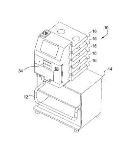
[00104] Referring now to the drawings, Fig. 1 illustrates one embodiment of a
system
sitting idle with its dust cover (not illustrated) removed. A handle 12 for a
cart 14 is
located in a lowered position to minimize the space that system 10 consumes.
Shelves 16 for
12
CA 2969785 2017-06-06
the supply bags (shown below) are also shown in a lowered or "down" position,
which
minimizes the height of system 10.
[00105] System 10 is programmed in an introductory state to instruct the user
to open
a door 18 shown in Fig. 2. Fig. 2 illustrates a close-up view of system 10
with a latch 34
pulled out to unlock door 18. Once door 18 is unlocked as seen in Fig. 3, it
swings open, e.g.,
about forty-five degrees, and is held in the open position by a stop (not
seen), so that a
disposable set (shown below) can be loaded or unloaded.
[00106] Fig. 3 illustrates instrument 20 of system 10 with door 18 held in the
open
position, exposing multiple peristaltic pump heads 22, a latch hook 24,
inductive heater coil
26 and a slotted area 28 for the blood lines (not illustrated) to run to and
from the patient.
Ultrasonic air bubble detectors and optical blood/saline/air detectors are
integrated into the
molded slotted area 28 just above a cutout in the slot for the venous and
arterial line clamps.
The cutout located in slotted area 28 accommodates the venous and the arterial
line clamps.
Figure 16 shows the venous and arterial line clamps 76 in the closed position,
in which the
clamps extend through a respective cutout. In an alternative embodiment, the
inductive
heater coil 26 is retracted into the system to facilitate loading.
[00107] In Fig. 4, door 18 is not shown for clarity to illustrate latch 34 and
latch hook
24, wherein latch 34 mechanically engages latch hook 24 to hold door 18 closed
against the
main portion of instrument 20. One suitable latch assembly is shown and
described in Figs.
11 and 13 of U.S. Pat. No. 6,261,065, "System and Methods for Control of Pumps
Employing
Electrical Field Sensing".
[00108] As seen in Fig. 5, once door 18 has been opened, system 10 prompts the
user
to load the disposable set. A cassette 40 of the disposable set is lowered
into the bag of
instrument 20 and moved to the right (with respect to the orientation of
instrument 20 in Fig.
4). Cassette 40 is loaded starting at the upper left side of open door 18, so
that the patient's
blood lines extending downwardly from cassette 40 do not interfere with the
loading
procedure. The patient's left hand can grasp a dialyzer 36 connected to
cassette 40, while the
patient's right hand can grasp a tubing bundle 38 formed by the supply and
drain lines. Single
handed loading is also possible, e.g., using right hand only grasp bundle 38
to move both
cassette 40 and dialyzer 36.
[0100] As seen in Figs. 6 and 7, door 18 pivots cassette 40 forward towards a
cassette
interface 50 of instrument 20 when an opening 42 in cassette 40 is located
directly over the
13
CA 2969785 2017-06-06
inductive heater transformer coil 26. In an alternative embodiment,
transformer coil 26 is
retracted to facilitate loading of cassette 40. In such case, coil 26 is then
extended into
operating position after cassette 40 is loaded against interface 50. A bezel
(not shown)
provides locating stops for stopping cassette 40 in the vertical and
horizontal directions.
[0101] As cassette 40 mates with the cassette interface 50, the peristaltic
pump tubing
loops 44 of cassette 40 slip over the vertically aligned pumping heads 22. A
pump race 46 is
retracted automatically upwardly when door 18 is opened to provide clearance
between the
pump heads 22 and pump race 26 to facilitate the loading of pump tubing 44 and
cassette 40.
[0102] Fig. 8 illustrates the supply lines 38a to 38e of bundle 38 (number of
supply
lines 38 can vary) passing over retracted pinch valves 48. System 10 also
retracts pinch
valves 48 automatically when door 18 is opened to facilitate the loading of
bundle 38 and
cassette 40 against interface 50 of instrument 20. System 10 opens and closes
pinch valves
48 in a controlled manner, eliminating the need for manual clamps on supply
lines 38a to 38e.
Fig. 9 is shown with supply lines 38 removed to more clearly illustrate pinch
valve plungers
48.
[0103] Fig. 10 further illustrates pinch valve 48/supply line 38 interaction.
Pinch
valves 48 pinch supply lines 38 closed against a strike plate 52. In Fig. 10,
four pinch valves
48 for supply lines 38b to 38e are pinching a respective supply line closed
against strike plate
52, while a fifth pinch valve 48 is retracted, allowing supply line 38a to be
open.
[0104] Figs. 11 and 12 illustrate a pinch valve subassembly 60, in which three
of the
five plungers 48 are extended (closed state). Clamp heads 54 are connected to
a pinch valve
body 62 of subassembly 60. Fig. 13 is shown with body 62 removed to illustrate
springs 56
that spring load pinch valve plungers 48, e.g., so as to be normally closed.
Springs 56
preload pinch valve plungers 48, allowing for variations in the wall thickness
of supply tubes
38. Fig. 13 also illustrates that clamp heads 54 are formed with cam followers
58, which ride
on associated cam lobes 62 coupled to a camshaft 64 (Figs. 11 and 14). A motor
66, e.g., a
stepper motor, is coupled to a drive camshaft 64. Fig. 14 illustrates that in
one embodiment,
the individual cam lobes 62 each define apertures configured fit onto a keyed
portion 68 of
shaft 64. Fig. 14 further illustrates the interaction of cam followers 58 and
cam lobes 62.
[0105] Fig. 15 illustrates that when cassette 40 is loaded into instrument 20
of system
10, blood lines 72 and 74 exit to the lower left of door assembly 90 with
venous and arterial
line clamps 76 (Fig. 16) open initially. Fig. 16 illustrates that venous and
arterial line clamps
14
CA 2969785 2017-06-06
76 pinch bloodlines 72 and 74 against housing portion 78 of instrument 20 to
close bloodlines
72 and 74. During normal operation, system 10 operates clamps 76 independently
as needed.
Fig. 17 is shown with housing portion 78 and door assembly 90 removed to more
fully
illustrate venous and arterial line clamp subassembly 70. A strike part of
housing portion 78
seen in Fig. 16 is located between the venous and arterial lines 72 and 74 and
pinches the
lines together with the clamping levers 76 when closed.
[0106] Fig. 18 illustrates the venous and arterial line clamp subassembly 70
less a
housing 77 shown in Fig. 17, in which clamps 76 are in the open position.
Subassembly 70
includes bellows 80 that hold clamps 76 open during normal operation.
Subassembly 70 also
allows for an Allen wrench 82 with a T-handle 84 to be used to operate a worm
gear 86 that
is coupled operably to a cam 88, which cooperate to manually open both the
venous and
arterial line clamps 76 if need be. In an alternative embodiment, subassembly
70 includes
dual worm gears and a split cam, so that the venous and arterial line clamps
76 can be
manually operated independently. Fig. 19 illustrates the placement of the T-
handle Allen
wrench 82 with respect to instrument 20 when the venous and arterial line
clamps 76 are
operated manually. In one embodiment, system 10 causes an, e.g., red, flag
(not illustrated)
to protrude when the clamps 76 have been opened manually. The flag retracts
when the
manual override is not engaged.
[0107] Fig. 20 illustrates an exploded view of the door assembly 90 taken from
inside
instrument 20. A pair of bellows or bladders 92a and 92b pushes a plate 94
having a gasket
96 to press the cassette 40 (not seen here) against the disposable interface
50 (not seen here).
A space between bladders 92a and 92b is provided to accommodate the inductive
heater coil
26 extending from disposable interface 50. Alternatively, instrument 20
provides a single
bellows (bladder) to press cassette 40 against the disposable interface 50,
which has an
internal opening to accommodate heater coil 26 extending from disposable
interface 50.
[0108] In an alternate failsafe embodiment (not illustrated), the bellows 92a
and 92b
are replaced by a cavity with a diaphragm that is connected sealably to front
pressure plate
18. Springs are located between front pressure plate 18 and the back wall of
the cavity and
press cassette 40 against disposable interface 50, except when a vacuum is
present within the
cavity. In the alternative embodiment, system 10 can also introduce positive
pressure into the
cavity to increase the sealing force.
CA 2969785 2017-06-06
[0109] Fig. 21 illustrates system 10 with the door cover 98 (Fig. 20) removed.
Pneumatic lines 102a and 102b to bellows 92a and 92b, respectively, are shown
teed together
before the exiting door 18 through a hollow hinge 104. A vertical metal bar
106 completes a
circuit for the inductive heater transformer primary coil 26 when the door 18
is closed against
interface 50 of instrument 20. Fig. 22 is also shown with door 18 removed to
illustrate the
inductive heating system including transformer coil 26 and a wave-shaped disk
or disks 108
located in disposable cassette 40, which form a secondary coil that heats
dialysis fluid due to
=2
R losses. Fig. 23 removes cassette 40 to show inductive heater 100 more
clearly. Heater
100 transfers energy from the inductive coil of the transformer 26 into wave
washers 108a
and 108b that are located within cassette 40. Washers 108a and 108b in turn
heat dialysate as
it flows through cassette 40.
[0110] Fig. 24 illustrates the front of the instrument 20 with door assembly
90 and
device housing hidden to expose a mechanism 110 that extends and retracts
triple peristaltic
pump race 46. Mechanism 110 includes four idler gears 112 that tie geared
triple cams 114
together to move race 46 to extend (towards tubing 44) and retract (from
tubing 44)
smoothly. Mechanism 110 is configured such that race 46 extends towards tubing
44 only
after door 18 is closed and latched to preclude the operator from being
exposed to any
moving components. The centers of pump heads 22 are aligned to provide
clearance between
the pump heads and triple race 46 when the race is retracted.
[0111] Fig. 25 illustrates the backside of the retractable triple peristaltic
pump race 46
and mechanism 110 for moving race 46. Cams 114 are located at each end of race
mechanism 110 and race 46. A middle cam 114 is also provided. Each idler gear
112 (Fig.
12) includes a shaft 113 that transmits rotational motion from the idler gears
to all three cams
114 simultaneously. Cams 114 each include lobes 116 that rotate simultaneously
and in
concert within large rounded end slots 118 to simultaneously and evenly extend
and retract
race 46. Shafts 113 of idler gears 112 (Fig. 24) maintain the horizontal
orientation of the
peristaltic pump race 46 as the race moves up and down.
[0112] Fig. 25 illustrates the cam lobes 116 rotated simultaneously and in
concert
upwardly, pushing the pump race 46 away from gear motors 120 that are coupled
to pump
heads 22. The open parts of the horizontally stabilizing idler guide slots are
above the shafts
113 of idler gears. Fig. 26 illustrates the cam lobes 116 rotated
simultaneously and in concert
downwardly, pushing pump race 46 towards the pump gear motors 120 coupled to
pump
16
CA 2969785 2017-06-06
heads 22. The open parts of the horizontally stabilizing idler guide slots 122
are now below
the shafts 113 of idler gears 112.
[0113] Fig. 27 illustrates molded support bosses 124 secured to instrument 20
that
support shafts 113 of the idler gears 112 and support the shafts 115 of cams
114 on one end.
A bar (not shown here but shown in Fig. 71), which mounts to bosses 124,
supports the shafts
113 of gears 112 and shafts 115 of cams 114 on their other ends. A motor (not
illustrated)
that drives cams 114, which operate the retractable pump race 46, is attached
to any of the
shafts 115 of any of cams 114. Attaching the motor to the shaft of center cam
114 may be
preferred so that clearance in the gear train is symmetric with respect to
outer cams 114.
[0114] Figs. 28 and 29 illustrate that system 10 includes a crank 130 that is
connected
to the blood pump head 22 to operate the head manually. Manual return of the
blood
contained within the extracorporeal circuit is necessary in the event of a
failure of system 10
or after an extended power failure. It is typically necessary to manually
operate the venous
and arterial line clamps 76 (from a failed closed state) before being able to
return the blood in
extracorporeal circuit to the patient. Fig. 29 also illustrates that door 18
in one embodiment
defines an opening or aperture 132 through which manual crank 130 for the
blood pump 22
can be inserted with the door closed. Crank 130 includes a large gripping
handle 134 and
crankshaft 136, which is sufficiently long to allow the user to easily turn
blood pump head
22. In an alternate embodiment, manual crank 130 is built into the door
assembly 90 and is
accessible to engage pump head 22 when door 18 is opened and hinged away from
machine
interface 50.
[0115] As seen in Fig. 30, in one bag management embodiment, system 10 prompts
the user initially to fold up all of bag shelves 16 except for the bottom
shelf 16. The user is
then able to break a peel seal of a dual chamber bag (if used), place the
first solution bag 140
on bottom shelf 16 and connect the bag to the bottom supply line 38e extending
from
disposable cassette 40, as shown in Fig. 31. When shelf sensors 138 detect
that the bag has
been placed onto first shelf 16 and that the peel seal 142 has been broken,
system 10 prompts
the user to place a second bag 140 on the second lowest shelf 16, and so on.
System 10
continues to prompt the user to place solutions bags 140 onto shelves 16 and
connect the bags
to supply lines 38 until all of shelves 16 are filled, as shown in Fig. 32.
[0116] As shown in Fig. 32, a peel seal 142 of dual chamber bag 140 present on
the
top shelf 16 is not broken, a condition which sensors 138 can sense, causing
system 10 to
17
CA 2969785 2017-06-06
instruct the user to break peel seal 142 before continuing with treatment. One
such sensor
arrangement and peel seal open check is described in U.S. Patent Application
No.
11/773,742, entitled "Mobile Dialysis System Having Supply Container
Detection", filed
July 5, 2007, assigned to the assignee of the present disclosure. Fig. 33
illustrates all solution
bags 140 with peel seals 142 broken, such that treatment can continue.
[0117] Fig. 34 illustrates one embodiment for the placement of the capacitive
sensors
138 that detect the presence of the solution bags, whether peel seal is
broken, and perhaps
even whether the same solution is present in each bag 140. Other sensors or
combinations of
sensors can be used alternatively, including optical sensors, inductive
sensors, bar code
readers, radio frequency identification ("RFID") tags and cameras.
[0118] Fig. 35 illustrates a luer connection assembly 144, which is located on
an end
of a heparin line 146, which in turn is connected to disposable cassette 40. A
heparin syringe
148 ranging in size from ten milliliters to sixty milliliters, can be
connected to luer
connection assembly 144 of the disposable set and is inserted with the plunger
150 pointing
down into a syringe pump 152 as shown in as shown in Fig. 36. The luer
connection
assembly 144 is then rotated to lock the syringe in place as shown in Fig. 37.
Syringe 148,
for sizes larger than 30 milliliters, is inserted with the plunger 150
pointing down into a
syringe pump 152 as shown in as shown in Fig. 38. The integral grip 149 on the
larger
heparin syringes is rotated forty-five degrees to lock the syringe 148 into
the syringe pump
152 as shown in Figs. 37 and 38 versus grip 149 shown in Fig. 36.
[0119] Syringe pump 152 is shown in more detail in Fig. 39. Pump 152 includes
a
stepper motor 154, gears 156, guide rails 158 and a concave push plate 160
that self-centers
on the end of the syringe plunger 150. Air exits syringe 148 above the heparin
and is purged
during the priming of the extracorporeal circuit because syringe 148 is
inverted for use.
Stepper motor 154 increments 0.9 degrees per step in one implementation. Pump
152 and
assembly 144 are sized to accept nearly any size of syringe 148. The user
inputs the syringe
stroke length and syringe stroke volume into system 10. System 10 can
thereafter determine
the volume of heparin to be delivered.
[0120] Smaller syringes 148 are visible through a window 162 in the side of
the pump
as shown in Fig. 40. Larger syringes housings are visible since they are not
inserted into
syringe pump 152 and remain outside of instrument 20 as illustrated in Fig.
38. Should a
saline or dialysate bag leak, or be spilled, onto instrument 20, the liquid
could flow into the
18
CA 2969785 2017-06-06
heparin pump and out the opening in side window 162 but would not flow inside
the
instrument, where the fluid could damage instrument 20.
[0121] Figs. 41 and 42 illustrate that heparin line 146 passes through an air
bubble
detector 164 to cassette 40. System 10 introduces heparin into the patient's
blood stream at
the outlet 166 of the blood pump just before the blood passes into the
dialyzer. The internal
volume of the heparin line is essentially that of a very small diameter tube
of minimum
length. A diaphragm actuated pinch valve 165 (plunger only shown in Fig. 41),
which does
not add to the internal volume of the heparin line, can be provided to block
the flow of
heparin to cassette 40.
[0122] Fig. 43 illustrates a support rod 168 that collapses into instrument 20
when not
in use. Support rod 168 supports a saline bag 170 that is used for priming
system 10 and
rinsing blood back to the patient at the end of the therapy. Alternatively,
rod 168 is
detachable from instrument 20 when not in use.
[0123] Figs. 43 and 44 illustrate that saline line 172 enters instrument 20
adjacent to
the entry of heparin line 164 (see also Fig. 41). Fig. 45 illustrates that two
saline flow control
valves 174a and 174b are located on each side of blood pump tubing loop 44.
The center port
from each of the valves feeds directly into blood flow into, or coming from,
the blood pump
as shown in Fig. 46. The third saline valve 174c is located on the backside of
cassette 40 as
seen in Figs. 45 and 46 and is positioned to put saline directly into a venous
air separation
(drip) chamber 176. The saline valve 174a on the blood pump outlet, and the
saline valve
174b leading to dialyzer 36, are opened sequentially to gravity prime the
arterial blood line
and the venous drip chamber 176 as illustrated later in Fig. 54.
[0124] As seen in Fig. 47, a normally evacuated dialyzer inlet line pressure
transducer
interface 178 is pressurized so that it operates as a flow control valve,
preventing saline from
backflowing into the dialyzer or filter 36. The gravity head from the saline
bag causes saline
to flow into the blood circuit and into the reversed rotating pump inlet 180
(the outlet under
normal operating flow) when saline valve 174a is opened. The reversed flow
blood pump
head 22 draws saline from the saline bag and pumps it through reversed flow
outlet 182 (the
inlet under normal operating conditions) and down the arterial line 186.
[0125] As seen in Fig. 48, the venous line 184 and arterial line 186 are
connected in
series during priming so that air is purged from both lines via venous line
drip chamber 176
shown in Fig. 49. Standard connections 188 (Fig. 48) can be used to connect
the venous line
19
CA 2969785 2017-06-06
184 and arterial line 186 in a closed loop. Gravity prevents air from being
drawn from the
saline bag as long as the bag contains saline. Saline flows slowly into the
venous air
separation chamber 176 in a "reverse" direction (from normal blood flow)
during priming.
[0126] In Fig. 49, the inverted-U shaped venous air separation chamber 176 has
a
vent port 190 located at its top, so that air can gather there and be vented
to the drain. Fig. 50
shows a valve 196 located on the opposite side of the cassette 40 from vent
port 190, which is
opened whenever air needs to be vented from the chamber. A second vent valve
192 also
shown in Fig. 50 can be placed optionally in series with first vent valve 196
and operated
sequentially so that predetermined volumetric increments of air can be vented
from system 10
to a controlled vent volume 194 shown in Fig. 51. As seen in Fig. 51, port 190
connected to
the center of the cassette-based diaphragm valve 196 communicates with air
separation
chamber 176 so that the "dead" volume needed for these apparatuses is
minimized. Valve
196 seals well against the pressure present in the venous air separation
chamber. Saline bags
can be replaced during a therapy since they can be primed directly into the
drip chamber 176
using the third saline valve 174c (Fig. 49).
[0127] Fig. 52 is a schematic of one embodiment of a fluid management system
associated with the disposable set. In general, the fluid management system
includes a blood
circuit 210 and a dialysate circuit 220. System 10 operates the disposable set
to provide the
hemodialysis therapy. Set 200 of Figs. 53A and 53B illustrates an embodiment
of a
disposable set 200 operable with system 10. Disposable set 200 includes
cassette 40, filter
36, pump tubes 44, supply tubes 38, balance tubes 202, arterial line 184 and
venous line 186,
etc., discussed herein.
[0128] Once disposable set 200 has been loaded into the hemodialysis system
10,
dialysate bags 140 have been connected, the saline bag 170 (Fig. 43) has been
connected and
the heparin syringe 148 has been loaded, system 10 primes itself automatically
starting with
the blood side circuit. The heparin pump plunger 150 is moved forward until
heparin is
detected by heparin line air detector AD-HL shown in Fig. 52. Heparin valve V-
H is then
closed. Next, saline is flowed from the saline bag 170 into the blood side
circuit 210 as
illustrated in Fig. 54, first through valve V-SA and then through valve V-SDC.
A level
sensor L-ATB in the AIR TRAP drip chamber detects saline flow into the drip
chamber 176
and determines when to close valves V-SA and V-SDC.
CA 2969785 2017-06-06
[0129] As shown in Fig. 55, the post pump blood valve V-PPB is then closed, V-
SV
is opened and PUMP-Blood pumps saline in a reverse flow direction. Pressure
sensor P-VL
and level sensor L-ATB are used to detelinine when to open air vent valves V-
AVB-P and V-
AVB-S. The blood pump pushes the saline backwards down the arterial line and
into the
venous line. When saline reaches the venous air separator (drip chamber 176),
the air will be
separated from the fluid and will be discharged into a drain line 206 through
vent valves V-
AVB-P and V-AVB-S until the air separation chamber 176 is flooded with saline.
[0130] Next, as seen in Fig. 55, saline is flowed up into the bottom of
dialyzer 36 and
up through its hollow fibers. Valve V-PPB is controllably opened so that the
air that exits the
top of the dialyzer 36 flows into the priming loop, becomes separated in air
trap 176 and
discharged to drain 206. Saline is also flowed through pours of the fibers of
dialyzer 36 to
fill the housing of dialyzer 36. System 10 monitors the pressure in the venous
line using
pressure sensor P-VL to maintain the blood side circuit 210 at a controlled
pressure during
priming.
[0131] As seen in Fig. 56, spent dialysate pump, PUMP-DS and valves V-DS, V-BI-
S1, V-BI-SO and V-DD vent air from the dialyzer housing to drain 206. Valves V-
DI-VEN,
CK-VEN, V-DI-FIL, V-DI-PRE and CK-PRE are opened controllably to allow a
predetermined volume of saline to be pushed into the dialysate circuit 220,
purging air from
associated dialysate lines. A second saline bag 170 can be replaced during a
therapy by
selecting "replace saline bag", causing the saline line to be primed
automatically into the air
trap 176.
[0132] As shown in Fig. 56, dialysate valve V-DB1 that is associated with the
dialysate bag on the top shelf is opened so that dialysate can flow into the
inlet of dialysate
PUMP-DF. PUMP-DF pushes the dialysate through the inline fluid heater and into
a
dialysate side air trap 208. Dialysate flows out the bottom of the air trap
208, through valve
V-FI and into balance tube B2, through valve V-B2-FI, pushing fluid out the
other side of
balance tube B2. The fluid exiting the other side of balance tube B2 flows
through valve V-
B2-SO and into the dialysate recirculating circuit 203 through valve V-DR. The
recirculating
circuit 223 tees into the supply line circuit 205 at the inlet to PUMP-DF.
Pump-DS is
operating at the same time drawing air, dialysate and/or saline from the blood
side of the
dialyzer, though the dialysate side of the dialyzer, into the remainder of the
dialysate circuit.
PUMP-DS pushes the fluid through valve V-B 1-SI and into balance tube B1,
pushing fluid
21
CA 2969785 2017-06-06
out the other side of balance tube B1. The fluid exiting the other side of
balance tube B1
flows through valve V-B1-F0 and valve V-DI-FIL into the dialysate side of the
dialyzer 36.
[0133] Fig. 57 is similar to Fig. 56 except the roles of balance tubes 202 B1
and B2
are reversed. As fluid enters the dialysate circuit 220, the pressure in the
circuit increases,
forcing air to be discharged under pressure to drain line 206 through open
vent valves V-
AVD-P and V-AVD-S.
[0134] Fig. 58 illustrates balance tubes 202. Instrument 20 includes pairs of
optical
sensors (not shown) operable with balance tubes 202 to determine an end of
travel of a
separator 212 located within each balance tube 202. The optical sensors in one
embodiment
are reflective, so that an emitter and receiver of each sensor can be on the
same (e.g., non-
door) side of balance tube 202. The sensors alternatively include emitters and
receivers
located on opposite sides of balance tubes 202. Outlets 214 on both ends of
both balance
tubes 202 are at the balance tube tops when mounted for operation as shown if
Fig. 58, so that
air will pass through the balance tubes and not become trapped in the tubes as
long as system
is level. Mechanical stops 216 limit the movement of separators 212 to that
visible to the
optical sensors.
[0135] Fig. 59 illustrates HHD system 10 performing hemodialysis. Here, fresh
dialysate is pushed from balance tubes 202 to dialyzer 36 via valve V-DI-FIL,
while spent
dialysate is removed from dialyzer 36 via valve V-DS to balance tubes 202.
[0136] Fig. 60 illustrates HHD system 10 performing pre-dilution
hemofiltration.
Here, fresh dialysate is pushed from balance tubes 202 to blood circuit 210
directly via valve
V-DI-PRE, while spent dialysate is removed from dialyzer 36 via valve V-DS to
balance
tubes 202.
[0137] Fig. 61 illustrates HHD system 10 perfoiming post-dilution
hemofiltration.
Here, fresh dialysate is pushed from balance tubes 202 to blood circuit 210
directly via valve
V-DI-VEN, while spent dialysate is removed from dialyzer 36 via valve V-DS to
balance
tubes 202.
[0138] Fig. 62 illustrates HHD system 10 performing post-dilution hemo-
diafiltration.
Here, fresh dialysate is pushed from balance tubes 202 to (i) dialyzer 36 via
valve V-DI-FIL
and (ii) blood circuit 210 directly via valve V-DI-VEN, while spent dialysate
is removed
from dialyzer 36 via valve V-DS to balance tubes 202.
22
CA 2969785 2017-06-06
[0139] Fig. 63 illustrates one embodiment for closing arterial line clamp V-
ALC,
opening a saline valve V-SA and infusing a saline bolus into blood circuit 210
during
therapy.
[0140] Fig. 64 illustrates one embodiment for recirculating fresh dialysate
through
Fluid Heater and recirculating circuit 223 and balance tubes B1 and B2 to
remove UF. In
Fig. 64, pump-DF pumps fluid in a loop that includes Fluid Heater since valve
V-DBY is
open. Valve V-FI is closed so no fresh dialysate is delivered to balance
chambers 202.
Pump-DS pulls spent fluid from the dialyzer 36 through valve V-DS and pushes
the spent
fluid through valve V-BI-SI and into the right side of balance tube Bl. Fresh
fluid then flows
from the left side of balance tube B1 through valves V-BI-FI and V-B2FI and
into the left
side of balance tube B2. Spent fluid then flows out the right side of balance
tube B2 through
valves V-B2-S0 and V-DD and into the drain line. In this manner, a volume of
spent fluid is
sent to drain 206 without a corresponding volume of fresh fluid delivered from
supply bags
140 to either balance chamber B1 or B2.
[0141] Fig. 65 illustrates one embodiment for closing venous line clamp V-VLC,
opening a saline valve V-SA and rinsing back the arterial line 184.
[0142] Fig. 66 illustrates one embodiment for closing arterial line clamp V-
ALC,
opening a saline valve V-SA and rinsing back the venous line 186.
[0143] Figs. 67A to 67C illustrate a cassette interface assembly 250, which
houses,
among other items, cassette interface 50, door latch 24, heater 26, a bellows
bladder 252 and
an internal module 260. Internal module 260 is bounded by interface plate 50
and a back
plate 254. Internal module 260 houses a plurality of gaskets 256, a pneumatic
valve
assembly 258, a pinch valve assembly 262, and a plurality of manifold plates
264.
[0144] All or most all of the valves, pressure sensors, level sensors, etc.,
can be
removed without disassembly of subassembly 250. The inductive heater mechanism
26 and
bellows bladder 252 (different from bladder 92 above) require removal of
internal module
260. To this end, four screws 266, each with a spring 268, fix a housing 270
of subassembly
250 to internal module 260. Internal module 260 can be unbolted from screws
266, so that
springs 268 push internal module 260 forward and out of the housing 270. Power
and control
connections (not shown) to subassembly 250 are also disconnected to remove
internal module
260 completely.
23
CA 2969785 2017-06-06
[0145] As seen additionally in Figs. 68 to 70, four springs 268 on the
backside of
subassembly 250 retract the internal interface module 260 when bellows bladder
252 is not
pressurized by pushing screens away from housing 270 and pulling interface
module 260
along with the screws. When the bellows bladder 252 is pressurized, internal
module 260 is
pushed forward and applies pressure to cassette 40, pushing the cassette
against a door
gasket, which seals fluid pathways on both the front side and the rear side of
the cassette 40.
The membrane gaskets 256 on the internal module 260 mate up against the
faceplate 50 of
the interface module 250. The faceplate 50 is configured so that it can
support a vacuum
between the cassette sheeting and pressure sensors, liquid level sensors,
etc., bringing the
sensors into intimate contact with the cassette sheeting and the fluid on the
other side of the
sheeting. System 10 is also configured to port a vacuum between the cassette
sheeting and
the thin sections of the membrane gasket 256 above the valves. This vacuum can
be used to
detect holes, tears or slits in the cassette sheeting before, and during a
therapy.
[0146] Fig. 71 is a view of the backside of system 10 with the cover removed.
The
open space houses interface assembly 250, hinged shelves 16, peristaltic pump
motors 120 a
pneumatic pump, a power supply, battery and electronics that operate the
system.
[0147] Fig. 72 illustrates system 10 operating alternatively with an online
dialysate
generation system 300. System 300 generates dialysate online or on-demand,
eliminating
bags 140, shelves 16 and multiple supply tubes 38. A single supply tube 38
feeds from
generation system 300 to instrument 20. Water inlet line 302 and drain lines
304 lead to and
from generation system 300, respectively.
[0148] Figs. 73A, 73B and 74 illustrate a cassette 40 diaphragm valve chamber
configuration 280, which solves an inherent problem with diaphragm valves have
when
attempting to seal against downstream pressure because the pressure that is
trying to seal off
the valve is acting on an area that is just slightly larger than an area upon
which the
downstream pressure is acting. The difference between the two areas is the
area defined by
the top of the "volcano". Also, if the downstream fluid volume is completely
fixed when the
diaphragm valve closes, further movement of the diaphragm is prevented after
the initiation
of the seal because of the incompressibility of the trapped fluid. The result
is that the
downstream pressure equals the valve sealing pressure. Diaphragm valve
configuration 280
provides a diaphragm valve that can seal against both upstream and downstream
pressure via
a connection of two diaphragm valve chambers 282 and 284 placed in series.
Diaphragm
24
CA 2969785 2017-06-06
valve chambers 282 and 284 are connected fluidly via a compliance chamber 286,
which
allows sheeting seals 288 of the cassette sheeting to close around respective
volcano ports
290 of both valve chambers 282 and 284.
[0149] Chamber configuration 280 in both Figs. 73A and 73B includes a rigid
middle
or base wall 281 from which valve ports 290 and the valve chamber walls extend
upwardly.
Wall 281 defines an aperture 283 for each valve chamber 282 and 284. Fluid
communicates
between valve chambers 282 and 284 and compliance chamber 286 via apertures
283.
[0150] Fig. 73A shows a cross-section of two diaphragm valve chambers 282 and
284
with an integral compliance chamber 286, wherein the diaphragms can readily
close seals 288
to ports 290. Here, a vacuum is applied to a lower diaphragm 289 at the
compliance chamber
286. Diaphragm 289 is flexible and has a relatively large cross-sectional area
to absorb the
kinetic energy created by a pneumatic valve actuator applying a positive
pressure Pa, such
that the positive sealing pressure applied to one valve chamber 282 or 284 is
much less likely
to harm an existing seal of a fluidly connected upstream or downstream valve
chambers. The
negative pressure pulls sheeting 288 down around ports 290 and allows valve
chamber 282 or
284 to be sealed against the backpressure applied by its own sealing pressure
(around the
outside of port 290) plus backpressure from a fluidly connected upstream or
downstream
valve chamber residing up through the center of port 290.
[0151] Compliance chamber 286 as seen in Fig. 73B is configured a little bit
differently and uses a portion of the membrane or sheeting seals 288 of valve
chambers 282
and 284 to provide a compliant material covering a relatively large cross-
sectional area 292
of chamber 286. Here, a vacuum applied to sheeting 288 at chamber 286 negates
the positive
pressure Pc applied around the outside of ports 290 and expands the relatively
large area 292
of the valve seal sheeting, pulling sheeting 288 down around the outside of
port 290. The
configuration of Fig. 73B is advantageous in one respect because positive and
negative
pressures are applied to the same side of the cassette at chamber
configuration 280, such that
associated pneumatics can be located on a single side of the cassette..
[0152] By changing the pressure seen at compliance chamber 286 from a positive
pressure when the valve chambers 282 and 284 are open to a negative value
after the valve
chambers results in that only the liquid side center of the volcano port 290
is exposed to high
positive pressure. The liquid annular area of valve chambers 282 and 284 on
the outside of
volcano ports 290 sees the applied vacuum, which allows the air sealing
pressure on the
CA 2969785 2017-06-06
outside of the cassette to seal against backpressures that would have
otherwise forced it open.
This allows valve chambers 282 and 284 to seals well in both upstream and
downstream
configurations.
[0153] In one example, suppose the total seal area of valve chambers 282 and
284 is
one square inch and that the sealing area at the top of volcano port 290 is
0.1 square inch over
the volcano. A positive ten psig air pressure would then apply an external
force of 10 lbs to
the entire valve chamber 282 or 284. A backpressure on the annular fluid side
of the
associated port 290 from the applied ten psig pressure plus a backpressure the
backpressure
up through the center of port 290 from a downstream sealed valve would exert
almost the
same opposite "unsealing" force of ten pound (only difference would be the
small annular
area of port 290 at the top, which is a function of the port wall thickness
and the diameter of
the tube), resulting in a potentially leaky valve chamber 282 or 284. A higher
positive
pressure, e.g., twenty psig, could be applied to valve chamber 282 or 284
forcing sheeting
288 to seal to port 290 against the 10 psig backpressure, however, the noise
generated to
create the twenty psig air pressure could objectionable to the user. There
would also be no
redundancy in the different valve pressures.
[0154] Back to back valve chambers 282 and 284 of Figs. 73A and 73B, on the
other
hand, separated by an applied negative pressure, e.g., 5 psig vacuum, both
seal independently
well. The ten psig air pressure would still apply 10 lbs external force to
seal both valves 282
and 284, however, the 10 psig pressure at the center of the volcano port 290
and the -5 psig
pressure on the annular area around the volcano would apply a total pressure
of ten psig * 0.1
sq in + (- 5 psig) * 0.9 sq in = -3.5 lbs. The net force to close the valve
would be 13.5 lbs so
that valve would seal very well.
[0155] It may be possible to not use a separate vacuum and instead rely on the
expansion of the flexible part of the compliance chamber 286 to absorb energy
from the
backpressure from one valve chamber 282 or 284 applied to the other valve
chamber 282 or
284. Here, apertures 283 allow the pressurized fluid inside chambers 282 and
284 and
around ports 290 to communicate with fluid inside compliance chamber 286 and
expand
diaphragm 289 or sheeting area 292, allowing the backpressure around ports 290
to dissipate.
[0156] Valves V-DI-PRE, CK-PRE, V-DI-VEN and CK-VEN in Fig. 52 (and other
flow schematics) and valve chambers 282 and 284 of valve configuration 280 of
cassette 40
shown in Fig. 74 are constructed as shown schematically in Figs. 73A and 73B
and can seal
26
CA 2969785 2017-06-06
against higher pressure in either direction. That is, not only does compliance
chamber 286
serve to not disrupt an existing upstream or downstream first valve chamber
closure when a
second valve chamber in fluid communication with the first valve chamber is
opened,
compliance chamber 286 also aids in the closure of a first valve chamber when
a second
valve chamber in communication with the first valve chamber (upstream or
downstream) has
been closed previously, which could otherwise create positive fluid pressure
against which
the closure of the first valve chamber would have to fight.
[0157] Fig. 75 illustrates that system 10 in one embodiment includes a wide
pump
head 22 that drives two dialysate pump segments 44 to mix two solutions in a
ratio that is
approximately equal to the ratio of the tube inside diameters squared (mix
ratio = (IDI/ID2)2),
assuming the wall thicknesses of tubes 44 is the same. For a 1:1 mix ratio,
consecutive
segments of tubing from the same roll of tubing can be taken to provide
segments of the same
wall thickness and good mixing accuracy. Mixing accuracy is optimized because
the inlet
pressure on the supply lines is controlled within about four inches of water
column by the bag
manager, the tubing inner diameter is controlled during the manufacture of the
disposable set,
the pump race diameters are the same and the pump actuator rotational speed is
the same for
the parallel tubing segments. System 10 also ensures that an initial supply
fluid temperature
of each of the different dialysis fluids in tubes 44 is within a few degrees
of each other.
[0158] It should be understood that various changes and modifications to the
presently preferred embodiments described herein will be apparent to those
skilled in the art.
Such changes and modifications can be made without departing from the scope of
the present
subject matter and without diminishing its intended advantages. It is
therefore intended that
such changes and modifications be covered by the appended claims.
27
CA 2969785 2017-06-06