Note: Descriptions are shown in the official language in which they were submitted.
CA 02969847 2017-06-05
WO 2016/094679 PCT/US2015/065029
GENETICALLY MODIFIED CELLS, TISSUES, AND ORGANS FOR
TREATING DISEASE
CROSS-REFERENCE
[0001] This application claims the benefit of U.S. Provisional Patent
Application No. 62/090,037,
filed on December 10, 2014, and U.S. Provisional Patent Application No.
62/253,493, filed on
November 10, 2015, which are both herein incorporated by reference in their
entirety.
SEQUENCE LISTING
[0002] The instant application contains a Sequence Listing which has been
submitted
electronically in ASCII format and is hereby incorporated by reference in its
entirety. Said ASCII
copy, created on December 10, 2015, is named 47190-701.601 SL.txt and is
681,444 bytes in size.
BACKGROUND OF THE DISCLOSURE
[0003] There is a shortage of organs, tissues or cells available for
transplantation in recipients such
as humans. Xenotransplantation or allotransplantation of organs, tissues, or
cells into humans
has the potential to fulfill this need and help hundreds of thousands of
people every year. Non-
human animals can be chosen as organ donors based on their anatomical and
physiological
similarities to humans. Additionally, xenotransplantation has implications not
only in humans,
but also in veterinary applications.
[0004] However, unmodified wild-type non-human animal tissues can be rejected
by recipients,
such as humans, by the immune system. Rejection is believed to be caused at
least in part by
antibodies binding to the tissues and cell-mediated immunity leading to graft
loss. For example,
pig grafts can be rejected by cellular mechanisms mediated by adaptive immune
cells.
INCORPORATION BY REFERENCE
[0005] All publications, patents, and patent applications herein are
incorporated by reference to the
same extent as if each individual publication, patent, or patent application
was specifically and
individually indicated to be incorporated by reference. In the event of a
conflict between a term
herein and a term in an incorporated reference, the term herein controls.
SUMMARY
[0006] Disclosed herein are compositions and methods for treating or
preventing diseases. Also
disclosed are genetically modified cells and methods of making the genetically
modified cells
1
CA 02969847 2017-06-05
WO 2016/094679 PCT/US2015/065029
for treating or preventing disease. Further disclosed are genetically modified
non-human
animals and methods of making genetically modified non-human animals that can
be used in
treating or preventing disease, e.g., by later extracting cells, tissues, or
organs from these
genetically modified non-human animals and transplanting them into a subject.
Also disclosed
herein are methods for treating or preventing diseases using the genetically
modified cells,
tissues, and organs. Additionally disclosed are methods for treating or
preventing diseases
using cells, tissues, and/or organs from genetically modified non-human
animals.
[0007] In one aspect, disclosed herein is a genetically modified animal with
reduced protein
expression of one or more first genes, where the genetically modified animal
is a member of the
Laurasiatheria superorder or is a non-human primate, where the one or more
first genes
comprise a) a component of a major histocompatibility complex (MHC) I-specific
enhanceosome, b) a transporter of an MHC I-binding peptide, and/or c)
complement component
3 (C3), where the reduced protein expression is in comparison to a non-
genetically modified
counterpart animal. In some cases, the member of the Laurasiatheria super
order is an
ungulate. In some cases, the ungulate is a pig. In some cases, the protein
expression of the one
or more first genes is absent in the genetically modified animal. In some
cases, the reduction of
protein expression inactivates a function of the one or more first genes. In
some cases, the
genetically modified animal has reduced protein expression of two or more the
first genes. In
some cases, the genetically modified animal comprises reduced expression of a
component of a
MHC I-specific enhanceosome, where the component of a MHC I-specific
enhanceosome is
NOD-like receptor family CARD domain containing 5 (NLRC5). In some cases, the
genetically modified animal comprises reduced expression of a transporter of a
MHC I-binding
peptide, where the transporter is transporter associated with antigen
processing 1 (TAP1). In
some cases, the genetically modified animal comprises reduced expression of
comprising C3.
In some cases, the genetically modified animal has reduced protein expression
of three or more
the first genes.
[0008] In some cases, the genetically modified animal further comprises
reduced protein
expression of one or more second genes, where the one or more second genes
comprise: a) a
natural killer (NK) group 2D ligand, b) an endogenous gene not expressed in a
human, c) a
CXC chemokine receptor (CXCR) 3 ligand, and/or d) MHC II transactivator
(CIITA), where
the reduced protein expression is in comparison to a non-genetically modified
counterpart
animal. In some cases, the protein expression of the one or more second genes
is absent in the
genetically modified animal. In some cases, the reduction of protein
expression inactivates a
2
CA 02969847 2017-06-05
WO 2016/094679 PCT/US2015/065029
function of the one or more second genes. In some cases, the genetically
modified animal
comprises reduced protein expression of a NK group 2D ligand, where the NK
group 2D ligand
is MHC class I polypeptide-related sequence A (MICA) or MHC class I
polypeptide-related
sequence B (MICB). In some cases, the genetically modified animal comprises
reduced
protein expression of an endogenous gene not expressed in a human, where the
endogenous
gene not expressed in a human is glycoprotein galactosyltransferase alpha 1,3
(GGTA1),
putative cytidine monophosphate-N-acetylneuraminic acid hydroxylase-like
protein (CMAH),
or [31,4 N-acetylgalactosaminyltransferase (B4GALNT2). In some cases, the
genetically
modified animal comprises reduced protein expression of a CXCR3 ligand, where
the CXCR3
ligand is C-X-C motif chemokine 10 (CXCL10).
[0009] In some cases, the genetically modified animal further comprises one or
more exogenous
polynucleotides encoding one or more proteins or functional fragments thereof,
where the one
or more proteins comprise: a) an MHC I formation suppressor, b) a regulator of
complement
activation, c) an inhibitory ligand for NK cells, d) a B7 family member, e)
CD47, 0 a serine
protease inhibitor, and/or g) galectin. In some cases, the one or more
proteins are human
proteins. In some cases, the genetically modified animal comprises one or more
exogenous
polynucleotides encoding an MHC I formation suppressor, where the MHC I
formation
suppressor is infected cell protein 47 (ICP47). In some cases, the genetically
modified animal
comprises one or more exogenous polynucleotides encoding a regulator of
complement
activation, where the regulator of complement activation is cluster of
differentiation 46 (CD46),
cluster of differentiation 55 (CD55), or cluster of differentiation 59 (CD59).
In some cases, the
genetically modified animal comprises one or more exogenous polynucleotides
encoding an
inhibitory ligand for NK cells, where the inhibitory ligands for NK cells is
leukocyte antigen E
(HLA-E), human leukocyte antigen G (HLA-G), or 3-2-microglobulin (B2M). In
some cases,
the genetically modified animal comprises one or more exogenous
polynucleotides encoding
HLA-G, where the HLA-G is HLA-G1, HLA-G2, HLA-G3, HLA-G4, HLA-G5, HLA-G6, or
HLA-G7. In some cases, the HLA-G is HLA-G1. In some cases, the genetically
modified
animal comprises one or more exogenous polynucleotides encoding a B7 family
member,
where the B7 family member is a programed death-ligand. In some cases, the
programed
death-ligand is programed death-ligand 1 (PD-L1) or programed death-ligand 2
(PD-L2). In
some cases, the one or more exogenous polynucleotides encode both PD-Li and PD-
L2. In
some cases, the genetically modified animal comprises one or more exogenous
polynucleotides
encoding a serine protease inhibitor, where the serine protease inhibitor is
serine protease
3
CA 02969847 2017-06-05
WO 2016/094679 PCT/US2015/065029
inhibitor 9 (Spi9). In some cases, the genetically modified animal comprises
one or more
exogenous polynucleotides encoding a galectin, where the galectin is galectin-
9.
[0010] In some cases, the genetically modified animal comprises reduced
protein expression of
NLRC5 or TAP1, C3, reduced protein expression of CXCL10, GGTA1, CMAH, and/or
B4GALNT2; and/or one or more exogenous polynucleotides encoding HLA-G1, HLA-E,
or a
functional fragment thereof, PD-Li or a functional fragment thereof, PD-L2 or
a functional
fragment thereof, and/or CD47 or a functional fragment thereof In some cases,
the one or more
exogenous polynucleotides are inserted adjacent to a ubiquitous promoter. In
some cases, the
ubiquitous promoter is a Rosa26 promoter. In some cases, the one or more
exogenous
polynucleotides are inserted adjacent to a promoter of a targeted gene or
within the targeted gene.
In some cases, the targeted gene is one of the first genes or one of the
second genes. In some
cases, the protein expression of the one or more first genes is reduced using
a CRISPR/cas
system. In some cases, the protein expression of the one or more second genes
is reduced using a
CRISPR/cas system.
[0011] In another aspect, disclosed herein is a genetically modified animal
that is a member of the
Laurasiatheria superorder or is a non-human primate comprising: an exogenous
polynucleotide encoding an inhibitory ligand for an NK cell or a functional
fragment thereof,
and reduced protein expression of an endogenous gene, where the reduced
protein expression
is in comparison to a non-genetically modified counterpart animal. In some
cases, the
inhibitory ligand for an NK cell is HLA-E or HLA-G. In some cases, the
inhibitory ligand for
an NK cell is HLA-G, where the HLA-G is HLA-G1, HLA-G2, HLA-G3, HLA-G4, HLA-
G5,
HLA-G6, or HLA-G7. In some cases, the HLA-G is HLA-G. In some cases, the
endogenous
gene is a gene not expressed in a human. In some cases, the endogenous gene is
GGTA1,
CMAH, and/or B4GALNT2.
[0012] In some cases, the genetically modified animal further comprises
exogenous
polynucleotides encoding: a) PD-Li or a functional fragment thereof, b) PD-L2
or a functional
fragment thereof, and/or c) CD47 or a functional fragment thereof In some
cases, the
exogenous polynucleotides are inserted adjacent to a ubiquitous promoter. In
some cases, the
ubiquitous promoter is a Rosa26 promoter. In some cases, the exogenous
polynucleotides are
inserted adjacent to a promoter of the endogenous gene, or within the
endogenous gene. In
some cases, the protein expression of the endogenous genes is reduced using a
CRISPR/cas
system.
4
CA 02969847 2017-06-05
WO 2016/094679 PCT/US2015/065029
[0013] Further disclosed herein is a population of genetically modified
animals comprising two or
more animals disclosed in the application. In some cases, at least two or more
animals have
identical phenotypes. In some cases, at least two or more animals have
identical genotypes.
[0014] In another aspect, disclosed herein is a genetically modified cell from
a member of the
Laurasiatheria superorder or a non-human primate, comprising reduced protein
expression of one
or more first genes, where the one or more first genes comprise: a) a
component of a MHC I-
specific enhanceosome, b) a transporter of a MHC I-binding peptide, and/or c)
C3, where the
reduced protein expression is in comparison to a non-genetically modified
counterpart cell. In
some cases, the genetically modified cell comprises reduced protein expression
of a component
of a MHC I-specific enhanceosome, where the component of MHC I-specific
enhanceosome is
NLRC5. In some cases, the genetically modified cell comprises reduced protein
expression of a
transporter of a MHC I-binding peptide, where the transporter of a MHC I-
binding peptide is
TAP1. In some cases, the genetically modified cell comprises reduced protein
expression of C3.
[0015] In some cases, the genetically modified cell further comprises reduced
protein expression of
one or more second genes, where the one or more second genes comprise: a) an
NK group 2D
ligands, b) an endogenous gene not expressed in a human, c) a CXCR3 ligand,
and/or d) CIITA,
where the reduced protein expression is in comparison to a non-genetically
modified
counterpart cell. In some cases, the genetically modified cell comprises
reduced protein
expression of an NK group 2D ligand, where the NK group 2D ligand is MICA
and/or MICB.
In some cases, the genetically modified cell comprises reduced protein
expression of an
endogenous gene not expressed in a human, where the endogenous gene not
expressed in a
human is GGTA1, CMAH, and/or B4GALNT2. In some casesõ the genetically modified
cell
comprises reduced protein expression of a CXCR3 ligand, where the CXCR3 ligand
is CXCL10.
[0016] In some cases, the genetically modified cell further comprises one or
more exogenous
polynucleotides encoding one or more proteins or functional fragments thereof,
where the one or
more proteins or functional fragments thereof comprise: an MHC I formation
suppressor, a
regulator of complement activation, an inhibitory ligand for NK cells, a B7
family member,
CD47, a serine protease inhibitor, and/or galectin. In some cases, the one or
more proteins or
functional fragments thereof are human proteins. In some cases, the
genetically modified cell
comprises one or more exogenous polynucleotides encoding an MHC I formation
suppressor,
where the MHC I formation suppressor is ICP47. In some cases, the genetically
modified cell
comprises comprising one or more exogenous polynucleotides encoding a
regulator of
complement activation, where the regulator of complement activation is CD46,
CD55, and/or
CA 02969847 2017-06-05
WO 2016/094679 PCT/US2015/065029
CD59. In some cases, the genetically modified cell comprises one or more
exogenous
polynucleotides encoding an inhibitory ligand for NK cells, where the
inhibitory ligands for NK
cells is HLA-E, HLA-G, and/or B2M. In some cases, the genetically modified
cell comprises the
inhibitory ligands for NK cells is HLA-G, and the HLA-G is HLA-G1, HLA-G2, HLA-
G3,
HLA-G4, HLA-G5, HLA-G6, and/or HLA-G7. In some cases, the genetically modified
cell
comprises one or more exogenous polynucleotides encoding a B7 family member,
where the B7
family member is a programed death-ligand. In some cases, the HLA-G is HLA-Gl.
In some
cases, the programed death-ligand is programed death-ligand 1 (PD-L1) and/or
programed death-
ligand 2 (PD-L2). In some cases, the programed death-ligand is both PD-Li and
PD-L2. In
some cases, the genetically modified cell comprises one or more exogenous
polynucleotides
encoding a serine protease inhibitor, where the serine protease inhibitor is
serine protease inhibitor
9 (Spi9). In some cases, the genetically modified cell comprises one or more
exogenous
polynucleotides encoding galectin, where the galectin is galectin-9.
[0017] In some cases, the genetically modified cell comprises reduced protein
expression of
NLRC5 or TAP1, C3, CXCL10, GGTA1, CMAH, and/or B4GALNT2; and/or exogenous
polynucleotides encoding i) HLA-G1, HLA-E, or a functional fragment thereof,
ii) PD-Li or a
functional fragment thereof, iii) PD-L2 or a functional fragment thereof,
and/or iv) CD47 or a
functional fragment thereof In some cases, the one or more exogenous
polynucleotides are
inserted adjacent to a ubiquitous promoter. In some cases, the ubiquitous
promoter is a Rosa26
promoter. In some cases, the one or more exogenous polynucleotides are
inserted adjacent to a
promoter of a targeted gene or within the targeted gene. In some cases, the
targeted gene is one
of the first genes or one of the second genes. In some cases, the protein
expression of the one
or more first genes is reduced using a CRISPR/cas system. In some cases, the
protein
expression of the one or more second genes is reduced using a CRISPR/cas
system.
[0018] In another aspect, disclosed herein is a genetically modified cell from
a member of the
Laurasiatheria superorder or a non-human primate, comprising: a) an exogenous
polynucleotide encoding an inhibitory ligand for an NK cell or a functional
fragment thereof,
and b) reduced protein expression of an endogenous gene, where the reduced
protein
expression is in comparison to a non-genetically modified counterpart cell.
[0019] In some cases, the inhibitory ligand for an NK cell is HLA-E or HLA-G.
In some cases, the
inhibitory ligand for an NK cell is HLA-G, and the HLA-G is HLA-G1, HLA-G2,
HLA-G3,
HLA-G4, HLA-G5, HLA-G6, or HLA-G7. In some cases, the HLA-G is HLA-Gl. In some
cases, the endogenous gene is not expressed in a human. In some cases, the
endogenous gene
6
CA 02969847 2017-06-05
WO 2016/094679 PCT/US2015/065029
is GGTA1, CMAH, and/or B4GALNT2. In some cases, the genetically modified cell
further
comprises exogenous polynucleotides encoding: a) PD-Li or a functional
fragment thereof, b)
PD-L2 or a functional fragment thereof, and/or c) CD47 or a functional
fragment thereof In
some cases, the exogenous polynucleotides are inserted adjacent to a
ubiquitous promoter. In
some cases, the ubiquitous promoter is a Rosa26 promoter. In some cases, the
exogenous
polynucleotides are inserted adjacent to a promoter of the endogenous gene, or
within the
endogenous gene. In some cases, the protein expression of the endogenous genes
is reduced
using a CRISPR/cas system. In some cases, the genetically modified cell is a
pancreatic,
kidney, eye, liver, small bowel, lung, or heart cell. In some cases, the
genetically modified
cell is a pancreatic islet cell. In some cases, the pancreatic islet cell is a
pancreatic 13. cell. In
some cases, the genetically modified cell is a spleen, liver, peripheral
blood, lymph nodes,
thymus, or bone marrow cell. In some cases, the genetically modified cell is a
porcine cell.
In some cases, the genetically modified cell is from an embryotic tissue, a
non-human fetal
animal, perinatal non-human animal, neonatal non-human animal, preweaning non-
human
animal, young adult non-human animal, or adult non-human animal.
[0020] In another aspect, also disclosed herein is vaccine suitable for use in
generating tolerance in a
subject to transplanting a cell, tissue or organ which comprises an injectable
composition
comprising cells as defined in the application. Disclosed herein also includes
a tolerizing
vaccine comprising the genetically modified cell described in the application.
In some cases,
the genetically modified cell is an apoptotic cell. In some cases, the
genetically modified cell is
a fixed cell. In some cases, the vaccine further comprises a non-fixed cell.
In some cases, the
fixed cell and the non-fixed cell are genetically identical. In some cases,
the fixed cell is fixed
by a chemical and/or the fixed cell induces anergy of immune cells in the
subject. In some
cases, the genetically modified cell is an 1-Ethy1-3-(3-
imethylaminopropyl)carbodiimide
(ECDI)-fixed cell.
[0021] In another aspect, disclosed herein is a tissue or organ comprising the
genetically modified
cell described in the application.
[0022] In another aspect, disclosed herein is a pancreas or pancreatic islet
comprising the genetically
modified cell described herein.
[0023] In another aspect, disclosed herein is a pharmaceutical composition
comprising the
genetically modified cell described herein and a pharmaceutically acceptable
excipient.
[0024] In another aspect, disclosed herein is a genetically modified cell,
tissue, or organ comprising
a genetically modified cell for use in transplanting to a subject in need
thereof to treat a
7
CA 02969847 2017-06-05
WO 2016/094679 PCT/US2015/065029
condition in the subject, where the subject is tolerized to the genetically
modified cell, tissue, or
organ by use of a vaccine. In some cases, the subject is administered one or
more
pharmaceutical agents that inhibit T cell activation, B cell activation,
and/or dendritic cell
activation.
[0025] In another aspect, disclosed herein is a method for treating a
condition in a subject in need
thereof comprising a) transplanting the genetically modified cell, tissue or
organ described in
the application; b) administering a vaccine described in the application to
the subject; and/or c)
administering one or more pharmaceutical agents that inhibit T cell
activation, B cell
activation, and/or dendritic cell activation to the subject.
[0026] In another aspect, disclosed herein is a method for treating a
condition in a subject in need
thereof comprising: a) administering a vaccine to the subject; and b)
transplanting a
genetically modified cell, tissue, or organ comprising a genetically modified
cell to the
subject. In some cases, administering to the subject one or more
pharmaceutical agents that
inhibits T cell activation, B cell activation, and/or dendritic cell
activation. In some cases, the
transplanted genetically modified cell is the genetically modified cell
described in the
application. In some cases, the vaccine is the vaccine described in the
application. In some
cases, the vaccine comprises from or from about 0.001 to 1.0 endotoxin unit
per kg
bodyweight of the subject. In some cases, the vaccine comprises from or from
about 1 to 10
aggregates per lal. In some cases, the vaccine is administered 7 days before
the transplantation
and 1 day after the transplantation. In some cases, the vaccine comprises at
least from or from
about 1 x 10⁸ to 4 x 10⁸ splenocytes or splenic B cells per kg bodyweight of
the subject. In
some cases, the splenocytes or splenic B cells comprise from or from about 80%
to 100%
CD21 positive SLA Class II positive B cells. In some cases, the vaccine is
provided
intravenously. In some cases, the transplanted cell, tissue, or organ is
functional for at least 7
days after transplanted to the subject. In some cases, the transplanting is
xenotransplanting. In
some cases, the pharmaceutical agent comprises a first dose of an anti-CD40
antibody. In
some cases, the first dose is given to the subject about 8 days before the
transplantation. In
some cases, the first dose comprises from or from about 30 mg to 70 mg of anti-
CD40 antibody
per kg body weight of the subject. In some cases, the method further comprises
administering
one or more additional immunosuppression agents to the subject. In some cases,
the one or
more additional immunosuppression agents comprise a B-cell depleting antibody,
an mTOR
inhibitor, a TNF-alpha inhibitor, an IL-6 inhibitor, a complement C3 or C5
inhibitor, and/or a
nitrogen mustard alkylating agent. In some cases, one of the additional
immunosuppression
8
CA 02969847 2017-06-05
WO 2016/094679 PCT/US2015/065029
agents is a nitrogen mustard alkylating agent. In some cases, one of the
nitrogen
mustard alkylating agent is cyclophosphamide. In some cases, the
cyclophosphamide is
administered 2 or 3 days after the administration of the vaccine.
[0027] In some cases, where the cyclophosphamide is administered at a dose of
from or from about
50 mg/kg/day and 60 mg/kg/day. In some cases, the subject is a human subject.
In some
cases, the subject is a non-human animal. In some cases, the non-human animal
is a cat or a
dog. In some cases, the condition is a disease. In some cases, the disease is
diabetes. In
some cases, the diabetes is type 1 diabetes, type 2 diabetes, surgical
diabetes, cystic fibrosis-
related diabetes, and/or mitochondrial diabetes.
[0028] In another aspect, disclosed herein is a method for immunotolerizing a
recipient to a graft
comprising providing to the recipient the vaccine described in the
application.
[0029] In another aspect, disclosed herein is method for treating a condition
in a subject in need
thereof comprising transplanting the genetically modified cell described in
the application.
[0030] In another aspect, disclosed herein is a genetically modified cell
described in the application,
or a tissue or organ comprising the genetically modified cell, for use in
transplanting to a
subject in need thereof to treat a condition in the subject, where the subject
is tolerized to the
genetically modified cell, tissue, or organ by the vaccine described in the
application, and
where one or more pharmaceutical agents that inhibit T cell activation, B cell
activation, and/or
dendritic cell activation, is administered to the subject. In some cases the
transplanting is
xenotransplanting.
[0031] In another aspect, disclosed herein is a genetically modified cell
described in the application,
or a tissue or organ comprising the genetically modified cell, for use in
administering to a
subject in need thereof to treat a condition in the subject.
[0032] In another aspect, disclosed herein is a vaccine described in the
application for use in
immunotolerizing a recipient to a graft.
[0033] In another aspect, disclosed herein is a method for making a
genetically modified animal
described in the application, comprising: a) obtaining a cell with reduced
expression of one or
more of a component of a MHC I-specific enhanceosome, a transporter of a MHC I-
binding
peptide, and/or C3; b) generating an embryo from the cell; and c) growing the
embryo into
the genetically modified animal. In some cases, the cell is a zygote.
[0034] In another aspect, disclosed herein is a method for making a
genetically modified animal
described in the application, comprising: a) obtaining a first cell with
reduced expression of
one or more of a component of a MHC I-specific enhanceosome, a transporter of
a MHC I-
9
CA 02969847 2017-06-05
WO 2016/094679 PCT/US2015/065029
binding peptide, and/or C3; b) transferring a nucleus of the first cell to a
second cell to
generate an embryo; and c) growing the embryo to the genetically modified
animal. In some
cases, the reducing is performed by gene editing. In some cases, the gene
editing is
performed using a CRISPR/cas system.
BRIEF DESCRIPTION OF THE DRAWINGS
[0035] The novel features of the invention are set forth with particularity in
the appended claims. A
better understanding of the features and advantages of the present invention
will be obtained by
reference to the following detailed description that sets forth illustrative
embodiments, in which
the principles of the invention are utilized, and the accompanying drawings of
which:
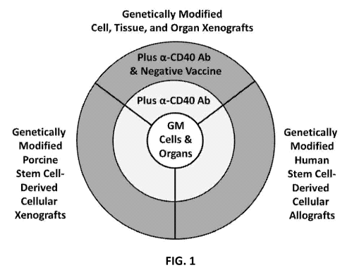
[0036] FIG. 1 demonstrates an immunotherapeutic strategy centered around the
use of genetically
modified cell and organ grafts lacking functional expression of MHC class I.
The need for
maintenance immunosuppression required for the prevention of graft rejection
is progressively
reduced (or the applicability of transplantation of cell and organ xenografts
and the
transplantation of stem cell-derived cellular allografts and xenografts is
progressively increased)
when the transplantation of genetically modified cells and organs is combined
with transient use
of antagonistic anti-CD40 antibodies and even more when combined with the
administration of
tolerizing vaccines comprising apoptotic donor cells under the cover of anti-
CD40 antibodies.
[0037] FIG. 2 demonstrates one strategy of making genetically modified pig
islet cells and
tolerizing vaccines. Two clonal populations of pigs are created. One
population having at least
GGTA1 knocked out can be used to create a tolerizing vaccine. The other clonal
population of
pigs that have at least GGTA1 and MHC I genes (e.g., NRLC5) knocked out, can
be used for
cell, tissues, and/or organ donors.
[0038] FIG. 3 demonstrates use of positive and tolerizing vaccines (also
referred to as a negative
vaccine).
[0039] FIG. 4 demonstrates an exemplary approach to extending the survival of
xenografts in a
subject with infusion of apoptotic donor splenocytes for tolerizing
vaccination under the cover of
transient immunosuppression.
[0040] FIG. 5 shows an exemplary approach to preventing rejection or extending
survival of
xenografts in a recipient in the absence of chronic and generalized
immunosuppression of the
xenograft recipient. This exemplary approach includes and integrates three
components: i)
genetically engineered islets with deficient and/or reduced expression of
aGal, MHC class I,
complement C3, and CXCL10 and transgenic expression the HLA-G; ii) genetically
engineered
CA 02969847 2017-06-05
WO 2016/094679 PCT/US2015/065029
donor apoptotic and non-apoptotic mononuclear cells (e.g., splenocytes) with
deficient and/or
reduced expression of aGal, Neu5Gc, and Sda/CAD as well as transgenic
expression of HLA-G
with or without human CD47, human PD-L1, human PD-L2 (e.g., the genetically
engineered
vaccine); and iii) the administration of transient immunosuppression including
antagonistic anti-
CD40 mAb, anti-CD20 mAb, rapamycin, and transient anti-inflammatory therapy
including
compstatin (e.g., the compstatin derivative APL-2), anti-IL-6 receptor mAb,
and soluble TNF
receptor.
[0041] FIG. 6 demonstrates an exemplary protocol for transplant rejection
prophylaxis in a pig-to-
cynomolgus monkey islet xenotransplantation. IE: islet equivalent; sTNFR:
soluble TNF
receptor (e.g., etanercept); a-IL-6R: anti-interleukin 6 receptor; Tx'd:
transplanted.
[0042] FIGs. 7A-7E demonstrate a strategy for cloning a px330-Ga12-1 plasmid
targeting GGTAl.
FIG. 7A shows a cloning strategy and oligonucleotides for making a guide RNA
targeting
GGTAl. FIG. 7B shows an insertion site on the px330 plasmid. FIG. 7C shows a
flow chart
demonstrating the cloning and verification strategy. FIG. 7D shows a cloning
site and
sequencing primers. FIG. 7E shows sequencing results.
[0043] FIGs. 8A-8E demonstrate a strategy for cloning a px330-CM1F plasmid
targeting CMAH.
FIG. 8A shows a cloning strategy and oligonucleotides for making a guide RNA
targeting
CMAHl. FIG. 8B shows an insertion site on the px330 plasmid. FIG. 8C shows a
flow chart
demonstrating the cloning and verification strategy. FIG. 8D shows a cloning
site and
sequencing primers. FIG. 8E shows sequencing results.
[0044] FIGs. 9A-9E demonstrate a strategy for cloning a px330-NL1 FIRST
plasmid targeting
NLRC5. FIG. 9A shows a cloning strategy and oligonucleotides for making a
guide RNA
targeting NLRC5. FIG. 9B shows an insertion site on the px330 plasmid. FIG. 9C
shows a
flow chart demonstrating the cloning and verification strategy. FIG. 9D shows
a cloning site
and sequencing primers. FIG. 9E shows sequencing results.
[0045] FIGs. 10A-10E demonstrate a strategy for cloning a px330/C3-5 plasmid
targeting C3. FIG.
10A shows a cloning strategy and oligonucleotides for making a guide RNA
targeting C3. FIG.
10B shows an insertion site on the px330 plasmid. FIG. 10C shows a flow chart
demonstrating
the cloning and verification strategy. FIG. 10D shows a cloning site and
sequencing primers.
FIG. 10E shows sequencing results.
[0046] FIGs. 11A-11E demonstrate a strategy for cloning a px330/B41 second
plasmid targeting
B4GALNT2. FIG. 11A shows a cloning strategy and oligonucleotides for making a
guide RNA
targeting B4GALNT2. FIG. 11B shows an insertion site on the px330 plasmid.
FIG. 11C
11
CA 02969847 2017-06-05
WO 2016/094679 PCT/US2015/065029
shows a flow chart demonstrating the cloning and verification strategy. FIG.
11D shows a
cloning site and sequencing primers. FIG. 11E shows sequencing results.
[0047] FIG. 12 demonstrates a map of Rosa26 locus sequenced in Example 2.
[0048] FIGs. 13A-13E demonstrate a strategy for cloning a px330/Rosa exon 1
plasmid targeting
Rosa26. FIG. 13A shows a cloning strategy and oligonucleotides for making a
guide RNA
targeting Rosa26. FIG. 13B shows an insertion site on the px330 plasmid. FIG.
13C shows a
flow chart demonstrating the cloning and verification strategy. FIG. 13D shows
a cloning site
and sequencing primers. FIG. 13E shows sequencing results.
[0049] FIG. 14A shows a map of the genomic sequence of GGTA1. FIG. 14B shows a
map of the
cDNA sequence of GGTA1.
[0050] FIG. 15 shows an exemplary microscopic view of porcine fetal
fibroblasts transfected with
pSpCas9(BB)-2A-GFP.
[0051] FIG. 16 shows a fluorescence in situ hybridization (FISH) to the GGTA1
gene by specific
probes revealing the location on chromosome 1.
[0052] FIGs. 17A-17B demonstrate an example of phenotypic selection of cells
with cas9/sgRNA-
mediated GGTA 1/NLCR5 disruption. FIG. 17A shows genetically modified cells,
which do not
express alpha-galactosidase. FIG. 17B shows non-genetically modified cells,
which express
alpha-galactosidase and were labeled with isolectin B4 (IB)-linked ferrous
beads.
[0053] FIGs. 18A-18C demonstrates validation of GGTA1, CMAH, and NLRC5
disruption in pig
cells. FIG. 18A demonstrates validation of GGTA1 disruption in pig cells. FIG.
18B
demonstrates validation of CMAH disruption in pig cells. FIG. 18C demonstrates
validation of
NLRC5 disruption in pig cells.
[0054] FIGs. 19A-19B demonstrate the inhibitory effects of an anti-SLA
antibody on the pig islet-
induced human CD8+ T cell activation. FIG. 19A shows the proliferation of CD8+
T cells, CD4
T cells and natural killer (NK) cells in a mixed culture with adult pig islets
for 7 days in the
presence (black bars) or absence (white bars) of the anti-SLA antibody. FIG.
19B shows the
viability (assessed by AO/PI staining) of adult pig islets cultured with or
without highly purified
lymphocytes for 7 days in the presence (black bars) or absence (white bars) of
the anti-SLA class
I antibody.
[0055] FIGs. 20A-20B demonstrate T cell activation induced by porcine islets.
FIG. 20A
demonstrates the result of ELISPOT assays. The results show the suppression of
a
posttransplant increase of anti-donor T cells with direct and indirect
specificity secreting IFN-y
in a cynomolgus monkey. The monkey was treated with peritransplant infusion of
apoptotic
12
CA 02969847 2017-06-05
WO 2016/094679 PCT/US2015/065029
donor splenocytes from a GT-K0 donor pig, and islets from the same donor pig
on day 0 under
the cover of transient immunosuppression with anti-CD40 monoclonal antibody,
rapamycin,
sTNFR, and anti-IL-6R monoclonal antibody. FIG. 20B demonstrates CD8 staining
of an
intraportally transplanted adult porcine islet undergoing rejection at 141
days after
transplantation.
[0056] FIGs. 21A-21D demonstrate porcine islet graft survival in a monkey in
the absence of
maintenance immunosuppression. FIG. 21A demonstrates blood glucose levels and
exogenous
insulin needed to maintain normal blood glucose level before and after
transplantation. FIG.
21B demonstrates serum porcine C-peptide level in a monkey. FIG. 21C
demonstrates blood
glucose levels in response to glucose challenges. FIG. 21D demonstrates serum
porcine C-
peptide levels in response to glucose challenges.
[0057] FIG. 22A demonstrates rejection of non-genetically modified porcine
islets by a monkey
transplanted with islets and receiving anti-CD40 antibody four times through
day 14 after
transplantation and maintenance immunosuppression with CTLA4-Ig and rapamycin.
FIG. 22B
demonstrates amelioration of diabetes by transplanted porcine islets in
monkeys receiving anti-
CD40 antibody four times through day 14 after transplantation and maintenance
immunosuppression with CTLA4-Ig and rapamycin.
[0058] FIG. 23A demonstrates amelioration of diabetes (restoration of
sustained normoglycemia
and insulin independence) by transplanted porcine islets in a monkey (ID
#13CP7) receiving
maintenance immunosuppression with rapamycin and anti-CD40 antibody weekly
after
transplantation. The monkey was given an anti-CD40 antibody and rapamycin for
21 days
starting from the day of transplantation. FIG. 23B demonstrates serum porcine
C-peptide levels
(fasted, random, and stimulated) in the same recipient (ID #13CP7).
[0059] FIG. 24 shows the increase of circulating CD8+ CD2hi CD28- effector
memory T cells in
two cynomolgus monkeys at the time of sacrifice (after presumed rejection)
compared with
baseline and the high prevalence of CD8+ CD2hi CD28- effector memory T cells
within the
CD8+ T cell compartment in liver mononuclear cells at the time of sacrifice.
Both monkeys
received intraportal xenotransplants of adult porcine islets. Pre Tx:
pretransplant; PBL:
peripheral blood leukocyte; Sac: sacrifice; Lym: lymphocyte; LMNC: liver
mononuclear cell.
[0060] FIG. 25 shows suppression of circulating CD8+ CD2hi CD28- effector
memory T cells by
peritransplant infusion of apoptotic donor splenocytes (MX-ECDI-vaccine)
compared with
controls that received the same transient immunosuppression but no apoptotic
donor splenocytes
13
CA 02969847 2017-06-05
WO 2016/094679 PCT/US2015/065029
(MX-ECDI-controls). Pre Tx: pretransplant; Sac: sacrifice; Lym: lymphocyte;
LMNC: liver
mononuclear cell.
[0061] FIG. 26 shows suppression of circulating CD8+ CD2hi CD28- effector
memory T cells by
apoptotic donor splenocytes and a-CD40 antibodies. CM: cynomolgus monkey; Pre
Tx:
pretransplant; D: day.
[0062] FIG. 27 shows suppression of circulating CD4+CD25hi FoxP3+ CD127low
regulatory T
cells by apoptotic donor splenocytes and a-CD40 antibodies. CM: cynomolgus
monkey; Pre Tx:
pretransplant; D: day.
[0063] FIG. 28 shows suppression of circulating CD8+CD122+ natural suppressor
cells by
apoptotic donor splenocytes and a-CD40 antibodies. CM: cynomolgus monkey; Pre
Tx:
pretransplant; D: day.
[0064] FIGs. 29A- 29B show sequencing of DNA isolated from fetal cells of two
separate litters
(Pregnancy 1: FIG. 29A or Pregnancy 2: FIG. 29B) subjected to PCR
amplification of the
GGTA1 (compared to Sus scrofa breed mixed chromosome 1, Sscrofal0.2 NCBI
Reference
Sequence: NC 010443.4) target regions and the resulting amplicons were
separated on 1%
agarose gels. Amplicons were also analyzed by sanger sequencing using the
forward primer
alone from each reaction. In FIG. 29 A, the results are shown from Pregnancy
l's fetuses 1, 2,
4, 5, 6, and 7, truncated 6 nucleotides after the target site for GGTA1. Fetus
3 was truncated 17
nucleotides after the cut site followed by a 2,511(668-3179) nucleotide
deletion followed by a
single base substitution. Truncation, deletion and substitution from a single
sequencing
experiment containing the alleles from both copies of the target gene can only
suggest a gene
modification has occurred but not reveal the exact sequence for each allele.
From this analysis it
appears that all 7 fetuses have a single allele modification for GGTA1. FIG.
29B shows
pregnancy 2 fetal DNA samples 1, 3, 4, and 5 were truncated 3 nucleotides from
the GGTA1
gene target site. Fetus 2 had variability in sanger sequencing that suggests a
complex variability
in DNA mutations or poor sample quality. However, fetal DNA template quality
was sufficient
for the generation of the GGTA1 gene screening experiment described above.
[0065] FIGs. 30A-30B show sequencing of DNA isolated from fetal cells of two
separate litters
(Pregnancy 1: FIG. 30A or Pregnancy 2: FIG. 30B) subjected to PCR
amplification of the
NLRC5 (consensus sequence) target regions and the resulting amplicons were
separated on 1%
agarose gels. Amplicons were also analyzed by sanger sequencing using the
forward primer
alone from each reaction. Sequence analysis of the NLRC5 target site for
fetuses from
Pregnancy 1 (FIG. 30A) was unable to show consistent alignment suggesting an
unknown
14
CA 02969847 2017-06-05
WO 2016/094679 PCT/US2015/065029
complication in the sequencing reaction or varying DNA modifications between
NLRC5 alleles
that complicate the sanger sequencing reaction and analysis. NLRC5 gene
amplicons from
Pregnancy 2 (FIG. 30B) were all truncated 120 nucleotides downstream of the
NLRC5 gene cut
site.
[0066] FIGs. 31A-31B show data from fetal DNA (wt and 1-7 (FIG. 31A: Pregnancy
1) or 1-5
(FIG. 31B: Pregnancy 2) isolated from hind limb biopsies. Target genes were
amplified by PCR
and PCR products were separated on I% agarose gels and visualized by
fluorescent DNA stain.
The amplicon band present in the wt lanes represent the unmodified DNA
sequence. An increase
or decrease in size of the amplicon suggests an insertion or deletion within
the amplicon,
respectively. Variation in the DNA modification between alleles in one sample
may make the
band appear more diffuse. Pregnancy 1 (FIG. 31A) resulted in 7 fetuses while
pregnancy 2
(FIG. 31B) resulted in 5 fetuses harvested at 45 and 43 days, respectively. A
lack of band as in
the MAC:5 gel in fetuses 1, 3, and 4 of FIG. 31A (bottom gel), suggests that
the modification to
the target region have disrupted the binding of DNA amplification primers. The
presence of all
bands in GGTA 1 in FIG. 31A (top gel) suggests that DNA quality was sufficient
to generate
DNA amplicons in the NLRC5 targeting PCR reactions. Fetuses 1 ,2, 4, and 5 of
Pregnancy 1
(FIG. 31A) have larger GGTA 1 amplicons than the WT suggesting an insertion
within the
target area. In fetus 3 of Pregnancy 1 (FIG. 31A), the GGTA 1 amplicon
migrated faster than
the WI control suggesting a deletion within the target area. Fetuses 6 and 7
of Pregnancy 1
(FIG. 31A) NLRC5 amplicons migrated faster than the WI suggesting a deletion
within the
target area. Fetuses 1-5 (FIG. 31B) GGIA1 amplicons were difficult to
interpret by size and
were diffuse as compared to the WI control. Fetuses 1-5 (FIG. 31B) NLRC5
amplicons were
uniform in size and density as compared to the wild type control.
[0067] FIGs. 32A-32B shows phenotypic analysis of fetuses from two separate
litters of pigs (FIG.
32A: Pregnancy 1 or FIG. 32B: Pregnancy 2). Fetuses were harvested at day 45
(Pregnancy 1)
or 43 days (Pregnancy 2) and processed for DNA and culture cell isolation.
Tissue fragments
and cells were plated in culture media for 2 days to allow fetal cells to
adhere and grow. Wild
type cells (fetal cells not genetically modified) and fetal cells from
pregnancy 1 and 2 were
removed from culture plates and labeled with IB4 lectin conjugated to alexa
fluor 488 or anti-
porcine MHC class I antibody conjugated to FITC. Flow cytometric analysis is
shown as
histograms depicting the labeling intensity of the cells tested. The histogram
for the WT cells
are included in each panel to highlight the decrease in overall intensity of
each group of fetal
cells. There is a decrease in alpha Gal and MHC class I labeling in pregnancy
1 (FIG. 32A)
CA 02969847 2017-06-05
WO 2016/094679 PCT/US2015/065029
indicated as a decrease in peak intensity. In pregnancy 2 (FIG. 32B) fetuses 1
and 3 have a large
decrease in alpha gal labeling and significant reduction in MHC class 1
labeling as compared to
WT fetal cells.
[0068] FIGs. 33A-33B shows the impact of decreased MHC class I expression in
cells from Fetus 3
(Pregnancy 1) as compared to wild type fetal cells from a genetic clone. The
proliferative
response of human CD8+ cells and CD4 T cells to porcine control fibroblast and
NLRC5
knockout fetal cells were measured. FIG. 33A. Cells were gated as CD4 or CD8
before
assessment of proliferation. FIG. 33B. CD8 T cell proliferation was reduced
following
treatments stimulation by porcine fetal GGTA1/NLRC5 knockout cells compared to
control
unmodified porcine fibroblast. Almost a 55% reduction in CD8 T cells
proliferation was
observed when human responders were treated with porcine fetal GGTA1/NLRC5
knockout
cells at 1:1 ratio. Wild type fetal cells elicited a 17.2% proliferation in
human CD8 T cells
whereas the MHC class I deficient cells from fetus 3 (Pregnancy 1) induced
only a 7.6%
proliferation. No differences were seen in CD8 T cells proliferative response
at 1:5 and 1:10
ratio compared to unmodified fetal cells. No changes were observed in CD4 T
cell proliferation
in response to NLRC5 knockout and control unmodified porcine fetal cells at
all ratios studied.
DETAILED DESCRIPTION OF THE DISCLOSURE
[0069] The following description and examples illustrate embodiments of the
invention in detail. It
is to be understood that this invention is not limited to the particular
embodiments described
herein and as such can vary. Those of skill in the art will recognize that
there are numerous
variations and modifications of this invention, which are encompassed within
its scope.
[0070] Failure of organ and tissue function can result in premature death of
individuals.
Transplantation can potentially solve this problem, which can prolong the
lives of many
individuals. However, there is a shortage of cells, organs, and/or tissues
that can be used for
transplantation.
[0071] Xenografts or allografts (e.g., embryonic or induced pluripotent stem
cells) can be used to
create an unlimited supply of cells, organs, and/or tissues used for
transplantation. In general,
some transplantation can lead to increased immune response which can
ultimately lead to
transplantation rejection. Isografts or autografts typically do not result in
rejection. However,
allografts and xenografts can result in immune reaction and can ultimately
lead to the
destruction of the graft. The risk of rejection in some cases can be mitigated
by suppressing the
immune response.
16
CA 02969847 2017-06-05
WO 2016/094679 PCT/US2015/065029
[0072] Traditionally, immunosuppressive drugs were used after transplantation.
However, there are
many detrimental effects associated with long-term treatment with
immunosuppressive drugs,
including but not limited to increased risk of cancer and infection.
Alternative methods to
prevent graft rejection and suppress the immune system were sought. The immune
response can
be tempered by use of various techniques, including those described herein.
For example, one
method described herein to prevent transplantation rejection or prolong the
time to
transplantation rejection without or with minimal immunosuppressive drug use,
an animal, e.g., a
donor non-human animal, could be altered, e.g., genetically. Subsequently, the
cells, organs,
and/or tissues of the altered animal, e.g., a donor non-human animal, can be
harvested and used
in allografts or xenografts. Alternatively, cells can be extracted from an
animal, e.g., a human or
non-human animal (including but not limited to primary cells) or cells can be
previously
extracted animal cells, e.g., cell lines. These cells can be used to create a
genetically altered cell.
[0073] Transplant rejection (e.g., T cells-mediated transplant rejection) can
be prevented by chronic
immunosuppression. However, immunosuppression is costly and associated with
the risk of
serious side effects. To circumvent the need for chronic immunosuppression, a
multifaceted, T
cell-targeted rejection prophylaxis was developed (FIG. 1) that
i) utilizes genetically modified grafts lacking functional expression of
MHC class I,
thereby interfering with activation of CD8+ T cells with direct specificity
and
precluding cytolytic effector functions of these CD8+ T cells,
ii) interferes with B cell (and other APC)-mediated priming and memory
generation
of anti-donor T cells using induction immunotherapy comprising antagonistic
anti-CD40 mAbs (and depleting anti-CD20 mAbs and a mTOR inhibitor), and/or
iii) deletes anti-donor T cells with indirect specificity via peritransplant
infusions of
apoptotic donor cell vaccines.
[0074] Described herein are genetically modified non-human animals (such as
non-human primates
or a genetically modified animal that is member of the Laurasiatheria
superorder, e.g., ungulates)
and organs, tissues, or cells isolated therefrom, tolerizing vaccines, and
methods for treating or
preventing a disease in a recipient in need thereof by transplantation of an
organ, tissue, or cell
isolated from a non-human animal. An organ, tissue, or cell isolated from a
non-human animal
(such as non-human primates or a genetically modified animal that is member of
the
Laurasiatheria superorder, e.g., ungulates) can be transplanted into a
recipient in need thereof
from the same species (an allotransplant) or a different species (a
xenotransplant). A recipient
can be tolerized with a tolerizing vaccine and/or one or more immunomodulatory
agents (e.g.,
17
CA 02969847 2017-06-05
WO 2016/094679 PCT/US2015/065029
an antibody). In embodiments involving xenotransplantation the recipient can
be a human.
Suitable diseases that can be treated are any in which an organ, tissue, or
cell of a recipient is
defective or injured, (e.g., a heart, lung, liver, vein, skin, or pancreatic
islet cell) and a recipient
can be treated by transplantation of an organ, tissue, or cell isolated from a
non-human animal.
DEFINITIONS
[0075] The term "about" in relation to a reference numerical value and its
grammatical equivalents
as used herein can include the numerical value itself and a range of values
plus or minus 10%
from that numerical value. For example, the amount "about 10" includes 10 and
any amounts
from 9 to 11. For example, the term "about" in relation to a reference
numerical value can also
include a range of values plus or minus 10%, 9%, 8%, 7%, 6%, 5%, 4%, 3%, no/ ,
or 1% from
that value.
[0076] The term "non-human animal" and its grammatical equivalents as used
herein includes all
animal species other than humans, including non-human mammals, which can be a
native
animal or a genetically modified non-human animal. A non-human mammal
includes, an
ungulate, such as an even-toed ungulate (e.g., pigs, peccaries,
hippopotamuses, camels, llamas,
chevrotains (mouse deer), deer, giraffes, pronghorn, antelopes, goat-antelopes
(which include
sheep, goats and others), or cattle) or an odd-toed ungulate (e.g., horse,
tapirs, and
rhinoceroses), a non-human primate (e.g., a monkey, or a chimpanzee), a
Canidae (e.g., a dog)
or a cat. A non-human animal can be a member of the Laurasiatheria superorder.
The
Laurasiatheria superorder can include a group of mammals as described in
Waddell et al.,
Towards Resolving the Interordinal Relationships of Placental Mammals.
Systematic Biology
48 (1): 1-5 (1999). Members of the Laurasiatheria superorder can include
Eulipotyphla
(hedgehogs, shrews, and moles), Perissodactyla (rhinoceroses, horses, and
tapirs), Carnivora
(carnivores), Cetartiodactyla (artiodactyls and cetaceans), Chiroptera (bats),
and Pholidota
(pangolins). A member of Laurasiatheria superorder can be an ungulate
described herein, e.g.,
an odd-toed ungulate or even-toed ungulate. An ungulate can be a pig. A member
can be a
member of Camivora, such as a cat, or a dog. In some cases, a member of the
Laurasiatheria
superorder can be a pig.
[0077] The term "pig" and its grammatical equivalents as used herein can refer
to an animal in
the genus Sus, within the Suidae family of even-toed ungulates. For example, a
pig can be a
wild pig, a domestic pig, mini pigs, a Sus scrofa pig, a Sus scrofa domesticus
pig, or inbred pigs.
[0078] The term "transgene " and its grammatical equivalents as used herein
can refer to a gene or
genetic material that can be transferred into an organism. For example, a
transgene can be a
18
CA 02969847 2017-06-05
WO 2016/094679 PCT/US2015/065029
stretch or segment of DNA containing a gene that is introduced into an
organism. When a
transgene is transferred into an organism, the organism can then be referred
to as a transgenic
organism. A transgene can retain its ability to produce RNA or polypeptides
(e.g., proteins) in a
transgenic organism. A transgene can comprise a polynucleotide encoding a
protein or a
fragment (e.g., a functional fragment) thereof The polynucleotide of a
transgene can be an
exogenous polynucleotide. A fragment (e.g., a functional fragment) of a
protein can comprise
at least or at least about 5%, 10%, 20%, 30%, 40%, 50%, 60%, 70%, 80%, 90%,
95%, or 99%
of the amino acid sequence of the protein. A fragment of a protein can be a
functional fragment
of the protein. A functional fragment of a protein can retain part or all of
the function of the
protein.
[0079] The term "genetic modification" and its grammatical equivalents as used
herein can refer to
one or more alterations of a nucleic acid, e.g., the nucleic acid within an
organism's genome.
For example, genetic modification can refer to alterations, additions, and/or
deletion of genes.
A genetically modified cell can also refer to a cell with an added, deleted
and/or altered gene. A
genetically modified cell can be from a genetically modified non-human animal.
A genetically
modified cell from a genetically modified non-human animal can be a cell
isolated from such
genetically modified non-human animal. A genetically modified cell from a
genetically
modified non-human animal can be a cell originated from such genetically
modified non-human
animal. For example, a cell
[0080] The term "islet" or "islet cells" and their grammatical equivalents as
used herein can refer
to endocrine (e.g., hormone-producing) cells present in the pancreas of an
organism. For
example, islet cells can comprise different types of cells, including, but not
limited to,
pancreatic a cells, pancreatic 13. cells, pancreatic 6 cells, pancreatic F
cells, and/or pancreatic c
cells. Islet cells can also refer to a group of cells, cell clusters, or the
like.
[0081] The term "condition" condition and its grammatical equivalents as used
herein can refer to
a disease, event, or change in health status.
[0082] The term "diabetes" and its grammatical equivalents as used herein can
refer to is a disease
characterized by high blood sugar levels over a prolonged period. For example,
the term
"diabetes" and its grammatical equivalents as used herein can refer to all or
any type of
diabetes, including, but not limited to, type 1, type 2, cystic fibrosis-
related, surgical, gestational
diabetes, and mitochondrial diabetes. In some cases, diabetes can be a form of
hereditary
diabetes.
19
CA 02969847 2017-06-05
WO 2016/094679 PCT/US2015/065029
[0083] The term "phenotype" and its grammatical equivalents as used herein can
refer to a
composite of an organism's observable characteristics or traits, such as its
morphology,
development, biochemical or physiological properties, phenology, behavior, and
products of
behavior. Depending on the context, the term "phenotype" can sometimes refer
to a composite
of a population's observable characteristics or traits.
[0084] The term "disrupting" and its grammatical equivalents as used herein
can refer to a process
of altering a gene, e.g., by deletion, insertion, mutation, rearrangement, or
any combination
thereof For example, a gene can be disrupted by knockout. Disrupting a gene
can be partially
reducing or completely suppressing expression (e.g., mRNA and/or protein
expression) of the
gene. Disrupting can also include inhibitory technology, such as shRNA, siRNA,
microRNA,
dominant negative, or any other means to inhibit functionality or expression
of a gene or
protein.
[0085] The term "gene editing" and its grammatical equivalents as used herein
can refer to genetic
engineering in which one or more nucleotides are inserted, replaced, or
removed from a
genome. For example, gene editing can be performed using a nuclease (e.g., a
natural-existing
nuclease or an artificially engineered nuclease).
[0086] The term "transplant rejection" and its grammatical equivalents as used
herein can refer to a
process or processes by which an immune response of an organ transplant
recipient mounts a
reaction against the transplanted material (e.g., cells, tissues, and/or
organs) sufficient to impair
or destroy the function of the transplanted material.
[0087] The term "hyperacute rejection" and its grammatical equivalents as used
herein can refer to
rejection of a transplanted material or tissue occurring or beginning within
the first 24 hours
after transplantation. For example, hyperacute rejection can encompass but is
not limited to
"acute humoral rejection" and "antibody-mediated rejection".
[0088] The term "negative vaccine", "tolerizing vaccine" and their grammatical
equivalents as used
herein, can be used interchangeably. A tolerizing vaccine can tolerize a
recipient to a graft or
contribute to tolerization of the recipient to the graft if used under the
cover of appropriate
immunotherapy. This can help to prevent transplantation rejection.
[0089] The term "recipient", "subject" and their grammatical equivalents as
used herein, can be used
interchangeably. A recipient or a subject can be a human or non-human animal.
A recipient or
a subject can be a human or non-human animal that will receive, is receiving,
or has received a
transplant graft, a tolerizing vaccine, and/or other composition disclosed in
the application. A
recipient or subject can also be in need of a transplant graft, a tolerizing
vaccine and/or other
CA 02969847 2017-06-05
WO 2016/094679 PCT/US2015/065029
composition disclosed in the application. In some cases, a recipient can be a
human or non-
human animal that will receive, is receiving, or has received a transplant
graft.
[0090] Some numerical values disclosed throughout are referred to as, for
example, "X is at least or
at least about 100; or 200 [or any numerical number]." This numerical value
includes the
number itself and all of the following:
i) X is at least 100;
ii) X is at least 200;
iii) X is at least about 100; and
iv) X is at least about 200.
All these different combinations are contemplated by the numerical values
disclosed
throughout. All disclosed numerical values should be interpreted in this
manner, whether it
refers to an administration of a therapeutic agent or referring to days,
months, years, weight,
dosage amounts, etc., unless otherwise specifically indicated to the contrary.
[0091] The ranges disclosed throughout are sometimes referred to as, for
example, "X is
administered on or on about day 1 to 2; or 2 to 3 [or any numerical range]."
This range includes
the numbers themselves (e.g., the endpoints of the range) and all of the
following:
i) X being administered on between day 1 and day 2;
ii) X being administered on between day 2 and day 3;
iii) X being administered on between about day 1 and day 2;
iv) X being administered on between about day 2 and day 3;
v) X being administered on between day 1 and about day 2;
vi) X being administered on between day 2 and about day 3;
vii) X being administered on between about day 1 and about day 2; and
viii) X being administered on between about day 2 and about day 3.
All these different combinations are contemplated by the ranges disclosed
throughout. All
disclosed ranges should be interpreted in this manner, whether it refers to an
administration of a
therapeutic agent or referring to days, months, years, weight, dosage amounts,
etc., unless
otherwise specifically indicated to the contrary.
[0092] The terms "and/or" and "any combination thereof' and their grammatical
equivalents as used
herein, can be used interchangeably. These terms can convey that any
combination is
specifically contemplated. Solely for illustrative purposes, the following
phrases "A, B, and/or
C" or "A, B, C, or any combination thereof' can mean "A individually; B
individually; C
individually; A and B; B and C; A and C; and A, B, and C."
21
CA 02969847 2017-06-05
WO 2016/094679 PCT/US2015/065029
I. GENETICALLY MODIFIED NON-HUMAN ANIMALS
[0093] Provided herein are genetically modified animals that can be donors of
cells, tissues, and/or
organs for transplantation. A genetically modified non-human animal can be any
desired
species. For example, a genetically modified non-human animal described herein
can be a
genetically modified non-human mammal. A genetically modified non-human mammal
can be
a genetically modified ungulate, including a genetically modified even-toed
ungulate (e.g., pigs,
peccaries, hippopotamuses, camels, llamas, chevrotains (mouse deer), deer,
giraffes, pronghorn,
antelopes, goat-antelopes (which include sheep, goats and others), or cattle)
or a genetically
modified odd-toed ungulate (e.g., horse, tapirs, and rhinoceroses), a
genetically modified non-
human primate (e.g., a monkey, or a chimpanzee) or a genetically modified
Canidae (e.g., a
dog). A genetically modified non-human animal can be a member of the
Laurasiatheria
superorder. A genetically modified non-human animal can be a non-human
primate, e.g., a
monkey, or a chimpanzee. If a non-human animal is a pig, the pig can be at
least or at least
about 5, 50, 100, or 300 pounds, e.g., the pig can be or be about between 5
pounds to 50
pounds; 25 pounds to 100 pounds; or 75 pounds to 300 pounds. In some cases, a
non-human
animal is a pig that has given birth at least one time.
[0094] A genetically modified non-human animal can be of any age. For example,
the genetically
modified non-human animal can be a fetus; from or from about 1 day to 1 month;
from or from
about 1 month to 3 months; from or from about 3 months to 6 months; from or
from about 6
months to 9 months; from or from about 9 months to 1 year; from or from about
1 year to 2
years. A genetically modified non-human animal can be a non-human fetal
animal, perinatal
non-human animal, neonatal non-human animal, preweaning non-human animal,
young adult
non-human animal, or an adult non-human animal.
[0095] A genetically modified non-human animal can comprise reduced expression
of one or more
genes compared to a non-genetically modified counterpart animal. A non-
genetically modified
counterpart animal can be an animal substantially identical to the genetically
modified animal
but without genetic modification in the genome. For example, a non-genetically
modified
counterpart animal can be a wild-type animal of the same species as the
genetically modified
animal. The non-human animal can provide cells, tissues or organs for
transplanting to a
recipient or subject in need thereof A recipient or subject in need thereof
can be a recipient or
subject known or suspected of having a condition. The condition can be
treated, prevented,
reduced, eliminated, or augmented by the methods and compositions disclosed
herein. The
recipient can exhibit low or no immuno-response to the transplanted cells,
tissues or organs.
22
CA 02969847 2017-06-05
WO 2016/094679 PCT/US2015/065029
The transplanted cells, tissues or organs can be non-recognizable by CD8+ T
cells, NK cells, or
CD4+ T cells of the recipient (e.g., a human or another animal). The genes
whose expression is
reduced can include MHC molecules, regulators of MHC molecule expression, and
genes
differentially expressed between the donor non-human animal and the recipient
(e.g., a human
or another animal). The reduced expression can be mRNA expression or protein
expression of
the one or more genes. For example, the reduced expression can be protein
expression of the
one or more genes. Reduced expression can also include no expression. For
example an
animal, cell, tissue or organ with reduced expression of a gene can have no
expression (e.g.,
mRNA and/or protein expression) of the gene. Reduction of expression of a gene
can inactivate
the function of the gene. In some cases, when expression of a gene is reduced
in a genetically
modified animal, the expression of the gene is absent in the genetically
modified animal.
[0096] The genetically modified non-human animal can comprise reduced
expression of one or more
MHC molecules compared to a non-genetically modified counterpart animal. For
example, the
non-human animal can be an ungulate, e.g., a pig, with reduced expression of
one or more
swine leukocyte antigen (SLA) class I and/or SLA class II molecules.
[0097] The genetically modified non-human animal can comprise reduced
expression of any genes
that regulate major histocompatibility complex (MHC) molecules (e.g., MHC I
molecules
and/or MHC II molecules) compared to a non-genetically modified counterpart
animal.
Reducing expression of such genes can result in reduced expression and/or
function of MHC
molecules (e.g., MHC I molecules and/or MHC II molecules). In some cases, the
one or more
genes whose expression is reduced in the non-human animal can comprise one or
more of the
following: components of an MHC I-specific enhanceosome, transporters of a MHC
I-binding
peptide, natural killer group 2D ligands, CXC chemical receptor (CXCR) 3
ligands,
complement component 3 (C3), and major histocompatibility complex II
transactivator
(CIITA). In some cases, the component of a MHC I-specific enhanceosome can be
NLRC5. In
some cases, the component of a MHC I-specific enhanceosome can also comprise
regulatory
factor X (RFX) (e.g., RFX1), nuclear transcription factor Y (NFY), and cAMP
response
element-binding protein (CREB). In some instances, the transporter of a MHC I-
binding
peptide can be Transporter associated with antigen processing 1 (TAP1). In
some cases, the
natural killer (NK) group 2D ligands can comprise MICA and MICB. For example,
the
genetically modified non-human animal can comprise reduced expression of one
or more of the
following genes: NOD-like receptor family CARD domain containing 5 (NLRC5),
Transporter
associated with antigen processing 1 (TAP1), C-X-C motif chemokine 10
(CXCL10), MHC
23
CA 02969847 2017-06-05
WO 2016/094679 PCT/US2015/065029
class I polypeptide-related sequence A (MICA), MHC class I polypeptide-related
sequence B
(MICB), complement component 3 (C3), and CIITA. A genetically modified animal
can
comprise reduced expression of one or more of the following genes: a component
of an MHC I-
specific enhanceosome (e.g., NLRC5), a transporter of an MHC I-binding peptide
(TAP1), and
C3.
[0098] The genetically modified non-human animal can comprise reduced
expression compared to a
non-genetically modified counterpart of one or more genes expressed at
different levels between
the non-human animal and a recipient receiving a cell, tissue, or organ from
the non-human
animal. For example, the one or more genes can be expressed at a lower level
in a human than
in the non-human animal. In some cases, the one or more genes can be
endogenous genes of the
non-human animal. The endogenous genes are in some cases genes not expressed
in another
species. For example, the endogenous genes of the non-human animal can be
genes that are not
expressed in a human. For example, in some cases, homologs (e.g., orthologs)
of the one or
more genes do not exist in a human. In another example, homologs (e.g.,
orthologs) of the one
or more genes can exist in a human but are not expressed.
[0099] In some cases, the non-human animal can be a pig, and the recipient can
be a human. In
these cases, the one or more genes can be any genes expressed in a pig but not
in a human.. For
example, the one or more genes can comprise glycoprotein galactosyltransferase
alpha 1,3
(GGTA1), putative cytidine monophosphate-N-acetylneuraminic acid hydroxylase-
like protein
(CMAH), and [31,4 N-acetylgalactosaminyltransferase (B4GALNT2). A genetically
modified
non-human animal can comprise reduced expression of B4GALNT2, GGTA1, or CMAH,
where the reduced expression is in comparison to a non-genetically modified
counterpart
animal. A genetically modified non-human animal can comprise reduced
expression of
B4GALNT2 and GGTA1, where the reduced expression is in comparison to a non-
genetically
modified counterpart animal. A genetically modified non-human animal can
comprise reduced
expression of B4GALNT2 and CMAH, where the reduced expression is in comparison
to a
non-genetically modified counterpart animal. A genetically modified non-human
animal can
comprise reduced expression of B4GALNT2, GGTA1, and CMAH, where the reduced
expression is in comparison to a non-genetically modified counterpart animal.
[00100] The genetically modified non-human animal can comprise reduced
expression compared to
a non-genetically modified counterpart of one or more of any of the genes
disclosed herein,
including NLRC5, TAP1, CXCL10, MICA, MICB, C3, CIITA, GGTA1, CMAH, and
B4GALNT2.
24
CA 02969847 2017-06-05
WO 2016/094679 PCT/US2015/065029
[00101] A genetically modified non-human animal can comprise one or more genes
whose
expression is reduced, e.g., where genetic expression is reduced. The one or
more genes whose
expression is reduced include but are not limited to NOD-like receptor family
CARD domain
containing 5 (NLRC5), Transporter associated with antigen processing 1 (TAP1),
Glycoprotein
galactosyltransferase alpha 1,3 (GGTA1), Putative cytidine monophosphate-N-
acetylneuraminic
acid hydroxylase-like protein (CMAH), C-X-C motif chemokine 10 (CXCL10), MHC
class I
polypeptide-related sequence A (MICA), MHC class I polypeptide-related
sequence B (MICB),
class II major histocompatibility complex transactivator (CIITA), Beta-1,4-N-
Acetyl-
Galactosaminyl Transferase 2 (B4GALNT2), complemental component 3 (C3), and/or
any
combination thereof
[00102] A genetically modified non-human animal can comprise 1, 2, 3, 4, 5, 6,
7, 8, 9, 10, 11, 12,
13, 14, 15, 16, 17, 18, 19, 20 or more genes whose expression is disrupted.
For illustrative
purposes, and not to limit various combinations a person of skill in the art
can envision, a
genetically modified non-human animal can have NLRC5 and TAP1 individually
disrupted. A
genetically modified non-human animal can also have both NLRC5 and TAP1
disrupted. A
genetically modified non-human animal can also have NLRC5 and TAP1, and in
addition to
one or more of the following GGTA1, CMAH, CXCL10, MICA, MICB, B4GALNT2, or
CIITA genes disrupted; for example "NLRC5, TAP1, and GGTA1" or "NLRC5, TAP1,
and
CMAH" can be disrupted. A genetically modified non-human animal can also have
NLRC5,
TAP1, GGTA1, and CMAH disrupted. Alternatively, a genetically modified non-
human animal
can also have NLRC5, TAP1, GGTA1, B4GALNT2, and CMAH disrupted. In some cases,
a
genetically modified non-human animal can have C3 and GGTA1 disrupted. In some
cases, a
genetically modified non-human animal can have reduced expression of NLRC5,
C3, GGTA1,
B4GALNT2, CMAH, and CXCL10. In some cases, a genetically modified non-human
animal
can have reduced expression of TAP1, C3, GGTA1, B4GALNT2, CMAH, and CXCL10. In
some cases, a genetically modified non-human animal can have reduced
expression of NLRC5,
TAP1, C3, GGTA1, B4GALNT2, CMAH, and CXCL10.
[00103] Lack of MHC class I expression on transplanted human cells can cause
the passive
activation of natural killer (NK) cells (Ohlen et al., 1989). Lack of MHC
class I expression
could be due to NLRC5, TAP1, or B2M gene deletion. NK cell cytotoxicity can be
overcome
by the expression of the human MHC class 1 gene, HLA-E, can stimulate the
inhibitory
receptor CD94/NKG2A on NK cells to prevent cell killing (Weiss et al, 2009;
Lilienfeld et al,
2007; Sasaki et al., 1999). Successful expression of the HLA-E gene can be
dependent on co-
CA 02969847 2017-06-05
WO 2016/094679 PCT/US2015/065029
expression of the human B2M (beta 2 microglobulin) gene and a cognate peptide
(Weiss et al.,
2009; Lilienfeld et al., 2007; Sasaki et al., 1999; Pascasova et al., 1999). A
nuclease mediated
break in the stem cell DNA can allow for the insertion of one or multiple
genes via homology
directed repair. The HLA-E and hB2M genes in series can be integrated in the
region of the
nuclease mediated DNA break thus preventing expression of the target gene (for
example,
NLRC5) while inserting the transgenes.
[00104] Expression levels of genes can be reduced to various extents. For
example, expression of
one or more genes can be reduced by or by about 100%. In some cases,
expression of one or
more genes can be reduced by or by about 99%, 95%, 90%, 85%, 80%, 75%, 70%,
65%, 60%,
55%, or 50% of normal expression, e.g., compared to the expression of non-
modified controls.
In some cases, expression of one or more genes can be reduced by at least or
to at least about
99% to 90%; 89% to 80%, 79% to 70%; 69% to 60%; 59% to 50% of normal
expression, e.g.,
compared to the expression of non-modified controls. For example, expression
of one or more
genes can be reduced by at least or at least about 90% or by at least or at
least about 90% to
99% of normal expression.
[00105] Expression can be measured by any known method, such as quantitative
PCR (qPCR),
including but not limited to PCR, real-time PCR (e.g., Sybr-green), and/or hot
PCR. In some
cases, expression of one or more genes can be measured by detecting the level
of transcripts of
the genes. For example, expression of one or more genes can be measured by
Northern
blotting, nuclease protection assays (e.g., RNase protection assays), reverse
transcription PCR,
quantitative PCR (e.g., real-time PCR such as real-time quantitative reverse
transcription PCR),
in situ hybridization (e.g., fluorescent in situ hybridization (FISH)), dot-
blot analysis,
differential display, serial analysis of gene expression, subtractive
hybridization, microarrays,
nanostring, and/or sequencing (e.g., next-generation sequencing). In some
cases, expression of
one or more genes can be measured by detecting the level of proteins encoded
by the genes.
For example, expression of one or more genes can be measured by protein
immunostaining,
protein immunoprecipitation, electrophoresis (e.g., SDS-PAGE), Western
blotting,
bicinchoninic acid assay, spectrophotometry, mass spectrometry, enzyme assays
(e.g., enzyme-
linked immunosorbent assays), immunohistochemistry, flow cytometry, and/or
immunoctyochemistry. Expression of one or more genes can also be measured by
microscopy.
The microscopy can be optical, electron, or scanning probe microscopy. Optical
microscopy
can comprise use of bright field, oblique illumination, cross-polarized light,
dispersion staining,
dark field, phase contrast, differential interference contrast, interference
reflection microscopy,
26
CA 02969847 2017-06-05
WO 2016/094679 PCT/US2015/065029
fluorescence (e.g., when particles, e.g., cells, are immunostained), confocal,
single plane
illumination microscopy, light sheet fluorescence microscopy, deconvolution,
or serial time-
encoded amplified microscopy. Expression of MHC I molecules can also be
detected by any
methods for testing expression as described herein.
Disrupted Genes
[00106] The inventors have found that cells, organs, and/or tissues having
different combinations of
disrupted genes, can result in cells, organs, and/or tissues that are less
susceptible to rejection
when transplanted into a recipient. For example, the inventors have found that
disrupting (e.g.,
reducing expression of) certain genes, such as NLRC5, TAP1, GGTA1, B4GALNT2,
CMAH,
CXCL10, MICA, MICB, C3, and/or CIITA, can increase the likelihood of graft
survival.
[00107] However, the disruptions are not limited to solely these genes. The
disruption can be of
any particular gene. It is contemplated that genetic homologues (e.g., any
mammalian version
of the gene) of the genes within this applications are covered. For example,
genes that are
disrupted can exhibit a certain identity and/or homology to genes disclosed
herein, e.g., NLRC5,
TAP1, GGTA1, B4GALNT2, CMAH, CXCL10, MICA, MICB, C3, and/or CIITA. Therefore,
it is contemplated that a gene that exhibits at least or at least about 50%,
55%, 60%, 65%, 70%,
75%, 80%, 85%, 90%, 95%, 99%, or 100% homology (at the nucleic acid or protein
level) can
be disrupted, e.g., a gene that exhibits at least or at least about from 50%
to 60%; 60% to 70%;
70% to 80%; 80% to 90%; or 90% to 99% homology. It is also contemplated that a
gene that
exhibits at least or at least about 50%, 55%, 60%, 65%, 70%, 75%, 80%, 85%,
90%, 99%, or
100% identity (at the nucleic acid or protein level) can be disrupted, e.g., a
gene that exhibits at
least or at least about from 50% to 60%; 60% to 70%; 70% to 80%; 80% to 90%;
or 90% to
99% identity. Some genetic homologues are known in the art, however, in some
cases,
homologues are unknown. However, homologous genes between mammals can be found
by
comparing nucleic acid (DNA or RNA) sequences or protein sequences using
publically
available databases such as NCBI BLAST. Genomic sequences, cDNA and protein
sequences
of exemplary genes are shown in Table 1.
[00108] Gene suppression can also be done in a number of ways. For example,
gene expression can
be reduced by knock out, altering a promoter of a gene, and/or by
administering interfering
RNAs (knockdown). This can be done at an organism level or at a tissue, organ,
and/or cellular
level. If one or more genes are knocked down in a non-human animal, cell,
tissue, and/or organ,
the one or more genes can be reduced by administrating RNA interfering
reagents, e.g., siRNA,
shRNA, or microRNA. For example, a nucleic acid which can express shRNA can be
stably
27
CA 02969847 2017-06-05
WO 2016/094679 PCT/US2015/065029
transfected into a cell to knockdown expression. Furthermore, a nucleic acid
which can express
shRNA can be inserted into the genome of a non-human animal, thus knocking
down a gene
with in a non-human animal.
[00109] Disruption methods can also comprise overexpressing a dominant
negative protein. This
method can result in overall decreased function of a functional wild-type
gene. Additionally,
expressing a dominant negative gene can result in a phenotype that is similar
to that of a
knockout and/or knockdown.
[00110] Sometimes a stop codon can be inserted or created (e.g., by nucleotide
replacement), in one
or more genes, which can result in a nonfunctional transcript or protein
(sometimes referred to
as knockout). For example, if a stop codon is created within the middle of one
or more genes,
the resulting transcription and/or protein can be truncated, and can be
nonfunctional. However,
in some cases, truncation can lead to an active (a partially or overly active)
protein. In some
cases, if a protein is overly active, this can result in a dominant negative
protein, e.g., a mutant
polypeptide that disrupts the activity of the wild-type protein.
[00111] This dominant negative protein can be expressed in a nucleic acid
within the control of any
promoter. For example, a promoter can be a ubiquitous promoter. A promoter can
also be an
inducible promoter, tissue specific promoter, and/or developmental specific
promoter.
[00112] The nucleic acid that codes for a dominant negative protein can then
be inserted into a cell
or non-human animal. Any known method can be used. For example, stable
transfection can
be used. Additionally, a nucleic acid that codes for a dominant negative
protein can be inserted
into a genome of a non-human animal.
[00113] One or more genes in a non-human animal can be knocked out using any
method known in
the art. For example, knocking out one or more genes can comprise deleting one
or more genes
from a genome of a non-human animal. Knocking out can also comprise removing
all or a part
of a gene sequence from a non-human animal. It is also contemplated that
knocking out can
comprise replacing all or a part of a gene in a genome of a non-human animal
with one or more
nucleotides. Knocking out one or more genes can also comprise inserting a
sequence in one or
more genes thereby disrupting expression of the one or more genes. For
example, inserting a
sequence can generate a stop codon in the middle of one or more genes.
Inserting a sequence
can also shift the open reading frame of one or more genes.
[00114] Knockout can be done in any cell, organ, and/or tissue in a non-human
animal. For
example, knockout can be whole body knockout, e.g., expression of one or more
genes is
reduced in all cells of a non-human animal. Knockout can also be specific to
one or more cells,
28
CA 02969847 2017-06-05
WO 2016/094679 PCT/US2015/065029
tissues, and/or organs of a non-human animal. This can be achieved by
conditional knockout,
where expression of one or more genes is selectively reduced in one or more
organs, tissues or
types of cells. Conditional knockout can be performed by a Cre-lox system,
where cre is
expressed under the control of a cell, tissue, and/or organ specific promoter.
For example, one
or more genes can be knocked out (or expression can be reduced) in one or more
tissues, or
organs, where the one or more tissues or organs can include brain, lung,
liver, heart, spleen,
pancreas, small intestine, large intestine, skeletal muscle, smooth muscle,
skin, bones, adipose
tissues, hairs, thyroid, trachea, gall bladder, kidney, ureter, bladder,
aorta, vein, esophagus,
diaphragm, stomach, rectum, adrenal glands, bronchi, ears, eyes, retina,
genitals, hypothalamus,
larynx, nose, tongue, spinal cord, or ureters, uterus, ovary, testis, and/or
any combination
thereof. One or more genes can also be knocked out (or expression can be
reduced) in one types
of cells, where one or more types of cells include trichocytes, keratinocytes,
gonadotropes,
corticotropes, thyrotropes, somatotropes, lactotrophs, chromaffin cells,
parafollicular cells,
glomus cells melanocytes, nevus cells, merkel cells, odontoblasts,
cementoblasts corneal
keratocytes, retina muller cells, retinal pigment epithelium cells, neurons,
glias (e.g.,
oligodendrocyte astrocytes), ependymocytes, pinealocytes, pneumocytes (e.g.,
type I
pneumocytes, and type II pneumocytes), clara cells, goblet cells, G cells, D
cells,
Enterochromaffin-like cells, gastric chief cells, parietal cells, foveolar
cells, K cells, D cells, I
cells, goblet cells, paneth cells, enterocytes, microfold cells, hepatocytes,
hepatic stellate cells
(e.g., Kupffer cells from mesoderm), cholecystocytes, centroacinar cells,
pancreatic stellate
cells, pancreatic a cells, pancreatic 13. cells, pancreatic 6 cells,
pancreatic F cells, pancreatic c
cells, thyroid (e.g., follicular cells), parathyroid (e.g., parathyroid chief
cells), oxyphil cells,
urothelial cells, osteoblasts, osteocytes, chondroblasts, chondrocytes,
fibroblasts, fibrocytes,
myoblasts, myocytes, myosatellite cells, tendon cells, cardiac muscle cells,
lipoblasts,
adipocytes, interstitial cells of cajal, angioblasts, endothelial cells,
mesangial cells (e.g.,
intraglomerular mesangial cells and extraglomerular mesangial cells),
juxtaglomerular cells,
macula densa cells, stromal cells, interstitial cells, telocytes simple
epithelial cells, podocytes,
kidney proximal tubule brush border cells, sertoli cells, leydig cells,
granulosa cells, peg cells,
germ cells, spermatozoon ovums, lymphocytes, myeloid cells, endothelial
progenitor cells,
endothelial stem cells, angioblasts, mesoangioblasts, pericyte mural cells,
and/or any
combination thereof
[00115] Conditional knockouts can be inducible, for example, by using
tetracycline inducible
promoters, development specific promoters. This can allow for eliminating or
suppressing
29
CA 02969847 2017-06-05
WO 2016/094679 PCT/US2015/065029
expression of a gene/protein at any time or at a specific time. For example,
with the case of a
tetracycline inducible promoter, tetracycline can be given to a non-human
animal any time after
birth. If a non-human animal is a being that develops in a womb, then promoter
can be induced
by giving tetracycline to the mother during pregnancy. If a non-human animal
develops in an
egg, a promoter can be induced by injecting, or incubating in tetracycline.
Once tetracycline is
given to a non-human animal, the tetracycline will result in expression of
cre, which will then
result in excision of a gene of interest.
[00116] A cre/lox system can also be under the control of a developmental
specific promoter. For
example, some promoters are turned on after birth, or even after the onset of
puberty. These
promoters can be used to control cre expression, and therefore can be used in
developmental
specific knockouts.
[00117] It is also contemplated that any combinations of knockout technology
can be combined.
For example, tissue specific knockout can be combined with inducible
technology, creating a
tissue specific, inducible knockout. Furthermore, other systems such
developmental specific
promoter, can be used in combination with tissues specific promoters, and/or
inducible
knockouts.
[00118] Knocking out technology can also comprise gene editing. For example,
gene editing can
be performed using a nuclease, including CRISPR associated proteins (Cas
proteins, e.g., Cas9),
Zinc finger nuclease (ZFN), Transcription Activator-Like Effector Nuclease
(TALEN), and
maganucleases. Nucleases can be naturally existing nucleases, genetically
modified, and/or
recombinant. For example, a CRISPR/cas system can be suitable as a gene
editing system.
[00119] It is also contemplated that less than all alleles of one or more
genes of a non-human
animal can be knocked out. For example, in diploid non-human animals, it is
contemplated that
one of two alleles are knocked out. This can result in decreased expression
and decreased
protein levels of genes. Overall decreased expression can be less than or less
than about 99%,
95%, 90%, 85%, 80%, 75%, 70%, 65%, 60%, 55%, 50%, 45%, 40%, 35%, 30%, 2,0,/0,
or 20%;
e.g., from or from about 99% to 90%; 90% to 80%; 80% to 70%; 70% to 60%; 60%
to 50%;
50% to 40%; 40% to 30%, or 30% to 20%; compared to when both alleles are
functioning, for
example, not knocked out and/or knocked down. Additionally, overall decrease
in protein level
can be the same as the decreased in overall expression. Overall decrease in
protein level can be
about or less than about 99%, 95%, 90%, 80%, 70%, 60%, 50%, 40%, 30%, or 20%,
e.g., from
or from about 99% to 90%; 90% to 80%; 80% to 70%; 70% to 60%; 60% to 50%; 50%
to 40%;
40% to 30%, or 30% to 20%; compared to when both alleles are functioning, for
example, not
CA 02969847 2017-06-05
WO 2016/094679 PCT/US2015/065029
knocked out and/or knocked down. However, it is also contemplated that all
alleles of one or
more genes in a non-human animal can be knocked out.
[00120] Knocking out of one or more genes can be validated by genotyping.
Methods for
genotyping can include sequencing, restriction fragment length polymorphism
identification
(RFLPI), random amplified polymorphic detection (RAPD), amplified fragment
length
polymorphism detection (AFLPD), PCR (e.g., long range PCR, or stepwise PCR),
allele
specific oligonucleotide (ASO) probes, and hybridization to DNA microarrays or
beads. For
example, genotyping can be performed by sequencing. In some cases, sequencing
can be high
fidelity sequencing. Methods of sequencing can include Maxam-Gilbert
sequencing, chain-
termination methods (e.g., Sanger sequencing), shotgun sequencing, and bridge
PCR. In some
cases, genotyping can be performed by next-generation sequencing. Methods of
next-
generation sequencing can include massively parallel signature sequencing,
polony sequencing,
pyrosequencing (e.g., pyrosequencing developed by 454 Life Sciences), single-
molecule rea-
time sequencing (e.g., by Pacific Biosciences), Ion semiconductor sequencing
(e.g., by Ion
Torrent semiconductor sequencing), sequencing by synthesis (e.g., by Solexa
sequencing by
Illumina), sequencing by ligation (e.g., SOLiD sequencing by Applied
Biosystems), DNA
nanoball sequencing, and heliscope single molecule sequencing. In some cases,
genotyping of a
non-human animal herein can comprise full genome sequencing analysis. In some
cases,
knocking out of a gene in an animal can be validated by sequencing (e.g., next-
generation
sequencing) a part of the gene or the entire gene. For example, knocking out
of NLRC5 gene in
a pig can be validated by next generation sequencing of the entire NLRC5. The
next generation
sequencing of NLRC5 can be performed using e.g. using forward primer 5'-
gctgtggcatatggcagttc -3' (SEQ ID No. 1) and reverse primer 5'-
tccatgtataagtetttta-3' (SEQ ID
No. 2), or forward primer 5'- ggcaatgccagatcctcaac -3' (SEQ ID No. 3) and
reverse primer 5'-
tgtctgatgtetttctcatg -3' (SEQ ID No. 4).
Table 1. Genomic sequences, cDNA and proteins of exemplary disrupted genes
Genomic cDNA protein
sequence
Gene SEQ ID SEQ ID Accession No. SEQ ID Accession No.
No. No. No.
NLRC5 5 6 KC514136.1 7 AGG68119.1
31
CA 02969847 2017-06-05
WO 2016/094679 PCT/US2015/065029
TAP1 8 9 NM 001044581 10 NP
001038046.1
GGTA1 11 12 AF221508 13 NP
998975.1
CMAH 14 15 NM 001113015 16 NP
001106486.1
CXCL10 17 18 NM 001008691.1 19 NP
001008691.1
CIITA 20 21 XM 013995652.1 22 XP
013851106.1
B4GALNT2 23 24 NM 001244330.1 25 NP
001231259.1
C3 26 27 NM 214009.1 28 NP
999174.1
MICA 29 30 NM 000247.2 31 NP
000238.1
MICB 32 33 NM 001289160.1 34 NP
001276089.1
Transgenes
[00121] Transgenes can be useful for overexpressing endogenous genes at higher
levels than
without the transgenes. Additionally, transgenes can be used to express
exogenous genes.
Transgenes can also encompass other types of genes, for example, a dominant
negative gene.
[00122] A transgene of protein X can refer to a transgene comprising a
nucleotide sequence
encoding protein X. As used herein, in some cases, a transgene encoding
protein X can be a
transgene encoding 100% or about 100% of the amino acid sequence of protein X.
In some
cases, a transgene encoding protein X can encode the full or partial amino
sequence of protein
X. For example, the transgene can encode at least or at least about 99%, 95%,
90%, 80%, 70%,
60%, 50%, 40%, 30%, 20%, 10%, or 5%, e.g., from or from about 99% to 90%; 90%
to 80%;
80% to 70%; 70% to 60%; or 60% to 50%; of the amino acid sequence of protein
X.
Expression of a transgene can ultimately result in a functional protein, e.g.,
a partially or fully
functional protein. As discussed above, if a partial sequence is expressed,
the ultimate result
can be in some cases a nonfunctional protein or a dominant negative protein. A
nonfunctional
protein or dominant negative protein can also compete with a functional
(endogenous or
exogenous) protein. A transgene can also encode an RNA (e.g., mRNA, shRNA,
siRNA, or
microRNA). In some cases, where a transgene encodes for an mRNA, this can in
turn be
translated into a polypeptide (e.g., a protein). Therefore, it is contemplated
that a transgene can
encode for protein. A transgene can, in some instances, encode a protein or a
portion of a
protein. Additionally, a protein can have one or more mutations (e.g.,
deletion, insertion, amino
acid replacement, or rearrangement) compared to a wild-type polypeptide. A
protein can be a
natural polypeptide or an artificial polypeptide (e.g., a recombinant
polypeptide). A transgene
can encode a fusion protein formed by two or more polypeptides.
32
CA 02969847 2017-06-05
WO 2016/094679 PCT/US2015/065029
[00123] Transgenes can be placed into an organism, cell, tissue, or organ, in
a manner which
produces a product of the transgene. For example, disclosed herein is a non-
human animal
comprising one or more transgenes. One or more transgenes can be in
combination with one or
more disruptions as described herein. A transgene can be incorporated into a
cell. For example,
a transgene can be incorporated into an organism's germ line. When inserted
into a cell, a
transgene can be either a complementary DNA (cDNA) segment, which is a copy of
messenger
RNA (mRNA), or a gene itself residing in its original region of genomic DNA
(with or without
introns).
[00124] A non-human animal can comprise one or more transgenes comprising one
or more
polynucleotide inserts. The polynucleotide inserts can encode one or proteins
or functional
fragments thereof In some cases, a non-human animal can comprise one or more
transgenes
comprising one or more polynucleotide inserts encoding proteins that can
reduce expression
and/or function of MHC molecules (e.g., MHC I molecules and/or MHC II
molecules). The
one or more transgenes can comprise one or more polynucleotide inserts
encoding MHC I
formation suppressors, regulators of complement activations, inhibitory
ligands for NK cells,
B7 family members, CD47, serine protease inhibitors, galectins, and/or any
fragments thereof
In some cases, the MHC I formation suppressors can be infected cell protein 47
(ICP47). In
some cases, regulators of complement activation can comprise cluster of
differentiation 46
(CD46), cluster of differentiation 55 (CD55), and cluster of differentiation
59 (CD59). In some
cases, inhibitory ligands for NK cells can comprise leukocyte antigen E (HLA-
E), human
leukocyte antigen G (HLA-G), and 3-2-microglobulin (B2M). An inhibitory ligand
for NK
cells can be an isoform of HLA-G, e.g., HLA-G1, HLA-G2, HLA-G3, HLA-G4, HLA-
G5,
HLA-G6, or HLA-G7. For example, inhibitory ligand for NK cells can be HLA-Gl.
A
transgene of HLA-G (e.g., HLA-G1, HLA-G2, HLA-G3, HLA-G4, HLA-G5, HLA-G6, or
HLA-G7) can refer to a transgene comprising a nucleotide sequence encoding HLA-
G (e.g.,
HLA-G1, HLA-G2, HLA-G3, HLA-G4, HLA-G5, HLA-G6, or HLA-G7). As used herein, in
some cases, a transgene encoding HLA-G (e.g., HLA-G1, HLA-G2, HLA-G3, HLA-G4,
HLA-
G5, HLA-G6, or HLA-G7) can be a transgene encoding 100% or about 100% of the
amino acid
sequence of HLA-G (e.g., HLA-G1, HLA-G2, HLA-G3, HLA-G4, HLA-G5, HLA-G6, or
HLA-G7). In other cases, a transgene encoding HLA-G (e.g., HLA-G1, HLA-G2, HLA-
G3,
HLA-G4, HLA-G5, HLA-G6, or HLA-G7) can be a transgene encoding the full or
partial
sequence of HLA-G (e.g., HLA-G1, HLA-G2, HLA-G3, HLA-G4, HLA-G5, HLA-G6, or
HLA-G7). For example, the transgene can encode at least or at least about 99%,
95%, 90%,
33
CA 02969847 2017-06-05
WO 2016/094679 PCT/US2015/065029
80%, 70%, 60%, or 50% of the amino acid sequence of HLA-G (e.g., HLA-G1, HLA-
G2,
HLA-G3, HLA-G4, HLA-G5, HLA-G6, or HLA-G7). For example, the transgene can
encode
90% of the HLA-G amino acid sequence. A transgene can comprise polynucleotides
encoding
a functional (e.g., a partially or fully functional) HLA-G (e.g., HLA-G1, HLA-
G2, HLA-G3,
HLA-G4, HLA-G5, HLA-G6, or HLA-G7). In some cases, the one or more transgenes
can
comprise one or more polynucleotide inserts encoding one or more of ICP47,
CD46, CD55,
CD59, HLA-E, HLA-G (e.g., HLA-G1, HLA-G2, HLA-G3, HLA-G4, HLA-G5, HLA-G6, or
HLA-G7), and B2M. The HLA-G genomic DNA sequence can have 8 exons by which
alternative splicing results in 7 isoforms. The HLA-Gl isoform can exclude
exon 7. The HLA-
G2 isoform can exclude exon 3 and 7. Translation of intron 2 or intron 4 can
result secreted
isoforms due to the loss of the transmembrane domain expression. The maps of
the genomic
sequence and cDNA of HLA-G are shown in FIGs. 14A-14B. In some cases, B7
family
members can comprise CD80, CD86, programed death-ligand 1 (PD-L1), programed
death-
ligand 2 (PD-L2), CD275, CD276, V-set domain containing T cell activation
inhibitor 1
(VTCN1), platelet receptor Gi24, natural cytotoxicity triggering receptor 3
ligand 1 (NR3L1),
and HERV-H LTR-associating 2 (HHLA2). For example, a B7 family member can be
PD-Li
or PD-L2. In some cases, a serine protease inhibitor can be serine protease
inhibitor 9 (Spi9).
In some cases, galectins can comprise galectin-1, galectin-2, galectin-3,
galectin-4, galectin-5,
galectin-6, galectin-7, galectin-8, galectin-9, galectin-10, galectin-11,
galectin-12, galectin-13,
galectin-14, and galectin-15. For example, a galectin can be galectin-9.
[00125] A genetically modified non-human animal can comprise reduced
expression of one or more
genes and one or more transgenes disclosed herein. In some cases, a
genetically modified non-
human animal can comprise reduced expression of one or more of NLRC5, TAP1,
CXCL10,
MICA, MICB, C3, CIITA, GGTA1, CMAH, and B4GALNT2, and one or more transgenes
comprising one or more polynucleotide inserts encoding one or more of ICP47,
CD46, CD55,
CD59, HLA-E, HLA-G (e.g., HLA-G1, HLA-G2, HLA-G3, HLA-G4, HLA-G5, HLA-G6, or
HLA-G7), B2M, PD-L1, PD-L2, CD47, Spi9, and galectin-9. In some cases, a
genetically
modified non-human animal can comprise reduced expression GGTA1, CMAH, and
B4GALNT2, and exogenous polynucleotides encoding HLA-G (e.g., HLA-G1, HLA-G2,
HLA-
G3, HLA-G4, HLA-G5, HLA-G6, or HLA-G7), CD47 (e.g., human CD47), PD-Li (e.g.,
human PD-L1), and PD-L2 (e.g., human PD-L2). In some cases, a genetically
modified non-
human animal can comprise reduced expression GGTA1, CMAH, and B4GALNT2, and
exogenous polynucleotides encoding HLA-E, CD47 (e.g., human CD47), PD-Li
(e.g., human
34
CA 02969847 2017-06-05
WO 2016/094679 PCT/US2015/065029
PD-L1), and PD-L2 (e.g., human PD-L2). In some cases, a genetically modified
non-human
animal can comprise reduced expression NLRC5, C3, CXC10, GGTA1, CMAH, and
B4GALNT2, and exogenous polynucleotides encoding HLA-G (e.g., HLA-G1, HLA-G2,
HLA-
G3, HLA-G4, HLA-G5, HLA-G6, or HLA-G7), CD47 (e.g., human CD47), PD-Li (e.g.,
human PD-L1), and PD-L2 (e.g., human PD-L2). In some cases, a genetically
modified non-
human animal can comprise reduced expression TAP 1, C3, CXClOGGTA1, CMAH, and
B4GALNT2, and exogenous polynucleotides encoding HLA-G (e.g., HLA-G1, HLA-G2,
HLA-
G3, HLA-G4, HLA-G5, HLA-G6, or HLA-G7), CD47 (e.g., human CD47), PD-Li (e.g.,
human PD-L1), and PD-L2 (e.g., human PD-L2). In some cases, a genetically
modified non-
human animal can comprise reduced expression NLRC5, C3, CXC10, GGTA1, CMAH,
and
B4GALNT2, and exogenous polynucleotides encoding HLA-E, CD47 (e.g., human
CD47), PD-
Li (e.g., human PD-L1), and PD-L2 (e.g., human PD-L2). In some cases, a
genetically
modified non-human animal can comprise reduced expression TAP1, C3, CXC10,
GGTA1,
CMAH, and B4GALNT2, and exogenous polynucleotides encoding HLA-E. In some
cases, a
genetically modified non-human animal can comprise reduced expression of GGTA1
and a
transgene comprising one or more polynucleotide inserts encoding HLA-E. In
some cases, a
genetically modified non-human animal can comprise reduced expression of GGTA1
and a
transgene comprising one or more polynucleotide inserts encoding HLA-G (e.g.,
HLA-G1,
HLA-G2, HLA-G3, HLA-G4, HLA-G5, HLA-G6, or HLA-G7). In some cases, a
genetically
modified non-human animal can comprise a transgene comprising one or more
polynucleotide
inserts encoding HLA-G (e.g., HLA-G1, HLA-G2, HLA-G3, HLA-G4, HLA-G5, HLA-G6,
or
HLA-G7) inserted adjacent to a Rosa26 promoter, e.g., a porcine Rosa26
promoter. In some
cases, a genetically modified non-human animal can comprise reduced expression
of NLRC5,
C3, GGTA1, CMAH, and B4GALNT2, and transgenes comprising polynucleotides
encoding
proteins or functional fragments thereof, where the proteins comprise HLA-G1,
Spi9, PD-L1,
PD-L2, CD47, and galectin-9. In some cases, a genetically modified non-human
animal can
comprise reduced expression of TAP 1, C3, GGTA1, CMAH, and B4GALNT2, and
transgenes
comprising polynucleotides encoding proteins or functional fragments thereof,
where the
proteins comprise HLA-G1, Spi9, PD-L1, PD-L2, CD47, and galectin-9. In some
cases, a
genetically modified non-human animal can comprise reduced expression of
NLRC5, TAP1,
C3, GGTA1, CMAH, and B4GALNT2, and transgenes comprising polynucleotides
encoding
proteins or functional fragments thereof, where the proteins comprise HLA-G1,
Spi9, PD-L1,
PD-L2, CD47, and galectin-9. In some cases, a genetically modified non-human
animal can
CA 02969847 2017-06-05
WO 2016/094679 PCT/US2015/065029
comprise reduced protein expression of NLRC5, C3, GGTA1, and CXCL10, and
transgenes
comprising polynucleotides encoding proteins or functional fragments thereof,
where the protein
comprise HLA-Gl or HLA-E. In some cases, a genetically modified non-human
animal can
comprise reduced protein expression of TAP1, C3, GGTA1, and CXCL10, and
transgenes
comprising polynucleotides encoding proteins or functional fragments thereof,
where the protein
comprise HLA-Gl or HLA-E. In some cases, a genetically modified non-human
animal can
comprise reduced protein expression of NLRC5, TAP1, C3, GGTA1, and CXCL10, and
transgenes comprising polynucleotides encoding proteins or functional
fragments thereof, where
the protein comprise HLA-Gl or HLA-E. In some cases, CD47, PD-L1, and PD-L2
encoded by
the transgenes herein can be human CD47, human PD-Li and human PD-L2.
[00126] A genetically modified non-human animal can comprise a transgene
inserted in a locus in
the genome of the animal. In some cases, a transgene can be inserted adjacent
to the promoter
of or inside a targeted gene. In some cases, insertion of the transgene can
reduce the expression
of the targeted gene. The targeted gene can be a gene whose expression is
reduced disclosed
herein. For example, a transgene can be inserted adjacent to the promoter of
or inside one or
more of NLRC5, TAP1, CXCL10, MICA, MICB, C3, CIITA, GGTA1, CMAH, and
B4GALNT2. In some cases, a transgene can be inserted adjacent to the promoter
of or inside
GGTAl.
[00127] For example, a non-human animal can comprise one or more transgenes
comprising one or
more polynucleotide inserts of Infected cell protein 47 (ICP47), Cluster of
differentiation 46
(CD46), Cluster of differentiation 55 (CD55), Cluster of differentiation 59
(CD 59), HLA-E,
HLA-G (e.g., HLA-G1, HLA-G2, HLA-G3, HLA-G4, HLA-G5, HLA-G6, or HLA-G7), B2M,
Spi9, PD-L1, PD-L2, CD47, galectin-9, any functional fragments thereof, or any
combination
thereof Polynucleotide encoding for ICP47, CD46, CD55, CD59, HLA-E, HLA-G
(e.g., HLA-
G1, HLA-G2, HLA-G3, HLA-G4, HLA-G5, HLA-G6, or HLA-G7), or B2M can encode one
or
more of ICP47, CD46, CD55, CD59, HLA-E, HLA-G (e.g., HLA-G1, HLA-G2, HLA-G3,
HLA-G4, HLA-G5, HLA-G6, or HLA-G7), B2M, Spi9, PD-L1, PD-L2, CD47, or galectin-
9
human proteins. Anon-human animal can comprise i,2, 3, 4, 5, 6, 7, 8, 9, 10,
11, 12, 13, 14,
15, 16, 17, 18, 19, 20, or more transgenes. For example, a non-human animal
can comprise one
or more transgene comprising ICP47, CD46, CD55, CD59, HLA-E, HLA-G (e.g., HLA-
G1,
HLA-G2, HLA-G3, HLA-G4, HLA-G5, HLA-G6, or HLA-G7), B2M, Spi9, PD-L1, PD-L2,
CD47, galectin-9, any functional fragments thereof, or any combination thereof
A non-human
animal can also comprise a single transgene encoding ICP47. A non-human animal
can
36
CA 02969847 2017-06-05
WO 2016/094679 PCT/US2015/065029
sometimes comprise a single transgene encoding CD59. A non-human animal can
sometimes
comprise a single transgene encoding HLA-G (e.g., HLA-G1, HLA-G2, HLA-G3, HLA-
G4,
HLA-G5, HLA-G6, or HLA-G7). A non-human animal can sometimes comprise a single
transgene encoding HLA-E. A non-human animal can sometimes comprise a single
transgene
encoding B2M. A non-human animal can also comprise two or more transgenes,
where the two
or more transgenes are ICP47, CD46, CD55, CD59, and/or any combination thereof
For
example, two or more transgenes can comprise CD59 and CD46 or CD59 and CD55. A
non-
human animal can also comprise three or more transgenes, where the three or
more transgenes
can comprise ICP47, CD46, CD55, CD59, or any combination thereof. For example,
three or
more transgenes can comprise CD59, CD46, and CD55. A non-human animal can also
comprise four or more transgenes, where the four or more transgenes can
comprise ICP47,
CD46, CD55, and CD59. A non-human animal can comprise four or more transgenes
comprising ICP47, CD46, CD55, and CD59.
[00128] A combination of transgenes and gene disruptions can be used. A non-
human animal can
comprise one or more reduced genes and one or more transgenes. For example,
one or more
genes whose expression is reduced can comprise any one of NLRC5, TAP1, GGTA1,
B4GALNT2, CMAH, CXCL10, MICA, MICB, C3, CIITA, and/or any combination thereof,
and one or more transgene can comprise ICP47, CD46, CD55, CD 59, any
functional fragments
thereof, and/or any combination thereof. For example, solely to illustrate
various combinations,
one or more genes whose expression is disrupted can comprise NLRC5 and one or
more
transgenes comprise ICP47. One or more genes whose expression is disrupted can
also
comprise TAP1, and one or more transgenes comprise ICP47. One or more genes
whose
expression is disrupted can also comprise NLRC5 and TAP1, and one or more
transgenes
comprise ICP47. One or more genes whose expression is disrupted can also
comprise NLRC5,
TAP1, and GGTA1, and one or more transgenes comprise ICP47. One or more genes
whose
expression is disrupted can also comprise NLRC5, TAP1, B4GALNT2, and CMAH, and
one or
more transgenes comprise ICP47. One or more genes whose expression is
disrupted can also
comprise NLRC5, TAP1, GGTA1, B4GALNT2, and CMAH, and one or more transgenes
comprise ICP47. One or more genes whose expression is disrupted can also
comprise NLRC5
and one or more transgenes comprise CD59. One or more genes whose expression
is disrupted
can also comprise TAP1, and one or more transgenes comprise CD59. One or more
genes
whose expression is disrupted can also comprise NLRC5 and TAP1, and one or
more
transgenes comprise CD59. One or more genes whose expression is disrupted can
also
37
CA 02969847 2017-06-05
WO 2016/094679 PCT/US2015/065029
comprise NLRC5, TAP1, and GGTA1, and one or more transgenes comprise CD59. One
or
more genes whose expression is disrupted can also comprise NLRC5, TAP1,
B4GALNT2, and
CMAH, and one or more transgenes comprise CD59. One or more genes whose
expression is
disrupted can also comprise NLRC5, TAP1, GGTA1, B4GALNT2, and CMAH, and one or
more transgenes comprise CD59.
[00129] Transgenes that can be used and are specifically contemplated can
include those genes that
exhibit a certain identity and/or homology to genes disclosed herein, for
example, ICP47,
CD46, CD55, CD59, HLA-E, HLA-G (e.g., HLA-G1, HLA-G2, HLA-G3, HLA-G4, HLA-G5,
HLA-G6, or HLA-G7), B2M, Spi9, PD-L1, PD-L2, CD47, galectin-9, any functional
fragments
thereof, and/or any combination thereof. Therefore, it is contemplated that if
gene that exhibits
at least or at least about 60%, 70%, 80%, 90%, 95%, 98%,nn
or
homology, e.g., at least or at
least about 99% to 90%; 90% to 80%; 80% to 70%; 70% to 60% homology; (at the
nucleic acid
or protein level), it can be used as a transgene. It is also contemplated that
a gene that exhibits
at least or at least about 60%, 65%, 70%, 75%, 80%, 85%, 90%, 95%, 98%, or 990
/0 identity
e.g., at least or at least about 99% to 90%; 90% to 80%; 80% to 70%; 70% to
60% identity; (at
the nucleic acid or protein level) can be used as a transgene.
[00130] A non-human animal can also comprise 1, 2, 3, 4, 5, 6, 7, 8,9, 10, 11,
12, 13, 14, 15, 16,
17, 18, 19, 20, or more dominant negative transgenes. Expression of a dominant
negative
transgenes can suppress expression and/or function of a wild type counterpart
of the dominant
negative transgene. Thus, for example, a non-human animal comprising a
dominant negative
transgene X, can have similar phenotypes compared to a different non-human
animal
comprising an X gene whose expression is reduced. One or more dominant
negative transgenes
can be dominant negative NLRC5, dominant negative TAP 1, dominant negative
GGTA1,
dominant negative CMAH, dominant negative B4GALNT2, dominant negative CXCL10,
dominant negative MICA, dominant negative MICB, dominant negative CIITA,
dominant
negative C3, or any combination thereof
[00131] Also provided is a non-human animal comprising one or more transgenes
that encodes one
or more nucleic acids that can suppress genetic expression, e.g., can
knockdown a gene. RNAs
that suppress genetic expression can comprise, but are not limited to, shRNA,
siRNA, RNAi,
and microRNA. For example, siRNA, RNAi, and/or microRNA can be given to a non-
human
animal to suppress genetic expression. Further, a non-human animal can
comprise one or more
transgene encoding shRNAs. shRNA can be specific to a particular gene. For
example, a
shRNA can be specific to any gene described in the application, including but
not limited to,
38
CA 02969847 2017-06-05
WO 2016/094679 PCT/US2015/065029
NLRC5, TAP1, GGTA1, B4GALNT2, CMAH, CXCL10, MICA, MICB, B4GALNT2, CIITA,
C3, and/or any combination thereof
[00132] When transplanted to a subject, cells, tissues, or organs from the
genetically modified non-
human animal can trigger lower immune responses (e.g., transplant rejection)
in the subject
compared to cells, tissues, or organs from a non-genetically modified
counterpart. In some
cases, the immune responses can include the activation, proliferation and
cytotoxicity of T cells
(e.g., CD8+ T cells and/or CD4+ T cells) and NK cells. Thus, phenotypes of
genetically
modified cells disclosed herein can be measured by co-culturing the cells with
NK cells, T cells
(e.g., CD8+ T cells or CD4+ T cells), and testing the activation,
proliferation and cytotoxicity of
the NK cells or T cells. In some cases, the T cells or NK cells activation,
proliferation and
cytotoxicity induced by the genetically modified cells can be lower than that
induced by non-
genetically modified cells. In some cases, phenotypes of genetically modified
cells herein can
be measured by Enzyme-Linked ImmunoSpot (ELISPOT) assays.
[00133] One or more transgenes can be from different species. For example, one
or more
transgenes can comprise a human gene, a mouse gene, a rat gene, a pig gene, a
bovine gene, a
dog gene, a cat gene, a monkey gene, a chimpanzee gene, or any combination
thereof For
example, a transgene can be from a human, having a human genetic sequence. One
or more
transgenes can comprise human genes. In some cases, one or more transgenes are
not
adenoviral genes.
[00134] A transgene can be inserted into a genome of a non-human animal in a
random or site-
specific manner. For example, a transgene can be inserted to a random locus in
a genome of a
non-human animal. These transgenes can be fully functional if inserted
anywhere in a genome.
For instance, a transgene can encode its own promoter or can be inserted into
a position where it
is under the control of an endogenous promoter. Alternatively, a transgene can
be inserted into
a gene, such as an intron of a gene or an exon of a gene, a promoter, or a non-
coding region.
[00135] Sometimes, more than one copy of a transgene can be inserted into more
than a random
locus in a genome. For example, multiple copies can be inserted into a random
locus in a
genome. This can lead to increased overall expression than if a transgene was
randomly
inserted once. Alternatively, a copy of a transgene can be inserted into a
gene, and another copy
of a transgene can be inserted into a different gene. A transgene can be
targeted so that it could
be inserted to a specific locus in a genome of a non-human animal.
[00136] Expression of a transgene can be controlled by one or more promoters.
A promoter can be
a ubiquitous, tissue-specific promoter or an inducible promoter. Expression of
a transgene that
39
CA 02969847 2017-06-05
WO 2016/094679 PCT/US2015/065029
is inserted adjacent to a promoter can be regulated. For example, if a
transgene is inserted near
or next to a ubiquitous promoter, the transgene will be expressed in all cells
of a non-human
animal. Some ubiquitous promoters can be a CAGGS promoter, an hCMV promoter, a
PGK
promoter, an 5V40 promoter, or a Rosa26 promoter.
[00137] A promoter can be endogenous or exogenous. For example, one or more
transgenes can be
inserted adjacent to an endogenous or exogenous Rosa26 promoter. Further, a
promoter can be
specific to a non-human animal. For example, one or more transgenes can be
inserted adjacent
to a porcine Rosa26 promoter.
[00138] Tissue specific promoter (which can be synonymous with cell-specific
promoters) can be
used to control the location of expression. For example, one or more
transgenes can be inserted
adjacent to a tissue-specific promoter. Tissue-specific promoters can be a
FABP promoter, a
Lck promoter, a CamKII promoter, a CD19 promoter, a Keratin promoter, an
Albumin
promoter, an aP2 promoter, an insulin promoter, an MCK promoter, an MyHC
promoter, a
WAP promoter, or a Co12A promoter. For example, a promoter can be a pancreas-
specific
promoter, e.g., an insulin promoter.
[00139] Inducible promoters can be used as well. These inducible promoters can
be turned on and
off when desired, by adding or removing an inducing agent. It is contemplated
that an inducible
promoter can be a Lac, tac, trc, trp, araBAD, phoA, recA, proU, cst-1, tetA,
cadA, nar, PL,
cspA, T7, VHB, Mx, and/or Trex.
[00140] A non-human animal or cells as described herein can comprise a
transgene encoding
insulin. A transgene encoding insulin can be a human gene, a mouse gene, a rat
gene, a pig
gene, a cattle gene, a dog gene, a cat gene, a monkey gene, a chimpanzee gene,
or any other
mammalian gene. For example, a transgene encoding insulin can be a human gene.
A
transgene encoding insulin can also be a chimeric gene, for example, a
partially human gene.
[00141] Expression of transgenes can be measured by detecting the level of
transcripts of the
transgenes. For example, expression of transgenes can be measured by Northern
blotting,
nuclease protection assays (e.g., RNase protection assays), reverse
transcription PCR,
quantitative PCR (e.g., real-time PCR such as real-time quantitative reverse
transcription PCR),
in situ hybridization (e.g., fluorescent in situ hybridization (FISH)), dot-
blot analysis,
differential display, Serial analysis of gene expression, subtractive
hybridization, microarrays,
nanostring, and/or sequencing (e.g., next-generation sequencing). In some
cases, expression of
transgenes can be measured by detecting proteins encoded by the genes. For
example,
expression of one or more genes can be measured by protein immunostaining,
protein
CA 02969847 2017-06-05
WO 2016/094679 PCT/US2015/065029
immunoprecipitation, electrophoresis (e.g., SDS-PAGE), Western blotting,
bicinchoninic acid
assay, spectrophotometry, mass spectrometry, enzyme assays (e.g., enzyme-
linked
immunosorbent assays), immunohistochemistry, flow cytometry, and/or
immunocytochemistry.
In some cases, expression of transgenes can be measured by microscopy. The
microscopy can
be optical, electron, or scanning probe microscopy. In some cases, optical
microscopy
comprises use of bright field, oblique illumination, cross-polarized light,
dispersion staining,
dark field, phase contrast, differential interference contrast, interference
reflection microscopy,
fluorescence (e.g., when particles, e.g., cells, are immunostained), confocal,
single plane
illumination microscopy, light sheet fluorescence microscopy, deconvolution,
or serial time-
encoded amplified microscopy.
[00142] Insertion of transgenes can be validated by genotyping. Methods for
genotyping can
include sequencing, restriction fragment length polymorphism identification
(RFLPI), random
amplified polymorphic detection (RAPD), amplified fragment length polymorphism
detection
(AFLPD), PCR (e.g., long range PCR, or stepwise PCR), allele specific
oligonucleotide (ASO)
probes, and hybridization to DNA microarrays or beads. In some cases,
genotyping can be
performed by sequencing. In some cases, sequencing can be high fidelity
sequencing. Methods
of sequencing can include Maxam-Gilbert sequencing, chain-termination methods
(e.g., Sanger
sequencing), shotgun sequencing, and bridge PCR. In some cases, genotyping can
be
performed by next-generation sequencing. Methods of next-generation sequencing
can include
massively parallel signature sequencing, polony sequencing, pyrosequencing
(e.g.,
pyrosequencing developed by 454 Life Sciences), single-molecule rea-time
sequencing (e.g., by
Pacific Biosciences), Ion semiconductor sequencing (e.g., by Ion Torrent
semiconductor
sequencing), sequencing by synthesis (e.g., by Solexa sequencing by Illumina),
sequencing by
ligation (e.g., SOLiD sequencing by Applied Biosystems), DNA nanoball
sequencing, and
heliscope single molecule sequencing. In some cases, genotyping of a non-human
animal
herein can comprise full genome sequencing analysis.
[00143] In some cases, insertion of a transgene in an animal can be validated
by sequencing (e.g.,
next-generation sequencing) a part of the transgene or the entire transgene.
For example,
insertion of a transgene adjacent to a Rosa26 promoter in a pig can be
validated by next
generation sequencing of Rosa exons 1 to 4, e.g., using the forward primer 5'-
cgcctagagaagaggctgtg-3' (SEQ ID No. 35), and reverse primer 5'-
ctgctgtggctgtggtgtag -3' (SEQ
ID No. 36).
41
CA 02969847 2017-06-05
WO 2016/094679
PCT/US2015/065029
Table 2. cDNA sequences of exemplary transgenes
SEQ ID No. Gene Accession No.
37 CD46 NM 213888
38 CD55 AF228059.1
39 CD59 AF020302
40 ICP47 EU445532.1
41 HLA-G1 NM 002127.5
42 HLA-E NM 005516.5
43 Human 3-2-microglobulin NM 004048.2
44 Human PD-Li NM 001267706.1
45 Human PD-L2 NM 025239.3
46 Human Spi9 NM 004155.5
47 Human CD47 NM 001777.3
48 Human galectin-9 NM 009587.2
Table 3. Sequences of proteins encoded by exemplary transgenes
SEQ ID No. Protein Accession No.
49 CD46 NP 999053.1
50 CD55 AAG14412.1
51 CD59 AAC67231.1
52 ICP47 ACA28836.1
53 HLA-Gl NP 002118.1
54 HLA-E NP 005507.3
55 Human 3-2-microglobulin NP 004039.1
56 Human PD-Li NP 001254635.1
57 Human PD-L2 NP 079515.2
58 Human Spi9 NP 004146.1
59 Human CD47 NP 001768.1
60 Human galectin-9 NP 033665.1
Populations of Non-Human Animals
42
CA 02969847 2017-06-05
WO 2016/094679 PCT/US2015/065029
[00144] Provided herein is a single non-human animal and also a population of
non-human animals.
A population of non-human animals can be genetically identical. A population
of non-human
animals can also be phenotypical identical. A population of non-human animals
can be both
phenotypical and genetically identical.
[00145] Further provided herein is a population of non-human animals, which
can be genetically
modified. For example, a population can comprise at least or at least about 2,
5, 10, 50, 100, or
200, non-human animals as disclosed herein. The non-human animals of a
population can have
identical phenotypes. For example, the non-human animals of a population can
be clones. A
population of non-human animal can have identical physical characteristics.
The non-human
animals of a population having identical phenotypes can comprise a same
transgene(s). The
non-human animals of a population having identical phenotypes can also
comprise a same
gene(s) whose expression is reduced. The non-human animals of a population
having identical
phenotypes can also comprise a same gene(s) whose expression is reduced and
comprise a same
transgene(s). A population of non-human animals can comprise at least or at
least about 2, 5,
10, 50, 100, or 200, non-human animals having identical phenotypes. For
example, the
phenotypes of any particular litter can have the identical phenotype (e.g., in
one example,
anywhere from 1 to about 20 non-human animals). The non-human animals of a
population can
be pigs having identical phenotypes.
[00146] The non-human animals of a population can have identical genotypes.
For example, all
nucleic acid sequences in the chromosomes of non-human animals in a population
can be
identical. The non-human animals of a population having identical genotypes
can comprise a
same transgene(s). The non-human animals of a population having identical
genotypes can also
comprise a same gene(s) whose expression is reduced. The non-human animals of
a population
having identical genotypes can also comprise a same gene(s) whose expression
is reduced and
comprise a same transgene(s). A population of non-human animals can comprise
at least or at
least about 2, 5, 50, 100, or 200 non-human animals having identical
genotypes. The non-
human animals of a population can be pigs having identical genotypes.
[00147] Cells from two or more non-human animals with identical genotypes
and/or phenotypes
can be used in a tolerizing vaccine. In some cases, a tolerizing vaccine
disclosed herein can
comprise a plurality of the cells (e.g., genetically modified cells) from two
or more non-human
animals (e.g., pigs) with identical genotypes and/or phenotypes. A method for
immunotolerizing a recipient to a graft can comprise administering to the
recipient a tolerizing
43
CA 02969847 2017-06-05
WO 2016/094679 PCT/US2015/065029
vaccine comprising a plurality of cells (e.g., genetically modified cells)
from two or more non-
human animals with identical genotypes or phenotypes.
[00148] Cells from two or more non-human animals with identical genotypes
and/or phenotypes
can be used in transplantation. In some cases, a graft (e.g., xenograft or
allograft) can comprise
a plurality of cells from two or more non-human animals with identical
genotypes and/or
phenotypes. In embodiments of the methods described herein, e.g., a method for
treating a
disease in a subject in need thereof, can comprise transplanting a plurality
of cells (e.g.,
genetically modified cells) from two or more non-human animals with identical
genotypes
and/or phenotypes.
[00149] Populations of non-human animals can be generated using any method
known in the art. In
some cases, populations of non-human animals can be generated by breeding. For
example,
inbreeding can be used to generate a phenotypically or genetically identical
non-human animal
or population of non-human animals. Inbreeding, for example, sibling to
sibling or parent to
child, or grandchild to grandparent, or great grandchild to great grandparent,
can be used.
Successive rounds of inbreeding can eventually produce a phenotypically or
genetically
identical non-human animal. For example, at least or at least about 2, 3, 4,
5, 10, 20, 30, 40, or
50 generations of inbreeding can produce a phenotypically and/or a genetically
identical non-
human animal. It is thought that after 10-20 generations of inbreeding, the
genetic make-up of a
non-human animal is at least 99% pure. Continuous inbreeding can lead to a non-
human animal
that is essentially isogenic, or close to isogenic as a non-human animal can
be without being an
identical twin.
[00150] Breeding can be performed using non-human animals that have the same
genotype. For
example, the non-human animals have the same gene(s) whose expression is
reduced and/or
carry the same transgene(s). Breeding can also be performed using non-human
animals having
different genotypes. Breeding can be performed using a genetically modified
non-human
animal and non-genetically modified non-human animal, for example, a
genetically modified
female pig and a wild-type male pig, or a genetically modified male pig and a
wild-type female
pig. All these combinations of breeding can be used to produce a non-human
animal of desire.
[00151] Populations of genetically modified non-human animals can also be
generated by cloning.
For example, the populations of genetically modified non-human animal cells
can be asexually
producing similar populations of genetically or phenotypically identical
individual non-human
animals. Cloning can be performed by various methods, such as twinning (e.g.,
splitting off one
44
CA 02969847 2017-06-05
WO 2016/094679 PCT/US2015/065029
or more cells from an embryo and grow them into new embryos), somatic cell
nuclear transfer,
or artificial insemination. More details of the methods are provided
throughout the disclosure.
GENETICALLY MODIFIED CELLS
[00152] Disclosed herein are one or more genetically modified cells that can
be used to treat or
prevent disease. These genetically modified cells can be from genetically
modified non-human
animals. For example, genetically modified non-human animals as disclosed
above can be
processed so that one or more cells are isolated to produce isolated
genetically modified cells.
These isolated cells can also in some cases be further genetically modified
cells. However, a
cell can be modified ex vivo, e.g., outside an animal using modified or non-
modified human or
non-human animal cells. For example, cells (including human and non-human
animal cells) can
be modified in culture. It is also contemplated that a genetically modified
cell can be used to
generate a genetically modified non-human animal described herein. In some
cases, the
genetically modified cell can be isolated from a genetically modified animal.
In some cases, the
genetically modified cell can be derived from a cell from a non-genetically
modified animal.
Isolation of cells can be performed by methods known in the art, including
methods of primary
cell isolation and culturing. It is specifically contemplated that a
genetically modified cell is not
extracted from a human.
[00153] Therefore, anything that can apply to the genetically modified non-
human animals
including the various methods of making as described throughout can also apply
herein. For
example, all the genes that are disrupted and the transgenes that are
overexpressed are
applicable in making genetically modified cells used herein. Further, any
methods for testing
the genotype and expression of genes in the genetically modified non-human
animals described
throughout can be used to test the genetic modification of the cells.
[00154] A genetically modified cell can be from a member of the Laurasiatheria
superorder or a
non-human primate. Such genetically modified cell can be isolated from a
member of the
Laurasiatheria superorder or a non-human primate. Alternatively, such
genetically modified
cell can be originated from a member of the Laurasiatheria superorder or a non-
human primate.
For example, the genetically modified cell can be made from a cell isolated
from a member of
the Laurasiatheria superorder or a non-human primate, e.g., using cell
culturing or genetic
modification methods.
[00155] Genetically modified cells, e.g., cells from a genetically modified
animal or cells made ex
vivo, can be analyzed and sorted. In some cases, genetically modified cells
can be analyzed and
sorted by flow cytometry, e.g., fluorescence-activated cell sorting. For
example, genetically
CA 02969847 2017-06-05
WO 2016/094679 PCT/US2015/065029
modified cells expressing a transgene can be detected and purified from other
cells using flow
cytometry based on a label (e.g., a fluorescent label) recognizing the
polypeptide encoded by the
transgene.
[00156] Stem cells, including, non-human animal and human stem cells can be
used. Stem cells do
not have the capability to generating a viable human being. For example, stem
cells can be
irreversibly differentiated so that they are unable to generate a viable human
being. Stem cells
can be pluripotent, with the caveat that the stem cells cannot generate a
viable human.
[00157] As discussed above in the section regarding the genetically modified
non-human animals,
the genetically modified cells can comprise one or more genes whose expression
is reduced.
The same genes as disclosed above for the genetically modified non-human
animals can be
disrupted. For example, a genetically modified cell comprising one or more
genes whose
expression is disrupted, e.g., reduced, where the one or more genes comprise
NLRC5, TAP1,
GGTA1, B4GALNT2, CMAH, CXCL10, MICA, MICB, C3, CIITA and/or any combination
thereof. Further, the genetically modified cell can comprise one or more
transgenes comprising
one or more polynucleotide inserts. For example, a genetically modified cell
can comprise one
or more transgenes comprising one or more polynucleotide inserts of ICP47,
CD46, CD55, CD
59, HLA-E, HLA-G (e.g., HLA-G1, HLA-G2, HLA-G3, HLA-G4, HLA-G5, HLA-G6, or
HLA-G7), B2M, Spi9, PD-L1, PD-L2, CD47, galectin-9, any functional fragments
thereof, or
any combination thereof A genetically modified cell can comprise one or more
reduced genes
and one or more transgenes. For example, one or more genes whose expression is
reduced can
comprise any one of NLRC5, TAP1, GGTA1, B4GALNT2, CMAH, CXCL10, MICA, MICB,
CIITA, and/or any combination thereof, and one or more transgene can comprise
ICP47, CD46,
CD55, CD 59, HLA-E, HLA-G (e.g., HLA-G1, HLA-G2, HLA-G3, HLA-G4, HLA-G5, HLA-
G6, or HLA-G7), B2M, Spi9, PD-L1, PD-L2, CD47, galectin-9, any functional
fragments
thereof, and/or any combination thereof. In some cases, a genetically modified
cell can
comprise reduced expression of NLRC5, C3, GGTA1, CMAH, and B4GALNT2, and
transgenes comprising polynucleotides encoding proteins or functional
fragments thereof, where
the proteins comprise HLA-G1, Spi9, PD-L1, PD-L2, CD47, and galectin-9. In
some cases, a
genetically modified cell can comprise reduced expression of TAP1, C3, GGTA1,
CMAH, and
B4GALNT2, and transgenes comprising polynucleotides encoding proteins or
functional
fragments thereof, where the proteins comprise HLA-G1, Spi9, PD-L1, PD-L2,
CD47, and
galectin-9. In some cases, a genetically modified cell can comprise reduced
expression of
NLRC5, TAP 1, C3, GGTA1, CMAH, and B4GALNT2, and transgenes comprising
46
CA 02969847 2017-06-05
WO 2016/094679 PCT/US2015/065029
polynucleotides encoding proteins or functional fragments thereof, where the
proteins comprise
HLA-G1, Spi9, PD-L1, PD-L2, CD47, and galectin-9. In some cases, CD47, PD-L1,
and PD-
L2 encoded by the transgenes herein can be human CD47, human PD-Li and human
PDO-L2.
In some cases, the genetically modified cell can be coated with CD47 on its
surface. Coating of
CD47 on the surface of a cell can be accomplished by biotinylating the cell
surface followed by
incubating the biotinylated cell with a streptavidin-CD47 chimeric protein.
The coated CD47
can be human CD47.
[00158] As discussed above in the section regarding the genetically modified
non-human animals,
the genetically modified cell can comprise 1, 2, 3, 4, 5, 6, 7, 8, 9, 10, 11,
12, 13, 14, 15, 16, 17,
18, 19, 20 or more disrupted genes. A genetically modified cell can also
comprise 1, 2, 3, 4, 5,
6, 7, 8, 9, 10, 11, 12, 13, 14, 15, 16, 17, 18, 19, 20, or more transgenes.
[00159] As discussed in detail above, a genetically modified cell, e.g.,
porcine cell, can also
comprise dominant negative transgenes and/or transgenes expressing one or more
knockdown
genes. Also as discussed above, expression of a transgene can be controlled by
one or more
promoters.
[00160] A genetically modified cell can be one or more cells from tissues or
organs, the tissues or
organs including brain, lung, liver, heart, spleen, pancreas, small intestine,
large intestine,
skeletal muscle, smooth muscle, skin, bones, adipose tissues, hairs, thyroid,
trachea, gall
bladder, kidney, ureter, bladder, aorta, vein, esophagus, diaphragm, stomach,
rectum, adrenal
glands, bronchi, ears, eyes, retina, genitals, hypothalamus, larynx, nose,
tongue, spinal cord, or
ureters, uterus, ovary and testis. For example, a genetically modified cell,
e.g., porcine cell, can
be from brain, heart, liver, skin, intestine, lung, kidney, eye, small bowel,
or pancreas. In some
cases, a genetically modified cell can be from a pancreas. More specifically,
pancreas cells can
be islet cells. Further, one or more cells can be pancreatic a cells,
pancreatic 13, cells, pancreatic
6 cells, pancreatic F cells (e.g., PP cells), or pancreatic c cells. For
example, a genetically
modified cell can be pancreatic 13, cells. Tissues or organs disclosed herein
can comprise one or
more genetically modified cells. The tissues or organs can be from one or more
genetically
modified animals described in the application, e.g., pancreatic tissues such
as pancreatic islets
from one or more genetically modified pigs.
[00161] A genetically modified cell, e.g., porcine cell, can comprise one or
more types of cells,
where the one or more types of cells include Trichocytes, keratinocytes,
gonadotropes,
corticotropes, thyrotropes, somatotropes, lactotrophs, chromaffin cells,
parafollicular cells,
glomus cells melanocytes, nevus cells, merkel cells, odontoblasts,
cementoblasts corneal
47
CA 02969847 2017-06-05
WO 2016/094679 PCT/US2015/065029
keratocytesõ retina muller cells, retinal pigment epithelium cells, neurons,
glias (e.g.,
oligodendrocyte astrocytes), ependymocytes, pinealocytes, pneumocytes (e.g.,
type I
pneumocytes, and type II pneumocytes), clara cells, goblet cells, G cells, D
cells, ECL cells,
gastric chief cells, parietal cells, foveolar cells, K cells, D cells, I
cells, goblet cells, paneth cells,
enterocytes, microfold cells, hepatocytes, hepatic stellate cells (e.g.,
Kupffer cells from
mesoderm), cholecystocytes, centroacinar cells, pancreatic stellate cells,
pancreatic a cells,
pancreatic 13. cells, pancreatic 6 cells, pancreatic F cells (e.g., PP cells),
pancreatic c cells,
thyroid (e.g., follicular cells), parathyroid (e.g., parathyroid chief cells),
oxyphil cells, urothelial
cells, osteoblasts, osteocytes, chondroblasts, chondrocytes, fibroblasts,
fibrocytes, myoblasts,
myocytes, myosatellite cells, tendon cells, cardiac muscle cells, lipoblasts,
adipocytes,
interstitial cells of cajal, angioblasts, endothelial cells, mesangial cells
(e.g., intraglomerular
mesangial cells and extraglomerular mesangial cells), juxtaglomerular cells,
macula densa cells,
stromal cells, interstitial cells, telocytes simple epithelial cells,
podocytes, kidney proximal
tubule brush border cells, sertoli cells, leydig cells, granulosa cells, peg
cells, germ cells,
spermatozoon ovums, lymphocytes, myeloid cells, endothelial progenitor cells,
endothelial stem
cells, angioblasts, mesoangioblasts, and pericyte mural cells. A genetically
modified cell can
potentially be any cells used in cell therapy. For example, cell therapy can
be pancreatic 13. cells
supplement or replacement to a disease such as diabetes.
[00162] A genetically modified cell, e.g., porcine cell, can be from (e.g.,
extracted from) a non-
human animal. One or more cells can be from a mature adult non-human animal.
However,
one or more cells can be from a fetal or neonatal tissue.
[00163] Depending on the disease, one or more cells can be from a transgenic
non-human animal
that has grown to a sufficient size to be useful as an adult donor, e.g., an
islet cell donor. In
some cases, non-human animals can be past weaning age. For example, non-human
animals
can be at least or at least about six months old. In some cases, non-human
animals can be at
least or at least about 18 months old. A non-human animal in some cases,
survive to reach
breeding age. For example, islets for xenotransplantation can be from neonatal
(e.g., age 3-7
days) or pre-weaning (e.g., age 14 to 21 days) donor pigs. One or more
genetically modified
cells, e.g., porcine cells, can be cultured cells. For example, cultured cells
can be from wild-
type cells or from genetically modified cells (as described herein).
Furthermore, cultured cells
can be primary cells. Primary cells can be extracted and frozen, e.g., in
liquid nitrogen or at -
20 C to -80 C. Cultured cells can also be immortalized by known methods, and
can be frozen
and stored, e.g., in liquid nitrogen or at -20 C to -80 C.
48
CA 02969847 2017-06-05
WO 2016/094679 PCT/US2015/065029
[00164] Genetically modified cells, e.g., porcine cells, as described herein
can have a lower risk of
rejection, when compared to when a wild-type non-genetically modified cell is
transplanted.
[00165] Disclosed herein is a vector comprising a polynucleotide sequence of
ICP47, CD46, CD55,
CD59, HLA-E, HLA-G (e.g., HLA-G1, HLA-G2, HLA-G3, HLA-G4, HLA-G5, HLA-G6, or
HLA-G7), B2M, Spi9, PD-L1, PD-L2, CD47, galectin-9, any functional fragments
thereof, or
any combination thereof These vectors can be inserted into a genome of a cell
(by transfection,
transformation, viral delivery, or any other known method). These vectors can
encode ICP47,
CD46, CD55, CD59, HLA-E, HLA-G (e.g., HLA-G1, HLA-G2, HLA-G3, HLA-G4, HLA-G5,
HLA-G6, or HLA-G7), B2M Spi9, PD-L1, PD-L2, CD47, and/or galectin-9 proteins
or
functional fragments thereof
[00166] Vectors contemplated include, but not limited to, plasmid vectors,
artificial/mini-
chromosomes, transposons, and viral vectors. Further disclosed herein is an
isolated or
synthetic nucleic acid comprising an RNA, where the RNA is encoded by any
sequence in
Table 2. RNA can also encode for any sequence that exhibits at least or at
least about 50%,
60%, 70%, 80%, 90%, 95%,
9 /0 or 100% homology to any sequence in Table 2. RNA can
also encode for any sequence that exhibits at least or at least about 50%,
60%, 70%, 80%, 90%,
95%, 99%, or 100% identity to any sequence in Table 2.
[00167] RNA can be a single-chain guide RNA. The disclosure also provides an
isolated or
synthesized nucleic acid comprising any sequence in Table 1. RNA can also
provide an isolated
or synthesized nucleic acid that exhibits at least or at least about 50%, 60%,
70%, 80%, 90%,
95%, 99%, or 100% homology to any sequence in Table 1. RNA can also provide an
isolated
or synthesized nucleic acid that exhibits at least or at least about 50%, 60%,
70%, 80%, 90%,
95%, 99%, or 100% identity to any sequence in Table 1.
[00168] Guide RNA sequences can be used in targeting one or more genes in a
genome of a non-
human animal. For example, guide RNA sequence can target a single gene in a
genome of non-
human animal. In some cases, guide RNA sequences can target one or more target
sites of each
of one or more genes in a genome of a non-human animal.
[00169] Genetically modified cells can also be leukocytes, lymphocytes, B
lymphocytes, or any
other cell such as islet cells, islet beta cells, or hepatocytes. These cells
can be fixed or made
apopototic by any method disclosed herein, e.g., by ECDI fixation.
[00170] A genetically modified cells can be derived (e.g., retrieved) from a
non-human fetal animal,
perinatal non-human animal, neonatal non-human animal, preweaning non-human
animal,
young adult non-human animal, adult non-human animal, or any combination
thereof In some
49
CA 02969847 2017-06-05
WO 2016/094679 PCT/US2015/065029
cases, a genetically modified non-human animal cell can be derived from an
embryonic tissue,
e.g., an embryonic pancreatic tissue. For example, a genetically modified cell
can be derived
(e.g., retrieved) from an embryonic pig pancreatic tissue from embryonic day
42 (E42).
[00171] The term "fetal animal" and its grammatical equivalents can refer to
any unborn offspring
of an animal. The term "perinatal animal" and its grammatical equivalents can
refer to an
animal immediately before or after birth. For example, a perinatal period can
start from 20th to
28th week of gestation and ends 1 to 4 weeks after birth. The term "neonatal
animal" and its
grammatical equivalents can refer to any new born animals. For example, a
neonatal animal can
be an animal born within a month. The term "preweaning non-human animal" and
its
grammatical equivalents can refer to any animal before being withdrawn from
the mother's
milk.
[00172] Genetically modified non-human animal cells can be formulated into a
pharmaceutical
composition. For example, the genetically modified non-human animal cells can
be combined
with a pharmaceutically acceptable excipient. An excipient that can be used is
saline. The
pharmaceutical composition can be used to treat patients in need of
transplantation.
[00173] A genetically modified cell can comprise reduced expression of any
genes, and/or any
transgenes disclosed herein. Genetic modification of the cells can be done by
using any of the
same method as described herein for making the genetically modified animals.
In some cases, a
method of making a genetically modified cell originated from a non-human
animal can
comprise reducing expression of one or more genes and/or inserting one or more
transgenes.
The reduction of gene expression and/or transgene insertion can be performed
using any
methods described in the application, e.g., gene editing.
Genetically modified cells derived from stem cells
[00174] Genetically modified cells can be a stem cell. These genetically
modified stem cells can be
used to make a potentially unlimited supply of cells that can be subsequently
processed into
fixed or apoptotic cells by the methods disclosed herein. As discussed above,
stem cells are not
capable of generating a viable human being.
[00175] The production of hundreds of millions of insulin-producing, glucose-
responsive pancreatic
beta cells from human pluripotent stem cells provides an unprecedented cell
source for cell
transplantation therapy in diabetes (Pagliuca et al., 2014). Other human stem
cell- (embryonic,
pluripotent, placental, induced pluripotent, etc.) derived cell sources for
cell transplantation
therapy in diabetes and in other diseases are being developed.
CA 02969847 2017-06-05
WO 2016/094679 PCT/US2015/065029
[00176] These stem cell-derived cellular grafts are subject to rejection. The
rejection can be
mediated by CD8+ T cells. In Type 1 diabetic recipients, human stem cell-
derived functional
beta cells are subject to rejection and autoimmune recurrence. Both are
thought to be mediated
by CD8+ T cells.
[00177] To interfere with activation and effector function of these allo-
reactive and auto-reactive
CD8+ T cells, established molecular methods of gene modification, including
CRISP/Cas9 gene
targeting, can be used to mutate the NLRC5, TAP1, and/or B2M genes in human
stem cells for
the purpose of preventing cell surface expression of functional MHC class Tin
the stem cell-
derived, partially or fully differentiated cellular graft. Thus, transplanting
human stem cell-
derived cellular grafts lacking functional expression of MHC class I can
minimize the
requirements of immunosuppression otherwise required to prevent rejection and
autoimmune
recurrence.
[00178] However, lack of MHC class I expression on transplanted human cells
will likely cause the
passive activation of natural killer (NK) cells (Ohlen et al, 1989). NK cell
cytotoxicity can be
overcome by the expression of the human MHC class 1 gene, HLA-E, which
stimulates the
inhibitory receptor CD94/NKG2A on NK cells to prevent cell killing (Weiss et
al., 2009;
Lilienfeld et al., 2007; Sasaki et al, 1999). Successful expression of the HLA-
E gene was
dependent on co-expression of the human B2M (beta 2 microglobulin) gene and a
cognate
peptide (Weiss et al. , 2009; Lilienfeld et al., 2007; Sasaki et al, 1999;
Pascasova et al., 1999).
A nuclease mediated break in the stem cell DNA allows for the insertion of one
or multiple
genes via homology directed repair. The HLA-E and hB2M genes in series can be
integrated in
the region of the nuclease mediated DNA break thus preventing expression of
the target gene
(for example, NLRC5) while inserting the transgenes.
[00179] To further minimize, if not eliminate, the need for maintenance
immunosuppression in
recipients of stem cell derived cellular grafts lacking functional expression
of MHC class I,
recipients of these grafts can also be treated with tolerizing apoptotic donor
cells disclosed
herein.
[00180] The methods for the production of insulin-producing pancreatic beta
cells (Pagliuca et al.,
2014) can potentially be applied to non-human (e.g., pig) primary isolated
pluripotent,
embryonic stem cells or stem-like cells (Goncalves et al, 2014; Hall et al. V.
2008). However,
the recipient of these insulin-producing pancreatic beta cells likely has an
active immune
response that threatens the success of the graft. To overcome antibody-
mediated and CD8+ T
cell immune attack, the donor animal can be genetically modified before
isolation of primary
51
CA 02969847 2017-06-05
WO 2016/094679 PCT/US2015/065029
non-human pluripotent, embryonic stem cells or stem-like cells to prevent the
expression of the
GGTA1, CMAH, B4Ga1NT2, or MHC class I-related genes as disclosed throughout
the
application. The pluripotent, embryonic stem cells or stem-like cells isolated
from genetically
modified animals could then be differentiated into millions of insulin-
producing pancreatic beta
cells.
[00181] Xenogeneic stem cell-derived cell transplants can be desirable in some
cases. For example,
the use of human embryonic stem cells may be ethically objectionable to the
recipient.
Therefore, human recipients may feel more comfortable receiving a cellular
graft derived from
non-human sources of embryonic stem cells.
[00182] Non-human stem cells may include pig stem cells. These stem cells can
be derived from
wild-type pigs or from genetically engineered pigs. If derived from wild-type
pigs, genetic
engineering using established molecular methods of gene modification,
including CRISP/Cas9
gene targeting, may best be performed at the stem cell stage. Genetic
engineering may be
targeted to disrupt expression of NLRC5, TAP1, and/or B2M genes to prevent
functional
expression of MHC class I. Disrupting genes such as NLRC5, TAP1, and B2M in
the grafts can
cause lack of functional expression of MHC class I on graft cells including on
islet beta cells,
thereby interfering with the post-transplant activation of autoreactive CD8+ T
cells. Thus, this
can protect the transplant, e.g., transplanted islet beta cells, from the
cytolytic effector functions
of autoreactive CD8+ T cells.
[00183] However, as genetic engineering of stem cells may alter their
potential for differentiation,
an approach can be to generate stem cell lines from genetically engineered
pigs, including those
pigs, in whom the expression of NLRC5, TAP1, and/or B2M genes has been
disrupted.
[00184] Generation of stem cells from pigs genetically modified to prevent the
expression also of
the GGTA1, CMAH, B4Ga1NT2 genes or modified to express transgenes that encode
for
complement regulatory proteins CD46, CD55, or CD59, as disclosed throughout
the application,
could further improve the therapeutic use of the insulin-producing pancreatic
beta cells or other
cellular therapy products. Likewise, the same strategy as described herein can
be used in other
methods and compositions described throughout.
[00185] Like in recipients of human stem cell-derived cellular grafts lacking
functional expression
of MHC class I, the need for maintenance immunosuppression in recipients of
pig stem cell-
derived grafts can be further minimized by peritransplant treatments with
tolerizing apoptotic
donor cells.
III. TOLERIZING VACCINES
52
CA 02969847 2017-06-05
WO 2016/094679 PCT/US2015/065029
[00186] Traditionally, vaccines are used to confer immunity to a host. For
example, injecting an
inactivated virus with adjuvant under the skin can lead to temporary or
permanent immunity to
the active and/or virulent version of the virus. This can be referred to as a
positive vaccine
(FIG. 3). However, inactivated cells (e.g., cells from a donor or an animal
genetically different
from the donor) that is injected intravenously, can result in tolerance of a
donor cells, or cells
with similar cellular markers. This can be referred to as a tolerizing vaccine
(also referred to as
a negative vaccine) (FIG. 3). The inactive cells can be injected without an
adjuvant.
Alternatively, the inactive cells can be injected with an adjuvant. These
tolerizing vaccines can
be advantageous in transplantation, for example, in xenotransplantation, by
tolerizing a recipient
and preventing rejection. Tolerization can be conferred to a recipient without
the use of
immunosuppressive therapies. However, in some cases, other immunosuppressive
therapies in
combination with tolerizing vaccines, can decrease transplantation rejection.
[00187] FIG. 4 demonstrates an exemplary approach to extending the survival of
transplanted
grafts (e.g., xenografts) in a subject (e.g., a human or a non-human primate)
with infusion (e.g.,
intravenous infusion) of apoptotic cells from the donor for tolerizing
vaccination under the
cover of transient immunosuppression. A donor can provide xenografts for
transplantation
(e.g., islets), as well as cells (e.g., splenocytes) as a tolerizing vaccine.
The tolerizing vaccine
cells can be apoptotic cells (e.g., by ECDI fixation) and administered to the
recipient before
(e.g., the first vaccine, on day 7 before the transplantation) and after the
transplantation (e.g.,
the booster vaccine, on day 1 after the transplantation). The tolerizing
vaccine can provide
transient immunosuppression that extends the time of survival of the
transplanted grafts (e.g.,
islets).
[00188] Tolerizing vaccines can comprise one or more of the following types of
cells: i) apoptotic
cells comprising genotypically identical cells with reduced expression of
GGTA1 alone, or
GGTA1 and CMAH, or GGTA1, CMAH, and B4GALNT2. This can minimize or eliminate
cell-mediated immunity and cell-dependent antibody-mediated immunity to organ,
tissue, cell,
and cell line grafts (e.g., xenografts) from animals that are genotypically
identical with the
apoptotic cell vaccine donor animal, or from animals that have undergone
additional genetic
modifications (e.g., suppression of NLRC5, TAP1, MICA, MICB, CXCL10, C3, CIITA
genes
or expression of transgenes comprising two or more polynucleotide inserts of
ICP47, CD46,
CD55, HLA-E, HLA-G (e.g., HLA-G1, HLA-G2, HLA-G3, HLA-G4, HLA-G5, HLA-G6, or
HLA-G7), B2M, CD59, or any functional fragments thereof), but are
genotypically similar to
the donor animal from which the apoptotic cell vaccine is derived; ii)
apoptotic stem cell (e.g.,
53
CA 02969847 2017-06-05
WO 2016/094679 PCT/US2015/065029
embryonic, pluripotent, placental, induced pluripotent, etc.)-derived donor
cells (e.g.,
leukocytes, lymphocytes, T lymphocytes, B lymphocytes, red blood cells, graft
cells, or any
other donor cell) for minimizing or eliminating cell-mediated immunity and
cell-dependent
antibody-mediated immunity to organ, tissue, cell, and cell line grafts (e.g.,
xenografts) from
animals that are genotypically identical with the apoptotic cell vaccine donor
animal or from
animals that have undergone additional genetic modifications (e.g.,
suppression of NLRC5,
TAP1, MICA, MICB, CXCL10, C3, CIITA genes or expression of transgenes
comprising two
or more polynucleotide inserts of ICP47, CD46, CD55, HLA-E, HLA-G (e.g., HLA-
G1, HLA-
G2, HLA-G3, HLA-G4, HLA-G5, HLA-G6, or HLA-G7), B2M, CD59, or any functional
fragments thereof), but are genotypically similar to the donor animal from
which the apoptotic
stem cell-derived cell vaccine is derived; iii) apoptotic stem cell (e.g.,
embryonic, pluripotent,
placental, induced pluripotent, etc.)-derived donor cells (leukocytes,
lymphocytes, T
lymphocytes, B lymphocytes, red blood cells, graft cells such as functional
islet beta cells, or
any other donor cell) for minimizing or eliminating cell-mediated immunity and
cell-dependent
antibody-mediated immunity to organ, tissue, cell, and cell grafts (e.g.,
allografts) that are
genotypically identical with the human stem cell line or to grafts (e.g.,
allografts) derived from
the same stem cell line that have undergone genetic modifications (e.g.,
suppression of NLRC5,
TAP1, MICA, MICB, CXCL10, C3, CIITA genes) but are otherwise genotypically
similar to
the apoptotic human stem cell-derived donor cell vaccine; iv) apoptotic donor
cells, where the
cells are made apoptotic by UV irradiation, gamma-irradiation, or other
methods not involving
incubation in the presence of ECDI. In some cases, tolerizing vaccine cells
can be adminstered,
e.g., infused (in some cases repeatedly infused) to a subject in need thereof
Tolerizing vaccines
can be produced by disrupting (e.g., reducing expression) one or more genes
from a cell. For
example, genetically modified cells as described throughout the application
can be used to make
a tolerizing vaccine. For example, cells can have one or more genes that can
be disrupted (e.g.,
reduced expression) including glycoprotein galactosyltransferase alpha 1,3
(GGTA1), putative
cytidine monophosphate-N-acetylneuraminic acid hydroxylase-like protein
(CMAH),
B4GALNT2, and/or any combination thereof For example, a cell can have
disrupted GGTA1
only, or disrupted CMAH only, or disrupted B4GALNT2 only. A cell can also have
disrupted
GGTA1 and CMAH, disrupted GGTA1 and B4GALNT2, or disrupted CMAH and
B4GALNT2. A cell can have disrupted GGTA1, CMAH, and B4GALNT2. In some cases,
the
disrupted gene does not include GGTA1. A cell can also express NLRC5
(endogenously or
exogenously), while GGTA1 and/or CMAH are disrupted. A cell can also have
disrupted C3.
54
CA 02969847 2017-06-05
WO 2016/094679 PCT/US2015/065029
[00189] A tolerizing vaccine can be produced with cells comprising
additionally expressing one or
more transgenes, e.g., as described throughout the application. For example, a
tolerizing
vaccine can comprise a cell comprising one or more transgenes comprising one
or more
polynucleotide inserts of Infected cell protein 47 (ICP47), Cluster of
differentiation 46 (CD46),
Cluster of differentiation 55 (CD55), Cluster of differentiation 59 (CD 59),
HLA-E, HLA-G
(e.g., HLA-G1, HLA-G2, HLA-G3, HLA-G4, HLA-G5, HLA-G6, or HLA-G7), B2M, PD-L1,
PD-L2, CD47, any functional fragments thereof, or any combination thereof In
some cases, a
tolerizing vaccine can comprise a genetically modified cell comprising reduced
protein
expression of GGTA1, CMAH, and B4GALNT2, and transgenes comprising
polynucleotides
encoding proteins or functional fragments thereof, where the proteins comprise
HLA-G1, PD-
L1, PD-L2, and CD47. In some cases, a tolerizing vaccine can comprise a
genetically modified
cell comprising reduced protein expression of GGTA1, CMAH, and B4GALNT2, and
transgenes comprising polynucleotides encoding proteins or functional
fragments thereof, where
the proteins comprise HLA-E, PD-L1, PD-L2, and CD47. In some cases, a
tolerizing vaccine
can comprise a cell coated with CD47 on its surface. Coating of CD47 on the
surface of a cell
can be accomplished by biotinylating the cell surface followed by incubating
these biotinylated
cells with a streptavidin-CD47 chimeric protein. For example, a tolerizing
vaccine can
comprise a cell coated with CD47 on its surface, where the cell comprises
reduced protein
expression of GGTA1, CMAH, and B4GALNT2, and transgenes comprising
polynucleotides
encoding proteins or functional fragments thereof, where the proteins comprise
HLA-G1, PD-
L1, and PD-L2. A CD47-coated cell can be a non-apoptotic cell. Alternative, a
CD47 coated
cell can be an apoptotic cell.
[00190] When administered in a subject, a cell of a tolerizing vaccine can
have a circulation half-
life. A cell of a tolerizing vaccine can have a circulation half-life of at
least or at least about 0.1,
0.5, 1, 2, 3, 4, 5, 6, 7, 8, 9, 10, 12, 18, 24, 36, 48, 60, or 72 hours. For
example, the circulation
half-life of the tolerizing vaccine can be from or from about 0.1 to 0.5; 0.5
to 1.0; 1.0 to 2.0; 1.0
to 3.0; 1.0 to 4.0; 1.0 to 5.0; 5 to 10; 10 to 15; 15 to 24; 24 to 36; 36 to
48; 48 to 60; or 60 to 72
hours. A cell in a tolerizing vaccine can be treated to enhance its
circulation half-life. Such
treatment can include coating the cell with a protein, e.g., CD47. A cell
treated to enhance its
circulation half-life can be a non-apoptotic cell. A cell treated to enhance
its circulation half-
life can be an apoptotic cell. Alternatively, a cell in a tolerizing vaccine
can be genetically
modified (e.g., insertion of a transgene such as CD47 in its genome) to
enhance its circulation
CA 02969847 2017-06-05
WO 2016/094679 PCT/US2015/065029
half-life. A cell genetically modified to enhance its circulation half-life
can be a non-apoptotic
cell. A cell genetically modified to enhance its circulation half-life can be
an apoptotic cell.
[00191] A tolerizing vaccine can have both one or more disrupted genes (e.g.,
reduced expression)
and one or more transgenes. Any genes and/or transgenes as described herein
can be used.
[00192] A cell that comprises one or more disrupted genes (e.g., reduced
expression) can be used
as, or be a part of, a tolerizing vaccine. In other words, a cell that
comprises one or more
disrupted genes can be or can be made into a tolerizing vaccine.
[00193] A tolerizing vaccine can have the same genotype and/or phenotype as
cells, organs, and/or
tissues used in transplantation. Sometimes, the genotype and/or phenotype of a
tolerizing
vaccine and a transplant are different. A tolerizing vaccine used for a
transplant recipient can
comprise cells from the transplant graft donor. A tolerizing vaccine used for
a transplant
recipient can comprise cells that are genetically and/or phenotypically
different from the
transplant graft. In some cases, a tolerizing vaccine used for a transplant
recipient can comprise
cells from the transplant graft donor and cells that are genetically and/or
phenotypically
different from the transplant graft. The cells that are genetically and/or
phenotypically different
from the transplant graft can be from an animal of the same species of the
transplant graft
donor.
[00194] A source of cells for a tolerizing vaccine can be from a human or non-
human animal.
[00195] Cells as disclosed throughout the application can be made into a
tolerizing vaccine. For
example, a tolerizing vaccine can be made of one or more transplanted cells
disclosed herein.
Alternatively, a tolerizing vaccine can be made of one or more cells that are
different from any
of the transplanted cells. For example, the cells made into a tolerizing
vaccine can be
genotypically and/or phenotypically different from any of the transplanted
cells. However in
some cases, the tolerizing vaccine will express NLRC5 (endogenously or
exogenously). A
tolerizing vaccine can promote survival of cells, organs, and/or tissues in
transplantation. A
tolerizing vaccine can be derived from non-human animals that are
genotypically identical or
similar to donor cells, organs, and/or tissues. For example, a tolerizing
vaccine can be cells
derived from pigs (e.g., apoptotic pig cells) that are genotypically identical
or similar to donor
pig cells, organs, and/or tissues. Subsequently, donor cells, organs, and/or
tissues can be used
in allografts or xenografts. In some cases, cells for a tolerizing vaccine can
be from genetically
modified animals (e.g., pigs) with reduced expression of GGTA1, CMAH, and
B4Ga1NT2, and
having transgenes encoding HLA-G (or HLA-E-), human CD47, human PD-Li and
human PD-
L2. Graft donor animals can be generated by further genetically modifying the
animals (e.g.,
56
CA 02969847 2017-06-05
WO 2016/094679 PCT/US2015/065029
pigs) for tolerizing vaccine cells. For example, graft donor animals can be
generated by
disrupting additional genes (e.g., NLRC5 (or TAP1), C3, and CXCL10) in the
abovementioned
animals for tolerizing vaccines cells (FIG. 5).
[00196] A tolerizing vaccine can comprise non-human animal cells (e.g., non-
human mammalian
cells). For example, non-human animal cells can be from a pig, a cat, a
cattle, a deer, a dog, a
ferret, a gaur, a goat, a horse, a mouse, a mouflon, a mule, a rabbit, a rat,
a sheep, or a primate.
Specifically, non-human animal cells can be porcine cells. A tolerizing
vaccine can also
comprise genetically modified non-human animal cells. For example, genetically
modified
non-human animal cells can be dead cells (e.g., apoptotic cells). A tolerizing
vaccine can also
comprise any genetically modified cells disclosed herein.
Treatment of cells to make a tolerizing vaccine
[00197] A tolerizing vaccine can comprise cells treated with a chemical. In
some cases, the
treatment can induce apoptosis of the cells. Without being bound by theory,
the apoptotic cells
can be picked up by host antigen presenting cells (e.g., in the spleen) and
presented to host
immune cells (e.g., T cells) in a non-immunogenic fashion that leads to
induction of anergy in
the immune cells (e.g., T cells).
[00198] Tolerizing vaccines can comprise apoptotic cells and non-apoptotic
cells. An apoptotic cell
in a tolerizing vaccine can be genetically identical to a non-apoptotic cell
in the tolerizing
vaccine. Alternatively, an apoptotic cell in a tolerizing vaccine can be
genetically different
from a non-apoptotic cell in the tolerizing vaccine. Tolerizing vaccines can
comprise fixed cells
and non-fixed cells. A fixed cell in a tolerizing vaccine can be genetically
identifical to a non-
fixed cell in the tolerizing vaccine. Alternatively, a fixed cell in a
tolerizing vaccine can be
genetically different from a non-fixed cell in the tolerizing vaccine. In some
cases, the fixed
cell can be a 1-ethy1-3-(3-dimethylaminopropy1)-carbodiimide (ECDI)-fixed
cell.
[00199] Cells in a tolerizing vaccine can be fixed using a chemical, e.g.,
ECDI. The fixation can
make the cells apoptotic. A tolerizing vaccine, cells, kits and methods
disclosed herein can
comprise ECDI and/or ECDI treatment. For example, a tolerizing vaccine can be
cells, e.g., the
genetically modified cell as disclosed herein, that are treated with 1-ethy1-3-
(3-
dimethylaminopropy1)-carbodiimide (ECDI). In other words, the genetically
modified cells as
described throughout can be treated with ECDI to create a tolerizing vaccine.
A tolerizing
vaccine can then be used in transplantation to promote survival of cells,
organs, and/or tissues
that are transplanted. It is also contemplated that ECDI derivatives,
functionalized ECDI,
and/or substituted ECDI can also be used to treat the cells for a tolerizing
vaccine. In some
57
CA 02969847 2017-06-05
WO 2016/094679 PCT/US2015/065029
cases, cells for a tolerizing vaccine can be treated with any suitable
carbodiimide derivatives,
e.g., ECDI, N,N'-diisopropylcarbodiimide (DIC), N,N'-dicyclohexylcarbodiimide
(DCC), and
other carbodiimide derivatives understood by those in the art.
[00200] Cells for tolerizing vaccines can also be made apoptotic methods not
involving incubation
in the presence of ECDI, e.g., other chemicals or irradiation such as UV
irradiation or gamma-
irradiation.
[00201] ECDI can chemically cross-link free amine and carboxyl groups, and can
effectively induce
apoptosis in cells, organs, and/or tissues, e.g., from animal that gave rise
to both a tolerizing
vaccine and a donor non-human animal. In other words, the same genetically
modified animal
can give rise to a tolerizing vaccine and cells, tissues and/or organs that
are used in
transplantation. For example, the genetically modified cells as disclosed
herein can be treated
with ECDI. This ECDI fixation can lead to the creation of a tolerizing
vaccine.
[00202] Genetically modified cells that can be used to make a tolerizing
vaccine can be derived
from: a spleen (including splenic B cells), liver, peripheral blood (including
peripheral blood B
cells), lymph nodes, thymus, bone marrow, or any combination thereof For
example, cells can
be spleen cells, e.g., porcine spleen cells. In some cases, cells can be
expanded ex-vivo. In some
cases, cells can be derived from fetal, perinatal, neonatal, preweaning,
and/or young adult, non-
human animals. In some cases, cells can be derived from an embryo of a non-
human animal.
[00203] Cells in a tolerizing vaccine can also comprise two or more disrupted
(e.g., reduced
expression) genes, where the two or more disrupted genes can be glycoprotein
galactosyltransferase alpha 1,3 (GGTA1), putative cytidine monophosphate-N-
acetylneuraminic
acid hydroxylase-like protein (CMAH), HLA-E, HLA-G (e.g., HLA-G1, HLA-G2, HLA-
G3,
HLA-G4, HLA-G5, HLA-G6, or HLA-G7), B2M, and B4GALNT2, any functional
fragments
thereof, or any combination thereof In some cases, the two or more disrupted
genes does not
include GGTAL As described above, disruption can be a knockout or suppression
of gene
expression. Knockout can be performed by gene editing, for example, by using a
CRISPR/cas
system. Alternatively, suppression of gene expression can be done by
knockdown, for example,
using RNA interference, shRNA, one or more dominant negative transgenes. In
some cases,
cells can further comprise one or more transgenes as disclosed herein. For
example, one or
more transgenes can be CD46, CD55, CD59, or any combination thereof
[00204] Cells in a tolerizing vaccine can also be derived from one or more
donor non-human
animals. In some cases, cells can be derived from the same donor non-human
animal. Cells can
58
CA 02969847 2017-06-05
WO 2016/094679 PCT/US2015/065029
be derived from one or more recipient non-human animals. In some cases, cells
can be derived
from two or more non-human animals (e.g., pig).
[00205] A tolerizing vaccine can comprise from or from about 0.001 and about
5.0, e.g., from or
from about 0.001 and 1.0, endotoxin unit per kg bodyweight of a prospective
recipient. For
example, a tolerizing vaccine can comprise from or from about 0.01 to 5.0;
0.01 to 4.5; 0.01 to
4.0, 0.01 to 3.5; 0.01 to 3.0; 0.01 to 2.5; 0.01 to 2.0; 0.01 to 1.5; 0.01 to
1.0; 0.01 to 0.9; 0.01 to
0.8; 0.01 to 0.7; 0.01 to 0.6; 0.01 to 0.5; 0.01 to 0.4; 0.01 to 0.3; 0.01 to
0.2; or 0.01 to 0.1
endotoxin unit per kg bodyweight of a prospective recipient.
[00206] A tolerizing vaccine can comprise from or from about 1 to 100
aggregates, per IA For
example, a tolerizing vaccine can comprise from or from about 1 to 5; 1 to 10,
or 1 to 20
aggregate per IA A tolerizing vaccine can comprise at least or at least about
1, 5, 10, 20, 50, or
100 aggregates.
[00207] A tolerizing vaccine can trigger a release from or from about 0.001
pg/ml to 10.0 pg/ml,
e.g., from or from about 0.001 pg/ml to 1.0 pg/ml, IL-1 beta when about 50,000
frozen to
thawed human peripheral blood mononuclear cells are incubated with about
160,000 cells of the
tolerizing vaccine (e.g., pig cells). For example, a tolerizing vaccine
triggers a release of from
or from about 0.001 to 10.0; 0.001 to 5.0; 0.001 to 1.0; 0.001 to 0.8; 0.001
to 0.2; or 0.001 to
0.1 pg/ml IL-1 beta when about 50,000 frozen to thawed human peripheral blood
mononuclear
cells are incubated with about 160,000 cell of the tolerizing vaccine (e.g.,
pig cells). A
tolerizing vaccine can trigger a release of from or from about 0.001 to 2.0
pg/ml, e.g., from or
from about 0.001 to 0.2 pg/ml, IL-6 when about 50,000 frozen to thawed human
peripheral
blood mononuclear cells are incubated with about 160,000 cells of the
tolerizing vaccine (e.g.,
pig cells). For example, a tolerizing vaccine can trigger a release of from or
from about 0.001 to
2.0; 0.001 to 1.0; 0.001 to 0.5; or 0.001 to 0.1 pg/ml IL-6 when about 50,000
frozen to thawed
human peripheral blood mononuclear cells are incubated with about 160,000
cells of the
tolerizing vaccine (e.g., pig cells).
[00208] A tolerizing vaccine can comprise more than or more than about 60%,
e.g., more than or
more than about 85%, Annexin V positive, apoptotic cells after a 4 hour or
after about 4 hours
post-release incubation at 37 C. For example, a tolerizing vaccine comprises
more than 60%,
70%, 80%, 9.0/ ,
U /0 or 99% Annexin V positive, apoptotic cells after about a 4 hour post-
release
incubation at 37 C.
[00209] A tolerizing vaccine can include from or from about 0.01% to 10%,
e.g., from or from
about 0.01% to 2%, necrotic cells. For example, a tolerizing vaccine includes
from or from
59
CA 02969847 2017-06-05
WO 2016/094679 PCT/US2015/065029
about 0.01% to 10%; 0.01% to 7.5%, 0.01% to 5%; 0.01% to 2.5%; or 0.01% to 1%
necrotic
cells.
[00210] Administering a tolerizing vaccine comprising ECDI-treated cells,
organs, and/or tissues
before, during, and/or after administration of donor cells can induce
tolerance for cells, organs,
and/or tissues in a recipient (e.g., a human or a non-human animal). ECDI-
treated cells can be
administered by intravenous infusion.
[00211] Tolerance induced by infusion of a tolerizing vaccine comprising ECDI-
treated splenocytes
is likely dependent on synergistic effects between an intact programmed death
1 receptor -
programmed death ligand 1 signaling pathway and CD4+CD25+Foxp3+ regulatory T
cells.
[00212] Cells in a telorizing vaccine can be made into apoptotic cells (e.g.,
tolerizing vaccines) not
only by ECDI fixation, but also through other methods. For example, any of the
genetically
modified cells as disclosed throughout, e.g., non-human cells animal cells or
human cells
(including stem cells), can be made apopototic by exposing the genetically
modified cells to UV
irradiation. The genetically modified cells can also be made apopototic by
exposing it to
gamma-irradiation. Other methods, not involving ECDI are also comtemplated,
for example, by
Et0H fixation.
[00213] Cells in a tolerizing vaccine, e.g., ECDI-treated cells, antigen-
coupled cells, and/or epitope-
coupled cells can comprise donor cells (e.g., cells from the donor of
transplant grafts). Cells in
a tolerizing vaccine, e.g., ECDI-treated cells, antigen-coupled cells, and/or
epitope-coupled cells
can comprise recipient cells (e.g., cells from the recipient of transplant
grafts). Cells in a
tolerizing vaccine, e.g., ECDI-treated cells, antigen-coupled cells, and/or
epitope-coupled cells
can comprise third party (e.g., neither donor nor recipient) cells. In some
cases, third party cells
are from a non-human animal of the same species as a recipient and/or donor.
In other cases,
third party cells are from a non-human animal of a different species as a
recipient and/or donor.
[00214] ECDI-treatment of cells can be performed in the presence of one or
more antigens and/or
epitopes. ECDI-treated cells can comprise donor, recipient and/or third party
cells. Likewise,
antigens and/or epitopes can comprise donor, recipient and/or third party
antigens and/or
epitopes. In some cases, donor cells are coupled to recipient antigens and/or
epitopes (e.g.,
ECDI-induced coupling). For example, soluble donor antigen derived from
genetically
engineered and genotypically identical donor cells (e.g., porcine cells) is
coupled to recipient
peripheral blood mononuclear cells with ECDI and the ECDI-coupled cells are
administered via
intravenous infusion.
CA 02969847 2017-06-05
WO 2016/094679 PCT/US2015/065029
[00215] In some cases, recipient cells are coupled to donor antigens and/or
epitopes (e.g., ECDI-
induced coupling). In some cases, recipient cells are coupled to third party
antigens and/or
epitopes (e.g., ECDI-induced coupling). In some cases, donor cells are coupled
to recipient
antigens and/or epitopes (e.g., ECDI-induced coupling). In some cases, donor
cells are coupled
to third party antigens and/or epitopes (e.g., ECDI-induced coupling). In some
cases, third
party cells are coupled to donor antigens and/or epitopes (e.g., ECDI-induced
coupling). In
some cases, third party cells are coupled to recipient antigens and/or
epitopes (e.g., ECDI-
induced coupling). For example, soluble donor antigen derived from genetically
engineered and
genotypically identical donor cells (e.g., porcine cells) is coupled to
polystyrene nanoparticles
with ECDI and the ECDI-coupled cells are administered via intravenous
infusion.
[00216] Tolerogenic potency of any of these tolerizing cell vaccines can be
further optimized by
coupling to the surface of cells one or more of the following: IFN-g, NF-1(13
inhibitors (such as
curcumin, triptolide, Bay-117085), vitamin D3, siCD40, cobalt protoporphyrin,
insulin B9-23,
or other immunomodulatory molecules that modify the function of host antigen-
presenting cells
and host lymphocytes.
[00217] These apoptotic cell vaccines can also be complemented by donor cells
engineered to
display on their surface molecules (such as FasL, PD-L1, galectin-9, CD8alpha)
that trigger
apoptotic death of donor-reactive cells.
[00218] Tolerizing vaccines dislosed herein can increase the duration of
survival of a transplant
(e.g., a xenograft or an allograft transplant) in a recipient. Tolerizing
vaccines disclosed herein
can also reduce or eliminate need for immunosupression following
transplantation. Xenograft
or allograft transplant can be an organ, tissue, cell or cell line. Xenograft
transplants and
tolerizing vaccines can also be from different species. Alternatively,
xenograft transplants and
the tolerizing vaccines can be from the same species. For example, a xenograft
transplant and a
tolerizing vaccine can be from substantially genetically identical individuals
(e.g., the same
individual).
[00219] The ECDI fixed cells can be formulated into a pharmaceutical
composition. For example,
the ECDI fixed cells can be combined with a pharmaceutically acceptable
excipient. An
excipient that can be used is saline. An excipient that can be used is
phosphate buffered saline
(PBS). The pharmaceutical compositions can be then used to treat patients in
need of
transplantation.
Tolerizing vaccines made from cells derived stem cells
61
CA 02969847 2017-06-05
WO 2016/094679 PCT/US2015/065029
[00220] Cells for making tolerizing vaccines can be derived from stem cells.
Such cells can include
tolerizing apoptotic donor cells that are either stem cell-derived functional
insulin-secreting islet
13. cells or other cells differentiated from the identical or genotypically
similar stem cell line.
These other cells can include leukocytes, lymphocytes, T lymphocytes, B
lymphocytes, red
blood cells, or any other donor cell.
[00221] These stem-cell derived tolerizing apoptotic donor cells need not be
genetically engineered
to lack functional expression of MHC class I. Functional expression of MHC
class I on
apoptotic donor cells can enhance their tolerogenic potential.
[00222] Stem cell-derived cells can be made apoptotic by UV irradiation, gamma-
irradiation, or
other methods not involving incubation in the presence of ECDI.
[00223] These negative cell vaccines can be infused intravenously
pretransplant or both
pretransplant and at intervals posttransplant, each under the cover of
transient
immunosuppression including but not limited to antagonistic anti-CD40
antibodies (e.g.,
humanized 2C10), B cell depleting or targeting antibodies (e.g., rituximab),
mTOR inhibitors
(e.g., rapamycin), and TNF-alpha inhibitors (e.g., sTNFR, including
etanercept), and IL-6
inhibitors (e.g., anti-IL-6R antibody, including tocilizumab).
[00224] Tolerogenic potency of any of these tolerizing cell vaccines can be
further optimized by
coupling to the surface of cells one or more of the following molecules: IFN-
g, NF-kB
inhibitors (such as curcumin, triptolide, Bay-117085), vitamin D3, siCD40,
cobalt
protoporphyrin, insulin B9-23, or other immunomodulatory molecules that modify
the function
of host antigen-presenting cells and host lymphocytes.
[00225] These apoptotic cell vaccines can also be complemented by donor cells
engineered to
display on their surface molecules (such as FasL, PD-L1, galectin-9, CD8alpha)
that trigger
apoptotic death of donor-reactive cells.
[00226] As with human stem cell derived tolerizing vaccines, tolerizing
apoptotic donor pig
vaccines can be derived from the same cell sources, can express MHC class I
antigen, made
apoptotic using the same methods, optimized by by coupling to the surface of
cells one or more
immunomodulatory molecules, and infused intravenously pretransplant or both
pretransplant
and at intervals posttransplant under the cover of concomitant immunotherapy.
IV. METHOD OF MAKING GENETICALLY MODIFIED NON-HUMAN ANIMALS
[00227] In order to make a genetically modified non-human animal as described
above, various
techniques can be used. Disclosed herein are a few examples to create
genetically modified
62
CA 02969847 2017-06-05
WO 2016/094679 PCT/US2015/065029
animals. It is to be understood that the methods disclosed herein are simply
examples, and are
not meant to limiting in any way.
Gene disruption
[00228] Gene disruption can be performed by any methods described above, for
example, by
knockout, knockdown, RNA interference, dominant negative, etc. A detailed
description of the
methods are disclosed above in the section regarding genetically modified non-
human animals.
[00229] CRISPR/cas system
[00230] Methods described herein can take advantage of a CRISPR/cas system.
For example,
double-strand breaks (DSBs) can be generated using a CRISPR/cas system, e.g.,
a type II
CRISPR/cas system. A Cas enzyme used in the methods disclosed herein can be
Cas9, which
catalyzes DNA cleavage. Enzymatic action by Cas9 derived from Streptococcus
pyo genes or
any closely related Cas9 can generate double stranded breaks at target site
sequences which
hybridize to 20 nucleotides of a guide sequence and that have a protospacer-
adjacent motif
(PAM) following the 20 nucleotides of the target sequence.
[00231] A vector can be operably linked to an enzyme-coding sequence encoding
a CRISPR
enzyme, such as a Cas protein. Non-limiting examples of Cas proteins include
Cast, Cas1B,
Cas2, Cas3, Cas4, Cas5, Cas5d, Cas5t, Cas5h, Cas5a, Cas6, Cas7, Cas8, Cas9
(also known as
Csnl or Csx12), Cas10, Csyl , Csy2, Csy3, Csy4, Csel, Cse2, Cse3, Cse4, Cse5e,
Cscl, Csc2,
Csa5, Csnl, Csn2, Csml, Csm2, Csm3, Csm4, Csm5, Csm6, Cmrl, Cmr3, Cmr4, Cmr5,
Cmr6,
Csbl, Csb2, Csb3, Csx17, Csx14, Csx10, Csx16, CsaX, Csx3, Csxl, Csx1S, Csfl,
Cs12, CsO,
Csf4, Csdl, Csd2, Cstl, Cst2, Cshl, Csh2, Csal, Csa2, Csa3, Csa4, Csa5,
homologues thereof,
or modified versions thereof An unmodified CRISPR enzyme can have DNA cleavage
activity, such as Cas9. A CRISPR enzyme can direct cleavage of one or both
strands at a target
sequence, such as within a target sequence and/or within a complement of a
target sequence.
For example, a CRISPR enzyme can direct cleavage of one or both strands within
about 1, 2, 3,
4, 5, 6, 7, 8, 9, 10, 15, 20, 25, 50, 100, 200, 500, or more base pairs from
the first or last
nucleotide of a target sequence. A vector that encodes a CRISPR enzyme that is
mutated to
with respect, to a corresponding wild-type enzyme such that the mutated CRISPR
enzyme lacks
the ability to cleave one or both strands of a target polynucleotide
containing a target sequence
can be used.
[00232] A vector that encodes a CRISPR enzyme comprising one or more nuclear
localization
sequences (NLSs) can be used. For example, there can be or be about 1, 2, 3,
4, 5, 6, 7, 8, 9, 10
NLSs used. A CRISPR enzyme can comprise the NLSs at or near the ammo-terminus,
about or
63
CA 02969847 2017-06-05
WO 2016/094679 PCT/US2015/065029
more than about 1, 2, 3, 4, 5, 6, 7, 8, 9, 10 NLSs at or near the carboxy-
terminus, or any
combination of these (e.g., one or more NLS at the ammo-terminus and one or
more NLS at the
carboxy terminus). When more than one NLS is present, each can be selected
independently of
others, such that a single NLS can be present in more than one copy and/or in
combination with
one or more other NLSs present in one or more copies.
[00233] CRISPR enzymes used in the methods can comprise at most 6 NLSs. An NLS
is considered
near the N- or C-terminus when the nearest amino acid to the NLS is within
about 50 amino
acids along a polypeptide chain from the N- or C-terminus, e.g., within 1, 2,
3, 4, 5, 10, 15, 20,
25, 30, 40, or 50 amino acids.
Guide RNA
[00234] As used herein, the term "guide RNA" and its grammatical equivalents
can refer to
an RNA which can be specific for a target DNA and can form a complex with Cas
protein. An
RNA/Cas complex can assist in "guiding" Cas protein to a target DNA.
[00235] A method disclosed herein also can comprise introducing into a cell or
embryo at least one
guide RNA or nucleic acid, e.g., DNA encoding at least one guide RNA. A guide
RNA can
interact with a RNA-guided endonuclease to direct the endonuclease to a
specific target site, at
which site the 5 end of the guide RNA base pairs with a specific protospacer
sequence in a
chromosomal sequence.
[00236] A guide RNA can comprise two RNAs, e.g., CRISPR RNA (crRNA) and
transactivating
crRNA (tracrRNA). A guide RNA can sometimes comprise a single-chain RNA, or
single
guide RNA (sgRNA) formed by fusion of a portion (e.g., a functional portion)
of crRNA and
tracrRNA. A guide RNA can also be a dualRNA comprising a crRNA and a tracrRNA.
Furthermore, a crRNA can hybridize with a target DNA.
[00237] As discussed above, a guide RNA can be an expression product. For
example, a DNA that
encodes a guide RNA can be a vector comprising a sequence coding for the guide
RNA. A
guide RNA can be transferred into a cell or organism by transfecting the cell
or organism with
an isolated guide RNA or plasmid DNA comprising a sequence coding for the
guide RNA and a
promoter. A guide RNA can also be transferred into a cell or organism in other
way, such as
using virus-mediated gene delivery.
[00238] A guide RNA can be isolated. For example, a guide RNA can be
transfected in the form of
an isolated RNA into a cell or organism. A guide RNA can be prepared by in
vitro transcription
using any in vitro transcription system known in the art. A guide RNA can be
transferred to a
64
CA 02969847 2017-06-05
WO 2016/094679 PCT/US2015/065029
cell in the form of isolated RNA rather than in the form of plasmid comprising
encoding
sequence for a guide RNA.
[00239] A guide RNA can comprise three regions: a first region at the 5 end
that can be
complementary to a target site in a chromosomal sequence, a second internal
region that can
form a stem loop structure, and a third 3' region that can be single-stranded.
A first region of
each guide RNA can also be different such that each guide RNA guides a fusion
protein to a
specific target site. Further, second and third regions of each guide RNA can
be identical in all
guide RNAs.
[00240] A first region of a guide RNA can be complementary to sequence at a
target site in a
chromosomal sequence such that the first region of the guide RNA can base pair
with the target
site. In some cases, a first region of a guide RNA can comprise from or from
about 10
nucleotides to 25 nucleotides (i.e., from 10 nts to 25nts; or from about lOnts
to about 25 nts; or
from 10 nts to about 25nts; or from about 10 nts to 25 nts) or more. For
example, a region of
base pairing between a first region of a guide RNA and a target site in a
chromosomal sequence
can be or can be about 10, 11, 12, 13, 14, 15, 16, 17, 18, 19, 20, 22, 23, 24,
25, or more
nucleotides in length. Sometimes, a first region of a guide RNA can be or can
be about 19, 20,
or 21 nucleotides in length.
[00241] A guide RNA can also comprises a second region that forms a secondary
structure. For
example, a secondary structure formed by a guide RNA can comprise a stem (or
hairpin) and a
loop. A length of a loop and a stem can vary. For example, a loop can range
from or from
about 3 to 10 nucleotides in length, and a stem can range from or from about 6
to 20 base pairs
in length. A stem can comprise one or more bulges of 1 to 10 or about 10
nucleotides. The
overall length of a second region can range from or from about 16 to 60
nucleotides in length.
For example, a loop can be or can be about 4 nucleotides in length and a stem
can be or can be
about 12 base pairs.
[00242] A guide RNA can also comprise a third region at the 3' end that can be
essentially single-
stranded. For example, a third region is sometimes not complementarity to any
chromosomal
sequence in a cell of interest and is sometimes not complementarity to the
rest of a guide RNA.
Further, the length of a third region can vary. A third region can be more
than or more than
about 4 nucleotides in length. For example, the length of a third region can
range from or from
about 5 to 60 nucleotides in length.
[00243] A guide RNA can be introduced into a cell or embryo as an RNA
molecule. For example, a
RNA molecule can be transcribed in vitro and/or can be chemically synthesized.
An RNA can
CA 02969847 2017-06-05
WO 2016/094679 PCT/US2015/065029
be transcribed from a synthetic DNA molecule, e.g., a gBlocks gene fragment.
A guide RNA
can then be introduced into a cell or embryo as an RNA molecule. A guide RNA
can also be
introduced into a cell or embryo in the form of a non-RNA nucleic acid
molecule, e.g., DNA
molecule. For example, a DNA encoding a guide RNA can be operably linked to
promoter
control sequence for expression of the guide RNA in a cell or embryo of
interest. A RNA
coding sequence can be operably linked to a promoter sequence that is
recognized by RNA
polymerase III (Pol III). Plasmid vectors that can be used to express guide
RNA include, but
are not limited to, px330 vectors and px333 vectors. In some cases, a plasmid
vector (e.g.,
px333 vector) can comprise two guide RNA-encoding DNA sequences.
[00244] A DNA sequence encoding a guide RNA can also be part of a vector.
Further, a vector can
comprise additional expression control sequences (e.g., enhancer sequences,
Kozak sequences,
polyadenylation sequences, transcriptional termination sequences, etc.),
selectable marker
sequences (e.g., antibiotic resistance genes), origins of replication, and the
like. A DNA
molecule encoding a guide RNA can also be linear. A DNA molecule encoding a
guide RNA
can also be circular.
[00245] When DNA sequences encoding an RNA-guided endonuclease and a guide RNA
are
introduced into a cell, each DNA sequence can be part of a separate molecule
(e.g., one vector
containing an RNA-guided endonuclease coding sequence and a second vector
containing a
guide RNA coding sequence) or both can be part of a same molecule (e.g., one
vector
containing coding (and regulatory) sequence for both an RNA-guided
endonuclease and a guide
RNA).
[00246] Guide RNA can target a gene in a pig or a pig cell. In some cases,
guide RNA can target a
pig NLRC5 gene, e.g., sequences listed in Table 4. In some cases, guide RNA
can be designed
to target pig NLRC5, GGTA1 or CMAH gene. Exemplary oligonucleotides for making
the
guide RNA are listed in Table 5.
Table 4. Exemplary Sequences of the NLRC5 gene to be targeted by guide RNAs
SEQ ID
No. Sequence (5'-3')
61 ggggaggaagaacttcacct
62 gtaggacgaccctctgtgtg
63 gaccctctgtgtggggtctg
64 ggctcggttccattgcaaga
65 gctcggttccattgcaagat
66 ggttccattgcaagatgggc
67 gtcccctcctgagtgtcgaa
68 gcctcaggtacagatcaaaa
66
CA 02969847 2017-06-05
WO 2016/094679
PCT/US2015/065029
69 ggacctgggtgccaggaacg
70 gtacccagagtcagatcacc
71 gtacccagagtcagatcacc
72 gtgcccttcgacactcagga
73 gtgcccttcgacactcagga
74 gtgcccttcgacactcagga
75 gggggccccaaggcagaaga
76 ggcagtcttccagtacctgg
67
CA 02969847 2017-06-05
WO 2016/094679 PCT/US2015/065029
Table 5. Exemplary oligonucleotides for making guide RNA constructs
Gene SEQ ID Forward sequence (5' to 3') SEQ
Reverse sequence (5' to 3')
No. ID No.
NLRC5 77 acaccggggaggaagaacttcacctg 78
aaaacaggtgaagttcttcctccccg
NLRC5 79 acaccgtaggacgaccctctgtgtgg 80
aaaaccacacagagggtcgtcctacg
NLRC5 81 acaccgaccctctgtgtggggtctgg 82
aaaaccagaccccacacagagggtcg
NLRC5 83 acaccggctcggttccattgcaagag 84
aaaactcttgcaatggaaccgagccg
NLRC5 85 acaccgctcggttccattgcaagatg 86
aaaacatcttgcaatggaaccgagcg
NLRC5 87 acaccggttccattgcaagatgggcg 88
aaaacgcccatcttgcaatggaaccg
NLRC5 89 acaccgtcccctcctgagtgtcgaag 90
aaaacttcgacactcaggaggggacg
NLRC5 91 acaccgcctcaggtacagatcaaaag 92
aaaacttttgatctgtacctgaggcg
NLRC5 93 acaccggacctgggtgccaggaacgg 94
aaaaccgttcctggcacccaggtccg
NLRC5 95 acaccgtacccagagtcagatcaccg 96
aaaacggtgatctgactctgggtacg
NLRC5 97 acaccgtacccagagtcagatcaccg 98
aaaacggtgatctgactctgggtacg
NLRC5 99 acaccgtgcccttcgacactcaggag 100
aaaactcctgagtgtcgaagggcacg
NLRC5 101 acaccgtgcccttcgacactcaggag 102
aaaactcctgagtgtcgaagggcacg
NLRC5 103 acaccgtgcccttcgacactcaggag 104
aaaactcctgagtgtcgaagggcacg
NLRC5 105 acaccgggggccccaaggcagaagag 106
aaaactcttctgccttggggcccccg
NLRC5 107 acaccggcagtcttccagtacctggg 108
aaaacccaggtactggaagactgccg
GGTA1 109 caccgagaaaataatgaatgtcaa 110
aaacttgacattcattattttctc
CMAH 111 caccgagtaaggtacgtgatctgt 112
aaacacagatcacgtaccttactc
Homologous recombination
[00247] Homologous recombination can also be used for any of the relevant
genetic modifications as
disclosed herein. Homologous recombination can permit site-specific
modifications in
endogenous genes and thus novel modifications can be engineered into a genome.
For example,
the ability of homologous recombination (gene conversion and classical strand
breakage/rejoining) to transfer genetic sequence information between DNA
molecules can
render targeted homologous recombination and can be a powerful method in
genetic
engineering and gene manipulation.
[00248] Cells that have undergone homologous recombination can be identified
by a number of
methods. For example, a selection method can detect an absence of an immune
response
against a cell, for example by a human anti-gal antibody. A selection method
can also include
assessing a level of clotting in human blood when exposed to a cell or tissue.
Selection via
antibiotic resistance can be used for screening.
Making transgenic non-human animals
Random insertion
[00249] One or more transgenes of the methods described herein can be inserted
randomly to any
locus in a genome of a cell. These transgenes can be functional if inserted
anywhere in a
68
CA 02969847 2017-06-05
WO 2016/094679 PCT/US2015/065029
genome. For instance, a transgene can encode its own promoter or can be
inserted into a
position where it is under the control of an endogenous promoter.
Alternatively, a transgene
can be inserted into a gene, such as an intron of a gene or an exon of a gene,
a promoter, or a
non-coding region.
[00250] A DNA encoding a transgene sequences can be randomly inserted into a
chromosome of a
cell. A random integration can result from any method of introducing DNA into
a cell known to
one of skill in the art. This can include, but is not limited to,
electroporation, sonoporation, use
of a gene gun, lipotransfection, calcium phosphate transfection, use of
dendrimers,
microinjection, use of viral vectors including adenoviral, AAV, and retroviral
vectors, and/or
group II ribozymes.
[00251] A DNA encoding a transgene can also be designed to include a reporter
gene so that the
presence of the transgene or its expression product can be detected via
activation of the reporter
gene. Any reporter gene known in the art can be used, such as those disclosed
above. By
selecting in cell culture those cells in which a reporter gene has been
activated, cells can be
selected that contain a transgene.
[00252] A DNA encoding a transgene can be introduced into a cell via
electroporation. A DNA can
also be introduced into a cell via lipofection, infection, or transformation.
Electroporation
and/or lipofection can be used to transfect fibroblast cells.
[00253] Expression of a transgene can be verified by an expression assay, for
example, qPCR or by
measuring levels of RNA. Expression level can be indicative also of copy
number. For
example, if expression levels are extremely high, this can indicate that more
than one copy of a
transgene was integrated in a genome. Alternatively, high expression can
indicate that a
transgene was integrated in a highly transcribed area, for example, near a
highly expressed
promoter. Expression can also be verified by measuring protein levels, such as
through Western
blotting.
Site specific insertion
[00254] Inserting one or more transgenes in any of the methods disclosed
herein can be site-specific.
For example, one or more transgenes can be inserted adjacent to a promoter,
for example,
adjacent to or near a Rosa26 promoter.
[00255] Modification of a targeted locus of a cell can be produced by
introducing DNA into cells,
where the DNA has homology to the target locus. DNA can include a marker gene,
allowing
for selection of cells comprising the integrated construct. Homologous DNA in
a target vector
69
CA 02969847 2017-06-05
WO 2016/094679 PCT/US2015/065029
can recombine with a chromosomal DNA at a target locus. A marker gene can be
flanked on
both sides by homologous DNA sequences, a 3 recombination arm, and a 5'
recombination arm.
[00256] A variety of enzymes can catalyze insertion of foreign DNA into a host
genome. For
example, site-specific recombinases can be clustered into two protein families
with distinct
biochemical properties, namely tyrosine recombinases (in which DNA is
covalently attached to
a tyrosine residue) and serine recombinases (where covalent attachment occurs
at a serine
residue). In some cases, recombinases can comprise Cre, fC31 integrase (a
serine recombinase
derived from Streptomyces phage fC31), or bacteriophage derived site-specific
recombinases
(including Flp, lambda integrase, bacteriophage HK022 recombinase,
bacteriophage R4
integrase and phage TP901-1 integrase).
[00257] Expression control sequences can also be used in constructs. For
example, an expression
control sequence can comprise a constitutive promoter, which is expressed in a
wide variety of
cell types. For example, among suitable strong constitutive promoters and/or
enhancers are
expression control sequences from DNA viruses (e.g., SV40, polyoma virus,
adenoviruses,
adeno-associated virus, pox viruses, CMV, HSV, etc.) or from retroviral LTRs.
Tissue-specific
promoters can also be used and can be used to direct expression to specific
cell lineages. While
experiments discussed in the Examples below will be conducted using a Rosa26
gene promoter,
other Rosa26-related promoters capable of directing gene expression can be
used to yield
similar results, as will be evident to those of skill in the art. Therefore,
the description herein is
not meant to be limiting, but rather disclose one of many possible examples.
In some cases, a
shorter Rosa26 5'-upstream sequences, which can nevertheless achieve the same
degree of
expression, can be used. Also useful are minor DNA sequence variants of a
Rosa26 promoter,
such as point mutations, partial deletions or chemical modifications.
[00258] A Rosa26 promoter is expressible in mammals. For example, sequences
that are similar to
the 5' flanking sequence of a pig Rosa26 gene, including, but not limited to,
promoters
of Rosa26 homologues of other species (such as human, cattle, mouse, sheep,
goat, rabbit and
rat), can also be used. A Rosa26 gene can be sufficiently conserved among
different
mammalian species and other mammalian Rosa26 promoters can also be used.
[00259] The CRISPR/Cas system can be used to perform site specific insertion.
For example, a nick
on an insertion site in the genome can be made by CRISPR/cas to facilitate the
insertion of a
transgene at the insertion site.
[00260] The methods described herein, can utilize techniques which can be used
to allow a DNA or
RNA construct entry into a host cell include, but are not limited to, calcium
phosphate/DNA
CA 02969847 2017-06-05
WO 2016/094679 PCT/US2015/065029
coprecipitation, microinjection of DNA into a nucleus, electroporation,
bacterial protoplast
fusion with intact cells, transfection, lipofection, infection, particle
bombardment, sperm
mediated gene transfer, or any other technique known by one skilled in the
art.
[00261] Certain aspects disclosed herein can utilize vectors. Any plasmids and
vectors can be used
as long as they are replicable and viable in a selected host. Vectors known in
the art and those
commercially available (and variants or derivatives thereof) can be engineered
to include one or
more recombination sites for use in the methods. Vectors that can be used
include, but not
limited to eukaryotic expression vectors such as pFastBac, pFastBacHT,
pFastBacDUAL,
pSFV, and pTet-Splice (Invitrogen), pEUK-C1, pPUR, pMAM, pMAMneo, pBIl 01,
pBI121,
pDR2, pCMVEBNA, and pYACneo (Clontech), pSVK3, pSVL, pMSG, pCH110, and pKK232-
8 (Pharmacia, Inc.), p3'SS, pXT1, pSG5, pPbac, pMbac, pMClneo, and p0G44
(Stratagene,
Inc.), and pYES2, pAC360, pBlueBa-cHis A, B, and C, pVL1392, pBlueBac111,
pCDM8,
pcDNA1, pZeoSV, pcDNA3, pREP4, pCEP4, and pEBVHis (Invitrogen, Corp.), and
variants or
derivatives thereof
[00262] These vectors can be used to express a gene, e.g., a transgene, or
portion of a gene of
interest. A gene of portion or a gene can be inserted by using known methods,
such as
restriction enzyme-based techniques.
Making genetically modified non-human animals using a zygote
[00263] Making a genetically modified non-human animal using a nucleic acid
from another
genetically modified non-human animal can be done using various techniques
known in the art,
for example, such as by zygote manipulation.
[00264] For example, zygotes can be used to make a similar genetically
modified non-human
animal. A method of making similar genetically modified non-human animals
comprising a)
producing a cell with reduced expression of one or more genes and/or comprise
exogenous
polynucleotides disclosed herein, b) generating an embryo using the resulting
cell of a); and c)
growing the embryo into the genetically modified non-human animal. The cell of
a) can be
produced by disrupting (e.g., reducing expression) one or more genes in the
cell (e.g., as
described above in a genetically modified non-human animals).
[00265] This method can be used to make a similar genetically modified non-
human animal
disclosed herein. For example, a method of making a genetically modified non-
human animal
can comprise: a) producing a cell with reduced expression of one or more genes
disclosed
herein e.g. (as disclosed above), where the one or more genes comprise NLRC5,
TAP1, and/or
71
CA 02969847 2017-06-05
WO 2016/094679 PCT/US2015/065029
C3; b) generating an embryo from the resulting cell of a); and c) growing the
embryo into the
genetically modified non-human animal.
[00266] Cells used in this method can be from any disclosed genetically
modified cells as described
herein. For example, disrupted genes are not limited to NRLC5, TAP1, and/or
C3. Other
combinations of gene disruptions and transgenes can be found throughout the
disclosure herein.
Furthermore, a genetically modified cell can be of any origin, such as from a
non-human animal
(as described herein) or genetically modified cells (as described herein).
[00267] A cell of a) in the methods disclosed herein can be a zygote (e.g., a
cell formed by joining
of a sperm and an ovum). A zygote can be formed by joining: i) of a sperm of a
wild-type non-
human animal and an ovum of a wild-type non-human animal; ii) a sperm of a
wild-type non-
human animal and an ovum of a genetically modified non-human animal; iii) a
sperm of a
genetically modified non-human animal and an ovum of a wild-type non-human
animal; and/or
iv) a sperm of a genetically modified non-human animal and an ovum of a
genetically modified
non-human animal. A non-human animal can be a pig.
[00268] One or more genes in a cell of a) in the methods disclosed herein can
be disrupted by
generating breaks at desired locations in a genome). For example, breaks can
be double-
stranded breaks (DSBs). DSBs can be generated using a nuclease comprising Cas
(e.g., Cas9),
ZFN, TALEN, and maganuclease. Nuclease can be a naturally-existing or a
modified nuclease.
A nucleic acid encoding a nuclease can also be delivered to a cell, where the
nuclease is
expressed.
[00269] Following DSBs, one or more genes can be disrupted by DNA repairing
mechanisms, such
as homologous recombination (HR) and/or nonhomologous end-joining (NHEJ).
[00270] A method can comprise inserting one or more transgenes to a genome of
the cell of a). One
or more transgenes can comprise ICP47, CD46, CD55, CD59, HLA-E, HLA-G (e.g.,
HLA-G1,
HLA-G2, HLA-G3, HLA-G4, HLA-G5, HLA-G6, or HLA-G7), B2M, any functional
fragments thereof, and/or any combination thereof
[00271] A method provided herein can comprise inserting one or more transgenes
where one or
more transgenes can be any transgene in any non-human animal or genetically
modified cell
disclosed herein. Transgenes can be inserted into a genome of a non-human
animal or
genetically modified cell in a random or targeted manner, as described herein.
[00272] Transgenes can also be inserted to a specific locus in a genome of a
non-human animal or
genetically modified cell, as disclosed herein. For example, a transgene can
be inserted adjacent
to a promoter. A transgene can be inserted near a promoter that can be at
least or at least about
72
CA 02969847 2017-06-05
WO 2016/094679 PCT/US2015/065029
1, 10, 50, 100, 500, or 1000 base pairs from a promoter. A gene in some cases
and be inserted
into a different chromosome and can still be control by a promoter. Transgenes
can also be
inserted at the 3' region of the sense strand from a promoter (e.g.,
downstream of a promoter).
Alternatively, transgenes can be inserted at the 5' region of the sense strand
from a promoter
(e.g., upstream of a promoter). Transgenes can be inserted adjacent to a
porcine promoter. For
example, transgenes can be inserted adjacent to porcine Rosa26 promoter.
[00273] A promoter that can be used herein are described throughout the
application. For example,
a promoter that can be used in methods can be a ubiquitous, tissue-specific or
an inducible
promoter. Expression of a transgene that is inserted adjacent to a promoter
can be regulated.
For example, if a transgene is inserted near or next to a ubiquitous promoter,
the transgene will
be expressed in all cells of a non-human animal. Some ubiquitous promoters can
be a CAGGS
promoter, an hCMV promoter, a PGK promoter, an 5V40 promoter, or a Rosa26
promoter.
[00274] A promoter can be homologous to a promoter sequence present within the
genome of a
human or a non-human animal, such as pig, human, cattle, sheep, goat, rabbit,
mouse or rat. A
promoter can exhibit at least or at least about 50%, 60%, 70%, 80, 90%, 95%,
96%, 97%, 98%,
or 99% homology to a promoter sequence present within the genome of a human or
a non-
human animal. A promoter can exhibit 100% homology to a promoter sequence
present within
the genome of a human or a non-human animal. A promoter can also exhibit at
least or at least
about 50%, 60%, 70%, 80, 90%, 95%, 96%, 97%, 98%, or 99% identity to a
promoter sequence
present within the genome of a human or a non-human animal. A promoter can
also exhibit at
100% identity to a promoter sequence present within the genome of a human or a
non-human
animal.
Making a similar genetically modified non-human animal using cell nuclear
transfer
[00275] An alternative method of making a genetically modified non-human
animal can be by cell
nuclear transfer. A method of making genetically modified non-human animals
can comprise a)
producing a cell with reduced expression of one or more genes and/or comprise
exogenous
polynucleotides disclosed herein; b) providing a second cell and transferring
a nucleus of the
resulting cell from a) to the second cell to generate an embryo generating an
embryo; c)
growing the embryo into the genetically modified non-human animal. A cell in
this method can
be an enucleated cell. The cell of a) can be made using any methods, e.g.,
gene disruption
and/or insertion described herein or known in the art.
[00276] This method can be used to make a similar genetically modified non-
human animal
disclosed herein. For example, a method of making a genetically modified non-
human animal
73
CA 02969847 2017-06-05
WO 2016/094679 PCT/US2015/065029
can comprise: a) producing a cell with reduced expression of NLRC5, TAP1
and/or C3; b)
providing a second cell and transferring a nucleus of the resulting cell from
a) to the second cell
to generate an embryo; and c) growing the embryo to the genetically modified
non-human
animal. A cell in this method can be an enucleated cell.
[00277] Cells used in this method can be from any disclosed genetically
modified cells as described
herein. For example, disrupted genes are not limited to NRLC5, TAP1, and/or
C3. Other
combinations of gene disruptions and transgenes can be found throughout
disclosure herein.
For example, a method can comprise providing a first cell from any non-human
animal
disclosed herein; providing a second cell; transferring a nucleus of the first
cell of a) to the
second cell of b); generating an embryo from the product of c); and growing
the embryo to the
genetically modified non-human animal.
[00278] A cell of a) in the methods disclosed herein can be a zygote. The
zygote can be formed by
joining: i) of a sperm of a wild-type non-human animal and an ovum of a wild-
type non-human
animal; ii) a sperm of a wild-type non-human animal and an ovum of a
genetically modified
non-human animal; iii) a sperm of a genetically modified non-human animal and
an ovum of a
wild-type non-human animal; and/or iv) a sperm of a genetically modified non-
human animal
and an ovum of a genetically modified non-human animal. A non-human animal can
be a pig.
[00279] One or more genes in a cell of a) in the methods disclosed herein can
be disrupted by
generating breaks at desired locations in the genome. For example, breaks can
be double-
stranded breaks (DSBs). DSBs can be generated using a nuclease comprising Cas
(e.g., Cas9),
ZFN, TALEN, and maganuclease. Nuclease can be a naturally-existing or a
modified nuclease.
A nucleic acid encoding a nuclease can be delivered to a cell, where the
nuclease is expressed.
Cas9 and guide RNA targeting a gene in a cell can be delivered to the cell. In
some cases,
mRNA molecules encoding Cas9 and guide RNA can be injected into a cell. In
some cases, a
plasmid encoding Cas9 and a different plasmid encoding guide RNA can be
delivered into a cell
(e.g., by infection). In some cases, a plasmid encoding both Cas9 and guide
RNA can be
delivered into a cell (e.g., by infection).
[00280] As described above, following DSBs, one or more genes can be disrupted
by DNA
repairing mechanisms, such as homologous recombination (HR) and/or
nonhomologous end-
joining (NHEJ). A method can comprise inserting one or more transgenes to a
genome of the
cell of a). One or more transgenes can comprise ICP47, CD46, CD55, CD59, HLA-
E, HLA-G
(e.g., HLA-G1, HLA-G2, HLA-G3, HLA-G4, HLA-G5, HLA-G6, or HLA-G7), B2M, any
functional fragments thereof, and/or any combination thereof
74
CA 02969847 2017-06-05
WO 2016/094679 PCT/US2015/065029
[00281] The methods provided herein can comprise inserting one or more
transgenes where the one
or more transgenes can be any transgene in any non-human animal or genetically
modified cell
disclosed herein.
[00282] Also disclosed herein are methods of making a non-human animal using a
cell from a
genetically modified non-human animal. A cell can be from any genetically
modified non-
human animal disclosed herein. A method can comprise: a) providing a cell from
a genetically
identified non-human animal; b) providing a cell; c) transferring a nucleus of
the cell of a) to the
cell of b); c) generating an embryo from the product of c); and d) growing the
embryo to the
genetically modified non-human animal. A cell of this method can be an
enucleated cell.
[00283] Further, cells of a) in the methods can be any cell from a genetically
modified non-human
animal. For example, a cell of a) in methods disclosed herein can be a somatic
cell, such as a
fibroblast cell or a fetal fibroblast cell.
[00284] An enucleated cell in the methods can be any cell from an organism.
For example, an
enucleated cell is a porcine cell. An enucleated cell can be an ovum, for
example, an enucleated
unfertilized ovum.
[00285] Genetically modified non-human animal disclosed herein can be made
using any suitable
techniques known in the art. For example, these techniques include, but are
not limited to,
microinjection (e.g., of pronuclei), sperm-mediated gene transfer,
electroporation of ova or
zygotes, and/or nuclear transplantation.
[00286] A method of making similar genetically modified non-human animals can
comprise a)
disrupting one or more genes in a cell, b) generating an embryo using the
resulting cell of a);
and c) growing the embryo into the genetically modified non-human animal.
[00287] A cell of a) in the methods disclosed herein can be a somatic cell.
There is no limitation on
a type or source of a somatic cell. For example, it can be from a pig or from
cultured cell lines
or any other viable cell. A cell can also be a dermal cell, a nerve cell, a
cumulus cell, an oviduct
epithelial cell, a fibroblast cell (e.g., a fetal fibroblast cell), or
hepatocyte. A cell of a) in the
methods disclosed herein can be from a wild-type non-human animal, a
genetically modified
non-human animal, or a genetically modified cell. Furthermore, a cell of b)
can be an
enucleated ovum (e.g., an enucleated unfertilized ovum).
[00288] Enucleation can also be performed by known methods. For example,
metaphase II oocytes
can be placed in either HECM, optionally containing or containing about 7-10
micrograms per
milliliter cytochalasin B, for immediate enucleation, or can be placed in a
suitable medium (e.g.,
an embryo culture medium such as CRlaa, plus 10% estrus cow serum), and then
enucleated
CA 02969847 2017-06-05
WO 2016/094679 PCT/US2015/065029
later (e.g., not more than 24 hours later or 16-18 hours later). Enucleation
can also be
accomplished microsurgically using a micropipette to remove the polar body and
the adjacent
cytoplasm. Oocytes can then be screened to identify those of which have been
successfully
enucleated. One way to screen oocytes can be to stain the oocytes with or with
about 3-10
microgram per milliliter 33342 Hoechst dye in suitable holding medium, and
then view the
oocytes under ultraviolet irradiation for less than 10 seconds. Oocytes that
have been
successfully enucleated can then be placed in a suitable culture medium, for
example, CRlaa
plus 10% serum. The handling of oocytes can also be optimized for nuclear
transfer.
[00289] The embryos generated herein can be transferred to surrogate non-human
animals (e.g.,
pigs) to produce offspring (e.g., piglets). For example, the embryos can be
transferred to the
oviduct of recipient gilts on the day or 1 day after estrus e.g., following
mid-line laparotomy
under general anesthesia. Pregnancy can be diagnosed, e.g., by ultrasound.
Pregnancy can be
diagnosed after or after about 28 days from the transfer. The pregnancy can
then checked at or
at about 2-week intervals by ultrasound examination. All of the microinjected
offspring (e.g.,
piglets) can be delivered by natural birth. Information of the pregnancy and
delivery (e.g., time
of pregnancy, rates of pregnancy, number of offspring, survival rate, etc.)
can be documented.
The genotypes and phenotypes of the offspring can be measured using any
methods described
through the application such as sequencing (e.g., next-generation sequencing).
[00290] Cultured cells can be used immediately for nuclear transfer (e.g.,
somatic cell nuclear
transfer), embryo transfer, and/or inducing pregnancy, allowing embryos
derived from healthy
stable genetic modifications give rise to offspring (e.g., piglets). Such
approach can reduce time
and cost, e.g., months of costly cell screening that may result in genetically
modified cells fail to
produce healthy piglets.
[00291] Embryo growing and transferring can be performed using standard
procedures used in the
embryo growing and transfer industry. For example, surrogate mothers can be
used. Embryos
can also be grown and transferred in culture, for example, by using
incubators. In some cases,
an embryo can be transferred to an animal, e.g., a surrogate animal, to
establish a pregnancy.
[00292] It can be desirable to replicate or generate a plurality of
genetically modified non-human
animals that have identical genotypes and/or phenotypes disclosed herein. For
example, a
genetically modified non-human animal can be replicated by breeding (e.g.,
selective breading).
A genetically modified non-human animal can be replicated by nuclear transfer
(e.g., somatic
cell nuclear transfer) or introduction of DNA into a cell (e.g., oocytes,
sperm, zygotes or
embryonic stem cells). These methods can be reproduced a plurality of times to
replicate or
76
CA 02969847 2017-06-05
WO 2016/094679 PCT/US2015/065029
generate a plurality of a genetically modified non-human animal disclosed
herein. In some
cases, cells can be isolated from the fetuses of a pregnant genetically
modified non-human
animal. The isolated cells (e.g., fetal cells) can be used for generating a
plurality of genetically
modified non-human animals similar or identical to the pregnant animal. For
example, the
isolated fetal cells can provide donor nuclei for generating genetically
modified animals by
nuclear transfer, (e.g., somatic cell nuclear transfer).
V. METHODS OF USE
[00293] Cells, organs, and/or tissues can be extracted from a non-human animal
as described herein.
Cells, organs, and/or tissues can be genetically altered ex vivo and used
accordingly. These
cells, organs, and/or tissues can be used for cell-based therapies. These
cells, organs, and/or
tissues can be used to treat or prevent disease in a recipient (e.g., a human
or non-human
animal). Surprisingly, the genetic modifications as described herein can help
prevent rejection.
Additionally, cells, organs, and/or tissues can be made into tolerizing
vaccines to also help
tolerize the immune system to transplantation. Further, tolerizing vaccines
can temper the
immune system, including, abrogating autoimmune responses.
[00294] Disclosed herein are methods for treating a disease in a subject in
need thereof can
comprise administering a tolerizing vaccine to the subject; administering a
pharmaceutical agent
that inhibits T cell activation to the subject; and transplanting a
genetically modified cell to the
subject. The pharmaceutical agent that inhibits T cell activation can be an
antibody. The
antibody can be an anti-CD40 antibody disclosed herein. The cell transplanted
to the subject can
be any genetically modified cell described throughout the application. The
tissue or organ
transplanted to the subject can comprise one or more of the genetically
modified cells. In some
cases, the methods can further comprise administering one or more
immunosuppression agent
described in the application, such as further comprising providing to the
recipient one or more of
a B-cell depleting antibody, an mTOR inhibitor, a TNF-alpha inhibitor, a IL-6
inhibitor, a
nitrogen mustard alkylating agent (e.g., cyclophosphamide), and a complement
C3 or C5
inhibitor.
[00295] Also disclosed herein are methods for treating a disease, comprising
transplanting one or
more cells to a subject in need thereof The one or more cells can be any
genetically modified
cells disclosed herein. In some cases, the methods can comprise transplanting
a tissue or organ
comprising the one or more cells (e.g., genetically modified cells) to the
subject in need thereof
[00296] Described herein are methods of treating or preventing a disease in a
recipient (e.g., a
human or non-human animal) comprising transplanting to the recipient (e.g., a
human or non-
77
CA 02969847 2017-06-05
WO 2016/094679 PCT/US2015/065029
human animal) one or more cells (including organs and/or tissues) derived from
a genetically
modified non-human animal comprising one or more genes with reduced
expression. One or
more cells can be derived from a genetically modified non-human animal as
described
throughout.
[00297] The methods disclosed herein can be used for treating or preventing
disease including, but
not limited to, diabetes, cardiovascular diseases, lung diseases, liver
diseases, skin diseases, or
neurological disorders. For example, the methods can be used for treating or
preventing
Parkinson's disease or Alzheimer's disease. The methods can also be used for
treating or
preventing diabetes, including type 1, type 2, cystic fibrosis related,
surgical diabetes,
gestational diabetes, mitochondrial diabetes, or combination thereof. In some
cases, the
methods can be used for treating or preventing hereditary diabetes or a form
of hereditary
diabetes. Further, the methods can be used for treating or preventing type 1
diabetes. The
methods can also be used for treating or preventing type 2 diabetes. The
methods can be used
for treating or preventing pre-diabetes.
[00298] For example, when treating diabetes, genetically modified splenocytes
can be fixed with
ECDI and given to a recipient. Further, genetically modified pancreatic islet
cells can be
grafted into the same recipient to produce insulin. Genetically modified
splenocytes and
pancreatic islet cells can be genetically identical and can also be derived
from the same
genetically modified non-human animal.
[00299] Provided herein include i) genetically modified cells, tissues or
organs for use in
administering to a subject in need thereof to treat a condition in the
subject; ii) a tolerizing
vaccine for use in immunotolerizing the subject to a graft, where the
tolerizing vaccine comprise
a genetically modified cell, tissue, or organ; iii) one or more pharmaceutical
agents for use in
inhibiting T cell activation, B cell activation, dendritic cell activation, or
a combination thereof
in the subject; or iv) any combination thereof
[00300] Also provided herein include genetically modified cells, tissues or
organs for use in
administering to a subject in need thereof to treat a condition in the
subject. The subject can
have been or become tolerized to the genetically modified cell, tissue or
organ by use of a
tolerizing vaccine. Further, the subject can be administered one or more
pharmaceutical agents
that inhibit T cell activation, B cell activation, dendritic cell activation,
or a combination
thereof
Transplantation
78
CA 02969847 2017-06-05
WO 2016/094679 PCT/US2015/065029
[00301] The methods disclosed herein can comprise transplanting. Transplanting
can be
autotransplanting, allotransplanting, xenotransplanting, or any other
transplanting. For
example, transplanting can be xenotransplanting. Transplanting can also be
allotransplanting.
[00302] "Xenotransplantation" and its grammatical equivalents as used herein
can encompass any
procedure that involves transplantation, implantation, or infusion of cells,
tissues, or organs into
a recipient, where the recipient and donor are different species.
Transplantation of the cells,
organs, and/or tissues described herein can be used for xenotransplantation in
into humans.
Xenotransplantation includes but is not limited to vascularized
xenotransplant, partially
vascularized xenotransplant, unvascularized xenotransplant, xenodressings,
xenobandages, and
xenostructures.
[00303] "Allotransplantation" and its grammatical equivalents as used herein
can encompasses any
procedure that involves transplantation, implantation ,or infusion of cells,
tissues, or organs into
a recipient, where the recipient and donor are the same species.
Transplantation of the cells,
organs, and/or tissues described herein can be used for allotransplantation in
into humans.
Allotransplantation includes but is not limited to vascularized
allotransplant, partially
vascularized allotransplant, unvascularized allotransplant, allodressings,
allobandages, and
allostructures.
[00304] After treatment (e.g., any of the treatment as disclosed herein),
transplant rejection can be
improved as compared to when one or more wild-type cells is transplanted into
a recipient. For
example, transplant rejection can be hyperacute rejection. Transplant
rejection can also be acute
rejection. Other types of rejection can include chronic rejection. Transplant
rejection can also
be cell-mediated rejection or T cell-mediated rejection. Transplant rejection
can also be natural
killer cell-mediated rejection.
[00305] "Improving" and its grammatical equivalents as used herein can mean
any improvement
recognized by one of skill in the art. For example, improving transplantation
can mean
lessening hyperacute rejection, which can encompass a decrease, lessening, or
diminishing of an
undesirable effect or symptom.
[00306] The disclosure describes methods of treatment or preventing diabetes
or prediabetes. For
example, the methods include but are not limited to, administering one or more
pancreatic islet
cell(s) from a donor non-human animal described herein to a recipient, or a
recipient in need
thereof The methods can be transplantation or, in some cases,
xenotransplantation. The donor
animal can be a non-human animal. A recipient can be a primate, for example, a
non-human
primate including, but not limited to, a monkey. A recipient can be a human
and in some cases,
79
CA 02969847 2017-06-05
WO 2016/094679 PCT/US2015/065029
a human with diabetes or pre-diabetes. In some cases, whether a patient with
diabetes or pre-
diabetes can be treated with transplantation can be determined using an
algorithm, e.g., as
described in Diabetes Care 2015;38:1016-1029, which is incorporated herein by
reference in its
entirety.
[00307] The methods can also include methods of xenotransplantation where the
transgenic cells,
tissues and/or organs, e.g., pancreatic tissues or cells, provided herein are
transplanted into a
primate, e.g., a human, and, after transplant, the primate requires less or no
immunosuppressive
therapy. Less or no immunosuppressive therapy includes, but is not limited to,
a reduction (or
complete elimination of) in dose of the immunosuppressive drug(s)/agent(s)
compared to that
required by other methods; a reduction (or complete elimination of) in the
number of types of
immunosuppressive drug(s)/agent(s) compared to that required by other methods;
a reduction
(or complete elimination of) in the duration of immunosuppression treatment
compared to that
required by other methods; and/or a reduction (or complete elimination of) in
maintenance
immunosuppression compared to that required by other methods.
[00308] The methods disclosed herein can be used for treating or preventing
disease in a recipient
(e.g., a human or non-human animal). A recipient can be any non-human animal
or a human.
For example, a recipient can be a mammal. Other examples of recipient include
but are not
limited to primates, e.g., a monkey, a chimpanzee, a bamboo, or a human. If a
recipient is a
human, the recipient can be a human in need thereof The methods described
herein can also be
used in non-primate, non-human recipients, for example, a recipient can be a
pet animal,
including, but not limited to, a dog, a cat, a horse, a wolf, a rabbit, a
ferret, a gerbil, a hamster, a
chinchilla, a fancy rat, a guinea pig, a canary, a parakeet, or a parrot. If a
recipient is a pet
animal, the pet animal can be in need thereof. For example, a recipient can be
a dog in need
thereof or a cat in need thereof.
[00309] Transplanting can be by any transplanting known to the art. Graft can
be transplanted to
various sites in a recipient. Sites can include, but not limited to, liver
subcapsular space, splenic
subcapsular space, renal subcapsular space, omentum, gastric or intestinal
submucosa, vascular
segment of small intestine, venous sac, testis, brain, spleen, or cornea. For
example,
transplanting can be subcapsular transplanting. Transplanting can also be
intramuscular
transplanting. Transplanting can be intraportal transplanting.
[00310] Transplanting can be of one or more cells, tissues, and/or organs from
a human or non-
human animal. For example, the tissue and/or organs can be, or the one or more
cells can be
from, a brain, heart, lungs, eye, stomach, pancreas, kidneys, liver,
intestines, uterus, bladder,
CA 02969847 2017-06-05
WO 2016/094679 PCT/US2015/065029
skin, hair, nails, ears, glands, nose, mouth, lips, spleen, gums, teeth,
tongue, salivary glands,
tonsils, pharynx, esophagus, large intestine, small intestine, rectum, anus,
thyroid gland, thymus
gland, bones, cartilage, tendons, ligaments, suprarenal capsule, skeletal
muscles, smooth
muscles, blood vessels, blood, spinal cord, trachea, ureters, urethra,
hypothalamus, pituitary,
pylorus, adrenal glands, ovaries, oviducts, uterus, vagina, mammary glands,
testes, seminal
vesicles, penis, lymph, lymph nodes or lymph vessels. The one or more cells
can also be from a
brain, heart, liver, skin, intestine, lung, kidney, eye, small bowel, or
pancreas. The one or more
cells are from a pancreas, kidney, eye, liver, small bowel, lung, or heart.
The one or more cells
can be from a pancreas. The one or more cells can be pancreatic islet cells,
for example,
pancreatic 13. cells. Further, the one or more cells can be pancreatic islet
cells and/or cell clusters
or the like, including, but not limited to pancreatic a cells, pancreatic 13.
cells, pancreatic 6 cells,
pancreatic F cells (e.g., PP cells), or pancreatic c cells. In one instance,
the one or more cells
can be pancreatic a cells. In another instance, the one or more cells can be
pancreatic 13. cells.
[00311] As discussed above, a genetically modified non-human animal can be
used in xenograft
(e.g., cells, tissues and/or organ) donation. Solely for illustrative
purposes, genetically modified
non-human animals, e.g., pigs, can be used as donors of pancreatic tissue,
including but not
limited to, pancreatic islets and/or islet cells. Pancreatic tissue or cells
derived from such tissue
can comprise pancreatic islet cells, or islets, or islet-cell clusters. For
example, cells can be
pancreatic islets which can be transplanted. More specifically, cells can be
pancreatic 13. cells.
Cells also can be insulin-producing. Alternatively, cells can be islet-like
cells. Islet cell clusters
can include any one or more of a, [3, 6, PP or c cells. Aptly the disease to
be treated by methods
and compositions herein can be diabetes. Aptly the transplantable grafts can
be pancreatic islets
and/or cells from pancreatic islets. Aptly the modification to the transgenic
animal is to the
pancreatic islets or cells from the pancreatic islets. Aptly the pancreatic
islets or cells from the
pancreatic islets are porcine. In some cases, cells from the pancreatic islets
are or include
pancreatic 13. cells.
[00312] Donor non-human animals can be at any stage of development including,
but not limited to,
fetal, neonatal, young and adult. For example, donor cells islet cells can be
isolated from adult
non-human animals. Donor cells, e.g., islet cells, can also be isolated from
fetal or neonatal
non-human animals. Donor non-human animals can be under the age of 10, 9, 8,
7, 6, 5, 4, 3, 2,
or 1 year(s). For example, islet cells can be isolated from a non-human animal
under the age of
6 years. Islet cells can also be isolated from a non-human animal under the
age of 3 years.
Donors can be non-human animals and can be any age from or from about 0
(including a fetus)
81
CA 02969847 2017-06-05
WO 2016/094679 PCT/US2015/065029
to 2; 2 to 4; 4 to 6; 6 to 8; or 8 to 10 years. A non-human animal can be
older than or than about
years. Donor cells can be from a human as well.
[00313] Islet cells can be isolated from non-human animals of varying ages.
For example, islet cells
can be isolated from or from about newborn to 2 year old non-human animals.
Islets cells can
also be isolated from or from about fetal to 2 year old non-human animals.
Islets cells can be
isolated from or from about 6 months old to 2 year old non-human animals.
Islets cells can also
be isolated from or from about 7 months old to 1 year old non-human animals.
Islets cells can
be isolated from or from about 2-3 year old non-human animals. In some cases,
non-human
animals can be less than 0 years (e.g., a fetus or embryo). In some cases,
neonatal islets can be
more hearty and consistent post-isolation than adult islets, can be more
resistant to oxidative
stress, can exhibit significant growth potential (likely from a nascent islet
stem cell
subpopulation), such that they can have the ability to proliferate post-
transplantation and
engraftment in a transplantation site.
[00314] With regards to treating diabetes, neonatal islets can have the
disadvantage that it can take
them up to or up to about 4-6 weeks to mature enough such that they produce
significant levels
of insulin, but this can be overcome by treatment with exogenous insulin for a
period sufficient
for the maturation of the neonatal islets. In xenograft transplantation,
survival and functional
engraftment of neo-natal islets can be determined by measuring donor-specific
c-peptide levels,
which are easily distinguished from any recipient endogenous c-peptide.
[00315] As discussed above, adult cells can be isolated. For example, adult
non-human animal
islets, e.g., adult porcine cells, can be isolated. Islets can then be
cultured for or for about 1-3
days prior to transplantation in order to deplete the preparation of
contaminating exocrine tissue.
Prior to treatment, islets can be counted, and viability assessed by double
fluorescent calcein-
AM and propidium iodide staining. Islet cell viability >75% can be used.
However, cell
viability greater than or greater than about 40%, 50%, 60%, 70%, 80%, 90%,
95%,
can be
used. For example, cells that exhibit a viability from or from about 40% to
50%; 50% to 60%;
60% to 70%; 70% to 80%; 80% to 90%; 90% to 95%, or 90% to 100% can be used.
Additionally, purity can be greater than or greater than about 80%
islets/whole tissue. Purity
can also be at least or at least about 40%, 50%, 60%, 70%, 80%, 90%, 95%, or
99%
islets/whole tissue. For example, purity can be from or can be from about 40%
to 50%; 50% to
60%; 60% to 70%; 70% to 80%; 80% to 90%; 90% to 100%; 90% to 950/,
or 95% to 100%.
[00316] Functional properties of islets, including dynamic perifusion and
viability, can be
determined in vitro prior to treatment (Balamurugan, 2006). For example, non-
human animal
82
CA 02969847 2017-06-05
WO 2016/094679 PCT/US2015/065029
islet cells, e.g., transgenic porcine islet cells can be cultured in vitro to
expand, mature, and/or
purify them so that they are suitable for grafting.
[00317] Islet cells can also be isolated by standard collagenase digestion of
minced pancreas. For
example, using aseptic techniques, glands can be distended with tissue
dissociating enzymes (a
mixture of purified enzymes formulated for rapid dissociation of a pancreas
and maximal
recovery of healthy, intact, and functional islets of Langerhans, where target
substrates for these
enzymes are not fully identified, but are presumed to be collagen and non-
collagen proteins,
which comprise intercellular matrix of pancreatic acinar tissue) (1.5 mg/ml),
trimmed of excess
fat, blood vessels and connective tissue, minced, and digested at 37 degree C
in a shaking water
bath for 15 minutes at 120 rpm. Digestion can be achieved using lignocaine
mixed with tissue
dissociating enzymes to avoid cell damage during digestion. Following
digestion, the cells can
be passed through a sterile 50mm to 1000mm mesh, e.g., 100 mm, 200 mm, 300 mm,
400 mm,
500 mm, 600 mm, 700 mm, 800 mm, 900 mm, or 1000 mm mesh into a sterile beaker.
Additionally, a second digestion process can be used for any undigested
tissue.
[00318] Islets can also be isolated from the adult pig pancreas (Brandhorst et
al., 1999). The
pancreas is retrieved from a suitable source pig, pen-pancreatic tissue is
removed, the pancreas
is divided into the splenic lobe and in the duodenal/connecting lobe, the
ducts of each lobes are
cannulated, and the lobes are distended with tissue dissociating enzymes. The
pancreatic lobes
are placed into a Ricordi chamber, the temperature is gradually increased to
28 to 32 C, and the
pancreatic lobes are dissociated by means of enzymatic activity and mechanical
forces.
Liberated islets are separated from acinar and ductal tissue using continuous
density gradients.
Purified pancreatic islets are cultured for or for about 2 to 7 days,
subjected to characterization,
and islet products meeting all specifications are released for transplantation
(Korbutt et al.,
2009).
[00319] Donor cells, organs, and/or tissues before, after, and/or during
transplantation can be
functional. For example, transplanted cells, organs, and/or tissues can be
functional for at least
or at least about 1, 5, 10, 20, 30 days after transplantation. Transplanted
cells, organs, and/or
tissues can be functional for at least or at least about 1, 2, 3, 4, 5, 6, 7,
8, 9, 10, 11, or 12 months
after transplantation. Transplanted cells, organs, and/or tissues can be
functional for at least or
at least about 1, 2, 3, 4, 5, 6, 7, 8, 9, 10, 15, 20, 25, or 30 years after
transplantation. In some
cases, transplanted cells, organs, and/or tissues can be functional for up to
the lifetime of a
recipient. This can indicate that transplantation was successful. This can
also indicate that
there is no rejection of the transplanted cells, tissues, and/or organs.
83
CA 02969847 2017-06-05
WO 2016/094679 PCT/US2015/065029
[00320] Further, transplanted cells, organs, and/or tissues can function at
100% of its normal
intended operation. Transplanted cells, organs, and/or tissues can also
function at least or at
least about 50, 60, 65, 70, 75, 80, 85, 90, 95, 99, or 100% of its normal
intended operation, e.g.,
from or from about 50 to 60; 60 to 70; 70 to 80; 80 to 90; 90 to 100%. In
certain instances, the
transplanted cells, organs, and/or tissues can function at greater 100% of its
normal intended
operation (when compared to a normal functioning non-transplanted cell, organ,
or tissue as
determined by the American Medical Association). For example, the transplanted
cells, organs,
and/or tissues can function at or at about 110, 120, 130, 140, 150, 175, 200%
or greater of its
normal intended operation, e.g., from or from about 100 to 125; 125 to 150;
150 to 175; 175 to
200%.
[00321] In certain instances, transplanted cells can be functional for at
least or at least about 1 day.
Transplanted cells can also functional for at least or at least about 7 day.
Transplanted cells can
be functional for at least or at least about 14 day. Transplanted cells can be
functional for at
least or at least about 21 day. Transplanted cells can be functional for at
least or at least about
28 day. Transplanted cells can be functional for at least or at least about 60
days.
[00322] Another indication of successful transplantation can be the days a
recipient does not require
immunosuppressive therapy. For example, after treatment (e.g.,
transplantation) provided
herein, a recipient can require no immunosuppressive therapy for at least or
at least about 1, 5,
10, 100, 365, 500, 800, 1000, 2000, 4000 or more days. This can indicate that
transplantation
was successful. This can also indicate that there is no rejection of the
transplanted cells, tissues,
and/or organs.
[00323] In some cases, a recipient can require no immunosuppressive therapy
for at least or at least
about 1 day. A recipient can also require no immunosuppressive therapy for at
least or at least
about 7 days. A recipient can require no immunosuppressive therapy for at
least or at least
about 14 days. A recipient can require no immunosuppressive therapy for at
least or at least
about 21 days. A recipient can require no immunosuppressive therapy for at
least or at least
about 28 days. A recipient can require no immunosuppressive therapy for at
least or at least
about 60 days. Furthermore, a recipient can require no immunosuppressive
therapy for at least
or at least about 1, 2, 3, 4, 5, 6, 7, 8,9, 10, 15, 20, 30, 40, or 50 years,
e.g., for at least or at least
about 1 to 2; 2 to 3; 3 to 4; 4 to 5; 1 to 5; 5 to 10; 10 to 15; 15 to 20; 20
to 25; 25 to 50 years.
[00324] Another indication of successful transplantation can be the days a
recipient requires reduced
immunosuppressive therapy. For example, after the treatment provided herein, a
recipient can
require reduced immunosuppressive therapy for at least or at least about 1, 5,
10, 50, 100, 200,
84
CA 02969847 2017-06-05
WO 2016/094679 PCT/US2015/065029
300, 365, 400, 500 days, e.g., for at least or at least about 1 to 30; 30 to
120; 120 to 365; 365 to
500 days. This can indicate that transplantation was successful. This can also
indicate that
there is no or minimal rejection of the transplanted cells, tissues, and/or
organs.
[00325] For example, a recipient can require reduced immunosuppressive therapy
for at least or at
least about 1 day. A recipient can also require reduced immunosuppressive
therapy for at least
7 days. A recipient can require reduced immunosuppressive therapy for at least
or at least about
14 days. A recipient can require reduced immunosuppressive therapy for at
least or at least
about 21 days. A recipient can require reduced immunosuppressive therapy for
at least or at
least about 28 days. A recipient can require reduced immunosuppressive therapy
for at least or
at least about 60 days. Furthermore, a recipient can require reduced
immunosuppressive
therapy for at least or at least about 1,2, 3, 4, 5, 6,7, 8, 9, 10, 15, 20,
30, 40, or 50 years, e.g.,
for at least or at least about 1 to 2; 2 to 3; 3 to 4; 4 to 5; 1 to 5; 5 to
10; 10 to 15; 15 to 20; 20 to
25; 25 to 50 years.
[00326] "Reduced" and its grammatical equivalents as used herein can refer to
less
immunosuppressive therapy compared to a required immunosuppressive therapy
when one or
more wild-type cells is transplanted into a recipient.
Immunosuppressive therapy
[00327] Immunosuppressive therapy can comprise any treatment that suppresses
the immune
system. Immunosuppressive therapy can help to alleviate, minimize, or
eliminate transplant
rejection in a recipient. For example, immunosuppressive therapy can comprise
immuno-
suppressive drugs. Immunosuppressive drugs that can be used before, during
and/or after
transplant are any known to one of skill in the art and include, but are not
limited to, MMF
(mycophenolate mofetil (Cellcept)), ATG (anti-thymocyte globulin), anti-CD154
(CD4OL),
anti-CD40 (2C10, ASKP1240, CCFZ533X2201), alemtuzumab (Campath), anti-CD20
(rituximab), anti-IL-6R antibody (tocilizumab, Actemra), anti-IL-6 antibody
(sarilumab,
olokizumab), CTLA4-Ig (Abatacept/Orencia), belatacept (LEA29Y), sirolimus
(Rapimune),
everolimus, tacrolimus (Prograp, daclizumab (Ze-napax), basiliximab
(Simulect), infliximab
(Remicade), cyclosporin, deoxyspergualin, soluble complement receptor 1, cobra
venom factor,
compstatin, anti C5 antibody (eculizumab/Soliris), methylprednisolone, FTY720,
everolimus,
leflunomide, anti-IL-2R-Ab, rapamycin, anti-CXCR3 antibody, anti-ICOS
antibody, anti-0X40
antibody, and anti-CD122 antibody. Furthermore, one or more than one
immunosuppressive
agents/drugs can be used together or sequentially. One or more than one
immunosuppressive
agents/drugs can be used for induction therapy or for maintenance therapy. The
same or
CA 02969847 2017-06-05
WO 2016/094679 PCT/US2015/065029
different drugs can be used during induction and maintenance stages. In some
cases,
daclizumab (Zenapax) can be used for induction therapy and tacrolimus (Prograp
and sirolimus
(Rapimune) can be used for maintenance therapy. Daclizumab (Zenapax) can also
be used for
induction therapy and low dose tacrolimus (Prograp and low dose sirolimus
(Rapimune) can be
used for maintenance therapy. Immunosuppression can also be achieved using non-
drug
regimens including, but not limited to, whole body irradiation, thymic
irradiation, and full
and/or partial splenectomy. These techniques can also be used in combination
with one or more
immuno-suppressive drug.
[00328] Transgenic pancreatic islet cells can be transplanted using any means
known in the art,
including, but not limited to, introduction via a recipient organism's portal
vein, liver
subcapsular space, splenic subcapsular space, renal subcapsular space,
omentum, gastric or
intestinal submucosa, vascular segment of small intestine, venous sac, testis,
brain, cornea or
spleen. For example, a method of xenotransplantation can be to transplant
pancreatic cells, e.g.,
porcine pancreatic cells, provided herein into a primate, e.g., a human, where
islets are
administered by intraportal infusion. A method of xenotransplantation can be
provided to
transplant pancreatic cells provided herein into a primate where islets are
administered via the
intraperitoneal space, renal subcapsule, renal capsule, omentum, or via
pancreatic bed infusion.
For example, transplanting can be subcapsular transplanting, intramuscular
transplanting, or
intraportal transplanting.
[00329] Both allotransplants and xenotransplants can sometimes be subject to
recurrent
autoimmunity. For example, with regards to islet cell transplantation, islet
13. cells can be
attacked and destroyed after transplantation by autoreactive T cells, for
example, by CD8+
autoreactive T cells, and autoreactive antibodies. When recipients are given
tolerizing vaccines
autoimmune recurrence can be prevented. For example, when tolerizing vaccines
are
engineered to also present autoantigens such as insulin B9-23 on the surface
of apoptotic carrier
cells or microparticles such as polystyrene particles, tolerance to
autoantigens can be restored,
and autoimmune recurrence can be prevented. For example, with respect to
diabetes, the
tolerizing vaccine as disclosed herein can prevent the onset of autoimmune
Type 1 diabetes or
prevent autoimmune recurrence in transplanted islet 13. cells.
[00330] The tolerizing vaccine can also be given to a recipient to prevent or
treat diabetes (e.g., type
1, type 2, gestational, surgical, cystic fibrosis-related diabetes, or
mitochondrial diabetes. In
some cases, a disease can be hereditary diabetes or a type of hereditary
diabetes).
86
CA 02969847 2017-06-05
WO 2016/094679 PCT/US2015/065029
[00331] Additionally, for both allotransplants and xenotransplants, disrupting
genes such as NLRC5,
TAP1, and B2M in the grafts can cause lack of functional expression of MHC
class I on graft
cells including on islet beta cells, thereby interfering with the
posttransplant activation of
autoreactive CD8+ T cells. Thus, this can protect the transplant, e.g.,
transplanted islet beta
cells, from the cytolytic effector functions of autoreactive CD8+ T cells.
Inducing the tolerance of transplant grafts in a recipient using tolerizing
vaccines
[00332] A tolerizing vaccine comprising ECDI-treated cells can be administered
before, after,
and/or during transplant of donor cells, organs, and/or tissues to induce
donor-specific tolerance
in a recipient. As an example show in FIG. 4, a pig islet is transplanted to a
recipient (e.g., a
human or a non-human animal) on day 0. Apoptotic cells (e.g., tolerizing
vaccine) derived from
the same donor pig can be first administered 7 days before islet transplant
(day -7) for inducing
tolerance to the xenograft (e.g., pig islet from the same donor). An
additional tolerizing vaccine
can be administered 1 day after islet transplant (day 1) to booster tolerance
(FIG. 4).
Furthermore, administration of a tolerizing vaccine can be accompanied by
administration of
transient immunosuppression (FIG. 4). In some cases, a tolerizing vaccine
comprising ECDI-
treated cells can be administered on or on about day -100, day -90, day -80,
day -70, day -60,
day -50, day -40, day -30, day -20, day -15, day -14, day -13, day -12, day -
11, day -10, day -9,
day -8, day -7, day -6, day -5, day -4, day -3, day -2 or day -1, relative to
transplant of donor
cells, organs, and/or tissues on day 0, e.g., on or on about day -100 to -50; -
50 to -40; -40 to-30;
-30 to -20; -20 to -10; -10 to -5; -7 to -1. For example, a tolerizing vaccine
comprising ECDI-
treated cells can be administered 7 days before (e.g., day -7) transplant of
donor cells, organs,
and/or tissues. In some cases, a tolerizing vaccine comprising ECDI-treated
cells can be
administered on the same day (e.g., day 0) as transplant of donor cells,
organs, and/or tissues.
In some cases, ECDI-treated cells can be administered on or on about day 100,
day 90, day 80,
day 70, day 60, day 50, day 40, day 30, day 20, day 15, day 14, day 13, day
12, day 11, day 10,
day 9, day 8, day 7, day 6, day 5, day 4, day 3, day 2 or day 1, relative to
transplant of donor
cells, organs, and/or tissues on day 0. For example, a tolerizing vaccine
comprising ECDI-
treated cells can be administered on 1 day after (e.g., day 1) transplant of
donor cells, organs,
and/or tissues. In some cases, the tolerizing vaccine can be administered
before and after the
transplantation of donor cells, organs, and/or tissues.
[00333] Genetically modified cells, tolerizing vaccines and antibodies can be
used together to
suppress transplant rejection. FIG. 5 demonstrates an exemplary approach to
preventing
rejection and/or extending survival of a graft (e.g., a xenograft). The
approach can integrate: i)
87
CA 02969847 2017-06-05
WO 2016/094679 PCT/US2015/065029
genetic engineering of the graft donor; ii) genetic engineering of the vaccine
donor, and iii) the
administration of the genetically engineered tolerizing vaccine (apoptotic
cells alone or with
non-apoptotic cells), and the graft under the cover of the transient
immunosuppression. A graft
donor and a vaccine donor can have the same genotype. Alternatively, a graft
donor and a
vaccine donor can have different genotypes. In some cases, a graft donor can
comprise reduced
expression of NLRC5, C3, CXCL10, and GGTA1, and transgenes comprising
polynucleotides
encoding HLA-G (e.g., HLA-G1) or HLA-E. In some cases, a graft donor can
comprise
reduced expression of TAP1, C3, CXCL10, and GGTA1, and transgenes comprising
polynucleotides encoding HLA-G (e.g., HLA-G1) or HLA-E. In some cases, a graft
donor can
comprise reduced expression of NLRC5 and TAP1, C3, CXCL10, and GGTA1, and
transgenes
comprising polynucleotides encoding HLA-G (e.g., HLA-G1) or HLA-E. A vaccine
donor
can have reduced expression of GGTA1, CMAH, B4GALNT2 and/or transgenes
comprising
polynucleotides encoding HLA-G (e.g., HLA-G1), CD47 (e.g., human CD47), PD-Li
(e.g.,
human PD-L1), and PD-L2 (e.g., human PD-L2). A vaccine donor can have reduced
expression
of GGTA1, CMAH, B4GALNT2 and/or transgenes comprising polynucleotides encoding
HLA-
E, CD47 (e.g., human CD47), PD-Li (e.g., human PD-L1), and PD-L2 (e.g., human
PD-L2).
The vaccines in some instances can be given to a transplant recipient before
(e.g., on day -7)
and/or after (e.g., on day 1). Other immunosuppression reagents, e.g., one or
more of anti-
CD40 antibodies, anti-CD20 antibodies, rapamycin, compstatin, anti-IL-6R
antibodies, and
sTNFR, a nitrogen mustard alkylating agent (e.g., cyclophosphamide) can also
be given to the
subject before and/or after transplant.
[00334] In addition to the genetically modified cells, tissues, organs,
tolerizing vaccines and anti-
CD40 antibodies disclosed herein, one or more additional immunosuppression
agents can also
be administered to a subject receiving the genetically modified cells,
tissues, organs, tolerizing
vaccines and/or anti-CD40 antibodies. The additional immunosuppression agent
can be
administered to a subject, e.g., to enhance the tolerogenic efficacy of a
tolerizing vaccine in the
subject. The additional immunosuppression agent can include a B-cell depleting
antibody, an
mTOR inhibitor, a TNF-alpha inhibitor, a IL-6 inhibitor, a nitrogen mustard
alkylating agent
(e.g., cyclophosphamide), a complement C3 or C5 inhibitor, or any combination
thereof
[00335] The additional immunosuppression agent, e.g., can be a nitrogen
mustard alkylating agent.
For example, the additional immunosuppression agent can be cyclophosphamide.
[00336] The additional immunosuppression agent can be administered before,
after, and/or during
the administration of a tolerizing vaccine. In some cases, the additional
immunosuppression
88
CA 02969847 2017-06-05
WO 2016/094679 PCT/US2015/065029
agent can be administered between day -100 and day 0, e.g., on day -90, day -
80, day -70, day -
60, day -50, day -40, day -30, day -20, day -15, day -14, day -13, day -12,
day -11, day -10, day
-9, day -8, day -7, day -6, day -5, day -4, day -3, day -2 or day -1, relative
to the administration
of a tolerizing vaccine. In some cases, the additional immunosuppression agent
can be
administered on or on about day -100 to -50; -50 to -40; -40 to-30; -30 to -
20; -20 to -10; -10 to
-5; -7 to -1, relative to the administration of a tolerizing vaccine. In some
cases, the additional
immunosuppression agent can be administered between day 0 and day 100, e.g.,
on day 100,
day 90, day 80, day 70, day 60, day 50, day 40, day 30, day 20, day 15, day
14, day 13, day 12,
day 11, day 10, day 9, day 8, day 7, day 6, day 5, day 4, day 3, day 2 or day
1 relative to the
administration of a tolerizing vaccine. For example, the immunosuppression
agent can be
administered on or on about day 100 to 50; 50 to 40; 40 to 30; 30 to 20; 20 to
10; 10 to 5; 7 to 1,
relative to the administration of a tolerizing vaccine. In some cases, the
additional
immunosuppression agent can be administered on the day when a tolerizing
vaccine is
administered. In other cases, the additional immunosuppression can be
administered before and
after the administration of the tolerizing vaccine. For example,
cyclophosphamide can be
administered on or on about day 3 after the administration of a tolerizing
vaccine.
[00337] A tolerogenic efficacy regulator (e.g., cyclophosphamide) can be
administered at dose from
or from about 5 to 100 mg/kg/day. The unit "mg/kg/day" can refer to the number
of milligrams
of the tolerogenic efficacy regulator given per kilogram of the subject's body
weight per day. In
some cases, a tolerogenic efficacy regulator (e.g., cyclophosphamide) can be
administered at a
dose of from or from about 20 mg/kg/day to 100 mg/kg/day; 30 mg/kg/day to 90
mg/kg/day; 40
mg/kg/day to 80 mg/kg/day; 50 mg/kg/day to 70 mg/kg/day; 50 mg/kg/day to 60
mg/kg/day; or
40 mg/kg/day to 60 mg/kg/day.
[00338] Cells (e.g., splenocytes) can be treated with ECDI in the presence of
suitable antigen(s)
and/or epitope(s) (e.g., CD4). ECDI-treatment can result in coupling of
antigen(s) and/or
epitope(s) to ECDI-treated cells. Other conjugates such as hexamethylene
diisocyanate,
propyleneglycol di-glycidylether which contain 2 epoxy residues, and
epichlorohydrin can also
be used to treat cells and couple antigens(s) and/or epitope(s) to make cells
for tolerizing
vaccines.
[00339] Antigen-coupled and/or epitope-coupled cells (e.g., ECDI-induced
coupling) can be
administered before, during, and/or after administration of donor transplant
cells, organs, and/or
tissues to induce tolerance for the cells, organs, and/or tissues in a
recipient (e.g., a human or a
non-human animal). In some cases, antigen-coupled and/or epitope-coupled cells
can be
89
CA 02969847 2017-06-05
WO 2016/094679 PCT/US2015/065029
administered on day -100, day -90, day -80, day -70, day -60, day -50, day -
40, day -30, day -
20, day -15, day -14, day -13, day -12, day -11, day -10, day -9, day -8, day -
7, day -6, day -5,
day -4, day -3, day -2 or day -1, relative to transplant of donor cells,
organs, and/or tissues on
day 0. In some cases, the antigen-coupled and/or epitope-coupled cells can be
administered on
or about on day -100 to -50; -50 to -40; -40 to-30; -30 to -20; -20 to -10; -
10 to -5; -7 to -1,
relative to transplant of donor cells, organs, and/or tissues on day 0. For
example, antigen-
coupled and/or epitope-coupled cells can be administered 7 days before (e.g.,
day -7) transplant
of donor cells, organs, and/or tissues. In some cases, antigen-coupled and/or
epitope-coupled
cells can be administered on the same day (e.g., day 0) as the transplant of
donor cells, organs,
and/or tissues. In some cases, antigen-coupled and/or epitope-coupled cells
can be administered
on day 100, day 90, day 80, day 70, day 60, day 50, day 40, day 30, day 20,
day 15, day 14, day
13, day 12, day 11, day 10, day 9, day 8, day 7, day 6, day 5, day 4, day 3,
day 2 or day 1,
relative to transplant of donor cells, organs, and/or tissues on day 0. For
example, the antigen-
coupled and/or epitope-coupled cells can be administered on or on about day
100 to 50; 50 to
40; 40 to 30; 30 to 20; 20 to 10; 10 to 5; 7 to 1, relative to transplant of
donor cells, organs,
and/or tissues on day 0. For example, antigen-coupled and/or epitope-coupled
cells can be
administered on 1 day after (e.g., day 1) transplant of donor cells, organs,
and/or tissues.
[00340] ECDI-treated cells, antigen-coupled cells, and/or epitope-coupled
cells can be administered
to a recipient prior to transplantation of donor cells, organs, and/or tissues
to a recipient. ECDI-
treated cells, antigen-coupled cells, and/or epitope-coupled cells can be co-
administered to a
recipient prior to transplantation of donor cells, organs, and/or tissues to a
recipient. ECDI-
treated cells, antigen-coupled cells, and/or epitope-coupled cells can be
administered to a
recipient following transplantation of donor cells, organs, and/or tissues to
a recipient.
Administration of ECDI-treated cells, antigen-coupled cells, and/or epitope-
coupled cells to a
transplant recipient before, during, and/or after transplantation can result
in increased tolerance
of transplanted cells, organs, and/or tissues. For example, ECDI-treated
cells, antigen-coupled
cells, and/or epitope-coupled cells can increase initial tolerance, long-term
tolerance, and/or
total acceptance of transplanted cells, organs, and/or tissues. In some cases,
administering
ECDI-treated cells (e.g. epitope-coupled cells) to a transplant recipient can
result in tolerance of
transplanted materials without additional immunosuppression or anti-rejection
therapies.
[00341] Tolerizing vaccines can reduce the dose or duration of
immunosuppression required to
prevent rejection of cells, organs, and/or tissues. Tolerizing vaccines can
reduce the dose of
immunosuppression required by at least or at least about 5%. For example,
Tolerizing vaccines
CA 02969847 2017-06-05
WO 2016/094679 PCT/US2015/065029
reduce the dose of immunosuppression required by at least or at least about
5%, 10%, 20%,
30%, 40%, 50%, 60%, 65%, 70%, 750o, 80%, 85%, 90%, 9,0/,
/0 or 1000o, e.g., by at least or at
least about 5 to 25; 25 to 50; 50 to 75; 75 to 85; 85 to 90; 90 to 95; 95 to
1000o. In some cases,
a transplant recipient can require no immunosuppression after administration
of a tolerizing
vaccine. The term "reduce" and its grammatical equivalents as used herein can
refer to using
less immunosuppression compared to a required dose of immunosuppression when
one or more
wild-type cells, organs, and/or tissues is transplanted into a recipient
(e.g., a human or a non-
human animal). The term "reduce" can also refer to using less
immunosuppressive drug(s) or
agent(s) compared to a required dose of immunosuppression when one or more
wild-type cells,
organs, and/or tissues is transplanted into a recipient (e.g., a human or a
non-human animal).
[00342] A recipient (e.g., a human or a non-human animal) can require a
reduced dose of
immunosuppression for at least or at least about 1, 5, 10, 20, 30, 40, 50, 60,
70, 80, 90, or 100
days after transplantation, e.g., for at least or at least about 1 to 5; 5 to
10; 10 to 20; 20 to 30; 30
to 60; 60 to 100 days. A recipient (e.g., a human or a non-human animal) can
require a reduced
dose of immunosuppression for at least or at least about 1, 2, 3, 4, 5, 6, 7,
8, 9, 10, 11, or 12
months after transplantation, e.g., for at least or at least about 1 to 2; 2
to 3; 3 to 6; 6 to 9; 9 to
12 months after transplantation. A recipient (e.g., a human or a non-human
animal) can require
a reduced dose of immunosuppression for at least or at least about 1, 2, 3, 4,
5, 6, 7, 8, 9, 10, 15,
20, 25, or 30 years after transplantation, e.g. for at least or at least about
1 to 2; 2 to 3; 3 to 4; 4
to 5; 1 to 5; 5 to 10; 10 to 15; 15 to 20; 20 to 25; 25 to 30 years after
transplantation. In some
cases, a recipient (e.g., a human or a non-human animal) can require a reduced
dose of
immunosuppression for up to the lifetime of the recipient.
[00343] A recipient (e.g., a human or a non-human animal) can require no
immunosuppression for
at least or at least about 1, 5, 10, 20, 30, 40, 50, 60, 70, 80, 90, or 100
days after transplantation,
e.g., for at least or at least about 1 to 5; 5 to 10; 10 to 20; 20 to 30; 30
to 60; 60 to 100 days. A
recipient (e.g., a human or a non-human animal) can require a reduced dose of
immunosuppression for at least or at least about 1, 2, 3, 4, 5, 6, 7, 8, 9,
10, 11, or 12 months
after transplantation e.g., for at least or at least about 1 to 2; 2 to 3; 3
to 6; 6 to 9; 9 to 12 months
after transplantation. A recipient (e.g., a human or a non-human animal) can
require a reduced
dose of immunosuppression for at least or at least about 1, 2, 3, 4, 5, 6, 7,
8, 9, 10, 15, 20, 25, or
30 years after transplantation, e.g. for at least or at least about 1 to 2; 2
to 3; 3 to 4; 4 to 5; 1 to
5; 5 to 10; 10 to 15; 15 to 20; 20 to 25; 25 to 30 years after
transplantation. In some cases, a
91
CA 02969847 2017-06-05
WO 2016/094679 PCT/US2015/065029
recipient (e.g., a human or a non-human animal) can require no
immunosuppression for up to
the lifetime of the recipient.
[00344] Immunosuppression described herein can refer to the immunosuppression
administered
immediately before, after, and/or during transplantation. Immunosuppression
described herein
can also refer to the maintenance immunosuppression administered at least or
at least about 1, 2,
3, 4, 5, 6,7, 8,9, 10, 20, 30, 40, 50, 60, 70, 80, 90, or 100 days (e.g., for
at least or at least about
1 to 5; 5 to 10; 10 to 20; 20 to 30; 30 to 60; 60 to 100 days) or 1, 2, 3, 4,
5, 6, 7, 8, 9, 10, 15, 20,
25, or 30 years (e.g., for at least or at least about 1 to 2; 2 to 3; 3 to 4;
4 to 5; 1 to 5; 5 to 10; 10
to 15; 15 to 20; 20 to 25; 25 to 30 years) after transplantation. Tolerizing
vaccines can increase
survival of cells, organs, and/or tissues without need for maintenance
immunosuppression.
[00345] Immunosuppression can be used in immunosuppressive therapy to suppress
transplant
rejection in a recipient. Immunosuppressive therapy can comprise any treatment
that suppresses
transplant rejection in a recipient (e.g., a human or a non-human animal).
Immunosuppressive
therapy can comprise administering immuno-suppressive drugs. Immunosuppressive
drugs that
can be used before, during ,and/or after transplant include, but are not
limited to, MMF
(mycophenolate mofetil (Cellcept)), ATG (anti-thymocyte globulin), anti-CD154
(CD4OL),
alemtuzumab (Campath), anti-CD20 (rituximab), anti-IL-6R antibody
(tocilizumab, Actemra),
anti-IL-6 antibody (sarilumab, olokizumab), CTLA4-Ig (Abatacept/Orencia),
belatacept
(LEA29Y), sirolimus (Rapimune), tacrolimus (Prograf), daclizumab (Ze-napax),
basiliximab
(Simulect), infliximab (Remicade), cyclosporin, deoxyspergualin, soluble
complement receptor
1, cobra venom factor, compstatin, anti C5 antibody (eculizumab/Soliris),
methylprednisolone,
FTY720, everolimus, anti-CD154-Ab, leflunomide, anti-IL-2R-Ab, rapamycin, anti-
CXCR3
antibody, anti-ICOS antibody, anti-0X40 antibody, and anti-CD122 antibody, and
human anti-
CD154 monoclonal antibody. One or more than one immunosuppressive agents/drugs
can be
used together or sequentially. One or more than one immunosuppressive
agents/drugs can be
used for induction therapy or for maintenance therapy. The same or different
drugs can be used
during induction and maintenance stages. For example, daclizumab (Zenapax) is
used for
induction therapy and tacrolimus (Prograp and sirolimus (Rapimune) is used for
maintenance
therapy. In another example, daclizumab (Zenapax) is used for induction
therapy and low dose
tacrolimus (Prograp and low dose sirolimus (Rapimune) is used for maintenance
therapy.
Immunosuppression can also be achieved using non-drug regimens including, but
not limited to,
whole body irradiation, thymic irradiation, and full and/or partial
splenectomy. These
techniques can also be used in combination with one or more immuno-suppressive
drug.
92
CA 02969847 2017-06-05
WO 2016/094679 PCT/US2015/065029
Antibody treatment
[00346] Both allografts and xenografts that escape fulminant, hyperacute,
and/or acute vascular
rejection are subjected to T cell mediated rejection. CD4+ and CD8+T
lymphocytes contribute
to rejection. These T cells can be activated via the direct pathway of immune
recognition
involving presentation by donor antigen presenting cells to T cells or via the
indirect pathway
involving presentation of internalized soluble donor antigen by host antigen
presenting cells.
CD8+T cells are main mediators of rejection. B cells promote proliferation of
activated anti-
donor CD4+ T cells, survival of anti-donor CD8+ T cells, and T cell memory
generation by
mechanisms such as antigen presentation, cytokine production, and co-
stimulation. The
compositions and methods disclosed herein can be used to reduce a recipient's
direct immune
responses, indirect immune responses, or both to a cell, tissue or organ
transplanted from a
donor. The methods of treatment as described herein can comprise providing
ECDI-treated cells
(e.g., tolerizing vaccine) and one or more biological or chemical substances
to a human. For
example, ECDI-treated cells can be porcine cells, e.g., porcine splenocytes.
[00347] One or more biological or chemical substances can be an antibody. An
antibody can be an
anti-CD40 or anti-IL-6R. An anti-CD40 antibody can be an anti-CD40 Ab 2C10
antibody, an
anti-CD40 mAb ASKP1240 (4D11) (e.g., as described in Watanabe et al.,
"ASKP1240, a fully
human anti-CD40 monoclonal antibody, prolongs pancreatic islet allograft
survival in
nonhuman primates," Am J Transplant. 13(8):1976-88 (2013) , or an anti-CD40
mAb CFZ533
(as described in Corodoba et al., "A Novel, Blocking, Fc-Silent Anti-
CD40 Monoclonal Antibody Prolongs Nonhuman Primate Renal AllograftSurvival in
the Absence of B Cell Depletion," Am J Transplant, 15(11):2825-36 (2015).
[00348] Methods described herein for immunotolerizing a recipient (e.g., a
human or a non-human
animal) for transplantation (e.g., xenotransplantation) can comprise providing
to a recipient
(e.g., a human or a non-human animal) two or more biological or chemical
substances selected
from a group consisting of: ECDI-treated cells, B cell depleting antibodies,
antagonistic anti-
CD40 antibodies, mTOR inhibitors, and TNF-alpha inhibitors, and IL-6
inhibitors, or any
combination thereof. Methods herein for prolonging transplantation survival in
a recipient (e.g.,
a human or a non-human animal) can comprise administering to the recipient
(e.g., a human or a
non-human animal) two or more biological substances selected from the group
consisting of
ECDI-treated cells, anti-CD40 Ab 2C10 antibody, sTNFR, anti-IL-6R antibody, or
any
combination thereof For example, the methods can comprise providing to a
recipient (e.g., a
human or a non-human animal) ECDI-treated cells, where the ECDI-treated cells
are disclosed
93
CA 02969847 2017-06-05
WO 2016/094679 PCT/US2015/065029
herein. The methods can comprise providing to a recipient (e.g., a human or a
non-human
animal) B cell depleting antibodies, for example, rituximab. The methods can
comprise
providing to a recipient (e.g., a human or a non-human animal) antagonistic
anti-CD40
antibodies, for example, humanized 2C10. The methods can comprise providing to
a recipient
(e.g., a human or a non-human animal) mTOR inhibitors, for example, rapamycin.
The
methods can comprise providing to a recipient (e.g., a human or a non-human
animal) TNF-
alpha inhibitors, for example, sTNFR. sTNFR can also be tocilizumab or
etanercept. The
methods can comprise providing to a recipient (e.g., a human or a non-human
animal) an IL-6
inhibitor, for example, an anti-IL-6R antibody. In some cases, the methods can
comprise
providing to a recipient (e.g., a human or a non-human animal) an antibody
(e.g., a monoclonal
antibody) targeting a non-redundant epitope on antigen presenting cells (APC).
In some cases,
the methods can comprise administering pharmaceutical agents that inhibit T
cell activation, B
cell activation, dendritic cell activation, or any combination thereof
[00349] The present disclosure can also provide a kit comprising two or more
of the following: a
splenocyte; anti-CD40 Ab 2C10 antibody; sTNFR; and anti-IL-6R antibody. For
example, a kit
can comprise a splenocyte and anti-CD40 Ab 2C10 antibody. A kit can comprise a
splenocyte
and sTNFR. A kit can comprise a splenocyte and anti-IL-6R antibody. A kit can
comprise an
anti-CD40 Ab 2C10 antibody and sTNFR. A kit can comprise an anti-CD40 Ab 2C10
antibody
and anti-IL-6R antibody. A kit can comprise a sTNFR and anti-IL-6R antibody. A
kit can
comprise a splenocyte, anti-CD40 Ab 2C10 antibody and sTNFR. A kit can
comprise a
splenocyte, anti-CD40 Ab 2C10 antibody and anti-IL-6R antibody. A kit can
comprise a
splenocyte, sTNFR and anti-IL-6R antibody. A kit can comprise anti-CD40 Ab
2C10 antibody,
sTNFR and anti-IL-6R antibody. A kit can comprise a splenocyte; anti-CD40 Ab
2C10
antibody; sTNFR; and anti-IL-6R antibody. A kit can further comprise a reagent
for ECDI
fixation.
[00350] The methods herein can comprise ECDI-treated cells, such as ECDI-
treated splenocytes.
In some cases, the methods can comprise providing to a recipient (e.g., a
human or a non-human
animal) ECDI-treated splenocytes and anti-CD40 Ab 2C10 antibody. In some
cases, the
methods can comprise providing to a recipient (e.g., a human or a non-human
animal) ECDI-
treated splenocytes and sTNFR. In some cases, the methods can comprise
providing to a
recipient (e.g., a human or a non-human animal) ECDI-treated splenocytes and
anti-IL-6R
antibody. In some cases, the methods can comprise providing to a recipient
(e.g., a human or a
non-human animal) anti-CD40 Ab 2C10 antibody and sTNFR. In some cases, the
methods can
94
CA 02969847 2017-06-05
WO 2016/094679 PCT/US2015/065029
comprise providing to a recipient (e.g., a human or a non-human animal) anti-
CD40 Ab 2C10
antibody and anti-IL-6R antibody. In some cases, the methods can comprise
providing to a
recipient (e.g., a human or a non-human animal) ECDI-treated splenocytes, anti-
CD40 Ab 2C10
antibody, and sTNFR. In some cases, the methods can comprise providing to a
recipient (e.g., a
human or a non-human animal) ECDI-treated splenocytes, anti-CD40 Ab 2C10
antibody, and
anti-IL-6R. In some cases, the methods can comprise providing to a recipient
(e.g., a human or
a non-human animal) ECDI-treated splenocytes, sTNFR, and anti-IL-6R antibody.
In some
cases, the methods can comprise providing to a recipient (e.g., a human or a
non-human animal)
ECDI-treated splenocytes, anti-CD40 Ab 2C10 antibody, sTNFR, and anti-IL-6R
antibody. In
some cases, the methods can comprise providing to a recipient (e.g., a human
or a non-human
animal) ECDI-treated splenocytes, and an antibody (e.g., a monoclonal
antibody) targeting a
non-redundant epitope on antigen presenting cells (APC).
[00351] A donor (e.g., a donor for a transplant graft and/or a cell in a
tolerizing vaccine) can be a
mammal. A donor of allografts can be an unmodified human cell, tissue, and/or
organ,
including but not limited to pluripotent stem cells. A donor of xenografts can
be any cell,
tissue, and/or organ from a non-human animal, such as a mammal. In some cases,
the mammal
can be a pig.
[00352] The methods herein can further comprise diagnosing a recipient (e.g.,
a human or a non-
human animal) with a disease. For example, a disease is diabetes, including
but not limited to,
type 1, type 2, gestational, surgical, cystic fibrosis-related diabetes, or
mitochondrial diabetes.
In some cases, a disease can be hereditary diabetes or a type of hereditary
diabetes.
[00353] The methods herein can comprise administering ECDI-treated cells
before, after, and/or
during transplant of donor cells, organs, and/or tissues to induce donor-
specific tolerance in a
recipient. In some cases, ECDI-treated cells can be administered on or on
about day -100, day -
90, day -80, day -70, day -60, day -50, day -40, day -30, day -20, day -15,
day -14, day -13, day
-12, day -11, day -10, day -9, day -8, day -7, day -6, day -5, day -4, day -3,
day -2 or day -1,
relative to transplant of donor cells, organs, and/or tissues on day 0. In
some cases, the antigen-
coupled and/or epitope-coupled cells can be administered on or about on day -
100 to -50; -50 to
-40; -40 to-30; -30 to -20; -20 to -10; -10 to -5; -7 to -1, relative to
transplant of donor cells,
organs, and/or tissues on day 0. For example, ECDI-treated cells can be
administered 7 days
before (e.g., day -7) transplant of donor cells, organs, and/or tissues. In
some cases, ECDI-
treated cells can be administered on the same day (e.g., day 0) as transplant
of donor cells,
organs, and/or tissues. In some cases, ECDI-treated cells can be administered
on or on about
CA 02969847 2017-06-05
WO 2016/094679 PCT/US2015/065029
day 100, day 90, day 80, day 70, day 60, day 50, day 40, day 30, day 20, day
15, day 14, day 13,
day 12, day 11, day 10, day 9, day 8, day 7, day 6, day 5, day 4, day 3, day 2
or day 1, relative
to transplant of donor cells, organs, and/or tissues on day 0. For example,
the antigen-coupled
and/or epitope-coupled cells can be administered on or on about day 100 to 50;
50 to 40; 40 to
30; 30 to 20; 20 to 10; 10 to 5; 7 to 1, relative to transplant of donor
cells, organs, and/or tissues
on day 0. For example, ECDI-treated cells can be administered on 1 day after
(e.g., day 1)
transplant of donor cells, organs, and/or tissues.
[00354] The methods herein can comprise administering at least or at least
about 0.25 x 10⁹ ECDI-
treated cells (e.g., donor splenocytes) per kg recipient body weight. For
example, at least or at
least about 1 x 10⁷, 1 x 10⁸, 0.25 x 10⁹, 0.50 x 10⁹, 0.75 x 10⁹, 1.00 x 10⁹,
1.25 x 10⁹, 1.50 x 10⁹,
1.75 x 10⁹ or 2x 10⁹ ECDI-treated cells (e.g., donor splenocytes) per kg
recipient body weight
ECDI-treated cells can be administered. ECDI-treated cells can also be splenic
B cells. The
methods herein can comprise administering from or from about 1 x 10⁸ to 2 x
109, e.g., 1 x 10⁸
to 2 x 10⁸, 1 x 10⁸ to 3 x 108,1 x 108 to 4 x 108,1 x 108 to 5 x 10⁸, 1 x 10⁸
to 1 x 10⁹, ECDI-
treated cells (e.g., donor splenocytes) per kg recipient body weight.
[00355] Donor splenocytes can be freshly isolated. Alternatively, ECDI-treated
cells can be ex-
vivo expanded. In some cases, donor splenocytes comprise at least or at least
about 10%, e.g.,
25%, CD21 positive SLA Class II positive B cells. For example, donor
splenocytes comprise at
least or at least about 10%, 15%, 20%, 25%, 30%, 35%, 40%, 45%, 50%, 55% or
60% CD21
positive SLA Class II positive B cells, e.g., at least or at least about 10 to
20; 20 to 30; 30 to 40;
or 40 to 50%. In some cases, splenic B cells comprise at least or at least
about 60%, e.g., 90%,
CD21 positive SLA Class II positive B cells. For example, splenic B cells
comprise at least or
at least about 60%, 65%, 70%, 75%, 80%, 85%, 90% or 95% CD21 positive SLA
Class II
positive B cells e.g., at least or at least about 60 to 70; 70 to 80; 80 to
90; or 90 to 95%. In some
cases, donor splenocytes comprise from or from about 50% to 100%, e.g., from
or from about
60% to 100% or 80% to 100%, CD21 positive SLA Class II positive B cells.
[00356] ECDI-treated cells can be given intravenously. ECDI-treated cells are
infused
intravenously. In some cases, ECDI-treated cells can be given intravenously in
a volume of at
least or at least about 1 ml, 2 ml, 3 ml, 4 ml, 5 ml, 10 ml, 20 ml, 30 ml, 40
ml or 50 ml per kg
recipient body weight, e.g., at least or at least about 1 to 2; 2 to 3; 3 to
4; 4 to 5; 1 to 5; 5 to 10;
to 20; 20 to 30; 30 to 40; or 40 to 50. For example, ECDI-treated cells are
given
intravenously in a volume of 7 ml per kg recipient body weight.
96
CA 02969847 2017-06-05
WO 2016/094679 PCT/US2015/065029
[00357] The methods herein can further comprise treating a disease by
transplanting one or more
donor cells to an immunotolerized recipient (e.g., a human or a non-human
animal).
[00358] The methods can comprise providing cells (e.g., ECDI-treated cells)
one or more disrupted
genes selected from NOD-like receptor family CARD domain containing 5 (NLRC5),
Transporter associated with antigen processing 1 (TAP1), GGTA1, B4GALNT2,
CMAH, C-X-
C motif chemokine 10 (CXCL10), MHC class I polypeptide-related sequence A
(MICA), MHC
class I polypeptide-related sequence B (MICB), or class II major
histocompatibility complex
transactivator (CIITA). ECDI-treated cells can be derived from the same donor.
Furthermore,
ECDI-treated cells can further comprise one or more transgenes selected from
ICP47, CD46,
CD55, CD59, or any combination thereof In some cases, donor cells can be islet
cells. In
some cases, the one or more disrupted gene does not include GGTAL
[00359] Antagonistic anti-CD40 monoclonal antibody 2C10 can be given in
combination with other
immunotherapy (sTNFR, anti-IL-6R, mTOR inhibitor, with and without anti-CD20
monoclonal
antibodies, and with or without CTLA4-Ig) and with or without intravenous
infusion of donor
apoptotic cells. This treatment can facilitate remarkable and unprecedented
islet allograft and
pig islet xenograft survival in primates, e.g., monkeys. For example, most
remarkable is the
maintanance of excellent blood glucose control in transplanted monkeys despite
discontinuation
of exogenous insulin and all immunosuppression on or on about day 21
posttransplant.
Examples include the mainatenance of excellent islet allograft function in 3
of 4 monkeys for at
least or at least about 200 days (2 without and 1 with administration of donor
apoptotic cells)
and the maintenance of excellent islet xenograft function in 1 of 1 monkeys
for at least or at
least about 100 days (with administration of donor apoptotic cells).
[00360] Other methods of use can include i) transient or infrequent use of
anti-CD40 monoclonal
antibody 2C10 or similar antibodies for prevention of rejection of genetically
modified grafts,
ii) transient or infrequent use of anti-CD40 monoclonal antibody 2C10 or
similar antibodies in
transplantation in conjunction with other immunotherapy targeting inflammation
(e.g.,
complement inhibitors and cytokine and chemokine inhibitors such as the IL-8
inhibitor
reparaxin), and the use of anti-CD40 monoclonal antibody 2C10 or similar
antibodies for
prevention of stem cell-derived cellular grafts such as functional human islet
beta cells.
[00361] The methods herein can comprise administering one or more dose of anti-
CD40 antibody to
a recipient before, after, and/or during transplant of donor cells, organs,
and/or tissues to induce
donor-specific tolerance in a recipient. In some cases, a first dose of anti-
CD40 antibody can be
given on or on about day -100, day -90, day -80, day -70, day -60, day -50,
day -40, day -30,
97
CA 02969847 2017-06-05
WO 2016/094679 PCT/US2015/065029
day -20, day -15, day -14, day -13, day -12, day -11, day -10, day -9, day -8,
day -7, day -6, day
-5, day -4, day -3, day -2 or day -1, relative to transplant of donor cells,
organs, and/or tissues
on day 0. In some cases, a first dose of anti-CD40 antibody can be given on or
on about day -
100 to -50; -50 to -40; -40 to-30; -30 to -20; -20 to -10; -10 to -5; -7 to -
1, relative to transplant
of donor cells, organs, and/or tissues on day 0. For example, a first dose of
anti-CD40 antibody
can be given 8 days (e.g., day -8) before transplant of donor cells, organs,
and/or tissues.
[00362] Different doses of anti-CD40 antibody can be given to a recipient
before, after, and/or
during transplant of donor cells, organs, and/or tissues to induce donor-
specific tolerance in a
recipient. In some cases, a first dose of anti-CD40 antibody can comprise at
least or at least
about 1 mg, 5 mg, 10 mg, 20 mg, 30 mg, 40 mg, 50 mg, 60 mg, 70 mg, 80 mg, 90
mg, 100 mg,
or 200 mg of the anti-CD40 antibody per kg recipient body weight. In certain
cases, a first dose
of anti-CD40 antibody can comprise at least or at least about 30 mg, 40 mg, 50
mg, 60 mg, 70
mg of the anti-CD40 antibody per kg recipient body weight. In some cases, a
first dose of anti-
CD40 antibody can comprise from or from about 1 mg to 200 mg, e.g., from or
from about 20
mg to 100 mg; 30 mg to 80 mg; 30 mg to 70 mg; 40 mg to 70 mg; 40 mg to 60 mg;
50 mg to 70
mg; or 60 mg to 80 mg of the anti-CD40 antibody per kg recipient body weight.
[00363] FIG. 6 demonstrates an exemplary protocol for transplant rejection
prophylaxis in pig-to-
cynomolgus monkey islet xenotransplantation. For a cynomolgus monkey
transplanted with
25,000 islet equivalents/kg on day 0 (the day of transplantion), the protocol
for transplant
rejection prophylaxis can include: administering ECDI-fixed, apoptotic donor
splenocytes on
days -7 and 1, administering an a-CD40 (e.g., 2C10) 50 mg/kg on days -8, -1,
7, 14,
administering an a-CD20 antibody (e.g., rituximab) 20 mg/kg on days -10, -3,
5, and 12,
adminitering rapamycin (target trough 15-25 ng/mL), administering sTNFR
(lmg/kg on days -7
and 0, 0.5 mg/kg on days 3, 7, 10, 14, and 21), and administering an a-IL-6R
antibody on days -
7, 0, 7, 14, and 21 to the cynomolgus monkey.
EXAMPLES
Example 1: Generating plasmids expressing guide RNA for disrupting GGTA1,
CMAH,
NLRC5, B4GALNT2, and/or C3 genes in pigs
[00364] Genetically modified pigs will provide transplant grafts that induce
low or no immuno-
rej ection in a recipient, and/or cells as tolerizing vaccines that enhance
immuno-tolerization in
the recipient. Such pigs will have reduced expression of any genes that
regulate MHC
molecules (e.g., MHC I molecules and/or MHC II molecules) compared to a non-
genetically
modified counterpart animal. Reducing expression of such genes will result in
reduced
98
CA 02969847 2017-06-05
WO 2016/094679 PCT/US2015/065029
expression and/or function of MHC molecules. These genes will be one or more
of the
following: components of an MHC I-specific enhanceosome, transporters of a MHC
I-binding
peptide, natural killer group 2D ligands, CXCR 3 ligands, C3, and CIITA.
Additionally or
alternatively, such pigs will comprise reduced protein expression of an
endogenous gene that is
not expressed in human (e.g., CMAH, GGTA1 and/or B4GALNT2). For example, the
pigs will
comprise reduced protein expression of one or more of the following: NLRC5,
TAP1, C3,
CXCL10, MICA, MICB, CIITA, CMAH, GGTA1 and/or B4GALNT2. In some cases, pigs
will comprise reduced protein expression of NLRC5, C3, CXCL10, CMAH, GGTA1
and/or
B4GALNT2.
[00365] This example shows exemplary methods for generating plasmids for
disrupting GGTA1,
CMAH, NLRC5, B4GALNT2, and/or C3 genes in pigs using the CRISPR/cas9 system.
The
plasmids were generated using the px330 vector, which simultaneously expressed
a Cas9 DNA
endonuclease and a guide RNA.
[00366] The px330-U6-Chimeric BB-CBh-hSpCas9 (#42230) plasmid was obtained
from Addgene
in a bacterial stab culture format. The stab culture was streaked onto a pre-
warmed LB agar
with ampicillin plate and incubated at 37 C overnight. The next day, a single
colony was
selected and inoculated in a liquid LB overnight culture with ampicillin (5 mL
for mini-prep, or
80-100 mL for maxi-prep). Mini-prep was performed using Qiagen kits according
to
manufacturer's instructions. Plasmid was eluted in nuclease free water and
stocks were stored
at -20 C. The oligonucleotides designed for targeting GGTA1, CMAH, NLRC5, C3,
and
B4GALNT2 are shown in Table 6. The oligonucleotides were synthesized by IDT.
FIGs. 7A-
7E, 8A-8E, 9A-9E, 10A-10E, and 11A-11E, show the cloning strategies for
cloning plasmids
targeting GGTA1 (i.e., px330/Ga12-1) (FIGs. 7A-7E), CMAH (i.e., px330/CM1F)
(FIGs. 8A-
8E), NLRC5 (i.e., px330/NL1 First) (FIGs. 9A-9E), C3 (i.e., px330/C3-5) (FIGs.
10A-10E),
and B4GALNT2 (i.e., px330/B41 second) (FIGs. 11A-11E). The constructed px330
plasmids
were validated by sequencing using sequencing primers shown in Table 7.
Oligonucleotides
were re-suspended at 100 M with nuclease free water and stored in the -20 C
freezer.
[00367] Vector digestion: The px330 vectors were digested in a reaction
solution containing 5 lag
px330 stock, 5 laL 10X FastDigest Reaction Buffer, 35 laL nuclease free water,
and 5 laL
FastDigest BbsI enzyme (Cutsite: GAAGAC). The reaction solution was incubated
at 37 C for
15 minutes, the heat inactivated at 65 C for 15 minutes. To desphosphorylate
the vector, 0.2 laL
(2 U; 1 U/1 pmol DNA ends) CIP was added and the resulting mixture was
incubated at 37 C
99
CA 02969847 2017-06-05
WO 2016/094679 PCT/US2015/065029
for 60 minutes. The linearized plasmid was purified using Qiagen PCR Cleanup
kit, and eluted
with nuclease free water and stored at-20 C until use.
[00368] Oligonucleotides Annealing and phosphorylation: a solution was made by
mixing 1 L
100uM Forward oligonucleotide, 1 L 100uM Reverse oligonucleotide, 1 L 10X T4
Ligase
Buffer, 6 L nuclease free water, 1 L Polynucleotide Kinase (PNK). The
resulting solution
was incubated on a thermal cycler running the following program: 37 C for 30
mm, 95 C for 5
mm, ramp down to 25 C at 0.1 C/second.
[00369] Ligation Reaction: a solution was made by mixing diluted annealed
oligonucleotides 1:250
with nuclease free water, 2 L diluted annealed oligonucleotides, 100 ng
linearized/dephosphorylated px330 vector, 5 L 10X T4 Ligase Buffer, nuclease
free water to
bring to 50 L final volume, and 2.5 L T4 DNA Ligase. The solution was
incubated at room
temp for 4 hours, then heat inactivated at 65 C for 10 minutes.
[00370] Transformation: TOP10 E. coli vials were thawed from -80 C freezer on
ice for 15 minutes
prior to transformation. 2 L of the ligation reaction product was added to
the cells and mixed
by gently flicking the tubes. The tubes were incubated on ice for 5 minutes,
heat shocked in
42 C water bath for 30 seconds, and placed back on ice for additional 2
minutes after heat
shock. 50 L of transformed cells were plated onto an LB agar with ampicillin
plate and spread
with pipette tip. The plates were incubated at 37 C overnight.
[00371] Colony PCR screening for correctly inserted oligonucleotides: 3x
colonies were selected
from the plate and labeled 1-3 on bottom of plate. Master mix for PCR reaction
was prepared
by mixing 15 L 10X Standard Taq Reaction Buffer, 3 L 10mM dNTP mix, 0.5 L
100uM
px330-F1 primer (SEQ ID No. 125 in Table 7), 0.5 L 100uM px330-R1 primer (SEQ
ID No.
126 in Table 7), 130 L nuclease free water, and 1 L Standard Taq Polymerase.
Master mix
was vortexed briefly, then aliquotted 50 L to 3x PCR tubes labeled 1-3. A
pipette tip was
dabbed into colony #1 on the agar plate and then pipetted up and down in PCR
tube #1.
Repeated for each colony being screened using a fresh tip for each colony.
Tubes were placed
in thermal cycler to run the following program: 95 C for 5 mm, 95 C for 30
seconds, 52 C for
30 seconds, 68 C for 30 seconds, cycle step 2-4 for 30 cycles, 68 C for 5 min,
hold at 4 C until
use. PCR Cleanup was performed using Qiagen PCR Cleanup Kit and followed
manufacturer's
protocol. The product was eluted in nuclease free water.
[00372] Preparing samples for sequencing: a solution was made by mixing 120 ng
PCR product, 6.4
pmols px330-F1 primer (1 L of 6.4 M stock), and nuclease free water that
brought the final
volume to 12 L. After the sequence data was obtained, correct sequence
inserts were
100
CA 02969847 2017-06-05
WO 2016/094679 PCT/US2015/065029
identified. Glycerol stocks of colonies with correct inserts were prepared. On
the LB agar plate
labeled during colony PCR with #1-3, the correctly inserted colonies were
inoculated in 5 mL
LB medium with ampicillin by dabbing with a pipette tip and ejecting into the
tube of medium.
Liquid culture was grown out until an OD was reached between 1.0 and 1.4. 500
laL of
bacterial culture was added to 500 laL of sterile 50% glycerol in a cryovial
and placed
immediately on dry ice until transfer to -80 C freezer.
Table 6. Exemplary oligonucleotides for making guide RNA constructs
targeting GGTA1, CMAH, NLRC5, C3, and B4GALNT2
SEQ ID SEQ
Gene Forward sequence (5' to 3') Reverse sequence (5' to 3')
No. ID No.
C3 113 acaccgcaaggggatattcgggtttg 114
aaaacaaacccgaatatccccttgcg
B4GALNT2 acaccgtgcttttggtcctgagcgtg
aaaacacgctcaggaccaaaagcacg
(optionl) 115 116
B4GALNT2 acaccgtcgatcctcaagatattgag
aaaactcaatatcttgaggatcgacg
(opition2) 117 118
GGTA1 119 acaccggggagagaagcagaggatgg 120 aaaaccatcctctgcttctctccccg
CMAH 121 acaccgtagaaaaggatgaagaaaag 122 aaaacttttcttcatccttttctacg
NLRC5 123 acaccggcctcagaccccacacagag 124 aaaactctgtgtggggtctgaggccg
Table 7. Exemplary sequencing primers for px330 plasmids
SEQ SEQ ID
Forward sequence (5' to 3') Reverse sequence (5' to 3')
ID No. No.
125 gccttttgctggccttttgctc 126 cgggccatttaccgtaagttatgtaacg
Example 2: Generating a plasmid expressing guide RNA targeting the Rosa26
locus in pigs
[00373] Pigs with MHC deficiencies will provide transplant grafts that induce
low or no immuno-
rejection in a recipient. Exogenous proteins that inhibit MHC functions will
be expressed in
pigs to cause MHC deficiencies. Another goal of ours further along in the
project is to insert one
or more exogenous polynucleotides encoding one or more proteins under the
control of a
ubiquitous promoter that will direct ubiquitous expression of the one or more
proteins. This
example show generating a plasmid expressing guide RNA targeting one of such
ubiquitous
promoter, Rosa26. Rosa26 promoter will direct ubiquitous expression of a gene
at the Rosa26
101
CA 02969847 2017-06-05
WO 2016/094679 PCT/US2015/065029
locus. Thus transgenic pigs will be generated by inserting transgenes encoding
the exogenous
proteins at the Rosa26 locus, so that the gene product will be expressed in
all cells in the pig. A
plasmid expressing guide RNA targeting Rosa26 will be used to facilitate
insertion of a
transgene into the Rosa26 locus. This example shows exemplary methods for
generating
plasmids for targeting the Rosa26 locus in pigs using the CRISPR/cas9 system.
The plasmids
were generated using the px330 vector, which was be used to simultaneously
express a Cas9
DNA endonuclease and a guide RNA.
[00374] Sequencing Rosa26:
[00375] For designing guide RNA targeting Rosa26 locus in a pig, Rosa26 in the
pig was sequenced
to provide accurate sequence information.
[00376] Primer Design: The Rosa26 reference sequence utilized to generate
primers was taken from
Kong et. al., Rosa26 Locus Supports Tissue-Specific Promoter Driving Transgene
Expression
Specifically in Pig. PLoS ONE 2014;9(9):e107945, Li et. al., Rosa26-targeted
swine models
for stable gene over-expression and Cre-mediated lineage tracing. Cell
Research
2014;24(4):501-504, and Li et. al., Identification and cloning of the porcine
ROSA26 promoter
and its role in transgenesis. Transplantation Technology 2014:2(1). The
reference sequence
was then expanded by searching the pig genome database (NCBI) and by using
Ensembl
Genome Browser. The base sequence was separated into four 1218 base pair
regions to
facilitate primer design. Primers were designed using Integrated DNA
Technologies'
PrimerQuest Tool and then searched against the Sus scrofa reference genomic
sequences using
Standard Nucleotide BLAST to check for specificity. Primer length was limited
to 200-250
base pairs. Primer annealing temperature was calculated using the New England
Tm Calculator
for a primer concentration of 1000nM and the Taq DNA Polymerase Kit.
[00377] PCR: PCR was performed using Taq DNA Polymerase with Standard Taq
Buffer (New
England Biolabs). DNA template used for the PCR was extracted from cells
isolated from the
cloned pig. PCR conditions were 30 cycles of: 95 C, 30 seconds; 50 C, 30
seconds, 51 C 30
seconds, 52 C 30 seconds, 53 C 30 seconds, 54 C 30 seconds, 55 C 30 seconds;
and an
extension step at 68 for 30 seconds. PCR products were purified using the
QIAquick PCR
Purification Kit (Qiagen). Primers used for sequencing are listed in Table 8.
Table 8: Exemplary PCR primers for sequencing Rosa26
SEQ ID No. Primer Name Sequence (from 5' to 3')
127 R26F008 tctgattggctgctgaagtc
102
CA 02969847 2017-06-05
WO 2016/094679
PCT/US2015/065029
128 R26F013 gtagccagcaagtcatgaaatc
129 R26R013 gggagtattgctgaacctca
130 R26F014 tcttgactaccactgcgattg
131 R26R014 gttaggagccagtaatggagtt
132 R26F015 agtgtctctgtctccagtatct
133 R26R015 ttggtaaatagcaatcaactcagtg
134 R26F016 tttctgctcaagtcacactga
135 R26R016 caagcaatgacaacaacctgata
136 R26F017 ttgctttctcctgatcccatag
137 R26R017 cagtgctaatctagagcactacc
138 R26F018 cattctcctgaagagctcagaat
139 R26R018 tccattgggctttgtctatactt
140 R26F019 gacaaaggaaattagcagagaacc
141 R26R019 aactggtctttcccttggatatt
142 R26F020 ctggctgcagcatcaatatc
143 R26R020 gcctctattaattgcctttccc
144 R26F021 ccattcacttcgcatccct
145 R26F005 cgggaagtcgggagcata
146 R26R005 gaggagaagcggccaatc
147 R26F006 ctgctcttctcttgtcactgatt
148 R26R006 gcgggagccactttcac
149 R26F008 tctgattggctgctgaagtc
150 R26R008 cgagagcaggtagagctagt
151 R26F010 ggagtgccgcaataccttta
152 R26R010 cctggactcatttcccatctc
153 R26F011 gggtggagatgggaaatgag
154 R26F012 gctacaccaccaaagtatagca
103
Mt
ff.e.poolieogel2aa 8 0119Z11 T 8 T
loolopulipoolapuouo
g0,111 08 T
12p2uieulge33aaRe L 0119Z11 6L T
oaallaA,apoaelou L0,19Z11 8L T
pl2ReulooaalRe C 0119Z11 LL T
Tollieu000aaoaffao C0,19Z11 9L T
12olool2ololl'eua 17 0119Z11 SL T
moapieu000lge 170,19Z11 17L I
uueaRellool2ogeo
0119Z11 EL T
ulapaaaaReal0000
0,19Z11 ZLT
ffaul2geoReacool2 Z0119Z11 T L T
oacuaoaaieue000l2 Z0,19Z11 OL T
oaugeueoom2ooffa T 0119Z11 69 T
12..e.p.a12..eol2Re T 0,19Z11 891
ooaapuomapooaa
00119Z1 L9 T
aall2III1212olu
00,19Z11 991
lo..e..m2loaa000lapo 6Z0119Z11 C9 T
oluipogeolgeu 6Z0,19Z11 1791
33 33 8 Z0119 Z11 9 T
m2Rel33l33Regege3 8Z0,19Z11 Z9 T
lolool2lolo..e.poul, 9Z0119Z11 T 9 T
aapueageouge.p000 9Z0,19Z11 09 T
lgeouc000moRea CZODZI1 6C T
Reooapalue000Reueo 17Z0,19Z11 8S T
li'cooll2loloaa Z0119Z11 LS I
alooplicapoul2 Z0,19Z11 9C T
llialoiplloual2121, Z T 0119 Z11 CC T
6Z0S90/SIOZSI1LIDd 6L9t60/910Z OM
SO-90-LTOZ LV8696Z0 VD
CA 02969847 2017-06-05
WO 2016/094679 PCT/US2015/065029
182 R26F021 ccattcacttcgcatccct
183 R26R021 ttgcagatgattgcttcctttc
184 R26F023 agggggtacacattctcctga
185 R26R023 gacctctgggttccattggg
186 R26F025 gaaggggctttcccaacagt
187 R26R025 gtggcgtatgccccagtatc
[00378] Sequencing Analysis: SnapGene software was used to align the DNA
sequences. After
DNA sequence results were received from the University of Minnesota
Biogenomics Center,
they were uploaded into the SnapGene software and aligned by the software for
analysis. Base
pair substitutions, deletions, and insertions were determined by referencing
to the
chromatograms and confirmed by comparing sequences of DNA fragments amplified
using
different primers. When all of the edits and confirmations were done, the
resulting new DNA
parent sequence was made by replacing the original parent DNA sequence with
the aligned one
(SEQ ID No. 188, map shown in FIG. 12). The Rosa26 sequence was different from
the
reference Rosa26 sequence. For example, there were base pair substitution, at
positions 223,
420, 3927, 4029, and 4066, and base pair deletion between positions 2692 and
2693.
Nucleotide substitutions and deletions make this sequence unique (FIG. 12).
Thus the
sequencing data provided more accurate sequence information for designing
guide RNA
targeting the Rosa26 locus.
Generating the plasmid expressing guide RNA targeting Rosa26
[00379] Oligonucleotides targeting Rosa26 was designed and synthesized by IDT.
The sequences of
the guide RNA are shown in Table 9. The px330 plasmid expressing guide RNA
targeting
Rosa26 was generated using methods described in Example 1. FIGs. 13A-13E show
cloning
strategies for cloning the plasmid targeting Rosa 26 (i.e., px330/ROSA exon 1)
(FIGs. 13A-
13E). The constructed px330 plasmid was validated by sequencing using
sequencing primers
shown in Table 7.
Table 9 . Exemplary oligonucleotides for making guide RNA constructs targeting
Rosa26
SEQ ID SEQ ID
Gene Forward sequence (5' to 3') Reverse sequence (5' to
3')
No. No.
Rosa26 189 acaccgccggggccgcctagagaagg 190
aaaaccttctctaggcggccccggcg
105
CA 02969847 2017-06-05
WO 2016/094679 PCT/US2015/065029
Example 3: Inserting HLA-G1 transgene at Rosa26 locus in porcine cells
[00380] A transgene-will be inserted into the Rosa26 locus in pigs so that the
pigs will express the
transgene in all cells. This example shows exemplary methods for inserting HLA-
G1 cDNA
into the Rosa26 locus in pig cells (e.g., porcine fetal fibroblasts). The
resulting cells will be
used to generate pigs expressing HLA-Gl controlled by the Rosa26 promoter,
which will direct
ubiquitous expression of HLA-G1 in the pigs.
[00381] The HLA-G1 gene constructs with 1000bp homology arms specific to the
GGTA1 or
Rosa26 will be created and verified by PCR and sequencing. The cDNA sequence
of HLA-G1
is shown in Table 2, and the genomic sequence of HLA-G is shown as SEQ ID: No.
191. The
maps of the genomic sequence and cDNA of HLA-G are shown in FIGs. 14A-14B. The
flanking regions of the GGTA1 and Rosa26 in the cells will be sequenced. The
expression of
HLA-Gl by the construct will be validated. After sequencing and expression
validation, the
gene-targeting constructs will be assembled with the transgene to create a
homologous domain
repair template that will be used to modify somatic pig cells. The CRISPR/Cas
technology will
be used to target the GGTA1 or Rosa26 with plasmid-expressed guide RNA oligos,
enabling
efficient gene targeting and modification. Double strand DNA breaks created by
guide RNA
will be created in the presence of HLA-Gl gene construct with 1000bp homology
arms
inducing DNA repair that incorporates the transgene. Insertion sites within
50bp of the
promoter sequence through determined open reading frames (excluding intronic
regions) will be
tested based on the presence of PAM sequences and promoter strength to drive
transgene
expression in the presence of additional Cas9 expressing plasmids. The
transgenic and
knockout phenotype will be evaluated by flow cytometry (e.g., detection of the
transgenes
expression in the cytosol and membrane surface), Western blotting, and DNA/RNA
sequencing.
Example 4: Generating plasmids that simultaneously express two guide RNAs
[00382] An alternative vector (e.g., px333) simultaneously expressing two
guide RNAs will also be
used for expressing guide RNA targeting two regions of a single gene.
Targeting two regions of
a single gene by CRISPR/cas9 system will result in removal of the entire gene
between the two
cut sites when the DNA is repaired back together. Targeting two regions will
increase the
chance of producing a biallelic knockout, resulting in better sorts, more
biallelic deletions, and
overall a higher chance to produce pigs with a negative genotype, comparing to
only targeting
one locus in the gene.
106
CA 02969847 2017-06-05
WO 2016/094679 PCT/US2015/065029
[00383] The oligonucleotide pairs used in the px333 plasmid construction will
contain higher G
content, lower A content, and as many GGGG quadraplexes as possible, compared
with the
oligonucleotides used for the px330 plasmid. The GGTA1 targets will span
nearly the entire
GGTA1 gene, which will remove the entire gene from the genome. Furthermore,
targeting
multiple sites with this strategy will be used when inserting transgenes,
which is another goal of
ours further along in the project.
Example 5: Isolating, culturing and transfecting porcine fetal fibroblasts for
making
genetically modified pigs
[00384] To generate genetically modified pigs using a px330 plasmid expressing
guide RNA
targeting a gene, the px330 plasmid was transfected into porcine fetal
fibroblasts. The
transfected fibroblasts will express the guide RNA that causes disruption of
one or more target
genes. The resulting fibroblasts were used for making genetically modified
pigs, e.g., by
somatic cell nuclear transfer. This example shows isolation and culturing
porcine fetal
fibroblasts, and transfection of the fibroblasts with a px330 plasmid.
[00385] Cell culture
[00386] Fetal fibroblasts cell lines used in the generation of genetically
modified pigs included:
Karoline Fetal (derived from female porcine ponor P1101, which provided a high
islet yield
after islet isolation), Marie Louise Fetal ( derived from female porcine donor
P1102, which
provided a high islet yield after islet isolation), Slaughterhouse pig #41
(Male; showed a high
number of islets in the native pancreas (as assessed by a very high dithizone
(DTZ) score)),
Slaughterhouse pig #53 (showed a high number of islets in the native pancreas
as assessed by a
high dithizone (DTZ) score).
[00387] Muscle and skin tissue samples taken from each of these pigs were
dissected and cultured to
grow out the fibroblast cells. The cells were then harvested and used for
somatic cell nuclear
transfer (SCNT) to produce clones. Multiple fetuses (up to 8) were harvested
on day 30.
Fetuses were separately dissected and plated on 150mm dishes to grow out the
fetal fibroblast
cells. Throughout culture, fetus cell lines were kept separate and labeled
with the fetus number
on each tube or culture vessel. When confluent, cells were harvested and
frozen back at about 1
million cells/mL in FBS with 10% DMSO for liquid nitrogen cryo-storage.
[00388] Culture medium preparation: 5 mL Glutamax, 5 mL pen/strep, and 25 mL
HI-FBS (for
standard 5% FBS medium; use 10% FBS for sorted cells) were added to a 500 mL
bottle of
DMEM, high glucose, no glutamine, no phenol red. Centrifuge settings for
spinning down all
fetal fibroblasts were 5 minutes at 0.4 rcf (160Orpm) at 4 C. Cells were
thawed from liquid
107
CA 02969847 2017-06-05
WO 2016/094679 PCT/US2015/065029
nitrogen storage by warming quickly to 37 C in water bath. The thawed cells
were quickly
transferred to about 25 mL fresh, pre-warmed culture medium (enough to dilute
the DMSO
sufficiently). The cells were then spun down, the supernatant was removed and
the cells were
re-suspended in 1-5 mL fresh culture medium for counting or plating. Cells
received a medium
change every 3-4 days with pre-warmed medium, and were passaged when 90-100%
confluent
using TrypLE Express Dissociation Reagent
[00389] Harvesting Adherent Fibroblasts: The medium was aspirated off the
cells. DPBS was added
to wash the cells. Pre-warmed (37 C) TrypLE Express reagent was added to the
cells.
Minimum amount of the reagent was used to cover the cell layer thinly. The
cells were
incubated at 37 C for 10 minutes. A volume of culture medium containing FBS
was added to
the TrypLE cell suspension to neutralize the enzyme. The cell suspension was
pipetted up and
down to dislodge all cells from the culture surface. The cell suspension was
transferred to a 15
or 50 mL conical tube on ice. The plate/flask was checked under a microscope
to ensure all
cells were collected. Sometimes a medium wash helped collect cells that were
left behind. The
cells were spun down, and then re-suspended with fresh culture medium (between
1-5 mL for
counting). If counting, a 1:5 dilution of the cells suspension was prepared by
adding 20 laL cell
suspension to 80 laL 0.2% Trypan Blue. The suspension was mixed well by
pipetting up and
down. 12-14 laL of the dilution was added to a hemocytometer to count the 4
corners. The
numbers were averaged. For example, counting 20, 24, 22, 22 for each corner
yielded an
average of 22. This number was multiplied by the dilution factor 5, yielding
110 x 104
cells/mL. The number was adjusted to 106 by moving the decimal two places to
the left, 1.10 x
106 cells/mL. Finally, the numbers were multiplied by how many mL's the
original sample was
taken from to get the total number of cells.
Trans fection of fetal fibroblasts
[00390] This experiment was to transfect fetal fibroblasts. The transfected
fetal fibroblasts were
used to generate genetically modified animal using the somatic cell nuclear
transfer technique.
[00391] The GFP plasmid used (pSpCas9(BB)-2A-GFP) for transfection was an
exact copy of the
px330 plasmid, except that it contained a GFP expression region.
[00392] GFP transfected control cells: Transfections were done using the Neon
Transfection System
from Invitrogen. Kits came in 10 laL and 100 laL tip sizes. A day or two
before the experiment,
cells were plated in appropriate culture vessel depending on size of
experiment and desired cell
number and density. About 80% confluence was achieved on day of transfection.
108
CA 02969847 2017-06-05
WO 2016/094679 PCT/US2015/065029
[00393] On the day of the experiment, Neon module and pipette stand was set up
in a biohood. A
Neon tube was placed in the pipette stand and 3 mL of Buffer E (Neon Kit) was
added to the
Neon tube. The module was turned on and adjusted to desired settings (for
fetal porcine
fibroblasts: 1300 V, 30 ms, 1 pulse). Cells were harvested using TrypLE and
counted to
determine the experimental setup. Needed amount of cells were transferred to a
new tube and
remaining cells were re-plated. Cells were spun down after counting, and re-
suspended in PBS
to wash. The cells were spun down and re-suspended in Buffer R (Neon Kit)
according to
Table 10 for the number of cells and tip sizes.
Table 10: Exemplary Neon plate formats, volumes, and recommended kits
DNA StR4A Buffer R 6170
nifOrOlAtm =edit
medium
Himinoi:m
Adherent 0.25-0.5 104 1-2 x 10⁴ 10
4/we11
96-well 10-200 1004
Suspension 0.5-1 104 2-5 x 10⁴ 10
4/well
Adherent 0.25-1 104 2.5-5 x 10⁴ 10 4/well
48-well 10-200 2504 5-12.5 x
Suspension 0.5-2 104 10 4/well
104
Adherent 0.5-2 104 0.5-1 x 10⁵ 10 4/well
24-well 10-200 5004
Suspension 0.5-3 104 1-2.5 x 10⁵ 10
4/well
Adherent 0.5-3 104 1-2 x 10⁵ 10
4/well
12-well 10-200 1 mL
Suspension 0.5-3 104 2-5 x 10⁵ 10
4/well
0.5-3
(10 4)
4/100 10 p.t1, or
Adherent 5-30 2-4x 10⁵
tL 100 4/well
(100
tLJ
6-well 10-200 2 mL
0.5-3
(10 4)
10 4/100 10 p.t1,
or
Suspension 5-30 0.4-1 x 10⁶
100 4/well
(100
tLJ
Adherent 5-30 10- 1004 0.5-1 x 10⁶ 100
4/well
60 mm 5 mL
200
Suspension 5-30 1004 1-2.5 x 10⁶ 100
4/well
Adherent 5-30 10- 1004 1-2 x 10⁶ 100
4/well
10 cm 10 mL
Suspension 5-30 200 1004 2-5 x 10⁶ 100
4/well
[00394] Appropriate amount of DNA according to Table 10 was added to cell
suspension and mixed
by pipetting up and down. A Neon tip was applied from the kit to the Neon
pipette to aspirate
109
CA 02969847 2017-06-05
WO 2016/094679 PCT/US2015/065029
the volume of cell suspension into the Neon tip. The pipette was placed into
the Neon tube in
the pipette stand so that the Neon tip was submerged in the Buffer E. START
was pressed on
module interface until a "complete" message appeared. The pipette was removed
from the
pipette stand to eject the cell suspension into a volume of pre-warmed culture
medium without
antibiotics in a well of appropriate size according to Table 10.
[00395] The above steps were repeated until the entire cell suspension was
used. Neon tips were
changed every 2 transfections, and Neon tubes were changed every 10
transfections. The cells
were incubated at 37 C for 24 hours, and then the medium was changed with
normal culture
medium containing antibiotics. The resulting cells were cultured for about 5
days to allow for
Cas9 cleavage, complete recycling of surface proteins after gene knockout, and
proper cell
division before sorting. The cells transfected with the GFP plasmid were shown
in FIG. 15.
Example 6: Verifying guide RNA production by px330 plasmids using RNA
polymerase
[00396] After a px330 plasmid is transfected to porcine fetal fibroblasts, the
expression of guide
RNA by the px330 plasmid will be verified using an RNA polymerase.
[00397] The guide RNA production by px330 plasmids will be verified by in
vitro transcription of
the correctly constructed plasmids by an RNA polymerase. The experiment will
use the T7
RNA polymerase with a promoter introduced through PCR of the target region.
Production of
sgRNA by the T7 RNA polymerase will indicate that the plasmid is transcribed
and the sgRNA
is present in the cells. Gel verification of the reaction product (e.g.,
sgRNA) size will be used to
confirm sgRNA transcription by the RNA polymerase.
Example 7: Fluorescence in situ hybridization (FISH) to the GGTA1 gene
[00398] Gene disruption by CRISPR/cas9 was verified using FISH in a cell. This
example shows
exemplary methods for detecting GGTA1 gene using fluorescence in situ
hybridization (FISH).
The methods here were used to verify the presence or absence of a GGTA1 gene
in a cell from
an animal (e.g., an animal with GGTA1 knocked out).
[00399] Preparation of FISH probes: GGTA1 DNA was extracted from an RP-44 pig
BAC clone
(RP44-324B21) using an Invitrogen PureLink kit. The DNA was labeled by nick
translation
reaction (Nick Translation Kit - Abbott Molecular) using Orange - 552 dUTP
(Enzo Life
Science). Sizes of the nick translated fragments were checked by
electrophoresis on a 1% TBE
gel. The labeled DNA was precipitated in COT-1 DNA, salmon sperm DNA, sodium
acetate
and 95% ethanol, then dried and re-suspended in 50% formamide hybridization
buffer.
[00400] Hybridization of FISH probes: The probe/hybridization buffer mix and
cytogenetic slides
from pig fibroblasts (15A527) were denatured. The probe was applied to the
slides, and the
110
CA 02969847 2017-06-05
WO 2016/094679 PCT/US2015/065029
slides were hybridized for 24 hours at 37 C in a humidified chamber. The probe
used is shown
as SEQ ID No: 192.
[00401] FISH detection, visualization and image capture: After hybridization,
the FISH slides were
washed in a 2xSSC solution at 72 C for 15 seconds, and counterstained with
DAPI stain.
Fluorescent signals were visualized on an Olympus BX61 microscope workstation
(Applied
Spectral Imaging, Vista, CA) with DAPI and FITC filter sets. FISH images were
captured
using an interferometer-based CCD cooled camera (ASI) and FISHView ASI
software. The
FISH image is shown in FIG. 16.
Example 8: Phenotypic selection of cells with Cas9/guide RNA-mediated GGTA1
knockout
[00402] Disruption of GGTA1 gene by the Cas9/guide RNA system were verified by
labeling
GGTA1 gene products. The GGTA1 knockout will be used as a marker for
phenotypic sorting
in knockout experiments. The GGTA1 gene encoded for a glycoprotein found on
the surface of
pig cells that if had been knocked out, would result in the glycoprotein being
absent on the
cell's surface. The lectin used to sort for GGTA1 negative cells was Isolectin
GS-1B4 Biotin-
XX conjugate, which selectively bound terminal alpha-D-galaetosyl residues,
such as the
glycoprotein produced by the GGTA1 gene.
[00403] Porcine fetal fibroblast cells were transfected with px330 plasmid
expressing guide RNA
targeting GGTA1 (generated in Example 1).
[00404] To select for negative cell after trauslection, the cells were allowed
to grow for about 5 days
to recycle their surface proteins. The cells were then harvested, and labeled
with the IBA lectin.
The cells were then coated with DynaBeads Biotin-Binder, which were 2.8 micron
supermagnetic beads that had a streptavklin tail that bound very tightly with
the biotin
conjugated lectin on the surface of the cells. When placed in a magnet, the
"positive" cells with
lectinibeads bound on the surface stick to the sides of the tube, while the
"negative" cells did
not bind any beads and remained floating in suspension for an easy separation.
[00405] In detail, the cells were harvested from a plate using a TrypLE
protocol and collected into a
single tube. The cells were spun down, and re-suspended in 1 mL of sorting
medium (DMEM,
no supplements) to count. If less than 10 million cells, the cells were spun
down and the
supernatant was discarded. In a separate tube, IB4 lectin (1 lig/iaL) was
diluted by 5 pi, to 1 mL
of sorting medium (final concentration 5 lig/mL). The cell pellet was re-
suspended with the 1
mL of diluted lectin. The cell suspension was incubated on ice for about 15-20
minutes, with
gentle sloshing every few minutes.
111
CA 02969847 2017-06-05
WO 2016/094679 PCT/US2015/065029
[00406] Biotin beads were prepared during incubation. A bottle of beads were
vortexed for 30
seconds. 20 laL beads/1M cells were added to 5 mL of sorting medium in a 15 mL
conical tube.
The tube was vortexed, placed in DynaMag-15 magnet and let stand for 3
minutes. Medium
were removed. 1 mL of fresh sorting medium was added and the tube was vortexed
to wash the
beads. The washed beads were placed on ice until use.
[00407] After cell incubation, cell suspension's volume was brought to 15 mL
with sorting medium
to dilute the lectin. The cells were spun and re-suspended with 1 mL of the
washed biotin
beads. The suspension was incubated on ice for 30 minutes in a shaking
incubator at 125 rpm.
The cell suspension was removed from shaking incubator and inspected. Small
aggregates
might be observed.
[00408] 5 mL of sorting medium was added to the cell suspension and the tube
was placed in the
DynaMag-15 for 3 minutes. The first fraction of "negatives" cells was
collected and
transferred to a new 15 mL conical tube. Another 5 mL sorting medium was added
to wash the
"positive" tube that was still on the magnet. The magnet was inverted several
times to mix the
cell suspension again. The tube was let stand for 3 minutes to separate cells.
The second
"negative" fraction was then removed and combined with the first fraction. 10
mL sorting
medium was added to the "positive" tube. The tube was removed from the magnet,
and placed
in an ice bath until ready to use.
[00409] The tube of "negative" fractions was placed onto the magnet to provide
a secondary
separation and remove any bead-bound cells that might have crossed over from
the first tube.
The tube was kept on the magnet for 3 minutes. The cells were pipetted away
from the magnet
and transferred into a newl5 mL conical tube. The original "positive" tube and
the double
sorted "negative" tube were balanced and cells in them were spun down. The
pellet of the
"positives" appeared a dark, rusty red. The "negative" pellet was not visible,
or appeared white.
[00410] Each pellet was re-suspended in 1 mL of fresh culture medium (10% FBS)
and plated into
separate wells on a 24-well plate. The wells were inspected under a microscope
and diluted to
more wells if necessary. The cells were cultured at 37 C. The genetically
modified cells, i.e.,
unlabeled cells were negatively selected by the magnet (FIG. 17A). The non-
genetically
modified cells, i.e., the labeled cells had accumulated ferrous beads on the
cell surface (FIG.
17B).
Example 9: Making GGTA1/CMAH/NLRC5 triple knockout pigs
112
CA 02969847 2017-06-05
WO 2016/094679 PCT/US2015/065029
[00411] This example shows exemplary methods for generating a triple knockout
pigs. A triple
knockout pig can have reduced protein expression of three of the following:
NLRC5, TAP1, C3,
CXCL10, MICA, MICB, CIITA, CMAH, GGTA1 and/or B4GALNT2. One of such triple
knockout pig was GGTA1/CMAH/NLRC5 triple knockout pigs using CRISPR/cas9
system.
The pigs provided islets for transplantation. Porcine islets with disrupted
GGTA1/CMAH/NLRC5 had MHC class I deficiency and will induce low or no immuno-
rejection when transplanted to a recipient.
Trans fection of fetal fibroblasts
[00412] The px330 plasmids expressing guide RNA targeting GGTA1, CMAH, and
NLRC5
generated in Example 1 were transected in porcine fetal fibroblasts. Pig fetal
fibroblasts were
cultured in DMEM containing 5-10% serum, glutamine and
penicillin/streptomycin. The
fibroblasts were co-transfected with two or three plasmids expressing Cas9 and
sgRNA
targeting the GGTA1, CMAH or NLRC5 genes using Lipofectamine 3000 system (Life
Technologies, Grand Island, NY) according to the manufacturer's instructions.
Counter-selection of GGTA1 KO cells
[00413] Four days after transfection, the transfected cells were harvested and
labeled with isolectin
B4 (1B4)-biotin. Cells expressing aGal were labeled with biotin conjugated IB4
and depleted
by streptavidin coated Dynabeads (Life Technologies) in a magnetic field. The
aGal deficient
cells were selected from the supernatant. The cells were examined by
microscopy. The cells
containing no or very few bound beads after sorting were identified as
negative cells.
DNA sequencing analysis of the CRISPR/Cas9 targeted GGTA1, CMAH and NLRC5
genes
[00414] Genomic DNA from the IB4 counter-selected cells and cloned pig fetuses
were extracted
using Qiagen DNeasy Miniprep Kit. PCR was performed with GGTA1, CMAH and NLRC5
specific primer pairs as shown in Table 11. DNA polymerase, dNTPack (New
England
Biolabs) was used and PCR conditions for GGTA1 were based on annealing and
melting
temperature ideal for those primers. The PCR products were separated on 1%
agarose gel,
purified by Qiagen Gel Extraction Kit and sequenced by the Sanger method (DNA
Sequencing
Core Facility, University of Minnesota) with the specific sequencing primers
as shown in Table
7. FIGs. 18A-18C show the sequences and agarose gel images of the PCR
products.
Table 11. Exemplary PCR primers for amplifying genomic DNA from
genetically modified cells and animals
Gene SEQ ID Forward sequence (5' to 3') SEQ
Reverse sequence (5' to 3')
113
CA 02969847 2017-06-05
WO 2016/094679 PCT/US2015/065029
No. ID No.
GGTA1 193 cttcgtgaaaccgctgtttatt 194 gactggaggactttgtcttctt
CMAH 195 tgagttccttacgtggaatgtg 196 tcttcaggagatctgggttct
NLRC5 197 ctgctctgcaaacactcaga 198 tcagcagcagtacctcca
Somatic cell nuclear transfer (SCNT)
[00415] SCNT was performed as described by Whitworth et al. Biology of
Reproduction 91(3):78,
1-13, (2014), which is incorporated herein by reference in its entirety. The
SCNT was
performed using in vitro matured oocytes (DeSoto Biosciences Inc., St.
Seymour, TN).
Cumulus cells were removed from the oocytes by pipetting in 0.1%
hyaluronidase. Only
oocytes with normal morphology and a visible polar body were selected for
SCNT. Oocytes
were incubated in manipulation media (Ca-free NCSU-23 with 5% FBS) containing
5 g/mL
bisbenzimide and 7.5 g/mL cytochalasin B for 15 min. Oocytes were enucleated
by removing
the first polar body plus metaphase II plate. A single cell was injected into
each enucleated
oocyte, fused, and activated simultaneously by two DC pulses of 180 V for 50
sec (BTX cell
electroporator, Harvard Apparatus, Hollison, MA, USA) in 280mM Mannitol, 0.1
mM CaC12,
and 0.05 mM MgC12. Activated embryos were placed back in NCSU-23 medium with
0.4%
bovine serum albumin (BSA) and cultured at 38.5 C, 5% CO2 in a humidified
atmosphere for
less than 1 hour, and transferred into the surrogate pigs.
Example 10: Making NLRC5 knockout non-human animals expressing an ICP47
transgene
[00416] This example shows exemplary methods for generating genetically
modified non-human
animals (e.g., pigs) with reduced expression of one or more endogenous genes
and meanwhile
expressing one or more transgenes. Such generating genetically modified non-
human animals
(e.g., pigs) will have reduced expression of one or more of NLRC5, TAP1, C3,
CXCL10,
MICA, MICB, CIITA, CMAH, GGTA1 and/or B4GALNT2, and meanwhile expressing one
or
more ICP47, CD46, CD55, CD59 HLA-E, HLA-G (e.g., HLA-G1, HLA-G2, HLA-G3, HLA-
G4, HLA-G5, HLA-G6, or HLA-G7), L2, Spi9, galectin-9 CD47, B2M, PD-L1, and/or
PD-L2.
One of such animal will have disrupted NLRC5 gene and meanwhile overexpressing
a
transgene encoding ICP47. NLRC5 disruption and ICP47 expression will suppress
MHC-1
assembly and function. Thus, cells, tissues, and/or organs from the
genetically modified non-
human animals (e.g., pigs) will induce low or no immuno-rejection when
transplanted to a
recipient.
Cloning Rosa26 promoter
114
CA 02969847 2017-06-05
WO 2016/094679 PCT/US2015/065029
[00417] The Rosa26 promoter sequence will be obtained by searching genome
databases (NCBI)
using mouse Rosa26 promoter sequence and human Rosa26 promoter sequence as
references.
The non-human animal's version of the Rosa26 will be obtained. Primers will be
designed to
amplify a DNA fragment harboring potential Rosa26 promoter (e.g., porcine
Rosa26 promoter)
by PCR using the non-human animal's (e.g., a domestic pig's) genomic DNA as a
template.
AscI and MluI sites will be added to the 5' of forward and reverse primers,
respectively. Pwo
SuperYield DNA Polymerase (Roche, Indianapolis, IN) will be used and PCR
conditions will
be as follows: 94 C, 2 minutes; 94 C, 15 seconds, 55 C, 30 seconds; 72 C 4
minutes for 15
cycles; 94 C, 15 seconds, 55 C, 30 seconds; 72 C 4 minutes and 5 seconds added
to each cycle
for 25 cycles; and a final extension step of 72 C for 8 minutes. The PCR
product will be
subsequently cloned into pCR-XL-TOPO vector (Invitrogen, Carlsbad, CA) to
generate pCR-
XL-Rosa26. The non-human animal's Rosa promoter (e.g., porcine Rosa26
promoter) will be
sequenced using designed primers.
Construction of transkenic vector
[00418] CMV promoter and multiple cloning site (MCS) of pEGFP-N1 (Clontech,
Palo Alto, CA)
will be replaced by a linker AseI-NruI-AscI-SalI-MluI-PvuI-BamHI. A 3.9 kb
fragment
containing the potential non-human animal's (e.g., porcine) Rosa26 promoter
will be excised
from pCR-XL-Rosa26 with AscI and MluI digestion and inserted to the
promoterless pEGFP-
N1 between AscI and Mlu I sites, resulting in plasmid pRosa26-EGFP. Human
ICP47 cDNA
will be cloned to replace EGFP in the plasmid. The control vector will be
constructed by
cloning of ICP47 cDNA to the downstream of murine MHC class I H-2Kb promoter
at EcoRI
site, resulting in plasmid pH-2Kb-ICP47.
Transient transfection
[00419] NLRC5 KO fetal fibroblast cells from the NLRC5 knockout non-human
animals (e.g., pigs)
made by the methods of Example 1 or Example 2 will be obtained. To compare
promoter
strength among Rosa26, H-2Kb, and CMV, fetal fibroblast cells will be
transfected with
pRosa26-ICP47, pH-2Kb- ICP47 and pEGFP-N1 by using NeonTM Transfection System
(Invitrogen, Carlsbad, CA) as per the manufacturer instructions. 3X10⁵ cells
will be mixed with
1.5 lag of each DNA, respectively, and electroporated at 1300V, 30 ms, 1
pulse. Then cells will
be cultured at 37 C with 5% CO2 and 10% 02. After 48 hours, cells will be
harvested and
ICP47 expression will be examined by Western blot and/or flow cytometry.
Untransfected fetal
fibroblasts will be used as a control.
Establishment of EGFP stable cell line
115
CA 02969847 2017-06-05
WO 2016/094679 PCT/US2015/065029
[00420] NLRC5 KO fetal fibroblasts at 80-90% confluence will be harvested with
trypsin and
washed with calcium and magnesium free DPBS (Invitrogen, Carlsbad, CA).
pRosa26-ICP47
will be linearized by Asc I digestion. Transfection will be performed by using
NeonTM
Transfection System (Invitrogen). Briefly, 106 cells will be suspended in 120
p,1 of R buffer and
2 p,g of linearized DNA will be added. Cells will be electroporated at 1300V,
30 ms, 1 pulse,
and plated onto collagen I coated plates (BD) in the culture media without
antibiotics. After 48
hours, the culture media will be replaced with selection media containing 100
p,g/m1 of G418
(Invitrogen). After 10 days of G418 selection the ICP47 positive cells will be
isolated by flow
sorting. The selected cells will be expanded and the second flow sorting will
be performed to
purify and enrich the ICP47 positive cells.
Somatic cell nuclear transfer
[00421] SCNT will be performed using in vitro matured oocytes (DeSoto
Biosciences Inc. St
Seymour, TN. and Minitub of America, Mount Horeb, WI.). Cumulus cells will be
removed
from the oocytes by pipetting in 0.1% hyaluronidase. Only oocytes with normal
morphology
and a visible polar body will be selected for cloning. Oocytes will be
incubated in manipulation
media (Ca-free NCSU-23 with 5% FBS) containing 5 mg/mL bisbenzimide and 7.5
mg/mL
cytochalasin B for 15 mM. Following this incubation period, oocytes will be
enucleated by
removing the first polar body and metaphase II plate, and one single cell will
be injected into
each enucleated oocyte. Electrical fusion will be induced with a BTX cell
electroporator
(Harvard Apparatus, Holliston, MA). Couples will be exposed to two DC pulses
of 140 V for
50 ms in 280 mM Mannitol, 0.001 mM CaC12, and 0.05 mM MgC12. One hour later,
reconstructed oocytes will be activated by two DC pulses of 120 V for 60 ms in
280 mM
Mannitol, 0.1 mM CaC12, and 0.05 mM MgC12. After activation, oocytes will be
placed back in
NCSU-23 medium with 0.4% bovine serum albumin BSA and cultured at 38.5 C, 5%
CO2 in a
humidified atmosphere, for less than 1 h before being transferred into the
recipient. Recipients
will be synchronized non-human animals (e.g., occidental pigs) on their first
day of estrus.
Genotvping of ICP47 transgenic fetuses
[00422] The pregnancy will be terminated at day 35 and fetuses will be
harvested. Genomic DNA
will be extracted using DNeasy Blood & Tissue Kit (Qiagen). PCR primers will
be designed to
detect ICP47 cDNA sequence in the genome. The 20 I of reaction mixture
contained 10 I of
2 x Go-Taq Green Master Mix (Promega, Madison, WI), 5 pmol of each primer, and
50 ng of
genomic DNA. PCR will be performed to detect the presence or absence of ICP47
insert.
Genomic DNA extracted from normal cells will be used as negative control.
116
CA 02969847 2017-06-05
WO 2016/094679 PCT/US2015/065029
Example 11: Making NLRC5 knockout non-human animals by injecting RNA encoding
Cas9
and guide RNA
[00423] An alternative approach to targeting a gene using CRISPR/cas will be
to directly inject an
RNA molecule encoding Cas9 and a guide RNA (e.g., single guide RNA (sgRNA))
into a cell
to disrupt a gene by CRISPR/cas9 system. This example shows exemplary methods
for
disrupting NLRC5 in non-human animals (e.g., pigs) by injection of RNA
encoding Cas9 and a
single guide RNA (sgRNA).
[00424] The sgRNA targeting the NLRC gene will be designed and synthesized. To
construct the
Cas9 encoding plasmid, the Cas9 coding sequence will be synthesized and then
cloned into the
pEASY-Tlvector, which harbors a T7 promoter for the in vitro transcription of
Cas9. A SV40
polyadenylation signal will be at the 3' end of the Cas9 cassette, and a
unique HindIII
restriction site will be outside of SV40 signal for linearization. The T7
promoter containing
sgRNA scaffold will also be ordered and cloned into a promoterless pUC19
vector. Two BsaI
restriction sites will be used for the spacer insertion and the plasmid will
be linearized by PsiI
for in vitro transcription. For targeting vector construction, a site-specific
20 nt spacer will be
synthesis and cloned into T7-sgRNA scaffold between BsaI restriction sites.
[00425] To prepare Cas9 mRNA, T7-Cas9 expression plasmid will be linearized by
HindIII, and
purified using DNA Clean & ConcentratorTM-5 (ZYMO Research).
[00426] To prepare sgRNA, the sgRNA vector will be linearized using PsiI and
purified by using
DNA Clean & ConcentratorTM-5 (ZYMO Research).
[00427] All the linearized plasmid will be in vitro transcribed by T7 High
Yield RNA Synthesis Kit
(NEB) following the manufacturer's instruction. To synthesize Cas9 mRNA, the
m7G(5')G
RNA Cap Structure Analog (NEB) will be additionally added to stabilized the
transcribed
mRNA. Prepared RNA will be purified using MicroElute RNA Clean-Up Kit (Omega)
and
recovered in DEPC water.
[00428] The zygotes from the non-human animal, e.g., Bama minipig, will be
collected on the next
of insemination, transferred to manipulation medium and subjected to a single
cytoplasmic
microinjection of 2-10 pl of 125 ng/ial Cas9 mRNA and 12.5 ng/ial sgRNA.
Alternatively,
fertilized oocytes of the non-human animal will be collected. The Cas9 and
sgRNA will be
injected into the fertilized oocytes to generate genetically modified
offspring. To test the
viability of non-human animal (e.g., pigs) embryos after RNA injection, in
vitro produced
parthenogenetic embryos will be used for preliminary experiment. For
parthenogenetic
embryos preparation, non-human animal (e.g., pig) ovaries will be collected,
washed with pre-
117
CA 02969847 2017-06-05
WO 2016/094679 PCT/US2015/065029
warmed saline and follicles aspirated. Oocytes will be washed in TL-HEPES
before culturing
in maturation medium for 44 hours. Matured MIT oocytes will be depleted off
surrounding
cumulus cells by gentle pipetting, followed by electrical activation by two
direct current pulses
(1-sec interval) of 1.2 kV/cm for 30 microseconds. Activated oocytes will be
transferred to TL-
HEPES medium and subjected to a single 2-10 pl cytoplasmic injection of 125
ng/ial Cas9
mRNA and 12.5 ng/ial sgRNA. Zygotes and activated oocytes will be cultured to
blastocyst
stage in PZM3 medium for 144 hours under 5% CO2, 39 C.
[00429] Cas9 mRNA and the guide RNA will be co-injected to non-human animal
zygotes (e.g., pig
zygotes). The in vitro developmental efficiencies of zygotes injected with
Cas9 mRNA/guide
RNA and zygotes injected with water will be measured to determine effect of
microinjection
manipulation the Cas9 mRNA/guide RNA on non-human animal (e.g., pig) early
embryonic
development.
[00430] The injected embryos will be transferred into surrogate non-human
animals (e.g., pigs) to
produce offspring (e.g., piglets). The survived embryos will be transferred
into the oviduct of
recipient gilts on the day or 1 day after estrus, following mid-line
laparotomy under general
anesthesia. Pregnancy will be diagnosed about after 28 days, and then checked
regularly at 2-
week intervals by ultrasound examination. All of the microinjected offspring
(e.g., piglets) will
be delivered by natural birth.
[00431] A total of 76 injected embryos will be transplanted into 5 surrogate
mothers in 5
independent experiments. Insertions or deletions in the targeting sites of the
NLRC5 gene will
be detected by T7 endonuclease I (T7EI) assay. Genotypes of the offspring
(e.g., piglets) will
be analyzed by Sanger sequencing of the PCR products containing the targeting
site of each
individual offspring (e.g., piglets).
Example 12: Identifying immune cells that respond to porcine islet xenografts
in nonhuman
primates
[00432] This example shows exemplary methods for identifying the targeting
immune cells for
immune intervention in cellular xenotransplantation. To this end, the
phenotypes of circulating
and graft T and B lymphocyte subsets with effector and regulatory functions
were studied in
cynomolgus macaques (CM) receiving immunosuppression without and with donor
antigen-
specific immunotherapy for the prevention of porcine islet xenograft
rejection.
[00433] Cellular immunity to intraportal porcine islet xenografts was
retrospectively analyzed in 4
cohorts of diabetic CM: induction with a-CD40 and maintenance with CTLA4-Ig
and
rapamycin (Cohort A; n=4; graft function 77 to 333 days); induction with CTLA4-
Ig and
118
CA 02969847 2017-06-05
WO 2016/094679 PCT/US2015/065029
maintenance with a-CD40 and rapamycin (Cohort B; n=3; stable graft function
more than180
days); induction with a-CD40, a-CD20, and rapamycin, no maintenance (Cohort C;
n=2; graft
function for 32 and 40 days), and induction with peritransplant infusions of
apoptotic donor
leukocytes under the cover of a-CD40, a-CD20, and rapamycin, no maintenance
(Cohort D;
n=3; graft function for 81, 100, and 113 days). The frequencies of circulating
immune cell
subsets and liver mononuclear cells (LMNC) at sacrifice were determined by
flow cytometry.
LMNC were also analyzed for effector molecules after ex-vivo stimulation with
donor antigen.
Statistical significance was determined using unpaired t test with or without
Welch's correction.
[00434] Baseline frequencies of circulating immune cell subsets were not
different between Cohorts.
Compared with Cohort A CM, through day 100 post-transplant, Cohort B CM
showed: i)
significant increases in ratios of naïve (CD3-CD2O+CD21+CD27-) vs. activated
memory (CD3-
CD2O+CD21+CD27+) and immature (CD3-CD19+CD27-IgM+) vs. mature (CD3-
CD19+CD27+CD38+) circulating B cells, ii) significant increases in circulating
Bregs
(CD19+CD24hiCD38hi), Tregs (CD4+CD25+FoxP3+CD127), and Natural Suppressor
Cells
(NSC; CD122+CD8+), and iii) comparable circulating frequencies of CD8+
effector memory
(TEM) cells (CD2hiCD28-CD8+). Cohort C but not D CM showed a significant
expansion of
CD8+ TEM at day 14 post-transplant. Compared with Cohort C CM, at day 50 10
post-
transplant, Cohort D CM showed significant increases in circulating
frequencies of Tregs and
NSC. By day 50, there was also a significant expansion of CD8+ TEM in Cohort
D. In CM
terminated because of presumed rejection, LMNC showed a substantial presence
of CXCR3+
CD4+ and CD8+ T cells and CD20+ B cells (including in a-CD20 treated Cohort C
and D CM);
CD8+ TEM were the predominant phenotype among LMNCs. Upon ex-vivo stimulation
with
donor antigen, these CD8+ TEM showed abundant staining for IFN-y, TNF-a, and
PerforM.
[00435] The results provided insights into the effects of immunotherapy on
cellular immunity in pig-
to-CM islet xenotransplantation and identify B cells and CD8+ TEM as targets
for immune
intervention in cellular xenotransplantation.
Example 13: Suppressing SLA on pig islets inhibited human CD8+ T cells
response to the pig
islets
[00436] To determine whether suppression of MHC in pig islets (e.g., SLA) can
inhibit T cell
activation in a human recipient, SLA antibodies were used to suppress MHC on
the pig islets, and
human T cells' response to the pig islets was examined.
[00437] Human peripheral blood mononuclear cells were cultured with adult pig
islets for 7 days
with or without an anti-SLA class I blocking antibody. Proliferation of highly
purified human
119
CA 02969847 2017-06-05
WO 2016/094679 PCT/US2015/065029
CD8+ T cells (hCD8), human CD4+ T cells (hCD4), and human natural killer cells
(hNK) were
measured. The proliferation of the highly purified human CD8+ T cells, but not
CD4+ T or NK
cells, was inhibited. The recognition of MHC class I molecules on pig islets
was blocked by the
anti-SLA class I blocking antibody after 7 days in the mixed culture (FIG.
19A).
[00438] Adult pig islets cultured with or without highly purified lymphocytes
for 7 days in the
present or absence of an anti-SLA class I blocking antibody. The viability of
the cultured cells
was assessed by acridine orange (AO) and propidium iodide (PI) staining.
Cytotoxicity of
purified CD8+ T cells was inhibited in the presence of the anti-SLA I antibody
(FIG. 19B). In
spite of significant proliferation, CD4+ T cells left islets relatively
unharmed when compared to
the cytotoxicity of CD8+ T cells (FIG. 19B).
Example 14: Suppressing T cell activation by ECDI-fixed splenocytes in a
monkey
transplanted with porcine islets.
[00439] To determine whether apoptotic splenocytes from a xenograft donor can
suppress immuno-
rej ection of the xenograft by a recipient, the apoptotic splenocytes from the
donor were
administered to the recipient before and after transplant. Then T cell
activation in the
recipient's PBMCs was examined.
[00440] Porcine islets were transplanted to a diabetic monkey. Apoptotic
splenocytes prepared from
islets donor were administered to the monkey 1 day before and 7 days after
transplant. PBMCs
were collected from the monkey before transplantation, and 7, 14, 28, 49, 77,
and 91 days after
transplantation. Direct and indirect T cell activation in the PBMCs was
examined by ELISPOT.
The ELISPOT result was shown as spot-forming cells (SFC)/ 106 PBMCs (FIG.
20A). On day
141, an islet was collected from the monkey and CD8 was detected by
immunohistochemistry
using anti-CD8 antibody (FIG. 20B). PBMCs from 42 non-transplanted monkeys
were used as
a negative control ("Controls"). PBMCs from 10 monkeys transplanted with non-
genetically
modified porcine islets were used as a positive control ("Rejectors").
Administration of
splenocytes significantly reduced T cell activation induced by the porcine
islets in the monkey.
Example 15: Treating diabetes by transplanting immuno-modulated porcine islets
and ECDI-
fixed splenocytes from the same donor in monkeys without maintenance of
immunosuppression.
[00441] In addition to testing the immunosuppression effect of ECDI-fixed
donor cells on immune
cells in Example 14, experiments in this example examined the
immunosuppression effect of
ECDI-fixed donor cells in vivo (in monkeys). The results showed that ECDI-
fixed splenocytes
from a pig reduced the immuno-rejection in a monkey transplanted with islets
from the pig.
120
CA 02969847 2017-06-05
WO 2016/094679 PCT/US2015/065029
[00442] A diabetic monkey was transplanted with porcine islets. The monkey was
given ECDI-
fixed donor splenocytes (by intravenous infusion) 7 days before and 1 day
after the
transplantation. Immunosuppression drugs were given from the day of
transplantation through
day 21 after the transplantation. Small doses of exogenous insulin were
administered through
day 21 after the transplantation. The exogenous insulin (shown in gray bars)
needed to
maintain normal blood glucose level was reduced on the day of transplantation
and completely
stopped on day 21. Blood glucose level (shown in lines) became normal
immediately after
transplantation and continued to be normal despite discontinuation of insulin
on day 21. The
blood glucose level kept normal without exogenous insulin over day 100 after
transplantation
(FIG. 21A). The blood C-peptide levels including the peak value after
transplantation, the
random level, and the level under fasting and glucose-stimulation conditions
were tested (FIG.
21B).
[00443] The glucose metabolism of the monkey was examined by intravenous
glucose tolerance test
(IVGTT) (FIGs. 21C and 21D). In IVGTT, exogenous glucose was injected to the
monkey,
and the blood glucose level was measured over time after the injection. IVGTT
was performed
on the monkey on day 28 and day 90 after transplantation. Non-transplanted
monkeys treated
with or without streptozotocin were used as controls. The non-transplanted
monkeys treated
with streptozotocin were used as a diabetic control. The blood glucose (FIG.
21C) and C-
peptide (FIG. 21D) levels were measured and compared with the controls.
Example 16: Tolerizing a recipient and transplantation with ECDI-fixed cells
[00444] Cells from a transplant donor will be fixed by ECDI and used to
suppress immuno-rejection
in a recipient. This example shows exemplary methods for tolerizing a
transplant recipient with
ECDI-fixed genetically modified cells. Human recipients in need of
transplantation will be
treated with ECDI fixed cells to tolerize the recipient to transplantation.
The ECDI fixed cells
will be genetically modified, for example, GGTA1 and CMAH will be knocked out.
B4GALNT2 will also be knocked in some of the ECDI fixed cells. Some or all of
the ECDI
fixed cells will also express one or more genes that are ICP47, CD46, CD55, or
CD59.
[00445] The ECDI fixed cells will be given to the recipient about 7 days
before transplantation and
again at about 1 day after transplantation.
[00446] A dose of an antagonistic anti-CD40 antibody will also be given to the
recipient about 8
days before transplantation and 7 and 14 days after transplantation. The dose
will be at least
about 30 mg anti-CD40 antibody per kg recipient body weight.
121
CA 02969847 2017-06-05
WO 2016/094679 PCT/US2015/065029
[00447] The recipient will recieve the transplant. The transplant will be
cells, tissues, and/or organ
from non-human animals, including but not limited to ungulates.
[00448] For example, islet cells will be extracted from unmodified ungulates
and transplanted into
human recipients suffering from diabetes. Because the recipient has been
properly tolerized
before transplantation, the human recipients will not reject the transplant.
Example 17: Treating diabetes by transplanting porcine islets in monkeys
receiving anti-CD40
antibody treatment.
[00449] This example compared the effects of anti-CD40 antibody administered
at different time
points on immuno-rejection in monkeys transplanted porcine islets.
[00450] A control diabetic monkey was transplanted with non-genetically
modified porcine islets
(FIG. 22A). The monkey was given anti-CD40 antibody on the day of
transplantation.
Exogenous insulin (shown in gray bars) needed to maintain normal blood glucose
level was
reduced on the day of transplant and completely stopped on day 21. Blood
glucose levels
(shown in lines) became normal immediately after transplantation and continue
to be normal
despite discontinuation of insulin on day 21 in both monkeys. However, the
blood glucose level
went after day 100 and exogenous insulin is needed to maintain normal blood
glucose level
after day 125.
[00451] Porcine islets collected from wild-type pigs were transplanted to a
diabetic monkey. After
transplantation, a monkey was given anti-CD40 antibody treatment four times
through day 14
after transplantation (FIG. 22B). Exogenous insulin (shown in gray bars)
needed to maintain
normal blood glucose level was reduced on the day of transplant and completely
stopped on day
21. Blood glucose levels (shown in lines) became normal immediately after
transplantation and
continue to be normal despite discontinuation of insulin on day 21 in the
monkey. The blood
glucose level remained normal without exogenous insulin on day 250 (FIG. 22B)
after
transplantation.
Example 18: Immunotolerizing diabetic monkeys transplanted with monkey islets
by
antibodies and ECDI-fixed splenocytes
[00452] This example compared the effects of anti-CD40 antibodies and
tolerizing vaccines on
immuno-rejection to allografts in a monkey (ID #13CP7). The results showed
both the anti-
CD40 antibodies and tolerizing vaccines effectively reduced the immuno-
rejection in the
monkey transplanted with monkey islets.
122
CA 02969847 2017-06-05
WO 2016/094679 PCT/US2015/065029
[00453] A diabetic monkey was transplanted with monkey islets. The monkey was
given an anti-
CD40 antibody and rapamycin for 21 days starting from the day of
transplantation. The
monkey was given exogenous insulin up to 21 days after transplantation. After
day 21, the
monkey had normal blood glucose level in the morning (fasting), but high blood
glucose level
in the afternoon (FIG. 23A). FIG. 23B demonstrates serum porcine C-peptide
levels (fasted,
random, and stimulated) in the same recipient (ID #13CP7).
Example 19: Immunotolerizing diabetic monkeys transplanted with porcine islets
by a-CD40
antibodies and CTLA4-Ig
[00454] Experiments in this example compared the effects of a-CD40 antibodies
and CTLA4-Ig on
maintaining immunosuppression induced by other drugs in monkeys transplanted
with porcine
islet cells. The results showed that the a-CD40 antibodies outperformed CTLA4-
Ig in
extending islet xenograft survival (Table 12).
[00455] Two groups of cynomolgus monkeys with streptozotocin-induced diabetes
(MX-LISA-A (4
monkeys) and MX-LISA-B (3 monkeys)) were intraportally transplanted with non-
genetically
modified porcine islets. For monkeys in the MX-LISA-A group, immunosuppression
was
induced by an a-CD25 anbitody, an a-CD40 antibody, sTNFR, and an a-IL-6R
antibody, and
maintained by CTLA4-Ig and Rapamycin. For monkeys in the MX-LISA-B group,
immunosuppression was induced by an a-CD25 anbitody, CTLA4-Ig, sTNFR, and an a-
IL-6R
antibody, and maintained by an a-CD40 antibody and Rapamycin. Longer islet
xenograft
survival was achieved when the immunosuppression was maintained by the a-CD40
antibody
(the MX-LISA-B group) compared to the MX-LISA-A group (Table 12).
Table 12. Immunotolerizing diabetic monkeys transplanted with
porcine islets by a-CD40 antibodies and CTLA4-Ig.
Group n ECDI-fixed Immunosuppression Islet Xenograft
Donor Survival
Splenocytes (Days)
Induction Maintenance
MX- 4 None a-CD25 CTLA4-Ig 77, 126, 135,
LISA-A + a-CD40 + Rapamycin 363
+ sTNFR
+ a-IL-6R
MX- 3 None a-CD25 a-CD40 >364, >365,
LISA-B + CTLA4-Ig + Rapamycin >365
+ sTNFR
+ a-IL-6R
123
CA 02969847 2017-06-05
WO 2016/094679 PCT/US2015/065029
Example 20: Immunotolerizing diabetic monkeys transplanted with porcine islets
by a-CD40
antibodies and ECDI-fixed donor splenocytes
[00456] This example examined the effects of apoptotic splenocytes on immuno-
rejection in
monkeys transplanted with porcine islets. The results showed that the
apoptotic splenocytes
extended islet xenograft survival (Table 13).
[00457] Two groups of cynomolgus monkeys with streptozotocin-induced diabetes
(MX-ECDI-
Control (2 monkeys) and MX-ECDI-Vaccine (3 monkeys)) were intraportally
transplanted with
non-genetically modified porcine islets. All of the monkeys were given an a-
CD20 antibody,
an a-CD40 antibody, sTNFR, an a-IL-6R antibody, and rapamycin from the day of
transplantation through day 21 after the transplantation. Monkeys in the MX-
ECDI-Vaccine
group were also given peritransplant intravenous infusions of 0.25X10⁹ per kg
bodyweight
apoptotic donor splenocytes 7 days before and 1 day after the transplantation.
The splenocytes
include those prepared from GGTA1 knockout pigs, and those infused under the
cover of the
aGal glycoconjugate GAS914, as described in Katapodis et al., J Clin
Invest.110(12):1869-187
(2002), which is incorporated by reference herein in its entirety. Prolonged
islet xenograft
survival was achieved in monkeys given apoptotic donor splenocytes under the
cover of
transient immunosuppression (MX-ECDI Vaccine) but not in recipients given
transient
immunosuppression only (MX-ECDI Control) (Table 13).
Table 13. Immunotolerizing diabetic monkeys transplanted with porcine islets
by a-CD40 antibodies and apoptotic donor spenocytes
Group n ECDI-fixed Transient Islet
Donor Immuno-suppression Xenograft Survival
Splenocytes (Days)
MX-ECDI- 2 None a-CD40 + a-CD20 32, 40
Control + sTNFR + a-IL-6R
+ Rapa Thru Day 21
MX-ECDI- 3 0.25x10⁹ on days a-CD40 + a-CD20 81, 100, 113
Vaccine -7 and +1 + sTNFR + a-IL-6R
+ Rapa Thru Day 21
Example 21: Suppression of circulating immune cells levels by ECDI-fixed donor
splenocytes
and a-CD40 antibodies
[00458] Experiments in this example examined ECDI-fixed cells (tolerizing
vaccines) and a-CD40
antibodies on the level of circulating immune cells after transplantation. The
levels of
circulating immune cells were indicators of transplant rejection. The results
showed that both
124
CA 02969847 2017-06-05
WO 2016/094679 PCT/US2015/065029
ECDI-fixed cells (tolerizing vaccines) and a-CD40 antibodies decreased the
levels of
circulating immune cells in the recipients after transplantation.
[00459] The circulating immune cells tested here were CD8+ CD2hi CD28-
effector memory T
cells, CD4+CD25hi FoxP3+ CD127low regulatory T cells, and CD8+ CD122+ natural
suppressor cells.
CD8+ CD2hi CD28- effector memory T cells
[00460] Cynomolgus monkeys were transplanted with porcine islets. No
tolerizing vaccine was
given to the monkeys. The level of circulating CD8+ CD2hi CD28- effector
memory T cells
was determined by flow cytometry (FIG. 24). The results showed that the level
of circulating
CD8+ CD2hi CD28- effector memory T cells in the monkeys undergoing
transplantation
(14GP04) was increased compared with baseline control (13CP04), and the CD8+
CD2hi
CD28- effector memory T cells have high prevalence within the CD8+ T cell
compartment in
liver mononuclear cells at the time of sacrifice (FIG. 24).
[00461] Circulating CD8+ CD2hi CD28- effector memory T cells in the two groups
of cynomolgus
monkeys (MX-ECDI-control and MX-ECDI-vaccine) transplanted with porcine islets
in
Example 24 were measured by flow cytometry. Monkeys in the MX-ECDI-vaccine
groups
received peritransplant infusion of apoptotic donor splenocytes as a
tolerizing vaccine. The
level of circulating CD8+ CD2hi CD28- effector memory T cells was determined
by flow
cytometry (FIG. 25). Flow cytometry results show that the peritransplant
infusion of apoptotic
donor splenocytes (MX-ECDI-vaccine) reduced at least temporarily the
posttransplant increase
of circulating CD8+ CD2hi CD28- effector memory T cells in the cynomolgus
monkeys
compared with control recipients that did not receive tolerizing vaccination
with apoptotic
donor splenocytes (MX-ECDI-control). At the time of sacrifice (after presumed
rejection), the
percentage of CD8+ CD8+ CD2hi CD28- effector memory T cells within the CD8+ T
cell
compartment in liver mononuclear cells was comparably high in both groups of
recipients (FIG.
25).
[00462] Circulating CD8+ CD2hi CD28- effector memory T cells in monkeys
transplanted with
porcine islets in Examples 28 (MX-LISA-A and MX-LISA-B) and Example 29 (MX-
ECDI-
control and MX-ECDI-vaccine) were measured by flow cytometry on the day of
transplantation, day 7, day 50, and day 100 after transplantation. The level
of circulating CD8+
CD2hi CD28- effector memory T cells from naïve monkeys was used as a control.
[00463] Flow cytometry results show that the peritransplant infusion of
apoptotic donor splenocytes
(MX-ECDI vaccine) suppresses at least temporarily the posttransplant increase
of circulating
125
CA 02969847 2017-06-05
WO 2016/094679 PCT/US2015/065029
CD8+ CD2hi CD28- effector memory T cells in cynomolgus monkeys compared with
control
recipients that did not receive tolerizing vaccination with apoptotic donor
splenocytes (MX-
ECDI Control). The level of suppression of posttransplant increases in CD8+
effector memory
T cells in MX-ECDI-vaccine recipients was comparable with the suppression in
recipients that
receive more potent and more prolonged immunosuppression after porcine islet
xenotransplantation (the MX-LISA-A and MX-LISA-B groups) (FIG. 26).
CD4+CD25hi FoxP3+ CD127low rekulatory T cells
[00464] The experiments in this example examined ECDI-fixed cells (tolerizing
vaccines) and a-
CD40 antibodies on the level of circulating CD4+CD25hi FoxP3+ CD127low
regulatory T cells
after transplantation. The level of circulating CD4+CD25hi FoxP3+ CD127low
regulatory T
cells was an indicator of transplant rejection.
[00465] Circulating CD4+CD25hi FoxP3+ CD127low regulatory T cells in monkeys
transplanted
with porcine islets (MX-LISA-A, MX-LISA-B, MX-ECDI-control, and MX-ECDI-
vaccine)
were measured by flow cytometry on the day of transplantation, day 7, day 50,
and day 100
after transplantation. The level of circulating CD4+CD25hi FoxP3+ CD127low
regulatory T
cells from naïve monkeys was used as a control.
[00466] Flow cytometry results show that the peritransplant infusion of
apoptotic donor splenocytes
(MX-ECDI-vaccine) promoted the increase in circulating CD4+CD25hi FoxP3+
CD127low
regulatory T cells in cynomolgus monkeys compared with control recipients that
did not receive
tolerizing vaccination with apoptotic donor splenocytes (MX-ECDI-control). The
posttransplant increase in these regulatory T cells in MX-ECDI-vaccine
recipients was
comparable with the increase in recipients that receive maintenance
immunosuppression with
anti-CD40 antibodies and rapamycin (MX-LISA-B) after porcine islet
xenotransplantation
(FIG. 27).
CD8+ CD122+ natural suppressor cells
[00467] Circulating CD8+ CD122+ natural suppressor cells in monkeys
transplanted with porcine
islets (MX-LISA-A, MX-LISA-B, MX-ECDI-control, and MX-ECDI-vaccine) were
measured
by flow cytometry on the day of transplantation, day 7, day 50, and day 100
after
transplantation. The level of circulating CD8+ CD122+ Natural Suppressor Cells
from naïve
monkeys was used as a control.
[00468] Flow cytometry results showing that the peritransplant infusion of
donor apoptotic
splenocytes (MX-ECDI-vaccine) promoted the increase in circulating CD8+CD122+
natural
suppressor cells in cynomolgus monkeys compared with control recipients that
did not receive
126
CA 02969847 2017-06-05
WO 2016/094679 PCT/US2015/065029
tolerizing vaccination with apoptotic donor splenocytes (MX-ECDI-control) and
MX-LISA-A
recipients. The posttransplant increased in these regulatory T cells in MX-
ECDI-vaccine
recipients is comparable with the increase in recipients that receive
maintenance
immunosuppression with anti-CD40 antibodies and rapamycin (MX-LISA-B) after
porcine islet
xenotransplantation (FIG. 28).
Example 22: Prolonging pig islet xenograft survival in monkeys by ECDI-fixed
cells,
rituximab, anti-CD40 Ab 2C10 antibody, sTNFR, anti-IL-6R antibody, and
rapamycin.
[00469] This example shows exemplary methods for suppressing immuno-rejection
using ECDI-
fixed donor cells in combination with other immunosuppression drugs.
[00470] The tolerogenic efficacy of the novel, tripartite protocol including
peritransplant i) antigen
delivery on ECDI-fixed cells, ii) rapamycin, rituximab, sTNFR, and anti-IL-6R
antibody, and
iii) anti-CD40 Ab 2C10 will be studied in the setting of intraportal
transplantation of adult pig
islets in monkeys.
[00471] ECDI-fixed donor splenocytes will be prepared from freshly prepared,
cytokine-mobilized
splenic B cells from cloned porcine donors. About 0.25 x10⁹/kg ECDI-fixed
donor splenocytes
will be administered via IV to the monkeys on day -7 (relative to same-donor
islet transplant on
day 0). Donor spleen will be freshly obtained from cloned porcine donors using
splenectomy.
Donor spleen B cells will be ex-vivo expanded and administered via IV infusion
to the monkeys
on day +1. Adult pig islet products (25,000 islet equivalents/kg) from cloned
porcine donors,
cultured for 7 days, and meeting all release criteria will be infused
intraportally on day 0 via a
portal venous vascular access port.
[00472] B cell depletion will be initiated with rituximab on day -10, i.e.
prior to islet transplantation
and also prior to the first infusion of ECDI-fixed donor cells. Four doses of
20 mg/kg will be
administered via IV on day -10, -3, +5, and +12 to the monkeys. The monkeys
will be
administered rapamycin on day -7 through day 21 post-transplant with the 12 to
15 ng/ml target
trough level. sTNFR will be subcutaneously administered on day -6 through day
+10.
Additionally, anti-IL-6R will be administered via IV on day -7, 0, 7, 14 and
21.
[00473] Monkeys will be tested to determine the efficacy of using
pharmaceutically active agents
together with ECDI-fixed donor cells in a xenotransplant animal model. Three
doses of 50
mg/kg anti-CD40 Ab 2C10 will be administered to a monkey via IV on day -1, +7,
and +14,
while four doses of 50 mg/kg anti-CD40 Ab 2C10 will be administered to a
different monkey
via IV on day -8, -1, +7, and +14.
127
CA 02969847 2017-06-05
WO 2016/094679 PCT/US2015/065029
[00474] Post-transplant monitoring of graft functions, including daily am
blood glucose (AM BG)
and pm blood glucose (PM BG), weekly C-peptide, monthly HbAlc, and bi-monthly
IVGTTs
with determination of acute C-peptide responses to glucose and glucose
disappearance rates,
will be measured. Successful engraftment will be defined as maintenance of
nonfasting BG <
200 mg/dL on greatly reduced (<33% of baseline) or no exogenous insulin. The
primary
efficacy outcome will be days to islet graft failure as defined as the first
of 3 consecutive days
(on stable low dose insulin or after discontinuation of insulin) with blood
glucose levels >200
mg/dL.
[00475] The islet graft function post-transplant will be further demonstrated
in the IV glucose
tolerance test (IVGTT). A dose of glucose will be ingested by IV and blood
levels are checked
at intervals. Serum porcine C-peptide responses to IV glucose before and after
diabetes
induction (pre and post STZ) will also be measured. A response to IV glucose
at day +28 will
indicate the reversal of the induced diabetic condition.
Example 23: Reducing immuno-rejection in a recipient by transplanting
genetically modified
transplant grafts and administering ECDI-fixed donor cells
[00476] This example shows exemplary methods for suppression of immuno-
rejection in a recipient
receiving a transplant from a donor by i) administering ECDI-fixed donor
cells; and ii)
genetically modifying the donor so that the transplant will induce low or no
immuno-rejection
in the recipient.
[00477] A human recipient in need of transplantation is tolerized to the graft
by treating the recipient
with ECDI fixed cells. After tolerization, the recipient will receive a
transplant. The transplant
will be cells, tissues, and/or organ from non-human animals, including but not
limited to
ungulates. These non-human animals will be genetically modified non-human
animals. The
genetic modification will include at least NLRC5/TAP1 knockout. Other genes
that will be
knocked out are listed in Tables 1 and 2. Genes that will be overexpressed are
listed in Tables 3
and 4.
[00478] For example, a human recipient with diabetes is transplanted with one
or more
NLRC5/TAP1 knockout islet cells overexpressing ICP47. The transplanted islet
cells will
overexpress a transgene coding a peptide homologous or identical to human
ICP47. The islet
cells will be from a genetically modified non-human animal, such as a pig.
[00479] Following the transplantation, the human recipient will have increased
endogenous insulin
levels and better glucose tolerance. When compared to a human recipient who is
transplanted
with wild-type islet cells, the human recipient transplanted with NLRC5
knockout islet cells
128
CA 02969847 2017-06-05
WO 2016/094679 PCT/US2015/065029
overexpressing human ICP47 will have significantly reduced transplant
rejection, thus requiring
little to no immunosuppression therapy.
Example 24: Preventing rejection or extending survival of porcine islet
xenografts in human
recipients in the clinical setting in the absence of chronic and generalized
immunosuppression of the recipients
[00480] This example shows an exemplary approach to preventing rejection or
extending survival of
porcine islet (and/or other cell, tissue, and organ) xenografts in human
recipients in the clinical
setting in the absence of chronic and generalized immunosuppression of the
xenograft recipient.
This approach will include and integrate three components: i) genetically
engineered porcine
islets with deficient and/or reduced expression of aGal, MHC class I,
complement C3, and
CXCL10 as well as transgenic expression the HLA-G; ii) genetically engineered
donor
apoptotic and non-apoptotic mononuclear cells (e.g., splenocytes) with
deficient/reduced
expression of aGal, Neu5Gc, and Sda/CAD as well as transgenic expression of
HLA-G with or
without human CD47, human PD-L1, human PD-L2 (the genetically engineered
vaccine); and
iii) the administration of transient immunosuppression including antagonistic
anti-CD40 mAb,
anti-CD20 mAb, rapamycin and transient anti-inflammatory therapy including
compstatin (e.g.,
the compstatin derivative APL-2), anti-IL-6 receptor mAb, and soluble TNF
receptor.
[00481] Vaccine donor pigs comprising disrupted GGTA1, CMAH, and B4Ga1NT2 and
transgenes
expressing HLA-G (or HLA-E), human CD47, human PD-Li and human PD-L2 will be
generated. These vaccine donor pigs will provide mononuclear cells (e.g.,
splenocytes) with
aGal-, Neu5Gc-, Sda/CAD- Deficiencies and expressing of HLA-G, human CD47,
human PD-
L1, and human PD-L2. Some of the mononuclear cells (e.g., splenocytes) will be
made
apoptotic by ECDI fixation. Apoptotic and non-apoptotic mononuclear cells
(e.g., splenocytes)
will be mixed to make tolerizing vaccines. The graft donor pigs will be made
by further
disrupting NLRC5 (or TAP1-), C3, and CXCL10 genes in the vaccine donor pigs.
The graft
donor pigs will provide cells, tissues or organs (e.g., islets) for transplant
in a human recipient.
The populations of vaccine donor pigs and graft donor pigs will be expanded by
cloning, e.g.,
using somatic nuclear transfer.
[00482] A graft from the graft donor pigs will be transplanted to a recipient.
Tolerizing vaccines
from cells provided by the vaccine donor pigs will administered to the human
recipient one day
before and 7 days after transplant. Immunosuppression agents such as a-CD40
antibodies, a-
CD20 antibodies and Rapamycin, and/or anti-inflammatory agents such as
compstatin, a-IL-6R
antibodies, and sTNFR will be administered from a time point before transplant
through day 21
129
CA 02969847 2017-06-05
WO 2016/094679 PCT/US2015/065029
after transplant. This approach will prevent rejection or extending survival
of porcine xenograft
(e.g., porcine islets) in the human recipient in the absence of chronic and
generalized
immunosuppression of the recipient (FIG. 5).
Example 25. Generation and characterization of GGTA1/NLRC5 knockout pigs
[00483] This example shows exemplary methods for generating knockout pigs. A
knockout pig can
have reduced protein expression of two or more of the following: NLRC5, TAP1,
C3, CXCL10,
MICA, MICB, CIITA, CMAH, GGTA1 and/or B4GALNT2. One of such knockout pig was a
GGTA1/ CMAH/NLRC5 knockout pig using CRISPR/cas9 system. The pigs provided
islets
for transplantation. Porcine islets with disrupted GGTA1/ CMAH/NLRC5 had MHC
class I
deficiency and will induce low or no immuno-rejection when transplanted to a
recipient.
Trans fection of fetal fibroblasts
[00484] The px330 plasmids expressing guide RNA targeting GGTA1, CMAH, and
NLRC5
generated in Example 1 were transected in porcine fetal fibroblasts. Pig fetal
fibroblasts were
cultured in DMEM containing 5-10% serum, glutamine and
penicillin/streptomycin. The
fibroblasts were co-transfected with two or three plasmids expressing Cas9 and
sgRNA
targeting the GGTA1, CMAH or NLRC5 genes using Lipofectamine 3000 system (Life
Technologies, Grand Island, NY) according to the manufacturer's instructions.
Counter-selection of GGTA1 KO cells
[00485] Four days after transfection, the transfected cells were harvested and
labeled with isolectin
B4 (1B4)-biotin. Cells expressing aGal were labeled with biotin conjugated IB4
and depleted
by streptavidin coated Dynabeads (Life Technologies) in a magnetic field. The
aGal deficient
cells were selected from the supernatant. The cells were examined by
microscopy. The cells
containing no or very few bound beads after sorting were identified as
negative cells.
DNA sequencing analysis of the CRISPR/Cas9 targeted GGTA1 and NLRC5 genes
[00486] Genomic DNA from the IB4 counter-selected cells and cloned pig fetuses
were extracted
using Qiagen DNeasy Miniprep Kit. PCR was performed with GGTA1 and NLRC5
specific
primer pairs as shown in Table 11. DNA polymerase, dNTPack (New England
Biolabs) was
used and PCR conditions for GGTA1 were based on annealing and melting
temperature ideal
for those primers. The PCR products were separated on 1% agarose gel, purified
by Qiagen Gel
Extraction Kit and sequenced by the Sanger method (DNA Sequencing Core
Facility,
University of Minnesota) with the specific sequencing primers as shown in
Table 7.
Somatic cell nuclear transfer (SCNT)
130
CA 02969847 2017-06-05
WO 2016/094679 PCT/US2015/065029
[00487] SCNT was performed as described by Whitworth et al. Biology of
Reproduction 91(3):78,
1-13, (2014). The SCNT was performed using in vitro matured oocytes (DeSoto
Biosciences
Inc., St. Seymour, TN). Cumulus cells were removed from the oocytes by
pipetting in 0.1%
hyaluronidase. Only oocytes with normal morphology and a visible polar body
were selected
for SCNT. Oocytes were incubated in manipulation media (Ca-free NCSU-23 with
5% FBS)
containing 5 g/mL bisbenzimide and 7.5 g/mL cytochalasin B for 15 min.
Oocytes were
enucleated by removing the first polar body plus metaphase II plate. A single
cell was injected
into each enucleated oocyte, fused, and activated simultaneously by two DC
pulses of 180 V for
50 sec (BTX cell electroporator, Harvard Apparatus, Hollison, MA, USA) in
280mM
Mannitol, 0.1 mM CaC12, and 0.05 mM MgC12. Activated embryos were placed back
in NCSU-
23 medium with 0.4% bovine serum albumin (BSA) and cultured at 38.5 C, 5% CO2
in a
humidified atmosphere for less than 1 hour, and transferred into the surrogate
pigs.
Produeink kenetically modified !Wks usink embryos
[00488] Embryos for transferring to the surrogate pigs were added to a petri
dish filled with embryo
transferring media. A 0.25 ml sterile straw for cell cryopreservation was also
be used.
Aspiration of embryos was performed at 25-35 C.
[00489] Aspiration of embryos was performed following this order: media layer-
air layer-media
layer-air layer-embryo layer-air layer-media layer-air layer-media layer. When
the straw
sterilized with EO gas was used, its interior was washed by repeating
aspiration and dispensing
of the medium for embryo transplantation 1-3 times, before aspiration of
embryos. After the
aspiration, the top end of straw was sealed by a plastic cap. To keep the
aspirated and sealed
straw sterile, a plastic pipette (Falcon, 2 ml) was cut in a slightly larger
size than the straw, put
therein, and sealed with a paraffin film. The temperature of the sealed straw
was maintained
using a portable incubator, until shortly before use.
[00490] Embryos and estrus-synchronized surrogate mothers were prepared.
Transferring of
embryos will be performed by exposing ovary through laparotomy of the
surrogate mothers.
After anesthetization, the mid-line of the abdominal region was incised to
expose the uterus,
ovary, oviduct, and fimbriae. The straw aspirating embryos were aseptically
taken from the
portable incubator, and inserted into the inlet of oviduct. The inserted straw
was moved up to
the ampullary-isthmic junction region. After the insertion procedure, the
straw was cut at the
air containing layer on the opposite using scissors. A 1 cc syringe was
mounted on the cut end,
and approximately 0.3 cc of air was injected to release the embryos and medium
from the straw
131
CA 02969847 2017-06-05
WO 2016/094679 PCT/US2015/065029
into the oviduct. At this time, 5 mm of the top end of a 0.2 ml yellow tip was
cut off and used
to connect the syringe and straw.
[00491] After the embryo transfer, the exposed uterus, ovary, oviduct, and
fimbriae were put in the
abdominal cavity, and the abdominal fascia was closed using an absorbable
suture material.
Then, the surgical site was cleaned with Betadine, and treated with
antibiotics and anti-
inflammatory and analgesic drugs. A pregnancy test of the surrogate mother
transplanted with
embryos was performed, followed by induction of delivery of non-human animals
that
successfully got pregnant.
Pregnancy and fetuses
[00492] Two litters of pig fetuses (7 from pregnancy 1 and 5 from pregnancy 2)
were obtained.
Fetuses were harvested at day 45 (pregnancy 1) or 43 (pregnancy 2) and
processed for DNA and
culture cell isolation. Tissue fragments and cells were plated in culture
media for 2 days to
allow fetal cells to adhere and grow. Wild type cells (fetal cells not
genetically modified) and
fetal cells from pregnancy 1 or 2 were removed from culture plates and labeled
with IB4 lectin
conjugated to alexa fluor 488 or anti-porcine MHC class I antibody conjugated
to FITC. Flow
cytometric analysis was performed and data shown in FIG. 32A: Pregnancy 1 or
FIG. 32B:
Pregnancy 2. The histogram for the WT cells are included in each panel to
highlight the
decrease in overall intensity of each group of fetal cells. Of specific
interested is the decrease in
alpha Gal and MHC class I labeling in pregnancy 1 indicated as a decrease in
peak intensity. In
pregnancy 2 fetus 1 and 3 have a large decrease in alpha gal labeling and
significant reduction
in MHC class 1 labeling as compared to WT fetal cells.
Genotypes of the fetuses
[00493] DNA from fetal cells was subjected to PCR amplification of the GGTA1
(compared to Sus
scrofa breed mixed chromosome 1, Sscrofal0.2 NCBI Reference Sequence: NC
010443.4) or
NLRC5 (consensus sequence) target regions and the resulting amplicons were
separated on 1%
agarose gels (FIG. 29A, 29B, 30A, and 30B). Amplicons were also analyzed by
sanger
sequencing using the forward primer alone from each reaction. The results are
shown as
Pregnancy 1 fetuses 1, 2, 4, 5, 6, and 7 truncated 6 nucleotides after the
target site for GGTAL
Fetus 3 was truncated 17 nucleotides after the cut site followed by a 2,511
(668-3179)
nucleotide deletion followed by a single base substitution. Truncation,
deletion and substitution
from a single sequencing experiment containing the alleles from both copies of
the target gene
can only suggest a gene modification has occurred but not reveal the exact
sequence for each
allele. From this analysis it appears that all 7 fetuses contained a single
allele modification.
132
CA 02969847 2017-06-05
WO 2016/094679 PCT/US2015/065029
Sequence analysis of the NLRC5 target site for fetuses from pregnancy 1 was
unable to show
consistent alignment suggesting an unknown complication in the sequencing
reaction or varying
DNA modifications between NLRC5 alleles that complicate the sanger sequencing
reaction and
analysis. Pregnancy 2 fetal DNA samples 1, 3, 4, and 5 were truncated 3
nucleotides from the
GGTA1 gene target site. Fetus 2 had variability in sanger sequencing that
suggests a complex
variability in DNA mutations or poor sample quality. However, fetal DNA
template quality
was sufficient for the generation of the GGTA1 gene screening experiment
described above.
NLRC5 gene amplicons were all truncated 120 nucleotides downstream of the
NLRC5 gene cut
site.
[00494] Fetal DNA (from wild type (WT) controls, and fetuses 1-7 from
pregnancy 1) was isolated
from hind limb biopsies and the target genes NLRC5 and GGTA were amplified by
PCR. PCR
products were separated on 1% agarose gels and visualized by fluorescent DNA
stain. The
amplicon bands in the WT lane represent unmodified DNA sequence. An increase
or decrease
in size of an amplicon suggested an insertion or deletion within the amplicon,
respectively.
Variations in the DNA modification between alleles in one sample might make
the band appear
more diffuse. Minor variations in the DNA modification were possible to
resolve by a 1%
agarose gel. The results are shown in FIGs. 31A-31B. A lack of band as in the
NLRC5 gel
(fetuses 1, 3, and 4 of Pregnancy 1; FIG. 31A bottom) suggested that the
modification to the
target regions was disrupted the binding of DNA amplification primers. The
presence of all
bands in GGTA1 targeting experiment suggests that DNA quality was sufficient
to generate
DNA amplicons in the NLRC5 targeting PCR reactions. Fetuses 1, 2, 4, and 5 of
Pregnancy 1
(FIG. 31A, top) had larger GGTA1 amplicons, suggesting an insertion within the
targeted area.
For fetus 3 of Pregnancy 1 (FIG. 31A, top), the GGTA1 amplicon migrated faster
than the WT
control, suggesting a deletion within the targeted area. For fetuses 6 and 7
of Pregnancy 1
(FIG. 31A, bottom), the NLRC5 amplicons migrated faster than the WT,
suggesting a deletion
with in the target area. Fetuses 1-5 of Pregnancy 2. (FIG. 31B, top) GGTA1
amplicons were
difficult to interpret by size and were diffuse as compared to the WI control.
Fetuses 1-5 (FIG.
31B, bottom) NLRC5 amplicons were uniform in size and density as compared to
the wild type
control.
[00495] Given the variation in phenotypic results for the alpha Gal and MHC
class 1 flow cytometric
labeling there is considerable variation in the bi-allelic mutations in the
GGTA1 and NLRC5
genes. This observation is supported by differences in band size in the
agarose gels, truncated
gene products, and sequencing challenges FIGS. 29A-29B, 30A-30B, 31A-31B, and
32A-32B.
133
CA 02969847 2017-06-05
WO 2016/094679 PCT/US2015/065029
Cloning of individual alleles will be performed to fully decipher the sequence
modifications.
However, the phenotypic, DNA sequencing, and functional analysis of fetuses
support the
creation of biallelic GGTA1 and NLRC5 gene modifications in fetal pigs.
Impact of gene knockout on proliferation of human immune cells
[00496] Next, with cells from fetus 3 of pregnancy 1, co-culture assays were
performed to evaluate
the impact of decreased MHC class I expression on proliferation of human
immune cells.
Mixed lymphocyte reaction (MLR)
[00497] Co-cultures were carried out in flat-bottom, 96-well plates. Human
PBMCs labeled with
Carboxyfluorescein succinimidyl ester (CFSE) (2.5 M/m1), were used as
responders at 0.3-0.9
x 105 cells/well. Wild type or Porcine fibroblasts at 0.1-0.3 x 10⁵ cells/well
(from wild type
pigs or the GGTA1/NLRC5 knockout fetuses) were used as stimulators at
stimulator¨responder
ratios of 1:1, 1:5 and 1:10. MLR co-cultures were carried out for 4 days in
all MLR assays. In
another parallel experiment, total PBMCs cells were stimulated with
phytohaemagglutinin
(PHA) (2ug/m1) as positive control.
[00498] Cultured cells were washed and stained with anti-CD3 antibody, anti-
CD4 antibody and
anti-CD8 antibody followed by formaldehyde fixation and washed. BD FACS Canto
II flow
cytometer was used to assess the proliferative capacity of CD8+ and CD4+ T
cells in response
to fibroblasts from the GGTA1/NLRC5 knockout fetus compared to unmodified
porcine
fibroblast cells. Data were analyzed using FACS diva/Flow Jo software (Tri
star, San Diego,
CA, USA), and percentage CFSE dim/low was determined on pre gated CD8 T cells
and CD4 T
cells.
[00499] The proliferative response of human CD8+ cells and CD4+ T cells to
wild type and
GGTA1/NLRC5 knockout fetal cells are shown in FIGs. 33A-33B. Cells were gated
as CD4+
or CD8+ before assessment of proliferation (FIG. 33A). CD8 T cell
proliferation was reduced
following treatments stimulation by fetal cells with GGTAUNLRC5 knockout
fibroblasts
compared to wild type fetal cells. Almost 55% reduction in CD8+ T cells
proliferation was
observed when the human responders were treated with GGTA1/NLRC5 knockout
fetal cells at
1:1 ratio (FIG. 33B). Wild type fetal cells elicited 17.2% proliferation in
human CD8+ T cells
whereas the GGTA1/NLRC5 knockout fetal cells from fetus 3 (pregnancy 1)
induced only 7.6%
proliferation (FIG. 33B). No differences were observed in CD8+ T cells
proliferative response
at 1:5 and 1:10 ratio compared to the wild type fetal cells (FIG. 33B). No
changes were
observed in CD4+ T cell proliferation in response to GGTA1/NLRC5 knockout
compared to the
wild type fetal cells (FIG. 33B).
134
CA 02969847 2017-06-05
WO 2016/094679 PCT/US2015/065029
[00500] While some embodiments have been shown and described herein, such
embodiments are
provided by way of example only. Numerous variations, changes, and
substitutions will now
occur to those skilled in the art without departing from the invention. It
should be understood
that various alternatives to the embodiments of the invention described herein
will be employed
in practicing the invention.
135