Note: Descriptions are shown in the official language in which they were submitted.
CA 02969912 2017-06-06
WO 2016/094720 PCT/US2015/065095
-1-
AUTOMATED FLOW CYTOMETRY ANALYSIS METHOD AND SYSTEM
RELATED APPLICATIONS
This application claims the benefit of the priority of U.S. Application No.
14/965,640, filed December 10, 2015, which is a non-provisional filing of U.S.
Provisional Application No. 62/090,316, filed December 10, 2014, which is
incorporated herein by reference in its entirety. This application is also
related to the
subject matter of U.S. Patent No. 8,628,810, the disclosure of which is
incorporated
herein by reference in its entirety.
FIELD OF THE INVENTION
The present invention relates to a method and system for automated analysis
of distributional data, particularly flow cytometry data, using support vector
machines.
BACKGROUND OF THE INVENTION
Flow cytometry is the measurement of characteristics of minute particles
suspended in a flowing liquid stream. A focused beam of laser light
illuminates each
moving particle and light is scattered in all directions. Detectors placed
forward of the
intersection point or orthogonal to the laser beam receive the pulses of
scattered light,
generating signals which are input into a computer analyzer for
interpretation. The
total amount of forward scattered light detected depends on particle size and
refractive
index but is closely correlated with cross-sectional area of the particle as
seen by the
laser, whereas the amount of side scattered light can indicate shape or
granularity.
One of the most widely used applications of flow cytometry is that of cellular
analysis for medical diagnostics, where the particles of interest are cells
suspended in
a saline-containing solution. Flow cytometry techniques offer a high-
throughput
system for collecting large amounts of cell data. Flow cytometry is an
effective tool
in detecting abnormalities such as MM, CLL, LGL, AML, ALL, MDS, CMML,
Lymphoma, MBL, etc. from samples of various types including bone marrow,
CA 02969912 2017-06-06
WO 2016/094720 PCT/US2015/065095
-2-
peripheral blood, and tissue. Further properties of the cell, such as surface
molecules
or intracellular constituents, can also be accurately quantitated if the
cellular marker
of interest can be labeled with a fluorescent dye; for example, an antibody-
fluorescent
dye conjugate may be used to attach to specific surface or intracellular
receptors.
Immunophenotyping by characterizing cells at different stages of development
through the use of fluorescent-labeled monoclonal antibodies against surface
markers
is one of the most common applications of flow cytometry. Other dyes have been
developed which bind to particular structures (e.g., DNA, mitochondria) or are
sensitive to the local chemistry (e.g., Ca++ concentration, pH, etc.).
While flow cytometry is widely used in medical diagnostics, it is also useful
in
non-medical applications, such as water or other liquid analysis. For example,
seawater may be analyzed to identify presence of or types of bacteria or other
organisms, milk can be analyzed to test for microbes, and fuels may be tested
for
particulate contaminants or additives.
The laser beam that is used is of a suitable color to excite the fluorochrome
or
fluorochromes selected. The quantity of fluorescent light emitted can be
correlated
with the expression of the cellular marker in question. Each flow cytometer is
usually
able to detect many different fluorochromes simultaneously, depending on its
configuration. In some instruments, multiple fluorochromes may be analyzed
simultaneously by using multiple lasers emitting at different wavelengths. For
example, the FACSCa1iburTM flow cytometry system available from Becton
Dickinson (Franklin Lakes, NJ) is a multi-color flow cytometer that is
configured for
four-color operation. The fluorescence emission from each cell is collected by
a
series of photomultiplier tubes, and the subsequent electrical events are
collected and
analyzed on a computer that assigns a fluorescence intensity value to each
signal in
Flow Cytometry Standard (FCS) data files. Analysis of the data involves
identifying
intersections or unions of polygonal regions in hyperspace that are used to
filter or
"gate" data and define a subset of sub-population of events for further
analysis or
sorting.
CA 02969912 2017-06-06
WO 2016/094720 PCT/US2015/065095
-3-
The International Society for Analytical Cytology (ISAC) has adopted the
FCS Data File Standard for the common representation of FCM data. This
standard is
supported by all of the major analytical instruments to record the
measurements from
a sample run through a cytometer, allowing researchers and clinicians to
choose
among a number of commercially-available instruments and software without
encountering major data compatibility issues. However, this standard stops
short of
describing a protocol for computational post-processing and data analysis.
Due to the large amount of data present in a flow cytometry analysis, it is
often difficult to fully utilize the data through a manual process. The high
dimensionality of data also makes it infeasible to use traditional statistical
methods
and learning techniques such as artificial neural networks. The support vector
machine is a kernel based machine learning technique capable of processing
high
dimensional data. It can be an effective tool in handling the flow data with
an
appropriately designed kernel.
The flow data of a single case typically consist of multiple tubes. Each tube
may contain simultaneous measurements of multiple assays. Each run typically
collects over 104 events when all the assays are measured, which can produce
on the
order of 106 measurements for analysis.
The traditional approach in analyzing the flow data typically involves a
"gating" method on the data to separate certain groups of cells and a manual
examination of a large collection of 2D plots of the data with two parameters
at a
time. The features of flow cytometry data useful for diagnostics are usually
presented
in the distribution of attribute values in a high dimensional space. As a
result it is
difficult for human readers to perceive the convoluted, high dimensional
patterns
within the data.
Modern technological advancements, such as flow cytometry, have created a
vast amount of data in many different forms. One of the greatest challenges
presented
to computer and information scientists by this information explosion is to
develop
effective methods to process large quantities of data and extract meaningful
information. Traditional statistical methods, though effective on low
dimensional
CA 02969912 2017-06-06
WO 2016/094720 PCT/US2015/065095
-4-
data, have proven to be inadequate in processing the "new data" which are
often
characterized by high complexity and high dimensionality. In particular, the
so called
"curse of dimensionality" is a serious limitation on the classical statistical
tools.
Machine learning represents a promising new paradigm in data processing and
analysis to overcome the limitations. It uses a "data-driven" approach to
automatically
"learn" a system, which can be used to make classifications or predictions on
future
data. Support Vector Machine (SVM) is a state-of-the-art machine learning
technology that has revolutionized the field of machine learning and has
provided
real, effective solutions to many difficult data analysis problems.
SVM combines the concepts of an optimal hyperplane in a high-dimensional
inner product space (often an infinite-dimensional Hilbert space) and a kernel
function defined on the input space to achieve the flexibility of data
representations,
computational efficiency, and regularization on model capacities. SVM can be
used to
solve both classification (pattern recognition) and regression (prediction)
problems. A
typical SVM pattern recognition setting is given below.
Given a set of training data:
y, = 1,2,.. , m
The SVM training can be formulated as a problem of finding an optimal
hyperplane:
. 1 2 m
min ¨211'4'11 +C 0, y,(<(13(x, ),142> +b)1 ¨
¨
Using Lagrange multipliers, it is transformed to the dual problem:
1 m
max la] ¨la)ylyik(x1,xI), a, 0, a,y, = 0
Solving the quadratic programming problem, we have the SVM solution:
( m
f (x) = sgn a,y,k(x, x,) + b
Due to the complexity of the flow cytometry data, it is difficult to
explicitly
extract necessary features or define patterns that will predict cytogenetic
results. The
CA 02969912 2017-06-06
WO 2016/094720 PCT/US2015/065095
-5-
SVM based system offers a distinctive advantage that it requires only a
similarity
measure between examples to construct the classifier.
BRIEF SUMMARY OF THE INVENTION
According to the present invention, a computer-assisted flow cytometry data
analysis system is provided to automate most of the tedious steps of the
analysis
process, by using advanced machine learning technologies and other
mathematical
algorithms. Support Vector Machines (SVM) with custom distribution kernel are
used
to detect abnormal flow distributions. Gaussian Mixture models (GMM) are
applied
to automatic clustering and gating. A special graph algorithm is developed for
automatic gate recognition.
This system retains the traditional features such as gating definition and
adjustment, 2D plots, and statistical tables. However, it provides automation
at all
analysis steps. Furthermore, the SVM method facilitates analyses far beyond
the 2D
or 3D limitation in the traditional approach.
The inventive system provides automated flow cytometry data analysis
including automatic gate prediction, automatic determination of normal versus
abnormal for each plot (each marker), automatic determination of abnormal
results
based on summary table, automated determination of disease type based on
combination of abnormalities (summary table, individual plots, and gates
distribution). The system provides a user with the ability to train and
customize
designation of normal versus abnormal. In some embodiments, the flow cytometry
analysis system provides means for distinguishing normal from abnormal by
displaying labeled plots and values with a visually-distinctive feature, which
can be
achieved using a specified color, e.g., red, by highlighting, underlining
bolding, or
any other visually-detectable indicator so clearly flag abnormal results for
the system
user. The flagged results will be recorded in the associated patient records
for
evaluation by a pathologist, physician or other medical personnel.
The inventive system will help pathologists significantly improve the accuracy
and efficiency in analyzing flow data. It will also provide a powerful tool in
discovery
of new patterns in flow cytometry.
CA 02969912 2017-06-06
WO 2016/094720 PCT/US2015/065095
-6-
Support vector machines, examples of which are generally disclosed in U.S.
Patents No. 6,760,715, No. 7,117,188 and No. 6,996,549, among others, which
are
incorporated herein by reference, are utilized to analyze flow cytometry data
generated by a conventional commercial flow cytometry set-up. Exemplary
systems
for practicing flow cytometry measurement are described in U.S. Patents No.
5,872,627, and No. 4,284,412, which are incorporated herein by reference. In
the
specific examples described herein, the data relates to a medical diagnostic
application, specifically for detecting hematological conditions such as
myelodysplastic syndrome (MDS). Flow cytometric immunophenotyping has proven
to be an accurate and highly sensitive method for detection of quantitative
and
qualitative abnormalities in hematopoietic cells even when combined morphology
and
cytogenetics were non-diagnostic. The automated flow cytometry data analysis
system disclosed herein provides the ability to automatically analyze the huge
volumes of data generated during flow cytometry measurement, enhancing the
accuracy, repeatability and versatility of flow cytometric methods. Such a
capability
enhances not only the diagnostic value of flow cytometry but also expands
research
applications of the method by enabling collection and analysis of massive
amounts of
flow cytometry data from many subjects for data mining and pattern recognition
that
go far beyond current limited approaches.
In one aspect of the invention, a method for analysis and classification of
flow
cytometry data, wherein the flow cytometry data comprises a plurality of
features that
describe the data, includes the steps of: downloading an input dataset
comprising
flow cytometry events for a population of cells into a computer system
comprising a
processor and a storage device, wherein the processor is programmed to execute
at
least one support vector machine and performs the steps of: defining a
hierarchical
structure of analytical elements, each analytical element corresponding to a
different
gating definition, wherein each analytical element applies a gating algorithm
to
classify a subpopulation of cells according to predetermined criteria on a
combination
of parameters, wherein the classification is performed using a support vector
machine
with a distributional kernel; and generating an output display at a display
device with
CA 02969912 2017-06-06
WO 2016/094720 PCT/US2015/065095
-7-
an identification of a flow cytometry data classification. In some
embodiments, the
method further includes selecting a subpopulation of cells and analyzing the
selected
subpopulation of cells using a different analytical element that applies a
different
gating algorithm to further classify the subpopulation. In a preferred
embodiment, the
distributional kernel comprises a Bhattacharya affinity having the form:
--1
k(p, q) = e-P(P'q) = + E2)/ 2 I exp{ 1 (M2 _m1)T Et + E2 (A4-2 mi)
VI El E2 8 2
where p and q are input data points, Al is the mean of a normal distribution
and E is
a covariance matrix. The hierarchical structure may be a tree having a
plurality of
branches, and further includes a conclusion analysis step for combining
results
produced by each branch into a diagnostic classification. The diagnostic
classification
may comprise either presence or absence of a disease. The different gating
definition
may be selected from the group consisting of sample tube identity, debris vs.
non-
debris, granulocytes, monocytes, lymphocytes, negative marker intensity and
diminished marker intensity.
In another aspect of the invention, a method for automatically analyzing flow
cytometry data includes the steps of detecting side scatter and forward
scatter events
for a sample; generating a plurality of plots of the side scatter and forward
scatter
events in two- or three dimensions, the plurality of plots comprising flow
cytometry
data; processing the plurality of plots using a hierarchical structure of
analytical
elements, each analytical element corresponding to a different gating
definition,
wherein each analytical element applies a gating algorithm to classify a
subpopulation
of cells according to predetermined criteria on a combination of parameters,
wherein
the classification is performed using a distributional kernel; and generating
an output
at a display device with an identification of one or more flow cytometry data
classifications. The method may further comprise selecting a subpopulation of
cells
and analyzing the selected subpopulation of cells using a different analytical
element
that applies a different gating algorithm to further classify the
subpopulation. In a
preferred embodiment, the distributional kernel is a Bhattacharya affinity
having the
form
CA 02969912 2017-06-06
WO 2016/094720 PCT/US2015/065095
-8-
v --1
k(p, q) = e-P(P'q, = + E2)/ 2 I exp{ 1 (M2 _M1)T E1 2 (M2
EI I. 1E2 8 2
where p and q are input data points, Al is the mean of a normal distribution
and E is
a covariance matrix. The hierarchical structure may be a tree having a
plurality of
branches, and may further include a conclusion analysis step for combining
results
produced by each branch into a diagnostic classification. The diagnostic
classification
may be either presence or absence of a disease. The different gating
definition is
selected from the group consisting of sample tube identity, debris vs. non-
debris,
granulocytes, monocytes, lymphocytes, negative marker intensity and diminished
marker intensity.
In still another aspect of the invention, a system for automated analysis of
flow
cytometry data includes a computer processor in communication with a memory
having stored therein flow cytometry data comprising a plurality of assays
performed
on a plurality of samples comprising cells, the flow cytometry data comprising
side
scatter and forward scatter events; and a computer-program product embodied in
a
non-transitory computer readable medium, the computer-program product
comprising
instructions for causing the computer processor to: receive the flow cytometry
data;
generate a plurality of plots of the side scatter and forward scatter events
in two- or
three dimensions; process the plurality of plots using a hierarchical
structure of
analytical elements, each analytical element corresponding to a different
gating
definition, wherein each analytical element applies a gating algorithm to
classify a
subpopulation of cells within the samples according to predetermined criteria
on a
combination of parameters, wherein the classification is performed using a
distributional kernel; and generate an output at a display device with an
identification
of one or more flow cytometry data classifications of the cells. The computer-
program product may further include instructions for causing the computer
processor
to select a subpopulation of cells and analyze the selected subpopulation of
cells using
a different analytical element that applies a different gating algorithm to
further
classify the subpopulation. In a preferred embodiment, the distributional
kernel
comprises a Bhattacharya affinity having the form:
CA 02969912 2017-06-06
WO 2016/094720 PCT/US2015/065095
-9-
--1
k(p, q) = e-P(P'q, = + E2)/ 2 I exp{ 1 (M2 _M1 )T Et + E2 (A4-2 mi)
VI El E2 8 2
where p and q are input data points, Al is the mean of a normal distribution
and E is
a covariance matrix. The hierarchical structure may be a tree having a
plurality of
branches, and the system may further include a conclusion analysis step for
combining results produced by each branch into a diagnostic classification. In
some
embodiments, the diagnostic classification comprises either presence or
absence of a
disease. The different gating definition is selected from the group consisting
of
sample tube identity, debris vs. non-debris, granulocytes, monocytes,
lymphocytes,
negative marker intensity and diminished marker intensity. In some
embodiments,
the memory is associated with a flow cytometry instrument and is specific to
an
individual subject, while in other embodiments, the memory may be a database
configured for storing accumulated flow cytometry data generated from samples
collected from multiple subjects.
BRIEF DESCRIPTION OF THE DRAWINGS
FIG. 1 is a diagrammatic view of a system for automated collection and
analysis of flow cytometry data according to the present invention.
FIG. 2 is an exemplary log-log display of distributions of populations of
interest in flow cytometry analysis for MDS.
FIG. 3 is a flow chart of the data analysis method according to the present
invention.
FIG. 4 is a diagrammatic view of an exemplary hierarchical structure for
analysis according to an embodiment of the invention.
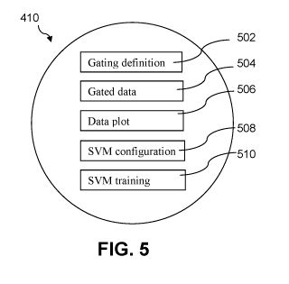
FIG. 5 is a block diagram of the structure of each node of the tree of FIG. 4
according to an implementation of the inventive system.
FIGs. 6A and 6B are examples of analysis results generated by the inventive
system.
FIG. 7 is a flow diagram for an exemplary branch of an analysis tree according
to an embodiment of the invention.
CA 02969912 2017-06-06
WO 2016/094720 PCT/US2015/065095
-10-
FIGs. 8A-8E are sample screenshots for an exemplary analysis sequence of
the branch of FIG. 7.
FIG. 9 is a sample screenshot of a 3-dimensional plot produced according to
an embodiment of the flow cytometry analysis system.
FIG. 10 is a sample screenshot of analysis results according to an embodiment
of the invention.
FIGs. 11A-11F are sample plots generated for six different analyses in which
FIGs. 11A-11C and 11F represent normal results and FIGs. 11D-11E are
highlighted
to indicate abnormal results.
FIG. 12 is a sample spreadsheet listing measured and calculated values for
different subpopulations.
FIG. 13 illustrates parameters for a subpopulation and the corresponding flow
cytometry data.
FIG. 14 illustrates parameters for another subpopulation and the corresponding
flow cytometry data.
DETAILED DESCRIPTION OF AN EXEMPLARY EMBODIMENT
According to the present invention, a method and system are provided for
analysis of flow cytometry data. In particular, the inventive method includes
creation
of kernels for use in the analysis of data of distributional nature. An input
data p in a
flow cytometry application is a collection of a large number of points in a
space. For
example, an image can be regarded as a set of points in a 2-dimensional space.
After
proper normalizations, p may be viewed as a probability distribution. To
define a
kernel on two such input data p and q to capture the distributional trends,
one must
define a function on p and q that measures the similarity between the two
entire
distributions rather than just the individual points in the distributions.
One way to construct such a "distributional kernel" is to use a distance
function (divergence) between the two distributions. If p(p,q) is a distance
function,
then the following is a kernel
k(p,q)= e-P(P'q) . (1)
CA 02969912 2017-06-06
WO 2016/094720 PCT/US2015/065095
-11-
There are many distance functions that measure the discrepancy between two
probability distributions.
Kullback-Leibler divergence, Bhattacharya affinity,
Jeffrey's divergence, Mahalanobis distance, Kolmogorov variational distance,
and
expected conditional entropy are all examples of such distances. Given a
distance
function, a kernel can be constructed based on the above formula.
For example, a special custom kernel can be constructed based on
Bhattacharya affinity. For normal distributions with mean Al and covariance
matrix
, Bhattacharya affinity has the form:
--1
1 T + 1 E)/ 1221
p (p q) = (Al 2 Mi i) 1 2 (M2 MO + ln ,(2)
8 2 2 V1E11.1E21
From this distance function, a new kernel is defined using the above equation.
v --1
k(p,q)= e-P(P'q) = 111(E1+E2)121 exp{ 1 (M2 _M1)T El + 2 (M2
¨M, ) (3)
V1E1 1.1E21 8 2
This distributional kernel is computationally efficient with a linear
complexity and
can handle large quantities of input data. A typical density estimation method
has a
computational complexity 0(n2), which might be too high for some applications.
The inventive distributional kernels can be applied directly in a SVM or other
machine learning systems to create classifiers and other predictive systems.
The
distributional kernels provide some distinctive advantages over the standard
kernels
that are frequently used in SVMs and other kernel machines. They capture the
similarities between the overall distributions of the large data components,
which may
be crucial in some applications.
FIG. 3 provides an exemplary process flow used for analysis of flow
cytometry data. As will be readily apparent to those in the art, flow
cytometry data is
provided as an example of distributional data, and other types of
distributional data
may be processed and classified using the techniques described in the
following.
The raw data generated by the flow cytometer 106 is input into a computer
processing system (step 302) which includes at least a memory and a processor
that is
programmed to execute one or more support vector machines. A typical personal
computer (PC) or APPLE MAC-type processor is suitable for such processing.
The
CA 02969912 2017-06-06
WO 2016/094720 PCT/US2015/065095
-12-
input data set may be divided into two portions, one for use in training the
support
vector machine, the other for use in testing the effectiveness of the
training. In step
304, feature selection algorithms are run on the training data set by
executing one or
more feature selection programs within the processor. In step 306, the
training data
set with the reduced feature set is processed using a support vector machine
with a
distributional kernel such as the Bhattacharya affinity-based kernel. The
effectiveness
of the training step is evaluated in step 308 by extracting the data
corresponding to the
features selected in step 304 in the independent test data set and processing
the test
data using the trained SVM with the distributional kernel. If the results of
the test
indicate a less than optimal result, the SVM will be re-trained and retested
until an
optimal solution is attained. If the training is determined to be
satisfactory, live data
corresponding to flow cytometry measurements taken on a patient sample is
input into
the processor in step 310. The features that were selected in step 304 are
selected
from the patient data and processed through the trained and tested SVM with
distributional kernel in step 312, with the result being a classification of
the patient
sample as normal or abnormal. In step 314, a report summarizing the results is
generated which may be displayed on a computer monitor 122, on a printed
report
124, and/or transmitted via e-mail or other network file transfer system to a
research
or clinical laboratory, hospital or physician's office. Histograms with one-
and two-
dimensional representations of the data groupings may also be displayed and/
or
printed. The results will also be stored, along with the raw data, histograms
and other
patient data within the computer memory or a patient database.
An optional additional diagnostic procedure may be combined with the flow
cytometry data and results to provide enhanced confidence in an automated
analysis
system. Using a scheme similar to that disclosed in U.S. Patent No. 7,383,237,
of
Zhang et al., which is incorporated herein by reference, the results of the
flow
cytometry testing may be combined with other types of testing. FIG. 3
illustrates an
optional flow path for performing computer-aided image analysis of cytogenetic
data
using SVMs by extracting features of interest from images of chromosomes
generated
in conventional procedures such as karyotyping or fluorescent in-situ
hybridization
CA 02969912 2017-06-06
WO 2016/094720 PCT/US2015/065095
-13-
(FISH), to identify deletions, translocations, inversions and other
abnormalities. In
step 320, training image data is input into the computer processor where it is
pre-
processed to identify and extract features of interest. In general, the
training image
data is pre-processed to identify features of interest (step 322), then used
to train the
image-processing SVM. Test image data are then used to verify that an optimal
solution has been attained (step 324). If not, step 324 will be repeated and
the SVM
will be re-trained and re-tested. If the optimal solution has been achieved,
live patient
image data will be input (step 326) for pre-processing (step 328) and
classification
(step 330).
In a preferred approach, as described in Patent No. 7,383,237, each feature of
interest within the image is separately pre-processed (step 322) and processed
by an
SVM which is optimized for that feature. The results of the analyses of all
features of
interest are combined in a 2' level image-processing SVM to generate an output
classifying the entire image. The trained SVM(s) is/are tested using pre-
processed
image test data (step 324). If the solution is optimal, images corresponding
to live
patient data (the same patient for whom the flow cytometry analysis is
performed) are
input into the processor (step 326). The patient image data is pre-processed
(step 328)
to identify the features of interest and each feature of interest is processed
through the
trained first level SVMs that are optimized for the specific feature. The
combined
results of the analyses of the features of interest are combined and input
into the
trained 2nd level image-processing SVM to generate an output classifying the
entire
image (step 330).
The results of step 330 can be communicated for storage in the patient's file
in
the patient database (step 316) and/or will be input into a 2nd level SVM for
analysis
in combination with the flow cytometry data results from step 312. This 2nd
level
SVM will have already been trained and tested using the training and test data
as
indicated by the dotted lines between steps 308, 324 and 340. The results of
step 316
and step 330 are combined for processing by trained 2nd level SVM for combined
analysis in step 342. The results of this combined processing with generally
be a
binary output, e.g., normal or abnormal, diseased or no disease, etc. The
combined
CA 02969912 2017-06-06
WO 2016/094720 PCT/US2015/065095
-14-
results may be output for display in step 314 and/or input into a memory or
patient
database for storage (step 316). Additional optional secondary flow paths may
be
provided to incorporate other types of data and analysis, such as expert
analysis,
patient history, etc., which may be combined to produce an ultimate diagnostic
or
prognostic score or other output that may be used for screening, monitoring
and/or
treatment.
Example 1: Detection of Myelodysplastic Syndrome (MDS)
The object of the present study is to investigate the potential connections
between Myelodysplastic Syndrome (MDS)-related chromosome abnormalities in
cytogenetics and the patterns in flow cytometry data. This immunophenotyping
analysis is one of the most common applications of flow cytometry and the
protocols
for sample collection and preparation are well known to those in the art.
Following
the sequence illustrated in FIG. 1, bone marrow aspirates 102 from patients
suspected
of having MDS are collected in a saline or sodium heparin solution to create a
cell
suspension in a number of tubes 104 or other containers that are adapted to
introduce
the suspension into the flow cell of flow cytometer system 106. Reagents
containing
monoclonal antibodies conjugated with different fluorochromes are introduced
into
the tubes, with each tube receiving different combinations of antibodies with
each
different combination conjugated with one of several possible fluorochromes.
Flow
cytometers are commercially available from numerous manufacturers including
the
FACSCa1iburTM from Becton Dickinson (Franklin Lakes, NJ) or the
Cytoron/AbsoluteTM from Ortho Diagnostics (Raritan, NJ). For the instant
example, a
FACSCa1iburTM system was used for four-color measurement. As will be apparent
to
those in the art, such systems provide automated handling of multiple samples
loaded
into a carousel, so that the illustrations are intended to be diagrammatic,
indicating
only the presence of a sample within the flow cytometer's analyzer field. The
forward scatter detector 108 and side scatter detectors 110 in the flow
cytometer
system 106 generate electrical signals corresponding to detected events as the
cells are
directed through the analysis stream. Fluorescence detectors, included among
the side
scatter detectors 110, measure the amplitudes of the fluorescent signals
generated by
CA 02969912 2017-06-06
WO 2016/094720 PCT/US2015/065095
-15-
expression of the antigens as indicated by the antibodies conjugated with the
different
fluorescent markers. Numerical values are generated based on pulse heights
(amplitudes) measured by each of the various detectors. The resulting signals
are
input into a processor within computer workstation 120 and used to create
histograms
(single or dual parameter) corresponding to the detected events for display on
a
graphical display monitor 122. Analysis of this data according to the present
invention, which involves classification of the input data according to normal
or
abnormal based on comparison to control samples, results in a report 124 which
may
be printed or displayed on the monitor 122. The raw data, histograms and
report will
also be saved in either or both of an internal memory in computer workstation
120
and a separate memory device, which may include a database server 130 which
may
be part of a data warehouse in a medical laboratory or other medical facility,
for
association with other records for the patient.
In an exemplary process sequence, the input dataset includes 77 cases
(patients) that have both flow cytometry and cytogenetics data. All patients
are
suspected of having MDS. Among the 77 cases, 37 had chromosome abnormalities
as
indicated by cytogenetic testing, which involves microscopic examination of
whole
chromosomes for changes in number or structure. The remaining 40 were found to
be
negative under cytogenetics.
The aspirated bone marrow samples in suspension were divided among 13
tubes for each patient. In a standard 4-color immunofluorescence protocol,
forward
light scatter (F SC) and right angle light scatter (S SC) were collected along
with 4-
color antibody combinations to perform seven different assays, one of which
was
blank. Each case typically had 20,000 ¨ 50,000 events where all of the assays
are
measured. The resulting flow cytometry dataset for each case had approximately
106
measurements. FIG. 2 illustrates an exemplary histogram showing side scatter
versus
CD45 expression with the different cell populations marked.
For each of the 13 tubes, FSC and SSC were measured, allowing gating to
exclude cellular debris, shown in the lower left corner of FIG. 2. In
addition, different
combinations of antigen specificities with fluorescence markers were used for
each
CA 02969912 2017-06-06
WO 2016/094720 PCT/US2015/065095
-16-
tube. Table 1 below lists the different combinations of monoclonal antibodies
with
the following markers: FITC (fluoroscein isothiocyanate), PE (phycoerythrin),
PerCP
(peridinin-chlorophyl), and APC (allophycocyanin). Monoclonal antibodies
conjugated with the identified fluorescent markers are commercially available
from a
number of different sources including Becton-Dickinson Immunocytometry Systems
(San Jose, CA), DakoCytomation (Carpinteria, CA), Caltag (Burlingame, CA) and
Invitrogen Corporation (Camarillo, CA). The CD45 antibody, used for
enumeration
of mature lymphocytes, is included in each combination for validation of the
lymphocyte gating.
TABLE 1
Tube FITC-conjugated PE-conjugated PerCP-conjugated APC-conjugated
1 IgG1 IgG1 +PI CD45/2D1/IgG1 IgG1
2 IgG2b IgG2b CD45/2D1/ IgG1 IgG2b
3 CD8/SK1/IgG1 CD2/RPA-2.10/IgG1 CD45/2D1/IgG1 CD4/SK3/IgG1
4 CD7/M-T701/IgG1 CD56/MY31/IgG1 CD45/2D1/IgG1 CD3/SK7/IgG1
5 CD19/SJ25C1/IgG1 CD23/M-L233/IgG1 CD45/2D1/IgG1 CD5/UCHT-2/IgG1
6 CD22/S-HCL-1/IgG2b CD10/HI10a/IgG1 CD45/2D1/IgG1 CD34/8G12/IgG1
7 CD10/HIlOallIgG1 CD11c/S-HCL-3/IgG2b CD45/2D1/IgG1 CD20/2H7/IgG2b
8 CD38/HB.7/IgG1 Dako Kappa/F(ab)2rab CD45/2D1/IgG1 CD20/2H7/IgG2b
9 CD38/HB.7/IgG1 Dako Lambda/F(ab)2rab CD45/2D1/IgG1 CD20/2H7/IgG2b
10 Kappa Caltag poly Lambda Caltag poly CD45/2D1/IgG1
CD19/SJ25C1/IgG1
11 HLA-DR/TU36/IgG2b CD117/104D2 CD45/2D1/IgG1 CD1 lb/Mac-1/IgG1
12 CD14/MoP9/IgG2b CD13/L138/IgG1 CD45/2D1/IgG1 CD64/10.1/IgG1
13 CD16/NKP15/IgG1 CD33/P67.6/IgG1 CD45/2D1/IgG1 CD34/8G12/IgG1
In order to provide data for both training the SVM and for evaluation of the
training,
the entire dataset for the 77 cases was divided into a training set and an
independent
test set. Forty cases (20 positive and 20 negative as determined by
cytogenetic
testing) were used to train the SVM. The remaining 37 cases (17 positive and
20
negative) were used to form an independent test set.
The previously-described custom kernel based on the Bhattacharya affinity
was used for analysis of the flow cytometry data to measure the discrepancy
between
two probability distributions.
CA 02969912 2017-06-06
WO 2016/094720 PCT/US2015/065095
-17-
Inclusion of data from all the assays in the classifier will not produce a
system
with the optimal performance. Therefore, a feature selection on the assays is
conducted based on the training set. Two performance measures were applied in
the
feature selection step. The first feature selection method, the leave-one-out
(L00)
error rate for SVM, involves training the SVM on the initial data set, then
updating
the scaling parameters by performing a gradient step so that LOO error
decreases.
These steps are repeated until a minimum of the LOO error is reached. A
stopping
criteria can be applied. The second feature selection method was the kernel
alignment. Such a technique is described in U.S. Patent No. 7,299,213 of
Cristianini,
which is incorporated herein by reference. Kernel alignment uses training data
only
and can be performed before training of the kernel machine takes place.
During the feature selection process, it was determined that a significant
number of features would not contribute to the accurate classification of the
data. The
result of the feature selection procedure is given in the Table 2.
TABLE 2
Assay Blank FSC SSC Marker
Tube #
F TIC PE PerCP APC
1 0 1 0 0 1 0 0
2 0 0 1 0 0 0 1
3 0 1 1 0 0 0 0
4 0 0 1 1 0 0 1
5 0 0 0 0 0 0 1
6 0 0 1 0 0 0 0
7 0 1 1 1 0 0 0
8 0 1 1 1 0 0 1
9 0 1 1 1 0 0 0
10 0 0 1 1 0 0 0
11 0 1 1 0 0 0 1
12 0 0 0 0 0 0 0
13 0 0 0 0 0 0 0
A value of "1" in an entry of Table 2 means that a particular assay
(tube/assay
combination) is selected; "0" means that the assay was not selected. This
reduced the
CA 02969912 2017-06-06
WO 2016/094720 PCT/US2015/065095
-18-
number of features to be considered from each case for classifying the data to
26,
down from the original 91. The data from the reduced number of assays was then
used to train the SVM with the distributional kernel.
Using the selected assays, the trained SVM is then tested with the 37
independent cases. The results at the cutoff of 0 were summarized using the
conventional statistical measure of the performance of a binary classification
test.
Sensitivity, or recall rate, provides a measure of the proportion of correctly
classified
positives to the total number of positives as determined by cytogenetic
testing.
Specificity measures the proportion of negatives which are correctly
identified. The
results of analysis of the test data were as follows:
Sensitivity: 15/17 = 88% Specificity: 19/20 = 95%
This produces an overall error rate of 3/37 = 8%. Using the estimated
standard deviation for binomial distribution, a = 0.0449, the test produced a
95%
confidence level that the error rate would be less than 15%.
FIG. 4 illustrates the hierarchical structure of the inventive system,
represented
by a rooted tree 400. Each node 410 of the tree represents a basic analytical
element
that performs various tasks pertaining to a specific gated flow data.
Depending on
the analysis being performed at a given node, multiple branches may grow out
of a
node. In the illustrated example, initial node 410 splits into three branches
402, 404,
406. The number of nodes and number of branches in the tree will vary
depending
upon the parameters to be analyzed. For example, in branch 402, the second
node
results in a split into branch 402a and 402b. Branch 404 splits at its second
node into
three branches 404a, 404b and 404c, then branch 404b splits at the third node
into
branches 404ba and 404bb. The tree structure reflects the hierarchical gating.
The
input data at each node is the result of gating from its parent node.
FIG. 5 shows the structure of each node 410 in the tree illustrated in FIG. 4.
Each node includes a gating definition 502, a gated data set 504, a graphical
plot of
the data 506, an SVM configuration 508, and a trained SVM data set 510.
CA 02969912 2017-06-06
WO 2016/094720 PCT/US2015/065095
-19-
Example 2: Sample Results for standard leukemia/lymphoma panel
Exemplary results produced by the inventive system are shown in FIGs. 6A
and 6B. The analysis software includes a function to read data files in the
standard
FCS format. It can also export the results in various formats. FIG. 6A is
split over
multiple pages to provide adequate resolution. In each case, the first page of
the
figure corresponds to the left panel 520 of the screenshot; the second page is
the
center panel 522, and the third page is the right panel 524. The left panel
520
displays files corresponding to the gated data. As illustrated, the first
gating
parameter 526 is the sample tube number (tube 1, tube 2,..., tube x). For
example,
this gating operation would correspond to the first node 410 in FIG. 4. The
next
gating 528 (subgating) is non-debris and non-debris+debris, which would be,
e.g., the
second node in branch 402a. The non-debris is then further subgated by
mononuclear
and lymphocytes. Following the prior example, this gating 530 and analysis
would
occur in the third node in branch 402a.
The center panel 522 of FIG. 6A displays the flow cytometry data marked
with the different subpopulations as determined by the parameters. In this
case, the
marker is CD45 KO as detected by SS INT LIN (side scatter intensity, linear).
The
right panel 524 of FIG. 6A provides a table listing the various parameters
used in the
gating and SVM analysis. As illustrated, parameters SS INT LIN and CD45 KO are
checked under the heading "in SVM", indicating that SVM analysis was performed
based on these parameters providing the data forp and q in the distributional
kernel in
Equation (3) above.
The bottom of the screenshot of FIG. 6B provides an exemplary list of
possible markers (antibodies) within the screening panel for the illustrated
test. Here,
24 markers are indicated: CD2, CD3, CD4, CD5, CD7, CD8, CD10, CD11 c, CD13,
CD14, CD16, CD19, CD20, CD23, CD33, CD34, CD38, CD45, CD56, CD64,
CD117, HLA-DR, kappa, and lambda, which represents a standard
leukemia/lymphoma panel, which is useful to assist in diagnosis of leukemia
and
lymphoma, and for post-treatment follow-up. While not all of the markers may
be
represented in this screenshot, FIG. 6B illustrates a sample screenshot of the
results of
CA 02969912 2017-06-06
WO 2016/094720
PCT/US2015/065095
-20-
the analysis, including two 2D flow cytometry plots for CD45 KO versus SS TNT
LIN
(upper left quadrant) and SS INT LIN versus FS TNT LIN (upper right quadrant.)
In
addition, as will be readily apparent to those in the art, selection of
appropriate
markers will depend on abnormality known or suspected to be present. For
example,
an extended leukemia/lymphoma panel may add CD11b, CD41, CD138, CD235a and
FMC-7 to the listed markers for a standard panel. Smaller panels of selected
markers
may be used for prognostics and therapy monitoring. Regardless of which
markers
are used, the same basic procedures will be followed to extract information
for
relevant subpopulations from the large volume of data.
One part of the software system facilitates the design of the gating
structure,
configuration and training of SVM, and the setting of default values. Gating
is defined
as any process that selects a subpopulation of cells based on specific
criteria on
observed parameters. Gating is an effective technique for reducing the
complexity of
the data and focusing the analysis on a specific subpopulation of the data.
However, in
order to address all aspects of the analysis, there will typically be a large
number of
gates and the gating structure itself may be complex.
The hierarchical structure of this system facilitates flexible and convenient
definitions of very general types of gating.
At each node, in step 502 a 2D gating is defined based on a selection of any
two parameters. A 2D plot 506 is the basis for defining the gating.
The gated data 504 at a node is the cumulative result of the chain of gating
at
the series of nodes preceding the current node. Because each node defines a 2D
gating
with any combination of parameters, the hierarchical scheme allows for the
definition
of virtually any gating configuration.
For example, a gating on FS (forward scatter) and SS (side scatter) can filter
out debris. On the Non-debris, another gating on FS and the CD45 marker can be
defined to separate five subpopulations: CD45-Dim (diminished marker),
Monocytes,
CD45-Negative (negative marker), Granulocytes, and Lymphocytes. The
mononuclear cells can be further gated to feed new nodes.
CA 02969912 2017-06-06
WO 2016/094720 PCT/US2015/065095
-21-
FIG. 7 provides a flow diagram that represents a possible gating sequence in
one branch of a tree 400 such as that shown in FIG. 4. The illustrated branch
includes three nodes, each of which has the structure of the node 410 shown in
FIG. 5,
including an SVM processing step to separate the event data into the selected
populations. For example, in step 650, the side scatter (SS) and forward
scatter (FS)
events are detected, then plotted in step 652, producing a 2D image with a
data
distribution. Using the plot of SS/FS data, in step 654, Node #1 executes a
gating
operation to separate the non-debris from the debris. This separation is
illustrated in
FIG. 8A in which the plot in the center panel of the screenshot shows a line
between
non-debris and debris. In step 656, non-debris is selected, then analysis is
directed to
the plot containing the non-debris data evaluated for CD45 and SS INT LIN.
This
plot is shown in the center panel of FIG. 8B. In step 658, Node #2 separates
the non-
debris data into 5 population groups: granulocytes, monocytes, lymphocytes,
CD45-
Dim and CD45-Neg. The plot in the center panel of FIG. 8C shows the groupings
that were identified by plotting SS TNT LIN data for the CD45 KO marker. (Note
the
checked parameters under "in SVM" in the right panel of FIG. 8C: "SS TNT LIN"
and "CD45 KO".) For the next step 660, the granulocyte data are excluded and
the
remaining mononuclear data, plotted in the center panel of FIG. 8D, are gated
in Node
#3 (step 662) to separate CD3 and CD5 cell surface receptors. The resulting
plot is
provided in FIG. 8E, which shows the flow cytometry data subgated into
quadrants
based on % positive on X and Y; % negative on X and Y; % double positive; and
%
double negative. This breakdown is generated by SVM analysis of the data in
the plot
using a distributional kernel. The upper portion of right panel of FIG. 8E
provides the
numerical values for the distributional analysis.
This process would be repeated for each tube of a patient sample. Additional
branches with different gating definitions could be run in parallel, for
example, a
branch could diverge from node #1 to perform a different set of separations.
An
optional final step would be to combine the results of each tree branch to
generate a
diagnostic conclusion taking into consideration the results achieved at the
end of each
branch. In the preferred embodiment, this final analytical step would be
performed by
CA 02969912 2017-06-06
WO 2016/094720 PCT/US2015/065095
-22-
a support vector machine, generating a diagnostic score, a binary, e.g.,
positive or
negative, result, a probability, a prognostic prediction, or other appropriate
indicator
of the subject's diagnosis or prognosis.
The following is an exemplary algorithm for automatic gate detection
according an embodiment of the invention:
The system automatically detects gate definitions from user specified points
and lines. A pseudo code for the algorithm is given below:
for each vertex v with outdegree > 0
add v to gate
find first edge (v,u) in counter-clockwise order
remove (v,u)
while u != v
v=u
add v to gate
find first egde (v,u) in counter-clockwise order
remove (v, u)
In some situations, the gating may require some adjustments for individual
cases. Because of the large number of gates involved in an analysis, this can
be a
tedious process.
The inventive system provides an automatic gating adjustment function based
on clustering. The gates in flow cytometry data are usually associated with
clusters of
cells. Automated clustering of the actual data provides a natural way to make
an
appropriate adjustment to the default gating template.
A Gaussian mixture model (GM_M) is a probability distribution that is a
weighted sum of Gaussian distributions:
f (x)=Iii) ig(x I
1
g(x I 14,E,)=2
(27)d/2 -1 1/2 e
The parameters in the GMM can be determined by a learning algorithm known as
Expectation-Maximization (EM) algorithm. In statistics, an
expectation¨maximization
algorithm is an iterative method for finding maximum likelihood or maximum a
CA 02969912 2017-06-06
WO 2016/094720 PCT/US2015/065095
-23-
posteriori (MAP) estimates of parameters in statistical models, where the
model
depends on unobserved latent variables.
The present system applies GMM to detect clusters in the flow data at a node.
The cluster information is then used to make adjustment on gating templates.
Users
also have the option to manually adjust the gating.
After gating, the characteristics (parameters) of each subpopulation is
captured
for analysis. Each node in the gating tree has an associated SVM, which is
defined on
the gated data present at the node. The SVM associated with a specific
subpopulation
is trained to analyze the distribution patterns in the data for that
subpopulation and to
provide a quantitative assessment of normality/abnormality for the data in the
subpopulation.
The SVM input is not limited to the 2D plot. Any combination of the
parameters, as well as the gated populations at each node, can be used for SVM
learning and subsequent SVM classification. The system may use different types
of
SVMs such as C-SVM, nu-SVM, and single-class-SVM.
Additional features of the software system includes functions to import data,
make gating adjustments, perform SVM analysis, and present results
graphically.
The distributed system of SVM based analysis nodes will provide a
quantitative indication of abnormality on an entire case.
In an embodiment of the software system, different visualization methods for
displaying data may be included. In addition to traditional 2D plots, 3D plots
are
available, as illustrated in FIG. 9, where the X axis is CD45 KO (CD45-Krome
Orange dye), the Y axis is SS INT LIN (side scatter intensity, linear) and the
Z axis is
FS INT UN (forward scatter intensity, linear.) Any three parameters may be
selected
for the 3D plot. A user may interactively move, rotate, and scale the 3D plot.
The 3D
function provides a significantly enhanced representation of the structure of
the flow
data.
Example 3: Highlighting of Abnormal Results
A key goal of the automated flow cytometry analysis system is to allow
laboratory technicians to more readily identify cases requiring pathologist
review.
CA 02969912 2017-06-06
WO 2016/094720 PCT/US2015/065095
-24-
This is achieved in part by displaying abnormal plots and values using a
visually-
distinguishable feature, such as using a specific color font or highlighting,
e.g., red, in
a display of the analysis results.
FIG. 10 provides an example of a screen display 600 on a monitor of a user
workstation. In this example, patient samples were subjected to flow cytometry
analysis. In one part of the analysis, a plot 610 is generated to illustrate
the
subpopulations identified during gating on SS and CD 45 to separate
subpopulations
and the relative percentages of CD45 Negative (0.93%), granulocytes (50.58%),
monocytes (3.78%) CD45-Dim (2.00%) and lymphocytes (42.70%), which are plotted
with X axis of CD45 KO (CD45-Krome Orange dye), and the Y axis of SS TNT LIN.
In this example, the lymphocyte count exceeds the normal range of 20-40%, so
the
plot is highlighted to signal to the user that an abnormal value was measured.
In a
color display, the upper bar 612 on the plot might be red, or the entire plot
might be
outlined in red. For purposes of illustration, the upper bar 612 of the plot
is
highlighted with wavy lines.
Plot 614 illustrates the results of gating on FS INT UN and SS TNT UN.
Because the results of this gating did not exhibit abnormal results, the plot
is not
highlighted, as indicated by the clear upper bar 616 of the plot. Table 618 in
the
display provides the numerical results for each subpopulation. Again, because
of the
abnormal value for lymphocytes, the displayed value is highlighted to indicate
to the
user that an abnormal value was measured. On a color display, the number
"42.70"
might appear in red or some other color to distinguish it from the other
values. For
purposes of illustration, the value is shown underlined, bolded and in
italics. Analysis
of the subpopulations shown in plot 610 included further gating of the
lymphocytes,
the numerical results of which are displayed in table 620 of the display. As
described
above, each sub-subpopulation is analyzed by a separate node that is branched
off
from the node that performed the initial gating and analysis. In the example,
lymphocytes are gated into subpopulations of T-cells (CD2, CD3), B-cells
(CD19,
CD20), NK-cells (CD16, (CD3-CD56)), and pre-B cells (CD1O+CD19). The
resulting numerical results are entered into table 620, which the abnormal
results
CA 02969912 2017-06-06
WO 2016/094720
PCT/US2015/065095
-25-
relating to B-cells indicated by highlighting the values 622 and 624 in the
display. In
table 630 of the display, another abnormal value, for CD4-CD8, is highlighted.
FIGs. 11A-11F provide further illustration of the display feature that
provides
an indication to the user of the presence of abnormal results following
analysis of the
second sample from the patient. FIG. 11A plots Kappa FITC against FS INT LIN.
The clear upper bar indicates normal results. Similarly, the results plotted
in FIG.
11B (Lambda PE vs. FS INT LIN) and FIG. 11C (CD23 ECD vs. FS INT LIN) are
normal. However, FIG. 11D (CD19 PC5.5 vs. FS INT LIN) and FIG. 11E (CD11 c
PC7 vs. FS INT LIN) are abnormal, as indicated by the highlighting in the bar
above
the plot.) FIG. 11E (CD10 APC vs. FS INT LIN) indicates normal results for
this
parameter.
FIG. 12 illustrates an exemplary spreadsheet 700 for capturing and quantifying
various parameters of each subpopulation. The spreadsheet listing includes the
node
number (column C), the gated parameter, e.g., tube number, non-debris (column
D),
subgate characteristics, e.g., non-debris, debris, gate 1, CD4 APCA, etc.
(column E).
Column F corresponds to the X-axis parameter, while column G provides the Y-
axis
parameter. Columns H through M provide the weight, X- and Y-means, and
covariance of each population, all of which are used in conjunction with the
distributional kernel for SVM analysis.
FIG. 13 provides additional detail of the process involved in flow cytometry
data analysis according to an embodiment of the invention. Plot 712 shows the
plotted flow cytometry data gated on Mononuclear 2 using the X- and Y-
markers,
CD20 V450 and CD23 ECD, respectively. Spreadsheet data 710 for the node used
to
perform this analysis (sample node number 65 (from column C of FIG. 12)) gated
on
mononuclear 2 then subgated into 4 quadrants: % positive on X and Y; %
negative on
X and Y; % double positive; and % double negative. The subgating into
quadrants
provides the weights corresponding to counts (percentages) of the cells
falling into the
different quadrants. The calculated means for each marker are provided in the
spreadsheet as are the distributions (covariance) for each population. Because
these
CA 02969912 2017-06-06
WO 2016/094720 PCT/US2015/065095
-26-
results are outside of normal values, upper band 714 of plot 712 is
highlighted to
indicate to the user that abnormal results have been identified.
FIG. 14 provides another example of the process involved in flow cytometry
data analysis according to an embodiment of the invention. Plot 812 shows the
flow
cytometry data gated on Lymphocytes 2 using X-marker CD20 V450 and Y-marker
Kappa FITC, Spreadsheet data 810 for sample node number 77 (from column C of
FIG. 12) is gated and subgated into 4 quadrants: % positive on X and Y; %
negative
on X and Y; % double positive; and % double negative. The calculated means for
each marker are provided in the spreadsheet as are the distributions
(covariance) for
each population. Because these results are outside of normal values, upper
band 814
is highlighted to indicate to the user that abnormal results have been
identified.
As will be apparent from the foregoing examples and accompanying figures,
any combination of parameters may be used to automatically analyze flow
cytometry
data. Each parameter is separately
In some embodiments, the system is configured to maintain a database to
collect data from analyzed cases. (See, e.g., database 130 in FIG. 1.) All
relevant
data, the reported statistical values, and the features for SVM evaluation are
saved in
this database. The general consensus among the flow cytometry experts is that
there
is more useful information in the volumes of flow cytometry data than what is
currently known. This database will help facilitate future research in
discovery of new
patterns and diagnostic information in flow data.
The software preferably includes user instructions with reminders to save the
data at the conclusion of an analysis. For multiple analyses of the same case,
options
are available to overwrite the old data or to save both versions of the data.
To ensure the integrity and security of the software system, a preferred
embodiment of the software system includes a real-time authentication
function. An
authentication server is established to process the authentication requests.
The client
software communicates with the server over the Internet through a secure
protocol.
In some embodiments, the analysis may be performed on a client machine that
is remote from the laboratory in which the flow cytometry instrumentation
resides.
CA 02969912 2017-06-06
WO 2016/094720 PCT/US2015/065095
-27-
For example, the raw data may be processed and transmitted via a network to
one or
more remote locations. The flow cytometry analysis software running on a
client
machine will be required to complete authentication before it is permitted to
begin
normal operations.
In one embodiment, the client will transmit an encrypted message to the server
containing the following fields:
Nonce
Timestamp
Account
Usage
Software signature
Hardware signature
Upon receiving the authentication request, the server will verify each of the
fields. If the authentication is successful, the server will send an encrypted
authentication message that matches the request back to the client. This
protocol is
designed to prevent a "replay attack". The use of nonce and timestamp will
ensure
that the messages are unique even for the same client.
The authentication function will help provide assurance that the software has
not been altered maliciously, the software is properly licensed, the system is
configured properly in a conforming environment, and all analyzed cases are
accounted for.
Flow cytometric immunophenotyping is an accurate and highly sensitive
method for detection of quantitative and qualitative abnormalities in
hematopoietic
cells even when combined morphology and cytogenetics were non-diagnostic. The
automated flow cytometry data analysis system disclosed herein provides the
ability
to automatically analyze the huge volumes of data generated during flow
cytometry
measurement, enhancing the accuracy, repeatability and versatility of flow
cytometric
methods. The capability provided by the methods disclosed herein enhances not
only
the diagnostic value of flow cytometry but also expands research applications
of the
CA 02969912 2017-06-06
WO 2016/094720
PCT/US2015/065095
-28-
technique by enabling collection and analysis of massive amounts of flow
cytometry
data from many subjects for data mining and pattern recognition that go far
beyond
current limited approaches.