Note: Descriptions are shown in the official language in which they were submitted.
CA 02970237 2017-06-07
WO 2016/096529
PCT/EP2015/078937
1
Stent and kit of stents for adjustable interventional reduction of blood
flow
Technical Field
.. The invention relates to a stent or a kit of stents for interventional
reduction of
blood flow. In particular a covered pulmonary stent for interventional
reduction of pulmonary artery blood flow in patients to avoid the development
of pulmonary artery hypertension and in patients with pulmonary hypertension
and established Eisenmenger's syndrome associated with uncorrected heart
.. disease.
Prior Art
Eisenmenger's syndrome is a systemic disease involving multiple organ
systems caused by longstanding congenital cardiac defects which cause a high
pulmonary artery blood pressure with reduced pulmonary blood flow,
decreased oxygen uptake and decreased oxygen saturation of the arterial
blood resulting in the so-called ''blue babies" or "blue children".
Eisenmenger's
syndrome is characterized by multiple clinical features such as cyanosis with
its typical blue tinge to the skin, swollen or clubbed finger tips, fainting
or
syncope, heart failure, arrhythmia or irregular heart rhythms, bleeding
disorders, coughing up blood, iron deficiency, kidney problems, stroke, gout
and gallstones. Eisenmenger patients do not grow normally, have a
dramatically decreased quality of life and severely limited physical capacity,
all
associated with a markedly decreased life expectancy. In other words, they
.. have just too much to die, but too little to have an acceptable life.
Congenital heart disease is the most common form of birth defects, occurring
in about 1% of life births (Ref 1-4, see below). Nowadays, the majority of
children born with congenital heart disease, if repaired in time, are expected
to lead normal lives. The privilege of early diagnosis and timely surgical
management is mostly restricted to children living in developed countries.
However, the majority of children born with congenital heart disease live in
CA 02970237 2017-06-07
WO 2016/096529
PCT/EP2015/078937
2
developing countries without access to timely treatment; hence, about 90% of
all children born yearly with congenital heart defects around the world
receive
suboptimal care (Ref 6, Ref 8).
Many countries with populations between 15 and 70 million people are without
.. a single specialized paediatric heart centre (Ref 7). In Africa, on
average,
there is only one centre capable to perform open-heart surgery and advanced
cardiac care per 33 million peoples compared to one centre per 1 million
peoples in the Western world, and even fewer centres will have the capacity to
treat children with congenital heart disease (Ref 6, 9).
.. Therefore, in these countries, the majority of children born with
congenital
heart disease does not have access to specialized surgical treatment and are
often diagnosed late (Ref 10). Furthermore, a substantial number of children
with severe forms of heart defects will not be diagnosed at all (Ref 10, Ref
11). Hence, the mortality of children with congenital heart disease living in
developing countries is considerably higher compared to those living in the
Western world (Ref 5). Indeed, mortality rates up to 75% have been reported
(Ref 5, Ref 12-14) and it has been assumed that in the developing world
millions of children die or suffer serious consequences from their cardiac
malformations which could effectively be prevented (Ref 7, Ref 9). The
.. sobering fact is that most children die while waiting for surgery. For
instance
in India, approximately 1-2 millions of children with congenital heart disease
is
awaiting surgery (Ref 15, Ref 16).
Excessive pulmonary blood flow and consecutive pulmonary hypertension is
observed in congenital heart defects with left-to-right shunts such as large
ventricular or atrial septal defects, which are among the most frequently
observed congenital heart diseases (Ref 13, Ref 17, Ref 18).
Under these circumstances, fixed secondary pulmonary hypertension develops
within years finally resulting in Eisenmenger's syndrome (Ref 18, Ref 19).
Pulmonary artery banding has frequently been performed as an interim
palliative procedure to reduce increased pulmonary blood flow and to protect
the pulmonary vasculature from hypertrophy and consecutive irreversible
pulmonary hypertension (Ref 19, Ref 20). However, surgical pulmonary artery
CA 02970237 2017-06-07
WO 2016/096529
PCT/EP2015/078937
3
banding (the Battista operation) is not feasible in many developing countries
as open heart surgery is often not available, technically demanding, and
expensive. Moreover, the majority of patients with congenital heart disease
resulting in fixed pulmonary artery hypertension have become inoperable and
this is also true in highly developed countries where latest technologies are
available. In fact, established Eisenmenger's syndrome represents an
inoperable, devastating disease with limited palliative treatment options
resulting in markedly reduced quality of life as well as reduced life
expectancy.
In addition, life-long palliative treatment of Eisenmenger's syndrom is only
possible with a multidisciplinary approach resulting in extremely high costs
of
treatment.
Placement of reductional stent into the main pulmonary artery would reduce
pulmonary blood flow as well as the development Eisenmenger syndrome.
Moreover, such a stent could replace surgical pulmonary artery banding and
allow treating fully developed established Eisenmenger's syndrome by
interventional technique. With this techniques, patients previously thought to
be inoperable for live, could undergo definite surgical correction of the
underlying heart disease, once the Eisenmenger syndrome has been reversed
by such a stent.
Reduction of pulmonary artery pressure over a period of six to 12 months is
known to normalize pulmonary artery pressure allowing secondary curative
surgical treatment of congenital heart defects, which, as mentioned above,
have become inoperable according to the current knowledge. This concept of
primary reduction of pulmonary artery blood flow followed by definitive
surgical correction of the underlying heart defect has already been proven by
surgical pulmonary artery banding via median sternotomy, known as Battista
operation for Eisenmenger's syndrome. However, surgical pulmonary artery
banding is associated with a considerable operative mortality because of a
highly unstable early postoperative course necessitating sophisticated
postoperative intensive care treatment which is far less developed than
surgery in developing contrast or even completely absent. For these reasons,
the Battista procedure has only been carried out in less than 50 patients with
CA 02970237 2017-06-07
WO 2016/096529
PCT/EP2015/078937
4
very few undergoing total correction of the underlying heart disease in a
second stage procedure. Nevertheless, the concept of primary banding of the
pulmonary artery with a subsequent decrease of the chronically high
pulmonary artery pressure to normal values followed by total correction of the
underling heart defect is proven.
In contrast to surgical pulmonary artery banding, interventional treatment of
Eisenmenger syndrome by stenting could easily be implemented in developing
countries, as it is a simple procedure performed in local anaesthesia without
need for cardiopulmonary bypass or post-interventional intensive care.
.. The insertion of a pulmonary artery reductional stent would prevent the
development of severe pulmonary artery hypertension and would even be able
to completely reverse fully developed Eisenmenger's syndrome allowing
complete surgical correction of the underlying cardiac malformation.
Anatomical correction normalizes growth of the patient, improves the physical
capacity as well as the quality of life and prolongs the life expectancy to
near
normal values allowing normal participation in social life.
W003074119 describes an intravascular flow restrictor comprising a braided
tubular structure designed to be placed in the main pulmonary artery for
limiting blood pressure in the lungs. The braided structure is designed to be
collapsed for placement in a delivery catheter but when ejected from the
delivery catheter, assumes a substantially larger diameter disk shaped device
having one or more longitudinal channels or passageways therethrough.
Adjustment of blood flow is not possible.
EP0647438 describes a reductional stent for reducing a diameter of a duct in a
body of a living creature. The stent includes a sleeve-like part having walls
provided with perforations, enlarged ends as well as an intermediate area
reduced in diameter by a constriction. Thrombogenic threads are provided on
an exterior of the sleeve-like part between the enlarged ends. When the stent
is in place it is only adjustable by expanding the diameter and thereby
increasing the blood flow. Adjustment in order to decrease the blood flow is
not possible.
CA 02970237 2017-06-07
WO 2016/096529
PCT/EP2015/078937
W010114585 describes a stent made of a bioabsorable, polymer and/or non-
polymer material having an elongated body with a proximate end, a distal
end, and at least one open channel formed on the exterior surface of the
elongated body to provide fluid communication between the proximal end and
5 the distal end. In one embodiment the stent has an elongated centre rod
having a proximate end and a distal end and a plurality of leaflets extending
outward from the centre rod and forming channels between two neighbouring
leaflets to provide fluid communication between the proximal end and the
distal end. The diameter of the stent can be reduced by compressing or
twisting the channel walls against each other to facilitate implantation.
However, once placed at the treatment site the flow cross section cannot be
adjusted.
W003028522 discloses a flow reducing stent. The stent comprising a hollow
element adapted for placement in the blood vessel defining a flow passage
therethrough. The flow passage comprises at least two sections, one with a
larger diameter and one with a smaller diameter, wherein said smaller
diameter is smaller than a cross section of the blood vessel. The stent may be
provided with an annular inflatable tube around a centre section of the stent.
In order to reduce the blood flow the tube is provide with a hose for
inflating
the tube. Thereby the diameter may be reduced just after positioning the stent
in a blood vessel as long as the hose is attached to the tube. However, later
adjustment after implantation of the stent and removing of the hose is not
possible as the tube cannot be inflated anymore.
US6120534 teaches a flow reducing stent for use in a pulmonary artery to
control damage to the lungs in a new born that exhibits multiple, life-
threatening cardio-pulmonary deformities. The stent comprises a deformable
mesh covered with a biocompatible material, the mesh having a conical
portion and a constricted region. The stent may be percutaneously and
transluminally delivered and deployed in a vessel. The constricted region may
then be selectively enlarged employing a conventional dilatation means or
device, e.g. a balloon, to adjust the flow impedance created by the
constricted
region. In an alternative embodiment, the constricted region is preferably
CA 02970237 2017-06-07
WO 2016/096529
PCT/EP2015/078937
6
formed from a shape-memory material, so that the maximum degree of
constriction may be recovered by heating the shape-memory material.
However, there is a certain risk of overheating and thereby damaging the
surrounding tissue.
W004014257 describes a flap type flow reducing implant. The flap type
reducing implant comprises three flaps that reduce blood flow in a flow
passage and/or promote changes in blood stream dynamics depending on the
angle of the flaps. The angle of the flaps may be adjusted with a special flap
angle adjusting tool. However, the flap type implant is made of a metal tube
with its diameter fixed and it cannot be adjusted to a growth in diameter of
the blood vessel.
EP1276437 describes a narrowing intraluminal stent comprising hollow body
with a flow passage there through. The hollow body has at least one portion of
an inner cross sectional dimension smaller than the cross sectional dimension
of the lumen, so as to artificially narrow a passage through the body lumen.
The stent may have an hourglass or bottleneck shape. When the stent is in
place it is only adjustable by expanding the diameter and thereby increasing
the blood flow. Adjustment in order to decrease the blood flow is not
possible.
Ref 1: Gillum RF. Epidemiology of congenital heart disease in the United
States. Am Heart] 1994;127:919-27.
Ref 2: Hoffman JI, Kaplan S. The incidence of congenital heart disease. J Am
Coll CardioI2002;39: 1890-900.
Ref 3: MareIli AJ, Mackie AS, Ionescu-Ittu R, Rahme E, Pilote L. Congenital
heart disease in the general population: changing prevalence and age
distribution. Circulation 2007; 15:163-72.
Ref 4: Jonas RA. Congenital heart surgery in developing countries. Sennin
Thorac Cardiovasc Surg Pediatr Card Surg Annu 2008:3-6.
Ref 5: Bernier PL, Stefanescu A, Samoukovic G, Tchervenkov CI. The
challenge of congenital heart disease worldwide: epidemiologic and
CA 02970237 2017-06-07
WO 2016/096529 PCT/EP2015/078937
7
demographic facts. Semin Tho rae Cardiovasc Surg PediatrCard SurgAnnu
2010;13:26-34.
Ref 6: Neirotti R. Paediatric cardiac surgery in less privileged parts of the
world. Cardiol Young 2004;14:341-6.
.. Ref 7: Yacoub MH. Establishing pediatric cardiovascular services in the
developing world: a wake-up call. Circulation 2007;116: 1876-8.
Ref 8: Tchervenkov CI, Jacobs JP, Bernier PL, et al. The improvement of care
for paediatric and congenital cardiac disease across the World: a challenge
for
the World Society for Pediatric and Congenital Heart Surgery. Cardiol
Ref 9: Young 2008; 18 Supp12:63-9. Zheleva B. Linked by a common purpose:
Global Efforts for Improving Pediatic Heart Health: A Report by Children's
Heart Link. Congenital Cardiology Today 2007;5: 1-15.
Ref 10: Mocumbi AO, Lameira E, Yaksh A, Paul L, Ferreira MB, Sidi D.
Challenges on the management of congenital heart disease in developing
countries. Int J Cardiol 2011;148:285-8.
Ref 11: Trucco SM, Barnoya], Larrazabal LA, Castaneda A, Teitel DF. Detection
rates of congenital heart disease in Guatemala. Cardiol Young 2011; 21:153-
60. 23.
Ref 12: Shah GS, Singh MK, Pandey TR, Kalakheti BK, Bhandari GP. Incidence
of congenital heart disease in tertiary care hospital. Kathmandu Univ Med J
(KUMJ) 2008;6:33-6.
Ref 13: Wickrannasinghe P, Lamabadusuriya SP, Narenthiran S. Prospective
study of congenital heart disease in children. Ceylon Med J 2001;46:96-8.
Ref 14: Samanek M, Slavik Z, Zborilova B, Hrobonova V, Voriskova M,
Skovranek J. Prevalence, treatment, and outcome of heart disease in live-born
children: a prospective analysis of 91 ,823 live-born children. Pediatr
Cardiol
1989; 10:205-11.
Ref 15: Saxena A. Congenital heart disease in India: a status report. Indian 3
Pediatr 2005;72:595-8.
8
Ref 16: Rao SG. Pediatric cardiac surgery in developing countries. Pediatr
Cardiol 2007;28: 144-8.
Ref 17: Guitti 3C. Epidemiological characteristics of congenital heart
diseases in
Londrina, Parana south Brazil. Arq Bras Cardiol 2000;74:395-404.
Ref 18: Penny D3, Vick GW, 3rd. Ventricular septal defect. Lancet 2011 ;377:
1103-12.
Ref 19: Beghetti M, Galie N. Eisenmenger syndrome a clinical perspective in a
new therapeutic era of pulmonary arterial hypertension. 3 Am Coll Cardiol
2009;53 :733-40.
Ref 20: Pinho P, Von Oppell UO, Brink 3, Hewitson J. Pulmonary artery banding:
adequacy and long-term outcome. Eur J Cardiothorac Surg 1997; 11: 105-11.
Ref 21: Schranz D, Rupp S, Muller M, et al. Pulmonary artery banding in
infants
and young children with left ventricular dilated cardiomyopathy: A novel
therapeutic strategy before heart transplantation. 3 Heart Lung Transplant
2013; 32:475-481.
Summary of the Invention
According to one aspect of the present invention, an object is to provide a
kit of
stents for adjustable interventional reduction of blood flow in a blood
vessel, the
kit comprising:
a first reduction stent having in an expanded conformation at least one
widened
section and a narrowed section, the narrowed section defining a central lumen
providing fluid communication between an upstream end and a downstream end
of the first reduction stent;
at least one expandable dilatation stent having a tubular form with a second
central lumen and being insertable into and expandable within the central
lumen
of the first reduction stent in order to enlarge the fluid communication;
at least one second reduction stent having a narrowed tubular section with a
third central lumen being insertable into the central lumen of the first
reduction
Date Recue/Date Received 2022-01-31
8a
stent or the central lumen of the dilatation stent in order to reduce the
fluid
communication, and having anchoring means at an upstream end of the second
reduction stent, the anchoring means having a larger maximal diameter than
the narrowed section.
According to another aspect of the present invention, an object is to provide
an
adjustable multi-lumen stent for interventional reduction of blood flow in a
blood
vessel, the multi-lumen stent having a main body with a proximal end and a
distal end, the main body comprising an inner tube-like segment defining a
central lumen of the multi-lumen stent and an outer tube-like segment defining
an outer lumen of the multi-lumen stent between an inner surface of the outer
tube-like segment and an outer surface of the inner tube-like segment; the
central lumen being adjustable in diameter and providing fluid communication
between the proximal end and the distal end of the multi-lumen stent; wherein
the inner surface of the outer tube-like segment and the outer surface of the
inner tube-like segment are spaced from each other during use of the multi-
lumen stent and the outer lumen being closed at a distal end by a annular cap-
like segment connecting the inner tube-like segment with the outer tube-like
segment, and being open at the proximal end allowing the introduction of
dilatation means, into the outer lumen along the whole length of the outer
lumen.
Other possible aspect(s), object(s), embodiment(s), variant(s) and/or
advantage(s) of the present invention, all being preferred and/or optional,
are
briefly summarized hereinbelow.
For example, another possible objective of the invention can be to provide a
stent or a kit of stents for adjustable interventional reduction of blood flow
in a
blood vessel, with which a reduced blood flow can be easily adjusted by
increasing or further reducing the blood flow cross-section through the
vessel,
even several months or years after implantation of the stent.
This is achieved by a kit of stents and/or by an adjustable multi-lumen stent
such as the one(s) described and/or illustrated in the present patent
specification.
Date Recue/Date Received 2022-01-31
8b
The kit of stents for adjustable interventional reduction of blood flow in a
blood
vessel comprises a first reduction stent having in an expanded conformation at
least one widened section and a narrowed section, the narrowed section
defining
a central lumen (herein also called inner or passage lumen) providing fluid
communication between an upstream end and a downstream end of the
Date Recue/Date Received 2022-01-31
CA 02970237 2017-06-07
WO 2016/096529 PCT/EP2015/078937
9
first reduction stent; at least one expandable, e.g. balloon-expandable,
dilatation stent having a tubular form with a central lumen and being
insertable into and expandable within the central lumen of the first reduction
stent in order to enlarge the fluid communication; at least one second
reduction stent having a narrowed tubular section with a third central lumen
being insertable into the central lumen of the first reduction stent or the
central lumen of the dilatation stent in order to reduce the fluid
communication, and having anchoring means at its upstream end, the
anchoring means having a larger maximal diameter than the narrowed
section.
The main part of the kit is the first blood flow reduction stent, which after
placement in a blood vessel in its expanded conformation reduces the blood
flow through the vessel due to its narrowed section defining an central
lumen/passage with a smaller cross-section as compared to the cross-section
of the vessel itself. The widened section of the stent is dimensioned to rest
against the blood vessel wall. The outer diameter - that is the maximal outer
diameter of the widened section - is chosen according to the diameter of the
blood vessel an can vary from patient to patient. The inner diameter of the
central lumen, i.e. the minimum inner diameter defining the smallest cross-
section of the reduction stent, is calculated and chosen according to the
reduced blood flow to be achieved by inserting the first reduction stent and
also depends on the patient.
In case the blood flow, i.e. the fluid communication, through the central
lumen
or inner passage of the first reduction stent is too small (i.e. the cross
section
defined by the inner diameter of the central lumen is too small), a tubular
dilatation stent can be placed into the central lumen of the first reduction
stent
and dilated (e.g. using a balloon) to a desired size in order to correct the
blood
flow. After placement of the dilatation stent, the flow passage is defined by
the
central lumen of the dilatation stent having an enlarged diameter as compared
.. to the previously placed first reduction stent thereby increasing the blood
flow.
In case the blood flow, i.e. the fluid communication, through the central
lumen
of the first reduction stent or through the inner lumen of the dilatation
stent is
CA 02970237 2017-06-07
WO 2016/096529
PCT/EP2015/078937
too large, the tubular second reduction stent can be placed in the central
lumen of the first reduction stent (in case no dilatation stent has been
placed)
or into the inner lumen of the dilatation stent (in case a dilatation stent
has
been place and opened to far). With the second reduction stent the blood flow
5 .. may be further corrected by reducing the blood flow cross-section.
With the dilatation stent and the second reduction stent it is possible to
adjust
the cross-section of the inner passage in order to obtain the desired reduced
blood flow in the blood vessel. If needed, the steps of placing a dilatation
stent
and/or second reduction stent may be repeated until the desired blood flow is
10 reached. It is also possible to increase or further reduce the blood
flow cross-
section through the vessel, even several months or years after implantation of
the first reduction stent by placing a further dilatation stent or second
reduction stent.
Further embodiments of the invention are set forth in the dependent claims.
In some embodiments the kit may have several first and/or second reduction
stents each having in an expanded conformation a different inner diameter.
Also the outer diameter of the widened section or the anchoring means may
be variable. Preferably, all first and/or second reduction stents have the
same
outer diameter in its maximal expanded conformation.
In some embodiments of the kit the anchoring means may be in the form of
an outwardly directed flange or shoulder at a downstream end of the narrowed
section of the second reduction stent.
In some embodiments the second reduction stent may have a widened section
and the anchoring means define an intermediate section between the
narrowed and the widened section of the stent.
The intermediate section is a section of the first or second reduction stent
connecting a narrowed section with a widened section. The narrowed an
widened sections may be tubular, whereas the intermediate section may have
a cone- or funnel-like shape.
In some embodiments of the kit the first reduction stent may have a
hourglass, barbell or bottleneck shape and/or the second reduction stent may
CA 02970237 2017-06-07
WO 2016/096529
PCT/EP2015/078937
11
have a bottleneck shape. In the case that first and second reduction stent
have a bottleneck shape the overall shape of the two stent types apart from
the chosen diameters is the same. In a kit having several first reduction
stent
with various inner diameters they may alse be used as second reduction stent
(i.e. the at least first reduction stent may also be the at least one second
reduction stent).
In some embodiments, the dilatation stent may be a conventional tubular
stent having in an expanded conformation approximately the same diameter
over its entire length. The length may be chosen to the length of the narrowed
section of the first reduction stent.
In some embodiments the kit may have several dilatation stent having
different maximal outer diameters.
In some embodiments of the kit the at least one first reduction stent and/or
the at least one second reduction stent and/or the dilatation stent may be
made of a flexible mesh of metal or plastic. The metal may be a self-
expandable metal alloy, preferably a nickel-titanium alloy.
At least an intermediate section between the narrowed section and the
widened section of the first and/or second reduction stent may be covered
with a biocompatible, plastic material, e.g. an expandable polymer sheet,
preferable ePTFE, to obtain impermeable walls. Preferably, the cover extends
to the regions of the narrowed and widened section which are adjacent to the
intermediate section. The cover may extend over the entire widened section
and/or the entire narrowed section.
In some embodiments of the kit the narrowed section and the anchoring
means of the second reduction stent may be covered with a biocompatible,
plastic material.
In some embodiments of the kit the first reduction stent may be a multi-lumen
stent having a main body with a proximal end and a distal end, the main body
comprising an inner tube-like segment defining the narrowed section with the
central lumen and an outer tube-like segment forming the widened section
defining an outer lumen of the multi-lumen stent between an inner surface of
CA 02970237 2017-06-07
WO 2016/096529 PCT/EP2015/078937
12
the outer tube-like segment and an outer surface of the inner tube-like
segment. The central lumen is adjustable in diameter and provides fluid
communication between the proximal end and the distal end of the multi-
lumen stent. The outer lumen is closed at its distal end by a annular cap-like
segment defining the intermediate segment connecting the inner tube-like
segment with the outer tube-like segment, and being open at the proximal
end. The tube-like segment forming the widened section and the tube-like
segment forming the narrowed section may have approximately the same
length.
In some embodiments the kit may further comprise at least one guide wire
and/or at least one dilatation means, e.g. a balloon. Preferably, the
dilatation
stents of the kit are pre-mounted on the dilatation means.
In each kit the maximal outer diameter of all the first and second reduction
stents may be the same and chosen according to the size of the blood vessel
of the patient. Each kit may thereby be adapted with the maximal outer
diameter to different patient groups (e.g. children, adults).
In some embodiments the kit may further comprise a chart, a table or a
spreadsheet in order to determine which inner diameter of the first reduction
stent should be chosen for a patient with a given diameter of the blood vessel
.. and a given blood flow rate in order to reach a desired blood flow rate.
The
given diameter and the given blood flow rate of the patient's blood vessel can
be measured. With the chart a suitable first reduction stent with a defined
inner diameter of the narrowed section can be easily determined using the
measured given values and the value of the desired blood flow rate. It may
happen that with the chosen first reduction stent the desired blood flow rate
is
not reached exactly. In this case the dilatation stent or a second reduction
stent may be employed to exactly reach desired blood flow rate.
The medical use of the kit for adjustable interventional reduction of blood
flow
may be the same as the use of the multi-lumen stent as described below. The
kit or the multi-lumen stent may be used to treat Eisenmenger syndrome,
pulmonary artery hypertension or left-ventricular cardiomyopathy, or as
transjugular intrahepatic portosystemic shunt (TIPS).
13
The above objective is further achieved by a multi-lumen stent such the one
described and/or illustrated in the present patent specification, including
the
one wherein the first reduction stent is a multi-lumen stent having a main
body
with a proximal end and a distal end, the main body comprising an inner tube-
like segment defining the narrowed section with the central lumen and an outer
tube-like segment forming the widened section defining an outer lumen of the
multi-lumen stent between an inner surface of the outer tube-like segment and
an outer surface of the inner tube-like segment; the central lumen being
adjustable in diameter and providing fluid communication between the proximal
end and the distal end of the multi-lumen stent; the outer lumen being closed
at a distal end by a annular cap-like segment defining an intermediate segment
connecting the inner tube-like segment with the outer tube-like segment, and
being open at the proximal end. This stent can be used on itself for
adjustable
interventional reduction of blood flow in a blood vessel or as a part of the
kit
described above.
The multi-lumen stent for interventional reduction of blood flow in a blood
vessel
has a main body with a proximal end and a distal end. The main body comprises
an inner tube-like segment defining a central lumen (herein also called inner
or
passage lumen) of the multi-lumen stent and an outer tube-like segment
defining an outer lumen of the multi-lumen stent between an inner surface of
the outer tube-like segment and an outer surface of the inner tube-like
segment.
The central lumen is adjustable in diameter and provides fluid communication
between the proximal end and the distal end of the multi-lumen stent. The
outer
lumen is closed at its distal end by a annular cap-like segment connecting the
inner tube-like segment with the outer tube-like segment, and is open at the
proximal end allowing the introduction of dilatation means. The cap-like
segment is flexible and preferably has a rounded shape in order to adjust to a
changing cross-section of the central lumen.
Adjustable in the context of the present invention means that the flow cross-
section of the multi-lumen stent can be increased or decreased even when it is
Date Recue/Date Received 2022-01-31
13a
placed inside a blood vessel and at any time after placement of the adjustable
multi-lumen stent inside a blood vessel.
When implanting the adjustable multi-lumen stent the outer tube-like segment
abuts to the inner surface of a blood vessel. The blood flow - directed from
the
proximal end towards the distal end - is then reduced to a predefined flow due
to a reduction of the flow cross-section of blood vessel to the flow cross-
section
defined by the central or inner lumen (also called passage lumen). After
placing
the multi-lumen stent at the desired position in the blood vessel, it may have
a
predefined flow cross-section resulting in a certain blood flow. In order to
increase the blood flow relative to the predefined flow, an appropriate
dilatation
means or instrument, e.g. a balloon, may be placed inside the central lumen to
expand the flow cross-section of the inner lumen.
Date Recue/Date Received 2022-01-31
CA 02970237 2017-06-07
WO 2016/096529
PCT/EP2015/078937
14
The dilatation means or instrument may already be part of the instrumentation
for placing the adjustable multi-lumen stent inside a blood vessel. Often it
may be necessary to further decrease the blood flow or to reduce the flow
cross-section relative to the predefined state right after placement. In order
to
decrease the blood flow, an appropriate dilatation means or instrument, e.g.
one or more balloons, may be placed in the outer lumen surrounding the
central lumen. Expanding the cross-section of the outer lumen leads to a
reduction of the cross-section of the inner lumen because the diameter of the
outer tube-like segment of the stent abuts against the inner surface of the
blood vessel and is thereby stabilized. Reduction of the cross-section of the
inner lumen results in a reduction of blood flow through the multi-lumen
stent.
Ideally, several balloons may be inserted in the outer lumen in a regularly
spaced manner to evenly decrease the diameter of the central lumen.
In order to help adjusting the flow cross-section of the multi-lumen stent to
the desired blood flow, pressure measurement distal and proximal of the
multi-lumen stent are possible. This allows a direct measurement of the
pressure gradient over adjustable the multi-lumen stent.
Because the central lumen and the outer lumen remain open at their proximal
end, even after implantation of the adjustable multi-lumen stent, its central
and outer lumen will still be accessible for suitable dilatation means in
order to
adjust the blood flow - that is the flow cross-section of the central lumen -
to
changing conditions of the patient.
The multi-stent is suitable for reduction of blood flow in a fully adjustable
manner, meaning that the blood flow can be increased or decreased even after
implantation of the stent. The adjustable multi-lumen stent may be used for
the reduction of pulmonary artery blood flow in patients to avoid the
development of pulmonary artery hypertension and in patients with pulmonary
hypertension and established Eisenmenger's syndrome associated with
uncorrected heart disease. The adjustable multi-lumen stent is further
suitable
to reduce pulmonary blood flow in patients with left ventricular dilated
cardiomyopathy.
Further embodiments of the invention are set forth in the dependent claims.
CA 02970237 2017-06-07
WO 2016/096529
PCT/EP2015/078937
In some embodiments the inner tube-like segment may be arranged
concentrically inside the outer tube-like segment.
In some embodiments the main body or at least one of the segments of the
main body, that is the inner tube-like segment, the outer tube-like segment or
5 and the cap-like segment, is made of a material with superelastic
properties
(also called pseudoelasticity). Such material also has a shape-memory effect.
The material may be metal or plastic. Preferably, it is a metal alloy with
superelastic properties, more preferably nitinol. An adjustable multi-lumen
stent made of material with superelastic properties may be introduced in a
10 crimped/compressed manner via a catheter into the blood vessel. When
released from a catheter at the desired position the adjustable multi-lumen
stent expands to its predefined size. Such a stent is also called self-
expandable. Advantageously, the predefined size of the outer tube-like
segment is chosen larger than actually required by the diameter of the target
15 blood vessel, such that an adjustable multi-lumen stent, implanted into
a still
growing patient, automatically adjusts itself to the increasing diameter of
the
blood vessel.
In some embodiments the main body may comprise one covered tubular
meshed stent folded back over itself at the distal end thereby forming the
inner tube-like segment, the outer tube-like segment and the cap-like
segment. The segment where the stent is folded defines the cap-like segment.
All segments may be covered with an expandable plastic cover.
In some embodiments the main body may comprise two tubular meshed
stents arranged within each other thereby forming the inner tube-like segment
and the outer tube-like segment. The segments may be covered by an
expandable plastic cover, which may also form the cap-like segment at the
distal end of the adjustable multi-lumen stent.
In some embodiments the main body or at least one segment of the main
body is/are made of flexible mesh of metal or plastic covered with an
expandable polymer sheet, preferably ePTFE, to obtain impermeable segment
walls. It is understood that the three segments of the main body should be
impermeable to blood in order to fulfil the desired function of interventional
CA 02970237 2017-06-07
WO 2016/096529
PCT/EP2015/078937
16
reduction of blood flow, due to reduction of the flow cross-section. However,
in
all embodiments some orifices may exist in order to prevent coagulation of
blood in the outer lumen as described further below.
In some embodiments the main body of the adjustable multi-lumen stent may
comprise two conventional meshed stents with different diameter defining the
inner and outer tube-like segment of the main body. The stent of the outer
tube-like segment and optionally the stent of the inner tube-like segment may
be of self-expanding material as described above. The two stents are covered
with an expandable polymer sheet to obtain impermeable stent walls. At the
same time the expandable sheet, which is folded back over itself defines the
cap-like segment of the main body. The two meshed stents may be
positionally stabilized to each other by stabilization means e.g. connecting
elements or wires at the distal end of the main body connecting the inner
tube-like segment with the outer tube-like segment in the region of the cap-
like segment. Additional connecting elements or connecting wires acting as
stabilization means may be provided at the proximal end of the main body.
As described previously the overall main blood flow is defined by the diameter
or cross-section of the central lumen. However, in some embodiments the
annular cap-like segment of the main body may be provided with at least one
orifice, preferably three orifices, to reduce the risk of coagulation of blood
in
the outer lumen, by allowing a little blood flow through the outer lumen. The
orifices in the cap-like segment are held small such that just enough blood
flows in order to prevent coagulation in the outer lumen, which would
otherwise lead to a considerable health risk and impair later adjustment of
the
blood flow through the adjustable multi-lumen stent. The same orifices may
also be used as a guide for correctly placing the dilatation means in the
outer
lumen via a guide wire. Therefore, the at least two orifices, preferably three
orifices, may be arranged in a regular pattern around the central axis of the
main body. The orifices may also be arranged at the distal end of the inner
tube-like segment fluidly connecting the distal end of the outer lumen with
the
distal end of the inner lumen.
CA 02970237 2017-06-07
WO 2016/096529
PCT/EP2015/078937
17
In some embodiments the adjustable multi-lumen stent may further comprise
at least two outer tubular stents, preferably three outer tubular stents,
arranged in the outer lumen of the main body in a regular pattern around the
central axis of the main body. Again, the orifices described beforehand may be
used to guide placement of the outer tubular stents by pushing the guide wire
into the orifice. The outer tubular stents facilitate regular placement of the
dilatation means in the outer lumen to decrease the diameter/cross-section of
the central lumen. They further act as stabilization means to stabilize the
inner
lumen centrally inside the outer tube-like segment defining the outer wall of
.. the outer lumen.
In some embodiments the adjustable multi-lumen stent may further comprise
an inner tubular stent arranged in the inner lumen of the main body. The
tubular stent may be a conventional expandable stent.
The outer and/or the inner tubular stents may be desirable in case the inner
tube-like segment is made of a material with superelastic properties (shape
memory effect), in order to provide the required force to adjust the inner
lumen to the desired cross-section. Otherwise the inner tube-like segment
may move back to its memorized shape or size after removal of the respective
dilatation means. However, when the inner tube-like segment does not have
superelastic properties (shape memory effect), the inner stent or even the
outer stent may be omitted.
When placing the adjustable multi-lumen stent into a blood vessel of a
patient,
the main body is in a crimped or compressed state inside a catheter in order
to direct it to the desired position. When released from the catheter the main
.. body expands to the desired size and the outer tube-like segment abuts the
inner surface of the blood vessel. As a next step an optional inner stent
and/or
one or more outer stents may be placed in the inner or outer lumen
respectively and the cross-section of the central lumen is adjusted to the
desired size by respective dilatation means introduced into the central lumen
.. or the inner stent (increasing blood flow) or into the outer lumen or the
outer
stents (decreasing blood flow).
CA 02970237 2017-06-07
WO 2016/096529 PCT/EP2015/078937
18
The invention further covers a kit of parts comprising an adjustable multi-
lumen stent with the main body as described above and at least one guide
wire and at least one dilatation means for placing and adjusting the multi-
lumen stent. The kit may further comprise several conventional tubular stents
for placement in the inner and/or outer lumen of the main body.
The adjustable multi-lumen stent may be used in medical indications where a
reduction of blood flow is desired, such as but not limited to the medical
indications described above. Recently pulmonary artery banding in infants and
young children with left ventricular dilated cardiomyopathy has been described
(Ref 21). Also for this medical indication the adjustable multi-lumen stent is
suitable to reduce blood flow.
It is understood that the multi-lumen stent can be regarded as an invention by
itself independent of the kit of parts, which includes first and second
reduction
stent and dilatation stent.
Brief Explanation of the Figures
The invention is described in greater detail below with reference to
embodiments that are illustrated in the figures. The figures show:
Fig. 1 a perspective view onto the proximal end of the main body of an
adjustable multi-lumen stent;
Fig. 2 a perspective view onto the distal end of the main body of an
adjustable multi-lumen stent;
Fig. 3 a cross section through the main body of an adjustable multi-
lumen
stent;
Fig. 4 a perspective view of the adjustable multi-lumen stent;
Fig. 5 a plan view onto the proximal end (a) and the distal end (b) of
the
adjustable multi lumen stent;
Fig. 6 a plan view onto the proximal end (a) and the distal end (b) of
the
adjustable multi-lumen stent with reduced flow cross-sections;
CA 02970237 2017-06-07
WO 2016/096529
PCT/EP2015/078937
19
Fig. 7 a perspective view onto the proximal end of the main body of an
adjustable multi-lumen stent with a covered mesh;
Fig. 8 a perspective view onto the distal end of the main body of an
adjustable multi-lumen stent with a covered mesh;
Fig. 9 an exploded view of the main body of an adjustable multi-lumen
stent with two meshed stents and a plastic cover;
Fig. 10 a first reduction stent, a dilatation stent and a second
reduction
stent as parts of a kit;
Fig. 11 a cross-sectional view of different steps (a)-(c) using the
stents of
Fig. 10; and
Fig. 12 a first reduction stent, a dilatation stent and a second
reduction
stent as parts of a kit, under (a) in an exploded view and under (b)
placed within each other.
Embodiments of the Invention
Fig. 1 and Fig. 2 each show a perspective view of an adjustable multi-lumen
stent for interventional reduction of blood flow in a blood vessel (hidden
lines
are shown as dashed lines). The adjustable multi-lumen stent comprises a
main body 10 with a proximal end 1 and a distal end 2. The main body 10
comprises a inner tube-like segment 3 defining a central lumen 4 (also called
passage lumen), which provides fluid communication between the proximal
end 1 and the distal end 2. The main body 10 further comprises an outer tube-
like segment 5 defining an outer lumen 6 (also called blocked lumen). The
outer lumen 6 is situated between an inner surface 9 of the outer tube-like
segment 5 and an outer surface 8 of the inner tube-like segment 3. At the
distal end 2 the outer lumen 6 is closed by a cap-like segment 7 of the main
body 10. In order to prevent coagulation of blood in the outer lumen 6, the
cap-like segment 7 or the distal end of the inner tube-like segment may be
provided with orifices 11 to allow a little blood flow through the outer
lumen.
CA 02970237 2017-06-07
WO 2016/096529
PCT/EP2015/078937
In the shown embodiment the cap-like segment has three orifices 11 regularly
spaced apart around the central axis of the main body 10.
In the implanted state the blood flows in direction from the proximal end 1 to
the distal end 2 only through the central or passage lumen 4 of the main body
5 10 (apart from the very little flow through the outer lumen to avoid
coagulation) and thereby reduced the flow cross section of the blood vessel.
In
order to increase the blood flow, the cross section / diameter of the inner
lumen 4 may be expanded by inserting an appropriate dilatation means, e.g. a
balloon, into the central lumen 4. In order to reduce the blood flow, the
cross
10 section / diameter of the inner lumen 4 may be reduced by inserting one or
more appropriate dilatation means or devices, e.g. a balloons, into the outer
lumen 6. The orifices 11 in the cap-like segment may be used as guiding holes
for placing the guide wire of the dilatation means. The multi-lumen stent with
such inner and outer lumen 6 is adjustable in both direction even after
15 implantation.
Fig. 3 show a cross sectional view of the main body 10 of an adjustable multi-
lumen stent with an inner tube-like segment 3 forming the central lumen 4
and an outer tube-like segment 5 forming the outer lumen 6. The outer lumen
6 is closed at its distal end 2 with a rounded cap-like segment 7. The main
20 blood flow leads through the central lumen 4 (thick arrow in Fig. 3) and
is
reduced to the flow cross-section of the central lumen 4. The cap-like segment
is provided with at least one orifice 11 to reduce the risk of coagulation of
blood in the outer lumen 6, by allowing a little blood flow through the outer
lumen (thin dashed arrow in Fig. 3). In the embodiment shown in Fig. 3 the
main body 10 comprises a meshed structure 3a, 5a, 7a covered by an
expandable plastic cover 3b, 5b, 7b. The meshed structure of the main body
10 may be manufactured from one tube-like mesh folded back over itself.
Fig. 4 shows a perspective view of an embodiment of an adjustable multi-
lumen stent comprising an additional inner stent 12 and three additional outer
stents 13. The inner stent 12 is arranged inside the inner lumen 4 to
stabilize
the inner flow cross-section of the adjustable multi-lumen stent. The outer
stents 13 are arranged in the outer lumen 6 in a regular pattern (spaced apart
CA 02970237 2017-06-07
WO 2016/096529
PCT/EP2015/078937
21
by 120 degrees) around the inner lumen 4. The outer stents 13 stabilize the
inner lumen 4 concentrically within the outer lumen 6 and are used to
decrease the flow cross-section of the inner lumen 4 by suitable dilatation
means as described above. Both the inner stent 12 and the outer stents 13
reach from the proximal end 1 of the adjustable multi-lumen stent to its
distal
end 2.
Fig. 5 and Fig. 6 show plan views onto the proximal end (Fig. 5(a) and Fig.
6(a)) and the distal end (Fig. 5(b) and Fig. 6(b)) of the multi lumen stent of
Fig. 4. The adjustable multi-lumen stent of Fig. 6 has a reduced flow cross-
section relative to the flow cross-section in Fig. 5.
Fig. 5(a) and Fig. 6(a) show the distal end of the adjustable multi-lumen
stent
with the cap-like segment 7 closing the outer lumen. The distal opening of the
inner lumen 4 is centrally arranged along the axis of the adjustable multi-
lumen stent. The orifices 11 in the cap-like segment 7 are evenly arranged
around the axis of the adjustable multi-lumen stent.
Fig. 5(b) and Fig. 6(b) show the proximal end of the adjustable multi-lumen
stent with the open inner lumen 4 and the open outer lumen 6. The outer
lumen 6 accommodates the three outer stents 13 evenly arranged around the
inner lumen 4 or the inner stent 12 and aligned with the orifices 11. Even
dilatation of the three outer stents 13 (arrows in Fig. 6(b)) leads to a
decrease
of the diameter or cross-section of the inner stent 12 (see Fig. 6(a) and Fig.
6(b)). Reversely, dilatation of the inner stent 12 (arrows in Fig. 6(a)) leads
to
decrease of the cross-section of the outer lumen 6. The flexible or rounded
cap-like segment 7 adjusts to the various cross-sections of the inner lumen 4.
Fig. 7 shows a perspective view onto the proximal end 1 of the main body 10
of an adjustable multi-lumen stent with a covered mesh. Fig. 8 shows a
perspective view onto the distal end of the main body of Fig. 7. The outer
tube-like segment 5 comprises a tube-like mesh 5a covered with an
expandable plastic cover 5b. The inner tube-like segment comprises a tube-
like mesh 3a covered with an expandable plastic cover 3b. The two tube-like
meshes 3a, 5a may be individual conventional meshed stents with different
diameter arranged concentrically within each other (as shown in Fig. 9). In
the
CA 02970237 2017-06-07
WO 2016/096529
PCT/EP2015/078937
22
shown embodiment the cover 3b, 5b may be a single plastic sheet folded back
over itself to form a cap-like segment 7 at the distal end of the main body
10.
Fig. 9 shows an exploded view of the main body 10 of an adjustable multi-
lumen stent comprising two meshed stents 3a, 5a and a plastic cover forming
the impermeable outer an inner wall (5a, 5b) of the respective tube-like
segments and the cap-like segment 7. The cover in the region of the cap-like
segment is provided with the above described orifices 11.
The meshed structure of the main body 10 may also be manufactured from
one tube-like mesh folded back over itself. In that case the plastic cover
would
cover the inner surface of the inner tube-like segment.
Fig. 10 and Fig. 11 show an alternative way for adjustable interventional
reduction of blood flow in a blood vessel. Fig. 10 shows three different stent
types as parts of a kit for performing this alternative way and Fig. 11 shows
under (a) to (c) three general steps for adjusting the blood flow by changing
the cross-section for fluid communication through the stent(s) using such a
kit.
The main part of the kit as shown in Fig. 10 is a first blood flow reduction
stent
in the form of the multi-lumen stent as described before in its simplest
version. The first reduction stent 20 has a main body with a proximal end 1
20 and a distal end 2. The main body comprises an inner tube-like segment
(narrowed section 22) defining a central lumen 4 of the first reduction stent
20
and an outer tube-like segment (widened section 21) defining an outer lumen
of the first reduction stent 20 located between an inner surface of the outer
tube-like segment and an outer surface of the inner tube-like segment. Both
segments have approximately the same length. The central/inner lumen 4
provides fluid communication between the proximal end and the distal end.
The outer lumen is closed at its distal end 2 by an annular cap-like segment 7
(intermediate section 23) connecting the inner tube-like segment with the
outer tube-like segment. The main structure of the first reduction stent 20
may be a mesh of self-expandable nitinol (a nickel-titanium alloy showing a
shape memory effect) with a predefined outer diameter and inner diameter
Dl. The outer diameter is chosen according to the diameter of the blood
CA 02970237 2017-06-07
WO 2016/096529
PCT/EP2015/078937
23
vessel. The inner diameter is calculated and chosen according to the reduced
blood flow to be achieved by placing the first reduction stent 20 into a blood
vessel of the patient.
The first reduction stent 20 may be placed inside a blood vessel (not shown)
to reduce the blood flow there through. Opposite to the placement as
described beforehand, in the alternative way for interventional reduction of
blood flow the distal end 2 of the first reduction stent is pointing upstream
and
the proximal end 1 is pointing downstream. The thick arrow in Fig. 10 and Fig.
11 shows the direction of the blood stream.
The kit further comprises at least one tubular dilatation stent 30 and at
least
one second reduction stent 40. The kit may have several of each type of stent
of various size regarding their length and outer and/or inner diameter.
After placement of the first reduction stent 20 into a blood vessel the distal
end having the cap-like segment 7 is pointing upstream. The self-expandable
first reduction stent 20 has a predefined inner diameter D1, chosen according
to the situation of the patient and the calculated interventional reduction of
blood flow needed for treatment.
In case the blood flow through the inner/passage lumen 4 of the first
reduction
stent 20 is too small (i.e. the cross section defined by the inner diameter D1
of
the inner lumen is too small), the tubular dilatation stent 30 can be placed
into
the inner lumen 4 of the first reduction stent 20 and dilated to a desired
size
in order to increase fluid communication. The dilatation stent 30 may be a
conventional expandable bare-metal stent without a cover, which can be
expanded with a balloon in order to open the inner lumen 4 of the first
reduction stent 20. After placement, the flow passage is defined by the inner
lumen 34 of the dilatation stent 30 having an enlarged diameter D2 with
respect to the previous diameter D1 allowing a larger blood flow (Fig. 11(b)).
The dilatation stent 30 may be in the form of an expandable mesh.
In case the blood flow through the inner lumen 4 of the first reduction stent
20
is too large (i.e. the cross section defined by the inner diameter D1 or D2 of
the inner lumen 4 or 34 is too large), the second reduction stent 40 can be
CA 02970237 2017-06-07
WO 2016/096529
PCT/EP2015/078937
24
placed in the inner lumen 4 of the first reduction stent 20 (in case no
dilatation
stent has been placed) or into the inner lumen 34 of the dilatation stent 30
(in
case a dilatation stent has been place and opened to far) in order to decrease
fluid communication. The second reduction stent 40 may also be self-
.. expandable and has an inner lumen 44 with predefined inner diameter D3. The
kit may therefore have several second reduction stents with different inner
diameters D3. By placing the second reduction stent 40 the blood flow (i.e.
fluid communication) is further reduced. Fig. 11(c) shows a second reduction
stent 40 placed inside a dilatation stent 30. In the embodiment shown in Fig.
10 and 11(c) the second reduction stent 40 has anchoring means 41 at least
at its downstream end in the form of an outwardly bent flange or widened
anchoring segment. The outer diameter of the flange or anchoring segment is
chosen smaller than the diameter of the blood vessel but larger than the inner
diameter D1 of the first reduction stent 20 after placement or the inner
diameter D2 of the dilatation stent 30 after placement.
If needed, the steps of placing a dilatation stent 30 and/or second reduction
stent 40 may be repeated until the desired blood flow is reached.
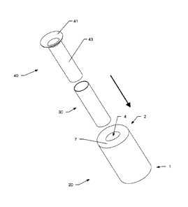
The stents of the kit may also have different shapes e.g. as shown in Fig. 12.
Here, the first blood flow reduction stent 20 has an hour-glass or barbell
shape
with two widened sections 21 on each side of a narrowed section 22. The
narrowed section defines the inner lumen 24 of the first reduction stent 20.
Again, the first reduction stent may be a self-expandable mesh. At least the
an
intermediate sections 23 between the narrowed and widened section
narrowed, and preferable the adjacent regions thereto, may be covered to
restrict blood flow through the mesh.
The second reduction stent 40 is in the form of a bottleneck with a widened
section 42 followed by a narrowed section 43 at its downstream end. The
narrowed section 43 forms the inner lumen 44 of the second reduction stent
40. The second reduction stent 40 may be a self-expandable mesh e.g. of
.. Nitinol (nickel-titanium alloy). At least the narrowed section 43 and an
intermediate section 45 between the narrowed and widened section may be
covered to restrict blood flow through the mesh. The intermediate section 45
CA 02970237 2017-06-07
WO 2016/096529 PCT/EP2015/078937
also provides the anchoring means 41 in order to hold the second reduction
stent 40 in place.
The first reduction stent may also have a bottleneck shape like the second
reduction stent (not shown). In this case first and second reduction stents
5 provided in a kit may be the same. The kit has then only one type of
reduction
stent with different inner diameters that may be used in the way of the first
and second reduction stents.
The three different stent types of Fig. 12 may be employed in the same way
as described with respect to Fig. 11.
10 In all embodiments of the kit, at least the narrowed section 22, 43 and
an
intermediate section 23, 45 (or anchoring means 41) between the narrowed
section 22, 43 and the widened section 21, 42 of the first and/or second
reduction stent 20, 40 may be covered with a biocompatible, plastic material,
e.g. an expandable polymer sheet, preferable ePTFE, to obtain impermeable
15 walls.
It is understood, that the kit of stents comprising a first reduction stent,
at
least one dilatation stent and at least one second reduction stent may be
regarded as a separate invention as well as the multi-lumen stent described
beforehand may be seen as a separate invention.
Reference Signs
1 proximal end
1' downstream end
2 distal end
2' upstream end
3 inner tube-like segment
3a meshed inner tube-like segement
3b cover
4 inner lumen / central lumen / passage lumen
CA 02970237 2017-06-07
WO 2016/096529
PCT/EP2015/078937
26
outer tube-like segment
5a meshed outer tube-like segement
5b cover
6 outer lumen / blocked lumen
5 7 cap-like segment
7a meshed cap-like segment
7b cover
8 inner surface
9 outer surface
10 main body
11 orifice
12 inner stent
13 outer stent
first reduction stent
15 21 widened section
22 narrowed section
23 intermediate section
24 central lumen / inner lumen / passage lumen
dilatation stent
20 34 inner lumen
second reduction stent
41 anchoring means
42 widened section
43 narrowed section
25 44 central lumen / inner lumen / passage lumen
CA 02970237 2017-06-07
WO 2016/096529
PCT/EP2015/078937
27
45 intermediate section