Note: Descriptions are shown in the official language in which they were submitted.
DEVICES AND METHODS FOR FRACTIONATED
PHOTOACOUSTIC FLOW CYTOMETRY
FIELD OF THE INVENTION
[0001] This application relates to systems and methods of non-
invasively detecting and imaging individual target objects in vivo using a
fractionated photoacoustic flow cytometry system and using a fractionated
optical
system with photoacoustic, photothermal, fluorescence, Raman, scattering, and
other analytical techniques. In particular, this application relates to a
fractionated
in vivo photoacoustic (PA) flow cytometer device and methods for detecting
individual circulating target objects in deep vessels by increased laser
energy in
the vessels while simultaneously keeping safe laser fluence without side
effects
(e.g., overheating, skin burning, and pain) in the superficial skin layer.
BACKGROUND
[0002] Despite significant progress in diagnostic techniques
(e.g.,
magnetic resonance imaging (MRI), positron emission tomography (PET), optical
and bio-assays), no clinically relevant method has yet been developed for in
vivo
real-time counting of individual normal and abnormal cells in blood
circulation. In
particular, despite in vivo clinical use of pulse oximetry and optical
coherence
tomography, none of these biophotonic instruments is able to count individual,
fast-flowing blood cells due to limited spatial and temporal resolution.
Although
fluorescent labeling in vivo shows promise for detecting flowing cells in
animal
models as a research tool, translation of this technology to humans is
problematic due to 1) the necessity to use fluorophores, most of which are
currently toxic, 2) undesirable immune responses to tags, and 3) the small
volume of blood (10-100 pL) that is sampled, because the technology assesses
only superficial microvessels with slow flow rates.
[0003] Laser-based PA spectroscopy and imaging currently are the
fastest-growing area of biomedical optics, providing higher sensitivity and
resolution in deeper tissues compared to other optical modalities. The
1
Date Recue/Date Received 2022-02-14
tremendous clinical potential of the viable PA-based techniques have been
successfully demonstrated in several trials in humans including diagnosing
breast tumors at a depth of 3 cm, and imaging blood vessels in deep tissue up
to
7 cm. Nevertheless, PA-based techniques have not yet proven to be suitable for
highly sensitive, rapid in vivo blood testing at the single-cell level.
Counting
individual rare normal and abnormal cells, aggregates, and many other objects
noninvasively in the blood vessels of a living organism with native cell flow
is an
exciting challenge that may enable revolutionary breakthroughs in early
disease
diagnosis including cancer, infection, and cardiovascular disorders by
analysis of
almost the entire blood volume.
[0004] A primary clinical goal is real-time multiparameter
monitoring
of blood composition at single cell or cell aggregate levels in 1-3-mm blood
vessels at a depth of 1-10 mm, which is well within the documented
capabilities
of PA-based methods that are capable of assessing deep (at least 1-2 cm, if
not
3-7 cm) and large (10-15 mm) human blood vessels. A need exists for a highly
sensitive and high-speed in vivo PA flow cytometry (PAFC) platform to assess
deep vessels.
[0005] The sensitivity of PAFC can be significantly improved by
increasing laser energy which, however, can damage the superficial skin layer
where energy is much higher (10-50 times) than in deeper tissues. Therefore,
there is a need for a system and method for analyzing a large portion of the
blood volume in vivo for rare target objects. A new PA schematic may decrease
the laser beam sizes leading to a decrease in the thermal relaxation time with
a
simultaneous increase in the number of beams with a certain spatial
configuration.
SUMMARY
[0006] Disclosed herein is a fractionated photoacoustic flow
cytometry (PAFC) system for the in vivo detection of target objects in a
biofluid
system of a living organism. The fractionated PAFC system may include a laser
system including at least one laser for providing at least one laser beam to
at
least one target object within the biofluid system; a fractionated optical
system
2
Date Recue/Date Received 2022-02-14
configured to separate the at least one laser beam into fractionated laser
beams
having a spatial configuration on the skin above the biofluid system of the
living
organism; and an acoustic system comprising at least one focused ultrasound
transducer for receiving more than one photoacoustic signal emitted by the at
least one target object in response to the fractionated laser beams.
[0007] The acoustic system may be a fractionated acoustic system
including more than one focused ultrasound transducer. Each focused ultrasound
transducer may be an independent amplifier for sending the photoacoustic
signal
received by each focused ultrasound transducer to a multichannel data
acquisition board. The multichannel data acquisition board may create traces
of
signals from the target object passing non-overlapped acoustic focal volumes
together covering the whole cross-section of the biofluid system. The at least
one
laser may be capable of providing more than one laser beam as fractionated
laser beams having a spatial configuration. The fractionated optical system
may
include an optical component selected from a non-transparent mask, a beam
splitter, an optical fiber array, a lens array, a microlens array, a mirror
array, a
diffraction element, a diffuser, a pinhole, and combinations thereof. The
fractionated laser beams from the fractionated optical system may not overlap
at
a location in the living organism with first temperature, pressure, or pain
receptors and wherein the fractionated laser beams spatially overlap at the
biofluid system. The spatial configuration of the fractionated laser beams may
include gaps of about 5 pm to about 1 cm between the individual laser beams on
the skin of the living organism. The spatial configuration of the fractionated
laser
beams may be one-dimensional or two-dimensional. The fractionated laser
beams may have a shape selected from circular, linear, strip, elliptical,
square,
and combinations thereof. The fractionated optical system may be configured to
scan the more than one laser beams across the biofluid system. Each focused
ultrasound transducer may include an acoustic focal volume and the
fractionated
acoustic system may be configured to scan the acoustic focal volumes across
the biofluid system. The focused ultrasound transducers may be focused
spherical ultrasound transducers. The laser system may include more than one
3
Date Recue/Date Received 2022-02-14
laser in a laser array. The more than one laser may be assembled in the laser
array as independent lasers or as microchip with individual emitters and a
triggering system for controlling the more than one pulsed lasers. The laser
system may include more than one laser providing laser pulses with different
wavelengths. The fractionated PAFC system may further include a triggering
system for providing time delays between the laser pulses with different
wavelengths.
[0008] Further provided herein is a fractionated PAFC system
which
may include a fractionated laser system including an array of more than one
laser for providing more than one laser beam in a spatial configuration to at
least
one target object within the biofluid system; an optical system configured to
deliver the more than one laser beam in a spatial configuration on the skin
above
the biofluid system of the living organism; and an acoustic system comprising
more than one focused ultrasound transducer for receiving more than one
photoacoustic signal emitted by the at least one target object in response to
the
more than one laser beam. In an aspect, the optical system may be non-
scanning or scanning.
[0009] This system may further include a triggering system for
controlling the more than one pulsed lasers and providing time delays between
laser pulses with different wavelengths. The system may further include a time-
resolved acoustic detection system. The acoustic system may be a fractionated
acoustic system including more than one focused ultrasound transducer. Each
focused ultrasound transducer may be an independent amplifier for sending the
photoacoustic signal received by each focused ultrasound transducer to a
multichannel data acquisition board. The focused ultrasound transducers may
be spherical or cylindrical and non-scanning or scanning. The laser array may
provide more than one laser beam as fractionated laser beams having a spatial
configuration. Each laser in the array may have a different wavelength. The
optical system may be a fractionated optical system configured to separate the
more than one laser beam into fractionated laser beams. The fractionated
optical
system may include an optical component selected from a non-transparent mask,
4
Date Recue/Date Received 2022-02-14
a beam splitter, an optical fiber array, a lens array, a microlens array, a
mirror
array, a diffraction element, a diffuser, a pinhole, and combinations thereof.
The
fractionated laser beams may not overlap at a location in the living organism
with
first pain receptors and wherein the fractionated laser beams spatially
overlap at
the biofluid system. The spatial configuration of the fractionated laser beams
may include gaps of about 5 pm to about 1 cm between the individual laser
beams on the skin of the living organism. The spatial configuration of the
fractionated laser beams may be one-dimensional or two-dimensional. The
fractionated laser beams may have a shape selected from circular, linear,
strip,
elliptical, square, and combinations thereof. The optical system may be
configured to scan the more than one laser beams across the biofluid system.
Each focused ultrasound transducer may include an acoustic focal volume and
the fractionated acoustic system may be configured to scan the acoustic focal
volumes across the biofluid system.
[0010]
Provided herein is a fractionated PAFC method for detecting
circulating target objects in a biofluid system of a living organism in vivo.
The
method may include providing the target object with a laser beam from a laser
in
a laser system at a first wavelength; separating the laser beam into
fractionated
laser beams in a fractionated optical system to form a spatial configuration
on the
skin above the biofluid system of the living organism; obtaining in a
fractionated
acoustic system more than one photoacoustic signal emitted by the circulating
target objects induced by the fractionated laser beams; and analyzing the
photoacoustic signals to calculate the combination of photoacoustic signals
emitted by the circulating target objects. The combination of photoacoustic
signals is characteristic of each circulating target object. The method may
further
include providing the target object with a second laser beam from a second
laser
in the laser system at a second wavelength. The method may further include
introducing time delay between laser pulses using a triggering system. The
method may further include color decoding by time-resolved detection of color-
coded photoacoustic signals. The method may further include generating
microbubbles or nanobubbles by the fractionated laser beams to detect the
Date Recue/Date Received 2022-02-14
circulating target objects with intrinsic photoacoustic contrast or the
circulating
target object labeled with a non-photoswitchable or photoswitchable
photoacoustic probe. The method may further include cooling the skin by
placing
clearing or cooling agents on the skin.
[0011] Provided herein is a fractionated PAFC method which may
include providing the target object with multiple laser beams from more than
one
laser in a laser array, each having a first wavelength; delivering the laser
beams
through an optical system and forming a spatial configuration of laser beams
on
the skin above the biofluid system of the living organism; obtaining in a
fractionated acoustic system more than one photoacoustic signal emitted by the
circulating target object induced by the fractionated laser beams; and
analyzing
the photoacoustic signals to calculate the combination of photoacoustic
signals
emitted by the circulating target object. The combination of photoacoustic
signals
is characteristic of the circulating target object. The method may further
include
separating the laser beams into fractionated laser beams in a fractionated
optical
system. The method may further include generating microbubbles or
nanobubbles when providing the circulating target object with the fractionated
laser beams.
[0012] While multiple embodiments are disclosed, still other
embodiments of the present disclosure will become apparent to those skilled in
the art from the following detailed description, which shows and describes
illustrative embodiments of the disclosure. As will be realized, the invention
is
capable of modifications in various aspects, all without departing from the
spirit
and scope of the present disclosure. Accordingly, the drawings and detailed
description are to be regarded as illustrative in nature and not restrictive.
BRIEF DESCRIPTION OF THE DRAWINGS
[0013] The following figures illustrate various aspects of the
disclosure.
[0014] FIG. 1 illustrates a fractionated photoacoustic (PA) flow
cytometry (PAFC) system that includes either a fractionated laser or laser
array
generating multiple beams of certain spatial profiles, or/and a fractionated
optical
6
Date Recue/Date Received 2022-02-14
system splitting of one or several laser beams from one or several lasers into
multiple beams, and a fractionated acoustic detection system using one or
multiple focused ultrasound transducers arrays with individual amplifiers
connecting to a recording system.
[0015] FIG. 2A, FIG. 2B, FIG. 2C, FIG. 2D, FIG, 2D, and FIG. 2E
provide[[s]] phenomenological schematics of conventional optical diagnostics
using relatively broad laser beams and new diagnostics in fractionated PAFC
using strongly focused beams with a small diameter.
[0016] FIG. 3A shows a phenomenological model for accumulative
thermal effects with a conventional broad laser beam at high laser pulse rate
(frequency) and FIG. 3B shows the absence these non-desired effects in
fractionated PAFC with a small diameter beam due to fast cooling of the laser-
heated absorbing zones.
[0017] FIG. 4A and FIG. 4B show main schematics of fractionated
PAFC with the fractionated laser beams allowing for dramatic improvement in
delivery of high laser energy in deep tissue without skin photodamage.
[0018] FIG. 5 illustrates a principle of a fractionated PA probe
with
integration of a fractionated laser beam with a fractionated acoustic
detection
system using multiple laser beams and focused transducers with non-
overlapping focal volumes covering the whole cross-section of a vessel.
[0019] FIG. 6 illustrates a combination of a focused laser beam
and
focused transducers in a fractionated PAFC with non-overlapping focal volumes
on the skin and into the vessel, respectively.
[0020] FIG. 7A, FIG. 7B, FIG. 7C, and FIG. 7D illustrate multiple
light beams spatial configurations in a fractionated PAFC system.
[0021] FIG. 8A, FIG. 8B, FIG. 8C, FIG. 8D, FIG. 8E, FIG. 8F, FIG.
8G, and FIG. 8H illustrate[[s]] different combinations of fractionated laser
beams
and transducers in a fractionated PAFC system.
[0022] FIG. 9 illustrates an example of a dashed linear laser
beam
on skin and near skin surface with two linear transducer arrays (e.g., 3
transducers in each array) located from both side of linear laser beam.
7
Date Recue/Date Received 2022-02-14
[0023] FIG. 10A and FIG. 10B illustrate a spatial configuration
of
ultrasound transducers in a linear array and on a spherical substrate,
respectively, allowing minimizing of the array's and substrate's sizes by
using the
transducers of small diameters with different focal lengths, and different
spatial
orientation.
[0024] FIG. 11 illustrates one fractionated laser for
fractionated
PAFC as a high-power laser diode with an active element including multiple
stacked bars.
[0025] FIG. 12A and FIG. 12B illustrate the optical system with
multiple mirrors providing fractionated laser beams.
[0026] FIG.13A is an illustration of optical system schematics
for
fractionated PAFC using a combination of cylindrical and spherical lenses.
FIG.
13B shows a typical linear laser beam image and its dimension.
[0027] FIG. 14A is an illustration of optical system schematics
for
fractionated PAFC using a combination of cylindrical and spherical lenses and
a
non-transparent mask. FIG. 14B is images of a laser mask for shaping a laser
beam obtained with the reflected light (top) and transmission microscopy
(bottom). FIG. 14C is an example of a laser spot created with the mask. FIG.
14D
is an example of a laser spot ("dashed" linear beam) created with the mask.
[0028] FIG. 15A illustrates optical system schematics with a
microlens array for creation of 1-D light distribution in a fractionated PAFC
system. FIG. 15B is a microlens array image. FIG. 15C is an example of laser
spots ("dashed linear beam").
[0029] FIGS. 16A-16C illustrate the images of laser beams for a
fractionated PAFC created with the microlens array in Fig. 15A. FIG. 16A shows
the light distribution on the focal plane. FIG. 16B shows the light
distribution
about 3 mm above the focal plane. FIG. 16C shows the light distribution in a
chess-board-like light distribution after rotation of the lens array.
[0030] FIG.17A illustrates optical system schematics for a
fractionated PAFC system with a microlens array for creation of 2-D light
distribution. FIG. 17B and FIG. 17C show laser spots for 300 pm and 150 pm
8
Date Recue/Date Received 2022-02-14
microlens arrays, respectively. FIG. 17D shows the light distribution above
the
focal point (1.5 mm for 150 um pitch lens array). FIG. 17E shows the light
distribution below the focal point (-3.0 mm for 150 um pitch lens array).
[0031] FIG. 18A illustrates an optical system schematic for a
fractionated PAFC with a diffuser (MultiDots array) configuration. FIG. 18B
shows an array of dots in the sample plane.
[0032] FIG. 19 illustrates an optical system for a fractionated
PAFC
using fast scanning of a fractionated linear beam across a vessel.
[0033] FIG. 20 illustrates an optical system for a fractionated
PAFC
system using a fiber array for delivery of multiple laser beams with fast
spatial
switching.
[0034] FIG. 21 illustrates an acoustic system for a fractionated
PAFC using a fast spatial scanning of ultrasound transducers across a vessel.
[0035] FIG. 22A and FIG. 22B illustrate a principle of a
fractionated
PAFC with two-beam time-of-flight mode.
[0036] FIG. 23 illustrates fractionated multicolor PAFC
schematics
with time-resolved color-barcoding.
[0037] FIG. 24 illustrates a signal-processing schematic diagram
for
a four-color fractionated PAFC system. Blocks on the left-hand side are
implemented by the digitizer in real-time, and the resulting spectral data are
saved. The remaining steps for acquiring PA traces are performed by a
workstation either in real-time or as post-processing. Peak analysis is
performed
on the full PA trace after the data acquisition is completed.
[0038] FIG. 25A is a flow chart illustrating signal processing in
four-
color fractionated PAFC system in the time domain. FIG. 25B is a flow chart
illustrating signal processing in the frequency domain. As an alternative
method,
a PA trace may be constructed using the spectral power of the PA waveforms as
illustrated in FIG. 25B instead of their peak-to-peak amplitudes as
illustrated in
FIG. 25A. This approach has advantages in certain cases. If the Fourier
transform is performed on the digitizer firmware and few representative
coefficients are transferred to the computer memory, the throughput is
9
Date Recue/Date Received 2022-02-14
significantly reduced. It can also improve signal-to-noise ratio (SNR),
especially
when there exist oscillating PA tails.
[0039] FIGS. 26A, 26B, and 26C illustrate multiplex
targeting/detection of biomarkers related to immune disorders. FIG. 26A
illustrates bio-barcoding using multicolor probes (nanoparticles) with
ultrasharp
PA resonances. FIG. 26B illustrates multi-color laser excitation with temporal
separation of laser pulses (i.e., time delay) with the different wavelengths.
FIG.
26C illustrates the time-resolved reading (decoding) of bio-color-coded PA
signals associated with the different markers.
[0040] FIG. 27A illustrates PA probes of a fractionated PAFC with
the focused cylindrical transducer having a central hole for lens or fiber-
based
delivery of a fractionated laser beam to skin. FIG. 27B shows a PA trace with
positive, negative, and combined signal contrast for red circulating emboli
[CE] or
melanoma CTCs, white CE, and white CE-CTC aggregates, respectively. FIG.
27C shows lateral resolution (-45 pm) of the cylindrical transducer is
represented
as a PA signal distribution from black type during single focused small (2 um)
laser beam scanning. FIG. 27D illustrates absorption spectra of RBCs,
melanoma, and platelets.
[0041] FIG. 28A illustrates a PA probes for a fractionated PAFC
with integrated optical and acoustic resolution using a focused cylindrical
transducer and cylindrical focusing fiber-based optics. FIG. 28B shows a
position
of cylindrical optical and acoustic focus in blood sample with melanoma cells.
[0042] FIG. 29 shows schematics of a fractionated PAFC system
for clinical applications.
[0043] FIG. 30A shows a clinical PAFC prototype. FIG. 30B shows
a PA probe with a cylindrical transducer (right) and a conventional ultrasound
probe (left). FIG. 30C demonstrates a time-resolved detection of PA signals
from
a human vein in the dorsum of the hand against PA signal from skin. FIG. 30D
illustrates a typical ultrasound image of examined vein in FIG. 30C. FIG. 30E
illustrates PA imaging of blood vessels.
Date Recue/Date Received 2022-02-14
[0044] FIG. 31A presents typical PA traces from melanoma CTCs
with positive contrasts in a cancer patient before signal filtration. FIG. 31B
presents typical PA traces from melanoma CTCs with positive contrasts in a
cancer patient after signal filtration. FIG. 31C presents typical PA traces
from
circulating emboli CE or clots before optimal signal averaging. FIG. 31D
presents
typical PA traces from circulating emboli CE or clots after optimal signal
averaging.
[0045] FIG. 32A presents a schematic of PAFC in vitro. FIG. 32B
demonstrates blood sample from melanoma patient with unusual high
concentration of CTCs, with fragments and large melanin aggregates in the
blood
plasma. FIG. 32C illustrates a PA signal trace from melanoma CTCs in a whole
blood in vitro. Inset, left: absorption spectra of melanin and blood. Inset,
right:
simultaneous 4-color detection of single CTC. FIG. 32C illustrates a typical
PA
signal trace from melanoma CTCs in WBC- rich samples without RBCs (lysed).
FIG. 32D illustrates a PA signal from melanoma CTCs. Inset, right top:
conventional flow cytometry data and image of MCSP+ cells labeled by Abs-PE.
Inset, right bottom; image of melanoma CTC and WBC after immune-staining.
[0046] FIG. 33A shows the image of linear laser beam in air
(laser
wavelength, 1060nm) with sizes of 8 pm x 1280 pm. FIG. 33B shows an image
of the same beam blurred to size of 72 pm x 1298 pm after propagation through
fresh mouse skin with thickness of 750 pm (transmission, 42.8%). FIG. 33C
shows an image of the same beam blurred to a size of 290 pm x 1320 pm after
propagation through double layer of fresh mouse skin with thickness of 1600 pm
(transmission, 29.2%). FIG. 33D shows an image of the same beam after
propagation through fresh mouse blood with thickness of 1800 pm):
(transmission, 9.2%). Fig. 33E illustrates a laser beam after 0.9 mm mouse
skin
(top) and signals from human 1-mm vein at depth of 1.3 mm (bottom) before
(left)
and after (right) optical clearing.
[0047] FIG. 34 is a graph summarizing an example PA trace with
different shapes from single and clusters of melanoma CTCs as well as emboli.
11
Date Recue/Date Received 2022-02-14
[0048] FIG. 35A is a PA signal trace from melanoma cells (C8161)
in human blood with a focused spherical ultrasound transducer with a focal
length of 6 mm and a lateral resolution of 45 pm. FIG. 35B is a PA signal
trace
from melanoma cells in human blood with a focused cylindrical ultrasound
transducer with a focal length of 6 mm and a lateral resolution of 45 pm.
[0049] FIG. 36A illustrates nonlinear PA signal amplification at
820
nm in melanoma cells (SK-MEL-1) with different pigmentation in static
conditions
as a function of laser energy fluence. FIG. 36B illustrates nonlinear PA
signal
amplification at 820 nm in melanoma cells (SK-MEL-1) with different
pigmentation in flow conditions as a function of laser energy fluence.
[0050] FIGS. 37A-37B illustrate examples of PA traces from moving
melanoma cells (B16F01) in mouse blood in 0.9 mm capillary tube at flow
velocity of 10 cm/s at laser fluence of 904 mJ/cm2 (FIG. 37A) and 33 mJ/cm2
(FIG. 37B) as modeling of fractionated and not fractionated PAFC at a laser
wavelength of 1060 nm, respectively.
[0051] FIG. 38A illustrates a TEM image of melanoma cell
(B16F10) with exosomes (arrows). FIG. 38B illustrates a dark field image of
melanoma cell (B16F10) with exosomes (arrows). FIG. 38C shows fluorescence
traces in 50-pm capillary tube with stained by PKH67 dye exosomes. FIG. 38D
shows PA traces in 50-pm capillary tube with stained by PKH67 dye exosomes.
FIG. 38E demonstrates a PA trace from 50-pm vessels in mouse ear after IV
injection of melanoma exosomes in a mouse tail.
[0052] FIG. 39A illustrates a dependence of pain threshold in
human hand on linear beam length at beam width of 6.5 pm at laser wavelength
of 1064 nm and pulse rate of 10 kHz. FIG. 39B illustrates a dependence of PA
signals on a linear beam length from a 1 mm hand vein at depth of 1.1 mm.
[0053] FIG. 40 is a PAFC schematic with a laser diode showing a
fragment of three fractionated beams (total 3 stacks in each 3 shown bars) and
a
temporal laser pulse shape with a width of 45 ns.
[0054] FIG. 41A is an image of a laser diode fractionated beam
including three strips. FIG. 41B is an image of a single melanoma cell
(B16F10,
12
Date Recue/Date Received 2022-02-14
dark spot) among mouse red blood cells in capillary with diameter of 100 um.
FIG. 41C is a typical PA signal from a single melanoma cell. FIG. 41D is a
graph
showing the dependence of PA signal amplitude from a melanoma cell on laser
diode pulse energy in vitro. FIG. 41E is a photo of a mouse with an ultrasound
transducer. FIG. 41F is a PA signal showing detection of a single circulating
melanoma cell in mouse abdominal blood microvessels.
[0055] FIG. 42A demonstrates schematics of integrated
fluorescence flow cytometry (FFC) and PAFC for controlled CTC release during
medical procedures. FIG. 42B shows a PA signal trace at pressure (-120 g)
impact on a 5-mm skin melanoma tumor (B16F10-GFP).
[0056] FIG. 43 illustrates schematics in vivo of an 8-color flow
cytometer integrating 4-color fluorescence flow cytometry (FFC) and 4-color
PAFC. DM, dichroic mirror; PMT, photomultiplier tube; F, filter; S, slit.
Inset: 4
linear beams.
[0057] FIG. 44A shows results of monitoring circulating bulk and
stem CTCs, WBCs and platelets, as well as stem CTC-clot aggregates in ear
vessels of a tumor bearing mouse using in vivo multicolor integrated flow
cytometry (FFC and PAFC) platform after IV injection of a PA and fluorescent
labeling cocktail. FIGS. 44B-E shows results of monitoring a vessel infected
with
malaria parasites RBCs with three color PAFC at 532 nm, 671 nm, and 802 nm.
FIG. 44B shows a principal optical schematic of malaria parasites detection by
PA and fluorescence flow cytometry (PAFFC) in linear mode. FIG. 44C presents
detection of infected RBCs and parasite expressing GFP in mice. FIG. 44D
illustrates light absorbance of hemozoin crystals. For GFP: EX-Absorption, Em-
Emission. Blood curve for approximately 70% of oxygenation (modified by
http://omIc.org). FIG. 44E demonstrates spectral identification of in vivo
linear PA
signals from hemozoins in infected with malaria parasites RBCs at three color
PAFC ( 532 nm, 671 nm, and 820 nm) above blood background.
[0058] FIG. 45 illustrates photoswitchable plasmonic gold
nanoclusters with light-sensitive links between individual nanoparticles.
13
Date Recue/Date Received 2022-02-14
[0059] FIG. 46A is a spaser schematic. FIG. 46B shows spaser
emission at 528 nm at different pump intensities at 488 nm. FIG. 46C is an
image
of a cancer cell with a spaser obtained with a lamp (cell background) and a
focused pump beam (bright sport). FIG. 46D is an image of a cancer cell in
blood. FIG. 46E is a fluorescence image below the spaser threshold (6 MW/cm2)
through 1.5 mm blood. FIG. 46F is a fluorescence image above the spaser
threshold (6 MW/cm2) through 1.5 mm blood.
[0060] FIG. 47 illustrates cooling effects with gel and water
between
transducers and mouse skin at 1060 nm, pulse rate of 10 kHz and energy
fluence of 100 mJ/cm2during 20 min of laser exposure with laser beam size of
6.5 pm x 790 pm.
[0061] FIG. 48 illustrates color-coding in multicolor
fractionated
PAFC using laser pulses with high rates and different wavelengths and the time
delays between corresponding laser pulses for fast spectral identification of
circulating melanoma cells in blood background.
[0062] Corresponding reference characters indicate corresponding
elements among the views of the drawings. The headings used in the figures
should not be interpreted to limit the scope of the claims.
DETAILED DESCRIPTION
[0063] Provided herein are systems and methods for the
improvement of blood tests for early diagnosis and prevention of
cardiovascular
disorders (e.g., stroke and heart attack), cancers, and infections (e.g.,
antibiotic
resistant bacteria or malaria) and which remain the main causes of death in
the
world with annual mortality particularly in the U.S of approximately 720,000,
580,000, and 140,000 people, respectively. The diagnosis of these and many
other diseases begins with a common medical procedure: examination of
extracted blood samples. The sensitivity of current blood testing is limited
by the
small volume of blood collected, in which no less than one disease-specific
marker (e.g., tumor cell and bacterium) can be detected. It can miss many
thousands of abnormal cells in the whole blood volume (-5 L in adults), which
can be sufficient for disease progression. As a result, barely treatable or
14
Date Recue/Date Received 2022-02-14
incurable disease complications may already be established by the time of the
initial diagnosis. For example, despite enormous efforts to detect circulating
tumor cells (CTCs) that lead to 90% of all cancer deaths as a result of the
development of deadly metastases, the mortality rates for metastatic cancer
have
still been significant. This failure is explained by the low sensitivity of
existing
CTC assays ex vivo, which, with a sensitivity of 1-10 CTCs/mL, miss up to
99.9% of CTCs in circulation. Likewise, in the case of cardiovascular
disorders,
one-third of people who die from heart attacks or stroke do not have the usual
risk factors such as family history, high blood pressure, or high cholesterol.
This
situation emphasizes the importance of the early detection of circulating
blood
clots (CBCs) called also emboli as precursors of large CBCs that cause the
final
fatal events.
[0064] The present application describes a clinically relevant,
noninvasive, universal platform for realizing the concept of in vivo reading
of what
is written in blood to improve the early diagnosis of, and potentially
prevent, life-
threatening diseases. Unlike typical blood sampling involving extraction of a
volume of blood ranging from 10 pL (drop) to a few milliliters (CTC assays),
in
vivo examination involves nearly the entire volume of blood passing through 1-
2-
mm-diameter peripheral vessels over 0.5-1 h (a few minutes in larger vessels)
and thus will enable a dramatic increase in diagnostic sensitivity, ultimately
up to
103-105 times, reflecting the ratio of the volume of blood sampled in vivo to
that
in vitro. In addition, the integration of simultaneous diagnosis and well-
timed
therapy¨theranostics¨can optimize therapy and control its efficacy.
[0065] The described diagnostic platform is relatively universal
and,
by either in label-free mode using intrinsic positive and negative contrast
agents
(e.g., melanin, hemozoin or platelet/fibrins/white blood cells [WBCs] for
melanoma, malaria or clot-related pulmonary embolism and stroke) or by
targeting of disease-specific markers with functionalized probes can be
applied to
the early diagnosis, prognosis, and prevention of many major diseases and
conditions, including stroke, heart attack, thrombosis, infections (e.g., S.
aureus,
E. coli, HIV, and malaria parasites producing hemozoin pigment), cancer,
Date Recue/Date Received 2022-02-14
Alzheimer's (through particle and exosome detection), sickle cell anemia, or
immune system dysfunction, as well as to the evaluation of blood chemistry.
This
will yield insights into blood epigenetics, hemodynamics, rheology, and red
blood
cell (RBC) aggregation.
[0066] The development of laser methods such as PA imaging,
optical coherent tomography (OCT), fluorescence spectroscopy and many others
has already revolutionized noninvasive optical disease diagnosis with focus on
superficial targets in dermatology, dentistry and ophthalmology. The assessing
of
targets deeper tissue, vessels and organs (e.g., brain, lung, or liver) is
challenging due to light scattering and absorption effects leading to
attenuation
laser energy and beam blurring. As a result, difficult to treat if not already
incurable disease complications (e.g., metastasis, sepsis, stroke) may be
developed at the time of initial diagnosis with the existing methods. Further
increasing the sensitivity of laser methods to assess deep tissue is needed to
provide diagnosis of fatal diseases at early stage when well-time therapy is
more
effective. As the optical signals from targets in many methods increase with
increasing laser energy, one of potential ways to increase the sensitivity of
these
methods is to improve delivery of high energy in deep tissue. It is believed
that
this approach is limited by possible photodamage of skin where laser energy is
much higher. Is also commonly accepted that maximum laser energy is regulated
by the laser safety standard establishing maximum permissible exposure (MPE).
In the spectral range of 500-1,100 nm, for nanosecond laser pulses, the MPE is
20-100 mJ/cm2, respectively, at a pulse rate f 10 Hz and drops at higher pulse
rates due to accumulative effects, for example to 0.1 mJ/cm2 at f=10 kHz at
1064
nm.
[0067] This safety standard establishes maximal laser power or
energy for safe laser applications in many areas from the public use (e.g.,
pointers and laser barcode scanners, laser shows, art holography), to science,
industry, and consumer electronic products. It is assumed that this standard
can
be applied for laser medical diagnosis. This is a reasonable requirement in
laser
imaging, eye's examination, or use pilot laser for scheme alignment in optical
16
Date Recue/Date Received 2022-02-14
medical instruments. Provided herein is a new concept of optical diagnostics
with
dramatically increased (up to 100-1000- times) sensitivity without violation
of a
current laser safety standard. It is achieved by replacing a conventional one
pulsed broad beam on many (up to 103-104) small laser beams of certain spatial
profiles allowing delivery much higher laser energy (103 -fold and in same
case
up to 105¨fold) in deep tissue. Moreover, without being limited to a
particular
theory, early laser diagnosis of fatal diseases such as cancer, infections,
and
cardiovascular disorder can be accompanied by nonessential skin alteration
only
without any the risk for humans. In the described methods and devices herein,
the energy fluences are still lower than those employed in many FDA-approved
laser cosmetic and therapeutic systems (up to 10 J/cm2) that have been broadly
used to treat blood vessel abnormalities (e.g., port-wine stains), skin
resurfacing
(e.g., wrinkle removal) or hair removal with no evidence of significant risk
that
that was confirmed during long (at least 30 years) application of laser
medical
devices.
[0068] A fractionated photoacoustic (PA) flow cytometry (PAFC)
system and methods for the in vivo detection of target objects in biofluidic
systems (e.g., blood, lymph, urine, serum, tear, or cerebrospinal fluid) of a
living
organism is described. The fractionated PAFC system may include a fractionated
laser system, a fractionated optical system, a fractionated acoustic system,
and
combinations thereof. The fractionated laser system includes at least one of a
laser or laser array for pulsing a target object within the circulatory vessel
with
fractionated focused laser beams. The fractionated optical system separates
one
or several laser beams into multiple beams in a spatial configuration on the
skin
above the circulatory vessel of the living organism. The fractionated acoustic
system includes multiple focused ultrasound transducers for receiving
photoacoustic signals emitted by the target object in response to the
fractionated
laser beams. The target objects have intrinsic photoacoustic contrast or may
be
labeled with photoswitchable or spaser-based probes. Fractioned beams may
also be used for diagnostics with other spectroscopic methods (e.g.,
fluorescence, Raman or scattering) and energy sources both coherent and
17
Date Recue/Date Received 2022-02-14
conventional such as lamp and LED in the broad spectral range from 10 A to 1
cm (e.g., X-ray, UV, visible, NIR or microwaves) in continuous wave and pulse
modes in the broad range of pulse duration from 10 ps to 1 ms.
[0069] Provided herein are various aspects of devices and methods
for fractionated PAFC with delivery of multiple (fractionated ) laser beams
with a
specific shape and gaps between the individual beams with high pulse rates and
different wavelengths to deep in vivo biotissue, in particular blood vessels,
for
early diagnosis of many diseases with focus on cancer, infections and
cardiovascular disorders by ultrasensitive detection using fractionated
acoustic
system of a single specific target object or marker or multiple markers in
vivo
using either intrinsic PA contrast agents or targeting of markers by
artificial PA
probes. The markers may be associated with normal cells and physiological
processes or may be associated with disease processes. In various aspects,
devices and methods for in vivo detection of individual cells using bio-
barcoding
and multi-color time-resolved detection of individual normal and abnormal
cells in
circulation in a subject in vivo are disclosed. In one aspect, the in vivo
detection
of the individual cells may be enabled using an in vivo fractionated PAFC
device
that may include multiple laser sources, optical systems for the delivery of
laser
radiation, PA probes, and a detection system.
[0070] The fractionated PAFC system provides for multiple beams
with a small diameter for a circular shape or width for a linear shape. In an
aspect, the individual laser beams may have one dimension of about 0.2 pm to
about 1 cm, in a preferred aspect, the beams may have one dimension of about
0.2 pm to about 200 pm. The small size of the beams may reduce thermal
relaxation time and heat spatial and temporal accumulation. It allows for
preventing overheating of superficial skin layers and accompanied pain while
simultaneously increasing laser energy in deep vessels to increase the
sensitivity
of PAFC and other optical methods (e.g. fluorescence, Raman, CARS, second
and third harmonic generation, multiphoton and others). Thus, the fractionated
PAFC and other methods may use specific beam shapes and spatial
configurations with specific the gaps between laser beams. Novel aspects of
the
18
Date Recue/Date Received 2022-02-14
devices and methods disclosed herein may include: fractionated delivery of
laser
radiation with multiple beams in PAFC; PAFC with a fractionated acoustic
detection system; integration of optical resolution (OR) and acoustic
resolution
(AR) in fractionated PAFC (OR-PAFC and AR-PAFC, respectively); multicolor
fractionated PAFC; bio-barcoding of multiple markers using narrow spectral
resonances followed by time-spectral reading (decoding); fractionated PAFC
with
photoswitchable PA probes; fractionated PAFC integrated with fluorescence flow
cytometry (FFC) using spaser as new super contrast multimodal multifunctional
probes and spasers; time-of-flight fractionated PAFC using two-beam and/or
multiple beams; and PA signal processing algorithms in multicolor and time-of
flight fractionated PAFC.
[0071] Physical and technical principles of fractionated PAFC are
based on irradiation of blood vessels with pulsed multiple beams followed by
the
detection of laser-induced acoustic waves (referred to as PA signals) from
individual target objects with a fractionated acoustic system using an
acoustic
focused ultrasound transducer array attached to the skin through a thin layer
of
water or ultrasound gel (for acoustic matching of the skin and transducer and
simultaneous skin cooling). In various aspects, the target object may be
circulating tumor cells (CTCs), such as melanoma, infections such as a virus
(HIV), bacteria, parasites (malaria), clots, and intrinsic (e.g., exosomes)
and
exogenous micro- and nanoparticles (NPs). The physical mechanism of the PA
method is associated with thermoelastic generation of acoustic waves by laser-
heated absorbing zones in target objects. According to thermal confinement,
absorption of a laser pulse with a width of tp at tp TT (TT, thermal
relaxation time),
for example by melanin or hemozoin as intrinsic PA contrast agents or by
functionalized artificial PA probes (e.g., plasmonic NPs, photoswitchable NPs,
or
spasers), leads to a maximal temperature increase (and maximal PA effects)
without the influence of heat loss due to thermal diffusion to surrounding
medium.
For spherical targets with radius R the thermal relaxation time may be
determined using Eqn. (I),
TT = R2/6.75k, Eqn. (I)
19
Date Recue/Date Received 2022-02-14
where k is thermal diffusivity. For R = 10 nm, 50 nm, and 5 pm, TT is about
160 ps, 4 ns, and 40 ps, respectively. The pulse width tp must also satisfy
acoustic confinement providing the generation of the maximum PA signal:
tp 2R/c, Eqn. (II)
where cs is the speed of sound. For RDTD = 12 pm (size of a whole cell)
and Rm = 0.3 pm (size of one melanosome in a melanoma cell), tp 10 ns and
400 ps, respectively. Each target object may be exposed at a laser pulse-
repetition rate, fr (VF)/d, where VF is blood flow velocity and d is the width
of the
laser beam or acoustic resolution of PAFC. In a 1-3-mm-diameter human vein,
VF is -5-15 cm/s, and ford = 50-100 pm, fr 0.5-2 kHz. Increases in fr improve
the signal-to-noise ratio (SNR). The SNR may be determined by the ratio of
flash
(transient) PA signals from single target objects to superposed background PA
signals from red blood cells (RBCs) in the detection volume, as well as to
noises
of different origins (e.g., electronic, acoustic, fluctuation in RBC number,
or laser
energy instability).
[0072] Laser-based devices may be capable of examining a much
larger volume of blood in vivo compared to conventional diagnostic techniques
involving the ex vivo examination of small samples. Such devices may exploit
the
well- established physiological fact that almost the complete volume of blood
in a
human adult (i.e., 5 liters) passes through peripheral blood vessels with
diameters of 2-3 mm within 0.5-1 hours. In a larger vessel, such as jugular
vein
or carotid artery (10-15-mm diameter), total circulation time may be on the
order
of 5-10 minutes or less. The examination of the entire blood volume of a
subject
may reduce diagnostic errors such as false positivity and false negativity for
rare
events. In addition, the detection limit may be significantly reduced,
resulting in a
threshold of sensitivity as low as 1 cell of interest (biomarkers) in 100 ml
of blood
(at least 100-fold more sensitive than existing assays, based on ratio of
sample
volume in vivo and in vitro methods) or one in 500-1000 m L.
[0073] The use of a laser-based device to conduct blood testing
of
the whole blood volume of a subject in vivo with greatly enhanced sensitivity
may
shift paradigms of the clinical role of blood tests from disease staging
and/or
Date Recue/Date Received 2022-02-14
assessment of therapy efficiency to early disease diagnosis¨hypothetically
before disease progression to an untreatable stage or at least to clinical
symptoms. Therefore, application of a well-timed, more effective, and
personalized therapy, in particular PT therapy, guided by real-time abnormal
cell
counting may be enabled by such a device.
I. Fractionated photoacoustic flow cytometry system
[0074] The sensitivity of most optical methods may be improved by
increasing laser energy, as optical signal amplitudes are often proportional
to
laser energy fluence (pulse mode)/intensity (continuous wave (CW) mode). An
increase in laser energy on the skin is believed to be limited by the maximum
permissible exposure (MPE) of skin. In the spectral range of 500-1,100 nm, for
nanosecond laser pulses at a rate f 10 Hz, the MPE for skin is 20-100 mJ/cm2,
respectively, and the MPE is lower (0.1-1 mJ/cm2) at higher pulse rates of 1-
10
kHz. However, RBCs and WBCs in the NIR range (800-850 nm) have high
photodamage thresholds at the level of 10-20 J/cm2 and 50-100 J/cm2,
respectively, which is 1,000-fold higher than the MPE. Moreover, the laser
safety
standard was introduced on the basis of a 3.5-mm-diameter laser beam. The
adverse effects at high laser pulse rates may be associated with temporal and
spatial overlapping of thermal effects in the irradiated volume. Therefore,
the
laser beam radius may be decreased, leading to a decrease in the thermal
relaxation time 'CT ¨ R2 and to reduce these thermal effects.
[0075] Provided herein is new concept of optical diagnosis in
vivo
using multiple small-diameter laser beams for the fractionated delivery of
higher
laser energy fluence levels (up to 100-1000 times or higher) to deep vessels
without side effects and a corresponding fractionated PAFC device. In an
aspect,
the vessels to be imaged/monitored may be about 0.5 mm to about 50 mm deep
below the skin, and even deeper with fractionated PAFC. For example, the
vessels may be a vein in the hand (about 0.5-3 mm deep) or a jugular vein or
carotid artery (about 15-20 mm deep). In one aspect, the vessels may be about
1 mm to about 5 mm deep. There may be no side effects because the laser
21
Date Recue/Date Received 2022-02-14
energy would not be averaged and hence heat may not accumulate at a lower
depth because the gaps between the individual laser beams (FIGS. 4A-4B and
7A-9) that prevent heat diffusion from one small beam to another during a
short
laser pulse. Thus, the gaps between beams prevent heat increase in the
superficial skin layer where the first temperature, pressure, and/or pain
receptors
are located (FIGS. 4A-4B). In various aspects, the first pain receptors may be
located at a depth below the skin of about 200 pm to about 400 pm. The shorter
thermal relaxation time for a smaller-diameter laser beam allows for
overcoming
the limitations of relatively large laser beams with higher relaxation time.
In an
aspect, in fractionated PAFC, each laser pulse leads to a short temperature
increase in the irradiated volume. Before the next pulse is delivered, heat
dissipates and the heated volume quickly cools (because of fast thermal
relaxation to about almost the initial temperature level or exceeds a little
of this
level by a few percent (FIG.3B)). Thus the heat dissipation out of the laser
volume after each laser pulse leads to only a non-significant average
temperature increase. On the contrary, for large laser beams with longer
thermal
relaxation times (FIGS. 2D and 3A), accumulation of heat in irradiated zone
leads to quick overheating of the surrounding zone and adverse high
temperature-induced effects such as cellular protein denaturation and
coagulation, skin surface burning, and the feeling of pain.
[0076] The sensitivity may be increased by increasing the laser
energy in deep tissue. This may be achieved not by increasing the energy in
one
beam but increasing the number of beams and keeping the energy of each beam
below the skin damage or pain threshold. Thus, an increase in energy of only
one beam would lead to increased heat in the irradiated superficial skin
layer,
while fractionated beams would reduce the heat in the irradiated superficial
skin
layer. FIG. 3A shows a phenomenological model for accumulative thermal effects
with a conventional broad laser beam at high laser pulse rate (frequency) and
FIG. 3B shows the absence these non-desired effects in fractionated PAFC with
a small diameter beam due to fast cooling of the laser-heated absorbing zones.
The significant blurring (extending) beam diameter in deep tissue due to light
22
Date Recue/Date Received 2022-02-14
scattering by tissue may lead to overlapping blurred laser beams at some
specific depth only (FIG. 4A), and result in an increased laser fluence within
deep
vessels with increasing beam number N (FIG. 4B). The overlapping of the
blurred laser beams at the vessels at a specific depth may be determined by
the
appropriate calculation of gaps between laser beams and dependence of
adverse effects (e.g., pain) and PA signal amplitude on laser beam parameters,
for example on the length of a linear beam at a fixed width (FIG. 39).
[0077] A fractionated PAFC system may provide enhanced
sensitivity for detection of target objects in deep vessels. In an aspect, the
fractionated PAFC system may integrate fractionated delivery of laser energy
with use of a "fractionated" single laser generating multiple beams (FIGS. 11
and
40), a laser array (i.e., several single beam lasers) with specific super-
position of
individual beam (e.g., FIG. 12B), and/or a fractionated optical system (FIGS.
12-
20) creating multiple beams of various spatial configurations. In some
aspects,
the fractionated PAFC system may further include a fractionated acoustic
detection system (FIG. 5) and various combinations with a fractionated optical
system (FIGS. 6, 8A-8H, 9, 10A-10B, and 28A). FIG. 5 illustrates a principle
of a
fractionated PA probe with integration of a fractionated laser beam with a
fractionated acoustic detection system using multiple laser beams and focused
transducers with non-overlapping focal volumes covering the whole cross-
section
of a vessel. FIG. 6 illustrates a combination of a focused laser beam and
focused
transducers in a fractionated PAFC with non-overlapping focal volumes on the
skin and into the vessel, respectively. A fractionated acoustic detection
system
including a focused spherical ultrasound transducer array may provide
detection
of circulating target objects in a whole cross-section of large vessels with a
high
signal-to-noise (SNR) because there may be minimal signal background from
RBCs in the smaller focal volume of each transducer (FIG. 35A). As illustrated
in
FIGS. 2B, 2C, 3B, 4A, and 4B, the goal of the fractionated PAFC is to enhance
the laser energy fluence in deep vessels while keeping the safe level of
energy in
the superficial skin layer within about 200-300 pm where the temperature and
pain receptors are located. Conventional flow cytometry (FC) in vitro uses
linear
23
Date Recue/Date Received 2022-02-14
beam shapes allowing for monitoring all the cells in the flow tube vessel. The
same linear beam shape may be used with in vivo PAFC to provide detection of
all cells in the blood vessel cross-section. However, increasing PAFC
sensitivity
by increasing laser energy in a linear beam may lead to high energy fluence in
the superficial skin layers exceeding either the laser safety threshold or
pain
threshold. Fractionated laser beams may overcome these problems.
[0078] In an aspect, a fractionated PAFC system for the in vivo
detection of target objects in a biofluid system or a circulatory vessel of a
living
organism is disclosed. In various aspects, the fractionated PAFC system may
include at least one of a fractionated laser system, fractionated optical
system, or
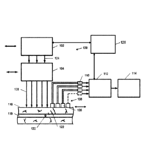
fractionated acoustic system. FIG. 1 illustrates a fractionated PAFC system
that
includes either a fractionated laser or laser array generating multiple beams
of
certain spatial profiles, or/and a fractionated optical system splitting of
one or
several laser beams from one or several lasers into multiple beams, and a
fractionated acoustic detection system using one or multiple focused
ultrasound
transducers arrays with individual amplifiers connecting to a recording
system.
As illustrated in FIG. 1, the system 100 may include a fractionated laser
system
102, a fractionated optical system 104, and a fractionated acoustic system
106.
The fractionated laser system 102 may include at least one pulsed laser for
pulsing at least one target object 120 within the circulatory vessel 118 with
at
least one pulse of laser energy 124. The fractionated optical system 104 may
be
configured to separate the at least one pulse of laser energy 124 into more
than
one laser beam 108 in a spatial configuration on skin above the circulatory
vessel
of the living organism. As also seen in FIGS. 5, 6, 8A-8H, and 9, the
fractionated
acoustic system 106 may include more than one focused ultrasound transducer
126 for receiving more than one photoacoustic signal 122 emitted by the at
least
one target object 120 in response to the more than one laser beam 108. In one
aspect, as illustrated in FIGS. 5 and 10A-10B, the fractionated acoustic
system
may include multiple ultrasound transducers on each side of the laser beams or
on a sem isphere with a central hole for delivery of the laser beams. For
24
Date Recue/Date Received 2022-02-14
example, the fractionated acoustic system may include about 3-5 ultrasound
transducers on each side of the laser beams as seen in FIG. 9.
[0079] The fractionated PAFC system may further include a
recording system 112 for recording the combination of photoacoustic signals
emitted by the at least one target object in response to the more than one
pulse
of laser energy. In one aspect, the recording system 112 may be a multichannel
data acquisition board. Each focused ultrasound transducer 126 may have an
independent preamplifier 110 for sending the photoacoustic signal 122 received
by each focused ultrasound transducer 126 to a multichannel data acquisition
board. At least one pulse of laser energy of the at least one pulsed laser 102
may have a wavelength from ultraviolet to radio wave in the range of about 200
nm to about 1 cm. The laser system 102 may include an array of more than one
pulsed laser. In an aspect, each laser in the laser array may have a different
wavelength for use in multicolor fractionated PAFC (FIG. 23). The system 100
may further include a triggering system 128 for controlling the more than one
pulsed lasers, synchronization of the laser pulses, and/or the time-resolved
recording system. In another aspect, the triggering system 128 may control the
spatial scanning of the laser 102, the fractionated optical system 104, or the
fractionated acoustic system 106. In various aspects, the fractionated laser
system 102, fractionated optical system 104, and/or fractionated acoustic
system
106 may scan independent from each other. In another aspect, the systems may
be synchronized to scan together. In various aspects, the triggering system
128
may communicate with the laser system 102, the recording system 112, and
combinations thereof.
[0080] In an aspect, the more than one laser beams 108 from the
fractionated optical system 104 may not overlap at a location in the living
organism with the first pain receptors. The more than one laser beams 108 may
spatially overlap at the circulatory vessel 118. The spatial configuration of
the
laser beams 108 may include gaps between the individual laser beams 108 on
the skin 116 of the living organism. The gaps may be about 5 pm to about 200
pm. As illustrated in FIGS. 7A-7D and 8A-8H, the spatial configuration of the
Date Recue/Date Received 2022-02-14
laser beams 108 may be one-dimensional (FIG. 7A and 7B) or two-dimensional
(FIG. 7C and 7D). The fractionated optical system 104 may include an optical
component for controlling the shape and number of laser beams. The optical
component may be selected from a non-transparent mask, a beam splitter, an
optical fiber array, a lens array, a microlens array, a mirror array, a
diffraction
element, a diffuser, a pinhole, and combinations thereof. The shape of the
laser
beams 108 may be selected from circular, linear, strip, elliptical, square,
and
combinations thereof. For example, FIG. 7A and FIG. 7C illustrate circular
beam
dimensions and FIG. 7B and FIG. 7D illustrate linear beam dimensions. The
laser system, optical system, and the acoustic systems may independently be
non-scanning or scanning. The fractionated optical system 104 may be
configured to scan the more than one laser beams 108 across the circulatory
vessel 118. Each focused ultrasound transducer 126 may have an acoustic focal
volume that does not overlap or partially overlap to cover the whole blood
vessel
cross-section (FIG. 5). The fractionated acoustic system 106 may be configured
to scan the acoustic focal volumes across the circulatory vessel. The focused
ultrasound transducers 126 may be focused spherical ultrasound transducers in
one aspect. Multiple beams are used in laser materials processing, optical
communications, optical image processing, microelectronics, and laser
treatment. However, the described multi-beam schematics have been never used
in PAFC, which brings new unpredictable effects. To use multiple beams with
PAFC, there is a need to increase the laser energy in deep tissue without
damaging the surface layers. On the contrary, in known laser treatment with
multiple beams the main goal is to damage the surface layer, which is not
appropriate for safe laser diagnostics in medical fields.
[0081] In an aspect, a method for detecting a circulating target
object in a circulatory vessel of a living organism may include pulsing the
target
object with a pulse of laser energy from a pulsed laser in a laser system at a
first
pulse wavelength, separating the pulse of laser energy into more than one
laser
beam in a fractionated optical system to form a spatial configuration on the
skin
above the circulatory vessel of the living organism, obtaining in a
fractionated
26
Date Recue/Date Received 2022-02-14
acoustic system more than one photoacoustic signal emitted by the circulating
tumor cell induced by the more than one laser beams, and analyzing the
photoacoustic signals to calculate the combination of photoacoustic signals
emitted by the circulating target object, wherein the combination of
photoacoustic
signals is characteristic of the circulating target object. The method may
further
include pulsing the target object with a second pulse of laser energy from a
second pulsed laser with a different wavelength and time delay compared to the
pulse from the first laser, as seen in FIGS. 26 and 48. The method may further
include generating microbubbles or nanobubbles around intrinsic (e.g. melanin
of
hemozoin NPs) or artificial probes (e.g., plasmonic and/or photoswichable NPs)
when pulsing the circulating target object with the laser pulse with increased
energy in the fractionated PAFC laser energy that leads to PA signal
enhancement (FIG.36).
[0082] In fractionated PAFC, decreasing the laser beam diameter
to
a few micrometers may significantly reduce the risk of photothermal (PT) -
induced superficial skin damage because of the consequent decrease in thermal
relaxation time, and hence heat accumulation, especially at a high pulse rate
(FIGS. 2B and 3B). In an aspect, increased PAFC sensitivity may be achieved
by increasing the laser energy fluence without adverse effects by using
fractionated delivery of laser energy via multiple laser beams. Various
optical
components may be used to create an array of laser beams. The optical
components may include microlens arrays, diffusers, pinholes, and optical
masks
(FIGS. 12-20). In various aspects, the laser beam array may be 1-D arrays of
multiple small-diameter laser beams, with varying spacing between them and at
different energy fluences in individual beams (FIGS. 7A-7D and 8A-8H).
[0083] Non-limiting examples of laser beam arrays include various
numbers of laser beams, such as 1x10, 1x30, 10 x10 or 20 x20. The laser
beams may have a cross-sectional shape of circular, linear, or elliptical. In
an
aspect, the diameter of the laser beams may range from about 0.25 pm to about
20 pm. Circular fractionated laser beams may have an individual diameter of
about 200 nm to about 100 pm. The width of dashed linear fractionated laser
27
Date Recue/Date Received 2022-02-14
beams may range from about 200 nm to about 200 pm. In various aspects, the
diameter of the laser beams may range from about 0.25 pm to about 1 pm, from
about 0.5 pm to about 5 pm, from about 3 pm to about 6 pm, from about 5 pm to
about 10 pm, from about 7 pm to about 12 pm, from about 10 pm to about 15
pm, from about 12 pm to about 17 pm, and from about 15 pm to about 20 pm. As
illustrated in FIG. 4A, the laser beams may be spaced apart at the skin such
that
the beams do not overlap on the skin or at the first pain receptors but do
overlap
at the vessel, which may be at a depth of greater than about 500 pm in one
aspect. In an aspect, the optical parameters may be optimized to avoid
overlapping of optical and thermal fields from each beam at the depth of the
first
pain receptors (200-400 pm), where laser energy is still high (maximal) before
attenuation in tissue, with simultaneous spatial overlapping of attenuated
light
energy at the depth of the vessels (greater than about 500 pm). In an aspect,
the
spatial configuration of the laser beams includes gaps between the beams and
the gaps may range from about 5 pm to about 1 cm. In various aspects, the
gaps may range from about 5 pm to about 25 pm, from about 20 pm to about 50
pm, from about 40 pm to about 100 pm, about 75 pm to about 125 pm, about
100 pm to about 150 pm, about 125 pm to about 175 pm, and about 150 pm to
about 200 pm. The fluences of the individual laser beams may be about 0.02
J/cm2to about 20 J/cm2. In various aspects, the fluences may range from about
0.02 J/cm2 to about 0.2 J/cm2, from about 0.1 J/cm2 to about 1 J/cm2, from
about
0.5 J/cm2 to about 10 J/cm2, from about 5 J/cm2 to about 15 J/cm2, and from
about 10 J/cm2 to about 20 J/cm2. The total fractionated laser beam area may
range from about 50 pm to about 20 mm.
[0084] The fractionated PAFC system provides for a dramatic
increase (10-100-fold, if not more) laser energy level at a depth of about 1-3
mm
up to about 10-15 cm without significant risk for harmful effects in the
superficial
skin area where the laser energy is still high before being redistributed
(blurred)
and attenuated in deeper tissue due to light scattering and absorption. The
effects of increasing laser energy is more profound in deeper tissue due to
more
28
Date Recue/Date Received 2022-02-14
effective overlapping (superposition) of larger blurred beams and the
possibility
of using a higher number of laser beams on a relatively large skin surface
area.
[0085] Fractionated PAFC can have positive and negative PA
contrasts. In general, laser irradiation of blood vessels creates constant PA
background signals associated with absorption by hemoglobin (Hb) in the many
RBCs in the detection volume. In positive-contrast mode, when melanoma CTCs
or red (Hb-rich) circulating emboli (CE) with higher absorption than the RBC
background pass through the irradiated volume, localized absorption
transiently
increases, resulting in a sharp positive PA peak (Fig. 27B). In the negative-
contrast mode, when white CE consisting of platelets, fibrin, or WBCs with
lower
(at least one-two orders of magnitude) absorption than the blood background
(Fig. 27D) pass through the detection volume (Fig. 27A), a decrease in
localized
absorption results in a sharp negative PA peak (Fig. 2B). Mixed white-red CE
or
white CE with CTCs produce a pattern of positive and negative signals (Fig.
27B). Two-color PAFC (Fig. 27A, inset, right) can distinguish red CE and
melanoma CTCs because the distinctive absorption spectra of Hb and melanin
(Fig. 27D) yield specific PA signal ratios.
[0086] Negative contrast (AP-/P) depends on CE and blood
absorption, vessel diameter (dv), volume of CE (VcE), and the detection volume
(VD) for a focused cylindrical transducer, VD Ad x Trdv2/4. A minimum
detectable
CE size (dcE)min (VcE)"3 can be estimated as (dcE)min APN/P x (VD)113, where P
and LPN are PA signal amplitude and fluctuation (Fig. 27B), respectively. P is
proportional to the number of RBCs (n) in the detection volume (e.g., n 220 at
dv = 50 pm and a hematocrit of 35-40%). In small vessels 20 pm, LPN is
determined by random changes in the number of RBCs in the detection volume,
while in larger vessels APN is determined rather by instability of the laser
pulse
energy (typically 3-5%), electrical and acoustic noise, vibration, or
physiological
rhythms (e.g., heart beating or breathing). For APN/P - 0.05, Ad = 50 pm, and
dv
= 50 pm and 1 mm, (cIcE)min 5-10 pm and 30-50 pm, respectively. These
estimations are in line with the experimentally achieved threshold of 12-20 pm
for small vessels. The duration of transient negative PA signals is short (10-
3-10-
29
Date Recue/Date Received 2022-02-14
4 s), while noise fluctuation and motion artifacts lie in the low-spectral-
frequency
range of <100 Hz. This may allow use of filtration and averaging to
significantly
(at least 5-10fold) reduce the influence of these factors (FIGS. 24, 25, and
31)
and monitor human vessel with a stable signal base over a few hours.
II. Fractionated laser sources
[0087] Fractionated laser beams may be created by the use of at
least one laser with multiple beams and/or a laser array of more than one
laser
which may generate multiple laser beams having a certain spatial
configuration.
In an aspect, a laser system may include a single pulsed laser diode to
produce
fractionated laser beams. The single pulsed laser diode may have high peak
power of about 200 W to about 800 W, a pulse energy up to about 5-20 pJ at 15-
100 ns pulse duration, and wavelengths in broad spectral range from about 640
nm to about 1600 nm. A high power laser diode may be composed of many bars
and stacks of active elements, as seen in FIG. 41, which can emit many
individual beams. This figure indicates only small fragment of a laser diode,
with
one stack including three bars. In fractionated PAFC, many more stacks and
more bars may be used (FIG. 7D). In an aspect, a laser diode used in PAFC may
include up to about 3-10 stacks and up to about 5-20 bars. The beams from a
pulsed laser diode, after passing through an optical system, may be directed
as
parallel or multiple focused beams to the skin above selected vessels. In an
aspect, the optical system may include a collimator or a focusing lens.
[0088] In various aspects, the laser beams may have at least one
dimension of about 0.2 pm to about 1 cm. The gaps between the laser beams
may range from about 5 pm to about 1 cm. The fractionated laser beams may be
one-dimensional or two-dimensional in configuration. In an aspect, the
individual
laser beams may have a shape selected from circular, linear, strip,
elliptical,
square and combinations thereof.
[0089] These fractionated beams may generate photoacoustic
signals from moving target objects in deep vessels with diameters of about 0.5
to
about 5 mm. Besides an increase in sensitivity, the shape of the array of
laser
beams may result in an appearance of consequent trains of PA signals produced
Date Recue/Date Received 2022-02-14
by the same target objects crossing the individual strips of beams if there
are not
overlapped superficial microvessels with a diameter of about 10-30 pm at a
depth
of about 30-100 pm (e.g., in a mouse ear). Knowing the time interval between
the signal trains and the distance between the strips in the focal spot, it
may be
possible to calculate the target object's velocity. The time interval between
two
consequent PA pulses may be measured and corresponds to the time of flight of
the targets between two strips in the focal spots. At a depth of about 500 pm
or
more, light scattering leads to blurring and spatial overlapping of laser
beams
that does not allow for effective use of the time-of-flight technique with
optical
resolution (OR-PAFC). However, summing of laser energy within deep blood
vessels provides the required increase in PAFC sensitivity.
III. Fractionated Optical System
[0090] In an aspect, the system may include a fractionated
optical
system for creating multiple laser beams from a pulse of laser energy from at
least one pulsed laser. The single or multiple beams may be easily separated
with a mirror array of various spatial configurations (FIG. 12A and 12B). A
linear
beam shape may be created by using a telescope to expand the laser beam after
laser with a combination of cylindrical and spherical lenses, as illustrated
in FIG.
13A. A dichroic mirror may be used to deflect a pilot beam laser for
triggering
data acquisition hardware and control laser energy fluctuation. The shaped
laser
beam may be focused into the sample using an objective. For example, a 10x
objective with an NA 0.3, working distance of 16 mm, and infinity corrected
may
be used. The size of the laser beam spot may be measured by a custom
microscope in transmission configuration by projecting the laser beam on to a
microscope calibration ruler to measure exact beam dimensions, as shown in
FIG. 13B.
[0091] Conversion of a linear laser beam into a line of
individual
laser spots may be performed using a non-transparent mask and /or an array of
microlenses. In an aspect, a laser mask (nontransparent barrier on the laser
beam path) may be used to create the required spatial distribution of the
laser
energy in the skin, as illustrated, for example, in FIG. 14A. In one aspect,
the
31
Date Recue/Date Received 2022-02-14
mask can be created by assembling several 40 pm steel wires into a regular
pattern on a flat glass plate, as shown in the images in FIG. 14B. The mask
may
be placed into the focal point between cylindrical and spherical lenses of the
optical system and block part of the laser light. In an aspect, the mask
transmission after the objective may be around 70%. The total laser power may
be required to be increase to compensate for the losses from the mask. The
dash period may be measured as a combined length of bright and dark parts.
[0092] In an aspect, a microlens array may be used to spatially
redistribute the energy of the laser beam in the fractionated optical system.
A
microlens array may be placed into the focal point between cylindrical and
spherical lenses and interact with a linear beam shaped by a cylindrical lens,
as
illustrated in FIG. 15A. Thus, only one column of microlenses (FIG. 15B), may
shape the beam, allowing to preserve the width of the laser beam in the
sample.
Laser spots in the sample may have a circular shape allowing it to concentrate
laser energy in these areas compared to the regular linear beam shape (FIG.
15C). The laser beam in the sample may be sensitive toward array orientation.
In various aspects, the optical system may include microlens arrays with a
pitch
between micro lenses of about 150 and about 300 pm. While smaller pitch
means smaller dash period (higher number of laser spots along the laser line)
it
also may result in slightly wider line due to the fact that two or more lens
columns
may interact with the beam. Better results (narrower laser beam and more
control
over laser beam parameters) may be achieved with the use of cylindrical lens
arrays having an appropriate pitch size between cylindrical lens elements.
[0093] FIG. 15A illustrates optical system schematics with a
microlens array for creation of 1-D light distribution and in some cases
"narrow"
2-D distribution (FIGS. 16A-16C). FIG. 16A shows the light distribution on the
focal plane. FIG. 16B shows the light distribution about 3 mm above the focal
plane. FIG. 16C shows the light distribution in a chess-board-like light
distribution
after rotation of the lens array.
[0094] The fractionated optical system may create a 2-D light
distribution of laser beams. In an aspect, the fractionated optical system may
32
Date Recue/Date Received 2022-02-14
allow a microlens array to interact with a large laser beam and thus, create
numerous laser spots in the sample separated by distances of only several
micrometers. In this case, microlens arrays may produce an image of the pump
beam into its image plane that was transferred into the sample by objective
conjugated to the same image plane (FIG. 17A). The use of a cylindrical lens
may allowed for the combination of multiple laser spots into a single general
line
accompanied by two additional lines created by the diffraction of the light.
In
general, 150 and 300 pm microlens arrays may create similar a distribution of
the
laser energy in the sample (FIG. 17B and FIG. 17C). However, the width of the
laser line may be better for a 300 pm array. FIG. 17D shows the light
distribution
above the focal point (1.5 mm for 150 um pitch lens array). FIG. 17E shows the
light distribution below the focal point (-3.0 mm for 150 um pitch lens
array).
[0095] To dramatically increase the number of individual focal
engineered laser diffusers may be used in the fractionated optical system
(FIG.
18A). A laser diffuser diffuses light by producing a diffraction picture from
the
numerous small optical elements on its surface. Thus, compared to microlens
array it can produce much higher number of focal spots as more lens-like
elements interact with light. However, the diffuser has low stability in high
power
laser beams (usually diffusers are based on plastic materials) and may have a
noisier picture due to presence of small scattering artifacts in its
structure. A
diffuser may be inserted into a large size laser beam expanded with two 2x and
one 3x telescopes to maximize the area of contact. The light right after the
diffuser may be collected by a 50 mm lens placed exactly at 50mm after the
diffuser. Thus, this lens and a 10X objective after it may create an image of
the
diffuser in the sample plane (FIG. 18B).
[0096] The resulting distribution of laser beams may be about 2.5
mm in diameter with spacing between dots of about 100 pm. The diameter and
spacing between the dots may be controlled by translation of a spherical lens
and accounting for the changes in the focal length of the system (laser
focusing
in the sample). In an aspect, the spherical lens may be a 50 mm spherical
lens.
In various aspects, the laser beams may have a range of distances from 50 to
33
Date Recue/Date Received 2022-02-14
130 pm. In one example, the energy fluence in the case of a MicroDots array
may be estimated using the following approximation: beam diameter of 8 um;
number of individual identical beams, 24 x24=576. All the light energy may be
equally distributed only through these beams. Thus, for a 300 pJ laser pulse
at
1064 nm, a laser fluence of about 0.3 J/cm2 may be achieved in the center of
the
system.
IV. Fractionated acoustic detection system
[0097] In an aspect, the system may include a fractionated
acoustic
system. The fractionated acoustic system may include more than one focused
ultrasound transducer. The more than one focused ultrasound transducer may be
selected from a spherical ultrasound transducer, a cylindrical ultrasound
transducer, and combinations thereof. In another aspect, the fractionated
acoustic system may have a one-dimensional or two-dimensional spatial
configuration.
[0100] In general, fractionated PAFC may be optical-resolution
PAFC (OR-PAFC) or acoustic-resolution PAFC (AR-PAFC). In OR-PAFC,
resolution is determined by optical parameters, in particular, the minimal
width of
a focused linear laser beam. Due to strong light scattering in tissue, the
high
spatial resolution at a level of about 1-10 pm can be achieved in superficial
30-50
pm in diameter vessels at a low depth of only about 0.1-0.3 mm. Thus, in
fractionated PAFC with OR-PAFC, when focusing laser beams on the skin
surface (FIG. 6) or a little deeper (to minimize the absorbing volume and
hence
the thermal relaxation time) it is important to keep individual beams separate
and
avoid their overlapping in the zone of first temperature and pain receptors at
a
depth of about 200-300 pm. In the case of detecting CTCs in superficial
vessels
(e.g., at high CTC concentration) the optical and acoustic focuses may be
spatially coincided. In AR-PAFC, in deeper tissue with strong light
scattering, the
resolution in the range of 40-100 pm depends upon ultrasonic focal parameters,
in particular, the transducer's frequency may be 10-50 MHz and in the same
condition up to 100 MHz. In one aspect, a higher frequency may be preferred.
For example, the resolution may be about 60-120-pm at a frequency of 10-60
34
Date Recue/Date Received 2022-02-14
MHz. Thus, in deeper tissue, optical resolution can be decreased to 100-500 pm
at a depth of about 1-5 mm due to significant blurring of the laser beams,
while
high resolution of fractionated PAFC at a lever of about 60-100 pm may be
achieved. Nevertheless, with further increases in frequency, the attenuation
of
the ultrasound waves in tissue increases. In one aspect, for a vessel deeper
than 3-5 mm, the frequency may be about 50-70 MHz.
[0101] A focused cylindrical ultrasound transducer may be better
suited for PA detection of circulating target objects because it provides a
minimal
detected volume due to high lateral resolution with simultaneous assessment of
the entire cross section of a vessel. The spherical transducer with smaller
detection volume provides higher SNR as compared to cylindrical transducers
(FIGS. 35A and 35B). However, circulating target objects flowing outside the
small detection volume may be missing.
[0102] In an aspect, a transducer array (fractionated acoustic
detection system) may be used with close located focal volumes across a
vessel,
as illustrated in FIGS. 5, 9, and 10A-10B. Each transducer may provide
monitoring of a small volume inside the blood vessels within its focal
acoustic
volume. The use several transducers with close- located focal volumes may
allow for overlap of the whole blood vessel cross-section. Thus, to minimize
background signal from RBCs and simultaneously enhance PAFC's ability to
detect all target objects or cells throughout a vessel cross-section, the
acoustic
system may be fractionated to include a focused ultrasound transducer array,
as
shown in FIG. 9. In an aspect, the focused ultrasound transducer array may be
an array of focused spherical ultrasound transducers, in which the partly
overlapping focal volumes of the transducers are oriented across the vessel,
thus
creating a virtual focused cylindrical transducer configuration. In an aspect,
each
transducer may have independent preamplifiers, as illustrated in FIG. 1. The
signals from the individual transducers and pre-amplifiers may be collected by
a
multichannel data acquisition board and presented as multiple PA signal
traces.
This may allow for simultaneous identification of several moving objects in
the
same plane but different spatial location in the vessel cross-section. The
Date Recue/Date Received 2022-02-14
fractionated acoustic system with a focused spherical transducer array
combines
the advantages of conventional spherical and cylindrical transducers: high SNR
with minimal background and detection of all target objects in a vessel cross-
section, respectively.
[0103] To minimize background signal from RBCs and
simultaneously enhance PAFC's ability to detect all target objects or cells
throughout a vessel cross-section, ultrasound transducers with fast spatial
(either
mechanical and/or acoustic) scanning of the focal volume across vessels may
also be used (FIG. 20). In an aspect, the spatial scanning may be at a rate of
about 1 kHz to about 30 kHz. In various aspects, the spatial scanning may
range
from about 1 kHz to about 10 kHz, from about 5 kHz to about 15 kHz, from about
kHz to about 20 kHz, from about 15 kHz to about 25 kHz, and from about 20
kHz to about 30 kHz. The high acoustic resolution of fractionated PAFC (AR-
PAFC) with focused ultrasound transducers may increase the SNR by
decreasing background signals from RBCs in a small detection volume.
Spherical focused transducers may provide maximal SNR, but target objects
flowing outside of the acoustic focal detection volume may be skipped.
Therefore, the combination of a cylindrical transducer with a linear focal
detection
volume and a linear laser beam may allow for detection of all target objects
in a
blood vessel cross-section; however, background signals from RBCs may be
increased due to the larger detection volume than that obtained with a
spherical
transducer. To overcome these limitations, the fractionated acoustic system
may
include fast spatial scanning (about 1-30 kHz) of at least one spherical
transducer, which may provide scanning of the focal acoustic volume across the
vessel using standard mechanical or acoustic scanners. For example, the
scanners may be piezoelectric or galvano-based scanners. In various aspects,
the fractionated acoustic system may include more than one spherical
transducer. In this aspect, lasers with high pulse repetition rates up to
about 30-
100 kHz and even 500 kHz may be required to collect many signals from moving
target objects during one transducer scan. In an aspect, at relatively low
blurring
laser beam up to about 50-100 um at a depth of about 0.5-1 mm, scanning
36
Date Recue/Date Received 2022-02-14
circular or short length linear beams in combination with not scanning a
focused
cylindrical transducer achieve similar positive effects as with scanning a
spherical
focused transducer. These effects include minimal background noise due to high
spatial resolution (OR-PAFC) at a level of about 50-100 pm and detection of
all
moving target objects in a whole blood cross-section because of fast spatial
scanning across the vessel.
V. Fractionated PAFC with multicolor ultrasharp spectral resonances for
bio-barcodinq of multiple markers
[0104] Most diseases may be difficult to diagnose by detecting a
single marker, due to decreasing marker expression during disease progression
or absence of a particular marker in some patients. The spectral selectivity
to
identify markers using the conventional PAFC platform is limited by the wide
near-infrared (NIR) spectral band (80-150 nm) of most PA contrast agents
(e.g.,
chromophores, dyes, or NPs) in linear laser mode, which typically enables
effectively the use of only two PAFC colors. Fractionated PAFC with enhanced
laser energy fluence in tissue overcomes this problem by exploiting the
spectrally
narrow (ultrasharp) PA resonances near the center of the absorption band,
where the relationship between laser energy and PA signal amplitude may show
strong nonlinearity, as seen in FIG. 26A.
[0105] The simultaneous targeting of several markers may be
realized in three interrelated steps: multicolor encoding using the time
delays
between laser pulses with different wavelengths (FIG. 26B), nonlinear signal
amplification leading to narrowing of PA spectra (FIG. 26A), and multicolor
decoding though time-resolved spectral reading of color-coded PA signals (FIG.
26C). Thus, fractionated PAFC using spatial laser beam coding (FIG.7A-D) for
every single laser pulse, may provide a unique opportunity for simultaneous
temporal color coding using laser pulses with different wavelengths (FIGS. 23,
24, 25, 26). This may be performed by using a laser array, in which each laser
with different wavelength (FIGS. 23 and 26A) provides the same fractionated
laser beam, as for example, in a laser diode (FIG.11). This may be performed
37
Date Recue/Date Received 2022-02-14
with a laser array, in which each laser generates a pulse with different
wavelengths and delay (FIG. 26B). In an aspect, each laser may have a single
non-fractionated beam, which may be split further by the optical system 104
(FIG. 1) into fractionated beams (spatial coding for each laser providing
already
temporal color coding). This may be also performed by using one laser
generating radiation in the broad spectral range including white color
radiation
with mixed wavelength from UV to red. In this aspect, temporal color coding
(FIG.
26B) and spatial beam coding (FIG. 7) may be performed in one optical system
(e.g., interferometer, spectral prism or diffraction grating), or several
optical
systems responsible for color coding (e.g., standard modules with optical
fiber
array introducing the time delay between pulses with different wavelengths)
and
beam fractionating (spatial coding), respectively.
[0106] Thus, each disease-associated marker may be identified in
the fractionated PAFC by a bio-barcoding process as a sequence of PA signals
with spectral-temporal encoding, as illustrated in FIGS. 26A-C.
VI. Multicolor fractionated PAFC
[0107] Provided herein is a system for the in vivo detection of
target
objects in a circulatory vessel of a living organism. The system may include
an in
vivo fractionated PAFC, a triggering system for controlling more than one
laser
pulse with different wavelengths from a fractionated laser system, a
fractionated
laser system and/or optical system for delivery of multiple laser beams, and a
fractionated acoustic system for detecting the combination of photoacoustic
signals emitted by the at least one target object in response to the more than
one
pulse of laser energy. The in vivo fractionated PAFC system may include a
laser
array including more than one pulsed lasers with different wavelengths for
pulsing at least one target object within the circulatory vessel with more
than one
pulse of laser energy, and a ultrasound transducers for receiving more than
one
photoacoustic signal emitted by the at least one target object in response to
the
more than one pulse of laser energy.
38
Date Recue/Date Received 2022-02-14
[0108] As seen in FIG. 23, a multicolor fractionated PAFC system
with one multispectral pulse laser or a multicolor laser array may be used for
bio-
barcoded detection of target objects or cells with multiple markers. This
system
may be used with particular commercially available laser diodes having the
necessary parameter set, including wavelengths in the NIR range, an adjustable
picosecond and nanosecond pulse width, a high-pulse-repetition rate, and
sufficient pulse energy for in vivo applications. In an aspect, the
multispectral
pulse laser may be an array of pulse lasers. In an aspect, the laser pulses
may
have a specific fractionated (i.e, multibeam) shape. The time delays between
laser pulses with different wavelengths may be selected to provide time-
resolved
detection of multiple PA signals from the same fast moving cells using a
fractionated laser beam.
[0109] In various aspects, the wavelengths of the lasers may
range
from about 650 nm to about 1200 nm, from about 650 nm to about 760 nm, from
about 760 nm to about 830 nm, from about 830 nm to about 904 nm, about 904
nm to about 1060 nm, and about 1060 nm to about 1200 nm. In one aspect, a
system may include an array of lasers, each having a wavelength of about 760
nm, about 830 nm, about 904 nm, and about 1060 nm, respectively. In an
aspect, the lasers may have a pulse width ranging from about 3 ps to about 1
ns,
from about 1 ns to about 1 ms, from about 100 ps to about 500 ps, from about
250 ps to about 750 ps, from about 500 ps to about 1 ns, from about 1 ns to
about 100 ps, from about 50 ns to about 500 ns, from about 1000 ns to about
0.1
ms, and from about 0.5 ms to about lms. The pulse-repetition rate of the
lasers
may range from about 1 Hz to about 100 kHz, from about 100 kHz to about 1
MHz, from about 1 MHz to about 10 MHz, from about 10 MHz to about 100 MHz,
from about 1 kHz to about 40 kHz, from about 10 kHz to about 30 kHz, from
about 20 kHz to about 60 kHz, from about 40 kHz to about 80 kHz, and from
about 60 kHz to about 100 kHz. The pulse energy of the lasers may be up to
about 1 pJ to about 10 mJ. In various aspects, the pulse energy of the lasers
may range from about 1 pJ to about 100 pJ, from about 100 pJ to about 500 pJ,
from about 500 pJ to about 1 mJ, and from about 1 mJ to about 10 mJ. In an
39
Date Recue/Date Received 2022-02-14
aspect, a beam may be split into fractionated beams in which each spatially
separated beam has a smaller energy from a few nJ to a few pJ. For example, in
laser diodes with multiple bars and stacks, laser energy may be up to about
100
pJ to about 2 mJ. In an aspect, the laser pulses may provide an energy fluence
up to about 0.001 J/cm2to about 100 J/cm2. The time delays between laser
pulses with different wavelengths may range from about 5 ps to about 20 ps,
from about 5 ps to about 15 ps, and from about 10 ps to about 1050 ps,
depending on the laser pulse rate. For example, the laser pulse rate may range
from about 1 kHz to about 100 kHz.
[0110] The multispectral laser array may include at least two
pulsed
lasers, at least four pulsed lasers, at least 8 pulsed lasers, or any number
of
pulsed lasers capable of pulsing the target object with a pulse of energy at a
wavelength different from the other lasers within the array. In one aspect,
the
laser array is on a microchip. The beam of the pulsed lasers at the same
wavelength may be separated in the array such that the laser beams from each
of the more than one beams in the laser beam array are separated by a distance
of about 5 pm to about 1 cm, about 5 pm to about 200 pm, and about 200 pm to
about 1 cm. The spacing of the laser beams may allow for laser beam from each
of the more than one lasers beams at the same wavelength to not overlap at a
location in the living organism with pain receptors, however, the laser beams
may
spatially overlap at the circulatory vessel. Simultaneously, laser beams with
different wavelengths may spatially overlap, partially overlap, or not
overlap.
[0111] In an aspect, the fractionated optical system passing
laser
pulses with different wavelengths may include an optical component. The
optical
component may include, but is not limited to, an optical fiber, a lens, a
microlens
array, a diffuser, a pinhole, an optical mask, diffraction elements, and
combinations thereof. The fractionated acoustic system may include a focused
spherical ultrasound transducer, a focused cylindrical ultrasound transducer,
or
combinations thereof. As illustrated in FIG. 27A, the ultrasound transducers
may
also include a central hole for delivery of a fractionated laser beam in one
aspect.
In an aspect, the focused spherical ultrasound transducer may have an
Date Recue/Date Received 2022-02-14
acoustical spatial scanning rate ranging from about 1 kHz to about 30 kHz. In
one aspect, the fractionated acoustic system may include an array of
ultrasound
transducers.
[0112] To make the system more adaptable to monitoring fast
moving target objects such as circulating tumor cells (CTCs), virus, bacteria,
parasites (e.g., malaria) and clots, high-pulse-repetition-rate lasers with
different
wavelengths may be used. In an aspect, the fast moving target objects may be
moving at a rate of about 5-10 cm/s in 1-2 mm blood vessels. The lifetime of
CTCs in the detection volume is short, in the range of 0.1-2 ms, which makes
it
extremely difficult for spectral identification of fast moving CTCs. To
address this
problem, fractionated PAFC systems and methods may include (a) a fast
spectrum scanning laser; (b) multiplex spectral detection by simultaneous
irradiation of moving objects with several laser beams at different
wavelengths,
modulated at different acoustic frequencies, and; (c) fast switching between
two
laser wavelengths (i.e., laser discrete frequency modulation).
[0113] In another aspect, real-time multicolor fractionated PAFC
may be conducted at discrete wavelengths with laser pulses at different
wavelengths and time pulse delays, in combination with time-resolved detection
of PA signals (FIG. 48). A pulse-repetition rate of about 2-30 kHz may be
selected for all lasers and a delay between laser pulses in the range of about
5-
20 ps, depending on wavelength number used. Laser pulses may be triggered by
a digital delay/pulse generator for time and color coding. Each laser may be
driven by an independent triggering channel at the selected pulse rate, with
delays between consecutive channels. Thus, the time delay between laser
pulses with different wavelengths may provide time-color coding for time-
resolved detection of different "color" PA signals, even using a single
ultrasound
transducer, such as a focused cylindrical transducer. However, a fractionated
acoustic detection system with a focused spherical transducer array may
provide
more sensitive detection (up to 10-20 fold) of all objects in any blood vessel
cross-section because of lower background from RBCs in a smaller focal volume
and many focal volumes covering the whole vessel cross-section, respectively.
41
Date Recue/Date Received 2022-02-14
Linear parallel laser beams (3-10 pm x 0.1- 2 mm) of different wavelengths can
be either overlapped at the sample plane or separated by adjustable gaps using
different optical modules and optical components including prisms, mirrors,
lenses, and fibers, for example as illustrated in FIGS. 19 and 20. The PA
signals
may be recorded with a high-speed analog-to-digital converter boards and after
acquisition and averaging (FIGS. 24 and 25), may be presented as signal traces
in which amplitudes, widths for each of the peaks, and coincidence at
different
traces may be analyzed with customized software. In an aspect, as seen in FIG.
31, 10-50 PA signals from each target object, such as a CTC, may be averaged.
The method in an aspect may allow for "in real-time" the ability to analyze PA
signals from the same target object at different laser wavelengths.
[0114] Nevertheless, spectral capability of PAFC may be limited
to
wide NIR spectral bands (50 ¨200 nm in width) of chromophores and NPs,
especially plasmonic NPs. Recently, it was unexpectedly discovered that
nanobubble-induced ultrasharp nonlinear PA resonances in various absorbing
structures may be used for enhancement of multicolor fractionated PAFC
capability through dramatic sharpening of spectral bands to 1-5 nm width. The
mechanism of these resonances is associated with laser-induced nonlinear
amplification (10-100-fold) of PA signals near the center of the absorption
bands
only. A tuning of the laser wavelength toward the absorption center may lead
to
increased absorbed energy, raising the temperature above the nanobubble-
formation threshold, accompanied by significant nonlinear signal
amplification. As
a result, spectrally dependent signal amplification may lead to the sharpening
of
PA resonances near the center of the absorption peaks at an optimal laser
energy. The fractionated PAFC is an ideal tool for ultrasharp resonances
because the energy necessary for such resonances for nonlinear PA effects may
be created in deep vessels due to the increased laser energy fluence in the
vessel. For example, these effects may amplify the PA signals even from small
absorption peaks of melanin in melanoma CTCs in the NIR range, carotid in S.
aureus at about 760 nm, hemozoin in malaria affected RBCs (FIG. 44B) near
650-665 nm, or exosomes (FIG. 38).
42
Date Recue/Date Received 2022-02-14
[0115] These phenomena are relatively universal and applicable to
various absorbing nanostructures. In particular, dynamic spectral sharpening
may occur in different single and clustered nanoparticles (NPs) and dyes: gold
nanospheres (GNSs), gold nanorods (GNRs), carbon nanotubes (CNTs), golden
nanotubes (GNTs), magnetic nanoparticles (MNPs), golden magnetic NP hybrid,
quantum dots (QDs), cellular chromophores (e.g., melanin, hemoglobin,
cytochromes, carotinoids, and hemozoin) and dyes (e.g., FITC and ICG),
photoswitchable proteins and NPs and their nanoclusters. More profound
sharpening (up to 0.8-1 nm in width) may be observed in plasmonic NPs (GNRs
and GNTs), compared to typical widths of PA resonances for other objects in
the
range of 2-10 nm. Nonlinear, ultrasharp PA spectral resonances may be
accompanied by significant amplification of PA signals that lead to dramatic
increases in both the specificity and sensitivity of fractionated PAFC and
enhanced efficiency of photothermal (PT)-based theranostics using laser-
induced
nanobubbles around overheated targets for target destruction either thermally,
mechanically or with both mechanisms.
VII. Time-resolved spectral reading of barcodes
[0116] To provide time-resolved multicolor probing of biobarcoded
markers (FIGS. 26A and 48), fast-flowing cells may be irradiated with high-
repetition-rate nanosecond and picosecond pulses from compact laser arrays at
different wavelengths and time delays. In an aspect, the repetition-rate may
range from about 1 kHz to about 500 kHz. In another aspect, the time delays
between laser pulses may be about 1ps, about 5 ps, about 10 ps, about 20 ps,
or
about 30 ps. In one aspect, the laser array may be on a microchip. Time-
resolved
detection of PA signals from rare abnormal cells in multi-file blood cell flow
may
be enabled using a focused ultrasound transducer. Nanosecond and picosecond
pulses are ideally matched to the characteristically short thermal and
acoustic
relaxation times of small target objects such as intrinsic melanin and
hemozoin
NPs or artificial or NPs. In an aspect, the thermal and acoustic relaxation
times
may range from about 20 ps to about 1000 ps. In various aspects, the target
43
Date Recue/Date Received 2022-02-14
objects or NPs may range in size from about 3 nm to about 500 nm, from about 3
nm to about 10 nm, from about 10 nm to about 100 nm, and from about 100 nm
to about 500 nm.
VIII. Nonlinear fractionated blood test with multiple laser beams
[0117] PA detection of single cells in vivo using the
fractionated
PAFC system may be limited by the blood absorption background determined by
the number of RBCs in the detected volume. The fractionated acoustic system
may provide 10-20-fold reduction of blood background due to the small
detection
volume of each focused spherical transducer. In addition, fractionated PAFC
with
enhanced laser fluence /intensity in targeted objects provides various other
approaches to reduce the influence of the absorption background, including: 1)
generation of second harmonic PA signals from saturated absorption in targeted
absorbing agents only (e.g., melanin, hemozoin, or NPs) in the presence of a
linear background from hemoglobin in RBCs; 2) multiphoton absorption in
targeted absorbing agents that only selectively increase absorbed energy and
hence PA signals from these targets; 3) two beam excitation with different
wavelengths and/or modulation frequencies, and detection of PA signals at
different frequencies; 4) discrimination of targets with different temperature-
dependent absorption and relaxation times; and 5) changes in blood
oxygenation, osmolarity, and hematocrit within physiologic norms.
[0118] Disclosed herein is a method for detecting a circulating
target objects with fractionated laser beams, including CTCs, in flowing
blood.
The method may include pulsing the circulating target object with a first
pulse of
laser energy from a first laser in a laser array at a first pulse wavelength,
obtaining a first photoacoustic signal emitted by the circulating target
object
induced by the first pulse of laser energy, pulsing the circulating target
object with
at least one additional pulse of laser energy from a second laser in the laser
array at a second pulse wavelength, obtaining a second photoacoustic signal
emitted by the circulating target object induced by the at least one
additional
pulse of laser energy, and analyzing the photoacoustic signals to calculate
the
44
Date Recue/Date Received 2022-02-14
combination of photoacoustic signals emitted by the circulating target object.
The combination of photoacoustic signals may be characteristic of the
circulating
target object. Analyzing the combination of photoacoustic signals may include
averaging consecutive photoacoustic signals to help reduce noise or increase
the
SNR.
[0119] In various aspects, a method of PA detection of single
cells
in vivo makes use of laser generation of nanobubbles as significant (5-fold to
50-
fold), PA signal amplifiers and PT therapy enhancers in strongly absorbing,
spatially localized targets in a relatively homogenous absorption background.
This approach may be used to enhance PA contrast of melanoma cells, NPs and
their nanoclusters in blood and histological, cancer-related samples with
simultaneous spectral burning of the absorption background with dominant
absorption at specific laser wavelengths coinciding with the maximum
absorption
of target objects or background.
[0120] Taking into account that laser-induced nanobubbles and
microbubbles may enhance PA signals and simultaneously destroy mechanically
CTCs, this approach may be applied for theranostics of individual CTCs. The
thresholds of nanobubble generation demonstrate high sensitivity to melanin or
NP clustering (e.g., larger nanocluster corresponds to a lower nanobubble
threshold) that was used to control the clustering of NPs in tumor cells. A
nanobubble-associated, nonlinear PAFC was detonated initially for label-free
detection of single melanoma cells (B16F10) in blood background in vitro and
in
vivo. Specifically, at low laser energy, the PA signal from a single low
pigmented
melanoma cell was below blood background as the super-position of PA signals
from individual RBCs in the detection volume. At higher energy, nonlinearly-
amplified PA signals from overheated melanin nanoclusters in melanoma cells
became detectable above the linear blood background. In particular, the
detailed
measuring of PA signals from melanoma cells with different pigmentations was
performed in blood as a function of laser fluences.
[0121] Significant (5-fold to 15-fold) signal amplification from
these
cells were observed as compared to linear effects from RBCs with relatively
Date Recue/Date Received 2022-02-14
spatially homogenous Hb distribution (FIG. 36A) that led to the detection of
more
CTC-associated PA peaks at higher fluences (FIG. 36B and 38A). As a result of
laser-induced nanobubbles acting as nonlinear PA signal amplifiers,
significant
PA signal amplification was observed at specific laser fluences (FIG. 36A and
37A) from melanoma cells with heterogeneous melanin distribution in human
blood. These cells exhibited linear signals only because of the relatively
homogeneous spatial distribution of Hb in RBCs without highly localized
absorbing zones as in melanoma cells. As a result, significantly a larger
number
of melanoma-associated PA peaks (40-fold) can be detected at higher fluences
(FIG. 36A and 36B).
[0122] However, weakly pigmented cells may require a high laser
pulse energy (up to 0.05-1 J/cm2) that exceeds the laser safety standards of
100
mJ/cm2 at 1064 nm and a pulse rate below 10 Hz, and of up to 0.1-1 mJ/cm2 at
higher pulse rates of 1-10 kHz. In view of an earlier finding that RBCs and
WBCs
have high photodamage thresholds in the NIR range (800-850 nm) at the level of
10-20 J/cm2 and 50-100 J/cm2, respectively, and the laser safety standard for
a
3.5-mm-diameter laser beam for procedures involving human subjects, studies
were performed on healthy volunteers using smaller-diameter laser beams. In
these studies, the subjects reported only a warming sensation with no pain or
observable changes in skin properties when laser fluence levels reached
approximately 5 J/cm2 and 250 mJ/cm2 (pulse-repetition rate, 10 kHz; linear
beam sizes, 6x660 pm and 20x1500 pm, respectively). Moreover, with a single
circular laser beam with a diameter of approximately 4 pm, a warming sensation
was reported by subjects at a fluence of 25 J/cm2, a fluence in excess of the
laser safety standard for a 3.5 pm beam by more than 104-fold.
[0123] Theoretical modeling indicated that the adverse effects of
laser pulses reported by the subjects are primarily associated with thermal
effects and temporal overlapping (accumulation) of these thermal effects at
high
laser pulse-repetition rates (FIG. 23). Additional data indicated that the
shorter
thermal relaxation time for a smaller-diameter laser beam enabled the use of
higher laser fluences, especially in the near-infrared (NIR) range. The
warming
46
Date Recue/Date Received 2022-02-14
sensation is thought to be associated with the thermal response of pain
receptors
located approximately 200-300 pm deep in the skin, while PA signals from
absorbing cells (e.g., melanoma) are proportional to averaged laser energy at
a
depth of 1-2 mm, as illustrated in FIG. 4A. Taken together, a novel method for
fractionated PA diagnosis may make use of multiple small-diameter laser beams
for fractionated delivery of laser energy that enable the delivery of higher
laser
energy to deep vessels (up to 103-fold higher laser fluence) without adverse
side
effects because laser energy would not be averaged and hence heat would not
accumulate at a lower depth than that of the temperature, pressure, and pain
receptors.
[0124] In various aspects, fractionated PAFC technology with
enhanced sensitivity for detection of weakly absorbing cells may integrate the
principle of nonlinear PAFC, fractionated delivery of laser energy, nanosecond
and picosecond lasers, and a focused spherical ultrasound transducer array.
Most absorbing targets (e.g., Hb, nanoparticle clusters) have sizes on the
order
of about 30 nm to about 100 nm. According to known acoustic confinement
principles, effective generation of PA signals from such small particles may
require a laser pulse width from about 20 ps to about 100 ps. In these various
aspects, the fractionated PAFC device may include picosecond lasers with a
wavelength of 650-1200 nm, pulse energies up to few mJ, high-pulse-repetition
rates of up to about 1-500 kHz, as well as fiber and/or lens-based delivery of
laser radiation. In one aspect, lens-based delivery of laser may be used to
reduce the risk of possible fiber photodam age at laser fluences in excess of
250-
400 mJ/cm2.
[0125] The use of fractional PAFC may decrease the laser beam
diameter pulses to a few micrometers, thereby reducing the risk of
photothermal
(PT) -induced superficial skin damage because of the consequent decrease in
thermal relaxation time, and hence heat accumulation, especially at a high
pulse
rates. Fractional PAFC laser pulse delivery may increase PAFC sensitivity by
increasing the laser energy fluence without adverse effects using delivery of
laser
energy via multiple laser beams. Different optical schemes (e.g., microlens
47
Date Recue/Date Received 2022-02-14
arrays, diffusers, pinholes, and optical masks) may be used to create 1-D
arrays
(e.g., from 1 x 10 to 1 x 30 beams) and 2-D arrays (e.g., 10 x 10 or 20 x 20
beams) of multiple small-diameter (3-6 pm) laser beams, with varying spacing
(5-
200 pm) between them and at different energy fluences in individual beams
(0.01-25 J/cm2). The optical parameters must be optimized to avoid overlapping
of optical and thermal fields from each beam at the depth of the first pain
receptors (200-300 pm), where laser energy is still high (maximal) before
attenuation in tissue, with simultaneous spatial overlapping of attenuated
light
energy at the depth of the vessels (2-3 mm).
[0126] This design in various aspects enables a key advantage of
laser energy-dependent PA diagnosis: a dramatic increase (at least 10-102-
fold)
in laser energy level for 2-4 mm-diameter vessels without significant risk for
harmful effects in superficial skin area where the laser energy is still high
before
being redistributed and attenuated in deeper tissue due to light scattering
and
absorption. Fractionated delivery of nanosecond or picosecond laser radiation
will enhance nonlinear PA signals in small absorbing targets which will be
detected with a focused ultrasound transducer array that minimizes background
signal from blood.
IX. Target Objects
[0127] In an aspect, the target objects may be detected with
fractionated PAFC within circulatory vessels at a depth ranging from about 1
mm
to about 5 cm below the surface of the skin. Non-limiting examples of
circulatory
vessels include capillaries, arterioles, venules, arteries, veins, and
lymphatic
vessels. The diameters of the circulatory vessels may range between about 10
pm and about 2 cm. The diameter of the circulatory vessel may be selected in
order to enhance the negative contrast of the clots relative to the
surrounding
blood flow. Leukocytes and the plasma layer within the blood flow may also
produce significantly lower PA signals compared to surrounding RBCs, resulting
in negative contrast signals that confound the analysis techniques used to
detect
clots (FIGS. 27B and 27D). Within small circulatory vessels such as
capillaries,
48
Date Recue/Date Received 2022-02-14
the confounding negative contrast from leukocytes, platelets, fibrin, and
plasma
is more pronounced; this confounding negative contrast is attenuated in larger-
diameter circulatory vessels. In an aspect, the circulatory vessels in which
target
objects are detected may have a mean diameter of at least about 25 pm.
[0128] The circulatory vessels targeted with fractionated PAFC
may
be located in various organs and tissues, including, but not limited to skin,
lips,
eyelid, interdigital membrane, retina, ear, nail pad, scrotum, brain, breast,
prostate, lung, colon, spleen, liver, kidney, pancreas, heart, testicles,
ovaries,
lungs, uterus, skeletal muscle, smooth muscle, and bladder. Target objects may
be detected with fractionated PAFC in the circulatory vessels of any organism
that possesses cells circulating in vessels or sinuses chosen from the group
of
organisms including mammals, reptiles, birds, amphibians, fish, mollusks,
insects, arachnids, annelids, arthropods, roundworms, and flatworms.
[0129] The target objects detected in various aspects may include
but are not limited to unlabeled biological cells, biological cell products,
unbound
contrast agents, biological cells labeled using contrast agents, clots,
aggregations of cells, platelet-rich white clots, red blood cell-rich clots,
heterogeneous clots comprising platelets and one or more other target object
types, and any combination thereof. The target objects may be unlabeled
endogenous or exogenous biological cells or cell products including but not
limited to normal, apoptotic and necrotic red blood cells and white blood
cells;
aggregated RBCs or clots; infected cells (e.g., RBCs infected with malaria
parasites); inflamed cells; stem cells; dendritic cells; platelets; metastatic
cancer
cells resulting from melanoma, leukemia, breast cancer, prostate cancer,
ovarian
cancer, and testicular cancer; bacteria (e.g., S. aureus); viruses; parasites
(e.g.
malaria); fungal cells; protozoa; microorganisms; pathogens; animal cells;
plant
cells; and leukocytes activated by various antigens during an inflammatory
reaction and combinations thereof.
[0130] The target objects detected by fractionated PAFC may also
be biological cell products, including but not limited to products resulting
from cell
metabolism or apoptosis, cytokines or chemokines associated with the response
49
Date Recue/Date Received 2022-02-14
of immune system cells to infection, exotoxins and endotoxins produced during
infections, specific gene markers of cells such as tyrosinase m RNA and p97
associated with cancer cells, MelanA/Mart1 produced by melanoma cells, PSA
produced by prostate cancer, and cytokeratins produced by breast carcinoma.
[0131] The target objects detected by fractionated PAFC may also
be contrast agents chosen from the group including indocyanine green dye,
melanin, fluoroscein isothiocyanate (FITC) dye, Evans blue dye, Lymphazurin
dye, trypan blue dye, methylene blue dye, propidium iodide, Annexin, Oregon
Green, C3, Cy5, Cy7, Neutral Red dye, phenol red dye, AlexaFluor dye, Texas
red dye, photoswitchable proteins and NPs, gold nanospheres, gold nanoshells,
gold nanorods, gold cages, carbon nanoparticles, prefluorocarbon
nanoparticles,
carbon nanotubes, carbon nanohorns, magnetic nanoparticles, quantum dots,
binary gold-carbon nanotube nanoparticles, multilayer nanoparticles, clustered
nanoparticles, liposomes, liposomes loaded with contrast dyes, liposomes
loaded
with nanoparticles, micelles, micelles loaded with contrast dyes, micelles
loaded
with nanoparticles, microbubbles, microbubbles loaded with contrast dyes,
microbubbles loaded with nanoparticles, dendrimers, aquasomes, lipopolyplexes,
nanoemulsions, polymeric nanoparticles, and combinations thereof.
[0132] The target objects detected by fractional PAFC may also be
labeled cells, clots, platelets, or other target objects listed herein above,
marked
with molecular markers and tags comprised of contrast agents listed herein
above. The molecular markers or tags may be attached to the cells without
modification, or the contrast agents may be functionalized for binding to the
cells
using molecules including, but not limited to: antibodies, proteins, folates,
ligands
for specific cell receptors, receptors, peptides, vitamins, wheat germ
agglutinin,
and combinations thereof. Non-limiting examples of suitable ligands include:
ligands specific to folate, epithelial cell adhesion molecule (Ep-CAM), Hep-2,
PAR, CD44, epidermal growth factor receptor (EGFR), as well as receptors of
cancer cells, stem cells receptors, protein A and lipoprotein receptors of
Staphylococcus aureus, chitin receptors of yeasts, ligands specific to blood
or
Date Recue/Date Received 2022-02-14
lymphatic cell endothelial markers, as well as polysaccharide and siderophore
receptors of bacteria.
[0133] Exogenous target objects such as unbound contrast agents
and exogenous unlabeled biological cells may be introduced into the
circulatory
vessels of the organism parenterally, orally, intradermally, subcutaneously,
or by
intravenous or intraperitoneal administration.
X. PA switchable probes
[0134] Photoswitchable fluorescent proteins (PFPs) that change
their emission color in response to light has led to breakthroughs in studying
static cells. However, use of PFPs for dynamic tracking of cells in vivo is
challenging. Moreover, conventional photoswitching methods are not readily
applicable to weakly fluorescent proteins. As an alternative, PA techniques,
and
in particular fractionated PAFC has tremendous potential for the study of
nonfluorescent structures in the visible and NIR ranges. However, little
progress
has been made in the combination of fractionated PAFC and photoswitchable PA
probes with controllable spectral shifts in absorption. In an aspect,
switchable PA
probes may be used for in vivo fractionated PAFC. By way of non-limiting
example, reversible magnetic¨PT switching of conventional and gold-coated
magnetic NPs and PT-based photoswitching of plasmonic resonances in gold
NPs, in particular GNR, may be used to track circulating target objects in
vivo.
These photoswitchable probes may enable the dynamic tracking of CTCs and
other circulating cells to provide insight on metastasis development and other
cell-mediated phenomena.
[0135] Knowledge is limited as to how CTCs or infections
disseminate through the body and cause primary and secondary metastases as
conceptualized by existing theories such as the cascade metastases model. For
example, CTCs or bacteria from a primary tumor or an initial metastatic site
or
site of first infection invasion may seed metastasis in other sites (i.e., new
seeding or re-seeding, respectively) and/or in the primary tumor (self-
seeding).
To date, the cascade metastasis model is supported preferentially by indirect
51
Date Recue/Date Received 2022-02-14
clinical and basic observations because current detection and imaging
techniques using conventional labeling are not suitable for identifying the
origin of
CTCs, bacteria, parasites, or viruses (i.e., from primary tumor and/or from
metastases) because all seeding cells (new seeds, re-seeds, and self-seeds)
are
identically labeled. Therefore, it is important to develop an
imaging/detection
approach that can label and track individual cells throughout the body. This
will
not only enhance the study of metastasis progression, but also elucidate the
mechanisms of in vivo cell biology related to behavior, long-term fate, and
pathways of dissemination, and recirculation of individual normal and abnormal
cells.
[0136] The development of photoswitchable (also termed
photoconvertible) fluorescent proteins (PFPs) (e.g., Dendra2, mEos2) that can
control the light/dark states or spectral shifts in emission in response to
light has
led to breakthroughs in the tracking of intracellular proteins, organelles,
and cells.
Use of these techniques in vivo, however, is challenging because of the
phototoxicity of high intensity UV¨violet light used with low penetration into
tissue 500 pm), toxicity and photobleaching of labels, and lack of PFPs for
the
near-infrared (NIR) window of biotissue transparency (750-1100 nm). In
addition,
these studies were conducted on static cells, using relatively long (0.5-10
sec)
photoconversion times, which are too slow to study dynamic biological
processes
or fast-moving cells in blood flow that are in the irradiated volume for only
1-10
ms.
[0137] PAFC, and particularly fractionated PAFC, using various
NPs offer promising alternatives to these limitations. However, despite
progress
in PAFC and NP development, integration of photoswitchable NPs (SNPs) and
fractionated PAFC have not yet been utilized for in vivo applications. In one
aspect, a platform is provided for engineering SNPs that can provide a new
class
of multicolor PA contrast agents undergoing ultrafast (nanosecond scale)
spectral shifts (up to 50-200 nm) in NIR absorption spectra in response to
short
laser pulses, suitable for single cell tracking with fractionated PAFC within
the
vascular system using ultrafast photolabeling of single cells in circulation.
This
52
Date Recue/Date Received 2022-02-14
approach may provide an innovative research tool to gain insights into the in
vivo
behavior of circulating normal and abnormal cells. In particular, it can
provide
insights into metastases progression through real-time dynamic monitoring of
the
release of CTCs from a primary tumor or metastasis and study self-seeding and
reseeding processes at the single-cell level with focus on multiplex
identification
and tracking of metastatically aggressive CTC population.
[0138] In an aspect, a method of targeting and tracking
circulating
cells using SNPs with ultrafast (10-8 sec) controllable laser switching of SNP
color
directly in the bloodstream is provided. Spectral selectivity for the
identification of
multiple markers is limited by the wide NIR spectral band (80-150 nm) of most
NPs which allows effective use of only two non-overlapping colors, at most. To
target multiple markers, photoswitchable multicolor SNPs with ultrasharp
nonlinear PA resonances in plasmonic NPs with spectral width up to 1 nm may
be used to label individual circulating cells. According to Rayleigh criteria,
up to
about 40 distinct colors, each color corresponding to a distinguishable PA
response in response to a particular wavelength of light pulse, may be
simultaneously distinguished in the NIR window of tissue transparency. In an
aspect, 6-8 colors of switchable multicolor SNPs may be used to label
circulating
cells for PAFC detection in vivo. With 8 SNP colors, 10 signal levels, and 3
switchable selected colors, these "ultrasharp rainbow" SNPs may enable bio-
barcoding characterized by an enormous number of color-code combinations
(theoretically up to about 107 combinations) in the NIR range at low NP
toxicity
and low laser energy levels.
[0139] In general, the fractionated PAFC platform in which
enhanced laser energy fluence in deep vessels will facilitate photoswitching
in
deep tissues and as a result, till provide a better understanding of poorly
known
mechanisms of early metastatic disease with focus on tracking of single bulk
of
cancer stem cells. In general, single-cell photolabeling can uniquely track
the fate
of any circulating group of cells of interest in different animal models to
discover
physiological and pathological mechanisms related to health and diseases,
including sepsis, clotting, immune system dysfunction (through tracing of
white
53
Date Recue/Date Received 2022-02-14
blood cell [WBC] sub-population such as neutrophils, lymphocytes, or
monocytes), and identification of abnormal red blood cells [RBC] such as
sickle
cells).
Xl. SNP switchable nanoparticles
[0140] Provided herein is a method for monitoring a
photoswitchable target object in a circulatory vessel in a living organism.
The
method may include pulsing the photoswitchable target object having a first
color
within the circulatory vessel with a first pulse of laser energy at a first
pulse
wavelength from a multicolor fractionated PAFC, obtaining a first
photoacoustic
signal emitted by the photoswitchable target object induced by the first pulse
of
laser energy, pulsing the photoswitchable target object with a second pulse of
laser energy at a second pulse wavelength to switch the color of the
photoswitchable target object to a second color, pulsing the photoswitchable
target object within the circulatory vessel with at least one additional pulse
of
laser energy at a third pulse wavelength, obtaining a second photoacoustic
signal
emitted by the photoswitchable target object induced by the at least one
additional pulse of laser energy; and analyzing the photoacoustic signals to
calculate the combination of photoacoustic signals emitted by the
photoswitchable target object. The combination of photoacoustic signals may be
characteristic of the photoswitchable target object within the circulatory
vessel.
[0141] The method may further include monitoring at least a
second
photoswitchable target object. In an aspect, up to about 8 colors from the
photoswitchable target object may be detected. The photoswitchable target
object may be associated with a circulating cell in the circulatory vessel. In
one
aspect, the circulating cell may be a circulating tumor cell. The pulse of
laser
energy may have a pulse width of about 0.1 Ps to about 20 ns to switch the
color
of the photoswitchable target object. The photoswitchable target object may
include, but is not limited to, a photoswitchable plasmonic gold nanocluster
and a
gold nanorod. In an aspect, pulsing the photoswitchable target object with a
54
Date Recue/Date Received 2022-02-14
second pulse of laser energy at a second pulse wavelength causes a red shift
in
absorption of the photoswitchable target object.
[0142] In various aspects, plasmonic reversible-cascade
phenomena may be used to enable the switching mechanism of the switchable
nanoparticles (SNPs). In these aspects, different plasmon modes may be
coupled for NPs with different shapes (rods, spheres, triangles, prisms,
wires,
etc.), sizes, compositions (e. g., Au, Fe, and polymer) and spatial (1-, 2-, 3-
D)
structures (e.g., chains or multilayers). By way of non-limiting example, in
SNP
clusters, the individual NPs may be connected by light-sensitive materials
(e.g.,
DNA, protein and polymer) that may act as photo-activated light-sensitive
materials. Laser-induced localized thermal-dependent or photochemical-
dependent (e.g., photoisomerization) reversible changes in the distances
between individual NPs, clustered together, may be accompanied by blue and
red shifts in the collective plasmon resonances as interparticle distances
increase and decrease, respectively, using different wavelength for switching,
as
illustrated in FIG. 45.
[0143] In various other aspects, photochrom ism may be used to
provide a switching mechanism. In these other aspects, porous silica NPs,
loaded with TiO2 and Ag matrix with absorption in the visible and far-red
ranges,
may be exposed to a first laser pulse in this absorption range to create
photogenerated electrons in the TiO2. The photogenerated electrons may lead to
the formation of Ag NP clusters with red-shifted absorption in the NIR range.
A
second laser pulse in the NIR range may be used to disintegrate the Ag NP
clusters into individual Ag NPs, returning the color of SNP back to the
visible (or
far-red) range. Any other known photochromic materials may be used without
limitation.
[0144] In various additional aspects, laser-induced modifications
of
gold NP size and shape may be used as a switching mechanism in fractionated
PAFC. More specifically, laser-induced temperature-dependent changes in the
shape of gold nanorods (GNRs) from cylindrical to ellipsoidal are accompanied
by a blue shift in longitudinal plasmon resonance. Red and blue shifts may
also
Date Recue/Date Received 2022-02-14
be induced in gold nanoshells (GNSs) due to laser-induced decreases or
increases in the thickness of the gold shell around the silica core. These
highly
localized effects may be enhanced in clusters of different NP types (e.g.,
GNRs,
GNSs) or synthesized by golden carbon nanotubes (GNTs) under a low pulse
energy fluence (1-20 mJ/cm2) that is safe for living cells. In an aspect, the
number of switchable colors may be increased up to 6-8 in the range of 700-
1100
nm using ultrasharp nonlinear PA resonances.
[0145] By way of non-limiting example, FIG. 23 illustrates a 4-
color
PAFC system mounted on an Olympus 1X71 microscope or with fiber delivery of
multicolor laser radiation to the skin may be used to dynamically monitor
circulating cells using the fast-switching phenomena. Both the available high-
pulse laser arrays with fixed wavelengths of 532/671/820/1064 nm and time
color
coding may be used, as schematically illustrated in FIG. 26A, along with new,
tunable, high-pulse rate lasers (spectral range, about 680-950 nm; pulse
width,
about 0.6-1 ns; pulse rate, 10 kHz; pulse energy, up to 300 pJ) providing
spectral
optimization of PT-based photoswitching phenomena. In one aspect, this system
may be used for detecting SNPs at low laser energy and for fast (nanosecond
scale) PT switching of SNPs produced by increasing the pulse energy within a
short time period. This system in various aspects may provide measurements of
ultrasharp PA resonances and may further enable the capability to switch the
SNPs in both static and flowing conditions.
[0146] The SNPs in various aspects may be used as PA contrast
agents with the capability for fast (10-20 ns) PT switching of the linear and
nonlinear (ultrasharp) plasmonic resonances in NIR range. With optimized SNPs
and laser parameters, spectral switching may be achieved in the range of 10-
100
nm and a fractionated PAFC sensitivity threshold of 5- 10 SNPs in the sample
volume. Ultrasharp nonlinear PT and PA spectral resonance in plasmonic SNPs
with widths up to 1-5 nm may enable control of small spectral shifts in the
broad
absorption spectra of conventional NPs (50-100 nm); this capability may enable
additional colors up to about 10-12 colors for the SNPs in the NIR range. SNPs
may be used for both PA detection and tracking of targeted and "switched" in
56
Date Recue/Date Received 2022-02-14
vivo CTCs that have the potential to provide insights on metastasis cascades.
SNPs may enable molecular targeting, and PT switching in the targeted cells.
By
way of non-limiting example, CTCs may be molecularly targeted with conjugated
gold-based NPs directly in the bloodstream in vivo. Moreover, gold nanorods
(GNRs) and spasers in single cells may be spectrally switched by nanosecond
laser pulses that allow them to be tracked in vivo within the complex blood
network.
[0147] The SNPs in various aspects may be used with in vivo
multicolor fractionated PAFC to offer real-time detection, molecular
identification,
and enumeration of CTCs with different profiles (e.g., bulk and stem) in blood
circulation. The SNPs in these aspects may enable 7-8 colors of marker, and
the
PAFC may male use of negative contrast to further enhance sensitivity. PT
switching in vivo directly in the blood flow may be enabled with optimized
SNPs
in an aspect. Using this approach, the understanding of metastatic progression
may be enhanced by elucidating: 1) dynamic changes in the origin of CTCs
during metastasis progression; 2) pathways of metastasis growth by processes
of self-seeding and re-seeding with aggressive variants of CTCs; and 3)
ability of
micrometastases to produce CTCs. Seeding cells may be detected in metastatic
lesions (likely in the lungs) with localization near the blood vessels, which
may
provide an indication of their potential aggressiveness. PT switching for SNPs
with small spectral shifts and broad absorption spectra may be enabled using
ultrasharp nonlinear PA resonances in vivo for selected SNPs.
XII. PA signal processing
[0148] A schematic diagram of the signal processing scheme of the
multicolor fractionated PAFC system in one aspect is illustrated in FIGS. 24
and
25. PA signals measured within the fractionated PAFC system in this aspect may
be characterized as wideband signals. Depending on the transducer used, high
sampling rates (f) for digitization may be required (at least twice the
maximum
frequency, per Nyquist criteria) to enable sufficient data for analysis. In
this
aspect, analog-to-digital conversion may be accomplished at 500 MHz. Since
57
Date Recue/Date Received 2022-02-14
there is a certain delay between the laser delivery and PA signal arrival, the
location of the PA peak may relatively stable. Thus, it is possible to define
a
region of interest (ROI), as illustrated by blue rectangular highlighting in
FIG. 25A
and FIG. 25B, and sample only this ROI. ROI selection enables the reduction of
the acquired data size, and the required computing time in subsequent steps.
It
also helps reduce the noise related with laser electronics and scattered
light. In
the next step of signal processing as illustrated in FIG. 25A, N consecutive
PA
signals are ensemble averaged. Averaging is performed on the FPGA firmware
of the digitizer, and resulting PA signals are transferred to the computer
memory.
By selecting N=1, no averaging is performed. While increasing SNR, averaging
also reduces the throughput by a factor of N.
[0149] In this aspect, data throughput may be calculated by Eqn.
(III):
Throughput = f [Hz] * f[Hz] * ROI length IS] *sample length [byte] / N Eqn.
(III)
[0150] By way of non-limiting example, for a 500 MHz sampling
rate, 2 bytes sample length, 10 kHz pulse rate, an ROI length of 4, and no
averaging (N=1), the throughput is 40 MB/s. In this case, a 1 hour long record
would produce 144 GB of data.
[0151] Further real-time processing of the PA signals may be
executed on the computer CPU in various aspects. Incoming PA signals may be
filtered and their peak-to-peak amplitudes may be traced as illustrated in
FIG.
25A. Thus, the PA trace consists of f/N points per second. A high f/N ratio is
desirable as it provides better time resolution in the PA trace. Raw PA
signals
and the resulting PA trace are optionally streamed to a hard disk drive in an
aspect. At the same time, the PA trace and a subset of the acquired PA signals
may be visualized in real time. Visualization in real time may enable the user
to
monitor system status and to intervene if necessary (e.g., loss of focus due
to
subject movement can be observed by change in PA background). Peak
detection routines using known methods may also be performed in real-time in
58
Date Recue/Date Received 2022-02-14
an aspect. More detailed peak detection and statistical analysis of the
results
may be performed during post-processing. All acquisition process steps after
averaging may be repeated with different parameters using recorded raw data in
one aspect. In this aspect, N is limited by Nnew = k*Nold, where k is an
integer.
[0152] The availability of raw PA signals may enable further
analysis of any detected PA peaks in a PA trace. PA signals may contain
important information about the size and location of the cell. The PA signals
may
also facilitate the identification of any random electromagnetic noise or the
artifacts introduced in post processing, by analyzing the signal shape. In
demanding applications such as CTC detection in vivo where only a few cells
are
detected, it is essential to go back to the raw data and closely examine the
raw
PA signal shapes to eliminate any false positivity. On the other hand, for a
repeatable application in which thousands of PA peaks are detected, false
positivity may not be as critical, hence the analysis of raw PA signals.
[0153] Performance characteristics for the fractionated PAFC
system may be selected according to the application in various aspects. Most
in
vivo applications involve continuous monitoring of more than an hour. Thus one
major characteristic is that the fractionated PAFC system should function in
real-
time. Otherwise, higher performance may be enabled by bursts of acquisition
until a limited size memory on the acquisition board is filled. Therefore, one
performance criteria is to maximize f/N ratio in one aspect, while recording
raw
data and performing real-time visualization of the data to enable user
intervention. Almost all parameters have a direct effect on the performance
(e.g.,
f, fROI length, filter type, etc.). Thus, any extreme parameters or complex
algorithms that may introduce a time lag in data processing may be implemented
in post-processing in various aspects.
XIII. PA peak analysis
[0154] In various aspects, recorded PA traces may be analyzed in
post-processing to identify events and perform statistical analysis of the
data.
Any object entering or leaving the detection volume transiently changes
59
Date Recue/Date Received 2022-02-14
amplitude of the signal, i.e., appears as a narrow peak in PA traces. It is a
challenge to detect these peaks in a noisy PA trace, especially for in vivo
experiments, where the background signals are relatively strong and
fluctuating
due to physiological effects, etc.
[0155] In an aspect, a PAFC peak analyzer is provided that
performs at least one or more of several steps to enable peak detection. The
PAFC peak analyzer may perform high-pass filtering (fc = 10 Hz) to eliminate
any
low frequency fluctuations in background level. The filtered trace may be
split into
short segments. Within each segment, the average value (m) and standard
deviation (s) may be calculated, and the peak threshold, th = m + cxs may be
determined (c is a coefficient determined as the largest value that does not
produce any false positive signals in control experiments). All peaks above
the
threshold may be identified; a peak may be defined as any data point that is
larger than both of its neighboring data points. The edges of the peaks may be
determined as the zero-crossing points closest to the peak. As identified in
this
aspect, a peak includes a part of the PA trace that consists of at least three
points. Any overlapping peaks may be aggregated and represented as a single
peak, and various parameters including, but not limited to amplitude,
normalized
amplitude, width, time stamp, area, etc. may be calculated for each peak.
[0156] A multicolor fractionated PAFC device used to obtain PA
traces in an aspect may be equipped with four high-pulse-repetition rate
nanosecond lasers with the following parameters: 1) wavelength, 532 nm; pulse
energy, 116 pJ; pulse width, 5.3 ns; and repetitions rate, up to 100 kHz
(model:
LUCE 532, Bright Solutions, Cura Carpignano, Italy); 2) 671 nm, 36 pJ, 25 ns,
and 100 kHz (model: QL671-500, CrystaLaser, Reno, NV, USA); 3) adjustable
spectral range, 690-890 nm (820 nm used in this study); 76 pJ, 8 ns, and 30
kHz
(model: LUCE 820, Bright Solutions); and 4) 1,064 nm, 350 pJ, 10 ns, and 750
kHz (model: MOPA-M-10, Multiwave Photonics, Portugal). A pulse-repetition rate
of 10 kHz was selected for all lasers, and the delay between laser pulses was
25
ps. Laser pulses were triggered by a digital delay/pulse generator (DG645,
Stanford Research Systems, Sunnyvale, CA) for time-color coding. PA signals
Date Recue/Date Received 2022-02-14
from the ultrasound transducers (see above) were recorded, digitized (14-bit
resolution, 125 mega-samples per second; model: custom AD484; 4DSP Inc.,
Reno, NV), and analyzed with custom-written software on the workstation
(Precision T7500, Dell, Round Rock, TX).
[0157] The acquired PA signals may have a bipolar shape
transformed into a pulse train because of reflection and resonance effects in
transducer holder. To address this potentially confounding factor, the
spectral
power in a small frequency band may be monitored as illustrated in FIG. 9B,
where increased power indicates a PA event. Because a significant reduction in
data occurs at the beginning of the process and before averaging, it is
possible to
continuously record raw data from all triggered events for later reprocessing
with
different parameters (averaging, spectral region) and hence avoid the loss of
temporal details. Using these spectral analysis methods, may enable the
detection of PA events with a higher SNR compared to waveform (time-domain)
analysis methods.
[0158] In various aspects, spectral analysis methods may be used
to analyze the PA traces obtained by the fractionated PAFC system as
illustrated
in FIG. 24. PA signals may be collected through one or multiple transducer(s)
and then digitized. The digitizer may perform a fast Fourier transform (FFT)
on
each triggered event, using its custom field-programmable gate array (FPGA)
firmware. A user can define several parameters for this operation: sampling
frequency, (f= 8; 80 or 120 MHz), FFT length (1,024 or 512 points), wait time
before acquisition after a trigger (0-10 ps), and frequency region of interest
(fROI)
(1-1,024 points) to be returned to the host program for further real-time and
post-
processing routines. The digitizer may be controlled with software custom-
written
in C++ for fast acquisition and recording of the raw spectral data. Other real-
time
and post- processing operations may be implemented in MATLAB.
[0159] The delay between a laser pulse and the resulting PA
signal
may depend on the distance between the laser focal point and the transducer.
Setting a wait time for acquisition is essentially a time-resolved gating that
enables accurate selection of PA signals and removal of any noise between the
61
Date Recue/Date Received 2022-02-14
trigger and the start of the PA signal, such as the electromagnetic noise
originating from the laser hardware. Fourier coefficients in the specified
frequency band (fROI) may be calculated from the gated time signals. Selection
of fROI may plays two roles in this method: 1) data compression (approximately
50x) by discarding any irrelevant frequency components and 2) SNR
improvement by focusing on the most significant frequencies discriminating PA
signals from noise.
[0160] For each trigger event, complex Fourier coefficients may
be
combined with a trigger counter and a laser flag, constituting a frame. A
laser flag
may be extracted from a second channel on the digitizer, which may function as
a trigger signal for one of the four lasers. The data may then be returned to
the
workstation and saved to hard-disk drive. For the typical settings of fs = 80
MHz,
FFT length = 1,024 points, fROI = 1.6-3.28 MHz (20th to 41st coefficients),
and 4f
= 40 kHz, the data rate was 440 MB/min.
[0161] The PA signals resulting from multicolor lasers may be
acquired from the same transducer. Hence, each series of frames may be
separated into abstract channels and associated with a laser using a flag or
other
index. Within each channel, coherent spectral averaging may be applied to N
frames in order to reduce variance and increase SNR by a factor of N112. N
consecutive frames may be selected with 50% or no overlapping. As a result,
the
number of frames was reduced by a factor of N/2 or N, respectively.
[0162] At this point, two types of PAFC traces may be created:
1)
by calculating the total spectral power in each frame (PA spectral power
trace)
and 2) by taking the inverse FFT of each frame and finding the maximum peak-
to-peak voltage in the reconstructed time-domain signal (PA amplitude trace).
In
this way, each triggered event may be translated into a number and traced
similarly to conventional fluorescence cytometry, in which each point in the
trace
represents a direct reading of light intensity. Peaks in this trace correspond
to PA
events in various aspects.
[0163] The digitizer may enable up to a 75-kHz and a 150-kHz
trigger rate for 1,024- and 512-point FFT, respectively. The performance of
the
62
Date Recue/Date Received 2022-02-14
software may depend on the selected process parameter values. For a typical
case of four lasers operating at 4f = 40 kHz, the digitizer may run in real-
time for
N 10.
However, because the raw data for all triggered events may be recorded,
it may be possible to recreate PA traces for different parameters requiring
heavier computation.
[0164] Recorded traces for each laser may be analyzed for PA
event detection and statistics. The custom written peak analyzer in one aspect
may finds a baseline in the trace, set the threshold level based on the mean
and
a multiple of the standard deviation, detect any peaks above this threshold,
and
perform statistical analysis by acquiring time stamps, amplitudes, and widths
of
the detected peaks.
IV. PhotochemicalIv induced blood flow manipulation with fractionated
PAFC
[0165] Photorheological fluids have rheological properties, such
as
viscosity, that may be dramatically (up to 104-fold) altered by illumination
with
light in a reversible manner. Existing photorheological fluids are typically
based
on specialized organic molecules, such as photoresponsive surfactants,
photoresponsive polymers, or wormlike micelles filled with a photochromic
chemical compound. For example, a photoisomerization from trans- to cis- forms
alters molecular packing at the micellar interface, resulting in
transformation of
the long micelles into much shorter entities and, in turn, a decrease in the
solution's viscosity. However, the use of existing techniques in vivo in blood
may
be challenging due to the phototoxicity of high-intensity UV¨violet light used
to
enable a photorheological change, the shallow penetration of UV light into
tissue
( 500
pm), the toxicity of the chemicals used, the lengthy photoconversion time
for dynamic study, and the lack of chemicals for the NIR window of biotissue
transparency (650-1100 nm).
[0166] Provided herein is a method for manipulating the viscosity
of
blood using fractionated PAFC including administering a light sensitive
material
to the blood and pulsing the light sensitive material with a pulse of laser
energy at
63
Date Recue/Date Received 2022-02-14
a pulse wavelength and a pulse width. The pulse of laser energy may cause a
photoconversion of the light sensitive material such that the viscosity of the
blood
is reduced. In various aspects, adding light-sensitive biocompatible materials
to
blood may allow manipulation of flow parameters (e.g., viscosity) with fast
(10-3-
10-5 sec) photoconversion of light sensitive materials to appropriate forms at
low
toxicity and low laser energy levels. In an aspect, the pulse width of the
laser
pulse may range from about 10 ps to about 1 ms. The wavelength of the laser
pulse may range from about 200 nm to about 2500 nm. In one aspect, the light-
sensitive biocompatible materials may respond to laser pulses in the NIR range
from about 650 nm to about 1400 nm. In various aspects, this method may be
used to reduce blood viscosity and inhibit blood flow to a full stop.
EXAMPLES
[0167] The following examples illustrate the invention.
Example 1: Basic PAFC technical platform.
[0168] The PAFC setup was equipped with a tunable optical
parametric oscillator (OPO; spectral range, 420-2,200 nm; pulse width, 5-8 ns;
pulse-repetition rate, 10 and 100 Hz; pulse energy, 2 mJ) and four (only one
was
proposed in the original application) high-pulse-repetition-rate lasers with
the
following parameters: wavelengths, 532, 671, 820, and 1,064 nm; pulse width, 5-
ns; pulse rate, 1-100 kHz; pulse energy, 50-100 pJ. Ultrasound transducers
for detecting PA signals included the following: 1) unfocused: frequency, 3.5
MHz; diameter, 5.5 mm (model 6528101; Imasonic); 2) focused cylindrical:
frequency, 20 MHz; focal length, 12.5 mm (model V316-SM; Panametrics); 3)
customized cylindrical without and with a hole to accommodate an optical fiber
or
free beam : 30-40 MHz; focal length, 4-8 mm; lateral resolution, 55-70 pm; and
4) customized miniature spherical transducers: 50 MHz, external diameter, 3.2
mm; focal length,4 mm. Individual PA signals with a typical bipolar shape
(waveform) or more complex temporal structures due to resonance effects in
transducers or transducer holder (FIGS. 8A-8H,9,41C) and a duration of 0.1-0.3
64
Date Recue/Date Received 2022-02-14
ps were then amplified (amplifier model 5662: bandwidth, 50 kHz-5 MHz; gain,
54 dB; and model 5678: bandwidth, 40 MHz; gain, 60 dB; both from Olympus
Panametrics-NDT). To collect PA signals, the setup was equipped with a high-
speed analog-to-digital converter board and LabVIEW and MATLAB software.
After digitation and averaging (e.g., 10-50 PA signals from each CTC), PAFC
data were represented as signal traces, in which amplitude and width of each
resulting peak exceeding the established background level were analyzed with
customized software. For the animal studies, this setup was built on the
platform
of an inverted Olympus IX-81 microscope integrated with transmission,
fluorescence, and PT modules
Example 2: Preclinical studies in animals.
[0169]
PAFC's diagnostic value in vitro and in vivo was evaluated in
healthy nude mice after melanoma cells were injected intravenously (i.v) in
the
tail vein, and in tumor-bearing nude mice that naturally produce CTCs. By
measuring the PA spectra, the optimal near-infrared (NIR) spectral ranges were
determined (e.g., 690-740 nm, 840-950 nm and 1,030-1,070 nm) with the
maximal PA spectral contrast of melanoma cells in blood and background tissue.
CTCs were monitored in blood vessels of ear and abdominal skin and in carotid
arteries (at a depth of 2-3 mm) with diameters of 30-70 pm, 100-300 pm, and
0.8-1 mm, respectively. CTC rates in these vessels at week 3 of tumor
inoculation were 0.05, 2.7, and 91 CTCs/min, respectively, that underscoring
the
higher probability of detecting CTCs in larger vessels with high flow rates.
Daily
monitoring of B16F10 tumor¨bearing mice revealed the capability of PAFC to
detect CTCs during the first week of tumor development with no sign of
metastasis by conventional assays. Intravenous injection of red and white
blood
cells (RBCs and WBCs) labeled with ICG (approved for use in humans), and of
melanoma cells in different functional states revealed their different
clearance
rates: 1-2 min for necrotic cells, 5-15 min for apoptotic cells, 30-60 min for
highly metastatic B16F10 cells, 1-2 h for tumor cells with lower metastatic
activity (e.g., SK-MEL-1), and 3-5 days and 1-3 days, respectively, for normal
Date Recue/Date Received 2022-02-14
RBCs and WBCs; these findings are in line with published data. As verified in
multiple studies using an in vivo mouse model and ex vivo human blood spiked
with melanoma cells (e.g., B16F10, HTB-65, C8161, SK-MEL-1), PAFC with NIR
lasers can detect single melanoma CTCs in the presence of 500-800 RBCs
because of the higher coefficient of absorption of melanin than hemoglobin
(Hb)
in NIR range. By i.v. injection of trypan blue as a high PA contrast agent at
5-10-
fold lower concentrations than those used for cell viability tests in vitro,
at 671 nm
rare PA signals associated with cells in necrotic or late-apoptotic stages
were
observed taking up dye directly in the bloodstream. This is important for
identifying viable CTCs, the putative drivers of metastasis, and for
monitoring of
the response to therapies that produce apoptosis and necrosis of CTCs. It was
also shown that melanoma CTCs can be molecularly targeted by magnetic
nanoparticles (MNPs) as PA contrast agents conjugated with monoclonal
antihuman melanoma-associated chondroitin sulfate proteoglycan (MCSP)
antibodies (Abs) directly in the mouse bloodstream.
Example 3: Schematics of one color fractionated photoacoustic (PA) flow
cytometry (PAFC)
[0170] A fractionated PAFC experimental setup was built on the
base of Yb-fiber laser YLPM-0.3-A1-60-18 (IPG Photonics Corp.) having 1060
nm wavelength, pulse repetition rate of 10-600 kHz, and pulse duration of 0.6-
0.8ns, 5 ns, and 10 ns (FIG.29). A "red" 635 nm pilot laser CPS180 (Thorlabs,
Inc.) was introduced through 757 nm dichroic mirror (Semrock, Inc). Laser
radiation was focused into the sample by an assembly of aspheric (C560TME-C)
and cylindrical (111310-L1-C) lenses (Thorlabs Inc.) resulting in a tight
focal
standard beam shape, e.g., single circular with diameter from 3 um to 20 um or
linear dimensions 3.5 x 790 pm or 6.5x1200 pm. To create the fractionated
beams, additional changeable optical components were used (FIGS. 13A-20).
Laser power was controlled in real time by power meter PM100USB with S302C
head (Thorlabs, Inc.). A mechanical chopper MC2000 (Thorlabs, Inc.) was
introduced into the system to allow single pulse picking and PAFC at pulse
66
Date Recue/Date Received 2022-02-14
repetition rates below 10 kHz, in particular 1, 2 and 5 kHz. 1060 nm laser
provided pulse energy of 240 pJ/pulse after focusing optics. Fast
photodetector
PDA10A (Thorlabs, Inc.) with 150 MHz bandwidth was used to trigger data
acquisition hardware. Dimensions of the laser spot were controlled using Xli
DX-
2M camera (Brunel Microscopes, Ltd, UK).
[0171] Laser-induced acoustic waves were detected using various
transducers (Example 1 and schematics in FIGS. 5, 27A-27D, and 28A)
including a single a custom-made cylindrical 28-pm polyvinylidene fluoride
(PVDF) ultrasound cylindrical focused transducer with broadband frequency
response, 0.2-32 MHz. The transducer was mounted on an independent XYZ-
stage to allow micrometer-precision adjustment of its position. Cylindrical
geometry of the transducer surface was custom designed to provide PA signal
acquisition across the capillary from a minimal blood volume (acoustic
resolution
PAFC). At focal distance of 8 mm its acoustic resolution was 45x1100 pm along
the short and long axes, respectively. The transducer signals were pre-
amplified
using 20 dB amplifier (0.05-100 MHz bandwidth, AH-2010-100, Onda Corp.)
attached to the transducer and amplified by a second amplifier (40dB, 0.2-40
MHz, 5678, Olympus-NDT Corp.). The signals were recorded using a fast
digitizer ATS9350, 12 bit, 500 MS/s digitizer with 128 MB dual-port memory
(Alazar Technologies, Inc.) on a Precision T3500 workstation (Dell, Inc.)
under
control of a custom MatLab (MathWorks, Inc.) based software.
[0172] Each measurement in studies and procedure (below) was
performed three times, and the average for all three experiments was used in
the
paper. Counted data (M counts) were presented as MatLab 2012 was used for
all the statistical calculations.
Example 4: Clinical studies
[0173] An IRB-approved PAFC prototype was developed on a
moveable cart (protocol 133965) (FIG. 30A). The prototype uses a picosecond-
nanosecond ( 600 ps, 5 ns, and 10 ns) width, high-pulse-repetition rate (1-10
kHz) ytterbium fiber laser (model YLP-R-0.3-A1-60-18; IPG Photonics) at a
67
Date Recue/Date Received 2022-02-14
wavelength of 1,060 nm, and includes in preliminary study a portable PA probe
(FIG. 30B) with a customized cylindrical ultrasound transducer (frequency, 32
MHz; focal length, 8 mm; lateral resolution, 60 pm) and optics to form
fractionated laser beams. Initial clinical trials were performed with a linear
laser
beam shape of 20x1,800 pm, than 3.5 x 800 um and then with dash linear
beams (short 50-100 pm linear beams) with the 30-50 um gaps between them.
The acquisition system consists of a fast digital board (ATS9350: 12 bit, 500
MS/s, 128-MB dual-port memory; AlazarTech, Inc.), and a workstation (Dell
Precision T3500) using customized MatLab software (MathWorks).
[0174] The human subjects were seated in a chair, and the
examined hand was gently fixed in a customized holder with flexible Velcro
strips
(FIG. 30A). Standard ultrasound gel provided acoustic coupling between the
transducer and the skin. The position of the selected vessel (0.9-2-mm-
diameter
vein with a flow velocity of 5-10 cm/s at a depth of 1-1.5 mm) in the dorsum
of
the hand was controlled by conventional ultrasound imaging (M7; Mindray DS
USA, Inc.) (FIG. 30D) and by time-resolved monitoring of pulsed PA signals
with
a width of 0.1-0.2 ps coming from these vessels with a well-resolved delay
(0.5-
2 ps) compared to signals from the pigmented skin layer (FIG. 30C). In
preclinical
studies, PA imaging by spatial scanning of a PA probe near the mouse's neck
area was tested (FIG. 30E). To obtain maximal PA signals from selected
vessels,
the positions of the laser beam and the acoustic focus were adjusted by means
of a customized miniature 3D translation stage controlled by joystick or
computer.
The PAFC setup was initially tested in 10 healthy volunteers (7 white and 3
African American) with different PAFC parameters and a monitoring time of -1
h.
At a wavelength of 1,060 nm, PA contrast from blood vessels was 2-5-fold
higher than the background signal of surrounding skin in white subjects
(FIG.30A, left) and 1.3-2 fold higher in African American subjects (i.e.,
pigmented skin produces higher background signal). At a pulse rate of 10 kHz
the white volunteers indicated a warming feeling at fluences of 200-300
mJ/cm2,
while volunteers with skin pigmentation indicated similar effects at fluences
2-3
times lower. At a pulse rate of 1 kHz the white volunteers indicated a warming
68
Date Recue/Date Received 2022-02-14
feeling at fluences of 8-10 J/cm2 (i.e., at least 104 compared to established
"conventional" laser safety threshold that confirms the advantages of
fractionated PAFC) at linear beam size of 6.5 x 1300 pm. For further study, a
fluence of 3 J/cm2 was used.
[0175] No CTC-associated PA peaks were observed in the healthy
volunteers. 18 white patients with stage III-IV melanoma (i.e., with diagnosed
metastases) were then tested. In most (94%) of the white patients with stage
III-
IV melanoma (i.e., with diagnosed metastases), it was observed that 1)
positive
PA peaks with different amplitudes (due to varied melanin pigmentation) above
the blood background associated with CTCs; 2) in 9 patients (50%), negative PA
peaks associated with white platelet-rich clots; and 3) combined positive and
negative PA peaks associated with CTC-platelet-WBC aggregates (emboli). The
positive PA peaks with complex shapes and larger widths (2-5 ms) than the
average width from single CTCs (0.6-0.8 ms) indicated the presence of CTC-
CTC aggregates and/or emboli, while shorter peaks (0.1-2 ms) were associated
with CTPs (FIGS. 31A-31D).
[0176] Hand movements caused some instability of the baseline PA
signal traces (FIG. 31A). Because the duration of PA peaks from CTCs was
shorter (0.1-1 ms) than that from various physiological artifacts (10 ms),
spectral filtration of the signal allowed to reduce the influence of these
factors
(FIG. 31B). Negative-signal averaging led to significant noise reduction (FIG.
31
C,D).
[0177] The presence of high concentrations of CTCs (>1 CTC/mL)
found in -30% of patients was independently confirmed by many ex vivo assays:
1) magnetic-activated cell sorting (MACS) using MCSP as a melanoma marker;
2) conventional fluorescence flow cytometry (FFC) using label-Ab conjugates to
target the melanoma markers CD146 and MCSP and the WBC marker CD45; 3)
in vitro four-color (532, 671, 820, and 1,060 nm) PAFC using a 0.8-mm-diameter
glass tube with a flow rate of 0.3 m L/min and a cylindrical transducer (32
MHz;
focal length, 6 mm) located in a water bolus around the tube (FIG. 32A); 4) RT-
PCR with seven melanoma markers, ABCB5, MAGEA3, MCAM, MLANA, PAX3,
69
Date Recue/Date Received 2022-02-14
TGFB2, and TYR, together with the housekeeping gene GAPDH; and 5)
immmunocytochemical staining using the HiDef Detection system (Cell Marque
Corp.) and the Pan Melanoma Ab cocktail (HMB45, MART-1, tyrosinase;
CBLPath, Inc.). WBCs were distinguished from CTCs by labeling them with anti-
CD45 antibody and by immunohistochemical staining (FIG. 32D, inset). The
presence of CTCs was also microscopically confirmed by their larger size,
averaging 12-16 pm compared to 5-8 pm for WBCs and RBCs (FIG. 32C). In a
few blood samples, unusually high concentrations of CTCs and free melanin
aggregates were observed (FIG. 32B). Label-free PAFC monitoring ex vivo of
whole blood samples for just 5-10 min (MACS requires 6-8 h) revealed a larger
(2-3-fold) number of CTCs (FIG. 32C) than in samples in which RBCs were
removed, confirming an -2-fold loss of CTCs during blood processing (FIG. 32D)
(a loss of up to 60-80% of CTCs with MACS). In vitro multicolor PAFC showed
that the distribution of PA signal amplitudes at 532, 671, 820, and 1,060 nm
(FIG.32C, inset, right) correlated more with the absorption spectrum of
melanin
than with that of Hb in RBCs (FIG.32C, insets, left). Testing large blood
samples
(up to 40 mL) revealed that PAFC in vitro is faster (100-fold) and more
sensitive
(3-10-fold 1CTC/10-20 mL) than CTC assays in vitro that allows to use in vitro
PAFC to verify PA data in vivo at high CTC counts (>1 CTC/mL).
[0178] PAFC in vivo revealed CTC counts in melanoma patients in
the range of 5-1,000 CTCs/100 mL with a threshold of -1 CTC/300 mL; this
result represents a -100-fold improvement over the detection limit of existing
assays. The low counts (<1 CTC/mL) found by us in most patients (-70%) may
explain the failure of conventional low-sensitivity assays to detect CTCs in
30-
60% of patients with metastatic cancer.
Example 5: Fractionated laser delivery
[0179] To demonstrate fractionated delivery of laser radiation
to
deep tissue, the following experiment was performed using smaller-diameter
laser beams. The volunteers reported only a warming sensation with no pain or
observable changes in skin properties when laser fluence levels reached -2
Date Recue/Date Received 2022-02-14
J/cm2 and 300 mJ/cm2 (pulse-repetition rate, 10 kHz; linear beam sizes, 6x660
pm and 20x1,800 pm, respectively). Moreover, with a single circular -4-pm-
diameter laser beam, a warming sensation occurred at 25 J/cm2 only for 10 Hz,
which exceeds the MPE 104-fold. Thus, the shorter thermal relaxation time for
a smaller-diameter laser beam enables overcoming the above limitations. These
energy fluences are still lower than those employed in many FDA-approved laser
pulsed therapeutic systems that have been broadly used to treat blood vessel
abnormalities (e.g., port-wine stains) and especially skin resurfacing and
hair
removal using NIR nanosecond laser pulses with fluence up to 10J/cm2 with no
evidence of significant risk. In particular, procedures with skin resurfacing
and
especially hair removal are frequently accompanied by local pain and red spots
healed within few days. However, use of a single small laser beam even at a
higher fluence reduces the laser energy delivered to deep vessels. In
addition,
the warming sensation is associated with the thermal response of pain
receptors
located -200-300 pm deep in the skin, while PA signals from CTCs are
proportional to averaged laser energy at a depth of 2-3 mm. This problem may
be solved by the proposed fractionated laser diagnosis with multiple small
beams: each beam has relatively low energy, but superposition energy from
many beams in local and large surface areas (up to almost whole body) allows
to
dramatically increase energy in deep tissue (up to 10-15 cm) in local and
especially large areas, in particular in neck, legs, breast, head, lung, liver
and
other organs with extensive circulation with large blood vessels. Fractionated
diagnosis can be performed using multiple modules (with laser and optical
system) to cover a large skin area.
Example 6: Fractionated PAFC system
[0180] As illustrated in FIG. 40, a fractionated PA flow
cytometry
(PAFC) system using a laser diode was built on the platform of an Olympus
BX51 microscope (Olympus America, Inc.). A laser diode, model 905D3S3J08X
(Power Technology, Inc.) operating at a 905 nm wavelength provided a peak
optical output power of 328W when driven with a peak current of 30 A. The duty
71
Date Recue/Date Received 2022-02-14
cycle was 0.1%, allowing it to be driven at repetition rate of up to 100 kHz
when
driven with pulse durations of 100 ns. A compact driver (Model IL30C, Power
Technology, Inc.) was used with the diode allowing the pulse duration to be
continuously varied from 15 ns to 120 ns. While using the laser diode with the
PAFC system, the laser beam passed through an aspheric collimating lens with
focal length of 11 mm (Model C220 TM-B, Thorlab), and with a mirror was
directed through a condenser (Model U-AC2, Olympus America, Inc.) into a
sample. FIG. 40 shows one 3-linear fragment of a fractionated laser beam,
while
the total was 9 with 3 stacks. PA signals from an ultrasonic transducer (model
6528A101, lmasonic SA) attached to the samples (e.g., microscopic slide,
animal
tissue, or human skin) and amplifier (Model 5660B, Panametrics) were recorded
with a PV with customized software.
Example 7: Detection of circulating melanoma cells in mouse abdominal
blood microvessels
[0181] Circulating melanoma cells injected intravenously were
detected in mouse abdominal blood microvessels (300 pm) using fractionated
PAFC. The measurement in vitro was performed in capillary with a flow (3-5
mm/s) of melanin particles (300 nm) in PBS or melanoma cells (B16F10) in
mouse blood. Concentration of melanin was low (- 2 pg/ml) to provide separate
melanin particles moving along the capillary. The measurement in vivo was
performed with nude mice. The diode was composed of 3 stacks of 3 active
elements with the size of each strip of 12x140 nm separated by an interval of
67
nm. The maximal laser pulse energy measured was 13.5 pJ at pulse width of 86
ns and pulse rate of 3 kHz. This energy corresponds to the laser fluence of
270
mJ/cm2. In this study the potential for the use of pulsed laser diodes with
fractionated beams for PA detection of melanin particles and melanoma cells in
blood flow phantom in vitro as well as in vivo in a mouse model was
demonstrated.
[0182] FIG. 41A is an image of a fragment of a laser diode
fractionated beam including three strips (total 3 stacks with 3 strips/bars).
FIG.
72
Date Recue/Date Received 2022-02-14
41B is an image of a single melanoma cell (B16F10, dark spot) among mouse
red blood cells in capillary with diameter of 100 um. FIG. 41C is a typical PA
signal from a single melanoma cell. FIG. 41D is a graph showing the
dependence of PA signal amplitude from a melanoma cell on laser diode pulse
energy in vitro. FIG. 41E is a photo of mouse with ultrasound transducers.
FIG.
41F is a PA signal showing detection of circulating melanoma cells in mouse
abdominal blood microvessels. Other examples of PA traces demonstrating time-
resolved CTC detection, identification of large CTC aggregates, emboli and
circulating tumor-associated particles (CTPs), as well as the influence of
energy
fluence on PA signal amplitudes are shown in FIGS. 11 and 37A-37B.
Example 8: The PAFC sensitivity increase by increasing laser energy
through laser-induced nanobubbles as PA signal amplifiers
[0183] Exploiting the role of laser-induced nanobubbles a
nonlinear
PA signal amplifiers, it was observed at specific laser fluences (FIG. 36A)
that
significant (5-15-fold) PA signal amplification from melanoma cells in vitro
with
heterogeneous melanin distribution in human blood. These cells exhibited
linear
signals only because of the relatively homogeneous spatial distribution of Hb
in
RBCs without highly localized absorbing zones as in melanoma cells. As a
result,
significantly a larger number of melanoma-associated PA peaks (40-fold) can be
detected at higher fluences (FIG. 36B).
Example 9: Comparison of focused cylindrical and spherical transducers
[0184] PAFC assessment of flowing blood spiked with melanoma
cells in a 0.8-mm-diameter tube revealed that a spherical transducer provides
a
2-4-fold higher signal-to-noise-ratio (SNR) but fewer PA peaks (FIG. 35A) than
a
cylindrical transducer (FIG. 35B). This difference is related to the smaller
detection volume of a spherical transducer and thus the presence of fewer RBCs
producing background signal; however, CTCs flowing outside the detection
volume of a single transducer would be missed. This result indicates potential
to
use a fractionated acoustic detection system with a focused spherical
ultrasound
73
Date Recue/Date Received 2022-02-14
transducer array to provide simultaneously low blood background and detection
of CTCs in a whole blood vessel cross-section.
Example 10: PAFC with optical clearance
[0185] Optical clearance (OC) PAFC may enhance the fractionated
PAFC's capability to assess deep vessels by decreasing beam blurring due to
light scattering in skin. FIGS. 33B, 33C and 33D show the examples of a laser
beam's blurring after passing one (FIG. 33B) and two (FIG. 33C) layers of 750
um mouse skin as well as blood phantom (FIG. 33D) compared to a laser beam
in air (FIG. 33A). FIG. 33E shows laser beams after passing through fresh 0.9-
mm-thick mouse skin (top) and PA signals from a 1-mm-diameter human vein at
depth of 1.3 mm (bottom) before (left) and after (right) optical clearance.
[0186] Studies were performed using glycerol in combination with
dermal ablation and then sonophoresis that allowed for achieving clearance for
10-20 min compared to 1-1.5 h for glycerol alone. A 6x600-pm linear laser beam
propagated through a fresh 0.9-mm-thick layer of mouse skin was attenuated
about 3-fold and blurred into an ellipsoidal shape with a width of 70-90 pm.
Topical administration of glycerol and the combined dermal treatment for 10
min
partly reduced the influence of scattering light, resulting in an about 2-fold
decrease in blurring of the laser beam (i.e., 2-fold increase in lateral
resolution)
(FIG. 33). Application of this procedure to a human subject's hand eventually
resulted in a 2.1-fold increase in PA signal from a 1-mm deep blood vessel.
These results suggest that fractionated PAFC's detection capability can be
improved by optical clearing.
Example 11: Effects of waveform averaging
[0187] In the presence of noise and the background signals from
blood, improved signal detection may enhance the detection of rare circulating
blood cells such as CTCs. Signal detection may be improved by averaging
consecutive PA signals, which reduces the random noise, and increases SNR.
However, in dynamic applications such as PAFC, the target objects may appear
74
Date Recue/Date Received 2022-02-14
only for a short time of life, ti_. In this case, N cannot exceed ti_ * f
where f is the
pulse repetition frequency of the laser. If N > ti_ * f , then the PA signals
from the
target may be averaged with the PA signals from background, which results in
loss of PA peaks in the trace or reduced SNR.
[0188] In addition to waveform averaging, other types of filters
may
be applied to PA waveforms and/or PA traces. For comparison, 3 minute-long in
vivo PAFC recordings from a melanoma patient were re-analyzed with different
parameters. When N = 2 and no additional filters used, analysis took 10 s, and
20 CTCs were detected in the peak analysis. When N was increased to 10,
analysis took 8 s, but only 4 CTCs were detected. When N was 2 and a wavelet
filter applied to all waveforms, analysis took 40 minutes, and 40 CTCs were
detected. SN Rs of the traces were 33, 51, and 76 respectively. SNR was
calculated as the ratio of peak amplitude (largest peak was taken as
reference)
to the standard deviation of the trace (5 second segment that does not contain
a
peak). It should be noted that SNR gain between described measurements were
not correlated with the detected CTCs, as it is more related with tr.
Comparison of
signals, on the other hand, may be more realistic since N was constant. In
this
comparison, SNR gain of 2.3 resulted in 2-fold CTC count. Although the wavelet
approach provided the best results, currently the method is prohibitively
expensive in terms of the processing time (13 times the record duration).
Example 12: Dependence of SNR on the selection of N and frequency region of
interest (fROI)
[0189] To estimate SNRs at different laser energy and
acquisition
parameters (i.e., N, fR01), PA signals were traced from human blood in a slide
after exposure to a 532-nm-wavelength laser. Each measurement lasted about 1
min on the same spot, with no visible damage to the sample. Between
measurements, the beam was blocked, the laser energy level was changed, and
the sample was moved with the microscope stage to avoid cumulative effects.
The baselines in the traces were noise, when there was no laser radiation. The
whole procedure was repeated twice for each energy level. The recorded data
Date Recue/Date Received 2022-02-14
were then post-processed for different N and fROI values. SNRs were calculated
as the ratio of the mean signal amplitude to the standard deviation of the
baseline. For comparison, PA waveform amplitudes were also recorded for each
measurement with the oscilloscope.
[0190] The mean amplitude remained the same in the PA spectral
power traces for different N values; however, reduced variance as the value of
N
increased resulted in significant SNR improvement. The relation between peak
amplitudes in the PA spectral power trace and the PA waveform amplitude is
nonlinear. This is the result of nonlinearity in the spectral power
calculation and is
apparent as nonlinear curves and as increased deviation for higher signals. PA
amplitude traces were obtained through inverse FFT. Although this conversion
is
lossy because of discarding of spectral bands, the SNR for this amplitude
trace
showed excellent agreement with the SNR of the PA waveform measurements
taken from the oscilloscope (N=512). SNR values were lower than those of the
spectral power traces; however, they were linear.
[0191] SNR dependence on the selection of fROI was analyzed by
changing the size and location of fROI. SNR did not change significantly as
the
size of fROI changed, as long as a specific region fell in the selection.
However,
when the size of fROI was kept constant but its location changed, the change
in
SNR was more significant, suggesting the importance of the selection of fROI.
Example 13: In Vivo 8-Color FC Integrating 4-Color PA and 4-Color
Fluorescence Detection Methods (PAFFC).
[0192] A single detection color limits the range of markers that
can
be employed. An 8-color integrated PAFFC platform (e.g., as a research tool
using animal models of human disease) was developed integrating PAFC and
fluorescence flow cytometry (FFC) modules (FIG. 43). Multicolor fractionated
PAFC is based on the irradiation of selected vessels with short laser pulses
at
different wavelengths, followed by detection of laser-induced acoustic waves
(referred to as PA signals) with an ultrasound transducer attached to the
skin.
The PAFC platform incorporates an Olympus inverted IX81 microscope equipped
76
Date Recue/Date Received 2022-02-14
with 4 high-pulse-repetition-rate lasers: 1) wavelength, 532 nm; pulse energy,
100 pJ; pulse width, 5 ns; and repetition rate, up to 100 kHz; 2) 671 nm, 35
pJ,
25 ns, and 100 kHz; 3) 820 nm, 75 pJ, 8 ns, and 30 kHz; and 4) 1064 nm, 100
pJ, 10 ns, and 10-500 kHz. Parallel linear laser beams of different
wavelengths
either overlap in the sample plane or are separated by narrow gaps (FIG. 43)
to
provide time-of-flight mode (below). PA waves are detected by ultrasound
transducers (e.g., unfocused: model 6528101; .5 MHz; diameter 5.5 mm;
lmasonic; and focused: model V316-SM; 20 MHz; focal length 12.5 mm;
Panametrics) and then amplified (amplifier model 5662: bandwidth 50 kHz-5
MHz; and model 5678: 40 MHz). In FFC, 3 continuous-wave (CW) lasers are
used with wavelengths of 488, 540, and 632.8 nm and 4 photomultiplier tubes
(FIG. 43). To collect PA and 3 fluorescent signals, the setup is equipped with
a
high-speed analog-to-digital converter board and LabVIEW and MATLAB
software. In particular, PA signals are sampled at 80 or 120 megasamples per
second (MSPS) with 14-bit resolution. The delay time (25 ps) between laser
pulses with different wavelengths, for the first time, allows time¨color
encoding
for time-resolved detection of PA signals from the same fast moving single
cells
with the use of one ultrasound transducer. Signals from the 4 PMTs are
continuously sampled at a rate of 4 MHz and down-sampled to a 10-kHz rate
with 400 points on average. Both signals are presented as signal traces in
which
amplitudes, locations, and widths of peaks are analyzed with customized
software. In general, PAFFC can detect 4 PA signals at 4 different laser
wavelengths (532, 671, 820, and 1064 nm) and 4 fluorescent signals
(emission/color near 510, 590, 630, and 720 nm).
Example 14: Multiplex Detection of Breast CTCs in Tumor-bearing Mice.
[0193] CTCs
were labeled in the bloodstream by intravenous (i.v.)
injection in the mouse tail vein of functionalized PA and fluorescent labels
such
as dyes, quantum dots (QDs) gold nanospheres (GNSs), and gold nanorods
(GNRs) with different emission and absorption spectra. Markers were selected
that were highly expressed in targeted cells (e.g., CTCs) but almost absent in
77
Date Recue/Date Received 2022-02-14
normal blood cells and vice versa, for example, EpCAM and folate receptors
(MDA-MB-231- GFP), CD45 (WBCs), and CD62P (activated platelets). To target
selected cells, the following labels were used conjugated with antibodies
(Abs) to
specific markers: EpCAM-GN R671, CD45-GN R820, CD24-GNR1060, folate-
GNS532, folate-QD590, CD62P-QD630, and CD44-PerCP-Cy5.5720.
[0194] The new PAFC platform was verified with a focus on label-
free (i.e., via intrinsic melanin) and NP-targeted detection, safe laser
energy
parameters, NP toxicity, labeling efficiency of cells of interest with
functionalized
NPs directly in the bloodstream, clearance rates of labels alone and of
labeled
cells, identification of nonspecific binding, false-positivity and false-
negativity, and
the influence of partly overlapping absorption spectra of multicolor NPs and
emission spectra of fluorescent labels. Dyes provide detectable fluorescent
signals typically at concentrations of 30-100 pg/cell, which produce PA
signals
(because abortions and partly nonradiative relaxation of absorbed energy)
below
those of the absorption background of blood. Labeling efficiency, typically in
the
range of 80-95%, was initially estimated in vitro with cells alone or spiked
with
fresh blood under static and flow conditions by means of conventional FC,
fluorescence microscopy, PAFC (with a flow module), and photothermal (PT)
cytometry-microscopy. Then, to estimate labeling efficiency in vivo, cells
(e.g.,
mimic CTCs) alone were injected into mouse models with subsequent injection of
labels. Depending on the cell marker and NP properties, the labeling procedure
in the mouse models took from 10 to 30 min. Surprisingly, the labeling time
with
NPs (e.g., GNRs) was shorter than with dye probes (e.g., 25 min and 120 min,
respectively, to target WBCs using CD45 receptor), which is an issue for
investigation in the proposed project. Likely, high labeling efficiency is
associated
with frequent NP-cell collisions in partly turbulent blood flow. In accordance
with
modeling, injection into a mouse's blood circulation of 1010 NPs in an -2-m L
volume provides, on average, 103 collisions/min with expected differences in
the
velocities of NPs and CTCs of mm/s, while their absolute velocities may be
5-
1 0 mm/s. This allows the capture of Abs by cell-surface markers, and the
capturing efficiency does not decrease at relative differences in the
velocities of
78
Date Recue/Date Received 2022-02-14
labels and cells of 1-1.5 mm/s or a shear stress <).5 dyn/cm2. PA signals from
NP-targeted cells, typically with 100-300 NPs/cell and more, are much higher
than the PA background signals from red blood cells (RBCs), unbound NPs
(which typically number of 4-8 in the detection volume), or NPs
nonspecifically
bound to normal blood cells (e.g., macrophages). Nonspecific NPs, ranging from
3% to 8% of the total injected, were verified through the coincidence of PA
signals from CTCs targeted by NPs and fluorescent signals from the same CTCs
with GFPs. NP clustering around naturally densely packed cancer markers led to
a highly localized NP absorption, increasing PA signals at least 5-10-fold in
linear mode and 50-100-fold in nonlinear mode because even a relatively low
energy fluence within the laser safety standard in the NIR range (e.g., 70-100
mJ/cm2 at wavelengths of 800-1100 nm) induced the formation of nanobubbles
as PA signal amplifiers (i.e., dynamic nanobubbles serve as super-contrast PA
signals). In most studies, the number of injected NPs were optimized in the
range
of 109-101 NPs per mouse. These NPs did not produce notable signals
immediately after injection, but later gradual increases in PA signal
amplitude
and rate indicated a successful labeling process. Occasionally, strong PA
signals
were observed immediately after injection of NPs, which were associated with
NP aggregates that were then quickly (typically within a few minutes)
disappeared/cleared from the circulation. To minimize this effect, NP clusters
were disaggregated by ultrasound or/and filtered. Gold NPs were used with a
polyethylene glycol (PEG) coating and were injected at concentrations of 109-
1011 per mouse. It should be noted that the targeting of CTCs directly in
blood
requires much lower (50-100-fold) concentrations of NPs than are required to
target tumors by delivery NPs through blood vessel walls.
[0195] The
optimized probe cocktails were injected into orthotropic
xenograft mouse models of breast cancer, and monitoring of multicolor PA and
fluorescent signals in -50-pm-diameter ear vessels followed. PAFFC
demonstrated the molecular targeting of naturally shed CTCs from the parent
tumor with different phenotypes and various signal traces (FIG. 44). The
coincidence of these signals at different wavelengths in the PA and
fluorescent
79
Date Recue/Date Received 2022-02-14
channels made it possible to identify cell types. Cells with an EpCAM+/CD45-
or
folate+/CD45- phenotype, or both, were defined as bulk CTCs, while cells with
an EpCAM+/CD44+/CD24-il w profile were considered as breast cancer stem
cells. CD45 was used as a known marker of WBCs to distinguish CTCs (CD45¨)
from WBCs (CD45+), which may nonspecifically take up GNRs. The
fluorescence module was used to count the number of bulk CTC¨GFPs (green
channel; excitation/emission: 488 nm/505-515 nm), which also enabled us to
control the efficiency of NP targeting by the coincidence of PA and
fluorescent
signals at a specific wavelength. In particular, the counting of PA signals
after the
injection of folate¨GN5520 and EpCAM¨GNR670 probes separately showed a
labeling efficiency of 79 5.6% and 21 3.7%, respectively, while injection
of a
cocktail of EpCAM¨GN R670 and folate¨GNR670 increased the labeling efficiency
of CTCs to 92 6.9%. Signals in the 671-nm PA channel and in the green-
fluorescence channel, combined with the absence of PA signals at 1024 nm and
820 nm, were associated with EpCAM1-/GFP /CD24-/CD45- CTCs that are
related to stem CTCs. The GFP+/CD44+/EpCAM+/CD62P+/CD24-/CD45-
combination was associated with stem CTC¨platelet aggregates,
GFP+/folate+/CD45¨ with bulk CTCs, CD45+ with WBCs, and CD62P+ with
platelets. At week 1 of tumor development, 44 10 signals/30 min were counted
and associated with EpCAM+/CD24-/CD45- CTCs; and at weeks 2, 53 7
signals/30 min were detected. At week 2, mice with a higher number of stem
CTCs showed the presence of micrometastases in lung. In contrast, mice with a
low number of stem CTCs showed no metastases, suggesting a significant role
for stem CTCs in metastasis progression.
Example 15: Multicolor Detection of Malaria
[0196] In
vivo flow cytometry has demonstrated a great potential for
detection of extremely rare abnormal circulating cells in whole blood volume.
However, this powerful method has not yet been applied for diagnosis of
malaria
despite its medical significance. The existing malaria tests using blood
smears
can detect the disease when 0.001- 0.1% of blood cells are infected that is
Date Recue/Date Received 2022-02-14
already accompanied by clinical symptoms such as a fever and nausea. FIG.
44B illustrates in vivo fractionated PAFC which provides label-free early
detection of malaria prior to clinical symptoms at an extremely low level of
parasitemia of 0.00000001%, which is at least -10 times better than the
existing
tests. Multicolor PAFC with high pulse repetition rate lasers at 532 nm, 671
nm,
and 820 nm provided rapid spectral identification of circulating infected red
blood
cells (iRBCs) carrying parasite-produced hemozoin as a high contrast PA agent
(FIG. 44C and 44E). Integration of PAFC with fluorescence flow cytometry (FIG.
44B) provided simultaneous detection of iRBCs and parasites expressing green
fluorescence proteins (GFP), respectively in vitro in flow conditions and in
blood
circulation in vivo at single RBC and parasite levels (FIG. 44B). High
sensitivity
fractionated PAFC provides detection of infected RBCs even in linear mode with
or without manifestation of laser-induced nanobubbles around overheated
hemozoins as an additional PA signal amplifier and cell killer. Fractionated
PAFC may be used to control laser therapy efficiency by counting infected RBCs
before, during and after laser treatment. The PAFC-FFC and fractionated PAFC
platforms represent a powerful tool to provide insight on malaria progression
both
in vitro and in vivo in animals and humans.
Example 16: Multicolor Detection of CTC release during medical
procedures as a result of surgery, drug, or radiotherapy
[0197] PAFC was integrated with FFC (FIG. 42A and 42B) to
form a universal FC platform (Example 13). The PAFC-FFC system provides for
simultaneous detection of melanoma CTCs expressing GFP with PAFC at 1,064
nm (or 820 nm) using melanin as intrinsic PA agent and GFP as a fluorescent
agent with a continuous-wave (CW) laser (excitation at 488 nm and emission
detection at 515 nm). Using PAFC, the fractionated schematics (e.g., linear
beam, increased energy fluence, etc.), and melanoma-bearing mice, it was
demonstrated that palpation, biopsy, and surgery might either initiate release
of
CTCs into the bloodstream where they were not previously present, or
dramatically increase (10-60-fold) CTC counts above their previous levels. For
81
Date Recue/Date Received 2022-02-14
example, the pressure of a 120-g weight (imitation of palpation or pressure
used
in breast cancer screening) notably increased the CTC count (Fig. 42B), which
eventually led to the appearance of lung metastases 3 weeks after tumor
implantation; in contrast, without the application of pressure or palpation,
metastases were absent at this time. Further, resection of a primary melanoma
tumor with a positive margin led to quick (within a few hours) disappearance
of
previously observed CTCs. In tumor-bearing mice with GFP-expressing cancer
cells (4T1), it was discovered during FFC monitoring of 50-pm-diameter ear
vessels that the CTC level significantly increased after irradiation (20 Gy, 2
min)
In addition, i.v. injection of tumor necrosis factor (TN F) at typical doses
also led
to notable increases in CTCs. Possible explanations of these poorly known
phenomena may be associated with a suction effect during surgery or drug- or
radiation-induced vascular permeability changes. Fractionated PAFC with
enhanced sensitivity is an ideal tool to explore these and similar phenomena.
Example 17: Detection of circulating tumor-associated nano- and
microparticles (CTPs).
[0198] To
verify the capability of PAFFC to detect CTPs, exosomes
were isolated from melanoma cells (B16F10) by well-established procedures
(e.g., ultracentrifugation). TEM, light transmission, and dark field imaging
(FIGS.
38A and 38B) revealed many exosomes with sizes in the range of 100-300 nm
and some portion of exosomes (around 36%) were relatively dark suggesting the
presence of melanin. Also rare larger clusters were observed with sizes up to -
1-
2 pm. The study of these samples with PAFFC in vitro in a 50-pm capillary flow
tube confirmed these findings: unstained sample produced many PA peaks, and
staining with the bright membrane fluorescence dye (PKH67) produced
coinciding fluorescence and PA peaks (FIGS. 38C and 38D). Thus, CTPs
secreted from melanoma cells can be detected with PAFC in label-free mode.
Intravenous (i.v) injection of exosomes isolated from melanoma cells followed
by
monitoring of mouse ear microvessels with PAFFC led also to appearance of PA
peaks (FIG. 38E), although the number of these peaks in the presence of
82
Date Recue/Date Received 2022-02-14
absorption and fluorescence background from skin and blood was notably lower
compared to ideal optical condition in vivo (FIG. 38D). The clearance rate
(life
time) of these intentionally introduced CTPs was around 10-15 min. Injection
of
tumor cells led the appearance of peaks with larger amplitude and wider peak
that allowed us to differentiate CTP and CTCs.
[0199] The sensitivity of the fractionated PAFFC can be further
increased, an advanced picosecond laser that is more optimal for effective
generation of PA signals from small absorbing targets like CTPs and spaser as
new supercontrast agents (below).
Example 18: Spaser as new super contrast multimodal agents for
fractionated PAFC.
[0200] Surface plasmon amplification by stimulated emission of
radiation (spaser) represents a new optical probe that may overcome the
limitations of conventional probes labels such as weak signal intensity masked
by
the strong autofluorescent background of blood and limited multimodal
capability
with only PA or/and fluorescent contrast at least in a research tools using
animal
models of humans diseases. Many modifications of spasers, also called
plasmonic nanolasers, have been developed; however, investigation of their
biomedical applications has been lacking. Spasers were synthesized consisting
of 10 2-nm-diameter spherical gold NPs surrounded by a 12 4-nm-thick
silica
shell doped with uranium (FIG. 46A), a low-toxicity, water-soluble form of
disodium fluorescein that is widely used in the medical field. Irradiation of
spasers in solution in a thin (-1 pm) slide with a focused 1.1-pm-diameter
pump
beam (488 nm) produced the highest ratio of stimulated emission intensity to
spontaneous emission background of 1.3x10⁴ ("giant spasing") and the
narrowest emission peak of 0.8 nm (FIG. 46B); these findings are 300-fold and
30-fold, respectively, better than those for quantum dots (QDs), which are one
of
the best conventional fluorescent probes. Because of strong absorption by gold
NPs in their cores, spasers also exhibited tremendous potential also as PA
probes in the fractionated PAFC, in which the enhanced laser energy in deep
83
Date Recue/Date Received 2022-02-14
tissue will allow to detect there small targets such as spaser-based
functionalized
probes. After incubation of spasers with MDA-MB-231breast cancer cells,
stimulated emission from individual cells alone were monitored (Fig. 46C)
through a 1.5-mm-thick layer of human blood in vitro (Fig. 46 D-F), and in the
tissue of live mice in vivo. The image contrast that was obtained revealed the
potential of spasers for detecting labeled single cells at depths of up to 0.5-
1 cm
in tissue, which is impossible with conventional QDs (light penetration of
only
-100 pm). Finally, no toxic effects were found in a broad range of spaser
concentrations, which is consistent with the low toxicity of gold and uranium.
The
pump fluence levels used were close to the laser safety standard for humans.
Photobleaching of spasers was negligible at the pump intensity level used,
which
yielded much brighter emissions than are possible with a dye alone because of
the stimulated nature of the emission. Thus, spasers as the smallest
biocompatible lasers (10-20 nm) with superbright, monochromatic (1-2 nm), and
plasmonic properties can dramatically improve sensitivity (102-103-fold) and
spectral specificity (up to 10-15 colors). These unique advantages may enhance
both fluorescence and PA signals from small targeted objects including CTPs.
Example 19
[0201] In other examples, gel and water between skin and
transducers may provide effective cooling skin allowing to delivery more laser
energy in skin ever with single beam (FIG. 47). Furthermore, increasing the
length of linear beams leads to an increase in the skin sensitivity and
related pain
(FIG. 39A) and PA signals from 1-mm human vein at depth of 1 mm. Results
show the potential to use different cooling devices to further increase laser
energy without temperature increase in skin (e.g., standard devices used
previously in laser therapy only including transparent optical plates or
plates with
central hole for light delivery and channels with running water or
thermoelectrical
effects). Results further show an increase laser energy in deep tissue with
increased linear beam length that allows for determining the optimal "dashed"
84
Date Recue/Date Received 2022-02-14
geometry (FIGS. 7C and 7D), including the length of individual linear beam
fragments (100-200 um) and the gaps between these fragments (20-100 um).
Date Recue/Date Received 2022-02-14