Note: Descriptions are shown in the official language in which they were submitted.
CA 02970585 2017-06-12
WO 2016/090435 PCT/AU2015/050786
- 1 -
INTUBATION DEVICE
Background of the Invention
[0001] The present invention relates to an intubation device for use in an
endotracheal
intubation procedure.
Description of the Prior Art
[0002] Endotracheal intubation is the procedure through which a medical
professional
introduces a flexible plastic conduit, an endotracheal tube, generally through
the mouth and
into the trachea. This allows artificial ventilation, which is required when
the breathing
ability is compromised by an illness or injury in an emergency situation or is
interfered by
drug-induced depression during surgery. It is a universal procedure and is
performed in the
same fashion all over the world.
[0003] Every day thousands of intubations are performed by a diverse range of
professionals,
particularly anaesthetics specialists, intensivists, emergency physicians and
pre-hospital
medics and paramedics. However endotracheal intubation is a high risk
procedure which can
lead to death or disability, requires considerable skill and occasionally
cannot be
accomplished. Even to highly trained professionals, it is often difficult and
sometimes
unsuccessful. New specialised instruments and advanced techniques are
continuously
developing with the aim to facilitate this difficult procedure and ensure
better success rates.
[0004] The aim of the operator is to successfully pass an endotracheal tube
through the
mouth, pharynx and larynx and into the trachea. The oropharyngeal passage is
curved and
narrow and ends at the entrance of both the larynx and the oesophagus. The
tongue tends to
fall back on to the pharynx when a patient is in supine position, the entrance
of the larynx can
vary in its position due to the particular anatomy of a patient and the
epiglottis lies over the
entrance of the larynx and usually needs to be moved to expose the glottic
opening.
[0005] The operator needs to identify the vocal cords at the entrance of the
larynx, the
epiglottis above the entrance of the larynx in the transversal view with the
patient supine, and
the oesophagus, below all previous structures on this view. This procedure
requires
extraordinary skills; it is easier for the endotracheal tube to follow the
path towards the
CA 02970585 2017-06-12
WO 2016/090435 PCT/AU2015/050786
- 2 -
oesophagus, it is often difficult to obtain a good view of the larynx, and
even with a good
view, it is difficult sometimes to introduce the endotracheal tube. Any delay
in successfully
finalising the procedure is a serious complication, and may potentially be
fatal.
[0006] The insertion of an endotracheal tube through all these anatomical
structures and into
the trachea is referred to as endotracheal intubation and typically requires
the use of an
instrument called laryngoscope. Figure 1 shows an example of a prior art
laryngoscope 1
which comprises a handle 2, and a blade 3. Different shapes of the blade 3 may
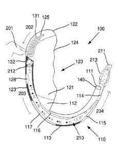
be used
depending on a range of factors such as the age or size of the patient and
different procedural
options. Laryngoscope blades are generally classified as curved or straight,
although a
number of styles of curved and straight blades are commercially available.
Some styles of
blades are designed to be positioned anterior to the epiglottis, and other
styles are designed to
be positioned posterior to the epiglottis, leading to slightly different
movements during the
procedure. A light source may be provided at the tip of the blade 3 to
illuminate the area
beyond. The light source may be powered by batteries within the handle 2.
[0007] During endotracheal intubation, with the patient laying supine, the
operator, standing
at the top of the head of the patient, introduces the blade 3 of the
laryngoscope 1 through the
mouth and into the pharynx and manipulates anatomical structures such as the
tongue and the
epiglottis (depending on the particular patient and type of blade) to expose
the entrance of the
larynx. Then, under direct visualisation, the operator inserts the tip of the
endotracheal tube
into the larynx and advances it into the trachea. In the conventional and
universal procedure,
the operator typically utilises the left hand to hold the laryngoscope 1 by
the handle 2 to
position the blade 3 and utilises the right hand to carefully introduce the
endotracheal tube,
pushing it along side the laryngoscope blade 3 and into the visualised
trachea.
[0008] Often, because of anatomical variations and challenges, and despite an
adequate
technique, direct visualisation is difficult. In most of these occasions,
adequate visualisation
could be obtained by manipulating some of the anatomical structures.
Unfortunately, with the
conventional laryngoscope and conventional procedure, the operator is
utilising both hands
and the hand being used to manually introduce the endotracheal tube cannot be
used to
manipulate anatomical structures to facilitate the procedure. Furthermore, a
second operator
could not have direct visual access to the entrance of the larynx to help
manipulating these
CA 02970585 2017-06-12
WO 2016/090435 PCT/AU2015/050786
- 3 -
structures and will interfere with the vision of the first operator, as the
mouth opening,
through which the first operator is obtaining the view, is very limited and
the operator
performing the intubation procedure will usually be in the best viewing
position.
[0009] Due to the degree of difficulty of the procedure itself, together with
the seriousness of
the potential complications, this procedure will only be performed by highly
skilled
professionals. This difficulty and serious complication risk have also meant
that the
procedure, and the instruments used to perform it, has essentially remained
unchanged for
decades. The physicians and other professionals who perform endotracheal
intubations are
unwilling to use new devices or to change the way this is conventionally done,
given the
difficulties and risks. A new intubation device therefore not only has to
offer obvious
procedural advantages in comparison to the conventional laryngoscopes, but
also has to
present similar characteristics in shape and weight and in its method of use,
to facilitate
adoption by operators already trained and comfortable in the use of
conventional
laryngoscopes in the often stressful circumstances of performing an intubation
procedure.
[0010] W02003047673 discloses an automatically operative medical insertion
device and
method including an insertable element which is adapted to be inserted within
a living
organism in vivo, a surface following element, physically associated with the
insertable
element and being arranged to follow a physical surface within the living
organism in vivo, a
driving subsystem operative to at least partially automatically direct the
insertable element
along the physical surface and a navigation subsystem operative to control the
driving
subsystem based at least partially on a perceived location of the surface
following element
along a reference pathway stored in the navigation subsystem. However, the
automatic
operation of this device requires a complex arrangement of hardware with a
significantly
different configuration to conventional laryngoscopes, resulting in a
relatively large and
costly device compared to conventional laryngoscopes.
[0011] US5184603 discloses an intubating instrument comprising a laryngoscopic
blade
having a rounded distal end adapted for introduction into a patient's throat
to expose the
laryngeal opening for endotracheal intubation; side walls formed integrally
with the blade
and forming an elongated channel for an endotracheal tube; the channel being
adapted to
retain the tube within the laryngoscopic blade during insertion and
manipulation of the
CA 02970585 2017-06-12
WO 2016/090435 PCT/AU2015/050786
- 4 -
instrument and to accommodate forward displacement of the tube beyond the
distal end; the
blade having a proximal end having a first quick-connect coupling associated
therewith; a
support handle housing for supporting the blade; a second quick-connect
coupling matable
with the first quick-connect coupling disposed at the lowermost portion of the
support handle
housing; the first and second quick-connect couplings being adapted to be
mechanically
engaged to lock the blade to the handle housing in a predetermined angular
relationship; an
endotracheal tube driver mounted in the handle housing; a tube driver
operatively associated
with the handle housing and the blade and adapted to engage a proximal portion
of an
endotracheal tube in the channel and to advance the tube beyond the distal end
of the blade to
introduce the distal end of the tube into the trachea; a finger-activated
trigger mounted on the
handle housing and adapted to initiate operation of the tube driver, whereby
the exposure of
the glottic opening and the introduction of the endotracheal tube may be
effected with one
hand while holding the handle housing. However, the instrument disclosed in
this document
has a significantly different configuration compared to conventional
laryngoscopes. The
positioning of the tube driver in particular is detrimental to the ergonomics
of the instrument
and may interfere with operator movements or anatomical structures during a
procedure.
[0012] US5776052 discloses a laryngoscope that has a handle including a
mechanism
adapted to engage and advance a flexible fiberoptic tube of a bronchoscope.
The mechanism
is operated by the hand that grasps the handle. The laryngoscope has a blade
extending from
the handle which defines a surface extending from the handle to the distal end
of the blade.
The mechanism includes a guide which overlies the surface to define a channel
through
which the fiberoptic tube is advanced to the distal end of the blade. The
mechanism is
positioned to advance the flexible fiberoptic tube through the channel. The
mechanism also
displaces the guide from the surface allowing the laryngoscope to be removed
from the
flexible fiberoptic tube. However, the laryngoscope disclosed in this document
only provides
a mechanism for advancing a fiberoptic tube, not an endotracheal tube.
[0013] W02011119521 discloses a fiberoptic intubating device which permits
visualization
of the vocal cords and automatic deployment of an endotracheal tube into the
trachea upon
visualization. The device includes a housing, a handle extending from the
housing, and an
extendable and retractable stylet extending from the distal end generally in
parallel with the
longitudinal axis. The device also includes a support member disposed on the
housing that is
CA 02970585 2017-06-12
WO 2016/090435 PCT/AU2015/050786
- 5 -
configured to support the endotracheal tube with respect to the housing and to
be selectively
movable in the longitudinal direction relative to the housing. The device is
configured to
automatically move the stylet relative to the housing upon actuation of a
trigger. Once the
stylet is positioned relative to the vocal cords, the device is configured to
deploy the
endotracheal tube into the trachea upon further actuation of the trigger.
However, the device
disclosed in this document lacks a laryngoscope blade and has a significantly
different
configuration and operational ergonomics compared to conventional
laryngoscopes.
[0014] The reference in this specification to any prior publication (or
information derived
from it), or to any matter which is known, is not, and should not be taken as
an
acknowledgment or admission or any form of suggestion that the prior
publication (or
information derived from it) or known matter forms part of the common general
knowledge
in the field of endeavour to which this specification relates.
Summary of the Present Invention
[0015] In a broad form the present invention seeks to provide an intubation
device for use in
an endotracheal intubation procedure, the intubation device including:
a) a laryngoscope blade having a tip and a base;
b) a handle attached to the base of the blade for allowing the intubation
device to be
held in a hand of a user;
c) a channel for receiving an endotracheal tube, the channel including:
i) a blade channel portion extending along the blade substantially from the
tip to
the base and including an outlet proximate to the tip for allowing a distal
end
of the endotracheal tube to be advanced from the outlet; and,
ii) a handle channel portion extending partially along the handle from the
blade
channel portion; and,
d) a tube movement mechanism in the handle for moving the endotracheal tube
through the channel to thereby advance the endotracheal tube, the tube
movement
mechanism including a thumb interface for allowing the user to operate the
tube
movement mechanism using a thumb of the hand that is holding the intubation
device, to thereby allow the user to hold the intubation device and advance
the
endotracheal tube in an endotracheal intubation procedure using a single hand.
CA 02970585 2017-06-12
WO 2016/090435 PCT/AU2015/050786
- 6 -
[0016] Typically the tube movement mechanism includes a tube engager for
engaging a
proximal end of the endotracheal tube located in the handle channel portion
and causing the
endotracheal tube to move through the channel in response to operation of the
thumb
interface.
[0017] Typically the thumb interface is coupled to the tube engager so that a
movement of
the thumb interface by the thumb of the user causes a corresponding movement
of the
endotracheal tube through the tube channel.
[0018] Typically the thumb interface is moveable in opposing first and second
directions,
such that a movement of the thumb interface in the first direction advances
the endotracheal
tube and a movement of the thumb interface in the second direction retracts
the endotracheal
tube.
[0019] Typically the thumb interface is mechanically coupled to the tube
movement
mechanism so that a movement of the thumb interface is mechanically translated
into a
corresponding movement of the endotracheal tube.
[0020] Typically the tube movement mechanism is configured so that a movement
of the
thumb interface by a thumb movement distance translates into a movement of the
endotracheal tube by a tube movement distance which is greater than the thumb
movement
distance.
[0021] Typically the tube movement distance is related to the thumb movement
distance by a
multiplication factor provided by mechanical advantage in the tube movement
mechanism.
[0022] Typically the tube movement mechanism includes at least one of a lever
arrangement
and a gear train.
[0023] Typically the tube movement mechanism includes an actuator for moving
the
endotracheal tube, the actuator being activated in response to operation of
the thumb
interface.
[0024] Typically operation of the thumb interface causes a control input to be
provided to the
actuator for controlling the activation of the actuator.
CA 02970585 2017-06-12
WO 2016/090435 PCT/AU2015/050786
- 7 -
[0025] Typically the thumb interface includes a press button, such that a
control input is
provided to the actuator when the press button is pressed by the thumb of the
user.
[0026] Typically the thumb interface includes a plurality of press buttons for
each providing
different control inputs to the actuator when pressed by the thumb of the
user.
[0027] Typically the actuator is electrically powered by a battery.
[0028] Typically the thumb interface includes a thumb slider such that the
thumb interface is
operated by the user slidingly moving the thumb slider using the thumb of the
user.
[0029] Typically the thumb interface includes a thumb wheel such that the
thumb interface is
operated by the user rolling the thumb wheel using the thumb of the user.
[0030] Typically the blade includes a tissue engaging anterior blade face and
an opposing
posterior blade face, and the handle includes a posterior handle face
extending from the
posterior blade face, the blade channel portion and the handle channel portion
being
respectively defined in the posterior blade face and the posterior handle
face.
[0031] Typically the posterior handle face and the posterior blade face
collectively define a
continuously curved posterior face of the intubation device, the channel being
defined in the
curved posterior face.
[0032] Typically the curved posterior face is rounded along each of the blade
and the handle.
[0033] Typically the channel includes an elongate opening extending along the
curved
posterior face.
[0034] Typically the blade channel portion and the handle channel portion are
respectively
defined in a lateral blade face and a lateral handle face.
[0035] Typically the channel includes an elongate opening extending along the
lateral blade
face and the lateral handle face.
[0036] Typically the channel defines a curved passageway for receiving the
endotracheal
tube.
CA 02970585 2017-06-12
WO 2016/090435 PCT/AU2015/050786
- 8 -
[0037] Typically the intubation device includes retention tabs partially
covering sections of
an elongate opening of the channel for retaining the endotracheal tube within
the channel.
[0038] Typically the retention tabs are configured to prevent the endotracheal
tube from
being displaced from the channel unless the endotracheal tube is positively
removed by a
user.
[0039] Typically the intubation device includes a light source located
proximate to the tip of
the blade.
[0040] Typically the intubation device includes a fiber optic viewing
arrangement.
[0041] Typically the intubation device includes a video camera located
proximate to the tip
of the blade.
[0042] Typically the intubation device includes a suction channel having a
suction outlet
proximate to the tip of the blade, the suction channel being configured to
receive a suction
tube to allow suction at the suction outlet.
[0043] Typically the blade is detachable from the handle.
[0044] Typically the intubation device is configured to allow the attachment
of different
blades having different shapes and sizes, depending on requirements for the
endotracheal
intubation procedure.
[0045] Typically the blade is hingedly connected to the handle to thereby
allow the blade to
be moved between an operational configuration and a collapsed configuration.
[0046] Typically the intubation device includes one or more detachable seals
for sealing at
least a part of the channel.
[0047] Typically the handle includes one or more openings associated with the
channel for
allowing the user to access a portion of the endotracheal tube within the
handle.
[0048] In another broad form the present invention seeks to provide an
intubation device for
use in an endotracheal intubation procedure, the intubation device including:
a) a laryngoscope blade having a tip and a base;
CA 02970585 2017-06-12
WO 2016/090435 PCT/AU2015/050786
- 9 -
b) a handle attached to the base of the blade for allowing the intubation
device to be
held in a hand of a user;
c) a channel for receiving an endotracheal tube, the channel including:
i) a blade channel portion extending along the blade substantially from the
tip to
the base and including an outlet proximate to the tip for allowing a distal
end
of the endotracheal tube to be advanced from the outlet; and,
ii) a handle channel portion extending partially along the handle from the
blade
channel portion; and,
d) a tube movement mechanism in the handle for moving the endotracheal tube
through the channel to thereby advance the endotracheal tube, the tube
movement
mechanism including a digit interface for allowing the user to operate the
tube
movement mechanism using one or more digits of the hand that is holding the
intubation device, to thereby allow the user to hold the intubation device and
advance the endotracheal tube in an endotracheal intubation procedure using a
single hand.
[0049] Typically the one or more digits of the hand that is holding the
intubation device
includes at least one of:
a) a thumb; and,
b) a finger.
[0050] In another broad form the present invention seeks to provide an
intubation device for
use in a bougie-assisted endotracheal intubation procedure, the intubation
device including:
a) a laryngoscope blade having a tip and a base;
b) a handle attached to the base of the blade for allowing the intubation
device to be
held in a hand of a user;
c) a channel for receiving a bougie, the channel including:
i) a blade channel portion extending along the blade substantially from the
tip to
the base and including an outlet proximate to the tip for allowing a distal
end
of the bougie to be advanced from the outlet; and,
ii) a handle channel portion extending partially along the handle from the
blade
channel portion; and,
CA 02970585 2017-06-12
WO 2016/090435 PCT/AU2015/050786
- 10 -
d) a tube movement mechanism in the handle for moving the bougie through the
channel to thereby advance the bougie, the tube movement mechanism including a
thumb interface for allowing the user to operate the tube movement mechanism
using a thumb of the hand that is holding the intubation device, to thereby
allow
the user to hold the intubation device and advance the bougie in a bougie-
assisted
endotracheal intubation procedure using a single hand.
Brief Description of the Drawings
[0051] An example of the present invention will now be described with
reference to the
accompanying drawings, in which: -
[0052] Figure 1 is a side view of a prior art laryngoscope;
[0053] Figure 2A is a perspective view of a first example of an intubation
device;
[0054] Figure 2B is a perspective view of the intubation device of Figure 2A
loaded with an
endotracheal tube and showing a typical positioning of a user's thumb in use;
[0055] Figure 2C is a perspective view of the intubation device of Figure 2B
showing the
user's thumb operating a thumb interface to advance the endotracheal tube;
[0056] Figure 2D is a detailed perspective view of the user's thumb operating
the thumb
interface to advance the endotracheal tube as shown in Figure 2C;
[0057] Figure 2E is a detailed perspective view of a coupling between the
thumb interface
and the endotracheal tube as shown in Figure 2D;
[0058] Figure 2F is a detailed perspective view of the coupling as shown in
Figure 2E
showing advancement of the coupling following operation of the thumb
interface;
[0059] Figure 2G is a detailed perspective view of the coupling as shown in
Figure 2F
showing disengagement of the endotracheal tube from the coupling;
[0060] Figure 3A is a perspective view of an example of a user using the
intubation device of
Figure 2A to perform an endotracheal intubation procedure on a subject;
CA 02970585 2017-06-12
WO 2016/090435 PCT/AU2015/050786
- 11 -
[0061] Figure 3B is a cross section view of the intubation device and the
subject of Figure
3A once the endotracheal tube has been placed into the trachea of the subject
and removed
from the intubation device;
[0062] Figure 4 is a perspective view of a second example of an intubation
device having an
alternative form of the thumb interface;
[0063] Figure 5 is a perspective view of a third example of an intubation
device having a
further alternative form of the thumb interface;
[0064] Figure 6A is a perspective view of a fourth example of an intubation
device in an
operational configuration;
[0065] Figure 6B is a perspective view of the intubation device of Figure 6A
in a collapsed
configuration;
[0066] Figure 6C is a perspective exploded view of the intubation device of
Figure 6A;
[0067] Figure 6D is a perspective view of the intubation device of Figure 6A
showing the
thumb interface in an advanced position;
[0068] Figure 6E is a further perspective view of the intubation device of
Figure 6A showing
the thumb interface in an advanced position;
[0069] Figure 6F is a perspective view of the intubation device of Figure 6A
loaded with an
endotracheal tube; and,
[0070] Figure 6G is a perspective view of the intubation device and
endotracheal tube of
Figure 6F following advancement of the endotracheal tube.
Detailed Description of the Preferred Embodiments
[0071] An example of an intubation device 100 for use in an endotracheal
intubation
procedure will now be described with reference to Figures 2A to 2G.
[0072] With regard to Figure 2A, the intubation device 100 includes a
laryngoscope blade
110 having a tip 111 and a base 112. A handle 120 is attached to the base 112
of the blade
CA 02970585 2017-06-12
WO 2016/090435 PCT/AU2015/050786
- 12 -
110 for allowing the intubation device 100 to be held in a hand of a user.
[0073] The intubation device 100 also includes a channel 101 for receiving an
endotracheal
tube 210, as shown in Figure 2B. The channel 101 includes a blade channel
portion 113
extending along the blade substantially from the tip 111 to the base 112, and
a handle channel
portion 123 extending partially along the handle 120 from the blade channel
portion 113. The
blade channel portion 113 includes an outlet 114 proximate to the tip 111 for
allowing a
distal end 211 of the endotracheal tube 210 to be advanced from the outlet
114, as shown in
Figure 2C.
[0074] The intubation device 100 further includes a tube movement mechanism
130 in the
handle 120 for moving the endotracheal tube 210 through the channel 101 to
thereby advance
the endotracheal tube 210. The tube movement mechanism 130 includes a thumb
interface
131 for allowing the user to operate the tube movement mechanism 130 using a
thumb 201 of
the hand that is holding the intubation device 100 (as shown in Figures 2B to
2F), to thereby
allow the user to hold the intubation device 100 and advance the endotracheal
tube 210
during an endotracheal intubation procedure using a single hand.
[0075] With reference to Figure 3A, it will be seen that the user can hold the
intubation
device 100 by the handle 120 in a hand 301 with the thumb 201 of that hand
being positioned
for operation of the thumb interface 131. The user can manoeuvre the blade 110
relative to
anatomical structures inside oropharyngeal passage of the patient 310 using
the handle 120,
to move the tip 111 of the blade 110 into position for advancement of the
endotracheal tube
210. Once the tip 111 is suitably positioned, the user can then operate the
thumb interface
131 to cause the tube movement mechanism to move the endotracheal tube 210
through the
channel 101 and advance the endotracheal tube 210 into the trachea of the
patient 310.
[0076] By enabling single handed operation of the intubation device 100 for
positioning the
blade 110 via the handle 120 and advancing the endotracheal tube 210, the
other hand 302 of
the user will remain free for other uses, such as clearing the airway using
another device,
such as a suction device 303, or other devices such as forceps or the like to
manipulate
anatomical structures and/or the endotracheal tube 210, during the
endotracheal intubation
procedure as may be required. It will be appreciated that the use of a single
hand only can
also help in avoiding visual obstructions during the procedure which would
otherwise be
CA 02970585 2017-06-12
WO 2016/090435 PCT/AU2015/050786
- 13 -
presented if the endotracheal tube was to be manually advanced as per
conventional
procedures.
[0077] The general arrangement of the blade 110 and the handle 120 will be
familiar to users
experienced in performing endotracheal intubation procedures with conventional
laryngoscopes, such that a suitably skilled user would be able to intuitively
hold the handle
120 and manipulate the blade 110 during the procedure via the handle 120 in a
generally
conventional manner. However, the tube movement mechanism 130 within the
handle 110
additionally provides the user with the capability of advancing the
endotracheal tube 210
simply by operating the thumb interface 131. With a suitably configured and
positioned
thumb interface 131, the user can cause the endotracheal tube 210 to be
advanced during the
procedure using intuitive thumb movements, and whilst continuing to hold the
intubation
device 100 by the handle 120 with a grip similar to that used for conventional
laryngoscopes.
[0078] Accordingly, it is expected that skilled users of conventional
laryngoscopes would be
able to use the intubation device 100 without requiring significant alteration
to the way the
user would hold and manipulate a conventional laryngoscope during an
endotracheal
intubation procedure. The main difference in performing the procedure will be
operating the
thumb interface 131 with the thumb of the hand holding the handle 110 to
advance the
endotracheal tube 210, rather than using their other hand to manually advance
the
endotracheal tube 210 as per conventional techniques.
[0079] However, it is noted that the user of the intubation device 100 may
optionally perform
an endotracheal intubation procedure in a completely conventional way using
the intubation
device 100, without using the thumb operated tube movement mechanism 130. For
instance,
the user may opt to use a more familiar conventional approach of manually
advancing the
endotracheal tube 210 alongside the blade 120 rather than through the channel
101, and the
intubation device 100 may be configured to permit this use. This may be useful
in a difficult
intubation in which the user is unable to successfully position the
endotracheal tube 210
using the thumb interface 131 and tube movement mechanism 130. The user may
withdraw
the endotracheal tube 210 from the channel 101 and manually insert the same
endotracheal
tube 210 so that it is guided by a surface of the blade 120. Alternatively, in
a more urgent
CA 02970585 2017-06-12
WO 2016/090435 PCT/AU2015/050786
- 14 -
scenario the user may manually introduce a second endotracheal tube 210
alongside the blade
120 in a similar manner.
[0080] In some embodiments, the intubation device 100 may be configured to
allow the user
to readily transition from using the thumb interface 131 and tube movement
mechanism 130
to advance the endotracheal tube 210 to manual advancement of the endotracheal
tube 210, if
this should be required. For example, a proximal end portion of the
endotracheal tube 210
may be displaced from the channel 101 to allow the user to use their other
hand to manually
move the endotracheal tube 210 along the channel 101 to manually advance the
endotracheal
tube 210. This may be facilitated, for example, by configuring the channel 101
to allow
displacement of the proximal end portion of the endotracheal tube 210 from the
handle
channel portion 123 whilst having another portion of the endotracheal tube 210
still retained
within the blade channel portion 113.
[0081] In any event, it will be appreciated that providing the option to use
manual
advancement techniques, to thereby use the intubation device 100 like a
conventional
laryngoscope, can be helpful in increasing the confidence of a user adopting
the use of the
intubation device 100 in the place of a conventional laryngoscope. Despite
this, it is expected
that users will readily adopt the use of the thumb operated tube movement
mechanism 130
due to its ease of use and the significant advantage of allowing single handed
operation,
freeing up the user's other hand for other activities.
[0082] The tube movement mechanism 130 and its thumb interface 131 can be
provided in
different forms depending on requirements. In one form, the tube movement
mechanism 130
is directly coupled to the thumb interface 131 so that a movement of the thumb
interface 131
results in an equivalent movement of the endotracheal tube 210 through the
channel 101. In
another form, the tube movement mechanism 130 may be configured to convert a
movement
of the thumb interface 131 into a longer movement of the endotracheal tube
210. In some
examples, the tube movement mechanism 130 includes an actuator which is
controlled by the
thumb interface 131.
[0083] It will be appreciated that the arrangement of the channel 101 to
extend along the
blade 110 and partially along the handle 120 allows a proximal end 212 of the
endotracheal
tube 210 to be located in the handle channel portion 123. Thus, the proximal
end 212 may be
CA 02970585 2017-06-12
WO 2016/090435 PCT/AU2015/050786
- 15 -
positioned in the handle 120 in proximity to the tube movement mechanism 130
provided in
the handle 120. It will be appreciated that this can result in an arrangement
in which the tube
movement mechanism 130 may be completely contained within the handle 120 and
engage
with the proximal end 212 of the endotracheal tube 210 to thereby move the
endotracheal
tube 210 through the channel 101 without any external protrusions from the
handle 120 or the
blade 110, that could otherwise interfere with anatomical structures or the
movements of the
user during the procedure. Preferred forms of the intubation device 100 will
thus allow the
user to go about the same movements of the hand holding the handle 120 as if
the user was
using a conventional laryngoscope, without interference by structures of the
device.
Accordingly, the channel 101 configuration including the handle channel
portion 123 and the
provision of the movement mechanism 130 within the handle 120 further
facilitates the
ability to provide the intubation device 100 in a familiar form which can be
readily adopted
by users experienced with the use of conventional laryngoscopes.
[0084] Turning back to the example embodiment of the intubation device 100
depicted in
Figure 2A, other optional features will now be described.
[0085] The blade 110 may be formed with a similar overall shape as
conventional
laryngoscope blades such as that depicted in Figure 1. As with conventional
laryngoscope
blades, the blade 110 may be provided in different sizes and with different
shapes (e.g.
straight or curved blades 110, blades 110 with different degrees of curvature,
blades 110 with
straight or curved tips 111), to suit different ages, sizes and shapes of
patients, different
oropharyngeal anatomies or different procedural options. The blade 110 may be
configured
in accordance with traditional curved laryngoscope blade styles (such as the
"Macintosh"
blade style) or straight laryngoscope blade styles (such as the "Miller" blade
style).
[0086] In some examples, the blade 110 may be detachable from the handle 120.
This can
allow the use of different blades 110 as required whilst having a single
handle 120 and tube
movement mechanism 130 provided within. A first end 121 of the handle 120 may
thus
include a coupling arrangement for allowing the base 112 of a blade 110 to be
coupled to or
detached from the handle 120. The intubation device may be configured to allow
the
attachment of different blades 110 having different shapes and sizes,
depending on
requirements for the endotracheal intubation procedure.
CA 02970585 2017-06-12
WO 2016/090435 PCT/AU2015/050786
- 16 -
[0087] It will be appreciated that this will allow the intubation device 100
to be used in a
range of different circumstances by attaching a blade 110 with a suitable
shape for the patient
and the procedural option selected by the user. The coupling arrangement may
utilise similar
coupling techniques as for conventional laryngoscopes to allow the blade to be
attached and
detached in a manner that is familiar to users with experience using
conventional
laryngoscopes. For example, the blade 110 and the handle 110 may be provided
with
complementary bayonet coupling interfaces, or any other suitable style of
coupling
interfaces. It should be noted, however, that the coupling arrangement should
ensure proper
alignment between the blade channel portion 113 and the handle channel 123
when the blade
110 is attached to the handle 120, to thereby form a continuous effective
channel 101.
[0088] The blade 110 and the handle 120 may be formed from any material
suitable for use
in medical devices. In some examples, the blade 110 and the handle 120 may be
formed from
moulded plastic components, which can allow for relatively inexpensive
manufacture. The
blade 110 and/or the handle 120 may even be provided as disposable items to
avoid the need
for sterilisation following a procedure. In such examples, it may be
preferable to provide the
tube movement mechanism 130 components in a simple and low-cost form. However,
reusable versions of the handle may be provided with a more sophisticated tube
movement
mechanism 130, and only the blade 110 which comes into contact with the
patient might be
disposed of. Alternatively, reusable metal components may be used to provide
the blade 110
and the handle 120 as is often the case for conventional laryngoscopes.
[0089] The thumb interface 131 may be mounted near a second end 122 of the
handle 120 for
appropriate positioning relative to the user's hand when gripping the handle
120. In this
example, the handle 120 has an ergonomic grip 124 for allowing the user to
comfortably yet
securely hold the handle 120 during use.
[0090] In this example intubation device 100, the thumb interface 131 is
provided in the form
of a thumb slider, such that the thumb interface 131 is operated by the user
slidingly moving
the thumb slider using the thumb of the user. The thumb slider of the thumb
interface 131
slides along a slot 125 formed in the handle 120, and the thumb interface 131
is mechanically
coupled to internal components of the tube movement mechanism 130 within the
handle 120
via the slot 125. Further details of the tube movement mechanism 130 and
operation of the
CA 02970585 2017-06-12
WO 2016/090435 PCT/AU2015/050786
- 17 -
thumb interface 131 will be provided in due course with reference to
illustrative usage
examples.
[0091] It should be appreciated that a range of different thumb interface 131
arrangements
other than that depicted in Figure 2A may be used. For example, Figure 4 shows
an example
of an intubation device 400 including press buttons 431, 432 as the thumb
interface 131, and
Figure 5 shows another example of an intubation device 500 including a thumb
wheel 531 as
the thumb interface 131. Different types of thumb interfaces 131 may be
coupled to the tube
movement mechanism 130 in different ways as will be discussed further in due
course.
[0092] In this example, the channel 101 is defined along posterior faces of
the blade 110 and
the handle 120. The blade 110 may include a tissue engaging anterior blade
face (being the
face of inside curvature for a curved style of blade 110 as depicted in the
Figures) and an
opposing posterior blade face, and the handle 120 may include a posterior
handle face
extending from the posterior blade face. The blade channel portion 113 and the
handle
channel portion 123 may be respectively defined in the posterior blade face
and the posterior
handle face. The channel 101 will thus provide an elongate opening extending
along the
posterior faces of the blade 110 and the handle 120. Once the endotracheal
tube 210 has been
positioned using the intubation device 100, the endotracheal tube 210 may be
removed from
the elongate opening channel 101 by pulling the endotracheal tube 210 in an
outward
direction relative to the intubation device 100, in this case posteriorly.
[0093] However, in other examples, the channel 101 may be defined along
lateral faces of
the blade 110 and the handle 120, adjacent to the posterior faces.
Accordingly, whilst the path
of the channel 101 may follow the shape of the posterior faces of the blade
110 and the
handle 120, the elongate opening of the channel 101 may be offset to a side of
the intubation
device 100. This arrangement can allow the endotracheal tube 210 to be removed
from the
elongate opening channel 101 by pulling the endotracheal tube 210 in an
outward direction
relative to the intubation device 100, in this case laterally. This lateral
positioning of the
elongate opening of the channel may allow the user to remove the endotracheal
tube 210
from the intubation device 100 using the user's other hand while maintaining a
constant grip
on the handle 120 with the first hand.
CA 02970585 2017-06-12
WO 2016/090435 PCT/AU2015/050786
- 18 -
[0094] In the depicted embodiment of the intubation device 100, the posterior
handle face
and the posterior blade face collectively define a continuously curved
posterior face of the
intubation device 100, with the channel 101 being defined in this curved
posterior face
(although as noted above, in other embodiments the channel 101 may be defined
in a lateral
face adjacent to this curved posterior face). In this case, the curved
posterior face is rounded
along each of the blade 110 and the handle 120. However, this is not essential
and the
respective posterior faces of the blade 110 and the handle 120 may include
straightened
portions, such as in the case of a blade 110 with a straight laryngoscope
blade type.
[0095] The channel 101 may define a curved passageway for receiving the
endotracheal tube
120. By forming the channel 101 in or adjacent to a curved posterior face,
this allows the
channel 101 to provide a suitable passageway whilst maintaining a consistent
depth relative
to the posterior faces. Despite this, the channel 101 may be formed with
variable depth to
provide a suitable curved passageway for the endotracheal tube 120 for
posterior face
geometries that are not curved or rounded.
[0096] The intubation device 100 may include retention tabs 115, 116, 117, 126
partially
covering sections of the elongate opening of the channel 101, for retaining
the endotracheal
tube 210 within the channel 101. The retention tabs 115, 116, 117, 126 may be
provided as
extensions of the above discussed faces across the elongate opening of the
channel 101, and
act to prevent unintentional dislocation of the endotracheal tube 210. The
number, shapes and
locations of the retention tabs 115, 116, 117, 126 may vary depending on the
intubation
device geometry and usage requirements, although in this case there are three
retention tabs
115, 116, 117 provided for the blade channel portion 113, and a single
retention tab 126
provided for the handle channel portion 123.
[0097] The retention tabs 115, 116, 117, 126 are preferably configured to
prevent the
endotracheal tube 210 from being displaced from the channel 101 unless the
endotracheal
tube 210 is positively removed by a user. Endotracheal tubes 210 are typically
be formed
from a flexible material, so the retention tabs 115, 116, 117, 126 may be
configured to
require some deformation of the endotracheal tube 210 when locating the
endotracheal tube
210 into the channel or removing the endotracheal tube 210 from the channel.
CA 02970585 2017-06-12
WO 2016/090435 PCT/AU2015/050786
- 19 -
[0098] The retention tabs 115, 116, 117, 126 should generally retain the
endotracheal tube
210 in the channel 101 without allowing significant radial movement. For a
channel 101
defined in posterior faces of the blade 110 and the handle 120, as per the
depicted example,
the retention tabs 115, 116, 117, 126 should prevent the endotracheal tube 210
from being
displaced outwardly from the channel 101 under normal movements of the
intubation device
100, unless the user positively removes the endotracheal tube 210 by pulling
it outwardly in a
posterior direction from the intubation device 100. In other examples having
the channel 101
defined in lateral faces of the blade 110 and the handle 120, the retention
tabs 115, 116, 117,
126 should prevent the endotracheal tube 210 from being displaced outwardly in
a lateral
direction from the channel 101 unless under a positive user action for
removing the
endotracheal tube 210.
[0099] The endotracheal tube 210 may be provided with a pre-curved
configuration so that it
is urged against the blade 110 and the handle 120 and easy to locate into the
channel 101
without requiring the retention tabs 115, 116, 117, 126 to provide significant
retaining force
on the endotracheal tube 210.
[0100] The intubation device 100 may also include a light source 140 located
proximate to
the tip 111 of the blade 110, for providing illumination during the procedure.
[0101] In some examples, the intubation device 100 may further include a fiber
optic viewing
arrangement for allowing the user to observe anatomical structures inside the
patient without
requiring a direct view. The fiber optic viewing arrangement may include a
flexible fiber
optic bundle with a lens positioned at one end proximate to the tip 111 of the
blade 110 and
an eyepiece positioned at the other end. The fiber optic bundle may run along
the blade 110
and into the handle 120. The eyepiece may be located on the handle 120 or on a
suitably
formed projection from the handle 120 to allow the user to look into the
eyepiece during the
procedure.
[0102] In other examples, the intubation device 100 may include a video camera
located
proximate to the tip 111 of the blade 110, for providing video imaging of
anatomical
structures inside the patient during the procedure. It will be appreciated
that this can provide
even more flexible viewing options compared to the fiber optic viewing
arrangement
discussed above. The video camera may be connected to a display for displaying
images
CA 02970585 2017-06-12
WO 2016/090435 PCT/AU2015/050786
- 20 -
from the video camera in real-time or near real-time during the procedure.
Whilst a small
display may be integrated with the intubation device 100, it may be preferable
to provide a
separate large display for displaying magnified images of the internal
anatomical structures,
in a more convenient viewing location for the user. The connection to the
display may be
achieved via a cable extending from the intubation device 100 or via a
wireless
communications connection which can avoid interference of user movements by a
cable.
[0103] In some embodiments, the intubation device 100 may include a suction
channel
having a suction outlet proximate to the tip 111 of the blade 110. The suction
channel may be
configured to receive a suction tube to allow suction at the suction outlet.
This can remove
the need for the user to use a separate suction device with the other hand
while using the
intubation device 100.
[0104] The operation of example embodiment of the intubation device 100
depicted in Figure
2A will now be described in further detail with reference to the subsequent
Figures 2B to 2G.
[0105] As shown in Figure 2B, the intubation device 100 may be loaded with an
endotracheal tube 210, by placing the endotracheal tube 210 in the channel 101
with the
distal end 211 of the tube positioned at the outlet 114 of the channel 101
near the tip 111 of
the blade 110. The proximal end 212 of the endotracheal tube 210 will be
positioned near the
end of the blade channel portion 123 within the handle 120. The endotracheal
tube 210 may
be placed into the channel 101 by pushing the endotracheal tube 210 into the
elongate
opening of the channel 101 past the retention tabs 115, 116, 117, 126, either
from a posterior
or lateral direction relative to the intubation device 100 depending on how
the channel 101 is
defined. Alternatively, the endotracheal tube 210 may be inserted through the
outlet 114 and
passed along the channel 101, although this may only be possible if the
endotracheal tube 210
is not provided with a tube fitting 220 or the like at its proximal end. In
either case, the
endotracheal tube 210 should be located substantially inside the channel 101
as shown in
Figure 2B.
[0106] The tube movement mechanism 130 may include a tube engager 132 (visible
in
Figures 2C to 2G) for engaging the proximal end 212 of the endotracheal tube
210 located in
the handle channel portion 123 and causing the endotracheal tube 210 to move
through the
channel 101 in response to operation of the thumb interface 131. The thumb
interface 131
CA 02970585 2017-06-12
WO 2016/090435 PCT/AU2015/050786
- 21 -
may be coupled to the tube engager 132 so that a movement of the thumb
interface 131 by
the thumb of the user causes a corresponding movement of the endotracheal tube
210 through
the tube channel.
[0107] In a simple form, the tube engager 132 may simply be provided as a
member that
abuts the proximal end 212 of the endotracheal tube 210 to allow the tube
movement
mechanism 130 to move the endotracheal tube 210 by pushing the tube engager
132 within
the handle channel portion. However, providing the tube engager 132 in such a
way will only
allow for movement of the endotracheal tube 210 in a direction that advances
the
endotracheal tube 210.
[0108] Accordingly, in the depicted embodiment of the intubation device 100,
the tube
engager 132 is configured to provide for movement in two directions, to
thereby allow
advancement and retraction of the endotracheal tube 210. The thumb interface
131 may thus
be moveable in opposing first and second directions, such that a movement of
the thumb
interface 131 in the first direction advances the endotracheal tube 210 and a
movement of the
thumb interface 131 in the second direction retracts the endotracheal tube
210.
[0109] In this example the tube engager 132 is provided in the form of a clip
arrangement
which allows pushing and pulling forces to be applied to the proximal end 212
of the
endotracheal tube 210. Further features of the tube engager 132 will be
discussed with regard
to subsequent Figures, in due course.
[0110] In any event, the thumb interface 131 may be coupled to the tube
engager 132 so that
a movement of the thumb interface 131 by the thumb of the user causes a
corresponding
movement of the endotracheal tube 210 through the tube channel 101.
[0111] Figure 2C shows the intubation device 100 and endotracheal tube 210
after the thumb
interface 131 has been moved by the user's thumb 201 in a direction as
indicated by arrow
202. This movement of the thumb interface 131 has caused a corresponding
movement of the
tube engager 132 and in turn the endotracheal tube 210, as indicated by arrow
203. As a
result, the distal end 211 of the endotracheal tube 210 is advanced from the
outlet 114 as
indicated by arrow 204.
CA 02970585 2017-06-12
WO 2016/090435 PCT/AU2015/050786
- 22 -
[0112] Figures 2D and 2E show progressively closer details of the handle 120
and interface
between the tube movement mechanism 130 and the proximal end 212 of the
endotracheal
tube 210 within the handle channel portion 123. In this example, the tube
engager 132
engages with a connector fitting 220 that is fitted to the proximal end 212 of
the endotracheal
tube 210. The connector fitting 220 may be designed specifically for use with
the intubation
device 100, although more preferably the connector fitting 220 will be a
standard type of tube
connector and the tube engager 132 will be configured to provide a suitable
interface.
[0113] Turning to the more detailed view of Figure 2E, it will be seen that
the connector
fitting 220 includes a first connector end 221 that is connected to the
proximal end 212 of the
endotracheal tube 210 and an opposing second connector end 222, which may be
adapted for
connection to further tubing coupled to assisted breathing apparatus or the
like. In this
example, the connector fitting 220 also includes a flange 223 between the
first and second
connector ends 222, as is commonly the case for standard endotracheal tube
connector
fittings.
[0114] The tube engager 132 of this embodiment of the intubation device 100 is
configured
to interface with the flange 223 to thereby facilitate movement of the
endotracheal tube 210.
In particular, the tube engager 132 clips on to the flange 223 to allow the
tube engager 132 to
push or pull on the flange 223 when the tube engager 132 moves in response to
operation of
the thumb interface. However, it should be appreciated that a range of
alternative tube
engager 132 configurations may be used.
[0115] Figure 2F shows a similar view to Figure 2E after the thumb interface
131 has been
moved in the direction of arrow 205 for the full extent of movement allowed by
the slot 125.
This represents the maximum extend of advancement of the endotracheal tube
using the tube
movement mechanism 130 in this case. In this state, the tube engager 132 has
fully extended
into the handle channel portion 123 and it can be seen that the clips of the
tube engager 132
are attached to the tube mechanism 130 within the handle by links 133, which
may be formed
from wire or another suitably rigid material for transferring loads to the
tube engager 132.
[0116] In this example, the thumb interface 131 is mechanically coupled to the
tube
movement mechanism 130 so that a movement of the thumb interface 131 is
mechanically
translated into a corresponding movement of the endotracheal tube 210. In a
simple form of
CA 02970585 2017-06-12
WO 2016/090435 PCT/AU2015/050786
-23 -
the tube movement mechanism 130, the tube engager 132 may be directly coupled
to the
thumb interface 131, such as by connecting the links 133 directly between the
thumb slide of
the thumb interface 131 and the tube engager 132. This will result in the
amount of
movement of the endotracheal tube 210 being equivalent to the amount of
movement of the
thumb interface. However, this may lead to a large range of thumb movement
being required
to complete an endotracheal intubation procedure.
[0117] Accordingly, in alternative examples, the tube movement mechanism 130
may be
configured so that a movement of the thumb interface 131 by a thumb movement
distance
translates into a movement of the endotracheal tube 210 by a tube movement
distance which
is greater than the thumb movement distance. In other words the intubation
device may allow
a relatively small thumb movement to translate into a larger tube movement.
[0118] Typically there will be a proportional relationship between the thumb
movement and
the resulting tube movement. The tube movement distance may be related to the
thumb
movement distance by a multiplication factor provided by mechanical advantage
in the tube
movement mechanism. For instance, the tube movement mechanism may include a
lever
arrangement or a gear train configured to multiply the movement of the thumb
interface 131.
It will be appreciated that such arrangements may allow for a smaller range of
thumb
movement to be used to advance the endotracheal tube 210. This could help to
prevent thumb
fatigue during intubation procedures, or simply allow for more comfortable
operation.
[0119] Although a mechanical tube movement mechanism 130 can provide a
relatively
simple and low cost capability for translating movement of the thumb interface
131 into
movement of the endotracheal tube 210, alternative forms of the tube movement
mechanism
130 may include an actuator (not shown) for moving the endotracheal tube 210,
the actuator
being activated in response to operation of the thumb interface 131. For
example, the actuator
may be in the form of an electric motor coupled to a suitable tube engager 132
via a rack and
pinion arrangement or the like for providing linear movement of the
endotracheal tube 210.
The actuator may be electrically powered by a battery, which may be housed
within the
handle along with the actuator and any other tube movement mechanism 130
components.
CA 02970585 2017-06-12
WO 2016/090435 PCT/AU2015/050786
- 24 -
[0120] In versions of the intubation device 100 including an actuator, the
tube movement
mechanism 130 may be configured so that operation of the thumb interface 131
causes a
control input to be provided to the actuator, for controlling the activation
of the actuator.
[0121] Accordingly, the use of an actuator may allow for the use of different
styles of thumb
interfaces which accept different forms of input from the user's thumb. For
example, the
thumb interface may include a press button, such that a control input is
provided to the
actuator when the press button is pressed by the thumb of the user. In further
examples, the
thumb interface may include a plurality of press buttons for each providing
different control
inputs to the actuator when pressed by the thumb of the user. The alternative
intubation
device 400 of Figure 4 shows an example of an arrangement with two press
buttons 431, 432
for advancing and retracting the endotracheal tube 120, respectively.
[0122] In an example of another different form of the thumb interface 131, the
further
alternative intubation device 500 of Figure 5 shows an example in which the
thumb interface
131 includes a thumb wheel 531, such that the thumb interface is operated by
the user rolling
the thumb wheel using the thumb of the user. The thumb wheel 531 may either be
mechanically coupled to tube engager 532, such as by way of a gear train, belt
arrangement
or the like, or may be connected to a suitable sensor for generating a control
input when the
thumb wheel is rolled.
[0123] Turning back to Figure 2G, it will be appreciated that the tube engager
132 in this
example allows the proximal end 212 of the endotracheal tube 210 to be
disengaged after the
endotracheal tube 210 has been successfully advanced. In particular, the user
may grip the
flange 223 between the user's thumb 201 and a finger 206 and remove this from
the clips of
the tube engager 132 by moving the tube connector 220 in the direction
indicated by arrow
207.
[0124] As shown in the cross section view of Figure 3B, the user can then
proceed to remove
the endotracheal tube 210 from the channel 101, past the retention tabs 115,
116, 117, 126, so
that the intubation device 100 can be removed from the mouth 311 of the
patient 310 while
leaving the endotracheal tube 210 in position within the patient's trachea
312.
CA 02970585 2017-06-12
WO 2016/090435 PCT/AU2015/050786
- 25 -
[0125] It is noted that the endotracheal tube 210 in this example includes a
balloon 213
which can be inflated after successful intubation to seal the tracheal passage
around the
endotracheal tube 210. The endotracheal tube 210 also includes an inflation
conduit 320 for
allowing the balloon 213 to be inflated. The channel 101 and its outlet 114
should be sized
accordingly, to accommodate the balloon 213, inflation conduit 320, and other
optional
features of the endotracheal tube 210.
[0126] Further general discussion of suitable embodiments of the intubation
device 100,
along with suitable methods of their use and associated advantages, are
provided below.
[0127] In view of the above, it will be appreciated that of the intubation
device 100 may be
provided in the form of a laryngoscope-like device with a generally
conventional size and
shape.
[0128] One difference in shape between the example intubation device 100 and
conventional
laryngoscopes is that the posterior face of the device is typically curved or
rounded along the
handle 120 and the blade 110. In particular, the intubation device 100 may
include a handle
120 that outwardly differs from conventional laryngoscopes in the shape of the
posterior face,
which is curved or rounded instead of linear. This curved or rounded face is
continued on the
posterior face of the blade 110, which may be attached to the handle 120 in a
similar manner
as in the conventional laryngoscope. Both of the curved or rounded faces,
namely the
posterior faces of the handle 120 and the blade 110, may create a continuously
curved
portion. In some embodiments this continuously curved portion may be in the
form of a
continuous semicircle.
[0129] Along this curved or rounded posterior face, the channel 101 is defined
for locating
the endotracheal tube 210 before being inserted into the trachea of the
subject. Retention tabs
115, 116, 117, 126 in the form of non-complete borders along both sides of the
channel 101
may prevent the endotracheal tube 210 from being displaced laterally from the
channel 101
until it is manually separated laterally in a positive action by the user
(usually once the distal
end 211 is allocated inside the trachea 312). Those borders maintain the
endotracheal tube
210 inside the channel 101 while it is moved forward but do not prevent the
endotracheal
tube 210 from being removed laterally, as they only partially close the
channel 101 laterally.
CA 02970585 2017-06-12
WO 2016/090435 PCT/AU2015/050786
- 26 -
[0130] In some examples, the endotracheal tube 210 may be grasped with a pin
in a superior
part of the channel 101 within the handle 120, and this pin may be connected
with a thumb
interface 131 in the form of a mechanical switch that can be activated with
the thumb of the
hand that is holding the intubation device 100. By moving the thumb down and
up over the
switch the endotracheal tube 210 is moved down and up through the channel 101.
The thumb
interface 131 will typically be located over the rounded posterior face of the
handle 120,
preferably on a superior area of the handle, so it can be easily reached with
the thumb of the
hand that is holding the intubation device 100.
[0131] Other features of the intubation device 100 may be common to those
found in
conventional laryngoscopes. For example, embodiments of the intubation device
100 may
include a power source and a light source 140 at the tip 111 of the blade 110.
The blade 110
may be formed separately from the handle 120 and may be interchangeable using
a bayonet
mounting, so different sizes and types can be used, depending on patient
anatomy and
operator preferences.
[0132] The endotracheal tube 210 can be introduced through the trachea by
positioning the
tip 211 of the endotracheal tube 210 in alignment with the entrance of the
larynx as shown in
Figure 3B and advancing the endotracheal tube 210 through the channel 101
inside the
intubation device 100 by moving the thumb of the same hand that is holding the
intubation
device 100. The user utilises the intubation device 100 in a similar fashion
as for a
conventional laryngoscope, to hold the tongue and facilitate the visualisation
of the entrance
to the larynx.
[0133] Once this has been achieved the user aligns the tip 211 of the
endotracheal tube 210
with the entrance of the larynx so the endotracheal tube 210 can be introduced
with the
movement of the same hand's thumb. This technique requires only a single hand,
as opposed
to a conventional endotracheal intubation procedure, leaving the other hand of
the user free to
help remove obstacles and facilitate the way of the endotracheal tube into the
trachea.
[0134] The user also has the ability to move the endotracheal tube 210
backwards with an
opposite movement of the thumb on the switch of the thumb interface 131, so a
failed
advance of the endotracheal tube 210 can be corrected and the operation can be
started again
until the correct placement of the endotracheal tube 210 in the trachea 312 is
achieved.
CA 02970585 2017-06-12
WO 2016/090435 PCT/AU2015/050786
- 27 -
[0135] Once the tip of the endotracheal tube 210 has been introduced through
the vocal cords
into the larynx and it has been advanced into the trachea 312, the proximal
end 212 of the
endotracheal tube 210 is detached from the pin that previously held it,
followed by the whole
endotracheal tube 210 being detached from the channel 101 along the handle 120
and the
blade 110 of the intubation device. Then, while leaving the endotracheal tube
210 in the
desired place, the intubation device 100 is removed from the mouth and pharynx
of the
patient, as in the conventional procedure.
[0136] The fact that the other hand of the user is left free represents a
significant advantage,
as this hand can be utilised to manipulate anatomical structures which often
impend the
access to the larynx. Typically only the user is able to see such
obstructions, and, with the
intubation device 100, the user is also able to manipulate them to facilitate
the endotracheal
intubation procedure. This results in a significant improvement of the
technique for
endotracheal intubation, which can significantly improve the success rate of
this difficult
procedure.
[0137] The depicted embodiment of the intubation device 100 intentionally has
a similar size
and shape compared to conventional laryngoscopes, except for the curved or
rounded
posterior face and channel 100 for the endotracheal tube 210. By being similar
otherwise to
the conventional laryngoscopes, the professionals used to them can start to
use this new
device and procedure safely, being perfectly familiar with the general
procedure up to the
point of the insertion of the endotracheal tube 210, and knowing that at any
time they can
decide to use this device as a normal laryngoscope and proceed to intubate in
the
conventional way.
[0138] In this regard, the intubation device 100 will preferably be configured
to allow the
user to disengage the proximal end 212 of the endotracheal tube 210 from the
tube movement
mechanism 130 and remove from the channel 101 at least a portion of the
endotracheal tube
210 near the proximal end 212, to facilitate manual advancement should this be
required. It
will be appreciated that this can be enabled by appropriately positioning the
retention tabs
115, 116, 117, 126, particularly the retention tab 126 in the handle channel
portion 123.
CA 02970585 2017-06-12
WO 2016/090435 PCT/AU2015/050786
- 28 -
[0139] A further example of an intubation device 600 will now be described
with regard to
Figures 6A to 6G. It should be noted that features similar to those of the
previous example of
the intubation device 100 have been assigned similar reference numerals,
increased by 500.
[0140] With initial regard to Figure 6A, it will be seen that the intubation
device 600 has an
overall configuration generally similar to the previous intubation device
examples. The
intubation device 600 includes a laryngoscope blade 610 having a tip 611 and a
base 612. A
handle 620 is attached to the base 612 for allowing the intubation device 600
to be held in a
hand of a user.
[0141] As per the previous examples, the intubation device 600 also includes a
channel 601
for receiving an endotracheal tube 210, as shown in Figure 6F. The channel 601
includes a
blade channel portion 613 extending along the blade 610 substantially from the
tip 611 to the
base 612, and a handle channel portion 623 extending partially along the
handle 620 from the
blade channel portion 613. The blade channel portion 613 includes an outlet
614 proximate to
the tip 611 for allowing a distal end 211 of the endotracheal tube 210 to be
advanced from the
outlet 114, as shown in Figure 6G. In this example, the blade channel portion
613 and the
handle channel portion 623 are respectively defined in a lateral blade face
and a lateral
handle face, and thus the channel 601 includes an elongate opening extending
along the
lateral blade face and the lateral handle face.
[0142] The intubation device 600 further includes a tube movement mechanism
630 in the
handle 620 for moving the endotracheal tube 210 through the channel 601 to
thereby advance
the endotracheal tube 210. The tube movement mechanism 630 includes thumb
interfaces
631 for allowing the user to operate the tube movement mechanism 630 using a
thumb of the
hand that is holding the intubation device 600, as previously described, to
thereby allow the
user to hold the intubation device 600 and advance the endotracheal tube 210
during an
endotracheal intubation procedure using a single hand.
[0143] This example of the intubation device 600 may also include a range of
optional
features as discussed with regard to previous examples. For instance, the
intubation device
600 includes a light source 640 for providing illumination during the
intubation procedure, as
best seen in Figure 6E.
CA 02970585 2017-06-12
WO 2016/090435 PCT/AU2015/050786
- 29 -
[0144] In this example, the intubation device 600 is formed as an assembly of
parts as best
seen in the exploded view of Figure 6C. The assembly includes separate parts
providing the
blade 610, the handle 620 and the tube movement mechanism 630, along with a
cap 650 and
a battery insert 660. The base 621 of the blade 610 is attached to the handle
620 at a first end
621 of the handle 620. The tube movement mechanism 630 is inserted into a
second end 622
of the handle 620 and positioned in a slot 625 extending from the second end
622 of the
handle 620. The cap 650 closes the second end 622 of the handle 620 and may be
secured in
position using a ball detent arrangement, where the ball 651 can be seen in
Figure 2C and the
detent 624 can be seen in Figure 6E. It will be appreciated that other methods
of securing the
cap 650 in position relative to the handle 620 may be used, such as by
providing a suitable
threaded fastener.
[0145] The battery insert 660 is provided to house and provide an electrical
connection for a
battery 670 for supplying power to the light source 640. In this example, the
battery insert
660 receives a button cell battery 670 as shown in Figure 6A and can inserted
into a suitably
defined receptacle inside the handle 620. Figure 6A shows the position of the
battery insert
660 after insertion into the handle 620 receptacle. The battery insert 660 is
inserted via an
opening 602 defined in the handle 620. As will be discussed in due course,
this opening 602
is also used to load the endotracheal tube 120 into the intubation device 600
and remove the
endotracheal tube 120 from the intubation device 600. The battery insert 660
may also
include a low battery warning light 663 that is visible from outside the
handle when the
battery insert 660 is inserted into the receptacle.
[0146] The intubation device 600 is configured so that the blade 610 is
hingedly connected to
the first end 621 of the handle 620. This hinged connection is achieved using
a hinge pin 619
provided at the base 612 of the blade 610, which is received in a
complimentary hinge socket
629 at a first end 621 of the handle 620. The blade 610 is secured in an
operational
configuration as shown in Figure 6A using a ball detent arrangement, where at
least one ball
628 is provided on the first end 621 of the handle 620 and at least one
corresponding detent
618 is provided on the base 612 of the blade 610. However, the ball detent
arrangement can
be disengaged by positive manual action to cause the blade 610 to swing about
the hinge pin
619 into a collapsed configuration as shown in Figure 6B.
CA 02970585 2017-06-12
WO 2016/090435 PCT/AU2015/050786
- 30 -
[0147] Furthermore, in this example, the blade 610 can be detached from the
handle 620 by
detaching the hinge pin 619 from the hinge socket 629 when the blade 610 is in
the collapsed
configuration. It will be appreciated that a range of different sizes and
shapes of blades 610
may be provided for attachment to the intubation using this form of hinged
connection.
Therefore the same handle 620 and other associated parts including the tube
movement
mechanism 630, the cap 650 and the battery insert 660 can be used in
intubation procedures
with different blades 610 selected to suit the anatomy of patient. For
instance, whilst the
depicted example shows a curved "Macintosh" blade style, a straight "Miller"
blade style
may be used, and different sizes may be selected depending on the age, size
and/or weight of
the patient.
[0148] In this example, the blade channel portion 613 extends along a curved
path from the
tip 614 to the base 612 of the blade 610 and retention tabs 615, 617 are
provided for retaining
the endotracheal tube 210 within the blade channel portion 613 in use as can
be seen in
Figures 6F and 6G, with their functionality being similar as discussed for
equivalent features
in the previous examples.
[0149] The handle channel portion 623 extends between the opening 602 and the
first end
621 of the handle 620, and continues the curved path of the blade channel
portion 613 into
the handle 620. The blade channel portion 613 and the handle channel portion
623 together
define a continuous curved channel 601 within which the endotracheal tube 210
is received
as shown in Figures 6F and 6G.
[0150] As described in previous examples, the endotracheal tube 210 is
advanced by having
the user operate the tube movement mechanism 630, particularly by interacting
with a thumb
interface 631 using a thumb of the same hand that is holding the handle 620 of
the intubation
device 600. In this example, the tube movement mechanism 630 includes a
plurality of thumb
interfaces 631 positioned at regular intervals along an elongate member 634
that extends
from a tube engager 632 for receiving the endotracheal tube 210.
[0151] The tube engager 632 is configured to interface with the flange 223 of
the
endotracheal tube 210 in a similar manner as described above for the earlier
example of the
intubation device, to thereby facilitate movement of the endotracheal tube
210. As can be
seen in Figure 6D, the tube engager 632 includes a pair of curved clips 633,
633 which define
CA 02970585 2017-06-12
WO 2016/090435 PCT/AU2015/050786
-31 -
a slot 634 therebetween. Now referring to Figure 6G, which shows the
endotracheal tube 210
loaded into the intubation device 600 and advanced to reveal the tube engager
632, the flange
223 is inserted into the slot 634 and the clips 633, 635 respectively engage
the connector ends
221, 222 of the endotracheal tube 210. This positively engages the
endotracheal tube 210 so
that the endotracheal tube 210 moves along with the tube movement mechanism
630 when it
is operated by the user's thumb via one of the thumb interfaces 631.
[0152] In this example, each thumb interface 631 is provided in the form of a
generally
trapezoidal protrusion extending outwardly from the elongate member 634,
although it will
be appreciated that the shape of the protrusion can vary depending on
requirements. In use,
the user will engage their thumb with one of the thumb interfaces 631 and push
the thumb
interface to move the elongate member 634 along the slot and thereby use the
tube movement
mechanism 630 to move an endotracheal tube 210 loaded into the intubation
device. Faces of
the thumb interfaces 631 may have ribs or other textured finishes for
enhancing engagement
of the thumb interfaces 631 by the thumb of a user.
[0153] It should be appreciated that the use of multiple thumb interfaces 631
can allow a
greater range of movement of the tube movement mechanism 630 without needing
the user to
reposition their hand or over extend their thumb. Rather, when the user
reaches a limit of
their thumb movement, the user can use their thumb to engage a more suitably
positioned
thumb interface 631 and continue moving the tube movement mechanism 630 using
that
thumb interface 631. This process may be iterated several times to fully
advance the
endotracheal tube. It should be appreciated the thumb interfaces 631 can be
used to move the
tube movement mechanism 630 in a reversed direction to retract the
endotracheal tube 210
during the intubation procedure if required, for instance if a first
intubation attempt is
unsuccessful. Following such a retraction, the intubation device 600 may be
reoriented or
repositioned relative to the patient's anatomy and the tube movement mechanism
630 may be
moved in the advancing direction to thereby advance the endotracheal tube 210
once again.
[0154] The elongate member 634 of the tube movement mechanism 630 is received
in the
slot 625 and allowed to slide along the slot 625 as the thumb interfaces 631
are operated. The
slot 625 may extend along the length of the handle 620, and as can be seen in
Figures 6F and
6G, the intubation device 600 may be designed so that the tube movement
mechanism 630 is
CA 02970585 2017-06-12
WO 2016/090435 PCT/AU2015/050786
- 32 -
able to slide along the length of the handle 620, to enable a significant
amount of thumb-
operated advancement of the endotracheal tube 210, without any requirement of
sophisticated
mechanisms to provide mechanical advantage to multiply the thumb movement
distance. In
some examples, the handle 620 may include a pocket that effectively extends
the slot 625
inside the handle 620 to allow an even greater range of movement of the tube
movement
mechanism 630 in the advancing direction.
[0155] It may be desirable to provide a relatively long elongate member 634
having many
thumb interfaces 631 to enable a correspondingly long range of movement of the
tube
movement mechanism 630 without requiring overextension of the user's thumb.
However,
the range of movement of the tube movement mechanism 630 will be limited by
the length of
the slot 625 and the above mentioned pocket (if provided). In some
embodiments, this may
be mitigated to some extent by forming at least the elongate member 634 (and
potentially a
greater portion or the entirety of the tube movement mechanism 630) from a
flexible material
and/or by using a hinged construction, so that the elongate member 634 is
capable of
compressing or collapsing inside the slot 625 (or pocket) after abutting a
stop at the end of
the slot 625 (or pocket). Thus the user may continue to advance the
endotracheal tube 210
even after an end of the elongate member 634 reaches the end of the slot 625
(or pocket).
[0156] As mentioned previously, the opening 602 is used to allow the
endotracheal tube 210
to be loaded into or removed from the intubation device 600, as best
appreciated with
reference to Figure 6G. The opening 602 will typically be sized to receive the
flange 223 and
the connector ends 221, 222 of the endotracheal tube 210 and allow these to be
engaged with
the tube engager 632 of the tube movement mechanism 630 when in an advanced
position as
shown in Figures 6D and 6E. A portion of the endotracheal tube 210 extending
from the
connector ends 221, 222 is loaded into the channel 601, with the retention
tabs 615, 617
retaining the intubation tube 210 inside the blade channel portion 623. The
endotracheal tube
210 can then be retracted by moving the tube movement mechanism 630 into a
retracted
position as shown in Figure 6F, with the distal end 211 of the endotracheal
tube 210
positioned proximate to the outlet 614 of the blade 610.
[0157] At this stage the endotracheal tube 210 is loaded into the intubation
device 600 and
ready for use in an endotracheal intubation procedure. During the procedure,
the user
CA 02970585 2017-06-12
WO 2016/090435 PCT/AU2015/050786
- 33 -
operates the tube movement mechanism 630 by engaging their thumb with the
thumb
interfaces 631 as need to advance the endotracheal tube 210 from the outlet
114, as shown in
Figure 6G. When the endotracheal tube 210 has been successfully advanced into
a suitable
position within the patient, the flange 223 and the connector ends 221, 222 of
the
endotracheal tube 210 will typically be aligned with the opening 602, and may
be disengaged
from the tuber engager 632, after which the proximal end 212 of the
endotracheal tube may
be removed from the opening 602 by the user. The remainder of the endotracheal
tube 210
will similarly be removed by displacing it from the channel 601, so that the
intubation device
600 can be withdrawn with the endotracheal tube 210 in position within the
patient.
[0158] Although the user may simply use another hand to pull the proximal end
212 of the
endotracheal tube 210 from the opening 602, in this example an access hole 603
is also
provided in the handle 620 opposite to the opening 602 for allowing the user
to access the
proximal end 212 endotracheal tube 210 from the other side and push the
endotracheal tube
210 through the opening 602. The access hole 603 may be positioned to allow
the user to
extend through the access hole 603 a finger from the same hand that is holding
the device, to
thereby enable one-handed disengagement of the endotracheal tube 210 from the
tube
engager 632 to facilitate removal of the endotracheal tube 210. For example,
the user may
extend their index finger through the access hole 603 and push the proximal
end 212 of the
endotracheal tube 210 out of the opening 602, all whilst maintaining the
user's grip on the
handle 620. Following this, the user may manipulate the endotracheal tube 210
with other
fingers of the same hand to further disengage and remove the endotracheal tube
210 from the
channel 601.
[0159] It will be appreciated that the above described example of the
intubation device 600
allows a large range of movement of the endotracheal tube 210 while using a
relatively
simple tube movement mechanism 630 which can be comfortably operated by the
user's
thumb whilst simultaneously holding the intubation device 600 with the same
hand. This
example also provides for easier loading and removal of the endotracheal tube
210 using the
opening 602 and the access hole 603, respectively. The detachable hinged
connection of the
blade 210 to the handle 220 also provides increased flexibility in terms of
storage in the
collapsed position and the capability of using a wide range of different blade
types and sizes.
CA 02970585 2017-06-12
WO 2016/090435 PCT/AU2015/050786
- 34 -
[0160] In one envisaged scenario, the intubation device 600 may be used to
intubate a patient
while the patient is being supplied with oxygen via a mask that includes a
suitably configured
orifice for allowing insertion of the intubation device 600, without any
interruption of the
supply of oxygen to the patient during the intubation procedure. In this case,
the intubation
device 600 may be provided with seals (not shown) across at least part of the
channel 601
and the openings 602, 603 to thereby substantially prevent air from escaping
from the mask
via the intubation device 600. In one example, the seals may be configured to
snap onto the
channel portions 613, 623 and opening 602, 603 and be removable to allow
disengagement of
the endotracheal tube 210 after successful intubation.
[0161] Whilst the above examples, have primarily discussed operation of the
intubation
device using a thumb of the same hand that is holding the device, it will be
appreciated that
the intubation device may be adapted for operation using any digit of the
user's hand,
including the thumb or any finger. Although thumb operation is considered to
provide the
most natural action for advancing the endotracheal tube with the same hand
that is holding
the device, one or more fingers could be used to operate the tube movement
mechanism with
a reversed grip, or an alternative form of the intubation device may be
provided with the tube
movement mechanism reconfigured for operation using one or more fingers as
opposed to a
thumb.
[0162] Accordingly, in one example, there may be provided an intubation device
for use in
an endotracheal intubation procedure, the intubation device including: a
laryngoscope blade
having a tip and a base; a handle attached to the base of the blade for
allowing the intubation
device to be held in a hand of a user; a channel for receiving an endotracheal
tube, the
channel including a blade channel portion extending along the blade
substantially from the
tip to the base and including an outlet proximate to the tip for allowing a
distal end of the
endotracheal tube to be advanced from the outlet, and a handle channel portion
extending
partially along the handle from the blade channel portion; and a tube movement
mechanism
in the handle for moving the endotracheal tube through the channel to thereby
advance the
endotracheal tube, the tube movement mechanism including a digit interface for
allowing the
user to operate the tube movement mechanism using one or more digits of the
hand that is
holding the intubation device, to thereby allow the user to hold the
intubation device and
advance the endotracheal tube in an endotracheal intubation procedure using a
single hand.
CA 02970585 2017-06-12
WO 2016/090435 PCT/AU2015/050786
- 35 -
[0163] It will be appreciated that the operation of such an intubation device
configured for
operation by one or more digits of the same hand that is holding the device
may have
fundamentally similar design features as the above described examples.
However, if the
intubation device is specifically adapted for operation by one or more fingers
as opposed to
the thumb, this may necessitate reconfiguration and/or repositioning of the
digit interface
compared to the thumb interface of the previous examples. Nevertheless, the
principle of
one-handed operation may generally be the same.
[0164] The intubation device 600 as discussed above may alternatively be used
to perform a
bougie-assisted endotracheal intubation procedure. A bougie is an elongate
flexible device
with a relatively small diameter compared to a standard endotracheal tube 210
and an angled
tip at its distal end, and may be used to facilitate endotracheal intubation
in difficult
circumstances, such as where a patient's anatomy does not permit adequate
visibility or
prevents insertion of a standard endotracheal tube 210.
[0165] In a conventional bougie-assisted endotracheal intubation procedure,
the bougie may
be inserted into the patient's trachea using a traditional laryngoscope, with
the angled tip
assisting in guiding the bougie through the patient's anatomical structures.
Once the bougie is
correctly positioned, the laryngoscope can be withdrawn and an endotracheal
tube 210 is
threaded over the bougie, and then passed along the bougie to thereby advance
the
endotracheal tube 210 so it is guided along the path the bougie has taken
through the patient's
anatomy. Following advancement of the endotracheal tube 210 into a suitable
position, the
bougie can be extracted leaving only the endotracheal tube 210, such that the
endotracheal
intubation procedure is complete.
[0166] It will be appreciated that the intubation device 600 may be used in
place of a
traditional laryngoscope to facilitate the insertion of the bougie in a bougie-
assisted
intubation procedure. To enable this, some small alterations to a standard
bougie may be
required to make the bougie compatible with the intubation device 600. These
alterations
may include shortening the length of the bougie to fit along the channel 601
of the intubation
device 600 and providing a suitable connector fitting 220 on a proximal end of
the bougie
opposing the angled distal end, to thereby allow the bougie to be engaged by
the tube engager
632 of the tube movement mechanism 630 of the intubation device 600. The
connector fitting
CA 02970585 2017-06-12
WO 2016/090435 PCT/AU2015/050786
- 36 -
220 will preferably be of the same configuration as used for the endotracheal
tube 210, so
that the tube engager 632 can engage the connector fitting 220 of the bougie
in the same
manner as the connector fitting 220 of the endotracheal tube 210.
[0167] Accordingly, in one example the intubation device may be provided for
use in a
bougie-assisted endotracheal intubation procedure, the intubation device
including: a
laryngoscope blade having a tip and a base; a handle attached to the base of
the blade for
allowing the intubation device to be held in a hand of a user; a channel for
receiving a
bougie, the channel including a blade channel portion extending along the
blade substantially
from the tip to the base and including an outlet proximate to the tip for
allowing a distal end
of the bougie to be advanced from the outlet and a handle channel portion
extending partially
along the handle from the blade channel portion; and a tube movement mechanism
in the
handle for moving the bougie through the channel to thereby advance the
bougie, the tube
movement mechanism including a thumb interface for allowing the user to
operate the tube
movement mechanism using a thumb of the hand that is holding the intubation
device, to
thereby allow the user to hold the intubation device and advance the bougie in
a bougie-
assisted endotracheal intubation procedure using a single hand.
[0168] The method of operating the intubation device 600 for inserting a
bougie in a bougie-
assisted intubation procedure will be essentially the same as described above
for a standard
intubation procedure using an endotracheal tube, with the only difference
being that the
bougie is loaded into and advanced from the intubation device 600 instead of
the
endotracheal tube 210. It will be appreciated that once the bougie has been
successfully
advanced and inserted into the correct position within the patient, an
endotracheal tube 210
can be threaded onto and passed along the bougie in a conventional manner to
thereby
intubate the patient using the bougie as a guide. Once the endotracheal tube
210 has been
suitably placed, the bougie can then be removed through the internal conduit
of the
endotracheal tube 210 to leave only the endotracheal tube 210 within the
patient.
[0169] In some examples, specialized versions of the intubation device 600 may
be provided
that are particularly configured for use in a bougie-assisted endotracheal
intubation
procedure. For instance, the channel 601 may be sized to accommodate the
bougie only,
which could allow for a smaller blade to be used in difficult intubations. The
tube engager
CA 02970585 2017-06-12
WO 2016/090435 PCT/AU2015/050786
- 37 -
630 may also have a different configuration to engage with a different type of
connector
fitting 220 specifically adapted to the bougie. Despite this option, it would
still be
advantageous to allow the same intubation device 600 to be used with either a
bougie or an
endotracheal tube 210, depending on the particular circumstances of an
endotracheal
intubation procedure. It will be appreciated that if an attempted intubation
procedure using
the intubation device 600 an endotracheal tube 210 is unsuccessful, the user
could then
attempt a bougie-assisted endotracheal intubation procedure using the same
intubation device
600 but with a bougie instead of the endotracheal tube 210.
[0170] In summary, the intubation device may provide a direct replacement for
a
conventional laryngoscope that allows intubation with a single hand. This
intubation device
100 therefore allows a new method for intubation. Since the size and the shape
of the
intubation device may be similar to conventional laryngoscopes, this will
allow not only
familiarity and easy adaptation to the new method, but also, if decided, the
option to proceed
as in the conventional method of intubation, with minimal differences
perceived.
[0171] It will be appreciated that this is a particularly beneficial aspect,
since endotracheal
intubation is a high risk procedure and the process of change to a new
procedure needs to be
safe. For the same reason, a new intubation device 100 and procedure for
endotracheal
intubation will only be accepted and adopted by the professionals involved if
the changes
from the conventional device and procedure are minimised and, even better, if
the new device
allows at any point, to proceed as in the conventional procedure.
[0172] Throughout this specification and claims which follow, unless the
context requires
otherwise, the word "comprise", and variations such as "comprises" or
"comprising", will be
understood to imply the inclusion of a stated integer or group of integers or
steps but not the
exclusion of any other integer or group of integers.
[0173] Persons skilled in the art will appreciate that numerous variations and
modifications
will become apparent. All such variations and modifications which become
apparent to
persons skilled in the art should be considered to fall within the spirit and
scope that the
invention broadly appearing before described.