Note: Descriptions are shown in the official language in which they were submitted.
TITLE OF THE INVENTION
METHOD OF QUALIFYING LIGHT SPOTS FOR OPTICAL MEASUREMENTS
AND MEASUREMENT INSTRUMENT EMPLOYING
METHOD OF QUALIFYING LIGHT SPOTS
BACKGROUND AND SUMMARY
[00021 Field.
(00031 This invention pertains to devices and methods for performing optical
= measurements using a plurality of light spots, and more particularly, to
a method of qualifying
light spots for use for optical measurements by a measurement instrument, and
a
measurement instrument employing such a method of qualifying the light spots
that are
CA 2970614 2017-06-13
employed in its measurements.
[0004] Description.
[0005] There are some devices which employ light spots to make optical
measurements.
One well-known example is the use of a Shack-Hartmann wavefront sensor.
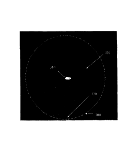
[0006] FIG. I illustrates some principal elements of a basic configuration of
a Shack-
Hartmann wavefront sensor 100. Shack-Hartmann wavefront sensor 100 comprises a
micro-
otitic leifsleratiiiillWand an optial
comprises a pixel array, for example, a charge-coupled device (CCD) camera or
CMOS array.
[0007] The lenslets of the lenslet array 110 dissect an incoming wavefront and
create a
pattern of light spots 130 that fall onto optical detector 120. In one typical
embodiment,
lenslet array 110 includes hundreds or thousands of lenslets, each on the size
scale of a
hundred microns. Meanwhile, optical detector 120 typically comprises many
pixels (e.g., 400
pixels) for each lenslet in lenslet array 110. Typically Shack-Hartmann sensor
100 is
assembled such that the pixel array 120 lies in the focal plane of lenslet
array 110.
[0008] Shack-Hartmann wavefront sensor 100 uses the fact that light travels in
a straight
line to measure the wavefront of light. By sensing the positions of light
spots 130, the
propagation vector of the sampled light can be calculated for each lenslet of
lenslet array 110.
The wavefront of the received light can be reconstructed from these vectors.
[0009] To better understand one or more aspects of this invention, it is
worthwhile to
discuss the operation of Shack-Hartmann wavefront sensor 100 in more detail.
However,
embodiments of the present invention extend to other types of optical
measurement devices
and systems such as topographers. In certain embodiments of the present
invention, a system
includes two or more optical measurement devices, for example, a combined
system
including both a wavefront sensor and a topographer.
2
CA 2970614 2017-06-13
=
1000101 In the case of the wavefront sensor 100, some optical system is
employed to deliver
a wavefront onto lenslet array 110, which samples the wavefront over the tiny
regions of each
lenslet. Beneficially, the lenslets are much smaller than the wavefront
variation. For the
purposes of this discussion, we define "isoplanatic" as the condition where
the wavefront is
well approximated by a plane wave over an area the size of a lenslet In that
case, the
wavefront is preferably isoplanatic over the sampled region. When detector
array 120 is in the
Totirpratie feltarefaitaYTIO; each lenalit iiiircreate a lieitipot on detector
array 120. The
location of these light spots reveals the average of the wavefront slopes
across each region.
That is, the shift in the location of a light spot is proportional to the
average of the slope of
the wavefront over the region sampled by the corresponding lenslet that
produced the light
spot. Software may compute the shift in each light spot.
[00011] In a typical operation, a reference beam (e.g., a plane wave) is first
imaged onto
lenslet array 110 and the locations of the resultant light spots ("reference
locations") on
detector array 120 is recorded. Then, a wavefront of interest is imaged onto
lenslet array 110,
and the locations of the light spots on detector array 120 produced by the
wavefront of
interest is recorded and compared against the reference locations.
[00012J FIGs. 2A-F illustrate an idealized example of this process where a
reference beam
and a wavefront of interest are imaged onto a detector array of a wavefront
sensor. This
idealization shows the process of measuring a spherical wave with a wavefront
sensor with
just 16 lenslets. The first step, as represented by the FICA. 2A-2C, is to
measure a plane wave
and measure the corresponding series of light spot locations 210 which are
used as reference
locations 220. The next step, as depicted in FIGs. 2D-2F, is to introduce a
wavefront of
interest and determine the shifts in the locations 240 of the light spots 230
from their
reference locations 220.
3
CA 2970614 2017-06-13
1
[000131 If the wavefront is not isoplanatic, the quality of the light spot
erodes rapidly and it
becomes more difficult to determine the location. However, where the
isoplanatic condition
is satisfied and where the light spot shift is consistent with the small angle
approximation of
Fresnel, then the light spot shift is exactly proportional to the average of
the wavefront slope
over the I enslet. 'Hie incident wavefront is then reconstructed from the
measurements of the
average oldie slopes for the hundreds or thousands of lenslets in the lens let
army.
1000141 Further details regarding the construction and operatiOn Of Shaek-
Harimann
wavefront sensor and a system for measuring aberrations in an eye using the
Shack-Hartman
wavefront sensor arc described in U.S. Patent 7,122,774, issued on 17 October
2006 to Daniel
R. Neal etal.
[000151 One important application for Shack-Hartmann wavefront sensors is in
the field of
ophthalmic aberrometry. In common practice, a measurement instrument employing
a Shack-
Hartmann wavefront sensor injects near infrared light into a patient's eye
which focuses on
the retina and scatters back toward the instrument. This light is imaged onto
the Shack-
Hartmann lenslet array, and each lenslet in the lenslet array focuses the
local portion of the
incident light it intercepts onto the detector array, as described above. Data
pertaining to the
locations of the light spots is used to derive slope information using a least
squares fit
method, and thereby to construct the wavefront of the received light. The
quality of the fit
data, usually evaluated using Zemike coefficients, is affected by the quality
of the light spot
location data, and every effort is made to ensure the data quality is adequate
to the
measurement accuracy and precision requirements.
[00016] FIG. 3 shows a typical raw image from a wavefront sensor. The
nominally
rectilinear array of light spots is produced by a rectilinear lenslet array.
The detailed analysis
4
CA 2970614 2017-06-13
of the locations of these light spots relative to their reference locations
(i.e., the locations that
result when a true plane wave is applied to the lenslet array) yields the
local gradient of the
incident wavefront. The overall area in which focal spots are present is
determined by the
patient's pupil, and analysis of this illuminated area yields the location
size and shape of the
pupil.
1000171 The application of Shack-Hartmann wavefront sensors to ophthalmic
aberrometry
has been a success. however, improvements may be provided by eliminating or
reducing the
effects of errors that may be caused by complicating factors inherent to the
measurement
method. Some of the important error sources are illustrated in FIG. 3 and will
be described
below.
1000181 The incident near infrared beam not only scatters from a patient's
retina, but also
reflects directly from the patient's cornea. The use of a Range Limiting
Aperture (RLA) in
the measurement instrument, as described in U.S. Patent 6,550,917 issued on 22
April 2003
to Daniel R. Neal et al., can significantly reduce the intensity of the
reflected light.
However, this so-called "corneal reflex" is generally orders of magnitude
brighter than the desired retinally scattered light, and¨ beneficially ¨ may
be excluded from
the wavefront calculations. Indeed, as is illustrated by reference numeral 310
in FIG. 3, the
reflex can affect a neighborhood of nearby focal spots by introducing stray
light that can alter
the true light spot location or mask the light spot entirely. The location and
intensity of the
corneal reflex is affected by corneal shape and the actual position of the
patient's eye when
the data is acquired. For these reasons, the qualification and/or exclusion of
light spot data in
and around the corneal reflex can be challenging.
1000191 The retinal scatter that is necessary for the aberrometer measurement
is highly
CA 2970614 2017-06-13
speckled because the retinal structure is quite rough compared to the
wavelength of the probe
beam. This leads to variability in the relative brightness of the focal spots.
A measurement
instrument may employ a broadband probe beam to reduce the speckle, but even
so, the
intensity of the light spots can vary by a factor of four in a normal clear
eye. Additional
variation can be introduced by cataracts, "floaters" and opaque regions in
pathological
crystalline lenses. Cataracts diffuse the incident and return beams causing
both reduced spot
mtenWafid boaden 1gh1ipit urc ihiiiii'
wavefront sensor may include dim light spots 320 and/or missing light spots
330.
[000201 Additional reflections of the probe beam may be produced by each
surface in eyes
implanted with intraocular lenses (IOLs). While similar to the corneal reflex
phenomenon,
multiple reflections are typically present in these patients and may be far
from the optic axis
of the wavefront sensor. Also, in subjects with diffractive IOLs, it is
expected that one lenslet
focal spot per diffraction order transmitted through the optical system may be
present. In
some cases this will lead to two or more focal spots that may or may not be
spatially
separated. Such focal spot distributions can lead to inaccurate spot location
and therefore
inaccurate wavefront measurements.
[00021] Another source of error in wavefront measurements is tear film
breakup. Tear film
break up can affect the location and sharpness of the light spots in the
vicinity of the breakup.
Tear film breakup is correlated to delays in blinking the eye. Some
measurement systems
may be designed to operate rapidly and reduce tear film breakup effects by
avoiding the need
to keep the patient from blinking for long periods. Nevertheless, it is still
possible that light
spots arc affected by tear film breakup. This can negatively impact the
resultant wavefront
measurements.
[00022] Thc spot location algorithms used with a typical wavefront measurement
instrument
6
CA 2970614 2017-06-13
are designed to work with data taken within the nominal linear range of the
detector device
(e.g., a CMOS detector). Obviously the spot location information is
compromised when the
spot brightness is poor compared to stray light and camera noise. As described
in U.S. Patent
6,550,917, a wavefront measurement instrument may employ a dynamic range
limiting
aperture (RLA) to significantly enhance its immunity to stray light. However
some
environmental factors may lead to increased stray light levels; e.g., pointing
the system
tolVarda-brieitliglif source. Rwavefronfineasurement
quality digital CMOS cameras to minimize the effects of camera dark noise. In
that case,
spots with many pixels that saturate the detector will yield less accurate
spot location
information.
[00023] Therefore, it would be desirable to provide one or more methods of
qualifying
which light spots are used for optical measurements by a measurement
instrument. It would
also be desirable to provide a measurement instrument employing a method of
qualifying the
light spots that are employed in its measurements.
1000241 In one aspect of the invention, a method employs an optical sensor to
determine a
property of an object. The method comprises: (a) illuminating the object with
light from one
or more light sources; (b) receiving light from the illuminated object; (c)
producing a group of
light spots from the received light; (d) qualifying a set of the light spots
for use in determining
a property of the object; and (e) determining the property of the object using
the qualified set
of light spots. Qualifying the set of light spots includes, for each light
spot in the group of
light spots: calculating a first calculated location of the light spot using a
first calculation
algorithm; calculating a second calculated location of the light spot using a
second calculation
algorithm different from the first calculation algorithm; and when a
difference between the
first and second calculated locations for the light spot is greater than an
agreement threshold,
7
CA 2970614 2017-06-13
excluding the light spot from the qualified set of light spots. In addition,
the light spots
excluded from the qualified set may be excluded from being employed in
determining the
property of the object. Alternatively, one or more of the spots excluded for
the qualified set of
light spots may be considered for inclusion in a second set of light spots.
Some or all of the
second set of light spots may also be used in determining the property of the
object, for
example, by assigning a lower weighting than those spots in the qualified set
Alternately,
some or id oilier secoild-Sel cifliglif spots mayIe used tei
4ktiEcrifearsiffe-,-cif
some feature of the optical system or eye, e.g., cataracts, tear film
conditions, surface
anomaly, or the like.
1000251 In another aspect of the invention, a device includes: one or more
light sources for
illuminating an object; a light spot generator adapted to receive light from
the illuminated
objected and to generate a group of light spots from the light received from
the illuminated
object; a detector adapted to detect the light spots and for outputting light
spot data pertaining
to each light spot; and a processor adapted to process the light spot data to
determine a
property of the object. The processor processes the light spot data by:
qualifying a set of the
light spots for use in determining the property, and determining the property
of the object
using the qualified set of light spots. Qualifying the set of light spots
includes, for each light
spot in the group of light spots: calculating a first calculated location of
the light spot from the
light spot data using a first calculation algorithm; calculating a second
calculated location of
the light spot from the light spot data using a second calculation algorithm
different from the
first calculation algorithm; and when a difference between the first and
second calculated
locations for the light spot is greater than an agreement threshold, excluding
the light spot
from the qualified set of light spots. Beneficially, in addition, the light
spots excluded from
the qualified set of light spots may be excluded from being employed in
determining the
8
CA 2970614 2017-06-13
property of the object.
[00026] In yet another aspect of the invention, a method comprises: producing
a first set
of first light spots from an eye with a corneal topography measurement;
producing a
second set of second light spots from the eye with a wavefront aberrometry
measurement; and qualifying one or more of the light spots within one of the
first and
second set of light spots based on the other of the first and second set of
light spots.
[00027] In still another aspect of the invention, a method is provided for
determining a
condition of an eye. The method comprises: providing a wavefront aberrometer
with a
first light source and a topographer with a second light source; illuminating
an eye with
the first light source to produce a first group of light spots; receiving the
first group of
light spots at a first detector array to produce a first signal containing a
first set of data;
illuminating the eye with the second light source to produce a second group of
light
spots; receiving the second group of light spots at a second detector array to
produce a
second signal containing a second set of data; comparing the first set of data
to the
second set of data; and based on the comparison, determining an abnormality of
the eye.
[00027a] In another aspect of the present invention, there is provided a
method of
determining a condition of an eye, comprising: providing a topographer with a
light
source; illuminating an eye with the light source to produce a group of light
spots;
receiving the group of light spots at a detector array and qualifying a set of
the light
spots to produce a signal containing a set of data from the qualified set of
light spots; and
determining an amount of astigmatism in a cornea of the eye based on the set
of data,
wherein qualifying the set of light spots includes, for each light spot in the
group of light
spots: calculating a first calculated location of the light spot using a first
calculation
9
CA 2970614 2018-08-20
algorithm; calculating a second calculated location of the light spot using a
second
calculation algorithm different from the first calculation algorithm; and when
a
difference between the first and second calculated locations for the light
spot is greater
than an agreement threshold, excluding the light spot from the qualified set
of light
spots.
[0002713] In another aspect of the present invention, there is provided a
method of
employing a wavefront measurement device to determine a wavefront of a
received light
beam, the method comprising: providing a topographer with a light source;
illuminating
an eye with the light source to produce a group of light spots; receiving the
group of
light spots at a detector array and qualifying a set of the light spots to
produce a signal
containing a set of data from the qualified set of light spots; and
determining an amount
of astigmatism in a cornea of the eye based on the set of data, wherein
qualifying the set
of light spots includes, for each light spot in the group of light spots:
calculating a first
calculated location of the light spot using a first calculation algorithm;
calculating a
second calculated location of the light spot using a second calculation
algorithm
different from the first calculation algorithm; and when a difference between
the first
and second calculated locations for the light spot is greater than an
agreement threshold,
excluding the light spot from the qualified set of light spots.
[00027c] In another aspect of the present invention, there is provided a
method of
determining a condition of an eye, comprising: providing a topographer with a
light
source; illuminating an eye with the light source to produce a group of light
spots;
receiving the group of light spots at a detector array and qualifying a set of
the light
spots to produce a signal containing a set of data from the qualified set of
light spots; and
9a
CA 2970614 2018-08-20
determining an amount of astigmatism in a cornea of the eye based on the set
of data,
wherein qualifying the set of light spots includes, for each light spot in the
group of light
spots: calculating a first calculated location of the light spot using a first
calculation
algorithm; calculating a second calculated location of the light spot using a
second
calculation algorithm different from the first calculation algorithm; and when
a
difference between the first and second calculated locations for the light
spot is greater
than an agreement threshold, excluding the light spot from the qualified set
of light
spots.
BRIEF DESCRIPTION OF THE DRAWINGS
[00028] FIG. 1 illustrates some principal elements of a basic configuration of
a Shack-
Hartmann wavefront sensor.
[00029] FIGs. 2A-F illustrate a reference beam and a wavefront of interest
being imaged
onto a detector array of a wavefront sensor.
[00030] FIG. 3 shows a typical raw image from a wavefront sensor.
[00031] FIG. 4 illustrates one embodiment of a measurement instrument
employing a
9b
CA 2970614 2018-08-20
=
wavefront sensor.
[00032] FIG. 5 shows a flowchart illustrating one embodiment of a method of
qualifying
light spot data for a wavefront measurement.
[00033] FIGs. 6A-D illustrate one embodiment of a first method of locating a
light spot in a
wavefront sensor.
[00034] FIGs. 7A-D illustrate one embodiment of a second method of locating a
light spot in
a wavefiont sensor-.
[00035] FIGs. 8A-B illustrate differences in the locations of light spots
determined by the
method of FIGs. 6A-D and locations determined by the method of FIGs. 7A-D for
an
exemplary image.
[00036] FIG. 9 shows a raw image from a wavefront sensor for a subject with an
intraocular
lens.
[00037] FIG. 10 shows a raw image from a wavefront sensor for a subject with
cataracts.
[00038] FIG. 11 shows a raw image from a wavefront sensor for a subject with a
weak
corneal reflex.
[00039] FIG. 12 is a histogram illustrating the summed intensity of pixels in
a light spot as a
function of occurrence for an exemplary image.
[00040] FIGs. 13A-B illustrate embodiments of two methods for determining the
location
and shape of a subject's pupil.
[00041] FIGs. 14A-B are plots illustrating the correlation of exemplary
measurements made
by the two methods illustrated in FIGs. 13A-B.
[00042] FIG. 15 shows one embodiment of a system for measuring wavefront
aberrations
and corneal topography of an eye.
[00043] FIG. 16 shows a flowchart illustrating one embodiment of a method of
qualifying
CA 2970614 2017-06-13
light spot data for a comeal topography measurement.
[000441 FIG. 17 shows an exemplary topographic image of an eye.
[00045] FIG. 18 shows exemplary wavefront data for an eye superimposed on a
topographic
image of the eye.
1000461 FIG. 19 shows an exemplary topographic image of an eye under a
condition of tear
fdm breakup.
-1000471- -FIG. 20 shows exernidary wavefront ditifor an eye siiiiiik
image of the eye under a condition of tear film breakup.
1000481 FIG. 21 shows another example of wavefront data for an eye
superimposed on a
topographic image of the eye
[00049] FIG. 22 shows wavefront data for an eye with an anomalous condition.
[00050] FIG. 23 shows a flowchart illustrating one embodiment of a method of
making a
wavefront measurement and a corneal topography measurement of an eye.
DETAILED DESCRIPTION
[00051] Methods of qualifying light spot data as described below can be
employed in a
variety of different measurement instruments. Exemplary embodiments will be
described in
some detail below so as to illustrate various aspects and advantages of these
methods.
However, it should be understood that the principles involved in these method
can be
employed in a variety of other measurement instruments which employ light spot
data.
[00052] FIG. 4 illustrates one embodiment of a measurement instrument
employing a
wavefront sensor. In particular, FIG. 4 illustrates a wavefront aberrometer
400 for making
wavefront measurements of a subject's eye 100. Among other components,
wavefront
11
CA 2970614 2017-06-13
aberrometer 400 includes a light source 410, a wavefront sensor 420, and other
components
on a moving stage 430, a processor 440, memory 450 associated with the
processor 440, and
an iris camera 460. Further details of the construction and operation of
wavefront
aberrometer 400 can be found in U.S. patent 7,494,220 issued an 24 February
2009 in the
names of Richard Copland at at.
1000531 Of particular relevanahere, wavefront sensor 420 operates
filtOlunctiOn With
processor 440 and associated memory 450 to perform wavefront measurements on
eye 100.
Wavefront sensor 420 includes a lenslet array 422 and a detector array 424.
Further details of
the construction and operation of lenslet array 422 and detector array 424 may
be understood
with reference to the description of wavefront sensor 100 of FIG. 1 provided
above. Light
spot data from detector array 424 is supplied to processor 440 and associated
memory 450 to
execute one or more algorithms to determine a wavefront of a light beam
received from the
eye 100. Beneficially, processor 440 may perfonn these algorithms in
accordance with
instructions stored in memory 450.
[00054] Beneficially, processor 440 executes an algorithm to apply certain
qualification
criteria to light spot data from detector army 424 to determine which of the
light spot data is
qualified to be employed in determining the wavefront of the light beam from
the eye 100,
and to exclude light spots that do not meet the qualification criteria.
Through such an
algorithm, processor 440 may exclude light spots that are of dubious quality
such as light
spots near the corneal reflex, spots that highly saturate detector array 424,
and light spots
distorted by cataracts, tear film breakup, or other ocular conditions.
1000551 In a particular embodiment, processor 440 qualifies light spots
produced by haslet
array 422 and a detector array 42A by performing one or more tests to
determine whether the
12
CA 2970614 2017-06-13
light spot data is believed to have been influenced by extraneous factors such
as: corneal
reflex: cataracts, "floaters" and opaque regions in the eye; intraocular
lenses, tear film
breakup; etc., and therefore, beneficially, may be excluded from the set of
light spots used for
the wavefront calculations.
[00056] Beneficially, processor 440 identifies light spots whose location
accuracy is
compromised by light scattered from the corneal reflex, cataracts, or a tear
film condition. In
-Ohednibodinfent; processor 440 Aiigifies inguipotiOrincTusion inThe wavefront
calculations by: calculating a first calculated location of the light spot
using a first calculation
algorithm; calculating a second calculated location of the light spot using a
second calculation
algorithm different from the first calculation algorithm; and when a
difference between the
first and second calculated locations for the light spot is greater than a
predetermined
agreement threshold, excluding the light spot from the set of light spots
and/or from being
employed in determining the wavefront of the received light beam. Other light
spot
qualification criteria may be employed as described in greater detail below.
1.000571 FIG. 5 shows a flowchart illustrating one embodiment of a method 500
of
determining a wavefront of a received light beam by qualifying which light
spot data is
employed for wavefront the measurements. In one embodiment, method 500 may be
performed by a system such as the system 1000 which will be described in
greater detail
below with respect to FIG. 15.
[00058] In a first step 505, a wavefront sensor receives a light beam. In an
arrangement such
as that shown in FIG. 4, the light beam is received back from the retina of a
subject's eye.
[00059] In a next step 510, a lenslet array of the wavefront sensor produces a
group of light
spots from the received light beam and images those light spots onto a
detector array.
[00060] In a step 515, a processor calculates a first calculated location of
each light spat in
13
CA 2970614 2017-06-13
the group of light spots using a first calculation algorithm. An exemplary
embodiment of
such a first calculation algorithm will be described below with respect to
FIGs. 6A-D.
[000611 In a step 520, a processor calculates a second calculated location of
each light spot
in the group of light spots using a second calculation algorithm that is
different from the first
calculation algorithm. An exemplary embodiment of such a second calculation
algorithm will
be described below with respect to FIGs. 7A-D.
[00fl62]liiip530,tlTe öboFc1cuTates ifdiffeieneibetWEElftho-firil in-
d¨seeTand"--
calculated locations for each light spot in the group.
[00063] In a step 535, for each light spot in the group, the processor
compares the difference
between the first and second calculated locations for the light spot, to a
predetermined
agreement threshold.
[00064] In a step 540, the processor excludes from a qualified set of light
spots those light
spots where the difference between the first and second calculated locations
is greater than the
agreement threshold. Beneficially, the qualified set of spots can be employed
in determining
the wavefront of the received light beam
[90065] In a step 545, the processor determines a summed intensity value of an
assigned
group of pixels of the detector array assigned to each light spot, and
excludes from the set of
qualified light spots those light spots whose summed intensity is less than a
predetermined
summed intensity threshold. Absolute intensity thresholds which are tested in
step 545 insure
that light spots are sufficiently bright to yield accurate data. An exemplary
embodiment of
such algorithm for performing step 545 will be described below with respect to
FIG. 12.
[00066] In a step 550, the remaining light spots in the qualified set of light
spots are checked
to insure that each light spot belongs to only one predetermined area of
interest (A0l) in the
detector array. In one embodiment, the distance between adjacent light spot
locations is
14
CA 2970614 2017-06-13
compared to a minimum distance threshold. In that case if the distance between
the adjacent
light spot locations is less than the distance threshold, then one or both
light spots are
excluded from the set of qualified light spots.
1000671 in a step 555, an algorithm is employed to insure that the received
light beam has
not been deleteriously affected by phenomena such as a subject blinking their
eye during the
measurements, an eyelash blocking part of the light path, etc. In step 555,
qualified light spot
ilita-R passed to a pujiiFaiiiirystcalgoritlun to Fo-catelhe pufiil-
afickleElie ifs shape and guard
against partial blinks. An exemplary embodiment of such an algorithm will be
described
below with respect to FIGs. 13A-B. When the location of the center of the
pupil as
determined by a first pupil location determination method differs from the
location of the
center of the pupil as determined by a second pupil location determination
method by more
than a pupil location agreement threshold, then the entire set of wavefront
data is discarded
and the process returns to step 510.
[00068] In a step 560, the processor determines the size of the largest
cluster of connected or
adjacent "missing" light spots in the image produced by the lenslet array on
the detector array
(see missing light spots 330 in FIG. 3) and compares it to a predetermined
cluster size
threshold (e.g., 21 connected light spots). If the number of connected or
adjacent missing
light spots is greater than the cluster size threshold, then the entire set of
wavefront data is
discarded and the process returns to step 510.
[000691 Assuming there are N lenslets in the lenslet array that are
illuminated by the light
beam from the subject's pupil, then ideally there also would be N light spots
in the set of
qualified light spots. However, as explained above, in general some light
spots may be
missing, or may be disqualified from the set of qualified light spots based on
the criteria
applied in one or more of the steps above.
CA 2970614 2017-06-13
[000701 In a step 565, the processor determines the number or percentage of
light spots that
are missing or disqualified from the set of qualified light spots, and
compares the number or
percentage to a missing light spot threshold (e.g., 20%). If the percentage
exceeds the
threshold, then the entire set of wavefront data is discarded and the process
returns to step
510.
1000711 In steps 555-565, the number of qualified spots within the pupil, the
percent fraction
of qualified-Spots wifini the pupiI,¨andllie -
tallied and compared to the predetermined threshold criteria to qualify the
frame of data for
wavefront analysis.
[00072] In a step 570, the wavefront measurement instrument determines the
wavefront of
the received light beam using the qualified set of light spots. Beneficially,
all missing or
disqualified data within the pupil may he interpolated from the qualified
light spots. In one
embodiment, the qualified light spots are used to determine the local
gradients at those
respective points. These slope values and positions are used in a Zernike
wavefront fit. The
coefficients are used to generate slope data at the missing light points.
1000731 As described above, method 500 employs two different methods to
determine light
spot locations and performs a spot by spot cross check on the light spot
locations using two or
more different light spot location determination methods. A variety of
different methods may
be employed for determining the locations of the light spots. In some
embodiments, intensity-
based methods may be employed to determine which pixels to include in the
light spot
location calculation. In some embodiments, spatially-based methods may use a
priori
information about what constitutes a "normal" light spot to determine which
pixels to include
in the light spot location calculation. In some embodiments, correlation based
methods are
employed using correlation values to determine which pixels to include in the
light spot
16
CA 2970614 2017-06-13
location calculation.
[00074] in one beneficial arrangement, both methods calculate the first
moments of the light
spot minus the background intensity, however the pixels used in the
calculations and the
background intensity values are determined differently for each method.
Beneficially, the
selected methods exhibit a high degree of agreement for typical light spot
distributions;
however they disagree for light spots with pathological distributions.
Beneficially, light spots
aro- dis¨qualified-wlienever the locations llie two mitho-di-dffeTby more
ilin-a
predetermined agreement threshold. In one embodiment, the agreement threshold
is set to one
pixel.
[00075] In some embodiments, the method 500 may be modified to exclude certain
of the
steps shown in FIG. 5. For example, the method 500 may be modified to exclude
one or
more of the exclusion criteria steps 535, 540, 545, and/or 550. Additionally
or alternatively,
one or more of the steps 555, 560, or 565 may be excluded from the method 500.
[00076] In certain embodiments, the method 500 may include additional steps.
For example,
in addition to, or in place of one of more of the frame qualification tests
performed in steps
555-565, an
[00077] In another example, for the purposes of determining the wavefront, the
qualified
spots may be assigned a weighting depending on an evaluation criteria (e.g.,
amount above or
below one of the thresholds used in the method 500, or distance from a nearest
neighbor).
Additionally or alternatively, some or all of the light spots excluded from
the set of qualified
light spots may be ftwther evaluated or processed. For example, some or all of
the excluded
light spots may be evaluated for inclusion in a second set of light spots. In
such
embodiments, the step 570 of method 500 may include the second set of light
spots in
determining the wavefront, for example, by assigning a reduced weight in
calculating the
17
CA 2970614 2017-06-13
wavcfront as compared to the weight or weights given to spots in the qualified
set.
1000781 Additionally or alternately, the second set of light spots may be used
to detect a
condition of the optical system or eye (e.g., a cataract condition) and/or
form the basis of a
qualitative or quantitative characterization of the mechanisms that caused the
disqualification.
For example, the location and severity of local phase and intensity
perturbations caused by
cataracts and/or large corneal surface deviation may be measured or estimated
based on the
seóOndSCf __________ light Biwa-
1000791 The method 500 may be adapted or modified for use with other types of
input data
and/or for making other types of caloulations. For example, the light spots
may be produced
by a corneal topographer, where light reflected from a cornea or other surface
are imaged onto
a detector to produce a set of light spots that are indicative of a local
slope of the cornea or
surface. The light spots may be processed using the steps and criteria shown
in FIG. 5 and/or
using other criteria suitable for evaluating or processing data from gradient
measurements
located on relatively non-rectangular grids, for example, as disclosed in co-
pending US patent
application numbers 12/347,909 and 121350,895.
1000801 It should be understood that the order of the steps illustrated in
FIG. 5 could be
rearranged in various ways. For example: the order of steps 540,545 and 550
can be
changed; one or more of steps 540,545 and 550 could be performed before steps
515-535; the
order of steps 555 and 560 can be changed; one or more of steps 555 and 560
could be
performed before steps 515-535 and/or steps 540, 545 and/or 550; etc.
[000111) FlOs. 6A-D illustrate one embodiment of a first method of locating a
light spot in a
method such as method 500 Illustrated in FIG. 5. The method illustrated in
FIGs. 6A-D is
hereinafter referred to as the 'Percent Threshold Method."
18
CA 2970614 2017-06-13
[00082] The Percent Threshold Method incorporates two parameters: an
Irradiance
Threshold 610 and a Percent Threshold 620.
[00083] The Irradiance Threshold 610 is used to eliminate camera noise and
stray light from
the data used in the light spot c,entroid location calculation and is assumed
to be constant
across the image. In one embodiment, the Irradiance Threshold 610 is nominally
set at a
value of 30 counts from those pixels of the detector array assigned to each
light spot (i.e, the
'Melcif"AOlJ
[000841 The Percent Threshold 620 is used to dynamically threshold the
intensity data in
proportion to the valid data brightness within the AOI to afford a wide
variance in the spot
brightness. This is in effect a local threshold that depends on the spot
brightness that is
crucial for use in instruments where a large variance is in spot brightness or
size can be
expected; e.g., ophthalmic aberrometers and laser metrology tools. Ills
generally quite robust
against spot brightness but assumes a constant background level, the
Irradiance Threshold
610.
[00085] The raw intensity values of the pixels within a given AOI are
thresholded with a
value equal to the Irradiance Threshold 610 plus the product of the Percent
Threshold 620
times the brightest intensity minus the Irradiance Threshold 610. The x and y
first moments
of the pixel data within the AOI are then calculated, as is the sum of the
valid intensity, i.e.,
the sum of the raw intensity values minus the Irradiance Threshold.
1000861 FIGS. 6A-B illustrate a well-fanned light spot, and the FIGS. 6C-D
show a light
spot just to the right of the corneal reflex. FIGs. 6A and 6C show the
intensity of the light
impinging on the pixels arranged in the x and y directions within the AOI. The
pixels in the
outlined areas are included in the first moment's calculation. FIGs. 6b and 6D
show the
intensity distribution in then direction for determining then component of the
first moment of
19
CA 2970614 2017-06-13
the intensity distribution within the AOI.
[00087] It can be seen from FIG. 6D that the Percent Threshold Method for
locating the light
spot fails in the case with a light spot just to the right of the corneal
reflex.
[00088] Accordingly, in one embodiment a first method of locating a light spot
comprises:
assigning a group of the pixels to the light spot; establishing a pixel
intensity threshold for the
light spot; determining an intensity value for light received at each pixel in
the group; and
calculating the first calculated location as a first moment of the pixel
intensity values for
those pixels whose intensity values are greater than the pixel intensity
threshold.
Furthermore, in one embodiment, the pixel intensity threshold for the light
spot is established
by: establishing a background intensity threshold value that is constant for
all light spots;
determining a maximum intensity value among the intensity values for all of
the pixels in the
group; establishing a percentage threshold value for the light spot; and
establishing the pixel
intensity threshold by multiplying the percentage threshold value by the
maximum intensity
value and subtracting the background intensity threshold value.
[00089] FIGs. 7A-D illustrate one embodiment of a second method of locating a
light spot in
a wavefront sensor. The method illustrated in FIGs. 7A-D is hereinafter
referred to as the
"Window Method."
[00090] The Window Method uses a priori information about the light spot
dimensions and
the location of the brightest pixel within the AOI to determine which pixels
to use in the first
moment's calculations, and what background value of light to subtract. In
contrast to the
Percent Threshold Method which uses local intensity criteria to determine
which pixels to
include in the first moments calculation, the Window Method uses a spatial
criteria and a
local background value.
[00091] In the Window Method, the raw intensity values of the pixels within a
given AOI
CA 2970614 2018-08-20
are reviewed to find the brightest pixel, and a window 710 having
predetermined dimensions
is centered on the brightest pixel. In a beneficial embodiment, window 710 is
a square whose
size is sufficiently large to enclose only the primary lobe of the light spot
pattern, but
considerably smaller than the AU!. In one embodiment, the size of the window
710 is set to
nine (9) pixels in each direction. In one embodiment, if the brightest pixel
in the A01 is
located within one half of the window's length from the edge of the AOI, then
the window is
Triiiititted tit the edge of the AOT.Oh1 pixelS within windciw Mare uSed-16
detirrniiiiiffe -
light spot's location. The intensity values of the pixels along the border of
the window are
reviewed and the brightest value is used to threshold the intensity of the
pixels within window
710. Then the x and y first moments and the sum of the thresholded intensity
values for
pixels in window 710 are calculated.
[00092] Significantly, the Window Method returns a light spot location and
intensity of zero
whenever the intensity of a pixel on the border of window 710 is equal to the
intensity of the
brightest pixel in the AOI. This routinely occurs in and around the corneal
reflex.
[00093] FIGs. 8A-B illustrate differences in the locations of light spots
determined by the
method of FIGs. 6A-D and locations determined by the method of FIGs. 7A-D for
an
exemplary image. In particular, FIG. 8A shows an exemplary histogram plot of
the
differences between the calculated locations of the light spots using the
Percent Threshold
Method and the calculated locations of the light spots using the Window
Method, in pixels,
for a bin size equal to 0.01 pixels, FIG. 8B shows the differences for each
light spot. The
differences for the AOIs that have many fully saturated pixels are of order
100's of pixels and
are not shown in F1Gs. 8A-B.
[00094] In the example illustrated in FIGs. 8A-B, for symmetric, non-saturated
light spots on
a nominally low and constant background, such as far from the corneal reflex,
the Percent
21
CA 2970614 2017-06-13
Threshold Method and the Window Method produce light spot locations that agree
with each
other to within a small fraction of a pixel. However, when the light spots are
asymmetric,
significantly broader than the size of the window employed in the Window
Method, reside on
a non-uniform or strong background, or are strongly saturated, such as near
the corneal reflex,
these methods will report spot locations that differ. The differences are
typically quite small
for well formed light spots, and quite large for pathological light spots. In
the example
illustrated in FlGs. 8A-M& well forma-light spots, the difference is typically
less than 0.1
pixels and for poorly formed light spots it is typically well above one pixel.
[00095] Accordingly, in one embodiment, the agreement threshold value is set
to 1 pixel, so
as to discriminate between well formed light spots and pathological light
spots. In that case,
light spots whose calculated locations using the Percent Threshold Method and
the Window
Method disagree by more than the agreement threshold are then disqualified or
excluded from
being employed in the wavefront calculations.
[00096] Disqualifications (or exclusions) of light spots affected by the
various error sources
described above can improve the quality of the waveform measurements. FIGS. 9-
11
illustrate examples of various error cases.
[00097] FIG. 9 shows a raw image from a wavefront sensor for a subject with an
intraocular
lens. An IOL patient shows multiple corneal reflexes. Light spots affected by
these multiple
corneal reflexes may be disqualified or excluded from being employed in the
wavefront
measurements by calculating their locations through two different location
calculation
algorithms and comparing the differences in the locations obtained by the two
algorithms to
an agreement threshold, as explained in examples above.
[00098] FIG. 10 shows a raw image from a wavefront sensor for a subject with
cataracts.
Images from cataract patients often contain focal spots that are quite broad
and dim because
22
CA 2970614 2017-06-13
of the scattering in the lens. Again, these dim or misshapen light spots may
be disqualified or
excluded from being employed in the wavefront measurements by calculating
their locations
through two different location calculation algorithms and comparing the
differences in the
locations obtained by the two algorithms to an agreement threshold, as
explained in examples
above.
[00099] FIG. 11 shows a raw image from a wavefront sensor for a subject with a
weak
comeaFrellex. Light spots iffictailliy even¨thii war come@ riff&
mayEe¨dilciiiiliefiecro-r¨
excluded from being employed in the wavefront measurements by calculating
their locations
through two different location calculation algorithms and comparing the
differences in the
locations obtained by the two algorithms to an agreement threshold, as
explained in examples
above.
[000100] In the method 500 described above with respect to FIG. 5, light spots
were also
screened according to their summed intensity. In one embodiment, light spots
whose
summed intensities are less than a predetermined summed intensity threshold
may be
disqualified or excluded from being employed in the wavefront measurements.
[000101] FIG. 12 is a histogram illustrating the summed intensity of pixels in
a light spot as
a function of occurrence for an exemplary image. From this data it is
suggestive that a
minimum summed intensity of around 30 counts will reject only about 2% of the
light spots,
most of which are from partially illuminated lenslets on the pupil boundary.
Accordingly, in
one embodiment the summed intensity threshold is set to 30.
10001021 FIGs. 13A-B illustrate embodiments of two methods for determining the
location
and shape of a subject's pupil. An embodiment of a first pupil location
determination method
is based on a center of mass algorithm. An embodiment of a second pupil
location
determination method is based on a Convex Hull algorithm, using a priori
knowledge that the
23
CA 2970614 2017-06-13
boundary shape of the pupil should be generally circular, and mapping the
light spots to
match the expected boundary shape - where perhaps a portion of the pupil has
been blocked
or obscured. The pupil center and diameter are calculated using on the same
light spot data
using the two different methods. The data in FIGs. 13A-B show a case where the
patient's
eyelashes and/or eyelid partially obscure the wavefront data. The center of
mass method is
illustrated in FIG. 13A and leads to erroneous pupil center and diameter in
this example. The
blinking compared to center of mass algorithm. In FIGs. 13A-B, the cross and
circle
represent the results of each method. In one embodiment, a pupil location
agreement
threshold is set to 200 i.un. In that case, the center of the pupil as
calculated by both methods
should agree to within 200 pm in order for the frame of data to be considered
valid for
wavefront measurement. Otherwise, the entire set of light spot data is
discarded and a new
image is employed for the wavefront measurement.
[000103] In an alternative arrangement, one of the two methods of determining
the location
and shape of the pupil may instead employ an image captured by an iris camera
(e.g. iris
camera 460 in FIG. 4) instead of the light spot data from the wavefront
sensor.
[000104] FIGs. 14A-B are plots illustrating the correlation of cxcmplary
measurements
made by the two Convex Hull and center of mass methods.
[000105] FIG. 15 shows one embodiment of a system 1000 for measuring
aberrations and
the corneal topography of an eye 10. System 1000 comprises a structure 1100
having a
principal surface 1120 with an opening or aperture 1140 therein; a plurality
of first (or
peripheral) light sources 1200 provided on the principal surface 1120 of the
structure 1100; a
plurality of second, or central, light sources 1300 (also sometimes referred
to as "Helmholtz
light sources"); a detector array 1400; a processor 1410; a third light source
1500 providing a
24
CA 2970614 2017-06-13
probe beam; a wavefront sensor 1550; and an optical system 1700 disposed along
a central
axis 1002 passing through the opening or aperture 1140 of the structure 1100.
Optical system
1700 comprises a quarterwave plate 1710, a rust beamsplitter 1720, a second
beamsplitter
1730, an optical element (e.g., a lens) 1740, a third beamsplitter 1760, and a
structure
including an aperture 1780. Beneficially, third light source 1500 includes a
lamp 1520, a
collimating lens 1540, and light source polarizing beamsplitter 1560.
Associated with third
light source 1500 and wavefront sensor 1550 in a wavefront analysis system
1600 also
comprising: a polarizing beamsplitter 1620; an adjustable telescope 1640
comprising a first
optical element (e.g., lens) 1642 and a second optical element (e.g., lens)
1644 and a movable
stage or platform 1646; and a dynamic-range limiting aperture 1650 for
limiting a dynamic
range of light provided to wavefront sensor 1550. It will be appreciated by
those of skill in the
art that the lenses 1642,1644, or any of the other lenses discussed herein,
may be replaced or
supplemented by another type of converging or diverging optical element, such
as a
diffractive optical element. Beneficially, system 1000 further comprises a
fixation target
system 1800, comprising light source 1820 and lenses 1840,1860, and 1880.
10001061 Further details of system 1000 can be found by reference to U.S.
Patent
Application Publication 2009/0002631, filed in the names of Charles E.
Campbell et al., and
published on 1 January 2009 .
10001071 The opemdon of the topographer portion of system 1000 may be
illustrated based
on the combined use of first and second light sources 1200,1300. In general,
the images of
first light sources 1200 that appear on detector array 1400 emanate from an
outer region of
the surface of the cornea, and the images of second light sources 1300 that
appear on detector
array 1400 emanate from a central or paraxial region of the surface of the
cornea.
CA 2970614 2017-09-19
Accordingly, even though information about the central region of the corneal
surface (e.g.,
surface curvature) cannot be determined from the images of first light sources
1200 on
detector array 1400, such information can be determined from the images of
second light
sources 1300 on detector array 1400.
[000108] Detector array 1400 detects the light spots projected thereon from
both second
light sources 1300 (detected at a central portion of detector array 1400) and
first light sources
1200-0etected at a peripheral portion of detector array 1400) and provides
corresponding
output signals to processor 1410. Processor 1410 determines the locations
and/or shapes of
the light spots on detector array 1400, and compares these locations and/or
shapes to those
expected based for a standard or model cornea, thereby allowing processor 1410
to determine
the comeal topography of eye 100. Accordingly, the topography of the entire
corneal surface
can be characterized by system 1000 without a "hole" or missing data from the
central comcal
region.
[000109] Data from the wavefront sensor 1550 may be analyzed using the method
500
described above.
10001101 FIG. 16 shows a flowchart illustrating one embodiment of a method
2000 of
qualifying light spot data for a corneal topography measurement by an
instrument such as
conical topographer portion of the system 1000 of FIG. 15. It will be
appreciated that the
method 2000 may be adapted, and modified as appropriate, to analyze data for
other
topographer systems used to measure the topography of a cornea or the surface
profile of
some other test object, such as a test mirror or lens.
10001111 In a first step 2005, a plurality of light sources is provided.
[000112] In a step 2010, a test object (e.g., the cornea of the eye 10 for the
system 1000) is
illuminated with light from the plurality of light sources.
26
CA 2970614 2017-06-13
[000113] In a step 2015, light that has illuminated the test object is
provided to an optical
system.
10001141 In a step 2020, a group of light spots corresponding to the light
sources are
produced on a detector array.
[000115] Next, a set of the light spots are qualified.
[000116] In a step 2025, a processor calculates a first calculated location of
each light spot
using a first calthilition
[000117] In a step 2030, a processor calculates a second calculated location
of each light
spot using a second calculation algorithm different from the first calculation
algorithm.
[000118] In a step 2035, the processor calculates a difference between the
first and second
calculated locations.
[000119] In a step 2040, the processor compares the difference between the
first and second
calculated locations for the light spot, to a predetermined agreement
threshold.
10001201 In a step 2045, the processor excludes from a qualified set of light
spots those light
spots where the difference between the first and second calculated locations
is greater than the
agreement threshold. The qualified set of spots is employed in determining a
property of the
eye 100, for example, a surface shape of the cornea of the eye 1Ø
[000121] In a step 2050 a property of the test object is determined using the
qualified set of
light spots.
[000122] In certain embodiments, the method 2000 may include additional steps.
For
example, for the purposes of determining the property of the test object, the
qualified spots
may be assigned a weighting depending on an evaluation criteria. Additionally
or
alternatively, some or all of the light spots excluded from the set of
qualified light spots may
be further evaluated or processed. For example, some or all of the excluded
light spots may
27
CA 2970614 2017-06-13
be evaluated for inclusion in a second set of light spots. In such
embodiments, the step 2050
of method 2000 may include the second set of light spots in determining the
property of the
test object, for example, by assigning a reduced weight in calculating the
wavefront as
compared to the weight or weights given to spots in the qualified set.
[000123] Additionally or alternately, the second set of light spots may be
used to detect a
condition of the test object (e.g., a cataract or dry eye condition or chronic
dry eye condition
wfiel the tesf oliject is thi eye TO) anclfor fiirm the'hi f a qualitative
or quantitative
characterization of the mechanisms that caused the disqualification. For
example, the
location, severity, and/or extent of tear film breakup may be measured or
estimated based on
the second set of light spots. Additionally or alternatively, the location and
severity of larger
local surface anomalies of a cornea surface caused by large corneal surface
deviation may be
measured or estimated based on the second set of light spots.
10001241 In certain embodiments, data from both the wavefront sensor 1550 of
system 1000
and the topographer portion of system 1000 may be used together to qualify
data from one
system and/or the other.
[000125] For example, FIG. 17 shows an exemplary topographic image of an eye
produced
from the corneal topographer portion of system 1000. FIG. 18 shows exemplary
wavefront
data of the same eye from wavefront sensor 1550 of system 1000, superimposed
on top of the
topographic image of FIG. 17. In this case, the data from both the wavefront
sensor and
topographer is generally well behaved and both may be used to provide
validated
measurements of the eye.
[000126] In contrast, FIG. 19 shows another exemplary topographic image of the
same eye
using the system 1000 is shown, where the blurred spot images in the bottom
portion of the
image give clear indication of the presences, severity, and extent of the tear
film breakup due
28
CA 2970614 2017-06-13
to a dry eye or chronic dry eye condition. FIG. 20 shows exemplary wavefront
data from the
wavefront sensor 1550 that is superimposed on top of the topographer image of
FIG. 19 for
the same eye under the same conditions. In this case, the wavefront data is
still fairly well
behaved, with only a few data points (dark squares within the generally
circular wavefront
data image) being disqualified. However, based on the topographic image in
FIG. 19, all or
portions of the wavefront data shown in FIG. 20 may be disqualified based on
the tear film
-bitakmritiditaredTroitftheitiFitifiiiiiherdaTa-Stiocviati- FIG:19:
corneal topography measurement may be used to modify or eliminate data
provided by the
wavefront sensor alone.
t0001271 FIG. 21 is another illustration of exemplary wavefront data from the
wavefront
sensor 1550 that is superimposed on top of a topographic image for an eye
taken under the
same conditions (e.g., at the same time). FIG. 21 illustrates an opposite case
to that explained
above with respect to FIGs. 19 and 20. In other words, in FIG. 21, the corneal
topography
image appears to be normal, but the wavefront data is mottled. In this case,
the wavefront
data may be used to modify or eliminate data provided by the corneal
topographer alone.
1000128] FIG. 22 shows data from the wavefront sensor 1550 for a condition in
which the
light spots in one region of the eye have relatively large deviations from a
nominal condition.
This anomalous region of the eye is indicated by the square in the overall
light spot image
shown on the left side of FIG. 22. A magnified view of this region is shown in
the box on the
right side of FIG. 22. The nominal light spot positions are indicated by the
lined grid in the
magnified view. As can be seen in the magnified view, many of the light spots
within the
enclosed area have visibly large deviations compared to the other light spots
outside the
enclosed area. In this case, the topography light spot for the same region of
the eye showed
relatively small deviations that were within expected nominal values for a
typical cornea.
29
CA 2970614 2017-06-13
Thus, the large deviations within the wavefront data in FIG. 22 could not be
accounted for by
corneal aberrations or an anomalous surface profile. Therefore, the source of
the large
deviations in FIG. 22 had to come from abnormality within the eye, for example
due to the
presence of a cataract within the natural lens. Thus, a comparison of the
wavefront data in
FIG. 22 with the corresponding topographer data may be used to detect or
determine an
abnormality of the eye, such as the presence of a cataract.
10001291¨FIa-23 shOFcj flbiiiehart illtiStittinfetTe-erTibi5dinieff
6111iiithoZ-23.00-or
performing a wavefront measurement and a corneal topography measurement of an
eye. In
one embodiment, method 2300 may be performed by a system such as the system
1000
illustrated in FIG. 15.
[0001301 In a first step 2305, a first set of light spots are obtained from a
wavefront
aberrometer such as the wavefront aberrometer of the system 1000 of FIG. 15.
In one
embodiment, step 2305 may include steps 505-510 of method 500 described above.
[000131] In an optional step 2310, the first light spots comprising wavefront
data may be
qualified to produce a first qualified set of light spots. In one embodiment,
step 2310 may
include steps 515-540 of method 500 described above.
[000132] In a step 2315, a second set of light spots are obtained from a
corneal topographer
instrument such as corneal topographer portion of the system 1000 of FIG. 15.
In one
embodiment, step 2305 may include steps 2005-2020 of method 2000 described
above.
Beneficially, step 2315 is performed at a same time as step 2305. That is,
beneficially the
corneal topography image of an eye produced by step 2315 is taken at the same
time that the
wavefront data for the same eye is produced by step 2305.
[000133] In a step 2320, the second light spots may be qualified to produce a
second
qualified set of light spots. In one embodiment, step 2310 may include steps
2025-2045 of
CA 2970614 2017-06-13
method 2000 described above. In one embodiment, step 2310 may include steps
2025-2045 of
method 2000 described above.
[000134] In a step 2325, the first light spots from the wavefront abeffometry
measurement
are qualified by the image formed by the second light spots produced by the
corneal
topography measurement. The first light spots and/or the second light spots
employed in step
2325 may be qualified by step 2310 and/or step 2320 as described above. An
example of
the wavefidnilight spots by a corneal topograph-Yimage is desee-d-ahirivi-
iVitli-
respect to FIGs. 17-20.
[000135] In a step 2330, the second light spots from the corneal topography
measurement
are qualified by the image formed by the first light spots produced by the
wavefront
aberrometry measurement. The first light spots and/or the second light spots
employed in
step 2330 may be qualified by step 2310 and/or step 2320 as described above.
An example of
qualifying the topography light spots by wavefront data is described above
with respect to
FIG 21.
[000136] In a step 2335, a determination is made as to whether the light spots
from the
wavefront measurement should be disqualified, based on its comparison to the
corneal
topography data. For example, if too many of the light spots from the
wavefront are
disqualified based on the corresponding corneal topography data, the entire
set of wavefront
data may be disqualified from use. Additionally, or alternatively, if the
corneal topography
data indicates the presence of a condition such as tear film breakup, the
entire set of
wavefront data may be disqualified from use. In that case, the process returns
to step 2305
and new data is captured.
[000137] In a step 2340, a determination is made as to whether the light spots
from the
corneal topography measurement should be disqualified, based on its comparison
to the
31
CA 2970614 2017-06-13
wavefront data. For example, if too many of the light spots from the corneal
topography
measurement are disqualified based on the corresponding wavefront data, the
entire set of
Corneal topography data may be disqualified from use. Additionally, or
alternatively, if the
wavefront data indicates the presence of an anomalous condition, the entire
set of corneal
topography data may he disqualified from use. In that case, the process
returns to step 2305
and new data is captured.
[0001381 A elinteal stay was perfonrusing a system siith1áIlithe-siiitern1000
¨ a
system containing both a wavefront aberrometer and corneal topographer. The
study
specifically looked at wavefront aberrometer and topography data for measuring
astigmatism
in a population of subject oyes. Based on wavefront aberrometer and topography
data from
the population, the inventors made various observations. Corneal and wavefront
aberrations
are weakly correlated for the entire population. For eyes with manifest
refraction less than ID
there is no correlation between corneal and total wavefront aberration. For
eyes with manifest
astigmatism greater than ID, the correlation is stronger and about 80% of the
aberration can
be attributed to the cornea. By examining combined corneal and wavefront data
the source of
wavefront aberration can be attributed to the cornea or other ocular
components.
10001391 While preferred embodiments are disclosed herein, many variations are
possible.
Such variations would become
clear to one of ordinary skill in the art alter inspection of the
specification, drawings and
claims herein. The scope of the claims should not be limited by the preferred
embodiments
or the examples, but should be given the broadest interpretation consistent
with the description
as a whole.
32
CA 2970614 2017-06-13