Note: Descriptions are shown in the official language in which they were submitted.
CA 02971111 2017-06-15
WO 2016/102508 PCT/EP2015/080858
Directed evolution of membrane proteins in eukaryotic cells with a cell wall
Description
Structural and detailed biochemical investigation of eukaryotic transmembrane
receptors is
still hampered despite technical advances in expression, purification and
crystallization
strategies. Important difficulties are the lacking ability to produce the
desired receptors in
sufficient amounts and their inherent instability.
A method to increase functional expression levels of GPCRs in Escherichia coil
by directed
evolution is known in the art (Sarkar et al. (2008), PNAS 105:14808-14813;
Dodevski &
Pluckthun (2011), J Mol Biol 408:599-615). Several high-expression variants of
GPCRs have
been isolated with this method. However, since E. coil is not able to perform
post-
translational modifications, lacks the secretory quality control and
translocation machinery for
eukaryotic membrane proteins, and differs in membrane composition, evolution
of eukaryotic
transmembrane receptors in E. coli is limited to receptors which inherently
express in a
prokaryotic system above the threshold required for successful evolution, thus
not having
strict requirements for eukaryotic processing or membrane composition.
Therefore the establishment of a fast, efficient and robust method to increase
functional
expression of transmembrane receptors in a eukaryotic host is required. In
principle the ideal
eukaryotic host would be cells that are used for the production of the evolved
high-
expression variants. However, these mammalian or insect cells are not easily
transformed
with libraries, which is essential for any type of directed evolution
approach. The usage of
"lower" eukaryotes such as the yeast Saccharomyces cerevisiae is common for
other
procedures involving libraries. However, yeast cells comprise a cell wall,
which limits
functionality assays by restricting access of administered ligands to
expressed receptors
located in the plasma membrane. For soluble proteins, a common solution to
this problem is
the use of methods such as yeast display, which expresses the protein of
interest as fusion
protein that is presented outside of the cell wall. However, this approach is
not suitable for
selection of functional high-expression variants of insoluble transmembrane
receptors, which
must be located in the plasma membrane, and thus below the cell wall, and
cannot be
expressed as fusion proteins attached to the cell wall.
The problem underlying the present invention is to provide novel efficient
methods to
facilitate and accelerate research and drug design in transmembrane receptors,
particularly
by identifying transmembrane protein variants resulting in optimized
functional expression in
eukaryotic cells comprising a cell wall. This problem is solved by the subject-
matter of the
independent claims.
1
CA 02971111 2017-06-15
WO 2016/102508 PCT/EP2015/080858
Terms and definitions
In the context of the present specification, the term alkaline pH is used in
its meaning known
in the art of chemistry; it refers to a measure of the basicity of an aqueous
solution. A pH
greater than 7 indicates alkaline or basic solutions and a pH less than 7
indicates an acidic
solution.
In the context of the present specification, the term reducing agent is used
in its meaning
known in the art of chemistry and biochemistry; it refers to an element or
compound that
donates an electron to another chemical substance in a redox reaction. In this
process the
reducing agent is oxidized.
In the context of the present specification, the terms fluorescent cell
sorting or fluorescence-
activated cell sorting (FACS) are used in their meaning known in the art of
cell biology; they
refer to a method for cell sorting, wherein cells are suspended in a stream of
fluid and pass a
detection device. Every passing cell can be directed into different
compartments according to
the intensity of a specific fluorescent signal of the passing cell.
In the context of the present specification, the term random mutagenesis is
used in its
meaning known in the art of cell biology and molecular biology; it refers to a
method wherein
DNA mutations are randomly introduced to produce mutant genes and proteins. A
multitude
of these mutant genes can then be compiled into a library. Non-limiting
examples for random
mutagenesis methods are error-prone PCR, UV radiation and chemical mutagens.
In the context of the present specification, the term library is used in its
meaning known in the
art of cell biology and molecular biology; it refers to a collection of
nucleic acid fragments.
One particular type of library is a randomized mutant library, generated by
random
mutagenesis. Another example would be a designed (or synthetic) library, which
comprises
specifically engineered DNA fragments.
In the context of the present specification, the term expression level is used
in its meaning
known in the art of cell biology and molecular biology; it refers to the level
of transcription
and/or translation of a DNA fragment and the derived mRNA, respectively. In
certain
embodiments an expression level is deemed to be high if the expression of a
mutant gene is
higher than a control gene or wild-type gene. In certain embodiments the
expression of the
mutant gene is at least two-fold higher than that of the control or wild-type
gene to be
deemed high.
According to a first aspect of the invention a method is provided for
selecting a sequence
from a library of expressed nucleic acid sequences, wherein the sequence is
selected
according to its expression level. The method comprises the following steps.
2
CA 02971111 2017-06-15
WO 2016/102508 PCT/EP2015/080858
a) A plurality of eukaryotic cells comprising a cell wall, particularly a
plurality of yeast
cells, is provided, wherein each of the eukaryotic cells comprises a nucleic
acid
sequence member of the library. The nucleic acid sequence member is a
transgene
to the cell, expressed under the control of a promoter sequence operable in
the cell,
as a target membrane protein in the plurality of eukaryotic cells comprising a
cell
wall.
b) The cell wall of the plurality of eukaryotic cells comprising a cell wall
is
permeabilized in a permeabilization step, yielding a plurality of viable
permeabilized
cells.
c) The plurality of viable permeabilized cells is contacted with a ligand
specifically
capable of binding to the target membrane protein in a labeling step. The
ligand
comprises a detectable label, yielding a plurality of viable permeabilized
cells.
d) The plurality of viable permeabilized cells is washed in a washing step,
thereby
removing most of, if not essentially all of, any ligand and detectable label
not having
bound specifically to the target membrane protein.
e) The presence of the detectable label for each of the plurality of viable
labelled cells
is detected and a subset of the plurality of viable labelled cells is selected
as a
function of detectable label present in the plurality of viable labelled cells
in a
selection step. In other words, the cells that show any label, or a label
quantity
above a certain threshold is selected, yielding a selection of viable cells.
f) The expressed nucleic acid sequence from the selection of cells is isolated
in an
isolation step.
In other words, the method according to the first aspect of the invention
allows the
expression of a library of transmembrane receptors in eukaryotic cells
comprising a cell wall
and allows for the selection of functional expression of these receptors. This
is made
possible by the permeabilization of the cell wall in a way that still
maintains viable and
structurally stable cells, which is different from other methods of
permeabilization of the cell
wall such as the preparation of spheroplasts. The permeabilization procedure
of this
invention allows even the binding of large ligands to the transmembrane
receptors. Selection
of the cells by the amount of bound ligand enables selection of the cells
according to the
amount of functional receptor on the plasma membrane and not by total receptor
amount,
which would also include non-functional receptors (e.g. receptor present in
intracellular
membranes).
In certain embodiments the method additionally comprises the following steps:
i. after the selection step e) the selection of viable cells is expanded in
an expansion
step, yielding an expanded selection of viable cells. The expanded selection
of viable
cells is subjected to the steps b) to e) in this sequential order, and
3
CA 02971111 2017-06-15
WO 2016/102508 PCT/EP2015/080858
ii. step i. is performed at least 1, 2, 3, 4, 5, 6 or 7 times, finally
followed by the isolation
step f).
In certain embodiments the selection step comprises a multitude of selection
procedures.
After a first selection the selection of cells is used again for the selection
of a subset of these
cells as a function of detectable label present in these cells. Selection of
cells is repeated at
least 1, 2, 3, 4 or 5 times.
In certain embodiments the library of expressed nucleic acid sequences is
obtained by
amplification of a nucleic acid sequence encoding the target membrane protein
by a process
introducing mutations into the amplified sequence.
In certain embodiments the method additionally comprises the following steps:
i. the expressed nucleic acid obtained in isolation step f) is introduced and
expressed in
a plurality of eukaryotic cells comprising a cell wall,
ii. the plurality of eukaryotic cells comprising a cell wall is subjected, in
this sequential
order, to steps b) to f) according to the first aspect of the invention, and
iii. steps i. and ii. are performed at least 1, 2, 3, 4, 5, 6 or 7 times.
In certain embodiments the expressed nucleic acid sequences obtained by the
method of the
invention are characterized by high expression levels and/or high
thermodynamic stability of
the encoded target membrane protein.
In certain embodiments according to all aspects of the invention in the
selection step e), the
selection of viable cells comprise the top 0.1% to 5% of the most fluorescent
cells.
In certain embodiments the method additionally comprises the following steps:
i. The expressed nucleic acid obtained in the isolation step f) is
amplified by a process
introducing mutations into the amplified sequence, yielding a second library
of nucleic
acid sequences.
ii. This second library of nucleic acid sequences is transferred to the
plurality of
eukaryotic cells comprising a cell wall.
iii. The plurality of eukaryotic cells comprising a cell wall, now
comprising a nucleic acid
sequence member of the second library, is submitted to the steps b) to f), in
this
sequential order, of the method according to the first aspect of this
invention.
In certain embodiments the eukaryotic cells comprising a cell wall are yeast
cells.
In certain embodiments the target membrane protein is a G-protein coupled
receptor
(GPCR).
In certain embodiments the permeabilization step comprises exposing the
plurality of
eukaryotic cells comprising a cell wall to an enzymatic and/or a chemical
treatment.
4
CA 02971111 2017-06-15
WO 2016/102508 PCT/EP2015/080858
In certain embodiments the enzymatic treatment in the permeabilization step
comprises
exposing the plurality of eukaryotic cells comprising a cell wall to enzymes
or enzyme
mixtures that permeabilize the cell wall. Non-limiting examples of such
enzymes are
glucanases, proteases, mannases and/or sulfatases. Non-limiting examples of
such enzyme
mixtures are Zymolyase, Lyticase, and/or Glusulase.
In certain embodiments the chemical treatment in the permeabilization step
comprises
exposing the plurality of eukaryotic cells comprising a cell wall to a buffer
of alkaline pH
comprising lithium ions, a reducing agent and/or chelating agent.
In certain embodiments non-limiting examples of buffering agents contained in
the buffer
used in the permeabilization step are:
- Bicine (2-(Bis(2-hydroxyethyl)amino)acetic acid) or
- HEPES (244-(2-Hydroxyethyppiperazin-1-Aethanesulfonic acid) or
- MOPS (3-morpholinopropane-1-sulfonic acid) or
- PIPES (1,4-Piperazinediethanesulfonic acid) or
- TAPS (3-[[1,3-Dihydroxy-2-(hydroxymethyppropan-2-yl]amino]propane-1-
sulfonic
acid) or
- TAPSO (31[1,3-Dihydroxy-2-(hydroxymethyl)propan-2-ynamino]-2-
hydroxypropane-1-
sulfonic acid) or
- TES (24[1,3-Di hyd roxy-2-(hydroxymethyppropan-2-yl]am ino]ethanesulfonic
acid) or
- Tricine (N-(2-Hydroxy-1,1-bis(hydroxymethypethypglycine) or
- Tris (2-Amino-2-hyd roxymethyl-propane-1, 3-d iol).
In certain embodiments the chelating agent used in the buffer of the
permeabilization step is
ethylenediaminetetraacetic acid (EDTA).
In certain embodiments the detectable label is a fluorescent dye and the
selection step is
accomplished by fluorescent cell sorting.
In certain embodiments non-limiting examples of reducing agent are:
- thiol-containing compounds, such as, dithiothreitol (DTT),
dithioerythritol (DTE),
mercaptoethanol or reduced glutathione or
- phosphine-containing compounds, such as tris-carboxyethyl-phosphine
(TCEP).
In certain embodiments the permeabilization step comprises the following
steps:
a) incubating the yeast cells in TELi buffer comprising 50 mM Tris-HCI pH 9, 1
mM
EDTA and 100 mM lithium acetate,
b) incubating the yeast cells in TELi buffer additionally comprising 50 mM DTT
for
30 min at 20 C,
c) washing the yeast cells at least once in TELi buffer at 4 C.
CA 02971111 2017-06-15
WO 2016/102508 PCT/EP2015/080858
In certain embodiments TELi buffer comprises 50 mM Tris-HCI pH 9, 1 mM EDTA
and
100 mM lithium acetate.
In certain embodiments the buffer used in the permeabilization step comprises:
i. lithium ions,
ii. alkaline pH,
iii. reducing agent, and/or
iv. chelating agent.
In certain embodiments the labeling step comprises exposing the yeast cells to
TELi buffer
comprising the ligand at 4 C.
In certain embodiments the nucleic acid sequences for the library of expressed
nucleic acid
sequences are generated by random mutagenesis preferably by error-prone PCR.
In certain embodiments the library of expressed nucleic acid sequences are
designed
(synthetic) libraries.
In certain embodiments the library of expressed nucleic acid sequences
comprises
homologous sequences of at least 60% sequence identity with each other.
In certain embodiments the eukaryotic cell comprising a cell wall is a yeast
cell, particularly
Saccharomyces cerevisiae, Pichia pastoris, Kluyveromyces lactis, Candida
boidinii, or
Hansenula polymorpha.
In certain embodiments the eukaryotic cells comprising a cell wall are a
mutant or genetically
engineered yeast strain not comprising a native cell wall.
In certain embodiments the eukaryotic cells comprising a cell wall is
Saccharomyces
cerevisiae, particularly the S. cerevisiae strain BY4741.
In certain embodiments the ligand specifically capable of binding to the
target membrane
protein is an agonist, antagonist or allosteric modulator.
In certain embodiments the ligand specifically capable of binding to the
target membrane
protein is an oligopeptide comprised of at least 3,4, 5, 6, 8, 10, 14, 18 0r25
amino acids.
In other embodiments, the ligand specifically capable of binding to the
receptor is an
antibody, antibody fragment or another binding protein, e.g., a scaffold
protein from the non-
limiting list of examples of DARPins, affibodies, anticalins, nanobodies,
affilins, fibronectin-
derived scaffolds and other scaffolds.
According to an alternative to this first aspect of the invention a method is
provided for
selecting a sequence from a library of expressed nucleic acid sequences,
wherein the
6
CA 02971111 2017-06-15
WO 2016/102508 PCT/EP2015/080858
sequence is selected according to its expression level. The method comprises
the following
steps.
a) A plurality of eukaryotic cells comprising a cell wall, particularly a
plurality of yeast
cells, is provided, wherein each of the eukaryotic cells comprises a nucleic
acid
sequence member of the library. The nucleic acid sequence member is a
transgene
to the cell, expressed under the control of a promoter sequence operable in
the cell,
as a target membrane protein in the plurality of eukaryotic cells comprising a
cell
wall.
b) The plurality of eukaryotic cells comprising a cell wall is contacted in a
labelling step
with a ligand specifically capable of binding to the target membrane protein.
The
ligand comprises a detectable label, yielding a plurality of labelled cells.
c) The plurality of labelled cells is washed in a washing step, thereby
removing most of
or any ligand and detectable label not having bound specifically to the target
membrane protein.
d) The presence of said detectable label for each of the plurality of labelled
cells is
detected and a subset of the plurality of labelled cells is selected as a
function of
detectable label present in the plurality of labelled cells in a selection
step. In other
words, the cells that show any label, or a label quantity above a certain
threshold is
selected, yielding a selection of viable cells.
e) The expressed nucleic acid sequence from the selection of cells is isolated
in an
isolation step.
In other words, the method according to this alternative aspect of the first
aspect of the
invention allows the expression of a library of transmembrane receptors in
eukaryotic cells
comprising a cell wall and allows for the selection of functional expression
of these receptors.
Selection of the cells by the amount of bound ligand enables selection of the
cells according
to the amount of functional receptor on the plasma membrane and not by total
receptor
amount, which would also include non-functional receptors (e.g. receptor
present in
intracellular membranes). This alternative aspect of the first aspect of the
invention might be
advantageous for the use of small ligands.
According to a second aspect of the invention a method for the selection of an
adapted yeast
cell with the ability for high expression levels of functional membrane
proteins is provided.
The method comprises the following steps:
a. A plurality of yeast cells is provided, wherein each of the yeast cells
comprises a
nucleic acid sequence member of a library of expressed nucleic acid sequences.
The
nucleic acid sequence member is expressed as a target membrane protein in the
plurality of yeast cells.
7
b. The cell wall of the plurality of yeast cells is permeabilized in a
permeabilization step.
This yields a plurality of viable permeabilized cells.
c. The plurality of viable permeabilized cells is contacted in a labelling
step with a ligand
capable of binding to the target membrane protein. The ligand comprises a
detectable
label and thereby yields a plurality of viable labelled cells.
d. The plurality of viable labelled cells is washed in a washing step.
e. A subset of the plurality of viable labelled cells is selected as a
function of detectable
label present in the plurality of viable labelled cells in a selection step,
yielding a selection
of viable cells. In other words, the cells that show any label, or a label
quantity above a
certain threshold is selected, yielding a selection of viable cells. f. The
selection of viable
cells is expanded in an expansion step, yielding an expanded selection of
viable cells.
g. The expanded selection of viable cells is submitted to steps b. to f. at
least 1, 2, 3, 4, 5, 6
or 7 times.
h. The expanded selection of viable cells is submitted to steps b. toe.
i. A subset of the selection of viable cells is selected as a function of
detectable label
present in the plurality of viable labelled cells. This yields the adapted
yeast cell with the
ability for high expression levels of functional membrane proteins from the
expanded
selection of viable cells.
Wherever alternatives for single separable features such as, for example, a
cell strain or
permeabilization buffers, sorting method or library type are laid out herein
as "embodiments", it is
to be understood that such alternatives may be combined freely to form
discrete embodiments of
the invention disclosed herein.
In certain embodiments, the present invention further provides the following
items:
1. A method for selecting a sequence from a library of expressed nucleic acid
sequences,
wherein the sequence is selected according to its expression level, the method
comprises
the steps of
a) providing a plurality of eukaryotic cells comprising a cell wall, wherein
each of
said eukaryotic cells comprises a nucleic acid sequence member of said
library,
and said nucleic acid sequence member is expressed as a G protein-coupled
receptor (GPCR) in the plasma membrane in said plurality of eukaryotic cells,
b) permeabilizing said cell wall of said plurality of eukaryotic cells in a
permeabilization step, wherein the permeabilization step comprises exposing
the
plurality of eukaryotic cells to a non-enzymatic chemical treatment, yielding
a
plurality of viable permeabilized cells,
8
Date Recue/Date Received 2022-06-08
c) contacting said plurality of viable permeabilized cells in a labelling step
with a
ligand capable of binding to said GPCR, wherein said ligand comprises a
detectable label, yielding a plurality of labelled cells,
d) washing said plurality of labelled cells in a washing step,
e) selecting a subset of said plurality of labelled cells as a function of
detectable
label present in said plurality of labelled cells in a selection step,
yielding a
selection of cells, and
f) isolating an expressed nucleic acid sequence from said selection of cells
in an
isolation step,
wherein the expressed nucleic acid sequence obtained in step f) is
characterized
by high expression levels of the encoded GPCR;
and wherein said detectable label is a fluorescent dye and said selection step
is
accomplished by fluorescent cell sorting.
2. The method according to item 1, wherein:
i. after said selection step e) said selection of viable cells is expanded
in an
expansion step, yielding an expanded selection of viable cells and said
expanded
selection of viable cells is subjected to said steps b) to e),
ii. said step i. is performed at least 1, 2, 3, 4, 5, 6 or 7 times, finally
followed by said
isolation step f).
3. The method according to item 1, wherein
i. the expressed nucleic acid obtained in said isolation step f) is
introduced and
expressed in a plurality of eukaryotic cells comprising a cell wall,
ii. said plurality of eukaryotic cells comprising a cell wall is subjected
to said steps
b) to f) according to item 1, and
iii. steps i. and ii. are performed at least 1, 2, 3, 4, 5, 6017 times.
4. The method according to any one of items 1 to 3, wherein said expressed
nucleic acid
obtained in said isolation step f) is amplified by a process introducing
mutations into the
amplified sequence, yielding a second library of nucleic acid sequences and
i. said second library of nucleic acid sequences is transferred to said
plurality of
eukaryotic cells comprising a cell wall, and
ii. said plurality of eukaryotic cells comprising a cell wall is submitted
to the method
according to any one of items 1 to 3.
5. The method according to any one of items 1 to 4, wherein the library of
expressed nucleic
acid sequences is obtained by amplification of a nucleic acid sequence
encoding said
GPCR by a process introducing mutations into the amplified sequence.
8a
Date Recue/Date Received 2022-06-08
6. The method according to any one of items 1 to 5, wherein said chemical
treatment
comprises a buffer of alkaline pH comprising lithium ions, a reducing agent
and/or a
chelating agent.
7. The method according to any one of items 1 to 6, wherein said plurality of
eukaryotic cells
is a plurality of yeast cells.
8. A method for the selection of an adapted yeast cell with the ability for
high expression
levels of functional G protein-coupled receptors (GPCRs), comprising the steps
of
a. providing a plurality of yeast cells, wherein each of said yeast cells
comprises a
nucleic acid sequence member of a library of expressed nucleic acid sequences,
and said nucleic acid sequence member is expressed as a GPCR in the plasma
membrane in said plurality of yeast cells,
b. permeabilizing the cell wall of said plurality of yeast cells in a
permeabilization
step, wherein the permeabilization step comprises exposing the plurality of
eukaryotic cells to a non-enzymatic chemical treatment, yielding a plurality
of
viable permeabilized cells,
c. contacting said plurality of viable permeabilized cells in a labelling step
with a
ligand capable of binding to said GPCR, wherein said ligand comprises a
detectable label, yielding a plurality of labelled cells,
d. washing said plurality of labelled cells in a washing step,
e. selecting a subset of said plurality of labelled cells as a function of
detectable
label present in said plurality of labelled cells in a selection step,
yielding a
selection of cells,
f. expanding said selection of cells in an expansion step, yielding an
expanded
selection of cells
g. submitting said expanded selection of cells to steps b. to f. at least
1, 2, 3, 4, 5, 6
or 7 times,
h. submitting said expanded selection of cells to steps b. to e., and
i. selecting a subset of said expanded selection of cells as a function of
detectable
label present in said plurality of labelled cells, yielding said adapted yeast
cell
with the ability for high expression levels of functional GPCRs from said
expanded selection of cells;
wherein said detectable label is a fluorescent dye and said selection step is
accomplished by fluorescent cell sorting.
8b
Date Recue/Date Received 2022-06-08
The invention is further illustrated by the following examples and figures,
from which further
embodiments and advantages can be drawn. These examples are meant to
illustrate the
invention but not to limit its scope.
Short description of the figures
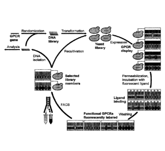
Fig. 1 shows the workflow for directed evolution of GPCRs in yeast. As a first
step, the wild-type
GPCR gene is randomized by error-prone PCR in order to create a DNA library.
The DNA library
is then combined with linearized yeast expression vector and the mixture is
used for
transformation of yeast. Insert DNA and vector backbone are assembled in vivo
by homologous
recombination. The obtained yeast library is cultivated and expression is
induced. After
expression, cells are permeabilized and incubated with fluorescent ligand,
which binds
exclusively to functional GPCRs in the plasma membrane. Subsequently, unbound
ligand is
removed by washing and cells are subjected to selection by FACS. Selection of
cells exhibiting
high fluorescence, correspondingly expressing GPCRs at high
8c
Date Recue/Date Received 2022-06-08
CA 02971111 2017-06-15
WO 2016/102508 PCT/EP2015/080858
functional level, allows isolation of desired high-expression GPCR variants.
During FRCS,
yeast cells are directly sorted into growth medium for subsequent propagation.
Selection by
FACS is repetitively performed in order to obtain a strong enrichment of cells
harboring the
best expressing GPCRs. Whenever desired, plasmid DNA can be isolated from
selected
cells for analysis of individual variants or additional diversity can be
introduced by random
mutagenesis for another round of evolution.
Fig. 2 shows (A), (C), (E) histogram plots of ligand-binding FC data of NTR1,
NK1R, and
KOR1 variants. Functional expression in yeast of wild-type GPCRs (left
panels), library pools
obtained after the second round of evolution (middle panel) and variants
evolved in the
E. coll-based system (right panels). Total signal was obtained by binding of
fluorescent
ligand to receptors in yeast cells expressing the corresponding GPCR variants
(black
curves). Nonspecific binding was measured in the presence of excess unlabeled
ligand
(gray, tinted). For the wild-type GPCRs no specific signal is detected due to
the low
functional expression levels at the surface. The selected library pools show
high specific
signals (functional receptors at the surface per cell) in expressing cells
with a small fraction
of cells not expressing any functional receptor at the surface (note double
peak of total
signal). Variants previously evolved in the E. coil-based system (NTR1-D03,
NK1R-E11)
show a specific signal, however not reaching the levels obtained for the yeast
library pools.
Furthermore, for a significantly higher fraction of cells no functional GPCR
expression is
detected at the surface. For instance, only about 50% of all cells measured
show a specific
signal for NTR1-D03 expression. (B), (D), (F) Measurement of average total
functional
receptors per cell by RLBA of NTR1, NK1R, and KOR1 variants. Expression levels
were
quantified with [31-1]-radioligands. Non-specific signal was measured in the
presence of [3N-
radioligands with excess of unlabeled ligand and was subtracted from the total
signal. Wild-
type NTR1 and KOR1 show low expression levels, while for NK1R no functional
expression
at all is detected. NTR1-D03 shows moderate receptor-per-cell levels, whereas
for NK1R-
Ell expression levels are low. The selected library pools show functional
production levels
of 100,000 ¨ 150,000 receptors per cell, an increase of 25 ¨ 50-fold and 5 ¨
20-fold
compared to the wild-type GPCRs and the variants evolved in E. coil,
respectively. Error bars
indicate standard deviations from triplicates of a representative expression
experiment.
Fig. 3 shows functional expression of selected NTR1 variants (NTR1-Y01 ¨ NTR1-
Y07) in
non-adapted yeast strains. (A) Histogram plots of ligand-binding FC data with
total signal
(black curves) and nonspecific signal (gray, tinted) are shown. Compared to
wild-type NTR1
all selected variants show an increase in functional surface expression (cf.
Fig. 2 A). In
comparison with the library pool NTR1 2.5, the individual variants depict an
increase of the
subpopulation not expressing functional receptor at the surface as well as
lower specific
signals (receptors per cell) in cells with surface expression of active
receptor (cf. Fig. 2 A).
9
CA 02971111 2017-06-15
WO 2016/102508 PCT/EP2015/080858
(B) Measurement of average total functional receptors per cell by RLBA. All
variants show an
increase in functional expression compared to wild-type NTR1, but do not reach
the
expression levels observed for the NTR1 2.5 pool (cf. Fig. 2 B). Non-specific
signal was
subtracted from total signal. Error bars indicate standard deviations from
triplicates of a
representative expression experiment.
Fig. 4 shows quantitative Western blot analysis of HA-tagged NTR1 variants
expressed in
non-adapted and adapted yeast strains. Whole cell lysate protein extraction
was performed
with equal numbers of cells for each sample after expression. As a loading
control, actin was
used. GPCRs were detected via their HA tag, with main bands corresponding to
monomeric
GPCRs running slightly below the expected molecular size (44 kDa) and bands of
higher
molecular weight, most likely representing GPCR dimers not disintegrated under
the
conditions used. For quantification (bar chart) intensities of all defined
bands were accounted
for and signals were normalized to the GPCR and actin intensities obtained for
wild-type
NTR1. In the non-adapted strains, the total GPCR produced increases from wild-
type NTR1
(lane 1) to the evolved variant (lane 2) by a factor of two. While total
receptor levels of NTR1-
Y06 also slightly increase in the adapted strain (lane 3) compared to the non-
adapted strain
(lane 2), the relative increase of total receptor produced is much lower than
the increase in
functional receptor (cf. Fig 13 B). For a negative control (lane 4), cells
expressing NTR1-Y06
without HA tag were used.
Fig. 5 shows confocal fluorescence microscopy studies of NTR1 variants with C-
terminal
fusion to mCh expressed in non-adapted and adapted yeast strains. Fluorescence
intensities
obtained by binding of fluorescent ligand (top row) or from mCh (middle row)
as well as
bright-field microscopy overlays (bottom row) are shown. For expression of
wild-type NTR1
(first column), no cells with a ligand-binding signal at the cell surface are
detected, while
some cells show distinct mCh signals, mainly located in the cell interior, and
thus reflecting
intracellularly retained receptors. Additionally, many cells lacking any
fluorescent signal are
detected, representing a non-expressing subpopulation. In contrast, expression
of NTR1-Y06
in non-adapted cells (second column) allows visualization of functional
receptor at the cell
surface by ligand binding. These cells show also strong signals for mCh,
however still to a
large extent localized in the cell interior. Similar to wild-type NTR1, NTR1-
Y06 expression in
the non-adapted strain gives rise to non-expressing cells, depicting neither a
signal for ligand
binding nor for mCh. For expression of NTR1-Y06 in the adapted strain (third
column),
significantly fewer non-expressing cells are detected and expressing cells
show at the
surface a strong signal for both ligand-binding and mCh, with only little mCh
detected in the
cell interior. For a negative control (fourth column) cells expressing NTR1-
Y06 without a mCh
fusion were incubated with fluorescently labeled neurotensin in excess of non-
labeled
neurotensin. Representative pictures are shown.
CA 02971111 2017-06-15
WO 2016/102508 PCT/EP2015/080858
Fig. 6 shows FC and RLBA analysis of non-adapted and adapted strains
expressing NTR1
variants with a C-terminal mCh fusion. (A) FC data of non-adapted strains
expressing NTR1
(left panel), NTR1-Y06 (middle panel), and the adapted strain expressing NTR1-
Y06 (right
panel) are shown. In the histogram plots of ligand-binding experiments (top
row), the surface
expression of active receptor variants are compared with total signal (black
curves) and
nonspecific signal (gray, tinted) shown. NTR1 shows only little active
receptor at the surface,
whereas surface expression is significantly increased for NTR1-Y06 in the non-
adapted and
the adapted strain (shift of the specific signal towards higher fluorescence
intensity).
Compared to the non-adapted strain expressing NTR1-Y06 with a mCh fusion,
adaptation
further leads to a substantial decrease of the fraction of cells showing no
surface expression
of active receptor. In the histograms of mCh expression (middle row) total
receptor produced
is quantified. Black curves depict the mCh signal, whereas autofluorescence of
cells
expressing NTR1-Y06 without mCh-fusion incubated with fluorescent ligand
represents the
background (gray, tinted). NTR1 and NTR1-Y06 expressions in non-adapted
strains have
very similar mCh signal profiles. Since for expression of NTR1 in ligand-
binding FC
experiments only little active receptor at the surface is detected, the strong
mCh signal
means that most of NTR1 must be intracellularly retained. A subpopulation of
cells not
expressing any receptor at all is seen for all variants (note double peak),
but this non-
expressing fraction is significantly decreased in the adapted strain. In
correlation analysis
(bottom row) mCh fluorescence intensity (total receptor produced) is compared
to ligand-
binding fluorescence intensity (functional receptor at the surface). For
expression of wild-type
NTR1, functional receptor at the surface does not correlate well with total
receptor produced.
This correlation is better for NTR1-Y06 expressed in the non-adapted strain,
and if NTR1-
Y06 is expressed in the adapted strain, functional receptor at the surface
correlates well with
total receptor produced. (B) Measurement of average total functional receptors
per cell by
RLBA. Functional expression levels of receptors with a mCh-fusion increase
from wild-type
receptor to the evolved variant by a factor of three in the non-adapted
strain. Compared to
expression of NTR1-Y06 in the non-adapted strain, adaptation leads to a
further increase in
average total functional expression by a factor of 6. Non-specific signal was
subtracted from
total signal. Error bars indicate standard deviations from triplicates of a
representative
expression experiment.
Fig. 7 shows functional expression levels of evolved GPCRs in St79 insect
cells and
signaling activity of NK1R variants. (A), (B), (C) Measurement of average
total functional
receptors per cell by RLBA of NTR1, NK1R, and KOR1 variants. Compared to wild-
type
GPCRs all evolved variants show a significant increase of average receptor-per-
cell levels.
NTR1-Y06 shows a fivefold increase compared to wild-type NTR1, NK1R-Y09 shows
a
fourfold and twofold increase compared to NK1R and NK1R-AC, respectively, and
the
11
strongest increase (27-fold) is detected for KOR1-Y05 compared to wild-type
KOR1. For each
GPCR two independent expression experiments were performed (separate bars).
Nonspecific
signal was subtracted from total signal. Error bars indicate standard
deviations from triplicates.
(D) Measurement of signaling activity of NK1 R variants by [35S]-GTPyS
binding. Equal amounts
of active GPCR and reconstituted G protein were assayed in the presence (grey)
and absence
(black) of substance P. All variants show a low basal activity without agonist
stimulation. Upon
addition of agonist, signaling is detected by [35S]-GTPyS binding. NK1R and
NK1R-AC have
identical signaling activities, and signaling of NK1R-Y09 remains similar to
wild-type receptor. For
each GPCR variant two independent signaling assays from two independent
expression
experiments were performed (separate bars). Error bars indicate standard
deviations from
triplicates.
Fig. 8: Scheme of GPCR topology depicting the different regions harboring
mutations (see Table
1).
Fig. 9 shows functional expression of selected NK1R and KOR1 variants in non-
adapted yeast
strains. (A), (B) Histogram plots of ligand binding FC data of NK1R and KOR1
variants,
respectively, with total signal (black curves) and nonspecific signal (gray,
tinted) shown.
Nonspecific signal was obtained in the presence of an excess of unlabeled
ligand. All variants
show an increase in functional expression compared to the corresponding wild-
type GPCRs
(NK1R and KOR1, cf. Fig. 2 C, E). While the specific signal of the expressing
cells of most
variants is similar or slightly lower compared to the corresponding library
pools (NK1R 2.5 and
KOR1 2.5, cf. Fig. 2 C, E), a higher fraction of cells not expressing any
functional receptor at the
surface is observed for the individual variants. (C), (D) Measurement of
average total functional
receptors per cell by RLBA. While all variants show an increase in functional
expression
compared to the corresponding wild-type GPCRs, for most variants the average
numbers of
receptors per cell measured by RLBA are lower than for the corresponding
library pools (cf. Fig.
2 D, F). Non-specific signal was subtracted from total signal. Error bars
indicate standard
deviations from triplicates of a representative expression experiment.
Fig. 10 shows functional expression levels of NTR1-Y06 in a yeast strain
directly isolated from
the NTR1 2.5 pool and a newly transformed strain subsequently adapted by
phenotypic selection
with FACS. (A), (B) Histogram plots of ligand-binding FC data with total
signal (black curves) and
nonspecific signal (gray, tinted) are shown. Non-specific signal was obtained
in the presence of
an excess of unlabeled ligand. The isolated strain shows a
12
Date Recue/Date Received 2022-06-08
CA 02971111 2017-06-15
WO 2016/102508 PCT/EP2015/080858
similar expression profile as the NTR1 2.5 library pool (cf. Fig. 2 A), in
contrast to the non-
adapted retransformed strain expressing NTR1-Y06 (cf. Fig. 3 A). In the
subsequently
adapted strain shown in (B), obtained after five rounds of phenotypic
selection by FACS with
the newly transformed strain, high surface expression levels are
reconstituted. (C)
Measurement of average total functional receptors per cell by RLBA. Compared
to the
retransformed strain expressing NTR1-Y06 (cf. Fig. 3 B), high functional NTR1-
Y06
expression in the isolated strain as well as reconstitution of this phenotype
in the adapted
strain is detected. Non-specific signal was subtracted from total signal.
Error bars indicate
standard deviations from triplicates of a representative expression
experiment.
Fig. 11 shows expression profiles of NTR1-Y06 after individual sorts during
phenotypic
selection for host adaptation. Histogram plots of ligand binding FC data with
total signal
(black curves) and nonspecific signal (gray, tinted) are shown. Non-specific
signal was
obtained in the presence of an excess of unlabeled ligand. In the course of
the phenotypic
selection in which the cells with the highest NTR1-Y06 expression were sorted
(gating of the
top 1% of the most fluorescent cells), the population of cells not expressing
any functional
receptor at the surface gradually decreases. At the same time, the specific
signal of the
expressing cells shifts to higher levels during the selection, indicating an
increase of
functional receptors per cell at the surface of these cells. Note that two
sorts are required to
induce the adaptation. While the expression profile is not significantly
changed after the first
two sorts, a clear decrease of the subpopulation with no surface expression of
active NTR1-
Y06 as well as a shift of the specific signal within the expressing
subpopulation is observed
after sort 3. This trend is continued in subsequent sorts, as seen in the
expression profile
after sort 4.
Fig. 12 shows functional expression of NTR1-Y06 in non-adapted, adapted, and
adapted
strain previously cultivated under non-expressing conditions in S. cerevisiae.
(A) Histogram
plots of ligand binding FC data with total signal (black curves) and
nonspecific signal (gray,
tinted) are shown. Non-specific signal was obtained in the presence of an
excess of
unlabeled ligand. Adaptation of newly transformed strain expressing NTR1-Y06
leads to a
higher specific signal (receptors per cell) of the expressing cell
subpopulation and a
decrease of the subpopulation not showing any surface expression of active
receptor. If the
adapted strain is repetitively cultivated under non-expressing conditions, the
average
expression level decreases again, depicted by a drop of the specific signal
(receptors per
cell) in the expressing subpopulation and an increase of the fraction of cells
with no surface
expression of active NTR1-Y06. (B) Measurement of average total functional
receptors per
cell by RLBA. Results from RLBA show the significant increase of the number of
average
receptors per cell from the non-adapted to the adapted strain, which in turn
drops again by
about 30% if the adapted strain is repetitively cultivated under non-
expressing conditions
13
CA 02971111 2017-06-15
WO 2016/102508 PCT/EP2015/080858
prior to inducing expression. The lack of complete reversion to the original
lower expression
level of the non-adapted strain may be attributed to leaky expression,
sufficient to partially
retain the high-expression phenotype even under non-inducing conditions. Non-
specific
signal was subtracted from total signal. Error bars indicate standard
deviations from
triplicates of a representative expression experiment.
Fig. 13 shows functional expression of NK1R-Y09 and KOR1-Y05 in non-adapted
and
adapted S. cerevisiae strain. (A), (C) Histogram plots of ligand binding FC
data with total
signal (red curves) and nonspecific signal (green, tinted) are shown. Non-
specific signal was
obtained in the presence of an excess of unlabeled ligand. While the specific
expression
signals (receptors per cell) of expressing cells in the adapted strains
slightly increase
compared to the non-adapted strains, a significant decrease of the
subpopulation with no
surface expression of active receptor is observed. (B), (D) Measurement of
average total
functional receptors per cell by RLBA. The decrease of the fraction of cells
which show no
surface expression of active receptor in the adapted strains, shown in the FC
data, leads to
an increase of the average functional expression measured with RLBA. Non-
specific signal
was subtracted from total signal. Error bars indicate standard deviations from
triplicates of a
representative expression experiment.
Fig. 14 shows functional expression of HA-tagged NTR1-Y06 in non-adapted and
adapted
S. cerevisiae strain. (A) Histogram plots of ligand-binding FC data with total
signal (black
curves) and nonspecific signal (gray, tinted) are shown. Non-specific signal
was obtained in
the presence of an excess of unlabeled ligand. Adaptation of the strain
expressing NTR1-
Y06 with a C-terminal HA-tag leads to a decrease of the fraction of cells
showing no surface
expression of active receptor and an increase of active receptors per cell at
the surface of
cells with surface expression (shift of specific signal towards higher
fluorescence intensity).
(B) Measurement of average total functional receptors per cell by RLBA.
Functional
expression levels increase from wild-type receptor to the evolved variant by a
factor of four in
the non-adapted strain. Compared to expression of HA-tagged NTR1-Y06 in the
non-
adapted strain, adaptation leads to a further increase in average total
functional expression
by a factor of 10. Non-specific signal was subtracted from total signal. Error
bars indicate
standard deviations from triplicates of a representative expression
experiment.
Fig. 15 shows purification of NK1R-Y09. (A) SEC profile of purified receptor
after IMAC. Two
peaks are obtained of which peak 1 most likely corresponds to defined higher
oligomeric
states of NK1R-Y09, while peak 2 represents the monomeric receptor fraction.
(B) Analysis
of purified NK1R-Y09 by SDS-PAGE under reducing conditions. Equal amounts of
total
protein were loaded in each lane. After IMAC (lane 1) pure NK1R-Y09 (38.5 kDa)
is
obtained, with detection of weak additional protein bands at higher molecular
weight. Such
bands represent most likely oligomers of NK1R-Y09, which are not disintegrated
under
14
conditions used. Next to a strong band for monomeric NK1 R-Y09, the bands of
higher molecular
weight are also detected in the fraction of peak 1 (lane 2), while in the
fraction of peak 2 (lane 3)
monomeric NK1 R-Y09 is the sole species. Note that NK1 R-Y09 was not further
engineered and
expressed in insect cells as it was obtained from the selection in yeast. For
purifications with the
aim to perform crystallization trials, some further engineering will be
required, for instance
removal of potential glycosylation sites, N-terminal truncations, loop
deletions, and/or introduction
of fusion proteins (e.g. T4 lysozyme or thermostabilized apocytochrome b562RIL
(Chun et al.
(2012), Structure 20:967-976)). Such measures potentially further improve
purification of the
receptor by reducing heterogeneity, restricting conformational flexibility,
and increasing stability
(Maeda & Schertler (2013), Curr Opin Struct Biol 23:381-392).
Examples
Example 1: Generation of high-expressing functional GPCRs by directed
evolution in yeast
With about 800 different members in the human genome, G protein-coupled
receptors (GPCRs)
comprise the largest superfamily of cell surface receptors. GPCRs evolved to
highly versatile
signaling mediators in eukaryotic life, responding to a wide variety of
ligands and transducing
signals via heterotrimeric G proteins as well as in a G protein-independent
fashion. The pivotal
role of GPCRs is reflected by the great number of human diseases linked to
aberrant GPCR
signaling, for instance obesity, diabetes, cardiovascular diseases,
osteoporosis, immunological
disorders, neurodegenerative diseases, and cancer. As a consequence, GPCRs
represent highly
relevant drug targets for the pharmaceutical industry. About 30 - 50% of
marketed drugs act on
GPCRs or GPCR-associated mechanisms, among them many top-selling drugs.
The major challenges in structural and detailed biochemical investigation of
GPCRs are the
difficulties to produce the desired receptors in sufficient amounts as well as
their inherent
instability and flexibility. Advances in expression and crystallization
strategies, like the use of the
baculovirusfinsect cell expression system, or the establishment of integral
membrane protein
crystallization in lipidic cubic phases (LCP), lead to breakthroughs in
determination of three-
dimensional receptor structures by X-ray crystallography. While the available
structural datasets
provided the scientific community with insights towards the mechanisms of GPCR
function at the
atomic level and might begin to enable rational structure-based drug design,
it is clear that
detailed understanding of receptor dynamics and conformational changes has
still remained
rather incomplete.
Furthermore, the 26 unique GPCR structures deposited in the Protein Data Bank
to date still
represent only a minor fraction of all receptors (<4%),
Date Recue/Date Received 2022-06-08
CA 02971111 2017-06-15
WO 2016/102508 PCT/EP2015/080858
reflecting that the bottlenecks for structural investigations of GPCRs
persist. For instance,
while insect cells were successfully used for production of recombinant
receptors for about
85% of all GPCR structures determined so far (source: PDB; excluding
structures obtained
from protein extracted from native tissues), this expression system does not
provide a
generic solution. Even in insect cells several members of the GPCR superfamily
are
expressed at low yields, suggesting fundamental issues with the biosynthesis
and membrane
insertion of these receptors also in eukaryotic cells. This problem cannot be
solved by simple
optimization of expression conditions, especially since there seems to be no
consistency of
optimal conditions for individual GPCRs. Thus, to facilitate and accelerate
GPCR research,
novel approaches are required.
Recently, the inventors developed a method to increase functional expression
levels of
GPCRs in Escherichia coil by directed evolution (Sarkar et al. (2008), Proc
Natl Acad Sci
USA 105:14808-14813; Dodevski & Pluckthun (2011), J Mol Biol 408:599-615).
Random
mutagenesis of wild-type GPCRs followed by selection with fluorescence-
activated cell
sorting (FACS) allowed isolation of high-expression variants from randomized
libraries of four
different GPCRs, namely neurotensin receptor 1 (NTR1), NK-1 receptor (NK1R,
also termed
tachykinin receptor 1 or substance-P receptor) and of the alpha-1A and alpha-
1B adrenergic
receptors. In successive studies for NTR1, the obtained first-generation
variants built the
basis for creation of second-generation mutants by extensive selection
(Schlinkmann et al.
(2012), Proc Natl Acad Sci USA 109:9810-9815) and exhaustive recombination
(Schlinkmann et al. (2012), J Mol Biol 422:414-428). The synthetic libraries
created in these
studies were also used to generate NTR1 variants with improved stability in
short-chain
detergents by a complementary directed evolution approach termed CHESS (Scott
&
Pluckthun (2013), J Mol Biol 425:662-677; Scott et al. (2014), Biochim Biophys
Acta
1838:2817-2824). Ultimately, these efforts resulted in the structures of three
different
variants of agonist-bound NTR1 from material produced in E. coli (Egloff et
al. (2014), Proc
Natl Acad Sci USA 111:E655-62), underlining the power of directed evolution in
membrane
protein engineering.
The successful results obtained with evolution in E. coil and the proven
effectiveness of
eukaryotic expression hosts demanded to further develop this approach towards
high
functional GPCR expression specifically in eukaryotic expression systems. The
inventors
hypothesized that it might be advantageous to perform evolution of GPCRs
directly in a
eukaryotic system in order to achieve specific sequence adaptation and thereby
improved
functional production in eukaryotes. Furthermore, compared to eukaryotic
hosts, E. coil is not
able to perform post-translational modifications, lacks the secretory quality
control and
translocation machinery for eukaryotic membrane proteins, and differs in
membrane
composition. Therefore, directed evolution of GPCRs in E. coil is limited to
receptors which
16
CA 02971111 2017-06-15
WO 2016/102508 PCT/EP2015/080858
inherently express in a prokaryotic system above the threshold required for
successful
evolution, thus not having strict requirements for eukaryotic processing or
membrane
composition.
Here the inventors disclose the establishment of a fast, efficient and robust
method to
increase functional expression of GPCRs in eukaryotic hosts by directed
evolution in the
yeast Saccharomyces cerevisiae. The method is generally applicable as
demonstrated with
three different GPCRs, leading, with only two rounds of evolution, to receptor
variants which
show high functional expression in both yeast and insect cells. In addition,
functional
expression levels in yeast can be further increased reproducibly by induced
host adaptation.
Directed evolution of three different GPCRs in yeast results in high
functional expression in
S. cerevisiae
The general approach of this method is depicted in Fig. 1. First, the wild-
type GPCR gene is
randomized by error-prone PCR and the resulting DNA library is used for
transformation of
yeast. The insert DNA and yeast expression vector backbone are assembled in
vivo via
designed homologous recombination sites. Next, expression in the obtained
yeast library is
induced, and subsequently cells are treated with an optimized buffer for
permeabilization of
the yeast cell wall. Permeabilization is necessary for allowing access and
thus binding of
administered fluorescent ligand to the functionally expressed receptors in the
plasma
membrane. After incubation with saturating concentrations of fluorescent
ligand, unbound
ligand is removed by washing, and cells are subjected to selection with FACS.
Yeast cells
expressing variants with the largest number of functional receptor molecules
at the surface
correspondingly exhibit the highest fluorescence. These cells are isolated by
gating the top
0.5 ¨ 1% of the most fluorescent yeast cells, and sorting them directly into
growth medium for
subsequent propagation. In order to achieve strong enrichment of cells
expressing GPCR
variants with the desired phenotype, several repetitive cycles of expression,
incubation with
fluorescent ligand, and FACS are performed. Plasmid DNA can be isolated from
selected
cells after FACS for analysis or introduction of further diversity by
additional random
mutagenesis, thereby starting the next round of evolution.
The inventors aimed to evolve three different GPCRs in parallel: (i) rat NTR1,
(ii) human
NK1R, and (iii) human kappa-type opioid receptor (KOR1). NTR1 has been shown
to be
readily evolvable in the E. coil-based system in several studies, and thus was
considered a
positive control. NK1R has been successfully subjected to directed evolution
towards higher
expression in E. coil as well (Dodevski & Pluckthun (2011), J Mol Biol 408:599-
615).
However, despite strong relative improvements, due to the very low expression
levels of wild-
type NK1R in the prokaryote, the evolved receptor variants expressed still at
only moderate
absolute levels compared to the other receptors evolved in E. coil (Sarkar et
al. (2008), Proc
17
CA 02971111 2017-06-15
WO 2016/102508 PCT/EP2015/080858
Natl Acad Sci USA 105:14808-14813; Dodevski & PlEickthun (2011), J Mol Biol
408:599-
615). Moreover, expression levels of these evolved NK1R variants were also low
in
S. cerevisiae (see below), suggesting that further improvement of functional
production might
be possible, which may benefit future studies on this receptor. Concerning
KOR1, no
attempts to evolve this receptor have been undertaken so far and it represents
a challenging
example regarding heterologous expression.
Following the procedure outlined above, for each of the three receptors only
two rounds of
evolution ¨ each round consisting of one randomization by error-prone PCR and
five
subsequent selections by FACS ¨ were sufficient to strongly increase
functional expression
levels. Fig. 2 shows a comparison of the functional expression levels in S.
cerevisiae of wild-
type GPCRs, NTR1 and NK1R variants previously evolved in the E. coll-based
system
(NTR1-D03 and NK1R-E11, respectively), and the library pools obtained after
the second
round of evolution, namely NTR1 2.5, NK1R 2.5, and KOR1 2.5 (library
nomenclature: GPCR
a.b, where a is the number of total randomizations, and b is the number of
total FAGS
selections). Functional expression levels were measured either with flow
cytometry (FC) or
radioligand binding assays (RLBAs). Note that FC ligand-binding experiments
determine
functional receptors exclusively in the plasma membrane at the surface of
intact individual
cells, whereas RLBAs account for the total amount of functional receptors
averaged across
an entire population of lysed cells, and thus also detect functional GPCRs in
intracellular
membranes. For the wild-type GPCRs no specific signal is obtained in FC
experiments, thus
no active receptor in the plasma membrane at the surface is detected (Fig. 2
A, C, E). By
contrast, in RLBAs for NTR1 and KOR1 low expression levels of functional
receptor are
measured, indicating that a small amount of active receptor is present,
however, most likely
retained in intracellular membranes (Fig. 2 B, F), For NK1R, no functional
receptor is
detected in any of the methods used (Fig. 2 C, D), consistent with what has
been reported
before (Butz et al. (2003), Biotechnol Bioeng 84:292-304).
Strikingly, the expression levels of the selected libraries are not only much
higher than the
wild-type receptors but also higher than the corresponding variants evolved in
E. coil (NTR1-
D03, NK1R-E11), illustrated by FC and RLBA analysis. 100,000 ¨ 150,000 total
functional
receptors per cell are measured in RLBA for the selected yeast libraries,
representing a 25 ¨
50-fold and 5 ¨ 20-fold increase compared to the corresponding wild-type GPCRs
and
E. coil-evolved variants, respectively (Fig. 2 B, D, F). The FC data confirm
active receptor at
the surface by showing a distinct specific signal for both evolved variants
from E. coil and
selected yeast libraries (Fig. 2 A, C, E). However, the specific signal of the
selected yeast
libraries is shifted towards higher fluorescence intensity compared to the E.
coil-evolved
variants, reflecting an increase in active receptors at the surface.
Furthermore, a significantly
18
smaller subpopulation of cells not expressing any functional receptor at the
surface is detected in
the selected yeast libraries.
Such a division into two subpopulations was reproducibly observed in FC
experiments upon
expression of all GPCRs tested in both yeast single clones and libraries.
Hence, this effect
appears to be an intrinsic feature of GPCR expression in S. cerevisiae, in
which functional
expression at the surface is lacking in a substantial fraction of cells. For
instance, depicted by the
FC data of the E. coll-evolved variants, active surface expression of NTR1-D03
is obtained in
about 50% of all cells, and for the low-expressing variant NKR1-E11 only a
minority of cells show
active surface expression (Fig. 2 A, C). It remains unclear why some cells of
the population
grown from one single clone do not produce any functional receptor at the
surface, but a loss of
the expression vector in these cells can be excluded since all cultivations
are performed
exclusively in selective minimal medium.
Improved expression levels of enriched receptor variants in yeast are further
increased by host
adaptation occurring concurrently with selection.
In order to identify enriched clones, plasmid DNA was isolated from the
library pools after the first
(stage 1.5 libraries) and second round (stage 2.5 libraries) of evolution for
DNA sequencing. A
summary of the selected clones depicting the different mutations of each clone
is given in Table
1, with a scheme of GPCR topology depicting the different regions harboring
mutations shown in
Fig. 8.
19
Date Recue/Date Received 2022-06-08
Table 1: Overview of the most highly enriched clones during evolution for each
GPCR. Mutations
of each clone are indicated and the corresponding wild-type amino acids are
given on top of the
mutation data. Positions of mutations are indicated by structural regions,
Ballesteros-Weinstein
numbering (3), and sequential amino acid numbering. Section A: NTR1 variants.
Section B:
NK1R variants. Section C: KOR1 variants. Scheme of GPCR topology depicting the
different
regions harboring mutations is shown in Fig. 8.
µ
A I A
2.44 2.50 449 5.36 5.49 5.58 562 5.69 6.45 7.40 7.42 7.47 7.54 8,53
I 44 107 113 133 237 248 257 251 us 260 264 202 318 356 358 363 370 379 386
392 398
N1111 A00 1 V F II A V SRNS A F A NV CRRTolimulitIon
1, TI-11 Y01 p ti s 5 4
(F-I1 Yrn, P II I. A A s e
Nun-vv., P 0 N s s s
1s1R1-Y04' p IS s s is s
ram YOG P N s s e C 6
NTR 1 -Y oc,' P N 6 8 0 TL 1 1 C 19
p.mi-yoT p N L A a e DAIL 1 A G 13
1861614,1 from Ninny: 'MP 1 ^ 5
011101 2.5
ri 1 40 559 5.64 i 6 31 7.34 7.47
g 40 ra 06 H. ¨, .as 141 217 222 2L4 ....-, 04 zoL 280 299 404 $31 335 040 037
NNW T RIIK DI. 0 T TWF N T V RIC TERN 114101mubikals
5vi RAC K ' 2
1^-1<11R- \=11- $ A = 5
1,I,1R-Y02 Y * 2
to,iFt-vc3' = 1
1,1,1R YO4 I K R = 4
to<ifR=vo5 I K R W K = 0
NI1.1R-YOC A I K R W K = 7
I K W K ^ 5
NK IR-YOB A a r 1 . 5
MIR- Yo1.1" a H K R F Mi K = 8
pipiR2e13 V K I/ r W K * 7
Isolslad from Imam "NK IR i 1
"OR 1R 2.5
C / =:, ;= -' = ,' .- ' - -,',, ,. 1.2 ' '-6 CI,
:".111
1 S 19 314 16628 21.01 21'011 21188C 2116: 31":; 34: 315437 3157 4. 'a
. 031 73: 353 377
KOR1 P GP 1311D T V INSREA 5 T
EN UN muladm
pons YO1 A 0 5 0 1 K e
KO R 1. YO2'' a a 0 0 8 C 0 1 K 0
Konl-yo' 8 0 0 8 C 1 K r
KOR I -Y04 R S 0 0 3 C I K e
SC R 1-Y05" 8 0 13 8 C V 1 K e
Km,. =Y0.5 SG 0 8CH0 I K 9
160p1 yor" 1 P SG D T8 C 1 K 0 11
WNW from Ilbram 'K3R1 15
103R1 25
Interestingly, whereas for NTR1 and KOR1 only full-length variants were
selected, all selected
NK1R variants contain a stop codon at a position within a narrow range of
seven amino acids
after the presumed helix 8 and palmitoylation site, resulting in a shortened C-
terminus. This
indicates that a full-length C-terminus is strongly selected against and a
truncation leads to a
significant increase in expression for this receptor. Since no functional wild-
type NK1R can be
detected in yeast, the inventors constructed an alternative reference variant,
termed NK1R-AC,
with the most prominently selected C-terminal truncation combined with the
wild-type sequence.
19a
Date Recue/Date Received 2022-06-08
This provided the inventors with a reference for the selected mutations and
also allowed them to
quantify how much a C-terminal truncation contributes to improved functional
expression for
NK1 R.
Individual clones were analyzed after retransformation and revealed that all
enriched receptor
variants were indeed significantly better expressed in yeast than the
corresponding wild-type
receptors (Fig. 3 and Fig. 9). This indicates that variants have indeed been
obtained that are
better adapted to the biosynthesis in this eukaryotic host.
While only a small fraction of cells not expressing any functional receptor at
the surface was
detected in the selected GPCR libraries (Fig. 2 A, C, E), a larger
subpopulation of such cells was
observed when the individual variants had been retransformed into fresh host
cells (Fig. 3 A and
Fig. 9 A, B). Furthermore, lower receptor-per-cell levels of individual NTR1
variants in cells with
active surface expression after retransformation were also detected, as
1 9 b
Date Recue/Date Received 2022-06-08
CA 02971111 2017-06-15
WO 2016/102508 PCT/EP2015/080858
indicated by the shift of the specific FC signal towards lower fluorescence
intensity (Fig. 3 A).
Consistent with these observations of a combined effect in FC, significantly
lower average
RLBA readings were obtained in retransformed strains expressing individual
NTR1 variants
(Fig. 3 B), compared to the selected GPCR library (Fig. 2 B).
The inventors hypothesized that in the course of selection an adaptation of
the expression
host occurred which led to a further increased level of membrane protein
expression in the
selected libraries. To test this hypothesis, a yeast clone, expressing a
strongly selected
variant termed NTR1-Y06, was directly isolated from the NTR1 2.5 library pool.
As expected,
the isolated strain expresses NTR1-Y06 at high functional levels, with a
similar expression
profile as the NTR1 2.5 library pool (Fig. 10 A, C). Sequencing of whole
plasmid DNA
isolated from both the retransformed and the isolated strain expressing NTR1-
Y06 revealed
no difference. Thus, the inventors concluded that the actual procedure of
repetitive
expression and sorting of the best expressing clones by FACS induced an
adaptation of
yeast towards improved GPCR production. To test this hypothesis, five
subsequent mock
selections (without any randomization of the sequence) with FAGS with the
retransformed
single clone expressing NTR1-Y06 were performed, in which the cells with the
highest
NTR1-Y06 expression were isolated by gating the top 1.0% of the most
fluorescent cells.
Indeed, in the course of this phenotypic selections the strain adapted to
higher expression of
NTR1-Y06 by a gradual decrease of cells with no active surface expression and
an increase
of functional receptors per cell in the subpopulation with surface expression
(Fig. 11). Finally
after adaptation, NTR1-Y06 is expressed at total functional levels as high as
the expression
levels obtained in the selected NTR1 2.5 pool with about 160,000 average
functional
receptors per cell and a similar expression profile (Fig. 10 B, C). However,
by repetitive
cultivation of the adapted strain under non-expressing conditions, a partial
reversion of the
adaptive effect by a 30% drop of the expression level was observed (Fig. 12).
In order to test whether the phenotypic adaptation might even have been the
main feature of
the selection, the inventors also tried to adapt the strain expressing wild-
type NTR1.
However, after FACS, the sorted cells did not propagate anymore, preventing
repetitive
selections. This observation clearly shows that the selection of mutations,
conferring
improved properties to the receptors, is a strict prerequisite of the host
adaptation, and that
the sequence change is actually the key evolutionary event. The failure to
induce adaptation
in cells expressing wild-type NTR1 may be explained by the combined stressful
effects of
expression of the toxic wild-type GPCR followed by selection, together
limiting cell survival.
Hence, adaptation induced by repetitive sorting appears to be restricted to
evolved GPCR
variants, which intrinsically are less toxic for the cells. Indeed, adaptation
of strains
expressing NK1R-Y09 and KOR1-Y05, evolved NK1R and KOR1 variants,
respectively, was
possible as well. Both variants were strongly enriched during selection and
newly
CA 02971111 2017-06-15
WO 2016/102508 PCT/EP2015/080858
transformed cells expressing NK1R-Y09 or KOR1-Y05 showed lower expression
levels than
the corresponding library pools. For each of the variants five rounds of
phenotypic selection
by FACS, identical to the procedure used to adapt the strain expressing NTR1-
Y06, were
performed. For both variants host adaptation towards higher expression levels
was
observed, gradually arising in the course of selection (Fig. 13). Even though
the host
adaptation effect for NK1R-Y09 and KOR1-Y05 is less pronounced than for NTR1-
Y06,
these results suggest that the adaptation is not receptor-specific, and can be
reproducibly
induced, if evolved receptors are expressed.
The fraction of functional receptor is increased in adapted strains, while the
amount of total
receptor produced is not significantly higher.
Since host adaptation was most pronounced for expression of NTR1-Y06, this
variant was
used to further study the effect. While ligand-binding FC analysis and RLBA
permit the
detection of active receptor, neither of these methods allow quantification of
total receptor
produced regardless of ligand-binding activity. Therefore, to detect total
receptor protein
produced in different strains by immunoblotting, NTR1 and NTR1-Y06 expression
constructs
with a hemagglutinin (HA) tag at the C-terminus of the receptors were created.
The strain expressing the HA-tagged NTR1-Y06 was adapted towards higher
expression by
five rounds of phenotypic selection with FACS (Fig. 14 A). Subsequently, total
receptor
production for the different strains expressing wild-type NTR1 and NTR1-Y06,
the latter in
non-adapted and adapted strain, was measured by HA tag detection in
quantitative Western
blots from whole cell lysates, using equal numbers of yeast cells after
expression (Fig. 4).
In non-adapted strains, the amount of total receptor produced increases from
wild-type NTR1
to the evolved variants by a factor of two (Fig. 4). The observed increase of
functional
expression by RLBA data is by a factor of four (Fig. 14 B). This indicates
that the ratio of
functional receptor to total receptor produced is just slightly increased from
wild-type to the
evolved receptor.
However, when comparing the adapted to the non-adapted strain, based on RLBA
data,
there is a tenfold increase in functional expression of NTR1-Y06 (Fig. 14 B)
but there is less
than a twofold increase of total receptor production (Fig. 4). Since the
amount of total
receptor produced increases only slightly, it can be deduced that in the
adapted strain the
fraction of functional receptor is significantly increased.
21
CA 02971111 2017-06-15
WO 2016/102508 PCT/EP2015/080858
Adaptation increases surface expression of active receptor, reduces the number
of cells
exclusively expressing intracellularly retained GPCRs, and reduces the non-
expressing cell
subpopulation.
To further describe the host adaptation effect by assessing cellular
localization and
functionality of NTR1 variants in non-adapted and adapted strains, NTR1 and
NTR1-Y06
constructs with a C-terminal mCherry (mCh) fusion were used for confocal
microscopy
studies and FC experiments: while total receptor is quantified by mCh, only
functional
receptors located at the cell surface in the plasma membrane will exhibit a
signal from
fluorescent ligand binding.
Identical to the HA-tagged variant, the strain expressing NTR1-Y06 with a mCh
fusion was
first adapted by phenotypic selection. Next, the non-adapted strains
expressing NTR1 and
NTR1-Y06, as well as the adapted strain expressing NTR1-Y06 were exposed to
fluorescent
ligand to perform co-localization studies with confocal fluorescence
microscopy (Fig. 5).
As seen in ligand-binding FC experiments (Fig. 6 A), levels of functional wild-
type NTR1 in
the plasma membrane are very low, and consequently no ligand-binding signal is
detected in
microscopy (Fig. 5). In contrast, clear signals of mCh are obtained for some
cells, however
exclusively located in the cell interior, thus reflecting cells expressing
intracellularly retained
NTR1-mCh fusion receptors. According to the low RLBA readings obtained for
wild-type
NTR1 (Fig. 6 B), most of the intracellular receptor must be inactive.
Additionally, many other
cells are detected without any mCh signal at all, representing a non-
expressing
subpopulation (Fig. 5). Notably, both of these subpopulations, cells
exclusively expressing
intracellularly retained receptor as well as non-expressing cells, cannot be
discriminated in
ligand-binding experiments with FC. Thus, these two subpopulations together
constitute the
fraction of cells not showing any surface expression in FC ligand-binding
data.
Evolved NTR1-Y06 expressed in the non-adapted strain shows detectable ligand
binding at
the surface as well as a mCh signal (Fig. 5). However, the signal of mCh at
the plasma
membrane is still rather weak with a strong mCh signal detected in the cell
interior, indicating
that, similar to wild-type NTR1, a substantial amount of receptor is retained
in intracellular
membranes. Furthermore, non-expressing cells without any signal, neither for
ligand binding
nor for mCh, are frequently detected as well. Hence, also for the evolved
receptor, the
subpopulation of cells with no surface expression detected in FC ligand-
binding experiments
comprises two types of cells: those without any receptor expression, and those
cells in which
expressed receptor is not translocated to the plasma membrane.
The situation is different for NTR1-Y06 expressed in the adapted strain, in
which strong
signals for ligand binding and mCh are detected in the plasma membrane (Fig.
5). Even
22
CA 02971111 2017-06-15
WO 2016/102508 PCT/EP2015/080858
though some signal is still seen in the cell interior, mCh is mainly localized
at the surface.
Furthermore, significantly fewer non-expressing cells are observed.
Further investigations by FC experiments (Fig. 6 A), comparing the ligand-
binding to the mCh
signal, confirmed the results obtained in the confocal microscopy studies:
signals for mCh in
the expressing subpopulation are detected for all variants, however, for wild-
type NTR1 the
mCh fluorescence intensity does not correlate well with ligand-binding signal
intensity. This is
reflecting the fact that most of the wild-type receptor is not translocating
to the cell surface.
The finding that fluorescence intensity of wild-type GPCRs fused to an
autofluorescent
protein (e.g. GFP or mCh) correlates only weakly with functional receptor
levels has been
described before. Interestingly, this correlation is better for NTR1-Y06
expression in the non-
adapted strain, and for NTR1-Y06 expressed in the adapted strain, total
receptor produced
and functional receptor correlate well. In this case, the receptor is mostly
transported to the
cell surface, reflecting the decrease of cells with exclusively intracellular
receptor expression.
Interestingly, the subpopulation showing not any mCh signal (non-expressing
cells) is also
decreased in the adapted strain, compared to the non-adapted strains, further
leading to an
overall increase of functional GPCR production levels in the adapted strain.
In summary, host adaptation of S. cerevisiae for GPCR expression increases
surface
expression of active receptor without increasing the total amount of receptor
produced, which
is equivalent to a higher percentage of active receptor produced. In contrast
to non-adapted
cells, for which a large fraction of cells does not show any functional
surface expression,
adapted cells show only small amounts of intracellularly retained receptor.
Since a significant
part of intracellular receptor is inactive, host adaptation leads to an
overall increase of active
receptor. Additionally, the subpopulation of cells not expressing any receptor
at all is also
decreased in the adapted strains compared to the non-adapted ones,
contributing further to
higher average functional production levels.
Evolved variants of all three GPCRs show high functional expression levels in
Spodoptera
frugiperda (Sf9) insect cells.
Ultimately, the inventor's goal was to increase functional GPCR expression not
only in yeast
but in insect cells as well. Thus, the functional expression levels of the
evolved GPCRs
NTR1-Y06, NK1R-Y09, and KOR1-Y05 were determined in Sf9 insect cells and
compared to
the corresponding wild-type receptors. For all GPCRs identical standard
expression
conditions (non-optimized) were used. Indeed, a significant increase of
functional expression
of the evolved variants was observed. For NTR1-Y06 compared to wild-type NTR1,
functional expression is increased 5-fold (Fig. 7 A), for NK1R-Y09 a 4-fold
increase is
measured over wild type (Fig. 7 B), and for KOR1-Y05 ¨ notably being the most
challenging
example regarding expression studied in this work ¨ a 27-fold increase of
functional
23
CA 02971111 2017-06-15
WO 2016/102508 PCT/EP2015/080858
expression is detected (Fig. 7 C). The measured average functional receptor-
per-cell levels
(4.1 x 10⁶ ¨ 5.5 x 10⁶ receptors per cell) are high and surpass expression
levels of first- and
even second-generation NTR1 mutants generated in the E. coil-based directed
evolution
system when those are expressed in SP9 insect cells (Schlinkmann et al.
(2012), J Mol Biol
422:414-428).
With such high expression levels, as obtained for all of our three GPCR
mutants tested,
expression cultures in liter scales are sufficient to produce enough material
for structural
investigations. To test this, purifications of NK1R-Y09 from insect cell
expression cultures
were performed, reproducibly yielding 4 ¨ 6 mg/L pure protein. Fig. 15 shows
the analysis of
purified NK1R-Y09 by size-exclusion (SEC) and SDS-PAGE.
Given the very high purification yields, NK1R-Y09 represents an interesting
and promising
candidate for structural investigations. Thus, the functionality of NK1R-Y09
was
characterized by signaling assays based on [36S]-GTPyS binding (Fig. 7 D).
NK1R-Y09
shows similar activity as wild-type NK1R and the C-terminally truncated
variant NK1R-L,C,
both regarding [36S]-GTPyS binding without stimulation (basal activity) and
upon agonist
stimulation, further underlining the potential of our approach.
Summary
Production of recombinant GPCRs in functional form still remains a demanding,
laborious,
and cost-intensive task. Given that most of the recently determined three-
dimensional GPCR
structures were obtained from receptors produced in eukaryotic expression
hosts and
encouraged by the success of protein engineering methods for increased
expression of
GPCRs in E. coli, the inventors aimed to transfer the directed evolution
approach to
S. cerevisiae to specifically improve functional GPCR production in
eukaryotes.
The yeast S. cerevisiae, characterized by fast growth, cost-effective
cultivation, and ease of
genetic manipulations, is an ideal eukaryotic host for protein engineering and
directed
evolution for several additional reasons: First, yeast surface display has
become a very
powerful technology in a range of different applications. Second, high-
diversity libraries are
nowadays easily obtained with yeast. Third, the yeast cell is equipped with
the cellular
machinery required for functional GPCR production, since S. cerevisiae
endogenously
expresses two different GPCRs with signaling pathways similar to higher
eukaryotes. Fourth,
yeast has already been extensively used for heterologous expression of several
different
GPCRs. And fifth, based on a widely used GPCR assay which couples heterologous
GPCRs
to the yeast endogenous signaling pathway, directed evolution approaches on
GPCRs have
been successfully used before with the aim to generate designer receptors
exclusively
activated by designer drugs (DREADDs) activated by new ligands (Armbruster et
al. (2007),
Proc Natl Acad Sci USA 104:5163-5168).
24
CA 02971111 2017-06-15
WO 2016/102508 PCT/EP2015/080858
So far, functional production yields of GPCRs in yeast have generally remained
low
(Sarramegna et al. (2003), Cell Mol Life Sci 60:1529-1546). Strategies to
increase GPCR
expression in S. cerevisiae included optimization of expression conditions, co-
expression of
molecular chaperones, screening for high-expressing host clones, screening of
engineered
GPCR variants, or host engineering. In this specification the inventors
disclose the general
applicability of this invention to directly obtain GPCR variants with
increased functional
GPCR expression levels by evolving three different GPCRs. Remarkably, only two
rounds of
evolution ¨ meaning two randomizations each followed by selection with FAGS ¨
were
required to obtain highly increased expression levels, and thus it was
possible to evolve all
three GPCRs efficiently in a short time. In the E. coll-based system, usually
four rounds of
evolution were required. Furthermore, the inventors disclose that the yeast
host cell can be
adapted towards increased GPCR production. This is an inherent feature of the
selection
system, which happens concurrently with the selection of improved receptor
variants. It is
important to reiterate that the directed evolution did lead to sequence
changes, whose
improved expression phenotype could be transferred to insect cells, and that
the host
adaptation is only an additional factor occurring in yeast during the
selection. Moreover, the
inventors found that expression of an evolved receptor is a prerequisite for
inducing the host
adaptation, as attempts to induce host adaptation in cells expressing a wild-
type GPCR
failed. The host adaptation of yeast can reproducibly be induced by repetitive
phenotypic
selection with FAGS. This usually requires 4 ¨ 5 sorts, and the effect
gradually increases
during the selection.
Adapted strains show a significant increase of surface-expressed active
receptor, leading to
a much higher fraction of functional receptors in the plasma membrane, with
only a small
increase of total receptor produced. On the contrary, in non-adapted strains,
and especially
for expression of wild-type receptors, a large amount of receptors remain in
intracellular
compartments, of which a significant amount represents inactive misfolded
protein. The
overall higher average functional production yields in expression cultures of
adapted cells
can be explained by a decrease of the subpopulation of cells expressing
intracellularly
retained inactive receptor as well as a decrease of the non-expressing
subpopulation.
As the inventors did not wish to be limited to S. cerevisiae as a production
host, it was
explored whether the obtained GPCR variants would permit higher expression in
other
eukaryotic hosts as well. Indeed, functional expression of GPCR variants
evolved in yeast
was also increased in insect cells. Notably, variants evolved in the E. coil-
based system
show less improved functional expression when moved to eukaryotic hosts like
insect cells,
compared to the variants generated in yeast (Schlinkmann et al. (2012), J Mol
Biol 422:414-
428). The high expression levels in Sf9 cells obtained with the novel yeast-
evolved variants
are sufficient for crystallographic investigations with expression cultures in
liter scales under
CA 02971111 2017-06-15
WO 2016/102508 PCT/EP2015/080858
standard conditions. More specifically, purification of the NK1R variant NK1R-
Y09 yielded
very large quantities of pure protein. Furthermore, NK1R-Y09 showed signaling
activities
similar to wild-type NK1R, illustrating that the disclosed method can give
rise to biologically
active variants able to perform their naturally intended function, which
consequently can be
studied conveniently with these variants.
In summary, the method for directed evolution of GPCRs in S. cerevisiae
disclosed herein
allows efficient generation of GPCR variants with high functional expression
in eukaryotic
expression hosts in a short time. Using S. cerevisiae as evolution host
resulted in GPCR
variants with high functional expression levels in both yeast and insect
cells, in the latter
system by up to a factor 27. Thus, the mutations are transferable and selected
variants
showed a greater increase in expression in yeast as well as in insect cells
than when
evolution had been performed in E. coll.
The transferability of the beneficial effects of the receptor mutations to
insect cells further
promotes SID cells as expression host for large-scale production of the
generated mutants.
Nonetheless, the fact that high expression levels in S. cerevisiae can now
also be obtained
as a result of GPCR evolution, combined with induced host adaptation, implies
a potential of
yeast as a large-scale production host. From a practical point of view, the
fact that in adapted
strains the ratio of functional receptor to total receptor produced is
improved is a great
advantage for purification strategies like IMAC, which cannot discriminate
between non-
functional and functional protein. This is of special interest for
purifications of receptors for
which no ligand affinity column, for instance like the cleavable ligand column
as established
for purification of NTR1 (Egloff et al. (2014), Protein Expr Purif, in press),
is available.
The versatility of S. cerevisiae broadens the portfolio of GPCRs amenable to
our approach,
allowing evolution of GPCRs for which the previously established system in E.
coil is
inadequate. For instance, for GPCRs like KOR1, which show very low functional
expression
levels even in eukaryotes, expression levels in E. coil may be below the
threshold needed for
successful evolution. Furthermore, while neither prokaryotes nor native yeast
do produce
cholesterol ¨ on which activity of some GPCRs relies ¨ cholesterol-producing
S. cerevisiae
strains have been engineered. In principle, the disclosed method should be
easily
transferable to such or any other engineered yeast strains.
Yeast strain, vectors, cultivation, and expression.
For all experiments S. cerevisiae strain BY4741 (MATa his3A1 leu2A0 met15A0
ura3A0),
obtained from EUROSCARF, was used. Standard expression was performed with
pMS03het, derived from p415 GAL1. For more details on different yeast
expression vectors
see below. BY4741 transformed with pMS03het vectors were cultivated at 30 C in
SDD-Leu-
medium (6.9 g/L yeast nitrogen base without amino acids (Formedium), 690 mg/L
complete
26
supplement mixture without leucine (Formedium), 20 g/L glucose, 35 nnM sodium
citrate tribasic,
35 mM citric acid). For expression, yeast cells in the logarithmic growth
phase grown in SOD-
Leu- medium at 30 C were centrifuged and subsequently resuspended in SDG-Leu-
medium
(identical to SDD-Leu- but with 20 g/L galactose instead of glucose). Initial
00600 was always
chosen to be 1.0 after resuspension in SDG-Leu- and expression was performed
at 20 C for 24
h.
Yeast expression vectors
To obtain pMS03het, the a-mating factor prepro sequence was cloned from pPICZa
A (Life
Technologies) into the multiple cloning site (XhollSpel) of p415 GAL1.
pMS03het contains
NhellBamHI restriction sites which allow efficient vector linearization for
high-efficiency
transformation or in-frame cloning of genes preceded by the a-mating factor
prepro sequence.
For expression of GPCRs with a C-terminal HA tag or fusion to mCherry, vectors
pMS03het_HA
or pMS03het_mCh were used, respectively. To obtain pMS03het_HA and
pMS03het_mCh,
sequences coding for HA tag or mCherry were cloned via BamHI into pMS03het.
Yeast library construction and transformation.
The wild-type gene of rat NTR1 (N-terminally truncated from amino acids 1 -
42) was a kind gift
from Reinhard Grisshammer (National Institutes of Health). Wild-type cDNA of
human NK1 R
and human KOR1 was obtained from the Missouri S&T cDNA Resource Center. All
wild-type
GPCR genes were cloned into pMS03het (NhellBamHI). DNA library construction
was performed
by amplification of wild-type genes or isolated DNA after the first round of
evolution with error-
prone PCR using the GeneMorph TM II Random Mutagenesis Kit (Agilent
Technologies) according
to the manufacturer's protocol. Transformation of BY4741 with DNA libraries
was performed by
square wave electroporation on a GenePulser XcellTM electroporator (Bio-Rad).
On average,
libraries with a diversity of 5 x 10⁷ - 1 x 10⁸ were obtained. For more
details on library construction
and high-efficiency transformation see below.
DNA library construction and high-efficiency transformation
DNA libraries of wild-type GPCRs or isolated versions after the first round of
evolution were
generated by performing two error-prone PCRs (epPCRs), one epPCR with 20 and
one with 25
cycles, of which the obtained products were subsequently pooled. Primers used
for epPCR
introduced sites homologous to linearized pMS03het (digested with NhellBamHI)
at each end of
the gene.
Forward primer:
5'-CTAAAGAAGAAGGGGTATCTCTCGAGAAACGTGAGGCGGAAGCGGCTAGC-3';
Reverse primer:
5'-ATTACATGACTCGACTCGATGCCGACGAGAGCGGCCGCCTATTAGGATCC-3'.
27
Date Recue/Date Received 2022-06-08
The obtained epPCR products were further amplified by standard PCR with the
identical primers
in order to obtain enough DNA material for transformation.
High-efficiency transformation by square wave electroporation was performed
analogously to a
previously published method (Van Deventer & Wittrup (2014), Methods Mol Biol 1
131:151-181)
with some minor adaptations. Yeast cells were grown in 60 ml. YPD at 30 C to
an 0D600 = 1.8 -
2Ø As soon as this cell density was reached, 50 mL of culture were
centrifuged, the medium
aspirated, and cells were treated in 25 mL conditioning solution (100 mM
lithium acetate, 10 mM
DTI) at 30 C for 15 min. Subsequently, cells were pelleted, washed in 25 mL
cold ddH20,
pelleted again, and resuspended in cold ddH20 to a total volume of 500 pL.
Henceforward, cells
were always kept at 4 C. For one transformation, 250 pL of yeast cells were
mixed with 4 pg of
linearized pMS03het and 12 pg PCR product and the transformation mixture was
transferred to a
2 mm electroporation Guyette. Square wave electroporation was performed with
one pulse with a
voltage of 500 V and a pulse length of 15 ms. After electroporation, cells
were allowed to recover
in 5 mL YPD without shaking at 30 C for 1 h. Finally, recovered cells were
pelleted, transferred to
500 mL SDD-Leu- for selective growth at 30 C for 20 - 24 h, and stored in
glycerol stocks
at -80 C. To obtain high-diversity libraries, always two transformations per
library were
performed.
Permeabilization of yeast cells and binding of fluorescent ligand.
After expression, cultures were centrifuged, medium was aspirated, and cells
were resuspended
in TELi buffer (50 mM Tris-HCI pH 9.0 (at 4 C), 1 mM EDTA, 100 mM lithium
acetate) at RT.
Next, cells were incubated in TELi Buffer supplemented with 50 mM DTT at 20 C
for 30 min and
subsequently washed twice in cold TELi Buffer. Henceforward, cells were always
kept at 4 C.
For fluorescent ligand binding, permeabilized cells were incubated with ligand
labeled with HiLyte
FluorTM 488 (AnaSpec) (NTR1 variants: 25 nM fluorescent neurotensin (8-13);
NK1 R variants:
20 nM fluorescent substance P; KOR1 variants: 10 nM fluorescent dynorphin A (1-
11)) in TELi
buffer at 4 C without exposure to light for 2 h. After incubation, cells were
washed once in TELi
buffer prior to measurements. Non-specific binding was determined in the
presence of a
1000-fold excess of unlabeled ligand (NTR1 variants: 25 pM neurotensin (8-13)
(AnaSpec); NK1
R variants: 20 pM substance P (AnaSpec); KOR1 variants: 10 pM dynorphin A (1-
11)
(GenScript)). For more details on fluorescent ligands see below.
Fluorescent ligands
All fluorescent ligands were labeled with HiLyte FluorTM 488 (Anaspec).
Neurotensin (8-13)
(KKPYIL) was covalently labeled at the N-terminal amino group. Substance P
(RPKPQQFFGLM)
was covalently labeled at the amino group of lysine-3. Dynorphin A (1-11)
(YGGFLRRIRPK) was
covalently labeled at the amino group of lysine-11.
Flow cytometiy and FACS.
28
Date Recue/Date Received 2022-06-08
Cells fluorescently labeled by ligand binding were kept in TELi buffer for
measurements. Flow
cytometry was performed on a BD FACSCantoTM II cytometer (BD Biosciences) or
on a BD
LSRFortessaTM cell analyzer (BD Biosciences) and FACS was performed on a BD
FACSAria TM
III sorter (BD Biosciences). For analytical measurements always 50,000 events
were recorded. In
FACS, 3 X10⁵- 5 X 10⁵ of the 0.5 - 1 .0% most fluorescent cells were sorted
into SDD-Leu-
medium for subsequent cultivation at 30 C for 24 h. For all samples identical
acquisition settings
were used in order to allow comparative analysis. Data were analyzed with
FlowJo vXØ7.
Radioligand binding assays.
RLBAs on whole yeast cells were performed as follows: First, 1 x 10⁸ cells
(assuming 0D600 =
1.0 corresponds to 107 cells/mL) were harvested after expression and treated
by consecutive
washing first in ddH20, then SPH1 buffer (1 M sorbitol, 25 mM EDTA, 50 mM DTT,
pH 8.0), and
finally in 1 M sorbitol. Next, cells were resuspended in SPH2 buffer (1 M
sorbitol, 1 mM EDTA, 10
mM potassium citrate tribasic, pH 5.8) and cell wall digestion was performed
by addition of 6
U/mL Zymolyase 20T (AMS Biotechnology) followed by incubation at 30 C for 30
min.
Henceforward, cells were kept at 4 C. Subsequently, cells were incubated at 4
C for 2 h in 50
mM Tris-HCI pH 7.4 (at 4 C) containing [3H]-labeled ligand (NTR1 variants: 20
nM [3,11-Tyrosy1-
3,5-3H(N)]-neurotensin (Perkin Elmer); NK1 R variants: 15 nM [Leucy1-3,4,5-
3H(N)]-substance P
(Perkin Elmer); KOR1 variants: 15 nM [15, 16-31-1]-diprenorphine (Perkin
Elmer)). Nonspecific
binding was determined in the presence of a 1000-fold excess of unlabeled
ligand (NTR1
variants: 20 pM neurotensin (8-13) (AnaSpec); NK1 R variants: 15 pM substance
P (AnaSpec);
KOR1 variants: 15 pM diprenorphine (Tocris Bioscience)). After incubation,
cells were filtered on
MultiScreenTM filter plates (Merck Millipore) with a vacuum manifold, filters
were washed four
times with cold 50 mM Tris-HCI pH 7.4 (at 4 C), transferred to lsoplate-96TM
scintillation plates
(Perkin Elmer), dried at 65 C for 2 h, and Optiphase SupermixTM scintillation
cocktail (Perkin
Elmer) was added. RLBA measurements were performed on a 1450 MicroBetaTM Plus
liquid
scintillation counter (Wallac).
RLBAs on whole S19 cells were performed as described previously (Schlinkmann
et al. (2012), J
Mol Biol 422:414-428; Egloff et al. (2014), Proc Natl Aced Sci USA 111:E655-
62.). Whole cells
were incubated in binding buffer (50 mM Tris-HCI pH 7.4 (at 4 C), 1 mM EDTA,
0.1 % (w/v) BSA
and 40 pg/mL bacitracin) containing 15 nM [3H]-labeled ligand. Nonspecific
binding was
determined in the presence of 15 pM unlabeled ligand. The same ligands as for
RLBAs with
yeast cells were used.
Quantitative Western blot.
After expression, 4 x 10⁷ cells (assuming (Mao = 1.0 corresponds to 107
cells/mL) for each
sample were centrifuged and whole cell protein extraction was performed
according to a
previously published protocol (Zhang et al. (2011), Yeast 28:795-798.).
Protein detection was
performed with the primary antibodies rabbit anti-HA (Sigma-Aldrich, H6908)
and mouse anti-
29
Date Recue/Date Received 2022-06-08
actin (Abcam, ab8224) and the secondary antibodies goat anti-rabbit conjugated
to Alexa FIuorTM
680 (Life Technologies, A-21076) and donkey anti-mouse conjugated to lRDyeTM
800 (Rockland
Immunochemicals, 610-732-124). Image acquisition was performed on an OdysseyTM
system
(LI-COR Biosciences) and quantification was performed with Image Studio LiteTM
version 3.1.4
(LI-COR Biosciences). For more details see below.
Yeast whole cell protein extraction, SDS-PAGE, and Western blot
For whole cell protein extraction, samples after expression were centrifuged,
medium was
aspirated, and pelleted cells were resuspended in 500 pL 2 M lithium acetate
for incubation on
ice for 5 min. Next, cells were pelleted, the supernatant was removed, 100 pL
of 0.4 M NaOH
were added, and samples were incubated on ice for 5 min. After incubation, the
samples were
centrifuged, the supernatant was removed and the cell pellets were resuspended
in 200 pL
reducing NuPAGETM LDS sample buffer (Life Technologies). Samples were
incubated at 20 C
for 15 min, centrifuged, and 5 pL of each sample were run on a NuPAGETM
NovexTM 4-12% Bis-
Tris protein gel (Life Technologies) in NuPAGETM MES SDS running buffer (Life
Technologies).
Wet blotting was performed onto ImmobilonTm-FL membranes (Merck Millipore).
Blocking of
membranes was performed in 1 x Casein blocking buffer (Sigma-Aldrich) in PBS
at RT for 20
min. Antibody binding was performed in 1 x Casein blocking buffer in PBST
(PBS, 0.05% (v/v)
Tween-20) at RT for 1 h, and PBST was used for all membrane washing steps.
Primary rabbit
anti-HA antibody was used at a dilution of 1:5,000, primary mouse anti-actin
antibody at a dilution
of 1:1 ,000, and the secondary antibodies (goat anti-rabbit conjugated to
Alexa FluorTM 680 and
donkey anti-mouse conjugated to IRDye TIVI 800) both at a dilution of
1:10,000.
Con focal fluorescence microscopy.
Yeast cells were permeabilized and binding of fluorescent neurotensin was
performed as
described above. After washing, cells were transferred into NuncTM Lab-TekTm
II chambered
coverglasses (Thermo Scientific) and confocal microscopy was performed on a
Leica Tm TCS
5P5 microscope (Leica Microsystems). For all samples magnification was 630-
fold and identical
acquisition settings were used in order to allow comparative analysis.
Spodoptera frugiperda (Sf9) vectors and expression.
Wild-type and evolved receptor constructs were amplified by PCR from yeast
expression vector
pMS03het and cloned via SLIC into a modified MultiBacTM pFL vector. The vector
designated as
pFL_mFLAG_Hisio_TEV_SLIC contains an expression cassette with an N-terminal
melittin signal
sequence followed by a FLAG tag, a deca-histidine tag, a TEV protease cleavage
site, and a
SLIC cloning site. E. coil DH 10 EM BacY cells were transformed with pFL
vectors containing the
different receptor genes and the resulting baculovirus genome was isolated.
For details on
generation of recombinant baculovirus and baculovirus-infected insect cell
stocks (BlICs) see
below. Expression was performed in Sf-900TM II SFM medium (Life Technologies)
by infection of
Date Recue/Date Received 2022-06-08
Sf9 cells at a density of 3 x 10⁶ cells/mL with 100-fold diluted BMC stocks
and cultivation at 27 C
for 4 d. After expression, cells were harvested by centrifugation, washed in
cold PBS and stored
at -80 C until use.
Generation of recombinant baculovirus and baculovirus-infected insect cell
stocks (BlICs).
Recombinant baculovirus was generated by transfecting 8 x 10⁵ Sf9 cells in 2
mL of Sf-9001m 11
SFM medium (Life Technologies) using 8 pL Cellfectin TM II reagent (Life
Technologies). After 4 h
of incubation in a humidified incubator at 27 C, the transfection medium was
removed and
replaced by 2 mL of fresh Sf-900TM II SFM. VO viral stock was harvested after
5 d at 27 C and
used to generate V1 high-titer virus stock (108 - 108 viral particles per mL).
V1 virus stock was
then used to generate BlICs. Briefly, Sf9 cells at a density of 106 cells/mL
were infected with a
multiplicity of infection (M01) of 5, incubated for 24 h in suspension,
harvested and frozen at -
80 C in aliquots in Sf-900TM II SFM containing Penicillin-Streptomycin (Life
Technologies) and
10% (v/v) DMSO.
r5SI-GTPyS binding assay.
Membranes used for [355]-GTPyS binding assays were isolated as previously
described (Egloff
et al. (2014), Proc Nati Acad Sci USA 111:E655-62). Briefly, cells were
disrupted by shear force
after osmotic shock. In order to reduce unspecific [355]-GTPyS binding,
membranes were
washed in a urea containing buffer. The isolated membranes were frozen in
aliquots and stored
at -80 C. Receptor levels on urea-washed membranes were determined by
radioligand binding
assays as described above.
The [355]-GTPyS binding assay was performed as previously described (Egloff et
al. (2014), Proc
Natl Acad Sci USA 111:E655-62). Briefly, 1 nM GPCR in urea-washed membranes
and 100 nM
G protein (Gaii3iYi, purified according to (Rasmussen et al. (2011), Nature
477:549-555.)) in
assay buffer (50 mM Tris-HCI pH 7.4 (at 4 C), 1 mM EDTA, 100 mM NaCI, 1 mM
DTT, 3 mM
MgSO4, 0.3% (w/v) BSA, 2 pM GDP, 4 nM [355]-GTPyS (Perkin Elmer)) were
incubated at 25 C
for 20 min in the presence or absence of 200 pM substance P (AnaSpec).
31
Date Recue/Date Received 2022-06-08
CA 02971111 2017-06-15
WO 2016/102508 PCT/EP2015/080858
Background counts arising from buffer, GPCR and G protein alone have been
taken into
account and subtracted. Therefore, given counts represent the GPCR-induced
[35S]-GTPyS
binding to G protein in the presence and absence of agonist.
Purification of NK1R variant NK1R-Y09
All steps were performed at 4 C. Frozen Sf9 cells were thawed and swelled in a
hypotonic
buffer (10 mM HEPES pH 7.4, 20 mM KCI, 10 mM MgCl2, cOmplete ULTRA EDTA-free
tablets (Roche), 1 pM substance P) for 1 h. Cell membranes were then disrupted
by
homogenization (Dounce homogenizer) and collected by centrifugation at 130,000
rcf. The
isolated membranes were extensively washed twice by repeated homogenization
(Dounce
homogenizer) and centrifugation in hypotonic buffer, followed by three
repetitions of this
procedure using a hypertonic buffer (10 mM HEPES pH 7.4, 1 M NaCI, 20 mM KCI,
10 mM
MgC12, cOmplete ULTRA EDTA-free tablets, 1 pM substance P). Purified membranes
were
resuspended in solubilization buffer (30 mM HEPES pH 7.4, 150 mM NaCI, 10 mM
MgCl2,
cOmplete ULTRA EDTA-free tablets, 1 pM substance P, 2 mg/mL iodoacetamide
(Sigma))
and stirred for 45 min. Membranes were then solubilized in 1.5% (w/v) n-
dodecy1-6-D-
maltopyranoside (DDM, Anatrace) and 0.3% (w/v) cholesteryl hemisuccinate (CHS,
Sigma).
After 3 h of stirring, non-solubilized material was removed by centrifugation
(130,000 rcf,
40 min, 4 C). The supernatant was adjusted to contain an end concentration of
800 mM
NaCI and 25 mM imidazole before being incubated overnight with 1.5 mL TALON
Superflow
resin (Clontech). The protein-bound resin was transferred into gravity flow
columns and then
washed with 10 column volumes (CV) each of Wash 1 buffer (25 mM HEPES pH 7.4,
800 mM NaCI, 10 mM MgCl2, 25 mM imidazole, 10% (v/v) glycerol, 0.5 pM
substance P,
0.3%/0.06% DDM/CHS), Wash 2 buffer (25 mM HEPES pH 7.4, 400 mM NaCI, 10 mM
MgC12, 40 mM imidazole, 10% (v/v) glycerol, 0.5 pM substance P, 0.2%/0.04%
DDM/CHS,
mM ATP), and Wash 3 buffer (25 mM HEPES pH 7.4, 200 mM NaCI, 40 mM imidazole,
10% (v/v) glycerol, 0.5 pM substance P, 0.2%/0.04% DDM/CHS). NK1R-Y09 was
eluted in
4 CV of elution buffer (25 mM HEPES pH 7.4, 200 mM NaCI, 300 mM imidazole, 10%
(v/v)
glycerol, 0.5 pM substance P, 0.2%/0.04% DDM/CHS). Purified receptor was
concentrated to
0.5 mL with 100 kDa molecular weight cut-off Vivaspin centrifuge concentrators
(Sartorius
Stedim Biotech). Size-exclusion chromatography (SEC) was performed on an Aekta
Pure
FPLC system (GE Healthcare) with a Superdex S200 Increase 10/300 GL column (GE
Healthcare) equilibrated with SEC buffer (20 ml\il HEPES pH 7.4, 150 mM NaCI,
0.05%0.01% DDM/CHS, 0.1 pM substance P).
32
CA 02971111 2017-06-15
WO 2016/102508 PCT/EP2015/080858
Example 2: Directed evolution of GPCRs in yeast
Directed evolution of GPCRs towards higher expression levels in Escherichia
coil has been
established previously (Sarkar et al. (2008), Proc Natl Acad Sci USA 105:14808-
14813;
Dodevski & Pluckthun (2011), J Mol Biol 408:599-615). This invention discloses
the
analogous method for directed evolution in the yeast Saccharomyces cerevisiae.
Several
GPCRs have been evolved with this method in S. cerevisiae, leading to high
functional
expression in yeast as well as in insect cells and outperforming expression
levels obtained
from variants evolved in the E. coil-based system. Usually, only two rounds of
evolution were
required to obtain highly expressed variants.
Interestingly, the yeast strain used can also be adapted towards increased
GPCR
production. This is automatically induced during evolution by repetitive
sorting with
fluorescence-activated cell sorting (FACS) and gradually arises in the course
of selection.
Thus, obtained expression levels of selected libraries are a result of GPCR
evolution
combined with host adaption. If individual GPCR variants are expressed in
newly
transformed yeast cells, the average expression level will be lower than for
selected libraries,
since these cells were not adapted. However, simply by repetitive FAGS
selections the
adaption can be induced for any evolved variant.
GPCR Expression Constructs
Several different GPCR expression constructs are available. All vectors are
derived from
p415 GAL1. The vectors are maintained as low-copy plasmids in S. cerevisiae
providing a
functional LEU2 gene for selective growth of strains auxotrophic for leucine
and can be easily
re-isolated from yeast after transformation. Being shuttle-vectors, they can
be propagated as
high-copy vectors in E. coil, in which correspondingly all cloning steps are
performed.
Every vector encodes the a-mating factor prepro sequence preceding the cloning
site for
insertion of the GPCR gene. Cloning is done via Nhel and BamHI restriction
sites, allowing
in-frame insertion of the gene of interest. Expression is under control of the
inducible GAL1
promoter. The different vectors vary in the sequences after the cloning site.
The standard
vector used for evolution, pMS03het, does not encode any tag or protein after
the cloning
site, thus GPCRs without any fusions are produced with pMS03het. The other
vectors
encode constructs containing a C-terminal
cleavable deca-histidine-tag
(pMS03het_3CHis10), a HA-tag (pMS03het HA), a fusion to mCherry
(pMS03het_mCh), or
an AviTagTm (pMS03het_Avi).
Permeabilization of Yeast Cells for Ligand Binding
This section describes the permeabilization of yeast cells for ligand binding
experiments.
Conditioning with TELi buffer and DTT permeabilizes the cell wall and allows
diffusion of
33
CA 02971111 2017-06-15
WO 2016/102508 PCT/EP2015/080858
ligands to the GPCRs in the cell membrane. The advantage of this
permeabilization method
is that the cells remain viable and are not as fragile as spheroplasts.
Therefore, this
procedure can be used for analytical flow cytometry and FACS, as well as for
any other
application in which ligand binding is detected (e.g. confocal fluorescence
microscopy,
RLBA).
Protocol:
= Cells are collected after expression by centrifugation (5,000 rcf, 10
min, RT) and the
supernatant is aspirated.
= Cells are resuspended in 1 mL TELi buffer at RT, centrifuged (4,000 rcf,
3 min, RT)
and the supernatant is aspirated.
= Cells are resuspended in 900 pL TELi buffer at RT and 100 pL freshly
prepared and
filter-sterilized 0.5 M DTT (dissolved in 100 mM lithium acetate solution) is
added.
This gives a final concentration of 50 mM DTT.
= Cells are incubated at 20 C in a thermomixer, shaking with 700 rpm for 30
min.
= Cells are centrifuged (4,000 rcf, 3 min, 4 C) and the supernatant is
aspirated.
= Cells must from now on always be kept on ice.
= Cells are resuspended in 1 mL cold TELi buffer, centrifuged (4,000 rcf, 3
min, 4 C)
and the supernatant is aspirated.
= Cells are resuspended in cold TELi buffer (the volume is chosen according
to the
experimental needs).
Measurement of GPCR Expression with Flow Cytornetry
Detection of functional GPCR expression at the cell surface can in principle
be performed by
fluorescent ligand binding measured in flow cytometry. Upon incubation with
fluorescent
ligand, only functionally expressed GPCRs located in the plasma membrane will
bind the
ligand. After removal of unbound ligand by washing, ligand binding is detected
by measuring
fluorescence intensity. However, the yeast cell wall acts as a protective
barrier, not allowing
larger molecules like peptide ligands to pass through. Therefore, yeast cells
need to be
permeabilized prior to incubation with fluorescent ligand. Next to analytical
quantification of
functional expression levels, fluorescent ligand binding is also used for
selections with FAGS.
Cells expressing a variant with a high-expression phenotype will
correspondingly exhibit a
higher fluorescence intensity. By sorting the most fluorescent cells, highly
expressing GPCR
variants can be isolated.
While incubation with fluorescent ligand allows measurement of the total
signal, it is
important to determine the non-specific signal as well. This can be done by
incubation of
fluorescent ligand with an excess of non-labeled ligand in a competition
binding experiment.
34
CA 02971111 2017-06-15
WO 2016/102508 PCT/EP2015/080858
The obtained non-specific signal in such measurements depicts the background.
Usually,
GPCR expression in yeast leads to two subpopulation of cells. Whereas one
fraction of cells
shows surface expression of active GPCRs, there is also a subpopulation for
which no active
surface expression is observed. Typically, this fraction of non-expressing
cells decreases in
adapted strains.
Workflow of Selections with FAGS
An overview of the workflow of selections is depicted in Fig. 1. For directed
evolution of
GPCRs the selection starts with the generation of a library. A new DNA library
is created by
random mutagenesis with error-prone PCR (epPCR) on a GPCR gene. The DNA
library has
to be amplified by PCR (ampPCR) with special primers generating an insert
suitable for high-
efficiency transformation. Once a yeast library has been created by
transformation with the
desired efficiency, selections with FAGS can be performed. Typically, 5 rounds
of FACS are
performed. Once the selections with FACS are done, the DNA of the selected
clones is
isolated. At this step, the selected clones can be analyzed. If desired,
another round of
directed evolution can be performed by creating a new library from the
selected clones by
epPCR and subsequent selections with FAGS. In previous directed evolutions of
GPCRs,
two rounds of evolution ¨ meaning two randomizations, each followed by five
selections with
FACS ¨ were required to obtain highly increased expression levels.
For adaptation of strains expressing an evolved GPCR variant, the workflow is
almost
identical. The only difference is that no library needs to be prepared. Strain
adaptation starts
with a single clone expressing the GPCR variant of choice which is subjected
to 5 rounds of
selection with FACS. After the selections, glycerol stocks of the adapted
strains can be
prepared.