Note: Descriptions are shown in the official language in which they were submitted.
CANNULA FOR OCULAR IMPLANT DELIVERY SYSTEM
FIELD OF THE INVENTION
[0003] This application is a divisional application of co-pending
application Serial No. 2,745,884,
filed December 7, 2009.
[0003.1] The present invention relates generally to devices that are
implanted within the eye.
BACKGROUND OF THE INVENTION
[0004] According to a draft report by The National Eye Institute (NEI) at
The United States National
Institutes of Health (NIH), glaucoma is now the leading cause of irreversible
blindness worldwide and the
second leading cause of blindness, behind cataract, in the world. Thus, the
NEI draft report concludes, "it
is critical that significant emphasis and resources continue to be devoted to
determining the
pathophysiology and management of this disease." Glaucoma researchers have
found a strong correlation
between high intraocular pressure and glaucoma. For this reason, eye care
professionals routinely screen
patients for glaucoma by measuring intraocular pressure using a device known
as a tonometer. Many
modern tonometers make this measurement by blowing a sudden puff of air
against the outer surface of
the eye.
[0005] The eye can be conceptualized as a ball filled with fluid. There are
two types of fluid inside
the eye. The cavity behind the lens is filled with a viscous fluid known as
vitreous humor. The cavities in
front of the lens are filled with a fluid known as aqueous humor. Whenever a
- I -
CA 2972136 2018-10-02
person views an object, he or she is viewing that object through both the
vitreous humor and the
aqueous humor.
[0006] Whenever a person views an object, he or she is also viewing that
object through the
cornea and the lens of the eye. In order to be transparent, the cornea and the
lens can include no
blood vessels. Accordingly, no blood flows through the cornea and the lens to
provide nutrition
to these tissues and to remove wastes from these tissues. Instead, these
functions are performed
by the aqueous humor. A continuous flow of aqueous humor through the eye
provides nutrition
to portions of the eye (e.g., the cornea and the lens) that have no blood
vessels. This flow of
aqueous humor also removes waste from these tissues.
[0007] Aqueous humor is produced by an organ known as the ciliary body. The
ciliary body
includes epithelial cells that continuously secrete aqueous humor. In a
healthy eye, a stream of
aqueous humor flows out of the anterior chamber of the eye through the
trabecular meshwork
and into Schlemm's canal as new aqueous humor is secreted by the epithelial
cells of the ciliary
body. This excess aqueous humor enters the venous blood stream from Schlemm's
canal and is
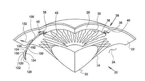
carried along with the venous blood leaving the eye.
[0008] When the natural drainage mechanisms of the eye stop functioning
properly, the
pressure inside the eye begins to rise. Researchers have theorized prolonged
exposure to high
intraocular pressure causes damage to the optic nerve that transmits sensory
information from the
eye to the brain. This damage to the optic nerve results in loss of peripheral
vision. As
glaucoma progresses, more and more of the visual field is lost until the
patient is completely
blind.
[00091 In addition to drug treatments, a variety of surgical treatments
for glaucoma have
been performed. For example, shunts were implanted to direct aqueous humor
from the anterior
chamber to the extraocular vein (Lee and Scheppens, "Aqueous-venous shunt and
intraocular
pressure," Investigative Ophthalmology (Feb. 1966)). Other early glaucoma
treatment implants
led from the anterior chamber to a sub-conjunctival bleb (e.g., US 4,968,296
and US 5,180,362).
Still others were shunts leading from the anterior chamber to a point just
inside Schlemm's canal
(Spiegel et al., "Schlemm's canal implant: a new method to lower intraocular
pressure in patients
with POAG?" Ophthalmic Surgery and Lasers (June 1999); US 6,450,984; US
6,450,984).
SUMMARY OF THE INVENTION
[0010] The invention pertains to aspects of ocular implants and ocular
implant delivery
systems. One aspect of the invention provides a cannula for an ocular implant
delivery system.
In some embodiments, the cannula includes a tubular member having a curved
portion, a distal
opening surrounded by a distal opening surface, and a distal tip, the distal
tip being adapted to be
- 2 -
CA 2972136 2017-06-29
inserted into an anterior chamber of a human subject's eye, through trabecular
meshwork and
into Schlemm's canal of the eye, a proximal portion of the tubular member
being adapted to
extend from a location exterior to the eye when the distal tip is in Schlemm's
canal of the eye,
the cannula being further adapted to cooperate with an advancement mechanism
to advance an
ocular implant through the tubular member toward and through the distal
opening into
Schlemm's canal of the eye when the distal tip is disposed in Schlernm's
canal.
[0011] In some embodiments, the cannula's tubular member also has a
tongue region
extending proximally from the distal tip on one side of the tubular member,
with the tongue
region forming at least part of the distal opening surface. In some
embodiments the distal
opening surface extends solely proximally from the distal tip, and the distal
opening surface may
be disposed in a distal opening plane. The tubular member curved portion may
also define a
curve plane, and the distal opening plane may be at an angle other than 90
degrees with respect
to the curve plane.
100121 In some embodiments of the cannula, the distal opening surface has
a first section
disposed in a distal opening plane disposed at a first section angle between 0
degrees and 90
degrees with respect to a longitudinal axis of the tubular member at the
distal opening and a
second section whose angle with respect to the longitudinal axis of the
tubular member varies
from an angle less than the first section angle at a distal limit of the
second section to an angle
greater than the first section angle at a proximal limit of the second
section.
[0013] In other embodiments of the cannula, the distal opening surface has
an edge formed
from a circumferential portion of a cylindrical envelope defined by the
tubular member, the
angular extent of the circumferential portion within the cylindrical envelope
increasing from the
distal tip proximally to a first point, the angular extent of the
circumferential portion within the
cylindrical envelope decreasing between the first point and a second point
proximal to the first
point, the angular extent of the circumferential portion within the
cylindrical envelope increasing
to 360 degrees between the second point and a third point proximal to the
second point.
[0014] In some embodiments of the cannula, the tubular member also a
second tongue region
and a stop member defining the distal opening surface.
[0015] In some embodiments, an external diameter of the tubular member at
a distal end of
the tubular member is less than an external diameter of the tubular member
proximal to the distal
opening. The curved portion of the tubular member may also have a bend angle
between 105
degrees and 165 degrees.
[0016] Another aspect of the invention provides an ocular implant system
including an
ocular implant having an inlet sized and configured to be disposed in an
anterior chamber of a
human subject's eye and a body sized and configured to be disposed in
Schlernm's canal of the
- 3 -
CA 2972136 2017-06-29
eye, the ocular implant being adapted to bend preferentially in a preferential
bending plane; and a
delivery cannula comprising a tubular member with a curved portion, a distal
opening
surrounded by a distal opening surface, and a distal tip, the distal tip being
adapted to be inserted
into an anterior chamber of a human subject's eye, through trabecular meshwork
and into
Sehlenun's canal of the eye, the tubular member being adapted to extend from a
location exterior
to the eye when the distal tip is in Schlemm's canal of the eye, the cannula
being further adapted
to cooperate with an advancement mechanism to advance the ocular implant
through at least the
curved portion of the tubular member toward and through the distal opening
into Schlenun's
canal of the eye when the distal tip of the delivery tool is disposed in
Schlemm's canal.
[0017] In some embodiments of the ocular implant system, a central axis of
the cannula
defines a cannula curvature plane, the ocular implant being oriented within
the cannula so that
the implant preferential bending plane is co-planar with the cannula curvature
plane.
[0018] Yet another aspect of the invention provides a method of deploying
an ocular implant
into Schlemm's canal of a human eye. The method may include the following
steps: inserting a
distal tip of a delivery tool within an anterior chamber of the eye through
trabecular meshwork of
the eye into Schlemm's canal of the eye; and advancing an ocular implant
through a curved
portion and a distal opening of the delivery tool to place a body portion of
the ocular implant in
Schlemm's canal and an inlet portion of the ocular implant in the anterior
chamber.
[0019] In some embodiments, the delivery tool has a curved distal
portion, the inserting step
including the step of aligning the curved distal portion with respect to
Schlemm's canal so that
the ocular implant is delivered into the center of Schlemm's canal or slightly
radially inward of
an outer wall of Schlemm's canal. The curved distal portion of the delivery
tool may have a
radius of curvature smaller than that of Schlemm's canal.
[0020] In some embodiments, the inserting step includes the step of
advancing the distal tip
into Schlemm's canal until a stop portion of a distal opening surface
surrounding the distal
opening engages the trabecular meshwork. The inserting step may also include
the step of
depressing trabecular meshwork and Schlemm's canal tissue with the distal tip
with a distal
opening surface surrounding the distal opening, the distal opening surface
being disposed at an
angle other than 90 degrees with respect to a longitudinal axis of the
delivery tool.
[0021] In embodiments in which the delivery tool has a distal opening
surface surrounding
the distal opening, the inserting step may include the step of inserting less
than all of the distal
opening surface into Schlemm's canal.
[0022] In some embodiments, the delivery tool has a distal opening
surface surrounding the
distal opening and the distal tip is disposed at the distal end of a tongue.
In such embodiments
the inserting step may include the step of inserting the tongue into Schlemm's
canal. The
- 4 -
CA 2972136 2017-06-29
advancing step may also include the step of advancing the ocular implant
through the distal
opening while a portion of the distal opening surface is disposed in Schlemm's
canal and a
portion of the distal opening surface is disposed outside of Schlemm's canal.
BRIEF DESCRIPTION OF THE DRAWINGS
[0023] The novel features of the invention are set forth with
particularity in the claims that
follow. A better understanding of the features and advantages of the present
invention will be
obtained by reference to the following detailed description that sets forth
illustrative
embodiments, in which the principles of the invention are utilized, and the
accompanying
drawings of which:
[0024] Figure 1 is a stylized perspective view depicting an exemplary
ocular implant
extending from a portion of a human eye.
[0025] Figure 2 is a perspective view showing a portion of the ocular
implant shown in
Figure I.
[0026] Figure 3 is a perspective view illustrating a volume defined by the
body of the ocular
implant shown in Figure 2.
[0027] Figure 4 is a perspective view illustrating a first plane and a
second plane that both
intersect an exemplary ocular implant.
[0028] Figure 5 is an enlarged perspective view showing a portion of the
ocular implant
shown in Figure 4.
100291 Figure 6 is stylized representationnf an exemplary medical
procedure in accordance
with the present disclosure.
[0030] Figure 7 is an enlarged plan view showing illustrating insertion
of an ocular implant
delivery system cannula into the eye shown in the previous figure.
[0031] Figure 8 is a further enlarged plan view illustrating insertion of
the ocular implant
delivery system cannula into the eye shown in the previous figure.
[0032] Figure 9 is an additional plan view of the eye shown in the
previous figure showing
advancement of an ocular implant through the cannula into Schlemm's canal of
the eye.
[0033] Figure 10 is an additional plan view of the eye shown in the
previous figure. In the
embodiment of Figure 10, a core that was used to position the ocular implant
has been
withdrawn.
[0034] Figure 11 is a plan view of the eye shown in the previous figure
showing the ocular
implant in Schlemm's canal after the cannula has been withdrawn.
100351 Figure 12 is a perspective view of an exemplary cannula assembly.
- 5 -
CA 2972136 2017-06-29
[0036] Figure 13 is an enlarged perspective view showing a portion of a
tubular member of
the cannula shown in the previous figure.
[0037] Figure 14 is a plan view further illustrating the cannula assembly
of Figure 12.
[0038] Figure 15 is an enlarged plan view showing a portion of the
tubular member shown in
the previous figure.
[0039] Figure 16 is a plan view further illustrating the cannula assembly
of Figure 12.
[0040] Figures 17A and 17B are plan views further illustrating the
tubular member of the
cannula assembly shown in Figure 12.
[0041] Figures 18A and 18B are plan views further illustrating the
tubular member of the
cannula assembly shown in Figure 12.
[0042] Figure 19 is a plan view illustrating an alternate embodiment of a
cannula assembly.
[0043] Figure 20 is a stylized perspective view showing a portion of the
tubular member
shown in the previous figure delivering an ocular implant into Schlemm's
canal.
[0044] Figure 21 is a perspective view of an another embodiment of an
ocular implant
delivery system cannula in accordance with this invention.
[0045] Figure 22 is a partial sectional and perspective view showing
portions of an ocular
implant delivery system into which an ocular implant has been loaded.
[0046] Figure 23 is an additional perspective view of the assembly shown
in Figure 22
showing delivery of the ocular implant into Schlemm's canal.
[0047] Figure 24 is an additional perspective view showing portions of the
implant and the
cannula shown in Figures 22 and 23.
[0048] Figure 25C is a plan view showing a cannula. Figure 25B is a cross
sectional view of
the cannula sectioned along cutting line B-B shown in Figure 25C. Figure 25A
is an axial plan
view created from the viewpoint illustrated by line A-A in Figure 25C,
[0049] Figures 26A, 26B, and 26C are three orthographic views of
illustrating the structural
features of an exemplary ocular implant delivery system cannula.
[0050] Figure 27 is an isometric view of the ocular implant delivery
system cannula
illustrating a tongue of the cannula.
[0051] Figure 28 is a schematic partial cross-sectional view showing the
distal tip of an
ocular implant delivery system cannula entering Schlemm's canal.
[0052] Figure 29 is a plan view of yet another embodiment of part an
ocular implant delivery
system cannula.
[0053] Figure 30 is a perspective view of a portion of the cannula of
Figure 29.
- 6 -
CA 2972136 2017-06-29
[0054] Figure 31 is a partial cross-sectional view and a partial plan
view showing an ocular
implant being delivered into Schlernm's canal using still another embodiment
of a delivery
system cannula according to this invention.
[0055] Figure 32 is an elevational view of a portion of the cannula of
the delivery system of
Figure 31.
[0056] Figure 33 is a side elevational view of a portion of the cannula
of Figure 32.
[0001] Figure 34 is a further partial cross-sectional view and partial
perspective view
showing the ocular implant being delivered into Schlemin's canal using a
delivery system
cannula according to the embodiment of Figure 31.
[0002] Figure 35 is a partial cross-sectional view and a partial plan view
of an implant in
place within Sehletrun's canal after delivery.
[0057] Figures 36A and 36B are partial section and perspective views
illustrating insertion of
the distal tip of an ocular implant delivery system cannula into Schlentm's
canal.
DETAILED DESCRIPTION OF THE INVENTION
[0058] The following detailed description should be read with reference
to the drawings in
which similar elements in different drawings are numbered the same. The
drawings, which are
not necessarily to scale, depict illustrative embodiments and are not intended
to limit the scope of
the invention.
[0059] Apparatus and methods in accordance with the present detailed
description may be
used to deliver an ocular implant into a subject's eye and to place distal
portion of an ocular
implant in Schlemm's canal of an eye. Figure 1 is a stylized perspective view
depicting a portion
of a human eye 20. Eye 20 can be conceptualized as a fluid filled ball having
two chambers.
Sclera 22 of eye 20 surrounds a posterior chamber 24 filled with a viscous
fluid known as
vitreous humor. Cornea 26 of eye 20 encloses an anterior chamber 30 that is
filled with a fluid
know as aqueous humor. The cornea 26 meets the sclera 22 at a limbus 28 of eye
20. A lens 32
of eye 20 is located between anterior chamber 30 and posterior chamber 24.
Lens 32 is held in
place by a number of ciliary zonules 34.
100601 Whenever a person views an object, he or she is viewing that
object through the
cornea, the aqueous humor, and the lens of the eye. In order to be
transparent, the cornea and the
lens can include no blood vessels. Accordingly, no blood flows through the
cornea and the lens
to provide nutrition to these tissues and to remove wastes from these tissues.
Instead, these
functions are performed by the aqueous humor. A continuous flow of aqueous
humor through
the eye provides nutrition to portions of the eye (e.g., the cornea and the
lens) that have no blood
vessels. This flow of aqueous humor also removes waste from these tissues.
- 7 =
CA 2972136 2017-06-29
[0061] Aqueous humor is produced by an organ known as the ciliary body.
The ciliary body
includes epithelial cells that continuously secrete aqueous humor. In a
healthy eye, a stream of
aqueous humor flows out of the eye as new aqueous humor is secreted by the
epithelial cells of
the ciliary body. This excess aqueous humor enters the blood stream and is
carried away by
venous blood leaving the eye.
[0062] In a healthy eye, aqueous humor flows out of the anterior chamber
30 through the
trabecular meshwork 36 and into Schlemm's canal 38, located at the outer edge
of the iris 42.
Aqueous humor exits Schlenun's canal 38 by flowing through a number of outlets
40. After
leaving Schlemm's canal 38, aqueous humor is absorbed into the venous blood
stream.
[0063] In Figure 1, an ocular implant 100 is disposed in Schlemm's canal 38
of eye 20.
Ocular implant 100 has a body 102 including a plurality of tissue supporting
frames 104 and a
plurality of spines 106. Body 102 also includes a first edge 120 and a second
edge 122 that
define a first opening 124. First opening 124 is formed as a slot and fluidly
communicates with
an elongate channel 126 defined by an inner surface 128 of body 102. With
reference to Figure
1, it will be appreciated that first opening 124 is disposed on an outer side
130 of body 102.
Accordingly, channel 126 opens in a radially outward direction 132 via first
opening 124.
[0064] Ocular implant 100 may be inserted into Schlenun's canal of a
human eye to facilitate
the flow of aqueous humor out of the anterior chamber. This flow may include
axial flow along
Schlemm's canal, flow from the anterior chamber into Schlemm's canal, and flow
leaving
Schlemm's canal via outlets communicating with Schlemm's canal. When in place
within the
eye, ocular implant 100 will support trabecular mesh tissue and Schlemm's
canal tissue and will
provide for improved communication between the anterior chamber and Schlemm's
canal (via
the trabecular meshwork) and between pockets or compartments along Schlemm's
canal. As
shown in Figure 1, the implant is preferably oriented so that the first
opening 124 is disposed
radially outwardly within Schlemm's canal.
[0065] Figure 2 is an enlarged perspective view showing a portion of
ocular implant 100
shown in the previous figure. Ocular implant 100 has a body 102 that extends
along a generally
curved longitudinal axis 134. Body 102 has a plurality of tissue supporting
frames 104 and a
plurality of spines 106. As shown in Figure 2, these spines 106 and frames 104
are arranged in a
repeating AB pattern in which each A is a tissue supporting frame and each B
is a spine. In the
embodiment of Figure 2, one spine extends between each adjacent pair of frames
104.
[0066] For example, frame 136 of ocular implant 100 is disposed between a
first spine 140
and a second spine 142. Frame 136 is formed as a first strut 144 that extends
between first spine
140 and second spine 142 and a second strut 146 extending between first spine
140 and second
spine 142. In the exemplary embodiment of Figure 2, struts 144 and 146 each
undulates in a
- 8 -
CA 2972136 2017-06-29
circumferential direction as it extends longitudinally between first spine 140
and second spine
142.
[0067] In the embodiment of Figure 2, body 102 has a longitudinal radius
of curvature 150
and a lateral radius of curvature 148. Body 102 of ocular implant 100 includes
a first edge 120
and a second edge 122 that define first opening 124. First opening 124 fluidly
communicates
with an elongate channel 126 defined by an inner surface 128 of body 102. A
second opening
138 is defined by a second edge 122A of first strut 144 and a second edge 122B
of second strut
146. First opening 124, second opening 138 and additional openings defined by
ocular implant
100 allow aqueous humor to flow laterally across and/or laterally through
ocular implant 100.
The outer surfaces of body 102 define a volume 152.
[0068] Figure 3 is an additional perspective view showing volume 152
defined by the body
of the ocular implant shown in the previous figure. With reference to Figure
3, it will be
appreciated that volume 152 extends along a generally curved longitudinal axis
134. Volume 152
has a longitudinal radius 150, a lateral radius 148, and a generally circular
lateral cross section
153.
[0069] Figure 4 is a perspective view showing a first plane 154 and a
second plane 155 that
both intersect ocular implant 100. In Figure 4, first plane 154 is delineated
with hatch marks.
With reference to Figure 4, it will be appreciated that spines 106 of body 102
are generally
aligned with one another and that first plane 154 intersects all spines 106
shown in Figure 4. In
the embodiment of Figure 4, body 102 of ocular implant 100 is generally
symmetric about first
plane 154.
[0070] lathe embodiment of Figure 4, the flexibility of body 102 is at a
maximum when
body 102 is bending along first plane 154, and body 102 has less flexibility
when bending along
a plane other than first plane 154 (e.g., a plane that intersects first plane
154). Accordingly, first
plane 154 may be generally referred to as a plane of preferential bending. In
the embodiment
shown in Figure 4, for example, body 102 has a second flexibility when bending
along second
plane 155 that is less than the first flexibility that body 102 has when
bending along first plane
154.
10071] Stated another way, in the embodiment of Figure 4, the bending
modulus of body 102
is at a minimum when body 102 is bent along first plane 154. Body 102 has a
first bending
modulus when bent along first plane 154 and a greater bending modulus when
bent along a plane
other than first plane 154 (e.g., a plane that intersects first plane 154).
For example, in the
embodiment shown in Figure 4, body 102 has a second bending modulus when bent
along
second plane 155 that is greater than the first bending modulus that body 102
has when bent
along first plane 154.
- 9 -
CA 2972136 2017-06-29
[0072] Figure 5 is an enlarged perspective view showing a portion of
ocular implant 100
shown in the previous figure. In the exemplary embodiment of Figure 5, a
bending moment M is
being applied to body 102 of ocular implant 100. Bending moment M acts about a
first axis 156
that is generally orthogonal to first plane 154. A second axis 158 and a third
axis 160 are also
shown in Figure 5. Second axis 158 is generally perpendicular to first axis
156. Third axis 160 is
skewed relative to first axis 156.
[0073] In the embodiment of Figure 5, the flexibility of body 102 is at a
maximum when
body 102 is bent by a moment acting about first axis 156, and body 102 has
less flexibility when
bent by a moment acting about an axis other than first axis 156 (e.g., second
axis 158 and third
axis 160). Stated another way, the bending modulus of body 102 is at a minimum
when body
102 is bent by a moment acting about first axis 156, and body 102 has a
greater bending modulus
when bent by a moment acting about an axis other than first axis 156 (e.g.,
second axis 158 and
third axis 160).
[0074] Figure 6 is stylized representation of an exemplary medical
procedure in accordance
with this detailed description. In the exemplary procedure of Figure 6, a
physician is treating an
eye 20 of a patient 620. In the exemplary procedure of Figure 6, a physician
is holding a
delivery system in his or her right hand RH. The physician's left hand (not
shown) may be used
to hold the handle H of a gonio lens 628. It will be appreciated that some
physician's may prefer
holding the delivery system handle in the left hand and the gonio lens handle
H in the right hand
RH.
[0075] During the exemplary procedure illustrated in Figure 6, the
physician may view the
interior of the anterior chamber using a microscope 626 and gonio lens 628.
Detail A of Figure
6 is a stylized simulation of the image viewed by the physician. A distal
portion of a cannula is
visible in Detail A. The distal end of the cannula is positioned near
Schlemm's canal SC of eye
.. 22. A shadow-like line indicates the location of Schlemm's canal SC which
is lying under
various tissue (e.g., the trabecular meshwork) that surround the anterior
chamber.
[0076] Figure 7 is an enlarged plan view showing a portion of the face
shown in the previous
figure. In the embodiment of Figure 7, cannula 708 extends through a cornea of
eye 20 so that
the distal end of cannula 708 is disposed in the anterior chamber of eye 20.
With reference to
Figure 7, it will be appreciated that the distal tip of cannula 708 is
positioned near the trabecular
mesh 36 of eye 20.
[0077] Figure 8 is a further enlarged plan view illustrating a portion of
eye 20 shown in the
previous figure. In the embodiment of Figure 8, the distal tip of cannula 708
has pierced through
trabecular mesh 36. The distal tip of cannula 708 has also pierced the wall of
Sehlemm's canal
.. 38 so that a distal opening 758 of cannula 708 is disposed in fluid
communication with
- 10 -
CA 2972136 2017-06-29
Schlemm's canal 38. In this embodiment, cannula 708 is a rigid curved tube
that has a sharp portion at its
distal end near the exit port 758. In some embodiments, cannula 708 is curved
to achieve substantially
tangential entry into Schlemm's canal 38.
[0078] Figure 9 is an additional plan view of eye 20 shown in the previous
figure. In the embodiment
of Figure 9, an ocular implant 900 has been advanced through distal opening
758 of eannula 708 and into
Schlemm's canal 38 of eye 20. With reference to Figure 9, it will be
appreciated that ocular implant 900 is
disposed about a core 754 which is movable with ocular implant 900 within
cannula 708 as part of an
implant advancement mechanism. Core 754 and cannula 708 are part of a delivery
system that may be
used to deliver ocular implant 900 into Schlemm's canal of eye 20.
[0079] Among other functions, one particular function of core 754 is to
block the openings formed in
ocular implant 900 so as to minimize interference between the implant and
tissue within Schlemm's canal
38 as the implant is advanced. The delivery system's advancement mechanism may
also include a push
tube (not shown) for selectively applying distally directed forces to the
proximal end of ocular implant
900. Core 754 may extend proximally into the push tube. A handheld actuator
(not shown) may be used
to advance the push tube, the core 754 and the ocular implant 900. The
handheld actuator may also be
used to provide relative motion between the push tube and the core 754. In the
embodiment of Figure 9,
ocular implant 900 has a blunt distal end 902 for avoiding damage to ocular
tissue. In other embodiments,
the blunt distal end may be provided at least in part by core 754. Further
details of aspects of ocular
implant delivery systems suitable for use with implants and cannulas of this
invention may be found in
U.S. Application No. 11/943,289, filed November 20, 2007; U.S. Application No.
12/398,847, filed
March 5, 2009; U.S. Provisional Application No. 61/224,156, filed July 9,
2009; and U.S. Provisional
Application No. 61/224,158, filed July 9,2009.
[0080] Figure 10 is an additional plan view of eye 20 shown in the previous
figure. In the
embodiment of Figure 10, core 754 has been withdrawn from ocular implant 900.
A hand held actuator
(not shown) may be used to apply a proximal force to the core to withdraw the
core proximally from the
ocular implant 900 while a push tube (not shown) applies a distally directed
force to hold ocular implant
900 in place. The core, the push tube, and the cannula 708 may then be
withdrawn from the eye, leaving
the implant in Schlemm's canal with its proximal inlet end within the anterior
chamber of eye 20.
[0081] Figure 11 is a plan view of eye 20 after cannula 708 has been
withdrawn leaving an inlet
portion 904 of ocular implant 900 in the anterior chamber and the remainder of
implant 900 in Schlemm's
canal. The presence of ocular implant 900 in Schlemm's canal may facilitate
the
- 11 -
CA 2972136 2017-06-29
flow of aqueous humor out of the anterior chamber. This flow may include axial
flow along
Schlemm's canal, flow from the anterior chamber into Schlemm's canal, and flow
leaving
Schlemm's canal via outlets communicating with Schlemm's canal. When in place
within the
eye, ocular implant 900 will support trabecular mesh tissue and Schletrun's
canal tissue and will
provide for improved communication between the anterior chamber and Schlemm's
canal (via
the trabecular meshwork) and between pockets or compartments along Schlemm's
canal.
[0082] Figure 12 is a perspective view of an exemplary cannula assembly
1200. Cannula
assembly 1200 comprises a tubular member 1202 that is fixed to a hub 1204.
Tubular member
1202 defines a proximal opening 1206, a distal opening 1158, and a lumen 1208
that extends
between proximal opening 1206 and distal opening 1158. Tubular member 1202
also comprises
a proximal portion 1210, a distal portion 1212, and a bent portion 1214
disposed between
proximal portion 1210 and distal portion 1212.
10083] Figure 13 is an enlarged perspective view showing a portion of
tubular member 1202
shown in the previous figure. With reference to Figure 13, it will be
appreciated that tubular
member 1202 comprises a beveled distal tip 1216 having a distal opening
surface 1218. In the
exemplary embodiment of Figure 13, beveled distal tip 1216 defines a distal
opening 1158
having a generally elliptical shape. A major axis 1220 and a minor axis 1222
of distal opening
1158 are illustrated using dashed lines in Figure 13. For purposes of
illustration, major axis
1220 and minor axis 1222 each extend beyond distal opening 1158 in Figure 13.
100841 In the exemplary embodiment of Figure 13, major axis 1220 and minor
axis 1222
define an exit plane 1224. Distal opening 1158 opens in a direction D that is
orthogonal to exit
plane 1224. Direction D is illustrated using an arrow in Figure 13. In some
useful embodiments,
an imaginary line representing direction D intersects the cornea of the eye
when the when the
tubular member is extending through the cornea and the distal opening is
fluidly communicating
with Schlemm's canal of the eye.
[0085] Figure 14 is a plan view further illustrating cannula assembly
1200. With reference
to Figure 14, it will be appreciated that tubular member 1202 of cannula
assembly 1200
comprises a proximal portion 1210, a distal portion 1212, and a bent portion
1214 disposed
between proximal portion 1220 and distal portion 1222. In the exemplary
embodiment of Figure
14, a hub 1204 is fixed to proximal portion 1210 of tubular member 1202. With
reference to
Figure 14, it will be appreciated that tubular member 1202 has a central axis
1226. Central axis
1226 of Figure 14 has a curved portion and straight portions. In Figure 14, a
bend angle BA is
shown extending between a first straight portion of central axis 1226 and a
second straight
portion of central axis 1226.
- 12 -
CA 2972136 2017-06-29
[0086] In some useful embodiments, bent portion 1214 of tubular member
1202 is
dimensioned to achieve substantially tangential entry into Schlemm's canal of
a human eye. In
these useful embodiments, bent portion 1214 may have a radius of curvature
between about 0.05
inches and about 0.3 inches, and an angular span between about 105 degrees and
about 165
degrees. In one exemplary embodiment, bent portion 1214 has a bend radius of
about 0.125
inches (measured to the tube centerline) and an angular span of about 132.5
degrees. In this
exemplary embodiment, distal portion 1212 may have a length of about 0.044
inches and
proximal portion 1210 may have a length of about 0.727 inches.
[0087] Figure 15 is an enlarged plan view showing a portion of tubular
member 1202 shown
in the previous figure. With reference to Figure 15, it will be appreciated
that tubular member
1202 has a central axis 1226 defining a bend plane 1228. Central axis 1226 of
Figure 15 has a
curved portion and straight portions. Tubular member 1202 of Figure 15 also
comprises a
beveled distal tip 1216 having a distal opening surface 1228. In the exemplary
embodiment of
Figure 15, beveled distal tip 1216 defines a distal opening 1158 having a
generally elliptical
shape. A major axis 1220 and a minor axis 1222 of distal opening 1158 are
illustrated using
dashed lines in Figure 15.
[0088] For purposes of illustration, major axis 1220 and minor axis 1222
each extend beyond
distal opening 1158 in Figure 15. In the exemplary embodiment of Figure 15,
major axis 1220
and minor axis 1222 define an exit plane 1224. In Figure 15, exit plane 1224
is shown
intersecting bend plane 1228. With reference to Figure 15, it will be
appreciated that exit plane
1224 is generally skewed relative to bend plane 1228. That is, the plane 1222
of distal opening
surface 1228 meets plane 1228 of the eannula curve at an angle other than 90
degrees.
[00891 Figure 16 is a plan view further illustrating cannula assembly
1200. With reference
to Figure 16, it will be appreciated that tubular member 1202 of cannula
assembly 1200
comprises a first portion 1230 having a first diameter DA, a second portion
1232 having a
second diameter DB, and a tapered portion 1234 disposed between first portion
1230 and second
portion 1232.
[0090] In the exemplary embodiment of Figure 16, first diameter DA is
greater than second
diameter DB, and tapered portion 1234 transitions between first diameter DA
and second
diameter D13. In some useful embodiments, tapered portion 1234 has an average
taper ratio
between about 0.01 and about 0.12. In one exemplary embodiment, tapered
portion 1234 has an
average taper ratio of about 0.068.
[0091] Tubular member 1202 defines a proximal opening (not shown), a
distal opening 1158,
and a lumen 1208 that extends between the proximal opening and the distal
opening. In the
exemplary embodiment of Figure 16, lumen 1208 has a generally circular cross-
sectional shape.
- 13 -
CA 2972136 2017-06-29
In some useful embodiments, lumen 1208 has a diameter that is substantially
uniform along the
length of tubular member 1202. This configuration reduces the likelihood that
an ocular implant
advanced through lumen 1208 will become hung up during delivery through the
lumen.
[0092] In some useful embodiments, second diameter DB is dimensioned so
that distal
opening 1158 can be placed in fluid communication with Schlemm's canal of a
human eye. Also
in some useful embodiments, first diameter DA is dimensioned to provide a
desirable level of
structural support when tubular member 1202 is advance through the cornea of a
human eye and
the distal end of beveled tip 1216 is inserted into Schlemm's canal.
[0093] In some useful embodiments first diameter DA is between about
0.010 and about
0.030 inches and second diameter DB is between about 0.005 and about 0.020. In
one
exemplary embodiment, first diameter DA is about 0.018 inches, second diameter
DB is about
0.016, and the diameter of lumen 1208 is about 0.0135 inches. With reference
to Figure 16, it
will be appreciated that tubular member 1202 comprises a bent portion 1214. In
the exemplary
embodiment of Figure 16, tapered portion 1234 is extends along a portion of
bent portion 1214
of tubular member 1202.
[0094] Figure 17A and Figure 17B are plan views further illustrating
tubular member 1202
of eannula assembly 1200. With reference to Figure 17A, it will be appreciated
that tubular
member 1202 comprises a beveled distal tip 1216 having a distal opening
surface 1218. In the
exemplary embodiment of Figure 17A, beveled distal tip 1216 defines a distal
opening 1158
having a generally elliptical shape. A major axis 1220 and a minor axis 1222
of distal opening
1158 are illustrated using dashed lines in Figure 17A. For purposes of
illustration, major axis
1220 and minor axis 1222 each extend beyond distal opening 158 in Figure 17A.
[0095] Figure 17B is an additional plan view showing the portion of
tubular member 1202
shown in Figure 17A. Figure 17B is taken from a viewpoint that is generally
orthogonal to the
viewpoint used to create Figure 17A. With reference to Figure 17B, it will be
appreciated that
tubular member 1202 has a central axis 1226 that includes both straight
portions and curved
portions.
[0096] Major axis 1220 of distal opening 1158 and central axis 1226 of
tubular member 1202
define a pitch angle PA of beveled distal tip 1216. In some useful
embodiments, pitch angle PA
is steep enough to tent open tissue (e.g., trabeeular mesh and the wall of
Schlemm's canal) when
the distal end of beveled tip 1216 is inserted into Schlemm's canal. Also in
some useful
embodiments, pitch angle PA is shallow enough to prevent tearing or cutting of
tissue when the
distal end of beveled tip 1216 is inserted into Schlemm's canal. In some
useful embodiments,
pitch angle PA is between about 5 degrees and about 35 degrees. In some
particularly useful
- 14 -
CA 2972136 2017-06-29
embodiments, pitch angle PA is greater than about 15 degrees and less than
about 25 degrees. In
one exemplary embodiment, pitch angle PA is about 20 degrees.
[0097] Figure 18A and Figure 18B are plan views further illustrating
tubular member 1202
of cannula assembly 1200. With reference to Figure 18B, it will be appreciated
that tubular
member 1202 has a central axis 1226 defining a bend plane 1228. Central axis
1226 of Figure
18B has a curved portion and straight portions. In the embodiment of Figure
18B, tubular
member 1202 also comprises a beveled distal tip 1216 having a distal opening
surface 1218. In
the exemplary embodiment of Figure 18B, beveled distal tip 1216 defines a
distal opening 1158
having a generally elliptical shape. A major axis 1220 and a minor axis 1222
of distal opening
1158 are illustrated using dashed lines in Figure 18B.
[0098] Figure 18A is an axial plan view showing tubular member 1202 and
distal opening
surface 1218. Figure 18A is taken from a viewpoint that is generally
orthogonal to the viewpoint
used to create Figure 18B. Bend plane 1228, major axis 1220 and minor axis
1222 are illustrated
using dashed lines in Figure 18A. With reference to Figure 18A, it will be
appreciated that
minor axis 1222 of distal opening 1158 and bend plane 1228 define a roll angle
RA.
[0099] In some useful embodiments, roll angle RA is selected so that a
physician using the
cannula assembly can see distal opening 1158 when the tubular member 1202 is
extending
through the cornea of a human eye and the distal end of beveled distal tip
1216 is inserted into
Schlemm's canal. In other words, the plane of distal opening surface 1218
meets bend plane
1228 at an angle other than 90 degrees. Also in some useful embodiments, roll
angle RA is
selected so that distal end of beveled distal tip 1216 is the first part of
tubular member 1202 to
touch tissue when the tubular member 1202 is extending through the cornea of a
human eye and
the distal end of beveled distal tip 1216 is inserted into Schlemm's canal.
[00100] Additionally, roll angle RA may be selected so that an ocular implant
travels over the
point of beveled distal tip 1216 as the ocular implant is advanced out of
distal opening 1158 and
into Schlemm's canal. In some useful embodiments, roll angle RA is greater
than about 100
degrees and less than about 110 degrees. In one exemplary embodiment, roll
angle RA is about
105 degrees.
[00101] Figure 19 is a plan view illustrating an alternate exemplary
embodiment of an ocular
implant delivery system cannula assembly. With reference to Figure 19, it will
be appreciated
that tubular member 1902 of cannula assembly 1900 comprises a first portion
1908 having a first
diameter DA and a second portion 1910 having a second diameter DB. A step 1912
is disposed
between first portion 1908 and second portion 1910. In some useful
embodiments, second
diameter DB is dimensioned so that distal opening 1904 can be placed in fluid
communication
with Schlemm's canal of a human eye. Also in some useful embodiments, first
diameter DA is
- 15 -
CA 2972136 2017-06-29
dimensioned to provide a desirable level of structural support when tubular
member 1902 is
advance through the cornea of a human eye and the distal end of beveled distal
tip 1906 is
inserted into Schlemm's canal. In some useful embodiments first diameter DA is
between about
0.010 and about 0.030 inches and second diameter DB is between about 0.005 and
about 0.020.
In one exemplary embodiment, first diameter DA is about 0.018 inches, second
diameter DB is
about 0.016, and the diameter of the inner lumen of tubular member 1902 is
about 0.0135 inches.
[001021 Figure 20 is a stylized perspective view showing a portion of tubular
member 1908
shown in the previous figure. In Figure 20, an ocular implant 900 is shown
extending through
distal opening 1904 of tubular member 1908 and into Schlemm's canal 38 of an
eye. The distal
end of beveled distal tip 1906 has penetrated the trabecular mesh 36 of the
eye, and distal
opening 1904 is in fluid communication with Schlemm's canal 38. In the
embodiment of Figure
20, ocular implant 900 is oriented so that the longitudinal channel of ocular
implant 900 opens
radially outward.
[00103] Figure 21 is a perspective view of a cannula 2108 in accordance with
the present
detailed description. Cannula 2108 of Figure 21 comprises a generally tubular
member 2162
having a central axis 2164. Generally tubular member 2162 of Figure 21
comprises a proximal
portion 2166, a distal end 2168, and a distal portion 2170 extending between
distal end 2168 and
proximal portion 2166. A distal opening surface 2167 surrounds a distal
opening 2169.
[00104] In the exemplary embodiment of Figure 21, proximal portion 2166 of
cannula 2108 is
substantially straight, distal portion 2170 of cannula 2108 is curved, and
central axis 2164
defines a curvature plane 2172. Curvature plane 2172 may be referred to as a
plane of curvature.
With reference to Figure 21, it will be appreciated that curvature plane 2172
divides cannula
2108 into a first portion PA and a second portion PB. In the exemplary
embodiment of Figure
21, second portion PB is substantially a mirror image of first portion PA. In
Figure 21, distal
portion 2170 is shown extending between distal end 2168 and proximal portion
2166 with no
intervening elements. In the exemplary embodiment of Figure 21, distal portion
2170 is curved
along its entire length.
[00105] An exemplary method in accordance with this detailed description may
include the
step of advancing the distal end 2168 of cannula 2108 through the cornea of a
human eye so that
distal end 2168 is disposed in the anterior chamber of the eye. Cannula 2108
may then be used
to access Schlenun's canal of the eye, for example, by piercing the wall of
Schlemm's canal with
the distal end 2168 of cannula 108. Distal opening 2169 of cannula 2108 may be
placed in fluid
communication with a lumen defined by Schlemm's canal. The ocular implant may
be advanced
out of a distal port of the cannula and into Schlemm's canal.
- 16 -
CA 2972136 2017-06-29
[001061 Figure 22 is a perspective view of an assembly 2182 including cannula
2108 shown in
the previous figure. For purposes of illustration, cannula 2108 is cross-
sectionally illustrated in
Figure 22. In Figure 22, an ocular implant 100 can be seen resting in a lumen
2184 defined by
cannula 2108. In the exemplary embodiment of Figure 22, ocular implant 100 is
disposed about
a core 754.
[001071 Ocular implant 100 extends along a generally curved longitudinal axis
2134.
Longitudinal axis 2134 defines a first plane 2154. In the embodiment of Figure
22, the
flexibility of ocular implant 100 is at a maximum when it is bending along
first plane 2154, and
implant 100 has less flexibility when bending along a plane other than first
plane 2154 (e.g., a
plane that intersects first plane 2154). Accordingly, first plane 2154 may be
generally referred to
as a plane of preferential bending.
[001081 Cannula 2108 of Figure 22 comprises a generally tubular member 2162
having a
central axis 2164. Generally tubular member 2162 of Figure 22 comprises a
proximal portion
2166, a distal end 2168, and a distal portion 2170 extending between distal
end 2168 and
proximal portion 2166. In the exemplary embodiment of Figure 22, proximal
portion 2166 of
cannula 2108 is substantially straight.
[001091 In the embodiment of Figure 22, central axis 2164 of cannula 2108 is
coaxial with the
longitudinal axis 2134 of ocular implant 100. With reference to Figure 22, it
will be appreciated
that distal portion 2170 of cannula 2108 is curved so that central axis 2164
of cannula 2108
defines a curvature plane 2172. Curvature plane 2172 may be referred to as a
plane of curvature.
With reference to Figure 22, it will be appreciated that curvature plane 2172
divides cannula
2108 into a first portion and a second portion PB. Only second portion PB of
cannula 2108 is
shown in the illustrative embodiment of Figure 22. In this embodiment,
curvature plane 2172 is
coincident with first plane 2154.
[001101 Figure 23 is an additional perspective view of assembly 2182 shown in
the previous
figure. In Figure 23, core 754 of the delivery system's advancement mechanism
and ocular
implant 100 are shown extending through distal port 2188 of cannula 2108. With
reference to
the previous figure, it will be appreciated that core 754 and ocular implant
100 have been moved
in a distal direction relative to the position of those elements shown
previously. Schlemm's canal
SC of an eye is illustrated using dashed lines in Figure 23. In the embodiment
of Figure 23, a
portion of ocular implant 100 has been advanced into Sehlemm's canal SC.
Ocular implant 100
is oriented so as to bend most easily in a direction conforming with the
natural curvature of
Schlemm's canal SC. In Figure 23, a distal end of a push tube PT of the
delivery system's
advancement mechanism is shown contacting a proximal end of ocular implant
100. In the
embodiment of Figure 23, push tube PT is disposed in the lumen defined by
cannula 2108.
- 17 -
CA 2972136 2017-06-29
[00111] Figure 24 is an additional perspective view showing ocular implant 100
and cannula
2108 shown in the previous figure. With reference to Figure 24, it will be
appreciated that ocular
implant 100 has been advanced to a position outside of cannula 2108. After
advancing ocular
implant 100 into Schlemm's canal, the core and the push tube have been
retracted into lumen
2184 defined by caimula 2108.
[00112] With reference to the figures described above, it will be appreciated
that methods in
accordance with the present detailed description may be used to position a
distal portion of an
implant in Schlemm's canal of an eye. An exemplary method in accordance with
the present
detailed description may include the step of advancing a distal end of a
cannula through a cornea
of the eye so that a distal portion of the cannula is disposed in the anterior
chamber of the eye.
The cannula may be used to access Schlemm's canal, for example, by piercing
the wall of
Schlemm's canal with a distal portion of the cannula.
[00113] Methods in accordance with the present detailed description can be
used to deliver an
implant into Schlemm's canal of an eye. In these exemplary methods, a distal
portion of the
ocular implant may be advanced out of the distal port of a cannula and into
Schlemm's canal,
Ocular implant 100 may be disposed on a core while the distal portion of the
implant is advanced
into Schlemm's canal. In some useful methods, the ocular implant comprises a
body defining a
plurality of apertures and the method includes the step of closing the
apertures with a core.
When this is the case, the distal portion of the ocular implant may be
advanced into Schlemm's
canal while the apertures are closed by the core. Closing the apertures as the
ocular implant is
advanced into Schlemm's canal may reduce the trauma inflicted on Schlemm's
canal by the
procedure. Once the ocular implant has reached a dcsircd position, the core
may be retracted
while a push tube prevents ocular implant from being pulled proximally.
[00114] Figure 25A is a cross sectional view of cannula 2108 sectioned along
cutting line A-A
shown in Figure 25C. Figure 25B is an axial plan view created from the
viewpoint illustrated by
line B-B in Figure 25C. Figure 25C is a plan view showing cannula 2108. Figure
25A, Figure
25B, and Figure 25C may be collectively referred to as Figure 25.
[00115] With reference to Figure 25, it will be appreciated that cannula 2108
comprises a
generally tubular member 2162 having a central axis 2164. In the embodiment of
Figure 25,
generally tubular member 2162 comprises a proximal portion 2166, a distal end
2168, and a
distal portion 2170 extending between distal end 2168 and proximal portion
2166. In the
exemplary embodiment of Figure 25, proximal portion 2166 is substantially
straight, and distal
portion 2170 is curved. A distal opening 2169 and distal opening surface 2167
form a tongue
2190. Distal opening 2169 fluidly communicates with a lumen 2184 defined by
generally
- 18 -
CA 2972136 2017-06-29
tubular member 2162. With reference to Figure 25, it will be appreciated that
distal portion 2170
is curved in the plane of Figure 25A and curved in the plane of Figure 25B.
[00116] Figures 26 and 27 provide additional views of cannula 2108. Distal
opening surface
2167 in tongue 2190 has two sections: a first section 2191 lying in a plane
that forms a first
section angle greater than 0 degrees and less than 90 degrees with respect to
longitudinal axis
2164 of the cannula tube 2162 and a notched section 2192 whose angle with
respect to axis 2164
varies from an angle less than that of the first section to an angle greater
than the first section.
[00117] Figure 28 is a schematic illustration of the use of a ocular implant
delivery system
cannula. As the distal tip 2168 passes through the trabecular meshwork 36 of
the human
subject's eye and into Schlemm's canal 38, the distal opening surface of first
and second sections
2191 and 2192 of tongue portion 2190 depresses the meshwork and Schlemm's
canal tissue in a
tenting area 37 to form a transition area for delivery of an ocular implant
into Schlemm's canal.
As shown, not all of the distal opening of the cannula has been inserted into
Schlemm's canal.
Instead, tongue 2190 causes the subject's tissue to form a ramp that, together
with the inner
surface of tongue 2190, guides insertion of the ocular implant into Schlemm's
canal.
[00118] In addition, since the curve of the cannula at the distal tip 2168 is
greater than the
curve of Schlemm's canal (i.e., the cannula at its distal end has a smaller
radius of curvature than
Schlemm's canal), the distal tip may be oriented so that the ocular implant is
delivered into the
center or possibly slightly radially inward of the outer wall of Schlemm's
canal. This
combination of cannula shape and cannula orientation helps guide the ocular
implant safely into
Schlemm's canal.
[00119] Figure 29 and 30 show yet another embodiment of a cannula tube 2962
for use in an
ocular implant delivery system. In this embodiment, a tongue region 2990
extending proximally
from the distal tip 2968 of the cannula is defined by a distal opening 2169
and a distal opening
surface 2167 with a complex shape. Tube 2962 is formed as a curved cylinder
which defines a
cylindrical envelope. Tongue 2990 can be described as a region in which the
angular extent of
material coverage within the cylindrical envelope increases from the distal
tip 2968 proximally
to a first point 2901, then decreases from point 2901 proximally to a second
point 2902, then
once again increases from point 2902 proximally to complete 360 degree
material coverage
.. within the cylindrical envelope at point 2903.
[00120] Figures 31-35 show an ocular implant 900 being delivered through a yet
another
embodiment of an ocular implant delivery system cannula 3102 into Schlemm's
canal 38.
(Schlemm's canal is shown in these figures as being straight instead of curved
for ease of
illustration.) The ocular implant shown is described in more detail in USSN
11/860,318, "Ocular
- 19 -
CA 2972136 2017-06-29
Implants," filed Sept. 24, 2007. It should be understood that other ocular
implants may be
delivered and deployed by the delivery system of this invention.
[00121] As shown in Figure 31, a distal portion of cannula 3102 has passed
through the
cornea to be within the anterior chamber 37 of the eye and has pierced the
trabecular meshwork
36 to enable a distal opening 3108 of cannula 3102 to communicate with
Schlemm's canal 38.
In this embodiment, cannula 3102 is a rigid curved tube that has a cutting
portion 3110 at the
distal opening 3108, as shown in more detail in Figures 32 and 33. In some
embodiments,
cannula 3102 is curved to achieve tangential entry into Schlemm's canal, such
as by forming an
arc of a circle having a radius of curvature less than about 0.1 inches. Other
embodiments may
have other shapes and curves.
[00122] In this embodiment, cutting portion 3110 is formed from two convex
edges 3112
meeting at a tip 3114. In other embodiments, the cutting edges can be concave
or straight. As
shown, edges 3112 extend from tip 3114 to a pair of optional stops 3116 formed
at the
intersection of edges 3112 with an optional cannula extension portion 3118. As
shown in Figure
31, the distal end of cannula 3102 may be advanced within the anterior chamber
37 toward the
trabecular meshwork 36. When the distal end of cannula 3102 meets the
trabecular meshwork,
tip 3114 and edges 3112 of cutting portion 3110 are advanced to extend through
the trabecular
meshwork into Schlemm's canal while a tongue or extension portion 3118 bends
back and
remains within the anterior chamber 37. Distal movement of cannula 3102 ceases
when stops
3116 engage the trabecular meshwork.
1001231 In some embodiments, cannula 3102 is formed from transparent
polycarbonate tubing
having a diameter less than about 0.030 inches, e.g., an outer diameter of
0.028 inches and an
inner diameter of 0.014 inches. In embodiments with cutting edges leading to
stops, the cutting
edges may be at angles of between about 10 degrees and 80 degrees with respect
to the cannula's
central axis, and the stops may be located approximately one-half diameter
inward of tip 3114.
In embodiments with a cannula extension portion, the extension portion 3118
may extend
approximately 1.5 mm beyond tip 3114. Among other functions, the bending of
tongue or
extension portion 3118 while forward pressure is maintained on the cannula (as
shown, e.g., in
Figure 31) provides feedback to the user of robust engagement with the
trabecular meshwork and
accurate positioning of the distal end of the cannula.
[00124] During delivery, ocular implant 900 is mounted on a core or carrier
754 which is
movable with implant 000 within cannula 3102. Among other functions, one
particular function
of core 754 is to block the openings 3122 formed in implant 900 so as to
minimize interference
between the implant and tissue within Schlemm's canal 38 as the implant is
advanced. The
-20 -
CA 2972136 2017-06-29
ocular implant 900 has a blunt distal end 902 in this embodiment to avoid
damage to ocular tissue. In
other embodiments, the blunt distal end may be provided at least in part by
the carrier.
[00125] Figures 36A and 36B are section views illustrating an exemplary
method in accordance with
the present detailed description. The picture plane of Figure 36A extends
laterally across Schlemm's canal
SC and the trabecular meshwork 596 overlaying Schlemm's canal SC. In the
embodiment of Figure 36A,
the distal end 501 of a cannula 502 has been positioned proximate Schlemm's
canal SC. An exemplary
method in accordance with the present detailed description may include the
step of advancing the distal
end of cannula 502 through the cornea of an eye so that a distal portion of
cannula 502 is disposed in the
anterior chamber 594 of the eye.
[00126] Figure 36B is an additional section view showing Schlemm's canal SC
shown in the previous
figure. In Figure 36, a distal end 501 of cannula 502 is shown extending
through a wall of Schlemm's
canal SC and trabecular meshwork 596. A distal opening 504 of cannula 502
fluidly communicates with
Schlemm's canal in the embodiment of Figure 36B.
[00127] While exemplary embodiments of the present invention have been
shown and described,
modifications may be made.
- 21 -
CA 2972136 2017-06-29