Note: Descriptions are shown in the official language in which they were submitted.
CA 02972468 2017-06-27
WO 2016/109801 PCT/US2015/068297
GLUCOSE TEST STRIP WITH INTERFERENCE CORRECTION
CROSS-REFERENCE TO RELATED APPLICATIONS
[0001]
The present application claims priority to and benefits of Provisional
Application No.
62/098,516, filed December 31, 2014 and U.S. Application No. 14/985,830, filed
December 31,
2015, the disclosures of both applications are incorporated herein by
reference in their entireties.
FIELD
[0002]
The present disclosure relates to electrochemical sensors and, more
particularly, to
systems and methods for electrochemically sensing a particular constituent
within a fluid through
the use of diagnostic test strips.
BACKGROUND
[0003]
Many industries have a commercial need to monitor the concentration of
particular
constituents in a fluid. In the health care field, individuals with diabetes,
for example, have a
need to monitor a particular constituent within their bodily fluids. A number
of systems are
available that allow people to test a body fluid, such as, blood, urine, or
saliva, to conveniently
monitor the level of a particular fluid constituent, such as, for example,
cholesterol, proteins, and
glucose. Such systems typically include a test strip where the user applies a
fluid sample and a
meter that "reads" the test strip to determine the level of the tested
constituent in the fluid
sample.
SUMMARY
[0004]
The present disclosure is directed to an apparatus for measuring a
concentration of an
analyte in a body fluid. In some embodiments, the systems of the present
disclosure may include
a test strip on which a reaction between an analyte (such as glucose) in a
blood sample and
suitable chemistry can take place and a meter in electrical communication with
the test strip to
measure an electrical signal generated by the reaction and to determine the
concentration of the
1
CA 02972468 2017-06-27
WO 2016/109801 PCT/US2015/068297
analyte. The test strip may include an electrode system for measuring glucose,
which may be
covered with a reagent comprising a mediator and analyte specific enzyme. The
test strip may
further include an electrode system for measuring hematocrit in the blood
sample. In some
embodiments, the electrodes for measuring the hematocrit may be free of
reagent. According to
some aspects of the present disclosure, the test strip may also include an
electrode system for
measuring an interference in the blood sample. In some embodiments, one or
more electrodes
may be shared between the electrode systems. The hematocrit and interference
data may be used
to correct the measurement of the analyte.
[0005] In some embodiments, a test strip is provided, which comprises a
base layer; a
hematocrit anode disposed on the base layer and configured to determine a
value corresponding
to a hematocrit level of the fluid sample, wherein the hematocrit anode may be
free of a reagent
or may have a reagent disposed over it to aid in providing more consistent
spreading of the
sample as well as more consistent wetting of the electrode surface; an
interference anode
disposed on the base layer and configured to determine a value corresponding
to a measurement
of an interference caused by one or more oxidizable substances in the sample
fluid, wherein the
interference anode electrode includes an interference reagent on its surface;
a glucose anode
disposed on the base layer, the glucose anode being configured to determine a
glucose level in
the fluid sample and is covered with a reagent comprising a mediator and an
analyte specific
enzyme; and one or more cathodes in a cooperative relation with the anodes to
measure
hematocrit, interference and glucose levels.
[0006] In some embodiments, the strip further comprises a proximal end
closer to the fluid
sample, and an opposing distal end, wherein the hematocrit anode is most
proximal, the glucose
anode is most distal, and the interference anode is positioned between the
hematocrit anode and
the glucose anode. In some embodiments, the one or more cathodes comprises a
hematocrit
cathode, an interference cathode, and a glucose cathode, all of which are
disposed on the base
layer in close proximity to the hematocrit anode, the interference anode and
the glucose anode
respectively. In some embodiments, the one or more cathodes comprises a
hematocrit cathode
and a second cathode, wherein the second cathode is shared by the interference
anode and the
glucose anode. In some embodiments, the one or more cathodes is a single
cathode shared by
the hematocrit anode, the interference anode, and the glucose anode, the
single cathode having a
2
CA 02972468 2017-06-27
WO 2016/109801 PCT/US2015/068297
full reagent deposited on upon its surface, and wherein the hematocrit level
is measured before
the measurement of interference or the determination of the glucose level. In
some
embodiments, the one or more cathodes comprises a hematocrit cathode, the test
strip having a
measurement path between the hematocrit anode and the hematocrit cathode of
from about 0.5
mm to about 5 mm.
[0007] In some embodiments, the hematocrit anode and the hematocrit cathode
are separated
by an electrically isolated region. In some embodiments, a surface of the
interference cathode
further comprises a reagent containing an analyte specific enzyme. In some
embodiments, the
mediator may be potassium ferricyanide or ruthenium hexaammine, and the
analyte specific
enzyme may be glucose oxidase or glucose dehydrogenase. In some embodiments,
the
hematocrit anode is shared with a drop detect anode, the shared anode being
located at a
proximal end of the strip, wherein a drop detect cathode is shared with the
glucose cathode and
the interference cathode, and wherein the strip further comprises at least one
isolation island
configured to separate regions of reagents from regions of no reagent. In some
embodiments, the
hematocrit anode is most proximal, the glucose anode is most distal, and the
interference anode
is positioned between the hematocrit anode and the glucose anode.
[0008] In some embodiments, the test strip further comprises at least one
hog out region and
may further comprise one or more isolation islands, the isolation islands
configured to separate
regions of the strip with a reagent from regions of the strip without a
reagent, or to separate
regions of the strip with a reagent from regions of the strip with a different
reagent. In some
embodiments, the test strip further comprises at least one reagent well and a
multi-well spacer in
which a reagent is drop dispensed.
[0009] In some embodiments, a system for measuring glucose concentration is
provided
which comprises a test strip and a test meter configured to accept the test
strip. The test strip
comprises a base layer, a hematocrit anode disposed on the base layer and
configured to
determine a value corresponding to a hematocrit level of the fluid sample,
wherein the
hematocrit anode is free of a reagent, an interference anode disposed on the
base layer and
configured to determine a value corresponding to a measurement of an
interference caused by
one or more oxidizable substances in the sample fluid, wherein the
interference anode electrode
includes an interference reagent on its surface, a glucose anode is disposed
on the base layer, the
3
CA 02972468 2017-06-27
WO 2016/109801 PCT/US2015/068297
glucose anode is configured to determine a glucose level in the fluid sample,
and one or more
cathodes in a cooperative relation with the anodes to measure hematocrit
level, interference and
glucose levels. The test meter is further configured to apply a voltage
between the anodes and
the one or more cathodes, measure current corresponding to hematocrit level,
glucose level and
interference, and determine a glucose concentration based on the detected
currents. In some
embodiments, the test strip further comprises at least one hog out region. In
some embodiments,
the test strip further comprises one or more isolation islands, the isolation
islands configured to
separate regions of the strip with a reagent from regions of the strip without
a reagent, or to
separate regions of the strip with a reagent from regions of the strip with a
different reagent.
[0010] In some embodiments, the hematocrit anode is shared with a drop
detect anode which
is located at a proximal end of the strip, this shared anode being the first
electrode that a fluid
sample will encounter. In some embodiments, the drop detect cathode also
serves as the glucose
and interference cathode. In some embodiments, the hematocrit cathode will be
covered with a
glucose reagent and the hematocrit anode will be reagent free. In some
embodiments, the strip
further comprises isolation islands (i/i) and hog out regions. The i/i areas
on the strip separate
areas of no reagent from areas of reagent, or in some embodiments the i/i
areas separate regions
of two different reagents.
[0011] In some aspects of the present disclosure, a method for measuring an
amount of
glucose in a sample of blood. The method comprises measuring a hematocrit
value in a sample
of blood placed onto a test strip, measuring an amount of glucose in the
sample, determining an
amount of interference from one or more interferents present in the sample,
and calculating, with
the meter, a final glucose value in the sample by adjusting the measured
amount of glucose with
both the measured hematocrit value and the determined amount of interference.
In some
embodiments, the test strip comprises a base layer having a hematocrit anode
configured to
determine a value corresponding to a hematocrit level of the fluid sample,
wherein the
hematocrit anode is free of a reagent, an interference anode configured to
determine a value
corresponding to a measurement of an interference caused by one or more
oxidizable substances
in the sample fluid, wherein the interference anode electrode includes an
interference reagent on
its surface, a glucose anode configured to determine a glucose level in the
fluid sample, and one
or more cathodes in a cooperative relation with the anodes to measure
hematocrit level,
4
CA 02972468 2017-06-27
WO 2016/109801 PCT/US2015/068297
interference and glucose levels. In some embodiments the hematocrit value may
be measured by
applying a voltage with the meter to a pair of hematocrit electrodes, wherein
the amount of
glucose is measured by applying a voltage with the meter to a pair of glucose
electrodes, and
wherein the amount of interference is determined by applying a voltage with
the meter to a pair
of interference electrodes. In some embodiments, the test strip is inserted
into a test meter, the
test meter being configured to accept the test strip, the test meter further
configured to (1) apply a
voltage between the anodes and the one or more cathodes, (2) measure current
corresponding to
hematocrit level, glucose level and interference, and (3) determine a glucose
concentration based
on the detected currents.
BRIEF DESCRIPTION OF THE DRAWINGS
[0012] The present disclosure is further described in the detailed
description which follows,
in reference to the noted plurality of drawings by way of non-limiting
examples of exemplary
embodiments, in which like reference numerals represent similar parts
throughout the several
views of the drawings, and wherein:
[0013] FIG. 1 is a side view of a test strip according to some embodiments
of the present
disclosure;
[0014] FIG. 2A illustrates a top plan view of a test strip according to
some embodiments of
the present disclosure;
[0015] FIG. 2B illustrates a top plan view of the test strip of FIG. 2A,
showing a dielectric
insulating layer;
[0016] FIG. 2C illustrates a top plan view of a test strip according to
some embodiments of
the present disclosure;
[0017] FIG. 2D illustrates a top plan view of the integrated test strip of
FIG. 2C, showing a
dielectric insulating layer;
[0018] FIG. 3A illustrates a top plan view of a test strip according to
some embodiments of
the present disclosure;
[0019] FIG. 3B illustrates a top plan view of the integrated test strip of
FIG. 3A, showing a
dielectric insulating layer;
CA 02972468 2017-06-27
WO 2016/109801 PCT/US2015/068297
[0020] FIG. 4A illustrates a top plan view of a test strip according to
some embodiments of
the present disclosure;
[0021] FIG. 4B illustrates a top plan view of the test strip of FIG. 4A,
showing a dielectric
insulating layer;
[0022] FIG. 5A and FIG. 5B illustrates a meter according to some
embodiments of the
present disclosure;
[0023] FIG. 6A shows a top view of a test strip inserted into a meter
according to some
embodiments of the present disclosure;
[0024] FIG. 6B is a side view of a test strip inserted into a meter
according to some
embodiments of the present disclosure; and
[0025] FIG. 7 illustrates a top view of a test strip with a long Hct path
according to some
embodiments of the present di scl o sure .
[0026] FIG. 8 illustrates a top view of a test strip with a long Hct path
according to some
embodiments of the present di scl o sure .
[0027] FIG. 9 illustrates a top view of a test strip with a common Hct,
glucose and
interference cathode according to some embodiments of the present disclosure.
[0028] FIG. 10 illustrates a top view of a test strip with a well design
for reagent containment
according to some embodiments of the present disclosure.
[0029] FIG. 11A andl 1B present a flow chart showing a test routine
according to some
embodiments of the present di scl o sure .
[0030] FIG. 12 presents a flow chart showing an algorithm for correcting
glucose
measurements according to some embodiments of the present disclosure.
[0031] FIG. 13 presents a flow chart showing a process for correcting
glucose measurements
according to some embodiments of the present disclosure.
[0032] While the above-identified drawings set forth presently disclosed
embodiments, other
embodiments are also contemplated, as noted in the discussion. This disclosure
presents
illustrative embodiments by way of representation and not limitation. Numerous
other
modifications and embodiments can be devised by those skilled in the art which
fall within the
scope and spirit of the principles of the presently disclosed embodiments.
6
CA 02972468 2017-06-27
WO 2016/109801 PCT/US2015/068297
DETAILED DESCRIPTION
[0033] The following description provides exemplary embodiments only, and
is not intended
to limit the scope, applicability, or configuration of the disclosure. Rather,
the following
description of the exemplary embodiments will provide those skilled in the art
with an enabling
description for implementing one or more exemplary embodiments. It being
understood that
various changes may be made in the function and arrangement of elements
without departing
from the spirit and scope of the disclosure as set forth in the appended
claims.
[0034] Specific details are given in the following description to provide a
thorough
understanding of the embodiments. However, it will be understood by one of
ordinary skill in the
art that the embodiments may be practiced without these specific details. For
example, systems,
processes, and other elements in the disclosure may be shown as components in
block diagram
form in order not to obscure the embodiments in unnecessary detail. In other
instances, well-
known processes, structures, and techniques may be shown without unnecessary
detail in order
to avoid obscuring the embodiments.
[0035] Also, it is noted that individual embodiments may be described as a
process which is
depicted as a flowchart, a flow diagram, a data flow diagram, a structure
diagram, or a block
diagram. Although a flowchart may describe the operations as a sequential
process, many of the
operations can be performed in parallel or concurrently. In addition, the
order of the operations
may be re-arranged. A process may be terminated when its operations are
completed, but could
have additional steps not discussed or included in a figure. Furthermore, not
all operations in any
particularly described process may occur in all embodiments. A process may
correspond to a
method, a function, a procedure, a subroutine, a subprogram, etc. When a
process corresponds to
a function, its termination corresponds to a return of the function to the
calling function or the
main function.
[0036] In accordance with the present disclosure provided herein are
electrochemical sensors
developed for measuring a concentration of an analyte, such as glucose, in a
fluid sample, such
as blood. It should be noted that the systems and methods of the present
disclosure will be
described in connection with ineasuring a concentration of glucose in blood,
the systems and
methods of the present disclosure can be used to measure other analytes in a
variety of fluids. In
some embodiments, the analytes may be any analyte of interest that has a
corresponding specific
7
CA 02972468 2017-06-27
WO 2016/109801 PCT/US2015/068297
and commercially available oxidase or dehydrogenase that may be measured using
a diagnostic
strip, such as uric acid, lactic acid, ethanol, beta hydroxybutyric acid,
gamma hydroxybutyric
acid. phenyl a anine and bi I irubin
[0037] In some embodiments, the systems of the present disclosure may
include a test strip
on which a reaction between an analyte (such as glucose) in a blood sample and
suitable
chemistry can take place and a meter in electrical communication with the test
strip to measure
an electrical signal generated by the reaction and to determine the
concentration of the analyte.
The test strip includes an electrode system for measuring an analyte such as
glucose. In some
embodiments, one or more of the electrodes may be covered with a reagent
comprising a
mediator and/or an analyte specific enzyme. In some embodiments, the glucose
cathode,
whether it is dedicated or shared, may be covered with reagent (enzyme and
mediator). In some
embodiments, the glucose cathode may be covered with mediator only
(interference reagent).
The test strip may further include an electrode system for measuring
hematocrit in the blood
sample. In some embodiments, the electrodes for measuring the hematocrit may
be free of
reagent. In some embodiments, the hematocrit electrodes may have a reagent
disposed on either
or both of the hematocrit anode and hematocrit cathode. The reagent may aid in
the spreading of
sample and in the wetting of the hematocrit electrode surfaces. The reagent
may comprise a low
amount of a buffer, small amounts of a surfactant, and polymers. The
surfactant may be, for
example, Triton X-100 and/or dioctyl sulfosuccinate. In some embodiments, a
test strip is
provided, which comprises a base layer; an interference anode disposed on the
base layer and
configured to determine a value corresponding to a measurement of an
interference caused by
one or more oxidizable substances in the sample fluid, wherein the
interference anode electrode
includes an interference reagent on its surface; a glucose anode is disposed
on the base layer, the
glucose anode electrode is configured to determine a glucose level in the
fluid sample; and one
or more cathodes in a cooperative relation with the anodes to measure
interference and glucose
level.
[0038] According to some aspects of the present disclosure, the test strip
may also include
an electrode system for measuring an interference in the blood sample. In some
embodiments,
one or more electrodes may be shared between the electrode systems. The
hematocrit and
interference data may be used to correct the measurement of the analyte. In
some embodiments,
8
CA 02972468 2017-06-27
WO 2016/109801 PCT/US2015/068297
all of the anodes may be paired with a cathode for functionality. The number
of electrodes
needed depends on which functions can be shared by the electrodes. In some
embodiments, the
strip has at least five detection / measurement functions: drop detect, fill
detect, hematocrit
measurement, interference measurement, and glucose measurement. In some
embodiments,
there is one anode that serves as the drop detect and Hct anode. In some
embodiments, there is a
shared fill, glucose and interference anode, and a shared glucose and
interference cathode. In
some embodiments, the drop detect cathode function may be shared with the Hct
cathode or the
shared glucose and interference cathode. In some embodiments, there is an
electrode that
functions as a shared Hct, glucose and interference cathode. In some
embodiments, the test strip
may have a width of from about 5.0 mm to about 9 mm, or of from about 5.5 mm
to about 8.7
mm.
[0039] In some embodiments, a test strip is provided, which comprises a
base layer; an
interference anode disposed on the base layer and configured to determine a
value corresponding
to a measurement of an interference caused by one or more oxidizable
substances in the sample
fluid, wherein the interference anode electrode includes an interference
reagent on its surface; a
glucose anode is disposed on the base layer, the glucose anode electrode is
configured to
determine a glucose level in the fluid sample; and one or more cathodes in a
cooperative relation
with the anodes to measure interference and glucose level.
[0040] FIG. 1 illustrates a general cross-sectional view of an embodiment
of a test strip 10
consistent with the present disclosure. In some embodiments, the test strip of
the present
disclosure can be formed using materials and methods described in commonly
owned U.S. Pat.
No. 6,743,635 and U.S. patent application Ser. No. 11/181,778, which are
hereby incorporated
by reference in their entireties. In some embodiments, the test strip 10 may
include a proximal
end 12, a distal end 14, and is formed with a base layer 16 extending along
the entire length of
test strip 10. For purposes of this disclosure, "distal" refers to the portion
of a test strip further
from the fluid source (i.e., closer to the meter) during normal use, and
"proximal" refers to the
portion closer to the fluid source (e.g., a fingertip with a drop of blood for
a glucose test strip)
during normal use Base layer 16 may be composed of an electrically insulating
material and has
a thickness sufficient to provide structural support to test strip 10. In some
embodiments, the
9
CA 02972468 2017-06-27
WO 2016/109801 PCT/US2015/068297
base layer 16 includes an electrically conductive layer covered with an
electrically insulating
material.
[0041]
Referring to FIGS. 2A-2B, in some embodiments, a conductive pattern may be
formed by laser ablating the electrically conductive material from the base
layer 16 to expose the
electrically insulating material underneath. Other methods may also be used to
dispose the
conductive pattern on the base layer, such as ablating away sputtered metal
deposited on a
surface of the nonconductive substrate using focused lasers (laser engraving).
In some
embodiments, a laser resistant mask may be used that has patterned openings in
the shape of the
desired conductive pattern. A high energy laser burst may ablate the
conductive material away
from the insulting substrate surface. This process is often called Masked
Excimer Laser Ablation
or Broad Field Laser Ablation and often employs a high powered UV laser. In
some
embodiments, conductive inks (carbon inks are common) may be deposited over a
nonconductive substrate to form a pattern. Conversely, insulating inks can be
deposited over a
conductive surface to create a conductive pattern. The conductive pattern may
include a
plurality of electrodes disposed on base layer 16 near proximal end 12, and a
plurality of
conductive traces electrically connecting the electrodes to a plurality of
electrical strip contacts
(not shown) at the distal end 14 to enable the meter to read current between
the electrodes. In
some embodiments, the plurality of electrodes may include a working electrode,
a counter
electrode, and fill-detect electrodes. In some embodiments, the conductive
pattern may include
multiple working electrodes for measuring different analytes, constituents or
characteristics of
the body fluid being tested. A constituent can be any defined component of the
blood such as
glucose, red blood cells, plasma, proteins, salts, etc. An analyte can be a
compound that is the
object of a chemical (electrochemical, immunochemical) analysis or
measurement. Common
analytes can be glucose, cholesterol, hormones, etc. A characteristic can be a
property or quality
of the blood that is reflective of its constituents in the aggregate. Some
blood characteristics of
interest are temperature, conductivity (resistivity) hematocrit, viscosity,
etc. In some
embodiments, the test strip 10 may have at least six electrodes, in some
embodiments the test
strip 10 may have five or less electrodes, and in some embodiments the test
strip 10 will have a
plurality of electrodes, some of which may be shared.
CA 02972468 2017-06-27
WO 2016/109801 PCT/US2015/068297
[0042] Referring back to FIG. 1, a dielectric insulating layer 18 may be
formed over the
conductive pattern along a portion of the test strip 10 between the measuring
electrodes (not
shown) and the plurality of electrical strip contacts (not shown) in order to
prevent scratching,
and other damage, to the electrical connection. As seen in FIG. 1, the
proximal end 12 of test
strip 10 may include a sample receiving location, such as the capillary
chamber 20 configured to
receive a user's fluid sample. The capillary chamber 20 may be formed in part
through a slot
formed between a cover 22 and the underlying measuring electrodes formed on
base layer 16.
The capillary chamber 20 has a first opening in the proximal end 12 of the
test strip 10 and a
second opening for venting the capillary chamber 20. The capillary chamber 20
may be
dimensioned so as to be able to draw the blood sample in through the first
opening, and to hold
the blood sample in the capillary chamber 20, by capillary action. The test
strip 10 may include a
tapered section (not shown) that is narrowest at the proximal end, in order to
make it easier for
the user to locate the first opening and apply the blood sample.
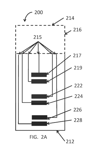
[0043] Referring to FIG. 2A, in some embodiments, an integrated test strip
200 may have a
base layer 216 and a plurality of electrodes 217, 219, 222, 224, 226, 228 that
make up at least
three systems on the test stri.p 200. For example, the first system includes a
first set of electrodes
or hematocrit electrodes that include a first counter electrode (hematocrit
cathode) 226 and a first
working electrode (hematocrit anode) 228. The second system includes a second
set of electrodes
or interference electrodes, such as a second counter electrode (interference
cathode) 222 and
second working electrode (interference anode) 224 disposed in the capillary
chamber 220 (see
FIG, 2B). The third system includes a third set of electrodes or glucose
electrodes, such as a third
counter electrode (glucose cathode) 219 and a third counter electrode (glucose
anode) 217. In
some embodiments, the electrodes 217, 219, 222, 224, 226, 228 may be at least
partially
disposed in the capillary chamber (see FIG. 2B) to expose the electrodes to
the blood sample in
the chamber. Further, conductive traces 215 electrically connect the plurality
of electrodes 217,
219, 222, 224, 226, 228 disposed on base layer 216 near the proximal end 212
to a plurality of
electrical contacts (not shown) located on the distal end 214 of the test
strip 200.
[0044] The three systems of the test strip 200, the first system having
hematocrit electrodes
226, 228, the second system having interference electrodes 222, 224 and the
third system ha.vin,g,
glucose electrodes 217, 219 are further explained below. In some embodiments,
the hematocrit
11
CA 02972468 2017-06-27
WO 2016/109801 PCT/US2015/068297
electrodes are located closest to the entry to the chamber (proximal end),
followed by the
interference electrodes, and then by glucose electrodes. As is discussed
below, in some
embodiments the hematocrit electrodes are reagent free, but alternatively in
some embodiments
the hematocrit electrodes may be coated with reagent. If a small amount of
ionic components in
either the glucose or interference reagent, such as the mediator or buffer is
carried into the
hematocrit area, it may interfere with the hematocrit measurement. Similarly,
in some
embodiments, the interference cathode does not include an enzyme. In some
embodiments, the
interference reagent may thus be proximal to the glucose reagent because if
any of the enzyme
washed onto the interference area it might render the interference signal
partially dependent on
the glucose level and eliminate its effectiveness. However, the order of the
tests may be
changed. In some embodiments, the order does not matter if the reagents were
so constituted
that there was not significant mobility of the ions or enzymes from one region
to another during
the time of a test. That is, the reagent can wet and become active without
truly dissolving and
migrating.
[0045] The hematocrit electrodes 226, 228 may be spaced at a predetermined
distance such
that hernatocrit level may be determined in the blood sample by measurement of
electrical
itnpedance or current between the two hematocrit electrodes in the capillary
chamber. In some
embodiments, the hematocrit electrodes 226, 228 are free of reagent. The use
of a reagent free
hematocrit electrodes can also allow for the use of a simpler electrical
measurement technique,
such as pulsed DC, rather than a more complicated electrical measurement
technique.
[0046] The requirement that the hematocrit measurement electrodes 226, 228
be free of
deposited reagent does not limit the placement relative to other electrodes on
the test strip. The
two hematocrit electrodes 226, 228 could be the first two electrodes traversed
by the blood
flowing into the strip or the last two.
[0047] It is possible the hematocrit measurement electrodes 226, 228 can
also be placed
between other electrodes on the test strip 200 that are used for other
purposes. Further, the
hematocrit electrodes 226, 228 may be placed adjacent to each other or apart
from each other
with other electrodes in between the two.
[0048] In some embodiments, the hematocrit electrodes 226, 228 free of
reagent may be
placed next to each other to ensure that the blood sample does not get exposed
to reagent during
12
CA 02972468 2017-06-27
WO 2016/109801 PCT/US2015/068297
hematocrit measurement. Reagent on the electrodes can impact the hematocrit
measurement. It is
preferable that the hematocrit cathode be free of reagent, but it is not
necessary. In some
embodiments, the test strip further comprises isolation islands. Isolations
islands are regions
where the sputtered metal film is laser ablated off of the plastic substrate
below is exposed. This
creates a hydrophobic region that inhibits reagent from spreading over it and
so isolates areas
that have no reagent from areas that have reagent. In some embodiments,
isolation islands can
prevent the mixing of two different types of reagents such as glucose reagent
and interference
reagent. For example, in FIG. 10 (discussed more fully below) there is
disclosed a strip 1000
that has a multi-well spacer into which reagent is drop dispensed. These wells
help separate
regions of the strip from each other. As the amount, distribution and
solubility of reagent may
differ slightly from strip to strip, having electrodes with no reagent may
lead to more accurate
and precise hematocrit measurements. In some embodiments, the placement of the
hematocrit
electrodes 226, 228 can be potentially advantageous where there are other
intervening electrodes
between the two hematocrit electrodes that can allow for a longer measurement
path and greater
discrimination between hematocrit levels than a shorter path would allow. A
short path can be
anything less than 2 mm between the hematocrit anode and cathode and only has
an electrically
isolated area between them. A long path can be anything longer than 2 mm and
can include other
electrodes between hematocrit anode and cathode. In comparison of a small path
(0.5 mm ¨ 2
mm) and a long path (2 mm ¨ 5 mm), testing has shown that a longer path length
increases
hematocrit resolution and therefore improves precision.
[0049] in some embodiments, the hematocrit electrodes may be separated by
an electrically
isolated region. in some embodiments, the distance between electrodes 226 and
228 may be
approximately about I min The distance between the hematocrit anode and
cathode can range
between about lmm and 5 mm, inclusive.
[0050] The second or interference system includes the interference anode
224 and the
interference cathode. In some embodiments, the interference anode 224 has
deposited upon its
surface a reagent that contains a redox mediator, but is free of an analyte
specific enzyme
(interference reagent) to correct for interfering substances that directly
react with the surface of
the analyte measuring anode electrode 224 or with the mediator. The
interference cathode 222
13
CA 02972468 2017-06-27
WO 2016/109801 PCT/US2015/068297
may be coated with the same reagent as the interference anode or with a
reagent containing the
analyte specific enzyme and mediator (full reagent).
[0051]
The glucose and/or interference cathode may be covered with glucose reagent
which
consists of enzyme and mediator. The electrochemical reaction occurring at the
cathode does not
involve the enzyme, just the mediator: Fe3+(CN)6 + e-
Fe2+(CN)6. This serves to
electrically balance the reverse reaction occurring at the anode (e- is an
electron). At the
interference anode, which contains no enzyme, the Fe2+(CN)6 (ferrocyanide) is
generated only
from the reaction of oxidizable compounds such as ascorbic acid and uric acid
directly with
Fe3+(CN)6 (ferricyanide). At the glucose anode the same reactions that are
described for the
interference anode are also occurring, but in addition there is more
ferrocyanide being generated
from the action of the enzyme on glucose. Therefore, the difference between
the signals from
the glucose and interference anodes results in just the signal from glucose.
So only the glucose
and/or interference cathode contain the full reagent with both mediator and
enzyme. The
interference anode is covered with reagent that contains only mediator.
[0052]
Referring to the second system of FIG. 2A and FIG. 2B, it is possible to use
the signal
generated by the interference anode 224 in different ways to correct for
oxidizable interferents.
The signal from this anode can be used to correct for any change in background
current that
occurs in strips stored in the vial over time. That is, it can improve the
stability of the strip and
thus increase its shelf-life. In some embodiments, to correct the analyte
value, a mathematically
modified interference current may be subtracted from the analyte specific
current to then
generate a corrected analyte value, which is further described in FIG. 12.
[0053]
Referring to the second system of FIG. 2A, it is possible to scale the
interference
current in a test strip lot specific manner so the subtraction can be
appropriate for each batch of
test strips, as may be further seen in FIG. 13.
[0054]
The third system of FIG. 2A may include a working anode electrode 219 and
counter
cathode electrode 217. These electrodes may be covered in their entireties by
the full reagent
layer to enable the level of glucose in the blood sample to be determined
electrochemically. The
reagent layer may include an enzyme specific for glucose, such as glucose
oxidase, and a
mediator, such as potassium ferricyanide or ruthenium hexaammine. The reagent
may also
include other components, such as buffering materials (e.g., potassium
phosphate), polymeric
14
CA 02972468 2017-06-27
WO 2016/109801 PCT/US2015/068297
binders (e.g., hydroxypropyl-methyl-cellulose, sodium alginate,
microcrystalline cellulose,
polyethylene oxide, hydroxyethylcellulose, and/or polyvinyl alcohol), and
surfactants (e.g.,
Triton X-100 or Surfynol 485). With these chemical constituents, the reagent
layer reacts with
glucose in the blood sample in the following way: The glucose oxidoreductase
initiates a reaction
that oxidizes the glucose to gluconic acid and in the process reduces the
ferricyanide to
ferrocyanide. When an appropriate voltage is applied to the working electrode,
relative to the
counter electrode, the ferrocyanide is oxidized back to ferricyanide, thereby
generating a current
that is related to the glucose concentration in the blood sample.
[0055] Referring to FIG. 2A, it should be noted that the electrodes 217,
219, 222, 224, 226,
228 can be located in any particular order and/or location on the test strip
200. In some
embodiments, the order (proximal to distal where proximal is the blood entry
portion) may be
hematocrit, interference then glucose. This order is impacted by blood flow.
Any mediator, salt
or buffer in the interference or working reagent that washes or back diffuses
over the hematocrit
anode may compromise the hematocrit measurement. Any enzyme in the glucose
reagent that
washes or back diffuses over the interference anode may compromise the
interference
measurement. That being said, if the reagents are properly constituted there
could be very little
flow or back diffusion over the sensitive electrodes during the time course of
the test so that in
theory any order is passible. In some embodiments, the fill electrode may be
the most distal
electrode, but other placements of the interference electrodes 222, 224 are
possible. For
example, the most distal electrode could be a shared fill and Hct cathode. The
glucose signal is
also dependent on the size of the glucose cathode that is covered since there
has to be sufficient
reactive area on the cathode to sink the current produced by the anode. This
is especially true for
samples that have high levels of glucose. For example, one other placement of
the interference
anode 224 can be upstream of the analyte measuring electrode interference
cathode 222. If the
solubility properties of the full (enzyme & mediator) and working reagent
along with the timing
of the analyte and interference measurements are properly adjusted, then,
among other things, the
interference anode 224 can be placed either upstream or downstream from the
interference
cathode 222.
[0056] FIG. 2B illustrates the top plan view of the first configuration of
the integrated test
strip 200 of FIG. 2A. FIG. 2B shows the dielectric insulating layer 218 formed
over the
CA 02972468 2017-06-27
WO 2016/109801 PCT/US2015/068297
conductive pattern, where conductive traces 315 are electrically connecting
the plurality of
electrodes 217, 219, 222, 224, 226, 228 to a plurality of electrical contacts
(not shown). It is also
noted that the plurality of electrodes 217, 219, 222, 224, 226, 228 are in
communication with the
capillary chamber 220.
[0057] Referring to FIG. 2C and FIG. 2D, in some embodiments, there may be
less than
three systems on the test strip 200. For example, and not limited by any
particular embodiment,
as seen in FIG. 2C there may only be two systems, such as a glucose anode 219
and a paired
glucose cathode 217, and an interference anode 232 with a paired interference
cathode 230.
Further, as described below, the systems may share an electrode to further
reduce the number of
electrodes on the test strip. In some embodiments the systems can have shared
functions. For
example, in some embodiments there may be no hematocrit measurement system on
the test strip
200. Further, the glucose system and the interference system may share a
cathode, such that the
electrodes are as following: a glucose anode 219, a shared glucose /
interference cathode 230, an
interference anode 232, and a fill detect cathode 217. By way of a non-
limiting example,
hematocrit effects may be mitigated using information from glucose decay
curves. Glucose
decay curve (current vs. time) characteristics, such as initial slope,
curvature, current magnitude
at a selected time, slope at a selected time, area under the decay curve, and
the presence and
timing of inflection points, may be mathematically manipulated to generate a
signal in which the
effect of hematocrit is greatly reduced or completely eliminated.
[0058] In reference to FIG. 3A and FIG. 3B, in some embodiments, in a test
strip 300 used to
measure an analyte concentration in a biological fluid, the interference
system and the glucose
system share the cathode 317.
[0059] The first system of FIG. 3A includes hematocrit electrodes 326, 328
and define a path
that is dedicated to the measurement of hematocrit in the test strip 300.
These electrodes may be
reagent free, that is, not covered by the reagent. The second system includes
an interference
anode 324 having a reagent with only a mediator and positioned distal to the
hematocrit cathode
326. The interference anode 324 can be optionally separated by a reagent
isolation island 330
from the hematocrit cathode 326 to ensure that the hematocrit electrodes are
free of any reagent.
However, as noted above. The glucose cathode and interference cathode are
combined into a
single cathode (or a glucose and interference cathode 317) that includes a
reagent with an
16
CA 02972468 2017-06-27
WO 2016/109801 PCT/US2015/068297
enzyme and a mediator. Since there is a large excess of ferricyanide in the
chemistry, the
electric potentials of the glucose and the interference cathodes are
independent of the
concentrations of analyte and interfering substances in the sample. Therefore,
the glucose and
interference cathodes can be combined into a single electrode allowing easier
manufacturing
process and a smaller strip design which at the same time allow the use of
smaller samples with
all the associated benefits. The third system of FIG. 3A includes a glucose
anode 319 but there
is no separate glucose cathode, but instead the interference system and the
glucose system share
the cathode 317.
[0060] In reference to FIG. 4A and FIG. 4B, in some embodiments, the three
systems of
electrodes may share the same cathode (or a glucose, interference and
hematocrit cathode 417).
Any relative configuration of cathode to the anode might work. The hematocrit
test is done at a
different time than the glucose and interference tests so where the hematocrit
anode is positioned
relative the cathode is unimportant. The interference tests and glucose tests
can be run at the
same time. For example, if the glucose anode is between the interference anode
and the common
cathode the electric field between the glucose anode and cathode might
interference with the
electric field between the interference anode and common cathode. In some
embodiments, the
common cathode may lie between the glucose and interference anodes (or working
electrodes).
But since electrochemistry occurs more at the surface of the electrodes, it
may be that the electric
fields do not play such an important role. Therefore, it is possible that any
configuration of
electrodes may work.
[0061] In reference to FIG. 4A and FIG. 4B, the electrode systems may
include a hematocrit
working electrode (anode) 428, an interference working electrode (anode) 426,
a glucose
working electrode (anode) 419, and a common cathode 417 with full reagent.
[0062] FIG. 5A and FIG. 5B illustrates a meter used to measure the glucose
level in a blood
sample. In some embodiments, the meter 500 has a size and shape to allow it to
be conveniently
held in a user's hand while the user is performing the glucose measurement.
Meter 500 may
include a front side 502, a back side 504, a left side 506, a right side 508,
a top side 510, and a
bottom side 512. The front side 502 may include a display 514, such as a
liquid crystal display
(LCD). A bottom side 512 may include a strip connector 516 into which test
strip 10 can be
inserted to conduct a measurement.
17
CA 02972468 2017-06-27
WO 2016/109801 PCT/US2015/068297
[0063] FIGS. 5A, 5B, 6A and 6D illustrate an exemplary embodiment of an
analyte meter
that may be used in connection with test strips of the present disclosure.
Referring to FIG. 5A
and FIG. 5B, the left side 506 of meter 500 may include a data connector 518
into which a
removable data storage device 520 may be inserted, as necessary. The top side
510 may include
one or more user controls 522, such as buttons, with which the user may
control meter 500, and
the right side 508 may include a serial connector (not shown).
[0064] FIG. 6A illustrates a top perspective view of a test strip 610
inserted within a meter
connector 30 consistent with the present disclosure. Test strip 610 includes a
proximal electrode
region 624, which contains the capillary chamber and measuring electrodes, as
described above.
Proximal electrode region 624 may be formed to have a particular shape in
order to distinguish to
the user the end receiving a fluid sample from distal strip contact region
626. Meter connector
630 includes channel 632 extending out to a flared opening for receiving the
test strip 610. Meter
connector 630 may further include tangs 636 extending a predetermined height
above the base of
channel 632. The predetermined height of tangs 636 is selected to limit the
extent, such as
through a corresponding raised layer of test strip 610, to which a test strip
610 can be inserted
into channel 632. Meter connector 630 may include a first plurality of
connector contacts 638,
disposed closer to the proximal end of meter connector 630, which are
configured to contact the
electrical strip contacts 619 upon insertion of the test strip 610 into the
meter connector 630. In
some embodiments, the test strip control circuit reader 640 may be disposed
closer to the distal
end of meter connector 630 to communicate with the test strip control circuit
650. In some
embodiments, the meter may be provided with one or more GPIO lines for
communication with
the IC. The one or more GPIO lines may replace digital coding lines (typically
3-5) utilizing
GPIOs.
[0065] FIG. 6B illustrates a general cross-sectional view of a test strip
inserted within meter
connector 630 of FIG. 6A, consistent with the present disclosure. Channel 632
depicts a
proximal row of connectors comprising a plurality of connector contacts 638
for connection the
electrical strip contacts 619 upon insertion of the test strip 610 into the
meter connector 630.
[0066] Referring to FIG. 7, illustrated is an embodiment of a diagnostic
strip 700 with a long
Hct path, which may be provided for better resolution of the results. The
strip 700 comprises a
fill detect cathode 701, a hematocrit cathode 702, a shared glucose and fill
anode 703, a shared
18
CA 02972468 2017-06-27
WO 2016/109801 PCT/US2015/068297
glucose, interference and drop detect cathode 704, an interference anode 705
which may be
coated with reagent only (mediator only), and a shared drop detect and
hematocrit anode 706.
The shared hematocrit drop detect anode 706 is at the proximal end of the
strip and is the first
electrode that the blood will encounter. Once the strip 700 is placed in the
meter (not pictured) it
is monitored for the addition of sample by measuring the current between the
drop detect anode
706 and cathode 704. The drop detect cathode 704 also serves as the glucose
and interference
cathode. Once the sample is detected, it has a fixed amount of time to reach
the fill cathode 701
at the distal end of the sample well of the strip 700. If this timing
criterion is satisfied, then the
remainder of the testing sequence will commence. In the strip 700
configuration demonstrated
by FIG. 7, the all of the measurements (hematocrit, glucose and interference)
will take place after
fill detect. In some embodiments, all three measurements cannot take place
simultaneously. The
preferred sequence will be that the hematocrit measurement will take place
first, followed by the
simultaneous measurement of glucose and interference. In this strip 700
configuration, the
hematocrit cathode 702 will be covered with glucose reagent and the hematocrit
anode 706 will
be reagent free. The i/i areas 707 on the strip are "isolation islands" that
separate areas of no
reagent (aH + aDD) from areas of reagent (aInt) or areas of two different
reagents (aInt vs. cG
+cInt + cDD).
[0067] FIG. 8 illustrates an embodiment of a diagnostic strip 800 with a
long Hct path, which
may be provided for better resolution of the results, and which may further
comprises a hog out
region 806. The strip 800 comprises a fill cathode 801, a shared glucose and
fill anode 802, a
shared glucose and interference cathode 803, an interference anode 804 which
may be coated
with reagent only (mediator only), a shared drop detect and hematocrit cathode
805, a hog out
region 806, a shared hematocrit and drop detect anode 807, and two isolation
islands (i/i) 808.
[0068] The hog out region may measure from about 1.2 mm to 2.0 mm. In
measuring the
resistance of the blood over an electrically isolated region, the resistance
of the blood is
proportional to its hematocrit. If the hog out distance increases, different
hematocrit levels may
be better distinguished from each other as the longer distance increases the
signal to noise ratio.
With a small separation, the variability in the distance between the
hematocrit anode and
electrode can make up a larger percentage of the gap. As the gap gets larger
the manufacturing
19
CA 02972468 2017-06-27
WO 2016/109801 PCT/US2015/068297
tolerances get relatively smaller and the resolution may improve. It should be
noted that, in some
embodiments, the hog out region may be removed or is optional, as seen in FIG.
4, 7 and 9.
[0069] FIG. 9 illustrates an embodiment of a diagnostic strip 900 with a
common Hct,
glucose and interference cathode 903. The strip 900 comprises a fill cathode
901, a shared
glucose and fill anode 902, a shared Hct, glucose, interference, and drop
detect cathode 903, an
interference anode 904, a shared Hct and drop detect anode 905, and two
isolation islands (i/i)
906. As a result of the shared design of the strip 900, the strip 900 only has
5 total electrodes.
[0070] FIG. 10 illustrates a diagnostic strip 1000 with a well design for
reagent containment.
The strip 1000 comprises a fill cathode 1001, a shared glucose and
interference cathode 1002, a
glucose anode 1003, an interference anode 1004, a shared Hct and drop detect
cathode 1005, a
hog out region 1006, a shared Hct and drop detect anode 1007, and three wells
for reagent
containment. A first well 1008 contains glucose reagent. A second well 1009
contains
interference reagent. A third well 1010 contains no reagent or a reagent with
only small amounts
of surfactant and/or polymer and/or buffer.
[0071] FIG. 11A and FIG. 11B illustrate a flow chart of an exemplary
process 1100 for
measuring analyte concentration using test strips of the present disclosure.
[0072] In reference to FIG. 11A and FIG. 11B, the meter may be battery
powered and may
stay in a low-power sleep mode 1101 when not in use in order to save power.
When the test strip
is inserted into the meter 1102, current flow to the meter causes the meter to
wake up and enter
an active mode 1103. Alternatively, the meter may be provided with a wake
button.
[0073] Next, the meter can connect to the control circuit to read the code
1104 information
from the control circuit and can then identify, for example, the particular
test to be performed, or
a confirmation of proper operating status. In addition, the meter can also
identify the inserted
strip as either a test strip or a check strip based on the particular code
information. If the meter
detects a check strip, it performs a check strip sequence 1105. If the meter
detects a test strip, it
performs a test strip sequence.
[0074] In addition, the meter can ensure that the test strip is authentic
and has not been
previously used 1106 and 1107. The meter will also measure the ambient
temperature 1105.
Diagnostics 1105 may include checksums or cyclic redundancy checks (CRC) of
portions of the
internal and/or external memory to establish confidence that the memory is not
corrupted
CA 02972468 2017-06-27
WO 2016/109801 PCT/US2015/068297
because the checksum/crc data calculated matches the programmed checksum/crc.
In some
embodiments, diagnostics test 1105 that may be performed is an LCD test to
verify the integrity
of the LCD to gain confidence it is not cracked and will display the proper
result to the user that
is sent to it. In some embodiments, diagnostic test 1105 may be an internal
calibration current
test to verify that the analog front end continues to measure an accurate
current within the margin
of error allowed.
[0075] If all information checks out, the meter can perform open contact
tests on all
electrodes to validate the electrodes 1107. The meter may validate the
electrodes by confirming
that there are no low-impedance paths between any of these electrodes. If the
electrodes are
valid, the meter indicates to the user 1108 that sample may be applied to the
test strip and the
meter can perform analyte measurements.
[0076] In some embodiments, the systems of the present disclosure may be
used to measure
glucose concentration in blood, among other measurements, as discussed above.
Once the meter
has performed an initial check routine 1104, 1105, 1106, 1107, as described
above, the meter
may apply a drop-detect voltage 1110 between a working and counter electrodes
and detect a
fluid sample, for example, a blood sample, by detecting a current flow between
the working and
counter electrodes (i.e., a current flow through the blood sample as it
bridges the working and
counter electrodes). For example, in some embodiments, the meter may measure
an amount of
components in blood which may impact the glucose measurement, such as, for
example, a level
of hematocrit 1111 or of an interferant 1111. The meter may later use such
information to adjust
the glucose concentration to account for the hematocrit level and the presence
of the interferants
in blood, among other things. These measurements may also be corrected based
on the
temperature.
[0077] Next, to detect that an adequate sample is present in the capillary
chamber and that
the blood sample has traversed the reagent layer and mixed with the chemical
constituents in the
reagent layer, the meter may apply a fill-detect voltage 1112 between the fill-
detect electrodes
and measure any resulting current flowing between the fill-detect electrodes.
If this resulting
current reaches a sufficient level within a predetermined period of time 1109,
the meter indicates
to the user that adequate sample is present and has mixed with the reagent
layer. The process of
adequate sample (fill) detection may occur at any time during the measurement
sequence.
21
CA 02972468 2017-06-27
WO 2016/109801 PCT/US2015/068297
[0078] In one embodiment, the test strip meter comprises a decoder for
decoding a
predetermined electrical property, e.g. resistance, from the test strips as
information. The decoder
operates with, or is a part of, a microprocessor.
[0079] The meter can be programmed to wait for a predetermined period of
time after
initially detecting the blood sample 1109 or after ensuring there is adequate
sample 1112, to
allow the blood sample to react with the reagent layer or can immediately
begin taking readings
in sequence. During a fluid measurement period, the meter applies an assay
voltage between the
working and counter electrodes and takes one or more measurements of the
resulting current
flowing between the working and counter electrodes. The assay voltage is near
the redox
potential of the chemistry in the reagent layer, and the resulting current is
related to the
concentration of the particular constituent measured, such as, for example,
the glucose level in a
blood sample.
[0080] In one example, the reagent layer may react with glucose in the
blood sample in order
to determine the particular glucose concentration 1113. In one example,
glucose oxidase is used
in the reagent layer. The recitation of glucose oxidase is intended as an
example only and other
enzymes can be used without departing from the scope of the disclosure. Other
possible
mediators include, but are not limited to compounds containing ruthenium or
osmium. During a
sample test, the glucose oxidase initiates a reaction that oxidizes the
glucose to gluconic acid and
reduces the ferricyanide to ferrocyanide. When an appropriate voltage is
applied to a working
electrode, relative to a counter electrode, the ferrocyanide is oxidized to
ferricyanide, thereby
generating a current that is related to the glucose concentration in the blood
sample. The meter
then calculates the glucose level based on the measured current and on
calibration data that the
meter has been signaled to access by the code data read from the second
plurality of electrical
contacts associated with the test strip.
[0081] The meter can then adjust the glucose level 1115, as necessary,
based on the
measurements of the temperature, hematocrit and the presence of interferants
1111. Non-
limiting examples of algorithms for glucose level correction are presented in
FIG. 12 and FIG.
13. Errors will be displayed 1114 if encountered.
[0082] FIG. 12 discloses an embodiment flow chart for correcting the
analyte value 1200,
wherein the analyte specific current is modified based on temperature and
hematocrit and
22
CA 02972468 2017-06-27
WO 2016/109801 PCT/US2015/068297
interference currents to then generate a corrected analyte value. For example,
equations may be
IC = IA ¨ S xII, where IC is the corrected current, IA is the current measured
from the analyte
anode, II is the current measured from the interference anode, and S is an
empirically derived
scaling factor. The present calculation may eliminate the need to make
complicated calculation
and/or voltage application schemes. The present calculation uses a
mathematically modified
(scaled) subtraction of the interference current from the current from the
analyte specific anode.
The interference current may be multiplied by an empirically determined
constant that is
dependent only on the relative areas of the two electrodes, not on the
relative effects of
hematocrit and temperature variations on the two currents. This is because the
two reagents
(analyte and interference) are formulated to respond the same way to
hematocrit and temperature
variations. Thus, referring to FIG. 12, the raw glucose signal 1201 would be
corrected with the
raw interference signal 1202 to obtain an interference corrected glucose
signal 1203, where a
temperature correction is incorporated to obtain an interference and
temperature corrected
glucose value 1204. The raw Hct signal 1205 is corrected to obtain a
temperature corrected Hct
1206. The interference & temperature corrected glucose value 1204 may then be
incorporated
with the temperature corrected Hct 1206 to obtain an interference, temperature
& Hct corrected
glucose value 1207.
[0083] It is also possible to first make temperature and hematocrit
adjustments to the
interference current and then subtract it from the raw analyte current and
then subject that
corrected current to another temperature and hematocrit adjustment. In some
embodiments, it
may be possible to correct the analyte and interference currents separately
for temperature and
hematocrit, and then convert each separately to an uncorrected glucose value
and to a glucose
equivalent value, respectively. Then the glucose equivalent value can be
subtracted from the
uncorrected glucose value to obtain a corrected glucose value.
[0084] FIG. 13 discloses five potential non-limiting ways to use the
current from the
interference anode in combination with the current from the glucose anode to
isolate the glucose
signal. Both temperature and hematocrit affect both the interference and the
glucose currents. In
some embodiments, hematocrit and temperature effects are virtually identical
for both currents
primarily because the reagent composition of the glucose reagent and the
interference reagent are
so similar. The glucose reagent contains a glucose oxidoreductase (glucose
dehydrogenase),
23
CA 02972468 2017-06-27
WO 2016/109801 PCT/US2015/068297
which is a protein, while the interference reagent contains an inactive
protein (which may be
Bovine Serum Albumin) that mimics the physical properties (viscosity,
solubility) of the enzyme
in the reagent. This allows use of Correction ID #1 in FIG. 13. The reason
that the scalar
(constant) is included in Correction ID #1 is that the area of the
interference anode is much larger
than that of the glucose anode in order to increase the signal to noise ratio
of the interference
current. Accordingly, current from the interference anode is much lower than
the current from
the glucose anode. In some embodiments, the properties of the interference and
glucose reagents
are not so similar, which leads to use of a correction method such as
Correction ID #2 or #3,
which contain separate hematocrit and temperature corrections for the
interference current and
corrected analyte current or the raw analyte current. Correction ID #4 would
be used in the case
that the interference regent had different temperature properties than, but
similar hematocrit
properties to the glucose reagent. Correction ID #5 would be used in the case
that the
interference regent had different hematocrit properties than, but similar
temperature properties to
the glucose reagent.
[0085] In some embodiments, it is possible to use the present calculation
to also first convert
the interference current to analyte equivalents and then subtract it from the
amount of analyte of
interference and subtract that number. That is, the correction can occur
before or after
mathematically processing the current. For example, by having the interference
anode larger for
improved signal to noise ratio due to the currents being so small, at least
one aspect includes
using a scaling factor and anodes of different surface area.
[0086] In some embodiments, the type of subtraction may be made conditional
on the level
of interference. For example, if the level of interference is low enough
relative the analyte, then
no subtraction is necessary. However, if the interference level proves to be
sufficiently high, then
the subtraction can be made to correct the reported analyte value. At least
one aspect of the
interference correction is to improve the accuracy of the reported glucose
value by cancelling the
effect of interfering substances. However, when subtracting two currents (or
two calculated
values) each with a certain amount of noise it is possible to increase the
precision error. For
example, at a very low level of interference where the accuracy correction is
minimal, it is
possible to not subtract out the interference correction because improvement
in accuracy can be
outweighed by the degradation in precision. For example the FDA may desire
that the glucose
24
CA 02972468 2017-06-27
WO 2016/109801 PCT/US2015/068297
readings from glucose measuring devices report glucose values within 7 mg/dL
of the
reference method for reference values < 70 mg/dL, and within
10% for reference values > 70 mg/dL, no less than 99% of the time. It may be
decided that
the total system error is minimized when the interference correction is made
only when it
amounts to a change of > 3.5 mg/dL when the reference value is < 70 mg/dL and
only when it is
> 5% of the uncorrected glucose value when the reference value > 70 mg/dL.
However, at least
one aspect considers to use cut off values of when the interference correction
will be applied by
determining which cut off values minimize the total system error. (TSE) At
least one way of
defining TSE is: TSE =1%Biasl+ 2xCV or 1Bias (mg/dL) 1+ 2x SD.
[0087] In some embodiments, the algorithm may use current subtraction.
Current
subtraction works as follows: In some embodiments, the interference anode is
larger than the
glucose anode because the interference anode current is typically small and a
larger surface area
is needed to improve the signal to noise ratio. Since the areas of the
interference and glucose
anodes are different, a simple equation will be used to modify the measured
current from the
interference anode to resize it correspond to that from the glucose anode:
iInt Resize = m*iInt
Raw + b. Where m & b are constants. Where m ( 1 and it is very likely that b =
0, but that is
not necessary. The resized current can be mathematically processed in a number
of ways to
yield a Corrected Interference Current: 1) no further correction is made; 2) a
temperature
correction is made (if the interference reagent changes with temperature in a
manner different
from that of the glucose reagent); 3) a hematocrit correction is made (if the
interference reagent
changes with hematocrit in a manner different from that of the glucose
reagent); and 4)
temperature and hematocrit corrections are made (if the interference reagent
changes with
temperature AND with hematocrit in ways different from that of the glucose
reagent). At this
point the corrected current from the interference anode is subtracted from the
current from the
glucose anode to get a current that represent the current from the oxidation
of glucose alone.
This current in turn is subjected to temperature correction, hematocrit
correction and finally to a
mathematic conversion to get a glucose value. The final mathematical
conversion is typically
(but not necessarily) in the form of a polynomial such as: Glucose = a*i2 +
b*1 + c, where a, b
& c are constants that can be tailored for each strip lot or where a, b & c
are selected from a
limited number of predetermined sets of such constants that best fit the strip
lot in question.
CA 02972468 2017-06-27
WO 2016/109801 PCT/US2015/068297
[0088] In some embodiment, it may be possible to process the interference
current as in Step
4) in the paragraph above and then apply a separate polynomial equation to the
interference
current to convert it to a glucose equivalent. This glucose equivalent will be
subtracted from a
glucose value derived from applying a temperature correction and a hematocrit
correction to the
glucose current and then applying a mathematical conversion to obtain a
glucose value. This
glucose value will be uncorrected for interference until the glucose
equivalent is subtracted from
it. The exact nature of all the possibilities of temperature and hematocrit
corrections are
numerous and should remained undefined. The meter then displays the calculated
glucose level
to the user.
[0089] It should be noted that while the operation of the system of the
present disclosure has
been described primarily in connection with determining glucose concentration
in blood, the
systems of the present disclosure may be configured to measure other analytes
in blood as well
as in other fluids, as discussed above.
[0090] Whereas many alterations and modifications of the present disclosure
will no doubt
become apparent to a person of ordinary skill in the art after having read the
foregoing
description, it is to be understood that the particular embodiments shown and
described by way
of illustration are in no way intended to be considered limiting. Further, the
disclosure has been
described with reference to particular embodiments, but variations within the
spirit and scope of
the disclosure will occur to those skilled in the art. It is noted that the
foregoing examples have
been provided merely for the purpose of explanation and are in no way to be
construed as
limiting of the present disclosure. While the present disclosure has been
described with
reference to exemplary embodiments, it is understood that the words, which
have been used
herein, are words of description and illustration, rather than words of
limitation. Changes may
be made, within the purview of the appended claims, as presently stated and as
amended, without
departing from the scope and spirit of the present disclosure in its aspects.
Although the present
disclosure has been described herein with reference to particular means,
materials and
embodiments, the present disclosure is not intended to be limited to the
particulars disclosed
herein; rather, the present disclosure extends to all functionally equivalent
structures, methods
and uses, such as are within the scope of the appended claims.
26