Note: Descriptions are shown in the official language in which they were submitted.
OCCLUSIVE DEVICES WITH ANCHORS EXTENDING FROM PERIPHERAL
EDGE OF THE OCCLUSIVE FACE
CROSS-REFERENCE TO RELATED APPLICATIONS
[0001] This application claims priority to US Provisional Application Number
61/535,830 filed September 16, 2011.
TECHNOLOGY FIELD
[0002] The disclosure relates to occlusive devices useful, for example, in
occluding structures or conduits within a patient, particularly an atrial
appendage in
the human heart, and methods of making and using the devices, including
delivering, deploying, and retrieving or repositioning the devices. Devices
described
herein can be delivered percutaneously or in an endovascular fashion.
BACKGROUND
[0003] Embolic stroke is the nation's third leading killer, and is a major
cause
of disability. There are over 780,000 strokes per year in the United States
alone. Of
these, roughly 110,000 are hemorrhagic, and 670,000 are ischemic (either due
to
vessel narrowing or to embolism). The most common cause of ischemic stroke of
cardiac origin is thromboemboli due to atrial fibrillation. One out of every
six strokes
(approximately 130,000 per year) is attributed to atrial fibrillation. Atrial
fibrillation is
the most common heart arrhythmia; it results in a rapid and chaotic heartbeat
that
lowers cardiac output and leads to irregular and turbulent blood flow in the
vascular
system. There are over eight million people worldwide with atrial
fibrillation, with
about eight hundred thousand new cases reported each year. Atrial fibrillation
is
associated with a greater risk of stroke compared with age-matched healthy
controls. A patient with atrial fibrillation typically has a significantly
decreased quality
of life due, in part, to the fear of stroke, and the pharmaceutical regimen
necessary
to reduce that risk.
[0004] When patients develop atrial thrombus from atrial fibrillation, the
clot
occurs in or originates from the left atrial appendage of the heart over
ninety percent
of the time. The left atrial appendage is a closed cavity that looks like a
small thumb
1
CA 2972536 2017-07-06
or windsock; it is connected to the anterolateral wall of the left atrium
between the
mitral valve and the root of the left pulmonary vein. The left atrial
appendage
contracts with the left atrium during a normal heart cycle, thus keeping blood
from
becoming stagnant. However, with atrial fibrillation, the left atrial
appendage often
fails to contract with any vigor due to disorganized electrical signals. As a
result,
thrombi can be predisposed to form in the stagnant blood within the left
atrial
appendage.
[0005] Pharmacological therapies for stroke prevention in atrial fibrillation
patients such as oral or systemic administration of warfarin have often been
generally inadequate due to serious side effects and lack of patient
compliance.
Invasive surgical or thorascopic techniques have been used to obliterate the
left
atrial appendage; however, many patients are not suitable candidates for such
procedures due to compromised condition or previous cardiac surgery. In
addition,
the perceived risks of these surgical procedures often outweigh the potential
benefits.
[0006] Many of the current commercial devices that attempt to occlude the
left atrial appendage for stroke prevention in atrial fibrillation patients
utilize a rigid,
cylindrical support frame with tissue-piercing fixation members that engage
tissue in
the appendage itself. The opening (ostium) of the left atrial appendage varies
in
geometry and size. Sealing the left atrial appendage with a rigid frame that
presupposes a circular ostium may not be effective in preventing thromboemboli
from entering systemic circulation.
[0007] Another concern with some of the current devices is with the filtering
type membranes used by the devices. These membranes are macroporous and
typically require significant periods of time to provide cessation of blood
flow through
the membrane. Such membranes can take hours to weeks to substantially occlude
the left atrial appendage. The possibility exists for thromboemboli to enter
the blood
stream while the clotting/occluding process of the filtering membrane takes
place.
Many of these atrial fibrillation patients are on some type of blood thinning
(anticoagulant or antiplatelet) medication, which could prolong the
clotting/occluding
process for these filtering membranes and expose patients to stroke risk.
2
CA 2972536 2017-07-06
SUMMARY
[0008] In a first general aspect, an occlusive device includes a frame element
having a distal end and a proximal end, and a delivery configuration and a
deployed
configuration. The occlusive device also includes an occlusive face having a
peripheral edge, where the occlusive face positioned toward the proximal end
of the
frame element. The occlusive device also includes at least one anchor
positioned at
the peripheral edge of the occlusive face, where the at least one anchor
extends at
an acute angle to the peripheral edge of the occlusive face.
[0009] In various implementations, the at least one anchor may include a
tissue engagement member that protrudes in a proximal direction with reference
to
an axial dimension of the device. The at least one anchor may include a tissue
engagement member that protrudes in a distal direction with reference to an
axial
dimension of the device. The at least one anchor may include a tissue
engagement
member that may extend tangentially from a portion of the frame element near
the
anchor. The at least one anchor may be located substantially within a plane
defined
by the peripheral edge. The occlusive face may have a concave orientation. The
occlusive face may have a convex orientation. The occlusive face may have a
substantially planar orientation. Multiple anchors may be disposed on the
peripheral
edge. The frame may include a tapered region. The occlusive device may also
include a membrane configured to inhibit passage of blood, where the membrane
covers at least a portion of the frame. The membrane may include a
fluoropolymer.
The membrane may include polytetrafluoroethylene. The membrane may include
expanded polytetrafluoroethylene. The frame may include a plurality of wires.
The
plurality of wires may include nitinol. The frame may include a cylindrical
region that
extends a first distance from the occlusive face in a generally distal
direction, and
the tapered region may extend from a distal end of the cylindrical region to
the distal
end of the frame. The occlusive device may also include one or more anchors
disposed near a junction of the cylindrical region and the tapered region. The
frame
element may include a petal shape and an apex of the petal shape, and wherein
the
apex of the petal shape includes a bend in the frame element. The at least one
anchor may be located at the apex of the petal shape. The at least one anchor
may
include a first cuff and a second cuff, where the frame element passes through
each
3
CA 2972536 2017-07-06
of the first and second cuffs, and where the first cuff is positioned on a
first side or
the apex and the second cuff is positioned of a second side of the apex that
is
different from the first side.
[0010] In a second general aspect, a method of occluding a vessel includes
providing an occlusive device that comprises (a) a frame element having a
distal end
and a proximal end and a delivery configuration and a deployed configuration;
(b) an
occlusive face having a peripheral edge, and positioned toward the proximal
end of
the frame element; and (c) at least one anchor positioned at the peripheral
edge of
the occlusive face, wherein at least a portion of the at least one anchor
extends at
an acute angle to the peripheral edge of the occlusive face. The method also
includes configuring the occlusive device in the delivery configuration and
advancing
the occlusive device to a delivery site, and deploying the occlusive device at
the
delivery site.
[0011] In various implementations, the delivery site may be a left atrial
appendage. The at least on anchor may engage tissue near an ostium of the left
atrial appendage.
[0012] Other advantages, benefits, and novel features of the embodiments of
the present disclosure will become apparent from the following detailed
description
and accompanying drawings.
BRIEF DESCRIPTION OF THE DRAWINGS
[0013] Figure 1 is side view of an example occlusive device that can be used
to occlude a hole, defect, or appendage within a patient.
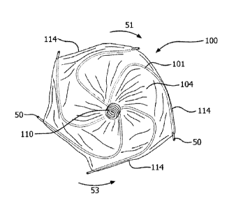
[0014] Figure 2 is a front view of a proximal end of the occlusive device of
Figure 1.
[0015] Figure 3 is a perspective view of an example frame of the occlusive
device of Figure 1.
[0016] Figures 4A and 4B are enlarged perspective views of a portion of the
frame of Figure 3.
[0017] Figure 5 is a perspective view of an example jig that can be used to
make the occlusive device of Figure 1.
4
CA 2972536 2018-09-10
[0018] Figure 6 is a perspective view of the jig of Figure 5 with wires of the
frame of Figure 3.
[0019] Figure 7 is a perspective view of the jig of Figure 5 with the wires of
Figure 6 shown in a winding pattern.
[0020] Figures 8A, 86, and 80 are perspective views of the jig of Figure 5
with
the wires of Figure 6 wound to form portions of the frame of Figure 3.
[0021] Figure 9 is a perspective view of a portion of the frame of Figure 3.
[0022] Figures 10A and 10B are perspective views of the frame of Figure 3 as
engaged with a center pin and prior to being expanded longitudinally.
[0023] Figures 11A and 11B are perspective views of the frame of Figure 3 as
engaged with a heat set mandrel prior to a heat treatment.
[0024] Figures 12A, 126, and 120 are perspective views of a heat set tool
that can be used to set the frame of Figure 3.
[0025] Figure 13 is a perspective view of the frame of Figure 3 as engaged
with the heat set mandrel of Figures 11A and 11B and following a heat
treatment.
[0026] Figures 14A and 14B are views of an example heat set mandrel.
[0027] Like reference numbers and designations in the various drawings
indicate like elements.
DETAILED DESCRIPTION
[0028] The devices and techniques discussed herein relate to occlusive
devices that can be used to occlude holes, defects, or appendages in the body
of a
patient, including the heart, and methods of making and using the devices.
Some
implementations of the devices can be used to occlude, without limitation,
right or
left atrial appendages, fistulas, aneurysms, and patent ductus arteriousus. In
some
embodiments, the occlusive devices provide a frame that is adequately or
sufficiently
compliant to conform to a wide variety of opening geometries and sizes.
Implementations of devices described herein can be easily loaded into a
catheter or
sheath, both at a time of initial deployment and at a later time, such as to
reposition
or remove the device from a deployed location within the body.
CA 2972536 2017-07-06
[0029] Although atrial fibrillation can result in blood clots originating in
the left
atrial appendage (LAA) and the occlusive devices illustrated herein will be
described
with regard to the LAA, the occlusive devices described herein can also be
used in
other areas of the body. Some embodiments of the devices may be used, for
example, in a right atrial appendage. In general, implementations of the
devices
may be used for placement across any appropriate aperture of the body,
including
apertures in the vasculature where there is a need to prevent blood clots from
escaping or to inhibit or substantially reduce blood flow.
[0030] Particularly, some embodiments of the occlusive devices can be
configured to occlude a LM. Implementations of devices described herein can be
used to conform to the anatomy of a variety of left atrial appendage ostia and
can
efficiently occlude the LAA, can demonstrate firm and secure anchoring with
reduced risk of trauma and bleeding from anchoring, and can provide rapid
cessation of blood flow across an occluding membrane included with the
devices.
The occlusive devices can include a frame that provides firm, secure anchoring
to
tissue of the LAA with significantly reduced clinical sequela from piercing,
or without
traumatic piercing, of the LAA tissue. As will be described in more detail
below,
different types of anchor features may be used with the devices disclosed
herein,
and the anchor features may be located at or associated with different areas
of the
devices.
[0031] Embodiments of the occlusive devices can include a membrane
configured to substantially or completely inhibit passage of blood through the
membrane. In some embodiments, the occlusive devices can include a membrane
that is configured to induce rapid tissue ingrowth and immediately occlude
passage
of blood through the membrane.
[0032] In some embodiments, the occlusive devices include an occlusive
face that is at least partially covered by the membrane and one or more
anchors
positioned on a peripheral edge of the occlusive face. In some embodiments,
one or
more anchors may be positioned on portions of the occlusive device that are
not on
the peripheral edge of the occlusive face.
[0033] Figures 1 and 2 illustrate an embodiment of an example occlusive
device 100 that can be used to occlude a structure or a conduit, such as an
LAA,
within a patient. The occlusive device 100 includes a proximal eyelet 110, a
distal
6
CA 2972536 2017-07-06
eyelet 112, an occlusive face 106, a generally cylindrical region 107
extending from
the occlusive face 106 in a distal direction, a tapered region 108 extending
from the
cylindrical region 107 toward the distal end of the device, and a membrane 104
covering a frame 102 (see Figure 3) of the occlusive device 100. A lumen can
extend through both eyelets 110 and 112 and through the length of device 100.
[0034] The occlusive face 106 is configured to conform, while in a deployed
configuration, to a shape of an ostium of the LAA, or other biological ostia.
For
example, the diameter of the occlusive face 106 can be altered or adjusted
during
deployment of the occlusive device 100 by transmitting torque to the frame 102
via
the delivery system. In the example illustrations of Figures 1 and 2, the
occlusive
face 106 has a concave shape. However, in other examples, the occlusive face
106
can have a convex shape or a flat or planar shape. An adaptability of the
occlusive
face 106 can allow versatility in sizing of the occlusive device 100 and
facilitate
placement of the occlusive device 100 in an ostium of an LAA, which are often
irregularly shaped and may differ substantially in size from one patient to
another.
[0035] In a general embodiment, the generally cylindrical region 107, which
can extend distally from the occlusive face 106, can be of any appropriate
length.
Accordingly, the length of the cylindrical region 107 can allow for variances
in the
ostium of the LAA or LAA shape variances. For example, in some embodiments,
the cylindrical region 107 may have a length from about 0.2 cm to about 0.7
cm, and
in some embodiments, a length of about 0.5 cm. Similarly, the tapered region
108,
which extends from the cylindrical region 107 to the distal eyelet 112, can be
of any
appropriate length. For example, in some embodiments, the tapered region 108
may have a length from about 0.6 cm to about 1.2 cm, and, in some embodiments,
a
length of about 1.0 cm. Furthermore, a profile of the tapered region 108 can
have
any suitable slope with respect to a longitudinal axis of the device to
provide
sufficiently secure positioning of the occlusive device 100 within an inner
region of
the LAA. For example, the tapered region 108 can be configured to conform to a
variable taper of the inner region of an LAA. A junction 130 may define a
boundary
between the cylindrical region 107 and the tapered region 108.
[0036] In the example of Figures 1 and 2, the eyelets 110 and 112 have a
substantially cylindrical shape. However, the eyelets 110 and 112 can
generally be
provided in a variety of shapes, such as a rectangular shape, other polygonal
shape,
7
CA 2972536 2017-07-06
or an irregular shape. One or both of eyelets 110 and 112 can be formed to
engage
one or more components of a delivery system (e.g., a delivery catheter) that
can be
used to deliver the occlusive device 100 to a delivery site within a patient.
For
example, engagement of a delivery catheter with either or both of the eyelets
110
and 112 may allow a torque to be applied to and maintained on the occlusive
device
100. In some embodiments, an application of torque to the occlusive device 100
may facilitate placement of the device and, in some embodiments may facilitate
engagement of anchors or anchor features of the device with tissue at the
delivery
site.
[0037] The device 100 can include anchors 50, 50a, 50b, 60 (FIG. 4A)
attached to portions of the frame 102 of the device. See FIGS. 1,2, 3, 4A, 4B,
10A,
10B, and 11A for examples of anchors that can be used. Some anchors 50 may be
attached to frame portions that form a peripheral edge 114 of the occlusive
face 106
of the occlusive device 100, as shown in FIGS. 1 and 2. The occlusive face 106
may be structurally formed from the proximal end of the multi-wire frame 102.
As
described above, in the depicted example of FIGS. 1 and 2, the occlusive face
is
concave, and this may facilitate projection of the anchors 50 on the
peripheral edge
114 of the occlusive face 106 in a proximal or partially proximal direction,
with
respect to a longitudinal dimension of the device. As such, in some
embodiments
the anchors 50 on the peripheral edge 114 of the occlusive face may be non-
planar
with the peripheral edge 114 of the occlusive face 106 (because they project
proximally). Anchors that protrude proximally along an axial orientation of
the device
may provide advantages for engaging tissue and preventing migration of the
device
following deployment (e.g., may prevent the device from moving from the
appendage).
[0038] In other embodiments, the anchors 50 on the peripheral edge 114 of
the occlusive face may be planar with the peripheral edge 114 of the occlusive
face
106 (that is, located within or substantially within a plane defined by the
peripheral
edge 114). For example, the anchors may project tangentially from a portion of
the
wire frame that is proximate to the anchor 50. In yet other embodiments, the
anchors may be shaped to project in a distal or partially distal direction
from the
peripheral edge 114 of the occlusive face 106, and may thus also be considered
non-planar with the peripheral edge 114.
8
CA 2972536 2017-07-06
[0039] Relatedly, for embodiments where the occlusive face has a convex
profile or a planar profile, in various implementations the anchors 50
positioned on a
peripheral edge of the occlusive face may similarly be oriented to project in
a
proximal, partially proximal, distal or partially distal direction with
respect to a
longitudinal dimension of the device, and in such cases may be considered non-
planar with the peripheral edge of the occlusive face. Alternatively, the
anchors may
be located within a same plane as the peripheral edge of the occlusive face.
In
some implementations, anchors may project tangentially from a portion of the
wire
frame that is proximate to the anchor 50.
[0040] As can be seen with reference to FIG. 10A, the frame may include
petals 21 that define the occlusive face 106 of the device 100. The petals 21
of the
frame 102 may be fanned in a same direction as the helical winding of the
wires 101
around the eyelets 110 and 112, as will be explained in more detail below. In
one
example, each petal 21 is offset by about 60 degrees relative to the adjacent
petal
21. Petal shape may be varied (e.g., by changing a radius from eyelet to petal
apex), and more or fewer petals 21 can be used. For implementations that use
different numbers of wires 101 and petals 21, the petals 21 may be offset by
other
amounts. For example, for a four-wire device with four petals, each petal may
be
offset by about 90 degrees relative to the adjacent petal. For a five-wire
device with
five petals, each petal may be offset by about 72 degrees relative to the
adjacent
petal. For an eight-wire device with eight petals, each petal may be offset by
about
45 degrees relative to the adjacent petal. As can be seen with reference to
Figure
10A, each petal 21 may overlap a portion of an adjacent petal 21. Petal width
may
change as more or fewer petals are included, for example. The petals include
apices 23. Petal width may be tuned to provide desirable apposition features
depending on application. For example, as petal width is increased, such that
a
larger radius from eyelet 110 to petal apex 23 is provided, less apposition
force may
be imparted from the device to surrounding tissue at the apex 23 of the petal
21, and
conversely as petal width is decreased, such that a smaller radius from eyelet
110 to
petal apex 23 is provided, more apposition force may be imparted from the
device to
surrounding tissue at the apex 23 of the petal 21. In this manner, the tissue
apposition characteristics of the device may be tuned based on device winding
parameters.
9
CA 2972536 2017-07-06
[0041] As can be seen with reference to FIG. 10A, anchors 50 may be
located at or near apices 23 of the petals 21 of the device. A first cuff 56
may be
located on a first side of the apex 23 and a second cuff 57 may be located on
a
second side of the apex 23. When the device is in an elongated delivery
configuration, such as when constrained within a lumen of a delivery catheter
or
sheath as the device is delivered, the eyelets 110, 112 are separated such
that the
elongate members 101 are pulled substantially straight or linear in the
delivery
configuration. In the delivery configuration, anchors 50 are similarly pulled
substantially straight or linear, such that a tissue engagement portion 54 of
the
anchors 50 may be substantially in contact with the corresponding elongate
member
101. For example, the elongate member 101 may tuck into an area proximate the
tissue engagement portion 54.
[0042] As the device is deployed from the catheter and enters the less
restrictive environment of the body cavity at the delivery site, the device
assumes its
deployed configuration (e.g., based on shape memory properties of the elongate
members 101). Accordingly, the elongate members 101 form bends with apices 23
in the deployed configuration, and the elongate members 101 cause the anchor
joining portion 55 that connects a first cuff 56 with a second cuff 57 of the
anchor to
bend and conform with the elongate member 101. The cuff joining portion 55 may
bend in this way because it may be more flexible than the elongate member 101,
in
some implementations. When this occurs, the tissue engagement portion 54 of
the
anchor may remain generally straight, so that as the apices 23 develop the
tissue
engagement portion 54 effectively creates a high contact force against tissue
at the
delivery site. In examples for occluding the LAA, the deployment of the device
may
create a high contact force in the area near the ostium of the appendage. In
some
examples, anchors are not included with the device, and the apices 23 of the
elongate members may create a high-contact force on deployment of the device,
and in such cases the elongate members themselves may anchor the device in
position. Similarly, in some examples the anchors 50 may include tissue
engagement portions 54 designed to atraumatically engage tissue without
penetrating the tissue.
[0043] In some examples, one or more anchors 50 may be disposed on the
frame 102 in the cylindrical region 107 on the frame 102, for example, just
proximal
CA 2972536 2017-07-06
to the junction 130 (see, e.g., anchor 50a in Figure 1). In some examples, one
or
more anchors 50 may be disposed on the frame 102 in the tapered region 108 on
the frame 102, for example, just distal to the junction 130 (see, e.g., anchor
50b in
Figure 1). In some examples, anchors 50 may be disposed on the frame 102 in
the
cylindrical region 107 and in the tapered region108. In such examples, the
anchors
may be disposed on bends 115 having relatively large bend radii or along a
portion
of the frame 102 that is substantially straight. In a general embodiment, the
occlusive device 100 can include any appropriate number of anchors 50. In some
implementations, anchors 50a and 50b may be omitted.
[0044] The anchors 50 may extend from the frame 102 (e.g., from the frame
102 in the cylindrical region 107, in the tapered region 108, at the junction
130, or
along the peripheral edge 114 of the occlusive face 106), or combinations and
sub-
combinations thereof, at various angles with respect to a portion of the frame
proximate the anchor (e.g., at an acute angle, at a right angle, or at an
obtuse
angle). In some examples, one or more of the anchors 50 may extend
tangentially
from a portion of the frame 102 near the anchor (e.g., from the frame 102 in
the
cylindrical region 107, in the tapered region 108, at the junction 130, or
along the
peripheral edge 114 of the occlusive face 106). In some examples, one or more,
or
all, of the anchors 50 may extend from the frame 102 in a generally clockwise
direction, as indicated by the arrow 51 in Figure 2. In some examples, one or
more,
or all, of the anchors 50 may extend from the frame 102 in a generally
counterclockwise direction, as indicated by the arrow 53 in Figure 2. In some
examples, an occlusive device 100 may include some anchors 50 that extend from
the frame 102 in a generally clockwise direction and some anchors 50 that
extend
from the frame 102 in a generally counterclockwise direction. The anchors 50
can
be made of any suitable material, such as a non-permanent biodegradable or
bioabsorbable material. For example, the anchors 50 can be made of NiTi, L605
steel, stainless steel, or any other appropriate biocompatible material. In
some
examples, anchors may be made of different materials (e.g., not all anchors
made of
same material).
[0045] An embodiment can have anchors protrude or project tangentially to
the peripheral edge 114 of the occlusive face 106. An embodiment can have
anchors protrude or project substantially tangentially to the peripheral edge
114 of
11
CA 2972536 2017-07-06
the occlusive face 106. An embodiment can have anchors protrude or project at
an
acute angle to the peripheral edge 114 of the occlusive face 106 in the same
or
substantially the same plane as the occlusive face 106. In some examples, the
tissue engagement portion of the anchor may protrude at an acute angle of
about
30-60 degrees, and in some cases at about 20 degrees, or about 30 degrees, or
about 40 degrees, or about 50 degrees, or about 60 degrees. In some
implementations, an anchor that protrudes at an acute angle to the peripheral
edge
114 and in the same plane with respect to the occlusive face 106 may provide
advantages for deliverability of the device and for recapturability of the
device into
the delivery catheter, for example if it is desired to remove or reposition
the device.
[0046] For additional information regarding types of anchors that can be used
with the devices disclosed herein, see co-pending U.S. Patent Application
titled,
"Medical Device Fixation Anchors," filed 13 September 2012, with Edward E.
Shaw
as inventor, now issued as US Patent No. 8,870,947.
[0047] The occlusive device 100 can be made from a multi-elongate-member
frame 102. In some implementations, the elongate members can be wires, and
hereafter may be referred to as wires for simplicity. Multi-wire frame 102 can
be
made from multiple individual lengths of relatively flexible, fatigue
resistant elongate
members 101, e.g., wires. The multi-wire frame 102 can be semi-rigid.
Expandable
frame 102 can be constructed from any number of fatigue resistant elongate
members 101. The expandable frame 102 can be formed in any size appropriate
for
an application. The size of a human left atrial appendage ostium ranges from
about
to about 32 mm with the average being about 21 mm plus or minus about 4 mm.
Device sizes can be manufactured to encompass the entire range of ostium
sizes.
An embodiment can have multiple elongate members, e.g. four, five, six, seven,
eight, nine, or more wires used in the manufacture of the device. The
expandable
frame 102 can be constructed from wires, for example fatigue resistant wires,
that
have elastic properties. The expandable frame 102 can be constructed of wires
that
have elastic properties that allow for expandable frame 102 to be collapsed
for
catheter-based delivery or thoracoscopic delivery, and to self-expand to the
desired
configuration once positioned in a cavity. The elastic wire can be a spring
wire, a
shape memory alloy wire or a super-elastic alloy wire. Any wire can be used
that
12
CA 2972536 2017-07-06
has biocompatible characteristics and is strong, flexible, and resilient. For
example,
the wire can be nitinol, L605 steel, stainless steel, or any other
biocompatible wire.
The elastic wire can also be of a drawn-filled type of nitinol containing a
different
metal at the core. The super-elastic properties of nitinol make it a useful
material for
this application. Nitinol wire can be heat set into a desired shape. Stainless
steel
wire is an alternative material. It can be plastically deformed into a desired
shape.
Wire that is formed with a centerless grind technique to have multiple
diameters can
also be used. Other shape memory or plastically deformable materials can also
be
suitable in this application. In one embodiment, expandable frame 102 can be
constructed of a drawn-filled type of NiTi wire containing a radiopaque metal
such as
platinum at the center. Upon deployment, the wire structure resumes its
deployed
shape without permanent deformation. Expandable frame 102 and other
embodiments of the expandable frames can be formed from elastic wire materials
that have outer diameters (OD) between about 0.12 and about 0.4 mm. Other
embodiments can be formed from wires with an OD of about 0.3 mm.
[0048] The multi-wire frame 102 can be partially or substantially covered with
membrane 104. As shown in Figures 1 and 2, a membrane component 104 is
configured to inhibit passage of blood. Embodiments can provide a membrane
component 104 configured to inhibit the passage of blood through the membrane,
i.e., substantially occludes the flow of blood through the membrane. Other
embodiments can provide a membrane component 104 that is configured to induce
rapid tissue ingrowth that and immediately occludes the passage of blood
through
the membrane. In an embodiment, the membrane component 104 provides for a
blood or body fluid impermeable membrane that occludes the flow of blood or
bodily
fluids through the membrane yet promotes the ingrowth and endothelialization.
Such an embodiment can comprise a fluoropolymer membrane such as an
expanded polytetrafluoroethylene polymer membrane. The inhibition of blood or
bodily fluid passage across the membrane component 104 may be immediate and
may not rely on the thrombotic process. Membrane component 104 can also serve
as a tissue ingrowth scaffold for durable occlusion and anchoring of the
device.
[0049] The microporous structure of the membrane component 104 can be
tailored to promote tissue ingrowth and/or endothelialization. The membrane
component 104 can be modified by various chemical or physical processes to
13
CA 2972536 2017-07-06
enhance certain mechanical or physical properties. A hydrophilic coating can
be
applied to membrane component 104 to promote its wetability and echo
translucency. Additionally, a physiochemical modification can be employed
whereby
the membrane component 104 includes chemical moieties that promote endothelial
cell attachment, migration, and/or proliferation or resist thrombosis. A
surface
modified with covalently attached heparin is one example of a membrane
modification. The membrane component 104 can be permanently implanted across
the ostium. The membrane component 104 can be made of any biocompatible
materials, including fluoropolymers such as polytetrafluoroethylene and
expanded
polytetrafluoroethylene; polyesters; silicones; urethanes; or other
biocompatible
polymers and combinations thereof. An embodiment can comprise a membrane
component comprising a fluoropolymer such as polytetrafluoroethylene or
expanded
polytetrafluoroethylene. In another embodiment, the membrane component
comprises expanded polytetrafluoroethylene.
[0050] Referring now to Figure 3, the occlusive device 100 includes a frame
102 formed of multiple elongate members or wires 101. While the frame 102 is
shown as including six wires 101 in the embodiment of Figure 3, a frame 102
can
generally include any appropriate number of wires 101 (e.g., four, five,
seven, eight,
nine, ten, or more wires 101). The wires 101 form, and extend from, proximal
eyelet
110 at a proximal end of the frame 102 to distal eyelet 112 at a distal end of
the
frame 102, where distal eyelet 112 is formed by the wires 101. Between the
eyelets
110 and 112, the wires 101 fan out to provide occlusive features and anchoring
features for the device 100. The occlusive face 106, for example, is provided
near
the proximal end of the frame 102. As shown in Figure 3, the frame 102 of the
device 100, and in particular the proximal eyelet 110 and the distal eyelet
112, are
shown mounted on a mandrel 44, which can be used in making the device 100, as
will be described below. A spacer tube 52 extends between the proximal eyelet
110
and the distal eyelet 112 and over mandrel 44 to separate the eyelets 110, 112
by a
desired amount.
[0051] Embodiments of anchors 50 and 60 are shown in Figs. 4A-B. Fig. 4A
depicts an anchor 60 cut from a length of nitinol tube and having a tissue
engagement member 54 (e.g., a barb) and an anchor retaining cuff 59. Anchor
retaining cuff 59 can be attached to wire 101 by any suitable method. Anchor
14
CA 2972536 2017-07-06
retaining cuff can be attached to wire 101 by means of mechanical fit, welding
or
adhesive. Fig. 4B depicts an anchor 50 with tissue engagement member 54, first
anchor retaining cuff 56, and second anchor retaining cuff 57. Anchor (50, 60)
can
be sized to have an inner diameter that would accommodate any of the wire 101
sizes needed to form a device 100. Any one or both of anchor 50 and 60 can be
used alone or in combination. Elongate member bends 115 may correspond to
apices 23 in FIG. 10A, for example.
[0052] In some examples, the bends 115 can provide, for example, anchoring
features to the frame 102 even if anchors 50, 60 are not used. For example,
the
bends 115 may be adapted to contact, engage, or puncture a tissue at a
delivery site
(e.g., the LAA) in order to anchor the occlusive device 100 to the delivery
site, and in
such examples the wire bends 115 themselves may be considered primary anchors
or to provide primary anchoring features. In this manner, one or more portions
of
the frame 102 of the device 100 may be used to anchor the device at a delivery
site.
[0053] Referring again to Figure 3, the wires 101 can be relatively flexible,
fatigue-resistant, and semi-rigid, such that the frame 102 can take on a
prescribed
shape in a deployed configuration and can collapse to a delivery configuration
upon
insertion into a component of a delivery system (e.g., a delivery sheath). The
wires
101 can be, for example, fatigue resistant wires that have elastic properties.
The
elastic properties can allow the frame 102 to collapse for catheter-based
delivery or
thoracoscopic delivery and to self-expand to a desired configuration when
positioned
in a cavity. The wires 101 can be spring wires, shape memory alloy wires, or
super-
elastic alloy wires. In some examples, one or more portions of the wires 101
may be
more or less flexible than one or more other portions of the wires 101. In
general,
the wires 101 can include any elongate member that has biocompatible
characteristics and is sufficiently strong, flexible, and resilient.
[0054] The wires 101 can be made of nitinol (NiTi), L605 steel, stainless
steel, or any other appropriate biocompatible material. The wires 101 can also
be
made of a drawn-filled type of NiTi and include a metal core made of a
different
material. Super-elastic properties of nitinol make NiTi a particularly good
candidate
material for such wires 101 (e.g., NiTi wires can be heat set into a desired
shape).
In some embodiments, wires 101 made of stainless steel can be plastically
deformed into a desired shape. In some embodiments, the wires 101 may be
CA 2972536 2017-07-06
formed with a centerless grind technique to have variable diameters. In some
embodiments, the wires 101 may be made of other shape memory or plastically
deformable materials. In some embodiments, the wires 101 may be made of a
drawn-filled type of NiTi wire that includes a radiopaque metal, such as
platinum, at
centers of the wires 101. Upon deployment, such wires 101 can resume their
deployed shape without being permanently deformed. In some embodiments, the
wires 101 may have an outer diameter of about 0.12 mm to about 0.4 mm (e.g.,
0.3mm). The wires 101 may have any appropriate cross-sectional shape. For
example, in some embodiments the wires 101 may have a round, oval, square,
rectangular, diamond, or other polygonal cross-sectional shape. In some
implementations, the wires 101 may include a textured surface that may provide
greater resistance to dislodgement when contacting tissue at a delivery site,
whether
in direct contact with the tissue or in contact via the membrane 104, which
may be
disposed between the wire 101 and the tissue.
[0055] Referring again to Figures 1 and 2, the frame 102 can be partially or
substantially covered with the membrane 104, which is configured to inhibit
passage
of blood (i.e., the membrane 104 can substantially occlude the flow of blood
through
the membrane 104). In some embodiments, the membrane 104 is configured to
induce rapid tissue ingrowth and can immediately occlude the passage of blood
through the membrane 104. In some embodiments, the membrane 104 is
impermeable to blood or other bodily fluids. In some examples, the inhibition
of
blood or bodily fluid passage across the membrane 104 is immediate and does
not
rely on a thrombotic process. In some embodiments, the membrane 104 can have a
microporous structure that provides a tissue ingrowth scaffold for durable
occlusion
and anchoring of the occlusive device 100. In some embodiments, the membrane
104 can provide a microporous structure that promotes endothelialization. Some
such embodiments of the membrane comprise a fluoropolymer such as an
expanded polytetrafluoroethylene (ePTFE) polymer.
[0056] In some examples, the membrane 104 can be modified by various
chemical or physical processes to enhance certain mechanical or physical
properties. For example, a hydrophilic coating can be applied to the membrane
104
to provide or improve wetability and echo-translucency of the membrane 104. In
some embodiments, the membrane 104 can be modified with chemical moieties that
16
CA 2972536 2017-07-06
promote one or more processes including endothelial cell attachment, cell
migration,
cell proliferation, and resistance to thrombosis. For example, the membrane
104
can be modified with covalently attached heparin. In some examples, the
membrane 104 may be configured to be permanently implanted across the ostium
of
the LAA. The membrane 104 can be made of any suitable biocompatible material,
including fluoropolymers, such as polytetrafluoroethylene (PTFE) and ePTFE;
polyesters; silicones; urethanes; or other biocompatible polymers and
combinations
thereof.
[0057] Still referring to Figures 1 and 2, the occlusive device 100 can, and
as
describe above, in some embodiments, include one or more anchors 50 disposed
on
one or more regions of the frame 102, where the anchors 50 can be adapted to
puncture a tissue at the delivery site in order to anchor the occlusive device
100 at
the delivery site. In some examples, the anchors may be configured to
atraumatically contact tissue without piercing the tissue. The membrane 104
can
include holes that allow the anchors 50 to pass through the membrane 104, or
the
anchors can simply puncture through the membrane 104 in some implementations.
[0058] In some examples, one or more anchors 50 can be disposed on one
or more of the bends or apices 115 (see Figures 3, 4A, 4B) along a peripheral
portion of the frame 102 and extend through the membrane 104 at a peripheral
edge
114 (see Figures 1, 2) of the occlusive face 106. In some examples, the
anchors 50
may be disposed on bends 115 that have larger or smaller radii than the bends
115
depicted in Figures 4A and 4B. In some embodiments, one or more anchors 50 may
be disposed on a region of the frame 102 that is spaced apart from the
peripheral
edge 114 of the occlusive face 106. For example, one or more anchors 50 may be
disposed on a bend 115 (e.g., a bend 115 that has a relatively large radius)
of the
frame 102 near a junction 130 where the occlusive device 100 transitions from
the
cylindrical region 107 to the tapered region 108.
[0059] With reference to Figures 5-8C, an example of assembling an
occlusive device, such as device 100, will be described. A 10% platinum drawn
filled
NiTi wire (e.g., from Fort Wayne Metals, Fort Wayne, IN.) with a diameter of
about
0.23 mm and a length of about 1 m is obtained to form the wires 101 of the
occlusive
device 100. Specific lengths of the wires 101 may or may not be measured, but
the
wires 101 should be long enough to complete a winding pattern as described in
the
17
CA 2972536 2017-07-06
following paragraph. In some examples, the wires 101 are obtained having been
electropolished. Electropolishing NiTi imparts certain well known properties.
For
example, electropolishing can induce spontaneous formation of a titanium
dioxide
layer on a surface of the wires 101, selectively reducing the amount of nickel
on the
surface of the wires 101, reducing some stresses in the wires 101, and thus
improving fatigue properties of the wires 101.
[0060] Figure 5 shows a base jig 8 that can be used to wind an occlusive
device, such as device 100. Three wires 101, each having a length of about 1
meter, are folded in half, and free ends of the wires 101 are fed through wire
feed
holes 10, 12, 14, 16, 18, and 20. For example, the wires 101 are passed
through a
funnel-shaped opening 19 and then exit the small feed holes 10, 12, 14, 16, 18
and
20 at a bottom of the opening 19. Referring particularly to Figure 6, the
wires 101
exit through the holes 10, 12, 14, 16, 18 and 20 at a flat end surface of the
base jig
8. Weights are attached to the free ends of the six wires 101 to hold the
wires 101
taut and in place. Referring particularly to Figures 5 and 7, the base jig 8
is secured
in a chuck of a lathe, and a center pin 22 is inserted into a center pin hole
24 (see
Figure 5) in the base jig 8, deep enough to securely seat the center pin 22
(see
Figure 7). The base jig 8 is positioned so that the wire feed holes 10, 12,
14, 16, 18
and 20 are oriented vertically above the center pin 22, and the wires 101 are
positioned on a trailing side of the center pin 22.
[0061] Referring particularly to Figure 7, a petal jig hole 36 is rotated
about
720 degrees to create the proximal eyelet 110 of the occlusive device 100 by
causing the wires 101 to wind around the center pin 22. Referring particularly
to
Figure 8A, a petal jig 38 is inserted into the petal jig hole 36. Without
crossing the
wires 101, the wires 101 are placed on top of the petal jig 38. In some
examples,
anchors, such as the anchors 50 shown in Figures 1 and 2, can be attached to
the
wires 101. For example, one or more anchors 50 may be attached (not shown) to
one or more wires 101 at or near an apex of the wires when the device 100 is
in a
deployed position, and the apex may correspond to a location where the wires
wrap
around a rounded edge 39 of the petal jig 38. In this manner, the anchors may
be
located at or near the peripheral edge 114 (see figure 1) of the occluding
face 106 of
the device 100 when in a deployed position. In other examples, one or more
anchors 50 may be attached to one or more wires away from an apex or the
location
18
CA 2972536 2017-07-06
where the wires wrap around a rounded edge 39 of the petal jig 38. For
example,
one or more anchors 50 may be attached to one or more wires 101 about 0.1 cm,
0.2 cm, 0.3 cm, or 0.4 cm from the location where the wires wrap around a
rounded
edge 39 of the petal jig 38, in either direction as appropriate (e.g., along
the gently
curved portion of the petal jig 38). The anchors may be attached by any of the
known attachment methods, such as adhesive, weld, crimp, entrapment,
interference, or by making them an integral part of the frame. In some
examples,
the anchors 50 may include a generally "V' shape and be formed from a tube
with
an inner diameter sized to accommodate an outer diameter of the wire 101. The
anchors 50 may be slipped over the wire and into a position with respect to
the petal
jig 38, as shown in Figures 8A, 8B, and 8C where the "V" shaped anchors are
positioned over the wires 101 at the rounded edge 39 of the petal jig 38. In
this
manner, the anchors may be located at an apex of the device 100 when in a
deployed position.
[0062] Referring particularly to Figures 8A and 8B, the base jig 8 is rotated
about 360 degrees to create petals 21 (see Figure 9) of frame 102 of the
occlusive
device 100. Anchors 77 may represent any of the anchors discussed herein, or
may
represent a different style of anchor. Referring particularly to Figure 8C,
the base jig
8 is rotated about another 720 degrees with the wires 101 placed on top of the
center pin 22 in order to create the distal eyelet 112. A wire pivot 7 is
inserted into a
wire pivot hole 9 of the jig 8. The wires 101 are fed around the wire pivot 7
and
placed under an anchor plate 11 of the base jig 8. The anchor plate 11 is
secured to
the base jig 8 with screws 15. The wires 101 are cut on a weighted side of the
anchor plate 11.
[0063] With the weights removed, the assembly can be placed in a
convection oven set to a temperature of about 475 C for about 15 minutes, for
example. The assembly can be removed from the oven and quenched in water. The
jigs 8 and 38 can then be disassembled, and the partially formed occlusive
device
can be removed (see Figure 9).
[0064] Referring to Figures 10A and 10B, the wire ends are trimmed to the
eyelets 110 and 112, and petals 21 of the frame 102 are fanned in the same
direction as the helical winding of the wires 101 around the eyelets 110 and
112,
such that each petal 21 is offset by about 60 degrees relative to the adjacent
petal
19
CA 2972536 2017-07-06
21. For implementations that use different numbers of wires 101 and petals 21,
the
petals 21 may be offset by other amounts. For example, for a four-wire device
with
four petals, each petal may be offset by about 90 degrees relative to the
adjacent
petal. For a five-wire device with five petals, each petal may be offset by
about 72
degrees relative to the adjacent petal. For an eight-wire device with eight
petals,
each petal may be offset by about 45 degrees relative to the adjacent petal.
As can
be seen with reference to Figure 10A, each petal 21 may overlap a portion of
an
adjacent petal 21.
[0065] Referring to Figures 11A and 11B, a heat set mandrel 44 is obtained.
A spacer tube 52 is placed between the eyelets 110 and 112. Referring to
Figures
12A-12C, the heat set mandrel 44 along with the partially formed occlusive
device is
then placed inside of a heat set tool 48, such that the petals 21 of the
device 100 are
positioned inside of the heat set tool 48. The heat set mandrel 44 is inserted
into a
center hole of a base plate 46. The heat set tool 48 is positioned to achieve
desired
angles of the petals 21, and the wires 101 are bound together using a twisted
tie
wire. The assembly can be placed in a convection oven set to a temperature of
about 475 degrees for about 15 minutes, removed, and quenched with water.
[0066] While maintaining a desired orientation of the petals 21, the partially
formed occlusive device may be powder coated with a fluorinated ethylene
propylene (FEP) powder in the following manner. The frame 102, spacer tube 52,
and heat set mandrel 44 are inserted into a blender (e.g., the Variable Speed
Lab
Blender, Waring, Torrington, CT). One end of the heat set mandrel 44 is
grounded.
An amount of FEP powder is added to the blender, while leaving tips of the
blender
blades exposed. The frame 102, spacer tube 52, and heat set mandrel 44 are
suspended in a central region of the blender, a lid is placed on the blender,
and the
blender is turned on to the highest setting for about 5 seconds. The frame
102,
spacer 52, and the heat set mandrel 44 are removed, and the heat set mandrel
44 is
tapped to achieve a more uniform powder coating on the frame 102. A slight
vacuum is applied to anchoring points to remove any excess FEP powder, and the
frame 102, spacer tube 52, and mandrel 44 are then hung inside a convection
oven
set to a temperature of about 320 C for about 3 minutes.
[0067] Referring now to Figure 13, the frame 102, spacer tube 52, and heat
set mandrel 44 are removed from the oven and allowed to cool. The mandrel 44
CA 2972536 2017-07-06
can then be extracted and spacer tube 52 can be removed from between the two
eyelets 110, 112. Referring to Figures 14A and 14B, a crimped mandrel 123 is
shown, and includes first and second crimps 124, spaced an appropriate
distance
from one another. The crimps may hold the eyelets apart during certain
processing
steps, such as powder coating and graft attach, for example. The frame 102 is
extended in length on the crimped mandrel 123 by grasping the proximal and
distal
eyelets 110 and 112 with tweezers. The eyelets 110 and 112 are fixed at a
position
beyond the crimps 124 in the mandrel 123.
[0068] Membrane 104 of the occlusive device 100 may include a porous
ePTFE film in some implementations. The membrane 104 may have the following
properties in some implementations: a methanol bubble point of about 0.7 psi;
a
mass/area of about 2.43 g/m2; a longitudinal matrix tensile strength of about
96,000
psi; an orthogonal matrix tensile strength of about 1,433 psi; a longitudinal
maximum
load of about 1.6 kg/in.; and a thickness of about 0.00889 mm. The methanol
bubble point can be measured using a custom built machine that has a 1 inch
diameter foot, a ramp rate of 0.2 psi/second, and a liquid media of methanol.
A
length and width of the material can be measured using a metal ruler. The
mass/area is measured using a balance (e.g., Model GF-400 Top Loader Balance,
ANG, San Jose, CA) with a 36 x 5 inch sample. The longitudinal maximum load is
measured using a materials test machine (e.g., Model 5564, Instron, Grove
City, PA)
equipped with a 10 kg load cell. The gauge length is 1 inch, and the cross
head
speed is 25 mm/minute. The sample width is 1 inch. The longitudinal tensile
test
measurements are acquired in a length direction of the material. The thickness
is
measured using a thickness gauge (e.g., Mitutoyo Digital Indicator 547-400)
with a
foot diameter of 1/4 inch. The longitudinal matrix tensile strengths (MTS) are
calculated using the following equation:
Matrix Tensile Strength = samder (p PIPE)
salopk)
wheie: p FIFE = 2.2 gramticc
a sank = (Maximum Load/Width)/Thickness
p sample = (Mass;Area)!Thidmess
Density is calculated as mass divided by volume.
21
CA 2972536 2017-07-06
[0069] A 30 mm film tube can be constructed from the ePTFE material in the
following manner. For a 25 mm diameter occlusive device, a film with a slit
width of
about 1.905 cm is wound on a mandrel having an outer diameter of 30 mm. A
degree of film overlap may vary, but preferably there will be at least some
overlap of
the edges. The tube may then be removed from the mandrel and stretched until
the
inner diameter of the tube is about 25 mm.
[0070] The film tube may then be slipped over the tensioned article using
ePTFE film, and the ends of the tube may be cinched around the two eyelets
110,
112. Another porous ePTFE film that is coated with a layer of FEP powder is
obtained having the following properties, in some implementations: a mass/area
of
about 36.1 g/ m2; a longitudinal maximum load of about 12.6 kg/in.; a
transverse
maximum load of about 0.3 kg/in.; and a thickness of about 0.0012 in. The FEP
thickness in the film is about 62.5%. FEP thickness (%) is calculated as ratio
of the
FEP thickness and the film thickness. The reported value represents the
average
measurements for five samples. FEP thickness and film thickness is measured
from
scanning electron microscope images of cross sections of the ePTFE/FEP
laminate
material in the following manner. A magnification is chosen to enable the
viewing of
the entire film thickness. Five lines perpendicular to the horizontal edge of
the
image are randomly drawn across the full thickness of the film. Thickness is
determined by measuring the thickness of the FEP and the thickness of the
film.
[0071] A 2 mm wide strip of the FEP-coated ePTFE film, with the FEP side
down, is wrapped four times around the cinched portions and heated with a
soldering iron to bond the film layers together. The occlusive device 100 (as
shown
in Figures 1 and 2) and the mandrel are placed inside a convection oven set to
a
temperature of about 320 C for about 3 minutes and then removed and allowed to
cool. The excess ePTFE material is trimmed.
[0072] Some of the examples described above have included embodiments
of occlusive devices with separate anchor members 50 that are attached to one
or
more wires 101 of the device frame 102 (see, e.g., Figures 1,2, 8A-C, and 9),
and
embodiments where bends 115 in the wires 101 of the frame 102 may themselves
be used to anchor the device (see, e.g., Figures 3, 4A, 4B) at a delivery
site. In
some implementations, an occlusive device can be formed so that one or more of
the wires of the occlusive device defines an anchor feature that is integrated
with the
22
CA 2972536 2017-07-06
device. In particular, an occlusive device can be formed so that one or more
of the
wires of the occlusive device defines an anchor feature that is integrated
with an
anchor arm of the device, and where anchor arms collectively define an anchor
region of the device.
[0073] In addition to being directed to the teachings described above and
claimed below, devices and/or methods having different combinations of the
features described above and claimed below are contemplated. As such, the
description is also directed to other devices and/or methods having any other
possible combination of the dependent features claimed below.
[0074] Numerous characteristics and advantages have been set forth in the
preceding description, including various alternatives together with details of
the
structure and function of the devices and/or methods. The disclosure is
intended as
illustrative only and as such is not intended to be exhaustive. It will be
evident to
those skilled in the art that various modifications may be made, especially in
matters
of structure, materials, elements, components, shape, size and arrangement of
parts
including combinations within the principles described herein, to the full
extent
indicated by the broad, general meaning of the terms in which the appended
daims
are expressed. To the extent that these various modifications do not depart
from the
scope of the appended claims, they are intended to be encompassed therein.
The scope of the claims should not be limited by the embodiments set forth
herein,
but should be given the broadest interpretation consistent with the
description as
a whole.
23
CA 2972536 2017-07-06