Note: Descriptions are shown in the official language in which they were submitted.
METHODS FOR TISSUE SAMPLE FIXATION USING AN EXTENDED
SOAK IN ALDEHYDE-BASED FIXATIVE SOLUTIONS
10
BACKGROUND OF THE DISCLOSURE
Field of the Invention
The present application relates to fixation methods for preserving tissue
samples.
Brief Description of Related Art
Proper medical diagnosis and patient safety require properly fixing prior to
staining. The most common method of fixation for clinical diagnostic purposes
is
to immerse the tissue sample in 10% neutral buffered formalin (NBF) at room
temperature. Unfortunately, many downstream analytical methods arc highly
sensitive to the amount of time spent in NBF. For example, if a tissues that
have
been exposed to formalin for a substantially extended period of time often do
not
work well for subsequent histoehemical processes. The widely expressed cancer
marker protein p53, for example, gradually loses all of its reactivity toward
monoclonal antibody PAb1801 when fixed in formaldehyde for between 6 and 24
hours. Silvcstrini et al., 87 J. Nat. Cancer Inst. 1020 (1995). Similarly, the
diagnostically important epithelial cell marker protein keratin gradually
becomes
unable to bind with a monoclonal anti-keratin antibody if the tissue is fixed
in
formaldehyde for up to 24 hours. Battifora & Kopinski, 34 J. Histochcm.
C'ytochern. 1095-1100 (1986). Other antibodies arc sensitive to fixation time
in
room temperature NBF, including, for example, lymphocyte antigens, vimcntin,
desmin, neurofilaments, cytokeratins. S100 protein, prostate specific antigen,
thyroglobulin. and carcinocmbryonic antigen. Leong & Gilham. 4 Pathology 266-
268 (1989). Similarly, nucleic acid analyses arc often sensitive to fixation
time.
See Srinivasan, Am J Pathol., vol. 161, issue 6, p. 1961-71 (2002); O'Leary et
al.,
CA 2972682 2020-03-27
CA 02972682 2017-06-29
WO 2016/120195
PCT/EP2016/051431
2
26 Histochem. J. 337-346 (1994); Greer et al., 95 Am. J. Clin. Pathol. 117-124
(1991); F. Karisen et al., 71 Lab. Invest. 604-611(1994). Others have shown
that
post-translational modifications to some proteins, such as phosphorylation,
are
sensitive to extended room temperature NBF exposure. See Mueller et al., PLoS
One, Vol. 6(8): e23780 (2011).
Thus, under current clinical practice, it is important to control the tissue
fixation time to achieve a compromise between the preservation of tissue
morphology and the loss of antigenicity. For example, ASCO guidelines suggest
fixation of tissues for at least 6 hours but no more than 72 hours if the
sample is to
be assayed for HER2 expression immunohistochemically. However, it often is not
practical to minimize the extent of exposure to NBF. For example, tissue
sample
collected toward the end of the week may often be stored at room temperature
in
fixative over a weekend before they can be further processed. In other cases,
the
tissue sample may be collected at one site and then transported to a second
site for
further processing, which can add to processing times. In each of these cases,
it is
not uncommon for the amount of time in room temperature NBF. Indeed, Leong
and Gilham report that the bulk of a typical surgical resection is often
retained in
NBF for future resampling, which may occur after 3 or more days. Similarly,
autopsy specimens are usually fixed for between 3 and 14 days, depending on
convenience of the technician. As a result, the quality of fixation for tissue
samples
is inconsistent, which can lead to variable results in downstream analytical
methods
and even missed diagnoses.
Some methods have been developed to address these problems.
For example, it is known to use fast freezing methods in order to halt the
action of modification enzymes. See Lawson et. al. Cryobiology, vol. 62, issue
2,
115-22 (2011). Although fast freezing may initially slow down the action of
such
enzymes, it does not completely inhibit their action upon thawing of the
sample
and thus does not always ameliorate loss of labile biomarkers. Additionally,
fast
freezing methods are not commonly used in commercial histology laboratories,
and
thus would require adoption of completely different reagents and systems.
US Patent 8,460,859 B2 discloses the use of a three-part special fixative to
achieve the stabilization of phosphoproteins. The fixative comprises a
preservation
component, a stabilizer component and a permeability enhancing component. In
CA 02972682 2017-06-29
WO 2016/120195
PCT/EP2016/051431
3
order to obtain long term preservation, the patent requires that the tissue
sample be
frozen. However, these methods are more complicated than can practically be
applied on a commercial scale.
Others have tried to mitigate the effect of endogenous degradation pathways
by fixing the tissues in the presence of exogenous protease and nuclease
inhibitors
to prevent loss of potential analytes during fixation. See WO 2011-130280 Al
and
WO 2008-073187 A2. However, direct inhibition of naturally occurring pathways
in the tissue can affect the end results. For example, WO 2008-073187 A2
teaches
that treatment of tissues with phosphatase inhibitors can cause "highly
abnormal
upward accumulation of abnormal levels of phosphoproteins." These methods thus
do not yield reliable results. Moreover, the amounts of inhibitors necessary
to
adequately block enzyme activity makes the methods cost-prohibitive to
implement
on a wide scale.
The present inventors are not aware of any existing methods to sufficiently
mitigate negative effects of extended exposure of tissue samples to fixative
solutions without resorting to special reagents or complicated processing
steps.
SUMMARY
The present invention is directed to improved methods for preserving
biomarkers when a tissue sample is subjected to aldehyde fixation. The
aldehyde-
based fixative solution and tissue sample are typically in contact with each
other at
the first temperature range for a period of time effective to allow the
aldehyde-
based fixative solution to diffuse throughout substantially the entire cross
section of
the tissue sample without significant diffusion inhibiting cross-linking
occurring
for up to 14 days. After exposure to fixative at the first temperature or
temperature
range the tissue sample is exposed to a second higher temperature for a second
period of time sufficient to induce cross-linking. The methods enable post-
fixation
processing of tissue samples to be delayed up to 14 days and perhaps longer
while
maintaining excellent preservation of tissue morphology, antibody reactivity,
and
labile biomarkers.
Embodiments of the method comprise applying a first aldehyde-based
fixative solution at a first temperature to a tissue sample, followed by
applying a
second aldehyde-based fixative solution to the tissue sample. In some
CA 02972682 2017-06-29
WO 2016/120195
PCT/EP2016/051431
4
embodiments of the present invention, a first temperature range is from at
least 0 C
to about 10 C. In at least one embodiment the temperature can be in the range
from about 2 C to about 8 C, while in another embodiment can be in the range
from about 4 C, plus or minus 3 C. Embodiments of the invention may have a
time range during which the tissue sample is exposed to the aldehyde-based
fixative solution at the first temperature of from about 72 hours up to about
14 days
or more.
The second aldehyde-based fixative solution may be different from the first
aldehyde-based fixative solution. For example, the solutions can be at
different
concentrations, or the second aldehyde-based fixative solution may comprise an
aldehyde different from the first aldehyde. The aldehyde typically is a lower
alkyl
aldehyde, such as formaldehyde, glyoxal, glutaraldehyde, or combinations
thereof
One disclosed exemplary embodiment of the present invention comprises
immersing a tissue sample into a formalin solution at a temperature of from
equal
to or greater than 0 C up to 7 C for a first period of from greater than 72
hours up
to about 14 days. The tissue sample is then immersed into a formalin solution
at a
second temperature greater than about 20 C up to at least 45 C for a second
time
period of from about 1 hour to about 4 hours. The formalin solution generally
is
10% - 30% NBF. These processing steps typically are followed by a series of
alcohol washes, further followed by a clearing solution wash, such as a xylene
wash, of from greater than 0 minutes up to at least about 30 minutes, or to
about 1,
about 2, about 3, or about 4 hours. Wax is then applied to the tissue sample
to form
a wax impregnated block.
Without being bound by a theory of operation, it currently is believed that
at reduced temperature, very little cross-linking occurs but fixative solution
does
penetrate into substantially the whole tissue section. Additionally, it may be
that
metabolic or enzymatic processes are dramatically reduced. Once diffused, the
temperature is rapidly raised, where cross-linking kinetics are greatly
increased. In
addition, since fixative solution has substantially diffused into the sample,
more
even morphologic and antigen preservation are observed. This protocol differs
from the prior art by separating the fixation process into a first process
step that
permits diffusion of fixative solution into a tissue sample but minimizes
cross-
linking, and a second process step that increases the rate of cross-linking,
during
CA 02972682 2017-06-29
WO 2016/120195
PCT/EP2016/051431
the time periods typically used for fixing a tissue sample in disclosed
working
embodiments.
In typical embodiments, the methods preserve post-translation modification
signals of proteins in the tissue sample significantly, for example, by
preserving at
5 least 30%, 50%, 70%, or 90% post-translation modification signals for up
to 14
days. The tissue fixation methods of the present invention can significantly
halt the
enzyme activities destroying the post-translation modification signals, such
as
halting the enzyme activities of phosphatase.
In another typical embodiment, the methods preserve signals of proteins in
the tissue sample significantly, for example, by preserving at least 30%, 50%,
70%,
or 90% post-translation modification signals. The tissue fixation methods of
the
present invention can significantly halt the enzyme activities degrading
proteins,
such as halting the enzyme activities of protease for up to 14 days.
In one exemplary embodiment, formaldehyde fixed-paraffin embedded
(FFPE) tissue samples are used. The present method offers several advantages
over existing attempts to preserve modification states from FFPE tissue. The
method uses a standard formalin solution that is in wide use in histology
practice.
The cold step can be carried out in a simple manner consisting of cold
formalin for
up to 14 days followed by heated formalin. The present invention for the first
time
in the art accomplishes long term preservation of modification states in FFPE
tissue.
In summary, the present method offers at least three improvements over
existing methods in the art. First, by allowing formalin to penetrate into the
tissue
section in a cold environment can significantly reduce enzyme activities for
up to
14 days. Second, by increasing the cross-linking kinetics by quickly raising
the
tissue sample temperature, the cellular constituents and biomarkers are
"locked"
into place more rapidly than what would be observed at room temperature. This
combination makes this technique superior over existing methods and for the
first
time allows modification states to be preserved in FFPE tissues. Third, this
represents a general method believed to be applicable to a wide variety of
modification states and enzymes. While other methods target a specific set of
modification enzymes, this method rapidly disables all modification enzymes
and
therefore preserve the general cellular status much better than gold standard
room
CA 02972682 2017-06-29
WO 2016/120195
PCT/EP2016/051431
6
temperature procedures. Since the invention is not limited to a specific set
of
biomolecules or biomolecules containing specific post-translations
modifications, it
is believed that this method represents a general method for preservation of
any
biomolecule or modification state. Thus, this invention can preserve with high
quality quantities of biomolecules and biomolecules containing specific post-
translations modifications.
The foregoing and other objects, features, and advantages of the invention
will become more apparent from the following detailed description, which
proceeds with reference to the accompanying figures.
BRIEF DESCRIPTION OF THE DRAWINGS
FIG. 1 illustrates 4mm Calu3 xeongraft tumor cores that were placed into
cooled formalin at 7 C, 10 C or 15 C for 2, 4 or 6 hours. 24 hour room
temperature fixation and 2+2 (i.e. 2 hours at 4 C followed by 2 hours at 45
C)
controls are also illustrated.
FIG. 2 are digital microscope images of 4mm Calu3 Xeongraft tumor cores
that were placed into cooled formalin at 4 C for 2 hours (Column A), 1 day
(Column B), 2 days (Column C), 5 days (Column D), 7 days (Column E) and 14
days (Column F), followed by two hours in formalin at 45 C.
FIGS. 3A and 3B are temperature profiles of shipping package 1 from
Example 3.
FIGS. 4A and 4B are temperature profiles of shipping package 2 from
Example 3.
FIGS. 5A and 5B are temperature profiles of shipping package 3 from
Example 3.
FIGS. 6A and 6B are temperature profiles of shipping package 4 from
Example 3.
FIG. 7 illustrates digital microscope images of tonsil tissue stained with
hematoxylin and eosin (H&E), or immunohistochemically stained for PD-L1,
FoxP3, and CD68 expression. Tissues sections were either shipped according to
Example 3 (row S) or fixed using the 2+2 process (row C). Column A corresponds
CA 02972682 2017-06-29
WO 2016/120195 PCT/EP2016/051431
7
to tissues used in shipment 1. Column B corresponds to tissues used in
shipment 2.
Column C corresponds to tissues used in shipment 3. Column D corresponds to
tissues used in shipment 4.
Fig. 8 illustrates digital images of tonsil samples immunohistochemically
stained for FoxP3 and heat maps showing the density of FoxP3 cells per mm2.
Row A are samples fixed for 24 hours in room temperature formalin. Row B are
samples fixed using an extended cold soak (4 days at ¨5 C, followed by 1 hour
at
45 C). Row C are samples fixed for 2 hours in 4 C formalin and then for 2
hours
in 45 C formalin.
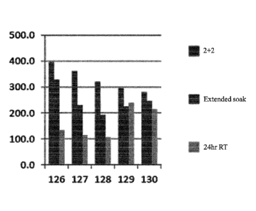
Fig. 9 is a bar graph illustrating the density of FoxP3 cells per mm2. 126-
130 indicate separate replicates. For each replicate, the bars represent
samples
subjected to (from left to right): (1) 2+2 fixation; (2) an extended soak (4
days at ¨5
C, followed by 1 hour at 45 C); and (3) 24 hours in room temperature
formalin.
FIG. 10 illustrates digital microscope images of Calu-3 xenografts
immunohistochemically stained for PR, Ki-67 and the phosphorylated AKT protein
(pAKT). Tissue sections were either shipped according to Example 3 (row S) or
fixed using the 2+2 process (row C). Column A corresponds to tissues used in
shipment 1. Column B corresponds to tissues used in shipment 2. Column C
corresponds to tissues used in shipment 3. Column D corresponds to tissues
used
in shipment 4.
Fig. 11 is a bar graph illustrating differences in p-AKT preservation
between using a 24 hour room temperature and the shipping conditions outlined
in
Example 3.
Figs. 12A-12K illustrate pAkt preservation using a variety of cold/hot
fixation conditions as set forth in Table 3. Images correspond to conditions
as
follows: 12B is Experiment 1.1; 12C is Experiment 1.2; 12D is Experiment 2.1;
12E is Experiment 2.2; 12F is experiment 2.3; 12G is experiment 2.4; 12H is
experiment 3.1; 121 is experiment 4.1; 12J is experiment 5.1; 12K is
experiment
6.1.
Fig. 13 is a digital image of tissue samples labeled for miR-21 or miR-200c
by in situ hybridization after fixation at 24 hours in room temperature NBF
(left
CA 02972682 2017-06-29
WO 2016/120195
PCT/EP2016/051431
8
column) or fixation in 4 C NBF for 2 hours followed by 45 C NBF for 2 hours
(right column).
DETAILED DESCRIPTION
I. Abbreviations and Definitions
In order to facilitate review of the various examples of this disclosure, the
following explanations of abbreviations and specific terms are provided:
H&E: Hematoxylin and eosin staining.
FFPE tissue: Formalin-fixed, paraffin-embedded tissue.
IHC: Immunohistochemistry.
ISH: In situ hybridization.
NBF: neutral buffered formalin.
Affinity histochemistry: A histochemical method in which the analyte-
binding entity is an agent other than an antibody, antibody fragment, or
nucleic acid probe.
Aldehyde-based fixative: Any composition suitable for fixation of a tissue
sample in which at least one of the agents primarily responsible for tissue
fixation is an aldehyde.
Analyte: An entity (such as a molecule, group of molecules,
macromolecule, subcellular structure, or cell) that is to be specifically
detected in a sample.
Analyte-binding entity: Any compound or composition that is capable of
specifically binding to an analyte. Examples of analyte-binding entities
include: antibodies and antibody fragments (including single chain
antibodies), which bind to target antigens; t-cell receptors (including single
chain receptors), which bind to MHC:antigen complexes; MHC: peptide
multimers (which bind to specific T-cell receptors); aptamers, which bind to
specific nucleic acid or peptide targets; zinc fingers, which bind to specific
nucleic acids, peptides, and other molecules; receptor complexes (including
single chain receptors and chimeric receptors), which bind to receptor
ligands; receptor ligands, which bind to receptor complexes; nucleic acid
probes, which hybridize to specific nucleic acids; and engineered specific
binding structures, including ADNECTINs (scaffold based on 10th FN3
9
fibronectin; Bristol-Myers-Squibb Co.), AFFIBODYs (scaffold based on Z
domain of protein A from S. aureus; Affibody AB, Solna, Sweden),
AVIMERs (scaffold based on domain MAX 'receptor; Amgen, Thousand
Oaks, CA), dAbs (scaffold based on VH or VL antibody domain;
GlaxoSmithKline PLC, Cambridge, UK), DARPins (scaffold based on
Ankyrin repeat proteins; Molecular Partners AG, Zurich, CH),
ANTICAL1Ns (scaffold based on lipocalins; Pieris AG, Freising, DE),
NANOBODYs (scaffold based on VHH. (camelid Ig); Ablynx NA', Ghent,
BE), TRANS-BODYs (scaffold based on Transferrin; Pfizer Inc., New
York, NY), SM.IPs (Emergent Biosolution.s, Inc., Rockville, MD), and
TETRANECTINs (scaffold based on C-type lectin domain (CTLD),
tetranectin; Borean Pharma A/S, Aarhus, DK). Descriptions engineered
specific binding structures are reviewed by Wurch et al., Development of
= Novel Protein Scaffolds as Alternatives to Whole Antibodies for Imaging
and Therapy: Status on Discovery Research and Clinical Validation,
Current Pharmaceutical Biotechnology, Vol. 9, pp. 502-509 (2008).
Antibody: The term "antibody" herein is used in the broadest sense and
encompasses various antibody structures, including but not limited to
monoclonal antibodies, polyclonal antibodies, multispeci.fic antibodies (e.g.,
bispecific antibodies), and antibody fragments so long as they exhibit the
desired antigen-binding activity.
Antibody fragment: A molecule other than an intact antibody that
= comprises a portion of an intact antibody that binds the antigen to which
the
intact antibody binds. Examples of antibody fragments include but are not
limited to Fv, Fab, Fab', Fab'-SH, F(ab')2; diabodies; linear antibodies;
single-chain antibody molecules (e.g. say); and multispecific antibodies
formed from antibody fragments.
Anti-phospho-antibody: An antibody or antibody fragment that binds to a
phosphorylated protein or amino acid residue, but not to a non-
phosphorylated version of the same protein or amino acid residue.
Examples of anti-phospho antibodies include:
CA 2972682 2020-03-27
CA 02972682 2017-06-29
WO 2016/120195
PCT/EP2016/051431
= antibodies specific for a specific phosphorylated amino acid residue,
such as phosphorylated histidine (anti-phospho-His),
phosphorylated serine (anti-phospho-Ser), phosphorylated threonine
(anti-phospho-Thr), and phosphorylated tyrosine (anti-phospho-Tyr);
5 and
= antibodies specific for a particular antigen containing a
phosphorylated amino acid, e.g. Akt phosphorylated at serine 473
(anti-phospho-Akt (Ser473)).
Antigen: A compound, composition, or substance that may be specifically
10 bound by the products of specific humoral or cellular immunity, such
as an
antibody molecule or T-cell receptor. Antigens can be any type of molecule
including, for example, haptens, simple intermediary metabolites, sugars
(e.g., oligosaccharides), lipids, and hormones as well as macromolecules
such as complex carbohydrates (e.g., polysaccharides), phospholipids,
nucleic acids and proteins. Common categories of antigens include, but are
not limited to, viral antigens, bacterial antigens, fungal antigens, protozoa
and other parasitic antigens, tumor antigens, antigens involved in
autoimmune disease, allergy and graft rejection, toxins, and other
miscellaneous antigens.
Cellular sample: A sample comprising a collection of cells obtained from a
subject or patient. Examples of cellular samples herein include, but are not
limited to, tumor biopsies, circulating tumor cells, serum or plasma,
primary cell cultures or cell lines derived from tumors or exhibiting tumor-
like properties, as well as preserved tumor samples, such as formalin-fixed,
paraffin-embedded tumor samples or frozen tumor samples.
Clinical cellular sample: A cellular sample obtained directly from a human
or veterinary subject for the purpose of diagnosing a disease or disorder,
determining a prognosis of a disease or disorder, and/or predicting response
of a disease or disorder to a particular course of treatment.
Clinical sample: A sample obtained directly from a human or veterinary
subject for the purpose of diagnosing a disease or disorder, determining a
prognosis of a disease or disorder, and/or predicting a response of a disease
or disorder to a particular course of treatment.
CA 02972682 2017-06-29
WO 2016/120195
PCT/EP2016/051431
11
Clinical tissue sample: A tissue sample obtained directly from a human or
veterinary subject for the purpose of diagnosing a disease or disorder,
determining a prognosis of a disease or disorder, and/or predicting response
of a disease or disorder to a particular course of treatment.
Formalin: A saturated aqueous solution of formaldehyde, which typically
contains ¨40% formaldehyde by volume (-37% by mass). Also referred to
as "100% formalin." In aqueous solution, formaldehyde forms a hydrate,
methanediol (H2C(OH)2), which exists in equilibrium with various
formaldehyde oligomers, depending on the concentration and temperature.
Therefore, a small amount of stabilizer, such as methanol, is usually added
to suppress oxidation and polymerization. A typical commercial grade
formalin may contain 10-15% methanol in addition to various metallic
impurities.
Histochemistry: A method of evaluating a tissue sample by contacting the
sample with an analyte-binding entity in a manner that causes a detectable
marker (such as a dye, chromogen, or a fluorophore) to deposited on the
sample in close proximity to the analyte. Examples of histochemistry
include primary staining (such as H&E stains, acid-fast bacterial stains,
etc.), immunohistochemistry, in situ hybridization, and affinity
histo chemistry.
Immunohistochemistry: A histochemical method in which the analyte-
binding entity comprises an antibody or antibody fragment.
In situ hybridization: A histochemical method in which the analyte is a
nucleic acid and the analyte-binding entity comprises a nucleic acid probe
complementary to the analyte nucleic acid.
Kinase: Any polypeptide ¨ or complex or fragment thereof¨ that catalyzes
the formation of a phosphate bond on a biomolecule.
Kinase inhibitor: Any molecule that specifically inhibits the ability of a
kinase to catalyze the formation of a phosphate bond.
Nuclease: Any polypeptide ¨ or complex or fragment thereof ¨ that
catalyzes the cleavage of the phosphodiester bonds between the nucleotide
subunits of nucleic acids.
CA 02972682 2017-06-29
WO 2016/120195
PCT/EP2016/051431
12
Nuclease inhibitor: Any molecule that specifically inhibits the ability of a
nuclease to catalyze the cleavage of the phosphodiester bonds between the
nucleotide subunits of nucleic acids.
Oligopeptide: A peptide from 2 to 20 amino acids in length.
Peptide: The term "peptide" is intended to encompass any arrangement of
two or more amino acids joined together by amide bonds, including
oligopeptides and polypeptides. When the amino acids are alpha-amino
acids, either the L-optical isomer or the D-optical isomer can be used.
Phosphatase: Any polypeptide ¨ or complex or catalytically-active
fragment thereof¨ that catalyzes the cleavage of a phosphate bond.
Phosphatase inhibitor: Any molecule that specifically inhibits the ability
of a phosphatase to cleave a phosphate bond.
Protease: Any polypeptide ¨ or complex or fragment thereof ¨ that
catalyzes the cleavage of a peptide bond.
Protease inhibitor: Any molecule that specifically inhibits the ability of a
protease to catalyze the cleavage of a peptide bond.
Polypeptide: A peptide longer than 20 amino acids in length. The terms
"polypeptide" or "protein" as used herein are intended to encompass any
amino acid sequence and include modified sequences such as glycoproteins.
Post-translation modification: A chemical modification of a protein after
its translation. It is one of the later steps in protein biosynthesis, and
thus
gene expression, for many proteins. The post-translational modification of
amino acids extends the range of functions of the protein by attaching it to
other biochemical functional groups (such as acetate, phosphate, various
lipids and carbohydrates), changing the chemical nature of an amino acid
(e.g. citrullination), or making structural changes (e.g. formation of
disulfide bridges). Also, enzymes may remove amino acids from the amino
end of the protein, or cut the peptide chain in the middle. For instance, the
peptide hormone insulin is cut twice after disulfide bonds are formed, and a
pro-peptide is removed from the middle of the chain; the resulting protein
consists of two polypeptide chains connected by disulfide bonds. Also,
most nascent polypeptides start with the amino acid methionine because the
"start" codon on mRNA also codes for this amino acid. This amino acid is
CA 02972682 2017-06-29
WO 2016/120195
PCT/EP2016/051431
13
usually taken off during post-translational modification. Other
modifications, like phosphorylation, are part of common mechanisms for
controlling the behavior of a protein, for instance activating or inactivating
an enzyme.
Sample: A biological specimen obtained from a subject or patient
containing genomic DNA, RNA (including mRNA), protein, or
combinations thereof. Examples include, but are not limited to, peripheral
blood, urine, saliva, tissue biopsy, surgical specimen, amniocentesis
samples and autopsy material.
Specific binding: Specific binding occurs when an entity binds to an
analyte in a sample to the substantial exclusion of binding to other potential
analytes. For example, an entity may be considered to specifically bind to a
given molecule when it has a binding constant that is at least 103 WI
greater, 104 M greater or 105 M-1 greater than a binding constant for other
molecules in the sample.
Tissue sample: A cellular sample that preserves the cross-sectional spatial
relationship between the cells as they existed within the subject from which
the sample was obtained. "Tissue sample" shall encompass both primary
tissue samples (i.e. cells and tissues produced by the subject) and xenografts
(i.e. foreign cellular samples implanted into a subject).
"X-% formalin": A liquid composition containing an equivalent amount of
formaldehyde as formalin (as defined above) diluted in a solvent to the
specified percentage on a volume to volume basis. Thus, for example, a
30% formalin solution is a solution that contains an equivalent amount of
formaldehyde as a solution containing 3 parts by volume formalin (as
defined above) to 7 parts by volume solvent.
Introduction
Fixation preserves a cellular sample for subsequent examination. Chemical
fixation involves immersing the sample in a volume of chemical fixative. The
fixative diffuses through the tissue sample and preserves structures (both
chemically and structurally) as close to that of living cells as possible.
Cross-
linking fixatives, typically aldehydes, create covalent chemical bonds between
CA 02972682 2017-06-29
WO 2016/120195
PCT/EP2016/051431
14
endogenous biological molecules, such as proteins and nucleic acids, present
in the
sample. Formaldehyde is the most commonly used fixative in histology.
Formaldehyde may be used in various concentrations for fixation, but it
primarily
is used as 10% neutral buffered formalin (NBF), which is about 3.7%
formaldehyde in an aqueous phosphate buffered saline solution.
Paraformaldehyde
is a polymerized form of formaldehyde, which depolymerizes to provide formalin
when heated. Glutaraldehyde operates in similar manner as formaldehyde, but is
a
larger molecule having a slower rate of diffusion across membranes.
Glutaraldehyde fixation provides a more rigid or tightly linked fixed product,
causes rapid and irreversible changes, provides good overall cytoplasmic and
nuclear detail, but is not ideal for immunohistochemistry staining. Some
fixation
protocols use a combination of formaldehyde and glutaraldehyde. Glyoxal and
acrolein are less commonly used aldehydes. Many other aldehyde-based fixatives
are also known.
It is well known that tissue fixation kinetics can be increased by raising the
temperature of the fixative. However, placing a tissue sample directly into a
heated
fixative can cause the outside of the tissue to cross-link well before
formalin
penetrated to the center of the tissue, which in turn retards or even prevents
further
diffusion of the fixative into the tissue. As a result, biomolecules in the
center of
the tissue are heated without any significant cross-linking, rendering these
molecules more susceptible to degradation and damage. It is also well-known
that
extended exposure of samples to fixative solutions can compromise the
integrity of
the sample and lead to loss of certain biomarkers, particularly labile
biomarkers.
It was previously demonstrated that the degree of degradation and damage
could be reduced by first pre-soaking the tissue samples in cold fixative to
allow
the fixative to diffuse throughout the sample, followed by a higher
temperature
treatment to spur cross-linking. See US 2012-0214195 Al. We have unexpectedly
found that the cold pre-soaking step can be extended for as long as 14 days
without
significant loss of tissue.
CA 02972682 2017-06-29
WO 2016/120195
PCT/EP2016/051431
III. Samples
In principle, the present methods may be used with any cellular sample type
that can be fixed with aldehyde-based fixatives, including tissue samples and
cytology samples.
5 In one
embodiment, the sample is a tissue sample. Typically, tissue
samples for immersion fixation are limited in size to ensure that fixative
diffusion
occurs quickly enough and adequately enough to preserve tissue morphology.
Thus, certain tissue samples, such as tumor resections and whole organs, must
be
dissected before fixation to ensure adequate diffusion of the fixative. This
is
10 particularly true when the tissue contains analytes of interest that are
subject to
degradation by residual enzyme activity in the tissue. The present methods,
however, increase diffusion speed and thus enable fixation of thicker-than-
normal
tissue samples. In an embodiment, the tissue may be as large as a tumor
resection
or a whole organ. In another embodiment, the tissue sample is a tissue biopsy,
15 such as a core needle biopsy.
The present methods and systems are especially useful in fixing clinical
samples in which the presence of labile biomarkers (including post-
translational
modifications to proteins and labile nucleic acids) will be evaluated. In some
embodiments, the sample is a clinical tissue sample.
IV. Fixative Compositions
The present methods are useful with aldehyde-based fixatives. In certain
embodiments, the fixative is an aldehyde-based cross-linking fixative, such as
glutaraldehyde- and/or formalin-based solutions. Examples
of aldehydes
frequently used for immersion fixation include:
= formaldehyde (standard working concentration of 5-10% formalin for most
tissues, although concentrations as high as 20% formalin have been used for
certain tissues);
= glyoxal (standard working concentration 17 to 86 mM);
= glutaraldehyde (standard working concentration of 200 mM).
In one embodiment, the fixative comprises a standard concentration of
formaldehyde, glyoxal, or glutaraldehyde. In one exemplary embodiment, the
aldehyde-based fixative solution is about 5% to about 20% formalin.
CA 02972682 2017-06-29
WO 2016/120195
PCT/EP2016/051431
16
Aldehydes are often used in combination with one another. Standard
aldehyde combinations include 10% formalin + 1% (w/v) Glutaraldehyde.
Atypical aldehydes have been used in certain specialized fixation
applications,
including: fumaraldehyde, 12.5% hydroxyadipaldehyde (pH 7.5), 10%
crotonaldehyde (pH 7.4), 5% pyruvic aldehyde (pH 5.5), 10% acetaldehyde (pH
7.5), 10% acrolein (pH 7.6), and 5% methacrolein (pH 7.6). Other specific
examples of aldehyde-based fixative solutions used for immunohistochemistry
are
set forth in Table 1:
Solution Standard Composition
Neutral Buffered Formalin 5-20% formalin + phosphate buffer
Formal Calcium 10% formalin + 10 g/L calcium chloride
Formal Saline 10% formalin + 9 g/L sodium chloride
Zinc Formalin 10% formalin + 1 g/L zinc sulphate
50 mL 100% formalin + 1 L aqueous solution
Helly's Fixative containing 25 g/L potassium dichromate + 10 g/L
sodium sulfate + 50 g/L mercuric chloride
2 mL 100% formalin + 20 mL aqueous solution
B-5 Fixative containing 6 g/L mercuric chloride + 12.5 g/L
sodium acetate (anhydrous)
100 mL 100% formalin + 15 mL Acetic acid + 1L
Hollande's Solution aqueous solution comprising 25g copper acetate
and 40g picric acid
250 mL 100% formalin + 750 mL saturated
Bouin's Solution
aqueous picric acid + 50 mL glacial acetic acid
Table 1
In certain embodiments, the fixative solution is selected from Table 1.
In the context of concentrations of components of the aldehyde-based
fixatives, the term "about" shall be understood to encompass all
concentrations
outside of the recited range that do not result in a statistically significant
difference
in diffusion rate in the same type of tissue having the same size and shape as
measured by Bauer et al., Dynamic Subnanosecond Time-of-Flight Detection for
CA 02972682 2017-06-29
WO 2016/120195
PCT/EP2016/051431
17
Ultra-precise Diffusion Monitoring and Optimization of Biomarker Preservation,
Proceedings of SPIE, Vol. 9040, 90400B-1 (2014-Mar-20).
Another feature of the methods and systems is that they do not need
exogenous degradation inhibitors (such as phosphatase inhibitors, kinase
inhibitors,
protease inhibitors, or nuclease inhibitors) to substantially preserve labile
biomarkers in a state that they can be detected by histochemistry. Therefore,
although such degradation inhibitors may be included in the fixative
solutions, they
are not required. In an embodiment, the aldehyde-based fixative solutions do
not
contain an effective amount of exogenously added phosphatase inhibitor or
kinase
inhibitor. In other embodiments, the aldehyde-based fixative solutions do not
contain an effective amount of phosphatase inhibitor, kinase inhibitor,
protease
inhibitor, or nuclease inhibitor.
V. Fixation Process
Certain disclosed embodiments concern a multi-step, typically a two-step,
tissue fixation process for infusing/diffusing a tissue sample using an
aldehyde-
based fixative solution. During a first processing step, a sample is treated
with the
aldehyde-based fixative solution under conditions that allow the fixative to
diffuse
throughout substantially the entire cross-section of the sample. This first
step is
conducted using a fixative composition for a first period of time, and at a
first
temperature, that effects substantially complete tissue infusion/diffusion.
The
second step is to subject the tissue sample to a fixative composition at a
second,
higher temperature to allow cross-linking to occur. In operation, the first
and
second processing steps are performed over the course of an extended time
period,
typically on the order of greater than two days. As shown in the Examples
below,
the process has been validated up to 14 days, although it likely can be
extended for
even longer than that.
First, an unfixed tissue sample is immersed in an aldehyde-based fixative
solution at a cold temperature. The temperature of the aldehyde-based fixative
solution is held at the cold temperature at least long enough to ensure that
the
fixative has diffused throughout the tissue sample. The minimum amount of time
to allow diffusion can be determined empirically using various time and
temperature combinations in cold fixatives and evaluating the resulting tissue
CA 02972682 2017-06-29
WO 2016/120195
PCT/EP2016/051431
18
samples looking at factors, such as preservation of tissue architecture and
loss of
for preservation of a target analyte by immunohistochemistry (if the analyte
is a
protein or phosphorylated protein, for example) or in situ hybridization (if
the
target analyte is a nucleic acid, such as miRNA or mRNA). Alternatively, the
minimum amount of time of time to allow for diffusion can be determined by
monitoring diffusion using, for example, a method as outlined in Bauer et al.,
Dynamic Subnanosecond Time-of-Flight Detection for Ultra-precise Diffusion
Monitoring and Optimization of Biomarker Preservation, Proceedings of SPIE,
Vol. 9040, 90400B-1 (2014-Mar-20). An effective temperature range for the
first
step can include any temperature between the freezing point of the aldehyde-
based
fixative solution and below 10 C, for example, about 0 C to about 7 C,
about 2
C to about 5 C, and about 4 C. In this context, the term "about" shall
encompass
temperatures that do not result in a statistically significant difference in
diffusion
rate in the same type of tissue having the same size and shape as measured by
Bauer et al., Dynamic Subnanosecond Time-of-Flight Detection for Ultra-precise
Diffusion Monitoring and Optimization of Bioniarker Preservation, Proceedings
of
SPIE, Vol. 9040, 90400B-1 (2014-Mar-20). Diffusion of the fixative composition
into the tissue sample is continued for a time period effective for diffusion
of the
composition throughout substantially the entire cross section of the sample.
Once the cold fixative solution has sufficiently diffused throughout the
tissue sample, it is stored for an extended period of time either in cold
storage (such
as a refrigerator or ice bucket) or at ambient temperature (i.e. a temperature
from
18 C to 28 C) for a cumulative time of greater than two days. In some
embodiments, the cumulative time is from greater than two days to up to two
weeks or longer, such as from at least 72 hours to 14 days. "Cumulative time"
in
this context is the sum of the diffusion time and the following cold or
ambient
temperature extended storage).
If the sample is stored at cold temperature, then it is subjected to a warm
temperature treatment (i.e. a temperature of from 18 C up to 55 C) for a
sufficient
amount of time to permit fixation. The temperature associated with the warm
temperature treatment typically is ambient or higher, such as higher than
about 18
C. In an embodiment, a temperature range is from ambient up to 50 C (such as
CA 02972682 2017-06-29
WO 2016/120195
PCT/EP2016/051431
19
from 20 C to 50 C). If the temperature is reaches around 55 C, however, the
sample generally begins to degrade, which may have a deleterious effect on
certain
subsequent histological reactions. Therefore, temperatures significantly above
50
C should be avoided for extended periods of time. Thus, in such an embodiment,
the upper temperature and second time period should be selected so as to
preserve
the sample in a state that permits subsequent analyses (such as in situ
hybridization,
histochemical analyses and/or H&E) to proceed effectively. The optimal upper
and
lower time and temperature limits should be determined empirically based on
the
particular analysis that will be performed and the sample type being used. In
particular, guardbanding of time and temperature ranges should be performed to
determine acceptable time/temperature combinations that do not unacceptably
compromise tissue architecture and/or analyte detection levels. In some
embodiments, the warm temperature treatment is performed in the same fixative
solution in which the first processing step is performed. In such an
embodiment,
the fixative solution may be brought to the second temperature range by active
heating (for example, by using a heating element or other heat source) or
passive
heating (such as by moving the fixative and sample from a cold environment to
a
warm environment and allowing the temperature of the fixative solution to
equilibrate with the environment). In other embodiments, the sample is placed
in
contact with a fixative solution at a second temperature range by removing the
sample from the fixative solution at the first temperature range and immersing
the
sample in a volume of an aldehyde-based fixative solution at the second
temperature range. For example, the fixative solution at the first temperature
range
could be disposed in a first vessel and the fixative solution at the second
temperature range could be disposed in a second vessel, in which case the
sample
may be physically moved from the first vessel to the second vessel after the
first
time period has expired. Alternatively, the fixative solution at the first
temperature
range may be removed from a vessel and replaced with the fixative solution at
the
second temperature range. As yet another alternative, only a portion of the
fixative
solution at the first temperature range may be removed, and a hot fixative
solution
may be added to the remaining fixative solution, such that the resulting
combination brings the temperature within the second temperature range. Many
other potential arrangements can be envisioned. In any of the embodiments in
this
CA 02972682 2017-06-29
WO 2016/120195
PCT/EP2016/051431
paragraph, the fixative solution at the first temperature range may be the
same or
different from the fixative solution at the second temperature (including
differ in
the concentration of aldehyde, identity of aldehyde, and/or overall
composition).
If the extended storage is at ambient temperature, then additional warm
5 temperature treatment is unnecessary before further tissue processing,
although it
can be done if desired.
VI. Further Tissue Processing
As used herein, the phrase "further tissue processing" shall encompass any
10 process following aldehyde fixation that is used to prepare the fixed
tissue sample
for storage and/or analysis. Many such processes are well-known and would be
well understood by a person of ordinary skill in the art. For example,
protocols for
using zinc formalin, Helly's fixative and Hollande's require a water wash
after
fixation to remove various contaminates. Some protocols for Bouin's and B-5
15 suggest storing the fixed samples in 70% ethanol before processing.
Additionally,
some specimens may be difficult to cut on a microtome because of calcium
carbonate or phosphate deposits, and thus may require decalcification. Other
post-
fixation tissue processing would be well-known to a person having ordinary
skill in
the art.
20 In one embodiment, post-fixation tissue processing comprises wax-
embedding. In the typical example, the aldehyde-fixed tissue sample is
subjected
to a series of alcohol immersions to dehydrate the sample, typically using
increasing alcohol concentrations ranging from about 70% to about 100%. The
alcohol generally is an alkanol, particularly methanol and/or ethanol. After
the last
alcohol treatment step the sample is then immersed into another organic
solvent,
commonly referred to as a clearing solution. The clearing solution (1) removes
residual alcohol, and (2) renders the sample more hydrophobic for a subsequent
waxing step. The clearing solvent typically is an aromatic organic solvent,
such as
xylene. Wax blocks are formed by applying a wax, typically a paraffin wax, to
the
sample. Typically, before tissue analysis, the blocks are sliced into thin
sections
using a microtome. The thin sections may then be mounted on a slide and stored
for later analysis and/or subjected to post-processing analysis.
21
In other examples, the tissue sample may be embedded in resin blocks (such
as epoxy or acrylic resins) instead of wax blocks. Exemplary resins include
methyl
methacrylate, glycol methacrylate, araldite, and epon. Each requires
specialized
post-fixation processing steps, which arc well known in the art.
VII. Post-processing analysis
Fixed tissue samples obtained by the processes and compositions disclosed
herein can be used together with any staining systems and protocol known in
the
art of histochemistry, as well as affinity histochemistry,
immunohistochcmistry and
in situ hybridization. The present invention can also be used together with
various
automated staining systems, including those marketed by Ventana Medical
Systems, Inc. (such as the VENTANA HE600, SYMPHONY, BENCHMARK, and
DISCOVERY series automated platforms), Dako (such as the COVERSTAINER,
OMN.IS, AUTOSTAINER, and ARTISAN series automated slide stainer), and the
LEICA ST series stainers. Exemplary systems are disclosed in U.S. Pat. No.
6352,861, U.S. Pat. No. 5,654,200, U.S. Pat. No. 6,582,962, U.S. Pat. No.
6,296,809, and U.S. Pat. No. 5,595,707.
Additional information concerning automated systems and methods also
can be found in PCT/US2009/067042.
In an embodiment, specific analytes arc detected using
immunohistochemistry (INC). In the typical 1HC protocol, a tissue sample is
contacted first with an analytc-specific antibody under conditions sufficient
to
permit specific binding of the analyte-specific antibody to the analyte. In
exemplary embodiments, detection of specific analytes is realized through
antibodies capable of specific binding to the analyte (or antibody fragments
thereof) conjugated with multiple enzymes (e.g. horse radish peroxidase (HRP),
alkaline phosphatase (AP). This enzyme-antibody conjugate is referred to as an
HRP or AP multimcr in light of the multiplicity of enzymes conjugated to each
antibody. Multimer technologies arc described in U.S. Patent No. 8,686,122.
This type of detection
chemistry technology is currently marketed by .Ventana Medical Systems Inc.,
as
ultniView Universal DAB detection kit (13/N 760-500), ultraVicw Universal AP
Red detection kit (P/N 760-501), ultraView Red ISH DIG detection kit (P/N 760-
CA 2972682 2020-03-27
22
505), and ultraView SISH DNP detection kit (P/N 760-098). In illustrative
embodiments, the approach uses non-endogenous haptens (e.g. not biotin, sec
U.S.
application Ser. No. 12/660,017).
In illustrative
embodiments, a tyramidc signal amplification may be used with this approach to
further increase the sensitivity and dynamic range of the detection (See
PCT/US2011/042849).
Any suitable enzyme/enzyme substrate system cart be used for the disclosed
in analysis/detection method. Working
embodiments typically used alkaline
phosphatase and horseradish peroxidase. If the enzyme is alkaline phosphatasc,
one
suitable substrate is nitro blue tetrazolium chloride/(5-bromo-4-chloro-1H-
indo1-3-
yOdihydrogen phosphate (NBT/BCIP). If the enzyme is horseradish peroxidasc,
then one suitable substrate is diaminobenzidine (DAB). Numerous other enzyme-
1.5 substrate
combinations arc known to those skilled in the art. For a general review
of these, see U.S. Pat. Nos. 4,275,149, and 4,318,980. In some embodiments,
the
enzyme is a peroxidasc, such as horseradish peroxidase or glutathione
peroxidase
or an oxidoreductase.
'U.S. Patent Publication 2008/0102006,
20 describes
robotic fluid dispensers that are
operated and controlled by microprocessors. U.S. Patent Publication
2011/0311123,
describes methods and systems for automated detection of immunohistochetnical
(.1HC) patterns. The automated detection systems disclosed in these patent
25 applications
can be used to detect analytes in the fixed tissue samples of the present
invention.
In some embodiments, the fixed tissue samples are analyzed by
immunohistochemistry for the presence of post-translationally modified
proteins.
In the typical process, the fixed tissue sample is contacted with an analytc-
binding
30 entity capable
of specifically binding to the post-translationally modified protein
under conditions sufficient to effect binding of the analyte-binding entity to
the
post-translationally modified protein; and binding of the analyte-binding
entity to
the post-translationally modified protein is detected. The precise conditions
ter
CA 2972682 2020-03-27
CA 02972682 2017-06-29
WO 2016/120195
PCT/EP2016/051431
23
effective IHC generally need to be worked on an individual basis, depending
upon,
for example, the precise antibody used, the type of sample used, sample size,
further processing steps, et cetera. In an embodiment, the post-translational
modification is one that is susceptible to loss during a standard aldehyde
fixation
process due to residual enzyme activity within the tissue sample. One could
determine whether a given post-translational modification is susceptible to
residual
enzyme activity by treating a sample with an entity that leads to increased
presence
of the post-translational modification. The sample could then be fixed using a
standard technique (such as 24 hour fixation in room temperature NBF) and a
fixation process as disclosed herein and the amount of signal detectable in
each of
the samples can be compared. If signal is absent or significantly lower in the
sample fixed according to standard techniques, then one can assume that the
post-
translational modification is susceptible to degradation by residual enzyme
activity.
Thus, in an embodiment, the post-translational modification is a post-
translational
modification that has a lower level of detection in a tissue fixed for 24
hours in
room temperature NBF without a cold temperature pre-treatment than in a
substantially identical tissue sample that has been fixed using a two-
temperature
fixation as described above. In an embodiment, the post-translational
modification
is a diagnostic or prognostic marker for a disease state of the tissue sample.
In an
embodiment, the post-translational modification is a predictive marker for an
effect
of a therapy on a disease state of the tissue. In an embodiment, the post-
translational modification is a phosphorylation.
In some embodiments, the fixed tissue samples are analyzed by in situ
hybridization for the presence of specific nucleic acids. In the typical
process, the
fixed tissue sample is contacted with a nucleic acid probe complementary to
the
analyte nucleic acid under conditions sufficient to effect specific
hybridization of
the probe to the analyte nucleic acid; and binding of the nucleic acid probe
to the
analyte nucleic acid is detected. The precise conditions for effective ISH
generally
need to be worked on an individual basis, depending upon, for example, the
precise
nucleic acid probe used, the type of sample used, sample size, further
processing
steps, et cetera. In an embodiment, the analyte nucleic acid is one that is
susceptible to loss during a standard aldehyde fixation process due to
residual
enzyme activity within the tissue sample. One could determine whether a given
CA 02972682 2017-06-29
WO 2016/120195
PCT/EP2016/051431
24
nucleic acid is susceptible to residual enzyme activity by treating a sample
with an
entity that leads to increased presence of the nucleic acid. The sample could
then
be fixed using a standard technique (such as 24 hour fixation in room
temperature
NBF) and a fixation process as disclosed herein and the amount of signal
detectable
in each of the samples can be compared. If signal is absent or significantly
lower
in the sample fixed according to standard techniques, then one can assume that
the
analyte nucleic acid is susceptible to degradation by residual enzyme
activity.
Thus, in an embodiment, the analyte nucleic acid has a lower level of
detection in a
tissue fixed for 24 hours in room temperature NBF without a cold temperature
pre-
treatment than in a substantially identical tissue sample that has been fixed
using a
two-temperature fixation as described above. In an embodiment, the analyte
nucleic acid is a diagnostic or prognostic marker for a disease state of the
tissue
sample. In an embodiment, the analyte nucleic acid is a predictive marker for
an
effect of a therapy on a disease state of the tissue. In an embodiment, the
analyte
nucleic acid is an RNA molecule, such as mRNA or miRNA.
EXAMPLES
The following examples are provided to illustrate certain features of
working embodiments of the present invention. A person of ordinary skill in
the
art will appreciate that the scope of the invention is not limited to the
features
recited in these examples.
Example I: Cold temperature guard banding
4mm Ca1u3 Xeongraft tumor cores that were placed into cooled formalin at
7, 10 or 15 C, respectively, for 2, 4 or 6 hours to form a 9 panel matrix
around
soak temperature. After the cold soak was completed, tumors were immediately
immersed into warm formalin at 45 C for 2 hours. Samples were then processed
further in a standard tissue processor set to an overnight cycle. Tissue was
sliced in
half and embedded cut side down to reveal the edges and middle of the tissue.
Control tissues consisted of comparison pieces of the same tumors being fixed
with
a two-temperature protocol (2 hours 4 C + 2 hours 45 C) and pieces of tumor
fixed
at RT for 24 hours. Tissues were then stained with anti-pAKT (CST #4060) at a
1:50 dilution on a Ventana DISCOVERY XT automated stainer using the
CA 02972682 2017-06-29
WO 2016/120195
PCT/EP2016/051431
OptiView DAB staining kit (Ventana Medical Systems, Inc.). Results are shown
at
Fig. 1. As can be seen, there were only small differences between 4 and 7 C
but
obvious changes were seen at 10 C and 15 C. This suggests that a protocol of
4
C plus or minus only a few degrees Celsius should give the best results.
5
Example 2: Preservation of phosphorylated proteins
Calu3 Xenograft tumors were harvested and placed into the experiment
with less than 10 minutes of cold ischemia time. Tumors were cored at 4mm
using
a disposable biopsy device to ensure all samples were roughly the same size.
To
10 test how long samples can sit in cold formalin, pieces of Ca1u3 tumors
(no more
than 4mm thick) were placed into 4 C formalin for up to 14 days. After the
cold
soak was completed, tumors were immediately immersed into warm formalin at
45 C for 2 hours. Samples were then processed further in a standard tissue
processor set to an overnight cycle. Tissues were sliced in half and embedded
cut
15 side down to reveal the edges and middle of the tissue.
Tissues were stained with anti-pAKT (CST #4060) at a 1:50 dilution on a
DISCOVERY XT automated stainer using the OptiView DAB staining kit
(Ventana Medical Systems Inc.). This dilution was previously chosen based on a
number of similar experiments utilizing Calu3 tumors and this same antibody.
To
20 reduce background staining from mouse tissue, staining was performed by
substituting a rabbit only form of the linker in the commercial kit.
FIG. 2 illustrates the effects of using 4 C pre-soak processing of Ca1u3
xenografts over a fourteen day period on phopsho-AKT levels. As can be seen,
all
samples showed robust staining in samples that had been soaked in 4 C cold
25 formalin for as long as 14 days. This suggests that tissue can be placed
and
transported or stored in cold formalin for up to at least 14 days without
significant
loss of pAKT staining.
Example 3: Shipping validation
To demonstrate a real-world application of the present fixation process, a
shipping study was conducted. A total of 20 Calu-3 xenograft tumors and 20
human tonsil samples were collected. Samples were staggered such that 5 Calu-3
CA 02972682 2017-06-29
WO 2016/120195
PCT/EP2016/051431
26
tumors and 5 tonsil samples were shipped in a week. The shipping schedules
tested
are reproduced below in Table 2:
Shipment
Number Sample Types Length of Shipment
Calu-3
1 6 days
Tonsi 1
Calu-3
2 52 hours
Tonsil
a Calu3 51 hours
3
Tonsil 117 hours
a Calu-3 28 hours
4
Tonsil 72 Hours
Table 2
Styrofoam-insulated shipping containers were retrofit with data loggers to
track the
temperature of the package during shipping and frozen inserts to maintain a
cold
temperature.
Shipment 1
5 Calu-3 tumors were split into 2 samples each. One half of the tumor was
fixed by the 2+2 method as a positive control for controlled fixation. The
other
half of the tumors were placed into histology cassettes, and the cassettes
were
labeled and loaded into specimen containers. This procedure was repeated in
the
afternoon for human tonsil samples that arrive in the afternoon. Specimen
containers were placed the data loggers and were placed into a Styrofoam grid
which contained a top and bottom for better insulation. Once assembled, the
Styrofoam block was placed into either a small or larger shipping container
that has
frozen inserts. After samples were shipped and received, the tissues were
placed
into heated formalin for an additional 2 hours, processed overnight into wax
blocks
and stained for a variety of IHC markers.
The temperature of the specimen containers during shipping is presented at
Figs. 3A & 3B. The temperature spiked to 14 C after packaging (likely due to
the
temperature of the data loggers) and slowly cooled to 7 C in the next 2 1/2
hours
(right graph). Once cooled to 5 C, the box maintained temperatures in the
safe
CA 02972682 2017-06-29
WO 2016/120195
PCT/EP2016/051431
27
zone for several days before slowly drifting to 15 C at which time the
samples
were removed.
Shipment 2
The setup for Shipment 2 was essentially the same as Shipment 1, except
that the data loggers were placed in a refrigerator overnight to cool. Samples
were
harvested in an identical manner to shipment 1 and the data loggers were out
of the
refrigerator approximately 10 minutes. The temperature of the specimen
containers
during shipping is presented at Fig. 4. As can be seen from the temperature
profiles, two temperature spikes were observed, when the samples were
harvested
and placed into the shipping container. The first spike corresponds to
xenografts
harvest and the second spike, several hours later when the tonsil samples were
harvested. However, the temperature spikes were just over 7 C.
Shipments 3 & 4
Between shipment 2 and 3, the collection procedure was modified slightly
to determine if we could maintain the temperature below 7 C for the entire
collection procedure. For this shipment, data loggers were never removed from
the
refrigerator, only the specimen containers. For example, Calu-3 tumors were
received in small batches (2-3 at a time). A corresponding number of specimen
containers were placed under a chemical hood and tumors were sectioned,
cassettes
labeled, clipped into container lids and placed back in the refrigerator
within 5
minutes. Specimen containers were placed directly into cooled data loggers and
the data loggers were started. When all samples had been processed in this
manner,
data loggers with corresponding specimen containers were placed into foam
packing and placed into a shipping box. The shipping box had been previously
conditioned and waiting for the samples. As can be seen, all data loggers
registered
temperatures below 5.5 C. Shipment 4 was essentially identical to shipment 3.
Staining of Shipping Samples
Human Tonsil ¨ Human tonsil samples were stained with Hematoxylin and
Eosin to determine if there were any tissue morphology issues throughout the
shipping process. Samples were compared to control tissues fixed with a 2+2
CA 02972682 2017-06-29
WO 2016/120195
PCT/EP2016/051431
28
fixation protocol. All tonsil samples shipped had excellent morphology with no
visible defects with any conditions tested (see upper H&E panel). Human tonsil
tissues were also stained with PD-Li, FoxP3 and CD68 according to the
validation
data. All tissues stained identically to control tissues fixed with a 2+2
protocol
with all shipping scenarios. FIG. 7 shows representative stains from a subset
of the
tissues tested. Additionally, when compared to 24 hour fixation, the shipped
samples showed significantly better preservation of FoxP3-positive cells. See
FIGS. 8 and 9.
Calu-3 ¨ Calu-3 samples were stained with PR, Ki-67 and an antibody
(CST4060) that recognizes the phosphorylated AKT protein. For total IHC
protein
staining (PR and Ki-67), results were indistinguishable between control
samples
fixed with a 2+2 protocol. Robust staining was evident regardless of the
shipping
conditions, even shipment 1 that had temperatures above the 7 C zone. It
appears
that these two proteins are expressed to high levels in the Calu-3 cell model
and are
stable to slightly elevated temperatures. A different result was obtained when
we
stained for pAKT. Levels of this labile epitope varied depending on the
shipment
and temperature conditions compared to controls with a 2+2 fixation protocol.
Shipment 1 had initial temperatures up to 14 C, which led to variable
staining
between the shipped samples and the 2+2 controls. Variable but better
consistency
was observed with shipment 2 which had temperatures that just peaked above 7
C.
Better staining consistency was observed with shipments 3 and 4 with almost
identical staining compared to the control. FIG. 10 shows representative
stains
from a subset of the tissues tested. Figure 11 is a bar graph demonstrating
the
difference in staining intensity between the various shipping samples and the
24
hour room temperature fixation control.
Example 4: Extended warm soak
Calu3 xenografts were fixed in 10% NBF under a variety of conditions as
set forth in Table 3 and evaluated for morphology by H&E stain. "Hot" in table
3
denotes 45 C for 1 hour. "Cold" indicates 4 C. Samples were scored on a +,
++,
or +++ scale, where + is poor morphology and +++ is the best morphology.
CA 02972682 2017-06-29
WO 2016/120195
PCT/EP2016/051431
29
Experiment Results (+,
++, +++)
Staining level Morphology
1.1: 48 hours cold, 2 weeks RT, hot ++ ++
1.2: 48 hours cold, 2 weeks 37 C, hot ++
2.1: 1 hour cold, 48 hours RT, hot +++ +++
2.2: 2 hours cold, 48 hours RT, hot +++ +++
2.3: 6 hours cold, 48 hours RT, hot +++ ++
2.4: 6 hours cold, 48 hours 37 C, hot
3.1: 48 hours RT, hot ++ ++
4.1: 2 hours cold, 4 hours RT, 48 hours
+++ ++
cold, hot
5.1: 2 hours RT, 48 hours cold, hot ++ ++
6.1: 48 hours cold, hot
Table 3
Additionally, the samples were immunohistochemically stained for pAkt. Results
are shown at Figs. 12A-12K. These results demonstrate that even a short cold
soak
enables extended room temperature storage without unacceptable loss of
morphology or labile markers.
Example 5: Preservation of nucleic acids (prophetic)
It has previously been demonstrated that nucleic acids (such as mRNA and
miRNA) can be sensitive to standard 24 hour room temperature fixation. See,
e.g.,
US 2012-0214195. To illustrate this, the preservation of two miRNA ¨miR-21
and
miR-200c ¨ was evaluated using standard 24 hour room temperature fixation and
cold soak followed by 1 hour fixation at 45 C. 4mm thick pieces of the same
human tonsil organ were placed into either room temperature (21-24 C) 10%
neutral buffered formalin for 24 hours or else 2 hours in 4 C formalin
followed by
1 hour in 45 C formalin (Cold/Hot). Tonsil samples were probed for the
expression of miR-21 or miR-200c with specific DNA probe sequences to each
CA 02972682 2017-06-29
WO 2016/120195
PCT/EP2016/051431
target. After application of the probe sequence, detection of the bound probe
occurred on a VENTANA DISCOVERY XT automated stainer with a silver
detection kit. Cold/Hot fixation resulted in an increase in the amount of
specific
signal in the samples indicating a greater preservation of the miRNA species.
5 Results are shown at FIG. 13. These results indicate that preservation of
RNA
molecules (such as mRNA and miRNA) can be improved by first exposing the
tissue sample to a cold fixative solution for a sufficient amount of time to
allow the
fixative solution to diffuse into the tissue sample. It is therefore proposed
to use a
fixation protocol as outlined above to preserve tissue samples for which
nucleic
10 acid analysis is desired. A prophetic example for doing so is provided
below.
The tissue sample is immersed in an aldehyde-based fixative solution at a
cold temperature (e.g., above the freezing point of the fixative solution but
less than
10 C, including for example in a range of from 2 to 7 C, 2 to 5 C, or about
4 C).
The temperature of the aldehyde-based fixative solution is held at the cold
15 temperature at least long enough to ensure that the fixative has
diffused throughout
the tissue sample. The minimum amount of time to allow diffusion can be
determined empirically using various time and temperature combinations in cold
fixatives and evaluating the resulting tissue samples for preservation of the
target
nucleic acid using an in situ hybridization procedure. Alternatively, the
minimum
20 amount of time of time to allow for diffusion can be determined by
monitoring
diffusion using, for example, a method as outlined in Bauer et al., Dynamic
Subnanosecond Time-of-Flight Detection for Ultra-precise Diffusion Monitoring
and Optimization of Biomarker Preservation, Proceedings of SPIE, Vol. 9040,
90400B-1 (2014-Mar-20).
25 Once the cold fixative solution has sufficiently diffused throughout
the
tissue sample, it is stored for an extended period of time either in cold
storage (such
as a refrigerator or ice bucket) or at ambient temperature (i.e. a temperature
from
18 C to 28 C) for a cumulative time of at least 72 hours. "Cumulative time"
in
this context is the sum of the diffusion time and the following cold or
ambient
30 temperature extended storage). If the sample is stored at cold
temperature, then it
is subjected to a warm temperature treatment (i.e. a temperature of from 18 C
up
to 55 C) for a sufficient amount of time to permit fixation. If the extended
storage
CA 02972682 2017-06-29
WO 2016/120195
PCT/EP2016/051431
31
is at ambient temperature, then additional warm temperature treatment is
unnecessary.
After the extended storage period, the tissue sample is subjected to post-
fixation processing to prepare it for in situ hybridization to detect the
target nucleic
acid. The tissue sample is washed (if the fixative used requires a wash step),
subjected to alcohol dehydration, a clearing solution, and then embedded in
paraffin according to standard techniques. The embedded tissue is then
sectioned
on a microtome, mounted on a slide, and stained for a target messenger RNA
(mRNA), microRNA (miRNA), or DNA molecule using an in situ hybridization
technique, for example, using an automated IHC/ISH slide stainer, such as the
VENTANA BENCHMARK or the VENTANA DISCOVERY automated stainer.