Note: Descriptions are shown in the official language in which they were submitted.
WO 2015/109003 PCT/US2015/011441
IDENTIFICATION OF FUNCTIONAL CELL STATES
FIELD OF THE INVENTION
Embodiments relate to fields of cell assays, physiology and drug development.
Embodiments
additionally relate to eytornetrY and to semi-automated and automated analysis
of multi-parametric
data, such as, cytometry data,
GOVERNMENT FUNDING
No government funds were used in making the invention herein disclosed and
claimed.
RELATED APPLICATIONS
This application claims priority of US Provisional Application number
61/927,247.
- I --
Phenotypic compound screening is an emerging technology for rapid assessment
of
pharmaceutical compounds. in recent years, a number of techniques have been
developed to
characterize phenotypic responses of cells to perturbants such as small
molecules or biologics. The
vast majority of reported work has used traditional bulk biochemical assays,
or single-cell techniques
based on high-content screening (automated microscopy). For instance, see
Abraham et al. ("High
content screening applied to lam-scale cell biology," Trends Biotechnol, 22,
15-22, 2004) and
Giuliano et al, ("Advances in High Content Screening for Drug Discovety."
ASSAY .Drug Del?,
Technol. 1, 565-577, 2003) .
More recently, novel statistical methods have been implemented in the analysis
of complex
screening data.sets. 'These methods can provide a means to determine
correlations between datasetse
Simple pathway-driven models for screening are described in Cong et al,
("Method for using division
arrested cells in screening assays," EP 1581645) .
Cong proposes studying signal transduction in growth-arrested cells and using
such
systems to screen for agents that modulate the activity of cell surface
receptors such as the 32
adrenergic receptors (1-l2AR), a type of Glorotein coupled receptor. Cong
demonstrates that even in
Date Recue/Date Received 2021-05-12
WO 2015/109003 PCT/US2015/011441
such growth arrested cells, treatment with isoproterenol (100 01) increases
secreted alkaline
phosphatase (SEAP) activity, which in turn establishes that growth arrested
cells still have intact
signal transduction pathways down to the transcriptional response, and enzyme
reporter assays can be
carried out using such systems.
A similar technique is provided by Hytopoulos et at. ("Methods for analysis of
biological
dataset profiles." US patent app. pub. No. 2007-0135997).
Hytopoulos discloses methods for evaluating biological dataset profiles.
The datasets comprising information for multiple cellular parameters are
compared and identified. A
typical dataset comprises readouts from multiple cellular parameters resulting
from exposure of cells
to biological factors in the absence or presence of a candidate agent. For
analysis of multiple context
defined systems, the output data from multiple systems are concatenated,
However, Hytopoulos does
not outline precise method steps for creating and forming the response
profiles. Additionally,
Hytopoulos does not provide any working embodiments for practicing the
methodology with a
biological specimen.
Berg et al. ("Function homology screening." US patent No. 8,467,970),
discloses methods for assessing functional homology
between drugs. The methods involve exposing cells to drugs and assessing the
effect of altering the
cellular environment by monitoring multiple output parameters. Two different
environments, such as
those with different compounds present in the environment, can be directly
compared to determine
similarities and differences, Based on these comparisons, the compounds can be
characterized at a
functional level, allowing identification of the pathways and prediction of
side effects of the
compounds. Berg also discloses a representation of the measured data in the
form of a "biomap,"
which is a very simplified heatmap showing gaphically all the measured
cellular parameters. Berg is
related to measuring biological signaling pathways, rather than physiological
responses to stress.
Friend et al, ("Methods of characterizing drug activities using consensus
profiles!' US patent
No. 6,801,859), disclose a
method for measuring biological response patterns, such as gene expression
patterns, in response to
different drug treatments. The response profiles (curves), which are created
by exposing biological
2
Date Recue/Date Received 2021-05-12
WO 2015/109003 PCT/US2015/011441
system to varying concentration of drugs, may describe the biological response
of cells to a particular
group or class of drugs The response curves are approximated using models. The
resultant data
vectors forming curves or profiles, or their parametrical models, can be
compared using various
measures of similarity. This comparison forms a distance matrix which can be
subsequently used in a
hierarchical clustering algorithm to build a.tree representing the similarity
of the profiles. However,
the profiles developed by Friend et al. are limited to simple vector-type and
parametric mathematical
models.
Moreover, profiting methods of the aforementioned applications to Berg et al.
and Friend et
al, publications are limited and, in particular, do not provide for using
distributions for developing
profiles of unknown candidate drugs.
Relatively little work in this area has been performed using flow cytometry,
which allows for
single-cell analysis of cell states cm large populations of cells. See, for
instance, Edwards et al.
("Flow cytometry for high-throughput, high-content screening," CUM Opin. Chem,
Biol. 8, 392---398,
2004, 2004); Oprea et al. ("Associating Drugs, Targets and Clinical Outcomes
into an Integrated
Network Affords a New Platform for Computer-Aided Drug Repurposing,"Mol.
Inform. 30, 100--
111, 2011); Robinson et al. ("High-throughput secondary screening at the
single-cell level." Lab.
Autom. 18, 85-98, 2013) and Sklar et al ("Flow cytometry for drug discovery,
receptor pharmacology
and high throughput screening." Curr. Opin. Pharmacol. 7, 527-534, 2007) .
However, the recent availability of high-throughput fluidic handling systems
for cytometry
has made it feasible to process an entire 96- or 384-well plate within few
minutes, sampling several
thousand cells per well, making cytometry increasingly attractive for high-
throughput cell assays, The
reports describing the use of high-throughput flow cytometry typically focus
on relatively simple
assays acquiring from I to 5 different variables describing cellular
physiology for the analyzed cells.
From a mathematical perspective, the data collected in these assays can be
described as an array in
which the rows store information about individual cells, and the columns
describe the measured
quantity (e.g. light-scatter characteristics, fluorescence intensity signals,
etc.), The measured features
can be summarized by a variety of statistics. Most commonly, mean fluorescence
intensity in a region
3
Date Recue/Date Received 2021-05-12
CA 02972960 2017-07-04
WO 2015/109003 PCT/US2015/011441
of interest is used. After data reduction, the results of an experiment are
represented by a vector with
elements being the values of the chosen summary statistics. If an experiment
involves testing a
number of different concentrations of a drug, the final outcome is a 2-1)
array, with individual
CONIT311S describing the response curves. Additional parameters (e.g,
different times of drug
incubation) may be used to add additional dimensions to the array.
Traditionally, drug response curves are approximated by an a priori
mathematical model
(such as a sigmoidal log-normal curve, log-logistic curve, Gompertz curve,
Weibull, etc.) and the
measured drug response information is reduced to a few parameters (or even a
single parameter) that
describe the curves. The entire process produces a heavily abbreviated
compound response summary:
typically a "signature" comprising a number of ECSO values, that is, values
representing a
concentration of a compound which induces a response halfway between the
baseline and maximum
after a specified exposure time.
Such approaches are subject to important inherent limitations that cannot be
alleviated easily,
if at all. First, they assume the prior existence of a known proper
mathematical model with an
appropriate parameterization describing the response of all the tested
compounds. Second, they
presume that a single parameter (EC50) derived from a sigmoidal curve carries
all the necessary
information about the compound response pattern. And third, they analyze the
responses manifested
by the measured parameters separately, i.e., in a one-dimensional manner. The
data analysis and
feature extraction leading to formation of the response curves is also
problematic.
Furthermore, traditional and well-established cytometrie data processing
relies on a so-called
gating process, which involves manual separation of the populations of
interest in order to compute
simple statistical features of these populations (mean, median, coefficient of
variance, etc.). This
gating can be highly subjective, and it is difficult to reproduce in an
automated setting. Additionally,
the computed features are not sealed or standardized to reflect the range of
possible biological
responses or the precision of the cytometiy measurements.
Embodiments herein described provide methods for overeoming, the significant
shortcomings
of current phenotypic screening methods, in some embodiments, by employing a
new methodology
for quant4ing compound responses. Embodiments described herein provide a
number of innovative
4
CA 02972960 2017-07-04
WO 2015/109003 PCT/US2015/011441
data acquisition and data processing techniques, which allow meaningful
comparisons of
multidimensional compound fingerprints without compromising information
quality, and without a
priori assumptions about responses.
-LI-
A few of the many embodiments encompassed by the present description are
summarized in
the following numbered paragraphs. The numbered paragraphs are self-
referential, in particular, the
phase "in accordance with any of the foregoing or the following" used in these
paragraphs refers to
the other paragraphs. The phrase means, in the following paragraphs,
embodiments herein disclosed
include both the subject matter described in the individual paragraphs taken
alone and the subject
matter described by the paragraphs taken in combination. In this regard, it is
explicitly applicant's
purpose in setting forth the following paragraphs to describe various aspects
and embodiments
particularly' by the paragraphs taken alone or in any combination. That is,
the paragraphs are a
compact way of setting out and providing explicit ',mitten description of all
the embodiments
encompassed by them individually and in any combination with one another.
Applicant specifically
reserves the right at any time to claim any subject matter set out in any of
the following paragraphs,
alone or together with any other subject matter of any one or more of the
other paragraphs, including
any combination of any values therein set forth, taken alone or in any
combination with any other
value therein set forth. Should it be required, applicant specifically
reserves the right to set forth all of
the combinations herein set forth in full in this application or in any
successor applications having
benefit of this application.
Methods and analPsis
Al, A method for characterizing one or more cellular responses to an
agent, comprising:
exposing cell populations to a plurality of concentrations, c, of an agent;
measuring by cytometry a plurality of physiological parameters p, of cells in
the
population at each concentration;
calculating a set of distances between populations and controls for each
parameter for
the cell population at each concentration; and
compiling one or more tensors for each compound from the calculated distances;
and
CA 02972960 2017-07-04
WO 2015/109003 PCT/US2015/011441
compressing the tensors by tensor decomposition to yield an abbreviated
compound
fingerprint in the form of a vector,
A2, A method for comparing one or more cellular responses to an agent,
comprising:
(A) exposing first cell populations to a plurality of concentrations
of a first agent;
measuring by cytometry a plurality of physiological parameters, p, of cells in
the population
at each concentration of said first agent;
compiling one or more tensors from the measurements, thereby describing said
first agent;
= compressing the tensors(s) via decomposition to obtain an abbreviated
compound fingerprint
in the form of a vector;
(B) exposing second populations of the cells to a second plurality of
concentrations of a
second agent;
measuring by cytometry the plurality of physiological parameters of cells in
the population at
each concentration of said second agent;
compiling one or more tensors from the measurements, thereby describing said
second agent;
compressing the tensor(s) via decomposition to obtain abbreviated compound
fingerprint(s) in
a form of a vector, and
(C) calculating a dissimilarity between the first and the second
abbreviated fingerprint to
determine the difference between the response of the cells to the first and
second agents,
Al A method for determining one or more responses of cells to am
agent, comprising;
measuring two or more cell physiology responses for one or more negative, one
or more
positive controls and for one or more concentrations of a compound;
selecting subpopulations of cells for the controls and the concentration
series by mathematical
restriction thereby gating the cells in a particular cell cycle compartment
and a particular
morphological class;
calculating a dissimilarity between the distributions of cellular measurements
for each
positive and negative control and each of the concentrations;
characterizing the response of the cells to the compound by the calculated
dissimilarity.
6
CA 02972960 2017-07-04
WO 2015/109003 PCT/US2015/011441
A4, A method according to any of the foregoing or the following, wherein
the respon,ses
are caleulated using a mathematical metric operating on distributions.
AS. A method in accordance with any of the foregoing or the following,
wherein the
measurements Com a multi-dimensional data point cloud.
A6. A method in accordance with any of the foregoing or the following,
wherein changes
in multidimensional data point-clouds are calculated as distances by any one
or more of a Wasserstein
metrie, a metric defined as a solution to the Kantorovich¨Rubinstein
transportation problem, a
quadratic-form distance, a quadratic chi-distance, Kullback-Leibler
divergence, a Jensen-Shannon
divergence, Kolmogorov metric, a Csiszar (.-divergence, a Burbea and Rao
divergence, and Bregman
divergence.
A7. A method according to any of the foregoing or the following, wherein
the value at 95-
percentile of the pairwise distances within a group of controls is chosen as
the limit of measurement
precision (limit of statistical significance).
A. A method according to any of the foregoing or the following, wherein
a robust
measure of dissimilarity between group of positive controls and a group of
negative controls defines a
unit of dissimilarity between the responses,
A9, A method in accordance with any of the foregoing or the following,
comprising
quantitating the changes in distribution of the measured multidimensional data
point-clouds at a
plurality of different concentrations of the compound.
MO. A method in accordance with any of the foregoing or the following,
further
employing dimensionality reduction by summarizing features from a plurality of
response tables
wherein every response vector is summarized by a number of derived quantities
smaller than the
number of concentrations of said compound.
All , A method in accordance with any of the foregoing or the following,
further
representing a response to an agent by a multiway tensor comprising
summarizing features.
Al2. A method in accordance with any of the foregoing or the following,
wherein tensors
calculated for different agents are compared to each other by computing a
dissimilarity measure,
7
CA 02972960 2017-07-04
WO 2015/109003 PCT/US2015/011441
A13, A method in accordance with any of the foregoing or the following,
wherein tensors
are decomposed using one or more of Tucker decomposition, CANDECOMP, PARAFAC,
.PARAFAC2, INDSCAL, CANDEUNC, DEDICOM or PARATUCK2 decompositions.
RtioNag.44..,.Y.tgrgigntg2n
A cellular response tensor in accordance with any of the foregoing or the
following,
wherein the response tensor is generated from a dataset of measured values of
a plurality of two or
more cell physiology parameters, wherein the response tensor quantifies a
multiparametric and
multifactorial cellular phenotype.
TSt2. A cellular response tensor in accordance with any of the foregoing or
the following
encoded in a tangible form so that it can be accessed, copied, used and/or
retrieved in whole or in part
by a user,
TSt.3. A cellular response tensor in accordance with any of the foregoing or
the following,
wherein a response tensor for a control dataset provides the basis for
determining whether a test
dataset is different from a control or a profile dataset.
Contras
Ctrl. A method according to any of the foregoing or the following, where
positive control
cells are treated with one or more known compounds that trigger a maximal
measurable effect on one
or more of the measured cell physiology responses.
Ctr2. A method according to any of the foregoing or the following, wherein the
negative
controls are untreated cells, cells treated with buffer, cells treated with
media, or cells treated with a
sham compound
Cell cycle
Ccyl.. A method in accordance with any of the foregoing or the following,
wherein the cell
state is a measurement of growth phase of the cells, preferably, a measurement
of cell division.
Ccy2, The method in accordance with any of the foregoing or the following,
wherein the cell
state or cell cycle stage is detected via flow cytornetly at single-cell
level,
Ccy3. A method according to any of the foregoing or the following, where one
of the
physiological parameters is the cell cycle,
8
CA 02972960 2017-07-04
WO 2015/109003 PCT/US2015/011441
Ccy4. A method according to any of the foregoing or the following, wherein one
of the
physiological parameter is cells cycle compartment GI, S. G2 and M.
Cey5. A method according to any of the foregoing or the following, wherein one
of the cell
cycle compartments is if!, 5, and 02/M.
Ccy6. A. method according to any of the foregoing or the following, wherein
ail of the
physiological responses are measured as a ilinction of cell cycle compartment,
Ccy7. A method in accordance with any of the foregoing or the following,
wherein cell
cycle phases are measured using fluorescence labels.
Ccy8. A method in accordance with any of the foregoing or the following,
wherein cell
cycle phases are measured using one or more fluorescent DNA intercalating
dyes.
Ccy9. A method in accordance with any of the foregoing or the following,
wherein cell
cycle phases are measured using one or more of the fluorescent intercalating
dyes HOECHST
33342(2'(4-Ethoxypheny1)-6-(4-methyl-1-piperaziny1)-11-1,3'H-2,5'-
hibenzimid.azole), DRAQSTM
(1,5-bis{[2-(di-methylamino) ethyl]amino}-4, 8-dihydroxyanthracene-9,10-
dione), YO-PRO-1
IODIDE (Quinolinium, 44(3-metity1-2(311)-benzoxazolylidene)methyl)-l-(3-
(trimethylaromonio)propyl)-, dilODIDE), DAPI (4', 6-diamidino-2-phenylindo1e)
and CYTRAK
ORANGE (derivative of I,5-bis{ [2-(di-methylamine) ethyliamino}-4, 8-
dihydroxyanthracene-9,10-
dione).
Ccy10. A method in accordance with any of the foregoing or the following,
wherein, cells
cycle phases are measured by immunolabelling of cell cycle-dependent proteins.
Coy ii A method in accordance with any of the foregoing or the following,
wherein cell
cycle phases are measured by immunolabelling one or more of eyclins A, cyclin
B and eyclin E.
Ccyl 2. A method in accordance with any of the foregoing or the following,
wherein cell
c3,,cle phases are measured by immunolabelling one or more phosphorylated
histone proteins,
Ccy13. A method in accordance with any of the foregoing or the following,
wherein cell
cycle phases are using genetically encoded cell-cycle dependent fluorochromes,
e.g., hyper-
phosphoiylated Rb protein and cell cycle can he measured by flow cytometry
(see Juan et al,
"Phosphorylation of retinoblastoma susceptibility gene protein assayed in
individual lymphocytes
9
WO 2015/109003 PCT/US2015/011441
darin.g their initogenic stimulation," Experimental Cell Res 239: 104410,
1998) ,
and cyclin protein expression (or their phosphorylation
status) can be monitored using flow cytoinctry (see Darzynkiewiez et al.
"Cytometty of cell cycle
regulatory proteins." Chapter in: Progress in Cell Cycle Research 5;533-542,
2003).
Ccyl 4.. A method in accordance with any of the foregoing or the following,
wherein cell
cycle phases are measured by expression of a. genetically encoded fusion
protein comprising a
naturally expressed oscillating protein linked to a fluorescent protein
moiety, e.g., cell cycle arrest at
G2,14 (Cheng et al., "Cell-cycle arrest at G2/J4 and proliferation inhibition
by adenovirus-expressed
mitofusin-2 gene in human colorectal cancer cell
lkieoplasma 60; 620-626, 2013); regulation of
S-phase entry (McGowan et 8.1., "Platelet-d.erived growth fictor-A regulates
lung fibroblast S-phase
entry through p27kipi and Fox03 a," Respiratory Research) 14;68-81, 2013); or
identification of live
proliferating cells using a eyelinBI-GFP fusion reporter (see Klochendler et
al., "A transgenie mouse
marking live replicating cells reveals in vivo transcriptional program of
proliferation," Developmental
Cell, 16681-690, 2012).
Ccy15. A method in accordance with any of the foregoing or the following,
wherein the cell
cycle is altered by an agent
Ccy16. A method in accordance with any of the foregoing or the following;
wherein the cell
eycle is altered by a variation in cell culturing method.
Ccy17. A method in accordance with any of the foregoing or the following,
wherein the cell
cycle is altered by changes in the levels of one or more of the following in
the culture medium:
glucose, essential and non-essential amino acids, 02 concentration, pH,
galastosc and/or
glutamine/glutamate.
Ccyl 8. The method in accordance with any of the foregoing or the following,
further
comprising detecting the cell state or cell cycle stage in a control
population of cells exposed to a
plurality of chemicals or agents which are known to perturb the state of the
cell cycle.
Cells.
Date Recue/Date Received 2021-05-12
CA 02972960 2017-07-04
WO 2015/109003 PCT/US2015/011441
Cis i A method in accordance with any of the foregoing or the following,
wherein the cells
are in vitro cultured cells.
A method in accordance with any of the foregoing or the following, wherein the
cells are
biopsy cells.
Cls2. A method in accordance with any of the foregoing or the following,
wherein the cells
are live cells.
CIA A method in accordance with any of the foregoing or the following,
wherein the cells
are fixed cells.
Cls4. A method in accordance with any of the foregoing or the following,
wherein the cells
are a cell line.
C1s5. A method in accordance with any of the foregoing or the following,
wherein the cells
characteristic of a naturally occurring healthy cell type.
Cls6. A method in accordance with any of the foregoing or the following,
wherein the cells
are characteristic of a disease.
Cls7. A method in accordance with any of the foregoing or the following,
wherein the cells
are characteristic of an inborn genetic disorder.
C138. A method in accordance with any of the foregoing or the following,
wherein .the cells
are characteristic of a cancer.
C1s9. A method according to any of the foregoing or the following, wherein the
cells are
characteristic of a metabolic disorder.
ClsI0. A method in accordance with any of the foregoing or the following,
wherein the cells
are animal cells.
Cisl 1. A method in accordance with any of the fbregoing or the following,
wherein the cells
are mammalian cells.
Cls12. A method in accordance with any of the foregoing or the following,
wherein the cells
are human cells.
Cls13, A method according to any of the foregoing or the following, wherein
the cells are
germ cells or stem cells, including, niuripotent stem cells,
11
CA 02972960 2017-07-04
WO 2015/109003 PCT/US2015/011441
Cls14. A method in accordance with any of the foregoing or the following,
wherein the cells
are somatic cells,
C1s15. A method in acnordance with any of the foregoing or the following,
wherein the cells
are stem cells.
Cls16. A method in accordance with any of the foregoing or the following,
wherein the cells
are embryonic stem cells.
Cis17, A method in accordance with any of the foregoing or the following,
wherein the cells
are pluripotent stem cells.
Cisl 8. A method in accordance with any of the foregoing or the following,
wherein the cells
are induced pluripotent stem cells.
Cls19. A method in accordance with any of the foregoing or the following,
wherein the cells
are blast cells.
Cls20. A method in accordance with any of the foregoing or the following,
wherein the cells
are differentiated cells.
Cls21 A method in accordance with any of the, fbregoing or the following,
wherein the cells
are terminally differentiated somatic cells,
Cls22. A method in accordance with any of the foregoing or the following,
wherein the cells
are eardiomyoeytes, hepatocytes, neurons or a combination thereof.
C1823 A method in accordance with any of the foregoing or the following,
wherein the cells
are one or more of the following cells: primary cells, transformed cells, stem
cells, insect cells, yeast
cells, preferably anchorage independent cells, such as, for example, human
hernatopoietic cell lines
(including, but not limited to, HL-60, K562, CCPS-CEM, Jurkat, 11P-1, etc.);
or anchorage-
dependent cell lines (including, but not limited to 111-29 (colon), 1-24
(bladder), SKBR (breast), PC-
3 (prostate), etc.).
Duration.
Durl. A method in accordance with any of the foregoing or the following,
wherein cells are
exposed to an agent for a plurality of durations or various times, e.g.,
measuring time course (kinetics)
for activation of signaling pathways in cells (see, cog,, Woost et al., "High-
resolution kinetics of
12
WO 2015/109003 PCT/US2015/011441
cytokine signaling in human CD34/CD117-positive cells in unfractionated bone
marrow," Blood, 117;
131-141, 2011). Embodiments
involving analysis of kinetics is preferred over embodiments which do not
require such analysis (see
Komblau at al. "Dynamic single-cell network profiles in acute myelogenous
leukemia are associated
with patient response to standard induction therapy," Clin Cancer Res, 16;3721-
3733, 2010, which
does not teach kinetic analysis).
Dur2. A method in accordance with any of the foregoing or the following.,
wherein the cells
are exposed to an agent for 0.1, 0.2, 0.3, 0.4, 0.5, 0.6, 0.7, 0.8, 0.9, 1, 2,
3, 4, 5,6, 7, 8.9, 10, 12, 14,
15, 16, 18, 20, 22, 24, 26, 28, 30, 35, 40, 44, 48, 52, 56, 60, 66, 72, 78 or
more hours or any
combination thereof.
..Concentratian
Cncl. A method in accordance with any of the foregoing or the following,
wherein a
plurality of two or more concentration series of an agent
Plurality iNienber) of Samples
Plrl . A method in accordance with any of the foregoing or the following,
comprising
wherein a plurality of samples are measured.
P1r2. A method according to any of the foregoing or the following,
comprising measuring
a plurality of samples disposed in wells of a multiwell plate.
P1r3. A method according to any of the foregoing or the following, comprising
measuring a
plurality of samples disposed in wells of 96, 384, or 1536-well plate.
Basic instrumentation / methods
ins 1, method in accordance with any of the foregoing or the following,
wherein the
responses are measured by eytometry.
Ins2. A method in accordance with any of the foregoing or the following,
wherein the
responses are measured by flow cytometry,
Ins3. A method M accordance with any of the foregoing or the following,
wherein
responses are measured by flow cytometry of live cells.
13
Date Recue/Date Received 2021-05-12
CA 02972960 2017-07-04
WO 2015/109003 PCT/US2015/011441
Ins4. A method in accordance with any of the foregoing or the following,
wherein
responses are measured by flow cytometry of fixed cells.
Ins 5. A method in accordance with any of the thregoing or the following,
wherein
responses are measured by imaging of immobilized cells.
1ns6. A method in accordance with any of the foregoing or the
following, wherein
responses are measured by fluorimetry.
Ins7. A method in accordance with any of the foregoing or the following,
wherein a
plurality of two or more response parameters is measured by a multichannel
sensor array.
Signal Processing
Sinl, A method in accordance with any of the foregoing or the following,
comprising
= decorrelating fluorescence signals via linear unmixing of the acquired
signals by multiplying the
vector of measured values by an inverse of the matrix containing in its
columns the spectra of the
employed fluorescent species; the said matrix being normalized per column to I
Sig2. A method in accordance with any of the foregoing or the following,
comprising
decorreiating fluorescence signals via linear unmixing of the acquired signals
by multiplying the
vector of measured values by an inverse of the matrix containing in its
columns the spectra of the
employed fluorescent species; the said matrix being normalized per diagonal to
1
Agents
Agtl, A method in accordance with any of the foregoing or the following,
wherein the cells
are exposed to a single compound.
Agt2. A method in accordance with any of the foregoing or the following
wherein .the cells
are exposed to two or more compounds.
Agt3. A method in accordance with any of the foregoing or the following
wherein one or
more of the compounds stimulate a physiological response,
A.gt4. A method in accordance with any c-if the foregoing or the fbllowing,
wherein the anent
may be a genetic agent, e.g, expressed coding sequence; or a chemical agent,
e.g, drug candidate,
nysiglogykyl Parameters
AMP
14
CA 02972960 2017-07-04
WO 2015/109003 PCT/US2015/011441
MMP1, A method in accordance with any of the foregoing or the following,
wherein
mitochondrial toxicity is measured,
MMP2, A method in accordance with any of the foregoing or the following,
wherein the loss
of mitochondrial membrane potential or integrity is measured,
MMP3. A method in accordance with any of the foregoing or the following,
wherein loss of
mitochondria] membrane potential or integrity is measured using a fluorescent
dye.
MMP4. A method in accordance with any of the foregoing or the following,
wherein loss of
mitochondria' membrane potential or integrity is measured using one or more of
3C-1 (5,5',6,6'-
tetraehloro-1.,l',3,3`4etraethylbenzimi- dazolylearbocyanine IODIDE), JC-9
((3,3'-dimethyl-p-
naphthoxazolium IODIDE, MITOPROBErm, Molecular Probes), JC-10 (e.g.,
derivative of
Di0C2(3) ((3,3`-diethyloxacarbocya.nine IODIDE; MITOPROBETm, Molecular
Probes), DiIC1(5)
((1,1',3,3,3',3'-hexamethylind.odicarbo cyanine IODIDE; MITOPROBErm, Molecular
Probes),
MITOTRACKERnd (Molecular Probes), ORANGE CMTMROS (chloromethyl-
dichlorodihydrofluorescein. diaeetate, M1TOTRACKERTm ORANGE, Molecular Probes)
and
CMXROS (1.H.,5F1,111-/,15.11-Xantheno[2,3,4-ij:5,6,7-indiquinol izin- I 8-ium,
944-
(chloromethyl)phenyl]-2,3,6,7,12,13,16,17-octahydrog chloride, MITOTRACKERTm
RED,
Molecular Probes).
Light scattering
LSO. A method in accordance with any of the foregoing or the following,
wherein a
physiological parameter of cell state is measured by light-scattering,
1.:Sg2. A method in accordance with any of the foregoing or the following,
wherein a
physiological parameter of cell state is measured by laser light-scattering,
LSg3. A method in accordance with any of the foregoing or the following,
wherein a
physiological parameter of cell state is measured by quantifying the amount of
laser light scattered
from individual cell at two or more angles.
ISg4. A method in accordance with any of the foregoing or the following,
wherein a.
physiological parameter of cell state is measured by laser light-scattering.
CA 02972960 2017-07-04
WO 2015/109003 PCT/US2015/011441
LSg5. A method in accordance with any of the foregoing or the following,
wherein a
physiological parameter of cell state is measured by laser light-scattering.,
wherein the wavelength of
light emitted by the laser is within the range of any one or more of 403-408
am, 483-493 nm,525-535
am, 635-635 um and 640-650 nal.
Cell Viability
Vial. A method in accordance with any of the foregoing or the following,
wherein cell
viability is measured.
Via.2. A method in accordance with any of the foregoing or the following,
wherein cell
membrane integrity is measured.
Vial A method in accordance with any of the foregoing or the following,
wherein cell
viability is determined my measuring membrane integrity.
Via4. A method in accordance with any of the foregoing or the following,
.wherein loss of
membrane integrity is detected using a dye.
Via5. A method in accordance with any of the foregoing or the following,
wherein loss of
membrane integrity is detected using a dye that enters cells with damaged
membranes characteristic
of dying or dead cells but does not enter cells with intact membranes
characteristic of live cells.
Via6. A method in accordance with any of the foregoing or the following,
wherein loss of
membrane integrity is detected using a dye that enters cells with damaged
membranes characteristic
of dying or dead cells but does not enter cells with intact membranes
characteristic of live cells,
wherein the dye fluoresces on binding to DNA.
Vie. A method in accordance with any of the foregoing or the following,
wherein loss of
membrane integrity is detected using one or more of the following dyes:
PROPIDIUM IODIDE,
DAN and 7-aminoactinomycin a
Via8. A method in accordance with any of the foregoing or the following,
wherein
membrane integrity is measured using one or more dyes that cross intact cell
membranes and
fluorescence upon interacting with intracellular enzymes and remain in the
cytoplasm of live cells but
diffuse out of lacking an intact cytoplasmic membranes.
16
CA 02972960 2017-07-04
WO 2015/109003 PCT/US2015/011441
Via.9. A method in accordance with any of the foregoing or the following,
wherein
membrane integrity is measured using one or more dyes that cross intact cell
membranes and
fluorescence upon interacting with intracellular enzymes and remain in the
cytoplasm of live cells but
diffuse out of cells lacking an intact cytoplasmic membrane, wherein the dyes
are one or more of
fluorescein diacetate, CALCEIN AM, BCECF AM, carboxyeosin diacetate,
CELLTRACKERIm
GREEN CMFDA, Chloromethyl SNARF-I acetate and OREGON GREEEN 488 carboxylic
acid
diacetate).
Vial , A method in accordance with any of the foregoing or the following,
wherein viability
is measured by any one or more of Annexin V, cleaved capases caspase
activation, including
phosphorylatior3 and/or nuclear lamin degradation.
GLU, ROS, WP, CAP and Viability
GRC I . A method in accordance with any of the foregoing or the following,
wherein one or
more of the following physiological parameters is measured: glutathione
concentration ("GLU"), free
radicals and/or reactive oxygen species ("ROS"), mitochondrial membrane
potential/permeability
("MMP"), cytoplasmic membrane permeability, and cell viability.
DX/I damage, Stress, lreammation, Metabolism, Apotosis
DSII A method in accordance with any of the foregoing or the following,
wherein one or
more the following physiological parameters is measured: DNA damage; a stress
response signaling
pathway constituent; an inflammatory response pathway constituent; a metabolic
pathway regulatory
constituent or an apotosis pathway constituent.
DSI.2õk method in accordance with any of .the foregoing or the following,
wherein the
stress response signaling pathway constituent S.APK is measured.
DSI3. A method in accordance with any of the foregoing or the following,
wherein
the inflammatory responses signaling pathway constituent NT-kB is measured.
DSI4. A method in. accordance with any of the foregoing or the following,
wherein the
metabolic pathway regulatoiy constituent measured is a lipid peroxidases,
GSk3B, and/or ribosomal
S6 kinase.
17
CA 02972960 2017-07-04
WO 2015/109003 PCT/US2015/011441
DS:15. A method in accordance with any of the foregoing or the following,
wherein the
apototic pathway constituent measured is PI3K, AKT and/or a Bel-family
protein.
Reference Banks
iThkl, A. method in accordance with any of the foregoing or the following,
wherein the
known perturbing chemicals or exogenous molecular agents are further sub-
grouped based on their
known effects.
Rbk2. A method in accordance with any of the foregoing or the following,
further
comprising creating response tables comprising information about changes in
cell viability,
initochondrial toxicity, and at least one additional physiological or
phenotypic descriptor at every
employed concentration of said compound computed for every stage of cell cycle
defined by cell-
cycle dependent markers.
Rbk3. A method in accordance with any of the foregoing or the following,
wherein tensors
describing known compounds used to treat a particular disease are grouped into
a single defined class
or a plurality of defined classes and the compound tensors are used as a
training set for a. supervised
learning which classifies unknown or not previously characterized compounds
into said defined
classes.
Rbk4 A method in accordance with any of the foregoing or the following,
wherein tensors
describing known compounds are grouped into classes on the basis of their off-
tamet responses, such
as, side-effects,
Rbk5. The method in accordance with any of the foregoing or the following,
wherein feature
tensors are used to discover clusters of similar compound using unsupervised
learning.
Rhk6, The method in accordance with any of the foregoing or the following,
wherein the
feature tensors are veetorized.
Classification of Agent Action
Cisi. A method for classifying biologically active compounds in accordance
with any
comprising detecting a plurality of cellular features from a population of
cells exposed to said
compounds, wherein said features are correlated to morphological properties
quantified
simultaneously by proportions of light scatter intensity measured at two or
more angles,
18
CA 02972960 2017-07-04
WO 2015/109003 PCT/US2015/011441
Cls2. A method in accordance with any of the foregoing or the follt)1,ving,
comprising
exposing a culture of said population of cells to a plurality of compounds and
detecting the
physiological response of said population of cells in the presence and absence
of said compound.
Cls3 A method in accordance with any of the foregoing or the following,
comprising
detecting the physiological response of individual cells sampled from said
culture
Cls4. A method in accordance with any of the foregoing or the following,
wherein the
physiological response is mitoehondrial toxicity, which is quantitated in
terms of loss of
mitochondria' membrane potential or a loss of mitochondrial membrane integrity
using one or more
fluorescence labels selected from the group consisting ofJC-1, JC-9, JC-10,
DI0C2(3), Di1C1(5),
MITO TRACKER ORANGE CMIMROS, MUG TRACKER RED CMXROS.
Cis5, A method in accordance with any of the foregoing or the following,
wherein the
physiological response is overall cell viability, which is quantitated in
terms of loss of cellular
membrane integrity using one or more fluorescence labels.
Clso. A method in accordance with any of the foregoing or the following,
wherein the
fluorescence labels are selected from groups consisting of
dyes which enter the cell interior resulting in a very bright fluorescence
(e.g., propidium
IODIDE and 7-aminoactinomycin D);
dyes which cross membranes of intact cell membranes and produce fluorescent
molecule
upon interaction with intracellular enzymes (e.g., fluorescein diacetateõ
CALCEIN AM, BCECF AM,
carboxyeosin diacetate, CELLTRACKERThl GREEN CMFD.A, Chloromethyl SNARE-1
acetate,
OREGON GREEN 488 carboxylic acid diacetate),
Cls7. A method in accordance with any of the foregoing or the following,
further
comprising detecting at least one additional physiological or phenotypic
descriptor from the group
consisting of concentration of glutathione, presence of reactive oxygen
species or free radicals,
Systems
Sys 1. A system for evaluating / comparing biological datasets, comprising a
non-transitory
computer readable storage medium storing a computer program that, when
executed on a computer,
causes the computer to perform any of the foregoing or following methods,
CA 02972960 2017-07-04
WO 2015/109003 PCT/US2015/011441
Sys2. A system for evaluating t comparing biological data.sets, comprising a
non-transitory
computer readable storage medium storing a computer program that, when
executed on a computer,
causes the computer to perform any of the foregoing or following methods for
characterizing one or
more cellular responses to an agent, said method comprising:
measuring by cytometry a plurality of physiological parameters p, of cells in
the population
which are exposed to a concentration, c, of said agent;
calculating a set of distances between populations and controls for each
parameter for the cell
population at each concentration; and
compiling a tensor or a set of tensors for each compound (where the tensors
contain
compound fingerprints); and
compressing the tensors via tensor decomposition to yield an abbreviated
compound
fingerprint in a form of a vector.
Sys3. A computer system fOr evaluating / comparing biological datasets,
comprising, a non-
transitory computer readable storage medium storing a computer program that,
when executed on a
computer, causes the computer to perform a method for characterizing one or
more cellular responses
to an agent, said method comprising:
(A) exposing first cell populations to a. plurality of concentrations, c, of a
first agent;
measuring by cytometry a plurality of physiological parameters p, of cells in
the population at
each concentration of said first agent;
compiling one or more tensors from describing said first agent;
compressing the fingerprint tensors(s) via decomposition to obtain abbreviated
compound
fingerprint in a form of a vector;
(B) exposing second cell populations to a second plurality of concentrations,
c2, of a second
agent;
measuring by cytometry a plurality of physiological parameters p, of cells in
the population at
each concentration of said second agent;
compiling one or more tensors from describing said second agent;
CA 02972960 2017-07-04
WO 2015/109003 PCT/US2015/011441
compressing the fingerprint tensors(s) via decomposition to obtain abbreviated
compound
fingerprint in a forrn of a vector and
(C) calculating a dissimilarity between the first and the second abbreviated
fingerprint to
determine the difference between the response of the cells to the first and
second agents,
Sys4. A computer system for evaluating / comparing biological datasets,
comprising, a non-
transitory computer readable storage medium storing a computer program that,
when executed on a
computer, causes the computer to perform a method for characterizing one or
more cellular responses
to an agent, said method comprising:
measuring two or more cell physiology responses for one or more negative, one
or more
positive controls and for one or more concentrations of a compound;
selecting subpopulation of cells for the controls and the concentration series
by gating the
cells in a particular cell cycle compartments and a particular morphological
class;
calculating a. dissimilarity between the distributions of cellular
measurements for each
positive and negative controls and each of the concentrations;
thereby to determine the response of the cells to the compound.
Datasets.and Databases.
Dbsl. A dataset comprising values for two or more cellular parameters
Dbs2. A dataset comprising measured values for multiple cellular parameters
for cells
exposed to biological factors in the absence or presence of a candidate agent.
Dbs3. A database comprising compound fingerprint datasets in form of compound
response
tensors.
MA A database of trusted profiles for the identification of test
profiles, where the trusted
profile is a response tensor of an a priori known and well-characterized
compound.
Dbs5. Datasets may be control datasets, or test datasets, or profile (leases
that ream the
parameter changes of known agents. For analysis of multiple context-defined
systems, the output data
from multiple systems may be concatenated,
Fingerprint
Flit'. A drug fingerprint comprising value of multiple cell response
parameters,
21
CA 02972960 2017-07-04
WO 2015/109003 PCT/US2015/011441
Flat2. A drug fingerprint of a genus of compounds, comprising an average of
repeated
measurements of state tensors,
F02. A drug fingerprint of a genus of compounds, comprising vector or a matrix
produced
by a tensor decomposition where said tensor contained measurements of multiple
compounds,
- HI -
BRIEF DESCRIPTION OF THE DRAWINGS
Various features and advantages of the embodiments herein described can be
fully
appreciated as the same becomes better understood when considered in light of
the accompanying
drawings:
IA shows cytometric profiles of normal cells as described in Example 1. Cells
.were
specifically labeled to measure cellular/nuclear thiol levels, predominantly
glutathione (GSM levels,
as measured with monobrotnobimane (MBBR). Cells were stained using various
fluorescence dyes, as
described in Example 1, Cytoplasmic membrane permeability is measured with
CALCE1N AM;
Reactive Oxygen Species EROS; related to mitochandrial function) is measured
with .NIETOSOX
RED''; Cytoplasmic and nuclear membrane permeability (live/dead) is measured
with
SYTOXRED.
FRG. 1B shows the data of FIG. IA in the form of a "radar" plot.
FIG. 2 is a set of plots of cells treated with 100 aNil inyxothiozol (an
inhibitor of the
mitochondrial eytochrome he! complex) analyzed as described in Example 2 for
forward scatter (FS),
glutathione, CALCEIN AM, ROS and cell viability. Comparing Fig. IA to FIG, 2
shows that
treatment with 100 gM myxothiozol perturbs cytoplasmic and nuclear membrane
permeability
FIG. 3 is a scatter plot of cells treated with 13 pIN4 carbonyl cyanide-4-
(trilluoromethoxy)phenylhydrazone "FCCP", an ionophore mobile ion carrier that
acts as an
uncoupling agent in the mitochondria) analyzed as described in Example 2 for
forward scatter,
giutathione, CALCEIN AM, ROE and viability. Arrows indicate a shift in the
cellular parameter
compared to control population displayed in Figure IA.
22
CA 02972960 2017-07-04
WO 2015/109003 PCT/US2015/011441
FIG. 4A is a scatter plot of cells treated with 100 u,M tinoxetine (a
selective serotonin
reuptake inhibitor) analyzed as described in Example 2 for forward scatter,
ghitathionc, CALCEIN
AM, ROS and viability.
FIG. 4B is a set of multidimensional profiles of FIG. 2, FIG. 3, and FIG. 4A,
plotted along
four axes: one for the cells treated with 100 M myxothiozol (top left), 33 uM
FCCP (top right) or
100 gM fitioxetine (bottom center).
FIG. 5 shows the results of a control analysis of cell cycle stages in live HL-
60 cells using
VYBRANVIOLEfm4 dye as described in Example 3. Cell count is indicated on the
vertical axis.
DNA content is indicated on the horizontal axis. The purple spike on the left
indicates cells in G1
phase, The broad pink area indicates cells in S-phase, The cross hatched
purple peak on the right
indicates cells in Gnel phase.
FIG. 6A is a graph showing mitochondrial membrane potential (MMP) of HL-60
cells
exposed to valinomycin, which causes rapid NM' de-polarization, as described
in Example 4A.
Depolarization increases green and red fluorescence signals of the dye JC-9.
Both red and green
fluroeseenee is significantly higher when the cells were exposed to the
vslinonayein, peaking at
approximately ¨ 0.045 M.
FIG. 6B is a graph showing JC-9 fluorescent in HI,60 cells treated with the
anthracycline
antileukemic drug idarubicin, an analog of daunorubicin, as describedin
Example 4B. Idarabicin
inserts into DNA and prevents unwinding during DNA replication. Red -
fluorescence ofJC-9 dye
begins to increase significantly at 1.23 uM idartibicin and continues to
increase above that. Green
fluorescence does not increase.
FIG. 7 shows the results of cell cycle analyses of fixed, permeabilized cells
as described in
Example 5A.
The upper left panel shows a clear delineation of cells in (31, S, and Cl/2M
stages of the cycle
by DNA staining (as described above for Figure 5),
The top right panel shows results for phosphorylated histone H3 ("P413")
measured using an
P-H3 antibody conjugated to ALEXA. FLUOR 647 as a function of the cell cycle.
DNA content
was measured as for the upper left panel. The results show that P-H3 is
expressed only in G2/M.
23
CA 02972960 2017-07-04
WO 2015/109003 PCT/US2015/011441
The bottom two panels show results tbr Cyclin A2 measured using an anti-A2
antibody
conjugated to PE as a function of the cell cycle. DNA content was measured as
for the upper left
panel, Cyclin A2 increases as cells progress through S. and form two
populations of differing Cyelin
A2 expression in 021M.
The lower right panel shows results for Cyclin A2 versus those for P413 during
cell
progression through late 5, to G2, to M.
By "gating" the subsequent analysis on only these cell cycle populations
(using DNA
content), it is evident that P413 is first expressed in cells with the highest
Cyclin A2 levels (G2
population), that P-1-13 is maintained at high levels while Cyelin A2 is
degraded, and that P-H3 is then
"lost" (by de-phosphoryiation of the specific Serine residue that was
phosphorytated upon entry into
G2) as cells progress from mitosis (M) back into 01. In traditional flow
c3,tometric analysis, these
sequences of changes in protein expression are established by careful manual
"gating" (selecting)
different cell populations (based on DNA content) and subsequently analyzing
the expression of
protein (or other targets defined by different antibody-conjugates).
FIG. 8 shows an example of signal transduction pathways downstream of Toll-
like receptor 4
(TLR4) found on peripheral blood unonocytes. The representative inhibitors of
P13 kinase (P1.3K) and
mitogen-associated protein kinase kinase (MAPKK) pathways are indicated by
arrows.
FIG, 9 shows kinetics (in minutes) of signal transduction responses in human
peripheral blood
monocytes to LPS in absence (left panel) and in the presence of the PI3 kinase
inhibitor GDC0941, as
described in Example 6.
Green - Mkt; Red - P-ERK; Orange - P-Akt; Blue - P-56.
As can be seen in the left hand panel. LPS treatment activates the Ix kinase
(results in the
proteasomal degradation of 1xB ¨ loss of geen/AL,EXA FLIJOR 488 fluorescence
signal), and
activates (phosphorylates) ERKõAkt and 56 (individual phosphoproteins detected
by flow cytometry
using antibodies to P-ERK, P-Akt, and P-S6 conjugated with ALEXA FLUOR 647,
PE, and Pacific
131ue, respectively).
The right hand panel shows that in the presence of the P13 kinase inhibitor
ODC0941., P-Akt
is not phosphorylated, and the kinetics of ERIK and 56 phosphorylation are
delayed, compared to the
24
CA 02972960 2017-07-04
WO 2015/109003 PCT/US2015/011441
control cells treated with [PS alone (left hand panel), it is further shown
that PI3K inhibition has no
impact on the proteasomal degradation of IKBa.
FIG. 10 shows the effect of exposing cells to GLEEVECTM (imatinib, or 571571),
the details
of which are provided in Example 7. As can be seen from the figure, treatment
of K562 cells for 30
mm El with 2. pM results in >95% inhibition of the phosphorylation of the
downstream STAT5 target,
Phosphorylated STAT5 acts as a transcriptional activator of several target
proteins, including Cyclin
D, and constitutive expression of Cyclin D maintains K562 cells in cell cycle.
As can be seen in the Figure, although the phosphoryiation of STAT5 is
inhibited after 30 min
imatinib exposure (as demonstrated by a shift in the total population of cells
that are P-Stat5 positive
from above the threshold line to below the threshold line in GLEIEVEC-treated
cells), there is no
concomitant change in the cell cycle, as measured by DNA content (see inset).
FIG, 11 provides an example in two dimensions and three dimensions of a multi-
way MDS
visualization of response data showing that compounds which appear to be close
together in two
dimensions may actually be distant from one another when mapped in three or
more dimensions.
FIG. 12 shows a drug fingerprint for the anti-diabetic drug troglitazone, the
details of which
are outlined in Example 8.
FIG. 13 shows representative dendograms for visualizing physiological
similarities between
various drugs based on the cellular phenotype (e.g., cellular and nuclear
membrane integrity or
presence of ROS) that is analyzed.
FIG. 14 shows an example of a multiway tensor representing a drug response
fingerprint.
FIG, 15 shows a representative cloud evolution, The changes in the space
defined by
dissimilarities between complex point-clouds form a complicated trajectory,
which uniquely describes
the characteristics of the compound eliciting those changes.
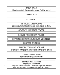
FIG. 16 is a flowchart showing general process steps for carrying out cell
physiology assays.
FIG. 17 is a flowchart showing steps in data analysis using tensor methods
described herein.
FIG, 18 shows the 2-D seatterplots of the results of a simulated flow
cytornetry analysis of
cells that were (A) exposed and (B) not exposed to a compound, (A) and (B)
represent positive and
negative controls for the agent, respectively. ELI and Fle"e,' are fluorescent
signals 1 and 2,
CA 02972960 2017-07-04
WO 2015/109003 PCT/US2015/011441
FIG, 19 shows the 2-11) scatterplots of the results of a simulated flow
cytometry analysis often
samples exposed to increasing concentrations of a biological agent, as
described in in Example 9.
Each sample contains 5,000 cells. The concentrations were increased from A to
J. The number of
cells in in the positive control-like cluster decreased from 4,909 in A to 29
in .1, FIL1 and FL2 are
fluorescent signals.
FIG. 20 provides two graphs of the response curves for the simulated flow
cytometry analysis
described in Example 9 and Figure 18. (A) shows the results for each sample as
a function of the
number of cells in the positive-like control cluster. (B) shows the same
results for each sample as a
function of the percent of cells in the positive-like control cluster.
FIG. 21 shows the four distances calculated for each sample to provide a
dissimilarity
measure, as described in Example 9. (1) The distance between the sample and
the negative control for
parameter one. (2) The distance between the sample and the positive control
for parameter 1. (3)
The distance between the sample and the negative control for parameter two.
(4) The distance
between the sample and the positive control for parameter 2.
FIG. 22 shows response curves fur the simulation described in Example 9
derived by polyadie
tensor decomposition.. The results were centered by subtracting the mean of
the vector and dividing
by the standard deviation. The results are expressed as a z-factor, and shown
in the graph on the left.
The graph on the right shows the results normalized to the difference between
the negative and
positive controls; i.e., in which the difference between the negative and
positive controls is defined as
unity (one) and the results are sealed to this difference.
FIG, 23 is a graph for the results of the simulation described in Example 9
expressed
conventionally in percentage of cells in the negative control-like cluster
along the vertical axis and
expressed in terms of distance/dissimilarity along the horizontal axis,
Distance/dissimilarity was
calculated for the same results using positive and negative controls as
illustrated in Figure 18 as
described in Example 9.
Fla 24 shows the 2-D scatterplots of the results of flow cytometry analyses of
ten samples
exposed to increasing concentrations of a valincanycin as described in in
Example 9B, The ratio of
Red (vertical axis) and green (horizontal axis) fluorescence ofJC9 were
measured to determine
26
CA 02972960 2017-07-04
WO 2015/109003 PCT/US2015/011441
mitochondria' membrane potential. Valinomycin concentration increased from A
(lowest
concentration) to J (highest concentration).
FIG. 25 shows dissimilarity response curves for the valinomycin analysis
(circles') described
in Example 9B and illustrated in Figure 24, and for idarubicin (diamonds) and
acetaminophen
(triangles) data obtained the same way. Dissimilarities were calculated as
described in the example
and illustrated in Figure 21. The graph on the left shows dissimilarity as
function of sample number.
The graph on the right shows dissimilarity as a function of concentration.
- IV -
Illustrative embodiments of the present invention provide automated, observer-
independent,
robust, reproducible, and generic methods to collect, compile, represent, and
mine complex
population based information, particularly, for instance, cytometry-based
information, as for example
for quantifying and comparing physiological responses of cells exposed to
chemical compounds, such
as drugs. Various embodiments provide methods for characterizes responses by
response tensors.
Illustrative embodiments provide for the use of various statistical measures
of distances between
distributions in one or more dimensions, and measures of dissimilarity between
response vectors
grouped into multiway tensors. In various embodiments the differences in cells
responses to two (or
more) chemical compounds is characterized as the difference between two
response tensors
(fingerprints") that represent said compounds. Embodiments provide methods for
generating said
fingerprints, and methods to manipulate, process, store, classify and use
them.
In various embodiments herein described, fix- example, biological datasets are
analyzed to
determine matches between them, often between test datasets and control, or
between test datasets and.
profile datasets. Comparisons may be made between two or more datasets, where
a typical dataset
comprises readouts from multiple cellular parameters, such as those resulting
from exposure of cells
to biological factors in the absence or presence of a candidate agent, where
the agent may be, for
instance, a genetic agent, e.g., expressed coding sequence; or a chemical
agent, e.g. drug candidate; or
an environmental toxin. In various embodiments, measurements are performed
using cytometiy, e.g.,
flow cytometry.
Cylometry
27
CA 02972960 2017-07-04
WO 2015/109003 PCT/US2015/011441
Methods of the various embodiments described herein are suitable for analysis
of complex
multi-parametric data on individual cells in cell populations, as determined
by cytometry. Cytometric
instruments and techniques, summarized herein (e.g., flow cytometry and
imaging cytometry) allow
for the simultaneous measurement of multiple intrinsic features (e.g., light
scatter, cell volume, etc)
or derived features (e.g., fluorescence, absorption, etc.) of individual
cells. Light scatter and
fluorescence represent the most commonly utilized measurements for current
cytometrie applications.
Fluorescence measurements can be performed either using either "intrinsic"
fluorophores naturally
present in cells (such as, for example, porphyrins, flavins, lipofuscins, NA
PH), fluorophores
genetically engineered for specific expression (e,g., GET, UP, etc.), or
fluorescent reporters which
target specific epitopes or structures in or on various cell types (e.g.,
finorophore corki.ugated
antibodies, aptamers, ph= display, or peptides, or reporters that are
converted from non-fluorescent
to fluorescent states by specific enzymes in or on cells).
Cytometric techniques useful in embodiments herein described utilize living
cells (e.g., using
probes which report cell on aspects of cell "physiology", such as, for
example, mitochondrial
membrane potential, ROS, glutathione content, or a combination thereof).
Cytometric techniques
useful in some embodiments additionally employ cells that are fixed and
permeabilized to allow
transport of fluorophores, conjugated reporters, etc., into the cytoplasm
and/or the nucleus.
General Methods for Cellular Assays Using Flaw Cytometry
General methods useful for cytometry in accordance with various aspects and
embodiments
herein described are described below and set out in generalized flowchart in
Figure 16
Culture of Anchorage Independent Cells
Cells and methods suitable for activity assays and analysis by flow cytometry
that are well
known and routinely employed in the art can be employed in carrying out
embodiments of inventions
described herein.
Cells for assays may be obtained from commercial or other sources. Cells
derived from
human cancer can be used, such as those from leukemias (e.g., 111,60 cells
cairrently used in cell
physiology assay), which grow unattached to the culture vessel. Cells
generally can be stored in
liquid nitrogen in accordance with standard cell methods. Frozen cells are
rapidly thawed in a 37 deg
28
CA 02972960 2017-07-04
WO 2015/109003 PCT/US2015/011441
C water bath, and cultured in stationary flasks in pre-warmed fresh tissue
culture medium in a 37 deg
C tissue culture incubator. Tissue culture media typically is replaced daily
for the first 2-4 days in
culture, to dilute out the DMSO cells are frozen in.
Once growth is established in stationary flasks (cell number and viability is
monitored using a
Vi-cell'' cell counter), aliquots of cells can be removed for freezer storage
(these early passage cells
are only used for backup). In addition, these cells can be used to establish
roller bottle cultures
needed to have sufficient cell numbers for plate assays. Cells growing in
flasks are placed in roller
bottles at relatively high cell concentration (-106 cells per ml in 200 ml
fresh tissue culture medium)
and cultured in a tissue culture incubator. Initially, roller bottle cultures
typically are fed by addition
of fresh tissue culture medium. Once growth is established, cells are removed
as needed to maintain
cells at a concentration of 0.54.5 x 10 viable cells/ ml. Many cell types
adapt to roller bottle cultures
slowly, and need weeks to successfully adapt to these types of cultures.
Successful roller bottle
adaptation is evidenced by continuous high viability (-95%) and consistent
growth rates (measured
using doubling time). When successfully adapted, stocks of cells are frozen
(in 50 ml sterile tubes
containing sufficient cells to initiate one new roller bottle culture) in orer
to maintain cells used for
assays at a similar low passage number (details below). Cells maintained in
roller bottles are
harvested for assay plates, centrifuged, and resuspended in fresh tissue
culture media at appropriate
cell concentration for the assay to be performed (cell number and viability
measured and recorded for
each harvest).
As indicated above, roller bottle adapted cells can be frozen for future use,
to maintain similar
low passage number cells for all plate assays. Roller bottle cell cultures can
be maintained for one
month before switching to a new lot of low passage frozen cells. During the
month of routine use,
one tube of frozen cells typically is thawed and re-established to roller
bottle culture. Once
successfully adapted to roller bottle culture (as above) the newest lot of
cells usually is first evaluated
for assay performance (see "Cross-Over" studies, below), before this lot of
cells is used in plate
assays. Establishing frozen cells to roller bottle culture and testing
routinely takes 10 to 21 days.
Cells generally are routinely tested at multiple steps in the culture process
for mycoplasma
contamination. These include initial flask cultures, roller bottle adapted
cells, and each tube of frozen
CA 02972960 2017-07-04
WO 2015/109003 PCT/US2015/011441
cells (tested before each "Cross-Over" study). Mycoplasma testing can be
provided by an external,
certified testing company, typically using a PCR-based assay,
Compound Storage and Compound Assay Preparations
Test compounds are generally obtained as 10 rnM stocks in DMS0 deposited in 96-
well
plates. Compound plates are stored sealed at room temperature in the dark. For
compound assays,
stock solutions are diluted (5 to 10 step compound dilutions in DMSO) and
deposited into assay plates
using a liquid handling system. All dilutions and compound deposition into
assay plates are
performed the same day as the assay is performed. The final volume of compound
deposited into
each well is 2 ul, giving a final concentration of 1% DMS0 after the addition
of cells.
Reproducibility of assays should be assessed using test compounds. A set of 16
compounds
that have well documented impacts on specific cell physiological measurements
have been used to
test the reproducibility of cell physiology assays. These compounds are
stored, as above, as 10 mM
assay solutions in DMSO in 96-well plates. For "Cross-Over" studies, 16
compound set are used to
compare the physiological responses of the newly thawed and roller bottle
adapted cells with current
lots of production cells.
cell Physiology Assays
For cell physiology assays it can be convenient to use 2 sets of 384 well
plates to measure the
impact of compounds on ten or more cellular response parameters. For both sets
of plates, compound
dilutions are first deposited into wells, and then 1 X 10⁶ assay cells are
added to each well, as shown
in step 2602. Compounds are routinely run with duplicate compound dilution
sets on the same plate;
at the start of a study for an individual client, duplicate plates are ran for
the first 16 to 32 compounds,
in order to measure reproducibility of responses. After thorough mixing,
plates are sealed (using an
02/CO2 permeant seal) and placed into a 37 deg C tissue culture incubator for
varying periods of time
(typically 4 hrs), as shown in step 2604. Plates are then centrifuged in step
2606, half the supernatant
fluid is removed, and replaced by the same volume of the appropriate dye mix
(fbr plate A, the dye
mix may include Monobromobimane, Calcein AM, MitoSox'TM, and Sytox Reet ftw
plate B, the dye
mix may include Vybrant Violet"' (live cell cycle), JC-9 (mitochondria'
membrane potential), and
Sytox Reem), followed by mixing, as shown in step 2608. Plates are returned to
the tissue culture
CA 02972960 2017-07-04
WO 2015/109003 PCT/US2015/011441
incubator for about 15 (plate A) or 30 (plate B) minutes in step 2610,
followed by a mix, and
immediately run on the flow cytometer in step 2612.
The data from positive and negative control wells on each row are used to
calculate the
responses as described in greater detail herein. The positive control
compounds used for plate A and
B are different, and are designed to provide a unique "signature" ("finger
print") in the cell responses
measured in plate A or B, using the disclosed embodiments.
High Throughput Flow qytorneuy
in a variety of assays the flow cytometer is set up using a standard procedure
on each day that
plates are assayed. Set up includes flow instrument QA/QC using fluorescent
beads which are used to
set each detector (PMT) to a standardized target. Bath well of a 384 well
plate is then sequentially
sampled using a 6 second sip time, followed by a I second air bubble between
samples. The sample
stream flows through the flow cytometer in a continuous fashion, sampling a
complete plate in 20 to
30 minutes (plates A and B, respectively).
The flow cytornetry data file (for one plate) is subsequently processed by the
disclosed
embodiments to identify individual well data (using some interaction and human
intervention), and is
then stored on a server as the list mode data (leMD) for each well from a
single plate.
QAIQC analysis of each plate
Both plates (A and B) contain negative controls (untreated samples), and
positive controls
(sample treated with a mixture of 50 uM FCCP plus 50 ul\il Myxathiazol for
plate A, or 25uM FCCP
for plate 13). The dissimilarity between positive controls and negative
controls does not define in this
assay the possible range of responses. However, it defines a unit of response.
During the time of
analysis of an entire plate, the dissimilarity between positive and negative
controls may change owing
to deteriorating physiological conditions in the plate (change in temperature,
02, etc). This is why a
certain minimum level of dissimilarity for every pair of controls is expected.
For each positive and
negative control within a single row, the disclosed embodiments determine the
OF distance between
the positive and negative populations for each dye response individually. The
disclosed embodiments
then plot the change in QF distance from the beginning (row A) to the end of
the plate (row P),
31
CA 02972960 2017-07-04
WO 2015/109003 PCT/US2015/011441
Experiments described herein generally have been carried out in accordance
with the
foregoing procedures.
Cytorneter Instrumentation
Current flow cytometry instruments are equipped with multiple lasers and
multiple separate
fluorescence detectors that can simultaneously quantitate many .fluorescence
signals plus intrinsic
optical features originating from individual cells. Thus, cytometric
techniques and instruments such as
those illustratively described below allow measurement of thousands to
millions of cells in a sample.
The resultant extremely large data sets present a significant challenge to the
presently-employed
eytometry data processing and visualization methods, These challenges are
handled effectively by
methods described herein.
Modem. cytometers typically are designed for simultaneously detecting several
different
signals from a sample. A variety of cytometers are available commercially that
can be used in
accordance with methods described herein. A typical instruments includes a
flow cell, one or more
lasers that illuminate the flow cells through a focusing lens, a detector or
light passing through the
flow cell, a detector for forward scattered light, several dichroic mirror -
detector arrangements to
measure light of specific wavelengths, typically to detect fluorescence. A
wide variety of other
instrumentation often is incorporated in commercial instruments.
in typical operations, the laser (or lasers) illuminates the flow cell and the
cells (or other
sample) flowing through it. The volume illuminated by .the laser is referred
to as the interrogation
point. Flow cells are made of glass, quartz and plastic, as well as other
material. Although lasers are
the most common source of light in cytometers, other light sources can also be
used. Almost all
cytometers can detect and measure a variety of parameter of forward scattered
and back scattered
light, and several wavelength of fluorescence emission as well. Detectors in
these instruments are
quite sensitive and easily quantify light scattering and fluorescence from
individual cells very short
periods of time. Signals form the detectors typically are digitized and
analyzed by computational
methods to determine a wide variety of sample properties, There are many texts
available on flow
cytometry methods that can be used in accordance with various aspects and
embodiments of the
inventions herein described. One useful reference in this regard is Practical
Flow Cytometry, 4th
WO 2015/109003 PCT/US2015/011441
Edition, Howard M. Shapiro, Wiley, New York (2003) ISBN: 978-0-471-11125-3,
particularly in pans pertinent to cytometry and cell analysis as may be used
in accordance with the methods herein described.
Spectral uninixiitg offlow cytometric signals
Since the signals emitted by the functional fluorescence labels are measured
by a series of
detectors in a cytomeny system (flow- or image-based), the detection systems
are prone to spectral
cross-talk. As a result, the intensities of individual fluorochromes cannot be
measured directly to the
exclusion of other fluorochromes. In order to minimize or eliminate noise due
to spectral cross-talk,
all of the collected signals can be modeled or processed as linear mixtures.
The signal mixture for
each measured cell is decomposed into approximations of individual signal
intensities by finding a
minimal deviance baween the measured results and approximated compositions
which are formed by
multiplying the estimator of the unmixed signal with the mixing matrix. The
mixing matrix (also
called "spillover matrix") describes the n-band approximation of fluorescence
spectra of the
individual labels (where n is the number of detectors employed in the system).
An application of a
minimization algorithm allows to find the best estimation of the signal
composition. This estimation
provides information about the abundances of different labels. In the simplest
case, if the
measurement error is assumed to be Gaussian, the immixing process may be
performed using ordinary
least-squares (OLS) minimization.
Variance stabilization
'Variance stabilization (VS) is a process designed to simplify exploratoiy
data analysis or to
allow use of data-analysis techniques that make assumptions about data
homoskedasticity for more
complex, often noisy, heteroskedastic data sets (i.e., random variables in the
sequence have different
finite variance). VS has been routinely widely applied to various biological
measurement systems
based on fluorescence. It is an important tool for analysis of microarrays.
In the context of flow cytometry and in microan-ay analysis, log
transformation has
traditionally been used. However, modern approaches, for example, in the
context of microarray
analysis are known. For example, see Rocke et al, (Approximate variance-
stabilizing transformations
for gene-expression microarray data," Bioinformaties, 19, 966-972, 2003) and
Huber et al. ("Variance
33
Date Recue/Date Received 2021-05-12
CA 02972960 2017-07-04
WO 2015/109003 PCT/US2015/011441
stabilization applied to tnicroarray data calibration and to the
quantification of differential
expression." Bioinfirmatics, 18, 896--S104, 2002). Huber describes the use of
a hyperbolic ars,;sine
function in variance stabilization, in the context of flow cytometric data
analysis, Moore et al.
("Automatic clustering of flow cytometry data with density-based merging," Adv
Bioiqformatics,
2009) uses logical transformation. Bagwell ("Hyperlog-a flexible log-like
transform for negative,
zero, and positive valued data." (ytomehy A. 64(1):34-42, 2005) describes the
use of hyperlog
transformation in the analysis of output from now cytometers.
In an embodiment of the present invention, in contrast, hyperbolic arsine
technique
(generalized logarithm) with an empirically found parameter is used in
variance stabilization.
glog(x) = asinh(x)= log( x+
glog(x,a,b,c)= asinh(a 'cc+ b)+ C
Comparisons
Certain embodiments described herein provide methods involving a comparing
step, wherein
the distribution of the unmixed signal intensities is compared to the
distribution of the unmixed
signals originating from controls or other test data. Depending on the
comparison method applied, the
distributions may be first normalized by dividing every distribution by its
integral.
The comparing step may involve compilation of response tensors containing
intbrmation
about dissimilarities between cellular populations such as before and after
treatment. The
dissimilarities are computed as distances between signal distributions of the
treated population of
cells, untreated populations ("negative" or "no effect" controls), and
populations treated with a
mixture of perturbants designed to maximize the observable physiological
response ("positive" or
"maximum effect" controls).
In order to standardize the result and render it unaffected by experimental
variability, the
measured dissimilarity can be expressed in units equal to mean dissimilarity
between positive and
negative controls.
Various measures of dissimilarity or distance can be applied, including (but
not limited to):
Wasserstein metric, quadratic-form distance (QFD), quadratic chi-distance,
Kohnogorov metric,
34
CA 02972960 2017-07-04
WO 2015/109003 PCT/US2015/011441
(symmetrized) Kullback-Leibler divergence, etc. In the preferred
implementation, the methods and
algorithms of the instant invention use Wasserstein metric or quadratic chi-
distance.
in illustrative methods below, the abundance distributions are typically
compared in one
dimension. However, some labels are encoded by two related signals (for
instance, JC-1, the
mitochondrial membrane potential label that emits fluorescence in two separate
channels). In this
case, a 2-D dissimilarity measure between distributions is eomputed. Finally,
it may be preferable to
compute 2-D or 3-D dissimilarity measures by utilizing multidimensional
distributions based on
morphology-related measurements (obtained via light scatter) and an abundance
(computed from the
fluorescence signal). A variety of distances or dissimilarity measures,
assuming that they are easily
generalizable to multiple dimensions, may be used. For instance, routine
methods based on the
Wasserstein metric or the QFD may be used in this context, but not the
Kolmogorov metric.
A representative equation for comparison of populations is provided below
(see, Fele et al.
The Quadratic-Chi Histogram Distance Family." Computer Vision -----ECCV 2010,
Lecture Notes in
Computer Science. Springer Berlin Heidelberg, pp. 749-762; Daniilidis, K.,
Maragos, P. Paragios, N.
(Eds.)):
gi(x, y) -:.:..goENõRdAdT),01,
fl¨j)
ARA .1-11'
wherein x and y are distributions of interest, and A is a positive-
semidefinite dissimilarity
matrix.
A nalvsis of cytometry data using tensors
Cytometrie multi-parametric data can be expressed as tensors and the
comparisons between
controls and tested samples can he described by compound fingerprint tensors.
A tensor is a
multidimensional array and can he considered as a generalization of a matrix.
A first-order (or one-
way) tensor is a vector; a second-order (two-way) tensor is a matrix. Tensors
of order three (three-
way) or higher are called higher-order tensors.
CA 02972960 2017-07-04
WO 2015/109003 PCT/US2015/011441
Biological measurements performed in a single-cell system individually for
every cell in a.
population form a distribution. A distance between a distribution of
measurements performed on cells
exposed to a presence of a compound, and a distribution of measurements
performed on cells not
exposed to the compound can be expressed by a single number (scalar value).
The cells may be
exposed to a number of different drug concentrations, and a biological
measurement can be performed
for each of these exposure levels. Such an experiment produces a series of
values that can be
expressed as a vector (e.g,, a one-way tensor). If multiple biological
parameters are measured, the
results can be arranged in a two-way tensor (or a matrix), in which every
column contains a different
measured parameter and every row describes a different concentration of the
compound.
This arrangement of data can be expanded further. if we attempt to measure the
distances
between the distributions of measurements obtained from treated cells and a
distribution of
measurements collected from population of cells exposed to another compound,
we can group the
results into another matrix. For instance, it may be beneficial to measure
dissimilarity between cells
treated with one compound and another group of cells treated with a different
and well characterized
compound that creates an easy to observe effect serving as a positive control.
The two matrices (two way tensors) put together produce a three-way tensor.
The
dimensionality of this three-way tensor is 1142 43, where 11 is the number of
concentrations, /2 is the
number of measured biological parameters, and 1.3 is the number of measured
dissimilarities/distances
(typically two: positive control distance measurement, and a negative control
distance measurement).
Therefore, for a tensor A representing the biological measurement, the element
(ij,k), denoted by au,k
describes a distance between measurements of parameter/ obtained from a cell
population exposed to
a compound at concentration i, and a control cell population k.
A column fiber of tensor A, denoted as aA contains a whole series of distances
between
measurements of parameter/ performed for a series of tested samples and a
control It. if 10
concentrations of a compound are tested, the column fiber ayk will be a 10-
element vector. A frontal
slice of the tensor A denoted Aõ,õ forms a matrix which describes the
distances measured to k-control.
A lateral slice kJ; is a matrix, showing measurements of distances for
parameter j.
36
CA 02972960 2017-07-04
WO 2015/109003 PCT/US2015/011441
The tensor A can be further expanded to account for multiple environments,
cell cultures or
phases of cell cycle. Therefore, multiple repeats of the measurements,
multiple phases of cell cycle in
which the measurements are performed, and so forth, can he stored in a
compound fingerprint tensor.
In fact, any MU itidimensional. screening experiment can be represented as
apkorder tensor
A i'xi2x¨YJP . For instance, one may group the data from multiple
experiments in a four-way tensor
with a dimensionality I xJ '<.13x14, where is the number of unique measured
compounds. In this
setting a slice Xis,/ contains dissimilarities between tested samples
containing compound land a
control k, computed for parameter/. If four repeats of a compound I were
measured, the resultant A./id
slice is a matrix of ten rows (concentrations), and four columns (repeats).
The tensor representation of the compound measurements can be used to define a
number of
compound-related metrics and operators, such as a compound
similarity/dissimilarity, compound
fingerprint, compressed compound fingerprint, compound normalization, etc. The
arrangement of the
phenotypic screening data in a tensor format enables the formulation of unique
insights regarding
compound characteristics through analysis of compound response similarities.
This is impossible
when the information regarding the compounds is represented simply as data
vectors. Specifically, the
techniques described below would be impossible to implement if compounds were
described only
using scalar values (such as traditionally utilized IC 50 value),
The foregoing analysis can he stated in general terms in the form of the
following equation
and operations herein referred to as
General -Tensor Analysis of Population Data
General Method and Equations -
In the first step a pair of distances between the positive controls (Cp) and
the negative control
(C,,) is computed for every marginal histogram tp. These distances are kept as
references for fitrther
use and resealing:
where ye
n p
The distance function D can be a quadratic form distance, a Wasserstein
distance, a quadratic-
X2 distance or any other distance operating on vectors representing
histograms.
37
CA 02972960 2017-07-04
WO 2015/109003 PCT/US2015/011441
f D(CC!.4)
Following this operation a series of distances for the biological samples are
calculated in an analogous
fashion. Distances are computed for every pair made of a control K and a
biological sample in the
series of concentration (Si, S29 Si), where i denotes the concentration of
a tested compound.
div) ---.D(CW,Stv))
x
The resultant values form a multidimensional array or pa-order tensor A ERY.
The size
and the dimensionality of the tensor depend on the number of controls, the
number of utilized one-
dimensional histograms, and the number of biological conditions at which the
measurements were
conducted.
A column fiber of tensor A, denoted as a[pc,/,1 is a vector which contains a
series of distances
between measurements of a biological parameter summarized by marginal
histogram vs performed for
a series of tested samples and a control K.
id 7
=
D(Cw) Siv)
a 2 )
x= r
=
A frontal slice of the tensor A denoted akiõ3, forms a matrix which describes
the distances for
biological parameter y,f, where the measurement involves all controls.
r
cl(v) ¨D(Civi),S{Y)) id(v) D(C{w),S,(v.) )7: i -D(C(v),S,m11
fC23 s AT;
*Iv) ,S(,7q ofw = D(Cw) ,Sr) Er( 01 ,Sr)
A?c: - , 2 , :
ttgi
D(C:(!l i.$411:1 el = ,s(o)
II K2,1 if n Scot .. K., .. ft
=
A lateral slice Am is a matrix showing measurements of distances for multiple
biological
parameters, where the measurement was performed using just a single control K.
38
WO 2015/109003 PCT/US2015/011441
Tei),D(c d(wo.D(oto,s{v,) dvip).D(c(t5) , P Xj. r P r
, 1
6") D(e/1,)?5(:11, dRIY o(e2) s{V, d(V.]
o(cl'Wdgedf
A. = ,
D(C",SN''=), C ,S -),=== ,=
7C, J., =
Therefore, the tensor A can be expressed as:,
at
a e,Afti
A=
H a = = = a
trc,=t$,) =
= O =
The biological measurements may be further normalized by dividing every
cil'P;) by the
corresponding 14' resulting in a normalized tensor of distances:
,
orovi,
A-
K.4,23 a.
The same analysis can be carried out for a variety of conditions, resulting in
a series of
normalized tensors of distances.
Compression
A tensor A obtained from a series of measurements forms a unique compound
fingerprint, as
it contains all the phenotypic characteristics of a tested compound. This
tensor A can be
"compressed" using a low-rank tensor approximation techniques such as polyadic
tensor
decomposition or other methods. See Kolda and Bader, "Tensor decompositions
and applications,"
SIAM Rev. 51, 455, 2009.
Tensor decomposition factorizes a tensor into a sum of component rank-one
tensors, A tensor
An by definition is rank one if it can be expressed as the outer
product ofp vectors:
A õ.õ. ao) õa(2)o,,a(p)
The goal of a canonical polyadio (C.',1)) decomposition is finding an
approximation of tensor A
denoted A, which satisfies the following criteria:
39
Date Recue/Date Received 2021-05-12
GA 02972960 2017-07-04
WO 2015/109003
PCT/US2015/011441
min A ¨ , where A=A; A"), A'2),:, A.,,a(i.÷ oat2) o = = = 0 a(?)
i
Tensors can be decomposed using other techniques as well, For instance Tucker
decomposition decomposes a tensor into a core tensor multiplied by a matrix,
along each mode:
G:.m(1) ,m(2) .,m(P) =V V a(1) 0 2(2)
611grp arr.!
The various methods for tensor decomposition allow us to approximate a
complicated
multidimensional compound fingerprint with a "compressed," abbreviated
version. This goal can be
accomplished by removing the part of a decomposition, which does not
contribute significantly to
tensor rebuilding accuracy.
The ex pie below illustrates the point. Assume that A is a three-way tensor
containing a
compound's information. The front slices of tensor A, denoted Aõ, and A:,2
are:
Ckr =?t C,33
29.92
:11L,R3 M.13. 13.55 L2,3 59.71t
Z=7. ;>E [3,3
i14,3 04. :.4. [4,] 55,41 MA,3 119.79
tI0A5 C5,34I.5,9k 14S..33
Slice Aõ, Slice Aõ2
After performing CF decomposition we obtain the following vectors:
[-0.1348, -0.2698, -0.4048, -0.5398, -0.6737]
=[o.236, 0.4133, -0.7798, 0,4011, -0.0657]
ii(12) := [0.2663, -0.5347, -0.802]
a?' = [0.1966, -0.8447, 0.4979]
43) =[06691, 0.7431]
14') = [-0,2009, -0.9796]
A, = 372.9803, = 0.5591139
The original tensor may be rebuilt from this result following the CF model:
= a") 0 ea") 4-- a a(2)a(3)
n 2 2 2 2
After rebuilding the approximated tensor A contains the following values:
CA 02972960 2017-07-04
WO 2015/109003 PCT/US2015/011441
ral .. E.11 .. r ,z3 r.,11
E1,] 8.954563 1$401-4i:3= 76.96164 [1;] 9.924882 20.08219 29.90171
E2,] 12,924562 53.93645' E2;] 19.372811 46.17834
59.66717
C3,3 2F,,9N335 r3,1 29.951619 59,62570 90.19114
[4,-3 15.805664 22.M.V5 laztAn.61tv [4;] 39.717539 50,17512 119.47475
[5 4J455 5;] 49.735276 99,80670 149.77195
Slice ki Slice
However, the tensor A can be rebuilt using only vectors at rather than al and
a2. Although the
resultant approximation has a higher error, it still captures most of the
information represent by A:
=
2%.559?3 W>4 24,4f.V4
[1a3 Li 33!1 .nto t1 W:3153l 5ee7
DJ 26,91111 5.01436 g,52566. [3,] ,5?$7
L4,] 15,87455 22.62t 4µ.8.038.81 .. EC] 39,111626 76.682 149,9All
[5,1 44.77559 0.aw5i.:z4,0,410=5jvoq4204 .....
Slice itõ1 Slice
As demonstrated above, the tensor A can be approximated using only ai vectors.
If the
resultant accuracy is sufficient, the decomposition effectively compresses
tensor A approximately 6-
fold -- it takes 60 numbers to encode the original tensor but only 11 numbers
to encode the
compressed tensor A (vectors a0), ar2), IP and value of 4 The decomposed form
of the tensor can he
further used for tensor-to-tensor comparison, as well as an input for
supervised machine. learning
methods, such as, for example, to support vector machine learning.
It should be noted that simple CP decomposition and the more complex Tucker
decomposition, although most commonly used for expressing the content of
tensor-arranged
information in a simpler, easier-to comprehend form, are not the only methods
useful in decomposing
tensors. Other tensor decomposition methods can be also used with the
screening data, as long as the
organization of the datasets follows an acceptable convention.
Moreover, as mentioned above, tensor decomposition can be applied to higher-
order tensors.
In this case the multiple measurements performed for multiple compounds will
be decomposed along
the modes of a tensor, revealing the crucial differences between compounds.
Also, multiple repeats of
the same tested compound can be used, effectively leading to computation of a
synthetic "averaged"
compound fingerprint which can then be compared to other fingerprints.
41
CA 02972960 2017-07-04
WO 2015/109003 PCT/US2015/011441
Automated gating based on morphalop.,
An embodiment provides for the use of model driven automatic gating (although,
the use of
gating algorithms is optional), Herein, state-of-art teohniques of mixture
modeling with or without
proprietary additions may be added to the algorithm. The system may rely on an
iterative approach to
improve efficiency of the assay.
In an embodiment, the gating technique comprises 3 skew-normal probability
distributions
representing "live cells," "dying cells," and "dead cells" (debris). Depending
on the data, an existing
(e.g., old validated) model may be used or a new generated based on the
controls. For example, it is
possible to proceed by calculating the .total (LL) for each mixture model.
Specific
models for which LL is higher are then retained for future use,
compound dissimilarM)
Compound dissimilarity measurements can be performed after decomposition of an
individual
compound fingerprint, following a decomposition of a result tensor in which
all the compounds (and
all the measurement repeats) are stored, or directly using the fall,
"uncompressed" version of the
fingerprints. If the comparison involves decomposed tensors, the problem is
essentially reduced to
comparison of vectors.
However, as mentioned above, the compound fingerprint tensors can be also
compared
directly. In this case the dissimilarity between compound tensors is expressed
as a vector storing
dissimilarities (distances) between mode-1 fibers aiik of the compound tensor
A. Since the fibers aim
store information about responses of cell cultures exposed to increasing
concentrations of the tested
compounds, the values form a series, which can be viewed as a polygonal curve.
Consequently, the
tensor-oriented arrangement of data allows us to utilize distances between
discrete curves (vectors).
One of such distances is the discrete Frechet distance, which is defined as
the minimal length
of a leash necessary for the dog and the handler to move from the starting
points of the two curves
describing their paths to the endpoints, provided that both dog and the
handler can only move
forward, although they can control their speed. Since the fibers can be viewed
as polygonal curves, in
this setting both the dog and the handler can only stop at vertices of the
curves and each of them can
either stay at their current vertex or iump to the. next one while walking.
See Aronov et al., "Frechet
42
WO 2015/109003 PCT/US2015/011441
-Distance for Curves," Revisited, in: Azar, Y., Erlehach, T. (Eds.),
Algorithms ESA 2006, Lechee
Notes in Computer Science. Springer Berlin Heidelberg, pp. 52-63, 2006.
A conceptually similar distance measure is dynamic time warping distance,
which measures
distance between two polygonal curves by warping them in a non-linear fashion
in order to find the
best alignment. The cost of performing the alignment is defined as the
distance value. See Berndt and
Clifford, "Using dynamic time warping to find patterns in time series," AAA194
Workshop on
Knowledge Discovery in Databases, pp. 359-370, 1994.
Therefore in a general case we can express the dissimilarity between two
compound
fingerprints (tensors) A and B as:
WA s D(A,B)= d(aim,kik)
where .1.) is the dissimilarity between compound tensors A and B, d is the
distance function comparing
mode-1 fibers of each of the tensors (such as Frechet distance or dynamic time-
warping distance), and
w is the distance vector. For a three-way tensor produced by experiments
involving a 10-dose
response curve, four parameters and positive and negative controls, the vector
w would have a length
of 8. Every element of the vector would be computed as a curve-distance
between corresponding
fibers of tensors A and B.
Graphical representations of compound dissimilarities
Similarities/dissimilarities between compounds can be demonstrated graphically
using
various methods depending on the function used to evaluate the dissimilarity.
An example is
illustrated Figure il, If an abbreviated compound fingerprint can be expressed
as a vector after a
decomposition (formed by multiple concatenated vectors), then one may apply an
a priori chosen
distance between vectors, or train a distance function using metric learning
methods. See Weinberger
and Saul, "Distance metric learning for large margin nearest neighbor
classification," J. Mack Learn.
Res. 10, 207-244, 2009. The final
43
Date Recue/Date Received 2021-05-12
WO 2015/109003 PCT/US2015/011441
result can be visualized using multidimensional scaling (MDS), hierarchical
clustering, or manifold
learning methods.
If decomposition/compression is not used, the visualization of similarities
between
compounds requires an approach utilizing vectors of dissimilarities, rather
than scalar measures of
dissimilarity. One such method is multi-mode MDS algorithm utilizing stress
majorization. See
Leettw and Mair, "Multidimensional Scaling Using Majorization: SMACOF," R.
Sten', Softw. 31, 1-
30, 2009 .
Utilizing the formed dissimilarity matrices by multi-way multi-dimensional
scaling
techniques place the measured compound signatures in a space defining inter-
compound similarity.
See, Groom et at majorization approach to multidimensional scaling for
.Minkowski
distances." I Classit 12, 3-19, 1995) . The compounds
localized close in the resultant space will he deemed "similar" in the sense
of the described process,
and the compound located far away will be considered "dissimilar."
Comparison of distributions using multiple cellular subsets
The control versus tested sample dissimilarity can be computed employing all
the measured
instances (cells) or calculated separately for each functionally defined
subset of the cellular
population. For instance, one may define two morphologically distinct subsets
of cells on the basis of
the measured light scatter. in this setting the two cellular subsets may be
compared to the controls
separately. What follows is that the number of comparisons is dependent on the
number of formed
subsets. For instance, if five functional la.beis are designated to
demonstrate five different aspects of
cell physiology, and one chooses to compare the treated outcome to untreated
outcome for each of the
morphologically different cell populations, one can produce up to 20 different
distances (5 labels x 2
controls x 2 cellular subsets).
Since the concentration of the perturbant resulting in the desired
physiological effect cannot
be ascertained a priori, a function describing the gradual increase of the
effect with the perturhant
concentration can be used to indicate properties of the perturbant, the
measurement being performed
with several concentrations of a drug. Therefore, for the scenario outlined
above assuming the use of
concentrations, the final data set would comprise 200 values (5 labels x 2
controls x 2
44
Date Recue/Date Received 2021-05-12
WO 2015/109003 PCT/US2015/011441
subpopulations x 10 concentrations). The measured values are not independent;
therefore they are
treated for further processing as a multi-way tensor. For instance, if only
one type of control is used
(e.g., a "positive" control), the data set would form a three-way tensor with
the dimensions of 5 x 10 x
2. FIG. 14 shows an example of a multiway tensor representing a drug response
finge,rprint.
Use (If a multi-dimensional fealure space
in an embodiment of the present invention, the evolution of a point-cloud in a
multidimensional feature space can be computed by directly computing distances
between
distributions. Examples of point clouds that evolve in a multi-dimensional
feature space include, but
are not limited to, for example, variables such as drug concentration,
incubation time, etc. The
changes in evolution of the cloud in the space defined by dissimilarities
between complex point
clouds form a complicated trajectory, which uniquely describes the
characteristics of the compound
eliciting those changes.
A representative cloud evolution is shown in FIG, 15.
Comparison of disiribMions using cell-cycle information
in embodiments, fingerprint tensors are created tbr each of the clusters or
cellular subsets
defined by cell-cycle-dependent labels. In this setting, the assessment of
cell-cycle phases is
performed simultaneously with other measurements such as by quantifying the
intensity of
fluorescence labels intercalating into DNA (such as HOECHST 33342, DRAQ5Tm, YO-
PRO-1
IODIDE, DAN, CYTRAK ORANGE), by quantifying the presence of immunolabeled
proteins
associated with the cell cycle (e.g., cyclins, phosphorylated histone
proteins, etc.), or by measuring the
signal of genetically encoded fluorochromes linked to cell-cycle phases, in
the simplest case, these
techniques allow one to determine three compartments of the cell cycle,
leading to a formation of a 5
x 10 x 3 result tensor (or more complex tensors, depending on the number of
additional labels). This
is illustrated in Example 5A.
Unsupervised and supervised classificatio
Embodiments provide classification methods, wherein subsequent analyses are
performed
using multiple approaches. These analytical techniques may be implemented
depending on the
preferred final representation or visualization of the results. For instance,
in the unsupervised setting
Date Recue/Date Received 2021-05-12
CA 02972960 2017-07-04
WO 2015/109003 PCT/US2015/011441
the distance tensor can be processed by a multi-mode multidimensional SMACOF
(Scaling by
MAjorizing a COmplicated Function) algorithm or other suitable mathematical
technique for scaling.
After application of the MDS method the drug fingerprints will be placed onto
a 2-D plane or onto
any other n-dimensional surface (e.g., sphere) defined by the researchers.
This allows easy
interpretation and visualization of the data sets, which form a "map" upon MSD
projection.
in the supervised. framework, the fingerprints representing compounds
belonging to various
groups defined by function, response type, chemical structure, etc., are used
to create a training library
with multiple classes. The training library can subsequently be used to train
a classifier (such as a
neural-network classifier, support-vector machine, or another type of machine-
learning system) that
will categorize previously unknown fingerprints into classes defined by known
compounds.
By extension, related embodiments of the present invention provide methods for
assessing the
effects of unknown perturbants (not known a priori to impact MMP) to determine
if they have a
differential impact on MMP (and other cell physiology measurements, e.g.,
SYTOXTM RED low
versus high cells) in different cell cycle phases. Therefore, for every tested
compound, there is
provided a multi-way measurement tensor (compound signature) which comprises
cell cycle-related
state tensor as one of the dimensions in which the cell state is evaluated.
The following is a representative method for comparing different perturbants
using the
systems and the algorithms of the instant invention:
First, for each fluorescence channel (for example, separately for live cells
and dying cells), a
distance matrix between all compounds is created using the above-described
technique. For example,
the distance can be the dynamic time warping distance (dtw) between response
curves. Subsequently a
multi-way mIti-dimensional scaling (mps) is used to combine several matrices
into a Euclidean
space. Then the MDS for one group of matrices containing only MMP data
(CALCE1N, SYTOXTu)
and another group with only permeability data (MBBR, MfIoSOXTM) may be
calculated. The
various physiological similarities between the compounds may be visualized
with the help of known
mapping and clustering techniques, e.g,, dendograms or graph visualization
(e.g., creating
intermediary nodes and exporting the data to any one of a variety of external
visualization software
packages. Furthermore, supervised approach using one or more classifiers may
also be employed.
46
CA 02972960 2017-07-04
WO 2015/109003 PCT/US2015/011441
Cell cycle
Embodiments herein described allow measurements of coordinated protein (or
otter marker)
expression in populations of cells as a function of cell cycle (e.g. Gl, 5,
G21,44), and to determine cell-
cycle-dependent effects of the test compounds. Multi-parametric analysis may
thus be conducted by
analyzing the effect of each perturbant at different concentrations and/or
time points to investigate the
effect of said compounds on the various cellular parameters (e.g.,
mitochondrial membrane potential,
nuclear or cytoplasmic membrane permeability, ROS, cell death or apoptosis).
An example of cell-cycle dependent analysis is based on the measurement of
Cyclin A2
expression in normal (unperturbed) cells. Herein, the possible "states"
include Cyan A2 negative,
Cyclin A2 low and Cyclin A.2 high. Similarly, for phospho-histone 3 (P-H3),
which is a second
marker in cell-cycle analysis, the possible "states" include "negative" and
"positive". These two cell-
cycle markers may also be analyzed in combination, thus yielding nine
different possible
combinations ("states"). It is not always necessary to investigate all
possible "states" because all the
states may not exist in normal biological space (sparse matrix).
Accordingly, depending on the cell cycle state a particular cell is in,
differential perturbations
caused by drugs or compounds of interest can be investigated by populating
cells in discrete (normal)
matrix elementsõAs an example, drugs which block normal progression from
mitosis back into GI,
which cause quantitative changes in "normal" matrix populations (i.e.,
accumulation of cells into
"late" (normal) cell cycle compartments (e.g. G2 and M)) and/or deplete cells
in the GI phase, can be
analyzed in concert using Cyclin A2 and/or P-H3 staining, Similarly, a drug
which prevents
separation of daughter nuclei would be expected to show a different
quantitative fingerprint pattern
compared to a drug which arrests cells in S-phase (e.g. a drug which inhibits
new DNA synthesis).
Accordingly; compounds which cause cells to appear in different matrix
elements not only creates a
unique signature, but also the specific matrix element that is occupied could
provide information
regarding the mechanism of drug action. For example, expression of Cyclin A2
in G I and or M can be
the result of a proteasome inhibitor preventing normal Cyan A2 degradation,
Altiltipie cell Owe. assay system
47
CA 02972960 2017-07-04
WO 2015/109003 PCT/US2015/011441
in an embodiment, the present invention provides for methods for assaying
cellular states
using a plurality of cell types, e.g., two or more cell lines (from tissue
culture) in a single assay. One
advantage of this approach is it allows analyses of DNA damage/responses, An
additional advantage
is that it allows studies of both constitutive and inducible signaling
pathways in the same assay (using
one cell line with constitutive expression and another that can activate the
same pathway using an
appropriate agonist). Using two (or more) cell lines simultaneously, it will
be possible to cover
multiple signaling pathways in one assay.
For example, using human myeloid cell lines (derived from patients with
myeloid leukemia),
one cell line responsive to LPS will activate NE-KB and P13 Kinase pathways,
while another
responsive to TNF-a will activate multiple MAP kinase pathways; in both cases,
upstream (Ix kinase
for NE-kB) and downstream (F-S6 for ERK and inTOR for PI3K) can be evaluated.
In addition, these
assays can include DNA damage/response markers, as indicated above. The
responding cell line in
cell mixtures can be identified using either DNA content (some cell lines are
diploid; others are
aneuploid with different ahnotin.al DNA content), or biological
characteristics (cell surface markers),
or cells can be "barcoded" (G. Nolan et al.). Finally, signaling assays can
include cell cycle analysis
(e.g. DNA content) to allow correlation of signal transduction pathway
responses with cell physiology
in response to the same drugs.
EXAMPLES
The following examples are provided by way of illustration only by means of
various
particular embodiments and are in no way exhaustive or exclusive.
GENERAL METHODOLOGY OF THE FOLLOWING EXAMPLES
Tissue cultures
Human tissue culture cells were obtained from Sigma-Aldrich (St Louis, MO,
USA) as frozen
stock. Cell lines are thawed and initially placed into static culture,
followed by continuous culture in
L roller bottles, in RPMI 1640/10%FBS/Glut (+/- 0.5-1 mM)/Penn/Strep. Cells
for in vitro screening
assays were maintained in log phase growth under highly standardized
conditions (input and harvest
cell density, viability, etc.).
Preparation of cell Physiology Assay Plates la and lb
48
CA 02972960 2017-07-04
WO 2015/109003 PCT/US2015/011441
Cells were deposited into 384 well plates (1 X 10⁶ cells in 40 ill tissue
culture media) and
serial dilutions of test compounds (or vehicle in the case of controls) were
deposited in individual
wel is using standard robotic techniques. Compound deposition and dilution was
performed by
BIONIEK FX (5 or 10 step dilutions from 100 to 0.005 uM), Dilutions were
prepared from stock
solutions of compounds dissolved initially in 100% DMSO. Final volume
deposited into each well is
2 RI of each compound dilution (compounds are in a final concentration of 1%
DMSO).
After incubation in a tissue culture incubator (5% CO, at 37 C) for varying
periods of time (typically
4 hours), plates were centrifuged at room temperature, followed by removal of
20 n1 supernatant fluid.
After vibration of the plate to re-suspend cells, a dye mix (Plate la
Monobromobimane, CALCEIN
AM. MITOSOXIm and SYTOSOXml RED; plate lb VYBRAI`*iVIOLEfim Live Cell Cycle
dye, IC-
9, and SYTOSOXTNI RED) was added in a total volume of 20 pi, followed by an
additional mixing
step. Plates were returned to the tissue culture incubator (5% CO, at 37 C)
for an additional 15 min
(plate la) or 30 min (plate lb), followed by immediate analysis using the
HYPERCYT-CYANTm.
Preparation of Cell Cycle or Cell Signaling Assay Plates 2 and 3
Cells were deposited onto dilutions of test compounds deposited into
individual wells of 96
well plates, as indicated above. Following incubation in a tissue culture
incubator (5% CO2 at 37 C)
for 30 minutes to 12 hr, the cells were pelleted by centrifugation, the
supernatant was removed, the
cells were re-suspended by vibration in the remaining supernatant fluid, and
50 n/ of fixative (1.2%
formaldehyde in PBS) was added. Following 15 min incubation (at 37 0C?), cells
were pelleted by.
centrifugation, supernatant removed, and the cells were suspended by vibrating
the plate.
Cell were perineabilized by adding 150 itI cold absolute methanol, followed by
incubation at
4 C for 15 minutes. Permeabilized cells were centrifuged, supernatant
removed, and cells were
washed three times with cold (4 C) PBS containing 4% PBS. Following the final
wash, cells were
centrifugedõ the supernatant was removed, and cells were re-suspended in an
antibody cocktail (total
volume 20 ni) appropriate for that experiment.
For DNA conternicell cycle analysis, cells were resuspended (following washes
to remove
unbound antibody conjugates) in PBS/PBS containing DAPI (1 ugtml), followed by
analysis using the
ITYPERCY'F-CYANTM.
49
CA 02972960 2017-07-04
WO 2015/109003 PCT/US2015/011441
HITERCYT-CYAN High Throughput now Cytometry
Each well of the 384-well plate was sampled with a 6 second sip time, plus I
second delay
between wells, resulting in a total read time for each plate of approximately
45 minutes, Data was
acquired using a CYAN TM flow cyi.ometer, with the ITYPER.CYTTm software used
to compile files for
each well on an individual plate using an FCS format. Data for each plate was
subsequently analyzed
for several quality assurance parameters (total number of events per well,
numbers of events
associated with morphologically normal events, number of dead cells), as
described below.
Example I Normal Cells
Flow cytometric analyses of intrinsic cell properties (forward light scatter
and light scattered
orthogonal to the laser beam) and fluorescence signals derived from the dyes
listed in Table I was
carried out for plates la and lb.
Table 1 List of dyes used in Plates in and lb with physiologic targets
Dye Plate ¨T Physiologic Response
Monobromobimane la Cellular/nuclear ¨SH. (predominantly Glutathione
(GSI-I) :
(MBBR)
C.ALCEIN AM la Cytoplasmic membrane permeability
MITOSOX REDTM la Reactive e5iig¨en Species (related to
mitochondrial
function)
=
SYTOXRED'm la and lb Cytoplasmic and nuclear membrane permeability
(live/dead)
Y BRANVIOLETTm. lb Cell cycle measurement including viable cells
SC-9 lb Mitoehorairial membrane potential (NEAP)
MOLECULAR PROBESINVITROGEN
The results are shown in the scatter plot in FIG. IA. As seen in the plot,
light scatter alone
allows the identification of cells having "normal" morphology. Cells in this
figure are not treated with
any drug, and the majority of cells (here, greater than 95% of all events)
show "normal" morpholoma
Cells shown in this figure were also treated with a dye mixture of Table I as
follows:
CA 02972960 2017-07-04
WO 2015/109003
PCT/US2015/011441
(1) Monobromobimane (MBBR) to measure intracellular glutathione (GSH)
content, as
described previously in Cossarizza et al. ("Simultaneous analysis of reactive
oxygen species and
reduced ..-;Iiitatitione content in living cells by polychromatic flow
cytometry," Not. Protoc. 4, 1790-
1797, 2009); Kosower et al. ("Bimane fluorescent labels: labeling of normal
human red cells under
physiological conditions." Fro:. Nat!. Acad. Sci. U. S. A. 76, 3382-3386,
979); Radkowsky et al.
("Bimanes. 17, (Haloalkyl)-1,5-diazabicyclo[3.3,0loctadienediones (halo-9,10-
dioxabitnanes):
reactivity toward the tripeptide thiol, glutathione." Jr. Am. (Them. Soc. 108,
4527-4531, 1986);
(2) CALCEIN AM to measure cytoplasmic membrane integrity, as described in
Ivnitski-
Steele et al. ("High-throughput flow cytometry to detect selective inhibitors
of A.BCB1, .ABCC1, and
ABC62 transporters." Assqy Drug Dev. Technol. 6, 263-276, 2008), CALCELN AM is
a
nonfluorescent compound that passes through the cytopla.smie membrane, and in
living cells, the
compound is converted into a strongly green fluorescent compound which cannot
pass through the
cytoplasmic membrane; if cells subsequently loose cytoplasmic membrane
integrity, the fluorescent
compound leaves the cell by passive diffusion;
(3) MITOSOXTh RED to measure intracellular reactive oxygen species, as
described in
Zielonka et al. ("Detection of 2-hydroxyethidium in cellular systems: a unique
marker product of
superoxide and bydroethidine." Nat. Protoc. 3, 8-21, 2008). M1TOSOX'Im RED is
a compound that
can diffuse into viable cells and reacts with superoxide anion species,
particularly products of
mitochondria] respiration; conditions such as cell stress can cause
mitochondria to respond
(sometimes rapidly) by increasing respiration and/or release superoxide
anions; and
(3) SYTOXREDTm to measure cell viability (loss of nuclear membrane
integrity).
SYTOMZEDTm is a high-affinity DNA stain that can only pass through the
cytoplasmic and nuclear
membranes of cells which have lost the integrity of both membranes; thus,
brightly fluorescent cells
are dead.
Based on the specific perturbation, cells can lose (or in some cases gain)
intracellular GSH,
resulting in a loss (or gain) of fluorescence signal.
The parameter tensors for the cloud plot population of normal cells were
calculated and are
depicted or described graphically in FIG, 113.
51
CA 02972960 2017-07-04
WO 2015/109003 PCT/US2015/011441
Example 2: Cells treated with myxothiazol, FCCP and fluoxetine
Cells were treated with a perturbant (e.g., myxothiazol, FCCP and fluoxetine)
and analyzed as
described in Example I.
The effects of exposing cells to 100 uM myxothiazol are shown in FIG. 2.
Treatment of the
cells with myxothiazol resulted in a significant change in cell morphology
(upper left panel), decrease
in GSH and CALCEIN AM (upper middle and right panels), and an increase in ROS
and
SYTOXREDTm (lower panels) in the majority of cells (dark blue in all panels).
Each of these
responses is visually different from the pattern for each dye in "normal"
cells, as shown in FIG. 1A.
Exposing cells to 33 1.t.NA FCCP elicited a different response curve, which is
shown in FIG. 3.
The results demonstrate yet another pattern, distinct from the "normal"
pattern and from the pattern
for 100 WA myxothiazol. A distinct population of cells with "normal
morphology" is present after
treatment with FCCP (green cello in upper left panel); these same cells are
colored green in all panels,
and as shown, with normal GSH and CALCEIN AM levels (upper center and right
panels), and intact
nuclear and cytoplasmic membranes, as determined by lack of SYTOXTm Red uptake
(lower right
panel). However, the cells treated with FCC? have increased ROS (an indicator
of mitochondrial
stress; lower left panel). In addition, there is a distinct population of
cells showing altered
morphology, reduced GSH, increased CALCEIN, increased ROS and higher
SYTOXRE)Tm (shown
in all panels in FIG. 3 as cells colored light blue) compared to cells with
"normal morphology."
Yet another response pattern is shown in FIG. 4A for cells exposed to 100 RNI
fluoxetine.
Approximately 30% of the cells have lost "normal morphology", show reduced
GSH, reduced
CALCEIN AM, increased ROS and significantly higher levels of SYTOXREDTt"'
(dark blue cells in
all panels in FIG. 4A). A distinct "signature" of this response is the pattern
seen in the SYTOXREDTm
histogram (lower right) --- all the cells with high SYTOXREDTm content show a
dye uptake that is
reading out the DNA content (cell cycle) of the parent HE-60 cell line,
indicating that within this
treatment period, cells are dying throughout the cell cycle, and have not yet
degraded their DNA (a
normal hallmark of apoptotie cells).
52
CA 02972960 2017-07-04
WO 2015/109003 PCT/US2015/011441
A set of multidimensional profiles of FIG. 2, FIG. 3, and FIG. 4A are plotted
along four axes
in FIG, 4B: one for the cells treated with 100 p.M myxothiozol (top left), 33
WA KO? (top right) or
100 itM fluoxetine (bottom center).
Example 3f Cell-cycle
Measurements identical to those described in Example 2 above were made using a
second
combination of dyes:
VYBRANVIOLETTm to measure cell cycle in live cells;
(2) JC-9 to measure mitochondrial membrane potential (MMP); and
(3) SYTOXREDTIA (as described herein above).
The cell cycle measurement of untreated cells, showing distinct populations of
cells in GI, S,
and G2M phases of the cell cycle, is depicted graphically in Fla 5. A first
population constituting
the sharp, tall peak on the left of the graph is cells in Gl, The flat middle
portion of the graph arises
from cells in S phase. The peak on the right of the graph is produced by cells
in G2/1v1.
The cell cycle compartments shown here are identical to those measured in
fixed HL-60
demonstrating equivalency of live cell staining with "conventional" approaches
using cell fixation to
allow DNA binding dyes access to nuclear DNA, as described below.
Example 4A: Effect of valluomyein
The measurement of mitochondrial membrane potential (MMP) with 5C-9 was
perfornied
with two independent fluorescence channels (green and red fluorescence
signals), as changes in TAMP
cause aggregation of the dye, with different fluorescence emission by the
aggregated versus non
aggregated dye molecules. As shown in FIG. 6A, both red and green fluorescence
initially increase in
response to increasing valinomycin concentration, with a peak green
fluorescence at 0.05 1.thi
valinarnycin. At higher valinornyein concentrations, red fluorescence
increases, with the red/green
fluorescence ratio increasing. A unique aspect of this assay is that it
correlates changes in IVIMP (IC-
9) and cell death (SYTOKREDIm) with the phases of the cell cycle (GI, S, and
G2M).
Example 411z Effect of idarnhicin
An identical study to the one described before was conducted with exposing
IlL60 cells
exposed to 1,23 WO idarubicin, an analog of datmorubicin which inserts into
DNA and prevents it
53
WO 2015/109003
PCT/US2015/011441
from unwinding during DNA replication and arrests cell division. As shown in
FIG, 613, the red
fluorescence of K2-9 dye is significantly higher in cells treated with
idarubic.:in than untreated control
cells (first two data points on the left indicate control cells).
Example 5: Analysis of fixed cells
Studies have established that mitochondria undergo significant structural
changes during
different phases of the normal cell cycle. See Barth et al. ("Static
cytofluoromety and fluorescence
morphology of mitochondria and DNA in proliferating fibroblasts." Biotech.
Histochem. Qtr. Publ.
Biol. Stain Comm. 71, 66-70, 1996); Margineantu et al. ("Cell cycle dependent
morphology changes
and associated mitochondrial DNA." _Mitochondrion 1, 425-435, 2002); Schieke
et al ("Coordination
of mitochondrial bioenergetics with Gi phase cell cycle progression." Cell
Cycle 7, 1782-1787, 2008)
and Sweet el al. ("Changes in mitochondria' mass, membrane potential, and
cellular adenosine
triphosphate content during the cell cycle of human leukemic (HL-60)
Plwsiol. 180,
91-96, 1999). Limited data suggests
differences in mitochondria! potential (zWiri) in Gl versus S4-G2M populations
(Schieke et al., Cell
Cycle 7, 1782-1787, 2008); however, there are no published reports that
correlate changes in IVIMP
and cell cycle phases on the single-cell level. Moreover, there are no studies
which investigate the
differential effects of different chemical compounds, e.g., compounds known to
change MNIP, at
different phases of the cell cycle. In the two illustrative embodiments
described below, analysis of cell
cycle (Example 5A) and signal transduction (Example 5B) was conducted using
fixed cells.
Example 5A: Analysis of cell cycle using fixed cells
Cell cycle phases can he measured in living cells using DNA binding dyes that
can pass
through the cytoplasmic and nuclear membranes (e.gõ VYBRANVIDLETrm, as shown
in FIG. 5).
However, this approach is sometimes problematic. Some DNA binding dyes are
transported out of
viable cells by cytoplasmic membrane "pumps" that remove potentially toxic
compounds (e.g., P-
glycoprotein transporters commonly found on cancer cells and stem-like cells).
An additional problem
is that the intact cytoplasmic and nuclear membranes block access to internal
proteins by antibody-
conjugates for fluorescence flow cytometry. To overcome these limitations,
cells were fixed and
permeabilized, using a combination of chemical fixatives, Which prevent
degradation of cellular
54
Date Recue/Date Received 2021-05-12
WO 2015/109003 PCT/US2015/011441
proteins, lipids, carbohydrates as well as cellular structure. Following
fixation, cytoplasmic and
nuclear membranes are permeabilized using routine detergents, which process
allows probe molecules
(e.g. antibody-conjugates) to access intracellular compartments.
Human chronic myelogenous leukemia K-562 cells (derived from a CML line) were
fixed and
permeabilized using known techniques (e.g., formaldehyde fixation followed by
permeabilization
using methanol at minus 20"C). These fixed cells are suitable for simultaneous
measurement of DNA
content (cell cycle) and intracellular and intranuclear proteins. See, for
example, Poilice et al,
("Sequential paraformaidehyde and methanol fixation for simultaneous now
cytometrie analysis of
DNA, cell sutface proteins, and intracellular proteins." Cytometry 13, 432-
444, 1992) .
Following washing, fixed and permeabilized cells
were incubated with one or more fluorophore-antibody conjugates, then washed
to remove unbound
antibodies, and analyzed by flow cytoncietry.
As shown in FIG. 7, cell cycle analysis, as indicated by DNA content, showed
similar pattern
of GI, S, and G2M populations (upper left panel) as shown for viable cell
cycle staining, in FIG. 5.
The same cells were also stained with an antibody to Cyclin A2 (conjugated to
PE) and with
an antibody to the pbosphorylated form of histone 113 protein (p-I-13),
conjugated to ALEXA
FLUOR 647, Analysis of cell cycle versus P-113 (upper right histogram) shows
that the
phosphorylated tbrm of histone 143 protein is only expressed in the G2M
population; analysis of cell
cycle versus Cyclin A2 protein expression (lower left histogram) shows a more
complex pattern of
increasing levels of Cyclin A2 during the progression through S. and two
populations of Cyclin A2
expression in G2M.
The biology of expression of Cyclin A2 in unperturbed cells is well
characterized, and after
reaching peak levels of expression during G2, Cyclin A2 is rapidly degraded on
entry into mitosis
(M). As shown in the lower right panel, analysis of the expression of Cyclin
A2 versus P-H3 reveals a
more complex pattern of the expression of these two proteins during
progression through late S, to
G2, to M. By "gating" the subsequent analysis on only these cell cycle
populations (using DNA.
content), it is evident that P-H3 is first expressed in cells with the highest
Cyclin A2 levels (G2
population), that P-I-13 is maintained at high levels while Cyclin A2 is
degraded, and that P--H3 is then
Date Recue/Date Received 2021-05-12
CA 02972960 2017-07-04
WO 2015/109003 PCT/US2015/011441
"lost" (by de-phosphorylation of the specific Scrim residue that was
phosphorylated upon entry into
(12) as cells progress from mitosis (M) back into (ii, En traditional flow
cytometric analysis, these
sequences of changes in protein expression are established by careful manual
"gating" (selecting)
different cell populations (based on DNA content) and subsequently analyzing
the expression of
protein (or other targets defined by different antibody-conjugates).
Example 6: Signal transduction
Signal Transduction pathways regulate multiple cell fate pathways, including
cell division
(proliferation), cell death (apoptosis, anoikis), and cell differentiation.
general, signaling acts
through cytoplasmic receptors that activate downstream pathways after binding
of a specific ligand
recognized by the receptor. The hallmark of signaling pathways is a cascade of
protein modifications
(frequently phosphorylation of serine, or threonine, or tyrosine residues,
though signaling can include
other protein modifications, including methylation, acetylation, ubiquination,
SUMOylation).
En phosphotylation, the most commonly studied signal transduction events at
present, the
activation of the cell surface receptor by its cognate ligartd generates a
kinase activity in the receptor.
This active kinase then phosphorylates specific amino acid residues on its
downstream partner,
usually generating a kinase activity. This cascade generally continues until
the terminal kinase
translocates to the nucleus, where it binds to specific transcription
activation sites on DNA. In
addition, many signaling pathways act "laterally" to activate or inhibit other
signaling pathways (e.g.
activated P13 kinase can activate MAP kinase pathways, in addition to the
downstream Akt> mTOR>
ribosomal S6 kinase > S6> protein synthesis pathway).
An example of signal transduction pathways downstream of Toll-like receptor 4
(ILR4)
found on peripheral blood monorytes is shown in FIG. 8, In contrast to most
surface receptor-ligand
interactions, in this highly unusual response, binding of the cognate lizand,
lipopolysaecharide (LPS),
to TLR4 activates multiple signal transduction pathways, including all three
MAP (mitogen activated
protein) kinases (ERK, p38 and SAP:TN-K. MAP kinases), PI3 kinase, and the NF-
KB pathway.
All of these pathways have been monitored using flow cytometry in peripheral
blood
monocytes. As above (for monitoring of proteins associated with cell cycle),
cells can be fixed and
permcabilized in order to allow intracellular (and nuclear) access for
fluerephore-antibody conjugates.
56
CA 02972960 2017-07-04
WO 2015/109003 PCT/US2015/011441
Herein, specific dyes that monitor cell membrane permeability (marker for cell
death),
nuclear membrane permeability (marker for cell death), signal transduction
pathways implicated in
DNA damage (phospho-Histone H2A, phospho-ATM/ATR, phospho-p53), signal
transduction
pathways implicated in inflammation (e.g., NF-kB), signal transduction via the
MAP kinase pathways
(ERK, SAKINK, o38), signal transduction via the P13 Kinase pathway (Akt,
GSK313, RS6K, S6),
early apoptotic pathways (ca., annexin V), mid apoptotic pathways (e.g.,
cleaved caspase), late
apoptotic pathways (e.g., PARP/DNA fragmentation), etc. may be employed.
The results of a representative experiment measuring the kinetics of four
signaling pathways
simultaneously using flow cytometry are presented in FIG. 9, Here, human whole
blood was treated
with LPS in presence (experimental sample) or absence (control sample) of the
P13 kinase inhibitor
GDC0941, The cells were fixed and permeabilized in accordance with the methods
previously
published in Shankey et al. ("Whole blood fixation and permeabilization
protocol with red blood cell
lysis for flow cytometry of intracellular phosphorylated epitopes in leukocyte
subpopulations,"
Coometry 67A;4-1 7, 2005) and stained with CD14-PECy7 (to identify moriocytes
in the cell mixture
in normal White Blood Cells), plus antibodies to It-Ba. (conjugated to
ALEXAFLUORTm488), P-ERK
(conjugated to ALEXAFUJORTm647), P-Akt (conjugated to PE), and P-S6
(conjugated to Pacific
Blue).
As shown in FIG. 9, LPS treatment activated the ik kinase, resulting in the
proteasomal
degradation of ixBa and loss of greeniALEXA FLUOR 488 fluorescence signal.
LPS treatment also
activated ERK, Mt and 56 (as assayed by phosphorylation of downstream
effectors), in the presence
of the PI3 kinase inhibitor GDC0941 (right panel), P-Akt was not
phosphorylated, and the kinetics of
ERR and 56 phosphorylation were delayed (compared to the cells treated with
LPS alone), PI3K
inhibition also had no impact on the proteasomal degradation of Mkt. The
results shown here
demonstrate:
(1) Reproducible kinetics of responses in a normal cell population
(2) interactions within (horizontal) pathways (P-ERK activates P-56 in
monocytes ¨ (3)
$6 is not activated by Akt, as seen in other non-hematopoietic cells)
(4) The use of well "mapped" inhibitors to study pathways.
57
CA 02972960 2017-07-04
WO 2015/109003 PCT/US2015/011441
Example 7: Tissue culture samples
In order to provide a more readily available and reproducible cell system (and
to avoid the
problems seen with existing methods), experimental systems based on tissue
culture cell lines may be
utilized to monitor the impact of drugs on signaling pathways.
Flow cytometrie methods using tissue culture cells have been routinely used
for investigating
the effects of drugs, for example, inhibitors of BcriAbl kinase that are
useful in the therapy of chronic
myeloid leukemia (CAC). CML is associated with the Philadelphia chromosome, a
genetic
translocation that fuses the Abll gene on chromosome 9 with part of the Wit
gene on chromosome
22. The resulting fusion protein contains a receptor tyrosine kinase that
constitutively activates several
downstream signaling pathways, including F-STAT5, P-Crkl, F-mTOR, and MISR The
Abl kinase is
the target of several therapeutics currently used clinically, including
imatinib (GLEEVEC314),
nilotinib, and da,i tinib. These compounds act by inhibiting the tyrosine
kinase activity at the receptor
level, and also concomitantly inhibit all downstream signaling pathways.
As a representative model of MIL, human K562 cell line, which expresses the
Ber/Abl
fusion protein and constitutively phosphorylates the downstream STAT5 target
(Cytoinetry 54A; 75-
88, 2003), was used in the following experiment. As shown in FIG. 10,
treatment of K562 cells for 30
mm with 2 sM GLEEVE.CTM (imatinib, or STI571) results in >95% inhibition of
the phosphorylation
of the downstream STAT5 target. Also, as shown in FIG, 10, although the
phosphotylation of STAT5
is inhibited after 30 min imatinib exposure, there is no change in the cell
cycle, as measured by DNA
content.
Phosphorylated STAT5 (P-STAT5) acts as a transcriptional activator of several
target
proteins, including Cyan D. Constitutive expression of Cyclin 0 (induced by P-
STAT5) maintains
1062 cells in cell cycle. It was found that exposure to imatinib for 24 hr
decreases S-phase (as a
marker of cell proliferation) by ¨50%, and further exposure to imatinib for an
additional 24 hr
decreases S-phase by an additional 50-70% (data not shown).
Example 8: Efftet of troglitazone
Cells were treated with troglitazone, followed by incubation with the various
fluorescence
dyes (CALCEIN AM, MBBR, MffOSOXTm and CYTOSOXThi). For each non-control well,
the
58
CA 02972960 2017-07-04
WO 2015/109003 PCT/US2015/011441
quadratic chi-distance distance to positive and negative control templates
(for each fluorescence
channel) was calculated and normalized with the scaling factor. The normalized
data are reorganized
into tensors, and the distance from positive and negative control templates
are calculated, The
similarities of the trioglitazone tensor to another compound tensor can be
computed using known
techniques by comparing the fiber columns of the tensors using dynamic time
warping distance (see,
Giorgino et al, "Computing and Visualizing Dynamic 'Hine Warping Alignments in
R: The div,,
Package." fournai of Statistical Software, 31(7), 1-24, 2009). Dissimilarities
or distances to all other
compounds can be calculated this way.
Results are shown in FIG. 12.
Example 9: Automated Derivation of Cellular IM1P Responses Without Gating
Measurements of mitoehondrial membrane potential are often performed in flow
cytometry
using mitochondrial sensors such as JC4 and JC-9, These chemical species are
dyes that exhibit
potential-dependent accumulation in mitochondria, indicated by a fluorescence
emission shift from
green part of the visible light spectrum (-525 nun for JC-1) to red (-590 rim
for IC-I). Therefore,
physiological changes in mitochondria are indicated by shift in intensity
radon between the two
observed bands of fluorescence.
In cytometry, the detection of global (population-wide) change in NMI' is
typically measured
by gating (selecting) the subpoptilations of cells characterized by mostly low-
and high- level of
IVIMP This requires manual data processing, or use of various clustering
algorithms, which attempt to
identify the relevant subpopulations of cells. When the subpopulations are
identified, it is possible to
create NIMP response curves, which illustrate functions linking increasing
compound concentrations
and corresponding change in MIVIP,
These functions (curves) are typically created using percentage of cells in a
specific gate (e.g.
a gate delineating "high MMP" population) as a proxy providing information
regarding response of a
cell population to a compound exposure. Therefore, the response curves
characterize the biochemical
compounds of interest and can be used to further group the compounds into
clusters, to mine the
compound characteristics from databases of chemical compounds, or to predict
the compounds
functionality.
59
CA 02972960 2017-07-04
WO 2015/109003 PCT/US2015/011441
Although gating is typically used in the described applications, Bernas et at
demonstrated
that it is feasible to determine differences between populations of cells in
flow cytometry samples
without it, by computing a robust dissimilarity metric (distance) between the
distributions describing
these populations (Berms et al. (2008): Quadratic form: A robust metric for
quantitative comparison
of flow cytometric histograms; Cytometry A 73A, 715-726.
doi:10,1002/cyto.a.20586). The metric
proposed by Berms (quadratic-form distance) can operate in one- or higher
number dimensions.
R.ajwa et al. teaches a method to compute these distances and use them in the
context of quantifying
differences between biological samples in flow eytometry (Rajwa et al. (2011):
Quantification of
differences between measured values and statistical validation based on the
differences, US Patent
Application Publication No. US20110066385 AL Robinson et al, teaches a method
to compute
response curves and derive accompanying parameters of these curves, such as
1050, asymptotes, etc.
by utilizing distance functions (Robinson et al. (2013): Gate-free flow cy-
tornetry data analysis, US
Patent Application Publication No. US20130226469 in this disclosure
Robinson computes
distances between a series of samples and a contra The used control could he a
positive control, or a
negative control. The disclosure demonstrates computing dissimilarities for
individual channels of
fluorescence.
An application of the methods disclosed by the references above would produce
four
complementary response curves: one encoding the dissimilarity between the
series of samples and a
positive control for the first measured band of fluorescence, a second one
encoding the dissimilarity
between the samples and the negative control in the same band, and two more
curves constructed
analogously for the second measured band. Although it is self-evident that the
four response curves
taken together contain the information similar to the information derived
using the traditional
approach utilizing percentages of cells in the identified cluster, it is not
clear how the derived curves
could be used in place of the single response curve, which has a well-
established utility in. biological
compound analysis and medicinal chemistry.
Therefore, the disclosures cited above cannot be applied to the NIMP case,
even though they
provide a practical solution to many other flow cytometry measurements.
CA 02972960 2017-07-04
WO 2015/109003 PCT/US2015/011441
In general, it can be said that the prior art does not include a method of
computing a single
response curve for samples in which information about biological response is
encoded in more than
one dimension, and the use of both positive control and negative control is
required. Bernaset et al,
mentions the fact that the proposed distance function is scalable to
multiparameter (multidimensional)
cytometry, the authors do not disclose a practically applicable implementation
or a demonstration of a
multidimensional case. Robinson et at, teach that multiple comparisons between
samples in a series
and a control can be arranged in a series leading; to a response curve similar
to a response curve
extracted from manual data gating. However, Robinson eta. are silent on the
use of the disclosed
method in two dimensional case such as measurements ofJC-, JC-9, JC-10 and
other ratiometric dyes.
This disclosure teaches the derivation of response curves using distance
functions for the MMP
measurements and other measurements in which two or more types controls are
used, and the
biological samples are characterized by two or more measurements.
The method described below is capable of deriving a single response curve
containing all the
information necessary to determine the characteristics of the response
exhibited by the biological
compound of interest when acting on the population of cells.
The method involves the application of distance functions to marginal
histograms (one-
dimensional histograms) formed by flow cytometry measurements,
(A) In Silica Simulation
A method that uses an application of a distance function to marginal
histograms (one-
dimensional histograms) formed by flow cytometry measurements in accordance
with embodiments
herein disclosed in illustrated in the f011owing in sine() example.
Figure 19 illustrates a computer simulation of ten flow cytometry samples
exposed to an
increasing concentration of an agent influencing mitochondrial membrane
potential. Figure 18 shows
negative and positive controls. The change in the number and percentage of
cells belonging to the
cluster of cells defined by the negative control is illustrated in Figure 20,
Panel A shows response
curves as a function of number of cells. B shows response curves as a function
of per cent of cells,
6 I
CA 02972960 2017-07-04
WO 2015/109003
PCT/US2015/011441
First, a pair of distances between the positive control (Ca) and the negative
control (C,) is
computed. The two distances are defined for FL I marginal histograms (C1), and
FL2 marginal
histograms (e). These distances are references for thrther use and resealing:
ji = D(CIIõCli)
fi, = D(CarõC11)
where i is the number indicating the sample in a dilution series, and roman
numerals (I, II) indicate
the channel number. The equation shows a two-dimensional case two marginal
histograms);
therefore, only two roman numerals are used. The distance function D can be a
quadratic form
distance, a Wasserstein distance, a quadrate-X2 distance or any other distance
operating on vectors
representing histograms.
Following this operation a series of distances for the biological samples are
calculated in an
analogous fashion. Distances are computed for every pair of the positive
control and a biological
sample in the series (Si, S2, ..,, SO, as well as the negative control the
samples (See Figure 21):
41= D (C109Sli)
4¨ D W1,510
di. J = D (csit)
D (C0p,S11)
The resultant values form an array. The size and the dimensionality of the
array depend on
the number of controls, and the number of utilized one-dimensional histograms
as explained further.
In the MWIP case, two controls are used, and two fluorescence channels are
measured.
: 4 r- fITC.4, tpe =IV S' )11ff te az .C4C6 ;.q zr u2 Dr' Sll =
s
.. .
D(C: ,S;)- ! .:,.. ed''' .11(C%S;') , 4,!' 4-- CV ,s1. :
=-,;,/ . e-e. r kis
,
6
1 t
. 1..,.
The array of measurements (two matrices shown above) can be also viewed as a
three-way
tensor. The dimensionality of this three-way tensor is IixT2x1.3, where ill is
the number of tested
62
CA 02972960 2017-07-04
WO 2015/109003 PCT/US2015/011441
concentrations (typically 10), 12 is the number of measured responses (in the
case of JC-like labels ¨
two), and 13 is the number of measured dissimilarities/distances (typically
two: positive control
distance measurement, and a negative control distance measurement). Therefore,
for a tensor A
representing, the biological measurement, the element j, k), denoted by ovk
describes a distance
between measurements of parameter/ obtained from a cell population exposed to
a compound at
concentration I, and a control cell population Jr.
In the third step, following the arrangement of data in the tensor A, tensor
decomposition is
performed. The goal of tensor decomposition is formation low-rank tensors,
which contain all or
most information of the higher order tensor. In the demonstrated example, the
utilized decomposition
is the polyadic tensor decomposition (CP). The tensor A is decomposed into two
decomposed in two
tensors.
The objective of the CP decomposition is finding an approximation of tensor A
denoted
which satisfies the following criteria:
mj=¨ Iõ whet. mkt oa1,34 4,114".er
where 0 denotes outer product.
The first vector building the first tensor II (henceforth denoted A for
simplification) a1 )
(henceforth denoted al) describes the overall change in response exhibited by
the series of the
biological samples over the rested concentration of compounds Figure 22A). The
al vector can be
further characterized by fitting a pre-conceived model of response (such as
log-logistic. Gompertz,
Weibull, Cedergreen-Ritz-Streibig, Brain-Cousens, etc) (Cedergreen et al.,
2005; Meddings et al.,
1989). The example of such fitting is shown in Figure 22B.
In all the in silico simulated samples shown in Figure 19 the total number of
cells is 5000.
The number of cells in the "positive control" ¨like cluster shown in Figures
in Figure 19A to Figure
19.1 is 49099 4753, 4409, 3650, 2478, 1370, 581, 234, 87, and 29,
respectively. As mentioned before,
the information about the change can be also visualized as a percentage of
cells in the cluster, or as a
difference between the percentage of cells in the cluster and the percentage
of cells belonging to the
cluster in the negative control. In the demonstrated example these percentages
are 98,18, 95.06,
63
WO 2015/109003 PCT/US2015/011441
88,18, 73, 49.56, 27.4, 11.62, 4.68, 1.74, and 0,58 for the samples from A to
J (Figure 19),
respectively, In all the case a sigmoidal shape characterizes the plotted
function.
The response curve computed by polya.dic tensor decomposition (curve described
by vector
al) is demonstrated in Figure 22A. The curve retains the siamoidal
characteristics providing the same
information about functional dependence between concentration (encoded by
sample number) and a
relative level of response. The curve can be re-sealed using distances between
controls. Following
the resealing the dissimilarity of 1 is equal to the dissimilarity between
negative and positive control.
The plot presenting correspondence between the response curves derived using
the traditional
methodology, and the described method is provided in Figure 23.
(B) Analysis of Cell Responses to Valinourycin and Two Other Agents
The procedure described above was performed on real flow cytometry data.
Results are
displayed in Figures 24 and 25. The data was obtained using the methods
described herein above.
Figure 24 shows a series of flow cytometry plots demonstrating the change in
cell populations
exposed to valinomyein. Figure 25A provides a response curves derived from the
flow cytometry
data. In addition to valinomyein in Figure 24, additionally samples exposed to
idaruhicin and
acetorniphen were used. The unit of dissimilarity in Figure 25A is the
difference between positive
and negative control, where the positive control is a sample treated with FCCP
at a concentration of
25 pin.
The computed response data can be also used to fit a defined sigmoidal-curve
model. For
instance, a log-iogistie four-parameter model fitted into the provided
examples, represent the
approximated functional dependence between concentration and sample response
in Figure 25B.
From the careful consideration of the foregoing description in light of the
references cited
herein, one skilled in the an can ascertain the characteristics of inventions
and embodiments herein
describe and will be enabled thereby to undertake a wide a variety of changes
and modifications
thereof without departing from the spirit and scope thereof.
64
Date Recue/Date Received 2021-05-12