Note: Descriptions are shown in the official language in which they were submitted.
PROSTHETIC MITRAL VALVES AND APPARATUS AND METHODS FOR DELIVERY
OF SAME
Cross-Reference to Related Applications
[0001] [Intentionally left blank]
[0002] [Intentionally left blank]
[0003] [Intentionally left blank]
Background
[0004] Embodiments are described herein that relate to devices and methods for
use in the delivery
and deployment of prosthetic valves, and particularly to devices and methods
for prosthetic heart
valves that provide for delivery of the prosthetic heart valves to within a
heart of a patient in an
inverted configuration.
[0005] Prosthetic heart valves can pose particular challenges for delivery and
deployment within
a heart. Valvular heart disease, and specifically, aortic and mitral valve
disease is a significant
health issue in the United States (US); annually approximately 90,000 valve
replacements are
conducted in the US. Traditional valve replacement surgery involving the
orthotopic replacement
of a heart valve is considered an "open heart" surgical procedure.
1
Date Recue/Date Received 2022-03-07
CA 02972966 2017-07-04
WO 2016/112085
PCT/US2016/012305
Briefly, the procedure necessitates surgical opening of the thorax, the
initiation of extra-
corporeal circulation with a heart-lung machine, stopping and opening the
heart, excision and
replacement of the diseased valve, and re-starting of the heart. While valve
replacement
surgery typically carries a 1-4% mortality risk in otherwise healthy persons,
a significantly
higher morbidity is associated to the procedure largely due to the necessity
for extra-
corporeal circulation. Further, open heart surgery is often poorly tolerated
in elderly patients.
Thus elimination of the extra-corporeal component of the procedure could
result in reduction
in morbidities and cost of valve replacement therapies could be significantly
reduced.
100061 While replacement of the aortic valve in a transcatheter manner is the
subject of
intense investigation, lesser attention has been focused on the mitral valve.
This is in part
reflective of the greater level of complexity associated to the native mitral
valve apparatus,
and thus, a greater level of difficulty with regards to inserting and
anchoring the replacement
prosthesis. A need exists for delivery devices and methods for transcatheter
mitral valve
replacements.
[00071 Some known delivery methods include delivering a prosthetic mitral
valve through an
apical puncture site. In such a procedure, the valve is placed in a compressed
configuration
within a lumen of a delivery catheter of, for example, 34-36 Fr (i.e. an outer
diameter of
about 11-12 mm). Delivery of a prosthetic valve to the atrium of the heart can
be
accomplished, for example, via a transfemoral approach, transatrially directly
into the left
atrium of the heart or via a jugular approach. In such cases, it is desirable
for the prosthetic
valve to have a small outer perimeter or profile to allow insertion through a
smaller delivery
catheter of; for example, 28Fr (i.e. an outer diameter of about 9 nun). l'hus,
a need exist for
prosthetic heart valves that can have a small profile during delivery while
still maintaining
the size and characteristics needed to perform their desired function within
the heart.
[00081 Thus, a need exist for prosthetic heart valves that can have a small
profile during
delivery while still maintaining the size and characteristics needed to
perform their desired
function within the heart.
[0009] A need also exists for devices and methods for delivering and deploying
a prosthetic
heart valve within a heart, with the valve disposed within a small diameter
delivery sheath
and then moving the valve to an expanded configuration within the heart.
2
CA 02972966 2017-07-04
WO 2016/112085
PCT/US2016/012305
Summary
100101 Apparatus and methods are described herein for various embodiments of a
prosthetic
heart valve that can be moved to an inverted configuration for delivery of the
prosthetic heart
valve to within a patient's heart. In some embodiments, an apparatus includes
a prosthetic
heart valve that includes an inner frame and an outer frame coupled to the
inner frame at
multiple coupling joints. The prosthetic valve is movable between a first
configuration and a
second configuration. The multiple coupling joints are configured to allow the
outer frame to
be moved between a first position relative to the inner frame and a second
position relative to
inner frame in which the outer frame is inverted relative to the inner frame.
The prosthetic
valve is in the first configuration when the outer frame is in the first
position, and in the
second configuration when the outer frame is in the second position.
[00111 In some embodiments, an apparatus includes a delivery sheath that
defines a lumen, a
valve holder movably disposable within the lumen of the delivery sheath, and a
prosthetic
heart valve disposed at least partially within the lumen of the delivery
sheath. in a collapsed
configuration. The prosthetic heart valve includes an outer frame coupled to
an inner frame
and the inner frame is releasably coupled to a distal end portion of the valve
holder. The
outer frame is movable between a first configuration relative to the inner
frame and a second
configuration relative to the inner frame in which the outer frame is inverted
relative to the
inner frame. The prosthetic heart valve is disposed within the lumen of the
delivery sheath
with the outer frame in the second configuration. A first actuation wire is
releasably coupled
to a first portion of an open free end portion of the outer frame and a second
actuation wire is
releasably coupled to a second portion of the open free end portion of the
outer frame. Each
of the first actuation wire and the second actuation wire has a first portion
extending
proximally from the outer frame and a second portion extending proximally from
the outer
frame. The first portion and the second portion of each of the first actuation
wire and the
second actuation wire are configured to be pulled proximally to urge the outer
frame from the
second configuration towards the first configuration relative to the inner
frame.
Brief Description of the Figures
[00121 FIGS. IA and I B are schematic illustrations of a portion of a
prosthetic heart valve,
according to an embodiment, shown in a first configuration and a second
configuration,
respectively.
3
CA 02972966 2017-07-04
WO 2016/112085
PCT1US2016/012305
[00131 FIGS. IC and ID are schematic illustrations of the portion of the
prosthetic heart
valve of FIGS. IA and 111, respectively, shown disposed within a delivery
sheath.
[00141 FIGS. 2A and 2B are schematic illustrations of the portion of a
prosthetic heart valve
of FIGS. IA and tB, shown in the first configuration and the second
configuration,
respectively.
[00151 FIGS. 3-5 are front, bottom, and top views of a prosthetic heart valve
according to an
embodiment.
[00161 FIG. 6 is an opened and flattened view of the inner frame of the
prosthetic heart valve
of FIGS. 3-5, in an unexpanded configuration.
[00171 FIGS. 7 and 8 are side and bottom views, respectively, of the inner
frame of FIG. 6 in
an expanded configuration.
[00181 FIG. 9 is an opened and flattened view of the outer frame of the valve
of FIGS. 3-5, in
an unexpanded configuration.
[00191 FIGS. 10 and 11 arc side and top views, respectively, of the outer
frame of FIG. 9 in
an expanded configuration.
(0020) FIGS. 12-14 are side, front, and top views of an assembly of the inner
frame of FIGS.
6-8 and the outer frame of FIGS. 9-11.
(0021) FIG. 15 is a side perspective view of an assembly of an inner frame and
an outer
frame shown in a biased expanded configuration, according to an embodiment.
[00221 FIG. 16 is a side perspective view of the assembly of FIG. 15 with the
outer frame
shown inverted.
[00231 FIG. 17 is side view of the assembly of FIG. 16 shown in a collapsed
configuration
within a lumen of a delivery sheath.
[00241 FIG. 18 is a side view of the assembly of FIG. 17 shown in a first
partially deployed
configuration.
(0025) FIG. 19 is a side view of the assembly of FIG. 17 shown in a second
partially
deployed configuration.
4.
CA 02972966 2017-07-04
WO 2016/112085
PCT1US2016/012305
[00261 FIG. 20 is a side view of the assembly of FIG. 17 shown in a third
partially deployed
configuration in which the inverted outer frame is substantially deployed
outside of the
delivery sheath.
[00271 FIG. 21 is a side view of the assembly of FIG. 17 shown in a fourth
partially deployed
configuration in which the outer frame has reverted and assumed a biased
expanded
configuration.
[00281 FIGS. 22-24 illustrate steps of a portion of a method to deliver the
prosthetic valve of
FIGS. 15-21 to an atrium of a heart and within the native mitral annulus.
[00291 FIGS. 25-28 are cross-sectional side views of a prosthetic valve,
according to an
embodiment, showing the progression of the prosthetic valve being reconfigured
and
reoriented, and emerging from a lumen of a portion of a delivery sheath.
[00301 FIG. 29A is a side view of a prosthetic valve, according to an
embodiment and shown
in an inverted orientation, and FIGS. 29B and 29C are each an enlarged detail
view of a
different portion of the prosthetic valve of FIG. 29A.
[00311 FIG. 30A is a side view of a portion of a prosthetic heart valve,
showing a coupling
joint and an outer frame of the valve in a first configuration, according to
an embodiment.
[00321 FIG. 30B is a side view of the portion of the prosthetic valve of FIG.
30A, showing
the coupling joint and the outer frame in a second configuration.
[00331 FIG. 31A is a side view of a portion of a prosthetic heart valve,
showing a coupling
joint and an outer frame of the valve in a first configuration, according to
an embodiment.
[00341 FIG. 31B is a side view of the prosthetic valve of FIG. 31A, showing
the coupling
joint and the outer frame in a second configuration.
[00351 FIGS. 32A is a perspective view of a portion of a prosthetic valve,
according to an
embodiment.
[00361 FIG. 32B is a detailed view of a portion of the valve of FIG. 32A,
showing a coupling
portion that includes a tab and slot arrangement in a disengagexl. position.
[00371 FIG. 32C is a detailed view of a portion of the valve of FIG. 32A,
showing a coupling
portion in an engaged position.
CA 02972966 2017-07-04
WO 2016/112085
PCT1US2016/012305
[00381 FIG. 33A illustrates a portion of the valve of FIG. 32A illustrating a
coupling joint
according to another embodiment.
100391 FIG. 33B illustrates a portion of the valve of FIG. 32A illustrating a
coupling joint
according to yet another embodiment.
[00401 FIG. 34A is a side view of a portion of a prosthetic heart valve,
showing a coupling
portion and an outer frame of the valve in a first configuration, according to
an embodiment.
100411 FIG. 34B is a side view of the portion of the prosthetic heart valve of
FIG. 34A
showing the coupling joint and the outer frame in a second configuration.
[00421 FIG. 35 is a side view of a portion of a prosthetic heart valve,
showing a coupling
portion that includes a pin joint, according to an embodiment.
100431 FIG. 36 is a side view of a portion of a prosthetic heart valve,
showing a coupling
portion that includes a pin joint, according to another embodiment.
[00441 FIG. 37 is a side view of a portion of a prosthetic heart valve,
showing a coupling
portion that includes a suture hinge mechanism, according to an embodiment.
[00451 FIG. 38 is a side view of a portion of a prosthetic heart valve,
showing a coupling
portion that includes a suture hinge mechanism, according to another
embodiment.
100461 FIG. 39 is a side view of a portion of a prosthetic heart valve,
showing a coupling
portion that includes a movable joint and tether mechanism, according to an
embodiment.
[00471 FIG. 40 is a side view of a portion of a prosthetic heart valve,
showing a coupling
portion that includes a movable joint and tether mechanism, according to
another
embodiment.
[00481 FIG. 41A is a side view of a portion of an inner frame of a prosthetic
heart valve,
according to an embodiment.
[00491 FIGS. 41.B and 41C are a side view and a front view, respectively, of
encircled
portion A in FIG. 41A.
[00501 FIG. 41D is a side view of the inner frame of the prosthetic heart
valve of FIG. 41A.
6
CA 02972966 2017-07-04
WO 2016/112085
PCT1US2016/012305
[00511 FIGS. 41E and 41F are a side view and a front view, respectively, of
encircled portion
B in FIG. 41D, illustrating a portion of suture attached to the inner frame.
[00521 FIG. 41G is a side view of the inner frame of FIG. 41A coupled to an
outer frame of
the prosthetic heart valve.
[00531 FIGS. 41H and 411 are a side view and a front view, respectively, of
encircled portion
C in FIG. 41G, illustrating a portion of suture coupling the outer frame to
the inner frame.
[00541 FIG. 42A is a front view of a portion of an inner frame of a prosthetic
heart valveõ
according to another embodiment.
[00551 FIGS. 42B and 42C area a front view and a side view, respectively, of
the portion of
the inner frame of FIG. 42A shown with a suture coupled thereto.
[00561 FIGS. 43-48 are each a cross-sectional illustration of a heart with
devices used during
various stages in a procedure to transfemorally deliver and deploy a
prosthetic mitral valve.
[00571 FIG. 49 is a cross-sectional illustration of a heart with a portion of
a delivery sheath
shown after deploying a prosthetic mitral valve with the assistance of a wire
assist structure,
according to an embodiment.
[00581 FIG. 50 is a perspective view of the wire assist structure of FIG. 49
coupled to a
portion of a prosthetic mitral valve, according to an embodiment.
[00591 FIG. 51 is a perspective view of an assist member according to another
embodiment
and coupled to a portion of a prosthetic mitral valve, according to an
embodiment.
[00601 FIG. 52 is a schematic illustration of a delivery device and prosthetic
heart valve,
according to an embodiment.
[00611 FIGS. 53-56 are progressional cross-sectional side views of a
prosthetic valve being
reconfigured and reoriented, and emerging from a lumen of a portion of a
delivery sheath,
according to an embodiment.
[00621 FIG. 57 is a perspective view of the outer frame assembly of the
prosthetic valve of
FIGS. 53-56.
[00631 FIG. 58A is a side view of a portion of the prosthetic valve of FIG. 53
shown within a
within a delivery sheath and coupled to a valve holder.
7
CA 02972966 2017-07-04
WO 2016/112085
PCT1US2016/012305
100641 FIG. 58B is a side view of an attachment member of the prosthetic valve
of FIG. 58A.
100651 FIG. 58C is an end view of the valve holder of FIG. 58A.
[00661 FIG. 59 is a cross-sectional side view of a prosthetic valve in an
inverted
configuration inside of a delivery sheath, according to an embodiment.
100671 FIG. 60 is a cross-sectional side view of a prosthetic valve in an
inverted
configuration inside of a delivery sheath, according to an embodiment.
100681 FIG. 61 is a cross-sectional side view of a prosthetic valve in an
inverted
configuration inside of a delivery sheath, according to an embodiment.
100691 FIG. 62 is a cross-sectional side view of a prosthetic valve in an
inverted
configuration inside of a delivery sheath, according to an embodiment.
100701 FIG. 63 is a partial cross-sectional side view of a delivery system and
prosthetic heart
valve, according to an embodiment.
[00711 FIG. 64 is a cross-sectional view taken along line 64-64 in FIG. 63
showing the
actuation wires coupled to a tube member of the delivery system.
100721 FIG. 65 is a proximal end view of a tube member of the delivery system
of FIG. 63.
[00731 FIG. 66A is a side view of a portion of the tube member of FIG. 65.
(0074) FIG. 66B is a side view of a portion of a multi-lumen tube member
according to
another embodiment and a distal retention element according to an embodiment.
[00751 FIG. 66C view of a portion of the multi-lumen tube member of FIG. 66B
and a distal
retention element, according to another embodiment.
(0076) FIGS. 67A-67D are each a side view of a different embodiment of an
actuation wire.
100771 FIG. 68 is a partial cross-sectional side view of the delivery system
and prosthetic
heart valve of FIG. 63, shown in a first partially deployed configuration.
100781 FIG. 69 is a partial cross-sectional side view of the delivery system
and prosthetic
heart valve of FIG. 63, showai in a second partially deployed configuration.
8
[0079] FIG. 70 is a partial cross-sectional side view of the delivery system
and prosthetic heart
valve of FIG.63, shown in a third partially deployed configuration.
[0080] FIG. 71 is a cross-sectional view taken along line A-A in FIG. 63
showing the actuation
wires in a partially released position.
[0081] FIG. 72 is a flowchart illustrating a method of delivering and
deploying a prosthetic mitral
valve within a heart.
Detailed Description
[0082] Apparatus and methods are described herein for prosthetic heart valves,
such as prosthetic
mitral valves, that can be configured to be moved to an inverted configuration
for delivery of the
prosthetic valve to within a heart of a patient. As described herein, in some
embodiments, a
prosthetic valve includes an outer frame that can be inverted relative to an
inner frame when the
prosthetic valve is in a biased expanded configuration. The prosthetic mitral
valve can be formed
with, for example, a shape-memory material. After inverting the outer frame,
the prosthetic valve
can be inserted into a lumen of a delivery sheath such that the prosthetic
valve is moved to a
collapsed configuration.
[0083] The delivery sheath can be used to deliver the prosthetic valve to
within a patient's heart
using a variety of different delivery approaches for delivering a prosthetic
heart valve (e.g.,
prosthetic mitral valve) where the inverted prosthetic valve would enter the
heart through the
atrium of the heart. For example, the prosthetic valves described herein can
be delivered using a
transfemoral delivery approach as described in International Application No.
PCT/US15/14572
(the`572 PCT application) or via a transatrial approach, such as described in
U.S. Provisional
Patent Application Serial No. 62/220,704, entitled "Apparatus and Methods for
Transatrial
Delivery of Prosthetic Mitral Valve," filed September 18, 2015 ("the'704
provisional
application"). In another example, an inverted valve as described herein could
be delivered via a
transjugular approach, via the right atrium and through the atrial septum and
into the left atrium.
The prosthetic valves described herein can also be delivered apically if
desired. After the delivery
sheath has been disposed within the left atrium of the heart, the prosthetic
mitral valve is moved
distally out of the delivery sheath such that the inverted outer frame reverts
and the prosthetic
valve assumes its biased expanded configuration. The prosthetic mitral valve
can then be
positioned within a mitral annulus of the heart.
9
Date Recue/Date Received 2022-03-07
CA 02972966 2017-07-04
WO 2016/112085
PCT/US2016/012305
(0084) In some embodiments, an apparatus includes a prosthetic valve that
includes an inner
frame and an outer frame coupled to the inner frame at multiple coupling
joints. The multiple
coupling joints are configured to allow the outer frame to be moved relative
to inner frame
such that the prosthetic valve can be moved between a first configuration and
a second
configuration. The outer frame and the inner frame collectively define a first
length of the
prosthetic valve when the prosthetic valve is in the first configuration and a
second length of
the prosthetic valve when the prosthetic valve is in the second configuration
and the second
length is greater than the first length. The inner frame has a length that is
the same when the
prosthetic valve is in both the first configuration and the second
configuration.
100851 in some embodiments, an apparatus includes a prosthetic heart valve
that includes an
inner frame and an outer frame coupled to the inner frame at multiple coupling
joints. The
prosthetic valve is movable between a first configuration and a second
configuration. The
multiple coupling joints are configured to allow the outer frame to be moved
between a first
position relative to the inner frame and a second position relative to inner
frame in which the
outer frame is inverted relative to the inner frame. The prosthetic valve is
in the first
configuration when the outer frame is in the first position, and in the
second
configuration when the outer frame is in the second position.
(0086) In some embodiments, an apparatus includes a prosthetic heart valve
that includes an
inner frame, and an outer frame coupled to the inner frame at multiple
coupling joints. The
multiple coupling joints are configured to allow the outer frame to be moved
relative to inner
frame such that the prosthetic valve can be moved between a first
configuration and a second
configuration. The outer frame has an outer frame coupling portion coupled to
the inner
frame at multiple coupling joints and an outer frame free end portion. The
inner frame has an
inner frame coupling portion coupled to the outer frame at the multiple
coupling joints. A
first end portion and an inner frame free end portion are on an opposite end
of the inner frame
from the first end portion. The multiple coupling joints are disposed between
the outer frame
five end portion and the first end portion of the inner frame when the
prosthetic valve is in the
first configuration. The multiple coupling joints are disposed between the
inner frame free
end portion and the outer frame five end portion when the prosthetic valve is
in the second
configuration.
100871 In some embodiments, an apparatus includes a prosthetic heart valve
that includes an
inner frame coupled to an outer frame at multiple coupling joints. The
multiple coupling
joints are configured to allow the outer frame to be moved relative to inner
frame such that
CA 02972966 2017-07-04
WO 2016/112085
PCT/US2016/012305
the prosthetic valve can be moved between a first configuration and a second
configuration.
The outer frame has an outer frame coupling portion coupled to the inner frame
at the
multiple coupling joints and an outer frame free end portion. The inner frame
has an inner
frame coupling portion coupled to the outer frame at the multiple coupling
joints and an inner
frame free end portion. The outer frame free end portion and the inner frame
free end portion
each open in the same direction when the prosthetic valve is in the first
configuration. The
outer frame free end portion and the inner frame free end portion open in
opposite directions
when the prosthetic valve is in the second configuration.
100881 In some embodiments, an apparatus includes a delivery sheath that
defines a lumen, a
valve holder movably disposable within the lumen of the delivery sheath and a
prosthetic
heart valve disposed at least partially within the lumen of the delivery
sheath in a collapsed
configuration. The prosthetic heart valve includes an outer frame coupled to
an inner frame
and the inner frame is removably coupled to a distal end portion of the valve
holder. The
outer frame is movable between a first configuration relative to the inner
frame and a second
configuration relative to the inner frame in which the outer frame is inverted
relative to the
inner frame. The prosthetic heart valve is disposed within the lumen of the
delivery sheath
with the outer frame in the second configuration. A first actuation wire is
releasably coupled
to a first portion of an open free end portion of the outer frame and a second
actuation wire is
releasably coupled to a second portion of the open free end portion of the
outer frame. Each
of the first actuation wire and the second actuation wire have a first portion
extending
proximally from the outer frame and a second portion extending proximally from
the outer
frame The first portion and the second portion of each of the first actuation
wire and the
second actuation wire are configured to be pulled proximally to urge the outer
frame from the
second configuration towards the first configuration relative to the inner
frame.
100891 In some embodiments, an apparatus includes an outer sheath that defines
a lumen, an
inner sheath movably disposed within the lumen of the outer sheath and
defining a lumen, a
tube member movably disposed within the lumen of the outer sheath and defining
a lumen, a
valve holder movably disposed within the lumen of the inner sheath and within
a lumen
defined by the tube member and a prosthetic heart valve disposed at least
partially within the
lumen of the outer sheath and at least partially within the lumen of the inner
sheath. The
prosthetic heart valve includes an outer frame coupled to an inner frame and
the inner frame
is removably coupled to a distal end portion of the valve holder. The outer
frame is movable
between a first configuration relative to the inner frame and a second
configuration relative to
CA 02972966 2017-07-04
WO 2016/112085
PCT/US2016/012305
the inner frame in which the outer frame is inverted relative to the inner
frame. The
prosthetic heart valve is disposed within the lumen of the outer sheath and
the lumen of the
inner sheath with the outer frame in the second configuration. A first
actuation wire is
releasably coupled to a first portion of an open free end portion of the outer
frame and
releasably coupled to the tube member at a first location on the tube member.
A second
actuation wire is releasably coupled to a second portion of the open free end
portion of the
outer frame and releasably coupled to the tube member at a second location on
the tube
member.
190901 In some embodiments, a method includes inserting a distal end portion
of a delivery
sheath into a left atrium of a heart. The delivery sheath having a prosthetic
mitral valve
disposed within a lumen of the delivery sheath and the prosthetic mitral valve
has an outer
frame coupled to an inner frame such that the outer frame can be moved between
a first
position relative to the inner frame and a second position relative to the
inner frame in which
the outer frame is inverted relative to the inner frame. The prosthetic valve
is disposed within
the lumen of the delivery sheath with the outer frame in the second positon
relative to the
inner frame. The prosthetic mitral valve is moved distally out of the delivery
sheath causing
the outer frame of the prosthetic mitral valve to revert back to the first
position relative to the
inner frame such that the prosthetic mitral valve at least partially assumes a
biased expanded
configuration. The prosthetic mitral valve is positioned within a mitral
annulus of the heart.
100911 FIGS. IA and 113 are schematic illustrations of a portion of a
prosthetic heart valve
100, according to an embodiment, shown in a first configuration and a second
configuration
respectively, and FIGS. IC and ID illustrate the portions of the prosthetic
heart valve 100 of
FIGS. IA and 113 , respectively, shown disposed within a lumen of a delivery'
sheath 126.
FIGS. 2A and 213 illustrate a portion of the prosthetic heart valve 100 of
FIGS. IA and 18,
respectively, and show length dimensions fir the prosthetic heart valve in
each of the first
configuration and the second configuration. The prosthetic heart valve 100
(also referred to
herein as "prosthetic valve- or "valve-) can be, for example, a prosthetic
mitral valve. The
valve 100 includes an outer frame 120 and an inner frame 150. The outer frame
120 and the
inner frame 150 are each formed as a tubular structure as described in more
detail below with
reference to FIGS. 3-15. The outer frame 120 and the inner frame 150 can be
coupled
together at multiple coupling joints 146 disposed about a perimeter of the
inner frame 150
and a perimeter of the outer frame 120 as described in more detail below. The
valve 100 can
also include other features, such as those described with respect to FIGS. 3-
15 below. For
12
CA 02972966 2017-07-04
WO 2016/112085
PCT/US2016/012305
illustration purposes, only the inner frame 150 and the outer frame 120 are
discussed with
respect to FIGS. 1A-213. The various characteristics and features of valve 100
described with
respect to FIGS. 1A-2B can apply to any of the prosthetic valves described
here.
100921 The outer frame 120 is configured to have a biased expanded or
undeformed shape
and can be manipulated and/or deformed (e.g., compressed or constrained) and,
when
released, return to its original (expanded or undeformed) shape. For example,
the outer frame
120 can be formed of materials, such as metals or plastics, that have shape
memory
properties. With regards to metals, Nit-Moe has been found to be especially
useful since it
can be processed to be austenitic, martensitic or super elastic. Other shape
memory alloys,
such as Cu-Zn-Al-Ni alloys, and Cu-Al-Ni alloys, may also be used. The inner
frame 150
can be formed from a laser-cut tube of Nitine. The inner frame 150 can also
have a biased
expanded or undefonned shape and can be manipulated and/or deformed (e.g.,
compressed
and/or constrained) and, when released, return to its original (expanded or
undeformed)
shape. 'Further details regarding the inner frame 150 and the outer frame 120
are described
below with respect to valve 200 and FIGS. 3-15.
100931 The valve 100 can be delivered and deployed within a left atrium of a
heart using a
variety of different delivery approaches including, for example, a
transfemoral delivery
approach, as described in the '572 PCT application, or a transatrial approach,
as described in
the '704 provisional application. As described above, in some situations, such
as when
delivering a prosthetic valve to the heart via a trans moral or transatrial
approach, because of
the smaller size of the lumen of the delivery sheath, the size of the
prosthetic valve during
delivery should be sized accordingly. Thus, it is desirable to have a
prosthetic valve that can
be reconfigured between a biased expanded configuration for implantation in
the heart (e.g.,
within a native mitral annulus) and a delivery configuration that has a
smaller outer perimeter
or profile to allow for delivery within the lumen of the delivery sheath. The
prosthetic valve
100 and the embodiments of a prosthetic valve described herein can be
constructed and
formed to achieve these desired functions and characteristics.
[00941 More specifically, the valve 100 can have a biased expanded
configuration (as shown
in FIGS. IA and 2A), an inverted configuration (as shown in FIGS. 1.B and 2B),
and a
compressed or collapsed configuration (as shown in FIGS. IC and ID). The
expanded
configuration allows the valve 100 to function when implanted within the
heart. The valve
100 can be moved to the inverted configuration and the compressed or collapsed
configuration for delivery of the valve 100 to the heart of a patient.
13
CA 02972966 2017-07-04
WO 2016/112085
PCT1US2016/012305
100951 To enable the valve 100 to be moved to the inverted configuration, the
outer frame
120 can be coupled to the inner frame 150 in such a manner to allow the outer
frame 120 to
move relative to the inner frame .150. More specifically, the coupling joints
146 can couple
the outer frame 120 to the inner frame 150 in such a manner to allow the outer
frame 120 to
be moved relative to the inner frame 150. For example, in some embodiments,
the coupling
joints 146 can be configured to allow the outer frame 120 to rotate about the
coupling joint
146 relative to the inner frame 150. In some embodiments, coupling joints can
provide a
pivotal coupling between the outer frame 120 and the inner frame 150. In some
embodiments, the coupling joints can provide a flexible attachment between the
outer frame
120 and the inner frame 150. The coupling joints 146 can be a variety of
different types and
configurations as described herein with reference to the various embodiments
of a prosthetic
valve. For example, the coupling joints 146 can include a living hinge, a
flexible member,
sutures, a suture wrapped through an opening, a pin or tab inserted through an
opening or any
combinations thereof.
100961 To move the valve 100 from the expanded configuration (FIG. 1A) to the
inverted
configuration (FIG. IB), the outer frame 120 is moved to a prolapsed or
inverted
configuration relative to the inner frame 150, as shown in FIGS. 1B, ID and
2B, by moving
(e.g., rotating, pivoting, flexing) the outer frame 120 about the coupling
joints 146. The
elastic or superelastic structure of outer frame 120 of valve 100 also allows
the outer frame
120 to be moved to, and disposed in, the prolapsed or inverted configuration
relative to the
inner frame 150. To move the outer frame 120 to the inverted configuration
relative to the
inner frame 150, the outer frame 120 is folded or inverted distally (to the
right in FIG. 1.B)
relative to the inner frame 150 via the coupling joints 146. As shown in FIGS.
IA and 2A,
the outer frame 120 is in a first position relative to the inner frame 150
prior to being inverted
in which an open or free end portion 116 (also referred to the atrium portion
116 of the outer
frame 120) is disposed proximally or to the left of the coupling joints 146
and in the same
direction as a free end portion 147 (also referred to as a second end portion
of the inner
frame) of the inner frame 150. When the outer frame 120 is moved to an
inverted
configuration (i.e., second positon relative to the inner frame 150)õ the free
end portion 116 is
disposed distally of the coupling joints 146 (or to the right in FIGS. 1B and
2B) and in an
opposite direction as the free end portion 147 of the inner frame 150. Said
another way,
when the valve 100 is in a biased expanded configuration (e.g., FIG. 1A), the
coupling joints
146 are disposed between a first end portion 144 (also referred to as a tether
coupling portion)
of the inner frame 150 and the free end portion 116 of the outer frame 120.
When the valve
14
CA 02972966 2017-07-04
WO 2016/112085
PCT1US2016/012305
100 is in the inverted configuration (e.g., FIG. 1B) (i.e., the outer frame
120 has been moved
to an inverted configuration or position), the coupling joints 146 are
disposed between the
free end portion or second end portion 147 of the inner flame 150 and the free
end portion
116 of the outer frame 120.
[00971 When in the inverted configuration, an overall length of the valve 100
is increased,
but a length of the inner frame 150 and a length of the outer frame 120
remains the same (or
substantially the same). For example, as shown in FIGS. 2A and 2B an overall
length Li of
the valve 100 in the biased expanded configuration (prior to being inverted as
shown in FIG.
2A) is less than the overall length L2 of the valve 100 when in the inverted
configuration
(FIG. 213). A length Li of the inner frame 150 and a length Lo of the outer
frame 120 is
substantially the same (or the same) when the valve 100 is in both the biased
expanded
configuration and the inverted configuration. In addition, in some instances,
depending on
the specific configuration of the outer frame, an overall outer perimeter or
outer diameter of
the valve 100 can be smaller when the valve 100 is in the inverted
configuration.
[00981 With the valve 100 in the inverted configuration, the valve 100 can be
placed within a
lumen of the delivery sheath 126 for delivery of the valve 100 to the left
atrium of the heart,
as shown in FIG. ID. When placed within the lumen of the delivery sheath 126,
the valve
100 is moved to the collapsed or compressed configuration in which the outer
diameter or
outer perimeter of the valve 100 is reduced. Because the valve 100 is in the
inverted
configuration, the valve 100 is able to be placed within a smaller delivery
sheath 126 than
would otherwise be possible. For example, for comparison purposes, FIG. IC
illustrates the
valve 100 placed within a lumen of a delivery sheath 126' where the valve 100
has not been
moved to an inverted configuration prior to being disposed within the delivery
sheath 126'.
As shown in FIG. IC, an outer diameter of the valve 100 is reduced, but not to
as small of a
diameter as fix the valve 100 when placed in a delivery sheath 126 when in the
inverted
configuration. Thus, in FIG. IC, the valve 100 has an overall outer perimeter
or outer
diameter DI and in FIG. ID, the valve 100 has an overall outer perimeter or
outer diameter
D2, which is less than Dl.
[00991 Thus, by disposing the outer frame 120 in the inverted configuration,
the valve 100
can be collapsed into a smaller overall diameter, i.e. placed in a smaller
diameter delivery
sheath 126, than would be possible if the valve 100 were merely collapsed
radially. This is
because when the valve is in the biased expanded configuration, the inner
frame 150 is nested
within an interior of the outer frame 120, and thus the outer frame 120 must
be collapsed
CA 02972966 2017-07-04
WO 2016/112085
PCT1US2016/012305
around the inner frame 150. In some embodiments, the inner frame 150 and the
outer frame
are disposed concentrically. Whereas in the inverted configuration, the inner
frame 150 and
the outer frame 120 are arranged axially with respect to each other (i.e., the
inner frame is not
nested within the outer frame 150), such that the outer frame 120 can be
collapsed without
needing to accommodate all of the structure of the inner frame 150 inside it.
In other words,
with the inner frame 150 disposed mostly inside or nested within the outer
frame 120, the
layers or bulk of the frame structures cannot be compressed to as small a
diameter. In
addition, if the frames are nested, the structure is less flexible, and
therefore, more force is
needed to bend the valve, e.g. to pass through tortuous vasculature or to make
tight turn in the
left atrium after passing through the atrial septum to be properly oriented
for insertion into
the mitral valve annulus.
[001001 FIGS. 3-14
illustrate another embodiment of a prosthetic heart valve
that can be delivered and deployed within a left atrium of a heart using a
variety of different
delivery approaches including, for example, a transfemoral delivery approach
or a transatrial
delivery approach. FIGS. 3-5 are front, bottom, and top views, respectively,
of a prosthetic
heart valve 200 according to an embodiment. Prosthetic heart valve 200 (also
referred to
herein as "valve" or "prosthetic valve") is designed to replace a damaged or
diseased native
heart valve such as a mitral valve. Valve 200 includes an outer frame assembly
210 and an
inner valve assembly 240 coupled to the outer frame assembly 210.
1001011 As shown,
outer frame assembly 210 includes an outer frame 220,
covered on all or a portion of its outer face with an outer covering 230, and
covered on all or
a portion of its inner face by an inner covering 232. Outer frame 220 can
provide several
functions for prosthetic heart valve 200, including serving as the primary
structure, as an
anchoring mechanism and/or an attachment point for a separate anchoring
mechanism to
anchor the valve to the native heart valve apparatus, a support to carry inner
valve assembly
240, and/or a seal to inhibit pamvalvular leakage between prosthetic heart
valve 200 and the
native heart valve apparatus.
[001021 Outer frame
220 has a biased expanded configuration and can be manipulated
and/or deformed (e.g., compressed and/or constrained) and, when released,
return to its
original unconstrained shape. To achieve this, outer frame 220 can be formed
of materials,
such as metals or plastics, that have shape memory properties. With regards to
metals,
Nitinol4t has been found to be especially useful since it can be processed to
be austenific,
16
CA 02972966 2017-07-04
WO 2016/112085
PCT1US2016/012305
martensitic or super elastic. Other shape memory alloys, such as Cu-Zn-Al-Ni
alloys, and
Cu-Al-Ni alloys, may also be used.
[001031 As best shown in FIG. 3, outer frame assembly 210 has an upper end
(e.g., at
the atrium portion 216), a lower end (e.g., at the ventricle portion 212), and
a medial portion
(e.g., at the annulus portion 214) therebetween. The upper end or atrium
portion 216 (also
referred to as "outer free end portion") defines an open end portion of the
outer frame
assembly 210. The medial or annulus portion 214 of the outer frame assembly
210 has a
perimeter that is configured (e.g., sized, shaped) to fit into an annulus of a
native
atrioventricular valve. The upper end of the outer frame assembly 210 has a
perimeter that is
larger than the perimeter of the medial portion. ha some embodiments, the
perimeter of the
upper end of the outer frame assembly 210 has a perimeter that is
substantially lamer than the
perimeter of the medial portion. As shown best in FIG. 5, the upper end and
the medial
portion of the outer frame assembly 210 has a D-shaped cross-section. ha this
manner, the
outer frame assembly 210 promotes a suitable fit into the annulus of the
native
atrioventricular valve.
[001041 Inner valve assembly 240 includes an inner frame 250, an. outer
covering 260,
and leaflets 270. As shown, the inner valve assembly 240 includes an upper
portion having a
periphery formed with multiple arches. The inner frame 250 includes six axial
posts or frame
members that support outer covering 260 and leaflets 270. Leaflets 270 are
attached along
three of the posts, shown as commissure posts 252 (best illustrated in FIG.
4), and outer
covering 260 is attached to the other three posts, 254 (best illustrated in
FIG. 4), and
optionally to commissure posts 252. Each of outer covering 260 and leaflets
270 are fbmied
of approximately rectangular sheets of material., which are joined together at
their upper, or
atrium end. The lower, ventricle end of outer covering 260 may be joined to
inner covering
232 of outer frame assembly 210, and the lower, ventricle end of leaflets 270
may form free
edges 275, though coupled to the lower ends of commissure posts 252.
1001051 Although inner valve assembly 240 is shown as having three
leaflets, in other
embodiments, an inner valve assembly can include any suitable number of
leaflets. The
leaflets 270 are movable between an open configuration and a closed
configuration in which
the leaflets 270 coapt, or meet in a sealing abutment.
1001061 Outer covering 230 of the outer frame assembly 210 and inner
covering 232 of
outer frame assembly 210, outer covering 260 of the inner valve assembly 240
and leaflets
17
CA 02972966 2017-07-04
WO 2016/112085
PCT1US2016/012305
270 of the inner valve assembly 240 may be formed of any suitable material, or
combination
of materials, such as those discussed above. In this embodiment, the inner
covering 232 of
the outer frame assembly 210, the outer covering 260 of the inner valve
assembly 240, and
the leaflets 270 of the inner valve assembly 240 are formed, at least in part,
of porcine
pericardium. Moreover, in this embodiment, the outer covering 230 of the outer
frame
assembly 210 is formed, at least in pan, of polyester.
[001.071 inner frame 250 is shown in more detail in FIGS. 6-8.
Specifically, FIGS. 6-8
show inner frame 250 in an undeformed, initial state (FIG. 6), a side view of
the inner frame
250 in an expanded configuration (FIG. 7), and a bottom view of the inner
flame 250 in the
expanded configuration (FIG. 8), respectively, according to an embodiment.
[001081 In this embodiment, inner frame 250 is formed from a laser-cut tube
of
Nitinoe'. Inner frame 250 is illustrated in FIG. 6 in an undeformed, initial
state, i.e. as laser-
cut, but cut and unrolled into a flat sheet for ease of illustration. Inner
frame 250 can be
divided into four portions, corresponding to functionally different portions
of the inner frame
250 in final form: atrial portion 247, body portion 242, strut portion 243,
and tether clamp or
connecting portion 244. Strut portion. 243 includes six struts, such as strut
243A, which
connect body portion 242 to tether connecting portion 244.
1001091 Tether connecting portion 244 (also referred to as first end
portion of inner
frame) includes longitudinal extensions of the struts, connected
circumferentially by pairs of
opposed, slightly V-shaped connecting members (or "micro-Vs"). Tether
connecting portion
244 is configured to be radially collapsed by application of a compressive
force, which
causes the micm-Vs to become more deeply V-shaped, with the vertices moving
closer
together longitudinally and the open ends of the V shapes moving closer
together
circumferentially. Thus, tether connecting portion 244 can be configured to
compressively
clamp or grip one end of a tether, either connecting directly onto a tether
line (e.g. braided
filament line) or onto an intermediate structure, such as a polymer or metal
piece that is in
term firmly fixed to the tether line.
[001101 In contrast to tether connecting portion 244, atrial portion 247
(also referred to
as "inner frame free end portion") and body portion 242 are configured to be
expanded
radially. Strut portion 243 forms a longitudinal connection and radial
transition between the
expanded body portion and the compressed tether connecting portion 244. Body
portion 242
provides an inner frame coupling portion 245 that includes six longitudinal
posts, such as
18
CA 02972966 2017-07-04
WO 2016/112085
PCT1US2016/012305
post 242A. The inner frame coupling portion 245 can be used to attach leaflets
270 to inner
frame 240, and/or can be used to attach inner assembly 240 to outer assembly
210, such as by
connecting inner frame 250 to outer frame 220. In the illustrated embodiment,
the posts
include openings through which connecting members (such as suture filaments
and/or wires)
can be passed to couple the posts to other structures.
(001111 inner frame 250 is shown in a fully deformed, i.e. the final,
deployed
configuration, in side view and bottom view in FIGS. 7 and 8, respectively.
[001121 Outer frame 220 of valve 200 is shown in more detail in FIGS. 9-11.
In this
embodiment, outer frame 220 is also formed from a laser-cut tube of
Nitino141'. Outer frame
220 is illustrated in FIG. 9 in an undeformed, initial state, i.e. as laser-
cut, but cut and
unrolled into a flat sheet for ease of illustration. Outer frame 220 can be
divided into an outer
frame coupling portion 271, a body portion 272, and a cuff portion 273 (which
includes the
atrium or free end portion 216), as shown in FIG. 9. Outer frame coupling
portion 271
includes multiple openings or apertures, such as 271A, by which outer frame
220 can be
coupled to inner frame 250, as discussed in more detail below.
[001131 Outer frame 220 is shown in a fully deformed, i.e. the final,
deployed
configuration, in side view and top view in FIGS. 10 and 11õ respectively. As
best seen in
FIG. 11, the lower end of outer frame coupling portion 271 forms a roughly
circular opening
(identified by "0" in FIG. 11). The diameter of this opening preferably
corresponds
approximately to the diameter of body portion 242 of inner frame 250, to
facilitate coupling
of the two components of valve 200.
[001141 Outer frame 220 and inner frame 250 are shown coupled together in
FIGS. 12-
14, in front, side, and top views, respectively. The two frames collectively
form a structural
support for a prosthetic valve such as valve 200. The frames support the valve
leaflet
structure (e.g., leaflets 270) in the desired relationship to the native valve
annulus, support the
coverings (e.g., outer covering 23(1, inner covering: 232, outer covering 260)
for the two
frames to provide a bather to blood leakage between the atrium and ventricle,
and couple to
the tether (e.g., tether assembly 290) (by the inner frame 250) to aid in
holding the prosthetic
valve 200 in place in the native valve annulus by the tether connection to the
ventricle wall.
The outer frame 220 and the inner frame 250 are connected at six coupling
points
(representative points are identified as "C"). In this embodiment, the
coupling points are
implemented with a mechanical fastener, such as a short length of wire, passed
through an
19
CA 02972966 2017-07-04
WO 2016/112085
PCT1US2016/012305
aperture (such as aperture 271A) in outer frame coupling portion 271 and
corresponding
openings in inner frame coupling portion 245 (e.g., longitudinal posts, such
as post 242A) in
body portion 242 of inner frame 250. Inner frame 250 is thus disposed within
the outer frame
220 and securely coupled to it.
[001151 FIGS. 15-21 illustrate a method of reconfiguring a prosthetic heart
valve 300
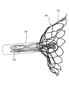
(e.g., prosthetic mitral valve) prior to inserting the prosthetic heart valve
300 into a delivery
sheath 326 (see, e.g., FIGS. 17-21) for delivery into the atrium of the heart.
The prosthetic
heart valve 300 (also referred to herein as "valve") can be constructed the
same as or similar
to, and function the same as or similar to the valves 100 and 200 described
above. Thus,
some details regarding the valve 300 are not described below. It should be
understood that
for features and functions not specifically discussed, those features and
functions can be the
same as or similar to the valve 200.
1001161 As shown in FIG. 15, the valve 300 has an outer frame 320 and an
inner frame
350. As discussed above for valves 100 and 200, the outer frame 320 and the
inner frame
350 of valve 300 can each be formed with a shape-memory material and have a
biased
expanded configuration. The outer frame 320 and the inner frame 350 can be
moved to a
collapsed configuration for delivery of the valve 300 to the heart. In this
example method of
preparing the valve 300 for delivery to the heart, the outer frame 320 of the
valve 300 is first
disposed in a prolapsed or inverted configuration as shown in FIG. 16.
Specifically, the
elastic or superelastic structure of outer frame 320 of valve 300 allows the
outer frame 320 to
be disposed in the prolapsed or inverted configuration prior to the valve 300
being inserted
into the lumen of the delivery sheath 326. As shown in FIG. 16, to dispose the
outer frame
320 in the inverted configuration, the outer frame 320 is folded or inverted
distally (to the
right in FIG. 16) such that an open free end 316 of the outer frame 320 is
pointed away from
an open free end 347 of the inner frame 350. As described above for valve 100,
in this
inverted configuration, the overall outer perimeter or outer diameter of the
valve 300 is
reduced and the overall length is increased. For example, the diameter DI
shown in FIG. 15
is greater than the diameter D2 shown in FIG. 16, and the length I.,1 (shown
in FIG. 12 for
valve 200) is less than the length L2 shown in FIG. 16 for valve 300. With the
outer frame
320 in the inverted configuration relative to the inner frame 350, the valve
300 can be placed
within a lumen of a delivery sheath 326 as shown in FIG. 17 for delivery of
the valve 300 to
the left atrium of the heart. By disposing the outer frame 320 in the inverted
configuration
relative to the inner frame 350, the valve 300 can be collapsed into a smaller
overall diameter,
i.e. when placed in a smaller diameter delivery sheath, than would be possible
if the valve 300 in
the configuration shown in FIG. 15 were collapsed radially without being
inverted. This is because
in the configuration shown in FIG. 15, the two frames are concentric or
nested, and thus the outer
frame 320 must be collapsed around the inner frame 350, whereas in the
configuration shown in
FIG. 16, the two frames are substantially coaxial but not concentric or
nested. Thus, in the
configuration shown in FIG.16 the outer frame 320 can be collapsed without the
need to
accommodate the inner frame 350 inside of it. In other words, with the inner
frame 350 disposed
mostly inside or nested within the outer frame 320, the layers or bulk of the
frame structures cannot
be compressed to as small a diameter. In addition, if the frames are nested,
the structure is less
flexible, and therefore, more force is needed to bend the valve, e.g. to pass
through tortuous
vasculature or to make tight turn in the left atrium after passing through the
atrial septum to be
properly oriented for insertion into the mitral valve annulus.
[00117] FIGS. 22-24 illustrate a portion of a procedure to deliver the valve
300 to the heart. In this
embodiment, the valve 300 is shown being delivered via a transfemoral delivery
approach as
described, for example, in the '572 PCT application. The delivery sheath 326,
with the valve 300
disposed within a lumen of the delivery sheath 326 and in an inverted
configuration as shown in
FIG.17, can be inserted into a femoral puncture, through the femoral vein,
through the inferior
vena cava, into the right atrium, through the septum Sp and into the left
atrium LA of the heart.
With the distal end portion of the delivery sheath 326 disposed within the
left atrium of the heart,
the valve 300 can be deployed outside a distal end of the delivery sheath 326.
For example, in
some embodiments, a pusher device 338 can be used to move or push the valve
300 out the distal
end of the delivery sheath 326. As shown in FIGS.22-24, a tether 336 can be
attached to the valve
300, and extend though the mitral annulus, through the left ventricle LV, and
out a puncture site
at the apex Ap. In some embodiments, the valve 300 can be moved out of the
delivery sheath 326
by pulling proximally on the tether 336. In some embodiments, the valve 300
can be deployed by
pushing with the pusher device and pulling with the tether.
[00118] As the valve 300 exits the lumen of the delivery sheath 326, the outer
frame assembly 310
exits first in its inverted configuration as shown in the progression of
FIGS.18-20 (see also FIG.
22). After the outer frame assembly 310 is fully outside of the lumen of the
delivery sheath 326,
the outer frame 320 can revert to its expanded or deployed configuration as
shown in FIG. 21, 23
and 24. In some embodiments, the outer frame 320 can revert
21
Date Recue/Date Received 2022-03-07
CA 02972966 2017-07-04
WO 2016/112085
PCT1US2016/012305
automatically after fully exiting the lumen of the delivery sheath due to its
shape-memory
properties. In some embodiments, a component of the delivery sheath or another
device can
be used to aid in the reversion of the outer frame assembly 310.In some
embodiments, the
pusher device and/or the tether can be used to aid in the reversion of the
outer frame
assembly 310. The valve 300 can continue to be deployed until the inner frame
350 is fully
deployed with the left atrium and the valve 300 is in. the expanded or
deployed configuration
(as shown, e.g., in FIG. 15 and 24). The valve 300 and the tether 336 can then
be secured to
the apex of the heart with an epicardial pad device 339 as shown in FIG. 24
and as describe
din more detail in the '572 PCT application.
1001191 FIG. 25-28 illustrate another embodiment of a prosthetic valve that
can be
moved between a biased expanded configuration and an inverted configuration in
which the
outer frame is inverted relative to the inner frame. The valve 400 can be
constructed the
same as or similar to, and function the same as or similar to any of the
valves described
herein and in the '572 PCT application, with the addition of the following
features. The
valve 400 has an outer frame 420 and an inner frame 450 coupled to the outer
frame 420. Tin
this embodiment, the outer frame 420 of the va1ve400 is formed of three
portions (e.g., three
cylinders), i.e., a first portion 427, a second portion 428, and a third
portion 429, all of which
are best shown in FIGS. 27 and 28. The portions 427, 428, 429 can be coupled
to one
another by any suitable coupling method to allow the portions 427, 428 and 429
to be moved
relative to each other. As shown best in FIGS. 27 and 29, the first portion
427 is movably
coupled to the second portion 428 via a first joint J I, and the second
portion 428 is moveably
coupled to the third portion 429 via a second joint J2. The first portion 427
of the outer frame
420 is also coupled to the inner frame 450 via a third joint .13 such that the
outer frame 420
can move (e.g., rotate or pivot) relative to the inner frame 450 as described
herein. For
example, any of the coupling methods described herein can. be used (e.g..
living hinge,
sutures, pins, tabs insertable through openings or slots, etc. or any
combination thereof) at
each of the joints JI. J2, and J3.
1001201 Although the outer frame 420 is shown and described as being famed
of three
separate portions which are joined together, in other embodiments, an outer
frame can be
formed of any suitable number of portions or cylinders (e.g., two portions,
four portions, five
portions, six portions, etc.), which can then be joined to form the outer
frame.
1001211 As shown in FIG. 26õ in use, to dispose the outer frame 420 in an
inverted
configuration relative to the inner frame 450, the outer frame 420 is folded
or inverted
22
CA 02972966 2017-07-04
WO 2016/112085
PCT1US2016/012305
distally such that the open free end portion of the outer frame 420 is pointed
in an opposite
direction as an open free end of the inner frame 450. With the outer frame 420
in the inverted
configuration, the valve 400 can be placed within a lumen of a delivery sheath
426 for
delivery of the valve 400 to the left atrium of the heart. The delivery sheath
426 can be the
same as or similar to any of the delivery sheaths described herein or in the
'572 PCT
application. In this embodiment, a pusher device 438 is movably disposed
within a lumen of
the delivery sheath 426 and removably attached to the valve 400.
Alternatively, a pusher
device similar to pusher 338 can be used that is not attached to the valve
400.
[001221 The joints (i.e., joint JI, J2 and joint J3) and outer frame
portions (i.e., the first
portion 427, the second portion 428, and the third portion 429) of the valve
400 can provide
for an easier (e.g., due to less rigidity of the outer frame 420 and/or more
focused and
selective control of the outer frame 420) and/or faster transition between an
inverted and
collapsed configuration of valve 400 and an expanded configuration, and vice
versa. Further,
the joints and portions of the valve 400 can allow the valve 400 to have a
lower profile or
footprint (e.g., occupy a smaller radial space or diameter), e.g., when
transitioning between
configurations and orientations.
[001231 In use, during reversion of the valve 400 from the inverted and
collapsed
configuration (e.g., within the delivery sheath 426) to its expanded
configuration, the first
portion 427 of the outer frame 420, the second portion 428 of the outer frame
420, and the
third portion 429 of the outer frame 420 can revert sequentially during the
procedure. In
other words, the portions 427, 428, 429 of the outer frame 420 can revert in
stages as the
outer frame 420 exits the delivery sheath 426 within the atrium of the heart.
Examples of
such stages are shown in FIGS. 26-28.
[001241 During delivery of the valve 400 from the deliver), sheath 426, as
shown by
progression in FIGS. 26-28, the second joint J2 disposed between the second
portion 428 of
the outer frame 420 and the third portion 429 of the outer frame 420 emerges
from the
delivery sheath 426, allowing the third portion 429 to begin to revert towards
an expanded or
deployed configuration (as shown, for example, in FIG. 26). Next, as the valve
400 moves
further towards the exit (e.g., a distal end opening) of the delivery sheath
426, the first joint
J1 disposed between the second portion 428 of the outer frame 420 and the
first portion 427
of the outer frame 420 emerges from the delivery sheath 426, allowing the
second portion
428 to further revert towards an expanded (as shown, for example, in FIG. 27).
Next, as the
valve 400 moves even further towards the exit of the delivery sheath 426, and
the inner frame
23
CA 02972966 2017-07-04
WO 2016/112085
PCT1US2016/012305
450 begins to emerge from the delivery sheath 426, including the third joint
J3 between the
inner frame 450 and the first portion 427 of the outer frame 420, the outer
frame 420 reverts
into its expanded configuration / size (as shown, for example, in FIG. 28).
[001251 Upon reversion of the outer frame 420 into its expanded
configuration, as
shown best in FIG. 28, the inner frame 450 can be decoupled from the pusher
device 438
and/or forced out of the delivery sheath 426 such that the inner frame 450
and/or the outer
frame 420 can expand further and be suitably seated in the native annulus of
the mitral valve.
1001261 Similar to the discussion above with respect to the valve 300, a
tether 436 (see
FIGS. 27 and 28), can be attached to the valve 400 and used to help move the
valve 400 out
of the lumen of the delivery sheath 426. As described for valve 300, the
pusher device 438
and/or the tether 436 can be used to deliver and deploy the valve 400.
1001271 FIGS. 29A-29C illustrate another embodiment of a prosthetic heart
valve 500
(also referred to herein as "valve") that can be delivered to a left atrium of
a heart in a
procedure similar to or the same as the procedures described above, the
procedures described
in the '572 PCT application, the '704 provisional application or other
delivery approach that
delivers the valve to the left atrium of the heart. Thus, some details
regarding the valve 500
and procedures performed therewith are not described herein. It should be
understood that
for features and functions not specifically discussed, those features and
functions can be the
same as or similar to the valves described above or in the '572 PCT
application. The valve
500 has an outer frame 520 and an inner frame 550 coupled to the outer -frame
520. FIG. 29A
illustrates the outer frame 520 in an inverted configuration or position
relative to the inner
frame 550 (as described above for previous embodiments). In this embodiment,
the outer
frame 520 of the valve 500 includes two portions, i.e., a first portion 527
and a second
portion 528 that can be coupled together at joints, as described in more
detail below.
1001281 Similar to the discussion with respect to the portions 427, 428,
429 of the
valve 400, and the joints 31 and J2, in this embodiment, the first portion 527
of the outer
frame 520 is coupled to the second portion 528 of the outer frame 520 via a
first joint J1, and
the first portion 527 is coupled to the inner frame 550 via a second joint 32.
In this
embodiment, first portion 527 of the outer frame 520 defines multiple
apertures 521 and the
inner frame 550 of the valve 500 defines multiple apertures 523 (see also,
e.g., the openings
of the body portion 242 of the inner frame 550 of the valve 500, described
above). As shown
best in FIG. 29C, the apertures 521 and the apertures 523 can be aligned, and
one or more
24
CA 02972966 2017-07-04
WO 2016/112085
PCT1US2016/012305
connecting members 531 (e.g., sutures or wires) are passed through the one or
more apertures
521 of the first portion 527 of the outer frame 520 and one or more apertures
523 of the inner
frame 550 to couple the first portion 527 of the outer frame 520 to the inner
frame 550 at the
second joint J2. In this manner, the inner frame 550 of the valve 500 and the
outer frame 520
of the valve 500 can be coupled together such that the outer frame 520 can
move relative to
the inner frame (e.g., rotate, pivot, articulate) between an inverted positon
relative to the inner
frame 550 and a non-inverted position in which the outer frame 520 can assume
a biased
expanded configuration with the inner frame 550 nested within the outer frame
520 as
described above for previous embodiments. .
1001291 As shown, for example, in FIG. 29B, the first joint .11 between the
first portion
527 and the second portion 528 of the outer frame 520 is formed by wrapping
one or more
connecting member 531 (e.g., suture or wire) around a strut of the first
portion 527 and
around a strut of the second portion 528. For example, the strut of the first
portion 527 and
the strut of the second portion 528 can. be aligned with one another prior to
application of the
connecting member(s) 531 thereto. In an embodiment of a valve having an outer
frame with
for example, three portions, such as valve 400, the second portion and the
third portion of the
outer frame can be coupled together using either of the coupling methods
described for joint
Ji and J2 of valve 500. While the valve 500 is shown as having a first joint
ii between the
first portion 527 and the second portion 528, and a second joint J2 between
the inner frame
550 and the outer frame 520, in some embodiments, any suitable number or type
of joints can
be used to couple any suitable number of portions of the outer frame 520
together, and the
inner frame 550 to the outer frame 520.
1001301 FIGS. 30A-42C illustrate various embodiments of a coupling joint(s)
for
coupling an inner frame to an outer frame of a prosthetic heart valve such
that the valve can
be reconfigured between a biased expanded configuration for implantation in
the heart (e.g.,
within a native mitral annulus) and a delivery configuration that has a
smaller outer perimeter
or profile (e.g., inverted configuration) as discussed above, for example,
with respect to valve
100. More specifically, the embodiments of FIGS. 30-42C illustrate various
embodiments of
different coupling joints for coupling the outer frame to the inner frame such
that the outer
frame can be moved (e.g., rotated, pivoted, flexed) relative to the inner
frame between a first
configuration and a second configuration in which the outer frame is inverted
relative to the
inner frame. Each of the embodiments of a prosthetic heart valve described
with respect to
FIGS. 30A-42C can include the same or similar features and can function the
same as or
CA 02972966 2017-07-04
WO 2016/112085
PCT1US2016/012305
similar to the prosthetic heart valves described above with respect to, for
example, valve 100,
200 and 300. Thus, some features and details of the embodiments described with
respect to
FIGS. 30A-42C are not described below.
1001311 FIGS. 3M and 30B illustrate a portion of a prosthetic heart valve,
which
includes an inner frame assembly having an inner frame 650 and an outer frame
assembly
having an outer frame 620. The prosthetic heart valve 600 (also referred to as
"prosthetic
valve" or "valve") can. be constructed similar to or the same as, for example,
the prosthetic
heart valve 100 or valve 200 described above. Specifically. FIGS. 30A and 30B
illustrate a
portion of the outer frame 620 and a portion of the inner frame 650 shown with
the outer
frame 620 in a first configuration and a second configuration, respectively.
The outer frame
620 and the inner frame 650 are coupled via a coupling joint 646. In the
illustrated
embodiment of FIGS. 30A and 30B, the coupling joint 646 includes a hinge
member 648. In
some embodiments, the hinge member 648 can include a living hinge. The hinge
member
648 can be formed of a material such that the hinge member 648 can flex or
bend and permit
movement of the outer frame 620 relative to the inner frame 650. For example,
as described
above for previous embodiments, the coupling joint 646 (i.e., hinge member
648) allows the
outer frame 620 to be moved from the first configuration as shown in FIG. 30A
to the second
configuration, as shown in FIG. 30B, in which the outer frame 620 is inverted
relative to the
inner frame 650.
1001321 The hinge member 648 can be made of any suitable material
including, but not
limited to, a polymer, an extracted natural tissue, an artificially engineered
tissue, an elastic
material (including superelastics), and/or the like. In some embodiments, the
hinge member
648 is made of the same materials as the outer frame 620 and/or the inner
frame 650. In
some embodiments, the hinge member 648 can be integrally formed and/or fused
with the
outer frame 620 and/or the inner frame 650. In other embodiments, the hinge
member 648
can be attached to the outer frame 620 and/or the inner frame 650 by any
suitable coupling
technique, including suturing, spin coating, and/or the like. As shown in FIG.
30B, when the
outer frame 620 is in the second configuration (i.e., inverted), the hinge
member 648 flexes or
bends with the outer frame 620.
[001331 FIGS. 31A and 31B illustrate a portion of a prosthetic heart valve
600' that
includes the inner frame 650 and the outer frame 620. In this embodiment, the
inner frame
650 and outer frame 620 are coupled at a coupling joint 646' that includes a
hinge member
648' that is disposed in a substantially V-shape between the inner frame 650
and the outer
26
CA 02972966 2017-07-04
WO 2016/112085
PCT1US2016/012305
frame 620. As with the previous embodiment, the hinge member 648 can be
coupled to the
outer frame 620 and the inner frame 650 in such a manner so as to allow for
the outer frame
620 to move relative to the inner frame 650 between a first configuration as
shown in FIG.
31A and a second configuration as shown in FIG 31B. The hinge member 648' can
be
formed with the same or similar materials as described above for hinge meinber
648, and can
be coupled to the inner frame 650 and the outer frame 620 with any of the
coupling
techniques described for hinge member 648. As shown in FIG. 31B, when the
outer frame
620 is moved to the second configuration (i.e., inverted), the hinge member
648' can bend or
flex with the outer frame 620.
[001341 FIGS. 32A-32C illustrate portions of a prosthetic heart valve 700.
The
prosthetic heart valve (also referred to as "prosthetic valve" or 'valve") 700
includes an outer
frame assembly 710 with an outer frame 720, and an inner frame assembly 740
with an inner
frame 750. The valve 700 also includes multiple coupling joints 746 to couple
the outer
frame 720 to the inner frame 750 such that the outer frame can be moved
between a first
configuration (as shown in FIGS. 32A-32C) and a second configuration (not
shown) in which
the outer frame 720 is inverted relative to the inner frame 750. As best
illustrated in FIGS.
32B-32C, each coupling joint 746 includes a support member 721 that defines
two slots 722A
and 72213. and corresponding coupling portions 755 of the inner frame 750 that
include tabs
761A and 76IB. Also defined by the tabs 761A and 7618 are holes 756A and 756B.
1001351 To couple the outer frame 720 to the inner frame 750, the tabs 761A
and 761B
are inserted into the corresponding slots 722A and 722B of the outer frame 720
as shown in
FIGS. 32A and 32C. Thus, the tab 756A is received within the slot 722A and the
tab 756B is
received within the slot 7228 as best shown in FIG. 32C. In this manner,
mechanical
attachment can be achieved between the outer frame 720 and the inner frame
750. In some
embodiments, the support member 721 can be formed integrally with the outer
frame 720. In
such an embodiment, the outer frame 720 (and support member 755) can be made
of any
suitable, flexible material (e.g., a shape memory metal such as Nitinol) that
permits
articulation between the frame portions of the outer frame 720, such as frame
portions 717
and 718 and the support member 721 such that the outer frame 720 can be moved
between
the first configuration and the second configuration (i.e., inverted). In some
embodiments, the
frame portions (e.g., frame portions 717, 718) can be formed separately from
the support
member 721 and can be coupled to the support member 721 with a pivot joint
such as a ball
and socket. In such an embodiment, the outer frame 720 can pivot relative to
the support
27
CA 02972966 2017-07-04
WO 2016/112085
PCT1US2016/012305
member 721 when moved between the first configuration and the second
(inverted)
configuration.
[001361 The interlocking nature of the tabs 761A and 761B within the slots
722A,
7228 provides for load bearing without additional components such as sutures.
In some
embodiments, as illustrated, the tabs 761A, 761B can include the holes 760A,
760B that can
receive a stop member (not shown) such as, for example, a pin and/or other
component to
prevent the tabs 761A, 761B from coming out of the slots 722A, 722B. In some
embodiments, one or more strands or suture can be used as the stop member. For
example,
the suture strands can be threaded through the holes 756A, 756B and/or wrapped
about the
tabs 761A, 76113. As best illustrated in FIGS. 3213-32C, the coupling portion
755 of the inner
frame 750 can also include one or more holes 757 for aiding in attachment
(e.g., via sutures)
of other components, such as, for example, the covering 232 illustrated in
FIG. 3.
[001371 Although two tabs 761A, 76IB and two slots 722A, 722B are included
at
coupling joints 746, it should be understood that variations from the
illustrated embodiment
in FIGS. 32A-32C are within the scope of this disclosure. For example, any
suitable number
of tabs and slots can be employed to attach the outer frame 720 and the inner
frame 750,
including a single tab/slot mechanism. As another example, any suitable
geometry of the
support member 721 and/or the coupling portion 755 can be employed.
1001381 To deliver the valve 700 to a heart of a patient, in some cases it
may be
desirable to deliver the outer frame 720 and the inner frame 750 prior to
being coupled
together at the coupling joints 746. In such an embodiment, the outer frame
720 and the
inner frame 750 can be loosely coupled with a suture strand 753 as shown in
FIG. 33A and
33B. The suture strand 753 is threaded through the opening 756 of the coupling
portion 755
of the inner frame 750 and through the slot 722 of the support member 721 of
the outer frame
720. Although a single suture strand 753 is shown, it should be understood
that a suture
strand 753 can be disposed through each of the slots 722 of the outer frame
720 and the
openings 756 of the inner frame 750. With the inner frame 750 coupled to the
outer frame
720 via the suture strand 753, the outer frame 720 can be inverted separately
from the inner
frame 750 and then loaded into a delivery- sheath (not shown) along with the
inner frame 750
such that the inner frame 750 is disposed proximal of the outer frame 720
within the delivery
sheath. As shown in FIG. 33A, a clip 766 and a stop element 767 are coupled to
the suture
strand 753 and can be used to secure the inner frame 750 to the outer frame
720 as described
in more detail below.
28
CA 02972966 2017-07-04
WO 2016/112085
PCT1US2016/012305
[001391 More specifically, to deploy the valve 700 into the heart of a
patient, a distal
end portion of the delivery sheath is disposed within the left atrium and the
outer frame 720 is
moved outside of the distal end of the delivery sheath and into the left
atrium. Upon being
moved outside of the delivery sheath, the outer frame 720 can revert and
assume a biased
expanded configuration. The inner frame 750 can be moved into a nested
position within the
outer frame 720 and the tabs 761A and 761B can be disposed within
corresponding slots
722A and 722B of the support member 721 of the outer frame 720. The suture
strand 753
can be pulled proximally such that the stop element 767 (coupled to the suture
strand 753) is
pulled against a distal side of the support member 721, and the clip 766 can
be slid distally
toward the support member 721 to secure the inner frame 750 to the outer frame
720. The
clip 766 can be capable of sliding along the suture strand 753 while having
sufficient friction
force to hold the clip 766 to the suture strand 753 in a desired location.
1001401 FIG. 33B illustrates a variation to the above embodiment in that
the suture
strand 753 does not have a clip or stop element coupled thereto. In this
example
embodiment, the suture strand 753 is threaded through the slot 722 of the
support member
721 from a distal side, through the opening 759 of the tab 761, and then back
through the slot
722 from a proximal side such that the two end portions of the suture strand
753 extend from
the distal side of the support member 721. The two end portions of the suture
strand 753 can
extend through a gap between the native mitral annulus, through the left
ventricle and out of
the apex of the heart. For example, a procedural catheter can be inserted
through a puncture
site at the apex and the two ends of the suture strand 753 can extend through
a lumen of the
procedural catheter. The outer frame 720 and the inner frame 750 can be
deployed as
described for FIG. 33A, and then the two suture end portions can be pulled
taut to secure the
tab 756 within the slot 722. For example, the two suture end portions can be
tied to form a
knot on the distal side of the support member 721 or otherwise secured in
place to prevent the
tab 761 from pulling out of the slot 722.
[001411 FIGS. 34A and 34B illustrate a portion of an. outer frame 820 and a
portion of
an inner frame 850 of a prosthetic heart valve 800, according to an
embodiment. The outer
frame 820 is coupled to the inner frame 820 at a coupling joint 846 such that
the outer frame
820 can be moved (e.g., rotated, pivoted) between a first configuration
relative to the inner
frame 850, as shown in FIG. 34A, and a second configuration relative to the
inner frame 850,
as shown in FIG. 34B, in which the outer frame 820 is inverted relative to the
inner frame
850. In this embodiment, the coupling joint 846 includes holes 856A and 856B
defined by
29
CA 02972966 2017-07-04
WO 2016/112085
PCT1US2016/012305
the inner frame 850 and a suture 858 that is threaded or wrapped through the
holes 856A and
856B. More specifically, a portion of the outer frame 820 is disposed against
a portion of the
inner frame 850 and the suture 858 is wrapped around the portion of the outer
frame 820 and
through the holes 856A and 856B. One or more sutures 858 can be used and/or
the suture(s)
858 can be wrapped multiple times around the portion of the outer frame 82()
and through the
holes 856A and 856B. The suture 858 can be made of any suitable material,
including shape
memory polymers, polyester, tissue, and/or the like. In some embodiments (not
shown), an
interface layer of Nitinol or other suitable material can be disposed between
the outer frame
820 and the inner frame 850 to prevent abrasive contact. Such an interface
material is
described with respect to valve 1100.
1001421 As also illustrated in FIGS. 34A and 3413, the outer frame 820
includes
protrusions 857A, 85713 formed thereon. The protrusions 857A, 8578 are sized
and located
such that, after the outer frame 820 and the inner frame 850 are coupled with
the suture 858,
the protrusion 857A is located on one side of the suture 858, and the
protrusion 8578 is
located on the opposite side of the suture 858. In this manner, the outer
frame 820 has
limited lateral mobility with respect to the inner frame 850 due to the suture
858 being
constrained between the protrusions 857A, 8578.
1001431 In some embodiments. , the position of the protrusions 857A, 85713
can be
manipulated to affect the desired lateral mobility of the outer frame 820. In
some
embodiments, while the holes 856A, 856B are illustrated as positioned to have
a distance
between them that is greater than the diameter/width of the outer frame 820,
in some
embodiments, the outer frame 820 can have a greater diameter/width than the
distance
between the holes 856A, 8568. In other words, the outer frame 820 overlaps a
portion of the
holes 856A, 856B.
1001441 FIG. 35 illustrates a portion of an outer frame 920 and a portion
of an inner
frame 950 of a prosthetic heart valve 900, according to another embodiment.
The outer
frame 920 is coupled to the inner frame 920 at a coupling joint 946 such that
the outer frame
920 can be moved (e.g., rotated, pivoted) between a first configuration
relative to the inner
frame 950, as shown in FIG. 35, and a second configuration relative to the
inner frame 850
(not shown), in which the outer frame 920 is inverted relative to the inner
frame 950. In this
embodiment, the coupling joint 946 includes a pin assembly 925 that can be
received through
through-holes 933A and 9338 defined by frame portions 917 and 918,
respectively, of outer
frame 920 and through a corresponding through-hole 956 defined by the inner
frame 950.
CA 02972966 2017-07-04
WO 2016/112085
PCT1US2016/012305
[001451 The pin 925 includes stops 934 disposod on the ends to prevent the
pin 925
from being removed. One or both of the stops 934 can be removably coupled to
the ends of
the pin 925. For example, the stops 934 can be coupled to the pin 925 in any
suitable manner
including, but not limited to, threaded attachment, compression fit, adhesive
(including
pressure sensitive adhesives) and/or the like. The diameter/width of the pin
925 is less than
the diameter/width of the through-hole 956 and the diameter/width of the
through-holes 933A
and 933B, and the diameter/width of the stops 934 is greater than the
diameter/width of the
through-hole 956 and the diameter/width of through-holes 933A, 93313. In this
manner, the
stops 934 can restrict lateral movement between the outer frame 920 and the
inner frame 950,
but permit the relative rotation of the outer frame 920 about an axis defined
by the length of
the pin 925.
[001461 In some embodiments, the pin 925 can be made of any suitable rigid
material,
such as metal. In other embodiments, the pm 925 can be made of any suitable
flexible
material, such as a polymer. In some embodiments (not shown), sutures are
employed
instead of the pin assembly 958 to restrict lateral movement between the outer
frame 920 and
the inner frame 950.
[001471 FIG. 36 illustrates a portion of an outer frame 1020 and a portion
of an inner
frame 1050 of a prosthetic heart valve 1000, according to an embodiment. The
outer frame
1020 is coupled to the inner frame 1050 at a coupling joint 1046 such that the
outer frame
1020 can be moved (e.g., rotated, pivoted) between a first configuration
relative to the inner
frame 1050, as shown in FIG. 36, and a second configuration relative to the
inner frame 1050
(not shown), in which the outer frame 1020 is inverted relative to the inner
frame 1050. The
coupling joint 1046 is described in detail below.
1001481 in this embodiment, the inner frame 1050 includes a loop structure
1051 that
includes arms 1051A, 1051B that define an opening 1015 between the arms 1051A,
1051B.
The loop structure 1051 also includes through-holes 1056A, 1056B defined by
arms 1051A,
1051B, respectively.
[001491 The outer frame 1020 includes a stub portion 1013 sized to be
disposable
within the opening 1015 of the inner frame 1050and defines a through-hole 1033
configured
to be substantially aligned with the through-holes 1056A, 10568 of inner frame
1050.
[001501 To couple the outer frame 1020 to the inner frame 1050, a pin 1025
is inserted
through the through-hole 1056A, the through-hole 1033 and the through-hole
105613. Similar
CA 02972966 2017-07-04
WO 2016/112085
PCT1US2016/012305
to the pin 925, and as illustrated in FIG. 36, the pin 1025 can include stops
1034 on ends
thereof to prevent the pin 1025 from being removed. The pin 1025 and the stops
1034 can be
constructed the same as or similar to the pin 925 and stops 934 described
above. The
diameter/width of the pin(s) 1025 is less than the diameter/width of the
through-holes 1056A,
1056B and the diameter/width of the through-hole 1033, while the
diameter/width of the
stops 1034 is greater than the diameter/width of the through-holes 1056A,
1.056B and the
diameter/width of the through-hole 1033. In this manner, the pin 1025 can
restrict lateral
movement between the outer frame 1020 and the inner frame 1050, but permit the
relative
rotation of the outer frame 1020 about an axis defined by the length of the
pin 1025.
1001511 FIG. 37 illustrates a portion of an outer frame 1120 and a portion
of an inner
frame 1150 of a prosthetic heart valve 1100, according to an embodiment. The
outer frame
1120 is coupled to the inner frame 1120 at a coupling joint 1146 such that the
outer frame
1120 can be moved (e.g., rotated, pivoted) between a first configuration
relative to the inner
frame 1150, as shown in FIG. 37, and a second configuration (not shown)
relative to the inner
frame 1150, in which the outer frame 1120 is inverted relative to the inner
frame 1010. In
this embodiment, the coupling joint 1146 includes sutures attached to the
inner frame 1150
and to the outer frame 1120 via holes defined in each as described in more
detail below.
[001521 More specifically, the inner frame 1150 defines an opening 1156.
The outer
frame 1150 includes frame portions 1117 and 1118 disposed on each side of the
inner frame
1150. The frame portions 1117 and 1118 each define a hole 1133A and 1133B,
respectively.
1001531 As shown in FIG. 37, a suture portion 1158A is threaded or looped
through the
opening 1133A and the opening 1156 and a suture portion 11588 is threaded or
looped
through the opening 1133B and the opening 1156. The suture portions 1158A,
1158B can be
tied or otherwise secured in position. The suture portions 1158A, 1158B thus
secure the
frame portions 1117 and 1.118 to the inner frame 1150, while allowing motion
of the frame
portions 1117 and 1118 relative to the inner frame 1150 to move the outer
frame 1120
between the first configuration and the second inverted configuration. The
sutures 1158A,
1158B provide for a hinge-like effect. It is understood that while sutures
1158A, 1.158B are
shown to be independent of each other, any suitable suture pattern can be
employed, such as
the use of a single suture running through the opening 1156 and the openings
1133A, 1133B,
a suture running between the openings 1133A and 1133B, combinations thereof,
and/or the
like. Furthermore, each of the suture portions 1158A and 11588 can include one
or more
suture strands and/or one or more wraps of the suture portion through the
openings.
32
CA 02972966 2017-07-04
WO 2016/112085
PCT1US2016/012305
[001541 FIG. 37 also illustrates interface members 1149A, 1149B that can be
disposed
between the inner frame 1150 and the frame portions 1117 and 1118,
respectively, of outer
frame 1120. The interface members 1149A, 11493 are sized and located to
substantially
prevent abrasive contact between the inner frame 1150 and the outer frame
1120, and/or to
substantially prevent slippage of the outer frame 1120. In some embodiments
(not shown),
the interface members I I49A, 1149B can be continuous with each other, and can
encircle the
inner frame 1150, save for openings consistent with the opening 1156. The
interface
members 1149A, 1149B can be formed of any suitable material, such as, for
example,
polyester.
[001551 FIG. 38 illustrates a portion of an outer frame 1220 and a portion
of an inner
frame 1250 of a prosthetic heart valve 1200, according to an embodiment. The
outer frame
1220 is coupled to the inner frame 1250 via coupling joints 1246. This
embodiment is
similar to the previous embodiment in that the coupling portion 1246 includes
sutures
attached to holes in the outer frame 1220 and holes in the inner frame 1250.
[001561 More specifically, the inner frame 1250 defines a pair of openings
1256A and
12563. The outer frame 1250 includes frame portions 1217 and 1218 disposed on
each side
of the inner frame 1250. The frame portion 1217 defines a pair of openings
1233A and
1233A' and the frame portion 1218 defines a pair of openings 1233B and 1233B'.
In this
embodiment, a suture portion 1258 is threaded or looped through the openings
1233A,
1133A' then a portion of the suture 1258 is inserted through a front side of
the opening
1256A, back through the opening 12563, into the opening 12333 and looped
through the
opening 1233W. The suture 1258 can be tied or otherwise secured in position.
[001571 The suture 1258 thus secures the frame portions 1217 and 1218 to
the inner
frame 1250, while allowing motion of the frame portions 1217 and 1218 relative
to the inner
frame 1250 to move (e.g., rotate, pivot) the outer frame 1220 between a first
configuration (as
shown in FIG. 38) and a second inverted configuration (not shown). The single
suture 1258
thiough openings 1256A and 1256B may help reduce binding during movement of
the outer
frame 1220. Although a single suture 1258 is shown, it should be understood
that the suture
1258 can include one or more strands of suture material. In addition, any
suitable suture
pattern can alternatively be employed and the suture 1258 one or more wraps of
the suture
through the openings. Also shown in FIG. 38, interface members 1249A and 1249B
can be
included to prevent abrasive contact between the inner frame 1250 and the
outer frame 1220.
33
CA 02972966 2017-07-04
WO 2016/112085
PCT1US2016/012305
The interface members 1249A, 1249B can be the same as the interface members
1149A,
1149B described above.
[001581 FIG. 39 illustrates a portion of an outer frame 1320 and a portion
of an inner
frame 1350 of a prosthetic heart valve 1300, according to another embodiment.
The outer
frame 1320 is coupled to the inner frame 1350 via coupling joints 1346. In
this embodiment,
the coupling joint 1346 includes a hinge member 1348 and a tether assembly
1365 as
described in more detail below.
[00159i The hinge member 1348 can be the same as or similar to the hinge
member
648 described above for valve 600. The hinge member 1348 can be flexible to
allow the
outer frame 1320 to move relative to the hitter frame 1350 between the first
configuration and
the second inverted configuration. In some embodiments, the hinge member 1348
can limit
the plane of motion of the outer frame 1320 with respect to the inner frame
1350. The hinge
member 1348 can function, for example, as a living hinge.
1001601 As shown in FIG. 39, the tether assembly includes a tether 1368, a
stop
member 1367 and an optional clip 1366. The tether 1368 can be received through
a through-
hole 1356 defined by the inner frame 1350 and a through-hole 1333 defined by
the outer
frame 1320. Similar to as described above with respect to the suture 753 for
valve 700 (FIG.
33A), the stop member 1367 can be disposed on an end of the tether 1368 and
the clip 1368
can be slidably coupled to the tether 1368. The stop member 1367 can be for
example, a knot
formed on the end of the tether 1368, or any other suitable stop member with a
size/diameter
larger than the through-holes, 1356 and 1333. The clip 1366 can be configured
to be slidable
along the tether 1368, yet have sufficient friction to maintain a desired
position along the
tether 1368. In some cases, a clip 1366 may not be desired. For example, when
the clip 1366
is disposed toward the apex side of the valve, as shown in FIG. 39,
unidirectional forces on
the tether 1368 can be sufficient to maintain the stop member 1367 against the
outer side
portion of the outer frame 1320. Thus, the clip member 1366 can optionally be
excluded.
[001611 The tether 1368 can be, for example, a flexible member similar to
the tether
290 described above for valve 200. The tether 1368 can be formed with any
suitable material
such as, fbr example, a braided string/thread. The stop member 1367 is
configured to be
disposed outside the through-hole 1356 and the through-hole 1333, on an outer
side of the
outer frame 1320. The clip 1366 is disposed on an outer side of the inner
frame 1350 and can
be configured to be slidable along the tether 1368, yet have sufficient
friction to maintain its
34
CA 02972966 2017-07-04
WO 2016/112085
PCT1US2016/012305
position along the tether 1368. Thus, the inner frame 1350 and the outer frame
1320 can be
disposed between the stop member 1367 and the clip 1366.
[00162j In use, the tether 1368 can be pulled in a direction of arrow A
such that the
stop member 1367 is pulled against the outer side of the outer frame 1320, and
the clip 1366
can be slid up against the outer side of the inner frame 1350. Thus, although
the hinge
member 1348 can couple the inner frame 1350 to the outer frame 1320 in a
similar manner as
described for valve 600, the tether 1368 can be used to provide further
securement without
interfering with the ability of the outer frame 1320 to rotate or pivot about
the hinge member
1348. In addition, the tether 1368 can be used in place of the proximal struts
of the valve
1300 (see, e.g., struts 243A in FIG. 6 and 8).
[001631 In some embodiments, the tether assembly 1365 can be used alone to
couple
the inner frame 1350 to the outer frame 1320, without the hinge member 1348.
In such an
embodiment, as described for valve 700 with respect to FIGS. 33A and 333, the
outer frame
1320 and inner frame 1350 can be delivered prior to being securely coupled
together. For
example, the outer frame 1320 and the inner frame 1350 can be loosely coupled
via the tether
assembly 1365 and be loaded into a delivery sheath (not shown) with the outer
frame inverted
upon itself and disposed distal of the inner frame 1350. A distal end portion
of the delivery
sheath can be disposed within the left atrium and the outer frame 1320 can be
moved outside
of the delivery sheath and into the left anium. Upon being moved outside of
the delivery
sheath, the outer frame 1320 can reven and assume a biased expanded
configuration. The
inner frame 1350 can be moved into a nested position within the outer frame
1320 and the
tether 1368 can be pulled such that the stop member 1367 is pulled against the
outer side of
the outer frame 1320 and the clip 1366 can be slid toward the outer side of
the inner frame
1350 to secure the inner frame 1350 to the outer frame 1320.
100164] FIG. 40 illustrates a portion of an outer frame 1420 and a portion
of an inner
frame 1450 of a prosthetic heart valve 1400, according to some embodiments. In
this
embodiment, the outer frame 1420 is coupled to the inner frame 1450 via a
coupling joint
1446 that includes a hinge member 1448 that can be the same as or similar to
and function the
same as or similar to the hinge member 648 of valve 600. The valve 1400 also
includes a
tether assembly 1465 that includes a tether 1468 and a stop member 1467. As
shown in FIG.
40, in this embodiment, the tether 1468 is disposed through only a through-
hole 1433 defined
by the outer frame 1420. Thus, the tether assembly 1468 in this embodiment
serves only to
replace the proximal struts of the valve 1400.
CA 02972966 2017-07-04
WO 2016/112085
PCT1US2016/012305
(001651 FIGS. 41A-41K illustrate a portion of a prosthetic heart valve
1500, =cording
to an embodiment. The prosthetic heart valve 1500 (also referred to as
"prosthetic valve" or
"valve") includes an inner frame assembly 1540 having an inner frame 1550, and
an outer
frame assembly 1510 having an outer frame 1520 (see FIGS. 41(1-411). The outer
frame
1520 is coupled to the inner frame 1550 via a coupling joint 1546 (see FIGS.
410-411). In
this embodiment, the coupling joint 1546 incudes sutures wound about the inner
frame 1550
and the outer frame 1520 as described in more detail below.
1001661 As shown in FIGS. 418 and 41C, which are detail side and front
views,
respectively, of encircled area A in FIG. 41A, frame portions of the inner
frame 1550 define a
pair of openings 1556A and 1556B. Each of the through-holes 1556A, 1556B is
generally in
a half-circle shape, although other shapes are within the scope of the
disclosure. For
example, FIGS. 42A-42C illustrate an example embodiment of an inner frame 1650
having
through-holes 1656A, 1656B that are generally round. As shown in FIGS. 42B and
42C, a
suture 1658 can be wound about the inner frame 1650 through through-holes
1656A and
1656B in a similar manner as described below for valve 1500.
1001671 As shown in FIGS. 41E and 4IF, a portion of suture 1558 is wound
through
the through-holes 1556A, 15568 and around a portion of the inner frame 1550
disposed
between the through-holes 1556A, 1556B. The half-circle shape of the through-
holes 1556A,
15568 provides a relatively flat surface such that when the suture 1558 is
wound about the
portion of the inner frame 1550, a relatively flat platform is provided for
interfacing with
additional sutures used to attach the outer frame 1550 to the inner frame 1550
(see FIGS.
400-401, described in detail later). .
1001681 Referring to FIGS. 41G-411, which illustrate the outer frame 1520
coupled to
the inner frame 1550, the frame members of the outer frame 1520 define a
through-hole 1533
through which a second suture .1558' is received. More specifically, the
second suture 1558'
is wound through the through-hole 1533, around an end of the outer frame 1520
and then
wound through the through-holes 1556A and 1556B of the inner frame 1550, as
best shown
in FIG. 41H. The second suture 1558' can be wound to form multiple layers of
windings that
run through the through-the 1533, and the through-holes 1556A, 15568. Thus the
suture
1558' is disposed over portions of the suture 1558 that is wound about the
inner frame 1550
within the through-holes 1556A, 1556B, providing frictional, non-sliding
contact. The end
portion of the outer frame 1520 abuts the portion of the suture 1558 disposed
on an exterior
surface of the inner frame 1550 as shown in FIG. 411-1. In this manner, the
suture 1558
$6
CA 02972966 2017-07-04
WO 2016/112085
PCT1US2016/012305
prevents abrasive contact between the outer frame 1520 and the inner frame
1550, while
permitting the outer frame 1520 to move (e.g., pivot, rotate) between the
first configuration
and the second inverted configuration relative to the inner frame 1550. The
suture 1558
and/or the suture 1558 can independently be made of any suitable material
including, but not
limited to, nylon, polyester, polyvinylidene fluoride (PVDF), polypropylene,
combinations
thereof, and/or the like.
[001691 FIGS. 43-48 illustrate an alternative method of delivering a
prosthetic valve
within an annulus of a heart via a transfemoral delivery approach. As shown in
FIG. 43, a
procedural catheter 1735 is inserted through an apical puncture (e.g., a .5F
apical puncture) in
a ventricular wall at the apex Ap of the heart H. A guide wire 1737 is
inserted through a
lumen (not shown) of the procedural catheter 1735 and extended through the
left ventricle
LV, through a rnitral valve gap and into the left atrium LA. A delivery sheath
1726 is
introduced through a femoral vein puncture and extended through the inferior
vena cava, into
the right atrium., and then through a transseptal puncture of the septum Sp of
the heart H., and
into the left atrium LA of the heart H. A snare device 1719 is movably
disposed within the
delivery sheath 1726 and used to grab or snare a distal end portion of the
guide wire 1737, as
shown in FIG. 43. The snare device 1719 can be used to pull the guide wire
1737 through the
delivery sheath 1726 such that the distal end portion of the guide wire 1737
extends outside
the femoral vein and a proximal end of the guide wire 1737 is disposed through
the
ventricular wall at the apex Ap of the heart H. as shown in FIG. 44. Although
not shown in
FIGS. 43 and 44, the procedural catheter 1735 is disposed outside the
patient's body, the
distal end of the guide wire 1737 extends outside the femoral vein and outside
the patient's
body, and the proximal end of the guide wire 1737 extends outside the apex Ap
and outside
the patient's body. Although the above described snare process describes
delivering the
guide wire 1737 to the left atrium of the heart and then snaring the guide
wire 1737 using the
snare device 1719, in alternative embodiments, the guide wire 1737 can be
delivered to the
left ventricle LV and the snare device 1719 and delivery sheath 1726 can be
inserted through
the mitral annulus and into the left ventricle LV to grab or snare the guide
wire 1737 as
described above.
[001701 After the guide wire 1737 has been extended between the apex Ap and
the
access site to the femoral vein, the delivery sheath 1726 can be removed. A
leader tube 1724
is loaded over the guide wire 1737 starting outside the heart (and outside the
procedural
catheter 1735) and exiting the femoral vein at the femoral puncture site as
shown in FIG. 45.
37
CA 02972966 2017-07-04
WO 2016/112085
PCT1US2016/012305
As shown in FIG. 45, the leader tube 1724 includes a balloon dilator member
1711 that is
inserted into a distal end of the delivery sheath 1726 and disposed partially
over a distal end
portion of the prosthetic valve 1700. For example, the balloon dilator member
1711 can have
a collapsed or uninflated configuration (not shown) for delivery over the
guide wire 1737 and
can then be inflated or otherwise moved to an expanded con-figuration as shown
in FIG. 45.
Also shown in FIG. 45, a pusher 1738 is disposed within the lumen of the
delivery sheath
1726 and can be used to move or push the prosthetic valve 1700 into the left
atrium LA, as
described in more detail below. With the leader tube 1724 disposed between the
femoral
puncture site and the apex of the heart, the guide wire 1737 can be removed.
Although not
shown in FIGS. 45-47, the procedural catheter 1735 remains inserted into the
left ventricle
LV of the heart as shown in FIGS. 43 and 44.
[001711 The prosthetic valve 1700 can be configured the same as or similar
to the
prosthetic valves described herein. The prosthetic valve 1700 (shown
schematically within
the delivery sheath 1726 in FIG. 45) can be disposed in an inverted
configuration within the
delivery sheath 1726 to reduce the overall outer perimeter of the prosthetic
valve 1700. A
tether 1736 is coupled to a distal end portion of the prosthetic valve 1700
(see FIGS. 47 and
48). The tether 1736 can be threaded through the leader tube 1724 prior to the
leader tube
1724 being disposed within the distal end of the delivery sheath 1726. For
example, as
previously described, the tether 1736 can include a valve leader member (not
shown) . The
valve leader member can have a tapered distal end to aid in the insertion and
maneuvering of
the valve leader member through the leader tube 1724. The valve leader member
can be
attached at a proximal end portion of the tether 1736, which is attached to
the valve 1700.
The tether 1736 can be formed, fbr example, as a braided rope or cord. The
tether 1736 can
be threaded through the leader tube 1724 with the valve leader member extended
out the apex
of the proximal end of the leader tube 1724 outside the apex of the heart.
Thus, the tether
1736 extends between the apex Ap and the femoral puncture site where it is
coupled to the
valve 1700.
1001721 The delivery sheath 1726 can then be inserted through the femoral
puncture
site and moved through the femoral vein, through the inferior vena cava, into
the right atrium,
and then through the septum Sp until a distal end portion of the delivery
sheath 1726 (with
the valve 1700) is disposed within the left atrium LA, as shown in FIG. 46.
The dilator
balloon member 1711 can provide a smooth lead-in to assist in maneuvering the
distal end of
the delivery sheath 1726 through the femoral vein and within the heart.
Although the
38
CA 02972966 2017-07-04
WO 2016/112085
PCT1US2016/012305
delivery sheath 1726 is used to deliver both the snare device 1719 and the
valve 1700, in
other embodiments, a different delivery sheath can be used to deliver the
snare device 1719
than is used to deliver the valve 1700.
1001731 With the distal end of the delivery sheath 1726 within the left
atrium LA, the
leader tube 1724 can be removed through the apex Ap, leaving the tether 1736
extended
between the valve 1700 and outside the apex Ap of the heart (see FIG. 47). For
example, the
balloon dilator member 1711 can be moved back to a collapsed configuration for
removal
through the procedural catheter 1735. The procedural catheter 1735 can then
also be
removed. The pusher 1738 can be used to push or move the valve 1700 out the
distal end of
the delivery sheath 1726 and within the left atrium LA of the heart as shown
in FIG. 47. As
the valve exits the distal end of the delivery sheath 1726 the valve 1700 can
revert and return
to its original undeformed shape as described above, for example, for valve
200. For
example, the valve 1700 can be formed with a shape-memory material and can
have a biased
undeformed shape and can be manipulated and/or deformed (e.g., compressed
and/or
expanded) and, when released, return to its original undefonned shape. The
valve can be, for
example, a valve constructed the same as or similar to, and function in the
same or similar
manner as, the prosthetic heart valve 200, described above.
1001741 As shown in FIG. 47, the tether 1736 extends from the valve 1700
through the
apical puncture and outside the patient's body. As the delivery sheath 2026 is
advanced, the
tether 1736 can optionally be pulled at the apex end to help move the delivery
sheath 1726,
with the valve 200 disposed therein, through the femoral vein, through the
septal puncture
and into the left atrium LA. The valve 1700 can then be fully deployed within
the left atrium
LA, as shown in FIG. 48, by using the pusher 1738 described above and/or by
pulling the
apex end portion of the tether 1736 until the valve 1700 is pulled out of the
lumen of the
deliveiy sheath 1726 and disposed within the left atrium LA.
1001751 In some embodiments, the pusher 1738 can also be used to aid in
positioning
the valve 1700 in a desired radial orientation within the left atrium LA. For
example, the
pusher device 1738 can define an internal lumen (not shown) that can be placed
over an inner
frame portion of the valve 1700 to hold the inner frame portion in a small
diameter, which
can help enable the valve 1700 to be positioned in a desired radial
orientation and be seated
within the annulus of the mitral valve. Further examples of such a valve
assist device are
described below with reference to FIGS. 49-51.
39
CA 02972966 2017-07-04
WO 2016/112085
PCT1US2016/012305
(001761 As shown in FIG. 48, as the valve 1700 is deployed within the left
atrium LA,
the valve 1700 is allowed to assume its biased expanded or deployed
configuration. The
delivery sheath 1726 can then be removed from the patient and the valve 1700
can be
positioned and tensioned using the tether 1736 to obtain the desired or
optimal location in the
native mitral annulus and minimize perivalvular leaks. An epicardial pad
device 1739 (as
described above) can be used to secure the tether 1736 and valve 1700 in
position within, the
mitral annulus. In some embodiments, rather than securing the prosthetic
mitral valve with a
tether and epicardial pad, the prosthetic mitral valve can be secured with
clips or other
coupling methods to a portion(s) of the mitral valve apparatus and/or the
ventricular wall of
the heart.
1001771 FIGS. 49 and 50 illustrate an optional wire assist structure that
can be used
during a procedure to deliver a prosthetic heart valve, for example,
transfemorally as
described above for previous embodiments. A wire assist structure 1801 can be
releasably
coupled to a valve 1800 as shown in FIG. 49. The valve 1800 can be constructed
the same as
or similar to, and function the same as or similar to, the valves described
above for previous
embodiments. For example, the valve 1800 can include an outer frame 1820 and
an inner
frame 1850. The wire assist structure 1801 can be releasably coupled to the
inner frame 1850
as shown in FIGS. 49 and 50. For example, releasable connectors (not shown)
can be used to
couple the wire assist structure 1801 to the inner frame 1850.
1001781 in use, the wire assist structure 1801 can be movably disposed
within a
delivery sheath 1826 used to deliver the valve 1800 to the heart. The wire
assist structure
1801 can hold the inner frame 1850 and provide positioning control of the
valve 1800 (i.e.,
clocking and advancement) while the outer frame 1850 of the valve 1800 is
fully expanded,
which allows the valve 1800 to be functioning during the positioning phase.
When the valve
1800 is in the desired final position, the wire assist structure 1801 can be
released from the
inner frame 1850 and removed with the delivery sheath 1826.
1001.791 FIG. 51 illustrates another optional assist member that can be
used during a
procedure to deliver a prosthetic heart valve transfemorally. An assist member
1902 can be
in the form of a tubular member defining a lumen with a diameter sized to
receive at least a
portion of the inner frame 1950 of a valve 1900. The valve 1900 can be
constructed the same
as or similar to, and function the same as or similar to, the valves described
above for
previous embodiments. For example, the valve 1900 can include an outer frame
(not shown)
and the inner frame 1950 as described above for previous embodiments.
CA 02972966 2017-07-04
WO 2016/112085
PCT1US2016/012305
(00180) in use, the assist member 1902 can be movably disposed within a
delivery
sheath (not shown) used to deliver the valve 1900 and be disposed over at
least a portion of
the inner valve assembly (e.g., inner frame .1950). As with the wire assist
structure 1801, the
assist member 1902 can hold the inner frame 1950 in a small compact
configuration and
allow for positioning control of the valve 1900 (i.e., clocking and
advancement) while the
outer frame of the valve 1900 is being expanded. This can in some cases allow
the valve
1900 to be functioning (or at least partially functioning) during the
positioning phase of the
valve 1900. With the inner frame 1950 held in a compact or small diameter form
factor, the
valve 1900 can be more easily positioned to help seal the ann.ulus with the
outer frame (not
shown) of the valve 1900. When the valve 1900 is in the desired final
position, the assist
member 1902 can be removed.
[001811 FIG. 52 is a schematic illustration of delivery system that can be
used to
deliver and deploy a prosthetic heart valve within a heart of patient with,
for example, a
tran.svascular approach. In this embodiment, a delivery system 2005 includes a
delivery
sheath 2026, a valve holder 2038 (also referred to as a "pusher"), and one or
more actuation
wires 2074 and 2076. In this schematic illustration, only two actuation wires
are illustrated,
but in other embodiments, only one actuation wire or more than two actuation
wires can be
used.
[001821 The delivery sheath 2026 can be used to deliver a valve 2000 that
includes an
inner valve assembly 2040 including an inner frame (not labeled in FIG. 52)
and an outer
valve assembly 2010 including an outer frame (not labeled in FIG. 52). The
valve 2000 can
be constructed the same as or similar to, and function the same as or similar
to, for example,
the prosthetic valves described herein (e.g., valves 100, 200, 300, 400, etc.)
and can be
moved between a deployed or expanded configuration and a delivery
configuration in which
the outer frame is disposed in an inverted positon relative to the inner frame
as described
above. As shown in FIG. 52, the valve 2000 can be disposed within a lumen of
the delivery
sheath 2026 when the valve is in the delivery configuration (i.e., the outer
frame is inverted
relative to the inner frame). The valve holder 2038 is coupled to the inner
valve assembly
2040 and the actuation wires are coupled to the outer fame assembly 2010. The
valve holder
2038 can be releasably coupled to the inner frame assembly 2040 with couplers,
as described
in more detail below with reference to FIGS. 53-58C.
[001831 The actuation wires 2074 and 2076 can be coupled to the outer fame
of the
outer valve assembly 2010 with a variety of different coupling methods. For
example, the
41
CA 02972966 2017-07-04
WO 2016/112085
PCT1US2016/012305
outer frame 2010 can include loops (as described in more detail below with
reference to
specific embodiments (see, e.g., FIG. 57)) through which the actuation wires
2074 and 2076
can be received or threaded. The number of loops on the outer frame can vary
and the
number of loops through which each actuation wire is connected can vary. For
example, in
some embodiments, the outer frame includes 12 loops and a first actuation wire
is threaded
through 6 of the loops and a second actuation wire is threaded through 6 of
the loops. In
other embodiments, the outer frame can include 12 loops and there can be 4
actuation wires,
each coupled to 3 of the loops. In some embodiments, a single actuation wire
is coupled
through all of the loops of the outer frame.
1001841 The delivery sheath 2026 can be used to deliver the valve 2000 to
the left
atrium of the heart as described above for previous embodiments. When the
distal end of the
delivery sheath 2026 is disposed within the left atrium, the valve 2000 is
moved out of the
lumen of the delivery sheath 2026 using the actuation wires 2074, 2076 to
assist in pulling
the valve 2000 out of the delivery sheath 2026. In some case, the valve holder
2038 can also
be used to push the valve 2000 out of the delivery sheath 2026. More
specifically, the
actuation wires 2074 and 2076 can extend from the outer valve assembly 2010
out a distal
end of the delivery sheath and extend proximally. In some embodiments, the
actuation wires
2074, 2076 extend proximally outside the delivery sheath 2126, then pass back
into the lumen
of the delivery sheath 2026 through side apertures or holes (not shown) and
then out a
proximal end of the delivery sheath 2026. Thus, a user (e.g., physician) can
pull the proximal
end portions of the actuation wires 2074 and 2076 to in turn pull the outer
frame assembly
2010 out of the distal end of the delivery sheath 2026. In some embodiments,
the actuation
wires 2074, 2076 extend proximally from the outer valve assembly 2010, back
through the
distal end of the delivery sheath 2026 and within the lumen of the delivery
sheath, and then
out a proximal end of the delivery sheath 2026. Various different embodiments
and
configurations are described in more detail below.
1001851 As the outer frame assembly 2010 exits the delivery sheath 2026 it
will still be
in an inverted configuration relative to the inner frame assembly 2040. After
the outer frame
assembly 2010 is at least partially outside of the lumen of the delivery
sheath 2026, the outer
frame assembly 2010 can begin to revert to its expanded or deployed
configuration (not
shown in FIG. 52). In this embodiment, however, the actuation wires 2074 and
2076 can
function to selectively (e.g., by an operator) assist and/or control the
expansion, deployment
and/or articulation of the valve 2000 as the valve 2000 is delivered to the
heart. In this
42
CA 02972966 2017-07-04
WO 2016/112085
PCT1US2016/012305
manner, in use, the proximal end portions of the actuation wires 2074, 2076
can be pulled
distally to manipulate the outer frame assembly 2010 to assist and control the
transition of the
outer frame assembly 2010 from its inverted configuration relative to the
inner frame
assembly 2040 to its expanded or deployed configuration (not shown). In some
embodiments, the actuation wires 2074, 2076 can be manually grasped by a user
to pull the
actuation wires proximally. In some embodiments, the actuation wires 2074,
2076 can be
operatively coupled to the delivery system 2005 such that the user does not
have to manually
handle the actuation wires. For example, the actuation wires can be coupled to
a delivery
sheath and/or to a handle assembly (not shown) of the delivery system 2005.
Various
embodiments of a delivery system are described in more detail below.
1001861 FIGS. 53-56 illustrate a delivery system 21.05 and method for
delivering and
deploying a prosthetic heart valve 2100 into a heart. The prosthetic heart
valve 2100 (also
referred to herein as "valve') can be constructed the same as or similar to,
and function the
same as or similar to, any of the valves described herein. Thus, some details
regarding the
valve 2100 are not described herein.
1001871 As shown in FIG. 53, the valve 2100 has an outer valve assembly
2110 with
an outer frame 2120, an inner valve assembly 2140 with an inner frame 2150,
and a tether
2136 coupled to the inner frame 2150. As described above for previous
embodiments (e.g.,
valve 100, 200, 300 etc.), the outer frame 2120 and the inner frame 2150 of
valve the 2100
can each be formed with a shape-memory material and have a biased, expanded or
deployed
configuration. The valve 2100 can also include an outer covering 2130. The
outer frame
2120 and the inner frame 2150 can be moved to a collapsed or undeployed
configuration for
delivery of the valve 2100 to the heart in which the outer frame 2120 is in an
inverted
configuration relative to the inner frame 2150. In this example method of
preparing the valve
2100 for delivery to the heart, the outer frame 2120 of the valve 2100 is fist
disposed in the
prolapsed or inverted configuration as shown in FIG. 53. Specifically, the
elastic or
superelastic structure of outer frame 2120 of valve 2100 allows the outer
frame 2120 to be
disposed in the prolapsed or inverted configuration relative to the inner
frame 2150.
[001881 To dispose the outer frame 2120 in its inverted configuration, the
outer frame
2120 is folded or inverted distally such that the outer frame 2120 is pointed
away from the
inner frame 2150. With the outer frame 2120 in the inverted configuration, the
valve 2100
can be placed within a lumen of a delivery sheath 2126 as shown in FIG. 53 for
delivery of
the valve 2100 to the left atrium of the heart. As discussed above, by
disposing the outer
43
CA 02972966 2017-07-04
WO 2016/112085
PCT1US2016/012305
frame 2120 of the valve 2100 in the inverted configuration, the valve 2100 can
be collapsed
into a smaller overall diameter, i.e., placed in a smaller diameter delivery
sheath, than would
be possible if the valve 2100 were collapsed radially when the inner frame
2.150 and the outer
frame 2120 are disposed concentric to one another. Said another way, when the
two frames
(i.e., the inner frame 2150 and the outer frame 2120) are concentric, the
outer frame 2120
must be collapsed around the inner frame 2150, whereas in the inverted
configuration, the
two frames are coaxial but not concentric, such that the outer frame 2120 can
be collapsed
without needing to accommodate the inner frame 2150 nested inside it.
1001891 In this embodiment, a valve holder 2138 (also referred to as a
"pusher") is
coupled to the inner frame 2150. In this manner, the valve holder 2138 can be
used to hold
the valve 2100 to aid in the control and manipulation of the valve 2100 as it
is being deployed
within a heart. In addition, the valve holder 2138 can limit radial expansion
of the inner
frame 2150 as the valve 2100 is moved within the lumen of the delivery sheath
2126 and
during deployment. In this embodiment, the valve holder 2138 is coupled to the
inner frame
2150 via couplers 2106 that are attached to the inner frame 2150 and that can
be releasably
received within corresponding recesses 2104 defined in the valve holder 2138
as best shown
in FIGS. 58A-58C. In this embodiment, the couplers 2106 are in the form of a T-
bar or
hammer shape. It should be understood that couplers with other configurations
and shapes
can be used.
1001901 As shown in FIG. 58A, the couplers 2106 are received within the
recesses
2104 and the valve 2100 and the valve holder 2138 can be disposed within the
lumen of the
delivery sheath 2126. The inner diameter of the delivery sheath 2126 can be
sized such that
when the valve holder 2138 and valve 2100 are disposed therein, the couplers
2106 are
unable to exit the recesses 2104. In other words, the inner walls of the
delivery sheath 2126
maintain the couplers 2106 within the recesses 2104. When the valve 2100 is
moved outside
of the delivery sheath 2126, the couplers 2106 will be able to freely exit the
recesses 2104
releasing the inner frame 2150 from the valve holder 2138.
[001911 In alternative embodiments, the valve holder 2138 can be removably
coupled
to the valve 2100 (e.g., the inner frame 2150 of the valve 2100) via wires or
sutures that can
be cut after delivery of the valve 2100 to he heart. In some cases, the valve
holder 2138 can
be decoupled from the valve 2100 when the valve is still disposed within the
delivery sheath
2126, while in other instances the valve holder 2138 can be decoupled from the
valve 2100
after the valve 2100 exits the delivery sheath 2126 within the heart.
44
100192] Although not shown, in other embodiments, the valve holder can contact
and push the
valve 2100 towards the distal end portion of the delivery sheath 2126, as
described for previous
embodiments, without securing the inner frame 2150 to the valve holder 2138.
In such
embodiments, in some instances, radial expansion of the inner frame 2150 can
be restricted by the
delivery sheath 2126 when the inner frame 2150 is disposed therein.
[00193] As with the embodiment of FIG. 52, in this embodiment a first
actuation wire 2174 and a
second actuation wire 2176 are coupled to the outer valve assembly 2110. More
specifically, the
outer frame 2120 of the outer valve assembly 2110 includes loops 2162 through
which the first
and second actuation wires 2174 and 2176 can be threaded or received
therethrough. In this
embodiment, the outer frame 2120 includes 12 loops 2162. The actuation wire
2174 is threaded
through 6 of the loops 2162 and the actuation wire 2176 is threaded through 6
of the loops 2162.
In other embodiments, there can be a different number of loops disposed on the
outer frame 2120
and there can be more than two actuation wires or a single actuation wires.
Further, each actuation
wire can be threaded or received through a different number of loops than
shown for this
embodiment.
[00194] When the valve 2100 is disposed within the lumen of the delivery
sheath 2126 as shown
in FIG. 53, the actuation wires 2174 and 2176 extend out the distal end of the
delivery sheath 2126,
along the outside of the delivery sheath 2126, back through side apertures
2109 defined by the
delivery sheath 2126, within the lumen of the delivery sheath 2126 and the two
end portions of
each of the actuation wires extend out a proximal end of the delivery sheath
2126. A user (e.g.,
physician) can grasp (directly, or via a control device, not shown) the end
portions of the actuation
wires 2174, 2176 to manipulate and control movement of the valve 2100 during
deployment within
a heart as described in more detail below. As described previously, in some
embodiments, the
actuation wires 2174, 2176 can be operatively coupled to the delivery system
such that the user
does not have to manually handle the actuation wires. For example, the
actuation wires can be
coupled to a delivery sheath and/or to a handle assembly (not shown) of the
delivery system 2105.
[00195] The procedure to deliver the valve 2100 to the heart can be the same
as or similar to any
of the procedures described herein or in '572 PCT Application. For example,
the valve 2100 can
be delivered to the left atrium of the heart in the same or similar manner as
described above with
reference to FIGS. 43-48. With the distal end portion of the delivery sheath
2126 disposed within
the left atrium of the heart, the valve 2100 can be deployed outside of the
delivery sheath 2126. In
this embodiment, to
Date Recue/Date Received 2022-03-07
CA 02972966 2017-07-04
WO 2016/112085
PCT1US2016/012305
deploy the valve 2100, the user can pull proximally on the actuation wires
2174 and 2176 to
pull the outer frame 2120 out of the delivery sheath in a controlled manner
and such that the
reversion of the outer frame 2.120 from its inverted configuration relative to
the inner frame
2150 can be controlled. In some cases, the tether 2136 coupled to the valve
2100 can be used
to help pull the valve 2100 out of the lumen of the delivery sheath 2126.
Alternatively, or in
addition to, the valve holder 21.38 can be used to push the valve 2100
distally out of the
delivery sheath 2126. Thus, the valve 2100 can be moved outside of the
delivery sheath 2126
by pushing with the valve holder 2138, pulling with the tether 2136, and/or
pulling on the
actuation wires 2174 and 2176. As the valve 2100 exits the lumen of the
delivery sheath
2126, the outer frame 2120 exits first in its inverted configuration as shown
in the progression
of FIGS. 53-56. When the outer frame 2120 is at least partially outside of the
lumen of the
delivery sheath 2126, the outer frame 2120 can begin to revert to its expanded
or deployed
configuration, as shown in the progression of FIGS. 54-56, which can be
similar to the
progression described above with respect to FIGS. 15-21 and valve 300. In this
embodiment,
however, as shown in FIGS. 53-56, the actuation wires 2174 and 2176 can be
used to
selectively (e.g., by an operator) assist and/or control expansion, deployment
and/or
articulation of the valve 2100 as the valve 2100 is delivered to the heart. As
shown in the
progression of FIGS. 54-56, as the outer frame 2120 is moved outside of the
lumen of the
delivery sheath 2126 using the actuation wires 2174 and 2176, the outer frame
2120 can be
transitioned from its inverted configuration to its expanded configuration as
shown in FIG.
55.
1001961 In addition, in some instances, the actuation wires 2174 and 2176
can assist in
the articulation and placement of the valve 2100 into its destination (e.g., a
native annulus of
an atrioventricular valve of a heart). For example, as shown in FIG. 56, the
actuation wires
2174 and 2176 can also be used to constiain, collapse, or otherwise bias the
valve 2100 (e.g.,
radially compress the outer frame 2120 of the valve 2100) after the valve 2100
exits the
delivery sheath 2126 and is in its reverted, expanded or partially expanded
configuration. In
this manner, the actuation wires 2174 and 2176 can be used to move or urge the
valve 2100
to a smaller configuration (i.e., smaller than its fully deployed, unbiased
configuration). This
may be desirable, for example, to reposition the valve 2100 within the heart
before fully
deploying the valve 2100. This is best shown by the sequence between FIG. 55
and FIG. 56.
1001971 As shown in FIG. 55, when the outer frame 2120 of the valve 2100 is
disposed
in its non-inverted and at least partially expanded configuration, the valve
2100 has a
46
CA 02972966 2017-07-04
WO 2016/112085
PCT1US2016/012305
diameter DI. As shown in FIG. 56, when the outer frame 2120 of the valve 2100
is then
moved to a compressed or restrained configuration, the valve 2100 has a
diameter D2. As
shown, DI is greater than D2.
1001981 When the valve 2100 is positioned in a desired location within the
heart, the
actuation wires 2174 and 2176 can be released such that the outer fame 2120
can then move
to its expanded configuration. The inner frame 2150 of the valve 2100 can then
be moved
distally out of the delivery sheath 2129 by moving the valve holder 2138
distally and/or by
pulling on the tether 2136. When the valve holder 2138 is outside of the
delivery sheath 2126
a sufficient distance to clear the recesses 2104, the couplers 2106 on the
inner frame 2150 can
freely be moved out of the recesses 2104, decoupling the inner frame 2150 from
the valve
holder 2138.
1001991 The actuation wires 2174 and 2176 can be decoupled from the outer
frame
2120 either before or after deploying the inner frame 2150. For example, in
this embodiment,
to decouple the actuation wires 2174 and 2176 from the outer frame 2120, one
end of each of
the actuation wires 2174, 2176 can be pulled proximally, which will pull the
opposite end of
the actuation wires 2174 and 2176 out through the loops 2162. In alternative
embodiments,
where the actuation wires are tied or otherwise attached to the outer frame,
the actuators can
be cut, for example, to release the outer frame from the actuation wires.
1002001 Further, the actuation wires 2174 and 2176 can be decoupled from
the outer
frame 2120 at any suitable sequence or time period within the procedure. For
example, in
some instances, the actuation wires 2174 and 2176 can be released after the
valve 2100 has at
least partially exited the delivery sheath 2126 but before the valve 2100 is
seated within the
native annulus of the atrioventricular valve. In other instances, for example,
the actuation
wires 2174 and 2176 can be released after the valve 2100 has at least
partially exited the
delivery sheath 2126 and after the valve 2100 is seated within the native
annulus of the
atrioventricular valve.
1002011 FIG. 59 illustrates another embodiment of a delivery system 2205
that can be
used to deliver and deploy a prosthetic heart valve 2200 (also referred to
herein as "valve")
within a heart in a procedure similar to or the same as the procedures
described above with
respect to previous embodiments. Thus, some details regarding the valve 2200
and
procedures performed therewith are not described herein. It should be
understood that for
features and functions not specifically discussed, those features and
fbnctions can be the same
47
CA 02972966 2017-07-04
WO 2016/112085
PCT1US2016/012305
as or similar to the valves described above. The valve 2200 can be constructed
the same as or
similar to, and function the same as or similar to any of the valves described
herein. For
example, the valve 2200 includes an outer valve assembly 2210 that has an
outer frame 2220,
an inner valve assembly 2240 that has an inner frame 2250, and a tether 2236
coupled to the
inner valve assembly 2240.
(002021 As with previous embodiments, the valve 2200 can be moved from a
biased
expanded configuration to an inverted configuration for delivery of the valve
2200 to the
heart. More specifically, to place the valve 2200 in the inverted
configuration, the outer
frame 2220 can be moved to an inverted configuration relative to the inner
frame 2250. In
this embodiment, the valve 2200 is placed at least partially within a lumen of
an inner sheath
2208 when the valve 2200 is in the inverted configuration, and disposed near a
distal end of
the inner sheath 2208. A valve holder 2238 is also disposed within the lumen
of the inner
sheath 2208. The inner frame 2250 can be releasably coupled to the valve
holder 2238 with
couplers 2206 in the same or similar manner as described above for valve 2100.
Similarly,
the outer frame 2220 includes loops 2262 through which actuation wires 2274
and 2276 can
be threaded through in the same or similar manner as described above for valve
2100. The
inner sheath 2208 is movably disposed within an outer delivery sheath 2226. As
shown in
FIG. 59, a portion of the valve 2200 is disposed outside of the inner sheath
2208 and within
the lumen of the outer delivery sheath 2226. In some cases, the entire valve
can be disposed
within the lumen of the inner sheath 2208 prior to performing the procedure to
deploy the
valve.
100203j In this embodiment, the inner sheath 2208 defines side apertures
2209 through
which the actuation wires 2274 and 2276 can pass through. More specifically,
as shown in
FIG. 59, when the valve 2200 is disposed within the lumen of the inner sheath
2208, the
actuation wires 2274 and 2276 extend proximally from the outer frame 2220,
along the
outside of the inner sheath 2208 and within the lumen of the outer delivery
sheath 2226, back
through side apertures 2209 defined by the inner sheath 2208, within the lumen
of the inner
sheath 2208, and the two end portions of each of the actuation wires 2274 and
2276 extend
out a proximal end of the inner sheath 2208. A user (e.g., physician) can
grasp the end
portions of the actuation wires 2274, 2276 to manipulate and control movement
of the valve
2200 during deployment within a heart as described above for valve 2100. As
described
previously, in some embodiments, the actuation wires 2274, 2276 can be
operatively coupled
to the delivery system such that the user does not have to manually handle the
actuation
48
CA 02972966 2017-07-04
WO 2016/112085
PCT1US2016/012305
wires. For example, the actuation wires 2274, 2276 can be coupled to a
delivery sheath
and/or to a handle assembly (not shown) of the delivery system 2205.
[002041 In this embodiment, at least a portion of the actuation wires 2274
and 2276
can be disposed within the interior of the delivery sheath 2226, thus limiting
the exposure of
the actuation wires 2274. 2276 to areas external to the delivery sheath 2226
for at least a
portion of the delivery and/or deployment of the valve 2200. The proximal end
portions of
the actuation wires 2274, 2276 can be used by the operator performing the
delivery procedure
to manipulate and control the deployment of the valve 2200. Although the side
apertures
2209 defined by the inner sheath 2208 are shown as disposed at or near the
distal end portion
of the inner sheath 2208, in other embodiments, side apertures 2209 can be
disposed at any
suitable location along the length of the inner sheath 2208 (e.g., towards a
middle portion or a
proximal portion of the management sheath).
[002051 In this embodiment, to deliver the valve 2200 to the heart, the
distal end of the
outer delivery sheath 2226, with the valve 2200, inner sheath 2208 and valve
holder 2238
disposed therein, is disposed within the left atrium of the heart. The
delivery sheath 2226 can
be pulled proximally to expose a portion of the valve 2200 and/or the inner
sheath 2208 and
valve holder 2238 can be moved distally to move at least the outer frame 2220
of the valve
2200 outside of the delivery sheath 2226. The actuation wires 2274 and 2276
can then be
used to pull the distal end portions of the outer frame 2220 (e.g.., where the
loops 2262 of the
outer frame 2220 are disposed) proximally to revert the outer frame to an
uninverted
configuration relative to the inner frame 2250. In addition, the tether 436
can also be used to
help pull at least a portion of the valve 2200 out of the lumen of the inner
sheath 2208 and the
lumen of the outer delivery sheath 2226 during the delivery of the valve 2200
as discussed
above for previous embodiments. Alternatively, or in addition to, the valve
holder 2238 can
be used to deliver and deploy the valve 22(X). Thus, the valve 2200 can be
deployed by
pushing with the pusher device 480, pulling with the tether 436, or both.
1002061 When the outer frame has been revertedõ the inner frame 2250 can be
moved
distally out of the inner sheath 2208 and decoupled from the valve holder 2238
in the same
manner as described above for valve 2100. When released from the valve holder
2238, the
inner frame 2250 can assume a biased expanded configuration. The actuation
wires 2274 and
2276 can also be released from the outer frame 2220 in the same manner as
described above
for valve 2100. With both the outer frame 2220 and inner frame 2250 disposed
within the
49
CA 02972966 2017-07-04
WO 2016/112085
PCT1US2016/012305
atrium and in their biased expanded configurations, the valve 2200 can be
seated in a desired
position within the native annulus of the atrioventricular valve (e.g., the
mitral valve).
[002071 FIG. 60 illustrates another embodiment of a delivery system that
can be used
to deliver and deploy a prosthetic heart valve 2300 (also referred to as
"valve") within a heart
in a procedure similar to or the same as the procedures described above with
respect to
previous embodiments. Thus, some details regarding the valve 2300 and
procedures
performed therewith are not described with respect to this embodiment. It
should be
understood that for features and functions not specifically discussed, those
features and
functions can be the same as or similar to the valves and associated
components described
above for previous embodiments.
1002081 As shown in FIG. 60, the valve 2300 has an outer valve assembly
2310 that
has an outer frame 2320, an inner valve assembly 2340 that has an inner frame
2350 and a
tether 2336 coupled to the inner valve assembly 2340. As with previous
embodiments, the
valve 2300 can be moved from a biased expanded configuration to an inverted
configuration
for delivery of the valve 2300 to a heart. More specifically, to place the
valve 2300 in the
inverted configuration, the outer frame 2320 can be moved to an inverted
configuration
relative to the inner frame 2350. In this embodiment, the valve 2300 is placed
at least
partially within a lumen of an inner sheath 2308 when the valve 2300 is in the
inverted
configuration, and disposed near a distal end of the inner sheath 2308. A
valve holder 2338
is also disposed within the lumen of the inner sheath 2308. The inner frame
2350 can be
releasably coupled to the valve holder 2338 with couplers 2306 received within
recesses 2304
defined by valve holder 2338 in the same or similar manner as described above
for valve
2100. Similarly, the outer frame 2320 includes loops 2362 through which
actuation wires
2374 and 2376 can be threaded through in the same or similar manner as
described above for
valve 2100. The inner sheath 2308 is movably disposed within an outer delivery
sheath
2326. As shown in FIG. 60, a portion of the valve 2300 is disposed outside of
the inner
sheath 2308 and within the lumen of the outer delivery sheath 2326. In some
cases, the entire
valve can be disposed within the lumen of the inner sheath 2308 prior to
performing the
procedure to deploy the valve.
(002091 In this embodiment, the actuation wires are routed from the outer
frame 2320
of the valve 2300, into the lumen of the outer delivery sheath 2326, through a
distal end
opening of the inner sheath 2308 and into the lumen of the inner sheath 2308.
The wire
actuators 2374 and 2376 extend proximally along an outer surface of the valve
holder 2338,
CA 02972966 2017-07-04
WO 2016/112085
PCT1US2016/012305
and the two end portions of each of the wire actuators 2374 and 2376 extend
out a proximal
end of the inner sheath 2308. Thus, a user (e.g., physician) can grasp the end
portions of the
actuation wires 2374, 2376 to manipulate and control movement of the valve
2300 during
deployment within a heart as described above for valves 2100 and 2200. As
described above,
in some embodiments, the actuation wires 2374, 2376 can be operatively coupled
to the
delivery system 2305 such that the user does not have to manually handle the
actuation wires.
For example, the actuation wires 2374, 2376 can be coupled to a delivery
sheath and/or to a
handle assembly (not shown) of the delivery system 2305. As described for the
previous
embodiment, with the actuation wires 2374 and 2376 disposed within the
interior of the
delivery system (e.g., the lumen of the delivery sheath 2326 and lumen of the
inner sheath
2308), limits the exposure of the actuation wires 2374, 2376 to areas external
to the delivery
system 2305 for at least a portion of the delivery and/or deployment of the
valve 2300.
[002101 FIG. 61 illustrates a variation of the delivery system 2305 in
which the
actuation wires 2374 and 2376 are routed or passed between the outer delivery
sheath 2326
and the inner sheath 2208 (i.e., through the lumen of the outer delivery
sheath 2326 and
external to the inner sheath 2308).
[002111 The delivery system 2305 (i.e., shown in FIG. 59 and FIG. 60), can
be used in
a procedure similar to or the same as the procedures described above with
respect to valve
2200 to deliver and deploy the valve 2300 within a heart.
1002121 FIG. 62 illustrates another embodiment of a delivery system that
can be used
to deliver and deploy a prosthetic heart valve 2400. In this embodiment, a
procedure similar
to or the same as the procedures described above with respect to previous
embodiments can
be performed to deliver the prosthetic heart valve 2300 (also referred to
herein as "valve").
Thus, some details regarding the valve 2400 and procedures performed therewith
are not
described herein. It should be understood that for features and functions not
specifically
discussed, those features and functions can be the same as or similar to the
valves and
associated components described above for previous embodiments.
[002131 As shown in FIG. 62, the valve 2400 includes an outer valve
assembly 2410
having an outer frame 2420, an inner valve assembly 2440 having an inner frame
2450 and a
tether 2436 coupled to the inner valve assembly 2440. As described above for
previous
embodiment, the outer frame 2420 of the valve 2400 can be moved between a
biased
expanded configuration and an inverted configuration in which the outer frame
2420 is
51
CA 02972966 2017-07-04
WO 2016/112085
PCT1US2016/012305
inverted relative to the inner frame 2450. In this embodiment with the outer
frame 2420 in
the inverted configuration, the valve 2400 is placed at least partially within
a lumen of an
inner sheath 2408, and disposed near a distal end of the inner sheath 2408. A
valve holder
2438 is also disposed within the lumen of the inner sheath 2408. The inner
frame 2450 is
releasably coupled to the valve holder 2438 with couplets 2406 in the same or
similar manner
as described above for valve 2100. Similarly, the outer frame 2420 includes
loops 2462
through which actuation wires 2474 and 2476 can be threaded through in the
same or similar
manner as described above for valve 2100. The inner sheath 2408 is movably
disposed
within an outer delivery sheath 2426. As shown in FIG. 62, a portion of the
valve 2400 is
disposed outside of the inner sheath 2408 and within the lumen of the outer
delivery sheath
2426. In some cases, the entire valve can be disposed within the lumen of the
inner sheath
2408 prior to performing the procedure to deploy the valve.
1002141 in this embodiment, the actuation wires 2474 and 2476 are routed
from the
outer frame 2420 of the valve 2400, into the lumen of the outer delivery
sheath 2426, through
side apertures 2409 defined by the inner sheath 2408 and into the lumen of the
inner sheath
2408, and then through apertures 2407 defined by the valve holder 2438 and
within a lumen
defined by the valve holder 2438. The two end portions of each of the
actuation wires 2474
and 2476 extend out a proximal end of the lumen of the valve holder 2438.
Thus, a user (e.g.,
physician) can grasp the end portions of the actuation wires 2474, 2476 to
manipulate and
control movement of the valve 2400 during deployment within a heart as
described above for
previous embodiments. As described above, in some embodiments, the actuation
wires 2474,
2476 can be operatively coupled to the delivery system 2405 such that the user
does not have
to manually handle the actuation wires. For example, the actuation wires 2474,
2476 can be
coupled to a delivery sheath and/or to a handle assembly (not shown) of the
delivery system
2405. The delivery system 2405 can be used in a procedure similar to or the
same as the
procedures described above with respect to valve 2400 to deliver and deploy
the valve 2400
within a heart.
1002151 In an alternative embodiment (not shown), including a delivery
apparatus that
includes an outer delivery sheath, an inner sheath and a valve holder, the
actuation wires can
be routed or passed from a distal end of the valve, through a lumen of the
outer delivery
sheath, through a distal end opening of the valve holder, and through a lumen
of the valve
holder. In some embodiments, the actuation wires can be routed from the outer
frame of the
valve, through side apertures defined by the outer delivery sheath, into the
lumen of the
52
CA 02972966 2017-07-04
WO 2016/112085
PCT1US2016/012305
delivery sheath, through a distal end opening of the inner sheath and into the
lumen of the
inner sheath, through side apertures of the valve holder and into the lumen of
the valve
holder. In other embodiments, various other routing paths for the actuation
wires can be
defined depending on the particular configuration of the delivery sheath
(e.g., with or without
side apertures), the inner sheath (e.g., with or without side apertures)
and/or the valve holder
(with or without a lumen and/or with or without side apertures).
1002:161 FIGS. 63-71 illustrate a delivery system 2505 for delivering and
deploying a
prosthetic heart valve, such as, prosthetic heart valve 2500, within a heart,
according to
another embodiment. The prosthetic heart valve 2500 (also referred to herein
as "valve") can
be constructed the same as or similar to, and function the same as or similar
to any of the
valves described herein. Thus, some details regarding the valve 2500 are not
described
herein. As shown in FIG. 63, the valve 2500 has an outer valve assembly 2510
with an outer
frame 2520 and an inner valve assembly 2540 with an inner frame 2550, and a
tether 2536
coupled to the inner frame 2550. As described above for previous embodiments
(e.g., valve
100, 200, 300 etc.), the outer frame 2520 and the inner frame 2550 of valve
the 2500 can each
be formed with a shape-memory material and have a biased, expanded or deployed
configuration. The outer frame 2520 and the inner frame 2550 can be moved to a
collapsed
or undeployed configuration for delivery of the valve 2500 to the heart in
which the outer
frame 2520 is inverted relative to the inner frame 2550. To prepare the valve
2500 for
delivery to the heart, the outer frame 2520 of the valve 2500 is first
disposed in a prolapsed or
inverted configuration as shown in FIG. 63. Specifically, the elastic or
superelastic structure
of outer frame 2520 of valve 2500 allows the outer frame 2520 to be disposed
in the
prolapsed or inverted configuration relative to the inner frame 2550 as
described above, for
example with respect to valve 100.
1002171 For example, to dispose the outer frame 2520 in its invetted
configuration
relative to the inner frame 2550, the outer frame 2520 is folded or inverted
distally such that
the outer frame 2520 is pointed away from the inner frame 2550. With the outer
frame 2120
in the inverted configuration, the valve 2500 can be placed within a lumen of
the delivery
system 2505 as shown in FIG. 63 for delivery of the valve 2500 to the left
atrium of the heart.
As discussed above, by disposing the outer frame 2520 of the valve 2500 in the
inverted
configuration, the valve 2500 can be collapsed into a smaller overall
diameter, i.e., placed in
a smaller diameter delivery sheath, than would be possible if the valve 2500
were collapsed
53
CA 02972966 2017-07-04
WO 2016/112085
PCT1US2016/012305
radially when the inner frame 2550 and the outer frame 2520 are disposed
concentric to one
another.
[002181 In this embodiment, the delivery system 2505 includes an outer
delivery
sheath 2526, an inner sheath 2508, a valve holder 2538 (also referred to as a
"pusher") and a
multi-lumen elongate tube member 2503 (also referred to as 'tube" or "tube
member" or
"multi-lumen elongate member"). As shown in FIGS. 63 and 68-70, the tube
member 2503
is movably disposed within a lumen 2582 defined by the inner sheath 2508. The
inner sheath
2508 is movably disposed within the lumen 2582 and within a lumen 2580 defined
by the
tube member 2503. The valve holder 2538 is movably disposed within a first
lumen 2583
and a second lumen 2585 defined by the inner sheath 2508 that are in fluid
communication
with each other.
[002191 To deploy the valve 2500 within a heart, the outer frame 2520 of
the valve
2500 is first moved or placed in its inverted configuration relative to the
inner frame 2550.
As shown in FIG. 63, a portion of the valve 2500 is placed within the lumen
2582 of the outer
sheath and a portion of the valve 2500 is placed within the lumen 2583 of the
inner sheath
2508. As described above for previous embodiments, when the valve 2500 is
placed within
the delivery system (e.g., outer sheath 2526 and inner sheath 2508) the valve
2500 can be
compressed or collapsed to a smaller configuration (e.g., a smaller outer
perimeter).
1002201 The inner frame 2550 can be relea.sably coupled to the valve holder
2538 via
couplers 2506 that are received within corresponding recesses 2504 defined by
the valve
holder 2538 in the same manner as described above for delivery system 2105
(see, e.g., FIGS.
58A-58C). In this manner, the valve holder 2538 can be used to hold the valve
2500 to aid in
the control and manipulation of the valve 2500 as it is being deployed within
a heart. In
addition, the valve holder 2538 can limit radial expansion of the inner frame
2550 as the
valve 2500 is moved within the lumen of the delivery sheath 2526 and during
deployment
outside of the delivery sheath 2526. As described above for valve 2100, an
inner diameter
2582 of the inner sheath 2508 can be sized such that when the valve holder
2538 and valve
2500 are disposed therein, the couplers 2506 are unable to exit the recesses
2504. In other
words, the inner walls of the inner sheath 2508 maintain the couplers 2506
within the
recesses 2504. When the valve 2500 is moved outside of the inner sheath 2508,
the couplers
2506 will be able to freely exit the recesses 2504, releasing the inner frame
2550 from the
valve holder 2538.
54
CA 02972966 2017-07-04
WO 2016/112085
PCT/US2016/012305
[002211 in alternative embodiments, the valve holder 2538 can be removably
coupled
to the valve 2500 (e.g., the inner frame 255(1 of the valve 2500) via wires or
sutures that can
be cut after delivery of the valve 2500 to he heart. in some cases, the valve
holder 2538 can
be decoupled from the valve 2500 when the valve is still disposed within the
outer delivery
sheath 2526, while in other instances the valve holder 2538 can be decoupled
from the valve
2500 after the valve 2500 exits the delivery sheath 2526 within the heart.
[002221 Although not shown, in. other embodiments, the valve holder 2538
can merely
contact and push the valve 2500 during deployment, as described for previous
embodiments,
without securing the inner frame 2550 to the valve holder 2538. In such
embodiments, in
some instances, radial expansion of the inner frame 2550 can be restricted by
the inner sheath
2508 when the inner frame 2550 is disposed therein.
[002231 In this embodiment a first actuation wire 2576, a second actuation
wire 2574,
a third actuation wire 2576 and a fourth actuation wire 2577 are each coupled
to the outer
valve assembly 2510. More specifically, the outer frame 250 of the outer valve
assembly
2510 includes loops 2562 through which the actuation wires 2574-2577 can be
threaded or
received therethrough. In this embodiment, the outer frame 2520 includes 12
loops 2562 and
each actuation wire 2574-2577 is threaded through 3 of the loops 2562. In
other
embodiments, there can be a different number of loops disposed on the outer
frame 2520 and
there can be a different number of actuators. Further, each actuation wire can
be threaded or
received through a different number of loops than shown for this embodiment.
[002241 When the valve 2500 is disposed within the delivery system 2505 as
shown,
for example, in FIG. 63, the actuation wires 2574-2577 each extend from the
outer frame
2520 proximally within the lumen 2582 of the outer sheath and along an outside
wall of the
inner sheath 2508, are tucked or placed behind one or more seals 2581 or other
holding
device, and pinned by an elongate pinning member 2578-1, 2578-2, 2578-3, 2578-
4
(collectively referred to as pinning members 2578) to the tube member 2503.
The seal 2581
can be configured such that the actuation wires 2574-2577 can slide relative
to the seal 2581
during actuation and deployment of the valve 2500 as described in more detail
below.
[002251 As shown in FIGS. 63 and 68-70, a first end of the actuation wire
2574 and a
first end of the actuation wire 2575 are pinned by a pinning member 2578-2,
and a first end
of the actuation wire 2576 and a first end of the actuation wire 2577 are
pinned by a pinning
member 2578-1. A second end of the actuation wire 2574 and a second end of the
actuation
CA 02972966 2017-07-04
WO 2016/112085
PCT1US2016/012305
wire 2576 are pinned by a pinning member 2578-4 (not shown in the partial
cross-sectional
views of FIGS. 63 and 68-70), and a second end of the actuation wire 2575 and
a second end
of the actuation wire 2577 are pinned by a pinning member 2578-3 (not shown in
the partial
cross-sectional views of FIGS. 63 and 68-70). The second ends of the actuation
wires are
shown detached in FIGS. 63 and 68-70 for ease of illustration.
(00226) FIG. 64 is a cross-sectional view taken along line 64-64 in FIG. 63
and
illustrates the pinning of the actuation wires 2574-2577. The actuation wires
2574-2577 are
shown unattached to the outer frame for illustration purposes. FIG. 67A
illustrates the
actuation wire 2574 and is representative of the other actuation wires 2575-
2577. FIGS. 67B,
678 and 67C illustrate alternative embodiments for the actuation wires labeled
2574', 2574"
and 2574¨. As shown in FIG. 67A, the actuation wires 2574-2577 each include a
loop on
both ends oldie actuation wire, which is pinned by the pinning members 2578.
In FIG. 678,
the pinning members can pin the smaller loop on one end of the actuation wire
2574' and the
end of the larger loop on the opposite end of the actuation wire 2574'. In
FIG, 67C, the
actuation wire 2475" is in the form of a closed loop and each end of the loop
can be pinned
by a pinning member. In FIG. 67D, the actuation wire 2574¨ includes two
elongate loops
and a center smaller loop. In this embodiment, the actuation wire 2574"' can
be pinned by
three pinning members, a first pinning member can pin an end of one of the
larger loops, a
second pinning member can pin an end of the other larger loop, and the small
loop can be
pinned by a third pinning member. In each of the embodiments of FIGS. 67B-67D,
a double
layer of the actuation wire would be passed or threaded through the loops of
the outer frame
of the valve. Other alternative configurations can also be used.
[002271 As shown in FIGS 65 and 66A, the multi-lumen tube member 2503
defines
four pinning member lumens 2579-1, 2579-2, 2579-3, 2579-4 (collectively
referred to as
pinning member lumens 2579). The end portions of the actuation wires 2574-2577
are
placed within the circumferential recess or groove 2584 defined by the tube
member 2503,
where the pinning members 2578 are received through the loops on the ends of
the actuation
wires 2574-2577, pinning the actuation wires 2574-2577 to the tube member
2503. Thus,
during deployment of the valve 2500 within a heart, a user (e.g., physician)
can use the tube
member 2503, to which the actuation wires 2574-2577 are coupled, to control
and/or
manipulate movement of the valve 2500 as described in more detail below.
[002281 FIGS. 668 and 66C, illustrate an alternative embodiment of a multi-
lumen
tube member 2603 that can be used with a distal retention element 2686 as
shown in FIG.
56
CA 02972966 2017-07-04
WO 2016/112085
PCT1US2016/012305
66B, or a distal retention element 2786 as shown in FIG. 66C. The distal
retention elements
2686 and 2786 can be disposed abutting a distal end of the multi-lumen tube
member 2603
and can defme at least in part a recess area to receive the loop ends of the
actuation wires, and
can provide increased overall strength and durability to the multi-lumen tube
member 2603
during delivery and deployment of the prosthetic valve. The distal retention
element 2686,
2786 can be formed with the same or a different material as the multi-lumen
tube member
2603. In some embodiments, in may be desirable for the distal retention
element 2686, 2786
to be formed of a material having greater strength characteristics than the
multi-lumen tube
member 2603. For example, the distal retention element 2686, 2786 can be
formed with a
metal or rigid plastic.
1002291 As shown in FIGS. 668 and 66C, the multi-lumen tube member 2603
(also
referred to herein as "tube member") can define a center lumen 2680 and
multiple pinning
member lumens, including pinning member lumens 2679-3 and 2679-4 (collectively
referred
to as 2679) shown in FIGS. 66B and 66C that can receive therein pinning
members, such as
pinning members 2578-3 and 2578-4, respectively. Although not show, the tube
member
2603 can also define pinning member lumens that can receive pinning members
2578-I and
2578-2 as shown for tube member 2503 in FIG. 65.
1002301 As shown in FIG. 66B, the distal retention element 2686 can be
received
within the lumen 2680 and can define a lumen 2687 through which the valve
holder 2538 can
be slidably received. Although not shown, the distal retention element 2686
can be coupled
to the tube member 2603 using various different coupling methods. For example,
in some
embodiments, the distal retention element 2686 can be bonded to the tube
member 2603. In
sonic embodiments the distal retention element 2686 can include a feature(s),
such as barbs,
that allow it to be inserted into the tube member 2603, but not removed. In
some
embodiments the distal retention element 2686 can include notches that
interlock with a
corresponding feature o the tube member 2603 and/or the tube member 2603 can
be reflowed
or molded over the retention element 2686. Various other coupling methods
and/or
combinations of securetnent strategies could be used to couple the distal
retention element
2686 to the tube member 2603. In some embodiments, the distal retention
element 2686 can
extend proximally within the lumen 2680 of the tube member 2603 and be coupled
at a
proximal end portion of the tube member 2603.
1002311 The distal retention element 2686 also defines pinning member
lumens 2669
that align with the pinning member lumens 2679 of the multi-lumen tube member
2603 such
57
that the pinning members 2578 can be received therein. A proximal shoulder
2688 can be disposed
abutting a distal end of the multi-lumen tube member 2603. The distal
retention element 2686 also
defines a circumferential recess area 2684 defined between the proximal
shoulder 2688 and a distal
end portion of the distal retention element 2686. As shown in FIG. 66B, the
loop ends of the
actuation wires 2574-2577 can be received within the recess area 2684 and
pinned by the pinning
members 2578 as described above for multi-lumen tube member 2503.
[00232] FIG. 66C illustrates a distal retention element 2786 disposed abutting
the distal end of the
multi-lumen tube member 2603. As with the previous embodiment, the distal
retention element
2786 can be received within the lumen 2680 and can define a lumen 2787 through
which the valve
holder 2538 can be slidably received. The distal retention element 2786 can be
coupled to the tube
member 2603 in the same manner as described above for distal retention element
2686. The distal
retention element 2786 also includes a proximal shoulder 2788 configured to
abut the distal end
of the multi-lumen tube member 2603. The distal retention element 2786 also
defines a
circumferential recess area 2784 that can receive the loop ends of actuation
wires 2574"-2577",
which can be pinned by the pinning members 2578 (2578-3 and 2578-4 shown in
FIG. 66C). In
this example, the actuation wires are configured as a closed loop as shown for
actuation wire
2574" in FIG. 67C.
[00233] The procedure to deliver the valve 2500 to the heart can be the same
as or similar to any
of the procedures described herein or in '572 PCT Application. For example,
the valve 2500,
disposed within the delivery system 2505 in an inverted configuration, can be
delivered to the left
atrium of the heart in the same or similar manner as described above with
reference to FIGS.43-
48. With the distal end portion of the delivery sheath 2526 disposed within
the left atrium of the
heart, the valve 2500 can be deployed outside of the delivery sheath 2526. For
example, as shown
in FIG. 68, the inner sheath 2508, valve holder 2538 and tube member 2503 can
be moved distally
relative to the outer sheath 2526, moving or pushing the valve 2500 outside
the lumen 2582 of the
outer sheath 2526. In addition, or alternatively, the outer sheath 2526 can be
moved or pulled
proximally, leaving at least a portion of the valve 2500 disposed within the
heart. In some cases,
the tether 2536 coupled to the valve 2500 can be used to help pull the valve
2500 out of the lumen
of the outer sheath 2526.
58
Date Recue/Date Received 2022-03-07
CA 02972966 2017-07-04
WO 2016/112085
PCT1US2016/012305
1002341 As described above for previous embodiments, as the outer frame
2520
becomes unconstrained by the outer sheath 2526, the outer frame 2520 can begin
to revert to
its expanded or uninverted configuration. The actuation wires 2575-2577 can be
used to
control the reversion of the outer frame 2520. More specifically, the tube
member 2503 can
be pulled proximally such that the actuation wires (pinned to the tube member
2503) pull the
distally disposed portion of the outer frame 2520 proximally (as shown in FIG.
69) in a
controlled manner and such that the reversion of the outer frame 2520 from its
inverted
configuration relative to the inner frame 2550 can be controlled.
1002351 In addition, in some instances, the actuation wires 2574-2577 can
assist in the
articulation and placement of the valve 2500 into its destination (e.g., a
native annulus of an
atrioventricular valve of a heart). For example, as shown in FIG. 70, the
actuation. wires
2574-2577 can also be used to constrain, collapse, or otherwise move the valve
2500 (e.g.,
radially compress the outer frame 2520 of the valve 2500) after the valve 2500
exits the outer
sheath 2526 and is in its reverted, expanded or partially expanded
configuration. More
specifically, in this embodiment, the tube member 2503 with the actuation
wires 2574-2577
pinned thereto, can be manipulated by a user to move or urge the outer frame
to a more
compressed configuration (as shown in FIG. 70) by pulling or moving the tube
member 2503
proximally. This may be desirable, for example, to reposition the valve 2500
within the heart
before fully deploying the valve 2500.
1002361 Referring back to FIG. 69, when the outer frame 2520 of the valve
2500 is
disposed in its non-inverted and at least partially expanded configuration,
and is in a desired
positon within the heart, the inner frame 2550 can be deployed. As described
above for valve
2100, to decouple the inner frame 2550 from the valve holder 2538, the valve
holder 2538
can be moved distally and/or the inner sheath 2508 can be moved proximally
such that the
valve holder 2538 is disposed outside of the lumen 2583 of the inner sheath
2508. As such,
the couplers 2506 can be released from the recesses 2504 releasing or
decoupling the inner
frame 2550 from the valve holder 2538. In some embodiments, the tether 2536
can be pulled
to help move the inner frame 2550 outside of the inner sheath 2508. When the
inner frame
2550 is released from the valve holder 2538 and disposed outside the inner
sheath 2508, the
inner frame 2550 can assume its biased expanded configuration.
1002371 The actuation wires 2574-2577 can also be released or decoupled
from the
outer frame 2520 before or after the inner frame 2550 is released form the
valve holder 2538.
To decouple the actuation wires 2574-2577 from the outer frame 2520, one end
of each of the
59
CA 02972966 2017-07-04
WO 2016/112085
PCT1US2016/012305
actuation wires 2574-2577 can be unpinned or decoupkd from the tubular member
2503. For
example, as shown in FIG. 71, the pinning member 2578-3 (See FIG. 64) can be
withdrawn
proximally from groove 2584 such that the second end of the actuation wire
2577 and the
second end of the actuation wire 2575 are each released or unpinned from the
tube member
2503, but remain pinned by pinning members 2578-2 and 2578-1, respectively.
Similarly,
the pinning member 2578-4 (see FIG. 64) can be withdrawn proximally from
groove 2584
such that the second end of the actuation wire 2574 and the second end of
actuation wire
2576 can each be released or unpinned from the tube member 2503, but remain
pinned by
pinning members 2578-2 and 2578-1, respectively. With one end of each of the
actuation
wires 2575-2577 coupled to the tube member 2503 (via pinning members 2578-1
and 2578-2
in this example), the tube member 2503 can be pulled proximally, which in turn
will pull the
opposite ends of the actuation wires 2574-2577 out of the loops 2562 of outer
frame 2520.
Thus with the actuation wires 2574-2577 detached from the outer frame 2520,
the outer
frame can assume a biased expanded or partially expanded configuration.
1002381 Although in the above example, the pinning members 2578-3 and 2578-
4 are
shown withdrawn to release the ends of the actuation wires 2574-2577,
alternatively, the
pinning members 2578-1 and 2578-2 can be withdrawn leaving the actuation wires
2574-
2577 pinned by pinning members 2578-3 and 2578-4. Further, the actuation wires
2574-
2577 can be decoupled from the outer frame 2520 at any suitable sequence or
time period
within the procedure. For example, in some instances it may be desirable for
the actuation
wires 2574-2577 to be released after the valve 2500 has at least partially
exited the delivery
sheath 2526 but before the valve 2500 is seated within the native annulus of
the
atrioventricular valve. In other instances, for example, the actuation wires
2574-2577 can be
released after the valve 2500 has at least partially exited the outer delivery
sheath 2526 and
after the valve 2500 is seated within the native annulus of the
atrioventricular valve.
1002391 FIG. 72 is a flowchart illustrating a method of delivering and
deploying a
prosthetic mitral valve within a heart. The method includes at 2889, inserting
a distal end
portion of a procedural catheter through a puncture site at the apex of the
heart, and
positioning the distal end within the left ventricle of heart. At 2891, a
guide wire is inserted
through the procedural catheter and a distal end of the guide wire is moved
into the left
atrium of the heart. At 2892, a distal end portion of the guide wire is
captured with a snare
and pulled through a delivery sheath. The delivery sheath can be inserted
through a puncture
site, for example, in the femoral vein as described above with reference to
FIGS. 43-48. In
CA 02972966 2017-07-04
WO 2016/112085
PCT/US2016/012305
some embodiments, the delivery sheath can be inserted through a different
puncture site and
inserted transatriaily (i.e., directly into the left atrium) or
transjugularly. At 2893, a leader
tube is moved or run over the guide wire from outside the apex, through the
heart and out the
puncture site. At 2894, the guide wire can be removed through the apex
puncture site on the
heart. At 2895, the nose cone of a balloon dilator member on the leader tube
can be inflated.
[002401 At 2896, the deliver); sheath with a prosthetic valve disposed
therein in an
inverted configuration along with the leader tube are moved into the left
atrium of the heart.
For example, the outer frame of the valve is in an inverted configuration
relative to the inner
frame of the valve. At 2897, the leader tube can be removed through the apex
puncture site
of the heart. At 2898, the prosthetic valve is deployed into the left atrium
of the heart such
that prosthetic valve reverts and assumes a biased expanded configuration. For
example, in
some embodiments, the outer frame of the valve can be moved to an un-inverted
or expanded
configuration relative to the inner frame by actuating actuation wires coupled
to the outer
frame. At 2899, the prosthetic valve is positioned within the native mitral
annulus, the
procedural catheter can be removed and an epicardial pad is secured to the
apex of the heart.
[002411 While various embodiments have been described above, it should be
understood that they have been presented by way of example only, and not
limitation. Where
methods described above indicate certain events occurring in certain order,
the ordering of
certain events may be modified. Additionally, certain of the events may be
performed
concurrently in a parallel process when possible, as well as performed
sequentially as
described above
1002421 Where schematics and/or embodiments described above indicate
certain
components arranged in certain orientations or positions, the arrangement of
components may
be modified. While the embodiments have been particularly shown and described,
it will be
understood that various changes in form and details may be made. Any portion
of the
apparatus and/or methods described herein may be combined in any combination,
except
mutually exclusive combinations. The embodiments described herein can include
various
combinations and/or sub-combinations of the functions, components, and/or
features of the
different embodiments described.
[002431 For example, although not specifically described for each
embodiment, any to
the embodiments of a delivery system can include a dilator device or member
such as balloon
dilator member 1711 shown and described with respect to FIGS. 43-48 and the
method of
61
CA 02972966 2017-07-04
WO 2016/112085
PCT/US2016/012305
delivery of FIG. 72. Further, the prosthetic heart valves described herein can
be secured to a
heart using an epicardial. pad device as described with respect to FIGS. 43-48
and 72.
1002441 Further, although not shown, any of the embodiments of a delivery
device or
system can include a handle or handle assembly to which the various delivery
sheaths and
components can be operatively coupled and which a user (e.g., physician) can
grasp and use
to manipulate the delivery device or system.
[002451 In addition, the systems and methods described herein can also be
adapted for
use with a prosthetic tricuspid valve. For example, in such a case, a
procedural catheter can
be inserted into the right ventricle of the heart, and the delivery sheath
delivered to the right
atrium of the heart either directly (transatrial), or via the jugular or
femoral vein.
62