Note: Descriptions are shown in the official language in which they were submitted.
-1-
ULTRASOUND-MEDIATED INDUCEMENT, DETECTION, AND ENHANCEMENT OF
STABLE CAVITATION
This application claims priority to U.S. Provisional Application Serial No.
61/162,061, filed March 20, 2009.
The present invention relates to methods and systems of inducing, detecting,
and enhancing
stable cavitation using ultrasound. More specifically, the present invention
relates to methods
and systems of inducing, passively detecting, and enhancing stable cavitation
during
sonothrombolysis.
Due to the prevalence of thrombo-occlusive disease worldwide and the need for
improved clinical treatments, ultrasound has been investigated, either alone
or in combination
with thrombolytic drugs, to improve recanalization in patients with this
disease. A common
thrombo-occlusive disease is ischemic stroke, whereby a clot within a vessel
in the brain
interrupts blood supply to the brain tissue. The occurrence of ischemic
strokes is widespread,
with greater than seven hundred thousand occurrences within the United States
each year.
Ischemic strokes occur as a result of a loss of blood supply to a portion of
the brain which
may be caused by thrombosis, embolism, or hypopedusion. Ischemic strokes can
lead to a
variety of physical complications including permanent neurological damage and
death.
When brain tissue is deprived of oxygen for more than 60-90 seconds, the brain
tissue loses
its function; when brain tissue is deprived of oxygen for greater than three
hours, irreversible
injury results, leading to infarction. Thus, the ability to promptly treat a
stroke is critical to
the survival of a patient suffering from ischemic stroke.
Currently, treatment of ischemic stroke is generally limited to thrombolytic
therapies,
whereby a blood clot is broken up or dissolved. The American Heart Association
recommends the administration of the thrombolytic agent tissue plasminogen
activator ("t-
PA") for the treatment of ischemic strokes. However, this therapy possesses a
number of
drawbacks. For example, the administration of recombinant tissue plasminogen
activator
CA 2973013 2017-07-11
-2-
("rt-PA") is only moderately efficacious, resulting in a 30% greater chance of
little or no
disability in rt-PA treated patients as compared to a control at 3 months.
Further, there is a
6.4% incidence of intracerebral hemorrhage in patients receiving this
thrombolytic therapy.
Thus, there is a substantial need for improved therapies to treat ischemic
strokes.
The addition of ultrasound with clinically relevant intensities and
frequencies has
been shown to enhance the rate of some thrombolytic therapies in vitro.
Moreover, a
correlation has recently been observed between stable cavitation and
ultrasound-enhanced
thrombolysis. Cavitation is the formation, oscillation, and/or collapse of
gaseous and/or
vapor bubbles in a liquid due to an acoustic pressure field. In particular,
stable cavitation
results in emissions at subharmonic and ultrahannonic frequencies of the main
excitation
frequency.
Currently, methods of detecting cavitation include a variety of techniques,
including
acoustic cavitation detection and optical cavitation detection. However, these
detection
methods are also limited. Further, detection methods have yet to be employed
to enhance
stable cavitation during sonothrombolysis. Thus, additional methods and
systems for
ultrasound-mediated inducement, detection, and enhancement of stable
cavitation are needed.
In one embodiment, a system for inducing and passively detecting stable
cavitation is
provided, the system comprising a dual-element annular transducer array having
a source
transducer and a detector transducer, and an ultrasonic driver adapted to
generate energy that
can be converted at the source transducer to ultrasonic energy suitable for
penetrating a
treatment zone of a patient. The system is adapted to provide a determined
level of ultrasonic
energy and to receive a scattered level of ultrasonic energy substantially
throughout the
treatment zone of the patient, in which the source transducer provides an
ultrasonic frequency
that is a fundamental ultrasonic frequency, and the detector transducer
receives an ultrasonic
frequency that is a derivative frequency of the fundamental ultrasonic
frequency selected
from the group consisting of a subhan-nonic frequency, an ultraharmonic
frequency, and
combinations thereof.
In another embodiment, the present invention relates to a method for inducing
and
passively detecting stable cavitation during sonothrombolysis. The method
comprises
CA 2973013 2017-07-11
-3-
providing a determined level of ultrasonic energy substantially throughout a
treatment zone
of a patient and detecting a scattered level of ultrasonic energy. The
determined level of
ultrasonic energy is produced by a source transducer and comprises a
fundamental ultrasonic
frequency. The scattered level of ultrasonic energy is received by a detector
transducer and
comprises a derivative frequency of the fundamental ultrasonic frequency
selected from the
group consisting of a subharmonic frequency, an ultrahannonic frequency, and
combinations
thereof, wherein detection of the derivative frequency is indicative of stable
cavitation during
sonothrombolysis.
In still another embodiment, a method for enhancing stable cavitation during
sonothrombolysis is provided, the method comprising administering a nucleating
agent and a
thrombolytic agent to a treatment zone of a patient and providing a deten-
nined level of
ultrasonic energy substantially throughout the treatment zone of the patient.
The determined
level of ultrasonic energy is produced by a source transducer and comprises a
fundamental
ultrasonic frequency, wherein the determined level of ultrasonic energy is
provided in
intervals separated by rest periods, wherein substantially no ultrasonic
energy is provided
during rest periods, such that the intervals of the determined level of
ultrasonic energy
enhance stable cavitation during sonothrombolysis.
These and other features and advantages of these and other various embodiments
according to the present invention will become more apparent in view of the
drawings,
detailed description, and claims provided herein.
The following detailed description of the embodiments of the present invention
can be
better understood when read in conjunction with the following drawings, where
like structure
is indicated with like reference numerals, and in which:
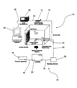
FIG. 1 is a schematic of an apparatus for inducing and passively detecting
stable cavitation
during ultrasound-enhanced thrombolysis experiments with video microscopy data
acquisition.
FIG. 2 is a schematic of a dual-element annular array for 120-kHz
sonothrombolysis
and 60 kHz passive cavitation detection.
CA 2973013 2017-07-11
-4-
FIG. 3 is a schematic of the determined level of ultrasonic energy being
provided in
intervals separated by rest periods, wherein substantially no ultrasonic
energy is provided
during the rest periods. The interval includes either continuous wave or
pulsed wave
ultrasound activity of the source transducer; the rest period is a quiescent
period. The interval
duration is determined by assessing the duration of stable cavitation and the
rest period
duration is selected to allow the in-flow of a nucleating agent or an
ultrasound contrast agent.
FIG. 4 is a block diagram of a passive stable cavitation detection and control
system
for ultrasound-enhanced thrombolysis.
FIG. 5 is a graph illustrating clot mass loss with treatment in an ex vivo
porcine
carotid artery model with physiologic flows and pressures of 0-8 ml/min and 80-
120 mmHg,
respectively.
FIG. 6 illustrates the computed cross-sectional beam pattern for a 120 kHz
unfocused
source transducer and surrounding annular 60 kHz passive cavitation detector.
FIG. 7 is a graph illustrating the average relative stable cavitation dose in
the ex vivo
porcine carotid artery model with physiologic flows and pressures. The stable
cavitation dose
was measured over a range of peak-to-peak acoustic pressures within a living,
excised
porcine carotid artery and was normalized by the maximum stable cavitation
dose within that
vessel to yield a relative dose in arbitrary units. Error bars represent the
standard deviation.
This data indicates a peak-to-peak pressure amplitude of about 0.44 MPa yields
the largest
stable cavitation dose on average.
FIG. 8 illustrates stable cavitation activity and a total cavitation dose
versus
ultrasound on-time (i.e. interval duration) in an ex vivo porcine carotid
artery model with
physiologic flows and pressures. Stable cavitation power decays as a function
of time. By
integrating the power signal in time over multiple pulses, the total 30 minute
cavitation dose
was calculated and the on-time that yielded the maximum cavitation dose was
calculated.
The system is operated with the on-time that provides the maximum cavitation
dose, or at the
center of the 90% width of the cavitation dose.
CA 2973013 2017-07-11
-5-
FIG. 9 is a graph illustrating optimization of on-time ultrasound in the ex
vivo porcine
carotid artery model with physiologic flows and pressures. For a selected
pressure (about
0.44 MPa), twelve trials are shown with the optimal on-time for each trial
shown in blue with
the error bars extending to the 90% of optimal on-time. The optimal on-time is
the time for
which a 30 minute trial would give the maximum cavitation dose.
Skilled artisans appreciate that elements in the figures are illustrated for
simplicity
and clarity and are not necessarily drawn to scale. For example, the
dimensions of some of
the elements in the figures may be exaggerated relative to other elements, as
well as
conventional parts removed, to help to improve understanding of the various
embodiments of
the present invention.
The following terms are used in the present application:
In the context of stable cavitation, the terms "inducing" and "inducement" are
used
interchangeably herein to refer to the nucleation or initiation of stable
cavitation.
In the context of passively detecting stable cavitation, the term "passively"
is used
herein to refer to receiving a signal with a transducer or hydrophone which is
used
exclusively to receive an emitted and/or scattered level of ultrasonic energy
from acoustically
activated bubbles. In the context of a system for inducing and passively
detecting stable
cavitation, the term "passive" is used herein to refer to a transducer and/or
a hydrophone
which is used exclusively to receive an emitted and/or scattered level of
ultrasonic energy
from acoustically activated bubbles.
The term "cavitation" is used herein to refer to the formation, oscillation,
and/or
collapse of gaseous and/or vapor bubbles in a liquid due to an acoustic
pressure field.
Cavitation is generally classified into two types: stable cavitation and
inertial cavitation. The
term "stable cavitation" is used herein to refer to a microbubble or
nanobubble oscillating in
an ultrasound field, whereby the predominant acoustic emissions occur not only
at the
fundamental ultrasonic frequency and harmonic frequency but also at the
subharmonic and
ultraharmonic frequencies. The origin of these emissions is a nonlinear
standing wave, i.e. a
Faraday wave, on the outer surface of the bubble, or nonlinear volumetric
oscillations of the
CA 2973013 2017-07-11
-6-
bubble during pulsation in the sound field. The term "inertial cavitation" is
used herein to
refer to cavitation which results in broadband emissions.
The term "thrombolysis" is used herein to refer to the dissolution or breaking
up of a
clot or thrombus. The term "sonothrombolysis" is used herein to refer to
ultrasound-
enhanced or ultrasound-mediated thrombolysis.
The term "determined level of ultrasonic energy" is used herein to refer to
the
ultrasound peak-to-peak pressure amplitude that is produced by a source
transducer.
In the case of thrombolysis, the term "treatment zone" is used herein to refer
to the
area comprising a blood clot. In one embodiment, the treatment zone is part of
a vascular
model and comprises a blood clot. In another embodiment, the treatment zone is
located
within a mammalian subject and refers to the area surrounding and comprising a
blood clot.
In a specific embodiment, in the case of sonothrombolysis of a treatment zone,
the term
"treatment zone" is to the area encompassed by the -6dB focal volume of the
source
transducer, which is confocally aligned with the -6dB focal volume of the
passive cavitation
detector.
The term "source transducer" is used herein to refer to a transducer which
produces a
determined level of ultrasonic energy. The term "detector transducer" is used
herein to refer
to a transducer which receives a scattered level of ultrasonic energy.
The term "fundamental ultrasonic frequency", as used herein, refers to the
frequency
of ultrasonic energy generated by a source transducer producing pressure
cycles per unit time.
The fundamental ultrasonic frequency employed herein can range from about 100
kHz to
about 10 MHz, or from about 100kHz to about 2 MHz. In a very specific
embodiment, the
fundamental ultrasonic frequency is about 120 kHz.
When the fundamental ultrasonic frequency activates nano- or microbubbles, the
bubbles scatter ultrasonic energy at a derivative frequency. Thus, the term
"scattered level of
ultrasonic energy" is used herein to refer to the pressure amplitude or the
intensity of the
ultrasound which is scattered from ultrasonically activated nano- and
microbubbles.
CA 2973013 2017-07-11
-7-
The term "derivative frequency" is used herein to refer to any ultrasonic
frequency or
combination of ultrasonic frequencies scattered by bubbles undergoing stable
cavitation. The
derivative frequency is selected from a subharmonic frequency and/or an
ultraharmonic
frequency of the fundamental ultrasonic frequency applied to a treatment zone.
The term "han-nonic frequency" is used herein to refer to integer multiples of
the
fundamental ultrasonic frequency. The term "subharmonic frequency" is used
herein to refer
to half the fundamental ultrasonic frequency. The detection of scattered
subharmonic
frequencies is indicative of stable cavitation. The term "ultraharmonic
frequency" is used
herein to refer to integer multiples of the subhan-nonic frequency, excluding
integer multiples
of the fundamental frequency. The detection of scattered ultraharmonic
frequencies is also
indicative of stable cavitation.
The term "dual-element annular transducer an-ay" is used herein to refer to an
array
consisting of two transducer elements, wherein an annular element surrounds a
central
circular element. The term "single element transducer" is used herein to refer
to a single
element transducer that produces ultrasonic pressure waves. The term "linear
array
transducer" is used herein to refer to a multi-element transducer composed of
a plurality of
transducer elements. The transducer elements are electrically separate
elements arranged
along a line or curve. The term "two-dimensional array transducer" is used
herein to refer to
a matrix of transducer elements which provide beam control over a cross-
sectional area. If
the matrix is arranged in annuli, or concentric circles, the beam control
provides spherical
focusing at different depths from the face of the array. In the context of a
transducer array,
individual elements of the array may be square, hexagonal, annular, circular,
or any other
pattern which fills the emitting area of the transducer and can be controlled
by a suitable
driver systein.
The term "Rayleigh distance" is used herein to refer to the natural focus of a
transducer, that is, the location from the transducer face at which all the
emitted waves are in
phase. The "Rayleigh distance" employed herein can range from about 0.1
centimeters to
about 30 centimeters, or from about 0.1 centimeters to 10 centimeters. As used
herein, the
terms "Rayleigh distance", "natural focus", and "focus" are interchangeable.
CA 2973013 2017-07-11
-8-
The term "hydrophone" is used herein to refer to a microphone configured to
record
and/or to listen to ultrasound scattered by acoustically active bubbles.
The term "ultrasonic driver" is used herein to refer to a device having a
radio
frequency signal source and a power amplifier. Impedance matching circuitry
between the
power amplifier and transducer may optionally be employed to increase the
efficiency of an
ultrasonic driver.
The term "signal" is used herein to refer an electronic signal converted from
a
pressure wave in ultrasound. The hydrophone or detector transducer converts a
pressure
wave into a voltage signal as a function of time. The term "gated signal" is
used herein to
refer to a detected signal that is truncated in time such that only certain
signals of the
scattered level of ultrasonic energy are detected, and such that certain
signals of the scattered
level of ultrasonic energy are disallowed. The signals of the scattered level
of ultrasonic
energy that are detected are those that are emitted from a scattering source
at a particular
distance from the detector transducer.
The ter-n "pre-amplifier" is used herein to refer to a device which prepares
an
electronic signal for recording and/or processing. The pre-amplifier circuitry
may or may not
be housed as a separate component. In the context of amplifying a signal, the
term
"amplifying" is used herein to refer to increasing the amplitude of the
signal.
The term "digital oscilloscope" is used herein to refer to a device which
converts
measured voltages into digital information. Waveforms are sampled with an
analog to digital
converter at approximately two times the frequency of the highest frequency
component of
the observed signal. The samples are stored and accumulate until a sufficient
amount are
taken to describe the waveform. The signals are then reassembled for display.
In the context
of storing a signal, the term "storing" is used herein to refer to a data set
that is stored in the
memory of a microprocessor.
In the context of acquiring a signal, the term "acquiring" is used herein to
refer to the
process of sampling the voltage received by the detector transducer,
hydrophone, or passive
cavitation detector and converting the resulting samples into digital numeric
values that can
be manipulated by a computer. In the context of acquiring a signal with a
computer, the term
CA 2973013 2017-07-11
-9-
"data acquisition" is used herein to refer to the conversion of analog
waveforms into digital
values for processing on a computer.
The term "duty cycle" is used herein to refer to the pulse duration divided by
the pulse
repetition period. The duty cycle employed herein can range from about 0.01%
to about
100%.
The term "bandwidth" is used herein to refer to the range of frequencies
wherein the
signal's Fourier transform has a power above about a quarter of the maximum
value. In a
specific embodiment, the bandwidth is about -6dB. As used herein, the detector
transducer is
configured to receive a bandwidth centered at one or more subharmonic and/or
ultraharmonic
frequencies of the fundamental frequency.
The term "ultrasonic pressure amplitude" is used herein to refer to the peak-
to-peak
pressure amplitude. In one embodiment, the ultrasonic pressure amplitude
employed herein
can range from about 0.1 MPa to about 10.0 MPa, or from about 0.1 MPa to about
10.0 MPa.
In the context of stable cavitation, the term "enhanced" is used herein to
refer to an
increase in the number of ultrasonically activated bubbles or to an increase
in the duration of
bubble activity. The term "ultrasonically activated bubbles" is used herein to
refer to bubbles
with larger vibrational amplitude excursions. In the context of thrombolysis,
the term
"enhanced" is used herein to refer to an increase in lytic efficacy or to a
reduced period of
time for lytic effect. For example, in the context of thrombolysis, the
percent clot mass lost
in the presence of a predetermined level of ultrasound was greater than about
80% in the
presence of a thrombolytic agent, a nucleating agent, and a determined level
of ultrasound;
whereas, in the presence of a thrombolytic agent and a nucleating agent
(without ultrasound),
the percent clot mass lost was less than about 35%. Thus, thrombolysis is
enhanced in the
presence of ultrasound, as compared with the absence of ultrasound.
The term "nucleating agent" is used herein to refer to an agent that initiates
cavitation.
The term "thrombolytic agent" is used herein to refer to a therapeutic agent,
such as a
pharmaceutical, used in medicine to dissolve blood clots or thrombi in order
to limit the
damage caused by the blockage of the blood vessel.
CA 2973013 2017-07-11
-10-
The term "interval" is used herein to refer to continuous wave or pulsed wave
ultrasound produced by a source transducer. The source transducer provides a
determined
level of ultrasonic energy in an interval. The term "interval duration" is
used herein to refer
to the period of time for which a determined level of ultrasonic energy is
provided. In one
embodiment, the interval duration employed herein can range from about 10
milliseconds to
about 5 minutes, or from about 10 milliseconds to about 10 seconds.
The term "rest period" is used herein to refer to providing substantially no
ultrasonic
energy. The terrn ''rest period duration" is used herein to refer to the
period of time for which
substantially no ultrasonic energy is provided. In one embodiment, the rest
period duration
employed herein can range from about 1 second to about 5 minutes, or from
about 1 second
to about 20 seconds.
The term "continuous wave ultrasound" is used herein to refer to a technique
in which
a transducer continuously emits ultrasound, wherein the ultrasound is varied
sinusoidally.
The term "pulsed wave ultrasound" is used herein to refer to a technique in
which a
transducer emits ultrasound in pulses or tone bursts.
In the context of enhancing stable cavitation, the term "adjusting the
determined level
of ultrasonic energy" is used herein to refer to increasing or decreasing the
peak-to-peak
pressure output of the source transducer.
The term "passive cavitation detector" is used herein to refer to a transducer
or a
hydrophone which receives a scattered level of ultrasound from acoustically
active bubbles.
The ter-n "transducer array" is used herein to refer to a transducer array
which receives a
scattered level of ultrasound from acoustically active bubbles. In one
embodiment, the
transducer array is a passive transducer array.
The term "nanobubble" is used herein to refer to bubbles on the size scale of
nanometers. The term "microbubble" is used herein to refer to bubbles on the
size scale of
micrometers.
CA 2973013 2017-07-11
-11-
The term "ultrasound contrast agent" is used herein to refer to gas-filled
vesicles
(containing nanobubbles or microbubbles), which are administered, for example,
intravenously to the systemic circulation to increase echogenicity on an
ultrasound image.
The term "protective material" is used herein to refer to a protein, lipid or
surface
active agent which prevents dissolution of an entrapped bubble.
The term "liposome" is used herein to refer to microscopic vesicle consisting
of a core
enclosed by one or more phospholipid layers, wherein hydrophobic compounds
and/or
hydrophilic compounds can be contained within the core. The term "echogenic
liposome" is
used herein to refer to a liposome which produces an echo when exposed to
ultrasound.
The term "beamwidth" is used herein to refer to the spatial extent of the
ultrasound
beam at the focus, natural focus, or Rayleigh distance of a transducer. In one
embodiment,
the beamwidth is about -6dB, such that the pressure output is at least a
quarter of the peak
value (-6dB beamwidth). The "beamwidth" can be controlled by changing the
diameter or
aperture of the transducer while keeping the frequency fixed. The beamwidth at
the Rayleigh
distance is about half of the diameter of the transducer. The beamwidth
employed herein can
range, for example, from about 0.1 centimeters to about 10 centimeters.
The terms "stable cavitation dose" and "dose" are used interchangeably herein
to refer
to the cumulative amount of acoustic energy detected that is directly
attributed to nonlinear
bubble activity generating at a subhannonic frequency, an ultraharmonic
frequency, and/or
combinations thereof.
Embodiments of the present invention relate to ultrasound-mediated methods and
systems of detecting and enhancing stable cavitation. In one embodiment, a
system for
inducing and passively detecting stable cavitation is provided, the system
comprising a dual-
element annular transducer array having a source transducer and a detector
transducer, and an
ultrasonic driver adapted to generate energy that can be converted at the
source transducer to
ultrasonic energy suitable for penetrating a treatment zone of a patient. The
system is
adapted to provide a determined level of ultrasonic energy and to receive a
scattered level of
ultrasonic energy substantially throughout the treatment zone of the patient,
in which the
source transducer provides an ultrasonic frequency that is a fundamental
ultrasonic
CA 2973013 2017-07-11
-12-
frequency, and the detector transducer receives an ultrasonic frequency that
is a derivative
frequency of the fundamental ultrasonic frequency selected from the group
consisting of a
subharmonic frequency, an ultraharmonic frequency, and combinations thereof.
As shown in FIGS. 1 and 2, in one aspect of this embodiment, the system for
inducing
and passively detecting stable cavitation 10 is adapted to provide a
determined level of
ultrasonic energy and to receive a scattered level of ultrasonic energy
substantially
throughout the treatment zone of a patient. In one particular aspect, the
system for inducing
and passively detecting stable cavitation 10 comprises a dual-element annular
transducer
array 20. The dual-element annular transducer array 20 has a source transducer
22 and a
detector transducer 24. The dual-element annular transducer array 20 provides
a determined
level of ultrasonic energy and receives a scattered level of ultrasonic
energy, such that
sonothrombolysis and stable cavitation detection may be achieved substantially
simultaneously. The size and configuration of the dual-element annular
transducer array 20
should be selected so that ultrasound waves, or energy, may be provided
substantially
throughout the treatment zone of a patient, while avoiding potentially harmful
bioeffects such
as tissue damage, petechial hemorrhage, blood brain barrier disruption,
thermal coagulation,
and/or cellular damage to the patient.
The source transducer 22 is adapted to provide a determined level of
ultrasonic
energy. In one particular aspect, the source transducer 22 has a circular
cross-section having
a diameter of about 3 centimeters, and the detector transducer 24 has an
annular cross-section
having an inner diameter of about 3 centimeters and an outer diameter of about
4 centimeters.
In another aspect, the detector transducer 24 has a circular cross-section
having a diameter of
about 3 centimeters, and the source transducer 22 has an annular cross-section
having an
inner diameter of about 3 centimeters and an outer diameter of about 4
centimeters.
However, the dual-element annular transducer array 20 should not be limited to
the particular
aspects disclosed herein, but may comprise any configuration wherein a source
transducer 22
is confocally aligned with a detector transducer 24. Moreover, the source
transducer 22 may
comprise the annular transducer element surrounding the central circular
transducer element,
or may comprise the central circular transducer element. Similarly, the
detector transducer
CA 2973013 2017-07-11
-13-
24 may comprise the annular transducer element surrounding the central
circular transducer
element, or may comprise the central circular transducer element.
The source transducer 22 provides an ultrasonic frequency that is a
fundamental
ultrasonic frequency. Suitable fundamental frequencies produced by the source
transducer 22
can range from about 100 kHz to about 10 MHz. In one particular aspect, the
source
transducer 22 can produce a fundamental ultrasonic frequency of from about 100
kHz to
about 2 MHz. In another aspect, the source transducer 22 can produce a
fundamental
ultrasonic frequency of about 120 kHz.
In one embodiment, the source transducer 22 is configured such that it is
adjustable to
vary the duty cycle of the ultrasonic energy produced. In one particular
aspect, the source
transducer 22 is adjustable to vary the duty cycle from about 0.01% to about
100%.
Moreover, the source transducer 22 can be configured such that it is
adjustable to vary the
beamwidth of the ultrasonic energy produced. The beamwidth may be varied such
that the
source transducer 22 provides a determined level of ultrasonic energy
substantially
throughout the treatment zone of a patient. In one aspect, the source
transducer 22 is
configured to provide a beamwidth of about 0.1 centimeters to about 10
centimeters.
Additionally, the source transducer 22 can be configured such that it is
adjustable to select an
ultrasonic pressure amplitude of the ultrasonic energy produced. In a
particular aspect, the
source transducer 22 is configured to provide an ultrasonic pressure amplitude
of from about
0.1 MPa to about 10.0 MPa. In a further aspect, the source transducer 22 is
configured to
provide an ultrasonic pressure amplitude of from about 0.1 MPa to about 1.0
MPa.
The detector transducer 24 is adapted to receive a scattered level of
ultrasonic energy
substantially throughout the treatment zone of a patient. In this particular
aspect, the detector
transducer 24 receives an ultrasonic frequency that is a derivative frequency
of the
fundamental ultrasonic frequency selected from the group consisting of a
subharmonic
frequency, an ultraharmonic frequency, and combinations thereof. In this
aspect, the detector
transducer 24 is configured to receive a bandwidth centered at one or more
subharmonic
frequency and/or ultraharmonic frequency of the fundamental frequency. In yet
another
CA 2973013 2017-07-11
-14-
aspect, the detector transducer 24 is configured to receive a bandwidth
centered at the
subharmonic frequency of about 60 kHz.
Detection of a derivative frequency selected from the group consisting of a
subharmonic frequency, an ultrahamionic frequency, and combinations thereof,
is indicative
of stable cavitation during sonothrombolysis. The scattering of incident wave
by
ultrasonically activated bubbles on the size scale of nanometers or
micrometers occurs at the
center frequency and harmonics of the insonifying pulse. However, the presence
of half of
the fundamental frequency (the subharmonic) and its odd multiples
(ultraharmonics) indicate
the presence of microbubbles or nanobubbles that are cavitating stably.
As shown in FIGS. 1 and 2, the ultrasonic driver 30 is adapted to generate
electrical
energy that can be converted at the source transducer 22 to ultrasonic energy
suitable for
penetrating a treatment zone of a patient. In one aspect, the ultrasonic
driver 30 includes a
function generator 40, an amplifier 50, and a matching network 60. The
ultrasonic driver 30
is electrically connected to the source transducer 22 with a cord 62, such
that the system for
inducing and passively detecting stable cavitation 10 is adapted to provide a
determined level
of ultrasonic energy substantially throughout the treatment zone of a patient.
The ultrasonic
driver 30 may be of a conventional design with an adjustable frequency
generator and/or an
adjustable power amplifier. The ultrasonic driver 30 should be configured such
that the
ultrasound waves or energy can be selected to provide a determined level of
ultrasonic energy
substantially throughout the treatment zone of a patient.
In one embodiment, the function generator 40 is electrically connected to the
amplifier 50 with a cord 42. The amplifier 50 amplifies the electrical energy
generated by the
function generator 40.
In another embodiment, the matching network 60 is electrically connected to
the
amplifier 50 with a cord 52. The matching network 60 increases the efficiency
of the
ultrasonic driver 30 by impedance matching circuitry between the amplifier 50
and the source
transducer 22. In this particular aspect, the matching network 60 is
electrically connected to
the source transducer 22 with a cord 62.
CA 2973013 2017-07-11
-15-
The detector transducer 24 converts the scattered level of ultrasonic energy
received
into an electronic signal. In this particular aspect, the derivative frequency
received by the
detector transducer 24 comprises a signal. In a further aspect of this
particular embodiment,
the signal received by the detector transducer 24 is gated. In one embodiment,
the signal is
filtered such that the detector transducer 24 receives ultrasonic frequencies
that are
substantially a derivative frequency of the fundamental ultrasonic frequency.
In one
particular aspect, the derivative frequency of the fundamental frequency
received by the
detector transducer 24 is selected from the group consisting of a subharmonic
frequency, an
ultraharmonic frequency, and combinations thereof.
In still another aspect of this embodiment, the system for inducing and
passively
detecting stable cavitation 10 further comprises a pre-amplifier 70. The pre-
amplifier 70 is
electrically connected to the detector transducer 24 with a cord 72. The pre-
amplifier 70
amplifies the signal received by the detector transducer 24.
In yet another aspect of this embodiment, the system for inducing and
passively
detecting stable cavitation 10 further comprises a digital oscilloscope 80.
The digital
oscilloscope 80 is electrically connected to the pre-amplifier 70 with a cord
82. The digital
oscilloscope 80 stores the signal amplified by the pre-amplifier 70.
In yet another aspect of this embodiment, the system for inducing and
passively
detecting stable cavitation 10 further comprises a computer 90. The computer
90 is
electrically connected to the digital oscilloscope 80 with a cord 92. The
computer 90
acquires the signal stored in the digital oscilloscope 80. The computer 90
provides data
acquisition from the signal stored in the digital oscilloscope 80.
In yet still another aspect of this embodiment, the system for inducing and
passively
detecting stable cavitation 10 further comprises a hydrophone (not shown). The
hydrophone
is adapted to receive a scattered level of ultrasonic energy substantially
throughout the
treatment zone of a patient. In a particular aspect, the hydrophone converts
the scattered
level of ultrasonic energy received into an electronic signal. In this
particular aspect, the
derivative frequency received by the hydrophone comprises a signal. In a
further aspect, the
CA 2973013 2017-07-11
-16-
signal received by the hydrophone is gated, such that the hydrophone receives
a scattered
level of ultrasonic energy that is truncated to receive only signals from a
selected distance.
In another embodiment of the present invention, a method for inducing and
passively
detecting stable cavitation during sonothrombolysis is provided, the method
comprising
providing a determined level of ultrasonic energy substantially throughout a
treatment zone
of a patient and detecting a scattered level of ultrasonic energy. The
determined level of
ultrasonic energy is produced by a source transducer 22 and comprises a
fundamental
ultrasonic frequency. The scattered level of ultrasonic energy is received by
a detector
transducer 24 and comprises a derivative frequency of the fundamental
ultrasonic frequency
selected from the group consisting of a subharmonic frequency, an
ultraharmonic frequency,
and combinations thereof, wherein detection of the derivative frequency is
indicative of
stable cavitation during sonothrombolysis.
The method for passively detecting stable cavitation comprises providing a
determined level of ultrasonic energy substantially throughout a treatment
zone of a patient,
wherein the determined level of ultrasonic energy is produced by a source
transducer 22, and
detecting a scattered level of ultrasonic energy, wherein the scattered level
of ultrasonic
energy is received by a detector transducer 24. In one particular aspect, the
source transducer
22 and the detector transducer 24 comprise a dual-element annular transducer
array 20. In a
further aspect, the source transducer 22 has a circular cross-section having a
diameter of
about 3 centimeters, and the detector transducer 24 has an annular cross-
section having an
inner diameter of about 3 centimeters and an outer diameter of about 4
centimeters. In
another aspect, the detector transducer 24 has a circular cross-section having
a diameter of
about 3 centimeters, and the source transducer 22 has an annular cross-section
having an
inner diameter of about 3 centimeters and an outer diameter of about 4
centimeters.
However, the dual-element annular transducer array 20 may comprise any
configuration
wherein the source transducer 22 is confocally aligned with the detector
transducer 24.
The method also comprises providing a deten-nined level of ultrasonic energy
substantially throughout the treatment zone of a patient, wherein the
determined level of
ultrasonic energy is produced by a source transducer 22. The determined level
of ultrasonic
CA 2973013 2017-07-11
-17-
energy is produced by a source transducer 22 and comprises a fundamental
ultrasonic
frequency. Suitable fundamental frequencies produced by the source transducer
22 can be,
for example, from about 100 kHz to about 10 MHz. In one particular aspect, the
source
transducer 22 can produce a fundamental ultrasonic frequency from about 100
kHz to about 2
MHz. In another aspect, the source transducer 22 can produce a fundamental
ultrasonic
frequency of about 120 kHz.
In another aspect, the source transducer 22 is configured such that it is
adjustable to
vary the Rayleigh distance to assist in concentrating or directing ultrasound
waves or energy
to the treatment zone so that ultrasound waves or energy may be provided
substantially
throughout the treatment zone of a patient. In one particular aspect, the
ultrasonic energy is
emitted from the source transducer 22 with a Rayleigh distance from about 0.1
centimeters to
about 30 centimeters. In a further aspect, the ultrasonic energy is emitted
from the source
transducer 22 with a Rayleigh distance from about 0.1 centimeters to about 10
centimeters.
Moreover, the source transducer 22 is configured such that it is adjustable to
vary the
beamwidth of the ultrasonic energy produced. The beamwidth may be varied such
that the
source transducer 22 provides a determined level of ultrasonic energy
substantially
throughout the treatment zone of a patient. In one aspect, the source
transducer 22 is
configured to provide a beamwidth of about 0.1 centimeter to about 10
centimeters.
The method also comprises detecting a scattered level of ultrasonic energy,
wherein
the scattered level of ultrasonic energy is received by a detector transducer
24. The scattered
level of ultrasonic energy is received by the detector transducer 24 and
comprises a derivative
frequency of the fundamental ultrasonic frequency selected from the group
consisting of a
subharmonic frequency, an ultraharmonic frequency, and combination thereof. In
one
specific aspect, the detector transducer 24 detects a subharmonic frequency of
the
fundamental ultrasonic frequency of about 60 kHz. As previously discussed,
detecting a
derivative frequency selected from the group consisting of a subharmonic
frequency, an
ultraharmonic frequency, and combinations thereof, is indicative of stable
cavitation during
sonothrombolysis as the presence of half of the fundamental frequency (the
subharmonic) and
its odd multiples (ultraharmonics) indicate the presence of microbubbles Or
nanobubbles that
are cavitating stably.
CA 2973013 2017-07-11
-18-
In yet still another aspect of this embodiment, the method for passively
detecting
stable cavitation further comprises detecting the scattered level of
ultrasonic energy with a
hydrophone.
In still another embodiment, a method for enhancing stable cavitation during
sonothrombolysis is provided, the method comprising administering a nucleating
agent and a
thrombolytic agent to a treatment zone of a patient and providing a detemained
level of
ultrasonic energy substantially throughout the treatment zone of the patient.
The determined
level of ultrasonic energy is produced by a source transducer and comprises a
fundamental
ultrasonic frequency, wherein the determined level of ultrasonic energy is
provided in
intervals separated by rest periods, wherein substantially no ultrasonic
energy is provided
during rest periods, such that the intervals of the determined level of
ultrasonic energy
enhance stable cavitation during sonothrombolysis.
In a further aspect, the method of enhancing stable cavitation further
comprises
detecting a scattered level of ultrasonic energy. The scattered level of
ultrasonic energy is
received by a detector transducer and comprises a derivative frequency of the
fundamental
ultrasonic frequency selected from the group consisting of a subharmonic
frequency, an
ultraharmonic frequency, and combinations thereof.
The method of enhancing stable cavitation comprises administering a nucleating
agent
to a treatment zone of a patient. The nucleating agent initiates cavitation,
and any agent
capable of initiating cavitation may be used. In one aspect, the nucleating
agent is gas
bubbles stabilized against dissolution in a fluid. In a further aspect, the
nucleating agent is a
gas releasably contained by a protective material.
The protective material is configured to allow the nucleating agent to be
released
when exposed to a determined level of ultrasonic energy; in one aspect, the
protective
material is capable of being ruptured by ultrasonic energy generated by the
source transducer
22. The protective material is also configured to allow circulation of the
encapsulated
nucleating agent throughout the patient. Suitable protective materials
include, but are not
limited to, lipids and/or liposomes. Liposomes can entrap microbubbles and
nanobubbles,
CA 2973013 2017-07-11
-19-
enabling enhanced echogenicity and cavitation nucleation. In one particular
aspect, the
liposome is an echogenic liposome ("ELIP").
Echogenic liposomes can be targeted to certain tissues by attaching specific
peptides,
ligands, or antibodies to the surface of the liposome. Additionally, echogenic
liposomes may
be fragmented with ultrasound near a target tissue. In one specific aspect,
echogenic
liposomes can be targeted with peptides or ligands to bind to receptors
characteristic of
intravascular diseases (or blood clots). Targeting echogenic liposomes enables
selective
accumulation of the nucleating agent to a specific area. In one particular
aspect, echogenic
liposomes could be targeted to a treatment area comprising a blood clot.
In another aspect, the nucleating agent may be selected from the group
consisting of
nanobubbles, microbubbles, and ultrasound contrast agents. In one embodiment,
ultrasound
contrast agents act as cavitation nuclei at the site of a blood clot.
Moreover, infusions of
ultrasound contrast agents may sustain the gentle bubble activity that is
indicative of stable
cavitation. In one specific aspect, the ultrasound contrast agent is
perflutren-lipid
microspheres, or Definity (Lantheus Medical Imaging, N. Billerica, MA).
The method of enhancing stable cavitation also comprises administering a
thrombolytic agent to a treatment zone of a patient. In one aspect, the
thrombolytic agent
may comprise tissue plasminogen activator ("t-PA"); t-PA is a protein
manufactured by
vascular endothelial cells that regulates clot breakdown in the body. t-PA can
be
manufactured using recombinant biotechnology techniques. W. F. Bennett & D. L.
Higgins,
Tissue Plasminogen Activator: The Biochemistry and Pharmacology of Variants
Produced by
Mutagenesis, 30 Annual Review of Pharmacology and Toxicology 91, 91-121
(1990).
Additional examples of thrombolytic agents include, but are not limited to,
recombinant
tissue plasminogen activator ("ft-PA"), streptokinase, urokinase, and
tenecteplase.
The method of enhancing stable cavitation also comprises providing a
determined
level of ultrasonic energy substantially throughout the treatment zone of a
patient. In one
aspect, the determined level of ultrasonic energy is produced by a source
transducer 22 and
comprises a fundamental ultrasonic frequency. In one particular aspect, the
source transducer
22 may be a single element transducer, a linear array transducer, or a two-
dimensional anay
CA 2973013 2017-07-11
-20-
transducer. In a further aspect, the source transducer 22 may have a circular
cross-section
having a diameter of about 3 centimeters.
In yet another aspect, the source transducer 22 is configured such that it is
adjustable
to vary the Rayleigh distance, natural focus, or focus to assist in
concentrating or directing
ultrasound waves or energy to the treatment zone so that ultrasound waves or
energy may be
provided substantially throughout the treatment zone of a patient. In a
further aspect, the
ultrasonic energy may be emitted from the source transducer 22 with a Rayleigh
distance,
natural focus, or focus of from about 0.1 cm to about 30 cm. In still a
further aspect, the
ultrasonic energy may be emitted from the source transducer 22 with a Rayleigh
distance,
natural focus, or focus of from about 0.1 cm to about 10 cm. As shown in FIG.
3, the
determined level of ultrasonic energy produced by a source transducer 22 is
provided in
intervals separated by rest periods, wherein substantially no ultrasonic
energy is provided
during the rest periods. The interval comprises either continuous wave or
pulsed wave
ultrasound produced by the source transducer 22; the rest period comprises a
quiescent
period. The interval duration is dictated by the duration of stable cavitation
and the rest
period duration is dictated by the in-flow of the nucleating agent or
ultrasound contrast agent.
The determined level of ultrasonic energy is provided in intervals to enhance
stable
cavitation. By providing a determined level of ultrasonic energy in intervals
separated by rest
periods, the nucleating agent is enabled to flow into the treatment zone of
the patient. The
bubble activity that elicits subharmonic frequencies, ultraharmonic
frequencies, and
combinations thereof, may be sustained using an intermittent or continuous
infusion of a
commercial contrast agent; thus, providing a determined level of ultrasonic
energy in
intervals separated by rest periods allows the nucleating agent to flow into
the treatment zone
of the patient and enhances stable cavitation.
In one aspect of this embodiment, the determined level of ultrasonic energy is
provided for an interval duration of from about 10 milliseconds to about 5
minutes. In a
further aspect, the determined level of ultrasonic energy is provided for an
interval duration
of from about 10 milliseconds to about 10 seconds. In still a further aspect,
the determined
level of ultrasonic energy is provided for an interval duration of about 8.5
seconds. In yet
CA 2973013 2017-07-11
-2 l -
another aspect of this embodiment, the rest period duration is from about 1
second to about 5
minutes. In a further aspect, the rest period duration is from about 1 second
to about 60
seconds. In a more specific aspect, the rest period duration is from about 1
second to about
30 seconds. In a very specific aspect, the rest period duration is about 19
seconds.
The source transducer 22 provides a determined level of ultrasonic energy
substantially throughout the treatment zone of a patient, wherein the
determined level of
ultrasonic energy is produced by a source transducer 22 and comprises a
fundamental
ultrasonic frequency. The determined level of ultrasonic energy may comprise
pulsed wave
or continuous wave ultrasound. Suitable fundamental frequencies produced by
the source
transducer 22 include frequencies from about 100 kHz to about 10 MHz. In one
particular
aspect, the source transducer 22 produces a fundamental ultrasonic frequency
from about 100
kHz to about 2 MHz. In another aspect, the treatment zone comprises a clot and
the source
transducer 22 produces a fundamental ultrasonic frequency of about 120 kHz.
As shown in FIG. 4, in another aspect, the method of enhancing stable
cavitation
further comprises detecting a scattered level of ultrasonic energy. The
scattered level of
ultrasonic energy is received by a detector transducer 24 and comprises a
derivative
frequency of the fundamental frequency selected from the group consisting of a
subharmonic
frequency, an ultraharmonic frequency, and combinations thereof. In one
aspect, the
scattered level of ultrasonic energy is received by a passive cavitation
detector. In a further
aspect, the passive cavitation detector is selected from the group consisting
of a hydrophone,
a detector transducer, and a passive transducer array.
In a further aspect of this embodiment, the method of enhancing stable
cavitation
further comprises adjusting the determined level of ultrasonic energy produced
by the source
transducer 22 in accordance with the detected scattered level of ultrasonic
energy received by
a passive cavitation detector. By monitoring the detected scattered level of
ultrasonic energy
received by a passive cavitation detector, stable cavitation may be. In
response to monitoring
stable cavitation, the source transducer 22 may be adjusted to provide a
modified determined
level of ultrasonic energy; additionally, in response to monitoring stable
cavitation, the
interval duration and rest period duration may also be modified to allow
inflow of the
CA 2973013 2017-07-11
-22-
nucleating agent. For example, if the scattered level of ultrasonic energy
received by the
passive cavitation detector indicates continued bubble activity, the source
transducer 22
remains on. In a further example, if the scattered level of ultrasonic energy
received by the
passive cavitation detector decreases or if cavitation is not detected, a rest
period is initiated
to allow the nucleating agent to flow into the treatment zone. In a specific
embodiment,
when the scattered level of ultrasonic energy of the derivative frequency
drops below about
twice the background noise level in the passive cavitation detection system, a
rest period is
initiated.
In one particular aspect of this embodiment, the treatment zone comprises a
blood clot
and thrombolysis is enhanced substantially throughout the treatment zone. In
one
embodiment, enhanced thrombolysis includes percent clot mass loss of about 20%
to about
500% greater than that observed without the provision of ultrasound. See, for
example, FIG.
5, wherein the percent clot mass lost is greater than about 80% 1% standard
deviation
wherein a blood clot is treated with rt-PA, Definity , and ultrasound; in
contrast, the percent
clot mass loss is less than about 35% 1% standard deviation wherein the
blood clot is
treated with rt-PA and Definity , wherein substantially no ultrasound is
provided.
It will be appreciated that the system and methods disclosed herein are useful
in
sonothrombolysis. Additionally, it will be appreciated that the system and
methods disclosed
herein are useful in the treatment of thrombo-occlusive diseases including but
not limited to
stroke, pulmonary emboli, myocardial infarction, deep vein thrombosis, and/or
arteriovenous
fistula thrombosis. Moreover, it will be appreciated that ultrasound-mediated
enhancement
of stable cavitation increases thrombolysis substantially throughout the
treatment zone.
Examples
The following non-limiting examples illustrate the methods and systems of the
present invention.
Example 1: Passive Cavitation Detection with Dual Element Annular Array
Dual Element Annular Array for 120-kHz Sonothrombolysis and 60-kHz Passive
Cavitation Detection. A dual element annular array (FIG. 1) was designed to
enable inducing
CA 2973013 2017-07-11
-23-
and passively detecting stable cavitation during sonothrombolysis. To test the
feasibility of
this design approach, acoustic radiation from the 3 cm, 120-kHz source was
computed using
an exact series solution for the field of a baffled circular radiator in a
homogeneous medium.
Using the same method, the spatial sensitivity pattern of the surrounding
annular passive
cavitation detector (inner diameter 3 cm, outer diameter 4 cm) was computed at
the
subhannonic frequency of 60 kHz. Cross sections of the beam patterns are shown
in FIG. 6.
The field of the 120-kHz source had a -6dB depth of field of 46 mm and a -6dB
beamwidth
of 1.4 cm. The annular broadband passive cavitation detector had a collimated
beam with
amplitude 0.84 (-1.5dB relative to surface excitation) and a beamwidth of 1.6
cm at the
Rayleigh distance of the 120 kHz source. The results demonstrated that both
uniform
sonication and passive cavitation detection may be achieved over the entire
region of interest
containing a blood clot.
Assessment of Dual Element 120-kHz/60-kHz Anay Beam Distortion. Acoustic field
profiles of the prototype an-ay output were performed. An omnidirectional
hydrophone was
mounted on a computer-controlled micropositioning system to scan the interior
of human and
pig skulls. The penetration of ultrasound (both 120 kHz and 60 kHz) was
through the
temporal and frontal bones for the human and the pig skulls, respectively.
Passive Cavitation Detection. A broadband passive cavitation detector (PCD)
was
employed to detect cavitating micron-sized bubbles. A dual element 120-kHz/60-
kHz array
transducer was used as a passive cavitation detector with porcine blood clots
in an ex vivo
porcine carotid artery model. The 60-kHz confocal annulus (Sonic Concepts,
Inc.,
Woodburn, WA) was employed to detect cavitation activity passively in the
sample volume
as shown in FIG. 2. The dual element array transducer was mounted on a
micrometer-
controlled 3-axis translation stage (Newport 423, Irvine, CA, USA) for precise
alignment
with the blood clots. Moreover, as shown in FIG. 6, the detector transducer 24
enables
monitoring of stable cavitation along the entire volume of the clot.
Detected Signal Analysis. Signals acquired by the PCD were gated to account
for
travel time of the pulse from the 120-kHz transducer to the clot and back to
the 60 kHz
element. The signal received by the PCD was amplified using a pre-amplifier
(Signal
CA 2973013 2017-07-11
-24-
Recovery 5185, Oak Ridge, TN, USA) and stored using a digital oscilloscope
(LeCroy Waver
Surfer 424, Chestnut Ridge, NY, USA). The acquired signal by the PCD was also
gated to
ensure that cavitation was monitored over a region encompassing the entire
clot and
surrounding fluid. The squared frequency spectra of received pulses was
processed in the
frequency domain.
Acoustic Pressure Threshold Determination. Using the PCD, the acoustic
pressure
threshold of stable and inertial cavitation at 120 kHz was detemlined in an ex
vivo porcine
carotid artery flow model with 1) plasma alone, and 2) rt-PA and Definity in
the flowing
plasma. Porcine whole blood clots were placed in excised, living porcine
carotid arteries
through which porcine plasma flowed and were maintained in a 37 C temperature-
controlled
water bath. The peak rarefactional pressure amplitude was increased slowly
until initially
stable and then inertial cavitation was detected by the PCD. The lowest peak
rarefactional
pressure amplitude which yielded stable and inertial cavitation was recorded
as the threshold
pressure for each fluid.
Example 2: Effects of Stable Cavitation on Thrombolysis
Cavitation Nucleation with Infusion of Contrast Agent in an In Vitro Human
Clot
Model. An approach for inducing cavitation using infusion of a contrast agent,
Definity ,
was tested experimentally in vitro. Human whole blood clots and rt-PA (96
Him') were
placed in human fresh frozen plasma in a thin-walled latex sample holder which
was placed
in a tank of water at 37 C. Percent clot mass loss was assessed as a function
of peak-to-peak
acoustic pressure for the following treatments: (a) no rt-PA, no Definity , no
pulsed
ultrasound ( the control); (b) rt-PA alone; (c) rt-PA, Definity infusions,
and pulsed
ultrasound (¨ 0.12 MPa peak-to-peak pressure amplitude); (d) rt-PA, Definity
infusions ,and
pulsed ultrasound (-0.21 MPa peak-to-peak pressure amplitude); (e) no rt-PA,
Definity
infusions, and pulsed ultrasound (-0.32 MPa peak-to-peak pressure amplitude);
(f)
phosphate buffered saline infusions (no Definity ) and pulsed ultrasound (-
0.32 MPa peak-
to-peak pressure amplitude); and (g) rt-PA, Definity infusions, and pulsed
ultrasound (-0.32
MPa peak-to-peak pressure amplitude). A sample size of six was used for each
treatment.
CA 2973013 2017-07-11
-25-
Human whole blood clots, when exposed to stable cavitation activity in the
presence of rt-PA,
resulted in the highest mass loss of 26.0 4%.
Sonothrombolysis Transducer. A single-element 120-kHz source transducer was
operated in pulsed mode over a range of peak-to-peak pressure amplitudes, with
an 80% duty
cycle, and 1667 Hz pulse repetition frequency. The peak-to-peak pressure
amplitudes were
selected such that no cavitation (-0.12 MPa and ¨0.21 MPa), or stable
cavitation (-0.32
MPa) was induced.
Stable Cavitation Detection. Stable cavitation was detected using a focused
polyvinylidine difluoride (PVDF) hydrophone immersed in the tank of water
aligned
confocally with the sonothrombolysis transducer.
Tracking Emissions to Obtain Feedback. Stable cavitation was monitored by
tracking
the ultraharmonic emissions during the combined ultrasound and thrombolytic
exposures in
the in vitro human blood clot model. Cavitation activity was monitored by
tracking
subharmonic and ultraharmonic emissions during the treatment. The emission's
energy was
integrated over time as a metric for the amount of stable cavitation. A
significant correlation
was observed between clot mass loss and ultraharmonic signals (r=0.8549,
p<0.0001, n=24).
Promotion of Stable Cavitation with Ultrasound Contrast Agent. A dual antibody
immunofluoresccnce technique was employed to measure penetration depths of rt-
PA and
plasminogen into the clots. The largest mean penetration depth of rt-PA (222
um) and
plasminogen (241 ium) was observed in the presence of stable cavitation
activity. Thus, it
was demonstrated that a contrast agent can be used to nucleate cavitation and
can result in a
desired therapeutic effect.
A contrast agent, Definity , was successfully used to promote and sustain the
nucleation of stable cavitation during pulsed ultrasound exposure at 120 kHz
for 30 minutes.
The largest percent clot mass loss of 26.2 2.6% was observed in human whole
blood clots
in the presence of sustained stable cavitation activity.
Model Thresholds for Bubble Activity vs. Bubble Size to Determine Optimal
Size.
The minimum inertial cavitation threshold estimated by the microbubble
response was
CA 2973013 2017-07-11
-26-
observed at the resonance size of the microbubble for all the frequencies
studied. The
minimum inertial cavitation threshold increased with increasing frequency. The
range of
bubble sizes that may cavitate stably decreases at higher frequencies. This
suggested that
higher frequencies would require the optimum sized nucleus to be present for
generating
stable cavitation.
Example 3: Effects of Stable Cavitation on Thrombolysis in an Ex Vivo Porcine
Artery
Model with Interval Ultrasound
Ex Vivo Porcine Carotid Artery Model with Physiologic Flows and Pressures.
Porcine whole blood clots were inserted into living, excised porcine carotid
arteries, and kept
viable in a thin-walled latex chamber filled with degassed artificial
cerebrospinal fluid while
oxygenated plasma flowed through the lumen. The chamber was placed in a tank
containing
degassed filtered water at 37 C. A series of experiments were performed by
infusing 1
ml/min plasma with 0.31 IA Definity per 1 ml plasma through a porcine artery
filled with a
porcine whole blood clot at a physiologic pressure of 100 15 mmHg. Each clot
and artery
were insonated with 120 kHz continuous wave ultrasound at peak-to-peak
pressures ranging
from 0.37 MPa to 0.54 MPa for 45 seconds. The signals were analyzed for stable
and inertial
cavitation power.
Ultrasound Source Transducer and Passive Cavitation Detector. A single-element
transducer operating at 120 kHz was used to insonate the porcine clots in
living, excised
porcine carotid arteries. A 2.25-MHz center frequency transducer was used as a
passive
cavitation detector to receive acoustic signals scattered from within the
vessel. These signals
were digitized and converted to power spectra.
To detect stable cavitation, the power spectra at ultraharmonic frequencies
(from 300
kHz to 3.8 MHz) of the fundamental frequency was accumulatively summed over
the
treatment period to yield a total stable cavitation dose. In a similar manner,
inertial cavitation
was detected by summing the power spectra at frequencies between the harmonic
frequencies
and ultraharmonic frequencies (from 300 kHz to 3.8 MHz) of the fundamental
frequency.
More particularly, the power spectra was accumulatively summed over the
treatment period
to yield an inertial cavitation dose.
CA 2973013 2017-07-11
-27-
Pressure Determination. An "optimal" peak-to-peak pressure output was selected
based on maximizing the amount of stable cavitation ("the dose") taking into
account variable
on- and off-times (i.e. intervals and rest periods, respectively). FIG. 7 is a
graph illustrating
the average relative stable cavitation dose in the ex vivo porcine carotid
artery model with
physiologic flows and pressures. The stable cavitation dose was measured over
a range of
peak-to-peak acoustic pressures within a living, excised porcine carotid
artery and was
normalized by the maximum stable cavitation dose within that vessel to yield a
relative dose
in arbitrary units. Error bars represent the standard deviation. The data
indicate that the peak-
to-peak pressure amplitude of about 0.44 MPa gave the largest stable
cavitation dose on
average.
Optimization of Ultrasound Duration. The duration of ultrasound on-time was
optimized for the particular peak-to-peak pressure amplitude yielding the
largest average
stable cavitation dose. As shown in FIG. 8 (left), stable cavitation activity
was recorded
passively as a function of time. As shown in FIG. 8 (right), the total stable
cavitation dose
during a treatment period was calculated as a function of the ultrasound on-
time. The on-
time yielding the maximum total stable cavitation dose was considered to be
optimal on-time
to promote sonothrombolysis. The optimal on-time is shown in FIG. 9 for twelve
experiments in the ex vivo porcine carotid artery model with physiologic flows
and pressures.
The error bars extend over the times that yielded at least 90% of the maximum
stable
cavitation dose. The mean of these optimal on-time values was 8.5 seconds and
this on-time
value was used for subsequent sonothrombolysis experiments, shown in FIG. 5.
Clot Mass Loss with Treatment. A second series of experiments were performed
to
determine the thrombolytic efficacy of ultrasound-enhanced thrombolysis using
the
optimized interval ultrasound exposure in the ex vivo porcine carotid artery
model with
physiologic flows and pressures. The pressure was 100 15 mmHg and the mean
flow
velocity was 2.7 1.8 ml/min. The ultrasound insonation parameters were 120-
kHz center
frequency and 0.44 MPa peak-to-peak pressure amplitude for 8.5 seconds and a
rest period
over a 30 minute treatment period. The rest periods were employed within a
pulsing
sequence to allow the contrast agent to entirely refill the target volume.
Treatments included:
1) plasma alone, 2) plasma and 3.15 p1 rt-PA /m1 plasma, 3) plasma with
interval ultrasound,
CA 2973013 2017-07-11
-28-
4) plasma with 3.15 pl/m1rt-PA and 0.31 1/m1 Definity microbubble contrast
agent, 5)
plasma with 3.15 pl/m1 rt-PA and interval ultrasound, and 6) plasma with 3.15
j.i1/m1 rt-PA,
0.31 p.1/m1 Definity , and interval ultrasound. Clots were weighed before and
after treatment
to yield percent clot mass loss.
FIG. 5 shows the mean clot mass loss for each treatment in the vascular model,
with
vertical error bars representing one standard deviation. A two-way analysis
of variance
("ANOVA'') with repeated measurements revealed that there were significant
differences in
mass loss among arteries perfused with the mixture rt-PA and Definity and
those perfused
with plasma alone, with and without ultrasound (F -= 60.5, p < 0.0001). The
ANOVA further
showed that the effects of rt-PA with Definity interact significantly with
the effects of
ultrasound.
This phenomenon was further studied with four paired t-tests (two-tailed). To
keep
the overall level of significance at 0.05, each individual t-test was
performed with an alpha of
0.0125 (0.05/4) in order to be considered significant. With ultrasound, there
was a difference
between groups with and without rt-PA with Definity (p<0.0001), and for those
arteries
exposed to ultrasound and those arteries which were not (p<0.0001). In the
absence of
ultrasound, rt-PA with Definity produces a significantly higher mass loss
than plasma alone
(p=0.0001). With no rt-PA or Definity present, however, the effect of
ultrasound was not
significant (p=0.19). A follow-up student's t-test showed no difference
between rt-PA-treated
arteries with or without Definity and without ultrasound.
It is noted that terms like "preferably," "generally," "commonly," and
"typically" are
not utilized herein to limit the scope of the claimed invention or to imply
that certain features
are critical, essential, or even important to the structure or function of the
claimed invention.
Rather, these terms are merely intended to highlight alternative or additional
features that
may or may not be utilized in a particular embodinient of the present
invention.
For the purposes of describing and defining the present invention it is noted
that the
term "substantially" is utilized herein to represent the inherent degree of
uncertainty that may
be attributed to any quantitative comparison, value, measurement, or other
representation.
The term "substantially" is also utilized herein to represent the degree by
which a quantitative
CA 2973013 2017-07-11
-29-
representation may vary from a stated reference without resulting in a change
in the basic
function of the subject matter at issue.
The citation of any document is not to be construed as an admission that
it is prior art with respect to the present invention.
While particular embodiments of the present invention have been illustrated
and
described, it would be obvious to one skilled in the art that various other
changes and
modifications can be made without departing from the spirit and scope of the
invention. It is
therefore intended to cover in the appended claims all such changes and
modifications that
are within the scope of this invention.
CA 2973013 2017-07-11