Note: Descriptions are shown in the official language in which they were submitted.
CA 02973076 2017-07-05
DESCRIPTION
TITLE OF THE INVENTION: ORAL MOUNTING FIXTURE
TECHNICAL FIELD
[0001]
The present invention relates to an oral mounting fixture. The present
invention
specifically relates to an oral mounting fixture that is mounted in an oral
cavity and effectively
promotes the secretion of saliva to thereby prevent halitosis.
BACKGROUND ART
[0002]
Many people worry about halitosis during conversation with other people.
Generally,
countermeasures for preventing halitosis such as thorough brushing of teeth
after meal, washing
liquids for preventing halitosis, sprays for preventing halitosis and
supplements for preventing
halitosis are used.
Furthermore, an apparatus for administering a deodorant agent and an air
freshener and the
like for preventing halitosis by diffusion in a continuous manner with natural
feeling without causing
unpleasant sensation and repulsion is described in, for example, Patent
Literature I.
[0003]
Furthermore, Patent Literature 2 describes that a mouth piece is pressed
against the insides
of the both buccas in an oral cavity and the both buccas are put into a state
that they are pushed
upward to stimulate the entirety in the oral cavity to thereby allow secretion
of a large amount of
saliva.
[0004]
Furthermore, Patent Literature 3 describes the following points. An instrument
to
promote the secretion of saliva is inserted in between a maxillary gum part in
an oral cavity and an
1
CA 02973076 2017-07-05
upper lip. A chewing part is inserted to and withdrawn from between upper
teeth and lower teeth
by using a tongue, and then slightly chewed. By this way, the secretion of
saliva is promoted,
halitosis is suppressed, and conversation is enabled without any problem.
[0005]
Furthermore, more saliva than usual is secreted by the chewing. Therefore,
Patent
Literature 4 describes a chewing exercise tool to be mounted along a lower
dentition.
CITATION LIST
PATENT LITERATURE
[0006]
Patent Literature 1: JP 2001-258910 A
Patent Literature 2: JP 3167198 U
Patent Literature 3: JP 2011-417 A
Patent Literature 4: JP 2001-128994 A
SUMMARY OF INVENTION
PROBLEMS TO BE SOLVED BY THE INVENTION
[0007]
The content of the description of Patent Literature 1 is not to promote the
secretion of
saliva by massaging ducts of the major salivary glands during mounting but to
prevent halitosis by
administering a drug. Therefore, it is difficult to effectively promote the
secretion of saliva.
[0008]
The content of the description of Patent Literature 2 is not that stimulation
is strengthened
and weakened as in massaging but that the both buccas are put into a state
that they are pushed
upward by mounting a flexible mouth piece in the oral cavity. This is
insufficient to effectively
2
CA 02973076 2017-07-05
promote the secretion of saliva as in the content of the description of Patent
Literature 1.
[0009]
The content of the description of Patent Literature 3 is to promote the
secretion of saliva
by chewing the chewing part, and thus is fundamentally different from
stimulating salivary glands.
In this method, the secretion of saliva cannot be effectively promoted during
mounting. The
content of the description of Patent Literature 4 is also that more saliva
than usual is secreted by the
chewing, and is not that promotion of the secretion of saliva during mounting.
[0010]
The object of the present invention is to solve the problems of the above-
mentioned
conventional technologies, and to enable use in public such as during
conversation with other people
when halitosis is of particular concern, enable use at any time with no limit
to the timing of use, and
to further effectively promote the secretion of saliva.
SOLUTIONS TO THE PROBLEMS
[0011]
In order to the achieve above-mentioned object, the oral mounting fixture
configured to be
mounted inside an oral cavity of a human and promote the secretion of saliva
of the present
invention includes a main body section that can be detachably mounted on a
part in the oral cavity,
and having a shape arranged so that the main body section conforms to the
shape of a portion on
which the oral mounting fixture is mounted, and plural protrusions that are
integrally formed in the
main body section and protruding from the surface of the main body section,
wherein the oral
mounting fixture is mounted so that the tips of the plural protrusions press
on a mucosa in the oral
cavity.
[0012]
Furthermore, it is desirable that the above-mentioned oral mounting fixture is
mounted so
3
CA 02973076 2017-07-05
that the tips of the plural protrusions press on at least either of an inner
surface part being a buccal
mucosa of the oral cavity, a submandibular part between a mandibular dentition
and a lingual base
and a sublingual part being a floor of the oral cavity between a dentition and
a lingual base.
[0013]
Furthermore, in the above, it is preferable that the protrusions each has a
rod-like shape
and are formed in the form of bristles from the main body section.
[0014]
Furthermore, it is preferable that the oral mounting fixture has a fitting
part that can be
fitted to the dentition.
Furthermore, in the above, it is desirable that the main body section has
plural fitting parts
capable of fitting to the dentition, and the fitting parts are linked by a
string-like linking part.
[0015]
Furthermore, in the above, it is desirable that the protrusions have plural
buccal
protrusions configured to press on an inner surface part of an oral cavity and
plural gingiva
protrusions configured to press on a gingiva, wherein the gingiva protrusions
each has a smaller
protrusion height than that of the buccal protrusions.
[0016]
Furthermore, it is preferable that the oral mounting fixture has parotid gland
protrusions
configured to press on parts in the vicinity of the parotid glands, which are
salivary glands.
Furthermore, it is desirable that the oral mounting fixture has a fitting part
being fittable to
at least back teeth of the dentition.
[0017]
Furthermore, in the above, it is preferable that the main body section has an
oral cavity
antrum having an expanded shape so as to fit to the shape of a tongue or a
lingual base.
[0018]
4
CA 02973076 2017-07-05
Furthermore, in the above, it is preferable that the main body section and the
protrusions
are integrally formed by a resin.
Furthermore, in the above, it is preferable that the protrusions are coated
with a metal that
generates a galvanic electrical current by saliva.
EFFECTS OF THE INVENTION
[0019]
The oral mounting fixture of the present invention has plural protrusions that
are integrally
formed in a main body section that can be detachably mounted on a part in the
oral cavity. The tips
of the plural protrusions press on a mucosa in the oral cavity. Therefore,
salivary glands that are
broadly dispersed in the mucosa in the oral cavity are stimulated like
massaging. Therefore, the
secretion of saliva is further effectively promoted. Furthermore, the shape is
arranged so as to
conform to the shape of a portion on which the oral mounting fixture is
mounted. Therefore, the
oral mounting fixture can be used at any time with no limit to the timing of
use. Furthermore, the
oral mounting fixture can be used in public such as during conversation with
other people when
halitosis is of particular concern.
BRIEF DESCRIPTION OF THE DRAWINGS
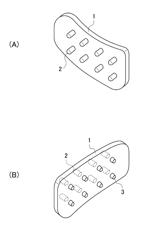
[0020]
Figs. 1(A) and 1(B) are each a perspective view showing an oral mounting
fixture
according to the embodiment of the present invention.
Figs. 2(A), 2(B) and 2(C) are each an embodiment view showing the shape of
protrusions
in an embodiment.
Fig. 3 is an explanatory view explaining the positions of the salivary glands
in a human
face.
CA 02973076 2017-07-05
Figs. 4(A) and 4(B) are a perspective view and a side view showing an oral
mounting
fixture according to another embodiment.
Figs. 5(A), 5(B) and 5(C) are each a plane view showing an oral mounting
fixture
according to a still another embodiment.
Figs. 6(A), 6(B) and 6(C) are each an explanatory view that explains an
example in which
the salivary glands are stimulated according to an embodiment of the present
invention.
Figs. 7(A) and 7(B) are each a plane view that shows an embodiment of a back
teeth-fit
type.
Figs. 8(A) and 8(B) are each a perspective view showing an embodiment in which
the
parts in the vicinity of the parotid glands are pressed on.
Figs. 9(A), 9(B) and 9(C) are each a perspective view showing an oral mounting
fixture
according to a still another embodiment.
Fig. 10 is an explanatory view showing a state that the oral mounting fixture
is fit to a
dentition.
DESCRIPTION OF EMBODIMENTS
[0021]
Saliva has effects to protect mucosae, natural purification, water content
equilibration,
lubrication, buffering, antibacterial, digestion, repair tissues,
recalcification and prevent
carcinogenesis, and the like. Saliva is essential for not only an oral cavity
but also a body to exert a
normal function. Furthermore, salivary glands are glands from which saliva is
secreted. Ducts are
opened in the oral cavity, and are classified into major salivary glands and
minor salivary glands.
Specifically, salivary glands include major salivary glands (parotid glands,
submandibular gland and
sublingual gland) and many minor salivary glands. Major salivary glands each
has a tube that
opens in the oral cavity, and saliva is flown out to the mouth through the
tube. Minor salivary
6
CA 02973076 2017-07-05
glands are widely distributed in the mucosae in the oral cavity. The outlets
for saliva in the minor
salivary glands are opened on the mucosae.
[0022]
Furthermore, saliva is secreted from major salivary glands (parotid glands,
submandibular
glands and sublingual glands) and many minor salivary glands (glandulae
labiales, buccales,
palatinae, molar glands and linguales). The minor salivary glands include
"glandulae labiales in the
labial mucosae, buccales present in the buccal mucosae, anterior linguales
present in the lower part
of tongue tip, "posterior linguales present in the posterior parts of the
lateral margins of the tongue
root and tongue, as well as molar glands, and palatinae, and the like.
[0023]
Saliva has a natural purification action to clean the inside of the oral
cavity and an
antibacterial action. Enzymes such as lysozyme suppress the propagation of
bacteria to prevent
generation of halitosis. Therefore, when the secretion of saliva is decreased,
the inside of the oral
cavity becomes dirty, and thus halitosis easily generates. The point of the
present embodiment is
that the secretion of less viscous saliva is maintained for a long period.
[0024]
Preferable embodiments of the oral mounting fixture according to the present
invention
will be explained below according to the attached drawings.
Fig. 1 is a perspective view showing an oral mounting fixture mounted in an
oral cavity,
which is a space in the mouth of a human, and a main body section 1, which is
a thick resin, can be
detachably mounted on a part in the oral cavity. The part in the oral cavity
is a part "between the
dentition and the lower lip", a part "between the oral cavity antrum, which is
outside of the dentition,
and the lower lip", specifically a part between "the dentition of back teeth
and the lingual base
through a gum" and "a floor of the oral cavity between the dentition and the
lingual base" or the like.
Fig. 1(A) is a perspective view in which the oral mounting fixture is seen
from the outside in the
7
CA 02973076 2017-07-05
case when the oral mounting fixture is mounted, for example, on the outside of
the dentition of the
like (the oral cavity part is not illustrated). Fig. 1(B) is a perspective
view seen from the opposite
side of Fig. 1(A) (the oral cavity part is not illustrated).
[0025]
Furthermore, the shape of the main body section 1 is arranged so that any
unpleasant
sensation is not caused, and the main body section 1 conforms to the shape of
a portion on which the
oral mounting fixture is mounted. For example, as shown in Fig. 1(A), the main
body section 1
slightly curves toward outside (so that the left side seen in the drawing has
a convex shape). The
four corners of the main body section 1 have been rounded so as to have
roundness. Furthermore,
the edges have been appropriately rounded at the ends in the thickness
direction so that the inside of
the oral cavity is not injured.
[0026]
Furthermore, plural protrusions 2 and 3 have been respectively formed on the
main body
section 1 so that they integrally protrude from the surface. During the oral
mounting fixture is
mounted, the tips of the protrusions 2 and 3 press on plural positions on the
mucosae in the oral
cavity. By using plural protrusions 2 and 3 and allow the main body section 1
to curve, salivary
glands that are widely distributed on the mucosa in the oral cavity are
stimulated like massaging.
[0027]
The oral mounting fixture is mounted so that the tips of the plural
protrusions 2 or 3 press
on at least either of an inner surface part being a buccal mucosa of the oral
cavity, a submandibular
part between the mandibular dentition and the lingual base and a sublingual
part being the floor of
the oral cavity between the dentition and the lingual base. Furthermore, the
protrusions 2 each has
a rod-like shape and are formed in the form of bristles from the main body
section 1. The tip of the
protrusion 2 presses on a mucosa on the inner surface of the oral cavity to
stimulate the mucosa to
thereby promote the secretion of saliva. As shown in Figs. 2(A), 2(B) and
2(C), it is preferable that
8
CA 02973076 2017-07-05
the tip of the protrusion 2 is rounded so that the inner surface of the oral
cavity is not injured.
[0028]
Figs. 2(A), 2(B) and 2(C) each shows the shape of the tip of the protrusion 2.
A
semi-discoid shape as shown in Fig. 2(A), a hemispherical shape as shown in
Fig. 2(B), or a shape of
a rod having a tip having a spherical shape as shown in Fig. 2(C) is
desirable. By imparting
flexibility or elasticity to a rod-like shape or columnar shape so that it is
bent, the mucosa on the
inner surface of the oral cavity can be adequately massaged. Furthermore, the
salivary glands are
massaged more effectively by moving the mouth while the oral mounting fixture
is mounted.
However, the shape of the protrusion 2 of the present invention is not limited
to these shapes. As
the shape of the protrusion 2, various shapes such as a spherical shape and a
hog-backed shape can
be adopted. In summary, the protrusion may have any shape as long as the
mucosa in the oral
cavity can be pressed by the tip of the protrusion.
[0029]
On the other hand, the protrusions 3 are minute protrusions each having a
smaller
protrusion height from the main body section 1 than that of the protrusions 2.
The protrusions 2 are
buccal protrusions that press on the buccas, which are the inner surface parts
of the oral cavity. The
protrusions 3 are gingiva protrusions that press on the gingivae.
[0030]
Fig. 3 is a drawing that shows the positions of the salivary glands in a human
face. The
salivary glands are mainly constituted by three: parotid glands 16, a
sublingual gland 18 and a
submandibular gland 20. The symbol 22 represents masseter muscles.
The parotid gland 16 is a salivary gland that is present by spreading in the
parotid buccal
part. A duct 16A that ejects saliva opens on the buccal mucosa. The sublingual
gland 18 is a
salivary gland that is present on the floor of the oral cavity and under the
mucosa in the lingual base.
Plural ducts 18A open in the vicinity of the gum. The submandibular gland 20
is a salivary gland
9
CA 02973076 2017-07-05
that is present as if it partially hides in the mandibular bone. A duct 20A is
opened between the
mandibular dentition and the lingual base.
[0031]
By the protrusions 3 that are minute protrusions, the part of the gum
(gingiva) 28 (see Figs.
6(A), 6(B) and 6(C) for the gum), specifically the vicinities of the back
teeth, is stimulated. By this
way, the inner surface part of the oral cavity of the sublingual gland 18, and
the duct 20A of the
submandibular gland 20 can be effectively stimulated. It is preferable that
the shape of the
protrusion 3 is a shape such that the gum is not injured. For example, a
minute protrusion having a
rod-like shape having a round tip, a minute protrusion having a semicircular
shape, and linear minute
protrusions in which plural hog-backed bumps are arranged, and the like can be
adopted. In the
case of a linear minute protrusion, a shape in which plural hog-backed bumps
are arranged in
parallel to a dentition may also be adopted. By this way, the linear minute
protrusion becomes
resistance against the gum, and thus the oral mounting fixture becomes
difficult to detach.
[0032]
The oral mounting fixture may have a shape like a mouth piece that is tightly
fitted to a
dentition (for example, the shapes as shown in Figs. 4(A) and 4(B), Figs.
5(A), 5(B) and 5(C), Figs.
7(A) and 7(B), Figs. 8(A) and 8(B), and Figs. 9(A), 9(B) and 9(C)). However,
it is not always
necessary that the shape of the oral mounting fixture has such shape, and may
have, for example, the
shape as shown in Figs. 1(A) and 1(B). The oral mounting fixture is formed by,
for example, a
thick thermoplastic resin in the case when it is fit to a dentition, and the
shape of the dentition shape
may be transferred to only the inner surface (the external surface is flat).
Alternatively, the oral
mounting fixture may be formed of a thin thermoplastic resin so that the shape
of the dentition is
transferred to the entirety (the inner surface and the external surface have
an identical shape). In
this case, it is desirable that the oral mounting fixture is formed of a
thermoplastic resin that is
softened at a temperature greater than the body temperature of a human.
CA 02973076 2017-07-05
[0033]
"Softened at a temperature greater than the body temperature of a human" means
that a
thermoplastic resin that forms an oral mounting fixture is softened to the
extent that the shape of the
dentition is transferred, when the oral mounting fixture is heated to a
temperature greater than the
body temperature of a human and then pressed on the maxillary dentition or
mandibular dentition.
Since heated water is generally preferable, the softening temperature as an
actual temperature is
preferably equal to or lower than 100 C, which is the boiling point of water.
[0034]
Furthermore, the thermoplastic resin used may be any resin as long as safeness
from a
hygiene perspective is ensured and the thermoplastic resin does not have any
effects such as allergy
on teeth and gums.
[0035]
Examples include ethylene-vinyl acetate copolymers (EVA), polyolefins
(polyethylene,
polypropylene, polybutadiene), polyvinyl acetates (PVA), polyurethane
elastomers and the like.
Among these resins, an ethylene-vinyl acetate copolymer (EVA) that satisfies a
softening
temperature equal to or more than 50 C, which is greater than the body
temperature of a human, and
an upper limit equal to or less than 100 C, which is the boiling point of
temperature of water, is
specifically preferable.
[0036]
Figs. 4(A) and 4(B) show another embodiment, and show a perspective view (A)
and a
side view (B) of an oral mounting fixture having a fitting part that can be
fitted to a dentition. The
main body section 1 has been folded into a U-shape, and has been arranged to
have a curved shape
seen from above so that the lower opening is fitted to a dentition. The oral
mounting fixture is
mounted so that the arrow is directed to the side a, which is the outside of
the dentition, i.e., the
buccal mucosa, and the opposite side is directed to "the side b, which is the
inner side of the
11
CA 02973076 2017-07-05
dentition. Therefore, the gum part is covered with the side parts of the U-
shaped convex part.
[0037]
Plural protrusions 2 are formed in the form of bristles from the main body
section 1 toward
the outside. The tips thereof press on the inner surface part of the oral
cavity, which is the buccal
mucosa, in the case when the tips are fitted to a transverse side dentition.
The protrusions 3 are
disposed on the convex part. The protrusions 3 are minute protrusions each
having a smaller
protrusion height from the main body section 1 than that of the protrusions 2.
The protrusions 3
press on the gum part (in the case when the main body section 1 is mounted on
the mandibular
dentition, the submandibular part between the mandibular dentition and the
lingual base). In the
case of fitting to the anterior dentition, the mounting is such that the side
b is directed to the anterior
direction. At this time, the protrusions 2 press on a sublingual part being
the floor of the oral cavity
between the dentition and the lingual base. The protrusions 3 press on the gum
part.
[0038]
Figs. 5(A), 5(B) and 5(C) each shows an oral mounting fixture as a mouth piece
12, and
this is fitted to the entirety of the mandibular dentition or maxillary
dentition of a human. In the
oral mounting fixture shown in Fig. 1 or 2, fitting parts are linked by a
linking part, and protrusions
2 and 3 (the protrusions 2 are first projection parts 14A, and the protrusions
3 are minute protrusions
a) are integrally formed. Furthermore, the mouth piece 12 of the main body
section 1 is fitted to at
least the back teeth parts of the maxillary dentition 24 or the mandibular
dentition 26 in Fig. 3. The
protrusions 14 press on at least one of the gum part in the vicinity of the
back teeth and the inner
surface part of the oral cavity in the vicinity of the salivary gland. By this
way, the three salivary
glands that secrete saliva are stimulated, whereby the secretion of saliva is
promoted.
[0039]
In the case when the parotid gland 16 in Fig. 3 is stimulated, the mouth piece
12 may be of
a type that is fitted to either the maxillary dentition 24 or the mandibular
dentition 26. However, as
12
CA 02973076 2017-07-05
is understood from Fig. 3, ducts 18A and 20A from which saliva is ejected of
the sublingual gland 18
and the submandibular gland 20 are present on the dentition part of the
mandible. Therefore, in
order to promote the secretion of saliva at the sublingual gland 18 and the
submandibular gland 20, a
type that is fitted to the mandibular dentition 26 is preferable.
[0040]
Figs. 5(A), 5(B) and 5(C) show preferable three embodiments wherein the
protrusions 2
and 3 are projection parts. In the embodiment of the mouth piece 12 of Fig.
5(A), first projection
parts 14A, which are the protrusions 2, protrude from the back teeth positions
of the mouth piece 12
in the direction of the inner surface parts of parotid glands 16, which are
the salivary glands of the
oral cavity. Fig. 5(A) shows a drawing in which one first projection part 14A
has been formed on
each of the left and right back teeth positions of the mouth piece 12.
However, plural first
projection parts 14A may also be formed.
[0041]
According to the mouth piece 12 of Fig. 5(A), as shown in Fig. 6(A), the first
projection
parts 14A stimulate the inner surface parts corresponding to the parotid
glands 16 of the oral cavity.
Therefore, the secretion of saliva from the parotid glands 16 can be promoted.
In Figs. 6(A), 6(B)
and 6(C), the symbol 28 represents gums.
[0042]
In the mouth piece 12 of Fig. 5(B), second projection parts 14B are each a
skirt-like
element that is disposed to straddle the back teeth position of the mouth
piece 12 so that the element
covers the gum of the back teeth. Minute protrusions a that correspond to
plural protrusions 3 are
formed on the inner surface of the skirt-like element, which is brought into
contact with the gum part
of the back teeth.
[0043]
According to the mouth piece 12 of Fig. 5(B), as shown in Fig. 6(B), the
second projection
13
CA 02973076 2017-07-05
parts 14B stimulate the inner surface part corresponding to the sublingual
gland 18 of the oral cavity
and the duct 20A of the submandibular gland 20, whereby the secretion of
saliva from the sublingual
gland 18 and the submandibular gland 20 is promoted.
[0044]
In the embodiment of the mouth piece 12 of Fig. 6(C), first projection parts
14A each
protrudes from the back teeth position in the direction of the inner surface
parts corresponding to the
parotid glands 16 of the oral cavity, and skirt-like elements each disposed to
straddle the back teeth
position of the mouth piece 12 to cover the gum part 28 of the back teeth, are
disposed. Second
projection parts 14B with minute protrusions a formed thereon have been
constituted on the inner
surfaces of the skirt-like elements, which are brought into contact with the
back teeth.
[0045]
The mouth piece 12 of Fig. 5(C) is the case when both the first projection
parts 14A in Fig.
5(A) and the second projection parts 14B in Fig. 5(B) are disposed. By this
way, according to the
mouth piece 12 of Fig. 5(C), as shown in Fig. 6(C), three kinds of salivary
glands: the parotid glands
16, the sublingual gland 18 and the submandibular gland 20 can be stimulate at
the same time.
[0046]
Figs. 7(A) and 7(B) show a modified example of the mouth piece 12, and is an
example in
which fitting parts 12A of the mouth piece 12 are disposed on only the back
teeth (hereinafter
referred to as "a back teeth-fit type"). Fig. 7(A) is a drawing showing the
external surface of the
mouth piece 12, and Fig. 7(B) is a drawing showing the inner surface. As shown
in Figs. 7(A) and
7(B), the mouth piece 12 of a back teeth-fit type is constituted by a pair of
fitting parts 12A and 12A,
which are fitted to only the left and right back teeth, and a string-like
linking part 12B having an
arc-like shape, which is configured to link the fitting parts 12A and 12A.
[0047]
As shown in Fig. 7(A), the external surface of the fitting part 12A is plane.
As shown in
14
CA 02973076 2017-07-05
Fig. 7(B), a dentition shape is formed on the inner surface of the fitting
part 12A. It is preferable
that the back teeth to which the fitting part 12A is fitted are three teeth:
front molar, middle molar
and back molar.
[0048]
According to the mouth piece 12 of a back teeth-fit type, the pair of fitting
parts 12A and
12A, which are fitted to only the left and right back teeth, are linked by the
string-like linking part
12B. By this way, more comfortable talking is possible while the mouth piece
12 is mounted, and
when the mouth is opened, the mounting of the mouth piece 12 is difficult to
be seen from outside.
[0049]
Protrusions 14 for promoting the secretion of saliva have been integrally
formed also on
the mouth piece 12 of a back teeth-fit type. In Figs. 7(A) and 7(B), the first
projection parts 14A
are similar to the first projection parts 14A in Fig. 5(A).
[0050]
Besides the first projection parts 14A, in order to further promote the
secretion of saliva,
the external surfaces of the first projection parts 14A may be coated with a
metal that generates a
galvanic electrical current by saliva. The protrusions 2 and 3 and parotid
gland protrusions 5
(mentioned below), or the entirety or a part of the main body section 1 of all
of the exemplary
embodiments of the present invention can be coated with the metal that
generates a galvanic
electrical current by saliva.
[0051]
A galvanic electrical current (also referred to as a Galvani's electrical
current) generally
generates by the contact of different kinds of metals. However,
electroconductivity has been
increased by saliva in the oral cavity. Therefore, one kind of metal (for
example, amalgam, gold,
silver, palladium, a copper alloy, a nickel-chromium alloy, a cobalt alloy,
aluminum or the like) is
ionized with saliva to cause a potential difference in the oral cavity.
Therefore, a galvanic electrical
CA 02973076 2017-07-05
current, which is a faint electrical current, generates. When this galvanic
electrical current
generates, tingling stimulation is felt.
[0052]
Accordingly, the stimulation intensity for stimulating at least one of the gum
parts 28 in
the vicinity of the back teeth and the inner surface parts in the vicinity of
the salivary glands of the
oral cavity is enhanced by coating with the metal that generates a galvanic
electrical current by
saliva. By this way, the secretion of saliva can further be promoted.
[0053]
Figs. 8(A) and 8(B) show the oral mounting fixtures shown in Figs. 4(A) and
4(B), which
include parotid gland protrusions 5 configured to press on parts in the
vicinity of the parotid glands,
which are salivary glands. In Fig. 8(A), plural spherical parts each having a
spherical shape are
linearly arranged in the direction of the parotid glands, which are salivary
glands extending to
around right in front of the ears from the main body section 1. By these
spherical parotid gland
protrusions 5, the parts in the vicinity of the parotid glands can be massaged
by pressing.
[0054]
Furthermore, this parotid gland protrusion 5 can press the gum upper surfaces
(in the case
of the mandible) or the gum lower surfaces (in the case of the maxilla) at the
back of the middle
molars or back molars (wisdom teeth). It was found by the intensive studies by
the present
inventors that, in the case when back molars have not teethed, the secretion
of saliva is promoted by
pressing on the gum upper surfaces (in the case of the mandible) or the gum
lower surfaces (in the
case of the maxilla) at the back of the middle molars, that is, the parts
where back molars are to
teethe, with the tips of the protrusions. Furthermore, they also found that,
in the case when back
molars have teethed, the secretion of saliva is promoted by pressing on the
gum upper surfaces (in
the case of the mandible) or the gum lower surfaces (in the case of the
maxilla) at the back of the
back molars with the tips of the protrusions. At this time, it is not always
necessary that the
16
CA 02973076 2017-07-05
protrusions are rod-like. As shown in the parotid gland protrusions 5 in Figs.
8(A) and 8(B), a
similar effect can be obtained also by the pressing by the curved surfaces of
the protrusions each
having a spherical shape or a hog-backed shape.
[0055]
In Fig. 8(B), the spherical parts of the parotid gland protrusion 5 are
changed to
hog-backed parts each having a hog-backed shape. The spherical parts in Fig.
8(A) or the
hog-backed parts in Fig. 8(B) have grooves therebetween, and thus can be
separated off one by one.
Therefore, the length can be easily adjusted according to the size of the oral
cavity of each person.
[0056]
The method for using a back teeth-fit type is explained. A user immerses the
oral
mounting fixture in heated water retained in a heating container to soften the
oral mounting fixture.
In the case when the material of the oral mounting fixture is an ethylene-
vinyl acetate copolymer
(EVA), a temperature at which accuracy of transfer to a dentition is fine is
appropriately selected in
the range of heating temperature at 50 to 100 C.
[0057]
Subsequently, the user presses the fitting part 12A of the oral mounting
fixture in a
softened state downward from the above of the left and right back teeth of the
mandibular dentition
26. In this state, the user clenches the fitting part 12A together with the
maxillary dentition 24.
By this way, the back teeth shape is transferred to the inner surface of the
main body section 1. The
user then waits in this state until the oral mounting fixture is sufficiently
cooled, and when the oral
mounting fixture has been cooled to the body temperature, the oral mounting
fixture is removed
from the mandibular dentition 26. By this way, for example, the shapes of the
three back teeth:
front molars, middle molars and back molars are transferred in the form of
grooves to the inner
surface.
[0058]
17
CA 02973076 2017-07-05
After the transfer, if any portion that hits to the inside of the oral cavity
to cause
unpleasant sensation, or the like is present, then the portion is scraped
away, for example, by means
of a knife or the like, whereby the shape is arranged. Thereafter, where
necessary, fine adjustment
by softening the oral mounting fixture again by putting it into hot water at a
temperature equal to or
greater than the softening point, fitting again the oral mounting fixture to
the back teeth of the
mandibular dentition 26 while the oral mounting fixture is soft, and,
clenching the oral mounting
fixture in the oral cavity. By this way, an oral mounting fixture that fits to
the shape of the oral
cavity of the user is formed.
[0059]
Furthermore, during the main body section 1 is in a softened state, the user
corrects the
directions and lengths of the protrusions 2 and 3 by fingers so that the inner
surface parts
corresponding to the parotid glands 16 of the oral cavity are appropriately
stimulated, and then
solidifies the main body section 1 by natural cooling in the corrected state.
Furthermore, the user
corrects the directions and lengths of the protrusions 2 and 3 by fingers so
that the minute
protrusions a of the protrusions 3 are appropriately brought into contact with
the surface of the gums
28 of the back teeth without any gap during the main body section 1 is in the
softened state.
[0060]
By this way, comfortable talking is possible while the oral mounting fixture
is mounted on
the dentitions, and the mounting of the oral mounting fixture is difficult to
be seen from outside even
if the mouth is opened. Furthermore, the oral mounting fixture can be used in
public such as during
conversation with other people when halitosis is of particular concern.
Furthermore, the oral
mounting fixture can be used at any time with no limit to the timing of use.
[0061]
In addition, in the above-mentioned present embodiment, the user obtains a
dentition
shape by softening the main body section 1. It is also preferable to ask a
dentist to take a more
18
CA 02973076 2017-07-05
elaborate dentition shape. In the case of preparation by asking a dentist, it
is also possible to use a
non-thermoplastic resin material. Furthermore, a mouth piece is generally
prepared through
preparation of a plaster figure. It is also possible to prepare a mouth piece
by means of a 3D printer
as follows. Specifically, the maxillary dentition 24 and the mandibular
dentition 26 are respectively
photographed by a camera from at least two directions. Three-dimensional
images of the maxillary
dentition 24 and the mandibular dentition 26 are obtained from these
photograph images.
Furthermore, an oral mounting fixture can be prepared by a 3D printer by using
three-dimensional
coordinate data obtained from these three-dimensional images.
[0062]
Figs. 9(A), 9(B) and 9(C) each show an example in which the oral mounting
fixture is a
mouth piece such that the main body section 1 is fitted to the entirety of a
dentition. Fig. 9(A) is a
perspective view seen from the side surface. Fig. 9(B) is a perspective view
of (A) seen from
another viewpoint. Fig. 9(C) is a perspective view of (A) seen from the
oblique lower part. The
main body section 1 is integrally formed from a back part la of the dentition
at the left side to a back
part la of the dentition at the right side through an oral cavity antrum lb.
The oral cavity antrum
lb is mounted so that the tongue is laid thereon, and has an expanded shape so
as to conform to the
shape of the tongue or lingual base. Furthermore, protrusions 2c are disposed
on the upper and
lower surfaces and front and rear surfaces of the oral cavity antrum lb, and
are brought into contact
with the tongue or lingual base.
[0063]
The size and shape of the entirety of the main body section 1 have adjusted to
be a shape
that gives no unpleasant sensation with the portion on which the oral mounting
fixture is mounted of
the user. In the back parts la, plural protrusions 2a that press on the buccal
mucosae outward and,
plural protrusions 2c that stimulate the tongue or lingual base respectively
project from the main
body section 1. Similarly, in the vicinity of the front teeth, plural
protrusions 2b that press on the
=
19
CA 02973076 2017-07-05
inside of the lower lip are disposed on the oral cavity antrum lb. The
protrusions 2b and
protrusions 2c each has a smaller height projected from the main body section
1 than that of the
protrusions 2a. By this way, each pressure for stimulation is adjusted in
accordance with the
movement in the oral cavity to give a suitable massage force.
[0064]
Throughout the dentitions, plural protrusions 3 are disposed on the inner
parts that are
fitted to the gums. The protrusions 3 are minute protrusions each having a
smaller protrusion
height from the main body section 1 than that of the protrusions 2b. In the
oral cavity antrum lb of
the main body section 1, the protrusions 2c each having a similar protrusion
height to that of the
protrusions 2b are disposed on the above and below. Furthermore, the
projection heights may also
be decreased in the order of 2a, 2b, 2c and 3.
[0065]
Furthermore, parotid gland protrusions 5 configured to press parts in the
vicinity of the
parotid glands, which are salivary glands, are disposed. In the parotid gland
protrusions 5, plural
spherical parts each having a spherical shape are linearly arranged in the
direction of the parotid
glands, which are salivary glands extending to around right in front of the
ears from the main body
section 1. By the spherical parts, the parts in the vicinity of the parotid
glands can be massaged by
pressing. The spherical parts may also be hog-backed parts each having a hog-
backed shape.
Furthermore, the spherical parts or hog-backed parts have grooves so that they
can separated off one
by one, and thus can be adjusted according to the size of the oral cavity.
[0066]
Fig. 10 shows a state in which the oral mounting fixture shown in Fig. 9 is
fitted to the
mandibular dentition. The main body section 1 is folded into a U-shape and
fitted so that the lower
opening straddles the back teeth of the mandibular dentition. The back teeth
shape may be
transferred by clenching the main body section 1 with the maxillary dentition
24 (see Fig. 3) in this
CA 02973076 2017-07-05
state. The tips of the protrusions 2a directed toward the buccal sides press
on the inner surface
parts of the oral cavity, which are the buccal mucosae. The side of the gum is
pressed by the
protrusions 3 (see Fig. 9(A), 9(B) and 9(C)). The protrusions 3 are minute
protrusions each having
a smaller protrusion height from the main body section 1 than that of the
protrusions 2. The
protrusions 2b press on the inside of the lower lip around the front teeth.
The tongue is laid on the
oral cavity antrum lb (see Figs. 9(A), 9(B) and 9(C)), and the protrusions 2c
are brought into contact
with the tongue or lingual base.
[0067]
Alternatively, the oral mounting fixture of the present exemplary embodiment
can be
mounted on the maxillary dentition. In this case, the oral mounting fixture is
mounted on the
maxillary dentition in the direction of turning of the oral mounting fixture
upside down from the
state shown in Fig. 10. By this way, the secretion of saliva can be promoted
by stimulating the
upper surface of the tongue with the protrusions 2c shown in Fig. 9(A).
[0068]
According to the present embodiment, not only the major salivary glands (the
parotid
glands, the submandibular gland and the sublingual gland) but also many glands
that are widely
distributed in the mucosae in the oral cavity such as the minor salivary
glands, the glandulae labiales
in the labial mucosa, the buccales in the buccal mucosa, the anterior
linguales present in the lower
part of the tongue tip, the posterior linguales present in the posterior parts
of the lateral margins of
the tongue root and tongue, and the molar glands and the palatinae can be
appropriately stimulated.
By this way, the secretion of saliva can further be effectively promoted
without unpleasant sensation.
DESCRIPTION OF REFERENCE SIGNS
[0069]
1 ... main body section, la ... back part, lb ... oral cavity antrum, 2 ...
protrusion, 2a ...
21
CA 02973076 2017-07-05
protrusion, 2b ... protrusion, 2c ... protrusion, 3 ... protrusion, 5 ...
parotid gland protrusion, 12 ...
mouth piece, 12A ... fitting part, 12B ... linking part, 14 ... protrusion,
14A ... first projection part,
14B ... second projection part, 16 ... parotid gland, 16A ... duct, 18 ...
sublingual gland, 18A ...
duct, 20 ... submandibular gland, 20A ... duct, 22 ... masseter, 24 ...
maxillary dentition, 26 ...
mandibular dentition, 28 ... gum (gingiva), a ... minute protrusion
22