Note: Descriptions are shown in the official language in which they were submitted.
CA 02973125 2017-07-05
WO 2016/120864
PCT/1L2016/050073
1
DEVICE AND METHOD FOR REMOVING OCCLUSIONS IN A BIOLOGICAL
VESSEL
FIELD AND BACKGROUND OF THE INVENTION
The present invention relates to a device for removing occlusions from a
biological vessel. Specific embodiments of the present invention relate to a
catheter for
dislodging and collecting thrombus material from arteries and in particular
brain arteries
without compromising the integrity of the thrombus mass.
The rapid and effective treatment of an ischemic stroke is a key factor in
minimizing the morbidity and mortality that may otherwise result from this
medical
emergency. In Ischemic stroke, thrombotic material causes occlusion of the
arterial
vessels that supply blood to the brain. In general, the removal of these
thrombi from an
occluded or partly occluded vessel may be attempted by enzymatically
disintegrating the
thrombus material via agents such as tissue plasminogen activator (tPA) or
alteplase
(thrombolysis) by administering, or by mechanically removing the thrombus
(thrombectomy).
Three general approaches are utilized for mechanically removing thrombus
material from a small blood vessel: a distal approach, a medial approach and a
proximal
approach.
In the distal approach, the distal end of the retrieval device (typically
fitted with a
distal basket or snare) is passed through the occlusion and positioned at a
distal side
thereof. The device is then pulled back (in a proximal direction) while the
distal end
engages the thrombus material. One example of a commercially-available device
employing this approach is the Merci retriever, manufactured by Concentric
Medical
Inc. and described in US 6,663,650.
In the proximal approach, the distal end of the retrieval device (fitted with
a
grasper or an aspirator) is brought into contact with the proximal side of the
thrombus
and the thrombus is then pulled proximally through the vasculature and finally
removed
from the body. One example of a device utilizing the proximal approach is the
Penumbra
device, manufactured by Penumbra Inc. and disclosed in EP 1799128.
CA 02973125 2017-07-05
WO 2016/120864
PCT/1L2016/050073
2
The medial approach is more commonly used and involves opening a stent-like
retrieval device inside the thrombus, compressing the thrombus material
against the
arterial wall and retrieving the device along with the compressed thrombus
material.
Although these approaches can be used to at least partially remove thrombus
material occluding an artery, such removal can oftentimes be associated with
an
increased risk of distal emboli and the release of thrombotic debris. In
addition, contact
of the device with the endovascular wall, especially in the case of stent-like
devices can
cause trauma to the vascular tissues as well as precipitate vasospasm.
As such, it would be highly advantageous to have an occlusion removal device
capable of removing occlusive material from a biological vessel such as a
blood vessel
while being devoid of the above limitations.
SUMMARY OF THE INVENTION
According to one aspect of the present invention there is provided a device
for
retrieval of an occlusion in biological vessel comprising an a plurality of
extensions
arranged around a distal portion of an elongated body, the plurality of
extensions each
including an array of surface-mounted projections spaced 0.01-500 microns
apart.
According to further features in preferred embodiments of the invention
described below, at least portion of an extension of the plurality of
extension is covered
by the array of surface-mounted projections.
According to still further features in the described preferred embodiments the
portion is a proximal portion of the extension.
According to still further features in the described preferred embodiments the
projections are angled with respect to the surface of an extension.
According to still further features in the described preferred embodiments the
angle is selected such that the projections penetrate the occlusion when the
plurality of
extensions are in contact with the occlusion and pulled proximally through the
biological
vessel.
According to still further features in the described preferred embodiments the
surface-mounted projections are configured with one or more hooks.
CA 02973125 2017-07-05
WO 2016/120864
PCT/1L2016/050073
3
According to still further features in the described preferred embodiments the
surface-mounted projections taper in diameter from tip to base and optionally
include
surface mounted protrusions which are mushroom-shaped.
According to still further features in the described preferred embodiments the
surface-mounted projections include protrusions along a length thereof.
According to still further features in the described preferred embodiments the
extensions are capable of folding against the elongated body when advanced
distally
through the occlusion in the biological vessel.
According to still further features in the described preferred embodiments the
extensions expand radially outward when the device is positioned within the
occlusion
in the biological vessel and pulled in a proximal direction.
According to still further features in the described preferred embodiments the
extensions are leaf-like in shape.
According to still further features in the described preferred embodiments an
.. internal surface of a portion of the extensions is concave.
According to still further features in the described preferred embodiments an
internal surface of a portion of the extensions is textured.
According to still further features in the described preferred embodiments the
extensions are arranged as pairs along the distal portion.
According to still further features in the described preferred embodiments
each
pair of the extensions is connected to the elongated body via a swivel.
According to still further features in the described preferred embodiments the
extensions are composed of a first material and further wherein the
projections are
composed of a second material (or the same material).
According to still further features in the described preferred embodiments the
first material is softer than the second material.
According to still further features in the described preferred embodiments the
extensions include an inward curving distal tip.
According to still further features in the described preferred embodiments the
occlusion is a thrombus.
According to still further features in the described preferred embodiments the
projections are 1-50 microns in length.
CA 02973125 2017-07-05
WO 2016/120864
PCT/1L2016/050073
4
According to another aspect of the present invention there is provided a
device
for retrieval of an occlusion in biological vessel comprising an a plurality
of extensions
arranged around a distal portion of an elongated body, the plurality of
extensions each
including an array of surface-mounted projections, wherein a diameter of a tip
of each
projection is 100 microns or less.
According to another aspect of the present invention there is provided a
method
of retrieving a thrombus from a blood vessel, the method comprising (a)
positioning in
the blood vessel the device described herein; and (b) advancing the distal
portion of the
device into a thrombus material; and (c) pulling the device proximally to
thereby
penetrate, dislodge and collect the thrombus material.
The present invention successfully addresses the shortcomings of the presently
known configurations by providing a device for effectively and non-
traumatically
retrieving an occlusion such as a thrombus from a biological vessel such as an
artery.
Unless otherwise defined, all technical and scientific terms used herein have
the
same meaning as commonly understood by one of ordinary skill in the art to
which this
invention belongs. Although methods and materials similar or equivalent to
those
described herein can be used in the practice or testing of the present
invention, suitable
methods and materials are described below. In case of conflict, the patent
specification,
including definitions, will control. In addition, the materials, methods, and
examples are
illustrative only and not intended to be limiting.
BRIEF DESCRIPTION OF THE SEVERAL VIEWS OF THE DRAWINGS
The invention is herein described, by way of example only, with reference to
the
accompanying drawings. With specific reference now to the drawings in detail,
it is
stressed that the particulars shown are by way of example and for purposes of
illustrative
discussion of the preferred embodiments of the present invention only, and are
presented
in the cause of providing what is believed to be the most useful and readily
understood
description of the principles and conceptual aspects of the invention. In this
regard, no
attempt is made to show structural details of the invention in more detail
than is
.. necessary for a fundamental understanding of the invention, the description
taken with
the drawings making apparent to those skilled in the art how the several forms
of the
invention may be embodied in practice.
CA 02973125 2017-07-05
WO 2016/120864
PCT/1L2016/050073
In the drawings:
FIGs. la-b illustrate a thrombus (Figure la) and the fibrin mesh component
(Figure lb) thereof.
FIGs. 2a-c illustrate one embodiment of the present device (Figure 2a), a
single
5 extension thereof (Figure 2b) and a magnified view of an inner surface of
the extension
(Figure 2c).
FIGs. 3a-c schematically illustrate a portion of the present device (Figure
3a)
showing an isolated extension (Figure 3b) and a magnified view of the inner
surface of
the extension showing the projections (Figure 3c).
FIGs. 3d-e are successive magnified views of the hook-like projections of
Figure
3c showing engagement with the biological mesh.
FIGs. 4a-c schematically illustrate a portion of the present device (Figure
4a)
showing an isolated extension (Figure 4b) and a magnified view of the inner
surface of
the extension showing the projections (Figure 4c).
FIGs. 4d-e are successive magnified views of the cylindrical (rod-like)
projections of Figure 4c showing engagement with the biological mesh.
FIGs. 5a-s illustrate various embodiments of the surface-mounted protrusions
of
the device of the present invention.
FIGs. 6a-c is a CAD drawing of a prototype device having conical projections
with mushroom-shaped protrusions.
DESCRIPTION OF THE PREFERRED EMBODIMENTS
The present invention is of a device which can be used to retrieve occlusions
from a biological vessel. The present invention is particularly useful for
unblocking
occluded arteries in various parts of the body including the brain.
The principles and operation of the present invention may be better understood
with reference to the drawings and accompanying descriptions.
Before explaining at least one embodiment of the invention in detail, it is to
be
understood that the invention is not limited in its application to the details
set forth in the
following description. The invention is capable of other embodiments or of
being
practiced or carried out in various ways.
Also, it is to be understood that the
CA 02973125 2017-07-05
WO 2016/120864
PCT/1L2016/050073
6
phraseology and terminology employed herein is for the purpose of description
and
should not be regarded as limiting.
In order to effectively clear an occlusion from an artery, thrombus material
must
be effectively penetrated, engaged/anchored, dislodged and retrieved from the
vessel
without releasing particles into circulation and while creating minimal
irritation/damage
to the vessel wall.
Catheters having clot retrieval heads designed for maximizing clot engagement
and retrieval are known in the art (e.g. US5895400, US7731731, US5702413,
US5827304, US6350271, US6692504 or US7008434). However, such catheters may be
less effective for retrieving thrombus material or minimizing damage to the
vessel wall
since there is oftentimes a tradeoff between effective thrombus engagement and
a need
to minimize damage to, and vasospasm of the arterial walls.
In a previously filed patent application, the present inventor described a
catheter
that is effective at penetrating, engaging, dislodging and retrieving thrombus
material
while minimizing damage to the vessel wall. This catheter includes relatively
soft leaf-
like structures attached to a relatively rigid stem which is in turn mounted
on an
elongated body. The surface of the leaf-like structures is covered with macro
and micro
structures for enhancing engagement between the 'leaf surface and the
thrombus.
While experimenting with several device prototypes, the present inventor
realized that engagement between the catheter 'leaves' (herein generally
referred to as
"extension") and occlusive material can be further enhanced by utilizing
surface
projections designed for specifically engaging a repeating structure forming a
part of the
occlusive material.
Thus, according to one aspect of the present invention there is provided a
device
for removing (clearing and optionally retrieving) occlusions in a biological
vessel. As
used herein, the phrase "biological vessel" refers to any vessel capable of
supporting
flow of a biological material. The vessel can be a natural vessel or a
synthetic vessel
implanted in a body. Examples of vessels include blood vessels such as veins
or
arteries, lymphatic vessels, urinary system vessels such as the urethra or
ureters, seminal
vessels, saliva ducts, bile ducts, synthetic vessels graft, such as
arteriovenous (AV) graft
and more. Occlusions are any flow limiting blockages in the vessel which are
caused by
CA 02973125 2017-07-05
WO 2016/120864
PCT/1L2016/050073
7
local buildup of atherosclerotic material, atherosclerotic emboli, migrating
blood clots,
biological stones, foreign bodies or the like.
The device includes an elongated body for delivering a plurality of extensions
arranged around a distal portion of the elongated body into the biological
vessel. The
device can be configured as a catheter for use with a guidewire in clearing
thrombus
material from a blood vessel. When configured as a catheter, the elongated
body can
include a longitudinal lumen sized for accepting a guidewire (e.g. 0.014",
0.018" or
0.035" or other guidewires). The lumen can be configured for use with over-the-
wire, or
rapid exchange systems.
The device can also be delivered within a hollow catheter/delivery tube
(guiding
catheter). In such cases, the catheter/delivery tube is positioned using a
guidewire which
is then removed to allow positioning of the present device.
The elongated body can be 10 to 200 cm in length with a width/diameter of 0.05-
50 mm when in closed configuration (suitable for delivery within a 0.1-30 F
sheath. The
elongated body is preferably shaped as shaft (rod or tube) and is fabricated
from any bio-
compatible material, including, for example, alloys such as stainless steel,
Nitinol or
polymers such as Polyimide (PI), Polyether Block Amide (PEBA) - Pebax. The
elongated body is preferably axially rigid in order to facilitate lodging of
the distal
portion (carrying the extensions) into the occlusion and yet flexible enough
to facilitate
navigation through torturous vessels while ensuring safety (e.g. blood vessels
in the
brain). Rigidity of the elongated body (catheter) is same range as catheters
commonly
used for navigating biological vessels such as blood vessels.
The distal portion of the elongated body includes extensions that project
radially
outward, preferably at an angle (of 0-90 degrees) towards the proximal end of
the
elongated body. The extensions can be of any shape (rectangle, triangle, oval,
polyangular-shaped, spiral, or a combination of several shapes including
simple or
complex shapes with fractal characteristics) and of any profile (round, oval,
rectangle).
The extensions can be directly connected to the elongated device body, or
connected
thereto through a joint element (e.g. stem).
The axial rigidity of the stem portion of the extension can be preferably
anywhere from 0.1 ¨ 100 grams (e.g. 10-90, 20-80, 30-70, 40-60) or more
depending on
the occlusion location, occlusion type and size, extension structure and
material the stem
CA 02973125 2017-07-05
WO 2016/120864
PCT/1L2016/050073
8
is constructed from. The axial rigidity of the extension can be anywhere from
0.0 ¨ 50
grams (e.g. 5-40, 10-30, 20-25) or more depending on the occlusion location,
occlusion
type and size and the structure and material the extension is constructed
from.
The extensions and optionally stems are preferably elastically deformable and
fabricated from elastomeric material such as thermoplastic elastomers (TPEs),
silicone,
other plastics or metal alloys such as Nitinol. Elasticity is selected such
that when the
device is advanced distally into an occlusion (thrombus) within the biological
vessel, the
extensions fold against the elongated body due to the forces exerted by the
occlusion/thrombus mass. This enables the extensions to penetrate an occlusion
(e.g.
thrombus) in the vessel without crossing or deploying distally outside to the
thrombus
mass and lodge therein. When the device is pulled in a proximal direction, the
extensions deploy outward (to the angle set by the stems or the vessel wall
limitation)
due to the drag forces exerted by the occlusion (thrombus) mass thereby
enabling the
device to engage/anchor to the occlusion material, dislodge it from the vessel
wall and
remove it.
Typical dimensions for the extensions can be 0.2 - 30 mm in length, 0.05-20
mm in width, 0.03-3 mm in thickness, with a single side surface area of 0.01-
600 mm2.
The stems portions can be 0.1-20 mm in length, 0.02-20 mm in width, 0.03-3
mm in thickness.
Any number of extensions can be carried on the elongated body depending on
the biological vessel, occlusion size and type and function of the device. A
typical
number of extensions can range from 1-20 or more. The extensions can be
carried as
pair, triplets etc on a fixed or swiveling joint.
The internal surface (facing towards the elongated body) of the extensions is
preferably concave in order to increase the surface area thereof and the
drag/resistance
force exerted on the internal surface by the thrombus mass. Such a concave
configuration also increases the ability of the extensions to collect (scoop)
the occlusion
material. The exterior surface of the extensions is preferably convex to
facilitate
delivery within the vessel and lodging of the projections into the occlusion
while folded
in a "close configuration" (arrow like) due to the drag forces exerted on the
extensions
by the occlusion material when the extensions are advanced into the occlusion.
CA 02973125 2017-07-05
WO 2016/120864
PCT/1L2016/050073
9
Although such a configuration is preferred, internal and external surfaces
having
alternative contours (e.g. flat on both sides) are also envisaged herein.
Each extension can also fold in half lengthwise to further improve penetration
into the occlusion material. Such folding can occur during use, in accordance
with the
mechanical forces exerted upon the extensions by the occlusion material and
the vessel
wall.
The distal portion (tip) of the extension is preferably curved inward in order
to
minimize trauma/damage to the vessel when the device is navigated within the
blood
vessels. To further decrease trauma and irritation to the vessel wall, the
tips can be
fabricated from a very soft material (softer than the rest of the leaf-like
structure).
The inward curving tips can also facilitate hooking of the projections into
the
occlusion material.
The inner (and optionally outer) surface of the extensions includes surface
mounted projections arranged as an array of specific size and distribution in
order to
enable the extensions to engage a repeating structure on the surface of the
occlusion.
The outer surface of the extensions (and optionally elongated body) can be
textured with numerous rounded bumps (several microns to several hundred
microns in
height and diameter) or hills and valleys or coated with a low friction
coating (e.g.
Parylene, polydimethylsiloxane) in order to minimize the contact area and
overall
friction between the outer surface of the extensions and the vessel wall. This
enables
the device to slide better against the vessel wall when navigated through the
torturous
cerebral vasculature.
In the case of a thrombus, this repeating structure is the fibrin mesh
component
of the thrombus. A blood clot or thrombus (Figure la) includes a fibrin mesh
(Figure
lb) with entrapped blood cells and platelets.
The fibrin mesh serves as the thrombus "skeleton structure" and provides
stability as well as imparting a gel-like property to the blood clot. The
fibrin fibers are
organized in a 3D mesh configuration with an average pore size of 0.1 ¨ 50
microns. The
fiber diameter is between 50 ¨ 500 nanometer. An experiment conducted by Liu
et al.
(The mechanical properties of single fibrin fibers; J Thromb Haemost. May
2010)
showed that the fibrin fiber can stretch to a length 2.5 ¨ 3.3 times the
relaxed length
before rupturing.
CA 02973125 2017-07-05
WO 2016/120864
PCT/1L2016/050073
Thus, in order to maximize engagement between an extension and the fibrin
mesh, the distribution of the projections on the surface of the extension and
the shape
and size of each projection must be designed to enable the following:
(i) penetration without "disturbing" the thrombus structure of at least a
tip of
5 the projection through an opening in the fibrin mesh;
(ii) attachment of at least the tip of the projection to the mesh fiber
following
penetration; and/or
(iii) maximizing contact area between the extension and the occlusion at
the
nano scale to harness intermolecular forces such as Van der Waals forces.
10 In
order to enable the above, the projections are preferably arranged as an array
of at least 100 projections (anywhere from several hundred to several millions
projections per cm2 of surface area) spaced apart by 0.01-500 microns (at the
surface-
contacting base). The array can be of any shape (circular, triangular, square
etc) and
can include one or more types (shapes) of projections. A projection can be
0.001-5,000
microns in height (length from base to tip) with a uniform or varying diameter
or width
throughout its length. Each projection can be angled at 90 degrees or less
with respect
to the surface of the extension in the direction of the base, tip or sides of
the extension.
The array can include projections that are identical or different with respect
to degree of
angulation and/or direction of angulation.
The projections can be simple (e.g. cylindrical rod) or complex (e.g.
'Christmas
tree' or 'mushroom') in shape and can include surface coating (composition for
enhancing attachment to occlusion) or surface texturing (e.g. "fractal-like"
texturing,
e.g. gecko-like texturing).
The unique configuration of the extensions and projections of the present
device
provides several advantages in clearing occlusions in a biological vessel.
(i) Delivery and penetration of occlusion material - when the present
device
is advanced in a distal direction the contour of the external surface and
elasticity of the
extensions enable folding thereof which reduces the profile of the device and
also
streamlines the outer surface of the folded extension. This enhances delivery
and
minimizes disruption of the occlusion (which can lead to release of embolic
particles).
(ii) Engagement/anchoring of occlusion material - when the present device
is
pulled in a proximal direction, drag forces are applied to the inner surfaces
of the
CA 02973125 2017-07-05
WO 2016/120864
PCT/1L2016/050073
11
extensions and causes them to open. This increases the cross sectional area of
the device
and its surface interaction with the occlusion and exposes the occlusion
material to the
array of surface mounted projections which penetrate and attach to the
repeating
structure forming a part of the occlusive material. In addition, exposure of
the inward
curving tips to the occlusion material, increases penetration and lodging of
the
extensions in the occlusion material (thereby forcing more projections into
the occlusion
material). The stem portion prevents the projections from flipping over
thereby ensuring
that a pulling force at the handle/proximal part of the device is efficiently
converted to
engagement/anchoring force. In cases where the drag forces on the extensions
is above
a certain threshold, the extensions will flip over in order to prevent injury
or retention of
the device. However, even in cases where the extensions flip over, the
projections and
protrusions will ensure that the thrombus remains attached to the extension.
(iii) Dislodgement of occlusion material - the pulling force at the
handle/proximal part of the catheter is also efficiently converted to a
proximal
movement of the catheter-occlusion complex. The extensions can be designed
such that
the forces applied thereby are matched to the type and location of occlusion.
The forces
applied by the extensions on the occlusion are a function of the occlusion
material, size
and the properties of the occlusion and the vessel surrounding it, thus
minimizing
unnecessary force and distortion of the thrombus natural configuration. In
addition,
cooperative engagement between numerous projections and occlusion material
further
enhances attachment of extensions to the occlusion.
(iv) Removal of occlusion ¨ the increased surface area, and the multiple
engagement areas (array of projections), as well as the unique scoop-like
shape of the
internal surface of the extensions facilitate collection of dislodged
material. The
occlusion material is trapped within the extension by the projections creating
a catheter-
thrombus complex that can be removed as one piece.
The present invention is described in greater detail hereinbelow with
reference to
Figures 2a-5s.
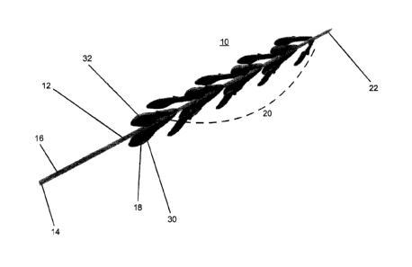
Referring now to the drawings, Figures 2a illustrates a thrombus retrieval
device
which is refelied to herein as device 10.
CA 02973125 2017-07-05
WO 2016/120864
PCT/1L2016/050073
12
Device 10 is configured suitable for entering, engaging/anchoring, dislodging
and collecting thrombus material from a blood vessel and in particular small
blood
vessels of the brain, as well as other blood vessels.
Device 10 includes an elongated body 12 having a handle 14 (user engaged
portion) at proximal end 16 and extensions 18 (16 shown) attached to a distal
portion 20.
Elongated body 12 includes a nose cone 22 for facilitating non-traumatic
delivery into a
vessel and also allows penetration into the occlusion/thrombus.
Extensions 18 are preferably arranged singly or as pairs (arrangements
including
3, 4, 5, 6 or more projections are also possible) around distal portion 20,
with each single
or pair rotated 0 - 180 degrees from an adjacent single pair.
Figure 2b illustrates an isolated extension 18 showing extension body 24
attached to a connector 26 via stem 27. Connector 26 can be glued or
mechanically
coupled to elongated body 12. Preferably, connector 26 is a cylindrical
connector which
is fitted around elongated body 12 and fixedly attached thereto or allowed to
swivel.
Extension 18 can alternatively be connected directly to elongated body 12
without use of
a connector.
Device 10 can further include a web like element interposed between extensions
18. Such an element can supplement the ability of device 10 to capture/harvest
dislodged occlusion material.
Extension body 24 is leaf-shaped and includes an inward curving tip 28 for
minimizing damage or irritation to the vessel wall when device 10 is pushed
and pulled
within the vessel. Inward curving tip 28 also functions to facilitate lodging
of extensions
18 into occlusion material (e.g. thrombus material) when device 10 is pulled
in a
proximal direction.
As is shown in Figure 2b, inner surface 30 of extension body 24 is concave to
increase surface contact area and drag forces when the device is pulled
proximally and to
scoop the occlusion material dislodged from the vessel wall.
Inner surface 30 can also be textured (e.g. micro/nano structures, not shown)
to
enhance surface contact area at the macro/micro/molecular level.
Outer surface 32 of extension body 24 (Figure 2a) is convex to decrease drag
forces when extensions 18 penetrate the thrombus mass. The convex outer
surface 32
also allows extensions 18 to fold into a compact streamlined configuration for
delivery
CA 02973125 2017-07-05
WO 2016/120864
PCT/1L2016/050073
13
into the vessel and occlusion. Additional hydrodynamic streamlining of
extensions 18
may be effected by providing outer surface 32 with one or more
bumps/protrusions/channels etc.
Extensions 18 can be fabricated from a single material or from two or more
materials. For example, in the embodiment shown in Figure 2a, extensions can
be
molded from a single material (e.g. silicone, teflon, nylon and any other
elastomer, metal
alloys such as Nitinol or elastomer with combination with metal alloys such as
Nitinol),
with the differential rigidity provided by varying the durometer of the
material (e.g.
molding stem 27 and optionally connector 26 from a different structure, a
silicone
having a higher Shore A value or increased thickness, or by using a different
material or
a combination of different materials).
Figure 2c is a magnification of inner surface 30 of extension body 24 (of the
region circled in Figure 2b) showing array 42 including a plurality of
projections 44.
Array 42 can be attached to a smooth or textured surface (such as the textured
surface
described above).
Projections 44 can be fabricated from the same material as the extensions 18,
or
from a different material. Examples of suitable materials for construction of
extension
18 include silicone, teflon, nylon and any other elastomer, metal alloys such
as Nitinol
or elastomer with combination with metal alloys such as Nitinol. The
projections can be
attached to the surface, co fonned therewith, or deposited thereupon using
well known
plasma deposition approaches.
Figures 3a-c illustrate a portion of device 10, an extension 18 thereof and a
magnified view of inner surface 30 of extension 18 showing projections. Figure
3d-e
are magnified views illustrating engagement between hook-like projections 44
and a
fibrin mesh (M) component of a thrombus.
Hook-like projections 44 can be 0.3-3.0 microns long, 0.2-1 microns in
diameter,
with a hook angle of 30-90 degrees relatively to the surface. The radius of
curvature of
the hook portion can be 0.2-1.0 microns.
When configured as hooks, projections 44 are designed to penetrate through the
openings in the fibrin mesh and hook onto the fibrin fiber when device 10 is
retracted.
Cooperative hooking of several projections 44 would substantially increase the
CA 02973125 2017-07-05
WO 2016/120864
PCT/1L2016/050073
14
engagement force between extension 18 and the thrombus mass thereby enabling
retrieval of the thrombus mass when device 10 is retracted out of the
vasculature.
Figures 4a-c illustrate a portion of device 10, an extension 18 thereof and a
magnified view of inner surface 30 of extension 18 showing cylindrical (rod-
like)
projections 44. Figures 4d-e are magnified views illustrating engagement
between
cylindrical projections 44 and a fibrin mesh component of a thrombus.
Cylindrical
projections 44 have a size similar to that of hook-like projections described
above.
Cylindrical projections 44 are designed to penetrate through the openings in
the
fibrin mesh and provide a large region of perpendicular contact between
projections 44
and the fibrin fibers. Cooperative penetrations of several projections 44
through several
openings in the fibrin mesh substantially increase the surface contact area
and the
engagement force between extension 18 and the thrombus mass thereby enabling
retrieval of the thrombus mass when device 10 is retracted out of the
vasculature.
Cylindrical projections 44 preferably include surface texturing or protrusions
45
(e.g. downward-pointing protrusions, see Figures 5b, d, k, m and n) which
engage the
fibrin fiber when device 10 is retracted.
Figures 5a-s illustrate several embodiments of projections 44. Each embodiment
is characterized by a specific configuration which facilitates engagement
between
projection 44 and the fibrin mesh. For example, projection 44 can be
configured with
side or downward pointing side protrusions 45 (Figures 5b, d and 5k-n), a
bulbous or
mushroom-shaped tip (Figure 5j), a branching tip (Figure 5o), a loop-gate
(e.g.
'carabiner') lock (Figure 5p), an upright or inverted tree-like structure
(Figure 5b, d and
5c respectively), sideward or downward projecting hair-like structures (Figure
5s),
comb-like structure, scales, and the like. A projection 44 can include one or
more of
these structures arranged along a length thereof.
These structures facilitate engagement between projections 44 and the fibrin
mesh component of a thrombus by collectively penetrating openings of the mesh
and
engaging mesh fibers.
It will be appreciated that not every projection 44 will engage the fibrin
mesh
since an orientation of array 42 with respect to the fibrin mesh of the
thrombus cannot be
controlled or predetermined. However, an array 42 which includes several
thousand
projections 44 or more, will likely engage the fibrin mesh of a thrombus
through at least
CA 02973125 2017-07-05
WO 2016/120864
PCT/1L2016/050073
several hundred projections 44 thus substantially increasing the 'adhesive'
force between
each extension and the thrombus.
In addition, in order to maximize engagement to a fibrin mesh of unknown
orientation, the present device can include extensions 18 on which projections
44 are
5 oriented in different directions, or include extensions having tips 46
(e.g. Figure 5c-d, m)
that guide projections 44 into the openings of the fibrin mesh.
This structural asymmetry of an array 42 enables engagement with the mesh
through one or more directions and thus can maximize engagement when the
specific
orientation of the fibrin mesh with respect to an extension 18 is unknown. In
addition,
10 since device 10 typically includes a number of extensions 18, having
various
configurations of array 42 on several extensions 18 again maximizes the
statistical
probability of mesh penetration by projections 44.
As is mentioned hereinabove, the embodiment of device 10 of Figure 2a is
configured for use in clearing obstructions in a blood vessel, preferably a
small brain
15 artery that is 0.5-7 millimeter in diameter. As such, elongated body 12
of device 10 is
preferably 10-200 centimeter in length, 0.5-7 millimeter in diameter when in
closed
configuration, while extensions 18 are preferably 0.2-30 mm in length. The
length of
extension body 24 is preferably 0.1-30 mm and the width (at the widest
thereof) is
preferably 0.05-20 mm. Stem portion 27 is preferably 0.1-20 mm in length and
0.02-20
mm in width (at the base).
Extensions 18 can be folded against elongated body 12 to an overall diameter
of
0.5-7 millimeter. When folded, device 10 can be packed into a 1.5-22 F sheath
for
delivery through an access site. Once pushed out of the sheath, extensions 18
are folded
outward to a position constrained by stem portion 27 (or vessel wall) while
distal portion
20 is advanced to the site of occlusion. Since extension body 24 includes a
non-
traumatic tip 28 (fabricated from a soft material such as silicone), advancing
device 10
in the distal direction (towards occlusion) does not traumatize or irritate
the vessel wall.
Once in position, pulling on handle/proximal catheter part 14 deploys
extensions 18 to
an angle limited by stem portions 27 or the vessel wall. Such an angle can be
90 degrees
or les, preferably 30-45 degrees. At such an angle, tip 28 is angled inward to
eliminate
trauma and irritation to vessel wall.
CA 02973125 2017-07-05
WO 2016/120864
PCT/1L2016/050073
16
The flexible nature of extensions 18 permits the device to automatically adapt
to
the inner diameter of the blood vessel in which device 10 is situated.
Stem portion 27 and/or extension body 24 can also be configured such that when
folded against elongated body 12, the longitudinal axis of extension body 24
is angled
with respect to the longitudinal axis of elongated body 12. This increases the
exposure of
inner surface 30 to the biological fluid in the vessel and to the occlusion
material and
increases drag and likelihood of deployment when device 10 is pulled in a
proximal
direction.
A roll angle can also be added such that each extension 18 has an "angle of
attack" relative to the movement vector (angle range 0-90 degrees) i.e. to the
anterior
edge of extension body 24 relative to movement of device 10. The angle of
attack in the
forward motion (when device 10 is pushed towards occlusion) will have
hydrodynamic
features and a curve design that will ensure an ability to optimally penetrate
and
minimally disrupt the thrombus structure. When device 10 is pulled proximally,
the
angle of attack (which is the opposite edge) can be shaped in a more acute
curve
structure in order to allow optimal drag forces of the thrombus on each
extension 18
thereby ensuring opening thereof. Extensions 18 can also be configured to
spiral around
elongated body 12.
The size shape and properties of extensions 18 and of projections 44 can be
configured according to the biological vessel and occlusion properties. For
example,
there are two type of thrombus occlusions, a 'red' thrombus (fresh, acute
whole blood
thrombus) and a 'white' thrombus (relatively chronic embedded with cholesterol
and
calcium). Extensions 18 of device 10 as well as projections 44 can be
configured with
rigidity properties that match the viscosity ranges of the thrombus.
When configured as a catheter, device 10 includes a lumen for accepting a
guidewire for guiding device 10 to a target occlusion within a vessel. The
lumen can
traverse the entire length of elongated body 12 (when use with an over-the-
wire system)
to an guidewire inlet opening in a proximal end of elongated body or
alternatively,
lumen can traverse a portion thereof (when used with a rapid exchange system)
to a
guidewire inlet opening at a side wall along a length of elongated body 12.
The lumen can also include one or more holes or other opening along a portion
of elongated body proximal to extensions 18. Such holes can be in fluid
communication
CA 02973125 2017-07-05
WO 2016/120864
PCT/1L2016/050073
17
with an opening at distal end and would thus enable blood to flow around the
occlusion
mass once extensions 18 penetrate the occlusion and the distal end crosses the
occlusion
and is positioned at its distal side.
This will allow reperfusion of the ischemic brain tissue located distally to
the
occlusion site. The relatively low flow of blood (through the catheter)
provides
controlled low flow, low pressure reperfusion to the Penumbra brain tissue
which is at a
metabolic "shutdown" state and thus might be vulnerable to high pressure
systolic blood
flow. This will prepare the tissue for restoration of full flow following
removal of the
thrombus.
In cases where delivery is effected through a catheter or guide tube (guiding
catheter), delivery and navigation of device 10 can be effected without a
guidewire.
In any case, a handle 14 or proximal portion of elongated body 12 can be used
to guide
device 10 (whether over a wire or not) through the vessel and position distal
portion 20
at a site of occlusion.
Device 10 can also include radio-opaque markers (e.g. gold, platinum, iridium
or
combined with the polymer itself or other radio-opaque markers) mounted on the
distal
end of elongated body 12 (at distal end).
The markers can be mounted on ends of extensions 18 (e.g. at tips 28). When
distal portion 20 is positioned outside of the occlusion, extensions 18 extend
out and
thus when visualized (fluoroscopy) the markers are a predetermined distance
apart (e.g.
several millimeters). When distal portion 20 is positioned inside an
occlusion,
extensions 18 fold against elongated body 12 and thus when visualized
(fluoroscopy) the
distance between the markers is reduced.
Alternatively, one of the markers can be mounted on a foldable wire (e.g.
Nitinol, platinum, other metal alloy or polymer wires) extending radially
outward from
elongated body 12 while a second marker can be attached to elongated body 12.
When
distal portion 20 is positioned inside an occlusion, the marker wire is folded
against
elongated body 12 and brought into proximity to the second marker and
optionally a
third marker. The distance between the markers can be visualized (fluoroscopy)
to
determine the extent of folding of the extension.
Marker material (e.g. iridium or platinum) can also be included in the
material
used to fabricate extensions 18 in order to facilitate identification thereof
by a surgeon.
CA 02973125 2017-07-05
WO 2016/120864
PCT/1L2016/050073
18
In any case, the markers assist the clinician in determining the correct
placement
of device 10 within a blood vessel and indicate when distal portion 20 enters
an
occlusion and extensions 18 are lodged therein.
In order to increase the ability of extension 18 to collect occlusion
material, inner
surface 30 and/or projections 44 can be coated with a substance that can bind
the
occlusion material. For example, in the case of a thrombus occlusion, inner
surface 30
and/or projections 44 can be coated with fibrin or fibrin derivatives.
Device 10 can be used to clear a thrombus from an artery as follows. A guide
catheter or guidewire is advanced from an access site (e.g. in a femoral
artery) to the
carotid artery under angiography. Device 10 is then inserted over-the-wire or
through the
guide catheter and navigated to the site of the thrombus. The surgeon then
advances the
distal end of device 10 into the thrombus until the distal end of device 10
reaches the
distal end of the thrombus (as visualized via the radio-opaque markers
described above).
The surgeon then applies a gentle pulling force on device 10 to open
extensions 18 and
lodge and engage/anchor them within the thrombus. The device is then pulled
along
with the trapped thrombus.
Device 10 of the present invention can also be configured for use in clearing
any
type of occlusion from any biological vessels.
In order to enable such functionality, the present device would be designed
with
surface projections that match the specific architecture of the occlusion.
Prior art devices which utilize macrostructures (e.g. hooks, bristles) to
pierce
through and engage the thrombus are more likely to cause embolic events since
piercing
through the thrombus mass can lead to thrombus disintegration.
The present device encapsulates the thrombus and externally engages it through
numerous points of contact using texture-specific micro and nano structures
positioned
on the surface of leaf-like extensions.
Thus, with the present device, engagement of the thrombus mass does not
compromise the integrity of the thrombus and use thereof may not require
additional use
of embolic protection or entrapment devices such as aspirators and traps which
complicate and lengthen the procedure and can lead to serious complications
such as
vessel injury.
As used herein the term "about" refers to 10 %.
CA 02973125 2017-07-05
WO 2016/120864
PCT/1L2016/050073
19
Additional objects, advantages, and novel features of the present invention
will
become apparent to one ordinarily skilled in the art upon examination of the
following
examples, which are not intended to be limiting.
EXAMPLES
Reference is now made to the following examples, which together with the above
descriptions, illustrate the invention in a non limiting fashion.
EXAMPLE I
Additive Manufacturing of The Present Device
Several configurations of the present device were designed using CAD software
(SolidWorksTm) and additive manufacturing (also known as 3D printing)
approaches
were tested for the ability to 'print' the entire device.
Since the projections and protrusions of the present device are micrometric or
nanometric in scale, a 3D printing approach capable of such resolution was
sought.
Several devices on the market are capable of 3D printing silicone or another
suitable polymer at a resolution of 100 nanometers including devices by WACKER
CHEMIE and Ingenieure GmbH; Fripp Design Research; NanoScribe, Old World Labs
and more.
For example, the OWL MC-2 (old World Labs) has the following manufacturer's
specifications:
= Resolution: 100 nm;
= Precision: 100 nm mechanical and software capability;
= Accuracy: +/- 50 nm
= Repeatability: 99%
= Build Volume: 6x6x6 in
= Build Speed: linch3/hr.
= Build Materials: Photopolymer
Additive manufacturing (AM) provides several advantages in manufacturing of
the present device:
(i) an entire device including projections and protrusions can be
manufactured within a few hours;
CA 02973125 2017-07-05
WO 2016/120864
PCT/1L2016/050073
(ii) it can be used to precisely control the rigidity of different parts of
the
device (e.g. stems and leaves);
(iii) any shape projection and/or protrusion can be manufactured to match
any
occlusion texture/composition;
5 (iv) projection/protrusion shape and size can be matched to specific
occlusion;
(v) device or projection portion thereof could be printed in-
hospital to match
specific patient needs (e.g. vessel size, occlusion type).
Figures 6a-c illustrate a configuration of the present device which is
optimized
10 for additive manufacturing.
The portion of the device shown in Figure 6a includes a tube with 6 pairs of
extensions (leaves). The tube and extensions are printed as a mono-structure
and can be
connected to a microcatheter for use (carrier tube can be fitted over a
microcatheter).
This specific design is optimized for removing occlusions in 2.5 mm blood
vessels. The
15 extension pairs are rotationally offset 90 degrees from each other to
ensure optimized
occlusion engagement and collection. The inner surface of each extension is
manufactured with projections (44) which are conical in shape and are randomly
yet
homogenously distributed on the surface (Figure 6b). Since these projections
are 3D
printed (along with the extension and carrier tube), exact structure and
dimensions can
20 be achieved. The diameter of the projections is 100 microns and the
height 200 microns;
average distance between projections is 300 microns. As is shown in Figure 6c,
each
projection is 'printed' with surface protrusions (45) which are mushroom-
shaped (stalk
and cap with rounded mushroom 'cap') and are 4 microns in height, 2 microns in
diameter (at base) and 3 microns in diameter (at top). Average distance
between
protrusions is 10 microns.
The size and shape parameters of the device shown in Figures 6a-c can be
varied
according to the occlusive material and patient. Occluding materials (e.g.
blood clot in
its various types, biological stones, foreign body and more) have different
characteristics
and physical/chemical properties which can vary from patient to patient. The
size of the
occluded vessel also varies from patient to patient.
Thus, in order to optimize engagement between the extensions of the present
device and the occlusive material and optimize delivery and retrieval of the
present
21
device, the overall shape and size of the device as well as the shape and size
of the
extensions, projections and protrusions can be matched to the patient and/or
occlusion.
The size of the vessel (diameter) and the shape, size and texture of the
occlusive
material can be determined from noninvasive imaging (including CT, MRI, Ultra
Sound,
Nuclear medicine and more); sampling (biopsy, microscopy) can be used to
determine
the composition of the occlusive material. Once the vessel size is determined
and the
occlusion is typed (size, shape, texture, composition), a suitable matching
device design
will be generated (including size and geometrical configuration or extensions,
projections and protrusions) and printed using additive manufacturing.
This approach could be used in real time in a hospital setting to manufacture
and
employ a patient-specific device optimized for retrieving a specific occlusion
in a
specific vessel.
It is appreciated that certain features of the invention, which are, for
clarity,
described in the context of separate embodiments, may also be provided in
combination
in a single embodiment. Conversely, various features of the invention, which
are, for
brevity, described in the context of a single embodiment, may also be provided
separately or in any suitable subcombination.
Although the invention has been described in conjunction with specific
embodiments thereof, it is evident that many alternatives, modifications and
variations
will be apparent to those skilled in the art.
In addition, citation or identification of any reference in this application
shall
not be construed as an admission that such reference is available as prior art
to the
present invention.
Date Recue/Date Received 2022-06-08