Note: Descriptions are shown in the official language in which they were submitted.
CA 02973128 2017-07-06
WO 2016/109877
PCT/CA2015/000012
OPTICAL PROBES FOR CORRIDOR SURGERY
FIELD
[0001] The specification relates generally to surgical instruments, and
specifically to
optical probes for corridor surgery.
BACKGROUND
[0002] Probes for optical measurements of tissue may provide a wide variety of
applications and modalities, for providing clinicians with details regarding
the state of
tissue to guide diagnosis, treatments and/or surgery. The optical modalities
for which
probes have been developed include broadband spectroscopy (ultraviolet,
visible, near
infrared, and short wave infrared), fluorescence, Raman spectroscopy, optical
coherence
tomography, photoacoustic tomography, coherence anti-Stokes Raman
spectroscopy,
confocal microscopy, among others.
[0003] Port-based surgery is a minimally invasive surgical technique where a
port
(generally a cylindrical plastic tube open on both ends) is introduced to
access the
surgical region of interest. Unlike other minimally invasive techniques, such
as
laparoscopic techniques, the port diameter is larger than the tool diameter,
allowing bi-
manual tool manipulation within the port. Hence, the tissue region of interest
is
accessible through the port. While a wide variety of optical probes have been
developed
for numerous modalities, optical probes for port-based surgery have not been
developed.
For example, current optical probes are not compatible with port-based surgery
due to
sizes of the probe, sterilization tolerance, lack of signal enhancing
mechanisms, lack of
integration with surgical tools, lack of position and orientation tracking,
and lack
integration with other optical systems. At present the lack of these features
hinders and
restricts the use and utility of optical probes for port-based surgery.
Similar respective
restrictions may also exist with other types of surgical techniques.
SUMMARY
[0004] The present specification provides an optical probe which is attachable
to a
surgical instrument, and specifically, a device that includes an optical probe
which may
be used for port surgery, and other types of surgeries. The optical probe may
be used for
1
CA 02973128 2017-07-06
WO 2016/109877
PCT/CA2015/000012
a variety of optical modalities including, but not limited to: ultraviolet
(UV), visible
(VIS), near infrared (NIR), and shortwave infrared (SWIR) broadband
spectroscopy;
Raman spectroscopy; fluorescence; both spectral domain and swept source
optical
coherence tomography (OCT), photoacoustic tomography, laser induced breakdown
spectroscopy (LIBS), surface enhanced Raman spectroscopy (SERS), coherent anti-
Stokes Raman spectroscopy (CARS), stimulate Raman scattering (SRS), probe-
based
microscopy, probe-based confocal microscopy, and the like. The probe may be
adapted to
include signal enhancing materials, nanoparticles, and the like, that may be
affixed to the
end of the probe to provide enhancement of an optical signal overall and/or to
conjugate
with specific biological molecules of interest and emit a molecular specific
signal. The
probe may be adapted to track a position thereof as part of a surgical
navigation system.
As positions of the probes are tracked, and the location of the measurements
is known,
and methods to relate a volume from which the optical signal originated to
pixels or
voxels from other imaging modalities may be utilized.
[0005] The probe may include a single optical fiber or collection of optical
fibers that
delivers illumination light to tissue, including, but not limited to, through
a port (e.g.,
broadband UV, VIS, NIR, and/or SWIR; laser; Raman excitation laser; OCT
broadband
or swept source light; photoacoustic excitation; fluorescence excitation; LIBS
excitation,
etc.), as well as A single optical or collection of optical fibers that
collect an optical
signal from tissue being accessed (e.g. through a port), including, but not
limited to, light
reflected from the tissue. These optical fibers may have a variety of
arrangements; for
example, the optical fibers may be arranged in a circular fashion with
illumination light
fiber(s) in the centre and collection light fiber(s) arranged around the
illumination
fiber(s). Optical lenses or lenslets may be used to control the solid angle of
illumination
and collection, as well as for focussing or collimation of either the
illumination or
collection light.
100061 The probe is generally inserted into a patient, for example through the
surgical
port, and placed in contact or short stand-off from the tissue to provide
tissue
characterization and differentiation, to assist in surgical decision making by
providing an
indication of tissue status. For example, a surgeon would identify a region of
tissue on
which they wish to perform a measurement, bring the probe into contact or
short stand-
2
CA 02973128 2017-07-06
WO 2016/109877
PCT/CA2015/000012
off from the tissue, and measure the optical signal. The measured signal may
be
processed at a light analyzer, and the like, and displayed as part of a
surgical visualization
system.
[0007] A size and/or geometry of the probe is controlled and/or configured to
enable
insertion of the probe into a port. For example, the probe may have be
cylindrical and
have a diameter of less than or equal to about 5 mm, and a length of greater
than or equal
to about 5 cm.
[0008] In this specification, elements may be described as "configured to"
perform one
or more functions or "configured for" such functions. In general, an element
that is
configured to perform or configured for performing a function is enabled to
perform the
function, or is suitable for performing the function, or is adapted to perform
the function,
or is operable to perform the function, or is otherwise capable of performing
the function.
[0009] It is understood that for the purpose of this specification, language
of "at least one
of X, Y, and Z" and "one or more of X, Y and Z" may be construed as X only, Y
only, Z
only, or any combination of two or more items X, Y, and Z (e.g., XYZ, XY, YZ,
ZZ, and
the like). Similar logic may be applied for two or more items in any
occurrence of "at
least one ..." and "one or more..." language.
[0010] An aspect of the present specification provides a device comprising: a
surgical
tool mounting adaptor configured for mounting to a surgical tool; an optical
probe
attached to the surgical tool mounting adaptor, the optical probe comprising:
an optical
interface end; an optical output end, distal the optical interface end, the
optical output end
comprising illumination optics and collection optics, the illumination optics
configured to
illuminate tissue proximal the optical output end, the collection optics
configured to
collect an optical signal from the tissue; one or more illumination optical
fibers
configured to convey illumination light from the optical interface end to the
illumination
optics; and, one or more collection optical fibers configured to convey the
optical signal
collected by the collection optics to the optical interface end.
[0011] The surgical tool mounting adaptor may be further configured for
removable
attachment to the surgical tool.
[0012] The surgical tool mounting adaptor may comprise a sleeve configured for
removable attachment to the surgical tool.
3
CA 02973128 2017-07-06
WO 2016/109877
PCT/CA2015/000012
[0013] The surgical tool mounting adaptor may be further configured to hold
the optical
probe in a fixed position relative to the surgical tool.
[0014] The surgical tool mounting adaptor may be configured for mounting to
one or
more of: a suction device, scissors, microscissors, a bipolar surgical tool, a
drill, a
resection device, a shaving device, a forceps, an ultrasonic cutting device,
and an
aspirator.
[0015] The device may further comprise a mount configured to removably attach
the
optical probe to the surgical tool mounting adaptor.
[0016] The optical probe may comprise a sleeve and a rigid optical probe
portion, the
sleeve configured to removably hold the rigid optical probe portion. The
sleeve may be
one or more of sterilizable and consumable, the sleeve may be further
configured to seal
the rigid optical probe portion from the tissue. The optical output end may be
integrated
into the sleeve, and the rigid optical probe portion may comprise the one or
more
illumination optical fibers and the one or more collection optical fibers. The
rigid optical
probe portion may be sealed.
[0017] The optical probe may be cylindrical, a diameter of the optical probe
is less than
or equal to about 5 mm, and a length of a rigid portion of the optical probe
is greater than
or equal to about 5 cm.
[0018] The optical interface end may comprise a connector configured to:
connect the
one or more illumination optical fibers to an illuminator configured to
provide the
illumination light; and connect the one or more collection optical fibers to a
light analyzer
configured to receive and analyze the optical signal from the one or more
collection
optical fibers.
[0019] The optical output end may further comprise a shield configured to one
or more
of: contact the tissue; and maintain a standoff distance between the tissue
and one or
more of the illumination optics and the collection optics.
[0020] The illumination optics and the collection optics may comprise one or
more of a
lens, a mirror, and a prism.
[0021] One or more of the illumination optics and the collection optics may
comprise at
least one of electromechanical components and MEMS (micro-electromechanical
systems) components to move optical components.
4
CA 02973128 2017-07-06
WO 2016/109877
PCT/CA2015/000012
[0022] The illumination optics and the collection optics may be configured to
vary one or
more of: a focal point, a depth of the focal point in the tissue, an
illumination spot size, a
voxel of the tissue, a numerical aperture of the collection optics, a
direction of the
illumination light, and a direction of the optical signal collected from the
tissue.
[0023] The optical output end may comprise one or more materials configured to
one or
more of: enhance the optical signal from the tissue; and, provide one or more
of a
molecular, protein, and cellular binding-specific signal. The materials may
comprise one
or more of: signal enhancing materials, bio-conjugation specific materials,
binding
materials, antibodies, arrays, microarrays, assays, and hollow-core photonic
crystal
fibers.
[0024] The device may fu rther comprise a mount configured to removably attach
a
tracking device to one or more of the surgical tool mounting adaptor and the
optical
probe.
[0025] The device may fu rther comprise a mount configured to removably attach
a
tracking device of a surgical navigation system to one or more of the surgical
tool
mounting adaptor and the optical probe.
CA 02973128 2017-07-06
WO 2016/109877
PCT/CA2015/000012
BRIEF DESCRIPTIONS OF THE DRAWINGS
[0026] For a better understanding of the various implementations described
herein and to
show more clearly how they may be carried into effect, reference will now be
made, by
way of example only, to the accompanying drawings in which::
[0027] Fig. 1 depicts a device that includes an optical probe for corridor
surgery,
according to non-limiting implementations.
[0028] Fig. 2 depicts the device of Fig. 1, in use, according to non-limiting
implementations.
[0029] Fig. 3 depicts a cross-section of the optical probe, according to non-
limiting
implementations.
[0030] Fig. 4 depicts an optical probe adapted with a shield, according to non-
limiting
implementations.
[0031] Fig. 5 depicts various illumination and collection optics of the
optical probe,
according to non-limiting implementations.
[0032] Fig. 6 depicts an alternative device that includes an optical probe for
corridor
surgery, according to non-limiting implementations.
[0033] Fig. 7 depicts an alternative device that includes an optical probe for
corridor
surgery and a mount for receiving a tracking device used with surgical
navigational
system, according to non-limiting implementations.
6
CA 02973128 2017-07-06
WO 2016/109877
PCT/CA2015/000012
DETAILED DESCRIPTION
[0034] The OCT (optical coherence tomography) technique described herein may
be
used to specifically visualize tissue exhibiting structural organization.
Examples of such
tissue structures include tendons that are attached to bones. Other examples
of tissue that
exhibit structural organization include ligaments, muscle, cartilage, tissue
connective
membrane, nerves, retina, blood vessel walls, some bone structures, trachea,
esophagus,
tongue and teeth.
[0035] Polarization Sensitive-OCT (PS-OCT) is a subset of OCT that detects
different
polarizations of light reflected from the sample. OCT in general does not
necessary detect
the intensity from the different polarization of light, but rather detects the
intensity of
randomly polarized light. PS-OCT commonly generates a heat map or pseudo
colored
image (reference: "Correlation of collagen organization with polarization
sensitive
imaging of in vitro cartilage: implications for osteoarthritis," W. Drexler
et.al, The
Journal of Rheumatology, Vol. 28, No. 6, 1311-1318) where tissue structures
with high
degree of organization appear highlighted. This system may be used in
orthopedic
surgery to visualize tendons and optionally avoid unintentional damage to this
tissue
during a procedure. These identified regions of tissue exhibiting high level
of structural
organization (e.g. tendons and ligaments that are often located near skeletal
structure)
may be used in conjunction with a priori information, such as known points of
attachment
of tendons to bones, to geometrically correlate PS OCT images to CT and MR
images
where bones are easily imaged.
[0036] The insertion sites, tendon-bone junctions and ligament-bone junctions,
are
known as entheses. The anatomical locations of entheses are well known and
landmarks
may be identified on the bone in the vicinity of these attachment points
(reference:
"Anatomy and biochemistry of enthuses," Michael Benjamin, Ann Rheum Dis 2000,
Vol.
59, Issue 12, pg:995-999). Hence, this a priori anatomical information about
the position
of the tendon or ligament relative to bone structures in the vicinity may be
used to
register intraopertive PS-OCT image of the tendons or ligaments with pre-
operative
images obtained using other modalities that accurately image the bone
structures.
[0037] For example the tendon-bone junction in the Achilles tendon enthesis is
immediately proximal to the superior tuberosity. This region is characterized
by a highly
7
CA 02973128 2017-07-06
WO 2016/109877
PCT/CA2015/000012
irregular interface at the attachment points or junction. This characteristic
structure of the
bone may be used to identify the junction where the tendon attaches to the
bone. The
geometric correlation of images that are thus obtained using different
modalities, and
often at different scales, is known as image registration or image fusion.
[0038] Common methods for multi-modal image registration mentioned above
include
those described in "Multi-modal image registration for pre-operative planning
and image
guided neurosurgical procedures," Risholm, et.al, Neurosurg Clin N Am, 2011,
April;
22(2): 197-206 and "Image registration of ex-vivo MRI to sparsely sectioned
histology of
hippocampal and neocortical temporal lobe speciments," Goubran et.al,
NeuroImage, 83
(2013); 770-781. Broad classes of image registration methods for medical
images is also
described in detail in "A survey of medical image registration," Maintz et.al,
Medical
Image Analysis (1998), Vol. 2, No. 1, pp: 1-36.
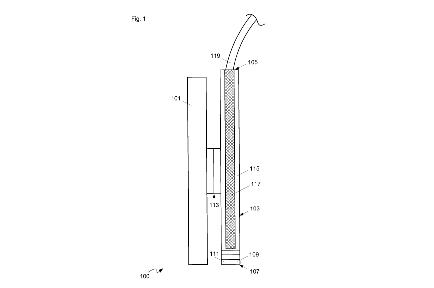
[0039] Attention is directed to Fig. 1 which depicts a schematic diagram of a
device 100
comprising: a surgical tool mounting adaptor 101 configured for mounting to a
surgical
tool; an optical probe 103 attached to surgical tool mounting adaptor 101,
optical probe
103 comprising: an optical interface end 105; an optical output end 107,
distal optical
interface end 105, optical output end 107 comprising illumination optics 109
and
collection optics 111, illumination optics 109 configured to illuminate tissue
proximal
optical output end 107, collection optics 111 configured to collect an optical
signal from
the tissue; one or more illumination optical fibers (not visible in Fig. 1)
configured to
convey illumination light from optical interface end 105 to illumination
optics 109; and,
one or more collection optical fibers (not visible in Fig. 1) configured to
convey the
optical signal collected by collection optics 111 to optical interface end
105. Surgical tool
mounting adaptor 101 will be interchangeably referred to hereafter as adaptor
101. One
or more illumination optical fibers will be interchangeably referred to
hereafter as
illumination optical fibers; and one or more collection optical fibers will be
interchangeably referred to hereafter as collection optical fibers.
[0040] As depicted, device 100 further comprises a mount 113 configured to
removably
attach optical probe 103 to adaptor 101.
[0041] Furthermore, as depicted, device 100 further comprises a sleeve 115 and
a rigid
optical probe portion 117, sleeve 115 configured to removably hold rigid
optical probe
8
CA 02973128 2017-07-06
WO 2016/109877
PCT/CA2015/000012
portion 117. In Fig. 1, sleeve 115 is depicted as transparent to show rigid
optical probe
portion 117 inside, however, sleeve 115 may be opaque and the optical
properties of
sleeve 115 are generally non-limiting, other than optical properties of
optical output end
107. Indeed, as depicted, optical output end 107 is integrated into sleeve
115, and rigid
optical probe portion 117 comprises the one or more illumination optical
fibers and the
one or more collection optical fibers. However, optical probe 103 may extend
beyond
sleeve 115 and rigid optical probe portion 117; for example, optical fibers of
optical
probe 103 may extend from optical interface end 105.
[0042] In other implementations, device 100 could comprise a flexible device
where the
direction and angle of bend may be controlled by tension members attached to a
wall of
optical probe 103. The tension members could be actuated mechanically or could
be
made of memory metal alloys (e.g. including, but not limited to, Nitinol) that
is actuated
electrically.
[0043] As depicted, mount 113 may comprise a first portion connected to
adaptor 101
and a second portion connected to optical probe 103 and/or sleeve 115, the
first portion
and the second portion configured to attach to one another. Mount 113 may hen
cc
comprise a magnetic mount, a slot and groove connector, respective clips, and
the like.
However, in other implementations, mount 113 may comprise a clip, and the
like,
attached to adaptor 101 configured to hold optical probe 103 and/or sleeve
115;
conversely, in yet further implementations, mount 113 may comprise a clip, and
the like,
attached to optical probe 103 and/or sleeve 115 configured to hold adaptor
101.
[0044] As depicted, optical interface end 105 comprises a connector 119
configured to:
connect the one or more illumination optical fibers to an illuminator
configured to
provide the illumination light; and connect the one or more collection optical
fibers to a
light analyzer configured to receive and analyze the optical signal from the
one or more
collection optical fibers. As depicted, connector 119 is located at optical
interface end
105 and/or at an end of rigid optical probe portion 117; however, in other
implementations, connector 119 may be located anywhere along optical fibers
extending
from rigid optical probe portion 117. While details of connector 119 are not
depicted,
connector 119 may comprise an optical fiber connector, and the like.
9
CA 02973128 2017-07-06
WO 2016/109877
PCT/CA2015/000012
[0045] Attention is next directed to Fig. 2, which is substantially to Fig. 1,
with like
elements having like numbers, and depicts a schematic diagram of device 100 in
use with
a surgical port 200, which is depicted schematically in cross-section, and a
surgical tool
201 to interact with tissue 203 through port 200. However, while present
implementations
are described with reference to port-based corridor surgery, device 100 may be
used with
other types of corridor and/or invasive and/or non-invasive surgeries. Fig. 2
further
depicts illumination light 205 from illumination optics 109 illuminating a
portion of
tissue 203 with which surgical tool 201 is interacting, and an optical signal
207 collected
from tissue 203 by collection optics 111 conveyed to a light analyzer 209
where optical
signal 207 is visually provided, for example as a function of wavelength.
While not
depicted, it is assumed that the system depicted in Fig. 2 further comprises
an illuminator
configured to provide illumination light 205, the illuminator connected to
illumination
optical fibers using connector 119. Hence, connector 119 is configured to:
connect the
illumination optical fibers to the illuminator configured to provide
illumination light 205;
and connect the collection optical fibers to light analyzer 209 configured to
receive and
analyze optical signal 207 from the collection optical fibers.
[0046] Specifically, in Fig. 2, adaptor 101 is mounted to surgical tool 201, a
handle (e.g.
a handle of a scissors) of surgical tool 201 depicted as protruding from a
first end of
adaptor 101 and a tissue interaction end (e.g. blades of the scissors) of
surgical tool 201
protruding from a second end of adaptor 101, distal the first end. Hence, it
is apparent
that, in depicted implementations, adaptor 101 comprises a sleeve configured
for
removable attachment to a surgical tool 201. While surgical tool is depicted
as a scissors,
surgical tool mounting adaptor 101 may be configured for mounting to one or
more of: a
suction device, a scissors, a microscissors, a bipolar surgical tool, a drill,
a resection
device, a shaving device, a forceps, an ultrasonic cutting device, and an
aspirator. While
adaptor 101 is depicted as a sleeve, adaptor 101 may comprise other devices
for
mounting optical probe 103 to a surgical tool including, but not limited to,
mechanical
clamps, magnetic clamps, and the like, with or without a sleeve. It is further
appreciated
that, while not depicted, adaptor 101 comprises one or more devices for fixing
a position
of adaptor 101 on surgical tool 201, including, but not limited to a clamp. In
general,
adaptor 101 is further configured for removable attachment to surgical tool
201
CA 02973128 2017-07-06
WO 2016/109877
PCT/CA2015/000012
[0047] From Fig. 2, it is apparent that a diameter of optical probe 103 is
less than an
inner diameter of port 200, and further that a length of a rigid portion of
optical probe 103
is greater than a length of port 200. Surgical ports may have inner diameter
of 13.5 mm
and lengths of 5, 6, and 7.5 cm, however, ports of other diameters and lengths
are
possible. In some implementations, a diameter of optical probe 103 is no more
than about
1/3 a diameter of a port with which optical probe 103 is to be used. In
specific non-
limiting implementations, optical probe 103 is cylindrical, a diameter of the
optical probe
is less than or equal to about 5 mm, and a length of the optical probe is
greater than or
equal to about 5 cm. However, optical probe 103 may be adapted for use with
surgical
ports of different diameters and different lengths; hence, for example, when a
surgical
port of 7.5 cm is to be used, a rigid portion of optical probe 103 may be
greater than or
equal to 7.5 cm. Indeed, different optical probes of different lengths (and/or
different
diameters and/or different geometries) may be provided, each adapted for use
with a
given surgical port.
[0048] It is further apparent from Fig. 2 adaptor 101 is further configured to
hold optical
probe 103 in a fixed position relative thereto and/or relative to surgical
tool 201. For
example, adaptor 101 may be rigid, at least when attached to surgical tool
201, and
adaptor 101 may be configured to hold optical probe 103 such that optical
output end 107
is proximal and/or adjacent a tissue interaction end of surgical tool 201
and/or such that
illumination light 205 is illuminating an area proximal a tissue interaction
end of surgical
tool 201. Hence, optical signal 207 is always received from a portion of
tissue 203 that is
interacting with, and/or being acted on, and/or being cut by, surgical tool
201. And, as the
tissue interaction end of surgical tool 201 moves, optical output end 107 of
optical probe
103 moves in a similar fashion.
[0049] Such relative motion may be further translated from adaptor 101 to
optical probe
103 using mount 113, which may be rigid and/or hold optical probe 103 in a
fixed
position relative thereto and/or relative to surgical tool 201.
100501 It is furthermore appreciated that the sizes and geometry of components
of Figs. 1
and 2 are not to scale. Hence, while in Fig. 2, illumination light 205 is
depicted as being
steered to the left (relative to a tissue interaction of surgical tool 201),
such steering is
depicted to illustrate that illumination light 205 is illuminating an area
proximal a tissue
11
CA 02973128 2017-07-06
WO 2016/109877
PCT/CA2015/000012
interaction end of surgical tool 201, and such steering may or may not occur
depending
on optical components of illumination optics 109 and collection optics 111,
and/or a
position of the tissue interaction end of surgical tool 201. For example, a
geometry and/or
size of adaptor 101 and optical probe 103 (and/or mount 113) may be adapted so
that
illumination light 205 illuminates an area of tissue 203 that is proximal the
tissue
interaction end of surgical tool 201 without changing a direction of
illumination light
205.
[0051] From Figs. 1 and 2, it is apparent that, as depicted, sleeve 115 holds
rigid optical
probe portion 117 therein such that an end of rigid optical probe portion 117
distal optical
interface end 105 interacts with optical output end 107 integrated into sleeve
115. In
some of these implementations, rigid optical probe portion 117 may be
removable from
sleeve 115 such that sleeve 115 and/or rigid optical probe portion 117 may be
modular
with other sleeves and/or other rigid optical probes. Specifically, sleeve 115
may be
removed from rigid optical probe portion 117, and optically further detached
from
adaptor 101, and sterilized separately there from.
[0052] Alternatively sleeve 115 may be consumable, so that sleeve 115 may be
replaced
each time surgery occurs and thrown away thereafter. In some of these
implementations,
illumination optics 109 and collection optics 111 may be integrated with rigid
optical
probe portion 117 so that illumination optics 109 and collection optics 111
are not
disposed of after each surgery; in these implementations, an end of sleeve 115
comprises
a window so that illumination light 205 and optical signal 207 may pass
through window.
[0053] In these implementations, rigid optical probe portion 117 may be sealed
for easy
sterilization and/or so that sterilization of rigid optical probe portion 117
need not be as
thorough as sterilization of sleeve 115, which is in contact with, and/or
adjacent to, tissue
203, and further shields rigid optical probe portion 117 from tissue 203.
[0054] In some implementations where sleeve 115 is consumable (and/or
disposable),
system 100 may comprise a standoff configured to offset optical output end 107
a given
distance from tissue 203, the given distance selected for imaging tissue 203
using one or
more given optical modalities. Furthermore, in other implementations where
sleeve 115
is consumable, the non-consumable (i.e. non-disposable) portions of system 100
may be
placed flushed with the consumable portions of system 100, to facilitate
cleaning thereof.
12
CA 02973128 2017-07-06
WO 2016/109877
PCT/CA2015/000012
[0055] Attention is next directed to Fig. 3, which depicts a lateral cross-
section of optical
probe 103, which is depicted as cylindrical, though optical probe 103 may be
of other
geometries. Sleeve 115 and rigid optical probe portion 117 are hence depicted
in cross-
section, and illumination optical fiber(s) 301, and collection optical
fiber(s) 303 are
depicted in cross-section in rigid optical probe portion 117. For clarity only
one
collection optical fiber 303 is indicated, though, eight collection optical
fibers 303 are
shown. One or more of illumination optical fiber(s) 301 and collection optical
fiber(s)
may comprise a single optical fiber and/or a fiber optic bundle. As depicted,
illumination
optical fiber(s) 301 comprises one or more optical fibers running along about
a centre of
rigid optical probe portion 117, while collection optical fiber(s) 303 are
arranged
circularly around illumination optical fiber(s) 301. However, other
arrangements are
within the scope of the present specification as long as illumination optical
fiber(s) 301
convey illumination light 205 to optical output end 107 and collection optical
fiber(s) 303
convey optical signal 207 from optical output end 107 to optical interface end
105.
[0056] In some implementations, optical output end 107 may be adapted to
include a
shield and/or a spacer. For example, attention is next directed to Fig. 4,
which depicts a
non-limiting implementation of optical output end 107 of optical probe 103,
optical
output end 107 adapted to comprise a shield 401 configured to one or more of:
contact
tissue 203; and maintain a standoff distance between tissue 203 and one or
more of
illumination optics 109 and collection optics 111. In other words, shield 401
may
comprise a spacer of a that maintains a distance between tissue 203 and one or
more of
illumination optics 109 and collection optics 111, that corresponds to a
length of shield
401, presuming an end of shield 401 is in contact with and/or proximal to
tissue 203. At
least the end of shield 401 in contact with and/or proximal to tissue 203
hence comprises
a window, an aperture and the like and/or is generally configured for
illumination light
205 and/or optical signal 207 to pass there through. For example, a length of
shield 401
may be adapted for a given size of a spot and/or focal length of illumination
light 205.
[0057] In some implementations, shield 401 may be configured to absorb laser
light; for
example when illumination light comprises laser light, shield 401 may protect
a user from
exposure to laser light, by absorbing such, to assist in preventing eye
damage. In yet
further implementations, system 100 may be adapted to include one or more
sensors
13
CA 02973128 2017-07-06
WO 2016/109877
PCT/CA2015/000012
configured to sense that laser light is imaging and/or irradiating tissue 203
and/or system
100 may be configured to determine when laser light is imaging and/or
irradiating tissue
203. For example, in some implementations, tissue 203 may be probed using OCT
before
and/or interleaved with imaging tissue 203 using other optical modalities; in
further
implementations, a position of optical probe 103 may be tracked relative to a
patient, for
example using a position tracking system as described below with reference to
Fig. 7, and
laser light light is activated only when optical probe 103 is determined to be
inside a
patient. Alternatively, a laser is prevented from being activated unless at
least optical
output end 107 is inside of the patient.
[0058] Attention is next directed to Fig. 5, which depicts various
implementations of
illumination optics 109 and collection optics 111 of optical output end 107 of
optical
probe 103. Indeed, illumination optics 109 and collection optics 111 comprise
one or
more of each of lenses, lenslets, minors, dichroic mirrors, conical minors,
prisms, and
the like, and/or combinations thereof. Furthermore, illumination optics 109
and collection
optics 111 may be combined into one set of optics that both conveys
illumination light
205 to tissue 203 and collects optical signal 207.
[0059] In any event, in implementation "A", illumination optics 109 and
collection optics
111, comprise one or more lenses 501 (including, but not limited to lens
systems, lenslets
and the like) configured to focus illumination light 205 from illumination
optical fibers
301 along a cylindrical and/or longitudinal axis of optical probe 103, and
collect and
convey optical signal 207 to collection optical fibers 303.
[0060] In contrast, in implementation "B", illumination optics 109 and
collection optics
111, comprise a mirror 503 configured to reflect illumination light 205 from
illumination
optical fibers 301 by about 900 relative to a cylindrical and/or longitudinal
axis of optical
probe 103 through a lens 505 similar to lens 501, but located along a lateral
axis of
optical probe 103. Hence, illumination light 205 illuminates an area along the
lateral axis
of optical probe 103, and optical signal 207 is collected from the same area.
Such
implementations may be used with surgical tools that interact with tissue 203
at a 90
angle.
[0061] Implementation "C" is similar to implementation "B", however a minor
507 splits
illumination light 205 into two portions, a first portion conveyed 90 towards
a first lens
14
CA 02973128 2017-07-06
WO 2016/109877
PCT/CA2015/000012
509 in a first lateral direction, and a second portion conveyed 900 towards a
second lens
511 in a second lateral direction, which may be 1800 from first lateral
direction (as
depicted) or another lateral direction. Hence, two areas of tissue 203 are
illuminated by
illumination light 205, and optical signal 207 is collected from the two
areas. Such
implementations may be used with surgical tools that interact with tissue 203
in two areas
at 90 angles.
[0062] Implementation "D" is similar to implementation "B", however a dichroic
mirror
513 splits illumination light 205 into two portions, a first portion conveyed
towards a first
lens 515 in a first direction along a cylindrical and/or longitudinal axis of
optical probe
103, and a second portion conveyed 90 towards a second lens 517 in a second
direction,
90 from the first direction (as depicted) or another direction. Hence, two
areas of tissue
203 are illuminated by illumination light 205, and optical signal 207 is
collected from the
two areas. Such implementations may be used with surgical tools that interact
with tissue
203 in two areas at a 00 angle and a 90 angle.
[0063] In some implementations, optical probe 103 may be adapted to include
multiple
heads that may be used one or more microscope type compound lenses, and
optionally
with different optical filters.
[0064] While Fig. 5 depicts optical components that direct illumination light
205 by 0
and/or 90 , and optionally splits illumination light 205 into two portions,
other optical
components that direct illumination light 205 by other angles and/or into more
than two
portions are within the scope of the present specification. For example,
optical end 107
may be adapted for a given surgical tool to direct illumination light 205 in
one or more
directions in accordance with a geometry of the given surgical tool.
Furthermore, when
sleeve 115 is interchangeable, a plurality of sleeves may be provided with
different optics
to adapt device 100 for given surgical tools.
[0065] Furthermore, optical probe 103 may be adapted to deliver different
types of
illumination light 205 in different directions and/or to different areas of
tissue 203, for
example each type of illumination light associated with different optical
modalities. In a
particular non-limiting example, optical probe 103 may include optical devices
that may
deliver light used in fluorescence-based modalities on one side of optical
probe 103 and,
and light used in Raman-based modalities on another side of optical probe.
CA 02973128 2017-07-06
WO 2016/109877
PCT/CA2015/000012
[0066] In some implementations, system 100 may include quantum dots as an
illumination source, which may be external to optical probe 103, with light
there from
delivered to optical output end 107 using optical fibres and/or such quantum
dots may be
incorporated at optical output end 107, with optical probe 103 including a
connection to a
an external power source to power the quantum dots.
[0067] Furthermore, one or more of illumination optics 109 and collection
optics 111
may comprise at least one of electromechanical components and MEMS (micro-
electromechanical systems) components to move optical components. For example,
such
components may be used to steer and/or change an angle of illumination light
205.
Similarly, one or more of illumination optics 109 and collection optics 111
may be
configured to vary one or more of: a focal point, a depth of the focal point
in tissue 203,
an illumination spot size, a voxel of tissue 203, a numerical aperture of the
collection
optics, a direction of illumination light 205, and a direction of optical
signal 207 collected
from tissue 203. For example, electromechanical components and MEMS (micro-
electromechanical systems) components may be used to move components of
illumination optics 109 and collection optics 111 to adjust interaction of
illumination
light 205 with tissue 203.
[0068] In yet further implementations, optical output end 107 may comprise one
or more
materials configured to one or more of: enhance optical signal 207 from tissue
203; and,
provide one or more of a molecular, protein, and cellular binding-specific
signal. In other
words, optical output end 107 may be coated and/or treated with materials that
interact
with tissue 203 to enhance a signal emitted from tissue 203. In particular non-
limiting
implementations, optical nose technology may be combined with optical probes
described herein.
[0069] Persons skilled in the art will appreciate that there are yet more
alternative
implementations and modifications possible. For example, attention is directed
to Fig. 6,
which depicts an alternative device 100a, which is substantially similar to
device 100,
device 100a comprising an adaptor 101a, similar to adaptor 101, and an optical
probe
103a, which is functionally similar to optical probe 103. While not all
components of
device 100a are shown and/or numbered, device 100a is nonetheless similar to
device 100
except that optical probe 103a is integrated with adaptor 101a, and is not
removable there
16
CA 02973128 2017-07-06
WO 2016/109877
PCT/CA2015/000012
from. Indeed, as depicted optical probe 103a does not comprise a sleeve and a
separate
rigid optical probe portion contained therein; rather, as depicted, optical
probe 103a has
illumination optical fibers, collection optical fibers and an optical output
end integrated
into one piece attached to adaptor 101a. However, in other implementations,
optical
probe 103a may have a structure similar to optical probe 103, with sleeve
attached to
adaptor 101a, and a removable rigid optical probe portion.
[0070] Attention is next directed to Fig. 7 which depicts an alternative
device 100b,
which is substantially similar to device 100, device 100b comprising an
adaptor 101b,
similar to adaptor 101, and an optical probe 103b, similar to optical probe
103. While not
all components of device 100b are shown and/or numbered, device 100a is
nonetheless
similar to device 100. However, device 100b further comprises a mount 701
configured
to removably attach a tracking device 703 of a surgical navigation system to
one or more
of surgical tool mounting adaptor 101b and optical probe 103b (including, but
not limited
to, a sleeve of optical probe 103b). While as depicted mount 701 is attached
to optical
probe 103b, in other implementations, mount 701 may be attached to adaptor
101b.
Mount 701 generally comprises a mount for receive an end of device 703 so that
tracking
elements of device 703 extend away from one or more of an optical interface
end and a
surgical port with which device 100b is being used, so that a camera, and the
like, of a
surgical navigation system may track a position of device 703 and hence a
position of
probe 103b in the surgical port. As depicted, tracking device 703 comprises
four
reflective spheres arranged in a configuration where each sphere is located at
about a
corner of a square. However, other numbers of spheres and other configurations
are
within the scope of present implementations. In particular or more of a
number,
arrangement, and configuration of such spheres may be selected to provide a
given
tracking accuracy, including, but not limited to, a tracking accuracy that is
less than about
half a diameter of a sensing array surface.
[0071] However, tracking device 703 may include tracking devices other than
reflective
spheres. For example, in some implementations, tracking device 703 may include
a
flexible sheath configured to measure tip position deflection, for example
deflection of a
tip of the flexible sheath.
17
CA 02973128 2017-07-06
WO 2016/109877
PCT/CA2015/000012
[00721 Hence, disclosed herein are contact optical probes for use in corridor
surgery, for
example port-based corridor surgery. Various geometries and sizes of the
optical probes
may be used with various surgical ports and may be mounted to a surgical tool
using a
surgical tool mounting adaptor. A sleeve, and the like, may be used to seal at
least a
portion of the probe from the outside environment. The probe may be adapted
for to
mount a tracking device. As the probe is tracked, and the location of the
measurements
are known, and a volume from which an optical signal originated may be related
with
pixels and/or voxels from other imaging modalities. Hence, probe measurements
may be
collected using an attached analyzer, and the measurements may also be input
into other
measurement or imaging datasets.
[0073] In yet further implementations, contact optical probes may be adapted
for use
with catheters rather than ports for corridor surgery. For example, one or
more of a size,
length, diameter and configuration of contact optical probes described herein
may be
adapted for use with a catheter.
[0074] Furthermore, optics may be provided to change the focus, collimation
and/or
direction of illumination light and collection of a resulting optical signal.
[0075] In general the probe: may be used inside of a surgical port; provides
good
ergonomics for use in a surgical port; provides a "universal" probe holder to
attach to
various surgical tools; allows attachment of tracking devices which allows a
position of
the probe to be tracked in a surgical navigation system; and may be sealed so
as to not
contact tissue. In some implementations, interrogation and collection volumes
may be
varied. Further, signal enhancing mechanisms and/or specific conjugation
agents may be
applied to an end of the probe that is to contact tissue. Signal enhancing
materials,
nanoparticles, etc. to be affixed to the end of the probe to provide
enhancement of the
overall signal and/or to conjugate with specific biological molecules of
interest and emit
a molecular specific signal.
[0076] Optical probes described herein may be used for a variety of optical
modalities
including, but not limited to: ultraviolet (UV), visible (VIS), near infrared
(NIR), and
shortwave infrared (SWIR) broadband spectroscopy; Raman spectroscopy;
fluorescence;
both spectral domain and swept source optical coherence tomography (OCT),
photoacoustic tomography, laser induced breakdown spectroscopy (LIBS), surface
18
CA 02973128 2017-07-06
WO 2016/109877
PCT/CA2015/000012
enhanced Raman spectroscopy (SERS), coherent anti-Stokes Raman spectroscopy
(CARS), stimulate Raman scattering (SRS), probe-based microscopy, probe-based
confocal microscopy, and the like.
[0077] In yet further implementations, optical probes described herein may be
adapted to
include one or more devices for sampling tissue that is provided to a mass
spectrometer
such that mass spectrometry of tissue may be combined with a variety optical
modalities
for characterizing the sampled tissue, in order to differentiate between
different tissue
types. For example, with reference to Fig. 2, tissue 203 from which optical
signal 207 is
received may be sampled using surgical tool 201, and the like, and collected
using a
suitable device (e.g. a tube, and the like, and associated suction device)
that transports the
sampled tissue to a mass spectrometer. Hence, a sample of tissue 203 from
which optical
signal 207 is received may also be characterized using mass spectrometry. In
other
words, a common sensing pathway may be used with both mass spectrometry and
one or
more optical modalities to differentiate between different tissue types. In
some
implementations, a mass spectrometer sampling device may be incorporated into
optical
probe 103, while in other implementations, a mass spectrometer sampling device
may be
provided through port 200 in addition to optical probe 103 and mounting
adaptor 101.
Furthermore, an intake of the mass spectrometer sampling device is about
proximal to
optical output end 107 of optical probe 103. In some implementations such a
mass
spectrometer sampling device may be mounted to one or more of optical probe
103 and
mounting adaptor so that that an intake of the mass spectrometer sampling
device is about
proximal to optical output end 107 of optical probe 103. In some of these
implementations, optical probe 103 may be adapted to include an aperture in
optical
output end 107. Hence, for example, optical output end 107 may a generally
transparent
window for optical imaging, the aperture may be connected to a passage in
optical probe
103 that is in turn in communication with both a vacuum for sampling tissue
203 and a
mass spectrometry device
[0078] In yet further implementations, system 100 may be adapted for
irrigation flush a
face of optical output end 107 during surgery such that tissue contamination
of optical
output end 107 and/or any apertures in a mass spectrometer sampling device may
be
reduced.
19
CA 02973128 2017-07-06
WO 2016/109877
PCT/CA2015/000012
[0079] The general form of the probe is a single optical fiber or collection
of optical
fibers that deliver illumination light to tissue (e.g., broadband UV, VIS,
NIR, and/or
SWIR; laser; Raman excitation laser; OCT broadband or swept source light;
photoacoustic excitation; fluorescence excitation; LIBS excitation, etc.) and
a single
optical fiber or collection of optical fibers that collect light reflected
from the tissue.
These fibers may have a variety of arrangements; in some implementations, they
are
arranged in a circular fashion with illumination light fiber(s) in the centre
and collection
light fiber(s) arranged around the illumination fiber(s). Optical lenses or
lenslets may be
used to control a solid angle of illumination and collection, as well as for
focussing or
collimation of either the illumination or collection light.
[0080] In some implementations, systems described herein may be configured to
interrogate a tissue state based on "a priori" information and/or other
imaging system
information. Informatics, and the like, may be used in such implementations.
For
example, an initial probe and/or image of tissue 203 may be obtained and
analyzed, and
further probing of tissue 203 may be performed based on the initial probe
and/or image.
[0081] The probe is inserted into a surgical port and placed in contact or
short stand-off
from tissue, with a wide variety of potential optical modality specific
probes. The probe
provides for tissue characterization and differentiation, to assist in
surgical decisions
making by providing an indication of tissue status. For example, a surgeon
could identify
a region of tissue on which they wish to perform a measurement, bring the
probe into
contact or short stand-off from the tissue, and measure the optical signal.
The measured
signal could be processed and displayed as part of an ove rall surgical
visualization
system; processing of the optical signal may occur to display the optical
signal in a
format that is readily interpretable by the surgeon.
[0082] In order for the probe to be inserted into a surgical port, a diameter
of the probe is
adapted to be less than and/or significantly less than the diameter of the
port such that the
surgeon may easily visualize the tissue at the bottom of the port while the
probe is in the
field-of-view and easily maneuver the probe within the port. As bi-manual tool
manipulation is an important advantage, in general, of port systems, the probe
may be
adapted to be small enough that another tool may be present in the port at the
same time.
Surgical ports presently used have an inside diameter of 13.5 mm and lengths
of 5, 6, and
CA 02973128 2017-07-06
WO 2016/109877
PCT/CA2015/000012
7.5 cm, however, ports of other diameters and lengths are possible. Given the
inner
diameter of a port, a probe diameter of aboutl /3 the inner diameter of the
surgical port
may be used so that tissue visualization at the bottom of the port occurs,
provide ease of
manoeuvring of the probe, and allow for additional tools to be present in the
port. From a
mechanical standpoint, as the probe may be in contact with living tissue, the
probe may
be adapted to be sealed, sterile, and re-sterilizable and/or consumable. As an
alternative
to the probe being sterile or sterilizable, the probe could be sealed inside a
sterile sleeve
during surgery, and the sleeve could either be re-sterilizable or consumable.
This sleeve
would remove some of the sterility requirements on the probe itself, thus
relaxing some
of the manufacturing constraints with regards to producing a sterile end
product.
[0083] A number of features may be incorporated in the probe to assist with
the
ergonomics of use inside the surgical port. For example, the a portion of the
probe that is
to be located inside the port may be rigid with a length greater than the
length of the port,
with enough length beyond the port to allow the surgeon to easily grasp the
rigid portion
of the probe, and easily maneuver the probe within the port. For example, such
a rigid
portion may comprise a rigid metal tube containing the illumination and
collection fibers.
This rigid portion that extends beyond the top of the port is useful for
manoeuvring
and/or positioning fiber optics within the port. However, other
implementations, the
probe could comprise a flexible device where the direction and angle of bend
may be
controlled by tension members attached to the wall of the probe. The tension
members
could be actuated mechanically or could be made of memory metal alloys (e.g.
including,
but not limited to, Nitinol) that is actuated electrically.
[0084] Furthermore, the probe may include a shield around an optical
output/tissue end
so that the tip of this shield may come into contact with tissue of interest
and maintain an
optimal stand-off distance for the probe optics to illuminate and collect
light from the
tissue. This also allows for ease of positioning of the probe during use. The
shield also
has the benefit of blocking ambient light from being collected by the probe.
The front of
the shield may either be left open via an aperture or sealed with a window;
sealing the
shield has the benefit of keeping the excitation and collection optics of the
probe away
from the tissue (ease of sterilization and cleaning) as well as better
maintaining the
21
CA 02973128 2017-07-06
WO 2016/109877
PCT/CA2015/000012
optimal stand-off distance from the tissue than a hollow opening, as tissue
may deform
and bulge when under contact pressure.
[0085] The optical output/tissue end of the probe may include optics to vary a
focal point
and/or size and/or area of illumination and collection to allow for variation
of the
illumination spot size (changing depth of the focal point) and/or changing the
numerical
aperture of the collection optics. This may be accomplished through movement
of optics
(including, but not limited to lens groups), and/or other components that may
modify the
refractive index and/or shape of the optics, such that the spot size of
illumination and/or
numerical aperture of collection optics are modified. This change in the
illumination spot
size and/or the numerical aperture of collection changes the volume of
interaction and
collection respectively. The ability to modify these volumes allows for
optical
measurements to be collected from a controllable approximate volume of tissue.
The
integration or collection volume as function of illumination spot size and
collection
numerical aperture may be calculated through various analytical methods such
as
diffusion theory or statistical methods including, but not limited to Monte
Carlo
modelling of light propagation in scattering. Using these methods a lookup
table of both
integration and collection volumes as function of illumination spot size and
collection
numerical aperture, respectively, may be constructed. These lookup tables
could be used
to tune to an approximated desired interrogation or collection volume; the
known
interrogation and collection volumes may then be controlled to relate the
optical
measurements to pixels or voxels from other imaging modalities, for example
MRI, CT,
Ultrasound, PET, SPECT, OCT, photoacoustics, etc., as part of an overall
multimodality
multi-resolutional operating room imaging system.
[0086] In addition, mirrors, beam splitters, and other reflective optical
elements may be
placed at the output/tissue end of the probe to modify the direction, shape,
or other
parameters of the illumination and/or collection beam, including, but not
limited to:
mirrors to redirect the illumination and collection beams to various
geometries; conical
mirrors to distribute an input parallel beam into a radial angularly
distributed beam; and
beam splitters to divide an input beam into two or more beams in perpendicular
directions.
22
CA 02973128 2017-07-06
WO 2016/109877
PCT/CA2015/000012
[0087] Instead of adapting various probes for specific uses (i.e., tool
mounted, varying
illumination/detection geometries, mounting tracking devices, etc.), a
"universal" probe
holder with standard housing, attachments, and optics may be utilized. The
holder may be
specific to a model of probe, however, the attachments, mounts, and optics may
be
modular. The general form of the probe may be a cylindrical sleeve that
grasps, holds,
and/or covers the probe, with an opening for light illumination and collection
at optical
output/tissue end of the probe. The opening of the probe holder may have an
optical
window to seal the probe from the outside environment, with window materials
including, but not limited to, sapphire, quartz, etc. This optical window may
also provide
the standoff distance from the probe optics to the tissue, allowing a user to
place the
probe in contact with the tissue of interest while ensuring that the tissue of
interest is at
an optimal distance from the receiver optics in the probe. Optimality of
placement may
be defined as presenting the expected tissue area (or volume) of interest to
the sensing
modality in the probe. The probe holder could contain optics to change the
direction (i.e.,
aiming) of the excitation and collection light paths, as described above, to
be optimal for
the specific use of the probe, for example forward viewing, side viewing,
various other
angles, etc. These optics could include one or more mirrors to change the
direction of the
beams as well as lenses to alter to focus and accordingly the optimal stand-
off from the
tissue for both illumination and collection; the optical assembly could also
include
electromechanical components and/or MEMS components to move optics (e.g. lens
sets)
in order to modify the focus and accordingly the volume of integration. The
optics of the
probe holder hence may be adapted for a specific use (direction, focus,
collimation,
shape, etc.), as opposed to producing probes for each specific use. As, in
these
implementations, the probe is contained within the holder, the sterility
constraints on the
probe itself may be relaxed if the probe holder is sealed and prevents the
probe from
coming into direct contact with the tissue. In this case, only the probe
holder and not the
probe itself would require complete sterilization; this potentially avoids
wear and tear on
the probe from the sterilization process (in particular on the seals, joints,
adhesives,
optical surfaces, coatings applied to optics etc.). The probe holder could
alternatively be a
consumable item that ships sterile and is disposed of after a single use.
23
CA 02973128 2017-07-06
WO 2016/109877
PCT/CA2015/000012
[0088] As the optical output/tissue end of the probe is in contact with the
tissue of
interest, it may be outfitted with various signal enhancing and/or bio-
conjugation specific
(binding) chemicals, materials, particles (nanoparticles), antibodies, arrays
(microarrays),
assays, hollow-core photonic crystal fibers, etc. in order to enhance the
optical signal
and/or provide a molecular, protein, or cellular binding-specific signal
(i.e., to confirm
the presence and quantify levels of such). Non-limiting examples of signal
enhancing and
bio-conjugation materials include metallic and/or other material
nanoparticles, surface
enhanced Raman materials/particles, fluorophores, quantum dots, etc. In the
case of
signal enhancing materials, the enhancing materials or chemicals on the
optical
output/tissue end of the probe would come in contact with the tissue and
amplify the
overall signal of interest (absorption, fluorescence, Raman, etc.) in a non-
specific manner
(i.e., the entire signal is amplified). For the conjugation/binding materials,
these agents at
the optical output/tissue end of the probe could be in contact with the tissue
of interest
and could bind specifically with their counterpart molecule, protein, cell,
etc., and
produce or amplify the optical signal of interest.
[0089] In addition, optical probes as described herein may be used with
"optical nose"
technology. In such implementations, labels are attached, for example, to an
optical
output end of an optical probe, the labels specific to a presence of given
chemicals. Such
technology may be referred to as an extension having a link of a contrast
agent and/or a
binding agent, and a sensing array of labels may be combined with the optical
probes
described herein. Hence, in these implementations, a contrast agent may not be
used
and/or administered, and further such contrast agents would not spill through
a surgical
cavity.
[0090] Alternatively, optical probes as described herein may be used with
contrast
agents, and systems described herein may be adapted to contrast agents loaded
alongside
an optical output end of an optical probe. Such contrast agents may be locally
deposited
alongside one or more optical modalities and delivered locally.
100911 In some implementations, optical probes as described herein may be used
with
MRI (magnetic resonance imaging) finger-printing that may combine information
from
multiple optical modalities to search a solution space and determine the type
of tissue
203. For example, such implementations may start with some MRI contrast
information
24
CA 02973128 2017-07-06
WO 2016/109877
PCT/CA2015/000012
from pre-and post-operative scanning, and may be used to seed a decision of a
tissue type
using optical modality data. Similarly, when a type of other local tissue has
been
confirmed in a patient, such data may be used to seed an initial guess of
tissue type of
tissue 203
[0092] In yet further implementations, optical probes, and/or systems, as
described herein
may be adapted to cycle through multiple optical interrogations of tissue
while iterating
through the different types of modalities (including, but not limited to,
Raman
spectroscopy, mass spectroscopy, Time-of-Flight Fluorescence and the like),
with each
set of information obtained during each modality informing a next decision.
[0093] Furthermore, instead of a specific probe and tool integration design,
probes
described herein may be mounted on surgical tools using a mounting adaptor
attached to
the optical probe. The mounting adaptor may be configured for use with a
specific
surgical tool and/or configured for mounting to various surgical tools. The
mounting
adaptor may include, but is not limited to a tool mounting sleeve which may be
configured for a specific surgical tool and/or mounting location. Both the
probe holder
and the tool mounting sleeve may be specific to the probe and tool
respectively, however
the mounting interface may be standard. This allows probes to be mounted on a
wide
variety of tools and allow flexibility in the location and orientation of the
probe
placement of the probe on the tool. The standard interface between the probe
and the
surgical tools means any number of probes may be mounted to a single probe and
vice
versa.
[0094] Probes described herein may be further adapted for use with a surgical
navigation
system. Either passive or active tracking devices may be mounted on the probe
as
described above. The position of the optical output/tissue end, including
illumination and
collection optics, and any stand-off from the tissue relative to the tracking
device may be
taken into account when recording position of the probe at the tissue. Hence,
the position
of the tissue being measured may be recorded at the time of measurement via
user input
indicating a beginning of a measurement and optionally an end of a
measurement. The
position variation during the measurement process may also be record based on
the
recorded information acquired from positions of the tracking device, assuming
at least a
start signal is provided by the user. Given a known volume of interaction for
a specific
CA 02973128 2017-07-06
WO 2016/109877
PCT/CA2015/000012
probe and any variations in the probe position, a volume of measurement could
be
constructed.
[0095] Hence, given that the position and volume of the probe measurements may
be
recorded, these measurement positions and volumes may be registered to pre-
operative
data sets and/or intra-operative data sets provided that the reference frames
of these data
sets are spatially calibrated to the tracking system's reference frame. The
spatially
registered optical measurements may be overlaid on the other data sets and
rendered for
visualization.
[00961 Persons skilled in the art will appreciate that there are yet more
alternative
implementations and modifications possible, and that the above examples are
only
illustrations of one or more implementations. The scope, therefore, is only to
be limited
by the claims appended hereto.
26