Note: Descriptions are shown in the official language in which they were submitted.
CA 02973883 2017-07-13
WO 2016/123342
PCT/US2016/015366
METHODS OF TREATING A SUBJECT WITH AN ALKALINE PHOSPHATASE
DEFICIENCY
This application claims priority to U.S. provisional patent application No.
62/108,689, filed
January 28, 2015, the contents of which are hereby incorporated by reference
in their
entirety.
BACKGROUND
Enzyme replacernent therapy (ERT) has been successfully implemented to treat
subjects with deficiencies in alkaline phosphatase (AP) activity. In
particular, such therapies are
useful for treating bone mineralization defects associated with deficient AP
activity. Several
factors regulate bone formation and resorption, including, for example, serum
calcium and
phosphate concentrations, and circulating parathyroid hormone (PTH). FGF23,
for example, is
a hormone that contributes to the regulation of calcium and phosphate
homeostasis- promoting
renal phosphate excretion and reducing circulating levels of active vitamin D
(diminishing
intestinal absorption of calcium). ERT treatment that leads to normalized bone
formation can
potentially have an effect on the production of modulators (e.g., hormones
such as, for example,
parathyroid hormone (PTH), or vitamin D) that regulate or are regulated by
bone mineralization
factors (e.g., serum calcium and phosphate).
PTH, also referred to as "parathormone" or "parathyrin," is secreted by the
parathyroid
gland as an 84-amino acid polypeptide (9.4 kDa). PTH acts to increase the
concentration of
calcium (Ca2+) in the blood by acting upon the parathyroid hormone 1 receptor
(high levels of
the parathyroid hormone 1 receptor are present in bone and kidney) and the
parathyroid
hormone 2 receptor (high levels of the parathyroid hormone 2 receptor are
present in the central
nervous system, pancreas, testes, and placenta).
PTH enhances the release of calcium from the large reservoir contained in the
bones by
affecting bone resorption by modulation of expression of key genes that
regulate bone
resorption and formation. Bone resorption is the normal degradation of bone by
osteoclasts,
which are indirectly stimulated by PTH. Since osteoclasts do not have a
receptor for PTH,
PTH's effect is indirect, through stimulation of osteoblasts, the cells
responsible for creating
bone. PTH increases osteoblast expression of the receptor activator of nuclear
factor kappa-B
ligand (RANKL) and inhibits the expression of osteoprotegerin (OPG). OPG binds
to RANKL
and blocks it from interacting with RANK, a receptor for RANKL. The binding of
RANKL to
RANK (facilitated by the decreased amount of OPG available for binding the
excess RANKL)
1
CA 02973883 2017-07-13
WO 2016/123342
PCT/US2016/015366
stimulates fusion of osteoclasts into multinucleated osteoclasts, ultimately
leading to bone
resorption. The dovvnregulation of OPG expression thus promotes bone
resorption by
osteoclasts.
PTH production (synthesis of PTH) is stimulated with high serum levels of
phosphates
(often present in late stages of chronic kidney disease) by direct effect of
serum phosphates on
PTH synthesis in the parathyroid gland by promoting the stability of PTH. PTH
negatively
impacts retention of phosphates in kidneys (promoting loss through urine)
affecting homeostasis
of phosphates and calcium. The importance of this signaling pathway in the
renal response to
PTH is highlighted by the renal resistance to PTH associated with deficiency
of PTH receptor G
protein subunit (Gsalpha) deficiency in patients with
pseudohypoparathyroidism. PTH also
enhances the uptake of phosphate from the intestine and bones into the blood.
In the bone,
slightly more calcium than phosphate is released from the breakdown of bone.
In the intestines,
absorption of both calcium and phosphate is mediated by an increase in
activated vitamin D.
The absorption of phosphate is not as dependent on vitamin D as is that of
calcium. The end
result of PTH release from the parathyroid gland is a small net drop in the
serum concentration
of phosphate.
Secretion of PTH is controlled chiefly by serum Ca2+ through negative
feedback.
Increased levels of calcium reduce PTH secretion, while diminished levels
increase PTH
secretion. Calcium-sensing receptors located on parathyroid cells are
activated when Ca 2+ is
elevated. G-protein coupled calcium receptors bind extracellular calcium and
are found on the
surface of a wide variety of cells distributed in the brain, heart, skin,
stomach, parafollicular cells
("C cells"), and other tissues. In the parathyroid gland, high concentrations
of extracellular
calcium result in activation of the Gq G-protein coupled cascade through the
action of
phospholipase C. This hydrolyzes phosphatidylinositol 4,5-bisphosphate (PIP2)
to liberate
intracellular messengers 1P3 and diacylglycerol (DAG). Ultimately, these two
messengers result
in a release of calcium from intracellular stores and a subsequent flux of
extracellular calcium
into the cytoplasmic space. The effect of this signaling of high extracellular
calcium results in an
intracellular calcium concentration that inhibits the secretion of preformed
PTH from storage
granules in the parathyroid gland. In contrast to the mechanism that most
secretory cells use,
calcium inhibits vesicle fusion and release of PTH.
Additional mechanisms that affect the amount of PTH available for secretion
involve, for
example, calcium-sensitive proteases in the storage granules. Upon activation
increase the
cleavage of PTH (1-84) into carboxyl-terminal fragment, further reducing the
amount of intact
PTH in storage granules.
2
CA 02973883 2017-07-13
WO 2016/123342
PCT/US2016/015366
PTH also increases the activity of 1-a-hydroxylase enzyme, which converts
25-hydroxycholecalciferol to 1,25-dihydrocholecalciferol, the active form of
vitamin D in
kidneys. Vitamin D decreases transcription of the PTH gene. Vitamin D
deficiency (often seen
in chronic renal disorders) thus causes increases in PTH production. FGF23 is
another
regulator of parathyroid function, it is secreted by osteocytes or osteoblasts
in response to
increased oral phosphate intake and other factors. It acts on kidney to reduce
expression
transporters of phosphates in kidney reducing phosphate retention. In early
stages of chronic
renal disease, levels of FGF23 are increased to help promote the urinary
excretion of
phosphates. Elevated FGF23 in chronic renal disorders reduces activity of the
Vitamin D
1-0.-hydroxylase enzyme and results low production of the active form of
vitamin D. In the
intestines, absorption of calcium is mediated by an increase in activated
vitamin D. Diminished
intestinal calcium absorption, which leads to serum hypocalcemia, does not
provide strong
negative feedback to production/release of PTH from parathyroid gland, causing
increased
release of PTH from the parathyroid gland. FGF23 appears to directly inhibit
PTH secretion as
well.
As AP replacement therapy replaces part of a complex pathway, for example, for
proper
bone formation, there is a need to further characterize the pathway, and to
identify analytes that
are indicative of therapeutic effects. Such tracking may indicate therapeutic
efficacy and/or may
identify additional therapies that may become necessary as a result of AP
replacement therapy.
SUMMARY
Described herein are methods for treating a subject with an alkaline
phosphatase
deficiency that comprise monitoring one or more analytes to determine
additional therapeutic
treatments and procedures.
One aspect of the disclosure is directed to a method of treating a subject
with an alkaline
phosphatase deficiency, comprising: administering a therapeutically effective
amount of an
alkaline phosphatase; and monitoring the concentration of one or more bone
mineralization
analytes, wherein the monitoring the concentration of one or more bone
mineralization analytes
is indicative for at least one additional treatment regimen for the subject. A
non-limiting example
for all methods described herein provides that the one or more bone
mineralization analytes is
at least one analyte selected from the group consisting of: vitamin D. Ca2*,
and parathyroid
hormone. A non-limiting example for all methods described herein provides that
the alkaline
phosphatase deficiency is hypophosphatasia. A non-limiting example for all
methods described
herein provides that the alkaline phosphatase is a tissue non-specific
alkaline phosphatase, a
3
CA 02973883 2017-07-13
WO 2016/123342
PCT/US2016/015366
placental alkaline phosphatase, an intestinal alkaline phosphatase, an
engineered alkaline
phosphatase, a fusion protein comprising an alkaline phosphatase moiety, or a
chimeric alkaline
phosphatase. A non-limiting example for all methods described herein provides
that the alkaline
phosphatase is asfotase alfa (STRENSIQO) (see, e.g., U.S. Patent No.
7,763,712; International
Pub. No. WO 2005/103263, both herein incorporated by reference in their
entirety). A
non-limiting example for all methods described herein provides that the bone
mineralization
analyte is Ca2+. A non-limiting example for all methods described herein
provides that the
subject is determined to be hypocalcemic, the method further comprising
treating the subject
with a therapeutically effective amount of calcium gluconate, calcium
chloride, calcium arginate,
vitamin D or a vitamin D analog or parathyroid hormone or a fragment or analog
thereof. A
non-limiting example for all methods described herein provides that the
subject is determined to
be hypercalcemic, the method further comprising treating the subject with a
therapeutically
effective amount of a calcimimetic, a bisphosphonate, prednisone, intravenous
fluids, or a
diuretic. A non-limiting example for all methods described herein provides
that the calcimimetic
is cinacalcet. A non-limiting example for all methods described herein
provides that the bone
mineralization analyte is parathyroid hormone. A non-limiting example for all
methods
described herein provides that the subject has a statistically significantly
low serum
concentration of parathyroid hormone, the method further comprising
administering a
therapeutically effective amount of calcium or vitamin D. A non-limiting
exarnple for all methods
described herein provides that the subject has a statistically significantly
high serum
concentration of parathyroid hormone, the method further comprising treating
the subject with
surgery or by administering a therapeutically effective amount of a
calcimimetic, parathyroid
hormone or an analog thereof, or a bisphosphonate. A non-limiting exarnple for
all methods
described herein provides that the calcimimetic is cinacalcet. A non-limiting
example for all
methods described herein provides that the bone mineralization analyte is
vitamin D. A
non-limiting example for all methods described herein provides that the
subject has a
statistically significantly low serum concentration of vitamin D, the method
further comprising
administering a therapeutically effective amount of vitamin D or an analog
thereof.
BRIEF DESCRIPTION OF THE DRAWING(S)
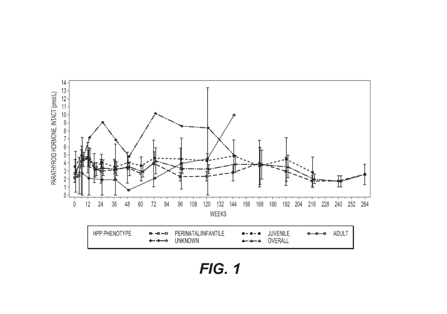
FIG. 1 shows the mean results for serum PTH (Intact pmol/L) over time by
disease onset
(HPP phenotype) and pooled safety set for all clinical trials (N=71). The time
axis shows length
of treatment with asfotase alfa in weeks. "Intact" indicates full-length PTH
(not the PTH
fragment). Bars at each timepoint represent 95% confidence intervals.
4
CA 02973883 2017-07-13
WO 2016/123342
PCT/US2016/015366
FIG. 2 shows mean laboratory test results over time for phosphate (mmol/L).
The time
axis refers to length of treatrnent with asfotase alfa in weeks. Bars at each
timepoint represent
95% confidence intervals.
FIG. 3 shows mean laboratory test results over time for 25-hydroxyvitamin D
(mmol/L).
The time axis refers to length of treatment with asfotase alfa in weeks. Bars
at each time point
represent 95% confidence intervals.
FIG. 4 shows mean results for calcium (mniol/L) over time by disease onset and
overall
safety set. The time axis refers to length of treatment with asfotase alfa in
weeks. Bars at each
timepoint represent 95% confidence intervals.
FIG. 5 shows calcium (top panel) and PTH levels (lower panel) with reference
ranges for
a single patient during treatment with asfotase.
FIG. 6 shows the mean results for serum PTH (Intact, pmol/L) over time through
week
312 by disease onset (HPP phenotype) and overall safety set. The time axis
shows length of
treatment with asfotase alfa in weeks. "Intact" indicates full length PTH (not
the PTH fragment).
Bars at each timepoint represent 95% confidence intervals.
FIG. 7 shows mean laboratory test results over time through week 312 for
phosphate
(mmol/L). The time axis refers to length of treatment with asfotase alfa in
weeks. Bars at each
timepoint represent 95% confidence intervals.
FIG. 8 shows mean laboratory test results over time through week 312 for 25
hydroxyvitamin D (mmol/L). The time axis refers to length of treatment with
asfotase alfa in
weeks. Bars at each time point represent 95% confidence intervals.
FIG. 9 shows mean results for calcium (mmol/L) over time through week 312 by
disease
onset and overall safety set. The time axis refers to length of treatrnent
with asfotase alfa in
weeks. Bars at each timepoint represent 95% confidence intervals.
FIG. 10 shows the patient values for calcium (mmol/L) and PTH (prnal/L) as a
function of
treatment week for the patient of FIG. 5. Vertical lines mark the start and
end of 3 mg/kg/week
dosing and the start of 6 mg/kg/week dosing.
FIG. 11 shows the amino acid sequence of asfotase alfa monorner (SEQ ID NO:
1).
Asfotase alfa exists as a dimer with inter-subunit disulfide bonds.
DETAILED DESCRIPTION
Described herein are materials and methods for monitoring and further treating
subjects
who are in need of treatment with an alkaline phosphatase or who are being
treated with an
alkaline phosphatase. The unexpected findings that additional analytes can be
monitored to
5
CA 02973883 2017-07-13
WO 2016/123342
PCT/US2016/015366
indicate additional treatment regimens led to the materials and methods
described herein.
Particular analytes can lead to additional treatments, for example, for
hypocalcemia,
hypercalcemia, osteoporosis, hyperparathyroidism, and vitamin D deficiency.
Various definitions are used throughout this document. Most words have the
meaning
that would be attributed to those words by one skilled in the art. Words
specifically defined
either below or elsewhere in this document have the meaning provided in the
context of the
present disclosure as a whole and as are typically understood by those skilled
in the art. For
example, as used herein, the singular forms "a," "an," and "the" include
plural references unless
the content clearly dictates otherwise. Unless otherwise defined, all
technical and scientific
terms used herein have the same meaning as commonly understood by one of
ordinary skill in
the art. Methods and materials are described herein for use in the present
disclosure; other
suitable methods and materials known in the art can also be used. In case of
conflict, the
present specification, including definitions, will control.
The materials and methods described herein relate to monitoring and further
treating
subjects who are in need of alkaline phosphatase (AP) replacement therapy or
who are
undergoing AP replacement therapy. The terms "individual," "subject," "host,"
and "patient" are
used interchangeably and refer to any subject for whom diagnosis, treatment,
or therapy is
desired, particularly humans. Other subjects may include cattle, dogs, cats,
guinea pigs,
rabbits, rats, mice, horses and the like. APs are responsible for
dephosphorylating a variety of
enzymes, and at least one isoform is substantially involved in bone
mineralization and
formation.
There are at least three APs in humans- intestinal (ALP!), placental (ALPP)
and tissue
non-specific (TNAP; sometimes referred to as liver/bone/kidney AP or ALPO, in
addition to
germline AP. TNAP is a membrane-anchored AP that is active extracellularly.
Defects in TNAP
result in, for example, elevated blood andlor urine levels of three
phosphocornpound substrates:
inorganic pyrophosphate (PPi), phosphoethanolamine (PEA) and pyridoxa1-5'-
phosphate (PLP)
(Whyte, M., Endocr. Rev., 15:439-61, 1994). TNAP is primarily responsible for
regulating serum
PPi levels (major inhibitor of hydroxyapatite crystal deposition in the bone
matrix), and,
therefore, is important for bone formation and mineralization. Genetic defects
in TNAP, for
example, lead to diseases, conditions, or disorders associated with low or
decreased bone
mineralization symptoms, e.g., hypophosphatasia (HPP).
Defects in TNAP activity can lead to a variety of diseases, disorders, and
symptoms.
Hypophosphatasia (HPP), for example, is a rare, heritable form of rickets or
osteomalacia
(Whyte, M. "Hypophosphatasia," In The Metabolic and Molecular Bases of
Disease, 8' ed.,
6
CA 02973883 2017-07-13
WO 2016/123342
PCT/US2016/015366
5313-29, Eds C. Scriver, A. Beaudet, W. Sly, D. Valle & B. Vogelstein.
Newyork: McGraw-Hill
Book Company, 2001). HPP is caused by loss-of-function mutation(s) in the gene
(ALPO that
encodes TNAP (Weiss, M. et al., Proc. Natl. Acad. Sci. USA, 85:7666-9, 1988;
Henthorn, P. et
al., Proc. Natl. Acad. Sci. USA, 89:9924-8, 1992; Henthom, P. & Whyte, M.,
Clin. Chem.;
38:2501-5, 1992; Zurutuza, L. et al., Hum. Mol. Genet., 8:1039-46, 1999). The
biochemical
hallmark is subnormal AP activity in serum (hypophosphatasemia).
HPP is an ultra-rare genetic disorder whereby TNAP activity is either absent
or barely
detectable in affected patients. While differences in patterns of inheritance
and mutations cause
variability in age at symptom onset and disease severity, all HPP patients
share the same
primary pathophysiological defect, the failure to mineralize bone matrix
(resulting in rickets or
osteomalacia) due to lack of TNAP. This primary defect in infants and
children, alone or in
combination with associated metabolic disturbances, can lead to deformity of
bones, impaired
growth, and decreased motor performance. This primary pathophysiological
mechanism can
rapidly lead to progressive damage to multiple vital organs, seizures due to a
CNS deficiency in
functional vitamin B6, and developmental delays. Subjects with HPP, left
untreated, can
develop, for example, hypercalcemia, and hyperphosphatemia.
All forms of HPP share the same underlying genetic and biochemical defect;
however,
the diagnosis of HPP actually encompasses a spectrum of disease. Published
classifications of
HPP have historically taken into account the age at which clinical
rmanifestation(s) first appear,
dividing the disease into the following categories: perinatal (onset in utero
and at birth), infantile
(onset post-natal to 6 months of age), juvenile (also described as childhood,
onset from 6
months to 18 years), and adult (onset after 18 years of age). Other milder
forms of the disease,
including benign perinatal HPP and odontohypophosphatasia, have also been
described.
HPP manifest in utero and may cause stillbirth. At the time of delivery, limbs
may be
shortened and deformed from profound skeletal hypomineralization, and
radiographic
examination often reveals an almost total absence of bony structures. Most
patients with
perinatal HPP have life-threatening disease, and death generally results from
respiratory
insufficiency due to pulmonary hypoplasia and poor functioning due to a
rachitic chest. Patients
with infantile-onset HPP often appear normal at birth but typically present
with skeletal
abnormalities and failure to thrive within the first six months life. These
patients can have a flail
chest from rachitic deformity of the thorax; and, together with rib fractures,
this may predispose
them to pneumonia and respiratory compromise. Mortality, usually due to
pulmonary
complications, has been reported to be as high as 50% (Whyte M.
Hypophosphatasia. In:
Glorieux FH, Pettifor JM, Juppner H, editors. Pediatric Bone: Biology and
Diseases. London,
CA 02973883 2017-07-13
WO 2016/123342
PCT/US2016/015366
UK, Academic Press; 2012: pp. 771-94: Caswell A. et al., Crit. Rev. Clin. Lab.
Sci., 28:175-232,
1991). Other clinical features may include, for example, functional
craniosynostosis with
resultant increased intracranial pressure and papilledema, and non-traumatic
fractures.
Hypercalcemia and hypercalciuria are also common, and nephrocalcinosis with
renal
compromise may occur. Weakness and delayed motor development are also common
complications of infantile-onset HPP and seizures may occur secondary to
vitamin B6 deficiency
in the central nervous system.
In juvenile-onset patients, radiographs of the long bones often reveal focal
bony defects
that project from the growth plates into the metaphyses; sometimes described
as "tongues" of
radiolucency. Physeal widening, irregularities of the provisional zones of
calcification, and
nietaphyseal flaring with areas of radiolucency adjacent to areas of
osteosclerosis may also be
present. Premature bony fusion of cranial sutures has also been observed in
some patients,
leading to potential increased intracranial pressure, proptosis, and cerebral
damage. Rachitic
deformities, including, for example, beading of the costochondral junctions,
either bowed legs or
knock-knees, and enlargement of the wrists, knees, and ankles from flared
nietaphyses, are
common, and often result in short stature. Walking is frequently delayed; and
a nonprogressive
myopathy characterized by limb weakness, especially of the proximal muscles of
the lower
extremities, has also been described (Seshia. S. et al., Arch. Dis. Childõ
65:130-1, 1990).
Skeletal pain and stiffness may also be present and non-traumatic fractures
are common.
Nephrocalcinosis may develop in juvenile-onset HPP as well.
First signs of HPP may also present later in life (as in the adult form of
HPP): however,
upon questioning, many adult patients report a history of early tooth loss or
rickets during
childhood. In adult HPP, hypomineralization manifests as osteornalacia. Adult
HPP patients
are subject to recurrent, poorly healing fractures, often in the rnetatarsals
and/or femur.
Complaints of pain in the thighs and hips from subtrochanteric femoral
pseudofractures are also
common. Radiographs often reveal the presence of osteopenia and
chondrocalcinosis. In
some patients, deposition of calcium pyrophosphate dehydrate occurs, leading
to PPi
arthropathy. Although adult HPP has been described as 'mild', manifestations
of the disease in
adults can be severe and debilitating; often requiring multiple surgeries and
the use of
supportive devices to perform activities of daily living.
Subjects with a defect in an endogenous AP, e.g., TNAP, are in need if AP
enzyme
replacement therapy (ERT). AP-ERT has been successful; for example, in
treating HPP. ERT
replaces an enzyme in subjects in whom that particular enzyme is deficient or
absent. ERT
does not affect the underlying genetic defect, but increases the concentration
of enzyme in
8
CA 02973883 2017-07-13
WO 2016/123342
PCT/US2016/015366
which the patient is deficient. The copy of the enzyme to be replaced; for
example; can be a
copy of the endogenous enzyme, an isoforni of the enzyme, an ortholog of the
enzyme, a
chimeric version of the enzyme; a fusion protein with the relevant active site
of the enzyme or
an otherwise engineered version of the enzyme. ERT can be accomplished; for
example, by
providing the enzyme itself or by causing the enzyme to be expressed in
particular tissues or
cells of the subject (e.g., through gene therapy methods, mRNA methods;
transcriptional or
translational activation methods, etc.).
Asfotase alfa or STRENS100, for example, is a dimeric fusion protein that
comprises
tvvo monomers with a TNAP phosphatase domain fused to an Fc chain and a bone
tag to target
the molecule to bone. The APs described herein can be, for example, intact
native proteins;
modified proteins or fusion proteins. Fusion proteins can comprise, for
example, sequences to
stabilize the protein, increase residence time in a patient, and/or target the
fusion protein to a
particular tissue, e.g., bone. Fusion proteins, for example; can comprise Fc
domains or albumin
moieties. Bone tags are typically negatively charged regions; e.g., poly-
aspartate or
poly-glutarnate sequences; e.g., between about 5 to about 50, between about 10
to about 25,
between about 67 to about 30, about 5, about 10, about 15, about 20, about 25,
about 30, about
35, about 40, about 45, about 50 or more aspartates, glutamates, or other
negatively charged
amino acids (natural or non-naturally occurring).
As described herein, treatment of a subject with AP-ERT can result in, for
example,
hypocalcemia; hyperparathyroidism, hypophosphatemia, vitamin D deficiency,
and/or symptoms
or side effects thereof. Such situations can occur, for example, in cases
where the mineral
defect is profound and availability of calcium and phosphorous for formation
of hydroxyapatite is
not adequate (e.g., not enough supplernentation in food, not enough
utilization of the minerals
available in the food or profound loss through urine). Treatment of one or
more of these effects
can lead to "overcorrection" of the effect, and, therefore, require additional
treatment to reverse
the overcorrection. As described herein, monitoring of calcium, PTH, phosphate
and vitamin D.
therefore, can improve the treatment of a subject in need of or being treated
with AP-ERT.
Described herein are also materials and methods for identifying subjects who,
prior to or
at the time of treatment, for example; AP-ERT, need to undergo treatment to
normalize one or
more metabolites associated with bone mineralization (e.g., PTH, Ca2, vitamin
D. and/or
phosphate). A subject in need of AP-ERT, for example, who is hypocalcemic
prior to AP-ERT
treatment, would benefit from having normalized calcium levels prior to AP-ERT
treatment.
Routine urinalysis and serum hematology and chemistries, for example, can be
obtained
before, during and after treatment using AP-ERT (e.g., treatment with asfotase
alfa). Calcium
9
CA 02973883 2017-07-13
WO 2016/123342
PCT/US2016/015366
and phosphate metabolism should be monitored periodically with measurements of
serum
calcium, phosphate and PTH levels and urinary calcium excretion. Dietary
intake of calcium
should be adjusted according to PTH levels and urinary calcium levels (ionized
and adjusted for
other markers, e.g., creatinine or albumin). When using asfotase alfa in
patients with
hypomineralization, e.g., with HPP rickets or osteomalacia, it is useful to
monitor calcium
concentration closely, as rapid intake of calcium into the bone matrix can
result in episodes of
hypocalcemia. In certain examples, this is particularly relevant during the
initial month or
months of treatment. To prevent sequelae of hypocalcemia, including potential
hypocalcemia-induced seizures, supplementation of calcium, or treatment with
calcimimetics,
for example, can be useful for those patients whose calcium levels are
statistically significantly
low or high.
As used herein, an "engineered" molecule is one that can be isolated from
natural
sources, synthesized and/or modified chemically. If the engineered molecule is
a biological
molecule, an engineered molecule can be one that is mutagenized, fused to a
second molecule,
e.g., forming a fusion protein, attached to a specific functional moiety,
e.g., a targeting domain,
purification domain, active site, etc., humanized, or made into a chimeric
protein by switching
particular domains with other proteins or isoforms. The engineering is the
process of modifying
the molecule in a particular manner to achieve a desirable result.
As used herein, "fusion protein" refers to an engineered protein that
comprises residues
of moieties from two or more different proteins. Fusion genes, which can be
used to generate
fusion proteins, are created through the joining of two or more coding
sequences that code for
separate proteins. Translation of a fusion gene results in a single or
multiple polypeptides with
functional properties derived from each of the original proteins. Recombinant
fusion proteins
are created by recombinant DNA technology.
As used herein, "chimeric proteins" are proteins that comprise moieties from
at least two
distinct proteins. The term refers to hybrid proteins made of polypeptides
having different
functions or physicochemical patterns.
The subjects described herein have an AP activity defect. Such a defect can
arise, for
example, due to a genetic anomaly (e.g., a mutation) that causes the AP enzyme
to not be
produced or to be produced in an inactive form. Although such a defect can
occur in any of the
AP isoforms, of particular interest for the present disclosure are AP defects
that lead to bone
mineralization defects, e.g., HPP.
Described herein are findings indicating that patients who are in need of or
who are
undergoing treatment, e.g., ERT-AP treatment, for a bone mineralization
disease, disorder,
CA 02973883 2017-07-13
WO 2016/123342
PCT/US2016/015366
condition or symptoms thereof, e.g., HPP; can be monitored for one or more
analytes that are
indicative of the need for additional treatments or a need to alter the
current treatment regimen,
e g., alter the dosage and/or frequency of dosage.
"Treatment" refers to the administration of a therapeutic agent or the
performance of
medical procedures with respect to a patient or subject, for any of
prophylaxis (prevention),
cure, or reduction of the symptoms of the disease, disorder, condition, or
symptoms from which
the subject suffers.
The treatments (therapies) described herein can also be part of "combination
therapies."
Combination therapy can be achieved by administering two or more agents, each
of which is
formulated and administered separately, or by administering two or more agents
in a single
formulation. One active ingredient can be, for example, useful for treating,
for example, a
disease, disorder, condition or symptoms associated with a TNAP defect, e.g.,
hypophosphatasemia, or symptoms associated with treatment by the active agent
("side
effects"). Other combinations are also encompassed by combination therapy. For
example,
two or more agents can be formulated together and administered in conjunction
with a separate
formulation containing a third agent. While the two or more agents in the
combination therapy
can be administered simultaneously, they need not be. For example,
administration of a first
agent (or combination of agents) can precede administration of a second agent
(or combination
of agents) by minutes, hours, days or weeks. Thus, the two or more agents can
be
administered within minutes of each other or within any number of hours of
each other or within
any number or days or weeks of each other.
As used herein, a "therapeutically effective dosage" or "therapeutically
effective amount"
results in a decrease in severity of disease, disorder, condition or symptoms
thereof (e.g.,
associated with aberrant AP activity, e.g , HPP), an increase in frequency and
duration of
disease symptom-free periods, or a prevention of impairment or disability due
to the disease
affliction.
As described herein, treatment with AP replacement therapy is more effective
or leads to
overall improved health or quality of life, when the treated subject is
further monitored for one or
more additional analytes. PTH, calcium (Ca2+), phosphate and vitamin D
concentrations can
each be monitored or individually monitored during AP-ERT, and the specific
concentrations are
indicative of, for example, efficacy of treatment and/or the need for one or
more additional
therapeutic reginien(s).
PTH acts on osteoblasts in bone and tubular cells within the kidney via G-
protein-linked
receptors that stimulate adenylate cyclase production of cyclic AMP. In bone,
within one or two
11
CA 02973883 2017-07-13
WO 2016/123342
PCT/US2016/015366
hours, PTH stimulates a process known as osteolysis in which calcium in the
minute fluid-filled
channels (canaliculillacunae) is taken up by syncytial processes of osteocytes
and transferred
to the external surface of the bone and, hence, into the extracellular fluid.
Some hours later, it
also stimulates resorption of mineralized bone: a process that releases both
Ca2+ and
phosphate into the extracellular fluid.
Monitoring PTH concentration in a sample obtained from a subject, for example,
is of
interest for better treating the subject, as AP-ERT can have an effect on PTH
concentration. A
determination that the treated subject's serum PTH concentration is
statistically significantly
lower or higher than normal, for example, can lead to revised treatment plans
(e.g., combining
the AP-ERT plan with one or more therapeutic agents for treating, for example,
hyperparathyroidism, e.g., with cinacalcet). In cases where PTH concentration
is determined to
be statistically significantly high, e.g., hyperparathyroidism, the subject
can be treated with
higher levels of the AP-ERT, e.g., asfotase alfa, to reduce PTH levels in the
case where a
patient does not show good response in term of bone mineralization to initial
dose.
As used herein, the term "sample" refers to biological material from a
subject. Although
serum concentration is of interest, samples can be derived from many
biological sources,
including, for example, single cells, multiple cells, tissues, tumors,
biological fluids, brain
extracellular fluid, biological molecules or supernatants or extracts of any
of the foregoing.
Examples include tissue removed for biopsy, tissue removed during resection,
blood, urine,
lymph tissue, lymph fluid, cerebrospinal fluid, amniotic fluid, mucous and
stool samples. The
sample used will vary based on the assay format, the detection method and the
nature of the
tumors, tissues, fluids, cells or extracts to be assayed. Methods for
preparing samples are
known in the art and can be readily adapted to obtain a sample that is
compatible with the
method utilized.
As used herein, "statistical significance" is a statistical term that informs
as to the
certainty that a difference or relationship exists, e.g., that a sample value
id statistically
significantly different from a normal or baseline value. It is conferred by
finding a low probability
of obtaining at least as extreme results given that the null hypothesis is
true. It is an integral
part of statistical hypothesis testing where it determines whether a null
hypothesis can be
rejected. In any experiment or observation that involves drawing a sample from
a population,
there is the possibility that an observed effect would have occurred due to
sampling error alone.
But if the probability of obtaining at least as extreme result (large
difference between two or
more sample means), given the null hypothesis is true, is less than a pre-
determined threshold
(e.g., 5% chance), then an investigator can conclude that the observed effect
actually reflects
12
CA 02973883 2017-07-13
WO 2016/123342
PCT/US2016/015366
the characteristics of the population rather than just sampling error. A test
for statistical
significance involves comparing a test value to some critical value for the
statistic. The
procedure to test for significance is the same- decide on the critical alpha
level (i.e., the
acceptable error rate), calculate the statistic and compare the statistic to a
critical value
obtained from a table. P-values, the probability of obtaining the observed
sample results (or a
more extreme result) when the null hypothesis is actually true, are often
coupled to a
significance or alpha (a) level, which is also set ahead of time, usually at
about 0.05 (5%).
Thus, if a p-value was found to be less than about 0.05, then the result would
be considered
statistically significant and the null hypothesis would be rejected. Other
significance levels, such
as about 0.1, about 0.075, about 0.025 or about 0.01 can also be used. As used
herein, a
"statistically significantly low" concentration of an analyte is one that is
lower than the normal or
baseline situation for a control, e.g., healthy, subject. Conversely, a
"statistically significantly
high" concentration of an analyte is one that is higher than the normal or
baseline concentration
of the analyte for a control subject.
PTH promotes osteoclast function and leads to bone resorption, thereby
increasing
serum Ca2+ and phosphate concentrations. Low levels of serum Ca2+ fail to
exhibit negative
feedback effect on the release or production of PTH from the thyroid, whereas
high
concentrations of serum Ca2+, by exhibiting a negative feedback effect on
release and/or
production of PTH, lead to decreased PTH levels in blood. In a related
pathway, vitamin D
increases adsorption of Ca2+ and phosphate in the intestine, leading to
elevated levels of serum
Ca2+ and, therefore, lower bone resorption. Effects of active vitamin D (1, 25
(OH)2D on bone,
however, are diverse and can affect formation or resorption.
Although, hypoparathyroidism (hypoPT) is one of the few major hormone
deficiency
diseases that is often not treated with the missing hormone, hormone
replacement therapies are
available. Bovine PTH has been purified and used as experimental treatment,
however utility as
a treatment was diminished, mainly because of antibody formation and costs.
Approval of fully
humanized truncated PTH (Teriparatide, PTH (1-34)) and intact parathyroid
hormone (Preotact,
PTH(1-84)) for treatment of osteoporosis, has made the PTH drugs more
accessible and
thereby made clinical trials with PTH treatment of hypoPT feasible. Patients
with hypoPT
experience an improved quality of life when treated with PTH compared with
conventional
treatment with la-hydroxylated vitamin D metabolites and calcium supplements,
although
hypoPT is still treated, for example, by supplementing calcium and/or vitamin
D.
Primary hyperparathyroidism results from a hyperfunction of the parathyroid
glands.
Over secretion of PTH can be due, for example, to a parathyroid adenoma,
parathyroid
13
CA 02973883 2017-07-13
WO 2016/123342
PCT/US2016/015366
hyperplasia or a parathyroid carcinoma. This disease is often characterized by
the presence of
kidney stones, hypercalcemia, constipation, peptic ulcers and depression.
Secondary hyperparathyroidism is due to physiological secretion of PTH by the
parathyroid glands in response to hypocalcemia (low blood calcium levels). The
most common
causes are vitamin D deficiency and chronic renal failure. Lack of vitamin D
leads to reduced
calcium absorption by the intestine leading to hypocalcemia and increased PTH
secretion. This
increases bone resorption. In chronic renal failure the problem is more
specifically failure to
convert vitamin D to its active form in the kidney. The bone disease in
secondary
hyperparathyroidism caused by renal failure is termed renal osteodystrophy.
Tertiary hyperparathyroidism is seen in patients with long-term secondary
hyperparathyroidism, which eventually leads to hyperplasia of the parathyroid
glands and a loss
of response to serum calcium levels.
Quaternary and quintary hyperparathyroidism are rare conditions that may be
observed
after surgical removal of primary hyperparathyroidism, when it has led to
renal damage that now
again causes a form of secondary (quaternary) hyperparathyroidisni that may
itself result in
autonomy (quintapy) hyperparathyroidisrn. Additionally, quaternary
hyperparathyroidism may
ensue from hungry bone syndrome after parathyroidectomy.
Primary hyperparathyroidism can be treated, for example, by surgery
(parathyroidectomy) if treatment, for example, with calcimimetics is
unsuccessful. Secondary
hyperparathyroidism can be treated, for example, by vitamin D supplementation
and/or by the
use of calcimimetics (e.g., cinacalcet). Other forms of hyperparathyroidism
are variations of
secondary hyperparathyroidism, and treatments involve approaches similar to
those used for
primary and secondary hyperparathyroidism.
Low plasma calcium stimulates PTH release (by negating the inhibition of PTH
release),
and PTH acts to resorb Ca2+ from the pool in bone and to enhance renal re-
absorption of Ca2+.
High plasma calcium stimulates calcitonin secretion, which lowers plasma
calcium by inhibiting
bone resorption.
Normal blood calcium level is between about 8.5 to about 10.5 mg/dL (2.12 to
2.62 mmol/L; some reports use the values of between about 8.0 to about 10.0
mg/dL) and that
of ionized calcium is 4.65 to 5.25 mg/dL (1.16 to 1.31 mmol/L). Hypocalcemia
or hypercalcemia
is characterized by a statistically significantly low or high serum calcium
concentration.
Hypocalcemic subjects, for example, typically display a serurn calcium
concentration of about
2.5 mg/dL or lower (Sorell, M. & Rosen, J., J. Pediatr., 87:67-70, 1975). A
hypocalcernic
14
CA 02973883 2017-07-13
WO 2016/123342
PCT/US2016/015366
subject, for example, can have serum calcium concentration of about 7.0 mg/dL
or lower, about
5.0 mg/dL or lower, about 1.0 mg/dL or lower, or about 0.5 mg/dL or lower.
Common causes of hypocalcemia include hypoparathyroidism, vitamin D deficiency
and
chronic kidney disease. Symptoms of hypocalcemia include, for example,
neuromuscular
irritability (including tetany as manifested by Chvostek's sign or Trousseau's
sign,
bronchospasm), electrocardiographic changes and seizures. Treatment options
include, for
example, supplementation of calcium and some form of vitamin D or its
analogues, alone or in
combination. Intravenous calcium gluconate 10% can be administered, or if the
hypocalcemia
is severe, calcium chloride can be given. Other treatments involve
multivitamin
supplementation, in oral, chewable, or liquid forms.
Hypercalcernia is an elevated Ca2+ level in the blood, which is often
indicative of other
disease(s). It can be due to excessive skeletal calcium release, increased
intestinal calcium
absorption or decreased renal calcium excretion. The neuromuscular symptoms of
hypercalcemia are caused by a negative bathmotropic effect due to the
increased interaction of
calcium with sodium channels. Since calcium blocks sodium channels and
inhibits
depolarization of nerve and muscle fibers, increased calcium raises the
threshold for
depolarization. Symptoms of hypercalcemia include, for example, renal or
biliary stones, bone
mineralization defects and bone pain, abdominal pain, nausea, vomiting,
polyuria depression,
anxiety, cognitive dysfunction, insomnia, coma, fatigue, anorexia and
pancreatitis.
Hypercalcemia is defined as a serum calcium level greater than about 10.5
mg/dL
(>2.5 mmol/L). Hypercalcemia can also be classified based on total serum and
ionized calcium
levels, as follows: Mild: total calcium 10.5-11.9 mg/dL (2.5-3 mmol/L) or
ionized calcium
5.6-8 mg/ca_ (1.4-2 mmol/L): Moderate: total calcium 12-13.9 mg/dL (3-3.5
mmol/L) or ionized
calcium 5.6-8 mg/dL (2-2.5 rnmol/L): Hypercalcemic crisis: total calcium: >14-
16 mg/dL
(3.5-4 mmol/L) or ionized calcium 10-12 mg/dL (2.5-3 mmol/L).
Hypercalcemia is treated a number of ways, including, for example, using
fluids and
diuretics for an initial therapy (hydration, increasing salt intake, and
forced diuresis). Diuretic
treatments include, for exarnple, furosemide, and they can be given to permit
continued large
volume intravenous salt and water replacement while minimizing the risk of
blood volume
overload and pulmonary edema. In addition, loop diuretics tend to depress
renal calcium
reabsorption thereby helping to lower blood calcium levels. Caution must be
taken to prevent
potassium or magnesium depletion. Additional therapies include, for example,
bisphosphonates, plicamycin, gallium nitrate, glucocorticoids and calcitonin.
Bisphosphonates
are pyrophosphate analogues with high affinity for bone, especially areas of
high bone turnover.
CA 02973883 2017-07-13
WO 2016/123342
PCT/US2016/015366
They are taken up by osteoclasts and inhibit osteoclastic bone resorption.
Available drugs
include, for example, etidronate, tiludronate, IV pamidronate, alendronate,
zoledronate, and
risedronate. Calcitonin blocks bone resorption and also increases urinary
calcium excretion by
inhibiting renal calcium reabsorption. Phosphate therapy can correct the
hypophosphatemia in
the face of hypercalcemia and lower serum calcium. Calcium mimetics, e.g.,
cinacalcet, are
also used to lower serum calcium concentrations.
Hypovitaminosis D is a deficiency of vitamin D. It can result from inadequate
nutritional
intake of vitamin D coupled with inadequate sunlight exposure (in particular
sunlight with
adequate ultraviolet B rays), disorders that limit vitamin D absorption, and
conditions that impair
the conversion of vitamin D into active metabolites including certain liver,
kidney, and hereditary
disorders. Deficiency results in impaired bone mineralization and leads to
bone softening
diseases including rickets in children and osteomalacia and osteoporosis in
adults.
Maintenance doses of both calcium and vitamin D are often necessary to prevent
further
decline.
EXEMPLIFICATION
The following examples do not limit the scope of the invention as disclosed
and
described in the claims.
EXAMPLE 1. PTH and Calcium
When evaluating results by age at disease onset, mean and median PTH levels
were
notably higher in patients with infantile- and juvenile-onset HPP during the
first 12 weeks of
treatment compared with later time points, and were likely associated with the
bone
mineralization process and the monitoring thereof. In some cases,
multivitamins, calcium,
vitamin D, vitamin A, vitamin K. cinacalcet, pyridoxal phosphate calcium,
andlor calcitonin were
administered to patients in order to normalize PTH, calcium, and phosphate
levels
concomitantly with the monitoring process.
Mean PTH levels in patients with adult-onset HPP tended to be lower than those
observed in the infantile- and juvenile-onset HPP patients through
approximately Week 72.
These comparisons, however, involved only two patients with adult-onset HPP.
FIG. 1 provides
the change in serum PTH over time in the clinical studies.
Patients were subdivided by Baseline PTH level, and the details of the changes
in the
initial period after the start of asfotase alfa treatment are as follows:
For the nine patients with low PTH at Baseline:
16
CA 02973883 2017-07-13
WO 2016/123342
PCT/US2016/015366
Seven patients with normal calcium levels at Baseline and lower calcium levels
post-treatment had a rise in PTH. All except one patient showed radiological
improvements, as determined by the RSS score (scoring of rickets).
One patient, who had high Baseline serum calcium, showed no change in serum
calcium levels and no change in PTH by Week 6, at which time the patient was
discontinued from the study. Note that this patient received only two doses of
asfotase
alfa and was subsequently withdrawn; therefore, no change in calcium or PTH
was
expected.
One patient with a high Baseline serum calcium level had normalization of
serum
calcium by Week 24. PTH data beyond Week 6 is not available for this patient
(at
Week 6, PTH was unchanged). This patient showed no improvements in rickets at
Week 12 (7 weeks after asfotase alfa dose was increased), however showed a
decrease
in RSS score by Week 24.
For the13 patients with normal PTH at Baseline:
12 patients with normal PTH and normal calcium levels at Baseline responded
with small or no change in serum calcium levels and no or slight change in PTH
levels.
All except 1 patient showed radiographic improvement.
One patient with calcium levels at the upper limit of normal at Baseline
responded with normalization of calcium levels and large increase in PTH. This
patient
showed radiographic improvement.
Only one patient had high PTH at Baseline with normal calcium levels. The
patient responded with lowering of serum calcium levels and large rise in PTH
levels.
This patient did not show radiological improvement during the periods of rise
in PTH.
One patient had no available Baseline PTH results, but the earliest result at
Week 6 showed values in the normal range. Calcium remained within normal
ranges
with some oscillation until Week 60. The patient showed worsening of rickets
(RGI-C
score at Week 12 was negative; -1.67) and growth scores on the initial dose of
asfotase
alfa of 6 mg/kg/week. After the dose was increased to 9 mg/kg/week due to
continued
worsening of growth delays, the patient showed improvement in radiographic
signs (at
Week 36, RSS score improved 4.5 points since week 24 assessment) and PTH
increased above the normal range. PTH further increased more steeply until it
peaked
at Week 72. Note that calcium levels showed a drop at Week 48 (although were
still
within the normal limits) and then fell below normal at Week 60, however
rebounded to
17
CA 02973883 2017-07-13
WO 2016/123342
PCT/US2016/015366
normal at Week 72. In this patient, low vitamin D levels and low urine
calcium/creatinine
ratios were found coincident with the observed elevation of PTH level.
EXAMPLE 2. Phosphate
Mean serum phosphate values tended to be variable through Week 24 in all HPP
onset
categories, and then appeared to normalize and stabilize with continued
treatment. Some
decreases in serum phosphate levels appeared to coincide with decreases in
serum calcium
levels during the first several weeks of treatment, which likely was due to
the intense bone
mineralization processes that occurred early in treatment.
Three patients (infantile-onset) had a shift from normal values for phosphate
at Baseline
to low values during treatment, and 21 patients (15 infantile-onset; 5
juvenile-onset; 1
adult-onset) with low or normal values at Baseline had a shift to high values
during treatment; at
the last visit, no patients had shifted from normal or high values to low
values, and 5 patients
(infantile-onset) had shifted frorn normal values to high values; see FIG. 2.
EXAMPLE 3. Vitamin D
Mean 25-OH vitamin D values were consistently higher in patients with adult-
onset HPP
than in patients in the other HPP onset categories; however, these values did
decrease slightly
over time, and there were only 2 patients with adult-onset HPP included in the
study. Mean and
median 25-0H vitamin D values in the other HPP onset categories were
relatively consistent
over time: see FIG. 3. In patients where vitarnin D values were monitored and
identified as
deficient or lower than desired by the clinician (i.e., less than than 20
ng/m1), vitamin D was
supplemented with vitamin D in the form of an oral medication, i.e., as
children's vitamins, adult
multivitamins, or vitamin D capsules. Vitamin D in some cases was administered
as an
intramuscular injection or in combination dosages with calcium, vitarnin A,
and/or vitamin K.
Calcitriol, cholecalciferol, and/or ergocalciferol were also administered as
needed.
EXAMPLE 4.
Systematic analyses of pre- and post-treatment serum calcium, parathyroid
hormone
(PTH), phosphate and vitamin D were performed.
Calcium: serum calcium levels were variable at Baseline, ranging from 1.92 to
4.03 mmol/L. Although the changes in mean and median calcium levels over the
course of
treatment with asfotase alfa were not remarkable, levels tended to stabilize
and become less
variable; calcium levels ranged from 1.82 to 2.80 mmoIlL at Week 24, and from
2.12 to
3.67 mmol/L at the last visit, basically eliminating the episodes of
hypocalcemia and the range
CA 02973883 2017-07-13
WO 2016/123342
PCT/US2016/015366
of hypercalcemia was lowered to nearly normal (i.e.; upper range is 3.5
mmol/L). Increases in
calcium above the normal range were generally small and, when present, were
most notable at
Baseline and tended to normalize over the course of asfotase alfa treatment
with the increased
calcium deposition in the bone as noted on X-ray. When evaluating results by
age at disease
onset, the small increases in calcium were generally seen in patients with
infantile-onset HPP,
and calcium levels in these patients tended to normalize during treatment
(Table 1 and FIG. 4).
Parathyroid Hormone: mean and median PTH levels increased with treatment, most
notably during the first 12 weeks of treatment with asfotase alfa. This
increase was likely due to
a physiologic response secondary to increases in the bone mineralization
process associated
with asfotase alfa treatment. The variability in PTH levels noted at Baseline
and throughout
treatment may be due to factors that affect PTH levels, including, but not
limited to: age, body
mass index (BMI), serum creatinine levels, serum calcium levels and vitamin D
levels. When
evaluating results by age at disease onset, mean and median PTH levels were
notably higher in
patients with infantile- and juvenile-onset HPP during the first 12 weeks of
treatment compared
with later time points, and were likely associated with the bone
mineralization process. Mean
PTH levels in patients with adult-onset HPP tended to be lower than those
observed in the
infantile- and juvenile-onset HPP patients through approximately Week 72;
there are several
variables that can affect PTH levels (Table 1 and FIG. 1).
Phosphate: mean serum phosphate values were variable through Week 24 in
patients
with infantile-, juvenile- and adult-onset HPP, and then appeared to normalize
and stabilize with
continued treatment with asfotase alfa. Some decreases in serum phosphate
levels appeared
to coincide with decreases in serum calcium levels during the first several
weeks of treatment,
which were likely due to the intense bone mineralization processes occurring
early in treatment
(Table 1 and FIG. 2).
Vitamin D: changes over time for vitamin D were not clinically meaningful:
some of the
variability seen in vitamin D results may be reflective of concomitant vitamin
D supplements
taken by some patients. When evaluating results by age at disease onset, mean
vitamin D
values were consistently higher in patients with adult-onset HPP than in
patients with infantile-
or juvenile-onset HPP, however, higher values did decrease slightly over time.
Mean and
median vitamin D values in patients with infantile- and juvenile-onset HPP
were relatively
consistent over time (Table 1 and FIG. 3).
19
CA 02973883 2017-07-13
WO 2016/123342
PCT/US2016/015366
Table 1. Changes from Baseline to Week 24 and last visit for serum Ca2+. PTH,
phosphate and
Vitamin D; pooled safety set overall.
Change from
Change from
Parameter Baseline to
Baseline to
Statistic Baseline Week 24 Week 24
Last Visit Last Visit
Calcium (mmol/L)
n 71 64 64 70 70
Mean (SD) 2.540 2.487 -0.055 (0.2459) 2.504 (0.2109)
-0.039 (0.2693)
(0.2727) (0.1580)
Median 2.500 2.485 -0.025 2.470 -
0.045
Range 1.92, 4.03 1.82, 2.80 -
1.33, 0.38 2.12, 3.67 -1.33, 0.67
Parathyroid Hormone (pmol/L)
n 57 61 51 69 56
Mean (SD) 2.68 (1.813) 3.41 (3.848) 0.86 (4.230)
3.66 (4.988) 0.73 (2.406)
Median 2.40 2.50 0.50 2.40 0.45
Range 0.6, 8.0 0.6, 27.9 -6.2, 26.7
0.6, 38.7 -4.4, 7.8
Phosphate (mmol/L)
n 70 63 62 70 69
Mean 1.814 1.900 (0.3520) 0.090 (0.3962)
1.771 (0.2967) -0.042
(SD) (0.3922)
(0.3960)
Median 1.870 1.970 0.025 1.760 -
0.090
Range 0.42, 2.74 0.90, 2.50 -
0.51, 1.49 1.00, 2.50 -0.71, 1.23
25-Hydroxy Vitamin D (pmol/mL)
n 68 65 63 67 64
Mean 76.5 86.2 (33.58) 9.8 (35.56)
78.3 (25.08) 3.5 (35.32)
(SD) (28.99)
Median 77.0 80.7 4.0 75.0 -1.0
Rang!: 17, 169 -- 23, 212 -- -54, 135 --- 18, 179 -- -83,
91
SD ... standard deviation.
EXAMPLE 5.
One patient had a low serum calcium level at Baseline, but upon initiation of
asfotase
alfa treatment it further decreased; and was responsive to changes in asfotase
alfa dosage
changes. At Week 24 the dose was reduced from 6nia/kgisNeek to 3mg/kg/week and
at Week
48 it was raised to 6mg/kg/week. During this period PTH levels initially rose
but stayed within
the normal range up to Week 72, when levels were temporarily elevated above
the normal
range. Simultaneously, calcium was raised and entered to the normal range. The
patient
showed signs of radiographic improvement starting at Week 12, although the RGI-
C score did
not reach 2 or above (meaning substantial improvement in radiographic signs of
rickets). At
Week 168, serum calcium fell below the normal range and PTH increased above
the normal
range, at which time six-minute walk test (6MVVT) results showed a large drop
compared with
the previous result at Week 120. See FIG. 5 and FIG. 10.
CA 02973883 2017-07-13
WO 2016/123342
PCT/US2016/015366
EXAMPLE 6.
An additional year of data was subsequently collected for the patients
contained in
Examples1-5 which demonstrated continuation of the previously described
trends, see FIGs.
6-9.
OTHER EMBODIMENTS
It is to be understood that while the present disclosure has been described in
conjunction with the detailed description herein; the foregoing description is
intended to illustrate
and not limit the scope as defined by the appended claims. Other aspects,
advantages, and
modifications are within the scope of the following claims. References cited
in the Specification
are herein incorporated by reference in their entireties.
21