Note: Descriptions are shown in the official language in which they were submitted.
CA 02974299 2017-07-18
WO 2016/126865 PCT/US2016/016438
MULTIPLE-EMULSION NUCLEIC ACID AMPLIFICATION
CROSS-REFERENCE
[0001] This application claims priority benefit of U.S. Provisional
Application No.
62/112,072, filed February 4, 2015, which application is incorporated herein
by reference
in its entirety and for all purposes.
GOVERNMENT SUPPORT
[0002] This invention was made with government support under grant
numbers
1R21HG007233-02, 1DP2AR068129-01, and 1R01EB019453-01 awarded by the National
Institutes of Health, grant numbers HR0011-12-C-0065, N66001-12-C-4211, and
HR0011-12-
C-0066 awarded by the Department of Defense, and grant number DBI-1253293
awarded by the
National Science Foundation. The government has certain rights in the
invention.
INTRODUCTION
[0003] The quantitation and sequencing of nucleic acids is central to
modern biology.
New methods for detecting and quantitating nucleic acids, such as digital
polymerase chain
reaction (PCR), provide greater accuracy than traditional real-time
quantitative PCR, and also
provide an absolute count of the number of molecules present, obviating the
need for an internal
standard. Droplet PCR has emerged as a convenient high-throughput method for
implementing
digital PCR. However, a drawback to existing droplet digital PCR techniques is
that the PCR
reactions are performed in aqueous-in-oil emulsions in which the carrier phase
of the emulsion
is inert oil. This prevents the droplet reactor from being detected,
quantitated, and sorted with
available methods, such as Fluorescence Activated Cell Sorting (FACS), which
are not
compatible with oil carrier phases. The present disclosure addresses the above
issues and
provides related advantages.
SUMMARY
[0004] The methods and systems described herein, referred to as multiple-
emulsion
nucleic acid amplification, allow nucleic acids contained in biological
systems to be detected,
quantitated and/or sorted based on their sequence as detected with nucleic
acid amplification
techniques, e.g., PCR. The nucleic acids can be free floating or contained
within living or
nonliving structures, including particles, viruses, and cells. The nucleic
acids can include, e.g.,
DNA or RNA. The present disclosure is based in part on the surprising
discovery that nucleic
1
CA 02974299 2017-07-18
WO 2016/126865 PCT/US2016/016438
acid amplification techniques, such as PCR, can be performed in multiple-
emulsion
microdroplets and Giant Unilamellar Vesicles (GUV) without destroying the
integrity of the
multiple-emulsion microdroplets and GUVs. Systems and devices for use in
practicing methods
of the disclosure are also provided.
[0005] In exemplary embodiments, the disclosed methods include
encapsulating a
sample, which may include a heterogeneous population of cells, viruses, and/or
nucleic acids, in
a plurality of multiple-emulsion microdroplets or GUVs, wherein each multiple-
emulsion
microdroplet or GUV includes a first miscible phase fluid surrounded by an
immiscible shell,
wherein the multiple-emulsion microdroplet or GUV is positioned in a second
miscible phase
carrier fluid. In some embodiments, the sample may be diluted prior to
encapsulation, e.g., so as
to encapsulate a controlled number of cells, viruses, and/or nucleic acids in
the multiple-
emulsion microdroplets or GUVs. Nucleic acid amplification reagents, e.g., PCR
reagents, may
be added to the multiple-emulsion microdroplets or GUVs at the time of
encapsulation or added
to the multiple-emulsion microdroplets or GUVs at a later time using one or
more of the
methods described herein. The multiple-emulsion microdroplets or GUVs are then
subjected to
nucleic acid amplification conditions, such that if a multiple-emulsion
microdroplet or GUV
contains a nucleic acid corresponding to a target of interest, e.g., a cell,
virus, or nucleic acid of
interest, the multiple-emulsion microdroplet or GUV becomes detectably
labeled, e.g.,
fluorescently labeled as a result of a fluorogenic assay, such as Sybr
staining of amplified DNA
or TaqMan PCR. To recover the target nucleic acids or entities comprising the
target nucleic
acids, the detectably labeled multiple-emulsion microdroplets or GUVs may be
sorted using
microfluidic (e.g., dielectrophoresis, membrane valves, etc.) or non-
microfluidic techniques
(e.g., FACS).
[0006] Multiple-emulsion microdroplets according to the present
disclosure may be
formed, for example, by (1) flowing a miscible phase fluid, including, e.g.,
cells, viruses, and/or
nucleic acids, along with nucleic acid amplification reagents in a channel of
a microfluidic
device; (2) contacting the miscible phase fluid with an immiscible phase
fluid, e.g., using a
single-emulsion droplet maker, wherein the contacting of the miscible phase
fluid solution of
nucleic acids and amplification reagents with the immiscible phase fluid
results in the formation
of miscible phase microdroplets (e.g., single emulsion microdroplets)
surrounded by the
immiscible phase fluid; (3) flowing the miscible phase microdroplets
surrounded by the
immiscible phase fluid in a channel of a microfluidic device; (4) contacting
the miscible phase
microdroplets surrounded by the immiscible phase fluid with a miscible phase
carrier fluid, e.g.,
using a double-emulsion droplet maker, wherein the contacting of the miscible
phase
2
CA 02974299 2017-07-18
WO 2016/126865 PCT/US2016/016438
microdroplets surrounded by the immiscible phase fluid with the miscible phase
carrier fluid
results in the formation of multiple-emulsion microdroplets (e.g., double-
emulsion
microdroplets), each multiple-emulsion microdroplet including a miscible phase
microdroplet
surrounded by the immiscible phase fluid, wherein the immiscible phase fluid
is surrounded by
the miscible phase carrier fluid. GUVs may be generated from multiple-emulsion
microdroplets
by inducing the multiple-emulsion microdroplets to undergo dewetting, wherein
the immiscible
phase fluid is expunged, leaving behind a membrane of surfactant with a small
immiscible phase
droplet adhered to the outside of the membrane. One or more steps of the
method may be
performed under microfluidic control.
[0007] In some embodiments, a sample including viruses is encapsulated in
multiple-
emulsion microdroplets or GUVs and subjected to nucleic acid amplification
conditions, e.g.,
PCR conditions. In some embodiments, the encapsulated viruses are subjected to
one or more
virus lysing techniques, such as proteinase k digestion or thermal lysis.
Nucleic acid
amplification assays specific to the viruses of interest can cause multiple-
emulsion
microdroplets or GUVs containing the viruses of interest, or nucleic acids
originating from the
viruses of interest, to become detectably labeled, e.g., fluorescently
labeled. The viruses and/or
the viral nucleic acids, may then be recovered by sorting the multiple-
emulsion microdroplets or
GUVs and recovering their contents via microdroplet rupture, e.g., through
chemical or
electrical means.
[0008] In some embodiments, a sample including cells is encapsulated in
multiple-
emulsion microdroplets or GUVs and subjected to nucleic acid amplification
conditions, e.g.,
PCR conditions. In some embodiments, the encapsulated cells are subjected to
one or more cell
lysing techniques, such as proteinase k digestion or thermal lysis. Nucleic
acid amplification
assays specific to the cells of interest can cause multiple-emulsion
microdroplets or GUVs
containing the cells of interest, or nucleic acids originating from the cells
of interest, to become
detectably labeled, e.g., fluorescently labeled. The cells and/or the cellular
nucleic acids may
then be recovered by sorting the multiple-emulsion microdroplets or GUVs and
recovering their
contents via microdroplet rupture, e.g., through chemical or electrical means.
[0009] In some embodiments, a sample including nucleic acids (e.g., DNA
and/or RNA)
is encapsulated in multiple-emulsion microdroplets or GUVs and subjected to
nucleic acid
amplification conditions, e.g., PCR or RT-PCR conditions. PCR or RT-PCR,
assays specific to
the nucleic acids of interest can cause droplets containing the nucleic acids
of interest to become
detectably labeled, e.g., fluorescently labeled. The nucleic acids of interest
may then be
3
CA 02974299 2017-07-18
WO 2016/126865 PCT/US2016/016438
recovered by sorting the multiple-emulsion microdroplets or GUVs and
recovering their
contents via microdroplet rupture, e.g., through chemical or electrical means.
[0010] Additional nucleic acid amplification reactions which may be
performed in
multiple-emulsion microdroplets or GUVs as described herein, include, e.g.,
multiple
displacement amplification (MDA), strand displacement amplification (SDA), and
rolling circle
amplification (RCA).
[0011] In one aspect of a method according to the present disclosure, a
method for
enriching for a target nucleic acid sequence is provided, wherein the method
includes
encapsulating a sample including nucleic acids in a plurality of multiple-
emulsion
microdroplets or GUVs; introducing polymerase chain reaction (PCR) reagents
and a plurality
of PCR primers into the multiple-emulsion microdroplets or GUVs; incubating
the multiple-
emulsion microdroplets or GUVs under conditions sufficient for PCR
amplification to produce
PCR amplification products, wherein the plurality of PCR primers include one
or more primers
that each hybridize to one or more oligonucleotides comprised by the target
nucleic acid
sequence, and wherein the PCR amplification products do not include the entire
target nucleic
acid sequence; introducing a detection component into the multiple-emulsion
microdroplets or
GUVs either before or after the incubating; detecting the presence or absence
of the PCR
amplification products by detection of the detection component, wherein
detection of the
detection component indicates the presence of PCR amplification products and
the target nucleic
acid sequence; and sorting the multiple-emulsion microdroplets or GUVs based
on detection of
the detection component, wherein the sorting separates multiple-emulsion
microdroplets or
GUVs including the PCR amplification products and the target nucleic acid
sequence, when
present, from multiple-emulsion microdroplets or GUVs which do not include the
PCR
amplification products and the target nucleic acid sequence; and pooling the
nucleic acid
sequences from the sorted multiple-emulsion microdroplets or GUVs to provide
an enriched
pool of target nucleic acid sequences, when present. One or more of these
steps may be
performed under microfluidic control.
[0012] In practicing the subject methods, several variations may be
employed. For
example, a wide range of different PCR-based assays may be employed, such as
quantitative
PCR (qPCR). The number and nature of primers used in such assays may vary,
based at least in
part on the type of assay being performed, the nature of the biological
sample, and/or other
factors. In certain aspects, the number of primers that may be added to a
microdroplet, e.g., a
multiple-emulsion microdroplet, or a GUV may be 1 to 100 or more, and/or may
include primers
to detect from about 1 to 100 or more different genes (e.g., oncogenes). In
addition to, or
4
CA 02974299 2017-07-18
WO 2016/126865 PCT/US2016/016438
instead of, such primers, one or more probes (e.g., TaqMan probes) may be
employed in
practicing the subject methods.
[0013] As used herein, the terms "drop," "droplet," and "microdroplet"
may be used
interchangeably to refer to tiny, generally spherical, microcompartments
containing at least a
first fluid phase, e.g., an aqueous phase (e.g., water), bounded by a second
fluid phase (e.g., oil)
which is immiscible with the first fluid phase. In some embodiments, the
second fluid phase will
be an immiscible phase carrier fluid. Microdroplets, including, e.g., multiple-
emulsion
microdroplets, generally range from about 0.1 to about 1000 p.m in diameter,
and may be used to
encapsulate cells, DNA, enzymes, and other components. In some embodiments,
microdroplets,
e.g., multiple emulsion microdroplets, have a diameter of about 1.0 p.m to
1000 p.m, inclusive,
such as 1.0 p.m to 750 p.m, 1.0 p.m to 500 p.m, 1.0 p.m to 100 p.m, 1.0 p.m to
10 p.m, or 1.0 p.m to
p.m, inclusive. Accordingly, the above terms may be used to refer to a
microdroplet, e.g., a
multiple emulsion microdroplet, produced in, on, or by a microfluidics device.
[0014] GUVs, which may be formed from double emulsion microdroplets, are
generally
of a similar size as the double emulsion microdroplets from which they
originate. Accordingly,
GUVs according to the present disclosure may range from about 0.1 to about
1000 p.m in
diameter.
[0015] The microdroplets, e.g., multiple-emulsion microdroplets, or GUVs
themselves
may vary, including in size, composition, contents, and the like.
Microdroplets or GUVs may
generally have an internal volume of from about 0.001 to 1000 picoliters or
more, e.g., from
about 0.001 picoliters to about 0.01 picoliters, from about 0.01 picoliters to
about 0.1 picoliters,
from about 0.1 picoliters to about 1 picoliter, from about 1 picoliter to
about 10 picoliters, from
about 10 picoliters to about 100 picoliters, or from about 100 picoliters to
about 1000 picoliters
or more. Further, microdroplets may or may not be stabilized by surfactants
and/or particles.
[0016] The means by which reagents are added to a microdroplet, e.g., a
multiple-
emulsion microdroplet, or GUV may vary greatly. Reagents may be added in one
step or in
multiple steps, such as 2 or more steps, 4 or more steps, or 10 or more steps.
In certain aspects,
reagents may be added to multiple-emulsion microdroplets or GUVs via one or
more
encapsulation and rupture steps. For example, in some embodiments, the
disclosed method may
include a step of encapsulating a suitable sample, e.g., a virus, cell or
nucleic acid, in a first
multiple-emulsion microdroplet or GUV, encapsulating one or more reagents and
the first
multiple-emulsion microdroplet or GUV in a second multiple-emulsion
microdroplet or GUV,
and rupturing the first multiple-emulsion microdroplet or GUV thereby bringing
the sample into
contact with the one or more reagents.
5
CA 02974299 2017-07-18
WO 2016/126865 PCT/US2016/016438
[0017] In one such embodiment, cells are encapsulated into double
emulsions or GUVs
along with a suitable lysis buffer, incubated under conditions sufficient for
cell lysis and/or
protein digestion, and heated to inactivate proteases. The double emulsions or
GUVs may then
be encapsulated into double emulsions or GUVs containing suitable nucleic acid
amplification
reagents and ruptured so as to release their contents into the encapsulating
double emulsions or
GUVs, thereby mixing the cell lysate with the nucleic acid amplification
reagents. The
remaining double emulsions or GUVs may then be incubated under conditions
suitable for
nucleic acid amplification.
[0018] As a variation on the above method, cells may be encapsulated into
single
emulsions with a suitable lysis buffer. Following an optional protease
inactivation step, single
emulsions may then be merged via droplet merger with single emulsions
containing suitable
nucleic acid amplification reagents. The merged single emulsion microdroplets
may then be
encapsulated into double emulsions or GUVs for subsequent nucleic acid
amplification.
Alternatively, cells may be encapsulated into single emulsions with a suitable
lysis buffer and
then, following an optional protease inactivation step, encapsulated into
nucleic acid
amplification reagent-containing double emulsions or GUVs. It should be noted
that steps of
encapsulation into single emulsions and steps of encapsulation into double
emulsions may be
performed on the same microfluidic device or using two or more different
microfluidic devices,
which may or may not be fluidically connected.
[0019] In some embodiments, the disclosed methods may include a step of
encapsulating
one or more reagents in a first multiple-emulsion microdroplet or GUV,
encapsulating a suitable
sample, e.g., a virus, cell or nucleic acid, and the first multiple-emulsion
microdroplet or GUV
in a second multiple-emulsion microdroplet or GUV, and rupturing the first
multiple-emulsion
microdroplet or GUV thereby bringing the sample into contact with the one or
more reagents.
[0020] In some embodiments, the disclosed methods may include a step of
adding a
reagent to the second multiple-emulsion microdroplet or GUV, wherein the
adding comprises
encapsulating a first multiple-emulsion microdroplet or GUV comprising the
reagent in the
second multiple-emulsion microdroplet or GUV and rupturing the first multiple-
emulsion
microdroplet or GUV within the second multiple-emulsion microdroplet or GUV to
bring the
reagent into contact with the contents of the second multiple-emulsion
microdroplet or GUV.
[0021] As mentioned above, where single emulsion droplets are utilized as
part of the
disclosed methods, a variety of techniques applicable to single emulsion
droplets may be
utilized, including, e.g., droplet coalescence, picoinjection, multiple
droplet coalescence, and the
like, as shall be described more fully herein. In certain embodiments,
reagents are added by a
6
CA 02974299 2017-07-18
WO 2016/126865 PCT/US2016/016438
method in which the injection fluid itself acts as an electrode. The injection
fluid may contain
one or more types of dissolved electrolytes that permit it to be used as such.
Where the injection
fluid itself acts as the electrode, the need for metal electrodes in the
microfluidic chip for the
purpose of adding reagents to a droplet may be obviated. In certain
embodiments, the injection
fluid does not act as an electrode, but one or more liquid electrodes are
utilized in place of metal
electrodes.
[0022] Various ways of detecting the absence or presence of nucleic acid
amplification
products may be employed, using a variety of different detection components.
Detection
components of interest include, but are not limited to, fluorescein and its
derivatives; rhodamine
and its derivatives; cyanine and its derivatives; coumarin and its
derivatives; Cascade Blue and
its derivatives; Lucifer Yellow and its derivatives; BODIPY and its
derivatives; and the like.
Exemplary fluorophores include indocarbocyanine (C3), indodicarbocyanine (C5),
Cy3, Cy3.5,
Cy5, Cy5.5, Cy7, Texas Red, Pacific Blue, Oregon Green 488, Alexa fluor-355,
Alexa Fluor
488, Alexa Fluor 532, Alexa Fluor 546, Alexa Fluor-555, Alexa Fluor 568, Alexa
Fluor 594,
Alexa Fluor 647, Alexa Fluor 660, Alexa Fluor 680, JOE, Lissamine, Rhodamine
Green,
BODIPY, fluorescein isothiocyanate (FITC), carboxy-fluorescein (FAM),
phycoerythrin,
rhodamine, dichlororhodamine (dRhodamine), carboxy tetramethylrhodamine
(TAMRA),
carboxy-X-rhodamine (ROX), LIZ, VIC, NED, PET, SYBR, PicoGreen, RiboGreen, and
the
like. Detection components may include beads (e.g., magnetic or fluorescent
beads, such as
Luminex beads) and the like. In certain aspects, detection may involve holding
a microdroplet at
a fixed position during thermal cycling so it can be repeatedly imaged. Such
repeated imaging
may involve the use of a Megadroplet Array, as shall be described more fully
herein. In certain
aspects, detection may involve fixing and/or permeabilizing one or more cells
in one or more
microdroplets, e.g., one or more multiple-emulsion microdroplets, or GUVs.
[0023] Suitable subjects for the methods disclosed herein include
mammals, e.g.,
humans. The subject may be one that exhibits clinical presentations of a
disease condition, or
has been diagnosed with a disease. In certain aspects, the subject may be one
that has been
diagnosed with cancer, exhibits clinical presentations of cancer, or is
determined to be at risk of
developing cancer due to one or more factors such as family history,
environmental exposure,
genetic mutation(s), lifestyle (e.g., diet and/or smoking), the presence of
one or more other
disease conditions, and the like. In certain aspects, the subject may be one
that has been
diagnosed with a microbial infection, exhibits clinical presentations of a
microbial infection, or
is determined to be at risk of developing a microbial infection due to one or
more factors such as
family history, environmental exposure, genetic mutation(s), lifestyle (e.g.,
diet and/or travel),
7
CA 02974299 2017-07-18
WO 2016/126865 PCT/US2016/016438
the presence of one or more other disease conditions, and the like. In certain
aspects, the subject
may be one that has been diagnosed with a viral infection, exhibits clinical
presentations of a
viral infection, or is determined to be at risk of developing a viral
infection due to one or more
factors such as family history, environmental exposure, genetic mutation(s),
lifestyle (e.g., diet
and/or travel), the presence of one or more other disease conditions, and the
like.
[0024] Microfluidic systems and devices are also provided by the present
disclosure. In
certain aspects, a microfluidic system according to the present disclosure
includes a sample
loading region, e.g., a cell loading region; a single emulsion droplet maker
in fluid
communication with the sample loading region, a double-emulsion droplet maker;
a nucleic acid
amplification region, and a detection region. In some embodiments, the double
emulsion droplet
maker is in fluid communication with the single emulsion droplet maker. In
other embodiments,
it may be provided as a distinct system component which is not connected
fluidically to the
single emulsion droplet maker. In some embodiments, the nucleic acid
amplification region may
include a thermal cycler. In some embodiments, the nucleic acid amplification
region is
fluidically connected to the double emulsion droplet maker. In some
embodiments, the system
includes a detection region, which detects the presence or absence of reaction
products from the
nucleic acid amplification region, and which may be fluidically connected to
the nucleic acid
amplification region. In some embodiments, the system includes one or more
chambers
fluidically connected to the single-emulsion droplet maker. Such chambers may
include, e.g.,
means for adding a first reagent to a single emulsion microdroplet, and/or a
heating element. In
some embodiments, the system includes a sorting region or a combination
detection/sorting
region fluidically connected to the nucleic acid amplification region. In some
embodiments,
alternatively or in addition to an "on-chip" sorting region, sorting of the
microdroplets may
occur "off-chip". For example, in the case of aqueous phase-in immiscible
phase-in aqueous
phase double emulsions, an off chip flow cytometry device, e.g., a FACS
device, may be utilized
for sorting. In some embodiments, a system including one or more elements as
described above
is embodied in one or more microfluidic devices.
BRIEF DESCRIPTION OF THE DRAWINGS
[0025] The invention may be best understood from the following detailed
description
when read in conjunction with the accompanying drawings. Included in the
drawings are the
following figures:
[0026] FIG. 1 is a schematic showing double emulsion formation via two-
step
emulsification according to some embodiments of the present disclosure. Panel
A depicts
8
CA 02974299 2017-07-18
WO 2016/126865 PCT/US2016/016438
schematically the formation of a miscible phase-in-immiscible phase single
emulsion, e.g., a
water-in-oil or oil-in-water single emulsion. Panel B depicts schematically
the formation of a
miscible phase-in-immiscible phase-in-miscible phase double emulsion, e.g., a
water-in-oil-in-
water double emulsion or an oil-in-water-in-oil double emulsion, after
reinjection of the miscible
phase-in-immiscible phase single emulsion. While depicted as separate
components in FIG. 1, it
should be noted that a single emulsion droplet maker and a double emulsion
droplet maker may
be provided as fluidically connected components in a single microfluidic
device in some
embodiments of the disclosed devices, methods and systems. Following formation
of the double
emulsion microdroplets, the double emulsion microdroplets are collected for
subsequent nucleic
acid amplification. Such nucleic acid amplification may occur in a nucleic
acid amplification
region of a microfluidic device, e.g., a microfluidic device including one or
more of the single
and double emulsion droplet makers, or "off chip" in a separate nucleic acid
amplification
apparatus.
[0027] FIG. 2 is a schematic showing a more detailed embodiment of the
double
emulsion formation via two-step emulsification shown in FIG. 1. FIG. 2, Panel
A, shows an
embodiment in which PCR reagents and nucleic acids are introduced into a flow
channel of a
microfluidic device in an aqueous fluid. A single emulsion droplet maker
introduces a fluid
which is immiscible with the aqueous fluid, e.g., oil, to form single emulsion
microdroplets
containing the PCR reagents and nucleic acids. The single emulsion
microdroplets are then
reinjected into a second flow channel of a microfluidic device (Panel B). A
double emulsion
droplet maker introduces a fluid which is miscible with the aqueous fluid,
e.g., water, along with
a suitable surfactant, e.g., a detergent, to form double emulsion
microdroplets containing the
PCR reagents and nucleic acids. While depicted as separate components in FIG.
2, it should be
noted that a single emulsion droplet maker and a double emulsion droplet maker
may be
provided as fluidically connected components in a single microfluidic device
in some
embodiments of the disclosed devices, methods and systems. Following formation
of the double
emulsion microdroplets, the double emulsion microdroplets are collected for
subsequent PCR
amplification. Such PCR amplification may occur in a thermalcycler integrated
into a
microfluidic device, e.g., a microfluidic device including one or more of the
single and double
emulsion droplet makers, or "off chip" in a separate thermalcycler.
[0028] FIG. 3 provides a schematic of an "M-junction" portion of a
microfluidic device
which can be used to prepare double emulsions in accordance with the present
disclosure.
[0029] FIG. 4 provides another schematic (top left) of an "M-junction"
portion of a
microfluidic device which can be used to prepare double emulsions in
accordance with the
9
CA 02974299 2017-07-18
WO 2016/126865 PCT/US2016/016438
present disclosure. FIG. 4 also provides an image (bottom right) of an "M-
junction" portion of a
microfluidic device which can be used to prepare double emulsions in
accordance with the
present disclosure.
[0030] FIG. 5 provides two images of "M-junction" portions of
microfluidic devices.
The top image shows the inner phase enveloped by oil in the formation of a
double emulsion for
the configuration shown at the bottom right, wherein the two middle channels
are taller than the
inner channel. The bottom image shows incomplete double emulsion formation,
wherein the
inner phase is outside the oil, for the configuration shown at the bottom left
having inner and
middle channels of the same height.
[0031] FIG. 6 is a schematic showing an exemplary double emulsion or GUV
PCR
workflow according to embodiments of the present disclosure. Generally, a
mixed population of
wild-type and mutant DNA is encapsulated into double emulsions or GUVs and
thermalcycled.
Double emulsions or GUVs containing the mutant DNA sequence are identified via
DNA dyes
that fluoresce when bound to the PCR product resulting from amplification of
the mutant DNA.
Using either one or a few microfluidic devices, nucleic acid molecules (Panel
A) are dispersed
into double emulsion microdroplets or GUVs together with PCR reagents (Panel
B). After
thermalcycling in a standard PCR machine, double emulsions or GUVs that
contain single
templates (or more than one) of interest will undergo amplification, whereas
empty double
emulsions or GUVs that have none will have no amplification. These double
emulsions or
GUVs can then be stained with an intercalating DNA dye that can diffuse into
the cores of the
double emulsions or GUVs and stain the amplicons (Panel C). These double
emulsions or GUVs
can then be subjected to "flow dropometry" (Panel D), whereby the double
emulsions or GUVs
are quantified on a flow cytometer. The percentage of bright drops or GUVs in
a sample can
indicate the absolute number of templates of interest, based on Poisson
statistics. These bright
drops or GUVs can be sorted using Fluorescence activated cell sorting (FACS)
machine for
subsequent applications such as sequencing.
[0032] FIG. 7 provides a double emulsion PCR fluorescence readout for E.
coli that
have been heat-lysed and assayed for a TolA genomic region as described in
Example 1. FIG. 4
shows a bright field image (left) of the PCR amplified double emulsions, a
FITC readout
(center), and a merged bright field-FITC readout (right).
[0033] FIG. 8 provides two different views of a three dimensional
schematic showing a
device which may be used to encapsulate single emulsions in double emulsions.
It includes a
channel in which the single emulsions, e.g., water in oil droplets, are
introduced, which channel
opens up into a large channel in which additional miscible phase fluid is
added. This focuses the
CA 02974299 2017-07-18
WO 2016/126865 PCT/US2016/016438
injected drops through an orifice, causing them to be encapsulated in an
immiscible phase
droplet and forming double emulsions, e.g., water-in-oil-in-water double
emulsions.
[0034] FIG. 9 provides two schematics of PDMS slabs that may be used to
construct a
double emulsification device. The slab on the left has channels with two
heights ¨ short channels
for the droplet reinjection and constriction channels (see previous Figure)
and tall channels for
the aqueous phase and outlets. The slab on the right has only the tall
channels. To complete the
device, the slabs are aligned and sealed together so that the channels are
facing. The devices are
bonded using plasma oxidation.
[0035] FIG. 10 provides a microscope image of a double emulsification
device
encapsulating reinjected single emulsions in double emulsions. The reinjected
single emulsions
enter from above and are encapsulated in the constriction shown in the center
of the device.
They then exit as double emulsions, four of which are shown towards the bottom
of the device.
[0036] FIG. 11 shows a schematic (left) and image (right) of a coaxial
flow focusing
double emulsification device which may be utilized to produce double emulsion
microdroplets
in connection with the disclosed methods, systems and devices. The inner,
middle, and carrier
phases merge at the junction which, due to an expansion of height and the high
flow rate of the
carrier phase, generates a coaxial cone of the inner and middle phases. The
cone is flow focused
through a constriction, ripping off monodisperse double emulsions. Varying the
ratio of flow
rates of the three phases allows small (left) and large (right) double
emulsions to be formed. The
scale bar is 50 um.
[0037] FIG. 12 shows an image of a double emulsion droplet maker using
planar,
spatially-patterned microfluidics. The device is chemically patterned to be
hydrophobic in its top
half and hydrophilic in its bottom half, allowing water-in-oil droplet
generation in the upper
junction and oil-in-water double emulsification in thelower junction.
[0038] FIG. 13 shows a dose response curve using the double emulsions
generator in
connection with digital PCR. Lambda DNA was diluted 1:2 from an expected range
of 950
copies of DNA per pL to 0.928 copies of DNA per pL, encapsulated, and brought
through the
dPCR technique described (n = 3; error bars = standard deviation).
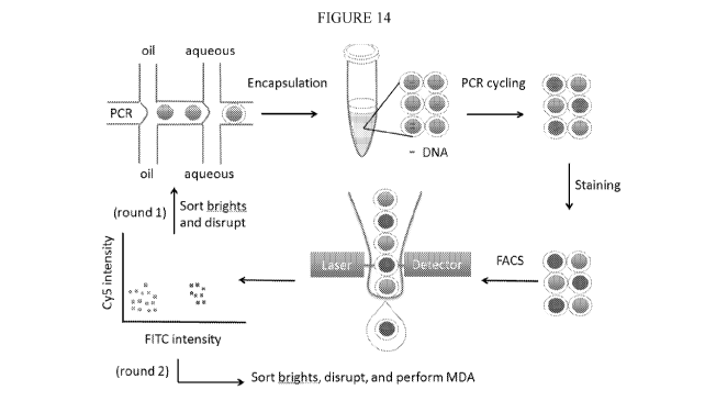
[0039] FIG. 14. provides a schematic of double emulsion digital PCR and
MESA
workflow. A sample comprising nucleic acids or cells is partitioned into
double emulsions using
two-step (shown) or one-step (not shown) double emulsification. The double
emulsions are
collected in a tube and thermocycled, resulting in, in the case of single
target molecule
encapsulation, digital amplification in double emulsions containing targets.
The double
emulsions are then subjected to FACS analysis and sorting.
11
CA 02974299 2017-07-18
WO 2016/126865 PCT/US2016/016438
[0040] FIG. 15. Digital PCR in double emulsions. Left panels shown
brightfield and
fluorescence images of double emulsions used to perform digital PCR, right
panels show FACS
plots of the double emulsions plotted as side scatter vs. forward scatter,
with gating parameters
shown. Red events denote double emulsions with fluorescence below the minimum
fluorescence
gate (not shown) while green events denote double emulsions with intensities
above this
threshold. The rows correspond to samples with different target DNA
concentration in
picograms, as labeled. Before PCR, no fluorescence positive events are
detected but post PCR,
many fluorescence positive events are detected, in which the number of events
scales with the
target DNA concentration.
[0041] FIG. 16. provides images of double emulsions used to performed
digital droplet
PCR presorting (upper panel) and post sorting for the positive collection
channel (middle panel)
and the negative collection channel (lower). Pre-sorting the population
consists of a mixture of
bright and dim droplets, but post sorting the positive collection contains
nearly all bright
droplets and the negative collection early all dim droplets.
[0042] FIG. 17 provides FACS data of double emulsions used to detect and
enrich
Lambda virus genomes out of a sample. Top panel shows the side scatter versus
forward scatter
of the double emulsions shown a clear population of large entities,
corresponding to the double
emulsions. Lower panels show the side scatter versus the fluorescence channel
(FITC) for the
gated population of "large" events on the upper right of the SSC x FSC plot.
As the
concentration of Lambda virus increases, more double emulsions are detected as
being
fluorescent as shown by comparing from left to right in the lower panel.
[0043] FIG. 18 provides a diagram of the HIV provirus genome, genes, and
locations of
primers used for genomic detection in droplets and qPCR analysis of sorted
molecules. The
provirus is embedded within the human genome at a location that can vary from
cell to cell. The
sample is fragmented into ¨100 kb molecules and the molecules sorted based on
whether they
contain a provirus genome. The gag primer set is used in the first sort, the
collected nucleic acids
are diluted and then sorted a second time using the env primer set.
[0044] FIG. 19 provides graphs showing quantitative PCR (par)
measurements of
enrichment of HIV provirus out of human genomic DNA using MESA. The four
panels
correspond to par measurements for the gag, poi, env, and LTR primer sets. The
red curves
corresponds to the original sample of human genomic DNA in which HIV provirus
is estimated
to be present at a rate of 10 copies per sample, which is used as a standard.
The blue curves
correspond to measurements of the sorted samples after droplet MDA and a 100-
fold dilution in
buffer, so that the concentration of DNA in the samples corresponding to the
blue and red curves
12
CA 02974299 2017-07-18
WO 2016/126865 PCT/US2016/016438
are equal as measured with a UV-spectrometer. The blue and red curves are
shifted by ¨12 Ct
curves, on average, corresponding to a ¨4000X difference in target
concentration. When
correcting for the 100X of the sorted materiel, this leads to a ¨400,000X
estimated increase of
the provirus in the MESA enriched sample. The sample was sorted using two
consecutive
rounds of MESA.
[0045] FIG. 20 provides images of TaqMan PCR for T4 as single emulsions
Panel (A)
and double emulsions Panel (B). Panel (C) provides an image showing
fluorescent double
emulsions containing T4 recovered from FACS sort. Panel (D) shows FSC-A (front
scatter-area)
against SSC-A (side scatter-area) log¨log plots and FITC channel fluorescence
frequency
histograms for double emulsions. Fluorescence plots are derived by gating
events in defined
areas on the FSC-A/SSC-A plots. The percentage of fluorescent droplets sorted
is indicated.
DETAILED DESCRIPTION
[0046] The methods and systems described herein, referred to as multiple-
emulsion
nucleic acid amplification, allow nucleic acids contained in biological
systems to be detected,
quantitated and/or sorted based on their sequence as detected with nucleic
acid amplification
techniques, e.g., PCR. The nucleic acids can be free floating or contained
within living or
nonliving structures, including particles, viruses, and cells. The nucleic
acids can include, e.g.,
DNA or RNA. Systems and devices for use in practicing methods of the
disclosure are also
provided.
[0047] Before the present invention is described in greater detail, it is
to be understood
that this invention is not limited to particular embodiments described, and as
such may, of
course, vary. It is also to be understood that the terminology used herein is
for the purpose of
describing particular embodiments only, and is not intended to be limiting,
since the scope of the
present invention will be limited only by the appended claims.
[0048] Where a range of values is provided, it is understood that each
intervening value,
to the tenth of the unit of the lower limit unless the context clearly
dictates otherwise, between
the upper and lower limits of that range is also specifically disclosed. Each
smaller range
between any stated value or intervening value in a stated range and any other
stated or
intervening value in that stated range is encompassed within the invention.
The upper and lower
limits of these smaller ranges may independently be included or excluded in
the range, and each
range where either, neither or both limits are included in the smaller ranges
is also encompassed
within the invention, subject to any specifically excluded limit in the stated
range. Where the
13
CA 02974299 2017-07-18
WO 2016/126865 PCT/US2016/016438
stated range includes one or both of the limits, ranges excluding either or
both of those included
limits are also included in the invention.
[0049] Unless defined otherwise, all technical and scientific terms used
herein have the
same meaning as commonly understood by one of ordinary skill in the art to
which this
invention belongs. Although any methods and materials similar or equivalent to
those described
herein can be used in the practice or testing of the present invention, some
potential and
exemplary methods and materials may now be described. Any and all publications
mentioned
herein are incorporated herein by reference to disclose and describe the
methods and/or
materials in connection with which the publications are cited. It is
understood that the present
disclosure supersedes any disclosure of an incorporated publication to the
extent there is a
contradiction.
[0050] It must be noted that as used herein and in the appended claims,
the singular
forms "a", "an", and "the" include plural referents unless the context clearly
dictates otherwise.
Thus, for example, reference to "a microdroplet" includes a plurality of such
microdroplets and
reference to "the multiple-emulsion microdroplet" includes reference to one or
more multiple-
emulsion microdroplets, and so forth.
[0051] It is further noted that the claims may be drafted to exclude any
element which
may be optional. As such, this statement is intended to serve as antecedent
basis for use of such
exclusive terminology as "solely", "only" and the like in connection with the
recitation of claim
elements, or the use of a "negative" limitation.
[0052] The publications discussed herein are provided solely for their
disclosure prior to
the filing date of the present application. Further, the dates of publication
provided may be
different from the actual publication dates which may need to be independently
confirmed. To
the extent such publications may set out a definition or disclosure that
conflicts with the explicit
or implicit definition or disclosure of the present disclosure, the definition
of the present
disclosure controls.
[0053] As will be apparent to those of skill in the art upon reading this
disclosure, each
of the individual embodiments described and illustrated herein has discrete
components and
features which may be readily separated from or combined with the features of
any of the other
several embodiments without departing from the scope or spirit of the present
invention. Any
recited method can be carried out in the order of events recited or in any
other order which is
logically possible.
14
CA 02974299 2017-07-18
WO 2016/126865 PCT/US2016/016438
METHODS
[0054] As summarized above, aspects of the present disclosure include
methods for the
detection and/or sorting of components from biological samples using multiple-
emulsion
microdroplets and/or GUVs. Aspects include methods for the detection,
quantification, and/or
genotyping of cells, e.g. normal mammalian cells (e.g., non-tumor cells),
tumor cells, e.g.,
circulating tumor cells (CTCs), or microbial cells. Additional embodiments of
interest include
PCR-based detection and/or and sorting of cells, PCR-based detection and/or
sorting of viral
particles and PCR-based detection and/or sorting of nucleic acids from a
heterogeneous
population of nucleic acids.
[0055] As used herein, the term "biological sample" encompasses a variety
of sample
types obtained from a variety of sources, which sample types contain
biological material. For
example, the term includes biological samples obtained from a mammalian
subject, e.g., a
human subject, and biological samples obtained from a food, water, or other
environmental
source, etc. The definition encompasses blood and other liquid samples of
biological origin, as
well as solid tissue samples such as a biopsy specimen or tissue cultures or
cells derived
therefrom and the progeny thereof The definition also includes samples that
have been
manipulated in any way after their procurement, such as by treatment with
reagents,
solubilization, or enrichment for certain components, such as polynucleotides.
The term
"biological sample" encompasses a clinical sample, and also includes cells in
culture, cell
supernatants, cell lysates, cells, serum, plasma, biological fluid, and tissue
samples. "Biological
sample" includes cells, e.g., bacterial cells or eukaryotic cells; biological
fluids such as blood,
cerebrospinal fluid, semen, saliva, and the like; bile; bone marrow; skin
(e.g., skin biopsy); and
antibodies obtained from an individual.
[0056] As described more fully herein, in various aspects the subject
methods may be
used to detect a variety of components from such biological samples.
Components of interest
include, but are not necessarily limited to, cells (e.g., circulating cells
and/or circulating tumor
cells), viruses, polynucleotides (e.g., DNA and/or RNA), polypeptides (e.g.,
peptides and/or
proteins), and many other components that may be present in a biological
sample.
[0057] "Polynucleotides" or "oligonucleotides" as used herein refer to
linear polymers of
nucleotide monomers, and may be used interchangeably. Polynucleotides and
oligonucleotides
can have any of a variety of structural configurations, e.g., be single
stranded, double stranded,
or a combination of both, as well as having higher order intra- or
intermolecular
secondary/tertiary structures, e.g., hairpins, loops, triple stranded regions,
etc. Polynucleotides
typically range in size from a few monomeric units, e.g. 5-40, when they are
usually referred to
CA 02974299 2017-07-18
WO 2016/126865 PCT/US2016/016438
as "oligonucleotides," to several thousand monomeric units. Whenever a
polynucleotide or
oligonucleotide is represented by a sequence of letters (upper or lower case),
such as
"ATGCCTG," it will be understood that the nucleotides are in 5'3' order from
left to right and
that "A" denotes deoxyadenosine, "C" denotes deoxycytidine, "G" denotes
deoxyguanosine, and
"T" denotes thymidine, "I" denotes deoxyinosine, "U" denotes uridine, unless
otherwise
indicated or obvious from context. Unless otherwise noted the terminology and
atom
numbering conventions will follow those disclosed in Strachan and Read, Human
Molecular
Genetics 2 (Wiley-Liss, New York, 1999).
[0058] The terms "polypeptide," "peptide," and "protein," used
interchangeably herein,
refer to a polymeric form of amino acids of any length. NH2 refers to the free
amino group
present at the amino terminus of a polypeptide. COOH refers to the free
carboxyl group present
at the carboxyl terminus of a polypeptide. In keeping with standard
polypeptide nomenclature,
Biol. Chem., 243 (1969), 3552-3559 is used.
[0059] In certain aspects, methods are provided for counting and/or
genotyping cells,
including normal cells or tumor cells, such as CTCs. A feature of such methods
is the use of
microfluidics.
[0060] As summarized above, the methods of the present disclosure
generally involve
nucleic acid amplification in multiple-emulsion microdroplets and/or GUVs
followed by
detection and/or sorting of the multiple-emulsion microdroplets and/or GUVs.
FIG. 1 presents a
schematic showing double emulsion formation via two-step emulsification
according to some
embodiments of the present disclosure. Panel A depicts schematically the
formation of a
miscible phase-in-immiscible phase single emulsion, e.g., a water-in-oil or
oil-in-water single
emulsion. Panel B depicts schematically the formation of a miscible phase-in-
immiscible phase-
in-miscible phase double emulsion, e.g., a water-in-oil-in-water double
emulsion or an oil-in-
water-in-oil double emulsion, after reinjection of the miscible phase-in-
immiscible phase single
emulsion. While depicted as separate components in FIG. 1, it should be noted
that a single
emulsion droplet maker and a double emulsion droplet maker may be provided as
fluidically
connected components in a single microfluidic device in some embodiments of
the disclosed
devices, methods and systems. Following formation of the double emulsion
microdroplets, the
double emulsion microdroplets are collected for subsequent nucleic acid
amplification. Such
nucleic acid amplification may occur in a nucleic acid amplification region of
a microfluidic
device, e.g., a microfluidic device including one or more of the single and
double emulsion
droplet makers, or "off chip" in a separate nucleic acid amplification
apparatus.
16
CA 02974299 2017-07-18
WO 2016/126865 PCT/US2016/016438
[0061] Alternatively, following formation of the double emulsion
microdroplets, the
double emulsion microdroplets may be subjected to dewetting conditions,
forming GUVs, in
which the immiscible phase fluid of the double emulsion is expunged from the
shell, leaving
behind a membrane of surfactant, with a small immiscible phase droplet adhered
to the outside
of the membrane.
[0062] FIG. 2 presents a schematic showing a more detailed embodiment of
the double
emulsion formation via two-step emulsification shown in FIG. 1. FIG. 2, Panel
A, shows an
embodiment in which PCR reagents and nucleic acids are introduced into a flow
channel of a
microfluidic device in an aqueous fluid. A single emulsion droplet maker
introduces a fluid
which is immiscible with the aqueous fluid, e.g., oil, to form single emulsion
microdroplets
containing the PCR reagents and nucleic acids. The single emulsion
microdroplets are then
reinjected into a second flow channel of a microfluidic device (Panel B). A
double emulsion
droplet maker introduces a fluid which is miscible with the aqueous fluid,
e.g., water, along with
a suitable surfactant, e.g., a detergent, to form double emulsion
microdroplets containing the
PCR reagents and nucleic acids. While depicted as separate components in FIG.
2, it should be
noted that a single emulsion droplet maker and a double emulsion droplet maker
may be
provided as fluidically connected components in a single microfluidic device
in some
embodiments of the disclosed devices, methods and systems. Following formation
of the double
emulsion microdroplets, the double emulsion microdroplets are collected for
subsequent PCR
amplification. Such PCR amplification may occur in a thermalcycler integrated
into a
microfluidic device, e.g., a microfluidic device including one or more of the
single and double
emulsion droplet makers, or "off chip" in a separate thermalcycler.
[0063] Alternatively, following formation of the double emulsion
microdroplets, the
double emulsion microdroplets may be subjected to dewetting conditions, in
which the
immiscible phase fluid of the double emulsion is expunged from the shell,
leaving behind a
membrane of surfactant, with a small immiscible phase droplet adhered to the
outside of the
membrane.
[0064] For embodiments in which a thermalcycler is integrated into a
microfluidic
device multiple-emulsion microdroplets or GUVs containing PCR reagents may be
flowed
through a channel that incubates the droplets under conditions effective for
PCR. For example,
the appropriate conditions may be achieved by flowing the multiple-emulsion
microdroplets
and/or GUVs through a channel that snakes over various zones maintained at 65
C and 95 C. As
the multiple-emulsion microdroplets and/or GUVs move through the zones, their
temperature
cycles, as needed for PCR. During the PCR reaction, if a multiple-emulsion
microdroplet and/or
17
CA 02974299 2017-07-18
WO 2016/126865 PCT/US2016/016438
GUV contains a nucleic acid which the selected primer(s) are designed to
detect, amplification is
initiated. The presence of these particular PCR products may be detected by,
for example, a
fluorescent output that turns the multiple-emulsion microdroplets and/or GUVs
fluorescent. The
multiple-emulsion microdroplets and/or GUVs may thus be scanned, such as by
using flow
cytometry, to detect the presence of fluorescent drops. In certain aspects,
the multiple-emulsion
microdroplets and/or GUVs may also be sorted using, for example, droplet
sorting to recover
drops of interest Using the nomenclature of the current disclosure, the steps
described above
are thus performed "under microfluidic control." That is, the steps are
performed on one or
more microfluidics devices, or at least in part on one or more microfluidic
devices.
[0065] Initial encapsulation of a component from a biological sample in a
single
emulsion microdroplet in accordance with the methods described herein may be
achieved by any
convenient means. Encapsulation approaches of interest also include, but are
not limited to,
hydrodynamically-triggered drop formation and those described by Link, et al.,
Phys. Rev. Lett.
92, 054503 (2004), the disclosure of which is incorporated herein by
reference.
[0066] A feature of certain methods of the present disclosure is the use
of a polymerase
chain reaction (PCR)-based assay to detect the presence of certain
oligonucleotides and/or
genes, e.g., oncogene(s) present in cells. Examples of PCR-based assays of
interest include, but
are not limited to, quantitative PCR (qPCR), quantitative fluorescent PCR (QF-
PCR), multiplex
fluorescent PCR (MF-PCR), single cell PCR, PCR-RFLP/real time-PCR-RFLP, hot
start PCR,
nested PCR, in situ polony PCR, in situ rolling circle amplification (RCA),
bridge PCR, picotiter
PCR, emulsion PCR and reverse transcriptase PCR (RT-PCR). Other suitable
amplification
methods include the ligase chain reaction (LCR), transcription amplification,
self-sustained
sequence replication, selective amplification of target polynucleotide
sequences, consensus
sequence primed polymerase chain reaction (CP-PCR), arbitrarily primed
polymerase chain
reaction (AP-PCR), degenerate oligonucleotide-primed PCR (DOP-PCR) and nucleic
acid based
sequence amplification (NABSA).
[0067] A PCR-based assay may be used to detect the presence of certain
gene(s), such as
certain oncogene(s). In such assays, one or more primers specific to each gene
of interest are
reacted with the genome of each cell. These primers have sequences specific to
the particular
gene, so that they will only hybridize and initiate PCR when they are
complementary to the
genome of the cell. If the gene of interest is present and the primer is a
match, many copies of
the gene are created. To determine whether a particular gene is present, the
PCR products may
be detected through an assay probing the liquid of the multiple-emulsion
microdroplet and/or
GUV, such as by staining the solution with an intercalating dye, like
SybrGreen or ethidium
18
CA 02974299 2017-07-18
WO 2016/126865 PCT/US2016/016438
bromide, hybridizing the PCR products to a solid substrate, such as a bead
(e.g., magnetic or
fluorescent beads, such as Luminex beads), or detecting them through an
intermolecular
reaction, such as FRET. These dyes, beads, and the like are each examples of a
"detection
component," a term that is used broadly and generically herein to refer to any
component that is
used to detect the presence or absence of nucleic acid amplification products,
e.g., PCR
products.
[0068] A number of variations of these basic approaches will now be
outlined in greater
detail below.
Detecting Cells (e.g., Tumor Cells) in Multiple-Emulsion Microdroplets and/or
GUVs
[0069] Aspects of the subject methods involve detecting the presence of
one or more
cells or subsets of cells (e.g., tumor cells) in a biological sample. Such
methods may include, for
example, steps of encapsulating a cell in a multiple-emulsion microdroplet
and/or GUV, the
multiple-emulsion microdroplet and/or GUV including a first miscible phase
fluid surrounded
by an immiscible shell, wherein the multiple-emulsion microdroplet and/or GUV
is positioned in
a second miscible phase carrier fluid; subjecting the multiple-emulsion
microdroplet and/or
GUV to conditions sufficient to effect lysis of the cell in the multiple-
emulsion microdroplet
and/or GUV; subjecting the multiple-emulsion microdroplet and/or GUV to
conditions sufficient
to deactivate or remove one or more materials which have an inhibitory effect
on nucleic acid
amplification; introducing nucleic acid amplification reagents into the
multiple-emulsion
microdroplet and/or GUV; subjecting the multiple-emulsion microdroplet and/or
GUV to
amplification conditions sufficient to result in amplification of a target
nucleic acid when
present; and detecting an amplification product resulting from the
amplification of the target
nucleic acid when present.
[0070] A biological sample (e.g., whole blood) may be recovered from a
subject using
any convenient means. The biological sample may be processed to remove
components other
than cells using, for example, processing steps such as centrifugation,
filtration, and the like.
Where desired, the cells may be stained with one or more antibodies and/or
probes prior to
encapsulating them into multiple-emulsion microdroplets and/or GUVs.
[0071] One or more lysing agents may also be added to the multiple-
emulsion
microdroplets and/or GUVs containing a cell, under conditions in which the
cell(s) may be
caused to burst, thereby releasing their genomes. The lysing agents may be
added after the cells
are encapsulated into multiple-emulsion microdroplets and/or GUVs. Any
convenient lysing
agent may be employed, such as proteinase K or cytotoxins. In particular
embodiments, cells
19
CA 02974299 2017-07-18
WO 2016/126865 PCT/US2016/016438
may be co-encapsulated in multiple-emulsion microdroplets and/or GUVs with
lysis buffer
containing detergents such as Triton X100 and/or proteinase K. The specific
conditions in which
the cell(s) may be caused to burst will vary depending on the specific lysing
agent used. For
example, if proteinase K is incorporated as a lysing agent, the multiple-
emulsion microdroplets
and/or GUVs may be heated to about 37-60 C for about 20 min to lyse the cells
and to allow the
proteinase K to digest cellular proteins, after which they may be heated to
about 95 C for about
5-10 min to deactivate the proteinase K.
[0072] In certain aspects, cell lysis may also, or instead, rely on
techniques that do not
involve addition of lysing agent. For example, lysis may be achieved by
mechanical techniques
that may employ various geometric features to effect piercing, shearing,
abrading, etc. of cells.
Other types of mechanical breakage such as acoustic techniques may also be
used. Further,
thermal energy can also be used to lyse cells. Any convenient means of
effecting cell lysis may
be employed in the methods described herein.
[0073] Primers may be introduced into the multiple-emulsion microdroplets
and/or
GUVs for each of the genes and/or genetic markers, e.g., oncogenes, to be
detected. Hence, in
certain aspects, primers for a variety of genes and/or genetic markers, e.g.,
all oncogenes may be
present in the multiple-emulsion microdroplets and/or GUVs at the same time,
thereby providing
a multiplexed assay. The multiple emulsion microdroplets and/or GUVs may be
temperature-
cycled so that multiple emulsion microdroplets and/or GUVs containing target
cells, e.g.,
cancerous cells, will undergo PCR. Alternatively, isothermal nucleic acid
amplification methods
may be utilized, e.g., loop-mediated isothermal amplification (LAMP), strand
displacement
amplification (SDA), helicase-dependent amplification (HDA), and nicking
enzyme
amplification reaction (NEAR). Only the primers corresponding to oncogenes
and/or genetic
markers present in the genome will induce amplification, creating many copies
of these
oncogenes and/or genetic markers in the multiple emulsion microdroplets and/or
GUVs.
Detecting the presence of these amplification products may be achieved by a
variety of ways,
such as by using FRET, staining with an intercalating dye, or attaching them
to a bead. The
multiple emulsion microdroplets and/or GUVs may be optically probed to detect
the
amplification products. In some embodiments, optically probing the multiple
emulsion
microdroplets and/or GUVs may involve counting the number of tumor cells
present in the
initial population, and/or allowing for the identification of the oncogenes
present in each tumor
cell.
[0074] The subject methods may be used to determine whether a biological
sample
contains particular cells of interest, e.g., tumor cells, or not. In certain
aspects, the subject
CA 02974299 2017-07-18
WO 2016/126865 PCT/US2016/016438
methods may include quantifying the number of cells of interest, e.g., tumor
cells, present in a
biological sample. Quantifying the number of cells of interest, e.g., tumor
cells, present in a
biological sample may be based at least in part on the number of multiple
emulsion
microdroplets and/or GUVs in which amplification products were detected. For
example,
multiple emulsion microdroplets and/or GUVs may be produced under conditions
in which the
majority of microdroplets are expected to contain zero or one cell. Those
multiple emulsion
microdroplets and/or GUVs that do not contain any cells may be removed, using
techniques
described more fully herein. After performing the PCR steps outlined above,
the total number of
multiple emulsion microdroplets and/or GUVs that are detected to contain
amplification
products may be counted, so as to quantify the number of cells of interest,
e.g., tumor cells, in
the biological sample. In certain aspects, the methods may also include
counting the total
number of multiple emulsion microdroplets and/or GUVs so as to determine the
fraction or
percentage of cells from the biological sample that are cells of interest,
e.g., tumor cells.
[0075] In some embodiments, the introduction of amplification reagents
into the
multiple-emulsion microdroplets and/or GUVs includes introducing the
amplification reagents
into the second miscible phase carrier fluid, wherein the amplification
reagents diffuse from the
second miscible phase carrier fluid, through the immiscible shell, and into
the first miscible
phase fluid of the multiple-emulsion microdroplets and/or GUVs.
[0076] The cells and/or cellular material of interest may be recovered by
sorting the
multiple-emulsion microdroplets and/or GUVs and recovering their contents via
microdroplet
rupture, e.g., through chemical, electrical, or mechanical means as described
in greater detail
herein. A variety of suitable sorting techniques and related devices may be
utilized to sort and
separate the multiple-emulsion microdroplets and/or GUVs containing
amplification products
including those described herein.
Nucleic Acid Detection in Multiple-Emulsion Micr droplets and/or GUVs
[0077] As discussed herein, the disclosed methods find use in the
detection of nucleic
acids, e.g., DNA or RNA, of interest from a variety of biological samples.
Such methods may
include, for example, steps of encapsulating a nucleic acid and amplification
reagents in a
multiple-emulsion microdroplet and/or GUV, the multiple-emulsion microdroplet
and/or GUV
including a first miscible phase fluid surrounded by an immiscible shell,
wherein the multiple-
emulsion microdroplet and/or GUV is positioned in a second miscible phase
carrier fluid; and
subjecting the multiple-emulsion microdroplet and/or GUV to amplification
conditions
sufficient to result in amplification of the nucleic acid; and detecting an
amplification product
21
CA 02974299 2017-07-18
WO 2016/126865 PCT/US2016/016438
resulting from the amplification of the nucleic acid. In some embodiments, the
second miscible
phase carrier fluid is a buffered aqueous phase carrier fluid, and in some
embodiments the first
and second miscible phase fluids are the same. The amplification conditions
may be PCR
conditions e.g., RT-PCR conditions, or isothermal amplification conditions,
e.g., loop-mediated
isothermal amplification (LAMP), strand displacement amplification (SDA),
helicase-dependent
amplification (HDA), and nicking enzyme amplification reaction (NEAR).
[0078] The nucleic acids of interest may be recovered by sorting the
multiple-emulsion
microdroplets and/or GUVs and recovering their contents via microdroplet
rupture, e.g., through
chemical, electrical, or mechanical means as described in greater detail
herein. A variety of
suitable sorting techniques and related devices may be utilized to sort and
separate the multiple-
emulsion microdroplets and/or GUVs containing amplification products including
those
described herein.
[0079] In one aspect, a method for enriching for a target nucleic acid
sequence is
provided, wherein the method includes encapsulating a sample including nucleic
acids in a
plurality of multiple-emulsion microdroplets and/or GUVs; introducing
polymerase chain
reaction (PCR) reagents and a plurality of PCR primers into the multiple-
emulsion
microdroplets and/or GUVs; incubating the multiple-emulsion microdroplets
and/or GUVs
under conditions sufficient for PCR amplification to produce PCR amplification
products,
wherein the plurality of PCR primers include one or more primers that each
hybridize to one or
more oligonucleotides comprised by the target nucleic acid sequence, and
wherein the PCR
amplification products do not include the entire target nucleic acid sequence;
introducing a
detection component into the multiple-emulsion microdroplets and/or GUVs
either before or
after the incubating; detecting the presence or absence of the PCR
amplification products by
detection of the detection component, wherein detection of the detection
component indicates
the presence of PCR amplification products and the target nucleic acid
sequence; and sorting the
multiple-emulsion microdroplets and/or GUVs based on detection of the
detection component,
wherein the sorting separates multiple-emulsion microdroplets and/or GUVs
including the PCR
amplification products and the target nucleic acid sequence, when present,
from multiple-
emulsion microdroplets and/or GUVs which do not include the PCR amplification
products and
the target nucleic acid sequence; and pooling the nucleic acid sequences from
the sorted
multiple-emulsion microdroplets and/or GUVs to provide an enriched pool of
target nucleic acid
sequences, when present. One or more of these steps may be performed under
microfluidic
control.
22
CA 02974299 2017-07-18
WO 2016/126865 PCT/US2016/016438
[0080] The above method allows, for example, for the enrichment of DNA
molecules
out of a heterogeneous system based on the presence of PCR-detectable
subsequences. The
DNA molecules can be short (e.g., hundreds of bases) or long (e.g., megabases
or longer). The
sample may be encapsulated in microdroplets such that target molecules are
detected in the
microdroplets digitally ¨ i.e., each microdroplet contains 0 or 1 target
molecule. The
microdroplets may then be sorted based on, e.g., fluorescence, to recover the
target molecules.
This method can be used to enrich for a large genomic region, e.g., on the
order of megabases in
length, in a heterogeneous sample of DNA fragments.
[0081] The above method enables a sufficient amount of DNA to be
recovered without
the need to perform PCR to amplify the DNA for sequencing. Amplification-free
DNA sample
prep is valuable, for example, where PCR does not preserve the sequences or
epigenetic factors
of interest, or cannot recover sequences that are of the needed length (e.g.,
> about 10 kb, the
practical limit of long-range PCR).
[0082] Another application of the above method is to enrich DNA for
epigenetic
sequencing. Epigenetic marks on DNA are not preserved by PCR, so sequencing
them requires
unamplified DNA from the host nucleic acids. With the above method, a
sufficient amount of
DNA can be obtained for sequencing without needing to perform PCR, and thus
preserving the
epigenetic marks.
[0083] The above methods have particular utility where the length of the
target nucleic
acid exceeds the practical limits of long-range PCR, e.g., where the nucleic
acid is greater than
about 10 kb, and/or where it is desirable to preserve epigenetic marks on the
DNA. In some
embodiments, the target nucleic acid to be enriched is greater than about 100
kb in length, e.g.,
greater than about 1 megabase in length. In some embodiments, the target
nucleic acid to be
enriched is from about 10 kb to about 100 kb, from about 100 kb to about 500
kb, or from about
500 kb to about lmegabase in length.
[0084] Post-amplification and/or purification, emulsions can be broken
using both
chemical and osmotic means for future analysis. For example, an equal volume
of 1H, 1H, 2H,
2H-Perfluoro-1-octanol can be added to a purified sample and mixed either
through pipetting or
vortexing. The resulting mixture can then be allowed to equilibrate, and the
aqueous layer can
be eluted off for further analysis. Similarly, a large excess of purified
water can be added to the
sample post-sort, mixed, and allowed to incubate at room temperature for
several hours. The
resulting mixture can then be analyzed directly for purified sample of
interest.
23
CA 02974299 2017-07-18
WO 2016/126865 PCT/US2016/016438
PCR
[0085] As summarized above, in practicing methods of the invention a PCR-
based assay
may be used to detect the presence of certain nucleic acids of interest, e.g.,
genes of interest
and/or genetic markers, e.g., oncogene(s), present in cells or a heterogeneous
sample of nucleic
acids. The conditions of such PCR-based assays may vary in one or more ways.
[0086] For instance, the number of PCR primers that may be added to a
multiple-
emulsion microdroplet and/or GUV may vary. The term "primer" may refer to more
than one
primer and refers to an oligonucleotide, whether occurring naturally, as in a
purified restriction
digest, or produced synthetically, which is capable of acting as a point of
initiation of synthesis
along a complementary strand when placed under conditions in which synthesis
of a primer
extension product which is complementary to a nucleic acid strand is
catalyzed. Such conditions
include the presence of four different deoxyribonucleoside triphosphates and a
polymerization-
inducing agent such as DNA polymerase or reverse transcriptase, in a suitable
buffer ("buffer"
includes substituents which are cofactors, or which affect pH, ionic strength,
etc.), and at a
suitable temperature. The primer is preferably single-stranded for maximum
efficiency in
amplification.
[0087] The complement of a nucleic acid sequence as used herein refers to
an
oligonucleotide which, when aligned with the nucleic acid sequence such that
the 5' end of one
sequence is paired with the 3' end of the other, is in "antiparallel
association." Complementarity
need not be perfect; stable duplexes may contain mismatched base pairs or
unmatched bases.
Those skilled in the art of nucleic acid technology can determine duplex
stability empirically
considering a number of variables including, for example, the length of the
oligonucleotide,
percent concentration of cytosine and guanine bases in the oligonucleotide,
ionic strength, and
incidence of mismatched base pairs.
[0088] The number of PCR primers that may be added to a multiple-emulsion
microdroplet and/or GUV may range from about 1 to about 500 or more, e.g.,
about 2 to 100
primers, about 2 to 10 primers, about 10 to 20 primers, about 20 to 30
primers, about 30 to 40
primers, about 40 to 50 primers, about 50 to 60 primers, about 60 to 70
primers, about 70 to 80
primers, about 80 to 90 primers, about 90 to 100 primers, about 100 to 150
primers, about 150 to
200 primers, about 200 to 250 primers, about 250 to 300 primers, about 300 to
350 primers,
about 350 to 400 primers, about 400 to 450 primers, about 450 to 500 primers,
or about 500
primers or more.
[0089] These primers may contain primers for one or more gene of
interest, e.g.
oncogenes. The number of primers for genes of interest that are added may be
from about one
24
CA 02974299 2017-07-18
WO 2016/126865 PCT/US2016/016438
to 500, e.g., about 1 to 10 primers, about 10 to 20 primers, about 20 to 30
primers, about 30 to
40 primers, about 40 to 50 primers, about 50 to 60 primers, about 60 to 70
primers, about 70 to
80 primers, about 80 to 90 primers, about 90 to 100 primers, about 100 to 150
primers, about
150 to 200 primers, about 200 to 250 primers, about 250 to 300 primers, about
300 to 350
primers, about 350 to 400 primers, about 400 to 450 primers, about 450 to 500
primers, or about
500 primers or more. Genes and oncogenes of interest include, but are not
limited to, BAX,
BCL2L1, CASP8, CDK4, ELK1, ETS1, HGF, JAK2, JUNB, JUND, KIT, KITLG, MCL1,
MET, MOS, MYB, NFKBIA, EGFR, Myc, EpCAM, NRAS, PIK3CA, PML, PRKCA, RAF1,
RARA, REL, ROS1, RUNX1, SRC, STAT3, CD45, cytokeratins, CEA, CD133, HER2,
CD44,
CD49f, CD146, MUC1/2, and ZHX2.
[0090] Such primers and/or reagents may be added to a multiple-emulsion
microdroplet
and/or GUV in one step, or in more than one step. For instance, the primers
may be added in two
or more steps, three or more steps, four or more steps, or five or more steps.
Regardless of
whether the primers are added in one step or in more than one step, they may
be added after the
addition of a lysing agent, prior to the addition of a lysing agent, or
concomitantly with the
addition of a lysing agent. When added before or after the addition of a
lysing agent, the PCR
primers may be added in a separate step from the addition of a lysing agent.
[0091] Once primers have been added to a multiple-emulsion microdroplet
and/or GUV
the multiple-emulsion microdroplet and/or GUV may be incubated under
conditions allowing
for PCR. The multiple-emulsion microdroplet and/or GUV may be incubated on the
same
microfluidic device as was used to add the primer(s), or may be incubated on a
separate device.
In certain embodiments, incubating the multiple-emulsion microdroplet and/or
GUV under
conditions allowing for PCR amplification is performed on the same
microfluidic device used to
encapsulate and lyse cells. Incubating the multiple-emulsion microdroplet
and/or GUV may
take a variety of forms. In certain aspects, the multiple-emulsion
microdroplet and/or GUV
containing the PCR mix may be flowed through a channel that incubates the
microdroplets
under conditions effective for PCR. Flowing the multiple-emulsion microdroplet
and/or GUV
through a channel may involve a channel that snakes over various temperature
zones maintained
at temperatures effective for PCR. Such channels may, for example, cycle over
two or more
temperature zones, wherein at least one zone is maintained at about 65 C and
at least one zone is
maintained at about 95 C. As the multiple-emulsion microdroplets and/or GUVs
move through
such zones, their temperature cycles, as needed for PCR. The precise number of
zones, and the
respective temperature of each zone, may be readily determined by those of
skill in the art to
achieve the desired PCR amplification.
CA 02974299 2017-07-18
WO 2016/126865 PCT/US2016/016438
[0092] In other embodiments, incubating the multiple-emulsion
microdroplets and/or
GUVs may involve the use of a Megadroplet Array. In such a device, an array of
hundreds,
thousands, or millions of traps indented into a channel (e.g., a PDMS channel)
sit above a
thermal system. The channel may be pressurized, thereby preventing gas from
escaping. The
height of the microfluidic channel is smaller than the diameter of the
multiple-emulsion
microdroplets and/or GUVs, causing multiple-emulsion microdroplets and/or GUVs
to adopt a
flattened pancake shape. When a multiple-emulsion microdroplet and/or GUV
flows over an
unoccupied indentation, it adopts a lower, more energetically favorable,
radius of curvature,
leading to a force that pulls the multiple-emulsion microdroplet and/or GUV
entirely into the
trap. By flowing multiple-emulsion microdroplets and/or GUVs as a close pack,
it is ensured
that all traps on the array are occupied. The entire device may be thermal
cycled using a heater.
[0093] In certain aspects, the heater includes a Peltier plate, heat
sink, and control
computer. The Peltier plate allows for the heating or cooling of the chip
above or below room
temperature by controlling the applied current. To ensure controlled and
reproducible
temperature, a computer may monitor the temperature of the array using
integrated temperature
probes, and may adjust the applied current to heat and cool as needed. A
metallic (e.g. copper)
plate allows for uniform application of heat and dissipation of excess heat
during cooling cycles,
enabling cooling from about 95 C to about 60 C in under about one minute.
[0094] Methods of the invention may also include introducing one or more
probes to the
multiple-emulsion microdroplets and/or GUVs. As used herein with respect to
nucleic acids, the
term "probe" refers to a labeled oligonucleotide which forms a duplex
structure with a sequence
in the target nucleic acid, due to complementarity of at least one sequence in
the probe with a
sequence in the target region. In some embodiments, the probe does not contain
a sequence
complementary to sequence(s) used to prime the polymerase chain reaction. The
number of
probes that are added may be from about one to 500, e.g., about 1 to 10
probes, about 10 to 20
probes, about 20 to 30 probes, about 30 to 40 probes, about 40 to 50 probes,
about 50 to 60
probes, about 60 to 70 probes, about 70 to 80 probes, about 80 to 90 probes,
about 90 to 100
probes, about 100 to 150 probes, about 150 to 200 probes, about 200 to 250
probes, about 250 to
300 probes, about 300 to 350 probes, about 350 to 400 probes, about 400 to 450
probes, about
450 to 500 probes, or about 500 probes or more. The probe(s) may be introduced
into the
multiple-emulsion microdroplets and/or GUVs prior to, subsequent with, or
after the addition of
the one or more primer(s). Probes of interest include, but are not limited to,
TaqMang probes
(e.g., as described in Holland, P. M.; Abramson, R. D.; Watson, R.; Gelfand,
D. H.
26
CA 02974299 2017-07-18
WO 2016/126865 PCT/US2016/016438
(1991). "Detection of specific polymerase chain reaction product by utilizing
the 5'----3'
exonuclease activity of Thermus aquaticus DNA polymerase". PNAS, 88 (16): 7276-
7280).
[0095] In certain embodiments, an RT-PCR based assay may be used to
detect the
presence of certain transcripts of interest, e.g., oncogene(s), present in
cells. In such
embodiments, reverse transcriptase and any other reagents necessary for cDNA
synthesis are
added to the multiple-emulsion microdroplets and/or GUVs in addition to the
reagents used to
carry out PCR described herein (collectively referred to as the "RT-PCR
reagents"). The RT-
PCR reagents are added to the multiple-emulsion microdroplets and/or GUVs
using any of the
suitable methods described herein. Once reagents for RT-PCR have been added to
a multiple-
emulsion microdroplet and/or GUV, the multiple-emulsion microdroplet and/or
GUV may be
incubated under conditions allowing for reverse transcription followed by
conditions allowing
for PCR as described herein. The multiple-emulsion microdroplet and/or GUV may
be incubated
on the same microfluidic device as was used to add the RT-PCR reagents, or may
be incubated
on a separate device. In certain embodiments, incubating the multiple-emulsion
microdroplet
and/or GUV under conditions allowing for RT-PCR is performed on the same
microfluidic
device used to encapsulate and lyse cells.
[0096] In certain embodiments, the reagents added to the multiple-
emulsion
microdroplet and/or GUV for RT-PCR or PCR further includes a fluorescent DNA
probe
capable of detecting RT-PCR or PCR products. Any suitable fluorescent DNA
probe can be
used including, but not limited to SYBR Green, TaqMang, Molecular Beacons and
Scorpion
probes. In certain embodiments, the reagents added to the multiple-emulsion
microdroplet
and/or GUV include more than one DNA probe, e.g., two fluorescent DNA probes,
three
fluorescent DNA probes, or four fluorescent DNA probes. The use of multiple
fluorescent DNA
probes allows for the concurrent measurement of RT-PCR or PCR products in a
single reaction.
Double PCR
[0097] To amplify rare transcripts, a multiple-emulsion microdroplet
and/or GUV that
has undergone a first-step RT-PCR or PCR reaction as described herein may be
further
subjected to a second step PCR reaction. In some embodiments, a first multiple-
emulsion
microdroplet and/or GUV that has undergone a first-step RT-PCR or PCR reaction
is
encapsulated in a second multiple-emulsion microdroplet and/or GUV containing
additional
PCR reagents, including, but not limited to enzymes (e.g. DNA polymerase), DNA
probes (e.g.
fluorescent DNA probes) and primers, followed by rupture of the first multiple-
emulsion
microdroplet and/or GUV. In certain embodiments, the second multiple-emulsion
microdroplet
27
CA 02974299 2017-07-18
WO 2016/126865 PCT/US2016/016438
and/or GUV containing the additional PCR reagents is larger than the
microdroplet that has
undergone the first step RT-PCR or PCR reaction. This may be beneficial, for
example, because
it allows for the dilution of cellular components that may be inhibitory to
the second step PCR.
The second step PCR reaction may be carried out on the same microfluidic
device used to carry
out the first-step reaction or on a different microfluidic device.
[0098] In some embodiments, the primers used in the second step PCR
reaction are the
same primers used in the first step RT-PCR or PCR reaction. In other
embodiments, the primers
used in the second step PCR reaction are different than the primers used in
the first step reaction.
Digital PCR
[0099] The methods and devices described herein can be used to quantitate
nucleic acids
using, for example, digital PCR. In digital PCR, target nucleic acids from a
solution are diluted
such that, when the sample is isolated in compartments, most compartments
encapsulate either
zero or one target molecule, although higher loading rates can often be used,
provided they can
be modeled. Reagents sufficient for amplification of the target nucleic acids
are also included in
the compartments, and the compartments subjected to conditions suitable for
amplification. The
compartments can have a variety of structures, including fabricated microwells
in a substrate or
single emulsion droplets. They may also be formed as, for example, the
multiple-emulsion
microdroplets, e.g., double emulsions, and/or GUVs of the present disclosure.
In some such
embodiments, the sample is compartmentalized in double emulsions and the
double emulsions
subjected to amplification. Droplets that contain a target undergo
amplification, while those that
do not, do not and therefore do not yield nucleic acid amplification products.
If a detection
component is included, double emulsions that comprise the target may fill with
a detectable
signal, allowing them to be identified by, for example, imaging or flow
dropometry. A powerful
advantage of using double emulsions to perform such digital PCR is that the
double emulsions
can be suspended in an aqueous carrier phase that is miscible with the
partitioned sample, and
can therefore readily be detected and/or sorted using commercially available
flow cytometers
and fluorescence activated cell sorters (FACS).
[00100] As described herein, this allows for enrichments of target
entities out of a sample
that is not possible with other methods in which sorting is not easily
accomplished. The
disclosed methods can be used to quantitate nucleic acids in solution by
counting the fraction of
double emulsions that are fluorescent and underwent amplification and thus
contained at least a
single target nucleic acid, in most instances; false amplification may occur
for stochastic reasons
or, for example, the encapsulation of dust or other contaminants that
interfere with the
28
CA 02974299 2017-07-18
WO 2016/126865 PCT/US2016/016438
specificity of the amplification reaction. TaqMan probes, molecular beacons,
SYBR, and other
kinds of detection components can also be included, allowing the use of
multiple optical spectra
for simultaneously detecting the amplification of different nucleic acid
sequences in the target or
due to multiple targets being encapsulated in the same double emulsions, which
may be
advantageous in some instances.
Measuring Lengths of Nucleic Acids
[00101] The methods and devices described herein can be used to measure
the length
distributions of nucleic acids in solution. This may be accomplished by
designing probe
sequences that anneal to the target nucleic acids at different regions of
known distance along
their lengths. The probes can then be mixed with the target nucleic acids and
compartmentalized
in multiple-emulsion microdroplets, e.g., double emulsions, and/or GUVs. Each
multiple-
emulsion microdroplet and/or GUV may contain, for example, two primer and
probe sets that
signal the presence of two different regions on the target a known distance
apart. This can be
repeated for different combinations of probes such that different pairs probe
different distances
and different regions of the target. The samples can be subjected to
amplification, analysis, and
sorting, if desired. In the analysis, one will find that some multiple-
emulsion microdroplets
and/or GUVs undergo amplification only with one of the probes while others,
for example,
amplify with only the other probe. This suggests that in these multiple-
emulsion microdroplets
and/or GUVs, one type contains the region just for one of the probes, while
the other type
contains the region of the other probe. In this population, may also be
multiple-emulsion
microdroplets and/or GUVs that undergo amplification with both probes,
indicating that the
target nucleic acid therein contained both regions. In this same suspension
will be a large
number of multiple-emulsion microdroplets and/or GUVs comprising a measurable
fraction of
each of the three types of droplets ¨ in addition to ones that, of course,
undergo no amplification
and, thus, presumably, do not contain the targeted regions. This data can be
used to infer the
lengths of the nucleic acids in solution.
[00102] For example, if the nucleic acids in solution are largely intact
as whole
molecules, than the majority of droplets undergoing amplification will exhibit
amplification with
both probe and primer sets and will thus show mixed signal. By contrast, if
the nucleic acid
targets are highly fragmented, most of the detection events will be one or the
other probe, with
only rare instances of both probes. Since the distances between the probes may
be known, this
allows one to estimate the lengths and fragmentation of the molecules in the
solution. This
process can be repeated with different probe sets targeting different regions
and/or having
29
CA 02974299 2017-07-18
WO 2016/126865 PCT/US2016/016438
different distances between them, to more fully characterize the fragmentation
of the target
nucleic acids.
MESA in Multiple-emulsion Microdroplets and/or GUVs
[00103] The methods described herein can be used to perform microfluidic
enrichment for
sequence analysis (MESA) of target nucleic acids. This is accomplished by
using the method to
encapsulate target nucleic acids in multiple-emulsion microdroplets and/or
GUVs and perform
amplification in those droplets, yielding fluorescent signals when the
droplets contain a target
sequence. These droplets can then be sorted, thereby enriching the nucleic
acids in the sorted
pool. The reaction may also be multiplexed, if desired, to differentiate
between molecules that
contain multiple, distinct subsequences. Amplification may also be used to
amplify the sorted
nucleic acids either prior to, simultaneous with, or post sorting, so as to
enable sequencing.
[00104] A key advantage of this approach is that the region that is
amplified in the
multiple-emulsion microdroplets and/or GUVs can be used simply as a "detection
region" ¨ the
amplicons need not comprise the molecules that are subjected to sequencing.
Instead, they signal
when a target molecule is present in a multiple-emulsion microdroplet and/or
GUV so that the
whole molecule can be recovered for downstream analysis. This is powerful
because it allows a
large nucleic acid, even one that is far too large to be efficiently
amplified, to be recovered for
downstream analysis. For example, suppose that there exists a gene that is
thought to be part of
an important biological pathway, e.g., signaling cascade, in a microorganism
that is as yet still
undiscovered. The goal is to recover the genes encoding the proteins involved
in this pathway so
that they can be sequenced and studied. This cannot easily be accomplished
using existing
enrichment methods since the microbe, being unknown may not be specifically
cultivable and,
in addition, the pathway, being largely of unknown sequence, cannot be
purified using
hybridization probes, since sequences for the probes to hybridize to are not
known aside from
the individual gene, which may be too small to pull out the entire pathway.
However, this can be
accomplished using the MESA method described herein.
[00105] In some embodiments, the nucleic acids from the target may be
fragmented to a
size large enough to encapsulate the entire pathway, such as, for example tens
or hundreds of
kilobases, or even megabases or longer fragments. If the pathway exists within
a fragment, it
may contain the known gene. The fragmented nucleic acids, most of which do not
contain the
target, are subjected to the techniques described herein resulting in multiple-
emulsion
microdroplets, e.g., double emulsions, and/or GUVs that, for the most part, do
not contain a
pathway and thus exhibit no amplification, while rare drops do contain the
pathway and undergo
CA 02974299 2017-07-18
WO 2016/126865 PCT/US2016/016438
amplification. The positive droplets can then be recovered by, for example,
FACS sorting
double emulsions that are fluorescently bright. These can then be subjected to
further
manipulations such as, if necessary, specific and non-specific amplification,
quantitation through
digital or quantitative PCR, and DNA sequencing. A powerful advantage of MESA
over other
enrichment strategies is that it allows very large nucleic acids, even up to
the size of an entire
genome, to be detected and recovered based on a short, known sequence of only
tens of
hundreds of base pairs. Few other enrichment methodologies have the ability to
enrich such
large nucleic acid sequences out of a heterogeneous pool using such limited
amounts of
information about the sequence.
[00106] The method can also be used to identify the DNA sequences of
individual
genomes. In this embodiment, nucleic acids from a target can be fragmented and
encapsulated in
multiple-emulsion microdroplets, e.g., double emulsions, and/or GUVs, with PCR
reagents and
primers specific to the DNA sequence of interest. After amplification, the
positive multiple-
emulsion microdroplets and/or GUVs can be sorted into individual compartments,
such as well
plate arrays, using FACS. Individual compartments can then be subjected to
further
manipulation, such as either specific or non-specific amplification. The
resulting amplicons can
then be used to make libraries for next generation sequencing techniques, or
as material used
directly in Sanger sequencing. This technique would be useful, for example, in
a method
designed to identify genetic differences in a retroviral population, such as
HIV, found in an
individual patient.
[00107] As discussed above, methods described herein can be used for
digital PCR and,
related, microfluidic enrichment for sequencing analysis (MESA). In some
embodiments, a
sample comprising nucleic acids, viruses, cells, particles, etc., is
partitioned in double emulsions
using microfluidic double emulsification. The double emulsion droplets are
collected into a
reservoir, such as a PCR tube, and incubated under conditions suitable for
amplification such as
thermal cycling. Isothermal methods can also be used, such as MDA, MALBAC,
LAMP, etc. A
fluorescent reporter can be included in the droplets or added to the carrier
phase to induce a
difference in fluorescence between droplets containing the target nucleic
acids and droplets
which do not contain the target nucleic acids.
[00108] For example Sybr green can be added to the carrier phase such that
it partitions
into the double emulsion. Since Sybr becomes much more fluorescent in the
presence of double
stranded DNA, droplets that undergo amplification will be fluorescently
brighter than those that
do not. To quantitate the number of target molecules in the sample, the
droplets can be subjected
to flow cytometric analysis, or even fluorescence activated cell sorting
(FACS), FIG. 14.
31
CA 02974299 2017-07-18
WO 2016/126865 PCT/US2016/016438
[00109] As the droplets flow through the flow cytometer, information about
their size and
fluorescence can be recorded. In the instance that the target molecules are
loaded at limiting
dilution, some droplets will be detected as fluorescent, because they
contained a target molecule,
and others will be detected as dim, because they do not, as shown in FIG. 14,
lower left. The
fraction of bright-to-dim droplets can be used, in accordance with a Poisson
distribution to
estimate the starting concentration of the target molecule in the original
sample. By using a
FACS to sort the droplets based on fluorescence, it is possible to recover the
double emulsions
that contain target molecules and, by breaking the double emulsions, to
retrieve the target
molecules. This can be used to screen large, heterogeneous populations of
nucleic acids to
selectively recover target sequences.
PCR Activated Cell Sorting (PACS) in Multiple-emulsion Microdroplets and/or
GUVs
[00110] The MESA technology enables the enrichment of naked nucleic acids
out of a
solution, but a similar approach can be applied to nucleic acids contained
within entities, such as
within cells, viruses, spores, particles etc., wherein the process is largely
the same. For example,
the entities comprising the target nucleic acids can be encapsulated in
multiple-emulsion
microdroplets, e.g., double emulsions, and/or GUVs, and subjected to
conditions sufficient to
amplify the target nucleic acids, as described above. The multiple-emulsion
microdroplets
and/or GUVs can then be sorted based on amplification, to recover entities
that have the target.
[00111] An important consideration when applying this technique to
entities, especially
ones that have a membrane or protective shell, is that the nucleic acids must
be accessible to
amplification for specific detection to occur, which may necessitate
specialized procedures. For
example the entities can be encapsulated in the multiple-emulsion
microdroplets and/or GUVs
with agents that release nucleic acids, such as proteases, lysozyme,
detergents, strong bases, etc.
They may also be encapsulated in multiple-emulsion microdroplets and/or GUVs
and then
soaked in solution that contain the lysing agent, which may partition through
the multiple-
emulsion microdroplets and/or GUVs shell to induce lysis. They may also be
encapsulated for
example in gel particles that can be soaked in lysing agent. Then, these gel
particles which will
contain the nucleic acids of the entities, can be encapsulated in the multiple-
emulsion
microdroplets and/or GUVs for the detection via amplification procedure. The
gel can be
selected such that, it does not inhibit the lysis or amplification reaction
such as, for example, by
ensuring that its pore size is sufficiently large so as to enable reagent to
diffuse through the gel
while trapping nucleic acids, or by enabling it to melt upon heating of the
multiple-emulsion
microdroplets and/or GUVs, as when using agarose. The gel may also be
functionalized, if
32
CA 02974299 2017-07-18
WO 2016/126865 PCT/US2016/016438
desired, to attach desired cell compounds, such as RNA molecules that may
otherwise leak out
of the gels and be undetectable. Yet another procedure that can be implemented
to enable access
of amplification reagents to target nucleic acids is to use electric current
to lyse cells, viruses,
particles, etc., as they are being encapsulated into the multiple-emulsion
microdroplets and/or
GUVs. This can be achieved, for instance, by flowing the cells through a
channel in which an
electric current flows, which can create pores in a cell membrane, for
example, and facilitate cell
lysis.
Live-cell PCR Activated Cell Sorting (PACS)
[00112] The application of multiple emulsion, e.g., double emulsion, PCR
and sorting to
cells described so far has included the lysis and, in most instances, death of
the organism.
However, by modifying the approach and using the methods described herein, it
is also possible
to recover live, intact cells. This can be accomplished by, for example,
encapsulating living cells
in multiple-emulsion microdroplets and/or GUVs under conditions such that cell
contents leak
into the encapsulating multiple-emulsion microdroplet and/or GUV while
maintaining the
viability of the cell. This is possible by, for instance, flowing the cell
through a channel in which
an electric current also flows, which can induce pore formation in the cell
membrane and allow
cell lysate to leak out. When the cell passes out of this channel, its
membrane may seal back up,
while the lysate that leaked out still exists around the cell. For laminar
flow conditions, this can
be performed such that the lysate around the cell flows with the cell and is
encapsulated in the
same compartment, such as a multiple-emulsion microdroplet and/or GUV.
Reagents suitable
for amplification of the cell nucleic acids or detection of other cellular
components can also be
included such that the lysate around the cell can interact with the reagents
when in the droplet.
The reaction can be designed such that a fluorescent signal is produced,
enabling droplets that
contain the target cell to be recovered via sorting, and allowing live
recovery of the cells. This is
a powerful use of the technology because it provides the benefits of PACS ¨
the ability to
differentiate between cells based on sequence biomarkers, such as molecules
and RNA ¨ while
preserving cell life so that other reactions and analyses can be performed.
Mass Spectrometry Activated Cell Sorting (MS-ACS)
[00113] The methods described herein rely, in some embodiments, on the
ability to
compartmentalize reactions in multiple-emulsion microdroplets, e.g., double
emulsions, and/or
GUVs, detect reaction products within the multiple-emulsion microdroplets
and/or GUVs, and
sort the multiple-emulsion microdroplets and/or GUVs to recover specific
entities based on
33
CA 02974299 2017-07-18
WO 2016/126865 PCT/US2016/016438
those products and perform suitable analyses. Many types of assays can be
performed, such as
enzymatic assays, e.g., PCR, to differentiate between different entities, such
as cells and viruses.
However, in some cases, enzymatic techniques may not be able to detect the
analyte of interest.
In these instances, other methods can be implemented, such as spectrographic
methods. A very
powerful detection method is mass spectrometry, because it is sensitive and
general. However, a
limitation of mass spectrometry is that it is a destructive technology,
destroying the sample that
it analyzes. If the goal is the recovery of information only, this may be
acceptable, but in some
instances it is desirable to additionally recover material from the system
which, normally, would
be destroyed by the mass spectrometer.
[00114] Using the methods described herein, mass spectrometry can be used
to analyze a
sample while still allowing recovery of the sample. For example, suppose that
the objective is to
identify cells expressing proteins involved in a pathway. The cells can be
loaded into multiple-
emulsion microdroplets, e.g., double emulsions, and/or GUVs and cultured, so
that there are
many in each multiple-emulsion microdroplet and/or GUV, and/or so that they
are allowed to
produce the products of the pathways, e.g., molecules, compounds, etc., which
will fill the
multiple-emulsion microdroplet and/or GUV. The multiple-emulsion microdroplets
and/or
GUVs can then be flowed into a device that will split off a portion of the
multiple-emulsion
microdroplets and/or GUVs, capturing some of the material from the cells or
cell secretions,
which can be subjected to destructive mass spectrometry. The other portion can
then be sorted.
The mass spectrometer can be used to analyze the compounds in the sampled
portion and this
information can be used to determine how to sort the sister portion of the
droplet. Using this
method, it is possible to use very sensitive and general mass spectrometry to
specifically sort
cells, while allowing recover of whole cells or cell lysates.
Colony Growth and Lysis
[00115] The ability to encapsulate cells in multiple-emulsion
microdroplets, e.g., double
emulsions, and/or GUVs, is valuable for culturing organisms, such as cells and
viruses. For
example, if cells are grown in a single, shared volume, competition between
cells may result in
certain cells taking over the population, such that they comprise the majority
of cells after some
culture time. By compartmentalizing the cells in multiple-emulsion
microdroplets and/or GUVs
and culturing them, competition can be controlled and/or mitigated. Moreover,
the permeability
of the multiple-emulsion microdroplets and/or GUVs can be set such that
certain molecules are
able to pass through while others are not. This allows, for example, signaling
molecules or other
34
CA 02974299 2017-07-18
WO 2016/126865 PCT/US2016/016438
molecules important for growth to pass freely through the multiple-emulsion
microdroplet
and/or GUV shells, to better control culture conditions.
Digital ELISA
[00116] In some embodiments, the disclosed methods and devices can be used
to
quantitate epitopes in a sample using a digital ELISA procedure. In some
embodiments, for
example, epitopes bound to a solid substrate, such as a planer substrate
surface or the surfaces of
beads, can be additionally bound with an affinity reagent labeled with an
enzyme catalyst. The
sample can be washed to remove unbound affinity reagent and enzyme. The
labeled epitopes or
a portion thereof can then be released in solution in a variety of ways. For
ease, the enzyme
catalyst may be bound to the affinity reagent through a bond that can be
degraded chemically or
with the application, for example, of heat or light. Alternatively, the
interaction between the
affinity reagent and the epitope can be broken, or the interaction between the
epitope and the
substrate can be broken. If the binding occurs on beads, then the beads can be
suspended in
solution after the washing step, thereby suspending the enzyme catalysts. The
suspended
enzyme catalysts can then be encapsulated in multiple-emulsion microdroplets,
e.g., double
emulsions, and/or GUVs, with reagents sufficient to detect the enzyme
catalyst, such as a
substrate that the enzyme catalyst can convert into a fluorescent product. The
multiple-emulsion
microdroplets and/or GUVs can then be incubated under conditions suitable for
catalysis,
resulting in multiple-emulsion microdroplets and/or GUVs containing a large
amount of reaction
product when the catalyst is present and a low amount when it is not. The
number of fluorescent
multiple-emulsion microdroplets and/or GUVs can then be quantitated compared
to the dim
multiple-emulsion microdroplets and/or GUVs, providing a measure of the number
of catalyst
molecules present in the sample. This information can then be used to infer
the concentration of
epitopes in the original sample.
[00117] Using the multiplexing methods described herein, this can also be
accomplished
without the need to wash the sample after binding. For example, two antibodies
detecting the
same target can be introduced into the sample, each labeled with a different
catalyst. The sample
can then be encapsulated in multiple-emulsion microdroplets, e.g., double
emulsions, and/or
GUVs. In the event that a target is present, it should be bound, in many
instances, by both
antibodies, as occurs in a typical "sandwich" ELISA, except in this case the
molecules are free
to diffuse in solution rather than being bound to a substrate. The results
will be, as in the
previous examples, multiple-emulsion microdroplets and/or GUVs that,
sometimes, contain just
one of the antibodies or that contain both antibodies, which can be detected
by monitoring the
CA 02974299 2017-07-18
WO 2016/126865 PCT/US2016/016438
presence of the catalyst reactions in the droplets. Provided the dilutions are
properly controlled
so that most droplets are empty, it should be possible to ascribe the presence
of both catalyst
products to a target being present in the droplet, while the presence of just
one of the catalyst
products likely corresponds to an unbound antibody. By quantitating the
fraction of double-
positive droplets, it is possible to estimate the fraction of targets in
solution without having to
perform washing procedures.
Multiplexing
[00118] In certain embodiments of the subject methods, multiple biomarkers
may be
detected and analyzed for a particular cell. Biomarkers detected may include,
but are not limited
to, one or more proteins, transcripts and/or genetic signatures in the cell's
genome or
combinations thereof. With standard fluorescence based detection, the number
of biomarkers
that can be simultaneously interrogated may be limited to the number of
fluorescent dyes that
can be independently visualized within each a multiple-emulsion microdroplet
and/or GUV. In
certain embodiments, the number of biomarkers that can be individually
detected within a
particular a multiple-emulsion microdroplet and/or GUV can be increased. For
example, this
may be accomplished by segregation of dyes to different parts of a multiple-
emulsion
microdroplet and/or GUV. In particular embodiments, beads (e.g. LUMINEX
beads)
conjugated with dyes and probes (e.g., nucleic acid or antibody probes) may be
encapsulated in a
multiple-emulsion microdroplet and/or GUV to increase the number of biomarkers
analyzed. In
another embodiment, fluorescence polarization may be used to achieve a greater
number of
detectable signals for different biomarkers for a single cell. For example,
fluorescent dyes may
be attached to various probes and a multiple-emulsion microdroplet and/or GUV
may be
visualized under different polarization conditions. In this way, the same
colored dye can be
utilized to provide a signal for different probe targets for a single cell.
The use of fixed and/or
permeabilized cells (as discussed in greater detail below) also allows for
increased levels of
multiplexing. For example, labeled antibodies may be used to target protein
targets localized to
cellular components while labeled PCR and/or RT-PCR products are free within a
multiple-
emulsion microdroplet and/or GUV. This allows for dyes of the same color to be
used for
antibodies and for amplicons produced by RT-PCR.
Microdroplets, Including Multiple-Emulsion Microdroplets, and Generation
Thereof
[00119] In practicing the methods of the present disclosure, the
composition and nature of
the microdroplets, e.g., multiple-emulsion microdroplets, may vary. For
instance, in certain
36
CA 02974299 2017-07-18
WO 2016/126865 PCT/US2016/016438
aspects, a surfactant may be used to stabilize the microdroplets. Accordingly,
a microdroplet
may involve a surfactant stabilized emulsion, e.g., a surfactant stabilized
single emulsion or a
surfactant stabilized double emulsion. Any convenient surfactant that allows
for the desired
reactions to be performed in the microdroplets may be used. In other aspects,
a microdroplet is
not stabilized by surfactants or particles.
[00120] The surfactant used depends on a number of factors such as the oil
and aqueous
phases (or other suitable immiscible phases, e.g., any suitable hydrophobic
and hydrophilic
phases) used for the emulsions. For example, when using aqueous droplets in a
fluorocarbon oil,
the surfactant may have a hydrophilic block (PEG-PPO) and a hydrophobic
fluorinated block
(Krytox FSH). If, however, the oil was switched to be a hydrocarbon oil, for
example, the
surfactant would instead be chosen so that it had a hydrophobic hydrocarbon
block, like the
surfactant ABIL EM90. In selecting a surfactant, desirable properties that may
be considered in
choosing the surfactant may include one or more of the following: (1) the
surfactant has low
viscosity; (2) the surfactant is immiscible with the polymer used to construct
the device, and
thus it doesn't swell the device; (3) biocompatibility; (4) the assay reagents
are not soluble in the
surfactant; (5) the surfactant exhibits favorable gas solubility, in that it
allows gases to come in
and out; (6) the surfactant has a boiling point higher than the temperature
used for PCR (e.g.,
95 C); (7) the emulsion stability; (8) that the surfactant stabilizes drops of
the desired size; (9)
that the surfactant is soluble in the carrier phase and not in the droplet
phase; (10) that the
surfactant has limited fluorescence properties; and (11) that the surfactant
remains soluble in the
carrier phase over a range of temperatures.
[00121] Other surfactants can also be envisioned, including ionic
surfactants. Other
additives can also be included in the oil to stabilize the microdroplets,
including polymers that
increase droplet stability at temperatures above 35 C.
[00122] The microdroplets described herein may be prepared as emulsions,
e.g., as an
aqueous phase fluid dispersed in an immiscible phase carrier fluid (e.g., a
fluorocarbon oil or a
hydrocarbon oil) or vice versa. In particular, multiple-emulsion microdroplets
of the present
disclosure may be provided as double-emulsions, e.g., as an aqueous phase
fluid in an
immiscible phase fluid, dispersed in an aqueous phase carrier fluid; quadruple
emulsions, e.g.,
an aqueous phase fluid in an immiscible phase fluid, in an aqueous phase
fluid, in an immiscible
phase fluid, dispersed in an aqueous phase carrier fluid; and so on. The
nature of the
microfluidic channel (or a coating thereon), e.g., hydrophilic or hydrophobic,
may be selected so
as to be compatible with the type of emulsion being utilized at a particular
point in a
microfluidic work flow. For example, a hydrophilic channel may be utilized in
connection with
37
CA 02974299 2017-07-18
WO 2016/126865 PCT/US2016/016438
a double emulsion stage whereas a hydrophobic channel may be utilized in
single emulsion
stage.
[00123] Single emulsions may be generated using microfluidic devices as
described in
greater detail below. Microfluidic devices can form emulsions consisting of
droplets that are
extremely uniform in size. The microdroplet generation process may be
accomplished by
pumping two immiscible fluids, such as oil and water, into a junction. The
junction shape, fluid
properties (viscosity, interfacial tension, etc.), and flow rates influence
the properties of the
microdroplets generated but, for a relatively wide range of properties,
microdroplets of
controlled, uniform size can be generated using methods like T-junctions and
flow focusing. To
vary microdroplet size, the flow rates of the immiscible liquids may be varied
since, for T-
junction and flow focus methodologies over a certain range of properties,
microdroplet size
depends on total flow rate and the ratio of the two fluid flow rates. To
generate an emulsion with
microfluidic methods, the two fluids are normally loaded into two inlet
reservoirs (syringes,
pressure tubes) and then pressurized as needed to generate the desired flow
rates (using syringe
pumps, pressure regulators, gravity, etc.). This pumps the fluids through the
device at the desired
flow rates, thus generating microdroplet of the desired size and rate.
[00124] Double emulsions may be generated using microfluidic devices as
described in
greater detail below. A double emulsion includes droplets contained within
droplets. A
particularly useful kind of double emulsion includes an aqueous droplet
encapsulated within a
slightly larger oil droplet, itself dispersed in a carrier aqueous phase.
Double emulsions are
valuable because the inner "core" of the structure can be used to contain
active compounds, like
dissolved solutes or biological materials, where they are shielded from the
external environment
by the surrounding oil shell. Double emulsions can be generated using a number
of microfluidic
techniques, including ones that generate them in two-step and one-step
processes. The benefit of
microfluidic generation of double emulsions is the same as microfluidic
generation of single
emulsions, which is that the double emulsion dimensions (inner and outer
droplet sizes) can be
controlled over a wide range and the microdroplets can be formed with a high
degree of
uniformity.
[00125] Generating double emulsions in microfluidics is more difficult
than generating
single emulsions because, at the relevant length scale (10-100 i.tm)
interfacial and wetting
properties dominate over inertial properties. Accordingly, the fluid utilized
for the encapsulating
phase depends on the wetting properties of the channels. For example, if the
channels are
hydrophilic, then the aqueous phase will tend to wet the channel walls and
displace the oil,
thereby encapsulating the oil phase and generating an oil-in-water emulsion.
If, on the other
38
CA 02974299 2017-07-18
WO 2016/126865 PCT/US2016/016438
hand, the channels are hydrophobic, the water will avoid the walls and be
encapsulated by the
oil, generating a water-in-oil emulsion. Overcoming this natural proclivity of
the device to form
a specific emulsion polarity based on its wetting can be challenging.
Generating a water-in-oil-
in-water emulsion can be accomplished using devices that either have two
wetting regions
(hydrophobic in one region to form water-in-oil droplets and hydrophilic in
another to generate
oil-in-water droplets) or are designed to overcome the natural wetting
properties of the device in
at least one of the regions.
[00126] Double emulsions may be formed with the use of spatially-pattered
wetting. To
generate double emulsions using wetting-controlled droplet formation, a device
with spatially
patterned wetting is utilized, or two devices with different wetting are
utilized sequentially. This
is because, for W/O/W double emulsions, a W/O emulsion is generally generated
first, and then
the emulsion encapsulated into an 0/W emulsion. To accomplish this, a first
flow focus droplet
generator can be functionalized to have hydrophobic wetting so that, when
water and oil are
introduced, W/O droplets will naturally be generated. This device can then
empty into a second
flow focus device functionalized to be hydrophilic, and in which a second
aqueous phase can be
introduced. This will encapsulate the oil phase encapsulating the inner
aqueous droplets in the
second aqueous phase, generating oil droplets encapsulating the first aqueous
droplets, which
are double emulsions. To form the inverse polarity, 0/W/0 double emulsions,
the first flow
focus device would be functionalized to be hydrophilic (to form 0/W) and the
second
hydrophobic (to form W/O). Higher order multiple emulsions, such as triple and
quadruple
emulsions, can also be generated by adding additional droplet generators with
alternating
wetting. Different droplet generation geometries, like T-junctions, can also
be used.
[00127] Double emulsions may be formed with coaxial flow focusing. Wetting-
based
double emulsification is robust and effective for forming multiple emulsions
of many different
compositions, but a drawback to it is that spatially-patterning wettability
requires additional
fabrication steps that can be challenging and with reduced reproducibility.
Methods for forming
double emulsions in devices with uniform wettability are thus valuable,
because they simplify
fabrication. One method for accomplishing this is to utilize a three-
dimensional coaxial flow-
focusing geometry that shears wetting phases from the channel walls so that
they are
encapsulated, even if it is their natural tendency to be the encapsulating
phase. For example, this
can be accomplished by injecting a wetting phase into a junction through a
small opening and,
simultaneously, injecting a carrier phase that would not normally encapsulate
the first phase
because of the wettability such that it surrounds the first phase and
encapsulates it. Hence, in
contrast to wetting-based droplet generation, in which the encapsulating phase
is determined by
39
CA 02974299 2017-07-18
WO 2016/126865 PCT/US2016/016438
the wettability of the channels, in this modality the encapsulating phase is
determined by the
geometry of the junction and the orientation in which the fluids are injected.
[00128] A two-step double emulsification with geometrical control method
may be
utilized to form double emulsions. In geometrically-controlled two step double
emulsification, a
first droplet generation junction is used, via its wetting (e.g., hydrophobic)
to form a single
emulsion (e.g., W/O). This device then empties into a coaxial flow focusing
junction in which
the wetting phase encapsulating the droplets is itself encapsulated in a third
phase (e.g., W)
using the geometrical control effect, generating double emulsions (e.g.,
W/O/W).
[00129] Encapsulation of sample materials and/or reagents, e.g., nucleic
acids and/or
amplification reagents (e.g., PCR amplification reagents), can be achieved via
a number of
methods, including microfluidic and non-microfluidic methods. In the context
of microfluidic
methods, there are a number of techniques that can be applied, including glass
microcapillary
double emulsification or double emulsification using sequential droplet
generation in wettability
patterned devices. Microcapillary techniques form droplets by generating
coaxial jets of the
immiscible phases that are induced to break into droplets via coaxial flow
focusing through a
nozzle. However, a potential disadvantage of this approach is that the devices
are generally
fabricated from microcapillary tubes that are aligned and glued together.
Since the drop
formation nozzle is on the scale of tens of microns, even small inaccuracies
in the alignment of
the capillaries can lead to a device failure. By contrast, sequential drop
formation in spatially
patterned droplet generation junctions can be achieved in devices fabricated
lithographically,
making them simpler to build and to create in large numbers while maintaining
uniformity over
dimensions. Examples of such devices are described herein. However, in some
cases the planar
nature of these devices may not be ideal for generating double emulsions,
since the separate
phases all enter the device while in contact with the channel walls,
necessitating that wettability
be carefully patterned to enable engulfment of the appropriate phases at the
appropriate
locations. This may make the devices more difficult to fabricate, and in some
cases, may prevent
emulsification of liquids whose wetting properties are not optimized for the
device.
Accordingly, in some embodiments, the present disclosure provides methods and
related devices
that combine the best attributes of the capillary and lithographically
fabricated devices.
[00130] For example, in some embodiments a simultaneous double
emulsification with
geometrical control method may be utilized. Such a method may be referred to
herein as an "M-
junction double emulsification" method. This methodology is similar to the two-
step
geometrical control method, except that there is only one junction into which
all three phases are
introduced and the inner and outer droplets form at approximately or exactly
the same time. In
CA 02974299 2017-07-18
WO 2016/126865 PCT/US2016/016438
this method, the inner phase (e.g., W) is injected into the geometrically-
controlled droplet
generator through a small channel and the second phase (e.g., 0) is injected
via two other
channels flanking it that are either the same height or taller than the first
channel. Because the
channel walls prefer the second phase (hydrophobic), this will cause the inner
phase to be
surrounded by and encapsulated in the second phase, although it need not yet
have broken to
form a droplet. The third phase (e.g., W) is also simultaneously introduced
into the junction so
that, via geometrical control, it encapsulates the second phase. This produces
a double-jet
structure, in which the first phase (e.g., W) is encapsulated in the second
phase (e.g., 0), which
is itself encapsulated in the third phase (e.g., W). The double jet can then
be broken into double
emulsions via several methods, including co-flow droplet generation and
coaxial flow-focusing.
Flow rates, channel dimensions, wettability, and fluid properties can all be
adjusted to control
the encapsulations of the phases and adjust double emulsion droplet size.
[00131] In some embodiments, a device including five input channels which
join at a
junction can be fabricated using lithographic methods. The device is designed
so that the inner
channel between the two flanking (middle) channels is of a shorter height than
the channels on
either side. In addition, the inner and two middle channels join two outer
channels at the
junction that are taller than each of the inner and two middle channels and,
from which, the
carrier fluid for the double emulsion is introduced. The three phases to be
formed into double
emulsions are then introduced into the device such that the inner encapsulated
phase is
introduced from the central, shortest inlet, and the middle shell fluid is
introduced from the two
flanking (middle) channels. The carrier fluid is introduced from the two outer
channels at the
junction in which all channels are joined. Due to the geometry of the
channels, the inner fluid
will be enveloped within the middle fluid which, itself, will be enveloped
within the carrier
fluid. This will generate a double-jet similar to observed in microcapillary
devices. The
composition of the double jet can be of any phases that can form double
emulsions, such as
water/oil/water, oil/oil/water, oil/water/oil, etc. Moreover, the orientation
of the fluids and, thus,
the structure of the double emulsions does not depend strongly on the
intrinsic wetting
properties of the fluid against the channel walls but, rather, on the
orientation in which they are
ordered in the device. The inner fluid will, in general, form the inner
droplet of the double
emulsion, while the fluid in the flanking side channels will form the shell
and the fluid in the
outer channel will form the carrier.
[00132] In exemplary devices, the middle fluid channels can be of the same
height or
shorter or taller than the inner fluid channels. In general, it is preferable
to have the middle
channels be taller than the inner channel, which facilitates envelopment of
the inner fluid by the
41
CA 02974299 2017-07-18
WO 2016/126865 PCT/US2016/016438
middle fluid in the intersection junction, since the inner fluid is more
surrounded by side
channels that are taller. After the coaxial get is formed, it can be induced
to break into droplets
using a variety of methods such as, for example, plugging based droplet
generation, coaxial flow
forcing through a constriction, or air-bubble triggered droplet generation. In
addition, the
droplets can be split into portions using geometrical breakup of double
emulsions, to make
smaller droplets.
[00133] Schematics of an "M-junction" portion of a microfluidic device
which can be
used to generate double emulsions as described herein are provided in FIGs. 3-
5. Inner, middle,
and outer fluid channels are shown.
[00134] After multiple emulsions, e.g., double emulsions, are formed,
additional
manipulations can be performed on them to modify their properties. For
example, in many
double emulsion formulations, the shells of the double emulsions are permeable
to certain
molecules, allowing these molecules to be passively diffused into or out of
the double
emulsions. This can be used for example, to modulate the environment in the
double emulsions.
Similarly, the inner droplets of the double emulsions can be shrunk or grown
by, for example,
allowing a solvent to diffuse into or out of them. For example, by dispersing
double emulsions
in a buffer comprising a high concentration of salt, aqueous phase fluid can
be induced to diffuse
out of the double emulsions until the osmolarities on the inner droplet and
outside carrier phase
are matched, at which point the droplet size will remain constant. This can be
used to change the
size of the inner droplet or, alternatively to concentrate or dilute reagents
contained within the
double emulsion by adding or removing excess solvent.
[00135] The shells of the double emulsions can also be modified using
techniques such as
solvent extraction to, for example, in the case of a water-in-oil-in water
double emulsion,
remove excess hydrophobic phase from the shell. This can induce other changes
in double
emulsions such as, for example, their transition into lipid vesicles,
polymersomes, or
colloidosomes via dewetting or other phenomena. Air bubbles may also be
introduced into the
double emulsions, for example, in the inner droplet or in the middle,
encapsulating phase. The
ability to expand and compress air can also be exploited, if desired, to, for
example, increase or
reduce the size of the double emulsion or the thickness of the double emulsion
shell, in some
embodiments. Air bubbles in the middle phase can, for example, be expanded by
reducing the
pressure of the system, which will exert forces on the inner droplet that, for
instance, can be used
to induce a transition into another structure, such as a polymersome or
vesicle.
42
CA 02974299 2017-07-18
WO 2016/126865 PCT/US2016/016438
Giant Unilamellar Vesicles (GUrs)
[00136] Double emulsions generally refer to emulsions within emulsions ¨
i.e., liquid
droplets that are contained within liquid droplets of a second immiscible
phase. They can be
stabilized by surfactant but, importantly, the middle phase "shell" includes a
liquid phase in
addition to the optional surfactant. As the volume of the shell is reduced,
double emulsions
resemble less droplets-within-droplets than vesicle-like structures, with a
core fluid encapsulated
in a thin membrane of surfactant molecules. Double emulsions can be used to
form such
"vesicles" by allowing them to undergo a de-wetting transition, in which the
middle liquid phase
fluid is expunged from the shell but a surfactant layer is maintained,
generating a vesicle
including the aqueous core with a thin layer of surfactant molecules
surrounding it, and a small
oil droplet that was originally the shell adhering to it.
[00137] The tendency of a double emulsion to de-wet depends on the
properties of the
different solutions and surfactants, especially the interfacial tensions of
the different phases with
respect to one another. An aqueous formulation including fluorinated oil, PEG-
Krytox
surfactant, Jeffamine (polyetheramine)-Krytox surfactant, and pluronic, when
added to the
carrier phase, appears capable of forming double emulsions and vesicles, both
of which are
thermostable to above 95 C. Krytox fluids are fluorinated synthetic oils
based on
hexfluoropropylene oxide combined with a functional end-group. Other
surfactants such as
Tween 20 (Polysorbate 20) and Span 80 (Sorbitane monooleate) may be utilized
with or
without thickening agents such as PEG, alginate, glycerol, etc., to induce GUV
formation from
double emulsions.
Surfactants and Thermostable Double Emulsions and GUrs
[00138] Without intending to be bound by any particular theory, it is
proposed that the
preparation of a thermostable double emulsion and/or GUV relies on the use of
a surfactant that
is able to form membranes or double emulsion interfaces that can withstand
high temperatures,
such as those associated with standard PCR reactions. One way to accomplish
this may be to use
a surfactant with a relatively high molecular weight so that when assembled at
the interface of a
droplet or in a membrane configuration, the energy required to remove the
surfactant from the
interface (or break the membrane) is higher than can be provided by kT.
[00139] Exemplary surfactants which may be utilized to provide
thermostable double
emulsions and/or GUVs are the "biocompatible" surfactants that include PEG-
PFPE
(polyethyleneglycol-perflouropolyether) block copolymers, e.g., PEG-Krytox
(see, e.g., Holtze
et al., "Biocompatible surfactants for water-in-fluorocarbon emulsions," Lab
Chip, 2008, 8,
43
CA 02974299 2017-07-18
WO 2016/126865 PCT/US2016/016438
1632-1639, the disclosure of which is incorporated by reference herein), and
surfactants that
include ionic Krytox in the oil phase and Jeffamine (polyetheramine) in the
aqueous phase
(see, e.g., DeJournette et al., "Creating Biocompatible Oil¨Water Interfaces
without Synthesis:
Direct Interactions between Primary Amines and Carboxylated Perfluorocarbon
Surfactants",
Anal. Chem. 2013, 85(21):10556-10564, the disclosure of which is incorporated
by reference
herein). Additional and/or alternative surfactants may be used provided they
form stable
interfaces. Many suitable surfactants will thus be block copolymer surfactants
(like PEG-
Krytox ) that have a high molecular weight. These examples include fluorinated
molecules and
solvents, but it is likely that non-fluorinated molecules can be utilized as
well.
[00140] Accordingly, in some embodiments, the present disclosure provides
thermostable
double emulsions, including, e.g., a miscible liquid (e.g., nucleic acids and
PCR reagents)
encapsulated by an immiscible liquid (e.g., oil and surfactant), which is in
turn encapsulated in a
second miscible liquid (e.g., 1% Pluronic F-56 and 10% PEG35K). These double
emulsions are
suitable for use in performing biological reactions, such as PCR, RT-PCR,
protein-protein
interaction studies, etc.
[00141] A consideration when forming emulsions, particularly double
emulsions, is
making them stable so that they remain double emulsions and do not rupture or
coalesce. This is
often accomplished using stabilizing agents, such as surfactants. However, in
some instances, it
may be advantageous to create extremely stable double emulsions. In the
methods described
herein, this can be accomplished, for example, by using a middle phase
(enveloping phase) that
can be cross linked, such as a polymer gel phase like polydimethylsiloxane.
Alternatively, the
surfactants themselves can be made to cross-link with one another by, for
example, creating a
cross linking group. This group can exist on the hydrophobic tail of the
surfactant or,
alternatively, on the hydrophilic head. It may crosslink the surfactants to
each other or,
alternatively, crosslinking may be induced by the addition of a reagent from
the aqueous phase,
such as a molecule that induces polymerization, covalent bond linkage, etc.
Biomolecules like
antibodies or biotin-streptavidin can also be used to generate surfactant-
surfactant crosslinks.
[00142] Crosslinking the interface is another way to render the double
emulsion shell
thermostable. For example, such crosslinking may be achieved by cross-linking
the oil phase or
by cross-linking the membrane vesicle. As discussed above, one method for
crosslinking the
interface uses biomolecules, such as streptavidin. For example, the head-group
of a Krytox
polymer may be biotinylated with multiple biotins. Streptavidin is then added
to the aqueous
phase thereby crosslinking different Krytox polymers together and generating
a cross-linked
44
CA 02974299 2017-07-18
WO 2016/126865 PCT/US2016/016438
shell at the water/oil interface. These shells can then be dispersed into
water directly or, if
desired, encapsulated as double emulsions.
Adding Reagents to Microdroplets, Multiple-Emulsion Microdroplets and GUrs
[00143] In practicing the subject methods, a number of reagents may need
to be added to
the microdroplets, in one or more steps (e.g., about 2, about 3, about 4, or
about 5 or more
steps). The means of adding reagents to the microdroplets may vary in a number
of ways
depending for example, on the emulsification stage of the microdroplets, e.g.,
different
approaches may be applicable to the addition of reagents to single emulsion
microdroplets
relative to multiple-emulsion microdroplets, such as double emulsion
microdroplets. Approaches
of interest include, but are not limited to, those described by Ahn, et al.,
Appl. Phys. Lett. 88,
264105 (2006); Priest, et al., Appl. Phys. Lett. 89, 134101 (2006); Abate, et
al., PNAS,
November 9, 2010 vol. 107 no. 45 19163-19166; and Song, et al., Anal. Chem.,
2006, 78 (14),
pp 4839-4849; the disclosures of which are incorporated herein by reference.
[00144] For instance, a reagent may be added to a single emulsion
microdroplet by a
method involving merging a microdroplet with a second microdroplet that
contains the
reagent(s). The reagent(s) that are contained in the second microdroplet may
be added by any
convenient means, specifically including those described herein. This
microdroplet may be
merged with the first microdroplet to create a microdroplet that includes the
contents of both the
first microdroplet and the second microdroplet.
[00145] One or more reagents may also, or instead, be added to single
emulsion
microdroplets using techniques such as droplet coalescence, and/or
picoinjection. In droplet
coalescence, a target microdroplet may be flowed alongside a microdroplet
containing the
reagent(s) to be added to the target microdroplet. The two microdroplets may
be flowed such
that they are in contact with each other, but not touching other
microdroplets. These
microdroplets may then be passed through electrodes or other means of applying
an electrical
field, wherein the electric field may destabilize the microdroplets such that
they are merged
together.
[00146] In picoinjection, a target microdroplet may be flowed past a
channel containing
the reagent(s) to be added, wherein the reagent(s) are at an elevated
pressure. Due to the
presence of the surfactants, however, in the absence of an electric field, the
microdroplet will
flow past without being injected, because surfactants coating the microdroplet
may prevent the
fluid(s) from entering. However, if an electric field is applied to the
microdroplet as it passes the
injector, fluid containing the reagent(s) will be injected into the
microdroplet. The amount of
CA 02974299 2017-07-18
WO 2016/126865 PCT/US2016/016438
reagent added to the microdroplet may be controlled by several different
parameters, such as by
adjusting the injection pressure and the velocity of the flowing drops, by
switching the electric
field on and off, and the like.
[00147] In other aspects, one or more reagents may also, or instead, be
added to a single
emulsion microdroplet by a method that does not rely on merging two
microdroplets together or
on injecting liquid into a microdroplet. Rather, one or more reagents may be
added to a
microdroplet by a method involving the steps of emulsifying a reagent into a
stream of very
small drops, and merging these small drops with a target microdroplet. Such
methods are
referred to herein as "reagent addition through multiple-drop coalescence."
These methods take
advantage of the fact that due to the small size of the drops to be added
compared to that of the
target microdroplet, the small drops will flow faster than the target
microdroplets and collect
behind them. The collection can then be merged by, for example, applying an
electric field. This
approach can also, or instead, be used to add multiple reagents to a
microdroplet by using
several co-flowing streams of small drops of different fluids. To enable
effective merger of the
tiny and target microdroplets, it is important to make the tiny drops smaller
than the channel
containing the target microdroplets, and also to make the distance between the
channel injecting
the target microdroplets from the electrodes applying the electric field
sufficiently long so as to
give the tiny drops time to "catch up" to the target microdroplets. If this
channel is too short, not
all tiny drops will merge with the target microdroplet and less than the
desired amount of
reagent may be added. To a certain degree, this can be compensated for by
increasing the
magnitude of the electric field, which tends to allow drops that are farther
apart to merge. In
addition to making the tiny drops on the same microfluidic device, they can
also, or instead, be
made offline using another microfluidic drop maker or through homogenization
and then
injecting them into the device containing the target microdroplets.
[00148] Accordingly, in certain aspects a reagent is added to a
microdroplet by a method
involving emulsifying the reagent into a stream of droplets, wherein the
droplets are smaller than
the size of the microdroplet; flowing the droplets together with the
microdroplet; and merging a
droplet with the microdroplet. The diameter of the droplets contained in the
stream of droplets
may vary ranging from about 75% or less than that of the diameter of the
microdroplet, e.g., the
diameter of the flowing droplets is about 75% or less than that of the
diameter of the
microdroplet, about 50% or less than that of the diameter of the microdroplet,
about 25% or less
than that of the diameter of the microdroplet, about 15% or less than that of
the diameter of the
microdroplet, about 10% or less than that of the diameter of the microdroplet,
about 5% or less
than that of the diameter of the microdroplet, or about 2% or less than that
of the diameter of the
46
CA 02974299 2017-07-18
WO 2016/126865 PCT/US2016/016438
microdroplet. In certain aspects, a plurality of flowing droplets may be
merged with the
microdroplet, such as 2 or more droplets, 3 or more, 4 or more, or 5 or more.
Such merging may
be achieved by any convenient means, including but not limited to by applying
an electric field,
wherein the electric field is effective to merge the flowing droplet with the
microdroplet.
[00149] As a variation of the above-described methods, the fluids may be
jetting. That is,
rather than emulsifying the fluid to be added into flowing droplets, a long
jet of this fluid can be
formed and flowed alongside the target microdroplet. These two fluids can then
be merged by,
for example, applying an electric field. The result is a jet with bulges where
the microdroplets
are, which may naturally break apart into droplets of roughly the size of the
target microdroplets
before the merger, due to the Rayleigh plateau instability. A number of
variants are
contemplated. For instance, one or more agents may be added to the jetting
fluid to make it
easier to jet, such as gelling agents and/or surfactants. Moreover, the
viscosity of the continuous
fluid could also be adjusted to enable jetting, such as that described by
Utada, et al., Phys. Rev.
Lett. 99, 094502 (2007), the disclosure of which is incorporated herein by
reference.
[00150] In other aspects, one or more reagents may be added using a method
that uses the
injection fluid itself as an electrode, by exploiting dissolved electrolytes
in solution.
[00151] In another aspect, a reagent is added to a microdroplet formed at
an earlier time
by enveloping the microdroplet to which the reagent is to be added (i.e., the
"target
microdroplet") inside a drop containing the reagent to be added (the "target
reagent"). In certain
embodiments such a method is carried out by first encapsulating the target
microdroplet in a
shell of a suitable hydrophobic phase, e.g., oil, to form a double emulsion.
The double emulsion
is then encapsulated by a microdroplet containing the target reagent to form a
triple emulsion.
To combine the target drop with the drop containing the target reagent, the
double emulsion is
then burst open using any suitable method, including, but not limited to,
applying an electric
field, adding chemicals that destabilizes the microdroplet interface, flowing
the triple emulsion
through constrictions and other microfluidic geometries, applying mechanical
agitation or
ultrasound, increasing or reducing temperature, or by encapsulating magnetic
particles in the
microdroplet that can rupture the double emulsion interface when pulled by a
magnetic field.
[00152] Aspects of the above-described methods of adding reagents to
microdroplets are
described in more detail in International PCT Application Publication No.
W02014/028378, the
disclosure of which is incorporated by reference herein in its entirety and
for all purposes.
[00153] While the above methods of adding reagents to microdroplets may be
suitable for
the addition of reagents to single emulsion microdroplets, one or more of the
above methods
may not be suitable for the addition of reagents directly to multiple-emulsion
microdroplets,
47
CA 02974299 2017-07-18
WO 2016/126865 PCT/US2016/016438
such as double emulsion microdroplets, and/or GUVs. This may be the case, for
example, where
such methods would disrupt the structure of the multiple-emulsion
microdroplets and/or GUVs.
The above methods may find use, however, in adding reagents to single emulsion
microdroplets
which are then encapsulated to form multiple-emulsion microdroplets and/or
GUVs.
Accordingly, additional methods of adding reagents to multiple-emulsion
microdroplets and/or
GUVs are described below. For example, in some embodiments, reagents, such as
detectable
labels designed to detectably label a nucleic acid amplification product
and/or nucleic acid
amplification reagents designed to produce a nucleic acid amplification
product, may be added
to a multiple-emulsion microdroplet and/or GUV by adding the reagents to a
miscible phase
carrier fluid, wherein the reagents diffuse from the miscible phase carrier
fluid, through the
immiscible shell of the multiple-emulsion microdroplet and/or GUV, and into
the first miscible
phase fluid of the multiple-emulsion microdroplet and/or GUV.
[00154] In some embodiments, a multiple-emulsion microdroplet and/or GUV
is a second
multiple-emulsion microdroplet and/or GUV and a method of adding nucleic acid
amplification
reagents to the second multiple-emulsion microdroplet and/or GUV includes
encapsulating a
nucleic acid, e.g., a target nucleic acid, in a first multiple-emulsion
microdroplet and/or GUV,
encapsulating the amplification reagents and the first multiple-emulsion
microdroplet in the
second-multiple emulsion microdroplet and/or GUV, and rupturing the first
multiple-emulsion
microdroplet and/or GUV thereby bringing the nucleic acid into contact with
the amplification
reagents.
[00155] In some embodiments, a multiple-emulsion microdroplet and/or GUV
is a second
multiple-emulsion microdroplet and/or GUV and a method of adding nucleic acid
amplification
reagents to the second multiple-emulsion microdroplet and/or GUV includes
encapsulating
nucleic acid amplification reagents in a first multiple-emulsion microdroplet
and/or GUV,
encapsulating a nucleic acid, e.g., a target nucleic acid, and the first
multiple-emulsion
microdroplet and/or GUV in the second-multiple emulsion microdroplet and/or
GUV, and
rupturing the first multiple-emulsion microdroplet and/or GUV thereby bringing
the nucleic acid
into contact with the amplification reagents.
[00156] In some embodiments, a multiple-emulsion microdroplet and/or GUV
is a first
multiple-emulsion microdroplet and/or GUV, and a suitable method includes
adding a reagent to
the first multiple-emulsion microdroplet and/or GUV by encapsulating the first
multiple-
emulsion microdroplet and/or GUV in a second multiple-emulsion microdroplet
and/or GUV
including the reagent and rupturing the first multiple-emulsion microdroplet
and/or GUV within
48
CA 02974299 2017-07-18
WO 2016/126865 PCT/US2016/016438
the second multiple-emulsion microdroplet and/or GUV to bring the reagent into
contact with
the contents of the first multiple-emulsion microdroplet and/or GUV.
[00157] In some embodiments, a multiple-emulsion microdroplet and/or GUV
is a second
multiple-emulsion microdroplet and/or GUV, and a suitable method includes
adding a reagent to
the second multiple-emulsion microdroplet and/or GUV by encapsulating a first
multiple-
emulsion microdroplet and/or GUV including the reagent in the second multiple-
emulsion
microdroplet and/or GUV and rupturing the first multiple-emulsion microdroplet
and/or GUV
within the second multiple-emulsion microdroplet and/or GUV to bring the
reagent into contact
with the contents of the second multiple-emulsion microdroplet and/or GUV.
Detecting PCR Products
[00158] In practicing the subject methods, the manner in which nucleic
acid amplification
products, e.g., PCR products, may be detected may vary. For example, if the
goal is simply to
count the number of a particular cell type, e.g., tumor cells, present in a
population, this may be
achieved by using a simple binary assay in which SybrGreen, or any other stain
and/or
intercalating stain, is added to each multiple-emulsion microdroplet and/or
GUV so that in the
event a characterizing gene, e.g., an oncogene, is present and PCR products
are produced, the
multiple-emulsion microdroplet and/or GUV will become fluorescent. The change
in
fluorescence may be due to fluorescence polarization. The detection component
may include the
use of an intercalating stain (e.g., SybrGreen).
[00159] A variety of different detection components may be used in
practicing the subject
methods, including using fluorescent dyes known in the art. Fluorescent dyes
may typically be
divided into families, such as fluorescein and its derivatives; rhodamine and
its derivatives;
cyanine and its derivatives; coumarin and its derivatives; Cascade Blue and
its derivatives;
Lucifer Yellow and its derivatives; BODIPY and its derivatives; and the like.
Exemplary
fluorophores include indocarbocyanine (C3), indodicarbocyanine (C5), Cy3,
Cy3.5, Cy5, Cy5.5,
Cy7, Texas Red, Pacific Blue, Oregon Green 488, Alexa fluor-355, Alexa Fluor
488, Alexa
Fluor 532, Alexa Fluor 546, Alexa Fluor-555, Alexa Fluor 568, Alexa Fluor 594,
Alexa Fluor
647, Alexa Fluor 660, Alexa Fluor 680, JOE, Lissamine, Rhodamine Green,
BODIPY,
fluorescein isothiocyanate (FITC), carboxy-fluorescein (FAM), phycoerythrin,
rhodamine,
dichlororhodamine (dRhodamine), carboxy tetramethylrhodamine (TAMRA), carboxy-
X-
rhodamine (ROX), LIZ, VIC, NED, PET, SYBR, PicoGreen, RiboGreen, and the like.
Descriptions of fluorophores and their use, can be found in, among other
places, R. Haugland,
Handbook of Fluorescent Probes and Research Products, 9th ed. (2002),
Molecular Probes,
49
CA 02974299 2017-07-18
WO 2016/126865 PCT/US2016/016438
Eugene, Oreg.; M. Schena, Microarray Analysis (2003), John Wiley & Sons,
Hoboken, N.J.;
Synthetic Medicinal Chemistry 2003/2004 Catalog, Berry and Associates, Ann
Arbor, Mich.; G.
Hermanson, Bioconjugate Techniques, Academic Press (1996); and Glen Research
2002
Catalog, Sterling, VA.
[00160] In other aspects, particularly if a goal is to further
characterize the nucleic acids
present, e.g., oncogenes, additional testing may be needed. For instance, in
the case of the
multiplex assays this may be achieved by having optical outputs that relate
which of the gene(s)
are amplified in the multiple-emulsion microdroplet and/or GUV. An alternative
approach
would be to use a binary output, for example, with an intercalated stain, to
determine which
multiple-emulsion microdroplets and/or GUVs have any oncogenes. These can then
be sorted to
recover these microdroplets and/or GUVs so that they could be analyzed in
greater detail to
determine which oncogenes they contain. To determine the oncogenes present in
such a
microdroplet and/or GUV, microfluidic techniques or nonmicrofluidic techniques
could be used.
Using non-microfluidic techniques, a microdroplet and/or GUV identified as
containing an
oncogene can be placed into a well on a wellplate where it will be diluted
into a larger volume,
releasing all of the PCR products that were created during the multiplexed PCR
reaction.
Samples from this well can then be transferred into other wells, into each of
which would be
added primers for one of the oncogenes. These wells would then be temperature-
cycled to
initiate PCR, at which point an intercalating stain would be added to cause
wells that have
matching oncogenes and primers to light up.
[00161] In practicing the subject methods, therefore, a component may be
detected based
upon, for example, a change in fluorescence. In certain aspects, the change in
fluorescence is
due to fluorescence resonance energy transfer (FRET). In this approach, a
special set of primers
may be used in which the 5' primer has a quencher dye and the 3' primer has a
fluorescent dye.
These dyes can be arranged anywhere on the primers, either on the ends or in
the middles.
Because the primers are complementary, they will exist as duplexes in
solution, so that the
emission of the fluorescent dye will be quenched by the quencher dye, since
they will be in close
proximity to one another, causing the solution to appear dark. After PCR,
these primers will be
incorporated into the long PCR products, and will therefore be far apart from
one another. This
will allow the fluorescent dye to emit light, causing the solution to become
fluorescent. Hence,
to detect if a particular oncogene is present, one may measure the intensity
of the microdroplet
and/or GUV at the wavelength of the fluorescent dye. To detect if different
oncogenes are
present, this would be done with different colored dyes for the different
primers. This would
CA 02974299 2017-07-18
WO 2016/126865 PCT/US2016/016438
cause the microdroplet and/or GUV to become fluorescent at all wavelengths
corresponding to
the primers of the oncogenes present in the cell.
Sorting
[00162] In practicing the methods of the present disclosure, one or more
sorting steps may
be employed. Sorting approaches of interest include, but are not necessarily
limited to,
approaches that involve the use of membrane valves, bifurcating channels,
surface acoustic
waves, and/or dielectrophoresis. Sorting approaches of interest further
include those described
by Agresti, et al., PNAS vol. 107, no 9, 4004-4009; the disclosure of which is
incorporated
herein by reference. A population may be enriched by sorting, in that a
population containing a
mix of members having or not having a desired property may be enriched by
removing those
members that do not have the desired property, thereby producing an enriched
population having
the desired property.
[00163] Sorting may be applied before or after any of the steps described
herein.
Moreover, two or more sorting steps may be applied to a population of
microdroplets, e.g.,
single emulsion microdroplets, multiple-emulsion microdroplets and/or GUVs,
e.g., about 2 or
more sorting steps, about 3 or more, about 4 or more, or about 5 or more, etc.
When a plurality
of sorting steps is applied, the steps may be substantially identical or
different in one or more
ways (e.g., sorting based upon a different property, sorting using a different
technique, and the
like).
[00164] Microdroplets may be sorted based on one or more properties.
Properties of
interest include, but are not limited to, the size, viscosity, mass, buoyancy,
surface tension,
electrical conductivity, charge, magnetism, fluorescence, and/or presence or
absence of one or
more components. In certain aspects, sorting may be based at least in part
upon the presence or
absence of a cell in the microdroplet. In certain aspects, sorting may be
based at least in part
based upon the detection of the presence or absence of nucleic acid
amplification products, e.g.,
PCR amplification products, e.g., as indicated by the detection of a
fluorescent PCR
amplification product.
[00165] Microdroplet sorting may be employed, for example, to remove
microdroplets in
which no cells are present. Encapsulation may result in one or more
microdroplets, including a
majority of the microdroplets, in which no cell is present. If such empty
microdroplets were left
in the system, they would be processed as any other microdroplet, during which
reagents and
time would be wasted. To achieve the highest speed and efficiency, these empty
microdroplets
may be removed with microdroplets sorting. For example, a drop maker may
operate close to the
51
CA 02974299 2017-07-18
WO 2016/126865 PCT/US2016/016438
dripping-to-jetting transition such that, in the absence of a cell, 8 [tm
drops are formed; by
contrast, when a cell is present the disturbance created in the flow will
trigger the breakup of the
jet, forming drops 25 [tm in diameter. The device may thus produce a bi-
disperse population of
empty 8 [tm drops and single-cell containing 25 [tm drops, which may then be
sorted by size
using, e.g., a hydrodynamic sorter to recover only the larger, single-cell
containing drops.
[00166] Passive sorters of interest include hydrodynamic sorters, which
sort
microdroplets into different channels according to size, based on the
different ways in which
small and large microdroplets travel through the microfluidic channels. Also
of interest are bulk
sorters, a simple example of which is a tube containing microdroplets of
different mass in a
gravitational field. By centrifuging, agitating, and/or shaking the tube,
lighter microdroplets that
are more buoyant will naturally migrate to the top of the container.
Microdroplets that have
magnetic properties could be sorted in a similar process, except by applying a
magnetic field to
the container, towards which microdroplets with magnetic properties will
naturally migrate
according to the magnitude of those properties. A passive sorter as used in
the subject methods
may also involve relatively large channels that will sort large numbers of
microdroplets
simultaneously based on their flow properties.
[00167] Picoinjection can also be used to change the electrical properties
of single-
emulsion microdroplets. This could be used, for example, to change the
conductivity of the
microdroplets by adding ions, which could then be used to sort them, for
example, using
dielectrophoresis. Alternatively, picoinjection can also be used to charge the
microdroplets, e.g.,
drops. This could be achieved by injecting a fluid into the microdroplets that
is charged, so that
after injection, the microdroplets would be charged. This would produce a
collection of
microdroplets in which some were charged and others not, and the charged
microdroplets could
then be extracted by flowing them through a region of electric field, which
will deflect them
based on their charge amount. By injecting different amounts of liquid by
modulating the
piocoinjection, or by modulating the voltage to inject different charges for
affixed injection
volume, the final charge on the microdroplets could be adjusted, to produce
microdroplets with
different charge. These would then be deflected by different amounts in the
electric field region,
allowing them to be sorted into different containers.
[00168] Flow cytometry (FC) may be utilized as an alternative to on-chip
microdroplet
sorting in any of the methods described herein. Such a method, along with
devices which may
be utilized in the practice of the method, are described in Lim and Abate, Lab
Chip, 2013, 13,
4563-4572; the disclosure of which is incorporated herein by reference in its
entirety and for all
purposes. Briefly, microdroplets may be formed and manipulated, e.g., using
techniques like
52
CA 02974299 2017-07-18
WO 2016/126865 PCT/US2016/016438
splitting and picoinjection as described herein, resulting in single
emulsions. These single
emulsions may then be double emulsified, e.g., to provide multiple-emulsion
microdroplets
and/or GUVs as described herein, e.g., using one or more devices as described
herein or in Lim
and Abate, Lab Chip, 2013, 13, 4563-4572. The double emulsions may then be
analyzed via FC,
e.g., FACS.
[00169] Devices which may be utilized to form double emulsions and/or GUVs
suitable
for FC analysis and the characterization and application thereof are described
in greater detail
herein with reference to FIGs. 1, 2, 6 and 8-12. A workflow scheme for an
embodiment
including sorting via FACS is provided in FIG. 6.
[00170] Multiple-emulsion microdroplets, e.g., double emulsions, and/or
GUVs,
generated using the methods and devices of the present disclosure can be used
to conduct a
variety of encapsulated chemical and biological reactions including, for
example, reactions
involving enzymes, such as PCR. In many instances, the result of the reaction
may be a product
that may be of interest to detect. In addition, it may be of interest to
recover multiple-emulsion
microdroplets and/or GUVs that have different levels of the product, or a
combination of
multiple products. This can be accomplished using the invention in a variety
of ways. For
example, reactions can be partitioned into the multiple-emulsion microdroplet
and/or GUV
reactors such that different multiple-emulsion microdroplets and/or GUVs react
to different
levels and have different final product concentrations. The multiple-emulsion
microdroplets
and/or GUVs can then be interrogated using, for example, spectrographic
techniques, such as
optical or fluorescent imaging, flow cytometry, Raman spectroscopy, mass-
spectrometry, etc.
These methods, or combinations thereof, can be used to determine the
concentrations of
different compounds in the multiple-emulsion microdroplets and/or GUVs. These
methods can
be combined with a mechanism for sorting multiple-emulsion microdroplets
and/or GUVs using,
for example, microfluidic based sorting or flow cytometry in the case of
double emulsions. The
contents of the positively and negatively sorted multiple-emulsion
microdroplets and/or GUVs
can be analyzed to identify different properties of these sorted pools.
[00171] Additionally, in some instances, it may be desirable to load
individual positively
sorted droplets into isolated wells for further study enabling, for example,
additional, detailed
individual analysis of each positively sorted droplet. As a non-limiting
example, the methods
and devices of the present disclosure can be used to interrogate viruses
containing a specific
nucleic acid sequence. Viruses from a heterogeneous population can, for
example, be loaded into
multiple-emulsion microdroplets, e.g., double emulsions, and/or GUVs with
reagents sufficient
for lysis and amplification of target nucleic acids. The multiple-emulsion
microdroplets and/or
53
CA 02974299 2017-07-18
WO 2016/126865 PCT/US2016/016438
GUVs can then be analyzed and sorted with flow cytometry to detect and recover
all droplets
that underwent amplification of the target nucleic acids. These droplets can
be sorted into a
single positive pool or sorted individually into wells on a well plate array,
for example. They
may even be loaded in specific groups, if desired, so that each well on the
array has a desired
combination of positive events, which may all be the same or exhibit different
amplification
targets. The sorted droplets can then be subjected to additional analysis such
as, for example,
mass spectrometry or next generation sequencing.
[00172] In the pooled analysis case, the nucleic acids from all cells
loaded into the
positive container will be mixed together and analyzed as a whole. However, by
loading single
droplets into wells, the contents of each well can be analyzed individually
such as, for example,
by barcoding the nucleic acids in each well before pooling and sequencing.
This permits, for
example, the lysis of single viral genomes of the target species to not only
detect the target
species but recover individual genomes so that comparisons between different
members of the
same species can be obtained. Such an analysis is useful for a variety of
applications such as
metagenomics or for studying viral diversity.
[00173] In some embodiments of the invention, it is desirable to amplify
the target
molecules in addition to the amplification that is used for detection to
enable, for example,
additional analyses on sorted target nucleic acids. For example, in some
applications, the target
will comprise nucleic acids desirable for sequencing, but the quantity of
nucleic acids provided
by the target will be too small to enable sequencing. In these instances, an
amplification
procedure, such as a specific PCR or non-specific multiple displacement
amplification can be
applied, before or after sorting of the multiple-emulsion microdroplets and/or
GUVs. For
example, in the case of a virus with a relatively small, linear genome, such
as polio or HIV, a
PCR can be performed prior to or post sorting to provide sufficient copies of
each genome after
sorting to enable sequencing analysis. For example individual genomes may be
encapsulated in
droplets and subjected to amplification of the whole or a portion of the
genome. Simultaneous
with or following this reaction, an additional amplification can be performed
to identify the
genome in the multiple-emulsion microdroplets and/or GUVs, and the multiple-
emulsion
microdroplets and/or GUVs sorted based on this information. These sorted
double emulsions,
now containing a large number of copies of the target nucleic acid, may then
be more easily
subjected to follow-on analyses.
[00174] Alternatively, individual genomes can be encapsulated and
subjected to the
detection amplification such that, for instance, each positive multiple-
emulsion microdroplet
and/or GUV contains just one copy of the full length target nucleic acid and a
large number of
54
CA 02974299 2017-07-18
WO 2016/126865 PCT/US2016/016438
the small detection region amplicons. Based on these amplicons, the multiple-
emulsion
microdroplets and/or GUVs can be recovered as a pool, providing for each
positive sorting event
one full length copy of the target genome. To prepare a sequencing library,
these positive
genomes can then be amplified using a PCR that is specific and has primers
that flank the
regions desired or, alternatively, a non-specific method to amplify the
entirety of the genome,
such as multiple displacement amplification (MDA) or multiple annealing and
looping based
amplification cycles (MALBAC). In addition, if the positive multiple-emulsion
microdroplets
and/or GUVs are not pooled, for example, if the positive multiple-emulsion
microdroplets
and/or GUVs are sorted into a well plate array, and then subjected to
amplification using a PCR
that is specific and has primers that flank the region desired, the resulting
individual amplicons
can be used directly as material for Sanger sequencing.
[00175] A powerful advantage of the disclosed methods and devices is its
ability to
perform a large number of independent, isolated reactions and then apply a
variety of
spectrographic techniques to detect reaction products and sort to recover
specific reactors that
underwent a desired reaction. A challenge that may arise in the performance of
the disclosed
methods is that, in some instances, positive events that are desired for
further analysis might be
very rare. For example, if the disclosed methods are used to detect a specific
virus in a large,
diverse pool of viruses, in which the desired virus is present at a very low
level, then a large
number of individual viruses might need to be analyzed in order to recover the
specific virus.
And, if it is desirable to recover multiple instances of the species, then an
even larger number of
total viruses might need to be analyzed. Since the number of reactions that
can be performed and
sorted with the disclosed methods is finite, there may be instances in which
the target is too rare
to detect reliably.
[00176] In certain instances, the methods of the present disclosure can be
used in a tiered
sorting process to recover extremely rare events, each sorting round providing
an enrichment
factor. By performing the sorting on the sample repeatedly, the sample can be
enriched for
targets so that the total enrichment becomes the multiplicative product of all
of the individual
enrichments. For example, suppose that a system of the present disclosure is
capable of
generating, analyzing, and sorting at most 1 million multiple-emulsion
microdroplets and/or
GUVs. Under ideal conditions, this means that an event that is present at, for
example, 1 in a
billion is unlikely to be detected with a straightforward usage of the system.
However, by
performing tiered sorting and enriching the target at each sorting round, such
rare events can be
recovered.
CA 02974299 2017-07-18
WO 2016/126865 PCT/US2016/016438
[00177] For example, in a first round, 10 billion entities for testing can
be isolated in the
million multiple-emulsion microdroplets and/or GUVs such that each multiple-
emulsion
microdroplet and/or GUV contains about 10,000 entities. If the target entity
is present at 1 in 1
billion, then in such a sample there will be at most 10 multiple-emulsion
microdroplets and/or
GUVs that contain the target and are thus positive. These will be sorted, each
providing 10,000
entities, yielding a total number of 100,000 entities in which the 10 desired
are mixed. In some
instances, this enrichment may be sufficient, but in others, it may be
desirable to enrich further,
even to 100% purity. In this case, the tiered sorting approach can be used,
loading the 100,000
entities into 1 million droplets such that, for example, 1 in 10 droplets
contains 1 entity, loading
in accordance with a Poisson distribution. In this instance, the majority of
droplets that are
determined to be positive for the target will contain only that target entity,
although due to the
random nature of Poisson loading, some will also contain negative off-target
entities that
happened to be co-encapsulated with a positive.
[00178] When the 1 million droplets are analyzed and sorted, 10 will again
be determined
to contain the target entity and will be recovered with sorting, providing a
highly enriched
population that is almost completely pure for the target. To enrich further,
additional round of
sorting can be performed. The power of tiered sorting is that in this instance
the final enrichment
is the multiplicative product of the individual enrichments. For example, if
the method is able to
enrich a maximum of 101\3 in one round, then by performing the sorting twice
on the same
sample the final enrichment will become 101\3 x 10"3 = 101\6, while another
round will provide
a final enrichment of, for example, 101\9. Additionally, the enrichments can
be similar in each
round or different, depending on the desires of the user. For example, a first
round with a small
number of relations can be used to provide an enrichment of, for instance,
101'3, and then a more
intensive round can be used to perform an enrichment of 101\6, yielding again
a 101\9 final
enrichment. These values can be adjusted as needed to optimize for the
particular application but
the tiered sorting methods generally provide the very powerful advantage of
being able to enrich
extremely rare events out of massive populations even with finite enrichment
power.
[00179] When using the disclosed methods to enrich with PCR
activated sorting,
special considerations may need to be taken to ensure that each enrichment is
successful and
increases the concentration of the target in the solution. For example, if the
goal is to detect a
very rare virus in a large population, then in the first round, amplification
primers can be
generated against a specific sequence in the viral genome. These will yield
many copies of that
region which will be collected into the sorted chamber. If this same region is
used in additional
sorting rounds, then the product amplicons of earlier rounds will be detected
and sorted, leading
56
CA 02974299 2017-07-18
WO 2016/126865 PCT/US2016/016438
to a large number of positive events that will erode the power of the method
for achieving large
enrichments. In this instance, the primers in later rounds can be modified so
as to not detect
amplification products from earlier rounds. This can be achieved in a number
of ways including,
for example, using a nested PCR approach in which the primers in later rounds
amplify from
beyond the region that is used in the early rounds so that products from early
rounds cannot be
amplified in later rounds. Alternatively, completely distinct regions can be
targeted in later
rounds, such as different portions of the same gene or different genes
altogether. Combinations
of these methods can also be used to achieve highly enriched samples.
Suitable Subjects and/or Samples
[00180] The subject methods may be applied to biological samples taken
from a variety of
different subjects. In many embodiments the subjects are "mammals" or
"mammalian", where
these terms are used broadly to describe organisms which are within the class
mammalia,
including the orders carnivore (e.g., dogs and cats), rodentia (e.g., mice,
guinea pigs, and rats),
and primates (e.g., humans, chimpanzees, and monkeys). In many embodiments,
the subjects are
humans. The subject methods may be applied to human subjects of both genders
and at any
stage of development (i.e., neonates, infant, juvenile, adolescent, adult),
where in certain
embodiments the human subject is a juvenile, adolescent or adult. While the
present invention
may be applied to a human subject, it is to be understood that the subject
methods may also be
carried-out on other animal subjects (that is, in "non-human subjects") such
as, but not limited
to, birds, mice, rats, dogs, cats, livestock and horses. Accordingly, it is to
be understood that any
subject in need of assessment according to the present disclosure is suitable.
[00181] Moreover, suitable subjects include those who have and those who
have not been
diagnosed with a condition, such as cancer. Suitable subjects include those
that are and are not
displaying clinical presentations of one or more cancers. In certain aspects,
a subject may one
that may be at risk of developing cancer, due to one or more factors such as
family history,
chemical and/or environmental exposure, genetic mutation(s) (e.g., BRCA1
and/or BRCA2
mutation), hormones, infectious agents, radiation exposure, lifestyle (e.g.,
diet and/or smoking),
presence of one or more other disease conditions, and the like.
[00182] As described more fully above, a variety of different types of
biological samples
may be obtained from such subjects. In certain embodiments, whole blood is
extracted from a
subject. When desired, whole blood may be treated prior to practicing the
subject methods, such
as by centrifugation, fractionation, purification, and the like. The volume of
the whole blood
sample that is extracted from a subject may be 100 mL or less, e.g., about 100
mL or less, about
57
CA 02974299 2017-07-18
WO 2016/126865 PCT/US2016/016438
50mL or less, about 30mL or less, about 15 mL or less, about 10mL or less,
about 5mL or less,
or about lmL or less.
[00183] The subject methods and devices provided herein are compatible
with both fixed
and live cells. In certain embodiments, the subject methods and devices are
practiced with live
cells. In other embodiments, the subject methods and devices are practiced
with fixed cells.
Fixing a cellular sample allows for the sample to be washed to extract small
molecules and
lipids that may interfere with downstream analysis. Further, fixing and
permeabilizing cells
allows the cells to be stained with antibodies for surface proteins as well as
intracellular
proteins. Combined with the nucleic amplification methods described herein,
such staining can
be used to achieve high levels of multiplexing because the antibodies are
localized to the cell
sample, while the nucleic amplification products are free within a multiple-
emulsion
microdroplet and/or GUV. Such a configuration allows for dyes of the same
color to be used for
antibodies and for amplicons produced by nucleic acid amplification. Any
suitable method can
be used to fix cells, including but not limited to, fixing using formaldehyde,
methanol and/or
acetone.
Reactions in Multiple-emulsion Microdroplets and/or GUVs Generally
[00184] The methods and devices disclosed herein generally facilitate the
performance of
a large numbers of compartmentalized reactions and the subsequent reading and
sorting of those
reactions using a variety of detection methods, such as spectroscopy, chemical
techniques,
biological techniques, sequencing, etc. Reactions can include organic or
inorganic reactions
performed without biomolecules, or reactions involving biomolecules and/or
cells, such as
enzymatic reactions, for example, PCR. Reactions may also involve cellular
materials or cell-
based extracts, including transcription and translation extracts that can
express DNA, RNA, and
protein without the use of living cells. This can be used for synthetic
biologic applications
including, for example, screening a pathway for activity.
[00185] For example, a pathway implemented by one or more proteins can be
encoded by
nucleic acids encapsulated in multiple-emulsion microdroplets, e.g., double
emulsions, and/or
GUVs with cell-free extracts capable of expressing the one or more pathway
proteins. Assay
components can also be included, allowing testing of the pathway. Based on the
pathway
activity and measurements of the assay, the reactors can be sorted to recover
multiple-emulsion
microdroplets and/or GUVs that happened to encapsulate particularly desirable
pathways. After
sorting they can be analyzed, amplified, etc., to continue the process, either
to perform screens
or, alternatively, to perform directed evolution and generate enhanced pathway
sequences.
58
CA 02974299 2017-07-18
WO 2016/126865 PCT/US2016/016438
[00186] Reactions in the multiple-emulsion microdroplets and/or GUVs can
also be used
for applications, such as nucleic acid manipulations, including the generation
of sequencing
libraries with less bias or to combine molecules with specific features. For
example, cells
expressing specific gene sequences can be encapsulated in the multiple-
emulsion microdroplets
and/or GUVs and then subjected to the methods of the present disclosure to
amplify and link the
sequences, generating a single molecule that can be analyzed or used in
additional applications.
For example, if the cells include human antibody generating cells, then the
genes corresponding
to the heavy and light chains of the cells can be linked together to create a
single molecule that
can be analyzed to detect the heavy and light chain pairing or to generate an
antibody like
molecule, such as an scFv or Fab.
Detecting Proteins or DNA with Enzyme-Linked Probes
[00187] The methods and devices described herein can be used in a variety
of ways for
detecting and sorting entities in a heterogeneous solution. Some embodiments
described thus far
accomplish this using nucleic acid amplification performed in multiple-
emulsion microdroplets,
e.g., double emulsions, and/or GUVs, but other methods are also enabled by the
present
disclosure. For example, when the disclosed methods and devices are used to
detect nucleic
acids, this can be accomplished by, for example, encapsulating individual
nucleic acid entities in
the multiple-emulsion microdroplets and/or GUVs and then subjecting them to
amplification
with primers specific for target nucleic acids, detecting the target
amplicons, and then sorting
based on amplification. However, other detectable signals can be generated
using other means,
such as by binding affinity reagents to the targets. For example, if the
target is a nucleic acid,
probes specific to the target can be synthesized that can hybridize to the
target when present,
these probes may be labeled with dyes or, in some cases, catalysts, such as
enzyme based or
non-enzyme based catalysts. The targets, now bound by their probes, can be
subjected to
purification to remove unbound probes and, the remaining material can be
encapsulated in
multiple-emulsion microdroplets and/or GUVs using the methods described
herein.
[00188] In the case of a catalyst-linked probe, the substrate for the
catalyst may also be
included in the multiple-emulsion microdroplets and/or GUVs. In this instance,
multiple
emulsions that contain targets will be bound with probes and, thus, will
comprise catalysts,
resulting in catalysis of the substrate and the generation of a product, which
may, for example,
be fluorescent. Over time, this will cause the multiple-emulsion microdroplets
and/or GUVs to
fill with fluorescent product. By contrast, multiple-emulsion microdroplets
and/or GUVs that are
empty or that contain off-target molecules will not contain catalysts,
resulting in no product
59
CA 02974299 2017-07-18
WO 2016/126865 PCT/US2016/016438
generation and, hence no detectable signal. The result of such an approach is
a large collection
of multiple-emulsion microdroplets and/or GUVs, some of which are fluorescent
and others
dim, enabling recovery of the targets by sorting the encapsulating fluorescent
multiple-emulsion
microdroplets and/or GUVs. This procedure can also be applied to other kinds
of targets, such as
biomolecules, viruses, cells, etc., that can be bound with affinity reagents,
such as antibodies. In
this case, affinity reagents would, for example, be bound with a catalyst, and
the procedure
would be performed as described above for nucleic acid targets bound by
nucleic acid probes.
[00189] In both of these examples, washing may be implemented to remove
unbound
catalysts, which would otherwise be encapsulated in multiple-emulsion
microdroplets and/or
GUVs and yield false positives. However, if washing to remove unbound
catalysts is not
desirable or possible, then an alternative approach would be to use a
multiplexed assay in which,
for example, the localization of two signals is used to identify a positive
event. For example, if
the goal is to detect a nucleic acid target that is in a solution, the probes
for two different
sequences on the target can be synthesized, each bound with a different
catalyst that performs,
for example, a reaction that yields a fluorescent product. In one embodiment,
the fluorescent
products for the distinct catalysts can be different colors, for example one
yielding a green
fluorescent product and the other a red fluorescent product. The probes can be
bound to the
targets, as normal. In this instance, while there will be many unbound probes
in solution, in the
majority of instances, the probes corresponding to the first type of catalyst
will not be physical
bound to the second probe with a different catalyst unless they are both bound
to the same target
nucleic acid.
[00190] The solutions can also be diluted as necessary to perform the
hybridization at a
high concentration. The concentration can then be reduced such that any given
droplet-
equivalent volume of solution will contain just one probe or a target with
both bound probes.
This solution can then be encapsulated with the substrates for the catalysts,
incubated, detected,
and sorted. In this embodiment, many multiple-emulsion microdroplets and/or
GUVs will
contain just a red or green catalyst, but others will contain both a red and a
green ¨ the ones that
are bound to the target. This will allow droplets containing the target
nucleic acid to be
differentiated from those that just contain catalysts by detecting the
droplets that emit
fluorescence at both wavelengths, without the need to wash.
[00191] Again, a false positive may occur when unbound probes of both
catalysts happen
to be co-encapsulated in the same droplet, but this can be mitigated by
diluting the solution
sufficiently to ensure that this event is substantially rarer than the
presence of the targets, so that
the double-positive multiple-emulsion microdroplets and/or GUVs identified can
most often be
CA 02974299 2017-07-18
WO 2016/126865 PCT/US2016/016438
associated with the presence of a target. Similar techniques can be applied to
other kinds of
targets like cells or proteins using different kinds of affinity reagents,
such as binding molecules
like antibodies, which can again be bound with catalysts of different
reactivity, etc.
Detecting Cancer
[00192] Methods according to the present disclosure also involve methods
for detecting
cancer. Such methods may include encapsulating in a multiple-emulsion
microdroplet and/or
GUV oligonucleotides obtained from a biological sample from the subject,
wherein at least one
oligonucleotide is present in the multiple-emulsion microdroplet and/or GUV;
introducing
polymerase chain reaction (PCR) reagents, a detection component, and a
plurality of PCR
primers into the multiple-emulsion microdroplet and/or GUV and incubating the
multiple-
emulsion microdroplet and/or GUV under conditions allowing for PCR
amplification to produce
PCR amplification products, wherein the plurality of PCR primers include one
or more primers
that each hybridize to one or more oncogenes; and detecting the presence or
absence of the PCR
amplification products by detection of the detection component, wherein
detection of the
detection component indicates the presence of the PCR amplification products.
[00193] Detection of one or more PCR amplification products corresponding
to one or
more oncogenes may be indicative that the subject has cancer. The specific
oncogenes that are
added to the microdroplet may vary. In certain aspects, the oncogene(s) may be
specific for a
particular type of cancer, e.g., breast cancer, colon cancer, and the like.
[00194] Moreover, in practicing the subject methods the biological sample
from which
the components are to be detected may vary, and may be based at least in part
on the particular
type of cancer for which detection is sought. For instance, breast tissue may
be used as the
biological sample in certain instances, if it is desired to determine whether
the subject has breast
cancer, and the like. In practicing the methods for detecting cancer, any
variants to the general
steps described herein, such as the number of primers that may be added, the
manner in which
reagents are added, suitable subjects, and the like, may be made.
DEVICES
[00195] As indicated above, embodiments of the invention employ
microfluidics devices.
Microfluidics devices of this invention may be characterized in various ways.
In certain
embodiments, for example, microfluidics devices have at least one "micro"
channel. Such
channels may have at least one cross-sectional dimension on the order of a
millimeter or smaller
(e.g., less than or equal to about 1 millimeter). One of skill in the art will
understand that for
61
CA 02974299 2017-07-18
WO 2016/126865 PCT/US2016/016438
certain applications, this dimension may be adjusted; in some embodiments the
at least one
cross-sectional dimension is about 500 micrometers or less. In some
embodiments, again as
applications permit, the cross-sectional dimension is about 100 micrometers or
less (or even
about 10 micrometers or less ¨ sometimes even about 1 micrometer or less). A
cross-sectional
dimension is one that is generally perpendicular to the direction of
centerline flow, although it
should be understood that when encountering flow through elbows or other
features that tend to
change flow direction, the cross-sectional dimension in play need not be
strictly perpendicular to
flow. It should also be understood that in some embodiments, a micro-channel
may have two or
more cross-sectional dimensions such as the height and width of a rectangular
cross-section or
the major and minor axes of an elliptical cross-section. Either of these
dimensions may be
compared against sizes presented here. Note that micro-channels employed in
this invention may
have two dimensions that are grossly disproportionate ¨ e.g., a rectangular
cross-section having
a height of about 100-200 micrometers and a width on the order or a centimeter
or more. Of
course, certain devices may employ channels in which the two or more axes are
very similar or
even identical in size (e.g., channels having a square or circular cross-
section).
[00196] In some embodiments, microfluidic devices of this invention are
fabricated using
microfabrication technology. Such technology is commonly employed to fabricate
integrated
circuits (ICs), microelectromechanical devices (MEMS), display devices, and
the like. Among
the types of microfabrication processes that can be employed to produce small
dimension
patterns in microfluidic device fabrication are photolithography (including X-
ray lithography, e-
beam lithography, etc.), self-aligned deposition and etching technologies,
anisotropic deposition
and etching processes, self-assembling mask formation (e.g., forming layers of
hydrophobic-
hydrophilic copolymers), etc.
[00197] In view of the above, it should be understood that some of the
principles and
design features described herein can be scaled to larger devices and systems
including devices
and systems employing channels reaching the millimeter or even centimeter
scale channel cross-
sections. Thus, when describing some devices and systems as "microfluidic," it
is intended that
the description apply equally, in certain embodiments, to some larger scale
devices.
[00198] When referring to a microfluidic "device" it is generally intended
to represent a
single entity in which one or more channels, reservoirs, stations, etc. share
a continuous
substrate, which may or may not be monolithic. A microfluidics "system" may
include one or
more microfluidic devices and associated fluidic connections, electrical
connections,
control/logic features, etc. Aspects of microfluidic devices include the
presence of one or more
fluid flow paths, e.g., channels, having dimensions as discussed herein.
62
CA 02974299 2017-07-18
WO 2016/126865 PCT/US2016/016438
[00199] In certain embodiments, microfluidic devices of this invention
provide a
continuous flow of a fluid medium. Fluid flowing through a channel in a
microfluidic device
exhibits many interesting properties. Typically, the dimensionless Reynolds
number is
extremely low, resulting in flow that always remains laminar. Further, in this
regime, two fluids
joining will not easily mix, and diffusion alone may drive the mixing of two
compounds.
[00200] Various features and examples of microfluidic device components
suitable for
use with this invention will now be described.
Substrate
[00201] Substrates used in microfluidic systems are the supports in which
the necessary
elements for fluid transport are provided. The basic structure may be
monolithic, laminated, or
otherwise sectioned. Commonly, substrates include one or more microchannels
serving as
conduits for molecular libraries and reagents (if necessary). They may also
include input ports,
output ports, and/or features to assist in flow control.
[00202] In certain embodiments, the substrate choice may be dependent on
the application
and design of the device. Substrate materials are generally chosen for their
compatibility with a
variety of operating conditions. Limitations in microfabrication processes for
a given material
are also relevant considerations in choosing a suitable substrate. Useful
substrate materials
include, e.g., glass, polymers, silicon, metal, and ceramics.
[00203] Polymers are standard materials for microfluidic devices because
they are
amenable to both cost effective and high volume production. Polymers can be
classified into
three categories according to their molding behavior: thermoplastic polymers,
elastomeric
polymers and duroplastic polymers. Thermoplastic polymers can be molded into
shapes above
the glass transition temperature, and will retain these shapes after cooling
below the glass
transition temperature. Elastomeric polymers can be stretched upon application
of an external
force, but will go back to original state once the external force is removed.
Elastomers do not
melt before reaching their decomposition temperatures. Duroplastic polymers
have to be cast
into their final shape because they soften a little before the temperature
reaches their
decomposition temperature.
[00204] Among the polymers that may be used in microfabricated device of
this invention
are poly(dimethylsiloxane) (PDMS), polyamide (PA), polybutylenterephthalate
(PBT),
polycarbonate (PC), polyethylene (PE), polymethylmethacrylate (PMMA),
polyoxymethylene
(POM), polypropylene (PP), polyphenylenether (PPE), polystyrene (PS) and
polysulphone
(PSU). The chemical and physical properties of polymers can limit their uses
in microfluidics
63
CA 02974299 2017-07-18
WO 2016/126865 PCT/US2016/016438
devices. Specifically in comparison to glass, the lower resistance against
chemicals, the aging,
the mechanical stability, and the UV stability can limit the use of polymers
for certain
applications.
[00205] Glass, which may also be used as the substrate material, has
specific advantages
under certain operating conditions. Since glass is chemically inert to most
liquids and gases, it
is particularly appropriate for applications employing certain solvents that
have a tendency to
dissolve plastics. Additionally, its transparent properties make glass
particularly useful for
optical or UV detection.
Surface Treatments and Coatings
[00206] Surface modification may be useful for controlling the functional
mechanics
(e.g., flow control) of a microfluidic device. For example, it may be
advantageous to keep fluidic
species from adsorbing to channel walls or for attaching antibodies to the
surface for detection
of biological components.
[00207] Polymer devices in particular tend to be hydrophobic, and thus
loading of the
channels may be difficult. The hydrophobic nature of polymer surfaces also
make it difficult to
control electroosmotic flow (EOF). One technique for coating polymer surface
is the application
of polyelectrolyte multilayers (PEM) to channel surfaces. PEM involves filling
the channel
successively with alternating solutions of positive and negative
polyelectrolytes allowing for
multilayers to form electrostatic bonds. Although the layers typically do not
bond to the channel
surfaces, they may completely cover the channels even after long-term storage.
Another
technique for applying a hydrophilic layer on polymer surfaces involves the UV
grafting of
polymers to the surface of the channels. First grafting sites, radicals, are
created at the surface by
exposing the surface to UV irradiation while simultaneously exposing the
device to a monomer
solution. The monomers react to form a polymer covalently bonded at the
reaction site.
[00208] Glass channels generally have high levels of surface charge,
thereby causing
proteins to adsorb and possibly hindering separation processes. In some
situations, it may be
advantageous to apply a polydimethylsiloxane (PDMS) and/or surfactant coating
to the glass
channels. Other polymers that may be employed to retard surface adsorption
include
polyacrylamide, glycol groups, polysiloxanes, glyceroglycidoxypropyl,
poly(ethyleneglycol)
and hydroxyethylated poly(ethyleneimine). Furthermore, for electroosmotic
devices it is
advantageous to have a coating bearing a charge that is adjustable in
magnitude by manipulating
conditions inside of the device (e.g. pH). The direction of the flow can also
be selected based on
the coating since the coating can either be positively or negatively charged.
64
CA 02974299 2017-07-18
WO 2016/126865 PCT/US2016/016438
[00209] Specialized coatings can also be applied to immobilize certain
species on the
channel surface ¨ this process is known by those skilled in the art as
"functionalizing the
surface." For example, a polymethylmethacrylate (PMMA) surface may be coated
with amines
to facilitate attachment of a variety of functional groups or targets.
Alternatively, PMMA
surfaces can be rendered hydrophilic through an oxygen plasma treatment
process.
[00210] Devices can be treated with chemical and/or gaseous materials in
order to modify
surface chemistry. In order to create transient hydrophobic channel junctions,
e.g., one or more
droplet forming junctions, the device may be incubated at elevated
temperatures for extended
periods of time. To make a permanent modification the device may be treated
with Aquapel.
These modifications will facilitate the creation of water-in-oil droplets,
e.g., microdroplets. In
contrast, in order to create transient hydrophilic channel junctions, e.g.,
one or more droplet
forming junctions, junctions may be exposed to oxygen plasma. To make a more
permanent
modification the channel junctions may be extracted using solvents as
described by Vickers, et
al., Anal. Chem, 2006, 78 (21), pp 7446-7452. These modifications will
facilitate the pinching
off of water-in-oil droplets by an aqueous phase, thus facilitating double
emulsion droplet
formation.
[00211] When using a microfluidic device in the generation of emulsions,
it may be
necessary to modulate the wettability of the channel walls, however, channel
wettability
modification can be difficult, requiring additional device fabrication steps
such as chemical or
UV patterning. In some embodiments, the methods described herein can be used
for facile
fabrication of microfluidic devices with channel wettability. For example, in
certain
embodiments, microfluidic devices with channels can be treated such that
certain channels
receive a larger dose of plasma oxidation than other channels.
[00212] For example, a device for generating double emulsions in a two-
step process
using a hydrophobic first droplet maker and a hydrophilic second droplet
making junction can
be created by blocking the inlets of the device, for example, with tape, and
leaving the outlet of
the device open. The device can then be subjected to oxygen plasma treatment.
Because the
outlet is open, reactive oxygen ions will diffuse into the outlet channel,
treating it and making it
hydrophilic. Near the outlet of the channel, this will result in a large
oxygen plasma dose and,
hence, hydrophilic walls, but further up the channel, away from the open
outlet, the oxygen will
have to diffuse farther, which will result in a lower dosage. This will cause
the plasma dosage to
vary gradually over the length of the channel, with a high dosage at the
outlet and a low dosage
far from the outlet, including near all of the blocked inlets. If the plasma
oxidation modifies the
wettability of the channels, this can be used to achieve spatially patterned
wettability in which
CA 02974299 2017-07-18
WO 2016/126865 PCT/US2016/016438
the wettability near the outlet will be hydrophilic and farther away will be
hydrophobic, as
necessary to generate double emulsions.
[00213] A similar method can be used to modify the chemical properties of
the device by,
for example, exploiting the different reactivity of surfaces that are plasma
oxidized compared to
those that are not. For example, using the methods described herein, the
channels can be given a
spatially-patterned dosage of plasma oxidation. They can then be filled with a
reagent that
selectively reacts to, for example, the oxidized or non-oxidized portions of
the device, thereby
reacting in certain regions but not in others. This can be used to enhance
chemical properties,
such as wettability, or create new surface properties by attaching novel
moieties to the channel
surface. Features of various types can also be used to guide and modulate
plasma dosage, such
as long channels with narrow cross section, posts with gaps of different
designed sizes, etc.
Spatial Wettability Patterning with Limited-Diffusion Plasma Oxidation
[00214] Common methods for patterning wettability rely on methods that can
modify
surface properties of channels with a high degree of spatial control, such as
light-activated
polymerization and flow-confinement of chemical treatments. These methods,
however, are
complex, requiring either highly controlled light patterns to be aligned with
the device, or
careful control of flow rates. One way to functionalize hydrophobic polymer
devices to make
them hydrophilic is with plasma oxidation, which has been shown to convert the
normally
hydrophobic wetting of PDMS with a water-oil contact angle of >90 deg. to
fully wetting
(contact angle 0 deg.). By spatially controlling where the plasma process is
applied, it may be
possible to use plasma oxidation to spatially control wettability.
[00215] Plasma oxidation relies on the ability to generate oxygen gas ions
near the surface
of the channels to be oxidized; if gas diffusion was limited to specific
regions of the device, it
would be possible to treat these regions while leaving others untreated. One
way to accomplish
this is to connect two reservoirs open to the atmosphere by a long, resistive
channel through
which gas diffusion between the reservoirs is relatively slow. If the first
reservoir is open to the
atmosphere and the second reservoir closed, for example by sealing the inlet
of the first with a
barrier, like tape, then molecules would only be able to diffuse into the
second reservoir via the
channel connecting it to the first, which would be relatively slow. This
provides a method for
spatially patterning wettability with plasma oxidation. The device, with the
first inlet open and
the second inlet tape-sealed, is placed into the plasma oxidizer. The chamber
is evacuated and
flushed with pure oxygen, as in standard oxygen plasma treatment. This allows
the gas
originally in the device to be replaced with pure oxygen. The plasma is then
switched on,
66
CA 02974299 2017-07-18
WO 2016/126865 PCT/US2016/016438
ionizing the oxygen gas and generating radicals that combine with the surface,
oxidizing it. For
the reaction to continue, the gas in the reservoirs may be replenished with
fresh oxygen. While
this happens rapidly for the first reservoir, which is connected to the open
inlet, replenishment is
much slower in the second reservoir, because the fresh oxygen can only enter
the second
reservoir via the long connecting channel. The result is that the first
chamber oxidizes faster than
the second chamber; if the treatment time is appropriately controlled, this
allows the first
chamber to be made hydrophilic while the second remains hydrophobic,
generating a device
with spatially-patterned wettability. Valves and other methods can be
implemented to control the
diffusion rate and path to generate patterns with controlled properties.
[00216] Spatially-patterning wettability with plasma oxidation may be
utilized for double
emulsification. Using oxygen plasma spatial wettability patterning, it is
possible to functionalize
a double emulsification device. To accomplish this, the device is first
designed to contain two
droplet generators connected via a relatively long and narrow channel that
will limit oxygen
diffusion during plasma treatment. For W/O/W double emulsions, the first
junction may be
hydrophobic and the second junction hydrophilic. Because most polymers
including PDMS are
natively hydrophobic and oxygen plasma treatment makes them hydrophilic, only
the second
junction needs to be treated. Accordingly, the inlets of the first droplet
generator are sealed and
those of the second droplet generator and the device outlet are left open. The
device is then
plasma treated, preferentially treating the second junction and making it
hydrophilic, and leaving
the first junction hydrophobic. One disadvantage of this method may be that
the wettability will
convert from hydrophilic to hydrophobic over a relatively long, smeared-out
path. However, it is
simple, scalable to the parallel patterning of large numbers of devices, and
yields robust and
reproducible patterns.
Microfluidic Elements
[00217] Microfluidic systems can contain a number of microchannels,
valves, pumps,
reactors, mixers and other components. Some of these components and their
general structures
and dimensions are discussed below.
[00218] Various types of valves can be used for flow control in
microfluidic devices of
this invention. These include, but are not limited to passive valves and check
valves (membrane,
flap, bivalvular, leakage, etc.). Flow rate through these valves are dependent
on various physical
features of the valve such as surface area, size of flow channel, valve
material, etc. Valves also
have associated operational and manufacturing advantages/disadvantages that
should be taken
into consideration during design of a microfluidic device.
67
CA 02974299 2017-07-18
WO 2016/126865 PCT/US2016/016438
[00219] Micropumps as with other microfluidic components are subjected to
manufacturing constraints. Typical considerations in pump design include
treatment of bubbles,
clogs, and durability. Micropumps currently available include, but are not
limited to electric
equivalent pumps, fixed-stroke microdisplacement, peristaltic micromembrane
and pumps with
integrated check valves.
[00220] Macrodevices rely on turbulent forces such as shaking and stirring
to mix
reagents. In comparison, such turbulent forces are not practically attainable
in microdevices;
mixing in microfluidic devices is generally accomplished through diffusion.
Since mixing
through diffusion can be slow and inefficient, microstructures are often
designed to enhance the
mixing process. These structures manipulate fluids in a way that increases
interfacial surface
area between the fluid regions, thereby speeding up diffusion. In certain
embodiments,
microfluidic mixers are employed. Such mixers may be provide upstream from
(and in some
cases integrated with) a microfluidic separation device of this invention.
[00221] Micromixers may be classified into two general categories: active
mixers and
passive mixers. Active mixers work by exerting active control over flow
regions (e.g. varying
pressure gradients, electric charges, etc.). Passive mixers do not require
inputted energy and use
only "fluid dynamics" (e.g. pressure) to drive fluid flow at a constant rate.
One example of a
passive mixer involves stacking two flow streams on top of one another
separated by a plate.
The flow streams are contacted with each other once the separation plate is
removed. The
stacking of the two liquids increases contact area and decreases diffusion
length, thereby
enhancing the diffusion process. Mixing and reaction devices can be connected
to heat transfer
systems if heat management is needed. As with macro-heat exchangers, micro-
heat exchanges
can either have co-current, counter-current, or cross-flow flow schemes.
Microfluidic devices
frequently have channel widths and depths between about 10 p.m and about 10
cm. A common
channel structure includes a long main separation channel, and three shorter
"offshoot" side
channels terminating in either a buffer, sample, or waste reservoir. The
separation channel can
be several centimeters long, and the three side channels usually are only a
few millimeters in
length. Of course, the actual length, cross-sectional area, shape, and branch
design of a
microfluidic device depends on the application as well other design
considerations such as
throughput (which depends on flow resistance), velocity profile, residence
time, etc.
[00222] Microfluidic devices described herein may include electric field
generators to
perform certain steps of the methods described herein, including, but not
limited to,
picoinjection, droplet coalescence, selective droplet fusion, and droplet
sorting. In certain
embodiments, the electric fields are generated using metal electrodes. In
particular
68
CA 02974299 2017-07-18
WO 2016/126865 PCT/US2016/016438
embodiments, electric fields are generated using liquid electrodes. In certain
embodiments,
liquid electrodes include liquid electrode channels filled with a conducting
liquid (e.g. salt water
or buffer) and situated at positions in the microfluidic device where an
electric field is desired.
In particular embodiments, the liquid electrodes are energized using a power
supply or high
voltage amplifier. In some embodiments, the liquid electrode channel includes
an inlet port so
that a conducting liquid can be added to the liquid electrode channel. Such
conducting liquid
may be added to the liquid electrode channel, for example, by connecting a
tube filled with the
liquid to the inlet port and applying pressure. In particular embodiments, the
liquid electrode
channel also includes an outlet port for releasing conducting liquid from the
channel. In
particular embodiments, the liquid electrodes are used in picoinjection,
droplet coalescence,
selective droplet fusion, and/or droplet sorting aspects of a microfluidic
device described herein.
Liquid electrodes may find use, for example, where a material to be injected
via application of
an electric field is not charged.
[00223] Liquid electrodes as described herein also have applicability
outside of the
specific microfluidic device applications discussed herein. For example,
liquid electrodes may
be utilized in a variety of devices in which metal electrodes are generally
used. In addition,
liquid electrodes may be particularly well suited for use in flexible devices,
such as devices that
are designed to be worn on the body and/or devices that must flex as a result
of their operation.
[00224] In certain embodiments, one or more walls of a microfluidic device
channel
immediately down-stream of a junction with one or more of an input
microchannel, pairing
microchannel and/or picoinjection microchannel includes one or more ridges.
Such ridges in the
walls of the microchannel are configured to trap a layer of a suitable phase,
e.g., a suitable
hydrophobic phase (e.g., oil) and thereby prevent an immiscible phase, e.g.,
an aqueous phase,
from touching the walls of the microchannel, which can cause wetting of the
channel walls.
Such wetting may be undesirable as it may lead to unpredictable drop formation
and/or allow
fluids to transfer between drops, leading to contamination. In certain
embodiments, the ridges
allow for the formation of drops at higher flow rate ratios R (Qaq/Q.).
[00225] In certain embodiments, the width of one or more of the
microchannels of the
microfluidic device (e.g., input microchannel, pairing microchannel,
pioinjection microchannel,
and/or a flow channel upstream or downstream of one or more of these channels)
is 100 microns
or less, e.g., 90 microns or less, 80 microns or less, 70 microns or less, 60
microns or less, 50
microns or less, e.g., 45 microns or less, 40 microns or less, 39 microns or
less, 38 microns or
less, 37 microns or less, 36 microns or less, 35 microns or less, 34 microns
or less, 33 microns or
less, 32 microns or less, 31 microns or less, 30 microns or less, 29 microns
or less, 28 microns or
69
CA 02974299 2017-07-18
WO 2016/126865 PCT/US2016/016438
less, 27 microns or less, 26 microns or less, 25 microns or less, 20 microns
or less, 15 microns or
less, or 10 microns or less. In some embodiments, the width of one or more of
the above
microchannels is from about 10 microns to about 15 microns, from about 15
microns to about 20
microns, from about 20 microns to about 25 microns, from about 25 microns to
about 30
microns, from about 30 microns to about 35 microns, from about 35 microns to
about 40
microns, from about 40 microns to about 45 microns, or from about 45 microns
to about 50
microns, from about 50 microns to about 60 microns, from about 60 microns to
about 70
microns, from about 70 microns to about 80 microns, from about 80 microns to
about 90
microns, or from about 90 microns to about 100 microns.
[00226] In certain embodiments, the base of each of the one or more ridges
is from about
microns to about 20 microns in length, e.g., from about 11 to about 19 microns
in length,
from about 12 to about 18 microns in length, from about 13 to about 17 microns
in length, from
about 14 to about 16 microns in length, or about 15 microns in length.
[00227] In certain embodiments, the peak of each of the one or more ridges
has a width of
about 1 to about 10 microns, e.g., from about 1 to about 9 microns, from about
2 to about 8
microns, from about 3 to about 7 microns, from about 4 to about 6 microns, or
about 5 microns.
In certain embodiments, the peak of each of the one or more ridges has a width
of from about 1
micron to about 2 microns, from about 2 microns to about 3 microns, from about
3 microns to
about 4 microns, from about 4 microns to about 5 microns, from about 5 microns
to about 6
microns, from about 6 microns to about 7 microns, from about 7 microns to
about 8 microns,
from about 8 microns to about 9 microns, or from about 9 microns to about 10
microns.
[00228] In certain embodiments, the height of each of the one or more
ridges is from
about 5 microns to about 15 microns, e.g., about 6 microns to about 14
microns, about 7 microns
to about 13 microns, about 8 microns to about 12 microns, about 9 microns to
about 11 microns,
or about 10 microns.
[00229] In certain embodiments, the ratio of the base of each of the one
or more ridges to
the height of each of the one or more ridges is from about 1.0:0.75 to about
0.75:1Ø In certain
embodiments, the ratio of the base of each of the one or more ridges to the
width of the peak of
each of the one or more ridges is about 1.0:0.5 to about 1.0:0.1, e.g, from
about 1.0:0.2, from
about 1.0:0.3, or from about 1.0:0.4.
[00230] In certain embodiments, the ratio of the base of each of the one
or more ridges to
the height of each of the one or more ridges to the width of the peak of the
one or more ridges is
about 1:0.75:0.5.
CA 02974299 2017-07-18
WO 2016/126865 PCT/US2016/016438
[00231] In certain embodiments, a channel as described herein is provided
with a plurality
of ridges which extend for a distance along the channel wall. This distance
may be, for example,
from about 50 microns to about 500 microns, e.g., from about 50 microns to
about 450 microns,
from about 100 microns to about 400 microns, from about 150 microns to about
350 microns,
from about 200 microns to about 300 microns, or about 250 microns. In certain
embodiments, a
plurality of ridges may be provided which extend for a distance along the
channel wall, wherein
the ratio between the distance along the channel wall and the width of the
channel is from about
10:1 to about 1:2, e.g., about 10:1, about 9:1, about 8:1, about 7:1, about
6:1 about 5:1, about
4:1, about 3:1, about 2:1, about 1:1, or about 1:2.
[00232] It should be noted that one or more of the various dimensions
discussed above
may be scaled up or down as appropriate for a particular application, for
example each of the
above dimensions may be scaled up or down by a factor of 2, 5, 10 or more as
appropriate.
[00233] In some embodiments, one or more channel junctions, e.g., one or
more droplet
forming junctions, such as a picoinjector junction, include a "step-down"
structure, wherein the
portion of the flow channel at the picoinjector junction and downstream of the
picoinjector
junction is wider than the portion of the flow channel upstream of the
picoinjector junction. This
step-down structure facilitates the pinching-off of droplets and thus
facilitates droplet formation.
The step size may be chosen based on the desired size of the droplet to be
formed, with larger
steps creating larger droplets. Such structures may also help to avoid
dripping of material from
the picoinjector following injection from the picoinjector into a droplet. In
some embodiments,
the width of the flow channel at the picoinjector junction and downstream of
the picoinjector
junction is from about 5% to about 50% wider than the width of the flow
channel immediately
upstream of the picoinjector junction, e.g., about 5 to about 10% wider, about
10 to about 20%
wider, about 20 to about 30% wider, about 30 to about 40% wider or about 40 to
about 50%
wider.
[00234] In some embodiments, one or more channel junctions, e.g., one or
more droplet
forming junctions, are treated with oxygen plasma. This treatment will
transiently change the
natural hydrophobicity of PDMS, facilitating the pinching off of water-in-oil
droplets by an
aqueous phase, thus facilitating double emulsion droplet formation.
Modification can be made
"permanent" through use of solvent extraction as described by Vickers, et al.,
Anal. Chem,
2006, 78 (21), pp 7446-7452.
[00235] In some embodiments, a coaxial flow-focusing device may be
utilized to prepare
double emulsions suitable for use in connection with the methods described
herein. The device
may include a channel which is, e.g., approximately 50 [tm tall, into which
single emulsion
71
CA 02974299 2017-07-18
WO 2016/126865 PCT/US2016/016438
drops are introduced as a close pack; close packing minimizes interstitial
oil, allowing the
formation of thin-shelled double emulsions. The double emulsification junction
includes a
channel taller and wider than the single emulsion channel; aqueous carrier
fluid is introduced
into the Y-shaped channel, as shown in FIGs. 8-11. The single emulsion channel
is centered
horizontally and vertically in the carrier phase channel; when the aqueous
carrier phase is
introduced at a sufficient velocity, this geometry ensures that the oil
encapsulating the single
emulsion lifts from the walls, forming a "cone" suspended in the flowing
aqueous phase, as
shown in FIG. 8-11. This non-planar geometry allows for the formation of
double emulsions in a
device that is uniformly hydrophobic.
[00236] Downstream of the cone is a constriction centered vertically and
horizontally in
the channel, as shown in the schematic of FIG. 8. This feature allows for the
formation of thin-
shelled double emulsions with just one core: as the cone extends into the
constriction, it is
hydrodynamically focused by the rushing carrier phase; this generates
sufficient shear to rip
individual drops from the tip of the cone, as illustrated in Fig. 8.Without
the constriction, the
double emulsions would likely contain multiple cores.
[00237] Generating multiple-emulsion microdroplets and/or GUVs with
microfluidic
devices often involves the use of one or more pumps to inject the requisite
solutions into the
device at the needed flow rates. Pumps are bulky, require power, and may need
to be integrated
together, often with a controller, to drive correct flow rates. In some
instances, it may be
desirable to generate droplets without the use of pumps, particularly double
emulsions. For
example, using methods described herein, it is possible to generate single or
multiple emulsions,
including double emulsions, without the use of pumps.
[00238] In some such embodiments, a device may be fabricated that has
channel
dimensions set such that fluid flow rate through them, for a specific
pressure, is controlled. For
example, if it is desirable to flow a particular fluid more slowly through a
channel, then the
channel can be lengthened or narrowed, increasing its hydrodynamic resistance
so that, for the
same pressure drop through the channel, the flow rate is reduced. Due to the
strong dependence
of hydrodynamic resistance on channel diameter (¨DA-4) and channel length
(¨L), it is possible
to modulate flow rate over a wide range for constant pressure drop using
geometrical control of
channel dimensions. This can then be combined with a method for generating a
pressure drop
across the device channels such as, for example, by pressurizing the inlets
with respect to the
outlets using an air pressure pump or reservoir with compressed air.
Alternatively, the outlet
pressure may be reduced via application of a vacuum, generating a pressure
drop from the inlet
to the outlet.
72
CA 02974299 2017-07-18
WO 2016/126865 PCT/US2016/016438
[00239] Another way to generate a pressure drop is using gravitational
potential energy.
For example, one method is to increase the heights of the fluids at the inlets
of the device
compared to the outlets. Other double emulsion forming geometries can also be
used, such as
glass capillary geometries, two-step and one-step formation with wettability
patterning, and even
geometries configured for the generation of higher order multiple emulsions.
Methods of Fabrication
[00240] Microfabrication processes differ depending on the type of
materials used in the
substrate and the desired production volume. For small volume production or
prototypes,
fabrication techniques include LIGA, powder blasting, laser ablation,
mechanical machining,
electrical discharge machining, photoforming, etc. Technologies for mass
production of
microfluidic devices may use either lithographic or master-based replication
processes.
Lithographic processes for fabricating substrates from silicon/glass include
both wet and dry
etching techniques commonly used in fabrication of semiconductor devices.
Injection molding
and hot embossing typically are used for mass production of plastic
substrates.
Glass, Silicon and Other "Hard" Materials (Lithography, Etching, Deposition)
[00241] The combination of lithography, etching and deposition techniques
may be used
to make microcanals and microcavities out of glass, silicon and other "hard"
materials.
Technologies based on the above techniques are commonly applied in for
fabrication of devices
in the scale of 0.1 ¨ 500 micrometers.
[00242] Microfabrication techniques based on current semiconductor
fabrication
processes are generally carried out in a clean room. The quality of the clean
room is classified
by the number of particles < 4 p.m in size in a cubic inch. Typical clean room
classes for MEMS
microfabrication are 1000 to 10000.
[00243] In certain embodiments, photolithography may be used in
microfabrication. In
photolithography, a photoresist that has been deposited on a substrate is
exposed to a light
source through an optical mask. Conventional photoresist methods allow
structural heights of
up to 10-40 p.m. If higher structures are needed, thicker photoresists such as
SU-8, or
polyimide, which results in heights of up to 1 mm, can be used.
[00244] After transferring the pattern on the mask to the photoresist-
covered substrate, the
substrate is then etched using either a wet or dry process. In wet etching,
the substrate ¨ area not
protected by the mask ¨ is subjected to chemical attack in the liquid phase.
The liquid reagent
used in the etching process depends on whether the etching is isotropic or
anisotropic. Isotropic
73
CA 02974299 2017-07-18
WO 2016/126865 PCT/US2016/016438
etching generally uses an acid to form three-dimensional structures such as
spherical cavities in
glass or silicon. Anisotropic etching forms flat surfaces such as wells and
canals using a highly
basic solvent. Wet anisotropic etching on silicon creates an oblique channel
profile.
[00245] Dry etching involves attacking the substrate by ions in either a
gaseous or plasma
phase. Dry etching techniques can be used to create rectangular channel cross-
sections and
arbitrary channel pathways. Various types of dry etching that may be employed
including
physical, chemical, physico-chemical (e.g., RIE), and physico-chemical with
inhibitor. Physical
etching uses ions accelerated through an electric field to bombard the
substrate's surface to
"etch" the structures. Chemical etching may employ an electric field to
migrate chemical
species to the substrate's surface. The chemical species then reacts with the
substrate's surface
to produce voids and a volatile species.
[00246] In certain embodiments, deposition is used in microfabrication.
Deposition
techniques can be used to create layers of metals, insulators, semiconductors,
polymers, proteins
and other organic substances. Most deposition techniques fall into one of two
main categories:
physical vapor deposition (PVD) and chemical vapor deposition (CVD). In one
approach to
PVD, a substrate target is contacted with a holding gas (which may be produced
by evaporation
for example). Certain species in the gas adsorb to the target's surface,
forming a layer
constituting the deposit. In another approach commonly used in the
microelectronics fabrication
industry, a target containing the material to be deposited is sputtered with
using an argon ion
beam or other appropriately energetic source. The sputtered material then
deposits on the
surface of the microfluidic device. In CVD, species in contact with the target
react with the
surface, forming components that are chemically bonded to the object. Other
deposition
techniques include: spin coating, plasma spraying, plasma polymerization, dip
coating, casting
and Langmuir-Blodgett film deposition. In plasma spraying, a fine powder
containing particles
of up to 100 p.m in diameter is suspended in a carrier gas. The mixture
containing the particles
is accelerated through a plasma jet and heated. Molten particles splatter onto
a substrate and
freeze to form a dense coating. Plasma polymerization produces polymer films
(e.g. PMMA)
from plasma containing organic vapors.
[00247] Once the microchannels, microcavities and other features have been
etched into
the glass or silicon substrate, the etched features are usually sealed to
ensure that the
microfluidic device is "watertight." When sealing, adhesion can be applied on
all surfaces
brought into contact with one another. The sealing process may involve fusion
techniques such
as those developed for bonding between glass-silicon, glass-glass, or silicon-
silicon.
74
CA 02974299 2017-07-18
WO 2016/126865 PCT/US2016/016438
[00248] Anodic bonding can be used for bonding glass to silicon. A voltage
is applied
between the glass and silicon and the temperature of the system is elevated to
induce the sealing
of the surfaces. The electric field and elevated temperature induces the
migration of sodium ions
in the glass to the glass-silicon interface. The sodium ions in the glass-
silicon interface are
highly reactive with the silicon surface forming a solid chemical bond between
the surfaces.
The type of glass used should ideally have a thermal expansion coefficient
near that of silicon
(e.g. Pyrex Corning 7740).
[00249] Fusion bonding can be used for glass-glass or silicon-silicon
sealing. The
substrates are first forced and aligned together by applying a high contact
force. Once in
contact, atomic attraction forces (primarily van der Waals forces) hold the
substrates together so
they can be placed into a furnace and annealed at high temperatures. Depending
on the material,
temperatures used ranges between about 600 and 1100 C.
Polymers /Plastics
[00250] A number of techniques may be employed for micromachining plastic
substrates
in accordance with embodiments of this invention. Among these are laser
ablation,
stereolithography, oxygen plasma etching, particle jet ablation, and
microelectro-erosion. Some
of these techniques can be used to shape other materials (glass, silicon,
ceramics, etc.) as well.
[00251] To produce multiple copies of a microfluidic device, replication
techniques are
employed. Such techniques involve first fabricating a master or mold insert
containing the
pattern to be replicated. The master is then used to mass-produce polymer
substrates through
polymer replication processes.
[00252] In the replication process, the master pattern contained in a mold
is replicated
onto the polymer structure. In certain embodiments, a polymer and curing agent
mix is poured
onto a mold under high temperatures. After cooling the mix, the polymer
contains the pattern of
the mold, and is then removed from the mold. Alternatively, the plastic can be
injected into a
structure containing a mold insert. In microinjection, plastic heated to a
liquid state is injected
into a mold. After separation and cooling, the plastic retains the mold's
shape.
[00253] PDMS (polydimethylsiloxane), a silicon-based organic polymer, may
be
employed in the molding process to form microfluidic structures. Because of
its elastic
character, PDMS is well suited for microchannels between about 5 and 500 p.m.
Specific
properties of PDMS make it particularly suitable for microfluidic purposes:
1) It is optically clear which allows for visualization of the flows;
CA 02974299 2017-07-18
WO 2016/126865 PCT/US2016/016438
2) PDMS when mixed with a proper amount of reticulating agent has elastomeric
qualities
that facilitates keeping microfluidic connections "watertight,"
3) Valves and pumps using membranes can be made with PDMS because of its
elasticity;
4) Untreated PDMS is hydrophobic, and becomes temporarily hydrophilic after
oxidation of
surface by oxygen plasma or after immersion in strong base; oxidized PDMS
adheres by
itself to glass, silicon, or polyethylene, as long as those surfaces were
themselves
exposed to an oxygen plasma.
5) PDMS is permeable to gas. Filling of the channel with liquids is
facilitated even when
there are air bubbles in the canal because the air bubbles are forced out of
the material.
But it's also permeable to non polar-organic solvents.
[00254] Microinjection can be used to form plastic substrates employed in
a wide range of
microfluidic designs. In this process, a liquid plastic material is first
injected into a mold under
vacuum and pressure, at a temperature greater than the glass transition
temperature of the plastic.
The plastic is then cooled below the glass transition temperature. After
removing the mold, the
resulting plastic structure is the negative of the mold's pattern.
[00255] Yet another replicating technique is hot embossing, in which a
polymer substrate
and a master are heated above the polymer's glass transition temperature, Tg
(which for PMMA
or PC is around 100 ¨ 180 C). The embossing master is then pressed against
the substrate with
a preset compression force. The system is then cooled below Tg and the mold
and substrate are
then separated.
[00256] Typically, the polymer is subjected to the highest physical forces
upon separation
from the mold tool, particularly when the microstructure contains high aspect
ratios and vertical
walls. To avoid damage to the polymer microstructure, material properties of
the substrate and
the mold tool may be taken into consideration. These properties include:
sidewall roughness,
sidewall angles, chemical interface between embossing master and substrate and
temperature
coefficients. High sidewall roughness of the embossing tool can damage the
polymer
microstructure since roughness contributes to frictional forces between the
tool and the structure
during the separation process. The microstructure may be destroyed if
frictional forces are
larger than the local tensile strength of the polymer. Friction between the
tool and the substrate
may be important in microstructures with vertical walls. The chemical
interface between the
master and substrate could also be of concern. Because the embossing process
subjects the
system to elevated temperatures, chemical bonds could form in the master-
substrate interface.
These interfacial bonds could interfere with the separation process.
Differences in the thermal
expansion coefficients of the tool and the substrate could create addition
frictional forces.
76
CA 02974299 2017-07-18
WO 2016/126865 PCT/US2016/016438
[00257] Various techniques can be employed to form molds, embossing
masters, and
other masters containing patterns used to replicate plastic structures through
the replication
processes mentioned above. Examples of such techniques include LIGA (described
below),
ablation techniques, and various other mechanical machining techniques.
Similar techniques
can also be used for creating masks, prototypes and microfluidic structures in
small volumes.
Materials used for the mold tool include metals, metal alloys, silicon and
other hard materials.
[00258] Laser ablation may be employed to form microstructures either
directly on the
substrate or through the use of a mask. This technique uses a precision-guided
laser, typically
with wavelength between infrared and ultraviolet. Laser ablation may be
performed on glass
and metal substrates, as well as on polymer substrates. Laser ablation can be
performed either
through moving the substrate surface relative to a fixed laser beam, or moving
the beam relative
to a fixed substrate. Various micro- wells, canals, and high aspect structures
can be made with
laser ablation.
[00259] Certain materials such as stainless steel make very durable mold
inserts and can
be micromachined to form structures down to the 10- m range. Various other
micromachining
techniques for microfabrication exist including -Electro Discharge Machining
( .-EDM), -
milling, focused ion beam milling. .-EDM allows the fabrication of 3-
dimensional structures in
conducting materials. In -EDM, material is removed by high-frequency electric
discharge
generated between an electrode (cathode tool) and a workpiece (anode). Both
the workpiece and
the tool are submerged in a dielectric fluid. This technique produces a
comparatively rougher
surface but offers flexibility in terms of materials and geometries.
[00260] Electroplating may be employed for making a replication mold
tool/master out
of, e.g., a nickel alloy. The process starts with a photolithography step
where a photoresist is
used to defined structures for electroplating. Areas to be electroplated are
free of resist. For
structures with high aspect ratios and low roughness requirements, LIGA can be
used to produce
electroplating forms. LIGA is a German acronym for Lithographic (Lithography),
Galvanoformung (electroplating), Abformung (molding). In one approach to LIGA,
thick
PMMA layers are exposed to x-rays from a synchrotron source. Surfaces created
by LIGA have
low roughness (around 10 nm RMS) and the resulting nickel tool has good
surface chemistry for
most polymers.
[00261] As with glass and silicon devices, polymeric microfluidic devices
must be closed
up before they can become functional. Common problems in the bonding process
for
microfluidic devices include the blocking of channels and changes in the
physical parameters of
the channels. Lamination is one method used to seal plastic microfluidic
devices. In one
77
CA 02974299 2017-07-18
WO 2016/126865 PCT/US2016/016438
lamination process, a PET foil (about 30 p.m) coated with a melting adhesive
layer (typically 5 ¨
p.m) is rolled with a heated roller, onto the microstructure. Through this
process, the lid foil
is sealed onto the channel plate. Several research groups have reported a
bonding by
polymerization at interfaces, whereby the structures are heated and force is
applied on opposite
sides to close the channel. But excessive force applied may damage the
microstructures. Both
reversible and irreversible bonding techniques exist for plastic-plastic and
plastic-glass
interfaces. One method of reversible sealing involves first thoroughly rinsing
a PDMS substrate
and a glass plate (or a second piece of PDMS) with methanol and bringing the
surfaces into
contact with one another prior to drying. The microstructure is then dried in
an oven at 65 C for
10 min. No clean room is required for this process. Irreversible sealing is
accomplished by first
thoroughly rinsing the pieces with methanol and then drying them separately
with a nitrogen
stream. The two pieces are then placed in an air plasma cleaner and oxidized
at high power for
about 45 seconds. The substrates are then brought into contact with each other
and an
irreversible seal forms spontaneously.
[00262] Other available techniques include laser and ultrasonic welding.
In laser welding,
polymers are joined together through laser-generated heat. This method has
been used in the
fabrication of micropumps. Ultrasonic welding is another bonding technique
that may be
employed in some applications.
[00263] It should be noted that while the nucleic acid amplification
techniques described
herein are frequently described with reference to polymerase chain reaction
(PCR) amplification
techniques, such description is not intended to be limiting. In certain
embodiments, non-PCR
amplification techniques may be employed such as various isothermal nucleic
acid amplification
techniques; e.g., real-time strand displacement amplification (SDA), rolling-
circle amplification
(RCA) and multiple-displacement amplification (MDA). Accordingly, wherever
technically
feasible, one or more suitable non-PCR amplification techniques, e.g., one or
more isothermal
nucleic acid amplification techniques, may be substituted for one or more of
the PCR
amplification techniques described herein.
[00264] Regarding PCR amplification modules, it will be necessary to
provide to such
modules at least the building blocks for amplifying nucleic acids (e.g., ample
concentrations of
four nucleotides), primers, polymerase (e.g., Taq), and appropriate
temperature control
programs). The polymerase and nucleotide building blocks may be provided in a
buffer solution
provided via an external port to the amplification module or from an upstream
source. In certain
embodiments, the buffer stream provided to the sorting module contains some of
all the raw
materials for nucleic acid amplification. For PCR in particular, precise
temperature control of
78
CA 02974299 2017-07-18
WO 2016/126865 PCT/US2016/016438
the reacting mixture is extremely important in order to achieve high reaction
efficiency. One
method of on-chip thermal control is Joule heating in which electrodes are
used to heat the fluid
inside the module at defined locations. The fluid conductivity may be used as
a temperature
feedback for power control.
[00265] In certain aspects, the multiple-emulsion microdroplets and/or
GUVs containing
the PCR mix may be flowed through a channel that incubates the droplets under
conditions
effective for PCR. Flowing the microdroplets through a channel may involve a
channel that
snakes over various temperature zones maintained at temperatures effective for
PCR. Such
channels may, for example, cycle over two or more temperature zones, wherein
at least one zone
is maintained at about 65 C and at least one zone is maintained at about 95 C.
As the
microdroplets move through such zones, their temperature cycles, as needed for
PCR. The
precise number of zones, and the respective temperature of each zone, may be
readily
determined by those of skill in the art to achieve the desired PCR
amplification.
[00266] In other embodiments, incubating the multiple-emulsion
microdroplets and/or
GUVs may involve the use of a Megadroplet Array. In such a device, an array
consists of
channels in which the channel ceilings are indented with millions of circular
traps that are about
25 p.m in diameter. Multiple-emulsion microdroplets and/or GUVs are
distributed into the
trapping channels using distribution plates¨large channels connecting the
inlets of the trapping
channels. Due to the large size of the distribution channels compared to the
trapping channels¨
the distribution channels are about 100 x 500 p.m in height and width,
compared to only about
15 x 100 p.m for the multiple-emulsion microdroplets and/or GUVs trapping
channels¨the
hydrodynamic resistance of the distribution channels is ¨1500 times lower than
that of the
trapping channels; this ensures that the distribution channel fills with
multiple-emulsion
microdroplets and/or GUVs before the trapping channels begin to fill, allowing
even distribution
of the multiple-emulsion microdroplets and/or GUVs into the trapping channels.
When the
drops flow into the trapping channels, they are slightly pancaked in shape
because the vertical
height of the channel is 15 p.m, or 10 p.m shorter than the drops. When a
multiple-emulsion
microdroplet and/or GUV nears a trap, its interface adopts a larger, more
energetically favorable
radius of curvature. To minimize its surface energy, the multiple-emulsion
microdroplet and/or
GUV entirely fills the trap, allowing it to adopt the lowest, most
energetically favorable, average
radius of curvature. After a trap is occupied by a multiple-emulsion
microdroplet and/or GUV,
no other multiple-emulsion microdroplets and/or GUVs are able to enter because
the trap is large
enough to fit only one multiple-emulsion microdroplet and/or GUVs; additional
multiple-
emulsion microdroplets and/or GUVs are diverted downstream, to occupy the
first vacant trap
79
CA 02974299 2017-07-18
WO 2016/126865 PCT/US2016/016438
they encounter. Because the array is filled using a close-packed emulsion,
every trap will be
occupied by a multiple-emulsion microdroplet and/or GUV, since this is the
most energetically
favorable state under low flow conditions. After the droplet array is filled,
oil is injected to
remove excess drops and the array is thermal cycled and imaged.
[00267] A variety of different ways can be used to fill the traps of the
device. For
instance, buoyancy effects and centrifugation can also be used to fill and
empty the traps by
flipping the device with respect to the earth's gravitational field, since the
droplet density is 63%
that of the fluorocarbon carrier oil. That is, if the multiple-emulsion
microdroplets and/or GUVs
were heavier than the oil phase, then the wells could be imprinted into the
"floor" of the device
so that when the emulsion was flowed over it, the multiple-emulsion
microdroplets and/or
GUVs would sink into the wells. The flow rate of the emulsion could be
adjusted to optimize
this and the drop size would be made to be approximately the same size as the
well so that the
well could only fit a single drop at a time. In other aspects, the multiple-
emulsion microdroplets
and/or GUVs could also, or instead, be stored in a large chamber with no
wells.
[00268] The device may achieve thermal cycling using a heater consisting
of a Peltier
plate, heat sink, and control computer. The Peltier plate allows heating
and/or cooling the chip
above or below room temperature by controlling the applied current. To ensure
controlled and
reproducible temperature, a computer monitors the temperature of the array
using integrated
temperature probes, and adjusts the applied current to heat and cool as
needed. A metallic (e.g.,
copper) plate allows uniform application of heat and dissipation of excess
heat during cooling
cycles, enabling cooling from 95 C to 60 C in under 1 min execution. In order
to image
microdroplets, certain embodiments may incorporate a scanner bed. In certain
aspects, the
scanner bed is a Canoscan 9000F scanner bed.
[00269] In order to effectively amplify nucleic acids from target
components, the
microfluidics system may include a cell lysing or viral protein coat-
disrupting module to free
nucleic acids prior to providing the sample to an amplification module. Cell
lysing modules may
rely on chemical, thermal, and/or mechanical means to effect cell lysis.
Because the cell
membrane consists of a lipid double-layer, lysis buffers containing
surfactants can solubilize the
lipid membranes. Typically, the lysis buffer will be introduced directly to a
lysis chamber via an
external port so that the cells are not prematurely lysed during sorting or
other upstream process.
In cases where organelle integrity is necessary, chemical lysis methods may be
inappropriate.
Mechanical breakdown of the cell membrane by shear and wear is appropriate in
certain
applications. Lysis modules relying mechanical techniques may employ various
geometric
features to effect piercing, shearing, abrading, etc. of cells entering the
module. Other types of
CA 02974299 2017-07-18
WO 2016/126865 PCT/US2016/016438
mechanical breakage such as acoustic techniques may also yield appropriate
lysate. Further,
thermal energy can also be used to lyse cells such as bacteria, yeasts, and
spores. Heating
disrupts the cell membrane and the intracellular materials are released. In
order to enable
subcellular fractionation in microfluidic systems a lysis module may also
employ an
electrokinetic technique or electroporation. Electroporation creates transient
or permanent holes
in the cell membranes by application of an external electric field that
induces changes in the
plasma membrane and disrupts the transmembrane potential. In microfluidic
electroporation
devices, the membrane may be permanently disrupted, and holes on the cell
membranes
sustained to release desired intracellular materials released.
Exemplary Non-Limiting Aspects of the Disclosure
[00270] Aspects, including embodiments, of the present subject matter
described above
may be beneficial alone or in combination, with one or more other aspects or
embodiments.
Without limiting the foregoing description, certain non-limiting aspects of
the disclosure
numbered 1-69 are provided below. As will be apparent to those of skill in the
art upon reading
this disclosure, each of the individually numbered aspects may be used or
combined with any of
the preceding or following individually numbered aspects. This is intended to
provide support
for all such combinations of aspects and is not limited to combinations of
aspects explicitly
provided below:
1. A nucleic acid amplification method including:
encapsulating a nucleic acid and amplification reagents in a multiple-emulsion
microdroplet, the multiple-emulsion microdroplet including a first miscible
phase
fluid surrounded by an immiscible shell, wherein the multiple-emulsion
microdroplet is
positioned in a second miscible phase carrier fluid; and
subjecting the multiple-emulsion microdroplet to amplification conditions
sufficient to result in amplification of the nucleic acid; and
detecting an amplification product resulting from the amplification of the
nucleic
acid.
2. The method of 1, wherein the second miscible phase carrier fluid is a
buffered aqueous
phase carrier fluid.
3. The method of 1, wherein the first and second miscible phase fluids are the
same.
81
CA 02974299 2017-07-18
WO 2016/126865 PCT/US2016/016438
4. The method of any one of 1-3, wherein subjecting the multiple-emulsion
microdroplet to
amplification conditions includes subjecting the multiple-emulsion
microdroplet to
polymerase chain reaction (PCR) conditions.
5. The method of any one of 1-3, wherein subjecting the multiple-emulsion
microdroplet to
amplification conditions includes subjecting the multiple-emulsion
microdroplet to
isothermal amplification conditions.
6. The method of 5, wherein the isothermal amplification conditions are
selected from loop-
mediated isothermal amplification (LAMP), strand displacement amplification
(SDA),
helicase-dependent amplification (HDA), and nicking enzyme amplification
reaction
(NEAR).
7. The method of any one of 1-6, including detectably labeling the
amplification product
subsequent to amplification.
8. The method of any one of 1-6, wherein the amplification reagents include
detectably
labeled primers and/or probes.
9. The method of any one of 1-8, including detectably labeling the
amplification product
with a fluorescent label and sorting the multiple-emulsion microdroplet via
fluorescence
activating cell sorting (FACS).
10. The method of any one of 1-9, including adjusting the composition of the
first miscible
phase fluid by adjusting the composition of the second miscible phase fluid.
11. The method of any one of 1-10, including detectably labeling the
amplification product
by adding a detectable label to the second miscible phase carrier fluid,
wherein the
detectable label diffuses from the second miscible phase carrier fluid,
through the
immiscible shell, and into the first miscible phase fluid.
12. The method of any one of 1-11, wherein the multiple-emulsion microdroplet
is a second
multiple-emulsion microdroplet and the encapsulating includes encapsulating
the nucleic
acid in a first multiple-emulsion microdroplet, encapsulating the
amplification reagents
and the first multiple-emulsion microdroplet in the second-multiple emulsion
microdroplet, and rupturing the first multiple-emulsion microdroplet thereby
bringing the
nucleic acid into contact with the amplification reagents.
13. The method of any one of 1-11, wherein the multiple-emulsion microdroplet
is a second
multiple-emulsion microdroplet and the encapsulating includes encapsulating
the
amplification reagents in a first multiple-emulsion microdroplet,
encapsulating the
nucleic acid and the first multiple-emulsion microdroplet in the second-
multiple
82
CA 02974299 2017-07-18
WO 2016/126865 PCT/US2016/016438
emulsion microdroplet, and rupturing the first multiple-emulsion microdroplet
thereby
bringing the nucleic acid into contact with the amplification reagents.
14. The method of any one of 1-11, wherein the multiple-emulsion microdroplet
is a first
multiple-emulsion microdroplet, and the method includes adding a reagent to
the first
multiple-emulsion microdroplet, wherein the adding includes encapsulating the
first
multiple-emulsion microdroplet in a second multiple-emulsion microdroplet
including
the reagent and rupturing the first multiple-emulsion microdroplet within the
second
multiple-emulsion microdroplet to bring the reagent into contact with the
contents of the
first multiple-emulsion microdroplet.
15. The method of any one of 1-11, wherein the multiple-emulsion microdroplet
is a second
multiple-emulsion microdroplet, and the method includes adding a reagent to
the second
multiple-emulsion microdroplet, wherein the adding includes encapsulating a
first
multiple-emulsion microdroplet including the reagent in the second multiple-
emulsion
microdroplet and rupturing the first multiple-emulsion microdroplet within the
second
multiple-emulsion microdroplet to bring the reagent into contact with the
contents of the
second multiple-emulsion microdroplet.
16. A nucleic acid amplification method including:
encapsulating a plurality of nucleic acids suspected of containing a target
nucleic
acid with amplification reagents in a plurality of multiple-emulsion
microdroplets, such that each multiple-emulsion microdroplet includes
amplification
reagents and zero or one nucleic acid encapsulated therein, wherein each
multiple-
emulsion microdroplet includes a first miscible phase fluid surrounded by an
immiscible
shell, and wherein each multiple-emulsion microdroplet is positioned in a
second
miscible phase carrier fluid; and
subjecting the multiple-emulsion microdroplets to amplification conditions
sufficient to result in amplification of the target nucleic acid when present;
and
detecting an amplification product resulting from the amplification of the
target
nucleic acid when present.
17. The method of 16, wherein the second miscible phase carrier fluid is a
buffered aqueous
phase carrier fluid.
18. The method of 16, wherein the first and second miscible phase fluids are
the same.
19. The method of any one of 16-18, wherein subjecting the multiple-emulsion
microdroplets to amplification conditions includes subjecting the multiple-
emulsion
microdroplets to polymerase chain reaction (PCR) conditions.
83
CA 02974299 2017-07-18
WO 2016/126865 PCT/US2016/016438
20. The method of any one of 16-18, wherein subjecting the multiple-emulsion
microdroplets to amplification conditions includes subjecting the multiple-
emulsion
microdroplets to isothermal amplification conditions.
21. The method of 20, wherein the isothermal amplification conditions are
selected from
loop-mediated isothermal amplification (LAMP), strand displacement
amplification
(SDA), helicase-dependent amplification (HDA), and nicking enzyme
amplification
reaction (NEAR).
22. The method of any one of 16-21, including detectably labeling the
amplification product
when present subsequent to amplification.
23. The method of any one of 16-21, wherein the amplification reagents include
detectably
labeled primers and/or probes.
24. The method of any one of 16-23, including detectably labeling the
amplification product
when present with a fluorescent label and sorting the multiple-emulsion
microdroplets
via fluorescence activating cell sorting (FACS) to identify multiple-emulsion
microdroplets containing the target nucleic acid when present.
25. The method of any one of 16-24, including adjusting the composition of the
first
miscible phase fluid by adjusting the composition of the second miscible phase
carrier
fluid.
26. The method of any one of 16-25, including detectably labeling the
amplification product
when present by adding a detectable label to the second miscible phase carrier
fluid,
wherein the detectable label diffuses from the second miscible phase carrier
fluid,
through the immiscible shell, and into the first miscible phase fluid.
27. The method of any one of 16-26, wherein the multiple-emulsion
microdroplets are
second multiple-emulsion microdroplets and the encapsulating includes
encapsulating
the plurality of nucleic acids in a plurality of first multiple-emulsion
microdroplets,
encapsulating the amplification reagents and the first multiple-emulsion
microdroplets in
the second-multiple emulsion microdroplets, and rupturing the first multiple-
emulsion
microdroplets in the second multiple-emulsion microdroplets thereby bringing
the
nucleic acids into contact with the amplification reagents.
28. The method of any one of 16-26, wherein the multiple-emulsion
microdroplets are
second multiple-emulsion microdroplets and the encapsulating includes
encapsulating
the amplification reagents in first multiple-emulsion microdroplets,
encapsulating the
plurality of nucleic acids and the first multiple-emulsion microdroplets in
the second-
84
CA 02974299 2017-07-18
WO 2016/126865 PCT/US2016/016438
multiple emulsion microdroplets, and rupturing the first multiple-emulsion
microdroplets
in the second multiple-emulsion microdroplets thereby bringing the nucleic
acids into
contact with the amplification reagents.
29. The method of any one of 16-26, wherein the multiple-emulsion
microdroplets are first
multiple-emulsion microdroplets, and the method includes adding a reagent to
the first
multiple-emulsion microdroplets, wherein the adding includes encapsulating the
first
multiple-emulsion microdroplets in second multiple-emulsion microdroplets
including
the reagent and rupturing the first multiple-emulsion microdroplets within the
second
multiple-emulsion microdroplets to bring the reagent into contact with the
contents of the
first multiple-emulsion microdroplets.
30. The method of any one of 16-26, wherein the multiple-emulsion
microdroplets are
second multiple-emulsion microdroplets, and the method includes adding a
reagent to the
second multiple-emulsion microdroplets, wherein the adding includes
encapsulating first
multiple-emulsion microdroplets including the reagent in the second multiple-
emulsion
microdroplets and rupturing the first multiple-emulsion microdroplets within
the second
multiple-emulsion microdroplets to bring the reagent into contact with the
contents of the
second multiple-emulsion microdroplets.
31. A nucleic acid amplification method including:
flowing a miscible phase fluid solution of nucleic acids and amplification
reagents in a channel of a microfluidic device;
contacting the miscible phase fluid solution of nucleic acids and
amplification
reagents with an immiscible phase fluid, wherein the contacting of the
miscible phase
fluid solution of nucleic acids and amplification reagents with the immiscible
phase fluid
results in the formation of miscible phase microdroplets surrounded by the
immiscible
phase fluid;
flowing the miscible phase microdroplets surrounded by the immiscible phase
fluid in a channel of a microfluidic device;
contacting the miscible phase microdroplets surrounded by the immiscible phase
fluid with a miscible phase carrier fluid, wherein the contacting of the
miscible
phase microdroplets surrounded by the immiscible phase fluid with the miscible
phase
carrier fluid results in the formation of multiple-emulsion microdroplets,
each multiple-
emulsion microdroplet including a miscible phase microdroplet surrounded by
the
immiscible phase fluid, wherein the immiscible phase fluid is surrounded by
the miscible
phase carrier fluid;
CA 02974299 2017-07-18
WO 2016/126865 PCT/US2016/016438
subjecting the multiple-emulsion microdroplets to amplification conditions
sufficient to result in amplification of a target nucleic acid when present in
the miscible
phase fluid solution of nucleic acids; and
detecting an amplification product resulting from the amplification of the
target
nucleic acid when present in the miscible phase fluid solution of nucleic
acids.
32. The method of 31, wherein the miscible phase carrier fluid is a buffered
aqueous phase
carrier fluid.
33. The method of 31, wherein the miscible phase fluid of the miscible phase
fluid solution
and the miscible phase carrier fluid are the same.
34. The method of any one of 31-33, wherein subjecting the multiple-emulsion
microdroplets to amplification conditions includes subjecting the multiple-
emulsion
microdroplets to polymerase chain reaction (PCR) conditions.
35. The method of any one of 31-33, wherein subjecting the multiple-emulsion
microdroplets to amplification conditions includes subjecting the multiple-
emulsion
microdroplets to isothermal amplification conditions.
36. The method of 35, wherein the isothermal amplification conditions are
selected from
loop-mediated isothermal amplification (LAMP), strand displacement
amplification
(SDA), helicase-dependent amplification (HDA), and nicking enzyme
amplification
reaction (NEAR).
37. The method of any one of 31-36, including detectably labeling the
amplification product
subsequent to amplification.
38. The method of any one of 31-36, wherein the amplification reagents include
detectably
labeled primers and/or probes.
39. The method of any one of 31-38, including detectably labeling the
amplification product
with a fluorescent label and sorting the multiple-emulsion microdroplets via
fluorescence
activating cell sorting (FACS).
40. The method of any one of 31-39, including adjusting the composition of the
miscible
phase fluid solution by adjusting the composition of the miscible phase
carrier fluid.
41. The method of any one of 31-40, including detectably labeling the
amplification product
by adding a detectable label to the miscible phase carrier fluid, wherein the
detectable
label diffuses from the miscible phase carrier fluid, through the immiscible
phase fluid,
and into the miscible phase fluid solution.
42. The method of any one of 31-41, wherein the multiple-emulsion
microdroplets are first
multiple-emulsion microdroplets, and the method includes adding a reagent to
the first
86
CA 02974299 2017-07-18
WO 2016/126865 PCT/US2016/016438
multiple-emulsion microdroplets, wherein the adding includes encapsulating the
first
multiple-emulsion microdroplets in second multiple-emulsion microdroplets
including
the reagent and rupturing the first multiple-emulsion microdroplets within the
second
multiple-emulsion microdroplets to bring the reagent into contact with the
contents of the
first multiple-emulsion microdroplets.
43. The method of any one of 31-41, wherein the multiple-emulsion
microdroplets are
second multiple-emulsion microdroplets, and the method includes adding a
reagent to the
second multiple-emulsion microdroplets, wherein the adding includes
encapsulating first
multiple-emulsion microdroplets including the reagent in the second multiple-
emulsion
microdroplets and rupturing the first multiple-emulsion microdroplets within
the second
multiple-emulsion microdroplets to bring the reagent into contact with the
contents of the
second multiple-emulsion microdroplets.
44. A nucleic acid amplification method including:
flowing a miscible phase fluid solution of nucleic acids and amplification
reagents
in a channel of a microfluidic device;
contacting the miscible phase fluid solution of nucleic acids and
amplification
reagents with an immiscible phase fluid, wherein the contacting of the
miscible phase
fluid solution of nucleic acids and amplification reagents with the immiscible
phase fluid
results in the formation of miscible phase microdroplets surrounded by the
immiscible
phase fluid, wherein each miscible phase microdroplet includes amplification
reagents
and zero or one nucleic acid encapsulated therein;
flowing the miscible phase microdroplets surrounded by the immiscible phase
fluid in a channel of a microfluidic device;
contacting the miscible phase microdroplets surrounded by the immiscible phase
fluid with a miscible phase carrier fluid, wherein the contacting of the
miscible
phase microdroplets surrounded by the immiscible phase fluid with the miscible
phase
carrier fluid results in the formation of multiple-emulsion microdroplets,
each multiple-
emulsion microdroplet including a miscible phase microdroplet surrounded by
the
immiscible phase fluid, wherein the immiscible phase fluid is surrounded by
the miscible
phase carrier fluid;
subjecting the multiple-emulsion microdroplets to amplification conditions
sufficient to result in amplification of a target nucleic acid when present in
the miscible
phase solution of nucleic acids; and
87
CA 02974299 2017-07-18
WO 2016/126865 PCT/US2016/016438
detecting an amplification product resulting from the amplification of the
target
nucleic acid when present in the miscible phase solution of nucleic acids.
45. The method of 44, wherein the miscible phase carrier fluid is a buffered
aqueous phase
carrier fluid.
46. The method of 44, wherein the miscible phase fluid of the miscible phase
fluid solution
and the miscible phase carrier fluid are the same.
47. The method of any one of 44-46, wherein subjecting the multiple-emulsion
microdroplets to amplification conditions includes subjecting the multiple-
emulsion
microdroplets to polymerase chain reaction (PCR) conditions.
48. The method of any one of 44-46, wherein subjecting the multiple-emulsion
microdroplets to amplification conditions includes subjecting the multiple-
emulsion
microdroplets to isothermal amplification conditions.
49. The method of 48, wherein the isothermal amplification conditions are
selected from
loop-mediated isothermal amplification (LAMP), strand displacement
amplification
(SDA), helicase-dependent amplification (HDA), and nicking enzyme
amplification
reaction (NEAR).
50. The method of any one of 44-49, including detectably labeling the
amplification product
subsequent to amplification.
51. The method of any one of 44-49, wherein the amplification reagents include
detectably
labeled primers and/or probes.
52. The method of any one of 44-51, including detectably labeling the
amplification product
with a fluorescent label and sorting the multiple-emulsion microdroplets via
fluorescence
activating cell sorting (FACS).
53. The method of any one of 44-52, including adjusting the composition of the
miscible
phase fluid solution by adjusting the composition of the miscible phase
carrier fluid.
54. The method of any one of 44-53, including detectably labeling the
amplification product
by adding a detectable label to the miscible phase carrier fluid, wherein the
detectable
label diffuses from the miscible phase carrier fluid, through the immiscible
phase fluid,
and into the miscible phase fluid solution.
55. The method of any one of 44-54, wherein the multiple-emulsion
microdroplets are first
multiple-emulsion microdroplets, and the method includes adding a reagent to
the first
multiple-emulsion microdroplets, wherein the adding includes encapsulating the
first
multiple-emulsion microdroplets in second multiple-emulsion microdroplets
including
the reagent and rupturing the first multiple-emulsion microdroplets within the
second
88
CA 02974299 2017-07-18
WO 2016/126865 PCT/US2016/016438
multiple-emulsion microdroplets to bring the reagent into contact with the
contents of the
first multiple-emulsion microdroplets.
56. The method of any one of 44-54, wherein the multiple-emulsion
microdroplets are
second multiple-emulsion microdroplets, and the method includes adding a
reagent to the
second multiple-emulsion microdroplets, wherein the adding includes
encapsulating first
multiple-emulsion microdroplets including the reagent in the second multiple-
emulsion
microdroplets and rupturing the first multiple-emulsion microdroplets within
the second
multiple-emulsion microdroplets to bring the reagent into contact with the
contents of the
second multiple-emulsion microdroplets.
57. A nucleic acid amplification method including:
flowing a miscible phase fluid solution of nucleic acids in a channel of a
microfluidic device;
contacting the miscible phase fluid solution of nucleic acids with an
immiscible
phase fluid, wherein the contacting of the miscible phase fluid solution of
nucleic acids
with the immiscible phase fluid results in the formation of miscible phase
microdroplets
surrounded by the immiscible phase fluid;
flowing the miscible phase microdroplets surrounded by the immiscible phase
fluid in a channel of a microfluidic device;
contacting the miscible phase microdroplets surrounded by the immiscible phase
fluid with a miscible phase carrier fluid including amplification reagents,
wherein the
contacting of the miscible phase microdroplets surrounded by the immiscible
phase fluid
with the miscible phase carrier fluid including amplification reagents results
in the
formation of multiple-emulsion microdroplets, each multiple-emulsion
microdroplet
including a miscible phase microdroplet surrounded by the immiscible phase
fluid,
wherein the immiscible phase fluid is surrounded by the miscible phase carrier
fluid, and
wherein amplification reagents diffuse from the miscible phase carrier fluid,
through the
immicible phase fluid, and into the miscible phase microdroplets;
subjecting the multiple-emulsion microdroplets to amplification conditions
sufficient to result in amplification of a target nucleic acid when present in
the miscible
phase fluid solution of nucleic acids; and
detecting an amplification product resulting from the amplification of the
target
nucleic acid when present in the miscible phase fluid solution of nucleic
acids.
58. A nucleic acid amplification method including:
encapsulating a cell in a multiple-emulsion microdroplet, the multiple-
emulsion
89
CA 02974299 2017-07-18
WO 2016/126865 PCT/US2016/016438
microdroplet including a first miscible phase fluid surrounded by an
immiscible
shell, wherein the multiple-emulsion microdroplet is positioned in a second
miscible
phase carrier fluid;
subjecting the multiple-emulsion microdroplet to conditions sufficient to
effect
lysis of the cell in the multiple-emulsion microdroplet;
subjecting the multiple-emulsion microdroplet to conditions sufficient to
deactivate or remove one or more materials which have an inhibitory effect on
nucleic
acid amplification;
introducing nucleic acid amplification reagents into the multiple-emulsion
microdroplet;
subjecting the multiple-emulsion microdroplet to amplification conditions
sufficient to result in amplification of a target nucleic acid when present;
and
detecting an amplification product resulting from the amplification of the
target
nucleic acid when present.
59. The method of 58, wherein the introducing of the amplification reagents
into the
multiple-emulsion microdroplet includes introducing the amplification reagents
into the
second miscible phase carrier fluid, wherein the amplification reagents
diffuse from the
second miscible phase carrier fluid, through the immiscible shell, and into
the first
miscible phase fluid.
60. The method of 58, wherein the multiple-emulsion microdroplet does not
include more
than one cell.
61. The method of any one of 58-60 including detectably labeling the
amplification product,
when present, with a fluorescent label and sorting the multiple-emulsion
microdroplet
via fluorescence activating cell sorting (FACS).
62. A nucleic acid amplification method including:
flowing a cell in a first miscible phase fluid in a channel of a microfluidic
device;
contacting the first miscible phase fluid with an immiscible phase fluid,
wherein
the contacting of the first miscible phase fluid with the immiscible phase
fluid results in
the formation of a miscible phase microdroplet including the cell and
surrounded by the
immiscible phase fluid;
flowing the miscible phase microdroplet surrounded by the immiscible phase
fluid in a channel of a microfluidic device;
contacting the miscible phase microdroplet surrounded by the immiscible phase
CA 02974299 2017-07-18
WO 2016/126865 PCT/US2016/016438
fluid with a second miscible phase fluid, wherein the contacting of the
miscible
phase microdroplet surrounded by the immiscible phase fluid with the second
miscible
phase fluid results in the formation of a multiple-emulsion microdroplet
including the
miscible phase microdroplet surrounded by the immiscible phase fluid, wherein
the
immiscible phase fluid is surrounded by the second miscible phase fluid;
subjecting the multiple-emulsion microdroplet to conditions sufficient to
effect
lysis of the cell in the multiple-emulsion microdroplet;
subjecting the multiple-emulsion microdroplet to conditions sufficient to
deactivate or remove one or more materials which have an inhibitory effect on
nucleic
acid amplification;
introducing nucleic acid amplification reagents into the multiple-emulsion
microdroplet;
subjecting the multiple-emulsion microdroplet to amplification conditions
sufficient to result in amplification of a target nucleic acid when present;
and
detecting an amplification product resulting from the amplification of the
target
nucleic acid when present.
63. The method of 62, wherein the introducing of the amplification reagents
into the
multiple-emulsion microdroplet includes introducing the amplification reagents
into the
second miscible phase fluid, wherein the amplification reagents diffuse from
the second
miscible phase fluid, through the immiscible phase fluid, and into first
miscible phase
fluid.
64. The method of 62, wherein the multiple-emulsion microdroplet does not
include more
than one cell.
65. The method of any one of 62-64, including detectably labeling the
amplification product,
when present, with a fluorescent label and sorting the multiple-emulsion
microdroplet
via fluorescence activating cell sorting (FACS).
66. A microfluidic device including:
a sample receiving channel including a miscible phase fluid;
a single emulsion droplet maker in fluid communication with the sample
receiving channel, the single emulsion droplet maker including one or more
channels including an immiscible phase carrier fluid, wherein the single
emulsion droplet
maker brings the immiscible phase carrier fluid into contact with the miscible
phase fluid
forming single emulsion droplets;
91
CA 02974299 2017-07-18
WO 2016/126865 PCT/US2016/016438
a double emulsion droplet maker in fluid communication with the single
emulsion droplet maker, wherein the double emulsion droplet maker includes one
or
more channels including a miscible phase carrier fluid, wherein the double
emulsion
droplet maker brings the miscible phase carrier fluid into contact with the
single
emulsion droplets forming double emulsion droplets;
a thermalcycler in fluid communication with the double emulsion droplet
maker, wherein the thermalcycler receives the double emulsion droplets and
thermalcycles the double emulsion droplets.
67. The microfluidic device of 64 including a detector, wherein the detector
detects the
presence or absence of a nucleic acid amplification product in the double
emulsion
droplets.
68. The microfluidic device of 66 or 68, including a microfluidic sorter in
fluid
communication with the thermalcycler.
69. A system including the microfluidic device of any one of 64-68 and a
Fluorescence
Activated Cell Sorter (FACS), wherein the FACS receives and sorts the double
emulsion
droplets based on the presence or absence of a nucleic acid amplification
product in the
double emulsion droplets.
EXAMPLES
[00271] As can be appreciated from the disclosure provided above, the
present disclosure
has a wide variety of applications. Accordingly, the following examples are
put forth so as to
provide those of ordinary skill in the art with a complete disclosure and
description of how to
make and use the present invention, and are not intended to limit the scope of
what the inventors
regard as their invention nor are they intended to represent that the
experiments below are all or
the only experiments performed. Those of skill in the art will readily
recognize a variety of
noncritical parameters that could be changed or modified to yield essentially
similar results.
Thus, the following examples are put forth so as to provide those of ordinary
skill in the art with
a complete disclosure and description of how to make and use the present
invention, and are not
intended to limit the scope of what the inventors regard as their invention
nor are they intended
to represent that the experiments below are all or the only experiments
performed. Efforts have
been made to ensure accuracy with respect to numbers used (e.g. amounts,
temperature, etc.) but
some experimental errors and deviations should be accounted for. Unless
indicated otherwise,
parts are parts by weight, molecular weight is weight average molecular
weight, temperature is
in degrees Celsius, and pressure is at or near atmospheric. Standard
abbreviations may be used,
92
CA 02974299 2017-07-18
WO 2016/126865 PCT/US2016/016438
e.g., bp, base pair(s); kb, kilobase(s); pl, picoliter(s); s or sec,
second(s); min, minute(s); h or hr,
hour(s); aa, amino acid(s); nt, nucleotide(s); and the like.
EXAMPLE 1: DETECTION OF NUCLEIC ACIDS USING DOUBLE EMULSION PCR
Materials and Methods
[00272] Microfluidic chips were fabricated using standard photolithography
techniques in
poly(dimethylsiloxane) (PDMS). To produce a master, a layer of SU-8
photoresist (Microchem)
was spun onto a silicon wafer, and then expose to UV light from a Blakray
device under a mylar
mask (Fineline Imaging). The wafer was then baked at 95 C on a hotplate for 1
min and then
developed in Propylene glycol monomethyl ether acetate (PGMEA). PDMS polymer
and
crosslinker mixed in an 11:1 ratio was then poured over the master and then
baked at 75 C for 4
hours. The device was then peeled from the master and holes were punched using
a 0.75mm
biopsy coring needle. After that, the device was bonded to a glass slide
following oxygen plasma
treatment. To make the device channels hydrophobic, Aquapel was flushed into
the channels,
after which the device was baked in an oven for 20 mins at 65 C. The thickness
of the
photoresist was maintained at 25[tm while the channel widths at the flow-
focusing junctions
were 20[tm.
[00273] A mixture including DNA molecules derived from heat-lysed E. Coli,
2%
Polyethylene Glycol 6K (Invitrogen), 2%Tween -20, TolA detection primers, and
a PCR
Master Mix (Phusion HF Flex 2x MM) was prepared. Primers were used at a
working
concentration of l[tM and were as follows: TolA Forward 5'-
GTTGATTCAGGTGCGGTAGTT-3' (SEQ ID NO:1), TolA Reverse 5'-
GCCTGCTGTTCCTTCATCTT-3' (SEQ ID NO:2). The mixture was loaded into a 1 ml
syringe
back-filled with HFE-7500 oil.
[00274] The mixture was introduced into a planar flow-focusing device with
a 20 p.m
nozzle at 400 tL hr'. The carrier oil phase, including HFE-7500 fluorinated
oil to which a
biocompatible fluorinated surfactant was added at 2% by weight, was introduced
at 400 tL hr'.
The biocompatible surfactant can include a PEG600 or similar molecular weight
molecule
bound to a Krytox FSH. Alternatively, the ionic form of Krytox can be added
to the oil phase
and Jeffamine to the aqueous phase, generating an ionic bond at the interface
that stabilizes the
double emulsion vesicles.
[00275] Using these flow rates and device dimensions, monodisperse single
emulsions
¨25 p.m in diameter were generated. The single emulsion drops were collected
into a 1 mL
polycarbonate syringe and allowed to cream for 2 min. To generate double
emulsion
93
CA 02974299 2017-07-18
WO 2016/126865
PCT/US2016/016438
microdroplets, the creamed single emulsion was introduced into another planar-
flow focusing
device with a 25 p.m nozzle at a flow rate of 200 hr'.
Simultaneously, the carrier aqueous
phase was introduced at 400 tL hr-'. The carrier phase is thickened with 10%
polyethylene
glycol (molecular weight 35K), which allowed for higher shear rates for these
flow rates,
enabling improved double emulsification. The carrier phase also contained
Pluronic F-68 at 1%
by weight, to stabilize the double emulsions generated.
[00276] The double emulsion microdroplets were collected into PCR tubes
and
centrifuged at 3000 rpm for 5 minutes to concentrate them. They were then
resuspended in lx
HF detergent-free buffer, and 1% Pluronic F-68. This suspension was
thermalcycled on a T100
thermocycler (Bio-Rad), using the following conditions: 10 min at 95 C, 35
cycles of 10 s at
95 C, 30 s at 55 C and 15 s at 72 C. The double emulsion microdroplets were
then concentrated
by centrifugation at 3000 rpm before resuspension into a lx SYBR green
(Invitrogen) solution
before imaging.
Results
[00277] The imaging results for the double emulsion PCR described above
are provided
in FIG. 7. Because the initial loading of the target molecules was random,
some double emulsion
microdroplets are fluorescent, seeded by at least one target molecule, and
others are not, devoid
of a target. By fitting the fluorescence statistics to a Poisson distribution,
it was possible to
correct for multiple encapsulations and obtain an accurate target molecule
concentration. These
data show that double emulsion microdroplets are thermostable at 25 p.m and
can be utilized for
digital PCR.
EXAMPLE 2: PROPHETIC EXAMPLE OF FACS ANALYSIS OF DOUBLE EMULSIONS
[00278] Double emulsion microdroplets may be sorted via FACS. Exemplary
conditions
for such sorting are as follows: Double emulsion microdroplets are diluted
with PBS in a 1:1
ratio and transferred to a 12 x 75 mm round bottom tube for analysis with a
FACSAria IIu (BD
Biosciences). PBS is used as the sheath fluid and events are run with a flow
rate that
corresponded to 200 events s-1. The cytometer is maintained at a temperature
of 4 C and the tube
is rotated at a speed of 300 rpm during event recording. For detection of FITC
and TaqMan
probes, FITC-BSA and the TaqMan probe are excited with a 488 nm laser and
their emission
passed through a 530 30 nm bandpass filter with a 505 nm low-pass filter in
series.
94
CA 02974299 2017-07-18
WO 2016/126865 PCT/US2016/016438
EXAMPLE 3: PROPHETIC EXAMPLE OF NUCLEIC ACID DETECTION VIA PCR IN GIANT
UNILAMELLAR VESICLES (GUV)
Demonstrating microfluidic generation and FACS sorting of thermostable
femtoliter GUVs
[00279] Giant Unilamellar Vesicles (GUVs) are sacs of membrane-bound
aqueous fluid
suspended in a miscible aqueous phase. Like droplets, they can encapsulate
regents and thus act
as compartments for biological reactions. Depending on the chemistry of the
membrane, GUVs
can be made selectively permeable, trapping macromolecules like DNA and RNA,
but allowing
small molecules, like nucleotides and ATP, to pass freely through their
membrane. This
selective permeability makes reactions in GUVs more efficient than in emulsion
droplets
because, as the reaction progresses, new reagents can diffuse in. The
challenge to performing
reactions in GUVs, however, is that they are fragile, consisting of a membrane
a few molecular
layers thick that can easily rupture due to mechanical stress or heat. To
perform PCR in GUVs, a
membrane composition must be identified that can withstand repeated
thermalcycling up to
95 C. The use of GUVs would allow reactors to be made much smaller than those
used in digital
droplet PCR, saving reagent, increasing throughput, and allowing more
reactions. This, in turn,
would increase the sensitivity of dPCR, allowing it to detect and quantitate
DNA with greater
accuracy.
[00280] Molecules and PCR reagents are introduced into a microfluidic
device and loaded
into GUVs. The concentrations of the molecules are fixed such that, on
average, 1 in 10 GUVs
contains a single molecule, and the others are empty. The GUVs, suspended in
an aqueous
phase, are centrifuged to concentrate them, the supernatant removed, and PCR
buffer containing
reagents essential for the reaction added. The GUVs are then thermocycled in a
PCR machine.
During thermocycling, GUVs containing single molecules undergo amplification,
whereas those
that are empty do not. Depending on the detection method used, this makes the
amplified GUVs
fluorescent either by unquenching dyes attached to the PCR probes or by
allowing intercalating
dyes like SYBR Green to diffuse into the cores and stain the amplicons. By
this point the GUVs
are ready for FACS quantitation and sorting. The thermocycled GUVs are
concentrated a second
time and resuspended into FACS buffer. During FACS, all GUVs are detected
using forward
and side scattering and, simultaneously, fluorescence values for channels
appropriate to the PCR
dyes are monitored. The FACS records the proportion of GUVs that are
fluorescent, providing a
measurement of the concentration of the target DNA in the original solution
and, if sorting is
turned on, sorts the GUVs falling within the specified gating parameters. The
sorted GUVs are
CA 02974299 2017-07-18
WO 2016/126865 PCT/US2016/016438
ruptured by mixing the suspension with perfluorooctanol and vortexing, and
then a nucleic acid
prep is used to recover the original target molecules and discard amplicons.
[00281] Micrqfluidic methods for generating GUVS. GUVs will be generated
by first
generating double emulsions and then inducing the double emulsions to undergo
dewetting. In
dewetting, the immiscible phase of the double emulsion, e.g., oil, is expunged
from the shell,
leaving behind a membrane of surfactant, with a small immiscible phase droplet
adhered to the
outside of the membrane. The size of the resulting GUV is governed by the size
of the double
emulsion from which it is borne: if the double emulsions are monodisperse,
then the resulting
GUVs are monodisperse as well. In preliminary experiments, the ability to
generate extremely
monodisperse double emulsions down to 10 p.m in diameter (FIG. 11) has been
demonstrated.
These double emulsions can be used to form monodisperse GUVs of the same size.
[00282] Identift microfluidic nozzle size to achieve 100 kHz production of
GUVs. In
microfluidic generation of GUVs, production rate increases as GUVs get
smaller. Therefore,
GUV production will be monitored and modified to identify a size that enables
the generation of
1 billion in a few hours. In coaxial flow focusing and plugging-based
generation, GUV size
depends on the dimensions and geometry of the shearing nozzle. Accordingly,
different nozzle
dimensions and geometries appropriate for forming 5 jtm GUVs at 100 kHz will
be tested. A
coaxial flow focusing device as shown in FIG. 11, which has been shown to make
double
emulsions down to 10 i_tm, will be utilized. This device has the benefits of
being simple, forming
the double emulsions rapidly in a one step process, and also requiring no
wettability patterning,
making the fabrication easy. If coaxial flow focusing is not adequate, flow
focusing in planar,
wettability patterned devices, will be tested, which have been shown to
generate monodisperse
double emulsions (FIG. 12). In this method, the microfluidic channels are
spatially patterned to
be hydrophobic in certain regions, enabling formation of aqueous-in-oil
emulsions, and
hydrophilic in others, allowing encapsulation of the aqueous droplet in an oil
droplet, and
generation of a water-in-oil-in-water double emulsion. The primary advantage
of this method is
that it allows the double emulsions to be generated with less carrier fluid,
making it more cost
effective. However, it is unable to produce droplets as rapidly as coaxial
flow focusing, because
it is limited to plugging-based droplet generation, whereas coaxial flow
focusing can operate in
the much faster jetting regime.
[00283] Adjust chemical formulation as necessary to produce thermostable
GUVs at
100kHz. The formulation of the shell and carrier phases of the GUVs will be
adjusted as needed
by including different surfactants (e.g.,Tween 20, Span 80, Pluronic, etc.)
and/or different
thickening agents (e.g., PEG, alginate, glycerol, etc.) in the carrier phase.
Surfactants should be
96
CA 02974299 2017-07-18
WO 2016/126865
PCT/US2016/016438
chosen carefully because they affect the generation rate and stability of the
GUVs. The viscosity
of the carrier phase should also be chosen to match the surfactant since, in
the droplet generation
process, size and rate depend on the capillary number which, itself, depends
on the interfacial
tensions of the fluids (and thus the surfactants) and the shear rate of the
carrier phase (and thus
the thickeners). A combination that is likely to work once a suitable nozzle
geometry is
identified is one which includes Pluronic surfactant in the carrier
phase,Tweeng and
Jeffaminegin the inner phase to shield unbound carboxylates in our fluorinated
polyether PEG
surfactant, and thickeners in the carrier phase including high molecular
weight PEG, BSA, and
glycerol. The chemistries of the emulsions may need to be adjusted to optimize
for 5 GUVs,
which have a higher curvature than 40 p.m GUVs. This can be done by
synthesizing surfactants
with different hydrophilic-hydrophobic block sizes and with additives included
in the carrier
phase.
[00284] Identift additional block-copolymer surfactants for generating
thermostable
GUVs. To generate thermostable vesicles, block-copolymer surfactants that have
high melting
temperature when assembled into vesicles in aqueous phases will be tested. The
melting
temperature of a vesicle membrane depends on its solubility in the carrier
phase which, in turn,
depends on its chemistry and molecular weight. Accordingly, high molecular
weight, fluorinated
surfactants will be tested. These will include fluorine end-capped
homopolymers of
hexafluoropropylene epoxide-polyethylene-glycol surfactants with varying
molecular weights
for the hydrophobic-hydrophilic blocks. The relative molecular weights of
these blocks are
important because they determine the geometry and packing of the molecule on
the vesicle
membrane which, in turn, influences dewetting, melting temperature, and the
natural membrane
curvature that is most stable. Molecular weights of 6,000-10,000 for the
hexafluoropropylene
epoxide blocks and 600-800 for the PEG block are effective at yielding
thermostable GUVs 40
p.m diameter. These weights may need to be varied to stabilize much smaller 5
GUVs by
making the PEG blocks smaller. There are a wide range of molecular weights
commercially
available for hexafluoropropylene epoxide (Krytox) and PEG (Jeffamine). PEG
blocks not
capped by polyethylene oxide, which can influence the geometry of the
surfactant and impact
GUV thermostability and size, will be tested. The use of additives to bind
dangling carboxylates
left over from incomplete synthesis of the surfactants which are difficult to
remove via
purification will be tested, including BSA and other proteins.
[00285] Measure maximum FACS detection and sorting of GUVs. GUVs are
membrane-
bound sacs of aqueous fluid dispersed in an aqueous phase. To a FACS, they
thus appear as
cells, with a similar composition and size as cells. As a result, FACS sorting
GUVs is identical
97
CA 02974299 2017-07-18
WO 2016/126865 PCT/US2016/016438
to sorting cells and can be performed on any commercially available system
without
modification to the instrument or process. When FACS sorting GUVs, the sorting
rate depends
on the flow injection rate and the GUV concentration. 40 i_tm GUVs can be FACS
analyzed at
the maximum flow rate (75 psi, FACSAria) and they survive sorting intact. It
is anticipated that
this will be the case for 5 jtm GUVs as well, since the robustness of a GUV
against breakup
depends on its Laplace pressure, p = 22/r, where r is the GUV radius, which
increases as the
GUVs get smaller as 1/r. It will be confirmed that 5 jtm GUVs can be FACS
sorted at these rates
and, if they cannot, different formulations will be tested to identify those
with increased
resistance to shear. To increase the sorting rate still further, the sample
may be concentrated. 40
jtm GUVs can be concentrated with centrifugation and then re-suspended to a
working
concentration by adding an appropriate amount of FACS buffer. Concentration
via
centrifugation of the smaller 5 jtm GUVs, which may be harder to spin-down due
to their
smaller gravitational mass, will be tested. The parameters to adjust will be
the spin speed and
time. Concentration via ultracentrifugation may also be tested. The viscosity
of the GUV carrier
phase will be varied since the settling velocity is governed by the ratio of
gravitational forces to
viscous drag, mg/riv. 40 i_tm GUVs survive centrifugation easily and therefore
it is expected that
jtm GUVs will survive as well since their higher Laplace pressure should make
them even
more robust to shear and compression forces. The maximum packing density
achieved will
likely be 64% which, for randomly-close packed spheres, is the jamming
transition. Above this
density the suspension will have an elastic modulus, which will likely cause
the FACS nozzle to
clog.
[00286] Characterize sorting error rate. As the sample becomes more
concentrated to
increase the sorting rate of the FACS, the probability that two GUVs enter at
the same time
increases. Consequently, if one of the GUVs is flagged for sorting, the other
will be sorted too.
The statistics that govern this are described by a Poisson distribution and
predict that increasing
sample density will increase positive sorting errors. This phenomenon will be
monitored and
queuing statistics will be used to model the error process. It is expected
that the model will
break down as the flow becomes non-Newtonian due to thickeners in the carrier
phase,
interactions between GUVs, and jamming effects. Accordingly, these parameters
will be varied
to test the validity of the model and determine what the absolute maximum
sorting rate is and
how error rate scales with system parameters.
[00287] Implement tiered sorting to highly enrich for very rare positive
GUVs. Because
the majority of FACS sorting errors result from the entrance of two or more
GUVs into the
sorter simultaneously, most sorting errors are positive errors: nearly all
positive GUVs are
98
CA 02974299 2017-07-18
WO 2016/126865 PCT/US2016/016438
recovered, but so are many negative ones. This type of sorting error is
amenable to tiered sorting
to continuously enrich for the population of interest. For example, for a
conservative enrichment
of 100X per sorting round, 1 billion GUVs can be sorted in ¨3 hrs to yield 10
million GUVs, in
which the positives are enriched by 100X. Slowing the sorting to 1 kHz results
in near perfect
sorting, allowing the remaining 10 million GUVs to be sorted in an additional
3 hrs to recover
an essentially pure solution of positive GUVs. Using tiered sorting, it should
be possible to
detect and recover tens of positive GUVs in a population of billions with ¨6
hrs of sorting. This
will make the disclosed methods useful for detecting and recovering molecules,
viruses, or cells
present at extremely low abundance in a diverse population.
[00288] FACS sorting of single GUVs into wells. Because GUVs are treated by
the FACS
as cells, like cells, they can be individually sorted into wells. Methods of
accomplishing this will
be tested and optimizes. GUVs can be ruptured with perfluorooctanol and, using
methods
described below, we should be able to remove contaminating amplicons generated
during the
GUV-PCR. After this point, the molecule can be amplified with nonspecific
methods (MDA,
MALBAC), the amplicons barcoded, and the barcoded reads sequenced. This should
allow for
the recovery and sequencing of molecules as large as chromosomes, and the
number of such
molecules is limited only by the number of wells that can be FACS-loaded
(e.g., 384 per plate)
and the amount of sequencing capacity available. This will be valuable for
screening large and
diverse populations to identify, recover, and sequence rare molecules,
viruses, or eukaryotic
cells.
Optimize and characterize GUV-PCR and benchmark efficiency against aqueous
droplets
[00289] As the size of a droplet is reduced, the efficiency of a reaction
performed inside it
goes down. Reaction inhibition is a consequence of two factors: As droplets
get smaller, their
volume reduces with the cube of the diameter, so that droplets half the size
have 118th the
volume. This rapid reduction in volume means that for very small droplets,
reagents can be
limiting. Another source of inhibition is that the oil-water interface of
droplets can denature
enzymes due to their amphiphilic composition. Because the surface-to-volume
ratio goes up as
droplets get smaller, interfacial inhibition becomes more prevalent. For these
reasons, even
though it is possible to generate droplets below 5 i_tm in diameter, the
practical lower limit for
most droplet-based reactions is ¨50 m. GUV formulations will be tested to
identify those that
afford higher biocompatibility than droplets to enable reactor size to be
reduced to 5 m, a
99
CA 02974299 2017-07-18
WO 2016/126865 PCT/US2016/016438
1000X reduction in volume. This will enable the performance of 1000X more
reactions with the
same volume of reagent.
[00290] Optimize PCR in 5 um GUVs. 5 jtm GUVs will be generated and the
effect of
different chemical formulations on their stability and PCR efficiency will be
determined. To
perform PCR in the GUVs, known concentrations of plasmids containing a BRAF
gene will be
dispersed so that they are present at an average of 0.1 per drop with DNA
polymerase, dNTPs,
PCR buffer, and pre-designed primers that amplify a hundred base fragment of
the gene. The
initial plasmid concentration will be determined fluorometrically using a
Qubit 2.0 fluorometer.
The GUVs will then be thermocycled and SYBR green added to the carrier phase,
which
chemically partitions through the GUV membrane and specifically stains GUVs
that undergo
amplification. To quantify GUV-PCR efficiency, the GUVs will be imaged to
measure the
fraction that are fluorescent and the average and standard deviations of the
endpoint values. To
benchmark the efficiency of this process against droplet reactions, this
experiment will be
repeated with a QX100 digital droplet PCR machine (Bio-Rad). The GUVs and
droplets from
the QX100 control experiments will be ruptured with perfluorooctanol and DNA
recovered and
analyzed with gel electrophoresis and fluorometry to estimate yields. This
workflow will be
performed for all mix combinations. Because this is a multi-parameter
optimization, all data will
be stored in an array format relating mix composition to droplet fluorescence
statistics and DNA
yields, allowing for the use of multiple regression to predict optimal mix
combinations, which
will be independently tested.
[00291] Investigate effect of additives on GUT/ stability and PCR
efficiency. Known
emulsion stabilizers and PCR additives will be tested to investigate their
effect on GUV-PCR,
including polyetheramines that can bind to the carboxylate groups of
surfactant and thus
suppress enzyme adsorption to the interface; this should markedly increase
reaction efficiency in
GUVs. JeffaminegED-600, ED-900, and ED-2003 will also be tested at molar
ratios of 0.1, 1,
and 10 to surfactant. Bovine serum albumin (BSA) will also be tested, which
adsorbs to
amphiphilic interfaces and generates a biocompatible "skin" that shields
enzymes from
denaturation. BSA concentrations of 0.5, 1, and 2% (w/v) will be tested in the
PCR mixes. The
carrier phase will also be supplemented with different concentrations of dNTPs
(starting at 200
M) and MgC12(1.5 mM), which can chemically partition into the GUVs and improve
efficiency as reagents are consumed by the reaction.
[00292] Characterize chemical partitioning through vesicle membrane and
identift
methods to control it. The permeability of GUV membranes will be investigated
and methods
for attenuating it will be identified. Macromolecules, like DNA and proteins,
remain well-
100
CA 02974299 2017-07-18
WO 2016/126865
PCT/US2016/016438
encapsulated in GUVs, but small molecules, like SYBR green, are able to
diffuse into them over
time. Membrane permeability will be characterized as a function of
temperature, small molecule
size and chemical properties utilizing different molecular weight FITC-
dextrans (5, 10, 20, 30,
40, 50 kDa). At the low-molecular weight scale, Xanthine family dyes
(fluorescein, rhodamine),
cyanine family dyes (indocarbocyanine, oxycarbocyanine), and coumarin
derivatives (4-
hydroxycoumarin) will be utilized. These results will be compared with the
release profile of
fluorescein at comparable concentrations. After permeability for these
molecules has been
characterized, different methods of attenuating the permeability will be
tested, including the
addition of 5% BSA (w/v) to the carrier phase, which greatly slows the rate of
SYBR
partitioning in 40 jtm GUVs. The smaller proteins ovalbumin, 0-casein and t4
gene protein 32
will also be tested at different concentrations. In addition to proteins,
polymers will be
investigated (JeffaminegED-600, ED-900, and ED-2003) at molar ratios of 0.1,
1, and 10 to
surfactant. Polyglycols (Dow), which can be functionalized with amines and
have been shown to
form massive dendritic polymers that better shield hydrophobic interfaces than
linear ones like
PEG, will also be investigated.
[00293]
Explore methods to multiplex PCR. Multiplexed TaqMan PCR is valuable for
applications in which two or more sequences must be correlated within a single
sample. With
GUV-PCR, this will enable the mapping of mutations on the same chromosome, or
the
identification of microbes having a specific combination of genes, to name
just two examples.
Multiplexed TaqMan PCR relies on the ability to label the TaqMan probes with
dyes of different
spectra, allowing the amplification of several regions to be measured
simultaneously with
spectrophotometric techniques. TaqMan PCR utilizes probes labeled with a
fluorophore and
quencher, and accompanying primer sets. During amplification, the probes
anneal to regions
between the primers and are cleaved by the 5'-3' exonuclease activity of
certain DNA
polymerases; cleavage releases the fluorophore, allowing it to fluoresce and
providing an optical
readout of amplification. TaqMan probes will be designed and tested using the
QX100 as a
control ¨ a commercial product that is validated for multiplexed single
emulsion PCR. Three
non-overlapping regions in the plasmid pUC19 will be targeted and
amplification efficiency will
be measured in a Stratagene Mx3005p qPCR machine. After validation, the probes
will be
retested in the QX100 to confirm that they work in digital formats as well;
this is an important
test because probes that are effective in conventional qPCR do not always
perform adequately in
droplet PCR, yielding heterogeneous endpoint fluorescence values that do not
provide an
accurate measurement of DNA concentration. If a probe set performs poorly in
the droplet
format, a new probe set with modified recognition sequences will be tested.
Some probes are
101
CA 02974299 2017-07-18
WO 2016/126865 PCT/US2016/016438
more effective in droplet formats than others and because probe suppliers do
not generally spec
their DNA for these formats, the only way to validate a probe set is with a
droplet PCR
experiment. Nevertheless, it is generally possible to identify a good probe
set for a specific
region in 2-3 attempts.
[00294] Molecular Beacons. Molecular beacons are PCR probes with higher
hybridization specificity than linear probes like TaqMan, due to their use of
secondary structure
to increase the entropy of hybridization. These probes include a hairpin-loop
and stem structure
with the reporter and quencher on the end of the stem. Like TaqMan, PCR causes
the reporter
and quencher to separate, yielding an increase in fluorescence. Other benefits
of these probes are
that they do not require a polymerase with exonuclease activity and also
remain intact through
the reaction, resulting in only macromolecules at the end of the PCR that are
well retained in the
GUVs. Like the TaqMan experiment, three regions of pUC19 will be targeted with
different
probes and all probes will be validated with qPCR and droplet PCR before
testing in GUVs.
[00295] Scorpion probes. Like Molecular Beacons, scorpion probes are bi-
labelled
hairpin-loop probes that utilize dye-quencher separation to yield fluorescence
under
amplification. However, these ¨25 nucleotide probes unfold and the loop region
then binds to
the target region of the amplicon. An advantage of this strategy is that the
probe remains bound
to the amplicon, again making it part of a macromolecular structure that will
be well confined in
the GUVs. In addition, because enzymatic cleavage of the hairpin is not
required and the probes
are part of the primer amplicons, the reaction tends to be more efficient than
Molecular Beacon
or TaqMan PCR. Like the Molecular Beacon and TaqMan experiments, three non-
overlapping
probe sets will be tested on pUC19.
[00296] Optimize methods to rupture GUVs. One of the principal benefits of
GUVs is that
they will allow the FACS sorting of individual DNA molecules detected with
digital PCR. To
fully exploit this, methods for robustly recovering molecules out of the GUVs
for downstream
sequencing are needed. In preliminary experiments, the controlled rupture of
40 p.m GUVs with
a buffer consisting of 1:1 (v/v) HFE-7500 to perfluorooctanol has been
demonstrated. This
breaking buffer ruptures GUVs by solubilizing the perfluorinated bilayer
membrane. Because
the GUV rupture is chemical in nature, it is anticipated that it will work
equally well for 5 p.m
GUVs. If this is not the case and it is found that 5 GUVs are more
difficult to rupture,
mechanical methods to enhance rupture will be tested, including flowing GUVs
through 1 p.m
filters, using high-shear homogenization at 50 Hz with bead beating, and
ultrasonication at 20-
100 kHz. The use of flow through microfluidic channels with a high-voltage AC
field applied
may also be tested, which has been shown in preliminary experiments to rupture
GUVs. These
102
CA 02974299 2017-07-18
WO 2016/126865 PCT/US2016/016438
methods will also be combined with chemical techniques designed to destabilize
the GUVs,
including adding chloroform that solubilizes the PEG moiety of the surfactant.
The use of
osmotic shock via the addition of hypertonic or hypotonic solutions, which
have been shown to
crush or explode GUVs, respectively, will also be tested. Yet another approach
which can be
tested, if necessary, is the use of high and low pH, which have been shown to
be effective in
rupturing emulsions.
[00297] Develop protocol to remove GUV-PCR amplicons from recovered DNA. In
preliminary experiments, it has been confirmed by qPCR, DNA sequencing, and
gel-
electrophoresis that target molecules >1 megabase survive digital PCR.
However, at the
conclusion of the process, these target molecules will be mixed in with
thousands of short
amplicons in the GUVs. These amplicons, after sorting, represent DNA
contaminants that
should be removed before sequencing. As a test system, one region of pUC19
will be amplified
using dUTPs instead of dTTPs, so that all amplicons generated in the GUV-PCR
will have uracil
in place of thymine. These amplicons can then be selectively digested using
uracil DNA
glycosylase, which catalyzes the hydrolysis of the N-glycosidic bond between
uracil and its
sugar, leaving only the original target molecules lacking in uracil. An
alternative method will be
to use biotinylated primers so that amplicons can be selectively removed with
streptavidin-
conjugated agarose beads. qPCR will be used to estimate the concentration of
contaminating
amplicons after these purifications.
Demonstrate 1000X greater sensitivity than competing platforms by performing
over 1 billion digital GUV-PCRs, and recovery of positive molecules with FACS
[00298] The objective of this experiment is to demonstrate that GUV-PCR can
be used to
quantitate DNA with 1000X greater sensitivity than the best commercial
alternatives and also to
demonstrate the ability to selectively recover molecules with FACS sorting. As
proof-of-
principal demonstrations, synthetic DNA samples and patient samples will be
screened. The
goal with the patient sample screen will be to show that GUV-PCR enables
detection of cancer
DNA at lower concentrations and with greater accuracy than existing commercial
products and
published droplet techniques. This example is not intended to be limiting: the
same workflow
without modification can be applied to detect DNA in the blood from other
sources (fetus,
pathogens), to screen large and diverse genomic fragment libraries to enrich
for regions of
interest, to sort chromosomes, and to recover, in a cultivation-free manner,
uncultivable viruses
and microbes in native ecologies.
103
CA 02974299 2017-07-18
WO 2016/126865 PCT/US2016/016438
[00299] Synthetic DNA spike-in experiment. A spike-in experiment will be
performed to
demonstrate the utility of GUV-PCR for detecting and recovering extremely rare
DNA variants.
A rare mutant allele of BRAF, a proto-oncogene, will be used as the model rare
molecule. This
variant of BRAF, V600E, will be cloned into a pUC19 vector. Wild type BRAF
will also be
cloned into pUC19 to provide a background molecule. Probes will be designed to
detect the
variant so that upon thermalcycling only GUVs with the mutant allele
fluoresce. After
transforming electrocompetent E. coli with plasmids for these two variants of
BRAF, the DNA
will be extracted and quantified with a Qubit fluorometer. A protocol has been
developed that
transforms without pre-amplifying sorted DNA and recovers as few as 10 sorted
molecules. This
will be used to calculate the molecular concentration of plasmid molecules in
the two DNA
preparations. Using these numbers, spike-ins of mutant-to-wild type will be
prepared at 10-9 to
10-4. Based on the anticipated throughput with microfluidic generation of 5
jtm GUVs, it should
be possible to detect and sort several mutants present at as few as 1 in 100
million for a Poisson
loading of 0.1. After thermalcycling, the GUVs will be subject to FACS at a
rate of 50 kHz,
allowing for the sorting of 1 billion GUVs in ¨5 hrs. The sorted GUVs will be
ruptured using the
protocol discussed above and qPCR will be used to quantitate the relative
amounts of amplicon
to original template. To confirm the enrichment of the mutant allele, PCR
amplification
followed by cloning into a TOPO-TA vector, transformation into E. coli, DNA
prep, and Sanger
sequencing will be performed. This process will be repeated varying spike in
ratios, DNA
concentrations, GUV loading rates, and FACS parameters to characterize the
sensitivity of the
approach and the limit of detection and sorting of rare molecules.
[00300] Detect, recover, and sequence Bcr/abl transcripts in blood
obtained from patients
with chronic myelogeneous leukemia. The majority of patients with chronic
myelogeneous
leukemia (CML) have a translocation of the long arms of chromosomes 9 and 22,
which
transposes the c-abl oncogene from chromosome 9q34 to the BCR gene on
chromosome 22q11.
This fusion provides specific markers for monitoring disease progression of
CML. qPCR
techniques are currently used to detect this marker but their limited
sensitivity and precision
prevent monitoring in many circumstances, particularly early in the disease
when the DNA is
present at very low concentrations. In this experiment, the utility of GUV-PCR
for this
diagnostic application will be demonstrated, enabling the detection of these
cancer markers at
1000X lower concentration and even recovery and sequencing by direct sorting
of the positive
molecules. Bone marrow samples will be procured from patients and other
unaffected donors.
RNA will be extracted with kits from Qiagen, quantified with the Qubit
fluorometer, and
subjected to reverse transcription with the Quantitect Reverse Transcription
kit. Primers will
104
CA 02974299 2017-07-18
WO 2016/126865 PCT/US2016/016438
amplify Bcr/abl transcripts only if they are fused. Total cDNA will be
encapsulated with PCR
reagents as discussed herein and the GUV workflow and FACS sorting will be
performed,
providing an absolute quantitation of total bcr/abl molecules. The positive
GUVs will be
individually FACS sorted into wells on a 384-well plate using FACSAria, the
GUVs ruptured,
and the sequences amplified with primers specific to bcr and abl. The
resulting PCR products
will be cloned into a TOPO-TA vector, transformed into electrocompetent E.
coli, and the
extracted DNA Sanger sequenced to quantify the statistics of Bcr/abl fusion in
the patient (i.e.
b2/a2, b3/a2, etc.).
EXAMPLE 4: PROPHETIC EXAMPLE OF PASSIVE GENERATION OF DOUBLE EMULSIONS USING
A HAND-PRESSURE PUMP
[00301] Double emulsions can be generated passively using a constant
pressure applied
by a hand pump, such as a syringe. For this method, the inlet reservoirs are
loaded with the
solutions to be double emulsified and the inlets sealed within a pressure-
holding vessel. The
outlet is maintained open to the atmosphere. The air within the inlet pressure
reservoir is then
compressed by a controlled amount, generating a controlled pressure and
pressure differential
through the device, by compressing the piston of a reservoir connected to the
inlet, such as a
syringe. This pressure differential pumps the fluids through the double
emulsifier, generating
double emulsion droplets.
[00302] The channel dimensions, fluid properties, and applied pressure may
be selected
so as to ensure proper formation of the desired double emulsions. Because the
volumes of the
pressure reservoir can be large compared to the device and inlets and the
compression volume
controlled using mechanical locks or graduation marks, this method allows
steady, long-lived,
and controlled application of pressure and generation of double emulsions.
EXAMPLE 5: PREPARATION OF DEVICE FOR DOUBLE EMULSION PRODUCTION
[00303] To produce a master for single-device double emulsion production,
a layer of SU-
8 photoresist was spun onto a silicon wafer, followed by exposure to UV light
in the presence of
a mylar mask. The wafer was then baked at 135 C for 1 minute. A second layer
of SU-8
photoresist was spun onto the exposed PDMS and exposed to UV light in the
presence of a
second mylar mask followed by a second bake at 95 C for 1 minute. The wafer
was then
developed in Propylene glycol monomethyl ether acetate. PDMS polymer and
crosslinker mixed
in an 11:1 ratio was then poured over the master and baked at 75 C for 4
hours. The device was
then peeled from the master and holes were punched using a 0.75mm biopsy
coring needle
105
CA 02974299 2017-07-18
WO 2016/126865 PCT/US2016/016438
followed by oxygen plasma treatment and bonding to a layer of PDMS. To make
the device
channels hydrophobic, the PDMS chip was incubated at 75 C for 2 days. The
channel
dimensions were 15x15[tm for the first inlet and 30x3O[tm for the second
junction. To create a
hydrophilic junction at a second junction while maintaining the hydrophobic
qualities at the first
junction, the oil inlet and aqueous inlet were blocked while the second
aqueous inlet and the
outlet remained exposed. The device was then treated with oxygen plasma for 2
minutes. To
create a more permanent modification of the surface chemistry, the device can
be treated with
solvent prior to oxygen plasma treatment as described in Vickers, et al.,
Anal. Chem, 2006, 78
(21), pp 7446-7452.
EXAMPLE 6: DNA AMPLIFICATION AND DETECTION IN DOUBLE EMULSIONS USING SINGLE-
DEVICE DOUBLE EMULSION PREPARATION
[00304] DNA molecules derived from lambda phage were amplified and
analyzed using a
single-device double emulsion preparation device prepared according to Example
5 and a
method as described below.
Materials and Methods
[00305] DNA molecules derived from lambda phage were systematically
diluted by a
factor of 2. A mixture including lambda phage DNA dilutions, 4% polyethylene
glycol 6K, 4%
Tween-20, lambda detection primers, and a PCR Master Mix was prepared. Primers
were used
at a working concentration of 1 M and were as follows: 5'-
CTTTGAATGCTGCCCTTCTTC
(SEQ ID NO:3) and 5'-CAGATAACCATCTGCGGTGATA (SEQ ID NO:4).
[00306] The mixture was loaded into a 1 ml syringe back-filled with HFE-
7500 oil. The
mixture was introduced into a planar flow-focusing device with a 20[tm nozzle
at 90 tL hr-1.
An immiscible phase, including HFE-7500 fluorinated oil to which a
biocompatible fluorinated
surfactant was added at 2% by weight, was introduced at 80 tL hr-1. A carrier
phase, containing
Pluronic F-68 at 1%, as-well-as Tween-20 at 4% and PEG35K at 10%, was
introduced at 250
tL hr-1. Double Emulsions were collected in 0.2mL PCR tubes.
[00307] Resulting double emulsions were incubated using the following
conditions: 2 min
at 95 C, 40 cycles of 30 s at 95 C, 1.5 min at 60 C and 20 s at 72 C. The
cycled emulsions were
treated with lx SYBR Green. Stained emulsions were injected onto a FACS Aria2,
where
positive fluorescence was determined as compared to negative controls.
Results
[00308] For double emulsion digital droplet PCR (3DPCR) to be effective
for quantitating
nucleic acid target molecules, the proportion of droplets that are fluorescent
should scale as a
106
CA 02974299 2017-07-18
WO 2016/126865 PCT/US2016/016438
function of the concentration of the target molecules in solution, such that
measurements of the
fraction of fluorescent droplets can be used to infer the original
concentration of molecules. To
illustrate this, three samples of nucleic acids were generated at different
concentrations of target
molecules, 0, 50, and 250 pg, FIG. 15. The samples were subjected to 3DPCR
analysis as
depicted in FIG. 14, and imaged in brightfield and fluorescent modes (FIG.
15). As expected,
the proportion of fluorescent droplets increased with increasing concentration
of the target. To
quantify these findings, the droplets were scanned with a FACS instrument. The
double
emulsions are relatively large objects that scatter strongly on the FACS. As a
result, when the
data is used to plot the measured side scattering as a function of forward
scattering for each
event, multiple populations are observed, a large population of small
scattering objects, likely oil
droplets and particulate, and a smaller population of large scattering events,
which are the
double emulsions and appear in the red gated population in FIG. 15, right.
Before PCR, it was
observed that all of the double emulsions were dim and fell below the defined
fluorescence
threshold of being PCR positive, but after thermal cycling, a fraction of the
double emulsions
appeared brightly fluorescent and fell above the threshold. As a result, with
increasing target
concentration, a population of positive droplets appeared (green points, FIG.
15, right) in which
the fraction of positive droplets increased with higher target concentrations.
[00309] Using the FACS data, the precise fraction of positive and negative
droplets can
be measured for samples at different, known target concentrations. When this
fraction is plotted
as a function of the target concentration, it is roughly linear over 3-4
orders of magnitude and in
good agreement with the expected ratio based on a Poisson distribution for
droplet loading, as
shown in FIG. 13. This shows that by measuring the fraction of positive double
emulsion
droplets using FACS, the concentration of the target molecule can be
accurately estimated.
[00310] FACS instruments have the additional capability of sorting
fluorescent entities at
extremely high speeds ¨ some instruments reporting maximum sorting speeds of
100 kHz. When
combined with 3DPCR, this provides an effective method for enriching target
molecules out of a
heterogeneous sample. This approach allows the sorting of single target
molecules to recover all
target molecules in a sample and discard off-target molecules. To illustrate
this, a sample
comprising target molecules at a specific concentration was generated. At this
concentration,
rare instances of positive double emulsions were observed mixed into a much
larger population
of dim double emulsions, as shown in FIG. 16, upper. After FACS sorting the
double emulsions,
however, it was observed that the collection container of positive events had
droplets that were
nearly all fluorescent, while the container for negative events had droplets
that were nearly all
dim, FIG. 16. This demonstrated that, using FACS, it is possible to sort 3DPCR
droplets based
107
CA 02974299 2017-07-18
WO 2016/126865 PCT/US2016/016438
on fluorescence which, when combined with PCR, enables sequence specific
sorting of DNA
molecules to perform MESA enrichment.
EXAMPLE 7: PROPHETIC EXAMPLE OF USING MULTIPLEXED DIGITAL PCR IN DOUBLE
EMULSIONS TO MEASURE GENOME SIZE DISTRIBUTION
[00311] The genome size distribution of a sample can be measured with
multiplexed
digital PCR reactions in double emulsions. Generally, Taqman probes are
designed for regions
spaced along the genome. The probe on one end of the genome has a first color
dye, and all of
the other Taqman probes have a second dye. Multiplexed digital PCR reactions
are performed in
double emulsions, e.g., double emulsions prepared as described herein, with
every pair of
probes. The genome size distribution of the sample can be measured by
comparing the number
of double positive double emulsion droplets for each pair of Taqman probes as
a function of
distance between the probes.
[00312] As a specific example, Taqman probes can be designed to evaluate
the size
distribution of a lambda phage sample. A first Taqman probe labeled with a
first dye (e.g., Cy5)
is designed to hybridize at one end of the genome, and additional Taqman
probes labeled with a
second dye (e.g., FAM) are designed to hybridize at various additional points
spanning the
genome.
[00313] The double emulsions are prepared in a single device. PCR mix,
e.g., Platinum
Multiplex Master Mix, primers and target DNA are encapsulated first by HFE
with a fluorinated
surfactant, which is in turn encapsulated by a second aqueous layer including
PEG, Tween,
Pluronic S-68, KC1, MgC12. Emulsions are collected in PCR reaction vessels
containing KC1
and MgC12. These reaction vessels are then directly cycled for PCR
amplification. Digital PCR
samples are prepared for every pair of Taqman probes. For example, for a
lambda phage genome
¨48.5 kb in length with the first probe hybridized at 1 kb and the remaining
probes hybridized at
4kb, 18kb, 35kb and 46kb, four separate digital PCR reactions would be
prepared:
1. Cy5 probe at 1 kb with the FAM probe at 4 kb
2. Cy5 probe at 1 kb with the FAM probe at 18 kb
3. Cy5 probe at 1 kb with the FAM probe at 35 kb
4. Cy5 probe at 1 kb with the FAM probe at 46 kb
[00314] The double emulsions are thermocycled according to the cycling
parameters
specified for the PCR mix and the samples are tested in triplicate. A
fluorescence microscope
can be used to image the thermocycled double emulsion droplets. Four
populations of double
emulsion droplets are identifiable: empty droplets with no signal, droplets
with signal from one
108
CA 02974299 2017-07-18
WO 2016/126865 PCT/US2016/016438
Taqman probe, droplets with signal from the other Taqman probe, and droplets
with signal from
both Taqman probes. The numbers of droplets that fall into these four
populations are quantified
using imaging analysis software. As an alternative to using microscopy and
imaging analysis,
microfluidic detectors or FACS can be used for analyzing the double emulsion
droplet
fluorescence.
[00315] If a double emulsion droplet is positive for both labeled Taqman
probes, then the
genomic sample was intact and contained the genomic regions targeted by both
Taqman probes.
Similarly, if a double emulsion droplet is positive for only one Taqman probe,
then that
encapsulated sample was fragmented and did not contain the genomic region
targeted by the
other probe.
[00316] The fraction of double positive double emulsion droplets is
plotted as a function
of distance between the probes on the genome. If a sample is perfectly intact,
then all of the
droplets that are positive will be positive for both Taqman probes regardless
of how far apart the
Taqman probes are spaced along the genome. If a sample has some amount of
fragmentation or
degradation, then the number of double positive double emulsion droplets will
decrease as the
distance between the Taqman probes increases. Samples that are more
extensively degraded will
have a greater decrease in the number of double positive droplets as a
function of increased
distance between the probes. Using this method, multiplexed digital PCR can be
used to
measure the genome size distribution of DNA and RNA samples.
EXAMPLE 8: DOUBLE-EMULSION MESA FOR THE ENRICHMENT OF HIV PROVIRUS FROM
HUMAN GENOMIC DNA
[00317] As an illustration of the ability to sort viral genomes with FACS,
an experiment
was performed in which lambda virus was loaded into a sample at different
concentrations, and
each sample was sorted using MESA and FACS, FIG. 17. A distinct population of
strongly
scattering droplets was observed (FIG. 17, upper). When gated on large
scattering events and
plotted as a function of side scattering versus fluorescence intensity, the
negative population
devoid of lambda produced no positive events, while the population with 62.5
copies per
microliter produced positive events and the sample at 250 copies per
microliter produced even
more positive events, as shown in FIG. 17, lower panels. This demonstrated
that 3DPCR/MESA
can be used to detect viral genomes in solution, that the detection is
quantitative, and that by
using the sorting capabilities of the instrument, it should be possible to
recover the viral
genomes.
[00318] The MESA methodology described herein provides a powerful method
for
109
CA 02974299 2017-07-18
WO 2016/126865 PCT/US2016/016438
enriching target nucleic acids for sequencing analysis. Compared to other
methods like oligo
hybridization capture, MESA uses far less information (e.g., <100 bp of known
sequence) to
specifically recover far larger molecules (megabases) or, if within cells or
viruses, even whole
genomes. To illustrate this, MESA was used to sequence the proviral HIV
repertoire in an
infected individual undergoing antiretroviral (ART) therapy. Patients
successfully treated with
ART often have few symptoms of infection, since the virus is prevented from
replicating in high
numbers. Correspondingly, the patient's T cells may only be infected at a rate
of 1 in 1000 cells.
This means that without enrichment or PCR amplification, recovering the
genomic sequences of
one HIV provirus requires sequencing ¨1000 human genomes equivalent of DNA ¨
something
that is often cost prohibitive. Sequencing a repertoire of just 1000 HIV
variants, on the other
hand, would require the sequencing of over a million human genomes equivalent
DNA, which is
impractical. Methods for enriching the provirus out of human genomic DNA are
often
ineffective because the viral genome is small, embedded in unknown locations
within the human
genome, and present at extremely low levels. In addition, PCR enrichment tends
to selectively
amplify short genomes that contain deletions, which are less likely to be of
biological relevance
since the corresponding viruses are often defective.
[00319] MESA provides a way to obtain the HIV provirus repertoire in an
infected
individual. In this experiment, primer sets specific to four different genes
in HIV, regions of gag,
poi, env, and the 3'LTR, as illustrated in FIG. 18, were generated. Gag and
env probes were
used for MESA sorting and the poi and 3'LTR probes were reserved for
downstream qPCR
confirmation of enrichment. Because HIV is present at such an extremely low
level, the number
of nucleic acids molecules that need to be sorted is extremely high. For
example, at a
conservative infection rate of 1 in 2000 human T cells, and fragmenting the
genome into 100 kb
molecules, ¨120 trillion molecules would have to be sorted to recover 1000 HIV
genomes. Since
current production is at about ¨10 million droplets, ¨12,000 genomic fragments
need to be
loaded into each double emulsion, one of which may contain a proviral genome.
After sorting
for several hours to recover all 1000 expected positive droplets, ¨12,000,000
100 kb molecules
were recovered, in which it is estimated that HIV is present at about 0.008%.
This is ¨10,000X
enrichment compared to the starting concentration, but still relatively low.
To make sequencing
practical, a second enrichment is performed. This time, the sorted output from
the first MESA is
taken and run and diluted such that each of the 10 million droplets contains
1.2 molecules on
average. Sorting for several hours, ¨1200 100kb molecules were recovered. Of
these recovered
molecules, it is expected that 83% will contain a proviral genome, enriching
the sample by
another 10,000X.
11()
CA 02974299 2017-07-18
WO 2016/126865 PCT/US2016/016438
[00320] The enrichment power of performing multiple MESA sorts back to
back comes at
a cost, which is that even if a large amount of material is used at the
beginning, very little
material is recovered at the end so that it may not be possible to directly
sequence the material,
which often requires nanogram quantities. To address this issue, the sorted
material was
amplified using non-specific multiple displacement amplification (MDA), for
which 100 kb
molecules are a good substrate and lead to accurate amplification. This allows
for the production
of sufficient material to generate sequencing libraries.
[00321] To quantitate the results of the double MESA sort and post-sort
MDA, qPCR
analysis was utilized. The sample was split into four reactions. Each was
diluted by 100-fold,
and combined with one of the primer sets for gag, poi, env, or 3'LTR, FIG. 19.
A comparison
sample was also created which consisted of the original, un-enriched human
genomic DNA at a
concentration such that the provirus was expected to be present at 10 copies.
The 100X dilution
ensures that the MESA sorted and comparison samples are at the same DNA
concentration,
allowing direct relation between the measured Ct values and the number of
copies of provirus in
the sorted samples. Performing qPCR analysis, it was found that the MESA
sorted samples rise
between 15-20 thermal cycles, while the standards rise at around 30 thermal
cycles. This
corresponds to an average Ct shift of ¨12.5 cycles, which, when combined with
the 100X
dilution, corresponds to a 105 - 107 fold enrichment over the starting
concentration. This
demonstrates that MESA, particularly when performed serially on the same
sample, can be used
to highly enrich for nucleic acids containing the target sequence.
[00322] To illustrate the general applicability of the process, it was
used to enrich T4
virus out of a sample, FIG. 20. As in standard single emulsion digital PCR
(FIG. 20, panel A),
digital droplet fluorescence was observed with 3DPCR (FIG. 20, panel B).
However, a powerful
advantage of the double emulsions used in 3DPCR is that the positive droplets
can be recovered
by sorting the droplets with FACS, as shown in FIG. 20, panel C. Again, a
clear population of
large scatterers was observed, which are the double emulsions, FIG. 20, panel
D and, when the
intensity histogram of the double emulsions was plotted, it was bimodal,
having a large
population of dim droplets and a smaller population (10.6%) of fluorescent
droplets, as shown in
FIG. 20, panel E. Combined with the data for Lambda virus and HIV, these
results demonstrate
the general applicability of MESA for enriching nucleic acids containing viral
sequences, as well
as any other sequence detectable with a PCR assay.
[00323] The preceding merely illustrates the principles of the invention.
It will be
appreciated that those skilled in the art will be able to devise various
arrangements which,
111
CA 02974299 2017-07-18
WO 2016/126865 PCT/US2016/016438
although not explicitly described or shown herein, embody the principles of
the invention and
are included within its spirit and scope. Furthermore, all examples and
conditional language
recited herein are principally intended to aid the reader in understanding the
principles of the
invention being without limitation to such specifically recited examples and
conditions.
Moreover, all statements herein reciting principles, aspects, and embodiments
of the invention as
well as specific examples thereof, are intended to encompass both structural
and functional
equivalents thereof Additionally, it is intended that such equivalents include
both currently
known equivalents and equivalents developed in the future, i.e., any elements
developed that
perform the same function, regardless of structure. The scope of the present
invention, therefore,
is not intended to be limited to the exemplary embodiments shown and described
herein. Rather,
the scope and spirit of present invention is embodied by the appended claims.
112