Note: Descriptions are shown in the official language in which they were submitted.
CA 02974359 2017-07-19
WO 2016/118642 PCT/US2016/014154
RECOMBINANT HUMAN/BOVINE PARAINFLUENZA VIRUS 3 (B/HPIV3) EXPRESSING A
CHIMERIC RSV/BPIV3 F PROTEIN AND USES THEREOF
RELATED APPLICATIONS
This application claims priority to U.S. Provisional Application No.
62/105,667, filed January 20,
2015, which is incorporated by reference in its entirety.
FIELD
This disclosure relates to recombinant paramyxoviruses that include a viral
genome including a
heterologous gene encoding an antigen of a heterologous virus. For example,
the recombinant
paramyxovirus can be a recombinant parainfluenza virus (PIV) that includes a
genome including a
heterologous gene encoding a respiratory syncytial virus (RSV) fusion (F)
protein.
BACKGROUND
Paramyxoviruses are a family of negative-sense single stranded RNA viruses
that account for
many animal and human deaths worldwide each year. The paramyxoviruses include
sub-families
Paramyxovirinae and Pneumovirinae. Respiratory syncytial virus (RSV) is an
enveloped non-segmented
negative-strand RNA virus in the family Paramyxoviridae, genus Pneumovirinae.
It is the most common
cause of bronchiolitis and pneumonia among children in their first year of
life. RSV also causes repeated
infections including severe lower respiratory tract disease, which may occur
at any age, especially among
the elderly or those with compromised cardiac, pulmonary, or immune systems.
Passive immunization
currently is used to prevent severe illness caused by RSV infection,
especially in infants with prematurity,
bronchopulmonary dysplasia, or congenital heart disease. Despite the burden of
RSV infection in certain
populations, development of an effective RSV vaccine remains elusive.
Parainfluenza virus (PIV) is another enveloped non-segmented negative-strand
RNA virus that,
like RSV, is in the paramyxovirus family. However, PIVs are in subfamily
Paramyxovirinae. PIVs
include members of the genus respirovirus (including PIV1, PIV3, Sendai virus)
and rubulavirus
(including PIV2, PIV4, PIV5). In addition the members of genus avulavirus
(including Newcastle disease
virus NDV) historically were termed PIVs and operationally can be considered
the same. The human
parainfluenza viruses (HPIVs, serotypes 1, 2, and 3) are second only to RSV in
causing severe respiratory
infections in infants and children worldwide, with HPIV3 being the most
important of the HPIVs in terms
of disease impact. The HPIV genome is approximately 15.5 kb, including a gene
order of 3'-N-P-M-F-
HN-L. Each gene encoding a separate mRNA that encodes a major protein: N,
nucleoprotein; P,
phosphoprotein; M, matrix protein; F, fusion glycoprotein; HN, hemagglutinin-
neuramindase
glycoprotein; L, large polymerase protein. The P gene contains one or more
additional open reading
frames (ORFs) encoding accessory proteins. Similar to RSV, development of an
effective HPIV vaccine
remains elusive.
1
CA 02974359 2017-07-19
WO 2016/118642 PCT/US2016/014154
SUMMARY
Recombinant paramyxoviruses including a viral genome encoding a heterologous
gene are
provided. In several embodiments, the recombinant paramyxovirus can be a
recombinant parainfluenza
virus comprising a viral genome comprising a heterologous gene encoding a type
I membrane protein
comprising a recombinant RSV F ectodomain linked to a cytoplasmic tail (CT),
or a transmembrane
domain (TM) and a CT, of an F protein of the paramyxovirus. The paramyxovirus
can be, for example, a
recombinant human/bovine parainfluenza virus 3 (B/HPIV3), a recombinant human
parainfluenza virus 1
(HPIV1), a recombinant human parainfluenza virus 2 (HPIV2), a recombinant
human parainfluenza virus
3 (HPIV3), or a recombinant bovine parainfluenza virus 3 (BPIV3).
Surprisingly, swapping the TM and CT of the heterologous RSV F protein for the
corresponding
TM and CT of the paramyxovirus F protein provided a multi-fold increase in RSV
F ectodomain
incorporation in the envelope of recombinant paramyxovirus, and dramatically
increased the elicitation of
an immune response to the ectodomain when the recombinant paramyxovirus was
administered to a
subject. Further, the induction of virus-neutralizing serum antibodies was
dramatically increased both in
quantity and in quality. Accordingly, in several embodiments, the disclosed
recombinant
paramyxoviruses can be included in immunogenic compositions for eliciting a
bivalent immune response
to the paramyxovirus and the heterologous RSV F protein.
The RSV F ectodomain encoded by the heterologous gene can be from a human RSV
F protein.
In several embodiments the RSV F ectodomain can include one or more amino acid
substitutions (such as
the "DS-Cav 1" substitutions, 5155C, 5290C, 5190F, and V207L) to stabilize the
ectodomain in a RSV F
prefusion conformation. In additional embodiments, the RSV F ectodomain can
include one more amino
acid substitutions to increase ectodomain expression or incorporation in the
viral envelope (such as the
"HEK" substitutions, K66E and Q101P).
In a non-limiting embodiment, the recombinant paramyxovirus can be a
recombinant B/HPIV3
and the RSV F ectodomain is linked to a TM and CT from a BPIV3 F protein. In
some such
embodiments, the RSV F ectodomain linked to the TM and CT from the BPIV3 F
protein comprises the
amino acid sequence set forth as SEQ ID NO: 21, or an amino acid sequence at
least 90% identical to
SEQ ID NO: 21.
In several embodiments, the recombinant paramyxovirus is a recombinant PIV
comprising a viral
genome comprising, from upstream to downstream: a PIV genomic promoter
followed by the N, P, M, F,
HN, and L genes. In some such embodiments, the heterologous gene included in
the viral genome can be
located between the genomic promoter and the gene encoding the N protein, or
between the genes
encoding the N and the P protein.
In additional embodiments, the heterologous gene included in the viral genome
of the
recombinant paramyxovirus can be codon-optimized for expression in human
cells. In more
embodiments, the recombinant paramyxovirus can be an attenuated virus. In
other embodiments, the
added gene and its encoded protein can provide attenuation needed for a
vaccine candidate.
Immunogenic compositions including the recombinant paramyxovirus are also
provided. The
compositions can further include an adjuvant. Methods of generating an immune
response in a subject by
2
CA 02974359 2017-07-19
WO 2016/118642 PCT/US2016/014154
administering an effective amount of a disclosed recombinant paramyxovirus to
the subject are also
disclosed. Further provided are isolated nucleic acid molecules including the
viral genome of any of the
recombinant paramyxoviruses disclosed herein.
The foregoing and other features and advantages of this disclosure will become
more apparent
from the following detailed description of several embodiments which proceeds
with reference to the
accompanying figures.
BRIEF DESCRIPTION OF THE FIGURES
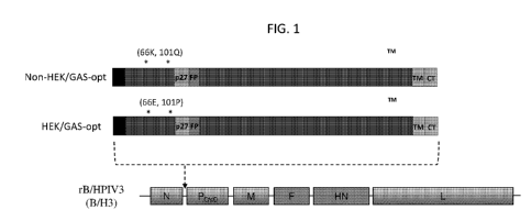
FIG. 1. Construction of rB/HPIV3 vectors expressing versions of the RSV F
protein containing
the non-HEK or HEK amino acid assignments. The F ORFs were codon-optimized for
human expression
using the GeneArt (GA) algorithm. The constructs were called non-HEK/GA-opt
and HEK/GA-opt. The
HEK (66E, 101P) and non-HEK (66K, 101Q) amino acid assignments are indicated
by asterisks. Other
annotations: S, signal sequence; p27, 27k protein fragment liberated by
cleavage-activation; FP, fusion
peptide; TM, transmembrane; CT, cytoplasmic tail. The RSV F ORFs were placed
under the control of
BPIV3 gene-start and gene-end transcription signals and inserted into the 2nd
genome position between the
N and P genes of the B/HPIV3 vector. The rB/HPIV3 vector includes N, P, M, and
L genes from BPIV3,
and F and NH genes from HPIV3. The same vector genome position and vector
transcription signals
were used for all of the other rB/HPIV3 vectors expressing RSV F protein
described in figures 1-35.
FIGs. 2A and 2B. The presence of the HEK assignments in the RSV F protein
resulted in
increased protein expression and a reduction in protein trimer mobility in
polyacrylamide gel
electrophoresis compared to that of non-HEK F protein. Vero cells were
infected with vectors expressing
HEK or non-HEK RSV F (from GA-optimized ORFs, shown in FIG. 1) at an MOI of 10
TCID50 at 32 C.
Cell lysates were prepared at 48 hours post-infection. Equal amounts of cell
lysates were analyzed by
electrophoresis after being boiled and reduced (A) or without being boiled and
reduced (B). Denatured
and reduced RSV F monomer was detected with a commercially-obtained RSV F-
specific mouse
monoclonal antibody (A). Native RSV F trimer was detected with polyclonal
antibodies raised in rabbits
by repeated immunizations with sucrose purified RSV particles (B).
FIGs. 3A and 3B. Formation of syncytia in Vero cell monolayers infected with
rB/HPIV3
vectors expressing non-HEK or HEK RSV F protein. Cells were infected with
rB/HPIV3 expressing GA-
codon-optimized RSV F (see FIG. 1) with (A) non-HEK or (B) HEK assignments at
an MOI of 10 TCID50
at 32 C. Images of the infected cells were acquired at 48 hours post-
infection. Representative syncytia are
marked with dashed outline.
FIG. 4. Construction of rB/HPIV3 vectors expressing RSV F ORFs that were codon-
optimized
(for human expression) by different algorithms and contained the HEK
assignments. The ORF encoding
the RSV F protein with HEK assignments was optimized for human codon usage
with the GA algorithm
(HEK/GA-opt, shown in FIG. 1), the DNA2.0 algorithm (HEK/D2-opt), or the
GenScript (GS) algorithm
(HEK/GS-opt). These codon-optimized ORFs were compared with the non-HEK, non-
optimized version
of the RSV F ORF (Non-HEK/non-opt). These RSV F ORFs were inserted into the
rB/HPIV3 vector in
exactly the same position and with the same vector signals as in FIG. 1.
3
CA 02974359 2017-07-19
WO 2016/118642 PCT/US2016/014154
FIGs. 5A and 5B. Increased in vitro expression of RSV F protein from rB/HPIV3
vectors due to
the HEK assignments and codon optimization. Expression of RSV F in (A) Vero
and (B) LLC-MK2 cells
was evaluated by Western blot analysis. Cells were infected at an MOI of 10
TCID50 at 32 C with the
indicated rB/HPIV3 vectors, and cell lysates were harvested at 48 hours post-
infection. Lysates were
subjected to gel electrophoresis under reducing and denaturing conditions and
analyzed by Western
blotting. Proteins were visualized by reaction with fluorescent antibodies and
detected by infrared
imaging. The experiment was performed with a total of three wells per virus. A
monoclonal antibody
specific to RSV F detected the uncleaved Fo precursor and cleaved F1 subunit.
RSV F1 band densities were
quantified and normalized to the band density of the Non-HEK/non-opt samples
indicated as "1".
Expression of the HPIV3 HN protein also was determined as an internal control
for vector protein
expression and to ensure equivalence of MOI and replication; I3-actin was used
as the loading control.
FIG. 6. Effects of HEK and codon-optimization of the F ORF on the formation of
syncytia in
vector-infected Vero cell monolayers. Cells were mock-infected (mock) or
infected with empty rB/HPIV3
vector (empty B/H3) or with rB/HPIV3 vector expressing the RSV F ORF that was
non-HEK and non-
optimized (Non-HEK/non-opt) or was HEK and GA-optimized (HEK/GA-opt) or HEK
and DNA2.0-
optimized (HEK/D2-opt) or HEK and GS-optimized (HEK/GS-opt). Infections were
performed at an MOI
of 10 TCID50 at 32 C and images were acquired at 48 hours post-infection.
Representative syncytia are
indicated with dashed outline in some of the panels.
FIGs. 7A and 7B. Multi-cycle in vitro replication of rB/HPIV3 vectors
expressing HEK or non-
HEK RSV F protein from non-optimized or codon-optimized ORFs. (A) LLC-MK2 and
(B) Vero cells
were infected in triplicate at 32 C at an MOI of 0.01 TCID50 with empty
rB/HPIV3 vector (empty B/H3)
or vector expressing the RSV F ORF that was non-HEK-containing and non-
optimized (Non-HEK/non-
opt) or was non-HEK-containing and GA-optimized (Non-HEK/GA-opt) or was HEK-
containing and
GA-optimized (HEK/GA-opt) or was HEK-containing and GS-optimized (HEK/GS-opt).
Aliquots of
medium supernatant were collected at 24 h intervals for 6 days and viral
titers were determined by
limiting dilution assay on LLC-MK2 cells at 32 C and reported as TCID50/ml.
Mean titers SEM from
three independent experiments are shown.
FIGs. 8A and 8B. Replication in hamsters of rB/HPIV3 vectors expressing HEK or
non-HEK
RSV F protein from non-optimized or codon-optimized ORFs. Golden Syrian
hamsters were infected
intranasally (IN) with 105 TCID50 of the indicated rB/HPIV3 vectors or 106PFU
of wt RSV (strain A2) in
a 0.1m1 inoculum. Hamsters were euthanized (n=6 per virus per day) on day 3
and 5 post-infection and the
(A) nasal turbinates and (B) lungs were removed and homogenized and viral
titers were determined by
limiting dilution on LLC-MK2 (rB/HPIV3 vectors) or Vero (RSV) cells at 32 C:
open and closed circles
indicate titers for animals sacrificed on day 3 and 5, respectively. Each
symbol represents an individual
animal, and the mean titer of each group is indicated by a dashed and a solid
horizontal line for day 3 and
5, respectively. The limit of detection (LOD) was 1.5 log 10 TCID50/g of
tissue, indicated with a dotted
line. The rB/HPIV3 vectors were titrated by limiting dilution assays on LLC-
MK2 cells and reported as
TCID50/g; RSV was titrated by plaque assays on Vero cells and reported as
PFU/g.
4
CA 02974359 2017-07-19
WO 2016/118642 PCT/US2016/014154
FIG. 9. Serum RSV-neutralizing antibody titers from hamsters infected with
rB/HPIV3 vectors
expressing HEK or non-HEK RSV F protein from non-optimized or codon-optimized
ORFs. Hamsters
(n=6 animals per virus) were inoculated IN with 105 TCID50 of the indicated
rB/HPIV3 vectors or 106
PFU of wt RSV in a 0.1m1 inoculum. Serum samples were collected at 28 days
post-immunization, and
RSV-neutralizing antibody titers were determined by using a 60% plaque
reduction neutralization test
(PRNT60) performed on Vero cells at 32 C in the presence of guinea pig
complement. Each symbol
represents an individual animal. The height of each bar represents the mean
titer of each group. The
values of mean titers are shown above the bars. The standard error of the mean
is shown by the horizontal
lines. The detection limit for the neutralization assay was 5.3 reciprocal
10g2 PRNT60, indicated with a
dotted line.
FIGs. 10A and 10B. Protection of immunized hamsters against RSV challenge. The
hamsters
(n=6 animals per virus) that had been immunized as shown in FIG. 9 with the
indicated rB/HPIV3 vectors
or with wt RSV, were challenged IN on day 31 post-immunization with 106 PFU of
wt RSV in a 0.1m1
inoculum. On day 3 post-challenge, hamsters were euthanized and (A) nasal
turbinates and (B) lungs were
collected. RSV titers in tissue homogenates were determined by plaque assay in
Vero cells. Each symbol
represents an individual animal and mean viral titers of the groups are shown
as horizontal lines. The
detection limit of the assay was log 10 2.7PFU/g of tissue, indicated as a
dashed line.
FIG. 11. Construction of rB/HPIV3 vectors expressing secreted (Ecto), post-
fusion, and stabilized
pre-fusion forms of the RSV F protein. Each of these modified proteins
contained the HEK assignments
and was expressed from a GA-optimized (for human expression) ORF. Annotations:
S, signal sequence;
p27, 27k protein fragment liberated by cleavage-activation; FP, fusion
peptide; TM, transmembrane; CT,
cytoplasmic tail. The HEK/GA-opt construct expresses full-length RSV F. The
ectodomain or "ecto" form
consisted of amino acids 1-513 of the RSV F protein; it lacks the CT and TM
anchor and would be
available for secretion. The "post-fusion" form was derived from the
ectodomain (1-513aa) by the further
deletion of the first 10 aa from the N-terminal end of the fusion peptide (FP;
137-146aa) (McLellan et al,
2011, J Virol 85:7788-96). "DS" and "DS-Cavl" are two versions of full-length
RSV F protein stabilized
in the pre-fusion form by the 5155C/5290C mutations (DS) or by the DS and
5190F/V207L (Cavl)
mutations (McLellan et al, 2013, Science 342:931). The ORFs encoding these
various forms of RSV F
were inserted into the rB/HPIV3 vector at the same position and with the same
vector signals as described
in FIGs. 1 and 4.
FIGs. 12A and 12B. Multi-cycle in vitro replication of rB/HPIV3 vectors
expressing secreted,
post-fusion, and stabilized pre-fusion forms of the RSV F protein. (A) LLC-MK2
and (B) Vero cells were
infected at an MOI of 0.01 TCID50 with empty rB/HPIV3 vector (empty B/H3) or
with the indicated
constructs: HEK/GA-opt; Ecto; Post-fusion; and DS (see FIG. 11 for
descriptions). Viral replication
during a period of 6 days at 32 C was determined by collecting medium
supernatant samples at 24-h
intervals and performing virus titration by limiting dilution on LLC-MK2
cells. See FIG. 11 for diagrams
of the mutant proteins. The asterisk * indicates that all of these RSV F
constructs were HEK and GA-
optimized.
5
CA 02974359 2017-07-19
WO 2016/118642 PCT/US2016/014154
FIGs. 13A and 13B. In vitro expression of secreted (Ecto), post-fusion, and
stabilized pre-fusion
forms of the RSV F protein from rB/HPIV3 vectors. Vero cells were infected
with the indicated
rB/HPIV3 vectors at an MOI of 10 TCID50 or with wt RSV at an MOI of 10 PFU.
Infected cells were
incubated at (A) 32 C or (B) 37 C for 48h. (A) Medium supernatants and lysates
of cells infected with the
-- rB/HPIV3 vectors expressing post-fusion, Ecto, or HEK/GA-opt, or with wt
RSV, and (B) lysates of cells
infected with rB/HPIV3 vectors with non-HEK/non-opt, HEK/GA-opt, DS, or DS-
Cavl forms of RSV F
were harvested and analyzed for RSV F expression by Western blot. The
constructs indicated by asterisk
* contained the HEK assignments and were GA-optimized.
FIGs. 14A and 14B. Replication in hamsters of rB/HPIV3 vectors expressing
secreted (Ecto),
-- post-fusion, and stabilized pre-fusion forms of the RSV F protein. Hamsters
were infected IN with 105
TCID50 of the indicated rB/HPIV3 vectors or 106PFU of wt RSV in a 0.1m1
inoculum. Hamsters were
euthanized (n=6 per virus per day) on days 3 and 5 post-infection and the (A)
nasal turbinates and (B)
lungs were removed and homogenized and viral titers were determined by
limiting dilution on LLC-MK2
cells (rB/HPIV3 vectors) or Vero (RSV) cells at 32 C: open and closed circles
indicate titers for animals
-- sacrificed on day 3 and 5, respectively. Each symbol represents an
individual animal, and the mean titer of
each group is indicated by a dashed or solid horizontal line for day 3 and 5,
respectively. Mean values of
day 5 titers are shown at the top. The rB/HPIV3 vectors were titrated by
limiting dilution assays on LLC-
MK2 cells and reported as TCID50/g; RSV was titrated by plaque assays on Vero
cells and reported as
PFU/g. The limit of detection (LOD) is 1.5 log 10 TCID50/g of tissue,
indicated with a dotted line. The
-- statistical significance of difference among peak titers was determined by
Tukey-Kramer test and
indicated by asterisks; *, P <0.05; **, P <0.01; or ***, P < 0.001. The
constructs indicated by asterisk *
contained the HEK assignments and were GA-optimized for human expression.
FIGs. 15A and 15B. Serum RSV-neutralizing antibody titers from hamsters
infected with
rB/HPIV3 vectors expressing secreted (Ecto), post-fusion, and stabilized pre-
fusion forms of the RSV F
-- protein. Hamsters (n=6 animals per virus) were inoculated IN with 105
TCID50 of the indicated rB/HPIV3
vectors or 106 PFU of wt RSV in a 0.1m1 inoculum. Serum samples were collected
at 28 days post-
immunization, and RSV-neutralizing antibody titers were determined by a 60%
plaque reduction
neutralization test (PRNT60) performed on Vero cells at 32 C (A) with and (B)
without added guinea pig
complement. The height of each bar represents the mean titer. The values of
mean titers are shown above
-- the bars. The standard error of the mean is shown by the horizontal lines.
The detection limit for the
neutralization assay is indicated with a dotted line. ND means neutralization
titer is below the detection
limit. The statistical significance of difference among groups was determined
by Tukey-Kramer test and
indicated by asterisks; *, P <0.05; **, P <0.01; or ***, P < 0.001; or ns, P
>0.05.
FIGs. 16A and 16B. Protection of immunized hamsters against RSV challenge. The
hamsters
-- (n=6 animals per virus) that had been immunized as shown in FIG. 15 were
challenged IN on day 31 post-
immunization with 106 PFU of wt RSV in a 0.1m1 inoculum. On day 3 post-
challenge, hamsters were
euthanized and (A) nasal turbinates and (B) lungs were collected. RSV titers
in tissue homogenates were
determined by plaque assay in Vero cells at 32 C. Each symbol represents an
individual animal and mean
6
CA 02974359 2017-07-19
WO 2016/118642 PCT/US2016/014154
viral titers of the groups are shown as horizontal lines. The detection limit
of the assay was log 102.7PFU/g
of tissue, indicated as a dotted line.
FIGs. 17A and 17B. Construction of rB/HPIV3 vectors expressing versions of RSV
F protein
engineered in an attempt to increase incorporation into the vector particle.
(A) Structures of F proteins.
(B) Sequences of the cytoplasmic tails (CT), transmembrane (TM) domains, and
adjoining regions of the
ectodomains of the RSV F protein (amino acid assignments in black) and BPIV3 F
protein (boldface),
with amino acid sequence positions indicated. Each of these modified proteins
contained the HEK
assignments and was expressed from a GA-optimized ORF. The HEK/GA-opt
construct expressed full-
length RSV F protein. "B3CT" has the CT of RSV F protein (amino acid sequence
positions 551-574)
replaced by the CT of BPIV3 F protein (positions 515-540, boldface). "B3TMCT"
has both the TM and
CT of RSV F protein (positions 530-574) replaced by the TM and CT of BPIV3 F
protein (positions 494-
540, boldface). "DS/B3CT", "DS/B3TMCT", "DS-Cav 1/B3CT", and "DS-Cav 1/B3TMCT"
are versions
of B3CT and B3TMCT containing the DS or DS-Cav 1 mutations designed to
stabilize the pre-fusion
conformation. The ORFs encoding these various forms of RSV F protein were
inserted into the rB/HPIV3
vector at the same position and with the same vector signals as described in
FIGs. 1, 4, and 11.
FIGs. 18A and 18B. Incorporation into the rB/HPIV3 vector particle of B3CT and
B3TMCT
versions of the RSV F protein. LLC-MK2 cells were infected with the indicated
rB/HPIV3 vectors at an
MOI of 0.01 TCID50 at 32 C. The medium supernatants were harvested 6-7 days
post-infection, clarified
by low speed centrifugation, and subjected to centrifugation on 10%-30%
sucrose gradients to obtain
partially-purified vector particles. Additional Vero cells were infected with
wt RSV at an MOI of 0.01
PFU and processed in the same way. The protein concentrations of the sucrose-
purified preparations were
determined by a standard commercial kit. (A) Western blot evaluation of the
packaging efficiency of the
RSV F protein into the rB/HPIV3 particles. To compare the relative amounts of
RSV F in the particles,
0.5Kg of sucrose-purified particles were lysed, denatured, reduced and
subjected to Western blot analyses.
The HPIV3 HN and BPIV3 N proteins of the vector particle were quantified for
comparison. (B) The
packaging efficiency of each form of RSV F into its respective vector particle
was calculated by
normalizing its band density against that of the BPIV3 N protein. The order of
the lanes is the same as in
part A. The packaging efficiencies of various forms of RSV F are shown
relative to the native F protein
set at "1". The packaging efficiency of the B3CT and B3TMCT forms of RSV F
into the vector particle
was judged to be similar to that of RSV F into the RSV particle because the
amount of modified RSV F
protein per 0.5 ng of vector particles (lanes 3, 4, 6, 7) was similar to the
amount of native RSV F protein
per 0.5 ng of RSV particles (lane 5). The constructs indicated by asterisk *
contained the HEK
assignments and were GA-codon-optimized for human expression.
FIGs. 19A-19F. Visualization of the incorporation of B3CT and B3TMCT versions
of the RSV F
protein into rB/HPIV3 particles by transmission electron microscopy (TEM).
Sucrose purified viruses
were labeled with an RSV F-specific murine monoclonal antibody and mouse-IgG-
specific second
antibodies that were labeled with 6nm gold particles. Virions and gold
particles were visualized with
TEM. Representative images of (A) RSV, (B) empty rB/HPIV3 vector (empty B/H3),
(C) vector
expressing HEK/GA-opt, (D) vector expressing B3CT, (E) vector expressing
B3TMCT, and (F) vector
7
CA 02974359 2017-07-19
WO 2016/118642 PCT/US2016/014154
expressing DS/B3TMCT are shown. Arrows point to sporadic gold particles in
HEK/GA-opt virions (C).
Substantially greater amounts of gold particles associated with the vector
particles are evident in D, E, and
F.
FIGs. 20A and 20B. Multi-cycle in vitro replication of rB/HPIV3 vectors
expressing B3CT and
B3TMCT versions of the RSV F protein. (A) LLC-MK2 and (B) Vero cells were
infected at 32 C with
an MOI of 0.01 TCID50 with empty rB/HPIV3 vector (empty B/H3) or vector
expressing HEK/GA-opt, or
B3CT (upper panels), or B3TMCT (upper panels), or DS/B3CT (lower panels) or
DS/B3TMCT (lower
panels). Aliquots of medium supernatant were collected at 24 h intervals for 6
days and viral titers were
determined by limiting dilution assay on LLC-MK2 cells at 32 C and reported as
TCID50/ml. The
constructs indicated by asterisk * contained the HEK assignments and were GA-
codon-optimized for
human expression. Multiplicity of infection in the assays was 0.01.
FIGs. 21A and 21B. In vitro expression of B3CT and B3TMCT versions of the RSV
F protein
with or without the DS or DS-Cavl mutations that stabilize the pre-fusion form
of RSV F protein.
Expression of (A) B3CT and B3TMCT; and (B) DS and DS-Cavl in combination with
B3CT and
B3TMCT. Vero cells were infected with the indicated rB/HPIV3 vectors at an MOI
of 10 TCID50, or with
RSV at an MOI of 10 PFU. Infected cells were incubated at (A) 32 C or (B) 37
C for 48h. Cell lysates
were analyzed for RSV F expression by Western blot. HPIV3 HN protein was used
as a control to show
equivalence of vector replication; GAPDH was used as loading control. The
constructs indicated by
asterisk * contained the HEK assignments and were GA-codon-optimized for human
expression.
FIG. 22. Formation of syncytia in Vero cell monolayers infected with rB/HPIV3
vectors
expressing the B3CT or B3TMCT version of the RSV F protein with or without the
DS mutations that
stabilize the pre-fusion form of RSV F protein. Vero cells were infected at an
MOI of 10 TCID50 with
rB/HPIV3 vectors expressing the indicated versions of RSV F protein and
incubated at 32 C. Images
were acquired at 48h post-infection. The constructs indicated by asterisk *
contained the HEK
assignments and were GA-codon-optimized for human expression.
FIGs. 23A and 23B. Replication in hamsters of rB/HPIV3 vectors expressing the
B3CT or
B3TMCT version of the RSV F protein with or without the DS mutations that
stabilize the pre-fusion
form of RSV F protein. Hamsters were infected IN with 105 TCID50 of rB/HPIV3
vectors or 106PFU of
wt RSV in a 0.1m1 inoculum. Hamsters were euthanized (6 per virus per day) on
day 3 and 5 post-
infection and the (A) nasal turbinates and (B) lungs were removed and
homogenized and viral titers were
determined by limiting dilution on LLC-MK2 (rB/HPIV3 vectors) or Vero (RSV)
cells at 32 C: open and
closed circles indicate titers for animals sacrificed on day 3 and 5,
respectively. Each symbol represents an
individual animal, and the mean titer of each group is indicated by a dashed
or solid horizontal line for
day 3 and 5, respectively. Mean values of day 5 titers are shown at the top.
The rB/HPIV3 vectors were
titrated by limiting dilution assays on LLC-MK2 cells and reported as
TCID50/g; RSV was titrated by
plaque assays on Vero cells and reported as PFU/g. The limit of detection
(LOD) is 1.5 log 10 TCID50/g of
tissue, indicated with a dotted line. The statistical significance of
difference among peak titers was
determined by Tukey-Kramer test and indicated by asterisks (*, P <0.05; **, P
<0.01; or ***, P < 0.001).
The constructs indicated by asterisk * along the x-axis contained the HEK
assignments and were GA-
8
CA 02974359 2017-07-19
WO 2016/118642 PCT/US2016/014154
codon-optimized for human expression. Constructs containing the DS-Cavl
modification were not
examined because they were not available at the time of this experiment.
FIGs. 24A and 24B. Serum RSV-neutralizing antibody titers from hamsters
infected with
rB/HPIV3 vectors expressing the B3CT or B3TMCT version of the RSV F protein
with or without the DS
mutations that stabilize the pre-fusion form of RSV F protein. Hamsters (n=6
animals per virus) were
inoculated IN with 105 TCID50 of the indicated rB/HPIV3 vectors or 106PFU of
wt RSV in a 0.1m1
inoculum. Serum samples were collected at 28 days post-immunization, and
antibody titers were
determined by a 60% plaque reduction neutralization test (PRNT60) with (A) or
without (B) added guinea
pig complement. The height of each bar represents the mean titer shown along
with the SEM. The values
of mean titers are shown above the bars. The detection limit for the
neutralization assay is indicated with a
dotted line. The statistical significance of difference in the mean titers was
determined by Tukey-Kramer
test and indicated by asterisks (*, P <0.05; **, P <0.01; ns, P >0.05). ND,
neutralization titer was below
the detection limit. The constructs indicated by asterisk * along the x-axis
contained the HEK assignments
and were GA-codon-optimized for human expression.
FIGs. 25A and 25B. Protection of immunized hamsters against RSV challenge. The
hamsters
(n=6 animals per virus) that had been immunized as shown in FIG. 24 were
challenged IN on day 31 post-
immunization with 106 PFU of wt RSV in a 0.1m1 inoculum. On day 3 post-
challenge, hamsters were
euthanized and (A) nasal turbinates and (B) lungs were collected. RSV titers
in tissue homogenates were
determined by plaque assay in Vero cells at 32 C. Each symbol represents an
individual animal and mean
viral titers of the groups are shown as horizontal lines. The detection limit
of the assay was log 102.7PFU/g
of tissue, indicated as a dotted line.
FIG. 26. Stability of expression of RSV F by rB/HPIV3 vectors during
replication in hamsters.
The percentage of recovered vector expressing RSV F in the nasal turbinates
and lungs at day 3 and 5
post-immunization was determined by double-staining plaque assay of vector
recovered directly from the
tissue homogenates. The results are expressed for the individual animals. The
percentages of rB/HPIV3
expressing RSV F protein in the tested specimens are indicated. Specimens with
100% expression of RSV
F protein were colored in yellow; those with 90-99% expression of RSV F were
colored in green; those
with 80- 89% expression of RSV F were colored in orange; those with less than
79% expression of RSV F
were colored in red. Specimens that did not generate plaques due to low titer
were marked as "NA". If the
total number of the plaques developed with a sample was less than 10, the
number of plaques was
recorded as "p = X" (X equals to the number of plaques) in the bracket.
FIG. 27. Temperature sensitivity phenotypes of B/HPIV3 vectors. The indicated
vectors were
evaluated for the ability to form plaques on LLC-MK2 cells at the indicated
temperatures. Reduction in
plaque formation of >100-fo1d is indicative of temperature sensitivity. The
lowest such restrictive
temperature for each virus is indicated in bold, underlining, and is called
the shut-off temperature.
FIG. 28. rB/HPIV3 constructs that were evaluated for attenuation and
immunogenicity in non-
human primates (Rhesus macaques). Rhesus macaques were infected by the
combined IN and
intratracheal routes with 106TCID50 per site of the following constructs: Non-
HEK/non-opt; HEK/GA-
opt/DS; and HEK/GA-opt/DS/B3TMCT in groups of five, five and four animals,
respectively.
9
CA 02974359 2017-07-19
WO 2016/118642 PCT/US2016/014154
FIGs. 29A and 29B. Replication of rB/HPIV3 vectors in rhesus macaques. Rhesus
macaques
were infected with the indicated rB/HPIV3 vectors as described in FIG. 28.
Vector replication in the
respiratory tract was assessed by collecting (A) nasopharyngeal swabs and (B)
tracheal lavages on the
indicated days and determining the viral titers by limiting dilution assay.
Limit of detection is 1.2
logioTCID50/mL shown as dotted line.
FIG. 30. Serum HPIV3-neutralizing antibody titers induced by rB/HPIV3 vectors.
Monkey sera
were collected at 0, 14, 21, 28, 35 and 56 days post-immunization and HPIV3-
neutralizing antibody titers
were determined by a 60% plaque reduction neutralization test (PRNT60) in the
presence of added guinea
pig complement. The detection limit for the neutralization assay is indicated
with a dotted line. The day of
RSV challenge is indicated.
FIGs. 31 and 32. Serum RSV-neutralizing antibody titers induced by rB/HPIV3
vectors. Monkey
sera were collected at 0, 14, 21, 28, 35 and 56 days post-immunization. (FIG.
31) RSV neutralizing
antibody titers at all time points were determined by a 60% plaque reduction
neutralization test (PRNT60)
in the presence of added guinea pig complement. (FIG. 32) RSV neutralizing
antibody titers at day 28
post-immunization were determined by a 60% plaque reduction neutralization
test (PRNT60) in the
absence of added complement. The detection limit for the neutralization assay
is indicated with a dotted
line. The statistical significance of difference in mean titers was determined
by Tukey-Kramer test and
indicated by asterisks (**, P <0.01; ***, P < 0.001). The day of RSV challenge
is indicated.
FIG. 33. Stability of expression of RSV F by rB/HPIV3 vectors during
replication in rhesus
macaques. The percentage of recovered vector expressing RSV F in nasal
pharyngeal swabs from day 4, 5
and 6 post-immunization was determined by double-staining plaque assay. The
percentages of rB/HPIV3
expressing RSV F in the tested specimens are indicated. Specimens with 100% of
viruses expressing RSV
F were colored in yellow; those with 99-90% of viruses expressing RSV F were
colored in green; those
that did not generate plaques due to low titer were marked as "NA".
FIG. 34. Construction of an rB/HPIV3 vector expressing a secreted version of
HEK/GS-opt/DS-
Cav 1 RSV F protein that contains a C-terminal "foldon" sequence. RSV F
protein containing the HEK
assignments and expressed from a GS-codon-optimized (for human expression) ORF
with DS-Cavl
mutations was engineered to contain the N-terminal 513 amino acids of the F
protein (i.e., lacking the TM
and CT domains), fused to the indicated 4-amino acid linker and the indicated
27-amino acid foldon
sequence from T4 phage (SEQ ID NO: 132, see Efimov et al. 1994, J Mol Biol
242:470-486; Miroshnikov
et al 1998 Protein Eng. 11:329-332). The ORF was inserted into the rB/HPIV3
vector at the same position
and with the same vector signals as described in FIGs. 1, 4, 11, and 17.
FIG. 35. Summary of exemplary rB/HPIV3 vectors expressing RSV F, annotated to
indicate
constructs that have been evaluated in two different studies in hamsters and
two different studies in rhesus
monkeys in Example 1.
FIG. 36. Construction of antigenomic cDNAs of the HPIV1 CD170 and LY942A
mutants containing
the RSV F gene insert at the first (F1), second (F2), or third (F3) genome
positions. The rHPIV1
backbones used for RSV F expression contained either of the two attenuating
mutations: namely the CD17
mutation (indicated by *) in the P/C gene or the LY942A mutation (indicated by
*) in L gene. For the
CA 02974359 2017-07-19
WO 2016/118642 PCT/US2016/014154
HPIV1-F1 constructs, the RSV F gene was inserted at the first genome position
before the HPIV1 N gene
at the Mit/I site located in the upstream non-translated region of the N gene.
In case of HPIV1-F2, the
RSV F gene was inserted between the HPIV1 N and P genes at the AscI site
located in the upstream non-
translated region of the P gene. For the HPIV1-F3, the RSV F gene was cloned
between the HPIV1 P and
M genes at the Nod site situated in the downstream non-translated region of
the P gene. For all
constructs, the RSV F ORF was codon optimized for human expression and
contained HEK amino acid
assignments. A copy of the N gene-end (GE), intergenic (IG) CTT triplet, and P
gene-start (GS) sequence
was added following (F1, F2) or before (F3) the RSV F insert so that it was
under the control of a set of
HPIV1 transcription signals. The sequences of SEQ ID NOs: 138-140 are shown
flanking the RSV F
insert under HPIV1-F1; the sequences of SEQ ID NOs: 141-143 are shown flanking
the RSV F insert
under HPIV1-F2, and the sequences of SEQ ID NOs: 144-145 are shown flanking
the RSV F insert under
HPIV1-F3.
FIGs. 37A-37D. Multistep replication of HPIV1/RSV-F viruses in Vero (37A and
37C) and
LLC-MK2 (37B and 37D) cells. Triplicate wells of cell monolayers in 6-well
plates were infected at an
MOI of 0.01 TCID50 with HPIV1 CA170 (A and B) or LY942A (C and D) viruses
expressing RSV F (F1, F2,
or F3), in parallel with wt HPIV1, HPIV1 LY942A, and HPIV1 CA170. Cultures
were incubated at 32 C.
Aliquots of cell culture medium were collected at 24 h intervals and virus
titers (logio TCID50/m1) were
determined by serial dilution on LLC-MK2 cells and hemadsorption assay at 32
C. Mean titers with
standard errors of the mean (SEM) are shown. The statistical significance of
difference between the titer
of each virus versus wt HPIV1 for day 2 post-infection was determined using
the one-way ANOVA with
Tukey's multiple comparisons test and is indicated by asterisks as follows: *,
p < 0.05; **, p < 0.01; ***,
p < 0.001; ****, p<0.0001.
FIGs. 38A-38C. Analysis of the RSV F and HPIV1 vector protein expression by
Western blot.
Vero cells were infected with the indicated viruses at an MOI of 5. At 48 h
post-infection cells were lysed
with SDS sample buffer. All samples were denatured, reduced and subjected to
SDS-PAGE and Western
blot. Proteins were transferred onto PVDF membranes and probed with either RSV
F-specific mouse
monoclonal antibody or HPIV1 N-, P-, HN-, or F-specific polyclonal antibodies
that had been raised by
immunizing rabbits separately with synthetic peptides representing the
respective proteins. (A) Bound
antibodies were visualized using corresponding anti-mouse (IRDye 680LT) and
anti-rabbit (IRDye
800CW) antibodies conjugated with infra-red dye. Images were acquired by
scanning the blots using the
Odyssey infrared imaging system. The images shown are from a single experiment
that is representative
of three independent experiments. (B and C) The intensity of protein bands for
the rHPIV1 CA17 (B) and
the rHPIV1 LY942A (C) constructs was quantified for three independent
experiments and expression is
shown relative to the F3 virus set at 1Ø Plots show data as mean + SEM from
three independent
experiments that were analyzed by one-way ANOVA with Dunnett multiple
comparisons test using 95%
confidence interval. Expression of the HPIV1 proteins by the Fl, F2 and F3
viruses was statistically
compared with that of their corresponding empty vector backbone. *, p<0.05;
**, p<0.01; ***, p<0.001.
FIGs. 39A-39I. Formation of cytopathic effects and syncytia on LLC-MK2 cell
monolayers
infected with the rHPIV1 vectors expressing RSV F. MK2 cells were infected at
an MOI of 0.01 TCID50,
11
CA 02974359 2017-07-19
WO 2016/118642 PCT/US2016/014154
incubated for 5 days and images were acquired at 40X magnification using phase
contrast with a light
microscope. Photomicrographs of (A) rHPIV1 C'170-F1; (B) rHPIV1 CA170-F2; (C)
rHPIV1 CA176-F3; (D)
rHPIV1 CA170; (E) rHPIV1 LY942A-F1; (F) rHPIV1 LY942A-F2; (G) rHPIV1 LY942A-
F3; (H) rHPIV1 LY942A;
and (I) wt HPIV1 are shown.
FIGs. 40A and 40B. Replication of RSV F expressing HPIV1 vectors in the nasal
turbinates
(40A) and lungs (40B) of hamsters. Hamsters were inoculated intra-nasally with
105 TCID50 of the wt
HPIV1, rHPIV1 CD176 or rHPIV1 LY942A empty vectors, rHPIV1 CD176 or rHPIV1
LY942A expressing RSV
F from three genome positions (F1, F2, or F3), rHPIV1-CR84GcD17OHN553ALY942A
(a previously-described
HPIV1 vaccine candidate (Bartlett et al 2007 Virol J 4:6)), or the rB/HPIV3-
F2, a chimeric bovine/human
PIV3 expressing RSV F from the 2nd position (also known as HEK/GA-opt, see
FIG. 1). Virus titers were
determined in LLC-MK2 cells by hemadsorption assay and reported as
LogioTCID5o/g of tissue. Titers for
individual animals (6 per group) are shown for day 3 (A) and day 5 (*), each
symbol representing an
individual animal. The mean values are shown for each group in boldface for
day 3 and in italicized type
for day 5. The limit of detection (LOD) was 1.5 logio TCID50/ml, indicated
with a dotted line across the
bottom of each graph. The statistical significance of the difference between
each virus versus wt HPIV1
(red asterisks) or versus rB/HPIV3-F2 (bar at the top) was determined by One-
way ANOVA at 95%
confidence interval using Tukey's multiple comparisons test for day 3 and day
5 p.i.. *, p < 0.05; ***, p <
0.001; ****, p < 0.0001; or ns, not significant.
FIGs. 41A and 41B. Protection against wt RSV challenge virus replication in
the nasal turbinates
(41A) and lungs (41B) of the immunized hamsters. Hamsters (n=6) in each group
were challenged
intranasally with 106 PFU of wt RSV A2 at 30 days post-immunization. Nasal
turbinates and lungs were
collected from euthanized animals on day 3 post-challenge, virus titers were
determined for each sample
by RSV specific plaque assay on Vero cells and reported as Logi PFU/g of
tissue. Mean value for each
group is shown in bold face number and by a horizontal bar. Statistical
significance of difference among
viruses was determined by one-way ANOVA at 95% confidence interval using
Tukey's multiple
comparisons test and is indicated by *, p<0.05; **,p<0.01; ****p<0.0001; or
ns, not significant.
FIG. 42 shows a table illustrating the attenuating mutations introduced in the
HPIV1 backbone in
the P/C or the L ORF. Nucleotide changes (deletion or substitution) in the wt
sequence are underlined.
FIG. 43 shows a table illustrating temperature sensitivity of recombinant
viruses on LLC-MK2
cell monolayers. For temperature sensitivity, the underlined values in
boldface indicate the virus shut-off
temperature indicating a temperature sensitive phenotype defined as the lowest
restrictive temperature at
which the mean logio reduction in virus titer at a given temperature vs. 32 C
was 2.0 logio or greater than
that of the wt rHPIV1 at the same two temperatures. For monolayers, serial
dilutions of each of the
indicated viruses on LLC-MK2 cells were incubated at various temperatures for
7 days. Virus titers were
determined by hemadsorption with guinea pig erythrocytes and reported as Logi
TCID50/m1 with a
detection limit of 1.2.
FIG. 44 shows a table illustrating the percentage of virus population
expressing RSV F after in
vivo replication. The percentage of virus population expressing RSV F after in
vivo replication (stability)
was determined by an immunofluorescent double-staining plaque assay. Vero
cells were infected with
12
CA 02974359 2017-07-19
WO 2016/118642 PCT/US2016/014154
serially diluted tissue homogenates of the nasal turbinates or lungs of
infected hamsters (n=6 per virus)
collected on day 3 and 5 p.i. (total 144 samples) and incubated for 6 days
under methylcellulose overlay.
Virus plaques were stained with mouse monoclonal anti-RSV F and goat
polyclonal anti-HPIV1 specific
antibodies followed by detection with the corresponding infrared dye
conjugated secondary antibodies.
Percentage of plaques expressing both RSV F and HPIV1 antigens are shown. The
stability of HPIV1
CD17 -F1, -F2, and F3 for lung samples and that for HPIV1 LY942A-F1, -F2, and
F3 in the URT and lungs
could not be tested due to their lack of replication in these tissues. Numbers
in parenthesis indicate the
RSV F expression status for the number of hamsters of the total 6 hamsters per
virus. ND, no plaques
were detected.
FIG. 45 shows a table listing results indicating that immunization of hamsters
with rHPIV1
expressing RSV F induces serum neutralizing antibodies against RSV. Groups of
six-week old hamsters
(n=6) were intranasally immunized with 105 TCID50 of each indicated virus in
0.1m1 inoculum. Serum
samples were collected prior to immunization and at 28 days post immunization.
Antibody titers against
RSV and HPIV1 were determined by using a 60% plaque reduction neutralization
test (PRNT60) using
green fluorescent protein (GFP)- or enhanced GFP (eGFP) expressing viruses
(rRSV-eGFPM or HPIV1-
GFP), and neutralizing antibody titers were presented as mean reciprocal
10g2+SE. Based on the initial
serum dilutions used in the assay, the PRNT60 assay has a titer detection
limit of 3.3 and 1.0 reciprocal
10g2 PRNT60 for RSV and HPIV1, respectively. Statistical significance of
difference among the groups for
RSV antibody titers was determined by one-way ANOVA with Tukey's multiple
comparisons test
(p<0.05) and that for HPIV1 antibody titers was determined by Unpaired t-test.
Mean neutralizing
antibody titers were categorized into groups (indicated in parenthesis as A,
B, C, and D). Mean antibody
titers of treatment groups with different letters are statistically different
from each other; titers shown with
two letters are not statistically different from those indicated with either
letter.
FIG. 46 and 47. Multi-cycle in vitro replication of rB/HPIV3 vectors
expressing GA-optimized
(GA-opt) prefusion form of RSV F with DS-Cavl mutations. (FIG. 46) Vero and
(FIG. 47) LLC-MK2
cells were infected in triplicate at 32 C at an MOI of 0.01 TCID50 with empty
rB/HPIV3 vector (empty
B/H3) or vector expressing the RSV F ORF that was HEK-containing, GA-opt, and
containing the DS-
Cav 1 prefusion stabilizing mutations (HEK/GA-opt/DS-Cav 1) or was HEK-
containing, GA-opt, and
containing the DS-Cav 1 mutations and BPIV3-specific TM and CT domains as
potential packaging
signals (HEK/GA-opt/DS-Cavl/B3TMCT). Aliquots of medium supernatants were
collected at 24 h
intervals for 6 days and viral titers were determined by limiting dilution
assay on LLC-MK2 cells at 32 C
and reported as TCID50/ml. Mean titers SEM from three independent
experiments are shown.
FIG. 48A and 48B. Multi-cycle in vitro replication of rB/HPIV3 vectors
expressing GS-
optimized (GS-opt) RSV F with different modifications. (A) Vero and (B) LLC-
MK2 cells were infected
in triplicate at 32 C at an MOI of 0.01 TCID50 with empty rB/HPIV3 vector
(empty B/H3) or vector
expressing the RSV F ORF that was HEK-containing and GS-opt RSV F (HEK/GS-
opt), or was HEK-
containing, GS-opt and bearing DS-Cav 1 prefusion stabilizing mutations
(HEK/GS-opt/DS-Cav 1), or was
HEK-containing, GS-opt, and bearing the DS-Cav 1 mutations and BPIV3-specific
TM and CT domains
(HEK/GS-opt/DS-Cavl/B3TMCT), or was a truncated RSV F with amino acids from 1
to 513 that was
13
CA 02974359 2017-07-19
WO 2016/118642 PCT/US2016/014154
fused to a four-amino acid linker and 27-amino acid oligomerization sequence
from T4 phage, which was
HEK-containing, GS-opt and bearing DS-Cavl mutations (HEK/GS-opt/DS-Cav1/(1-
513)Foldon).
Aliquots of medium supernatants were collected at 24 h intervals for 6 days
and viral titers were
determined by limiting dilution assay on LLC-MK2 cells at 32 C and reported as
TCID50/ml. Mean titers
SEM from three independent experiments are shown.
FIGs. 49A-49D. Comparison of multi-cycle in vitro replication of rB/HPIV3
vectors expressing
GS-opt and GA-opt RSV F. FIG. 49A and 49B: (A) Vero and (B) LLC-MK2 cells were
infected in
triplicate at 32 C at an MOI of 0.01 TCID50 with empty rB/HPIV3 vector (empty
B/H3) or vector
expressing RSV F ORF that was HEK-containing, GS-opt, and bearing the DS-Cavl
mutations (HEK/GS-
opt/DS-Cavi), or was HEK-containing, GA-opt, and bearing the DS-Cavl mutations
(HEK/GA-opt/DS-
Cav 1). FIG. 49C and 49D: (C) Vero and (D) LLC-MK2 cells were infected at 32 C
at an MOI of 0.01
TCID50 with empty rB/HPIV3 vector (empty B/H3) or vector expressing RSV F ORF
that was HEK-
containing, GS-opt, and bearing the DS-Cavl and B3TMCT modifications (HEK/GS-
opt/DS-
Cavl/B3TMCT), or was HEK-containing, GA-opt, and contained the DS-Cav 1 and
B3TMCT
modifications (HEK/GA-opt/DS-Cavl/B3TMCT). Aliquots of medium supernatant were
collected at 24 h
intervals for 6 days and viral titers were determined by limiting dilution
assay on LLC-MK2 cells at 32 C
and reported as TCID50/ml. Mean titers SEM from three independent
experiments are shown.
FIGs. 50A-50C. Expression of various modified forms of RSV F by rB/HPIV3
vectors in cell
culture. (A) Vero and (B, C) LLC-MK2 cells were infected with empty rB/HPIV3
vector (lane 1), or
rB/HPIV3 vector expressing the indicated modified forms of RSV F (lanes 2-5
and 8), or wt RSV (wt
RSV, lane 6) at MOI of 3 PFU/cell, or uninfected (mock, lane 7). Infected Vero
(A) and LLC-MK2 (B)
cells were incubated at 32 C, and LLC-MK2 (C) cells were incubated at 37 C.
Cell lysates and medium
supernatant of Vero cells were collected at 48hpi and were subjected to
Western blot analysis for the
expression of RSV F, which was detected as cleaved F1 and/or un-cleaved Fo
forms. BPIV3 N was used as
an internal control for the expression of vector protein; GAPDH was used as a
loading control.
FIGs. 51A and 51B. Replication of rB/HPIV3 vectors in the upper and lower
respiratory tract of
hamsters. Hamsters were infected IN with 105 TCID50 of the indicated rB/HPIV3
vectors or 106PFU of wt
RSV in a 0.1m1 inoculum. Hamsters were euthanized (n=6 per virus per day) on
days 4 and 5 post-
infection and the (A) nasal turbinates and (B) lungs were removed and
homogenized, and viral titers were
determined by limiting dilution on LLC-MK2 cells at 32 C and reported as
TCID50/g (rB/HPIV3 vectors)
or were determined by plaque assays on Vero cells at 32 C and reported as
PFU/g (wt RSV). The limit of
detection (LOD) is 1.5 logio TCID50/g of tissue, indicated with a dotted line.
Open and closed circles
indicate titers for individual animals sacrificed on day 4 and 5,
respectively. The mean titers of each group
are indicated by a dashed and solid horizontal line for day 4 and 5,
respectively. The values of the mean
titers on day 4 and 5 are shown at the top. The mean viral titers on day 5
were assigned to different groups
using the Tukey-Kramer test: mean titers with different letters are
statistically different (p< 0.05), whereas
titers indicated with two letters are not significantly different than those
indicated with either letter.
FIGs. 52 and 53. Serum RSV-neutralizing antibody titers from hamsters infected
with the
indicated rB/HPIV3 vectors expressing the GA-opt or GS-opt RSV F protein with
or without the DS or
14
CA 02974359 2017-07-19
WO 2016/118642 PCT/US2016/014154
DS-Cav 1 or B3TMCT modifications. Hamsters (n=6 animals per virus) were
inoculated IN with 105
TCID50 of the indicated rB/HPIV3 vectors or 106PFU of wt RSV in a 0.1m1
inoculum. Serum samples
were collected at 28 days post-immunization, and antibody titers were
determined by a 60% plaque
reduction neutralization test (PRNT60) with (FIG. 52) or without (FIG. 53)
added guinea pig complement.
The height of each bar represents the mean titer shown along with the SEM. The
values of mean titers are
shown above the bars. The pairwise student t-test was used to evaluate the
statistical significance of
differences between values: in each of the three horizontal lines over the
mean titers, the value indicated
with a vertical bar was compared pair-wise to each of the others and recorded
as being significantly (*, p<
0.05) or not significantly (ns) different. The detection limit for the
neutralization assay is indicated with a
dotted line. ND, neutralization titer was below the detection limit.
FIGs. 54A and 54B. Protection against RSV challenge of hamsters immunized with
the indicated
rB/HPIV3 vectors. The hamsters (n=6 animals per immunization group) that had
been immunized as
shown in FIG. 53 were challenged IN on day 30 post-immunization with 106 PFU
of wt RSV in a 0.1m1
inoculum. On day 3 post-challenge, hamsters were euthanized and (A) nasal
turbinates and (B) lungs were
collected. RSV titers in tissue homogenates were determined by plaque assay in
Vero cells at 37 C. Each
symbol represents an individual animal and mean values of viral titers of the
groups are shown above the
symbols and indicated as short horizontal lines. The pairwise student t-test
was used to evaluate the
statistical significance of differences between values: in each of the
horizontal lines over the mean titers,
the value(s) indicated with a vertical bar(s) was compared pair-wise to each
of the others and recorded as
being significantly (*, p< 0.05) or not significantly (ns) different. The
detection limit of the assay was
logio1.7PFU/g of tissue, indicated as a dotted line.
FIG. 55. rB/HPIV3 constructs that were evaluated for attenuation and
immunogenicity in non-
human primates (Rhesus macaques). Rhesus macaques were infected by the
combined IN and
intratracheal routes with 106TCID50 per site of the following constructs:
HEK/GA-opt/DS/B3TMCT;
HEK/GA-opt/DS-Cavl/B3TMCT; and HEK/GS-opt/DS-Cavl/B3TMCT in groups of four,
six and six
animals, respectively.
FIGs. 56A and 56B. Replication of rB/HPIV3 vectors in rhesus macaques. Rhesus
macaques
were infected with the rB/HPIV3 vectors indicated in FIG. 55. Vector
replication in the respiratory tract
was assessed by collecting (A) nasopharyngeal swabs and (B) tracheal lavages
on the indicated days and
determining the viral titers by limiting dilution assay. Limit of detection is
1.2 log loTCID50/mL shown as
dotted line.
FIGs. 57A and 57B. Serum RSV-neutralizing antibody titers induced by rB/HPIV3
vectors. From
the experiment shown in FIG. 55 and 56, sera were collected at 0, 14, 21, and
28 days post-immunization.
FIG. 57A: RSV neutralizing antibody titers at the indicated time points were
determined by a 60% plaque
reduction neutralization test (PRNT60) in the presence of added guinea pig
complement. The statistical
significance of difference in mean titers of each time point was determined by
pairwise student-t test (ns,
P >0.05). FIG. 57A: RSV neutralizing antibody titers at day 28 post-
immunization were determined by
PRNT60 in the absence of added complement. The detection limit for the
neutralization assay is indicated
CA 02974359 2017-07-19
WO 2016/118642 PCT/US2016/014154
with a dotted line. The statistical significance of difference in mean titers
of each time point was
determined by pairwise student-t test (ns, P >0.05).
FIGs. 58A and 58B. Construction of a rB/HPIV3 vector expressing HEK/GS-opt/DS-
Cavl/B3TMCT from the pre-N position, and modification of the amino acid
sequence of the HPIV3 HN
protein to achieve increased phenotypic stability of the vector. FIG. 58A:
Insertion of the HEK/GS-
opt/DS-Cavl/B3TMCT insert into the first gene position of rB/HPIV3. FIG. 58A:
Corrections of the
HPIV3 HN gene that conferred increased phenotypic stability. The HN gene in
the original recombinant
HPIV3 made by reverse genetics (Durbin et al Virology 235:323-332 1997) had
two engineered
nucleotide substitutions in the HN gene at antigenome positions 7913 and 7915
that resulted in the amino
acid substitution P370T, and an adventitious mutation at antigenome position
7593 that resulted in the
amino acid substitution T263I. Here, these mutations were changed back to the
"wild-type" assignments,
i.e., that found in biologically derived HPIV3 strain JS (Genbank Z11575.1;
Stokes et al Virus Res 25:91-
103. 1992).
FIGs. 59A and 59B. Intracellular expression of RSV F and vector proteins by
vectors expressing
various versions of RSV F protein in the first gene position (pre-N) or in the
second gene position (N-P).
Analysis of rB/HPIV3-wt HN-HEK/GS-opt/DS-Cavl/B3TMCT/pre-N, the construct
diagrammed in FIG.
58A. Vero (FIG. 59A) and LLC-MK2 (FIG. 59B) cells were infected with empty
rB/HPIV3 vector
(empty B/H3, lane 1), or the wtHN/HEK/GS-opt/DS-Cavl/B3TMCT/pre-N construct
(pools CL20a,
CL24a, lanes 3 and 4), or vector with the same version of RSV F inserted in
the second (N-P) position
(HEK/GS-opt/DS-Cavl/B3TMCT/N-P, lane 5), or vector with Non-HEK/non-opt
version of RSV F
inserted in the pre-N position (lane 6), or wt RSV (lane 2), or mock-infected
(lane 7). The vectors were
infected at an MOI of 10 TCID50/cell, and wt RSV at MOI of 3 PFU/cell.
Infected monolayers were
incubated at 32 C. Cell lysates were collected at 48 hpi and subjected to
Western blot analysis. RSV F in
the forms of cleaved F1 and/or uncleaved Fo were detected. BPIV3 N and P
proteins were used to evaluate
effects on vector protein expression. GAPDH was used as a loading control.
FIG. 60. HPIV1 vector: sequences of the cytoplasmic tails (CT), transmembrane
(TM) domains,
and adjoining regions of the ectodomains of the RSV F protein (strain A2,
amino acid assignments) and
HPIV1 F protein (boldface), with the amino acid sequence positions indicated.
RSV-F-TMCT is a
chimeric protein consisting of the ectodomain of RSV F protein attached to the
TM and CT domains of
HPIV1 F protein.
FIGs. 61 and 62. Construction of HPIV1-C''7 vectors expressing versions of
RSV F protein
designed to be stabilized in the prefusion conformation (DS-Cavl) and to have
increased incorporation
into the HPIV1 vector particle. Each of these modified RSV F inserts contained
the HEK assignments
(HEK) and was codon-optimized by GS for human expression (GS-opt). The RSV F
insert was
engineered to be stabilized in the prefusion conformation by the DS and Cav 1
mutations (DS-Cavl) alone
(upper construct in FIGs. 61 and 62) or with further modification by the
replacement of its TMCT domain
with those from HPIV1 F (TMCT, lower construct in FIGs. 61 and 62). The
resulting HEK/GS-opt/DS-
Cavl and HEK/GS-opt/DS-Cavl/TMCT versions of RSV F were modified by flanking
sequence and
inserted into the HPIV1-C''7 vector (see Example 2 for an explanation of the
HPIV1 vector and CA17
16
CA 02974359 2017-07-19
WO 2016/118642 PCT/US2016/014154
mutation) at the (FIG. 61) first gene position (MittI site), or (FIGS. 62)
second gene position (AscI site). In
each case, the RSV F was under the control of HPIV1 transcription signals for
expression as a separate
mRNA. Nucleotide numbering is relative to the complete antigenome RNA sequence
of the final
construct. The sequences of SEQ ID NOs: 146 and 147 are shown flanking the RSV
F insert under the
diagrams for Fl/HEK/GS-opt/DS-Cavl, Fl/HEK/GS-opt/DS-Cavl/TMCT, F2/HEK/GS-
opt/DS-Cav1,
and F2/HEK/GS-opt/DS-Cavl/TMCT.
FIG. 63. Kinetics of multi-cycle growth in Vero cells of rHPIV1-C17 vectors
expressing RSV F
stabilized in the prefusion conformation (DS-Cavi), without or with TMCT from
HPIV1 F protein. Vero
cells were infected with the constructs in triplicate at an MOI of 0.01 and
incubated for 7 days at 32C. At
24 h intervals, 0.5 mL of the total 3 mL culture supernatant was collected
over 7 days. After sample
collection, 0.5 mL fresh media was added to each culture to restore the
original volume. Virus titration of
the collected samples was performed on LLC-MK2 cells by hemadsorption assay
and values are plotted as
means + SEM.
FIG. 64. Incorporation into HPIV1-C17 virion particles of RSV F protein
stabilized in the
prefusion conformation (DS-Cavl) without or with TMCT from HPIV1 F protein.
The indicated virus
constructs (the designations HEK/GS-opt were omitted for the sake of brevity)
were grown in LLC-MK2
cells and virions were purified by sucrose gradient centrifugation. Protein
concentration of the purified
viruses was determined by BCA assay. 1 ng total protein from each purified
virus was lysed in RIPA lysis
buffer, reduced, denatured, and subjected to SDS-PAGE and Western blot
analysis. RSV F (top panel)
and HPIV1 proteins (second, third, and fourth panels) were detected with mouse
monoclonal and rabbit
polyclonal HPIV1-peptide-specific (N, F, and HN) antibodies, respectively.
Infared-labeled secondary
antibodies were used to detect bound primary antibodies. The chimeric RSV-F-DS-
Cavl/TMCT protein
in lanes 2 and 4 (fourth panel) are visible because the antipeptide serum
specific to HPIV1 F protein was
raised using a synthetic peptide containing the C-terminal 18 amino acids of
the CT domain, and thus
reacts with RSV F protein bearing the HPIV1 F protein TMCT domains.
FIG. 65. Expression in infected Vero cells of RSV F protein stabilized in the
prefusion
conformation (DS-Cavl) without and with TMCT from HPIV1 F protein. Vero cell
monolayers in 6-well
plates were inoculated with the indicated viruses (the designations HEK/GS-opt
were omitted for the sake
of brevity) including the wt HPIV1 and rHPIV1-C'17 empty vector controls at
an MOI of 5 and incubated
for 48 h at 32 C. Cell lysates were prepared by lysing the monolayers in 200
iL LDS sample buffer.
Protein samples were reduced and denatured, and 45 iL of each sample were
electrophoresed followed by
protein transfer to PVDF membranes. RSV F and HPIV1 proteins were detected
using the same primary
and secondary antibodies as described for FIG. 64.
FIG. 66. Sequences of the cytoplasmic tails (CT), transmembrane (TM) domains,
and adjoining
regions of the ectodomains of the RSV F protein (amino acid assignments) and
HPIV3 F protein
(boldface), with the amino acid sequence positions indicated. RSV-F-H3TMCT is
a chimeric protein
consisting of the ectodomain of RSV F protein attached to the TM and CT
domains of HPIV3 F protein.
FIGs. 67A and 67B. Construction of rHPIV3 vectors expressing versions of RSV F
protein
designed to be stabilized in the prefusion conformation (DS-Cavl) and to have
increased incorporation
17
CA 02974359 2017-07-19
WO 2016/118642 PCT/US2016/014154
into the rHPIV3 vector particle. The vector is wild type rHPIV3 strain JS
which was modified to contain
the 263T and 370P amino acid assignments in the HN protein (see FIG. 58B),
which were found to confer
phenotypic stability to the vector. In addition, the rHPIV3 vector was
modified by the creation of a BlpI
site at positions 103-109 (A, top construct), for insertion of RSV F (or
potentially any other insert) in gene
position 1, or the creation of an AscI site at positions 1675-1682 (B, top
construct), for insertion of RSV F
in gene position 2. Each of the modified RSV F inserts contained the HEK
assignments (HEK) and was
codon-optimized by GS for human expression (GS-opt). In addition, the RSV F
insert was engineered to
be stabilized in the prefusion conformation by the DS and Cav 1 mutations (DS
Cavil alone (A and B,
second construct) or with further modification by the replacement of its TMCT
domains with those from
rHPIV3 F (H3TMCT, A and B, third construct). The resulting HEK/GS-opt/DS-Cavl
and HEK/GS-
opt/DS-Cavl/H3TMCT versions of RSV F were modified by flanking sequence and
inserted into the (A)
first gene position (BlpI site), or (B) second gene position (AscI site) of wt
rHPIV3 JS. In each case, the
RSV F was under the control of HPIV3 transcription signals for expression as a
separate mRNA.
Nucleotide numbering is relative to the complete antigenome RNA sequence of
the final construct. The
sequence of SEQ ID NOs: 148 is shown under the diagram for rHPIV3 wt-JS. The
sequences of SEQ ID
NOs: 149 and 150 are shown flanking the RSV F insert under the diagrams for
Fl/HEK/GS-opt/DS-Cav 1,
F1/HEK/GS-opt/DS-Cav1/H3TMCT, F2/HEK/GS-opt/DS-Cav1, and F2/HEK/GS-opt/DS-
Cav1/H3TMCT.
SEQUENCE LISTING
The nucleic and amino acid sequences listed in the accompanying sequence
listing are shown
using standard letter abbreviations for nucleotide bases, and three letter
code for amino acids, as defined
in 37 C.F.R. 1.822. Only one strand of each nucleic acid sequence is shown,
but the complementary
strand is understood as included by any reference to the displayed strand. The
Sequence Listing is
submitted as an ASCII text file in the form of the file named "Sequence.txt" (-
344 kb), which was
created on January 19, 2016 which is incorporated by reference herein. In the
accompanying sequence
listing:
DETAILED DESCRIPTION
A previous study (Zimmer et al J Virol 2005 79:10467-77) evaluated the
expression of RSV F
protein from a heterologous gene in the Sendai virus, which is a murine
relative of HPIV1 and also is
closely related to HPIV3. That study showed that very little RSV F protein was
incorporated into the
Sendai virus vector particle. The investigators replaced the CT or CT plus TM
of the RSV F protein with
the corresponding sequences from the Sendai F protein on the premise that this
would improve the
efficiency of interaction of the foreign RSV F protein with the vector
particle. These modifications
indeed increased incorporation of the engineered RSV F into the Sendai
particle, but only if the Sendai F
protein gene was also deleted. The requirement to delete the vector F protein
is incompatible with the
generation of infectious, attenuated viruses for vaccination and also would
remove one of the vector
protective antigens, which are believed to be needed to generate a bivalent
vaccine.
18
CA 02974359 2017-07-19
WO 2016/118642 PCT/US2016/014154
As disclosed herein, when expressed by rB/HPIV3, HPIV3, or HPIV1, the RSV F
protein
including RSV F TM and CT is incorporated into the vector particle only in
trace amounts. However,
swapping the TM and CT of the heterologous RSV F protein for the corresponding
TM and CT of the
paramyxovirus F protein provided a multi-fold increase in RSV F ectodomain
incorporation in the
envelope of recombinant paramyxovirus, such that the packaging of RSV F into
the vector was as
efficient (e.g. B/HPIV3) or more efficient (e.g. HPIV1) per ng of purified
virion than that of RSV itself.
This was effective when the TM and CT were swapped together, or when the CT
was swapped alone.
However, unexpected effects of increased fusogenicity of the chimeric RSV F
specific to CT alone
provide guidance that TMCT is preferred.
Efficient packaging of RSV F into the vector particle dramatically increased
the elicitation of an
immune response to the ectodomain (bearing all of the neutralization epitopes)
when the recombinant
paramyxovirus was administered to a subject. Unexpectedly, the virus-
neutralizing serum antibody
response was dramatically increased in quality, which was assessed by
comparing RSV-neutralization
activity in vitro in the absence of complement (which measures strongly-
neutralizing antibodies) or in its
presence (which augments neutralization by weak or non-neutralizing
antibodies). This unanticipated
increase in antibody quality is of particular importance for RSV, which is
noted for inducing incomplete
immune protection. The expression and efficient packaging of a foreign
glycoprotein bearing the TMCT
domains of a vector glycoprotein had the obvious potential to disrupt vector
replication and
morphogenesis: however, constructs are provided in which this effect was
minimal.
To further increase immunogenicity, stabilization of the RSV F protein in the
pre-fusion
conformation was evaluated. On its own, pre-fusion stabilization also resulted
in an increase in titers of
strongly-neutralizing antibodies, suggestive of stabilization of
neutralization epitopes. In the hamster
model, the effect of pre-fusion stabilization on increased immunogenicity and
protection appeared to be
additive to that of efficient packaging conferred by TMCT. However, when
evaluated in non-human
primates, the effect of packaging appeared to be greater than that of pre-
fusion stabilization.
Given the challenge of achieving protection against RSV, maximal
immunogenicity is desired.
Extensive experimentation uncovered other aspects of vector and insert
construction (e.g., use of various
insertion sites, use of codon-optimization, and use of an early-passage RSV F
protein sequence) that
provided increased expression of RSV F and reduced the cytopathic effects of
syncytia formation
mediated by the highly fusogenic RSV F protein.
It is noteworthy that a prototype vaccine virus based on rB/HPIV3 expressing
an unmodified RSV
F protein, which in clinical trials had disappointing RSV immunogenicity
(Bernstein, et al. 2012. Pediatric
Infectious Disease Journal 31:109-114), was confirmed by the methods of the
present disclosure to induce
RSV-neutralizing serum antibodies that were of poor quality, possessing
neutralization activity in vitro
only in the presence of added complement. In contrast, disclosed constructs
induced, in African green
monkeys, high titers of serum antibodies capable of efficiently neutralizing
RSV in vitro in the absence of
complement.
19
CA 02974359 2017-07-19
WO 2016/118642 PCT/US2016/014154
I. Summary of Terms
Unless otherwise noted, technical terms are used according to conventional
usage. Definitions of
common terms in molecular biology may be found in Benjamin Lewin, Genes X,
published by Jones &
Bartlett Publishers, 2009; and Meyers et al. (eds.), The Encyclopedia of Cell
Biology and Molecular
Medicine, published by Wiley-VCH in 16 volumes, 2008; and other similar
references.
As used herein, the singular forms "a," "an," and "the," refer to both the
singular as well as plural,
unless the context clearly indicates otherwise. For example, the term "an
antigen" includes single or
plural antigens and can be considered equivalent to the phrase "at least one
antigen." As used herein, the
term "comprises" means "includes." It is further to be understood that any and
all base sizes or amino
acid sizes, and all molecular weight or molecular mass values, given for
nucleic acids or polypeptides are
approximate, and are provided for descriptive purposes, unless otherwise
indicated. Although many
methods and materials similar or equivalent to those described herein can be
used, particular suitable
methods and materials are described herein. In case of conflict, the present
specification, including
explanations of terms, will control. In addition, the materials, methods, and
examples are illustrative only
and not intended to be limiting. To facilitate review of the various
embodiments, the following
explanations of terms are provided:
Adjuvant: A vehicle used to enhance antigenicity. Adjuvants include a
suspension of minerals
(alum, aluminum hydroxide, or phosphate) on which antigen is adsorbed; or
water-in-oil emulsion, for
example, in which antigen solution is emulsified in mineral oil (Freund
incomplete adjuvant), sometimes
with the inclusion of killed mycobacteria (Freund's complete adjuvant) to
further enhance antigenicity
(inhibits degradation of antigen and/or causes influx of macrophages).
Immunostimulatory
oligonucleotides (such as those including a CpG motif) can also be used as
adjuvants. Adjuvants include
biological molecules (a "biological adjuvant"), such as costimulatory
molecules. Exemplary adjuvants
include IL-2, RANTES, GM-CSF, TNF-a, IFN-y, G-CSF, LFA-3, CD72, B7-1, B7-2, OX-
40L, 4-1BBL,
immune stimulating complex (ISCOM) matrix, and toll-like receptor (TLR)
agonists, such as TLR-9
agonists, Poly I:C, or PolyICLC. The person of ordinary skill in the art is
familiar with adjuvants (see,
e.g., Singh (ed.) Vaccine Adjuvants and Delivery Systems. Wiley-Interscience,
2007). Adjuvants can be
used in combination with the disclosed recombinant.
Administration: The introduction of a composition into a subject by a chosen
route.
Administration can be local or systemic. For example, if the chosen route is
intranasal, the composition
(such as a composition including a disclosed recombinant paramyxovirus) is
administered by introducing
the composition into the nasal passages of the subject. Exemplary routes of
administration include, but are
not limited to, oral, injection (such as subcutaneous, intramuscular,
intradermal, intraperitoneal, and
intravenous), sublingual, rectal, transdermal (for example, topical),
intranasal, vaginal, and inhalation
routes.
Amino acid substitution: The replacement of one amino acid in a polypeptide
with a different
amino acid or with no amino acid (i.e., a deletion). In some examples, an
amino acid in a polypeptide is
substituted with an amino acid from a homologous polypeptide, for example, and
amino acid in a
recombinant group A RSV F polypeptide can be substituted with the
corresponding amino acid from a
CA 02974359 2017-07-19
WO 2016/118642 PCT/US2016/014154
group B RSV F polypeptide. Reference to a "66E" amino acid in a RSV F protein
refers to an RSV F
protein comprising a glutamate residue at position 66. The amino acid can be
present due to substitution
from a reference sequence. Reference to a "K66E" substitution in an RSV F
protein refers to an RSV F
protein comprising a glutamate residue at position 66 that has been
substituted for a lysine residue in a
reference (e.g., native) sequence.
Attenuated: A paramyxovirus that is "attenuated" or has an "attenuated
phenotype" refers to a
paramyxovirus that has decreased virulence compared to a reference wild type
paramyxovirus under
similar conditions of infection. Attenuation usually is associated with
decreased virus replication as
compared to replication of a reference wild-type paramyxovirus under similar
conditions of infection, and
thus "attenuation" and "restricted replication" often are used synonymously.
In some hosts (typically
non-natural hosts, including experimental animals), disease is not evident
during infection with a
reference paramyxovirus in question, and restriction of virus replication can
be used as a surrogate marker
for attenuation. In some embodiments, a recombinant paramyxovirus (e.g., RSV,
PIV3) that is attenuated
exhibits at least about 10-fold or greater decrease, such as at least about
100-fold or greater decrease in
virus titer in the upper or lower respiratory tract of a mammal compared to
non-attenuated, wild type virus
titer in the upper or lower respiratory tract, respectively, of a mammal of
the same species under the same
conditions of infection. Examples of mammals include, but are not limited to,
humans, mice, rabbits, rats,
hamsters, such as for example Mesocricetus auratus, and non-human primates,
such as for example
Ceroptihecus aethiops. An attenuated paramyxovirus may display different
phenotypes including without
limitation altered growth, temperature sensitive growth, host range restricted
growth, or plaque size
alteration.
Cytoplasmic Tail (CT): A contiguous region of a transmembrane protein that
includes a
terminus (either N- or C-terminus) of the protein and extends into the
cytoplasm of a cell or enveloped
virus from the cytoplasmic surface of the cell membrane or viral envelope. In
the case of a type I
transmembrane protein, the CT includes the C-terminus of the protein. In the
case of a type II
transmembrane protein, the CT includes the N-terminus of the protein.
Degenerate variant: In the context of the present disclosure, a "degenerate
variant" refers to a
polynucleotide encoding a polypeptide that includes a sequence that is
degenerate as a result of the genetic
code. There are 20 natural amino acids, most of which are specified by more
than one codon. Therefore,
all degenerate nucleotide sequences encoding a peptide are included as long as
the amino acid sequence of
the peptide encoded by the nucleotide sequence is unchanged.
Gene: A nucleic acid sequence, typically a DNA sequence, that comprises
control and coding
sequences necessary for the transcription of an RNA, whether an mRNA or
otherwise. For instance, a
gene may comprise a promoter, one or more enhancers or silencers, a nucleic
acid sequence that encodes a
RNA and/or a polypeptide, downstream regulatory sequences and, possibly, other
nucleic acid sequences
involved in regulation of the expression of an mRNA.
Heterologous: Originating from a different genetic source. A heterologous gene
included in a
recombinant viral genome is a gene that does not originate from that viral
genome. In one specific, non-
limiting example, a heterologous gene encoding an ectodomain of a RSV F
protein is included in the
21
CA 02974359 2017-07-19
WO 2016/118642 PCT/US2016/014154
genome of a recombinant PIV vector. Methods for introducing a heterologous
gene in a viral vector are
well known in the art and also described herein.
Host cells: Cells in which a vector can be propagated and its nucleic acid
expressed. The cell
may be prokaryotic or eukaryotic. The term also includes any progeny of the
subject host cell. It is
understood that all progeny may not be identical to the parental cell since
there may be mutations that
occur during replication. However, such progeny are included when the term
"host cell" is used.
Immune response: A response of a cell of the immune system, such as a B cell,
T cell, or
monocyte, to a stimulus. In one embodiment, the response is specific for a
particular antigen (an
"antigen-specific response"). In one embodiment, an immune response is a T
cell response, such as a
CD4+ response or a CD8+ response. In another embodiment, the response is a B
cell response, and results
in the production of specific antibodies.
Immunogen: A compound, composition, or substance that can stimulate the
production of
antibodies or a T cell response in an animal, including compositions that are
injected or absorbed into an
animal. An immunogen reacts with the products of specific humoral or cellular
immunity, including those
induced by heterologous antigens, such as a disclosed recombinant
paramyxoyirus. Administration of an
immunogen to a subject can lead to protective immunity against a pathogen of
interest.
Immunogenic composition: A composition comprising an immunogen that induces a
measurable T cell response against an antigen, or induces a measurable B cell
response (such as
production of antibodies) against an antigen, included on the immunogen or
encoded by a nucleic acid
molecule included in the immunogen. In one example, an immunogenic composition
is a composition
that includes a disclosed recombinant paramyxoyirus that induces a measurable
CTL response against
RSV and/or PIV, or induces a measurable B cell response (such as production of
antibodies) against RSV
and/or PIV, when administered to a subject. An immunogenic composition can
include an isolated
recombinant paramyxoyirus as disclosed herein. For in vivo use, the
immunogenic composition will
typically include a recombinant paramyxoyirus in a pharmaceutically acceptable
carrier and may also
include other agents, such as an adjuvant.
Isolated: An "isolated" biological component has been substantially separated
or purified away
from other biological components, such as other biological components in which
the component naturally
occurs, such as other chromosomal and extrachromosomal DNA, RNA, and proteins.
Proteins, peptides,
nucleic acids, and viruses that have been "isolated" include those purified by
standard purification
methods. Isolated does not require absolute purity, and can include protein,
peptide, nucleic acid, or virus
molecules that are at least 50% isolated, such as at least 75%, 80%, 90%, 95%,
98%, 99%, or even 99.9%
isolated.
Linked: The terms "linked," "linkage," and "linking" refer to making two
molecules into one
contiguous molecule; for example, linking two polypeptides into one contiguous
polypeptide by
recombinant means. Reference to a gene encoding a type I membrane protein
comprising a RSV F
ectodomain "linked" to a TM and CT of a heterologous F protein refers to
genetic linkage between the
nucleic acid sequence encoding the RSV F ectodomain and the nucleic acid
sequence encoding the TM
and CT of the heterologous F protein in the gene by recombinant means, such
that expression of the gene
22
CA 02974359 2017-07-19
WO 2016/118642 PCT/US2016/014154
leads to production of a protein including, in the N- to C-terminal direction,
the RSV F ectodomain, the
TM, and the CT. In some embodiments, the C-terminal residue of the RSV F
ectodomain can be directly
linked (by peptide bond) to the N-terminal residue of the TM. In some
embodiments, the C-terminal
residue of the RSV F ectodomain can be indirectly linked to the N-terminal
residue of the TM via a
peptide linker (such as a glycine-serine linker).
Linker: A bi-functional molecule that can be used to link two molecules into
one contiguous
molecule. Non-limiting examples of peptide linkers include glycine-serine
linkers.
Native protein, sequence, or di-sulfide bond: A polypeptide, sequence or di-
sulfide bond that
has not been modified, for example by selective mutation. For example,
selective mutation to focus the
antigenicity of the antigen to a target epitope, or to introduce a di-sulfide
bond into a protein that does not
occur in the native protein. Native protein or native sequence are also
referred to as wild-type protein or
wild-type sequence. A non-native di-sulfide bond is a disulfide bond that is
not present in a native
protein, for example a di-sulfide bond that forms in a protein due to
introduction of one or more cysteine
residues into the protein by genetic engineering.
Nucleic acid molecule: A polymeric form of nucleotides, which may include both
sense and
anti-sense strands of RNA, cDNA, genomic DNA, and synthetic forms and mixed
polymers of the above.
A nucleotide refers to a ribonucleotide, deoxynucleotide or a modified form of
either type of nucleotide.
The term "nucleic acid molecule" as used herein is synonymous with "nucleic
acid" and "polynucleotide."
A nucleic acid molecule is usually at least 10 bases in length, unless
otherwise specified. The term
includes single- and double-stranded forms of DNA. A polynucleotide may
include either or both
naturally occurring and modified nucleotides linked together by naturally
occurring and/or non-naturally
occurring nucleotide linkages.
Operably linked: A first nucleic acid sequence is operably linked with a
second nucleic acid
sequence when the first nucleic acid sequence is placed in a functional
relationship with the second
nucleic acid sequence. For instance, a promoter is operably linked to a coding
sequence if the promoter
affects the transcription or expression of the coding sequence. Generally,
operably linked nucleic acid
sequences are contiguous and, where necessary to join two protein-coding
regions, in the same reading
frame.
Paramyxovirus: A family of enveloped non-segmented negative-sense single-
stranded RNA
viruses. Examples of paramyxoviruses include, but are not limited to, human
parainfluenza virus (HPIV)
including types 1, 2, 3, 4A, and 4B (HPIV1, HPIV2, HPIV3, HPIV4A, and HPIV4B,
respectively), mouse
parainfluenza type 1 (Sendai virus, MPIV1), bovine parainfluenza virus type 3
(BPIV3), parainfluenza
virus 5 (PIV5, previously called simian virus 5, 5V5), simian virus 41 (5V41),
and mumps virus. HPIV1,
HPIV3, MPIV1, and BPIV3 are classified in the genus Respirovirus. HPIV2,
HPIV4, 5V5, 5V41, and
mumps virus are classified in the genus Rubulavirus. MPIV1, PIV5, and BPIV3
are animal relatives of
HPIV1, HPIV2, and HPIV3, respectively (Chancock et al., Parainfluenza Viruses,
Knipe et al. (Eds.), pp.
1341-1379, Lippincott Williams & Wilkins, Philadelphia, 2001). HPIV1, HPIV2,
and HPIV3 represent
distinct serotypes and do not elicit significant cross immunity. HPIVs are
etiological agents of respiratory
infections such as croup, pneumonia, or bronchitis.
23
CA 02974359 2017-07-19
WO 2016/118642 PCT/US2016/014154
Parainfluenza virus (PIV): A number of enveloped non-segmented negative-sense
single-
stranded RNA viruses from family Paramyxoviridae that are descriptively
grouped together. This
includes all of the members of genus respirovirus (e.g., HPIV1, HPIV3) and a
number of members of
genus rubulavirus (e.g. HPIV2, HPIV4, PIV5). Members of genus avulavirus
(e.g., NDV) historically
have been called PIVs and can be considered as part of this group. HPIV
serotypes 1, 2, and 3 are second
only to RSV in causing severe respiratory infections in infants and children
worldwide, with HPIV3 being
the most important of the HPIVs in terms of disease impact. PIVs are made up
of two structural modules:
(1) an internal ribonucleoprotein core, or nucleocapsid, containing the viral
genome, and (2) an outer,
roughly spherical lipoprotein envelope. The PIV viral genome is approximately
15,000 nucleotides in
length and encodes at least eight polypeptides. These proteins include the
nucleocapsid structural protein
(NP, NC, or N depending on the genera), the phosphoprotein (P), the matrix
protein (M), the fusion
glycoprotein (F), the hemagglutinin-neuraminidase glycoprotein (HN), the large
polymerase protein (L),
and the C and D proteins. The P gene contains one or more additional open
reading frames (ORFs)
encoding accessory proteins. The gene order is 3' -N-P-M-F-HN-L-5', and each
gene encodes a separate
protein encoding mRNA. Exemplary PIV strain sequences are known to the person
of ordinary skill in
the art, such as the sequences of the HPIV1, HPIV2, HPIV3, and BPIV3 viruses.
Pharmaceutically acceptable carriers: The pharmaceutically acceptable carriers
of use are
conventional. Remington's Pharmaceutical Sciences, by E. W. Martin, Mack
Publishing Co., Easton, PA,
19th Edition, 1995, describes compositions and formulations suitable for
pharmaceutical delivery of the
disclosed immunogens.
In general, the nature of the carrier will depend on the particular mode of
administration being
employed. For instance, parenteral formulations usually comprise injectable
fluids that include
pharmaceutically and physiologically acceptable fluids such as water,
physiological saline, balanced salt
solutions, aqueous dextrose, glycerol or the like as a vehicle. For solid
compositions (e.g., powder, pill,
tablet, or capsule forms), conventional non-toxic solid carriers can include,
for example, pharmaceutical
grades of mannitol, lactose, starch, or magnesium stearate. In addition to
biologically neutral carriers,
pharmaceutical compositions to be administered can contain minor amounts of
non-toxic auxiliary
substances, such as wetting or emulsifying agents, preservatives, and pH
buffering agents and the like, for
example sodium acetate or sorbitan monolaurate. In particular embodiments,
suitable for administration
to a subject the carrier may be sterile, and/or suspended or otherwise
contained in a unit dosage form
containing one or more measured doses of the composition suitable to induce
the desired immune
response. It may also be accompanied by medications for its use for treatment
purposes. The unit dosage
form may be, for example, in a sealed vial that contains sterile contents or a
syringe for injection into a
subject, or lyophilized for subsequent solubilization and administration or in
a solid or controlled release
dosage.
Polypeptide: Any chain of amino acids, regardless of length or post-
translational modification
(e.g., glycosylation or phosphorylation). "Polypeptide" applies to amino acid
polymers including naturally
occurring amino acid polymers and non-naturally occurring amino acid polymer
as well as in which one
or more amino acid residue is a non-natural amino acid, for example an
artificial chemical mimetic of a
24
CA 02974359 2017-07-19
WO 2016/118642 PCT/US2016/014154
corresponding naturally occurring amino acid. A "residue" refers to an amino
acid or amino acid mimetic
incorporated in a polypeptide by an amide bond or amide bond mimetic. A
polypeptide has an amino
terminal (N-terminal) end and a carboxy terminal (C-terminal) end.
"Polypeptide" is used
interchangeably with peptide or protein, and is used herein to refer to a
polymer of amino acid residues.
Prime-boost vaccination: An immunotherapy including administration of a first
immunogenic
composition (the primer vaccine) followed by administration of a second
immunogenic composition (the
booster vaccine) to a subject to induce an immune response. The booster
vaccine is administered to the
subject after the primer vaccine; the skilled artisan will understand a
suitable time interval between
administration of the primer vaccine and the booster vaccine, and examples of
such timeframes are
disclosed herein. Additional administrations can be included in the prime-
boost protocol, for example a
second boost.
Recombinant: A recombinant nucleic acid molecule is one that has a sequence
that is not
naturally occurring: for example, includes one or more nucleic acid
substitutions, deletions or insertions,
and/or has a sequence that is made by an artificial combination of two
otherwise separated segments of
sequence. This artificial combination can be accomplished by chemical
synthesis or, more commonly, by
the artificial manipulation of isolated segments of nucleic acids, for
example, by genetic engineering
techniques.
A recombinant virus is one that includes a genome that includes a recombinant
nucleic acid
molecule.
A recombinant protein is one that has a sequence that is not naturally
occurring or has a sequence
that is made by an artificial combination of two otherwise separated segments
of sequence. In several
embodiments, a recombinant protein is encoded by a heterologous (for example,
recombinant) nucleic
acid that has been introduced into a host cell, such as a bacterial or
eukaryotic cell, or into the genome of a
recombinant virus.
Respiratory Syncytial Virus (RSV): An enveloped non-segmented negative-sense
single-
stranded RNA virus of the family Paramyxoviridae. The RSV genome is ¨15,000
nucleotides in length
and includes 10 genes encoding 11 proteins, including the glycoproteins SH, G
and F. The F protein
mediates fusion, allowing entry of the virus into the cell cytoplasm and also
promoting the formation of
syncytia. Two antigenic subgroups of human RSV strains have been described,
the A and B subgroups,
based primarily on differences in the antigenicity of the G glycoprotein. RSV
strains for other species are
also known, including bovine RSV. Exemplary RSV strain sequences are known to
the person of
ordinary skill in the art. Further, several models of human RSV infection are
available, including model
organisms infected with hRSV, as well as model organisms infected with species
specific RSV, such as
use of bRSV infection in cattle (see, e.g., Bern et al., Am J, Physiol. Lung
Cell Mol. Physiol., 301: L148-
L156, 2011; and Nam and Kun (Eds.). Respiratory Syncytial Virus: Prevention,
Diagnosis and
Treatment. Nova Biomedical Nova Science Publisher, 2011; and Cane (Ed.)
Respiratory Syncytial Virus.
Elsevier Science, 2007.)
RSV Fusion (F) protein: An RSV envelope glycoprotein that facilitates fusion
of viral and
cellular membranes. In nature, the RSV F protein is initially synthesized as a
single polypeptide precursor
CA 02974359 2017-07-19
WO 2016/118642 PCT/US2016/014154
approximately 574 amino acids in length, designated Fo. Fo includes an N-
terminal signal peptide that
directs localization to the endoplasmic reticulum, where the signal peptide
(approximately the first 22
residues of Fo) is proteolytically cleaved. The remaining Fo residues
oligomerize to form a trimer which is
again proteolytically processed by a cellular protease at two conserved furin
consensus cleavage
sequences (approximately Fo positions 109/110 and 136/137; for example,
RARRIoo (SEQ ID NO: 1,
residues 106-109) and RKRR136 (SEQ ID NO: 1, residues 133-136) to excise the
pep27 polypeptide and
generate two disulfide-linked fragments, F1 and F2. The smaller of these
fragments, F2, originates from
the N-terminal portion of the Fo precursor and includes approximately residues
26-109 of Fo. The larger
of these fragments, F1, includes the C-terminal portion of the Fo precursor
(approximately residues 137-
574) including an extracellular/lumenal region (¨ residues 137-529), a TM
(¨residues 530-550), and a CT
(¨residues 551-574) at the C-terminus.
Three F2-F1 protomers oligomerize in the mature F protein, which adopts a
metastable "prefusion"
conformation that is triggered to undergo a conformational change (to a
"postfusion" conformation) upon
contact with a target cell membrane. This conformational change exposes a
hydrophobic sequence,
known as the fusion peptide, which is located at the N-terminus of the F1
polypeptide, and which
associates with the host cell membrane and promotes fusion of the membrane of
the virus, or an infected
cell, with the target cell membrane.
The extracellular portion of the RSV F protein is the RSV F ectodomain, which
includes the F2
protein and the F1 ectodomain. An RSV F ectodomain trimer includes a protein
complex of three RSV F
ectodomains.
The RSV F protein adopts a "prefusion" conformation prior to triggering of the
fusogenic event
that leads to transition of RSV F to the postfusion conformation and following
processing into a mature
RSV F protein in the secretory system. The three-dimensional structure of an
exemplary RSV F protein in
a prefusion conformation is known, and disclosed for example in W02014160463,
which is incorporated
by reference herein. In the prefusion state, the RSV F protein includes an
antigenic site at its membrane
distal apex termed "antigenic site 0," that includes RSV F residues 62-69 and
196-209, and also includes
the epitopes of the D25 and AM22 monoclonal antibodies. Thus, a recombinant
RSV F protein stabilized
in a prefusion conformation can be specifically bound by an antibody that
binds the pre- but not post-
fusion conformation of the RSV F protein, such as an antibody that
specifically binds to an epitope within
antigenic site 0, for example, the D25 or AM22 antibody. Additional RSV F
prefusion specific
antibodies include the 5C4 and MPE8 antibodies.
Sequence identity: The similarity between amino acid sequences is expressed in
terms of the
similarity between the sequences, otherwise referred to as sequence identity.
Sequence identity is
frequently measured in terms of percentage identity (or similarity or
homology); the higher the
percentage, the more similar the two sequences are. Homologs, orthologs, or
variants of a polypeptide will
possess a relatively high degree of sequence identity when aligned using
standard methods.
Methods of alignment of sequences for comparison are well known in the art.
Various programs
and alignment algorithms are described in: Smith & Waterman, Adv. Appl. Math.
2:482, 1981; Needleman
26
CA 02974359 2017-07-19
WO 2016/118642 PCT/US2016/014154
& Wunsch, J. Mol. Biol. 48:443, 1970; Pearson & Lipman, Proc. Natl. Acad. Sci.
USA 85:2444, 1988;
Higgins & Sharp, Gene, 73:237-44, 1988; Higgins & Sharp, CABIOS 5:151-3, 1989;
Corpet et al., Nuc.
Acids Res. 16:10881-90, 1988; Huang et al. Computer Appls. in the Biosciences
8, 155-65, 1992; and
Pearson et al., Meth. Mol. Bio. 24:307-31, 1994. Altschul et al., J. Mol.
Biol. 215:403-10, 1990, presents a
detailed consideration of sequence alignment methods and homology
calculations.
Once aligned, the number of matches is determined by counting the number of
positions where an
identical nucleotide or amino acid residue is present in both sequences. The
percent sequence identity is
determined by dividing the number of matches either by the length of the
sequence set forth in the
identified sequence, or by an articulated length (such as 100 consecutive
nucleotides or amino acid
residues from a sequence set forth in an identified sequence), followed by
multiplying the resulting value
by 100. For example, a peptide sequence that has 1166 matches when aligned
with a test sequence having
1554 amino acids is 75.0 percent identical to the test sequence
(1166+1554*100=75.0). The percent
sequence identity value is rounded to the nearest tenth. For example, 75.11,
75.12, 75.13, and 75.14 are
rounded down to 75.1, while 75.15, 75.16, 75.17, 75.18, and 75.19 are rounded
up to 75.2. The length
value will always be an integer.
The NCBI Basic Local Alignment Search Tool (BLAST) (Altschul et al., J. Mol.
Biol. 215:403,
1990) is available from several sources, including the National Center for
Biotechnology Information
(NCBI, Bethesda, MD) and on the internet, for use in connection with the
sequence analysis programs
blastp, blastn, blastx, tblastn and tblastx. A description of how to determine
sequence identity using this
program is available on the NCBI website on the internet.
Homologs and variants of a polypeptide (such as a RSV F ectodomain) are
typically characterized
by possession of at least about 75%, for example at least about 80%, 85%, 90%,
91%, 92%, 93%, 94%,
95%, 96%, 97%, 98% or 99% sequence identity counted over the full length
alignment with the amino
acid sequence of interest. Proteins with even greater similarity to the
reference sequences will show
increasing percentage identities when assessed by this method, such as at
least 80%, at least 85%, at least
90%, at least 95%, at least 98%, or at least 99% sequence identity. When less
than the entire sequence is
being compared for sequence identity, homologs and variants will typically
possess at least 80% sequence
identity over short windows of 10-20 amino acids, and may possess sequence
identities of at least 85% or
at least 90% or 95% depending on their similarity to the reference sequence.
Methods for determining
sequence identity over such short windows are available at the NCBI website on
the internet. One of skill
in the art will appreciate that these sequence identity ranges are provided
for guidance only; it is entirely
possible that strongly significant homologs could be obtained that fall
outside of the ranges provided.
For sequence comparison of nucleic acid sequences, typically one sequence acts
as a reference
sequence, to which test sequences are compared. When using a sequence
comparison algorithm, test and
reference sequences are entered into a computer, subsequence coordinates are
designated, if necessary,
and sequence algorithm program parameters are designated. Default program
parameters are used.
Methods of alignment of sequences for comparison are well known in the art.
Optimal alignment of
sequences for comparison can be conducted, e.g., by the local homology
algorithm of Smith & Waterman,
Adv. Appl. Math. 2:482, 1981, by the homology alignment algorithm of Needleman
& Wunsch, J. Mol.
27
CA 02974359 2017-07-19
WO 2016/118642
PCT/US2016/014154
Biol. 48:443, 1970, by the search for similarity method of Pearson & Lipman,
Proc. Nat'l. Acad. Sci. USA
85:2444, 1988, by computerized implementations of these algorithms (GAP,
BESTFIT, FASTA, and
TFASTA in the Wisconsin Genetics Software Package, Genetics Computer Group,
575 Science Dr.,
Madison, WI), or by manual alignment and visual inspection (see, e.g.,
Sambrook et al. (Molecular
Cloning: A Laboratory Manual, 4th ed, Cold Spring Harbor, New York, 2012) and
Ausubel et al. (In
Current Protocols in Molecular Biology, John Wiley & Sons, New York, through
supplement 104, 2013).
One example of a useful algorithm is PILEUP. PILEUP uses a simplification of
the progressive alignment
method of Feng & Doolittle, J. Mol. Evol. 35:351-360, 1987. The method used is
similar to the method
described by Higgins & Sharp, CABIOS 5:151-153, 1989. Using PILEUP, a
reference sequence is
compared to other test sequences to determine the percent sequence identity
relationship using the
following parameters: default gap weight (3.00), default gap length weight
(0.10), and weighted end gaps.
PILEUP can be obtained from the GCG sequence analysis software package, e.g.,
version 7.0 (Devereaux
et al., Nuc. Acids Res. 12:387-395, 1984.
Another example of algorithms that are suitable for determining percent
sequence identity and
sequence similarity are the BLAST and the BLAST 2.0 algorithm, which are
described in Altschul et al.,
J. Mol. Biol. 215:403-410, 1990 and Altschul et al., Nucleic Acids Res.
25:3389-3402, 1977. Software for
performing BLAST analyses is publicly available through the National Center
for Biotechnology
Information (ncbi.nlm.nih.gov). The BLASTN program (for nucleotide sequences)
uses as defaults a
word length (W) of 11, alignments (B) of 50, expectation (E) of 10, M=5, N=-4,
and a comparison of both
strands. The BLASTP program (for amino acid sequences) uses as defaults a word
length (W) of 3, and
expectation (E) of 10, and the BLOSUM62 scoring matrix (see Henikoff &
Henikoff, Proc. Natl. Acad.
Sci. USA 89:10915, 1989). An oligonucleotide is a linear polynucleotide
sequence of up to about 100
nucleotide bases in length.
As used herein, reference to "at least 90% identity" refers to "at least 90%,
at least 91%, at least
92%, at least 93%, at least 94%, at least 95%, at least 96%, at least 97%, at
least 98%, at least 99%, or
even 100% identity" to a specified reference sequence.
Subject: Living multi-cellular vertebrate organisms, a category that includes
human and non-
human mammals. In an example, a subject is a human. In a particular example,
the subject is a newborn
infant. In an additional example, a subject is selected that is in need of
inhibiting of an RSV infection.
For example, the subject is either uninfected and at risk of RSV infection or
is infected in need of
treatment.
Transmembrane domain (TM): An amino acid sequence that spans a lipid bilayer,
such as the
lipid bilayer of a cell or virus or virus-like particle. A transmembrane
domain can be used to anchor an
antigen to a membrane. In some examples a transmembrane domain is a RSV F
transmembrane domain.
Vaccine: A preparation of immunogenic material capable of stimulating an
immune response,
administered for the prevention, amelioration, or treatment of infectious or
other types of disease. The
immunogenic material may include attenuated or killed microorganisms (such as
bacteria or viruses), or
antigenic proteins, peptides or DNA derived from them. An attenuated vaccine
is a virulent organism that
has been modified to produce a less virulent form, but nevertheless retains
the ability to elicit antibodies
28
CA 02974359 2017-07-19
WO 2016/118642 PCT/US2016/014154
and cell-mediated immunity against the virulent form. An inactivated (killed)
vaccine is a previously
virulent organism that has been inactivated with chemicals, heat, or other
treatment, but elicits antibodies
against the organism. Vaccines may elicit both prophylactic (preventative or
protective) and therapeutic
responses. Methods of administration vary according to the vaccine, but may
include inoculation,
ingestion, inhalation or other forms of administration. Vaccines may be
administered with an adjuvant to
boost the immune response.
Vector: An entity containing a DNA or RNA molecule bearing a promoter(s) that
is
operationally linked to the coding sequence of an antigen(s) of interest and
can express the coding
sequence. Non-limiting examples include a naked or packaged (lipid and/or
protein) DNA, a naked or
packaged RNA, a subcomponent of a virus or bacterium or other microorganism
that may be replication-
incompetent, or a virus or bacterium or other microorganism that may be
replication-competent. A vector
is sometimes referred to as a construct. Recombinant DNA vectors are vectors
having recombinant DNA.
A vector can include nucleic acid sequences that permit it to replicate in a
host cell, such as an origin of
replication. A vector can also include one or more selectable marker genes and
other genetic elements
known in the art. Viral vectors are recombinant nucleic acid vectors having at
least some nucleic acid
sequences derived from one or more viruses.
II. Recombinant Viral Vectors
Recombinant paramyxoviruses are provided that include antigens from multiple
viral pathogens,
and can be used to induce an immune response to those viral pathogens. The
recombinant
paramyxoviruses include a genome encoding a heterologous gene. The recombinant
paramyxoviruses
comprise a genome comprising a heterologous gene encoding the ectodomain of a
transmembrane protein
(e.g., a viral glycoprotein) of a heterologous viral pathogen. The ectodomain
can be linked to a CT, or a
TM and a CT of an envelope protein from the paramyxovirus to allow for
expression of the ectodomain of
the transmembrane protein from the heterologous virus on the paramyxovirus
envelope. For example, the
recombinant paramyxovirus can be a recombinant PIV comprising a genome
comprising a heterologous
gene encoding the ectodomain of an RSV F protein linked to the TM and CT of
the F protein from the
PIV. Additional description of the recombinant paramyxovirus and modifications
thereof is provided
herein.
The paramyxovirus genome includes genes encoding N, P, M, F, HN, and L
proteins. The
genome also includes a genomic promoter and anti-promoter, with the order of
promoter - N, P, M, F,
HN, L ¨ antipromoter. The heterologous gene included in the genome of the
recombinant paramyxovirus
can be located at any position between genes of the paramyxovirus genome, or
between the promoter and
the N gene, or the L gene and the antipromoter. The heterologous gene can be
flanked by appropriate
gene start and gene-end sequences to facilitate expression from the viral
genome. In a preferred
embodiment, the heterologous gene can be located between the promoter and the
N gene, or between the
N gene and the P gene.
In an embodiment, the heterologous gene included in the genome of the
recombinant
paramyxovirus encodes the ectodomain of a type I transmembrane protein (e.g.,
a type I viral
29
CA 02974359 2017-07-19
WO 2016/118642 PCT/US2016/014154
glycoprotein) linked to a CT, or TM and CT, of the F protein of the
paramyxovirus. In other
embodiments, the heterologous gene included in the genome of the recombinant
paramyxovirus encodes
the ectodomain of a type II transmembrane protein (e.g., a type II viral
glycoprotein) linked to a CT, or
TM and CT, of the HN protein of the paramyxovirus.
The recombinant paramyxovirus can be a recombinant HPIV1, a HPIV2, a HPIV3, a
BPIV3, a
PIV5, a Sendai virus, or a NDV, or a chimera thereof, for example. Additional
description of such
recombinant paramyxovirus is provided below.
General methods of generating a recombinant paramyxovirus including a genome
including a
heterologous gene are known to the person of ordinary skill in the art, as are
viral sequences and reagents
for use in such methods. Non-limiting examples of methods of generating a
recombinant PIV vector
(such as a recombinant HPIV1, HPIV2, HPIV3, or H/BPIV3 vector) including a
heterologous gene,
methods of attenuating the vectors (e.g., by recombinant or chemical means),
as well as viral sequences
and reagents for use in such methods are provided in US Patent Publications
2012/0045471;
2010/0119547; 2009/0263883; 2009/0017517; 8084037; 6,410,023; 8,367,074;
7,951,383; 7,820,182;
7704509; 7632508; 7622123; 7250171; 7208161; 7201907; 7192593, and Newman et
al. 2002. Virus
genes 24:77-92, Tang et al., 2003. J Virol, 77(20):10819-10828; each of which
is incorporated by
reference herein in its entirety. Non-limiting examples of methods of
generating a recombinant NDV
vector including a heterologous gene, as well as viral sequences and reagents
for use in such methods are
provided in US Patent Publications 2012/0064112; and Basavarajappa et al. 2014
Vaccine, 32: 3555-
3563, and McGinnes et al., J. Virol., 85: 366-377, 2011, each of which is
incorporated by reference herein
in its entirety. Non-limiting examples of methods of generating a recombinant
Sendai vector including a
heterologous gene, as well as viral sequences and reagents for use in such
methods are provided in US
Patent Publications 20140186397, and Jones et al., Vaccine, 30:959-968, 2012,
each of which is
incorporated by reference herein in its entirety.
A. HPIV1 vectors
In some embodiments, the recombinant paramyxovirus can be a recombinant HPIV1
including a
viral genome encoding HPIV1 N, P, C, M, F, HN, and L proteins. Nucleic acid
sequences of HPIV1
genomes, and the genes therein, are known in the art, as are structural and
functional genetic elements that
control gene expression, such as gene start and gene end sequences and viral
genome and anti-genome
promoters. An exemplary HPIV1 Washington/1964 strain genome sequence is
provided as GenBank
Acc. No. AF457102.1, which is incorporated by reference herein in its
entirety. This exemplary HPIV1
Washington/1964 strain genome sequence encodes N, P, C, M, F, HN, and L
proteins set forth as:
HPIV1 N, SEQ ID NO: 24 (GenBank protein ID# AAL89400.1, incorporated by
reference herein)
HPIV1 P, SEQ ID NO: 25 (GenBank protein ID# AAL89402.1, incorporated by
reference herein)
HPIV1 C, SEQ ID NO: 26 (ORF of P, GenBank protein ID# AAL89403.1, incorporated
by reference
herein)
HPIV1 M, SEQ ID NO: 27 (GenBank protein ID# AAL89406.1, incorporated by
reference herein)
HPIV1 F, SEQ ID NO: 28 (GenBank protein ID# AAL89407.1, incorporated by
reference herein)
CA 02974359 2017-07-19
WO 2016/118642 PCT/US2016/014154
HPIV1 HN, SEQ ID NO: 29 (GenBank protein ID# AAL89408.1, incorporated by
reference herein)
HPIV1 L, SEQ ID NO: 30 (GenBank protein ID# AAL89409.1, incorporated by
reference herein)
The corresponding gene-start and gene-end sequences for these HPIV1 genes are
provided below:
Gene Gene start SEQ ID Gene end SEQ ID Intergenic
N agggttaaag 54 aagtaagaaaaa
55 ctt
P agggtgaatg 56 Aattaagaaaaa
57 ctt
M agggtcaaag 58 Aaataagaaaaa 59 ctt
F agggacaaag 60 Aagtaagaaaaa 55 ctt
HN agggttaaag 61 Gaataagaaaaa 62 ctt
L agggttaatg 63 Tagtaagaaaaa
64 ctt
Further, viral leader/genome promoter and trailer/antigenome promoter of the
HPIV2 V94 strain
as set forth in GenBank Acc. No. AF457102.1 as nucleotides 1-96 and 15544-
15600, respectively.
The recombinant paramyxovirus can be a recombinant HPIV1 including a viral
genome encoding
HPIV1 N, P, C, M, F, HN, and L proteins as set forth above, or encoding HPIV1
N, P, C, M, F, HN, and
L proteins individually having at least 90% (such as at least 95%) sequence
identity to the HPIV1 N, P, C,
M, F, HN, and L proteins set forth above.
In some embodiments the recombinant paramyxovirus can be a recombinant HPIV1
including a
genome including a heterologous gene encoding a recombinant viral glycoprotein
ectodomain from a type
I membrane protein (such as RSV F ectodomain) linked to a HPIV1 F protein TM
and CT as set forth
below, or encoding a recombinant viral glycoprotein ectodomain from a type I
membrane protein (such as
RSV F ectodomain) linked to a HPIV1 F protein TM and CT having at least 90%
(such as at least 95%)
sequence identity to the HPIV1 F protein TM and CT as set forth below. In some
embodiments the
recombinant paramyxovirus can be a recombinant HPIV1 including a genome
including a heterologous
gene encoding a recombinant viral glycoprotein ectodomain from a type I
membrane protein (such as
RSV F ectodomain) linked to a HPIV1 F protein CT as set forth below, or
encoding a recombinant viral
glycoprotein ectodomain from a type I membrane protein (such as RSV F
ectodomain) linked to a HPIV1
F protein CT having at least 90% (such as at least 95%) sequence identity to
the HPIV1 F protein CT as
set forth below. HPIV1 F protein TM and CT sequences are known (see, e.g.,
GenBank accession #
AF457102.1, incorporated by reference herein). Exemplary HPIV1 F protein TM
and CT sequences are
set forth as:
HPIV1 F TM: QIIMIIIVCILIIIICGILYYLY, residues 1-23 of SEQ ID NO: 31
HPIV1 F CT: RVRRLLVMINSTHNSPVNAYTLESRMRNPYMGNNSN, residues 24-59 of SEQ ID
NO: 31
HPIV1 F TM+CT: QIIMIIIVCILIIIICGILYYLYRVRRLLVMINSTHNSPVNAYTLESRMRNPYMGN
NSN, SEQ ID NO: 31
31
CA 02974359 2017-07-19
WO 2016/118642 PCT/US2016/014154
B. HPIV2 Vectors
In some embodiments the recombinant paramyxovirus vector can be a recombinant
HPIV2
including a viral genome encoding HPIV2 N, P, V, M, F, HN, and L proteins. The
nucleic acid sequences
of the gene encoding these HPIV2 proteins are known in the art, as are
structural and functional genetic
elements that control gene expression, such as gene start and gene-end
sequences and viral genome and
anti-genome promoters. An exemplary HPIV2 V94 strain genome sequence is
provided as GenBank Acc.
No. AF533010.1, which is incorporated by reference herein in its entirety.
This exemplary HPIV2 V94
strain genome sequence encodes N, P, V, M, F, HN, and L proteins set forth as:
HPIV2 N, SEQ ID NO: 32 (encoded by GenBank No. AF533010.1, incorporated by
reference herein)
HPIV2 P, SEQ ID NO: 33 (encoded by GenBank No. AF533010.1, incorporated by
reference herein)
HPIV2 V, SEQ ID NO: 34 (ORF of P, encoded by GenBank No. AF533010.1,
incorporated by reference
herein)
HPIV2 M, SEQ ID NO: 35 (encoded by GenBank No. AF533010.1, incorporated by
reference herein)
HPIV2 F, SEQ ID NO: 36 (encoded by GenBank No. AF533010.1, incorporated by
reference herein)
HPIV2 HN, SEQ ID NO: 37 (encoded by GenBank No. AF533010.1, incorporated by
reference herein)
HPIV2 L, SEQ ID NO: 38 (encoded by GenBank No. AF533010.1, incorporated by
reference herein)
The corresponding gene-start and gene end sequences for these HPIV2 genes are
provided below:
Gene Gene start SEQ ID Gene end SEQ ID Intergenic
SEQ ID
N Agattccggtgccg 65 aatttaagaaaaaa
66 acat
ttaaaagaagttaagtaaaatttaaagaacacaat 117
P aggcccggacgggttag 67 aatttaataaaaaa 68
agagaaaacct
M Aggtccgaaagc 69 aatctaacaaaaaaa 70 ctaaacattcaataataaatcaaagttc 118
F Aggccaaattat 71 aatttaagaaaaaa 72
cctaaaat 119
taatctttatataatgtaacaatactactaagattata 120
HN Aagcacgaaccc 73 tatttaagaaaaaa 74
atat
L Aggccaga 75 tatttaagaaaaa 76
Further, viral leader/genome promoter and trailer/antigenome promoter of the
HPIV2 V94 strain
as set forth in GenBank Acc. No. AF533010.1 are set forth as nucleotides 1-175
and 15565-15654,
respectively.
The recombinant paramyxovirus can be a recombinant HPIV2 including a viral
genome encoding
HPIV2 N, P, V, M, F, HN, and L proteins as set forth above, or encoding HPIV2
N, P, V, M, F, HN, and
L proteins individually having at least 90% (such as at least 95%) sequence
identity to the HPIV2 N, P, V,
M, F, HN, and L proteins set forth above.
In some embodiments the recombinant paramyxovirus can be a recombinant HPIV2
including a
genome including a heterologous gene encoding a recombinant viral glycoprotein
ectodomain from a type
I membrane protein (such as RSV F ectodomain) linked to a HPIV2 F protein TM
and CT as set forth
32
CA 02974359 2017-07-19
WO 2016/118642 PCT/US2016/014154
below, or encoding a recombinant viral glycoprotein ectodomain from a type I
membrane protein (such as
RSV F ectodomain) linked to a HPIV2 F protein TM and CT having at least 90%
(such as at least 95%)
sequence identity to the HPIV2 F protein TM and CT as set forth below. In some
embodiments the
recombinant paramyxovirus can be a recombinant HPIV2 including a genome
including a heterologous
gene encoding a recombinant viral glycoprotein ectodomain from a type I
membrane protein (such as
RSV F ectodomain) linked to a HPIV2 F protein CT as set forth below, or
encoding a recombinant viral
glycoprotein ectodomain from a type I membrane protein (such as RSV F
ectodomain) linked to a HPIV2
F protein CT having at least 90% (such as at least 95%) sequence identity to
the HPIV2 F protein CT as
set forth below. HPIV2 F protein TM and CT sequences are known (see, e.g.,
GenBank accession #
AF533010.1, incorporated by reference herein). Exemplary HPIV2 F protein TM
and CT sequences from
the HPIV3 JS strain are set forth as:
HPIV2 F TM domain: TLYSLSAIALILSVITLVVVGLLIAYII, residues 1-28 of SEQ ID NO:
39
HPIV2 F CT: KLVSQIHQFRALAATTMFHRENPAVFSKNNHGNIYGIS, residues 29-66 of SEQ ID
NO: 39
HPIV2 F TM+CT: TLYSLSAIALILSVITLVVVGLLIAYIIKLVSQIHQFRALAATTMFHRENPAVFS
KNNHGNIYGIS, SEQ ID NO: 39
C. HPIV3 Vectors
In some embodiments the recombinant paramyxovirus can be a recombinant HPIV3
including a
viral genome encoding HPIV3 N, P, C, M, F, HN, and L proteins. The nucleic
acid sequences of the gene
encoding these HPIV3 proteins are known in the art, as are structural and
functional genetic elements that
control gene expression, such as gene start and gene end sequences and viral
genome and anti-genome
promoters. An exemplary HPIV3 JS strain genome sequence is provided as GenBank
Acc. No. Z11575,
which is incorporated by reference herein in its entirety. For this exemplary
HPIV3 JS strain genome
sequence nucleic acid sequences encoding the N, P, C, M, F, HN, and L proteins
are set forth below.
HPIV3 N, SEQ ID NO: 40 (encoded by nucleotides 111-1658 of GenBank No. Z11575,
incorporated by
reference herein)
HPIV3 P, SEQ ID NO: 41 (encoded by nucleotides 1784-3595 of GenBank No.
Z11575, incorporated by
reference herein)
HPIV3 C, SEQ ID NO: 114 (encoded by nucleotides 1794-2393 of GenBank No.
Z11575, incorporated
by reference herein)
33
CA 02974359 2017-07-19
WO 2016/118642 PCT/US2016/014154
HPIV3 M, SEQ ID NO: 42 (encoded by nucleotides 3753-4814 of GenBank No.
Z11575, incorporated by
reference herein),
HPIV3 F, SEQ ID NO: 43 (encoded by nucleotides 5072-6691 of GenBank No.
Z11575, incorporated by
reference herein),
HPIV3 HN, SEQ ID NO: 44 (encoded by nucleotides 6806-8524 of GenBank No.
Z11575, incorporated
by reference herein)
HPIV3 L, SEQ ID NO: 45 (encoded by nucleotides 8646-15347 of GenBank No.
Z11575, incorporated by
reference herein)
In some embodiments, the HN gene in HPIV3 vector encodes a HPIV3 HN protein
comprising
the amino acid sequence set forth as
MEYWKHTNHGKDAGNELETSMATHGNKLTNKIIYILWTHLVLLSIVFIIVUNSIKSEKAHESLLQ
DINNEFMEITEKIQMASDNTNDLIQSGVNTRLLTIQSHVQNYIPISLTQQMSDLRKFISEITIRNDNQ
EVLPQRITHDVGIKPLNPDDFVVRCTSGLPSLMKTPKIRLMPGPGLLAMPTTVDGCVRTPSLVINDL
IYAYTSNLITRGCQDIGKSYQVLQIGIITVNSDLVPDLNPRISHTFNINDNRKSCSLALLNIDVYQLC
S TPKVDERS DYAS S GIEDIVLDIVNYDGS IS TTRFKNNNIS FDQPYAALYP S VGPGIYYKGKIIFLGY
GGLEHPINENVIC NTTGCPGKTQRDCNQAS HS TWFS DRRMVNS IIVVDKGLNS IPKLKVWTIS MR
QNYWGSEGRLLLLGNKIYIYTRSTSWHSKLQLGIIDITDYSDIRIKWTWHNVLSRPGNNECPWGH
SCPDGCITGVYTDAYPLNPTGSIVSSVILDSQKSRVNPVITYSTATERVNELAILNRTLSAGYTTTS
CITHYNKGYCFHIVEINHKSLNTFQPMLFKTEIPKSCS (SEQ ID NO: 101)
An exemplary DNA sequence encoding SEQ ID NO: 101 is provided as follows:
atggaatactggaagcataccaatcacggaaaggatgctggtaatgagctggagacgtctatggctactcatggcaaca
agctcactaataagataatata
catattatggacaataatcctggtgttattatcaatagtcttcatcatagtgctaattaattccatcaaaagtgaaaag
gcccacgaatcattgctgcaagacata
aataatgagtttatggaaattacag aaaag atccaaatggcatcgg ataataccaatg atctaatac
agtcaggagtgaatacaaggcttcttacaattcag a
gtcatgtccagaattacataccaatatcattgacacaacagatgtcagatcttaggaaattcattagtgaaattacaat
tagaaatgataatcaagaagtgctg
ccacaaagaataacacatgatgtaggtataaaacctttaaatccagatgatttttggagatgcacgtctggtcttccat
ctttaatgaaaactccaaaaataag
gttaatgccagggccgggattattagctatgccaacgactgttgatggctgtgttagaactccgtctttagttataaat
gatctgatttatgcttatacctcaaatc
taattactcgaggngtcaggatataggaaaatcatatcaagtatacagatagggataataactgtaaactcagacttgg
tacctgacttaaatcctaggatct
ctcataccntaacataaatgacaataggaagtcatgactctagcactcctaaatatagatgtatatcaactgtgttcaa
ctcccaaagttgatgaaagatcag
attatgcatcatcaggcatagaagatangtacttgatangtcaattatgatggncaatctcaacaacaagantaagaat
aataacataagattgatcaacc
atatgctgcactatacccatctgttgg accaggg atatactac
aaaggcaaaataatatttctcgggtatggaggtcttgaac atccaataaatgag aatgta
atctgcaacacaactgggtgccccgggaaaacacagagagactgtaatcaagcatctcatagtacttggattcagatag
gaggatggtcaactccatcatt
gttgttgacaaaggcttaaactcaattccaaaattgaaagtatggacgatatctatgcgacaaaattactgggggtcag
aaggaaggttacttctactaggta
acaagatctatatatatacaagatctacaagttggcatagcaagttacaattaggaataattgatattactgattacag
tgatataaggataaaatggacatgg
cataatgtgctatcaagaccaggaaacaatgaatgtccatggggacattcatgtccagatggatgtataacaggagtat
atactgatgcatatccactcaatc
ccacagggagcattgtgtcatctgtcatattagactcacaaaaatcgagagtgaacccagtcataacttactcaacagc
aaccgaaagagtaaacgagctg
gccatcctaaacagaacactctcagctggatatacaacaacaagctgcattacacactataacaaaggatattgttttc
atatagtagaaataaatcataaaa
gcnaaacacatttcaacccatgagncaaaacagagattccaaaaagctgcagttaa (SEQ ID NO: 102)
The corresponding gene-start and gene end sequences for these HPIV3 genes are
provided below:
Gene Gene start SEQ ID Gene end SEQ ID
N aggattaaagac 77 aaataagaaaaa
78
P Aggattaaag 79 aaataagaaaaa
80
M Aggattaaag 81 aaataaaggataatcaaaaa 82
F Aggacaaaag 83 aattataaaaaa 84
HN Aggagtaaag 85 aaatataaaaaa 86
L Aggagcaaag 87 aaagtaagaaaaa
88
34
CA 02974359 2017-07-19
WO 2016/118642 PCT/US2016/014154
Further, viral genome and anti-genome promoters of the HPIV3 JS strain as set
forth in GenBank Acc.
No. Z11575 are provided as nucleotides 1-96 (genomic promoter) and nucleotides
15367-15462
(antigenomic promoter), respectively.
The recombinant paramyxovirus can be a recombinant HPIV3 including a viral
genome encoding
HPIV3 N, P, C, M, F, HN, and L proteins as set forth above, or encoding HPIV3
N, P, C, M, F, HN, and
L proteins individually having at least 90% (such as at least 95%) sequence
identity to the HPIV3 N, P, C,
M, F, HN, and L proteins set forth above.
In some embodiments the recombinant paramyxovirus can be a recombinant HPIV3
including a
genome including a heterologous gene encoding a recombinant viral glycoprotein
ectodomain from a type
I membrane protein (such as RSV F ectodomain) linked to a HPIV3 F protein TM
and CT as set forth
below, or encoding a recombinant viral glycoprotein ectodomain from a type I
membrane protein (such as
RSV F ectodomain) linked to a HPIV3 F protein TM and CT having at least 90%
(such as at least 95%)
sequence identity to the HPIV3 F protein TM and CT as set forth below. In some
embodiments the
recombinant paramyxovirus can be a recombinant HPIV3 including a genome
including a heterologous
gene encoding a recombinant viral glycoprotein ectodomain from a type I
membrane protein (such as
RSV F ectodomain) linked to a HPIV3 F protein CT as set forth below, or
encoding a recombinant viral
glycoprotein ectodomain from a type I membrane protein (such as RSV F
ectodomain) linked to a HPIV3
F protein CT having at least 90% (such as at least 95%) sequence identity to
the HPIV3 F protein CT as
set forth below. HPIV3 F protein TM and CT sequences are known (see, e.g.,
protein encoded by
nucleotides 5072-6691 of GenBank No. Z11575). Exemplary HPIV3 F protein TM and
CT sequences
from the HPIV3 JS strain are set forth as:
HPIV3 F TM domain: IIIILIMIIILFIINITIITIAI, residues 1-23 of SEQ ID NO: 46
HPIV3 F CT: KYYRIQKRNRVDQNDKPYVLTNK, residues 24-46 of SEQ ID NO: 46
HPIV3 F TM+CT: IIIILIMIIILFIINITIITIAIKYYRIQKRNRVDQNDKPYVLTNK, SEQ ID NO: 46
D. Bovine PIV3 and chimeric human/bovine PIV3 Vectors
In some embodiments the recombinant paramyxovirus can be bovine PIV3 (BPIV3)
or a chimeric
paramyxovirus including a viral genome encoding a combination of N, P, C, V,
M, F, HN, and L proteins
from BPIV3 and HPIV3. For example, the chimeric viral genome can encode HPIV3
F and HN proteins
and BPIV3 N, P, C, V, M, and L proteins. The nucleic acid sequences of the
genes encoding these HPIV3
and BPIV3 proteins are known in the art, as are structural and functional
genetic elements that control
gene expression, such as gene start and gene end sequences and viral genome
and anti-genome promoters.
An exemplary BPIV3 Kansas genome sequence is provided as GenBank Acc. No.
AF178654, which is
incorporated by reference herein in its entirety. This exemplary BPIV3 Kansas
strain genome sequence
encodes N, P, C, V, M, F, HN, and L proteins set forth below:
BPIV3 N, SEQ ID NO: 47 (GenBank Acc. No.: AAF28254, encoded by nucleotides 111-
1658 of
GenBank No. AF178654, each of which is incorporated by reference herein)
CA 02974359 2017-07-19
WO 2016/118642 PCT/US2016/014154
BPIV3 P, SEQ ID NO: 48 (GenBank Acc. No.: AAF28255, encoded by nucleotides
1784-3574 of
GenBank No. AF178654, each of which is incorporated by reference herein)
BPIV3 C, SEQ ID NO: 115 (encoded by nucleotide 1794-2399 of GenBank No.
AF178654, incorporated
by reference herein)
BPIV3 V, SEQ ID NO: 116 (encoded by nucleotide 1784-3018 of GenBank No.
AF178654 with an
inserted nucleotide g between nucleotide 2505-2506 at a gene editing site
located at nucleotide 2500-
2507)
BPIV3 M, SEQ ID NO: 49 (GenBank Acc. No.: AAF28256, encoded by nucleotides
3735-4790 of
GenBank No. AF178654, each of which is incorporated by reference herein)
BPIV3 F, SEQ ID NO: 50 (GenBank Acc. No.: AAF28257, encoded by nucleotides
5066-6688 of
GenBank No. AF178654, each of which is incorporated by reference herein)
BPIV3 HN, SEQ ID NO: 51 (GenBank Acc. No.: AAF28258, encoded by nucleotides
6800-8518 of
GenBank No. AF178654, each of which is incorporated by reference herein)
BPIV3 L, SEQ ID NO: 52 (GenBank Acc. No.: AAF28259, encoded by nucleotides
8640-15341 of
GenBank No. AF178654, each of which is incorporated by reference herein)
In some embodiments, the HPIV3 HN gene included in chimeric B/HPIV3 vector
encodes a
HPIV3 HN protein comprising the amino acid sequence set forth as SEQ ID NO:
101 or SEQ ID NO: 44,
or a variant thereof. An exemplary DNA sequence encoding SEQ ID NO: 101 is
provided SEQ ID NO:
102.
In some embodiments, the chimeric B/HPIV3 vector can include a HPIV3 F gene in
place of the
BPIV3 F gene, for example a gene encoding a HPIV3 F amino acid sequence set
forth as SEQ ID NO: 43,
or a variant thereof.
The corresponding gene-start and gene end sequences for these BPIV3 genes are
provided below:
Gene Gene start SEQ ID Gene end SEQ ID
Aggattaaagaa 89 caagtaagaaaaa 90
Aggattaatgga 91 tgattaagaaaaa 92
Aggatgaaagga 93 gaaaaatcaaaaa 94
Aggatcaaaggg 95 aaaagtacaaaaaa 96
HN Aggaacaaagtt 97 gaaataataaaaaa 98
Aggagaaaagtg 99 aaagtaagaaaaa 100
Further, viral genome and anti-genome promoters of the BPIV3 Kansas strain as
set forth in GenBank
Acc. No. AF178654 are provided as nucleotides 1-96 (genomic promoter) and
nucleotides 15361-15456
(antigenomic promoter), respectively.
The recombinant paramyxovirus including a viral genome encoding N, P, C, V, M,
F, HN, and L
proteins from HPIV3 and BPIV3 viruses can encode a mixture of the HPIV3 and
BPIV3 N, P, C, V, M, F,
HN, and L proteins as set forth above, or can encode a mixture of the BPIV3
and HPIV3 N, P, C, V, M, F,
36
CA 02974359 2017-07-19
WO 2016/118642 PCT/US2016/014154
HN, and L proteins individually having at least 90% (such as at least 95%)
sequence identity to the BPIV3
or HPIV3 N, P, C, V, M, F, HN, and L proteins set forth above.
In some embodiments, the recombinant paramyxovirus can include a viral genome
encoding
HPIV3 F and HN proteins and BPIV3 N, P, C, V, M, and L proteins as set forth
above, or encoding
HPIV3 F and HN proteins and BPIV3 N, P, C, V, M, and L proteins individually
having at least 90%
(such as at least 95%) sequence identity to the corresponding HPIV3 F and HN
protein or BPIV3 N, P, C,
V, M, and L protein set forth above.
In some embodiments, the recombinant paramyxovirus including a genome encoding
N, P, C, V,
M, F, HN, and L proteins from BPIV3 can further include a heterologous gene
encoding a recombinant
viral glycoprotein ectodomain from a type I membrane protein (such as RSV F
ectodomain) linked to a
TM and CT of the BPIV3 F protein as set forth below, or linked to a TM and CT
having at least 90%
(such as at least 95%) sequence identity to the TM and CT of the BPIV3 F
protein as set forth below. In
some embodiments, the recombinant paramyxovirus including a genome encoding N,
P, C, V, M, F, HN,
and L proteins from BPIV3 can further include a heterologous gene encoding a
recombinant viral
glycoprotein ectodomain from a type I membrane protein (such as RSV F
ectodomain) linked to a CT of
the BPIV3 F protein as set forth below, or linked to a CT having at least 90%
(such as at least 95%)
sequence identity to the CT of the BPIV3 F protein as set forth below.
In some embodiments, the recombinant paramyxovirus including a genome encoding
N, P, C, V,
M, F, HN, and L proteins from HPIV3 and BPIV3 viruses (such as HPIV3 F and HN
proteins and BPIV3
N, P, C, V, M, and L proteins) can further include a heterologous gene
encoding a recombinant viral
glycoprotein ectodomain from a type I membrane protein (such as RSV F
ectodomain) linked to a TM and
CT of the BPIV3 F protein as set forth below, or linked to a TM and CT having
at least 90% (such as at
least 95%) sequence identity to the TM and CT of the BPIV3 F protein as set
forth below. In some
embodiments, the recombinant paramyxovirus including a genome encoding N, P,
C, V, M, F, HN, and L
proteins from HPIV3 and BPIV3 viruses can further include a heterologous gene
encoding a recombinant
viral glycoprotein ectodomain from a type I membrane protein (such as RSV F
ectodomain) linked to a
CT of the BPIV3 F protein as set forth below, or linked to a CT having at
least 90% (such as at least 95%)
sequence identity to the CT of the BPIV3 F protein as set forth below.
Exemplary BPIV3 F protein TM
and CT sequences from the BPIV3 Kansas strain are set forth as:
BPIV3 F TM domain: ITIIIVMIIILVIINITIIVV, residues 1-21 of SEQ ID NO: 53
BPIV3 F CT: IIKFHRIQGKDQNDKNSEPYILTNRQ, residues 22-57 of SEQ ID NO: 53
BPIV3 F TM+CT: ITIIIVMIIILVIINITIIVVIIKFHRIQGKDQNDKNSEPYILTNRQ, SEQ ID NO: 53
In some embodiments, the recombinant paramyxovirus including a genome encoding
N, P, C, V,
M, F, HN, and L proteins from HPIV3 and BPIV3 viruses (such as HPIV3 F and HN
proteins and BPIV3
N, P, C, V, M, and L proteins) can further include a heterologous gene
encoding a recombinant viral
glycoprotein ectodomain from a type I membrane protein (such as RSV F
ectodomain) linked to a TM and
CT of the HPIV3 F protein as set forth above, or linked to a TM and CT having
at least 90% (such as at
least 95%) sequence identity to the TM and CT of the HPIV3 F protein as set
forth above. In some
embodiments, the recombinant paramyxovirus including a genome encoding N, P,
C, V, M, F, HN, and L
37
CA 02974359 2017-07-19
WO 2016/118642 PCT/US2016/014154
proteins from HPIV3 and BPIV3 viruses can further include a heterologous gene
encoding a recombinant
viral glycoprotein ectodomain from a type I membrane protein (such as RSV F
ectodomain) linked to a
CT of the HPIV3 F protein as set forth below, or linked to a CT having at
least 90% (such as at least 95%)
sequence identity to the CT of the HPIV3 F protein as set forth below.
E. Sendai virus
In an embodiment, the recombinant paramyxovirus can be a recombinant Sendai
virus including a
recombinant viral genome encoding Sendai virus N, P, C, V, M, F, HN, and L
proteins including a
heterologous gene encoding a recombinant viral glycoprotein ectodomain from a
type I membrane protein
(such as RSV F ectodomain) linked to a TM and CT of the Sendai virus F
protein, or linked to a TM and
CT having at least 90% (such as at least 95%) sequence identity to the CT of
the Sendai virus F protein.
In an embodiment, the recombinant paramyxovirus can be a recombinant Sendai
virus including a
recombinant viral genome encoding Sendai virus N, P, C, V, M, F, HN, and L
proteins including a
heterologous gene encoding a recombinant viral glycoprotein ectodomain from a
type I membrane protein
(such as RSV F ectodomain) linked to a CT of the Sendai virus F protein, or
linked to a CT having at least
90% (such as at least 95%) sequence identity to the CT of the Sendai virus F
protein. Sendai virus F
protein TM and CT sequences are known (see, e.g., GenBank accession #
BAN84670, incorporated by
reference herein). Exemplary Sendai virus F protein TM and CT sequences are
set forth as:
Sendai F TM domain: VITIIVVMVVILVVIIVIIIV (residues 1-21 of SEQ ID NO: 103)
Sendai F CT: LYRLRRSMLMGNPDDRIPRDTYTLEPKIRHMYTNGGFDAMAEKR (residues 22-65
of SEQ ID NO: 103)
Sendai F TM+CT: VITIIVVMVVILVVIIVIIIVLYRLRRSMLMGNPDDRIPRDTYTLEPKIRHMYTNG
GFDAMAEKR, SEQ ID NO: 103
F. NDV
In some embodiments the recombinant paramyxovirus can be a recombinant NDV
virus including
a recombinant viral genome encoding NDV N, P, V, M, F, HN, and L proteins
including a heterologous
gene encoding a recombinant viral glycoprotein ectodomain from a type I
membrane protein (such as
RSV F ectodomain) linked to a TM and CT of the NDV F protein as set forth
below, or linked to a TM
and CT having at least 90% (such as at least 95%) sequence identity to the TM
and CT of the NDV F
protein as set forth below. In some embodiments the recombinant paramyxovirus
can be a recombinant
NDV virus including a recombinant viral genome encoding NDV N, P, V, M, F, HN,
and L proteins
including a heterologous gene encoding a recombinant viral glycoprotein
ectodomain from a type I
membrane protein (such as RSV F ectodomain) linked to a CT of the NDV F
protein as set forth below, or
linked to a CT having at least 90% (such as at least 95%) sequence identity to
the CT of the NDV F
protein as set forth below. NDV virus F protein TM and CT sequences are known
(see, e.g., GenBank
accession # AAC28374, incorporated by reference herein). Exemplary NDV virus F
protein TM and CT
sequences are set forth as:
NDV F TM domain: IVLTIISLVFGILSLILACYL (residues 1-21 of SEQ ID NO: 104)
38
CA 02974359 2017-07-19
WO 2016/118642 PCT/US2016/014154
NDV F CT: MYKQKAQQKTLLWLGNNTLDQMRATTKM (residues 22-49 of SEQ ID NO: 104)
NDV F TM+CT: IVLTIISLVFGILSLILACYLMYKQKAQQKTLLWLGNNTLDQMRATTKM, SEQ
ID NO: 104
G. Heterologous Genes
The recombinant paramyxovirus vector includes a recombinant genome including
one or more
heterologous genes encoding an ectodomain of one or more heterologous envelope
proteins (or antigenic
fragment thereof) of a heterologous viral pathogen, wherein the ectodomain is
linked to a TM and CT of
an envelope protein from the recombinant paramyxovirus. For example, one or
more heterologous
envelope proteins (or antigenic fragment thereof) from measles virus, subgroup
A or subgroup B
respiratory syncytial viruses, mumps virus, human papilloma viruses, type 1 or
type 2 human
immunodeficiency viruses, herpes simplex viruses, cytomegalovirus, rabies
virus, Epstein Barr virus,
filoviruses, bunyaviruses, flaviviruses, alphaviruses, human metapneumovirus,
ebola virues (such as Zaire
ebola virus), influenza viruses, or highly pathogenic coronaviruses (SARS,
MERS) can be expressed by
the disclosed recombinant paramyxovirus. Examples of useful envelope proteins
include, but are not
limited to, measles virus HA and F proteins, subgroup A or subgroup B
respiratory syncytial virus F, G,
and SH proteins, mumps virus HN and F proteins, human papilloma virus Ll
protein, type 1 or type 2
human immunodeficiency virus gp160 protein, herpes simplex virus and
cytomegalovirus gB, gC, gD, gE,
gG, gH, gI, gJ, gK, gL, and gM proteins, rabies virus G protein, Epstein Barr
Virus gp350 protein,
filovirus G protein, bunyavirus G protein, flavivirus pre E, and NS1 proteins,
human metapneuomovirus
(HMPV) G and F proteins, Ebola virus GP protein, alphavirus E protein, and
SARS and MERS S protein,
and antigenic domains, fragments and epitopes thereof. Exemplary methods of
inserting one or more
heterologous genes or transcriptional units into a paramyxovirus viral genome
or antigenome are
described in W004/027037 and US2013/0052718, each of which is incorporated by
reference herein.
In several embodiments, the heterologous gene included in the recombinant
paramyxovirus
genome encodes the ectodomain of a RSV F protein, such as a bovine RSV F
protein or a human RSV F
protein. Human RSV can be classified into two groups: A and B. Groups A and B
include subgroups Al,
A2, Bl, and B2, based mainly on sequence variability of the attachment (G) and
fusion (F) proteins. The
RSV F ectodomain can be derived from any RSV group (such as Group A or Group
B) or subgroup of
RSV, such as subgroup Al, A2, Bl, or B2.
Exemplary human RSV F protein sequence from subgroup A2 and corresponding
GenBank
reference (which is incorporated by reference herein in its entirety) are set
forth below:
RSV F A2 HEK protein sequence:
MELLILKANAITTILTAVTFCFASGQNITEEFYQSTCSAVSKGYLSALRTGWYTSVITIELSNIKENKCNGTDAKVKL
IKQELDKYKNAVTELQLLMQSTPATNNRARRELPRFMNYTLNNAKKTNVTLSKKRKRRFLGFLLGVGSAIASGVAVSK
VLHLEGEVNKIKSALLSTNKAVVSLSNGVSVLTSKVLDLKNYIDKQLLPIVNKQSCSISNIETVIEFQQKNNRLLEIT
REFSVNAGVTTPVSTYMLTNSELLSLINDMPITNDQKKLMSNNVQIVRQQSYSIMSIIKEEVLAYVVQLPLYGVIDTP
CWKLHTSPLCTTNTKEGSNICLTRTDRGWYCDNAGSVSFFPQAETCKVQSNRVFCDTMNSLTLPSEVNLCNVDIFNPK
YDCKIMTSKTDVSSSVITSLGAIVSCYGKTKCTASNKNRGIIKTFSNGCDYVSNKGVDTVSVGNTLYYVNKQEGKSLY
VKGEPIINFYDPLVFPSDEFDASISQVNEKINQSLAFIRKSDELLHNVNAGKSTTNIMITTIIIVIIVILLSLIAVGL
LLYCKARSTPVTLSKDQLSGINNIAFSN (SEQ ID NO: 1)
39
CA 02974359 2017-07-19
WO 2016/118642 PCT/US2016/014154
RSV F B1 HEK protein sequence, Accession No. AAB82436:
MELLIHRLSAIFLTLAINALYLISSQNITEEFYQSTCSAVSRGYFSALRIGWYTSVITIELSNIKETKCNGTDTKVKL
IKQELDKYKNAVTELQLLMQNTPAANNRARREAPQYMNYTINTIKNLNVSISKKRKRRFLGFLLGVGSAIASGIAVSK
VLHLEGEVNKIKNALLSINKAVVSLSNGVSVLISKVLDLKNYINNQLLPIVNQQSCRISNIETVIEFQQKNSRLLEIN
REFSVNAGVITPLSTYMLINSELLSLINDMPITNDQKKLMSSNVQIVRQQSYSIMSIIKEEVLAYVVQLPIYGVIDTP
CWKLHISPLCITNIKEGSNICLTRIDRGWYCDNAGSVSFFPQADTCKVQSNRVFCDTMNSLTLPSEVSLCNTDIFNSK
YDCKIMISKTDISSSVITSLGAIVSCYGKIKCIASNKNRGIIKTFSNGCDYVSNKGVDTVSVGNTLYYVNKLEGKNLY
VKGEPIINYYDPLVFPSDEFDASISQVNEKINQSLAFIRRSDELLHNVNIGKSTINIMITTIIIVIIVVLLSLIAIGL
LLYCKAKNIPVILSKDQLSGINNIAFSK (SEQ ID NO: 2, GenBank Accession No. AAB82436,
Incorporated by reference herein In Its entirety)
RSV F B1 HEK Nucleic acid sequence:
auggagcugcugauccacagguuaagugcaaucuuccuaacucuugcuauuaaugcauuguaccucaccucaagucag
aacauaacugaggaguuuuaccaaucgacauguagugcaguuagcagagguuauuuuagugcuuuaagaacagguugg
uauaccagugucauaacaauagaauuaaguaauauaaaagaaaccaaaugcaauggaacugacacuaaaguaaaacuu
auaaaacaagaauuagauaaguauaagaaugcagugacagaauuacagcuacuuaugcaaaacacaccagcugccaac
aaccgggccagaagagaagcaccacaguauaugaacuauacaaucaauaccacuaaaaaccuaaauguaucaauaagc
aagaagaggaaacgaagauuucugggcuucuuguuagguguaggaucugcaauagcaagugguauagcuguauccaaa
guucuacaccuugaaggagaagugaacaagaucaaaaaugcuuuguuaucuacaaacaaagcuguagucagucuauca
aauggggucaguguuuuaaccagcaaaguguuagaucucaagaauuacauaaauaaccaauuauuacccauaguaaau
caacagagcugucgcaucuccaacauugaaacaguuauagaauuccagcagaagaacagcagauuguuggaaaucaac
agagaauucagugucaaugcagguguaacaacaccuuuaagcacuuacauguuaacaaacagugaguuacuaucauug
aucaaugauaugccuauaacaaaugaucagaaaaaauuaaugucaagcaauguucagauaguaaggcaacaaaguuau
ucuaucaugucuauaauaaaggaagaaguccuugcauauguuguacagcuaccuaucuaugguguaauagauacaccu
ugcuggaaauuacacacaucaccucuaugcaccaccaacaucaaagaaggaucaaauauuuguuuaacaaggacugau
agaggaugguauugugauaaugcaggaucaguauccuucuuuccacaggcugacacuuguaaaguacaguccaaucga
guauuuugugacacuaugaacaguuugacauuaccaagugaagucagccuuuguaacacugacauauucaauuccaag
uaugacugcaaaauuaugacaucaaaaacagacauaagcagcucaguaauuacuucucuuggagcuauagugucaugc
uaugguaaaacuaaaugcacugcauccaacaaaaaucgugggauuauaaagacauuuucuaaugguugugacuaugug
ucaaacaaaggaguagauacugugucagugggcaacacuuuauacuauguaaacaagcuggaaggcaagaaccuuuau
guaaaaggggaaccuauaauaaauuacuaugacccucuaguguuuccuucugaugaguuugaugcaucaauaucucaa
gucaaugaaaaaaucaaucaaaguuuagcuuuuauucguagaucugaugaauuacuacauaauguaaauacuggcaaa
ucuacuacaaauauuaugauaacuacaauuauuauaguaaucauuguaguauuguuaucauuaauagcuauugguuug
cuguuguauugcaaagccaaaaacacaccaguuacacuaagcaaagaccaacuaaguggaaucaauaauauugcauuc
agcaaauag (SEQ ID NO: 3, GenBank Accession No.: AF013254.1, nucleotides 5666-
7390, Incorporated by reference herein In Its entirety)
As illustrated by the sequences above, the hRSV F protein exhibits remarkable
sequence
conservation, with sequence identity of more than 85% across hRSV subgroups.
In view of the
conservation and breadth of knowledge of RSV F sequences, the person of
ordinary skill in the art can
easily identify corresponding RSV F amino acid positions between different RSV
F strains and subgroups.
The numbering of amino acid substitutions disclosed herein is made with
reference to the exemplary
hRSV F protein sequence from the A2 stain set forth as SEQ ID NO: 1, unless
context indicates
otherwise.
For illustration purposes, the signal peptide, F2 polypeptide, pep27, F1, F1
ectodomain,
transmembrane domain, and cytosolic domain of the RSV F protein from an A2
strain (SEQ ID NO: 1),
are set forth as follows:
Signal peptide (SEQ ID NO: 1, residues 1-22): MELLILKANAITTILTAVTFCF
F2 polypeptide (SEQ ID NO: 1, residues 23-109): ASGQNITEEFYQSTCSAVSKGYLSALRTG
WYTSVITIELSNIKENKCNGTDAKVKLIKQELDKYKNAVTELQLLMQSTPATNNRARR
Pep27 (SEQ ID NO: 1, residues 110-136): ELPRFNINYTLNNAKKTNVTLSKKRKRR
FI(SEQ ID NO: 1, residues 137-574):
CA 02974359 2017-07-19
WO 2016/118642 PCT/US2016/014154
FLGFLLGVGSAIASGVAVSKVLHLEGEVNKIKSALLSINKAVVSLSNGVSVLISKVLDLKNYIDKQLLPIVNKQSCSI
SNIETVIEFQQKNNRLLEITREFSVNAGVITPVSTYMLINSELLSLINDMPITNDQKKLMSNNVQIVRQQSYSIMSII
KEEVLAYVVQLPLYGVIDTPCWKLHISPLCTINTKEGSNICLTRIDRGWYCDNAGSVSFFPQAETCKVQSNRVFCDTM
NSLTLPSEVNLCNVDIFNPKYDCKIMISKTDVSSSVITSLGAIVSCYGKIKCIASNKNRGIIKTFSNGCDYVSNKGVD
TVSVGNTLYYVNKQEGKSLYVKGEPIINFYDPLVFPSDEFDASISQVNEKINQSLAFIRKSDELLHNVNAGKSTINIM
ITTIIIVIIVILLSLIAVGLLLYCKARSTPVTLSKDQLSGINNIAFSN
F1 ectodomain of mature protein (SEQ ID NO: 1, residues 137-529):
FLGFLLGVGSAIASGVAVSKVLHLEGEVNKIKSALLSINKAVVSLSNGVSVLISKVLDLKNYIDKQLLPIVNKQSCSI
SNIETVIEFQQKNNRLLEITREFSVNAGVITPVSTYMLINSELLSLINDMPITNDQKKLMSNNVQIVRQQSYSIMSII
KEEVLAYVVQLPLYGVIDTPCWKLHISPLCTINTKEGSNICLTRIDRGWYCDNAGSVSFFPQAETCKVQSNRVFCDTM
NSLTLPSEVNLCNVDIFNPKYDCKIMISKTDVSSSVITSLGAIVSCYGKIKCIASNKNRGIIKTFSNGCDYVSNKGVD
TVSVGNTLYYVNKQEGKSLYVKGEPIINFYDPLVFPSDEFDASISQVNEKINQSLAFIRKSDELLHNVNAGKSTINIM
ITT
F1 Transmembrane domain (SEQ ID NO: 1, residues 530-550):
IIIVIIVILLSLIAVGLLLYC
F1 CT (SEQ ID NO: 1, residues 551-574): KARSTPVTLSKDQLSGINNIAFSN
In some embodiments, the heterologous gene included in the recombinant
paramyxovirus genome
encodes the ectodomain of a human RSF F protein, wherein the RSV F ectodomain
comprises an amino
acid sequence at least 85% (such as at least 90%, or at least 95%) identical
to the RSV ectodomain of one
of SEQ ID NOs: 1 (WT RSV F A), 2 (WT RSV F B), 12 (A2 HEK), 14 (A2 HEK+DS), or
19 (A2
HEK+DS-Cav1), or comprises the amino acid sequence of the RSV ectodomain of
SEQ ID NO: 12, 14, or
19.
In some embodiments the recombinant paramyxovirus can include a genome
including a
heterologous gene encoding a recombinant hRSV F protein that has been codon-
optimized for expression
in a human cell. For example, the gene encoding the recombinant hRSV F protein
can be codon-
optimized for human expression using a GA, DNA2.0 (D2), or GenScript (GS)
optimization algorithm
(see Example 1). Non-limiting examples of nucleic acid sequences encoding the
RSV F protein that have
been codon-optimized for expression in a human cell are provided as follows:
GeneArt optimized RSV F A2 HEK DNA sequence:
atggaactgctgat cctgaaggccaacgccatcacaacaatcctgaccgccgtgacct
tctgcttcgccagcggccag
aacatcaccgaggaattctaccagagcacctgtagcgccgtgtccaagggctacctgagcgccctgagaaccggctgg
tacaccagcgtgatcaccatcgagctgtccaacatcaaagaaaacaagtgcaacggcaccgacgccaaagtgaagctg
atcaagcaggaactggacaagtacaagaacgccgtgaccgagctgcagctgctgatgcagtccacccccgccaccaac
aaccgggccagaagagaactgccccggttcatgaactacaccctcaacaacgccaaaaagaccaacgtgaccctgagc
aagaagcggaagcggcggttcctgggcttcctgctgggcgtgggcagcgccattgcctctggcgtggccgtgtctaag
gtgctgcacctggaaggcgaagtgaacaagatcaagagcgccctgctgtccacaaacaaggccgtggtgtccctgagc
aacggcgtgtccgtgctgacctccaaggtgctggatctgaagaactacatcgacaagcagctgctgcccatcgtgaac
aagcagagctgcagcatcagcaacatcgagacagtgatcgagttccagcagaagaacaaccggctgctggaaatcacc
cgcgagttcagcgtgaacgccggcgtgaccacccccgtgtccacctacatgctgaccaacagcgagctgctgtccctg
atcaatgacatgcccatcaccaacgaccagaaaaagctgatgagcaacaacgtgcagatcgtgcggcagcagagctac
tccatcatgagcatcatcaaagaagaggtgctggcctacgtggtgcagctgcccctgtacggcgtgatcgacaccccc
tgctggaagctgcacaccagccccctgtgcaccaccaacacaaaagagggcagcaacatctgcctgacccggaccgac
cggggctggtactgcgataatgccggcagcgtgtcattctttccacaggccgagacatgcaaggtgcagagcaaccgg
gtgttctgcgacaccatgaacagcctgaccctgccctccgaagtgaacctgtgcaacgtggacatcttcaaccctaag
tacgactgcaagatcatgaccagcaagaccgacgtgtccagctccgtgatcacctccctgggcgccatcgtgtcctgc
tacggcaagaccaagtgcaccgccagcaacaagaaccggggcat
catcaagaccttcagcaacggctgcgactacgtg
tccaacaagggggtggacaccgtgtccgtgggcaacaccc
tgtactacgtgaacaaacaggaaggcaagagcctgtac
gtgaagggcgagcccatcatcaacttctacgaccccctggtgtt
ccccagcgacgagttcgacgccagcatcagccag
gtcaacgagaagatcaaccagagcctggccttcatcagaaagagcgacgagctgctgcacaatgtgaatgccggcaag
agcaccacaaacatcatgatcaccactatcatcatcgtgatcat cgtcatcctgctgagt
ctgatcgccgtgggcctg
ctgctgtactgcaaggccagatccacccctgtgaccctgtccaaggatcagctgtccggcatcaacaatatcgccttc
tccaactga (SEQ ID NO: 4)
41
CA 02974359 2017-07-19
WO 2016/118642 PCT/US2016/014154
GenScript optimized RSV F A2 HEK DNA sequence:
atggaactgctgatcctgaaagccaacgct
attactactatcctgaccgccgtgacattttgcttcgcatctggacag
aacattactgaggaattctaccagtcaacatgcagcgccgtgtccaaaggatacctgagcgccctgcggaccggctgg
tatacatcagtgattactatcgagctgtccaacatcaaggaaaacaaatgtaatgggaccgacgcaaaggtgaaactg
atcaagcaggagctggataagtacaaaaatgccgtgacagaactgcagctgctgatgcagtccacaccagcaactaac
aatcgcgcccggagagagctgccccggttcatgaactataccctgaacaatgctaagaaaaccaatgtgacactgtcc
aagaaacgcaagaggcgcttcctgggatttctgctgggcgtggggtctgccatcgctagtggagtggccgtctctaaa
gtcctgcacctggagggcgaagtgaacaagatcaaaagcgccctgctgtccactaacaaggcagtggtcagtctgtca
aatggcgtgtccgtcctgacctctaaggtgctggacctgaaaaattatattgataagcagctgctgcctatcgtcaac
aaacagagctgctccatttctaatatcgagacagtgatcgaatt
ccagcagaagaacaatagactgctggagattacc
agagagttcagcgtgaacgccggcgtcaccacacccgtgtccacctacatgctgacaaatagtgagctgctgtcactg
attaacgacatgcctatcaccaatgatcagaagaaactgatgtccaacaatgtgcagatcgtcagacagcagagttac
tcaatcatgtctatcattaaggaggaagtcctggcctacgtggtccagctgccactgtatggcgtgatcgacaccccc
tgctggaaactgcatacatctcctctgtgcactaccaacacaaaggaaggaagtaatatctgcctgactcgaaccgac
cggggatggtactgtgataacgcaggcagcgtgtccttctttccacaggccgagacctgcaaggtccagagcaacagg
gtgttctgtgacactatgaatagcctgaccctgccttccgaagt
caacctgtgcaatgtggacatctttaatccaaag
tacgattgtaagatcatgactagcaagaccgatgtcagctcctctgtgattacttctctgggggccatcgtgagttgc
tacggaaagacaaaatgtactgccagcaacaaaaatcgcggcat
cattaagaccttctccaacgggtgcgactatgtc
tctaacaagggcgtggatacagtgagtgtcgggaacactctgtactatgtcaataagcaggagggaaaaagcctgtac
gtgaagggcgaacccatcattaacttctatgaccccctggtgtt
cccttccgacgagtttgatgcatctattagtcag
gtgaacgaaaaaatcaatcagagtctggcctttattcggaagtcagatgagctgctgcacaacgtgaatgctggcaaa
tctacaactaacatcatgatcaccacaatcatcatcgtgattatcgtcattctgctgtcactgatcgctgtggggctg
ctgctgtactgtaaggcaagaagcaccccagtcactctgtcaaaagaccagctgtcagggattaacaacattgccttc
agtaactga (SEQ ID NO: 5)
Additional examples of codon-optimized (for human expression) sequences are
provided below.
The RSV F protein encoded by the heterologous gene can include one or more
amino acid
substitutions that improve expression of the RSV F protein, availability of
the RSV F protein on the virion
envelope, or stability of the RSV F protein, for example, in a prefusion
conformation. In some
embodiments, the RSV F protein can include a glutamic acid substitution at
position 66, a proline
substitution at position 101, or both. For example the RSV F protein can
include the "HEK" substitutions
of a K66E substitution and a Q101P substitution. Exemplary DNA and protein
sequences for a RSV F
protein from the A2 subgroup including the HEK amino acid substitutions are
set forth below.
RSV F A2 protein with HEK substitutions (RSV F_A2_HEK): SEQ ID NO: 1
GeneArt optimized RSV F_A2_HEK DNA sequence:
atggaactgctgatcctgaaggccaacgccatcacaacaatcctgaccgccgtgaccttctgcttcgccagcggccag
aacatcaccgaggaattctaccagagcacctgtagcgccgtgtccaagggctacctgagcgccctgagaaccggctgg
tacaccagcgtgatcaccatcgagctgtccaacatcaaagaaaacaagtgcaacggcaccgacgccaaagtgaagctg
atcaagcaggaactggacaagtacaagaacgccgtgaccgagctgcagctgctgatgcagtccacccccgccaccaac
aaccgggccagaagagaactgccccggttcatgaactacaccct
caacaacgccaaaaagaccaacgtgaccctgagc
aagaagcggaagcggcggttcctgggcttcctgctgggcgtgggcagcgccattgcctctggcgtggccgtgtctaag
gtgctgcacctggaaggcgaagtgaacaagatcaagagcgccctgctgtccacaaacaaggccgtggtgtccctgagc
aacggcgtgtccgtgctgacctccaaggtgctggatctgaagaactacatcgacaagcagctgctgcccatcgtgaac
aagcagagctgcagcatcagcaacatcgagacagtgatcgagttccagcagaagaacaaccggctgctggaaatcacc
cgcgagttcagcgtgaacgccggcgtgaccacccccgtgtccacctacatgctgaccaacagcgagctgctgtccctg
atcaatgacatgcccatcaccaacgaccagaaaaagctgatgagcaacaacgtgcagatcgtgcggcagcagagctac
tccatcatgagcatcatcaaagaagaggtgctggcctacgtggtgcagctgcccctgtacggcgtgatcgacaccccc
tgctggaagctgcacaccagccccctgtgcaccaccaacacaaaagagggcagcaacatctgcctgacccggaccgac
cggggctggtactgcgataatgccggcagcgtgtcattctttccacaggccgagacatgcaaggtgcagagcaaccgg
gtgttctgcgacaccatgaacagcctgaccctgccctccgaagtgaacctgtgcaacgtggacatcttcaaccctaag
tacgactgcaagatcatgaccagcaagaccgacgtgtccagctccgtgatcacctccctgggcgccatcgtgtcctgc
tacggcaagaccaagtgcaccgccagcaacaagaaccggggcatcatcaagaccttcagcaacggctgcgactacgtg
tccaacaagggggtggacaccgtgtccgtgggcaacaccctgtactacgtgaacaaacaggaaggcaagagcctgtac
gtgaagggcgagcccatcatcaacttctacgaccccctggtgttccccagcgacgagttcgacgccagcatcagccag
gtcaacgagaagatcaaccagagcctggccttcatcagaaagagcgacgagctgctgcacaatgtgaatgccggcaag
agcaccacaaacatcatgatcaccactatcatcatcgtgatcatcgtcatcctgctgagtctgatcgccgtgggcctg
ctgctgtactgcaaggccagatccacccctgtgaccctgtccaaggatcagctgtccggcatcaacaatatcgccttc
tccaactga (SEQ ID NO: 6)
42
CA 02974359 2017-07-19
WO 2016/118642 PCT/US2016/014154
GenScript optimized RSV F_A2_HEK DNA sequence:
atggaactgctgatcctgaaagccaacgct
attactactatcctgaccgccgtgacattttgcttcgcatctggacag
aacattactgaggaattctaccagtcaacatgcagcgccgtgtccaaaggatacctgagcgccctgcggaccggctgg
tatacatcagtgattactatcgagctgtccaacatcaaggaaaacaaatgtaatgggaccgacgcaaaggtgaaactg
atcaagcaggagctggataagtacaaaaatgccgtgacagaactgcagctgctgatgcagtccacaccagcaactaac
aatcgcgcccggagagagctgccccggttcatgaactataccctgaacaatgctaagaaaaccaatgtgacactgtcc
aagaaacgcaagaggcgcttcctgggatttctgctgggcgtggggtctgccatcgctagtggagtggccgtctctaaa
gtcctgcacctggagggcgaagtgaacaagatcaaaagcgccctgctgtccactaacaaggcagtggtcagtctgtca
aatggcgtgtccgtcctgacctctaaggtgctggacctgaaaaattatattgataagcagctgctgcctatcgtcaac
aaacagagct gctccatttctaat at cgagacagtgat cgaatt ccagcagaagaacaat
agactgctggagat ta cc
agagagttcagcgtgaacgccggcgtcaccacacccgtgtccacctacatgctgacaaatagtgagctgctgtcactg
attaacgacatgcctatcaccaatgatcagaagaaactgatgtccaacaatgtgcagatcgtcagacagcagagttac
tcaatcatgt ct at catt aaggaggaagtcctggcctacgtggt ccagctgccact gt
atggcgtgatcgacaccccc
tgctggaaactgcatacatctcctctgtgcactaccaacacaaaggaaggaagtaatatctgcctgactcgaaccgac
cggggatggtactgtgataacgcaggcagcgtgtccttctttccacaggccgagacctgcaaggtccagagcaacagg
gtgttctgtgacactatgaatagcctgaccctgccttccgaagt
caacctgtgcaatgtggacatctttaatccaaag
tacgattgtaagatcatgactagcaagaccgatgtcagctcctctgtgattacttctctgggggccatcgtgagttgc
tacggaaagacaaaatgtactgccagcaacaaaaatcgcggcat catt
aagaccttctccaacgggtgcgactatgtc
tctaacaagggcgtggatacagtgagtgtcgggaacactctgtactatgtcaataagcaggagggaaaaagcctgtac
gtgaagggcgaacccatcattaactt ctatgaccccctggtgtt
cccttccgacgagtttgatgcatctattagtcag
gtgaacgaaaaaatcaatcagagtctggcctttattcggaagtcagatgagctgctgcacaacgtgaatgctggcaaa
tctacaactaacatcatgatcaccacaatcatcatcgtgattatcgtcattctgctgtcactgatcgctgtggggctg
ctgctgtactgtaaggcaagaagcaccccagtcactctgtcaaaagaccagctgtcagggattaacaacattgccttc
agtaactga (SEQ ID NO: 7)
In additional embodiments, the RSV F protein can include one or more amino
acid substitutions
that stabilize the ectodomain of the RSV F protein in a prefusion
conformation. For example, the RSV F
protein can include the "DS" substitution of a pair of cysteine substitutions
at positions 155 and 290 that
form a non-natural disulfide bond to stabilize the RSV F protein in its
prefusion conformation. In some
embodiments, the RSV F protein can include one or more cavity filling amino
acid substitutions at
positions 190 and/or 207 to stabilize the protein in a prefusion conformation.
For example, the RSV F
protein can include a 190F substitution and/or a 207L substitution. In some
embodiments, the RSV F
protein can include the "Cavl" substitutions of 5190F and a F207L. In some
embodiments, the RSV F
protein can include the DS-Cavl substitutions of 5155C, 5290C, 5190F, and
V207L to stabilize the
protein in a prefusion conformation. Exemplary DNA and protein sequences for
an RSV F protein (with a
chimeric TM and/or CT domain) from the A2 subgroup including the DS-Cavl amino
acid substitutions
are set forth as SEQ ID NOs: 10-11 and 21-23.
Additional amino acid substitutions and protein modifications that can be used
to stabilize the
RSV F ectodomain in a prefusion conformation are disclosed, for example, in
W02014160463, which is
incorporated by reference in its entirety. The HEK substitutions can be
combined with any of the amino
acid substitutions for stabilizing the RSV F protein in a prefusion
conformation.
In several embodiments, the heterologous gene included in the recombinant
paramyxovirus
genome encodes a recombinant RSV F ectodomain linked to a TM and CT of the F
protein of the
recombinant paramyxovirus.
In an embodiment, the recombinant paramyxovirus is a recombinant HPIV1
including a
recombinant HPIV1 genome including a heterologous gene encoding a recombinant
hRSV F ectodomain.
The RSV F ectodomain can be linked to a TM and CT from HPIV1 F protein, for
example as set forth as
43
CA 02974359 2017-07-19
WO 2016/118642 PCT/US2016/014154
residues 1-23 of SEQ ID NO 31 (TM), residues 24-59 of SEQ ID NO: 31 (CT), or
SEQ ID NO: 31
(TM+CT). Exemplary sequences are provided below:
hRSV F protein from an A2 strain including HEK and DS-Cavl substitutions, and
HPIV1 F CT
domain (RSV F A2_HEK_DS-Cavl_H1CT):
MELLILKANAITTILTAVTFCFASGQNITEEFYQSTCSAVSKGYLSALRTGWYTSVITIELSNIKENKCNGTDAKVKL
IKQELDKYKNAVTELQLLMQSTPATNNRARRELPRFMNYTLNNAKKTNVTLSKKRKRRFLGFLLGVGSAIASGVAVCK
VLHLEGEVNKIKSALLSTNKAVVSLSNGVSVLTFKVLDLKNYIDKQLLPILNKQSCSISNIETVIEFQQKNNRLLEIT
REFSVNAGVTTPVSTYMLTNSELLSLINDMPITNDQKKLMSNNVQIVRQQSYSIMCIIKEEVLAYVVQLPLYGVIDTP
CWKLHTSPLCTTNTKEGSNICLTRTDRGWYCDNAGSVSFFPQAETCKVQSNRVFCDTMNSLTLPSEVNLCNVDIFNPK
YDCKIMTSKTDVSSSVITSLGAIVSCYGKTKCTASNKNRGIIKTFSNGCDYVSNKGVDTVSVGNTLYYVNKQEGKSLY
VKGEPIINFYDPLVFPSDEFDASISQVNEKINQSLAFIRKSDELLHNVNAGKSTTNIMITTIIIVIIVILLSLIAVGL
LLYCRVRRLLVMINSTHNSPVNAYTLESRMRNPYMGNNSN (SEQ ID NO: 133)
GenScript optimized RSV F A2_HEK_DS-Cavl_H1CT DNA sequence:
atggaactgctgatcctgaaagccaacgctattactactatcctgaccgccgtgacattttgcttcgcatctggacag
aacattactgaggaattctaccagtcaacatgcagcgccgtgtccaaaggatacctgagcgccctgcggaccggctgg
tatacatcagtgattactatcgagctgtccaacatcaaggaaaacaaatgtaatgggaccgacgcaaaggtgaaactg
atcaagcaggagctggataagtacaaaaatgccgtgacagaactgcagctgctgatgcagtccacaccagcaactaac
aatcgcgcccggagagagctgccccggttcatgaactataccctgaacaatgctaagaaaaccaatgtgacactgtcc
aagaaacgcaagaggcgcttcctgggatttctgctgggcgtggggtctgccatcgctagtggagtggccgtctgcaaa
gtcctgcacctggagggcgaagtgaacaagatcaaaagcgccctgctgtccactaacaaggcagtggtcagtctgtca
aatggcgtgtccgtcctgaccttcaaggtgctggacctgaaaaattatattgataagcagctgctgcctatcctgaac
aaacagagctgctccatttctaatatcgagacagtgatcgaattccagcagaagaacaatagactgctggagattacc
agagagttcagcgtgaacgccggcgtcaccacacccgtgtccacctacatgctgacaaatagtgagctgctgtcactg
attaacgacatgcctatcaccaatgatcagaagaaactgatgtccaacaatgtgcagatcgtcagacagcagagttac
tcaatcatgtgcatcattaaggaggaagtcctggcctacgtggtccagctgccactgtatggcgtgatcgacaccccc
tgctggaaactgcatacatctcctctgtgcactaccaacacaaaggaaggaagtaatatctgcctgactcgaaccgac
cggggatggtactgtgataacgcaggcagcgtgtccttctttccacaggccgagacctgcaaggtccagagcaacagg
gtgttctgtgacactatgaatagcctgaccctgccttccgaagtcaacctgtgcaatgtggacatctttaatccaaag
tacgattgtaagatcatgactagcaagaccgatgtcagctcctctgtgattacttctctgggggccatcgtgagttgc
tacggaaagacaaaatgtactgccagcaacaaaaatcgcggcatcattaagaccttctccaacgggtgcgactatgtc
tctaacaagggcgtggatacagtgagtgtcgggaacactctgtactatgtcaataagcaggagggaaaaagcctgtac
gtgaagggcgaacccatcattaacttctatgaccccctggtgttcccttccgacgagtttgatgcatctattagtcag
gtgaacgaaaaaatcaatcagagtctggcctttattcggaagtcagatgagctgctgcacaacgtgaatgctggcaaa
tctacaactaacatcatgatcaccacaatcatcatcgtgattatcgtcattctgctgtcactgatcgctgtggggctg
ctgctgtactgtcgagtgcggagactgctggtcatgattaacagcacccacaattoccccgtcaacgcctacacactg
gagtctaggatgcgcaatccttatatggggaacaatagcaactgatag (SEQ ID NO: 134)
hRSV F protein from an A2 strain including HEK and DS-Cavl substitutions, and
HPIV1 F TM
and CT domains (RSV F A2_HEK_DS-Cavl_H1TMCT):
MELLILKANAITTILTAVTFCFASGQNITEEFYQSTCSAVSKGYLSALRTGWYTSVITIELSNIKENKCNGT
DAKVKLIKQELDKYKNAVTELQLLMQSTPATNNRARRELPRFMNYTLNNAKKTNVTLSKKRKRRFLGFLLGVGSAIAS
GVAVCKVLHLEGEVNKIKSALLSTNKAVVSLSNGVSVLTFKVLDLKNYIDKQLLPILNKQSCSISNIETVIEFQQKNN
RLLEITREFSVNAGVTTPVSTYMLTNSELLSLINDMPITNDQKKLMSNNVQIVRQQSYSIMCIIKEEVLAYVVQLPLY
GVIDTPCWKLHTSPLCTTNTKEGSNICLTRTDRGWYCDNAGSVSFFPQAETCKVQSNRVFCDTMNSLTLPSEVNLCNV
DIFNPKYDCKIMTSKTDVSSSVITSLGAIVSCYGKTKCTASNKNRGIIKTFSNGCDYVSNKGVDTVSVGNTLYYVNKQ
EGKSLYVKGEPIINFYDPLVFPSDEFDASISQVNEKINQSLAFIRKSDELLHNVNAGKSTTNIMITTQIIMIIIVCIL
IIIICGILYYLYRVRRLLVMINSTHNSPVNAYTLESRMRNPYMGNNSN (SEQ ID NO: 135)
GenScript optimized RSV F A2_HEK_DS-Cavl_H1TMCT DNA sequence:
atggaactgctgatcctgaaagccaacgctattactactatcctgaccgccgtgacattttgcttcgcatctggacag
aacattactgaggaattctaccagtcaacatgcagcgccgtgtccaaaggatacctgagcgccctgcggaccggctgg
tatacatcagtgattactatcgagctgtccaacatcaaggaaaacaaatgtaatgggaccgacgcaaaggtgaaactg
atcaagcaggagctggataagtacaaaaatgccgtgacagaactgcagctgctgatgcagtccacaccagcaactaac
aatcgcgcccggagagagctgccccggttcatgaactataccctgaacaatgctaagaaaaccaatgtgacactgtcc
aagaaacgcaagaggcgcttcctgggatttctgctgggcgtggggtctgccatcgctagtggagtggccgtctgcaaa
gtcctgcacctggagggcgaagtgaacaagatcaaaagcgccctgctgtccactaacaaggcagtggtcagtctgtca
aatggcgtgtccgtcctgaccttcaaggtgctggacctgaaaaattatattgataagcagctgctgcctatcctgaac
aaacagagctgctccatttctaatatcgagacagtgatcgaattccagcagaagaacaatagactgctggagattacc
44
CA 02974359 2017-07-19
WO 2016/118642 PCT/US2016/014154
agagagttcagcgtgaacgccggcgtcaccacacccgtgtccacctacatgctgacaaatagtgagctgctgtcactg
attaacgacatgcctatcaccaatgatcagaagaaactgatgtccaacaatgtgcagatcgtcagacagcagagttac
tcaatcatgtgcatcattaaggaggaagtcctggcctacgtggtccagctgccactgtatggcgtgatcgacaccccc
tgctggaaactgcatacatctcctctgtgcactaccaacacaaaggaaggaagtaatatctgcctgactcgaaccgac
cggggatggtactgtgataacgcaggcagcgtgtccttctttccacaggccgagacctgcaaggtccagagcaacagg
gtgttctgtgacactatgaatagcctgaccctgccttccgaagtcaacctgtgcaatgtggacatctttaatccaaag
tacgattgtaagatcatgactagcaagaccgatgtcagctcctctgtgattacttctctgggggccatcgtgagttgc
tacggaaagacaaaatgtactgccagcaacaaaaatcgcggcatcattaagaccttctccaacgggtgcgactatgtc
tctaacaagggcgtggatacagtgagtgtcgggaacactctgtactatgtcaataagcaggagggaaaaagcctgtac
gtgaagggcgaacccatcattaacttctatgaccccctggtgttcccttccgacgagtttgatgcatctattagtcag
gtgaacgaaaaaatcaatcagagtctggcctttattcggaagtcagatgagctgctgcacaacgtgaatgctggcaaa
tctacaactaacatcatgatcaccacacagatcattatgatcattatcgtgtgcattctgattatcattatctgtggc
atcctgtactatctgtaccgagtgcggagactgctggtcatgattaacagcacccacaattoccccgtcaacgcctac
acactggagtctaggatgcgcaatccttatatggggaacaatagcaactgatag (SEQ ID NO: 136)
In an embodiment, the recombinant paramyxovirus is a recombinant HPIV2
including a
recombinant HPIV2 genome including a heterologous gene encoding a recombinant
hRSV F ectodomain.
The RSV F ectodomain can be linked to a TM and CT from a HPIV2 F protein, for
example as set forth as
residues 1-28 of SEQ ID NO: 39 (TM), residues 29-66 of SEQ ID NO: 39 (CT), or
SEQ ID NO: 39
(TM+CT).
In an embodiment, the recombinant paramyxovirus can be a recombinant HPIV3
including a
genome including a heterologous gene encoding a recombinant hRSV F ectodomain.
The recombinant
RSV F ectodomain can be linked to a TM and CT from a HPIV3 F protein, for
example as set forth as
residues 1-23 of SEQ ID NO 46 (TM), residues 24-46 of SEQ ID NO: 46 (CT), or
SEQ ID NO: 46
(TM+CT). Exemplary sequences are provided below:
hRSV F protein from an A2 strain including HEK and DS-Cavl substitutions, and
HPIV3 F
CT domain (RSV F_HEK_DS-Cavl_H3CT) protein sequence:
MELLILKANAITTILTAVTFCFASGQNITEEFYQSTCSAVSKGYLSALRTGWYTSVITIELSNIKENKCNGTDAKVKL
IKQELDKYKNAVTELQLLMQSTPATNNRARRELPRFMNYTLNNAKKTNVTLSKKRKRRFLGFLLGVGSAIASGVAVCK
VLHLEGEVNKIKSALLSTNKAVVSLSNGVSVLTFKVLDLKNYIDKQLLPILNKQSCSISNIETVIEFQQKNNRLLEIT
REFSVNAGVTTPVSTYMLTNSELLSLINDMPITNDQKKLMSNNVQIVRQQSYSIMCIIKEEVLAYVVQLPLYGVIDTP
CWKLHTSPLCTTNTKEGSNICLTRTDRGWYCDNAGSVSFFPQAETCKVQSNRVFCDTMNSLTLPSEVNLCNVDIFNPK
YDCKIMTSKTDVSSSVITSLGAIVSCYGKTKCTASNKNRGIIKTFSNGCDYVSNKGVDTVSVGNTLYYVNKQEGKSLY
VKGEPIINFYDPLVFPSDEFDASISQVNEKINQSLAFIRKSDELLHNVNAGKSTTNIMITTIIIVIIVILLSLIAVGL
LLYCKYYRIQKRNRVDQNDKPYVLTNK (SEQ ID NO: 8)
GenScript optimized RSV F_HEK_DS-Cavl_H3CT DNA sequence:
atggaactgctgatcctgaaagccaacgctattactactatcctgaccgccgtgacattttgcttcgcatctggacag
aacattactgaggaattctaccagtcaacatgcagcgccgtgtccaaaggatacctgagcgccctgcggaccggctgg
tatacatcagtgattactatcgagctgtccaacatcaaggaaaacaaatgtaatgggaccgacgcaaaggtgaaactg
atcaagcaggagctggataagtacaaaaatgccgtgacagaactgcagctgctgatgcagtccacaccagcaactaac
aatcgcgcccggagagagctgccccggttcatgaactataccctgaacaatgctaagaaaaccaatgtgacactgtcc
aagaaacgcaagaggcgcttcctgggatttctgctgggcgtggggtctgccatcgctagtggagtggccgtctgcaaa
gtcctgcacctggagggcgaagtgaacaagatcaaaagcgccctgctgtccactaacaaggcagtggtcagtctgtca
aatggcgtgtccgtcctgaccttcaaggtgctggacctgaaaaattatattgataagcagctgctgcctatcctgaac
aaacagagctgctccatttctaatatcgagacagtgatcgaattccagcagaagaacaatagactgctggagattacc
agagagttcagcgtgaacgccggcgtcaccacacccgtgtccacctacatgctgacaaatagtgagctgctgtcactg
attaacgacatgcctatcaccaatgatcagaagaaactgatgtccaacaatgtgcagatcgtcagacagcagagttac
tcaatcatgtgcatcattaaggaggaagtcctggcctacgtggtccagctgccactgtatggcgtgatcgacaccccc
tgctggaaactgcatacatctcctctgtgcactaccaacacaaaggaaggaagtaatatctgcctgactcgaaccgac
cggggatggtactgtgataacgcaggcagcgtgtccttctttccacaggccgagacctgcaaggtccagagcaacagg
gtgttctgtgacactatgaatagcctgaccctgccttccgaagtcaacctgtgcaatgtggacatctttaatccaaag
tacgattgtaagatcatgactagcaagaccgatgtcagctcctctgtgattacttctctgggggccatcgtgagttgc
tacggaaagacaaaatgtactgccagcaacaaaaatcgcggcatcattaagaccttctccaacgggtgcgactatgtc
tctaacaagggcgtggatacagtgagtgtcgggaacactctgtactatgtcaataagcaggagggaaaaagcctgtac
gtgaagggcgaacccatcattaacttctatgaccccctggtgttcccttccgacgagtttgatgcatctattagtcag
CA 02974359 2017-07-19
WO 2016/118642 PCT/US2016/014154
gtgaacgaaaaaatcaatcagagtctggcctttattcggaagtcagatgagctgctgcacaacgtgaatgctggcaaa
tctacaactaacatcatgatcaccacaatcatcatcgtgattatcgtcattctgctgtcactgatcgctgtggggctg
ctgctgtactgtaagtactaccgtatccagaagaggaacagagttgaccagaacgataagccatacgtgctcactaac
aagtga (SEQ ID NO: 9)
hRSV F protein from an A2 strain including HEK and DS-Cavl substitutions, and
HPIV3 F
TM and CT domains (RSV F_HEK_DS-Cavl_H3TMCT) protein sequence:
MELLILKANAITTILTAVTFCFASGQNITEEFYQSTCSAVSKGYLSALRTGWYTSVITIELSNIKENKCNGTDAKVKL
IKQELDKYKNAVTELQLLMQSTPATNNRARRELPRFMNYTLNNAKKTNVTLSKKRKRRFLGFLLGVGSAIASGVAVCK
VLHLEGEVNKIKSALLSTNKAVVSLSNGVSVLTFKVLDLKNYIDKQLLPILNKQSCSISNIETVIEFQQKNNRLLEIT
REFSVNAGVTTPVSTYMLTNSELLSLINDMPITNDQKKLMSNNVQIVRQQSYSIMCIIKEEVLAYVVQLPLYGVIDTP
CWKLHTSPLCTTNTKEGSNICLTRTDRGWYCDNAGSVSFFPQAETCKVQSNRVFCDTMNSLTLPSEVNLCNVDIFNPK
YDCKIMTSKTDVSSSVITSLGAIVSCYGKTKCTASNKNRGIIKTFSNGCDYVSNKGVDTVSVGNTLYYVNKQEGKSLY
VKGEPIINFYDPLVFPSDEFDASISO.VNEKINQSLAFIRKSDELLHNVNAGKSTTNIMITTIIIILIMIIILFIINIT
IITIAIKYYRIQKRNRVDQNDKPYVLTNK (SEQ ID NO: 10)
GenScript optimized RSV F_HEK_DS-Cavl_H3TMCT DNA sequence:
atggaactgctgatcctgaaagccaacgctattactactatcctgaccgccgtgacattttgcttcgcatctggacag
aacattactgaggaattctaccagtcaacatgcagcgccgtgtccaaaggatacctgagcgccctgcggaccggctgg
tatacatcagtgattactatcgagctgtccaacatcaaggaaaacaaatgtaatgggaccgacgcaaaggtgaaactg
atcaagcaggagctggataagtacaaaaatgccgtgacagaactgcagctgctgatgcagtccacaccagcaactaac
aatcgcgcccggagagagctgccccggttcatgaactataccctgaacaatgctaagaaaaccaatgtgacactgtcc
aagaaacgcaagaggcgcttcctgggatttctgctgggcgtggggtctgccatcgctagtggagtggccgtctgcaaa
gtcctgcacctggagggcgaagtgaacaagatcaaaagcgccctgctgtccactaacaaggcagtggtcagtctgtca
aatggcgtgtccgtcctgaccttcaaggtgctggacctgaaaaattatattgataagcagctgctgcctatcctgaac
aaacagagctgctccatttctaatatcgagacagtgatcgaattccagcagaagaacaatagactgctggagattacc
agagagttcagcgtgaacgccggcgtcaccacacccgtgtccacctacatgctgacaaatagtgagctgctgtcactg
attaacgacatgcctatcaccaatgatcagaagaaactgatgtccaacaatgtgcagatcgtcagacagcagagttac
tcaatcatgtgcatcattaaggaggaagtcctggcctacgtggtccagctgccactgtatggcgtgatcgacaccccc
tgctggaaactgcatacatctcctctgtgcactaccaacacaaaggaaggaagtaatatctgcctgactcgaaccgac
cggggatggtactgtgataacgcaggcagcgtgtccttctttccacaggccgagacctgcaaggtccagagcaacagg
gtgttctgtgacactatgaatagcctgaccctgccttccgaagtcaacctgtgcaatgtggacatctttaatccaaag
tacgattgtaagatcatgactagcaagaccgatgtcagctcctctgtgattacttctctgggggccatcgtgagttgc
tacggaaagacaaaatgtactgccagcaacaaaaatcgcggcatcattaagaccttctccaacgggtgcgactatgtc
tctaacaagggcgtggatacagtgagtgtcgggaacactctgtactatgtcaataagcaggagggaaaaagcctgtac
gtgaagggcgaacccatcattaacttctatgaccccctggtgttcccttccgacgagtttgatgcatctattagtcag
gtgaacgaaaaaatcaatcagagtctggcctttattcggaagtcagatgagctgctgcacaacgtgaatgctggcaaa
tctacaactaacatcatgatcaccacaatcatcatcatcttgatcatgatcatcatcctgttcatcatcaacatcaca
atcatcaccatcgctatcaagtactaccgtatccagaagaggaacagagttgaccagaacgataagccatacgtgctc
actaacaagtga (SEQ ID NO: 11)
In an embodiment, the recombinant paramyxovirus is a chimeric PIV including a
recombinant
viral genome encoding HPIV3 F and HN proteins and BPIV3 N, P, C, V, M, and L
proteins, wherein the
viral genome further includes a heterologous gene encoding a recombinant hRSV
F ectodomain linked to
a TM and/or CT from a BPIV3 F protein, for example as set forth as residues 1-
21 of SEQ ID NO 53
(TM), residues 22-57 of SEQ ID NO: 53 (CT) or SEQ ID NO: 53 (TM+CT). Exemplary
DNA and
protein sequences for recombinant RSV F proteins of the A2 subgroup that
include the HEK, DS, and/or
Cav 1 substitutions, as well as a heterologous TM and/or CT domains from BPIV3
F protein that can be
used in a disclosed recombinant paramyxovirus are set forth below.
hRSV F protein from an A2 strain including HEK substitutions, and BPIV3 F TM
and CT
domains (RSV F_A2_HEK_B3TMCT) protein sequence:
MELLILKANAITTILTAVTFCFASGQNITEEFYQSTCSAVSKGYLSALRTGWYTSVITIELSNIKENKCNGTDAKVKL
IKQELDKYKNAVTELQLLMQSTPATNNRARRELPRFMNYTLNNAKKTNVTLSKKRKRRFLGFLLGVGSAIASGVAVSK
VLHLEGEVNKIKSALLSTNKAVVSLSNGVSVLTSKVLDLKNYIDKQLLPIVNKQSCSISNIETVIEFQQKNNRLLEIT
REFSVNAGVTTPVSTYMLTNSELLSLINDMPITNDQKKLMSNNVQIVRQQSYSIMSIIKEEVLAYVVQLPLYGVIDTP
CWKLHTSPLCTTNTKEGSNICLTRTDRGWYCDNAGSVSFFPQAETCKVQSNRVFCDTMNSLTLPSEVNLCNVDIFNPK
YDCKIMTSKTDVSSSVITSLGAIVSCYGKTKCTASNKNRGIIKTFSNGCDYVSNKGVDTVSVGNTLYYVNKQEGKSLY
46
Lt
(ST :ON CI OM PElqbEobbooPPE0P 09
bqooqpopq000bpbooqoppbppopboppbpoopbbppobbbpooqpbboopooqqbpoqpoqpqbqopqbqobqo
bgoobbbgbooboqpbqoobpbqobqooqpbgbogsql.pbgbogpogpoqppopoopoqpbqpoqpqppoopoopoog
bppobbooboppbqbqppopobwbqobpbqpbooqbPPpbpoqpoggoobbwobpbpooppoqpbppbpboppbqb
pp000gogpobpoobqpboggbpbopbobp0000qqbqbbw0000pbopqoqqoppoqpoqp000bpbobbbppbqb
opqbwobpbppobbppbbpopppoppbqbopqopqbwoopoppobbbqbqoqbqboopopbbqbbbbbppoppooq
cc
bgbopqopbobqobboppobpoggoopbppogpogpobbbbooppbppoppobpooboopobgbppoopbppobbopq
obwoqbgbogpoobobbbgoobppopoqpbgboogobpooqbgbopboopbppoogoopbgpoqpbppobqopbopq
bppg000ppoggogpopbbgboppobqbwqppbgbppboog000bwoopbwobpoppbgpoopopbobqoqqbqb
bbooppobpbpobqbbppobqpopbpboobppopooqqwqqpowqboowbboobqppqpbobqopqbbqobbpbp
opboopbb000pbqoobqaTeoppoowbbbpbpppoopoppoopoopabqbqowoobpoopopabqobppbbwbq oc
00000popboqpbqbobbopqbqowobwbpobqbbqbopwobbqobqbbpbppbpppoqpoqpobqbqpoqpooq
opqobpbpobpobbobqboqpbpobqboppoppobpbqpbwbpppppbpoopboppoopoqp000bqpopboppoqp
bg000qbqobwbpbobpoppoopbqobqpopwopooqbgb00000poopbgbobbqoboppbgbobpoqqbpbobo
oopoqpppbbqobqobbooppoppbppbpobpooqqbpboqpbqbpopbboqpoppobpoqpobpobwbpbpobpp
oppbqboqp000bqobqobpopppopboqpopqoppbppbwqpbbwbqbbppobpoopbqobqbooqbqbobbopp
ct
obpbqoqoqbqbbqboobbppoppoopobpbwbwoobooqbppoqpbppoppbqbppbobbppbbwopobqobqb
bppobqbgboobbgbpbbobpqobqqpoobobppbbbgbpbbbqobwqqqobbbqooqqbbobbobppbbobppbpp
obpbwoopbgboppoopbpppppooboppoppbwoopopqoppbqpoT4Pbp000bqoppbpbppbpoobbboopp
oppoopoob00000pobpbpobqpbqobwbpobqoppboopbgboobqppbppopqbppopbbqoppbbpobppoqp
bgabppbgbpppoobopboopobboppobgbppoppppbpppogpoppobpbwbpbogpoopoqpbgbobpoopopq
ot
bbqobbooppbpbq000bobpbwopqobbbppooqbgboobobpqbqoopobpbpoopqoqqppbbpboopogpopp
bpoobbobpoobTawbgoggoopbgbooboopbwoqpoopoopoqpooboppoobbppbqopqpbqobqoppbbqp
:amonbas VNU I3CEI SU mall ZV A ASII Pazuupdo livaua9
WC :ON CI s) OENIgiAdzsNxcNOGNeOIEHENIIDArig
geAvirisrigIAIIAIIIIIININIIsHeyNANHgrizcsNEIEvrisONINzNAOsisvcEzcsdEArlacAENIId
zeNA .g
ArisNezONNAAATINeAsAIcAeNNsA,TmeNsEINIIeENNNsvIoNINexosAiverisIiAsssAcINsININoc
x
NaNEIGANYINAzsarnrisNNIcoEAENsOANDIzvOdEEsAsevNcoAmeEcIEIrmiNsezNINIIorlasImrix
mo
di0iAeArldrIOAAAvriAzzxiioNisxs00)1AIOANNsHrINNO0NIIaNcNigsgrizsNIgNAIsAdIIAevN
AsEDI
IizgrIENNNOCIEzIAIziNsisosOmNAIdgrIONGIANNricrIANsIgAsAeNsrisAAvNNIsgrivsNINNAz
ezrimriA
NoAvAesvivseAernEerIEEExEmmsrlIANINNITN=ANNEEdrIDIEvENNIvaIs0=OrizIAvNNANcrinxi
0
7mAxvcIeNoN=INsTmliAsIxmalErivsrixeNsAvsalsOAET=INOesvEDEIAvIrlIIIIvNvNgigr=
:33uanbas uploid (13E1 SU mall ZV A ASII) Wm%) 13 A CAIdEl Pug Wm%)
NJ, i AsHq µsuopnwscins su pue Nall 5u!pnpu! wells zv ue tuoij uploml i AsHq
(ET :ON CI OES) PbqbPabbOOPPPOP
bqooqpopwoobpbooqoppbppopboppbpoopbbppobbbpooqpbboopooqqbPPTI_PTI_Pbgbogbogpoqp
cz
popogpoppogpoqpbgbogoogpoqpqqpbqpbqbqqpogpogpoopoqppopoopoqpbgrogPqppoopoopoog
bppobbooboppbqbqppopobwbgobpbqpboombPPpbpoqpoggoobbwobpbpooppoqpbppbpboppbqb
pp000gogpobppobqpboggbpbopbobp0000qqbqbbwoopopbopqoqqoppoqpoqp000bpbobbbppbqb
opqbwobpbppobbppbbpopppoppbgbopqopqbg000poppobbbqbqoqbgboopopbbqbbbbbppoppoog
bgbopqopbobwbboppobpoggoopbppogpogpobbbbooppbppoppobpooboopobgbppoopbppobbopq
oz
obwoqbgbogpoobobbbgoobppopoqpbgboogobpooqbgbopboopbppoogoopbgpoqpbppobqopbopq
bppwooppoqwqpopbbqboppobqbwqppbqbppboowoobwoopbwobpoppbqpoopopbobwqqbqb
bbooppobpbpobqbbppobqpopbpboobppopooqqwqqpowqboowbboobqppqpbobqopqbbqobbpbp
opboopbb000pbgpobqoqpoppoogobbbpbpppoopoppooppopobqbwwoobpoppopobqobppbbwbq
00000popboqpbqbobbopqbqowobwbpobqbbqbopqoobbqobqbbpbppbpppoqpqqpobpbqpoqpooq
cT
opqabpbpobpobbobgboqpbpobgboppoppobpbqpbwbpppppbpoopboppoopoqp000bqpopboppoqp
bwooqbqobwbpbobpoppoopbqobqpopwopooqbgb00000poopbgbobbqoboppbgbobpoqqbpbobo
oopoqpppbbwbgobbooppoppbppbpobpooggbpboqpbgbpopbpbogpoppobpogpobpobwbpbpobpp
oppbgbogpoopbqobqobpopppopbogpopqoppbppbqoqpbbwbgbbppobpoopbqobgbooqbgbobbopp
abpbwwqbqbbqboobbppoppoopobpbqabwoobooqbppoqp5ppoppbqbppbobbppbbqoopobqobqb OT
bppwqbqboobbqbpbbobpwbqqpoobobppbbbqbpbbbqobwqqqobbbwoqqbbobbobppbbobppbpp
abpbwoopbgboppoopbsrepppooboppoppbwoopopqoppbgpoT4Pbp000bqoppbpbppbpoobbboopp
oppoopoob0000ppobpbpobqpbqobwbpobqoppboopbgboobqppbppopqbppopbbqoppbbpobppoqp
bqobppbqbpppoobopboopobboppobqbppoppppbpppoqpoppobpbwbpboqpoopoqbqbobpoopopq
bbqobbooppbpbq000bobpbwopqobbbppooqbqboobobpqbqoopobpbpoopqoqqppbbpboopogpopp
c
bpoobbobpoobqqqobwqqoopbgbooboopbwoqpoopoopoqpooboppoobbppbqopqpbqobqoppbbqp
:amonbas VNU I3NICEI Max ZV A ASII Pazuupdo 1.110113-9
(ZT :ON CI s) OENIrliAdzsNxcNOGNeOIEHENIIAAII
IINIIArIIIINAIIIIIIIININIIsNevNANIFIT=NEIEvrisONINzNAOsisvcEzcsdEArlacAENII=NA
tiiti0/9IOZSII/I3c1 Zt9811/910Z OM
61-LO-LTOZ 6SEVL6Z0 VD
8t
(81 :ON CI s) bE0PbEOPPOOP
bqoqqpopq000bpbobpqpppppqpboppbpoopbbppobbbpooqpbboopooqqbppqqpoqpqbqopqbqobqo
bqobbbbqbqoboqpbqopoqbqobqoqqpoqbogsql_Pbgbogpogpoqppopoopoqpbqpogpoppqoppopqoq
pppobbqabqppbqboppopobqobqobpbqpbpoqbPPbboqqpqqqoobbqoqbpbpoqppoqppppppboppbqb
cc
bpogbpqqpqoqpobqpbqqqbpbopboogg000qqbqbbw0000pbqpqoqqoppqqpoqp000ppbobbbppbqb
opqbwobpppppbbbpbbpobppqppoqbqpqopqbqoqopoppbbboqb4bpbgbpopqPbbgbobbbppoppwq
aq_EqPqopbobqbbboppoogoggoopbyPT4pogpobboboqpppppoppobpoobqopqbqPpppopbpppbbopq
abqqbpbgbogpoobbbbbqoqoqqopqq_PbqbqoqoogobpoqbqpboopbppobpqoPbqPoqpbppq&44pbopq
bpppooqppqqqoqpopbbqbqppobqbwoppoqbppbooggoobwoopbwobpqpebgrgopopbqbqoqqbqb oc
bbpoppobpbpooqbbppobwopbpboobbpopooqqwqwoqbqbobpobbpoboppqPbqbqopqbbqPbbbbo
opbooppboqopbgoobgaTeqppqbppbbppbbpppopoppoopqopabgbqogoogogpo-egpabqopypbbwbq
00000popboqpbgbobbqPqbqopoobwbpooqbbgbopgoobbwogbpPbbpbbppqqpoqpobqbqpoqppoq
opqqbpbpobpopbpoqboqpbpobqbqppoppooqbqpbqopppbppbpoqpbqppoopoqpqoobqpopboppqqp
bqopoqbqobwbpbqbpqpppopbqobqpopqoopooqbqb000popoopoqbobbooboppbqbobpoqqbpbpbu
ct
oopqqpbpbbqobqopbpqppoppbppbpobpooggppboqpbgbpopbpboqpqppqoqqqpoogobgabpbpoppp
oppbgooqpqoobqobqobpobppqpbqqpqpqqpppppbwopbbwbgbbppoggoopbgoogbooqbgbobbqpp
poqbwqbpoqbbgbpobbppoppqopooqbqobwoobobppppogrbppoppbgbppbobbbpbbqoopobqooqb
pppobqogboobbgbpbbgbpqobogpoobwqbbbbgbobbbqobwqqqs,bbbqooggobobbpbppobopppbpp
33.4.6qopopbqbqppooppppbppqobqppoppbwoopqpqoppbqpoqqbb0000bwbpbpbpbb000boboqps,
0-17
oppqoppobpoopopoogbpobqpbqobwbpabqoppbpopbgboobqppsrepopqbppqpbbgabpbbpobppogr
bqopppbqbbpppobopboopbbbqppqbqpppoppppbbppoqpoppooqbwbpboqpqoPT4PbqbpoqpoPqpq
bbqobboopbbobg000bobpbwopqpbbpppooqbgboobobpobgpoppogbpoop.43.4.4P-
2_65pbqopqqpopr
bpopbbqogpoboqqobqqqqpopbgbooboopbwoqpqopqopqqqoboppoobpppfylooqpbqobqoppbbqp
:aauanbas VNU ,13E1 TAu3-93 mall ZV A ASTI Pazpupdo ldpa5ua9
S
(LT :0N ci s) pbgbpobboopppop
bgoogpopwoobpbooqoppbppopboppbpoopbbppobbbpooqpbboopooggbpogpoqpqbqopqbqobqo
bgoobbbgbooboqpbwobpbqobqooqpbgboqpqqrbgbogpogpoqppopoopoqpbqpoqpqppoopoopoog
bppobbooboppbqbqppopobwbgobpbqpboogbPPpbpoqpoggoobbwobpbpooppoqpbsrebpboppbqb
pp000gogpobpoobqpboggbpbopbobp0000qqbqbbw0000pbopqoqqoppoqpoqp000bpbobbbppbqb
1H
opqbwobpbppobbppbbpopppoppbqbopqopqbq000poppobbbqbqoqbqboopopbbqbbbbbppoppooq
bgbopqopbobqobboppobpoggoopbppogpogpobbbbooppbppoppobpooboopobgbppoopbppobbopq
obwoqbqboqpoobobbbqoobppopoqpbqbooqobpooqbqbopboopbppooqoopbqpoqbppobqopbopq
bppg000ppoggogpopbbgboppobqbwqppbgbppboog000bwoopbwobpoppbgpoopopbobqoqqbqb
bbooppobpbpobqbbppobqpopbpboobppopooqqqoqqpowqbooqobboobqppqpbobqopqbbqobbpbp
cz
opboopbb000pbgoobqogpoppoogobbbpbpppoopoppoopoopobqbqowoobpoopopobqobppbbwbq
00000popboqpbgbobbopqbqogoobwbpobqbbgbopqoobbwbgbbpbppbpppogpogpobqbqpoqpoog
opqabpbpobpobbobgboqpbpobgboppoppobpbqpbwbpppppbpoopboppoopoqp000bqpopboppoqp
bwooqbqobwbpbobpoppoopbwbqpopwopooqbqb00000poopbqbobbwboppbqbobpoqqbpbobo
oopoqpvpbbwbqobbooppoppbppbpobpooqqbpboqpbqbpopbpboqpoppobpoqpobpobwbpbpobpp
oz
oppbqqogp000bqobqobpopppopbogpopqoppbppbwqpbbwbgbbppoggoopbqobgbooqbgbobbopp
obpbwwqbqbbqboobbppoppoopobpbqobwoobooqbppoqpbppoppbqbppbobbppbbqoopobqobqb
bppobqbgboobbgbpbbobpqobqqpoobobppbbbgbpbbbqobwqqqobbbqooqqbbobbobppbbobppbpp
obpbwoopbgboppoopbpppppooboppoppbwoopopqoppbgpoT4Pbp000bqoppbpbppbpoobbboopp
oppoopoob00000pobpbpobqpbqobwbpobqoppboopbqboobqppbppopqbppopbbqoppbbpobppoqp
cT
bwbppbgbpppoobopboopobboppobgbppoppppbpppogpoppobpbgabpbogpoopoqpbgbobpoopopq
bbqobbooppbpbq000bobpbwopqobbbppooqbgboobobpqbqoopobpbpoopqoqqppbbpboopoqpopp
bpoobbobpoobqqwbgoggoopbgbooboopbwoqpoopoopoqpooboppoobbppfylooqpbqobqoppbbqp
:aauanbas VNU ,13E1 TAu3-93 Max ZV A ASTI Pazpupdo livaua9
(91 :ON CI s) OENIgiAdzsNxcNOGNeOIEmzxiioxgri OT
geAvirisrigIAIIAIIIIIININIIsxsyNANHgrizcsNEIzvrisONINzNAOsisvozzcsazArlacAzNiid
zeNA
ArisNezONNAAATINeAsAIcAeNNsAAcoeNszINIIeENNNsvIoNINexosAiverisIiAsssAcINsININoc
A
NaNzicANYINAzsarnrisNNIcozAENsOANDIzvOdzzsAsevNcoAmeEcIEIrmiNsezNINIIorlasImrix
mo
di0iAeArldrIOAAAvriAzzxiiowisAs00AIOANNsHrINNO0NIIaNcNigsgrizsNIgNAIsAdIIAevNAs
zzE
IizgrIENNNOOzzIAIziNsisonyNgiagrIONGIANNricrIANzIgAsAeNsrisAAvNNIsgrivsNINNAzez
rimrIA c
NoAvAesvivseAegrizerLDIExEmNsrlIANINNITN=ANNzEdrIDIEvENNIvaIs0=OrizIAvNNANcrinx
i
qmAxvcIeNoN=INsTmliAsIxmalErivsrixeNsAvsals0==INOesvzozIAvIrlIIIIvNvNgigr=
:aauanbas upjoid (1,3ECIAu3-93 mall ZV A ASTI) utuumP 13 A CAIdEl Pug PigumP
NJ, i Asm µsuopmpscins pu3-su puu Nall 5u!pniau! 'limps zv uu tuoij uploid i
AsHq
tiT1'I0/9TOZSII/I3c1
Zt9811/910Z OM
61-LO-LTOZ 6SEVL6Z0 VD
6t
(LET :ON CI s) PbqbPopbPqppoop
bqooqpopq000bpbobpqpppppqpboppbpoopbbppobbbpoqqpbboopooqqbppqqpoqpoqbbqbqqpoqp
popogpoppggpogpoqbb.43.44Poqpqqpbqpbgboqpqgpogpoppoqppopoopoqpbqpoqpoppqoppopqo
q
pppobbgabgpFbgb3ppopobwbgobpbqpbpoT5PPbboqqpqqqoob6qoqbpbpoqppoqppppppboppbqb
cc
bpogbpqqpqoqpobqpbqqqbpbopboogg000qqbqbbw0000pbqpqoqqoppqqpoqp000ppbobbbppbqb
opqbwobpppppbbbpbbpobppqppoqbqpqopqbqoqopoppbbboqb4bpbgbpopqPbbgbobbbppoppwq
aq_EqPqopb3bqbbboppoogoggoopbyPT4pogpobb3boqpppppoppobpoobqopqbqPpppopbpppbb3pq
abqqbpbgbogpoobbbbbqoqoqqopqq_PbqbqoqoogobpoqbqpboopbppobpqoPbqPoqpbppq&44pbopq
bpppooqppqqqoqpopbbqbqppobqbwoppoqbppbooggoobwoopbwobpqpebgrgopopbqbqoqqbqb oc
bbpoppobpbpooqbbppobwopbpboobbpopooqqwqwoqbqbobpobbpoboppqPbqbqopqbbqPbbbbo
opbooppboqopbgoobgaTeqppqbppbbppbbpppopoppoopqopabgbqogoogogpo-egpabqopypbbwbq
00000popboqpbgbobbqPqbqopoobwbpooqbbgbopgoobbwogbpPbbpbbppqqpoqpobqbqpoqppoq
opqqbpbpobpopbpoqboqpbpobqbqppoppooqbqpbqopppbppbpoqpbqppoopoqpqoobqpopboppqqp
bqopoqbqobwbpbqbpqpppopbqobqpopqoopooqbqb000popoopoqbobbooboppbqbobpoqqbpbpbu
ct
oopqqp5pbbqobqopbpqppoppbppbpobpooggppboqpbgbpopbpboqpqppqoqqqpoogobgabpbpoppp
oppogboqpqoabgobqobpobppqpbqqpqpqqpppppbwopbbwbgbbppoogoopbgoogbooqbgbobbqpp
poqbwqbpoqbbgbpobbppoppqopooqbqobwoobobppppogrbppoppbgbppbobbbpbbqoopobqooqb
pppobqogboobbgbpbbgbpqobogpoobwqbbbbgbobbbqobwqqqs,bbbqooggobobbpbppobopppbpp
33.4.6qopopbqbqppooppppbppqobqppoppbwoopqpqoppbqpoqqbb0000bwbpbpbpbb000boboqps,
0-17
oppqoppobpoopopoogbpobqpbqobwbpabqoppbpopbgboobqppsrepopqbppqpbbgabpbbpobppogr
bqopppbqbbpppobopboopbbbqppqbqpppoppppbbppoqpoppooqbwbpboqpqoPT4PbqbpoqpoPqpq
bbqobboopbbobg000bobpbwopqpbbpppooqbgboobobpobgpoppogbpoop.43.4.4PP&E,pbqopqqpo
pr
bpopbbwgpoboggobT4T4Popbgbooboopb-looqpqopwrqq-loboppoobpprfylooqpbqobqoppbbqp
:amanbas VNU I3NICEI SU Max ZV A ASII PazImpdo ldpasaua9
S
(oz :ON (HI s) pbqbpobbooPPPoP
bgoogpopwoobpbooqoppbppopboppbpoopbbppobbbpooqpbboopooqqbPPTI_PTI_Pbqboqboqpoqp
popogpoppogpoqpbgbogoogpoqpqqpbqpbqbqqpoqpoqpoopoqppopoopoqpbgrogPqppoopoopoog
bppobbooboppbqbqppopobwbgobpbqpboogbPPpbpoqpoggoobbwobpbpooppoqpbsrebpboppbqb
pp000gogpobpoobqpboggbpbopbobp0000qqbqbbw0000pbopqoqqoppoqpoqp000bpbobbbppbqb
1H
opqbwobpbppobbppbbpopppoppbqbopqopqbq000poppobbbqbqoqbqboopopbbqbbbbbppoppooq
bgbopqopbobqobboppobpoggoopbppogpogpobbbbooppbppoppobpooboopobgbppoopbppobbopq
obwoqbqboqpoobobbbqoobppopoqpbqbooqobpooqbqbopboopbppooqoopbqpoqbppobqopbopq
bppg000ppoggogpopbbgboppobqbwqppbgbppboog000bwoopbwobpoppbgpoopopbobqoqqbqb
bbooppobpbpobqbbppobqpopbpboobppopooqqqoqqpowqbooqobboobqppqpbobqopqbbqobbpbp
cz
opboopbb000pbgoobqogpoppoogobbbpbpppoopoppoopoopobqbqowoobpoopopobqobppbbwbq
00000popboqpbgbobbopqbqogoobwbpobqbbgbopqoobbwbgbbpbppbpppogpogpobqbqpoqpoog
opqabpbpobpobbobgboqpbpobgboppoppobpbqpbwbpppppbpoopboppoopoqp000bqpopboppoqp
bwooqbqobwbpbobpoppoopbwbqpopwopooqbqb00000poopbqbobbwboppbqbobpoqqbpbobo
oopoqpppbbwbqobbooppoppbppbpobpooqqbpboqpbqbpopbpboqpoppobpoqpobpobwbpbpobpp
oz
oppbgbogp000bqobqobpopppopboqpopqoppbppbwqpbbwbgbbppobpoopbqobgbooqbgbobbopp
obpbwwqbqbbqboobbppoppoopobpbqobwoobooqbppoqpbppoppbqbppbobbppbbqoopobqobqb
bppobqbgboobbgbpbbobpqobqqpoobobppbbbgbpbbbqobwqqqobbbqooqqbbobbobppbbobppbpp
obpbwoopbgboppoopbpppppooboppoppbwoopopqoppbgpoT4Pbp000bqoppbpbppbpoobbboopp
oppoopoob00000pobpbpobqpbqobwbpobqoppboopbqboobqppbppopqbppopbbqoppbbpobppoqp
cT
bwbppbgbpppoobopboopobboppobgbppoppppbpppogpoppobpbgabpbogpoopoqpbgbobpoopopq
bbqobbooppbpbq000bobpbwopqobbbppooqbgboobobpqbqoopobpbpoopqoqqppbbpboopoqpopp
bpoobbobpoobqqwbqoggoopbgbooboopbwoqpoopoopoqpooboppoobbppfylooqpbqobqoppbbqp
:amanbas VNU 131VICEI SU max ZV A ASII Paznupdo 1.110113-9
(61 :ON CI Ozs) OENIgiAdzsNxcNOGNeOIE=IIIAAII OT
IINIIArIIIINAIIIIIIIININIIsxsyNANHgrizcsNEIzvrisONINzNAOsisvozzcsazArlacAzNiidz
eNA
ArisNezONNAAATINeAsAIcAeNNsAAcoeNszINIIeENNNsvIoNINexosAiverisIiAsssAcINsININoc
A
NaNzicANYINAzsarnrisNNIco3AENsOANDIzvOdzzsAsevNcoAmeEcIEIrmiNsezNINIIorlasImrix
mo
di0iAeArldrIOAAAvriAzzxiiowisAs00AIOANNsHrINNO0NIIaNcNigsgrizsNIgNAIsAdIIAevNAs
zzE
IizgrIENNNOOzzIAIziNsisosOmNAIdgriONGIANNricrIANsIgAsAeNsrisAAvNNIsgrivsNINNAze
zrimriA c
NoAvAesvivseAegrizerLDIExEmysTIANINNITN=ANNzEdrIDIEvENNIvaIs0=OrizIAvNNANcrinxi
qmAxvcIeNoN=INsTmliAsIxmalErivsrixeNsAvsals0==INOesvzozIAvIrlIIIIvNvNgigr=
:aauanbas uploid (IX/UN SU mall ZV A ASII) sutemoP 13 Pug
NI A CAIdEl Pug µsuopnlpscins su pue Nall 5tqpniau! wells zv ue tuoij uploid i
AsHq
tii1'i0/9IOZSII/I3c1
Zt9811/910Z OM
61-LO-LTOZ 6SEVL6Z0 VD
OS
(CZ :ON CI s) PbqbPopbPqppoop
bqooqpopq000bpbobpqpppppqpboppbpoopbbppobbbpoqqpbboopooqqbppqqpoqpoqbbqbqqpoqp
popogpoppggpogpoqbb.43.44Poqpqqpbqpbgboqpqgpogpoppoqppopoopoqpbqpoqpoppqoppopqo
q
pppobbgabgpFbgb3ppopobwbgobpbqpbpoT5PPbboqqpqqqoob6qoqbpbpoqppoqppppppboppbqb
cc
bpogbpqqpqoqpobqpbqqqbpbopboogg000qqbqbbw0000pbqpqoqqoppqqpoqp000ppbobbbppbqb
opqbwobpppppbbbpbbpobppqppoqbqpqopqbqoqopoppbbboqb4bpbgbpopqPbbgbobbbppoppwq
aq_EqPqopb3bqbbboppoogoggoopbyPT4pogpobb3boqpppppoppobpoobqopqbqPpppopbpppbb3pq
abqqbpbgbogpoobbbbbqoqoqqopqq_PbqbqoqoogobpoqbqpboopbppobpqoPbqPoqpbppq&44pbopq
bpppooqppqqqoqpopbbqbqppobqbwoppoqbppbooggoobwoopbwobpqpebgrgopopbqbqoqqbqb oc
bbpoppobpbpooqbbppobwopbpboobbpopooqqwqwoqbqbobpobbpoboppqPbqbqopqbbqPbbbbo
opbooppboqopbgoobgaTeqppqbppbbppbbpppopoppoopqopabgbqogoogogpo-egpabqopypbbwbq
00000popboqpbgbobbqPqbqopoobwbpooqbbgbopgoobbwogbpPbbpbbppqqpoqpobqbqpoqppoq
opqqbpbpobpopbpoqboqpbpobqbqppoppooqbqpbqopppbppbpoqpbqppoopoqpqoobqpopboppqqp
bqopoqbqobwbpbqbpqpppopbqobqpopqoopooqbqb000popoopoqbobbooboppbqbobpoqqbpbpbu
ct
oopqqpbpbbqobqopbpqppoppbppbpobpooggppboqpbgbpopbpboqpqppqoqqqpoogobgabpbpoppp
oppbgooqpqoobqobqobpobppqpbqqpqpqqpppppbwopbbwbgbbppoggoopbgoogbooqbgbobbqpp
poqbwqbpoqbbgbpobbppoppqopooqbqobwoobobppppogrbppoppbgbppbobbbpbbqoopobqooqb
pppobqogboobbgbpbbgbpqobogpoobwqbbbbgbobbbqobwqqqs,bbbqooggobobbpbppobopppbpp
33.4.6qopopbqbqppooppppbppqobqppoppbwoopqpqoppbqpoqqbb0000bwbpbpbpbb000boboqps,
0-17
oppqoppobpoopopoogbpobqpbqobwbpabqoppbpopbgboobqppsrepopqbppqpbbgabpbbpobppogr
bqopppbqbbpppobopboopbbbqppqbqpppoppppbbppoqpoppooqbwbpboqpqoPT4PbqbpoqpoPqpq
bbqobboopbbobg000bobpbwopqpbbpppooqbgboobobpobgpoppogbpoop.43.4.4PP&E,pbqopqqpo
pr
bPoPbbqoqpoboTlobqqqqpopbqbooboopbwoqpqopwrqqqoboppoobpppfylooqpb-lobqoppbbqp
:aauanbas VNU I31/VICEI Iitg3-93 mall ZV A ASTI Pazpupdo ldpa5ua9
S
(zz :ON (HI s) pbqbpobboopppop
bgoogpopwoobpbooqoppbppopboppbpoopbbppobbbpooqpbboopooqqbPPTI_PTI_Pbqboqboqpoqp
popogpoppogpoqpbgbogoogpoqpqqpbqpbqbqqpoqpoqpoopoqppopoopoqpbgrogPqppoopoopoog
bppobbooboppbqbqppopobwbgobpbqpboogbPPpbpoqpoggoobbwobpbpooppoqpbsrebpboppbqb
pp000gogpobpoobqpboggbpbopbobp0000qqbqbbw0000pbopqoqqoppoqpoqp000bpbobbbppbqb
1H
opqbwobpbppobbppbbpopppoppbqbopqopqbq000poppobbbqbqoqbqboopopbbqbbbbbppoppooq
bgbopqopbobqobboppobpoggoopbppogpogpobbbbooppbppoppobpooboopobgbppoopbppobbopq
obwoqbqboqpoobobbbqoobppopoqpbqbooqobpooqbqbopboopbppooqoopbqpoqbppobqopbopq
bppg000ppoggogpopbbgboppobqbwqppbgbppboog000bwoopbwobpoppbgpoopopbobqoqqbqb
bbooppobpbpobqbbppobqpopbpboobppopooqqqoqqpowqbooqobboobqppqpbobqopqbbqobbpbp
cz
opboopbb000pbgoobqogpoppoogobbbpbpppoopoppoopoopobqbqowoobpoopopobqobppbbwbq
00000popboqpbgbobbopqbqogoobwbpobqbbgbopqoobbwbgbbpbppbpppogpogpobqbqpoqpoog
opqabpbpobpobbobgboqpbpobgboppoppobpbqpbwbpppppbpoopboppoopoqp000bqpopboppoqp
bwooqbqobwbpbobpoppoopbwbqpopwopooqbqb000popoopbqbobbwboppbqbobpoqqbpbobo
oopoqpvpbbwbqobbooppoppbppbpobpooqqbpboqpbqbpopbpboqpoppobpoqpobpobwbpbpobpp
oz
oppbqqogp000bqobqobpopppopbogpopqoppbppbwqpbbwbgbbppoggoopbqobgbooqbgbobbopp
obpbwwqbqbbqboobbppoppoopobpbqobwoobooqbppoqpbppoppbqbppbobbppbbqoopobqobqb
bppobqbgboobbgbpbbobpqobqqpoobobppbbbgbpbbbqobwqqqobbbqooqqbbobbobppbbobppbpp
obpbwoopbgboppoopbpppppooboppoppbwoopopqoppbgpoT4Pbp000bqoppbpbppbpoobbboopp
oppoopoob00000pobpbpobqpbqobwbpobqoppboopbqboobqppbppopqbppopbbqoppbbpobppoqp
cT
bwbppbgbpppoobopboopobboppobgbppoppppbpppogpoppobpbgabpbogpoopoqpbgbobpoopopq
bbqobbooppbpbq000bobpbwopqobbbppooqbgboobobpqbqoopobpbpoopqoqqppbbpboopoqpopp
bpoobbobpoobqqqobqoggoopbqbooboopbwoqpoopoopoqpooboppoobbppfylooqpbqobqoppbbqp
:aauanbas VNU I31/VICEI Iitg3-93 max ZV A ASTI Pazpupdo livaua9
(-CZ :ON CI OES) OENIgIAdESNNCNOCNeOIEHENIIAAII OT
IINIIA7IIIINAIIIIIIIININIISHSYNANH7ITECSNEIEV7ISONINENAOSISVCEECSdEAgdCAENIIdEe
NA
AgSNeEONNAXAgINeASAICAeNNSAACOSNSEINIIeENNNSVIONINeXOSAIVegSIIASSSACINSININOCA
NdNEICANYINAESdgIgSNNICOEAENSOANOIEVOdEESASeVNCOAMeECIEIJOINSeENINII071dSIII7IN
MO
dI0IAeAgd710AAAV7IATENIIONISASOOAIOANNSWINNO0NIIdNCNIgS7171ESNI7INAISAdIIAeVNAS
EEE
IIE7171ENNNOOZEIAIEINSISOSOYN7lId71710NCIANN71C7IANEIgASAeNS7ISAAVNNIS7171VSNIN
NAEeE71H71A C
NOAVAeSVIVSeAeggEegEEENEMNS7IIANINNVNN7IIANNEEdgEEEVENNIVdISON7171071EIAVNNANCT
EONI
'IMANVCIeNONNENINSTEIIIASIAMeI=SgAeNSAVSOISOAETEIINOeSVEDEIAVIgIIIIVNVN7II7ITEN
:aauanbas uploid (I31/VICECIAu3-93 mall ZV A ASTI) suiguloP 13 Pug NI
A CAIdEl Pug µsuopippscins pu3-su puu Nall 5u!pniau! 'limps zv uu tuoij uploid
i AsHq
tii1'i0/9IOZSII/I3c1
Zt9811/910Z OM
61-LO-LTOZ 6SEVL6Z0 VD
CA 02974359 2017-07-19
WO 2016/118642 PCT/US2016/014154
In an embodiment, the recombinant paramyxovirus includes a recombinant Sendai
virus genome
including a heterologous gene encoding a recombinant hRSV F ectodomain. In
such embodiments, the
TM and CT linked to the RSV F ectodomain can be from a Sendai virus F protein,
for example as set forth
as residues 1-21 of SEQ ID NO: 103 (TM), residues 22-65 of SEQ ID NO: 103
(CT), or SEQ ID NO: 103
(TM+CT). For example, in some embodiments, the recombinant hRSV F ectodomain
linked to the Sendai
virus TM and/or CT can include the amino acid sequence set forth as one of SEQ
ID NOs: 105-108:
hRSV F protein from an A2 strain including HEK substitutions, and Sendai virus
F CT
domain (RSV F_A2_HEK_SeVCT) protein sequence.
MELLILKANAITTILTAVTFCFASGQNITEEFYQSTCSAVSKGYLSALRTGWYTSVITIELSNIKENKCNGTDAKVKL
IKQELDKYKNAVTELQLLMQSTPATNNRARRELPRFMNYTLNNAKKTNVTLSKKRKRRFLGFLLGVGSAIASGVAVSK
VLHLEGEVNKIKSALLSTNKAVVSLSNGVSVLTSKVLDLKNYIDKQLLPIVNKQSCSISNIETVIEFQQKNNRLLEIT
REFSVNAGVTTPVSTYMLTNSELLSLINDMPITNDQKKLMSNNVQIVRQQSYSIMSIIKEEVLAYVVQLPLYGVIDTP
CWKLHTSPLCTTNTKEGSNICLTRTDRGWYCDNAGSVSFFPQAETCKVQSNRVFCDTMNSLTLPSEVNLCNVDIFNPK
YDCKIMTSKTDVSSSVITSLGAIVSCYGKTKCTASNKNRGIIKTFSNGCDYVSNKGVDTVSVGNTLYYVNKQEGKSLY
VKGEPIINFYDPLVFPSDEFDASISQVNEKINQSLAFIRKSDELLHNVNAGKSTTNIMITTIIIVIIVILLSLIAVGL
LLYCLYRLRRSMLMGNPDDRIPRDTYTLEPKIRHMYTNGGFDAMAEKR (SEQ ID NO: 105)
hRSV F protein from an A2 strain including HEK substitutions, and Sendai virus
F TM and
CT domains (RSV F_A2_HEK_SeVTMCT) protein sequence.
MELLILKANAITTILTAVTFCFASGQNITEEFYQSTCSAVSKGYLSALRTGWYTSVITIELSNIKENKCNGTDAKVKL
IKQELDKYKNAVTELQLLMQSTPATNNRARRELPRFMNYTLNNAKKTNVTLSKKRKRRFLGFLLGVGSAIASGVAVSK
VLHLEGEVNKIKSALLSTNKAVVSLSNGVSVLTSKVLDLKNYIDKQLLPIVNKQSCSISNIETVIEFQQKNNRLLEIT
REFSVNAGVTTPVSTYMLTNSELLSLINDMPITNDQKKLMSNNVQIVRQQSYSIMSIIKEEVLAYVVQLPLYGVIDTP
CWKLHTSPLCTTNTKEGSNICLTRTDRGWYCDNAGSVSFFPQAETCKVQSNRVFCDTMNSLTLPSEVNLCNVDIFNPK
YDCKIMTSKTDVSSSVITSLGAIVSCYGKTKCTASNKNRGIIKTFSNGCDYVSNKGVDTVSVGNTLYYVNKQEGKSLY
VKGEPIINFYDPLVFPSDEFDASISQVNEKINQSLAFIRKSDELLHNVNAGKSTTNIMITTVITIIVVMVVILVVIIV
IIIVLYRLRRSMLMGNPDDRIPRDTYTLEPKIRHMYTNGGFDAMAEKR (SEQ ID NO: 106)
hRSV F protein from an A2 strain including HEK and DS-Cavl substitutions, and
Sendai
virus F CT domains (RSV F_A2_HEK_SeVCT) protein sequence
MELLILKANAITTILTAVTFCFASGQNITEEFYQSTCSAVSKGYLSALRTGWYTSVITIELSNIKENKCNGTDAKVKL
IKQELDKYKNAVTELQLLMQSTPATNNRARRELPRFMNYTLNNAKKTNVTLSKKRKRRFLGFLLGVGSAIASGVAVCK
VLHLEGEVNKIKSALLSTNKAVVSLSNGVSVLTFKVLDLKNYIDKQLLPILNKQSCSISNIETVIEFQQKNNRLLEIT
REFSVNAGVTTPVSTYMLTNSELLSLINDMPITNDQKKLMSNNVQIVRQQSYSIMCIIKEEVLAYVVQLPLYGVIDTP
CWKLHTSPLCTTNTKEGSNICLTRTDRGWYCDNAGSVSFFPQAETCKVQSNRVFCDTMNSLTLPSEVNLCNVDIFNPK
YDCKIMTSKTDVSSSVITSLGAIVSCYGKTKCTASNKNRGIIKTFSNGCDYVSNKGVDTVSVGNTLYYVNKQEGKSLY
VKGEPIINFYDPLVFPSDEFDASISQVNEKINQSLAFIRKSDELLHNVNAGKSTTNIMITTIIIVIIVILLSLIAVGL
LLYCLYRLRRSMLMGNPDDRIPRDTYTLEPKIRHMYTNGGFDAMAEKR
(SEQ ID NO: 107)
hRSV F protein from an A2 strain including HEK and DS-Cavl substitutions, and
Sendai
virus F TM and CT domains (RSV F_A2_HEK_SeVTMCT) protein sequence
MELLILKANAITTILTAVTFCFASGQNITEEFYQSTCSAVSKGYLSALRTGWYTSVITIELSNIKENKCNGTDAKVKL
IKQELDKYKNAVTELQLLMQSTPATNNRARRELPRFMNYTLNNAKKTNVTLSKKRKRRFLGFLLGVGSAIASGVAVCK
VLHLEGEVNKIKSALLSTNKAVVSLSNGVSVLTFKVLDLKNYIDKQLLPILNKQSCSISNIETVIEFQQKNNRLLEIT
REFSVNAGVTTPVSTYMLTNSELLSLINDMPITNDQKKLMSNNVQIVRQQSYSIMCIIKEEVLAYVVQLPLYGVIDTP
CWKLHTSPLCTTNTKEGSNICLTRTDRGWYCDNAGSVSFFPQAETCKVQSNRVFCDTMNSLTLPSEVNLCNVDIFNPK
YDCKIMTSKTDVSSSVITSLGAIVSCYGKTKCTASNKNRGIIKTFSNGCDYVSNKGVDTVSVGNTLYYVNKQEGKSLY
VKGEPIINFYDPLVFPSDEFDASISQVNEKINQSLAFIRKSDELLHNVNAGKSTTNIMITTVITIIVVMVVILVVIIV
IIIVLYRLRRSMLMGNPDDRIPRDTYTLEPKIRHMYTNGGFDAMAEKR
(SEQ ID NO: 108)
In an embodiment, the recombinant paramyxovirus includes a recombinant NDV
genome
including a heterologous gene encoding a recombinant hRSV F ectodomain. In
such embodiments, the
TM and CT linked to the RSV F ectodomain can be from a NDV virus F protein,
cytoplasmic tail, for
51
CA 02974359 2017-07-19
WO 2016/118642 PCT/US2016/014154
example as set forth as residues 1-21 of SEQ ID NO: 104 (TM), residues 22-49
of SEQ ID NO: 104 (CT),
or SEQ ID NO: 104 (TM+CT). For example, in some embodiments, the recombinant
hRSV F
ectodomain linked to the NDV TM and/or CT can include the amino acid sequence
set forth as one of
SEQ ID NOs: 109-113:
hRSV F protein from an A2 strain including HEK substitutions, and NDV F CT
domains
(RSV F_A2_HEK_NDVCT) protein sequence
MELLILKANAITTILTAVTFCFASGQNITEEFYQSTCSAVSKGYLSALRTGWYTSVITIELSNIKENKCNGTDAKVKL
IKQELDKYKNAVTELQLLMQSTPATNNRARRELPRFMNYTLNNAKKTNVTLSKKRKRRFLGFLLGVGSAIASGVAVSK
VLHLEGEVNKIKSALLSTNKAVVSLSNGVSVLTSKVLDLKNYIDKQLLPIVNKQSCSISNIETVIEFQQKNNRLLEIT
REFSVNAGVTTPVSTYMLTNSELLSLINDMPITNDQKKLMSNNVQIVRQQSYSIMSIIKEEVLAYVVQLPLYGVIDTP
CWKLHTSPLCTTNTKEGSNICLTRTDRGWYCDNAGSVSFFPQAETCKVQSNRVFCDTMNSLTLPSEVNLCNVDIFNPK
YDCKIMTSKTDVSSSVITSLGAIVSCYGKTKCTASNKNRGIIKTFSNGCDYVSNKGVDTVSVGNTLYYVNKQEGKSLY
VKGEPIINFYDPLVFPSDEFDASISQVNEKINQSLAFIRKSDELLHNVNAGKSTTNIMITTIIIVIIVILLSLIAVGL
LLYCMYKQKAQQKTLLWLGNNTLDQMRATTKM (SEQ ID NO: 109)
hRSV F protein from an A2 strain including HEK substitutions, and NDV F TM and
CT
domains (RSV F_A2_HEK_NDVTMCT) protein sequence
MELLILKANAITTILTAVTFCFASGQNITEEFYQSTCSAVSKGYLSALRTGWYTSVITIELSNIKENKCNGTDAKVKL
IKQELDKYKNAVTELQLLMQSTPATNNRARRELPRFMNYTLNNAKKTNVTLSKKRKRRFLGFLLGVGSAIASGVAVSK
VLHLEGEVNKIKSALLSTNKAVVSLSNGVSVLTSKVLDLKNYIDKQLLPIVNKQSCSISNIETVIEFQQKNNRLLEIT
REFSVNAGVTTPVSTYMLTNSELLSLINDMPITNDQKKLMSNNVQIVRQQSYSIMSIIKEEVLAYVVQLPLYGVIDTP
CWKLHTSPLCTTNTKEGSNICLTRTDRGWYCDNAGSVSFFPQAETCKVQSNRVFCDTMNSLTLPSEVNLCNVDIFNPK
YDCKIMTSKTDVSSSVITSLGAIVSCYGKTKCTASNKNRGIIKTFSNGCDYVSNKGVDTVSVGNTLYYVNKQEGKSLY
VKGEPIINFYDPLVFPSDEFDASISQVNEKINQSLAFIRKSDELLHNVNAGKSTTNIMITTIVLTIISLVFGILSLIL
ACYLMYKQKAQQKTLLWLGNNTLDQMRATTKM (SEQ ID NO: 110)
hRSV F protein from an A2 strain including HEK and DS-Cavl substitutions, and
NDV F
CT domains (RSV F_A2_HEK_NDVCT) protein sequence
MELLILKANAITTILTAVTFCFASGQNITEEFYQSTCSAVSKGYLSALRTGWYTSVITIELSNIKENKCNGTDAKVKL
IKQELDKYKNAVTELQLLMQSTPATNNRARRELPRFMNYTLNNAKKTNVTLSKKRKRRFLGFLLGVGSAIASGVAVCK
VLHLEGEVNKIKSALLSTNKAVVSLSNGVSVLTFKVLDLKNYIDKQLLPILNKQSCSISNIETVIEFQQKNNRLLEIT
REFSVNAGVTTPVSTYMLTNSELLSLINDMPITNDQKKLMSNNVQIVRQQSYSIMCIIKEEVLAYVVQLPLYGVIDTP
CWKLHTSPLCTTNTKEGSNICLTRTDRGWYCDNAGSVSFFPQAETCKVQSNRVFCDTMNSLTLPSEVNLCNVDIFNPK
YDCKIMTSKTDVSSSVITSLGAIVSCYGKTKCTASNKNRGIIKTFSNGCDYVSNKGVDTVSVGNTLYYVNKQEGKSLY
VKGEPIINFYDPLVFPSDEFDASISQVNEKINQSLAFIRKSDELLHNVNAGKSTTNIMITTIIIVIIVILLSLIAVGL
LLYCMYKQKAQQKTLLWLGNNTLDQMRATTKM (SEQ ID NO: 112)
hRSV F protein from an A2 strain including HEK and DS-Cavl substitutions, and
NDV F
TM and CT domains (RSV F_A2_HEK_NDVTMCT) protein sequence
MELLILKANAITTILTAVTFCFASGQNITEEFYQSTCSAVSKGYLSALRTGWYTSVITIELSNIKENKCNGTDAKVKL
IKQELDKYKNAVTELQLLMQSTPATNNRARRELPRFMNYTLNNAKKTNVTLSKKRKRRFLGFLLGVGSAIASGVAVCK
VLHLEGEVNKIKSALLSTNKAVVSLSNGVSVLTFKVLDLKNYIDKQLLPILNKQSCSISNIETVIEFQQKNNRLLEIT
REFSVNAGVTTPVSTYMLTNSELLSLINDMPITNDQKKLMSNNVQIVRQQSYSIMCIIKEEVLAYVVQLPLYGVIDTP
CWKLHTSPLCTTNTKEGSNICLTRTDRGWYCDNAGSVSFFPQAETCKVQSNRVFCDTMNSLTLPSEVNLCNVDIFNPK
YDCKIMTSKTDVSSSVITSLGAIVSCYGKTKCTASNKNRGIIKTFSNGCDYVSNKGVDTVSVGNTLYYVNKQEGKSLY
VKGEPIINFYDPLVFPSDEFDASISQVNEKINQSLAFIRKSDELLHNVNAGKSTTNIMITTVITIIVVMVVILVVIIV
IIIVLYRLRRSMLMGNPDDRIPRDTYTLEPKIRHMYTNGGFDAMAEKR
(SEQ ID NO: 113)
H. Additional Description of Recombinant Paramyxovirus
Particular embodiments
In some embodiments, a recombinant paramyxovirus is provided, comprising a
recombinant
parainfluenza virus (PIV) comprising a viral genome comprising, from upstream
to downstream, a PIV
genomic promoter followed by PIV N, P, M, F, HN, and L genes, and further
comprising a heterologous
gene encoding a type I membrane protein comprising a recombinant RSV F
ectodomain, wherein the
52
CA 02974359 2017-07-19
WO 2016/118642
PCT/US2016/014154
heterologous gene is located between the genomic promoter and the gene
encoding the N protein, and
wherein the RSV F ectodomain comprises 66E, 101P, 155C, 290C, 190F, and 207L
substitutions and is
linked to a TM and CT of the PIV F protein.
In some embodiments, a recombinant paramyxovirus is provided, comprising a
recombinant
HPIV1 comprising a viral genome comprising, from upstream to downstream, a
HPIV1 genomic
promoter followed by HPIV1 N, P, M, F, HN, and L genes, and further comprising
a heterologous gene
encoding a type I membrane protein comprising a recombinant RSV F ectodomain,
wherein the
heterologous gene is located between the genomic promoter and the gene
encoding the N protein, and
wherein the RSV F ectodomain comprises 66E, 101P, 155C, 290C, 190F, and 207L
substitutions and is
linked to a TM and CT of the HPIV1 F protein.
In some embodiments, a recombinant paramyxovirus is provided, comprising a
recombinant
HPIV1 comprising a viral genome comprising, from upstream to downstream, a
HPIV1 genomic
promoter followed by HPIV1N, P, M, F, HN, and L genes, and further comprising
a heterologous gene
encoding a type I membrane protein comprising a recombinant RSV F ectodomain,
wherein the
heterologous gene is located between the genes encoding the N and P proteins,
and wherein the RSV F
ectodomain comprises 66E, 101P, 155C, 290C, 190F, and 207L substitutions and
is linked to a TM and
CT of the HPIV1 F protein.
In some embodiments, a recombinant paramyxovirus is provided, comprising a
recombinant
HPIV3 comprising a viral genome comprising, from upstream to downstream, a
HPIV3 genomic
promoter followed by HPIV3N, P, M, F, HN, and L genes, and further comprising
a heterologous gene
encoding a type I membrane protein comprising a recombinant RSV F ectodomain,
wherein the
heterologous gene is located between the genomic promoter and the gene
encoding the N protein, and
wherein the RSV F ectodomain comprises 66E, 101P, 155C, 290C, 190F, and 207L
substitutions and is
linked to a TM and CT of the HPIV3 F protein.
In some embodiments, a recombinant paramyxovirus is provided, comprising a
recombinant
HPIV3 comprising a viral genome comprising, from upstream to downstream, a
HPIV3 genomic
promoter followed by HPIV3 N, P, M, F, HN, and L genes, and further comprising
a heterologous gene
encoding a type I membrane protein comprising a recombinant RSV F ectodomain,
wherein the
heterologous gene is located between the genes encoding the N and P proteins,
and wherein the RSV F
ectodomain comprises 66E, 101P, 155C, 290C, 190F, and 207L substitutions and
is linked to a TM and
CT of the HPIV3 F protein.
In some embodiments, a recombinant paramyxovirus is provided, comprising a
recombinant
HPIV3 comprising a viral genome comprising, from upstream to downstream, a
HPIV3 genomic
promoter followed by HPIV3 N, P, M, F, HN, and L genes, and further comprising
a heterologous gene
encoding a type I membrane protein comprising a recombinant RSV F ectodomain,
wherein the
heterologous gene is located between the genomic promoter and the gene
encoding the N protein, and
wherein the RSV F ectodomain comprises 66E, 101P, 155C, 290C, 190F, and 207L
substitutions and is
linked to a TM and CT of the BPIV3 F protein.
53
CA 02974359 2017-07-19
WO 2016/118642 PCT/US2016/014154
In some embodiments, a recombinant paramyxovirus is provided, comprising a
recombinant
HPIV3 comprising a viral genome comprising, from upstream to downstream, a
HPIV3 genomic
promoter followed by HPIV3 N, P, M, F, HN, and L genes, and further comprising
a heterologous gene
encoding a type I membrane protein comprising a recombinant RSV F ectodomain,
wherein the
heterologous gene is located between the genes encoding the N and P proteins,
and wherein the RSV F
ectodomain comprises 66E, 101P, 155C, 290C, 190F, and 207L substitutions and
is linked to a TM and
CT of the BPIV3 F protein.
In some embodiments, a recombinant paramyxovirus is provided, comprising a
recombinant
BPIV3 comprising a viral genome comprising, from upstream to downstream, a
BPIV3 genomic promoter
followed by BPIV3 N, P, M, F, HN, and L genes, and further comprising a
heterologous gene encoding a
type I membrane protein comprising a recombinant RSV F ectodomain, wherein the
heterologous gene is
located between the genomic promoter and the gene encoding the N protein, and
wherein the RSV F
ectodomain comprises 66E, 101P, 155C, 290C, 190F, and 207L substitutions and
is linked to a TM and
CT of the BPIV3 F protein.
In some embodiments, a recombinant paramyxovirus is provided, comprising a
recombinant
BPIV3 comprising a viral genome comprising, from upstream to downstream, a
BPIV3 genomic promoter
followed by BPIV3 N, P, M, F, HN, and L genes, and further comprising a
heterologous gene encoding a
type I membrane protein comprising a recombinant RSV F ectodomain, wherein the
heterologous gene is
located between the genes encoding the N and P proteins, and wherein the RSV F
ectodomain comprises
66E, 101P, 155C, 290C, 190F, and 207L substitutions and is linked to a TM and
CT of the BPIV3 F
protein.
In some embodiments, a recombinant paramyxovirus is provided, comprising a
recombinant
B/HPIV3 comprising a viral genome comprising, from upstream to downstream, a
BPIV3 genomic
promoter followed by BPIV3 N, P, and M genes, HPIV3 F and HN genes, and a
BPIV3 L gene, and
further comprising a heterologous gene encoding a type I membrane protein
comprising a recombinant
RSV F ectodomain, wherein the heterologous gene is located between the genomic
promoter and the gene
encoding the N protein, and wherein the RSV F ectodomain comprises 66E, 101P,
155C, 290C, 190F, and
207L substitutions and is linked to a TM and CT of the BPIV3 F protein.
In some embodiments, a recombinant paramyxovirus is provided, comprising a
recombinant
B/HPIV3 comprising a viral genome comprising, from upstream to downstream, a
BPIV3 genomic
promoter followed by BPIV3 N, P, and M genes, HPIV3 F and HN genes, and a
BPIV3 L gene, and
further comprising a heterologous gene encoding a type I membrane protein
comprising a recombinant
RSV F ectodomain, wherein the heterologous gene is located between the genes
encoding the N and P
proteins, and wherein the RSV F ectodomain comprises 66E, 101P, 155C, 290C,
190F, and 207L
substitutions and is linked to a TM and CT of the BPIV3 F protein.
In some embodiments, a recombinant paramyxovirus is provided, comprising a
recombinant
B/HPIV3 comprising a viral genome comprising, from upstream to downstream, a
BPIV3 genomic
promoter followed by BPIV3 N, P, and M genes, HPIV3 F and HN genes, and a
BPIV3 L gene, and
further comprising a heterologous gene encoding a type I membrane protein
comprising a recombinant
54
CA 02974359 2017-07-19
WO 2016/118642 PCT/US2016/014154
RSV F ectodomain, wherein the heterologous gene is located between the genomic
promoter and the gene
encoding the N protein, and wherein the RSV F ectodomain comprises 66E, 101P,
155C, 290C, 190F, and
207L substitutions and is linked to a TM and CT of the HPIV3 F protein.
In some embodiments, a recombinant paramyxovirus is provided, comprising a
recombinant
B/HPIV3 comprising a viral genome comprising, from upstream to downstream, a
BPIV3 genomic
promoter followed by BPIV3 N, P, and M genes, HPIV3 F and HN genes, and a
BPIV3 L gene, and
further comprising a heterologous gene encoding a type I membrane protein
comprising a recombinant
RSV F ectodomain, wherein the heterologous gene is located between the genes
encoding the N and P
proteins, and wherein the RSV F ectodomain comprises 66E, 101P, 155C, 290C,
190F, and 207L
substitutions and is linked to a TM and CT of the HPIV3 F protein.
In some embodiments, a recombinant paramyxovirus is provided, comprising a
recombinant
sendai virus comprising a viral genome comprising, from upstream to
downstream, a sendai virus
genomic promoter followed by sendai virus N, P, M, F, HN, and L genes, and
further comprising a
heterologous gene encoding a type I membrane protein comprising a recombinant
RSV F ectodomain,
wherein the heterologous gene is located between the genomic promoter and the
gene encoding the N
protein, and wherein the RSV F ectodomain comprises 66E, 101P, 155C, 290C,
190F, and 207L
substitutions and is linked to a TM and CT of the sendai virus F protein.
In some embodiments, a recombinant paramyxovirus is provided, comprising a
recombinant
sendai virus comprising a viral genome comprising, from upstream to
downstream, a sendai virus
genomic promoter followed by sendai virus N, P, M, F, HN, and L gene, and
further comprising a
heterologous gene encoding a type I membrane protein comprising a recombinant
RSV F ectodomain,
wherein the heterologous gene is located between the genes encoding the N and
P proteins, and wherein
the RSV F ectodomain comprises 66E, 101P, 155C, 290C, 190F, and 207L
substitutions and is linked to a
TM and CT of the sendai virus F protein.
In some embodiments, a recombinant paramyxovirus is provided, comprising a
recombinant NDV
comprising a viral genome comprising, from upstream to downstream, a NDV
genomic promoter
followed by NDV N, P, M, F, HN, and L genes, and further comprising a
heterologous gene encoding a
type I membrane protein comprising a recombinant RSV F ectodomain, wherein the
heterologous gene is
located between the genomic promoter and the gene encoding the N protein, and
wherein the RSV F
ectodomain comprises 66E, 101P, 155C, 290C, 190F, and 207L substitutions and
is linked to a TM and
CT of the NDV F protein.
In some embodiments, a recombinant paramyxovirus is provided, comprising a
recombinant NDV
comprising a viral genome comprising, from upstream to downstream, a NDV
genomic promoter
followed by NDV N, P, M, F, HN, and L gene, and further comprising a
heterologous gene encoding a
type I membrane protein comprising a recombinant RSV F ectodomain, wherein the
heterologous gene is
located between the genes encoding the N and P proteins, and wherein the RSV F
ectodomain comprises
66E, 101P, 155C, 290C, 190F, and 207L substitutions and is linked to a TM and
CT of the NDV F
protein.
CA 02974359 2017-07-19
WO 2016/118642 PCT/US2016/014154
In some embodiments, a recombinant paramyxovirus is provided, comprising a
recombinant PIV5
comprising a viral genome comprising, from upstream to downstream, a PIV5
genomic promoter
followed by PIV5 N, P, M, F, HN, and L genes, and further comprising a
heterologous gene encoding a
type I membrane protein comprising a recombinant RSV F ectodomain, wherein the
heterologous gene is
located between the genomic promoter and the gene encoding the N protein, and
wherein the RSV F
ectodomain comprises 66E, 101P, 155C, 290C, 190F, and 207L substitutions and
is linked to a TM and
CT of the PIV5 F protein.
In some embodiments, a recombinant paramyxovirus is provided, comprising a
recombinant PIV5
comprising a viral genome comprising, from upstream to downstream, a PIV5
genomic promoter
followed by PIV5 N, P, M, F, HN, and L gene, and further comprising a
heterologous gene encoding a
type I membrane protein comprising a recombinant RSV F ectodomain, wherein the
heterologous gene is
located between the genes encoding the N and P proteins, and wherein the RSV F
ectodomain comprises
66E, 101P, 155C, 290C, 190F, and 207L substitutions and is linked to a TM and
CT of the PIV5 F
protein.
In some embodiments, a recombinant paramyxovirus is provided, comprising a
recombinant
parainfluenza virus (PIV) comprising a viral genome comprising, from upstream
to downstream, a PIV
genomic promoter followed by PIV N, P, M, F, HN, and L genes, and further
comprising a heterologous
gene encoding a type I membrane protein comprising a recombinant RSV F
ectodomain, wherein the
heterologous gene is located between the genomic promoter and the gene
encoding the N protein, and
wherein the RSV F ectodomain comprises 66E, 101P, 155C, 290C, 190F, and 207L
substitutions and is
linked to a CT of the PIV F protein.
In some embodiments, a recombinant paramyxovirus is provided, comprising a
recombinant
HPIV1 comprising a viral genome comprising, from upstream to downstream, a
HPIV1 genomic
promoter followed by HPIV1 N, P, M, F, HN, and L genes, and further comprising
a heterologous gene
encoding a type I membrane protein comprising a recombinant RSV F ectodomain,
wherein the
heterologous gene is located between the genomic promoter and the gene
encoding the N protein, and
wherein the RSV F ectodomain comprises 66E, 101P, 155C, 290C, 190F, and 207L
substitutions and is
linked to a CT of the HPIV1 F protein.
In some embodiments, a recombinant paramyxovirus is provided, comprising a
recombinant
HPIV1 comprising a viral genome comprising, from upstream to downstream, a
HPIV1 genomic
promoter followed by HPIV1N, P, M, F, HN, and L genes, and further comprising
a heterologous gene
encoding a type I membrane protein comprising a recombinant RSV F ectodomain,
wherein the
heterologous gene is located between the genes encoding the N and P proteins,
and wherein the RSV F
ectodomain comprises 66E, 101P, 155C, 290C, 190F, and 207L substitutions and
is linked to a CT of the
HPIV1 F protein.
In some embodiments, a recombinant paramyxovirus is provided, comprising a
recombinant
HPIV3 comprising a viral genome comprising, from upstream to downstream, a
HPIV3 genomic
promoter followed by HPIV3N, P, M, F, HN, and L genes, and further comprising
a heterologous gene
encoding a type I membrane protein comprising a recombinant RSV F ectodomain,
wherein the
56
CA 02974359 2017-07-19
WO 2016/118642 PCT/US2016/014154
heterologous gene is located between the genomic promoter and the gene
encoding the N protein, and
wherein the RSV F ectodomain comprises 66E, 101P, 155C, 290C, 190F, and 207L
substitutions and is
linked to a CT of the HPIV3 F protein.
In some embodiments, a recombinant paramyxovirus is provided, comprising a
recombinant
HPIV3 comprising a viral genome comprising, from upstream to downstream, a
HPIV3 genomic
promoter followed by HPIV3 N, P, M, F, HN, and L genes, and further comprising
a heterologous gene
encoding a type I membrane protein comprising a recombinant RSV F ectodomain,
wherein the
heterologous gene is located between the genes encoding the N and P proteins,
and wherein the RSV F
ectodomain comprises 66E, 101P, 155C, 290C, 190F, and 207L substitutions and
is linked to a CT of the
HPIV3 F protein.
In some embodiments, a recombinant paramyxovirus is provided, comprising a
recombinant
HPIV3 comprising a viral genome comprising, from upstream to downstream, a
HPIV3 genomic
promoter followed by HPIV3 N, P, M, F, HN, and L genes, and further comprising
a heterologous gene
encoding a type I membrane protein comprising a recombinant RSV F ectodomain,
wherein the
heterologous gene is located between the genomic promoter and the gene
encoding the N protein, and
wherein the RSV F ectodomain comprises 66E, 101P, 155C, 290C, 190F, and 207L
substitutions and is
linked to a CT of the BPIV3 F protein.
In some embodiments, a recombinant paramyxovirus is provided, comprising a
recombinant
HPIV3 comprising a viral genome comprising, from upstream to downstream, a
HPIV3 genomic
promoter followed by HPIV3 N, P, M, F, HN, and L genes, and further comprising
a heterologous gene
encoding a type I membrane protein comprising a recombinant RSV F ectodomain,
wherein the
heterologous gene is located between the genes encoding the N and P proteins,
and wherein the RSV F
ectodomain comprises 66E, 101P, 155C, 290C, 190F, and 207L substitutions and
is linked to a CT of the
BPIV3 F protein.
In some embodiments, a recombinant paramyxovirus is provided, comprising a
recombinant
BPIV3 comprising a viral genome comprising, from upstream to downstream, a
BPIV3 genomic promoter
followed by BPIV3 N, P, M, F, HN, and L genes, and further comprising a
heterologous gene encoding a
type I membrane protein comprising a recombinant RSV F ectodomain, wherein the
heterologous gene is
located between the genomic promoter and the gene encoding the N protein, and
wherein the RSV F
ectodomain comprises 66E, 101P, 155C, 290C, 190F, and 207L substitutions and
is linked to a CT of the
BPIV3 F protein.
In some embodiments, a recombinant paramyxovirus is provided, comprising a
recombinant
BPIV3 comprising a viral genome comprising, from upstream to downstream, a
BPIV3 genomic promoter
followed by BPIV3 N, P, M, F, HN, and L genes, and further comprising a
heterologous gene encoding a
type I membrane protein comprising a recombinant RSV F ectodomain, wherein the
heterologous gene is
located between the genes encoding the N and P proteins, and wherein the RSV F
ectodomain comprises
66E, 101P, 155C, 290C, 190F, and 207L substitutions and is linked to a CT of
the BPIV3 F protein.
In some embodiments, a recombinant paramyxovirus is provided, comprising a
recombinant
B/HPIV3 comprising a viral genome comprising, from upstream to downstream, a
BPIV3 genomic
57
CA 02974359 2017-07-19
WO 2016/118642 PCT/US2016/014154
promoter followed by BPIV3 N, P, and M genes, HPIV3 F and HN genes, and a
BPIV3 L gene, and
further comprising a heterologous gene encoding a type I membrane protein
comprising a recombinant
RSV F ectodomain, wherein the heterologous gene is located between the genomic
promoter and the gene
encoding the N protein, and wherein the RSV F ectodomain comprises 66E, 101P,
155C, 290C, 190F, and
207L substitutions and is linked to a CT of the BPIV3 F protein.
In some embodiments, a recombinant paramyxovirus is provided, comprising a
recombinant
B/HPIV3 comprising a viral genome comprising, from upstream to downstream, a
BPIV3 genomic
promoter followed by BPIV3 N, P, and M genes, HPIV3 F and HN genes, and a
BPIV3 L gene, and
further comprising a heterologous gene encoding a type I membrane protein
comprising a recombinant
RSV F ectodomain, wherein the heterologous gene is located between the genes
encoding the N and P
proteins, and wherein the RSV F ectodomain comprises 66E, 101P, 155C, 290C,
190F, and 207L
substitutions and is linked to a CT of the BPIV3 F protein.
In some embodiments, a recombinant paramyxovirus is provided, comprising a
recombinant
B/HPIV3 comprising a viral genome comprising, from upstream to downstream, a
BPIV3 genomic
promoter followed by BPIV3 N, P, and M genes, HPIV3 F and HN genes, and a
BPIV3 L gene, and
further comprising a heterologous gene encoding a type I membrane protein
comprising a recombinant
RSV F ectodomain, wherein the heterologous gene is located between the genomic
promoter and the gene
encoding the N protein, and wherein the RSV F ectodomain comprises 66E, 101P,
155C, 290C, 190F, and
207L substitutions and is linked to a CT of the HPIV3 F protein.
In some embodiments, a recombinant paramyxovirus is provided, comprising a
recombinant
B/HPIV3 comprising a viral genome comprising, from upstream to downstream, a
BPIV3 genomic
promoter followed by BPIV3 N, P, and M genes, HPIV3 F and HN genes, and a
BPIV3 L gene, and
further comprising a heterologous gene encoding a type I membrane protein
comprising a recombinant
RSV F ectodomain, wherein the heterologous gene is located between the genes
encoding the N and P
proteins, and wherein the RSV F ectodomain comprises 66E, 101P, 155C, 290C,
190F, and 207L
substitutions and is linked to a CT of the HPIV3 F protein.
In some embodiments, a recombinant paramyxovirus is provided, comprising a
recombinant
sendai virus comprising a viral genome comprising, from upstream to
downstream, a sendai virus
genomic promoter followed by sendai virus N, P, M, F, HN, and L genes, and
further comprising a
heterologous gene encoding a type I membrane protein comprising a recombinant
RSV F ectodomain,
wherein the heterologous gene is located between the genomic promoter and the
gene encoding the N
protein, and wherein the RSV F ectodomain comprises 66E, 101P, 155C, 290C,
190F, and 207L
substitutions and is linked to a CT of the sendai virus F protein.
In some embodiments, a recombinant paramyxovirus is provided, comprising a
recombinant
sendai virus comprising a viral genome comprising, from upstream to
downstream, a sendai virus
genomic promoter followed by sendai virus N, P, M, F, HN, and L gene, and
further comprising a
heterologous gene encoding a type I membrane protein comprising a recombinant
RSV F ectodomain,
wherein the heterologous gene is located between the genes encoding the N and
P proteins, and wherein
58
CA 02974359 2017-07-19
WO 2016/118642 PCT/US2016/014154
the RSV F ectodomain comprises 66E, 101P, 155C, 290C, 190F, and 207L
substitutions and is linked to a
CT of the sendai virus F protein.
In some embodiments, a recombinant paramyxovirus is provided, comprising a
recombinant NDV
comprising a viral genome comprising, from upstream to downstream, a NDV
genomic promoter
followed by NDV N, P, M, F, HN, and L genes, and further comprising a
heterologous gene encoding a
type I membrane protein comprising a recombinant RSV F ectodomain, wherein the
heterologous gene is
located between the genomic promoter and the gene encoding the N protein, and
wherein the RSV F
ectodomain comprises 66E, 101P, 155C, 290C, 190F, and 207L substitutions and
is linked to a CT of the
NDV F protein.
In some embodiments, a recombinant paramyxovirus is provided, comprising a
recombinant NDV
comprising a viral genome comprising, from upstream to downstream, a NDV
genomic promoter
followed by NDV N, P, M, F, HN, and L gene, and further comprising a
heterologous gene encoding a
type I membrane protein comprising a recombinant RSV F ectodomain, wherein the
heterologous gene is
located between the genes encoding the N and P proteins, and wherein the RSV F
ectodomain comprises
66E, 101P, 155C, 290C, 190F, and 207L substitutions and is linked to a CT of
the NDV F protein.
In some embodiments, a recombinant paramyxovirus is provided, comprising a
recombinant PIV5
comprising a viral genome comprising, from upstream to downstream, a PIV5
genomic promoter
followed by PIV5 N, P, M, F, HN, and L genes, and further comprising a
heterologous gene encoding a
type I membrane protein comprising a recombinant RSV F ectodomain, wherein the
heterologous gene is
located between the genomic promoter and the gene encoding the N protein, and
wherein the RSV F
ectodomain comprises 66E, 101P, 155C, 290C, 190F, and 207L substitutions and
is linked to a CT of the
PIV5 F protein.
In some embodiments, a recombinant paramyxovirus is provided, comprising a
recombinant PIV5
comprising a viral genome comprising, from upstream to downstream, a PIV5
genomic promoter
followed by PIV5 N, P, M, F, HN, and L gene, and further comprising a
heterologous gene encoding a
type I membrane protein comprising a recombinant RSV F ectodomain, wherein the
heterologous gene is
located between the genes encoding the N and P proteins, and wherein the RSV F
ectodomain comprises
66E, 101P, 155C, 290C, 190F, and 207L substitutions and is linked to a CT of
the PIV5 F protein.
In some embodiments, a recombinant paramyxovirus is provided, comprising a
recombinant
parainfluenza virus (PIV) comprising a viral genome comprising, from upstream
to downstream, a PIV
genomic promoter followed by PIV N, P, M, F, HN, and L genes, and further
comprising a heterologous
gene encoding a type I membrane protein comprising a recombinant RSV F
ectodomain, wherein the
heterologous gene is located between the genomic promoter and the gene
encoding the N protein, and
wherein the RSV F ectodomain comprises 66E, 101P, 155C, 290C substitutions and
is linked to a TM and
CT of the PIV F protein.
In some embodiments, a recombinant paramyxovirus is provided, comprising a
recombinant
HPIV1 comprising a viral genome comprising, from upstream to downstream, a
HPIV1 genomic
promoter followed by HPIV1 N, P, M, F, HN, and L genes, and further comprising
a heterologous gene
encoding a type I membrane protein comprising a recombinant RSV F ectodomain,
wherein the
59
CA 02974359 2017-07-19
WO 2016/118642 PCT/US2016/014154
heterologous gene is located between the genomic promoter and the gene
encoding the N protein, and
wherein the RSV F ectodomain comprises 66E, 101P, 155C, 290C substitutions and
is linked to a TM and
CT of the HPIV1 F protein.
In some embodiments, a recombinant paramyxovirus is provided, comprising a
recombinant
HPIV1 comprising a viral genome comprising, from upstream to downstream, a
HPIV1 genomic
promoter followed by HPIV1N, P, M, F, HN, and L genes, and further comprising
a heterologous gene
encoding a type I membrane protein comprising a recombinant RSV F ectodomain,
wherein the
heterologous gene is located between the genes encoding the N and P proteins,
and wherein the RSV F
ectodomain comprises 66E, 101P, 155C, 290C substitutions and is linked to a TM
and CT of the HPIV1 F
protein.
In some embodiments, a recombinant paramyxovirus is provided, comprising a
recombinant
HPIV3 comprising a viral genome comprising, from upstream to downstream, a
HPIV3 genomic
promoter followed by HPIV3N, P, M, F, HN, and L genes, and further comprising
a heterologous gene
encoding a type I membrane protein comprising a recombinant RSV F ectodomain,
wherein the
heterologous gene is located between the genomic promoter and the gene
encoding the N protein, and
wherein the RSV F ectodomain comprises 66E, 101P, 155C, 290C substitutions and
is linked to a TM and
CT of the HPIV3 F protein.
In some embodiments, a recombinant paramyxovirus is provided, comprising a
recombinant
HPIV3 comprising a viral genome comprising, from upstream to downstream, a
HPIV3 genomic
promoter followed by HPIV3 N, P, M, F, HN, and L genes, and further comprising
a heterologous gene
encoding a type I membrane protein comprising a recombinant RSV F ectodomain,
wherein the
heterologous gene is located between the genes encoding the N and P proteins,
and wherein the RSV F
ectodomain comprises 66E, 101P, 155C, 290C substitutions and is linked to a TM
and CT of the HPIV3 F
protein.
In some embodiments, a recombinant paramyxovirus is provided, comprising a
recombinant
HPIV3 comprising a viral genome comprising, from upstream to downstream, a
HPIV3 genomic
promoter followed by HPIV3 N, P, M, F, HN, and L genes, and further comprising
a heterologous gene
encoding a type I membrane protein comprising a recombinant RSV F ectodomain,
wherein the
heterologous gene is located between the genomic promoter and the gene
encoding the N protein, and
wherein the RSV F ectodomain comprises 66E, 101P, 155C, 290C substitutions and
is linked to a TM and
CT of the BPIV3 F protein.
In some embodiments, a recombinant paramyxovirus is provided, comprising a
recombinant
HPIV3 comprising a viral genome comprising, from upstream to downstream, a
HPIV3 genomic
promoter followed by HPIV3 N, P, M, F, HN, and L genes, and further comprising
a heterologous gene
encoding a type I membrane protein comprising a recombinant RSV F ectodomain,
wherein the
heterologous gene is located between the genes encoding the N and P proteins,
and wherein the RSV F
ectodomain comprises 66E, 101P, 155C, 290C substitutions and is linked to a TM
and CT of the BPIV3 F
protein.
CA 02974359 2017-07-19
WO 2016/118642 PCT/US2016/014154
In some embodiments, a recombinant paramyxovirus is provided, comprising a
recombinant
BPIV3 comprising a viral genome comprising, from upstream to downstream, a
BPIV3 genomic promoter
followed by BPIV3 N, P, M, F, HN, and L genes, and further comprising a
heterologous gene encoding a
type I membrane protein comprising a recombinant RSV F ectodomain, wherein the
heterologous gene is
located between the genomic promoter and the gene encoding the N protein, and
wherein the RSV F
ectodomain comprises 66E, 101P, 155C, 290C substitutions and is linked to a TM
and CT of the BPIV3 F
protein.
In some embodiments, a recombinant paramyxovirus is provided, comprising a
recombinant
BPIV3 comprising a viral genome comprising, from upstream to downstream, a
BPIV3 genomic promoter
followed by BPIV3 N, P, M, F, HN, and L genes, and further comprising a
heterologous gene encoding a
type I membrane protein comprising a recombinant RSV F ectodomain, wherein the
heterologous gene is
located between the genes encoding the N and P proteins, and wherein the RSV F
ectodomain comprises
66E, 101P, 155C, 290C substitutions and is linked to a TM and CT of the BPIV3
F protein.
In some embodiments, a recombinant paramyxovirus is provided, comprising a
recombinant
B/HPIV3 comprising a viral genome comprising, from upstream to downstream, a
BPIV3 genomic
promoter followed by BPIV3 N, P, and M genes, HPIV3 F and HN genes, and a
BPIV3 L gene, and
further comprising a heterologous gene encoding a type I membrane protein
comprising a recombinant
RSV F ectodomain, wherein the heterologous gene is located between the genomic
promoter and the gene
encoding the N protein, and wherein the RSV F ectodomain comprises 66E, 101P,
155C, 290C
substitutions and is linked to a TM and CT of the BPIV3 F protein.
In some embodiments, a recombinant paramyxovirus is provided, comprising a
recombinant
B/HPIV3 comprising a viral genome comprising, from upstream to downstream, a
BPIV3 genomic
promoter followed by BPIV3 N, P, and M genes, HPIV3 F and HN genes, and a
BPIV3 L gene, and
further comprising a heterologous gene encoding a type I membrane protein
comprising a recombinant
RSV F ectodomain, wherein the heterologous gene is located between the genes
encoding the N and P
proteins, and wherein the RSV F ectodomain comprises 66E, 101P, 155C, 290C
substitutions and is
linked to a TM and CT of the BPIV3 F protein.
In some embodiments, a recombinant paramyxovirus is provided, comprising a
recombinant
B/HPIV3 comprising a viral genome comprising, from upstream to downstream, a
BPIV3 genomic
promoter followed by BPIV3 N, P, and M genes, HPIV3 F and HN genes, and a
BPIV3 L gene, and
further comprising a heterologous gene encoding a type I membrane protein
comprising a recombinant
RSV F ectodomain, wherein the heterologous gene is located between the genomic
promoter and the gene
encoding the N protein, and wherein the RSV F ectodomain comprises 66E, 101P,
155C, 290C
substitutions and is linked to a TM and CT of the HPIV3 F protein.
In some embodiments, a recombinant paramyxovirus is provided, comprising a
recombinant
B/HPIV3 comprising a viral genome comprising, from upstream to downstream, a
BPIV3 genomic
promoter followed by BPIV3 N, P, and M genes, HPIV3 F and HN genes, and a
BPIV3 L gene, and
further comprising a heterologous gene encoding a type I membrane protein
comprising a recombinant
RSV F ectodomain, wherein the heterologous gene is located between the genes
encoding the N and P
61
CA 02974359 2017-07-19
WO 2016/118642 PCT/US2016/014154
proteins, and wherein the RSV F ectodomain comprises 66E, 101P, 155C, 290C
substitutions and is
linked to a TM and CT of the HPIV3 F protein.
In some embodiments, a recombinant paramyxovirus is provided, comprising a
recombinant
sendai virus comprising a viral genome comprising, from upstream to
downstream, a sendai virus
genomic promoter followed by sendai virus N, P, M, F, HN, and L genes, and
further comprising a
heterologous gene encoding a type I membrane protein comprising a recombinant
RSV F ectodomain,
wherein the heterologous gene is located between the genomic promoter and the
gene encoding the N
protein, and wherein the RSV F ectodomain comprises 66E, 101P, 155C, 290C
substitutions and is linked
to a TM and CT of the sendai virus F protein.
In some embodiments, a recombinant paramyxovirus is provided, comprising a
recombinant
sendai virus comprising a viral genome comprising, from upstream to
downstream, a sendai virus
genomic promoter followed by sendai virus N, P, M, F, HN, and L gene, and
further comprising a
heterologous gene encoding a type I membrane protein comprising a recombinant
RSV F ectodomain,
wherein the heterologous gene is located between the genes encoding the N and
P proteins, and wherein
the RSV F ectodomain comprises 66E, 101P, 155C, 290C substitutions and is
linked to a TM and CT of
the sendai virus F protein.
In some embodiments, a recombinant paramyxovirus is provided, comprising a
recombinant NDV
comprising a viral genome comprising, from upstream to downstream, a NDV
genomic promoter
followed by NDV N, P, M, F, HN, and L genes, and further comprising a
heterologous gene encoding a
type I membrane protein comprising a recombinant RSV F ectodomain, wherein the
heterologous gene is
located between the genomic promoter and the gene encoding the N protein, and
wherein the RSV F
ectodomain comprises 66E, 101P, 155C, 290C substitutions and is linked to a TM
and CT of the NDV F
protein.
In some embodiments, a recombinant paramyxovirus is provided, comprising a
recombinant NDV
comprising a viral genome comprising, from upstream to downstream, a NDV
genomic promoter
followed by NDV N, P, M, F, HN, and L gene, and further comprising a
heterologous gene encoding a
type I membrane protein comprising a recombinant RSV F ectodomain, wherein the
heterologous gene is
located between the genes encoding the N and P proteins, and wherein the RSV F
ectodomain comprises
66E, 101P, 155C, 290C substitutions and is linked to a TM and CT of the NDV F
protein.
In some embodiments, a recombinant paramyxovirus is provided, comprising a
recombinant PIV5
comprising a viral genome comprising, from upstream to downstream, a PIV5
genomic promoter
followed by PIV5 N, P, M, F, HN, and L genes, and further comprising a
heterologous gene encoding a
type I membrane protein comprising a recombinant RSV F ectodomain, wherein the
heterologous gene is
located between the genomic promoter and the gene encoding the N protein, and
wherein the RSV F
ectodomain comprises 66E, 101P, 155C, 290C substitutions and is linked to a TM
and CT of the PIV5 F
protein.
In some embodiments, a recombinant paramyxovirus is provided, comprising a
recombinant PIV5
comprising a viral genome comprising, from upstream to downstream, a PIV5
genomic promoter
followed by PIV5 N, P, M, F, HN, and L gene, and further comprising a
heterologous gene encoding a
62
CA 02974359 2017-07-19
WO 2016/118642 PCT/US2016/014154
type I membrane protein comprising a recombinant RSV F ectodomain, wherein the
heterologous gene is
located between the genes encoding the N and P proteins, and wherein the RSV F
ectodomain comprises
66E, 101P, 155C, 290C substitutions and is linked to a TM and CT of the PIV5 F
protein.
In some embodiments, a recombinant paramyxovirus is provided, comprising a
recombinant
parainfluenza virus (PIV) comprising a viral genome comprising, from upstream
to downstream, a PIV
genomic promoter followed by PIV N, P, M, F, HN, and L genes, and further
comprising a heterologous
gene encoding a type I membrane protein comprising a recombinant RSV F
ectodomain, wherein the
heterologous gene is located between the genomic promoter and the gene
encoding the N protein, and
wherein the RSV F ectodomain comprises 66E, 101P, 155C, 290C substitutions and
is linked to a CT of
the PIV F protein.
In some embodiments, a recombinant paramyxovirus is provided, comprising a
recombinant
HPIV1 comprising a viral genome comprising, from upstream to downstream, a
HPIV1 genomic
promoter followed by HPIV1 N, P, M, F, HN, and L genes, and further comprising
a heterologous gene
encoding a type I membrane protein comprising a recombinant RSV F ectodomain,
wherein the
heterologous gene is located between the genomic promoter and the gene
encoding the N protein, and
wherein the RSV F ectodomain comprises 66E, 101P, 155C, 290C substitutions and
is linked to a CT of
the HPIV1 F protein.
In some embodiments, a recombinant paramyxovirus is provided, comprising a
recombinant
HPIV1 comprising a viral genome comprising, from upstream to downstream, a
HPIV1 genomic
promoter followed by HPIV1N, P, M, F, HN, and L genes, and further comprising
a heterologous gene
encoding a type I membrane protein comprising a recombinant RSV F ectodomain,
wherein the
heterologous gene is located between the genes encoding the N and P proteins,
and wherein the RSV F
ectodomain comprises 66E, 101P, 155C, 290C substitutions and is linked to a CT
of the HPIV1 F protein.
In some embodiments, a recombinant paramyxovirus is provided, comprising a
recombinant
HPIV3 comprising a viral genome comprising, from upstream to downstream, a
HPIV3 genomic
promoter followed by HPIV3N, P, M, F, HN, and L genes, and further comprising
a heterologous gene
encoding a type I membrane protein comprising a recombinant RSV F ectodomain,
wherein the
heterologous gene is located between the genomic promoter and the gene
encoding the N protein, and
wherein the RSV F ectodomain comprises 66E, 101P, 155C, 290C substitutions and
is linked to a CT of
the HPIV3 F protein.
In some embodiments, a recombinant paramyxovirus is provided, comprising a
recombinant
HPIV3 comprising a viral genome comprising, from upstream to downstream, a
HPIV3 genomic
promoter followed by HPIV3 N, P, M, F, HN, and L genes, and further comprising
a heterologous gene
encoding a type I membrane protein comprising a recombinant RSV F ectodomain,
wherein the
heterologous gene is located between the genes encoding the N and P proteins,
and wherein the RSV F
ectodomain comprises 66E, 101P, 155C, 290C substitutions and is linked to a CT
of the HPIV3 F protein.
In some embodiments, a recombinant paramyxovirus is provided, comprising a
recombinant
HPIV3 comprising a viral genome comprising, from upstream to downstream, a
HPIV3 genomic
promoter followed by HPIV3 N, P, M, F, HN, and L genes, and further comprising
a heterologous gene
63
CA 02974359 2017-07-19
WO 2016/118642 PCT/US2016/014154
encoding a type I membrane protein comprising a recombinant RSV F ectodomain,
wherein the
heterologous gene is located between the genomic promoter and the gene
encoding the N protein, and
wherein the RSV F ectodomain comprises 66E, 101P, 155C, 290C substitutions and
is linked to a CT of
the BPIV3 F protein.
In some embodiments, a recombinant paramyxovirus is provided, comprising a
recombinant
HPIV3 comprising a viral genome comprising, from upstream to downstream, a
HPIV3 genomic
promoter followed by HPIV3 N, P, M, F, HN, and L genes, and further comprising
a heterologous gene
encoding a type I membrane protein comprising a recombinant RSV F ectodomain,
wherein the
heterologous gene is located between the genes encoding the N and P proteins,
and wherein the RSV F
ectodomain comprises 66E, 101P, 155C, 290C substitutions and is linked to a CT
of the BPIV3 F protein.
In some embodiments, a recombinant paramyxovirus is provided, comprising a
recombinant
BPIV3 comprising a viral genome comprising, from upstream to downstream, a
BPIV3 genomic promoter
followed by BPIV3 N, P, M, F, HN, and L genes, and further comprising a
heterologous gene encoding a
type I membrane protein comprising a recombinant RSV F ectodomain, wherein the
heterologous gene is
located between the genomic promoter and the gene encoding the N protein, and
wherein the RSV F
ectodomain comprises 66E, 101P, 155C, 290C substitutions and is linked to a CT
of the BPIV3 F protein.
In some embodiments, a recombinant paramyxovirus is provided, comprising a
recombinant
BPIV3 comprising a viral genome comprising, from upstream to downstream, a
BPIV3 genomic promoter
followed by BPIV3 N, P, M, F, HN, and L genes, and further comprising a
heterologous gene encoding a
type I membrane protein comprising a recombinant RSV F ectodomain, wherein the
heterologous gene is
located between the genes encoding the N and P proteins, and wherein the RSV F
ectodomain comprises
66E, 101P, 155C, 290C substitutions and is linked to a CT of the BPIV3 F
protein.
In some embodiments, a recombinant paramyxovirus is provided, comprising a
recombinant
B/HPIV3 comprising a viral genome comprising, from upstream to downstream, a
BPIV3 genomic
promoter followed by BPIV3 N, P, and M genes, HPIV3 F and HN genes, and a
BPIV3 L gene, and
further comprising a heterologous gene encoding a type I membrane protein
comprising a recombinant
RSV F ectodomain, wherein the heterologous gene is located between the genomic
promoter and the gene
encoding the N protein, and wherein the RSV F ectodomain comprises 66E, 101P,
155C, 290C
substitutions and is linked to a CT of the BPIV3 F protein.
In some embodiments, a recombinant paramyxovirus is provided, comprising a
recombinant
B/HPIV3 comprising a viral genome comprising, from upstream to downstream, a
BPIV3 genomic
promoter followed by BPIV3 N, P, and M genes, HPIV3 F and HN genes, and a
BPIV3 L gene, and
further comprising a heterologous gene encoding a type I membrane protein
comprising a recombinant
RSV F ectodomain, wherein the heterologous gene is located between the genes
encoding the N and P
proteins, and wherein the RSV F ectodomain comprises 66E, 101P, 155C, 290C
substitutions and is
linked to a CT of the BPIV3 F protein.
In some embodiments, a recombinant paramyxovirus is provided, comprising a
recombinant
B/HPIV3 comprising a viral genome comprising, from upstream to downstream, a
BPIV3 genomic
promoter followed by BPIV3 N, P, and M genes, HPIV3 F and HN genes, and a
BPIV3 L gene, and
64
CA 02974359 2017-07-19
WO 2016/118642 PCT/US2016/014154
further comprising a heterologous gene encoding a type I membrane protein
comprising a recombinant
RSV F ectodomain, wherein the heterologous gene is located between the genomic
promoter and the gene
encoding the N protein, and wherein the RSV F ectodomain comprises 66E, 101P,
155C, 290C
substitutions and is linked to a CT of the HPIV3 F protein.
In some embodiments, a recombinant paramyxovirus is provided, comprising a
recombinant
B/HPIV3 comprising a viral genome comprising, from upstream to downstream, a
BPIV3 genomic
promoter followed by BPIV3 N, P, and M genes, HPIV3 F and HN genes, and a
BPIV3 L gene, and
further comprising a heterologous gene encoding a type I membrane protein
comprising a recombinant
RSV F ectodomain, wherein the heterologous gene is located between the genes
encoding the N and P
proteins, and wherein the RSV F ectodomain comprises 66E, 101P, 155C, 290C
substitutions and is
linked to a CT of the HPIV3 F protein.
In some embodiments, a recombinant paramyxovirus is provided, comprising a
recombinant
sendai virus comprising a viral genome comprising, from upstream to
downstream, a sendai virus
genomic promoter followed by sendai virus N, P, M, F, HN, and L genes, and
further comprising a
heterologous gene encoding a type I membrane protein comprising a recombinant
RSV F ectodomain,
wherein the heterologous gene is located between the genomic promoter and the
gene encoding the N
protein, and wherein the RSV F ectodomain comprises 66E, 101P, 155C, 290C
substitutions and is linked
to a CT of the sendai virus F protein.
In some embodiments, a recombinant paramyxovirus is provided, comprising a
recombinant
sendai virus comprising a viral genome comprising, from upstream to
downstream, a sendai virus
genomic promoter followed by sendai virus N, P, M, F, HN, and L gene, and
further comprising a
heterologous gene encoding a type I membrane protein comprising a recombinant
RSV F ectodomain,
wherein the heterologous gene is located between the genes encoding the N and
P proteins, and wherein
the RSV F ectodomain comprises 66E, 101P, 155C, 290C substitutions and is
linked to a CT of the sendai
virus F protein.
In some embodiments, a recombinant paramyxovirus is provided, comprising a
recombinant NDV
comprising a viral genome comprising, from upstream to downstream, a NDV
genomic promoter
followed by NDV N, P, M, F, HN, and L genes, and further comprising a
heterologous gene encoding a
type I membrane protein comprising a recombinant RSV F ectodomain, wherein the
heterologous gene is
located between the genomic promoter and the gene encoding the N protein, and
wherein the RSV F
ectodomain comprises 66E, 101P, 155C, 290C substitutions and is linked to a CT
of the NDV F protein.
In some embodiments, a recombinant paramyxovirus is provided, comprising a
recombinant NDV
comprising a viral genome comprising, from upstream to downstream, a NDV
genomic promoter
followed by NDV N, P, M, F, HN, and L gene, and further comprising a
heterologous gene encoding a
type I membrane protein comprising a recombinant RSV F ectodomain, wherein the
heterologous gene is
located between the genes encoding the N and P proteins, and wherein the RSV F
ectodomain comprises
66E, 101P, 155C, 290C substitutions and is linked to a CT of the NDV F
protein.
In some embodiments, a recombinant paramyxovirus is provided, comprising a
recombinant PIV5
comprising a viral genome comprising, from upstream to downstream, a PIV5
genomic promoter
CA 02974359 2017-07-19
WO 2016/118642 PCT/US2016/014154
followed by PIV5 N, P, M, F, HN, and L genes, and further comprising a
heterologous gene encoding a
type I membrane protein comprising a recombinant RSV F ectodomain, wherein the
heterologous gene is
located between the genomic promoter and the gene encoding the N protein, and
wherein the RSV F
ectodomain comprises 66E, 101P, 155C, 290C substitutions and is linked to a CT
of the PIV5 F protein.
In some embodiments, a recombinant paramyxovirus is provided, comprising a
recombinant PIV5
comprising a viral genome comprising, from upstream to downstream, a PIV5
genomic promoter
followed by PIV5 N, P, M, F, HN, and L gene, and further comprising a
heterologous gene encoding a
type I membrane protein comprising a recombinant RSV F ectodomain, wherein the
heterologous gene is
located between the genes encoding the N and P proteins, and wherein the RSV F
ectodomain comprises
66E, 101P, 155C, 290C substitutions and is linked to a CT of the PIV5 F
protein.
In any of the embodiments of a recombinant paramyxovirus disclosed herein that
includes a viral
genome including a heterologous gene encoding an RSV F ectodomain (such as any
of the recombinant
paramyxoviruses discussed above, the heterologous gene encoding the
recombinant RSV F ectodomain
can encodes a polypeptide sequence comprising RSV F positions 1-529.
Additional description
The disclosed recombinant paramyxoviruses are self-replicating, that is they
are capable of
replicating following infection of an appropriate host cell. In several
embodiments, the recombinant
paramyxoviruses have an attenuated phenotype, for example when administered to
a human subject.
Attenuation of the recombinant paramyxoviruses can be achieved using various
methods known
in the art, for example, by introduction of one or more mutations that cause a
change in the biological
function of the recombinant paramyxoviruses result in the attenuated
phenotype. Insertion of the
heterologous gene can also result in an attenuated phenotype. Preferably, the
paramyxovirus comprising a
genome encoding a heterologous gene is attenuated about 100 to 5000 fold or
more in a cell or mammal
compared to wild type paramyxovirus.
The disclosed recombinant paramyxoviruses can be tested in well-known and in
vitro and in vivo
models to confirm adequate attenuation, resistance to phenotypic reversion,
and immunogenicity. In in
vitro assays, the modified paramyxovirus can be tested for one or more desired
phenotypes, such as, for
example, temperature sensitive replication. The disclosed recombinant
paramyxoviruses can also be
tested in animal models of infection with PIV and/or the viral pathogen of the
heterologous gene included
in the recombinant virus (e.g., RSV). A variety of animal models are known.
The recombinant attenuated paramyxoviruses are preferably attenuated about 100
to 5000 fold in
a cell or mammal compared to wild type paramyxovirus. In some embodiments, it
is preferred that the
level of viral replication in vitro is sufficient to provide for production of
viral vaccine for use on a wide
spread scale. In some embodiments, it is preferred that the level of viral
replication of attenuated
paramyxovirus in vitro is at least 106, more preferably at least 107, and most
preferably at least 108 perml.
The attenuating mutation is preferably one that is stable. A recombinant
paramyxovirus with at least two,
three, four or ever more attenuating mutations is likely to be more stable.
66
CA 02974359 2017-07-19
WO 2016/118642 PCT/US2016/014154
Ongoing preclinical studies have identified a number of mutations or
modifications that are
attenuating for HPIV1, HPIV2, and HPIV3, and which can be introduced by
reverse genetics to produce
attenuated strains as potential vaccines and vector backbones. The inclusion
of a foreign gene into an
HPIV backbone also is attenuating on its own. This may due to a variety of
effects including the increase
in genome length and gene number as well as the effects of the foreign
protein. Whatever the cause, the
attenuating effect of the insert also has to be taken into account when
attempting to achieve the
appropriate level of attenuation.
Attenuated strains of HPIV1, 2, and 3 have been in or are presently in
clinical studies in
seronegative infants and children (Karron, et al. 2012. Vaccine 30:3975-3981;
Schmidt, et al. 2011.
Expert Rev. Respir. Med. 5:515-526). These attenuated HPIV1, HPIV2, and HPIV3
strains, or versions
thereof, are potential vectors for expressing the heterologous RSV F protein.
Examples of modifications to the genome of a paramyxovirus that provide for an
attenuated
phenotype are known in the art and have been described, for example, in US
Patent Publications
2012/0045471; 2010/0119547; 2009/0263883; 2009/0017517; 8084037; 6,410,023;
8,367,074;
7,951,383; 7,820,182; 7704509; 7632508; 7622123; 7250171; 7208161; 7201907;
7192593;
2012/0064112; 20140186397; and Newman et al. 2002. Virus genes 24:77-92, Tang
et al., 2003. J Virol,
77(20):10819-10828; Basavarajappa et al. 2014 Vaccine, 32: 3555-3563; McGinnes
et al., J. Virol., 85:
366-377, 2011; and Jones et al., Vaccine, 30:959-968, 2012, each of which is
incorporated by reference
herein in its entirety. For example, attenuation of PIV3 can be achieved by
the presence of BPIV3-derived
genes, which confers a host range restriction in primates including humans,
such as the B/HPIV3 virus
that contains BPIV3 genes except for the F and HN from HPIV3 (Skiadopoulos MH
et al J Virol 77:1141-
8, 2003). Sendai virus also is restricted in primates due to a host range
restruction (Jones BG et al Vaccine
30:959-968 2012). Another means of attenuation is exemplified by missense
mutations that can occur in
multiple genes, such as in the cp45 HPIV3 virus (Skiadopoulos MH et al J Virol
73:1374-81 1999). Other
examples of attenuating point mutations are provided for HPIV1 in Example 2,
below. Deletion of one or
several codons also can confer an attenuation phenotype, as exemplified by
HPIV1 in Example 2. As also
exemplified in Example 1, the presence of vector TM plus CT, or CT domains
linked to a heterologous
ectodomain can strongly attenuate the vector. Other examples of attenuating
mutations in HPIV1 are
described by Bartlett EJ et al Virol J 4:67 2007), and for HPIV2 by Nolan SM
et al, Vaccine 23:4765-
4774 2005). The deletion of all or part of one or more accessory genes also is
a means of attenuation
(Durbin A Virology 261:319-330 1999).
Immunogenicity of a recombinant attenuated paramyxovirus can be assessed in an
animal model
(such as a non-human primate, for example an African green monkey) by
determining the number of
animals that form antibodies to the paramyxovirus after one immunization and
after a second
immunization, and by measuring the magnitude of that response. In some
embodiments, a recombinant
paramyxovirus has sufficient immunogenicity if about 60 to 80% of the animals
develop antibodies after
the first immunization and about 80 to 100% of the animals develop antibodies
after the second
immunization. Preferably, the immune response protects against infection by
both the originating
67
CA 02974359 2017-07-19
WO 2016/118642 PCT/US2016/014154
paramyxovirus and the viral pathogen from which the heterologous gene included
in the recombinant
paramyxovirus is derived.
I. Additional Vectors
It will be appreciated that the recombinant RSV F proteins and nucleic acid
molecules encoding
same can be included (or expressed) on vectors other than a PIV vector. For
example, plasmid vectors, as
well as other viral vectors can be used, for example, for expression of the
recombinant RSV F protein or
fragment thereof in a host cell, or for immunization of a subject as disclosed
herein. In some
embodiments, the vectors can be administered to a subject as part of a prime-
boost vaccination. In several
embodiments, the vectors are included in a vaccine, such as a primer vaccine
or a booster vaccine for use
in a prime-boost vaccination.
In several examples, the vector can be a viral vector that is replication-
competent and/or
attenuated. The viral vector also can be conditionally replication-competent.
In other examples, the viral
vector is replication-deficient in host cells.
A number of viral vectors have been constructed, that can be used to express
the recombinant
RSV F protein or immunogenic fragment thereof, including polyoma, i.e., SV40
(Madzak et al., 1992, J.
Gen. Virol., 73:15331536), adenovirus (Berkner, 1992, Cur. Top. Microbiol.
Immunol., 158:39-6; Berliner
et al., 1988, Bio Techniques, 6:616-629; Gorziglia et al., 1992, J. Virol.,
66:4407-4412; Quantin et al.,
1992, Proc. Natl. Acad. Sci. USA, 89:2581-2584; Rosenfeld et al., 1992, Cell,
68:143-155; Wilkinson et
al., 1992, Nucl. Acids Res., 20:2233-2239; Stratford-Perricaudet et al., 1990,
Hum. Gene Ther., 1:241-
256), vaccinia virus (Mackett et al., 1992, Biotechnology, 24:495-499), adeno-
associated virus
(Muzyczka, 1992, Curr. Top. Microbiol. Immunol., 158:91-123; On et al., 1990,
Gene, 89:279-282),
herpes viruses including HSV and EBV (Margolskee, 1992, Curr. Top. Microbiol.
Immunol., 158:67-90;
Johnson et al., 1992, J. Virol., 66:29522965; Fink et al., 1992, Hum. Gene
Ther. 3:11-19; Breakfield et
al., 1987, Mol. Neurobiol., 1:337-371; Fresse et al., 1990, Biochem.
Phannacol., 40:2189-2199), Sindbis
viruses (H. Herweijer et al., 1995, Human Gene Therapy 6:1161-1167; U.S. Pat.
Nos. 5,091,309 and
5,2217,879), alphaviruses (S. Schlesinger, 1993, Trends Biotechnol. 11:18-22;
I. Frolov et al., 1996, Proc.
Natl. Acad. Sci. USA 93:11371-11377) and retroviruses of avian (Brandyopadhyay
et al., 1984, Mol. Cell
Biol., 4:749-754; Petropouplos et al., 1992, J. Virol., 66:3391-3397), murine
(Miller, 1992, Curr. Top.
Microbiol. Immunol., 158:1-24; Miller et al., 1985, Mol. Cell Biol., 5:431-
437; Sorge et al., 1984, Mol.
Cell Biol., 4:1730-1737; Mann et al., 1985, J. Virol., 54:401-407), and human
origin (Page et al., 1990, J.
Virol., 64:5370-5276; Buchschalcher et al., 1992, J. Virol., 66:2731-2739).
Baculovirus (Autographa
californica multinuclear polyhedrosis virus; AcMNPV) vectors are also known in
the art, and may be
obtained from commercial sources (such as PharMingen, San Diego, Calif.;
Protein Sciences Corp.,
Meriden, Conn.; Stratagene, La Jolla, Calif.).
In several embodiments, the viral vector can include an adenoviral vector that
expresses a
disclosed recombinant RSV F protein or immunogenic fragment thereof (such as
the RSV F ectodomain).
Adenovirus from various origins, subtypes, or mixture of subtypes can be used
as the source of the viral
genome for the adenoviral vector. Non-human adenovirus (e.g., simian,
chimpanzee, gorilla, avian,
68
CA 02974359 2017-07-19
WO 2016/118642 PCT/US2016/014154
canine, ovine, or bovine adenoviruses) can be used to generate the adenoviral
vector. For example, a
simian adenovirus can be used as the source of the viral genome of the
adenoviral vector. A simian
adenovirus can be of serotype 1, 3, 7, 11, 16, 18, 19, 20, 27, 33, 38, 39, 48,
49, 50, or any other simian
adenoviral serotype. A simian adenovirus can be referred to by using any
suitable abbreviation known in
the art, such as, for example, SV, SAdV, SAV or sAV. In some examples, a
simian adenoviral vector is a
simian adenoviral vector of serotype 3, 7, 11, 16, 18, 19, 20, 27, 33, 38, or
39. In one example, a
chimpanzee serotype C Ad3 vector is used (see, e.g., Peruzzi et al., Vaccine,
27:1293-1300, 2009).
Human adenovirus can be used as the source of the viral genome for the
adenoviral vector. Human
adenovirus can be of various subgroups or serotypes. For instance, an
adenovirus can be of subgroup A
(e.g., serotypes 12, 18, and 31), subgroup B (e.g., serotypes 3, 7, 11, 14,
16, 21, 34, 35, and 50), subgroup
C (e.g., serotypes 1, 2, 5, and 6), subgroup D (e.g., serotypes 8, 9, 10, 13,
15, 17, 19, 20, 22, 23, 24, 25,
26, 27, 28, 29, 30, 32, 33, 36-39, and 42-48), subgroup E (e.g., serotype 4),
subgroup F (e.g., serotypes 40
and 41), an unclassified serogroup (e.g., serotypes 49 and 51), or any other
adenoviral serotype. The
person of ordinary skill in the art is familiar with replication competent and
deficient adenoviral vectors
(including singly and multiply replication deficient adenoviral vectors).
Examples of replication-deficient
adenoviral vectors, including multiply replication-deficient adenoviral
vectors, are disclosed in U.S.
Patent Nos. 5,837,51 1; 5,851 ,806; 5,994,106; 6,127,175; 6,482,616; and
7,195,896, and International
Patent Application Nos. WO 94/28152, WO 95/02697, WO 95/16772, WO 95/34671, WO
96/22378, WO
97/12986, WO 97/21826, and WO 03/02231 1.
111. Recombinant Methods, Vectors, and Host cells
The recombinant paramyxoviruses and polynucloetides disclosed herein can be
produced by
synthetic and recombinant methods. Accordingly, polynucleotides encoding
infectious paramyxovirus
clones and host cells including the infectious clone, as well as methods of
making such vectors and host
cells by recombinant methods are also provided.
Isolated nucleic acid molecules encoding any of the recombinant RSV F proteins
disclosed herein
are also provided.
As discussed above, the disclosed paramyxovirus or polynucleotides may be
synthesized or
prepared by techniques well known in the art. See, for example, W094/027037
and US20130052718.
Nucleotide sequences for wild type paramyxovirus genomes are known and readily
available, for
example, on the Internet at GenBank (accessible at www-ncbi-nlm-
nihgov/entrez). The nucleotide
sequences encoding the disclosed recombinant paramyxovirus may be synthesized
or amplified using
methods known to those of ordinary skill in the art including utilizing DNA
polymerases in a cell free
environment. Further, one of skill in the art can readily use the genetic code
to construct a variety of
functionally equivalent nucleic acids, such as nucleic acids which differ in
sequence but which encode the
same protein sequence.
Exemplary nucleic acids can be prepared by cloning techniques. Examples of
appropriate cloning
and sequencing techniques, and instructions sufficient to direct persons of
skill through many cloning
exercises are known (see, e.g., Sambrook et al. (Molecular Cloning: A
Laboratory Manual, 4th ed, Cold
69
CA 02974359 2017-07-19
WO 2016/118642 PCT/US2016/014154
Spring Harbor, New York, 2012, and Ausubel et al. (In Current Protocols in
Molecular Biology, John
Wiley & Sons, New York, through supplement 104, 2013). Product information
from manufacturers of
biological reagents and experimental equipment also provide useful
information. Such manufacturers
include the SIGMA Chemical Company (Saint Louis, MO), R&D Systems
(Minneapolis, MN),
Pharmacia Amersham (Piscataway, NJ), CLONTECH Laboratories, Inc. (Palo Alto,
CA), Chem Genes
Corp., Aldrich Chemical Company (Milwaukee, WI), Glen Research, Inc., GIBCO
BRL Life
Technologies, Inc. (Gaithersburg, MD), Fluka Chemica-Biochemika Analytika
(Fluka Chemie AG,
Buchs, Switzerland), Invitrogen (Carlsbad, CA), and Applied Biosystems (Foster
City, CA), as well as
many other commercial sources known to one of skill.
The genome of the recombinant paramyxovirus can include one or more variations
(for example,
mutations that cause an amino acid deletion, substitution, or insertion) as
long as the resulting
recombinant paramyxovirus retains the desired biological function, such as a
level of attenuation or
immunogenicity. These variations in sequence can be naturally occurring
variations or they can be
engineered through the use of genetic engineering technique known to those
skilled in the art. Examples
of such techniques are found in see, e.g., Sambrook et al. (Molecular Cloning:
A Laboratory Manual, 4th
ed., Cold Spring Harbor, New York, 2012) and Ausubel et al. (In Current
Protocols in Molecular Biology,
John Wiley & Sons, New York, through supplement 104, 2013, both of which are
incorporated herein by
reference in their entirety.
Modifications can be made to a nucleic acid encoding described herein without
diminishing its
biological activity. Amino acid substitutions, insertions, and deletions can
be made using known
recombinant methods such as oligonucleotide-mediated (site-directed)
mutagenesis, alanine scanning,
PCR mutagenesis, site-directed mutagenesis, cassette mutagenesis, restriction
selection mutagenesis, and
the like (see, e.g., Sambrook et al. (Molecular Cloning: A Laboratory Manual,
4th ed, Cold Spring
Harbor, New York, 2012, and Ausubel et al. (In Current Protocols in Molecular
Biology, John Wiley &
Sons, New York, through supplement 104, 2013). Some modifications can be made
to facilitate the
cloning, expression, or incorporation of the targeting molecule into a fusion
protein. Such modifications
are well known to those of skill in the art and include, for example,
termination codons, a methionine
added at the amino terminus to provide an initiation, site, additional
nucleotides placed on either terminus
to create conveniently located restriction sites.
"Conservative" amino acid substitutions are those substitutions that do not
substantially affect or
decrease a function of a protein, such as the ability of the protein to induce
an immune response when
administered to a subject. The term conservative variation also includes the
use of a substituted amino
acid in place of an unsubstituted parent amino acid. Furthermore, one of
ordinary skill will recognize that
individual substitutions, deletions or additions which alter, add or delete a
single amino acid or a small
percentage of amino acids (for instance less than 5%, in some embodiments less
than 1%) in an encoded
sequence are conservative variations where the alterations result in the
substitution of an amino acid with
a chemically similar amino acid.
CA 02974359 2017-07-19
WO 2016/118642 PCT/US2016/014154
Conservative amino acid substitution tables providing functionally similar
amino acids are well
known to one of ordinary skill in the art. The following six groups are
examples of amino acids that are
considered to be conservative substitutions for one another:
1) Alanine (A), Serine (S), Threonine (T);
2) Aspartic acid (D), Glutamic acid (E);
3) Asparagine (N), Glutamine (Q);
4) Arginine (R), Lysine (K);
5) Isoleucine (I), Leucine (L), Methionine (M), Valine (V); and
6) Phenylalanine (F), Tyrosine (Y), Tryptophan (W).
The disclosed recombinant paramyxovirus can be produced from virus isolated
from biological
samples. The polynucleotides and vectors may be produced by standard
recombinant methods known in
the art, such as polymerase chain reaction (Sambrook et al. (Molecular
Cloning: A Laboratory Manual,
4th ed, Cold Spring Harbor, New York, 2012, and Ausubel et al. (In Current
Protocols in Molecular
Biology, John Wiley & Sons, New York, through supplement 104, 2013). Methods
of altering or
modifying nucleic acid sequences are also known to those of skill in the art.
The paramyxovirus genome may be assembled from polymerase chain reaction
cassettes
sequentially cloned into a vector including a selectable marker for
propagation in a host. Such markers
include dihydrofolate reductase or neomycin resistance for eukaryotic cell
culture and tetracycline or
ampicillin resistance genes for culturing in E. coli and other bacteria.
The polynucleotide may be inserted into a replicable vector for cloning using
standard
recombinant methods. Various vectors are publicly available. The vector may,
for example, be in the
form of a plasmid, cosmid, viral particle, or phage. The appropriate nucleic
acid sequence may be
inserted into the vector by a variety of procedures. In general, a nucleic
acid is inserted into an
appropriate restriction endonuclease site(s) using techniques known in the
art. Vector components
generally include, but are not limited to, one or more of a signal sequence,
an origin of replication, one or
more marker genes, an enhancer element, a promoter, and a transcription
termination sequence.
Construction of suitable vectors including one or more of these components
employs standard ligation
techniques that are known to the skilled artisan.
Examples of suitable replicable vectors include, without limitation, pUC19 or
pTM1. The
polynucleotide can be operably linked to an appropriate promoter such as, for
example, T7 polymerase
promoter, cytomegalovirus promoter, cellular polymerase II promoter, or SP1
promoter. The replicable
vectors may further include sites for transcription initiation, transcription
termination, and a ribosome
binding site for translation.
Introduction of a recombinant vector composed of a paramyxovirus genome or
polynucleotide
encoding a paramyxovirus protein into a host cell, such as for example a
bacterial cell or eukaryotic cell,
can be affected by calcium phosphate transfection, DEAE-dextran mediated
transfection, cationic lipid-
mediated transfection, electroporation, electrical nuclear transport, chemical
transduction,
electrotransduction, infection, or other methods. Such methods are described
in standard laboratory
manuals such as Sambrook et al. (Molecular Cloning: A Laboratory Manual, 4th
ed, Cold Spring Harbor,
71
CA 02974359 2017-07-19
WO 2016/118642 PCT/US2016/014154
New York, 2012, and Ausubel et al. (In Current Protocols in Molecular Biology,
John Wiley & Sons,
New York, through supplement 104, 2013. Commercial transfection reagents, such
as Lipofectamine
(Invitrogen, Carlsbad, Calif.) and FuGENE 6TM (Roche Diagnostics,
Indianapolis, Ind.), are also
available. Suitable host cells include, but are not limited to, HEp-2 cells,
FRhL-DBS2 cells, LLC-MK2
cells, MRC-5 cells, and Vero cells.
IV. Immunogenic Compositions
Immunogenic compositions comprising a recombinant paramyxoviruses as described
herein (such
as a recombinant PIV including a genome encoding a heterologous recombinant
RSV F protein) and a
pharmaceutically acceptable carrier are also provided. Such compositions can
be administered to subjects
by a variety of administration modes known to the person of ordinary skill in
the art, for example, by an
intranasal route. Actual methods for preparing administrable compositions will
be known or apparent to
those skilled in the art and are described in more detail in such publications
as Remingtons
Pharmaceutical Sciences, 19th Ed., Mack Publishing Company, Easton,
Pennsylvania, 1995.
Thus, a recombinant paramyxovirus described herein can be formulated with
pharmaceutically
acceptable carriers to help retain biological activity while also promoting
increased stability during
storage within an acceptable temperature range. Potential carriers include,
but are not limited to,
physiologically balanced culture medium, phosphate buffer saline solution,
water, emulsions (e.g.,
oil/water or water/oil emulsions), various types of wetting agents,
cryoprotective additives or stabilizers
such as proteins, peptides or hydrolysates (e.g., albumin, gelatin), sugars
(e.g., sucrose, lactose, sorbitol),
amino acids (e.g., sodium glutamate), or other protective agents. The
resulting aqueous solutions may be
packaged for use as is or lyophilized. Lyophilized preparations are combined
with a sterile solution prior
to administration for either single or multiple dosing.
Formulated compositions, especially liquid formulations, may contain a
bacteriostat to prevent or
minimize degradation during storage, including but not limited to effective
concentrations (usually 1%
w/v) of benzyl alcohol, phenol, m-cresol, chlorobutanol, methylparaben, and/or
propylparaben. A
bacteriostat may be contraindicated for some patients; therefore, a
lyophilized formulation may be
reconstituted in a solution either containing or not containing such a
component.
The pharmaceutical compositions of the disclosure can contain as
pharmaceutically acceptable
vehicles substances as required to approximate physiological conditions, such
as pH adjusting and
buffering agents, tonicity adjusting agents, wetting agents and the like, for
example, sodium acetate,
sodium lactate, sodium chloride, potassium chloride, calcium chloride,
sorbitan monolaurate, and
triethanolamine oleate.
The pharmaceutical composition may optionally include an adjuvant to enhance
the immune
response of the host. Suitable adjuvants are, for example, toll-like receptor
agonists, alum, A1PO4,
alhydrogel, Lipid-A and derivatives or variants thereof, oil-emulsions,
saponins, neutral liposomes,
liposomes containing the vaccine and cytokines, non-ionic block copolymers,
and chemokines. Non-ionic
block polymers containing polyoxyethylene (POE) and polyxylpropylene (POP),
such as POE-POP-POE
72
CA 02974359 2017-07-19
WO 2016/118642
PCT/US2016/014154
block copolymers, MPLTM (3-0-deacylated monophosphoryl lipid A; Corixa,
Hamilton, IN) and IL-12
(Genetics Institute, Cambridge, MA), among many other suitable adjuvants well
known in the art, may be
used as an adjuvant (Newman et al., 1998, Critical Reviews in Therapeutic Drug
Carrier Systems 15:89-
142). These adjuvants have the advantage in that they help to stimulate the
immune system in a non-
specific way, thus enhancing the immune response to a pharmaceutical product.
In some embodiments, the composition can include a recombinant paramyxovirus
encoding an
RSV F ectodomain from one particular RSV subgroup or strain and also a
recombinant paramyxovirus
encoding an RSV F ectodomain from a different RSV subgroup or strain. For
example, the composition
can include recombinant paramyxovirus including recombinant RSV F proteins
from subtype A and
subtype B RSV. The different vectors can be in an admixture and administered
simultaneously, or
administered separately. Due to the phenomenon of cross-protection among
certain strains of RSV,
immunization with one paramyxovirus encoding a RSV F ectodomain from a first
strain may protect
against several different strains of the same or different subgroup.
In some instances it may be desirable to combine a recombinant viral vector,
or a composition
thereof, with other pharmaceutical products (e.g., vaccines) which induce
protective responses to other
agents, particularly those causing other childhood illnesses. For example, a
composition including a
recombinant paramyxovirus as described herein can be can be administered
simultaneously (typically
separately) or sequentially with other vaccines recommended by the Advisory
Committee on
Immunization Practices (ACIP; cdc.gov/vaccines/acip/index.html) for the
targeted age group (e.g., infants
from approximately one to six months of age). These additional vaccines
include, but are not limited to,
IN-administered vaccines. As such, a recombinant paramyxovirus including a
recombinant RSV F
protein described herein may be administered simultaneously or sequentially
with vaccines against, for
example, hepatitis B (HepB), diphtheria, tetanus and pertussis (DTaP),
pneumococcal bacteria (PCV),
Haemophilus influenzae type b (Hib), polio, influenza and rotavirus.
Recombinant paramyxoviruses for use in an immunogenic composition, such as for
example a
vaccine, are selected based on their attenuation and immunogenicity. These
vaccine selection criteria are
determined according to well-known methods. Preferably, candidate viruses have
a stable attenuation
phenotype, exhibit replication in an immunized host, and effectively elicit
production of an immune
response in a recipient, preferably a protective immune response. Preferably,
the candidate viruses
stimulate and expand the immune response, e.g., induce an immune response
against different viral strains
or subgroups and/or stimulate an immune response mediated by a different
immunologic basis (e.g.,
secretory versus serum immunoglobulins, cellular immunity, and the like).
The pharmaceutical composition typically contains a effective amount of a
disclosed
paramyxovirus and can be prepared by conventional techniques. Typically, the
amount of recombinant
virus in each dose of the immunogenic composition is selected as an amount
which induces an immune
response without significant, adverse side effects. In some embodiments, the
composition can be
provided in unit dosage form for use to induce an immune response in a
subject, for example, to prevent
PIV and/or RSV infection in the subject. A unit dosage form contains a
suitable single preselected dosage
73
CA 02974359 2017-07-19
WO 2016/118642 PCT/US2016/014154
for administration to a subject, or suitable marked or measured multiples of
two or more preselected unit
dosages, and/or a metering mechanism for administering the unit dose or
multiples thereof. In other
embodiments, the composition further includes an adjuvant.
V. Methods of Eliciting an Immune Response
Provided herein are methods of eliciting an immune response in a subject by
administering one or
more of the disclosed recombinant paramyxoviruses to the subject. In a
particular example, the subject is
a human. The immune response can be a protective immune response, for example
a response that
prevents or reduces subsequent infection with the paramyxovirus or the virus
of the heterologous gene
included in the recombinant paramyxovirus. Elicitation of the immune response
can also be used to treat
or inhibit viral infection and illnesses associated therewith. In several
embodiments, the method includes
administration of an immunogenic composition including an attenuated
recombinant parainfluenza virus
including a viral genome including a heterologous gene encoding a recombinant
RSV F ectodomain
linked to a PIV F protein transmembrane (TM) domain and cytoplasmic tail.
A subject can be selected for treatment that has, or is at risk for developing
a paramyxovirus
infection, such as a RSV and/or a PIV infection, for example because of
exposure or the possibility of
exposure to RSV and/or PIV. Following administration of a disclosed immunogen,
the subject can be
monitored for paramyxovirus infection or symptoms associated therewith, or
both.
Methods of intra-nasal administration of recombinant paramyxovirus to a
subject are known to
the person of ordinary skill in the art, as are methods of selecting subjects
for administration, preparing
immunogenic compositions including the recombinant paramyxovirus for
intranasal administration, and
evaluating the subject for an immune response to the recombinant
paramyxovirus. Exemplary description
of such methods can be found, for example, in Karron et al, 2012. Vaccine,
30(26), 3975-3981, which is
incorporated by reference herein in its entirety.
Typical subjects intended for treatment with therapeutics and methods of the
present disclosure
include humans, as well as non-human primates and other animals. Because
nearly all humans are
infected with RSV and PIV by the age of 5, the entire birth cohort is included
as a relevant population for
immunization. This could be done, for example, by beginning an immunization
regimen anytime from
birth to 6 months of age, from 6 months of age to 5 years of age, in pregnant
women (or women of child-
bearing age) to protect their infants by passive transfer of antibody, family
members of newborn infants or
those still in utero, and subjects greater than 50 years of age. The scope of
this disclosure is meant to
include maternal immunization. In several embodiments, the subject is a human
subject that is
seronegative for RSV or PIV3 specific antibodies. In additional embodiments,
the subject is no more than
one year old, such as no more than 6 months old, no more than 3 months, or no
more than 1 month old.
Subjects at greatest risk of RSV and/or PIV infection with severe symptoms
(e.g. requiring
hospitalization) include children with prematurity, bronchopulmonary
dysplasia, and congenital heart
disease are most susceptible to severe disease. During childhood and
adulthood, disease is milder but can
be associated with lower airway disease and is commonly complicated by
sinusitis. Disease severity
increases in the institutionalized elderly (e.g., humans over 65 years old).
Severe disease also occurs in
74
CA 02974359 2017-07-19
WO 2016/118642 PCT/US2016/014154
persons with severe combined immunodeficiency disease or following bone marrow
or lung
transplantation. Thus, these subjects can be selected for administration of a
disclosed recombinant
paramyxovirus.
To identify subjects for prophylaxis or treatment according to the methods of
the disclosure,
accepted screening methods are employed to determine risk factors associated
with a targeted or suspected
disease or condition, or to determine the status of an existing disease or
condition in a subject. These
screening methods include, for example, conventional work-ups to determine
environmental, familial,
occupational, and other such risk factors that may be associated with the
targeted or suspected disease or
condition, as well as diagnostic methods, such as various ELISA and other
immunoassay methods, which
are available and well known in the art to detect and/or characterize
paramyxovirus infection. These and
other routine methods allow the clinician to select patients in need of
therapy using the methods and
pharmaceutical compositions of the disclosure. In accordance with these
methods and principles, a
composition can be administered according to the teachings herein, or other
conventional methods known
to the person of ordinary skill in the art, as an independent prophylaxis or
treatment program, or as a
follow-up, adjunct or coordinate treatment regimen to other treatments.
The administration of a disclosed recombinant paramyxovirus can be for
prophylactic or
therapeutic purpose. When provided prophylactically, the immunogen can be
provided in advance of any
symptom, for example in advance of infection. The prophylactic administration
serves to elicit an
immune response that can prevent or ameliorate any subsequent infection. In
some embodiments, the
methods can involve selecting a subject at risk for contracting a
paramyxovirus infection, and
administering an effective amount of a disclosed recombinant paramyxovirus to
the subject. The
recombinant paramyxovirus can be provided prior to the anticipated exposure to
paramyxovirus so as to
elicit an immune response that can attenuate the anticipated severity,
duration or extent of an infection
and/or associated disease symptoms, after exposure or suspected exposure to
the virus, or after the actual
initiation of an infection. In some examples, treatment using the methods
disclosed herein prolongs the
time of survival of the subject.
Administration of the disclosed recombinant paramyxoviruses including RSV and
PIV antigens to
a subject can elicit the production of an immune response that is protective
against serious lower
respiratory tract disease, such as pneumonia and bronchiolitis, or croup, when
the subject is subsequently
infected or re-infected with a wild-type RSV or PIV. While the naturally
circulating virus is still capable
of causing infection, particularly in the upper respiratory tract, there is a
reduced possibility of rhinitis as a
result of the vaccination and a possible boosting of resistance by subsequent
infection by wild-type virus.
Following vaccination, there are detectable levels of host engendered serum
and secretory antibodies
which are capable of neutralizing homologous (of the same subgroup) wild-type
virus in vitro and in vivo.
In many instances the host antibodies will also neutralize wild-type virus of
a different, non-vaccine
subgroup. To achieve higher levels of cross-protection, for example, against
heterologous strains of
another subgroup, subjects can be vaccinated with a composition including
recombinant viral vectors
including RSV F proteins from at least one predominant strain of both RSV
subgroups A and B.
CA 02974359 2017-07-19
WO 2016/118642 PCT/US2016/014154
The recombinant viral vectors described herein, and immunogenic compositions
thereof, are
provided to a subject in an amount effective to induce or enhance an immune
response against the
antigens included in the virus in the subject, preferably a human. An
effective amount will allow some
growth and proliferation of the virus, in order to produce the desired immune
response, but will not
produce viral-associated symptoms or illnesses. Based on the guidance provided
herein and knowledge in
the art, persons skilled in the art will readily be able to determine the
proper amount of virus to use in the
live vaccine. The precise amounts will depend on several factors, for example,
the subject's state of health
and weight, the mode of administration, the degree of attenuation of the
virus, the nature of the
formulation, and whether the immune system of the subject is compromised.
An immunogenic composition including one or more of the disclosed recombinant
paramyxoviruses can be used in coordinate (or prime-boost) vaccination
protocols or combinatorial
formulations. In certain embodiments, novel combinatorial immunogenic
compositions and coordinate
immunization protocols employ separate immunogens or formulations, each
directed toward eliciting an
anti-viral immune response, such as an immune response to RSV and PIV
proteins. Separate
immunogenic compositions that elicit the anti-viral immune response can be
combined in a polyvalent
immunogenic composition administered to a subject in a single immunization
step, or they can be
administered separately (in monovalent immunogenic compositions) in a
coordinate (or prime-boost)
immunization protocol.
It is contemplated that there can be several boosts, and that each boost can
be a different disclosed
immunogen. It is also contemplated in some examples that the boost may be the
same immunogen as
another boost, or the prime.
Upon administration of a disclosed recombinant paramyxovirus the immune system
of the subject
typically responds to the immunogenic composition by producing antibodies
specific for viral protein.
Such a response signifies that an immunologically effective dose was delivered
to the subject.
For each particular subject, specific dosage regimens can be evaluated and
adjusted over time
according to the individual need and professional judgment of the person
administering or supervising the
administration of the immunogenic composition. In some embodiments, the
antibody response of a
subject will be determined in the context of evaluating effective
dosages/immunization protocols. In most
instances it will be sufficient to assess the antibody titer in serum or
plasma obtained from the subject.
Decisions as to whether to administer booster inoculations and/or to change
the amount of therapeutic
agent administered to the individual can be at least partially based on the
antibody titer level. The
antibody titer level can be based on, for example, an immunobinding assay
which measures the
concentration of antibodies in the serum which bind to an antigen including,
for example, an RSV F
protein. The actual dosage of disclosed immunogen will vary according to
factors such as the disease
indication and particular status of the subject (for example, the subject's
age, size, fitness, extent of
symptoms, susceptibility factors, and the like), time and route of
administration, other drugs or treatments
being administered concurrently, as well as the specific pharmacology of the
composition for eliciting the
desired activity or biological response in the subject. Dosage regimens can be
adjusted to provide an
optimum prophylactic or therapeutic response.
76
CA 02974359 2017-07-19
WO 2016/118642
PCT/US2016/014154
Determination of effective dosages is typically based on animal model studies
followed up by
human clinical trials and is guided by administration protocols that
significantly reduce the occurrence or
severity of targeted disease symptoms or conditions in the subject, or that
induce a desired response in the
subject (such as a neutralizing immune response). Suitable models in this
regard include, for example,
murine, rat, porcine, feline, ferret, non-human primate, and other accepted
animal model subjects known
in the art. Alternatively, effective dosages can be determined using in vitro
models (for example,
immunologic and histopathologic assays). Using such models, only ordinary
calculations and adjustments
are required to determine an appropriate concentration and dose to administer
a therapeutically effective
amount of the composition (for example, amounts that are effective to elicit a
desired immune response or
alleviate one or more symptoms of a targeted disease). In alternative
embodiments, an effective amount
or effective dose of the composition may simply inhibit or enhance one or more
selected biological
activities correlated with a disease or condition, as set forth herein, for
either therapeutic or diagnostic
purposes. In one embodiment, a general range of virus administration is about
i0 toabout 107plaque
forming units (PFU) or more of virus per human subject, including about iO4
toabout 105PFU virus per
human subject.
Administration of an immunogenic composition that induces an immune response
to reduce or
prevent an infection, can, but does not necessarily completely, eliminate such
an infection, so long as the
infection is measurably diminished, for example, by at least about 50%, such
as by at least about 70%, or
about 80%, or even by about 90% the infection in the absence of the agent, or
in comparison to a
reference agent. Those in need of treatment include the general population
and/or patients infected with
or at risk of infection with a paramyxovirus, such as RSV and/or PIV
In one example, a desired response is to inhibit or reduce or prevent RSV
and/or PIV infection or
reinfection. The RSV and/or PIV infection does not need to be completely
eliminated or reduced or
prevented for the method to be effective. For example, administration of an
effective amount of a
disclosed recombinant paramyxovirus can decrease subsequence RSV and/or PIV
infection (for example,
as measured by infection of cells, or by number or percentage of subjects
infected by RSV and/or PIV) by
a desired amount, for example by at least 10%, at least 20%, at least 50%, at
least 60%, at least 70%, at
least 80%, at least 90%, at least 95%, at least 98%, or even at least 100%
(elimination or prevention of
detectable RSV and/or PIV infection, as compared to a suitable control.
The dosage and number of doses will depend on the setting, for example, in an
adult or any one
primed by prior paramyxovirus infection or immunization, a single dose may be
a sufficient booster. In
naive subjects, in some examples, at least two doses can be given, for
example, at least three doses. In
some embodiments, an annual boost is given, for example, along with an annual
influenza vaccination.
Following immunization of a subject, serum can be collected from the subject
at appropriate time
points, frozen, and stored for assay of antibody titer and/or neutralization
testing. Quantification of
antibody levels can be performed by subtype-specific Neutralization assay or
ELISA. Methods to assay
for neutralization activity are known to the person of ordinary skill in the
art and are further described
herein, and include, but are not limited to, plaque reduction neutralization
(PRNT) assays,
microneutralization assays, flow cytometry based assays, single-cycle
infection assays. In some
77
CA 02974359 2017-07-19
WO 2016/118642 PCT/US2016/014154
embodiments, the serum neutralization activity can be assayed using a panel of
RSV or PIV
pseudoviruses. Virus-neutralizing antibody titres were determined in serum
samples by a PRVN assay as
described previously (de Graaf et al., J. Virol Methods, 143: 169-174, 2007).
In brief, serum samples can
be diluted and incubated for 60 min at 37 C with approximately 50 p.f.u. of
NL/1/00 or NL/1/99,
expressing an enhanced green fluorescent protein. Subsequently, the
virus¨serum mixtures are added to
Vero-118 cells in 24-well plates and incubated at 37 C. After 2 h, the
supernatants are replaced by a
mixture of equal amounts of infection medium and 2% methyl cellulose. Six days
later, fluorescent
plaques are counted using a Typhoon 9410 Variable Mode Imager (GE Healthcare).
Antibody titres are
expressed as the dilution resulting in 50% reduction of the number of plaques,
calculated according to the
method of Reed & Muench, Am. J. Hyg., 27, 493-497, 1938.
Additional Embodiments:
Clause 1. A recombinant paramyxovirus, comprising (a) a viral genome
comprising a
heterologous gene encoding the ectodomain of a type I transmembrane protein of
a heterologous virus
linked to the transmembrane domain (TM) and cytoplasmic tail (CT) of the F
protein of the
paramyxovirus; or (b) a viral genome comprising a heterologous gene encoding
the ectodomain of a type
II transmembrane protein of a heterologous virus linked to the TM and CT of
the HN protein of the
paramyxovirus.
Clause 2. The recombinant paramyxovirus of clause 1, wherein the recombinant
paramyxovirus
is a recombinant human/bovine parainfluenza virus 3 (B/HPIV3), a recombinant
human parainfluenza
virus 1 (HPIV1), a recombinant human parainfluenza virus 1 (HPIV2), a
recombinant human
parainfluenza virus 1 (HPIV3), a recombinant parainfluenza virus 5 (PIV5) a
recombinant Sendai virus, or
a recombinant Newcastle disease virus (NDV).
Clause 3. The recombinant paramyxovirus of clause 2, comprising: a recombinant
parainfluenza
virus (PIV) comprising a viral genome comprising a heterologous gene encoding
a recombinant
respiratory syncytial virus (RSV) F ectodomain linked to a PIV F protein TM
and CT; a recombinant
NDV comprising a viral genome comprising a heterologous gene encoding a
recombinant RSV F
ectodomain linked to a NDV F protein TM and CT; or a recombinant Sendai virus
comprising a viral
genome comprising a heterologous gene encoding a recombinant RSV F ectodomain
linked to a Sendai
virus F protein TM and CT.
Clause 4. The recombinant paramyxovirus of any of clauses 1-3, comprising: a
recombinant PIV
comprising a viral genome comprising a heterologous gene encoding a
recombinant RSV F ectodomain
linked to a PIV F protein TM and CT.
Clause 5. The recombinant paramyxovirus of clause 4, wherein the RSV F
ectodomain is from a
human RSV (hRSV) F protein.
Clause 6. The recombinant paramyxovirus of clause 4 or clam 5, wherein the
hRSV F protein is
from a subtype A hRSV or subtype B hRSV.
78
CA 02974359 2017-07-19
WO 2016/118642 PCT/US2016/014154
Clause 7. The recombinant paramyxovirus of any one of clauses 4-6, wherein the
RSV F
ectodomain is stabilized in a RSV F prefusion-conformation by one or more
amino acid substitutions
compared to a native RSV F protein sequence.
Clause 8. The recombinant paramyxovirus of any one of clauses 4-7, wherein the
RSV F
ectodomain comprises amino acids set forth as: (a) 66E; (b) 101P; (c) 155C and
290C; (d) 190F; (e) 207L;
or (f) a combination of (a) and (b); (a) and (c); (a) and (d); (a) and (e);
(a), (d), and (e); (a), (c), (d), and
(e); (a), (b), and (c); (a), (b), and (d); (a), (b), and (e); (a), (b), (e),
and (d); (a), (b), (c), (d), and (e); (c) and
(d); or (c) and (e); or (c), (d), and (e), wherein the amino acid numbering
corresponds to the RSV F
protein sequence set forth as SEQ ID NO: 1.
Clause 9. The recombinant paramyxovirus of clause 8, wherein the RSV F
ectodomain comprises
amino acid substitutions are set forth as: (a) K66E; (b) Q101P; (c) S155C and
5290C; (d) 5190F; (e)
V207L; or (f) a combination of (a) and (b); (a) and (c); (a) and (d); (a) and
(e); (a), (d), and (e); (a), (c),
(d), and (e); (a), (b), and (c); (a), (b), and (d); (a), (b), and (e); (a),
(b), (e), and (d); (a), (b), (c), (d), and
(e); (c) and (d); or (c) and (e); or (c), (d), and (e).
Clause 10. The recombinant paramyxovirus of clause 8 or clause 9, wherein the
RSV F
ectodomain comprises 66E, 101P, 115C, 290C, 190F, and 207L.
Clause 11. The recombinant paramyxovirus of any one of clauses 4-10, wherein
the RSV F
ectodomain comprises an amino acid sequence at least 85% identical to the RSV
ectodomain of one of
SEQ ID NOs: 1 (WT RSV F A), 2 (WT RSV F B), 12 (A2 HEK), 14 (A2 HEK+DS), or 21
(A2
HEK+DS-Cav1), or comprises the amino acid sequence of the RSV ectodomain of
SEQ ID NO: 12, 14, or
21.
Clause 12. The recombinant paramyxovirus of any one of clauses 4-11, wherein
the PIV is a
recombinant PIV1, a recombinant PIV2, or a recombinant PIV3.
Clause 13. The recombinant paramyxovirus of clause 12, wherein the recombinant
PIV is: a
recombinant PIV1, and the TM and CT linked to the RSV F ectodomain are from a
PIV1 F protein; a
recombinant PIV2, and the TM and CT linked to the RSV F ectodomain are from a
PIV2 F protein; or a
recombinant PIV3, and the TM and CT linked to the RSV F ectodomain are from a
PIV3 F protein.
Clause 14. The recombinant paramyxovirus of clause 12 or clause 13, wherein
the recombinant
PIV is: a recombinant HPIV1 and the PIV F TM and CT linked to the RSV F
ectodomain are from a
HPIV1 F protein; a recombinant HPIV2 and the PIV F TM and CT linked to the RSV
F ectodomain are
from a HPIV2 F protein; a recombinant HPIV3 and the PIV F TM and CT linked to
the RSV F
ectodomain are from a HPIV3 F protein; or a recombinant B/HPIV3 and the PIV F
TM and CT linked to
the RSV F ectodomain are from a BPIV3 F protein.
Clause 15. The recombinant paramyxovirus of any one of clauses 4-14, wherein
the RSV F
ectodomain is from a hRSV F protein, and the TM and CT are from a BPIV3 F
protein.
Clause 16. The recombinant paramyxovirus of any one of clauses 4-15, wherein
the recombinant
PIV is: a recombinant HPIV1 and the PIV F TM and CT linked to the RSV F
ectodomain comprise the
amino acid sequence set forth as SEQ ID NO: 31, or an amino acid sequence at
least 90% identical to
SEQ ID NO: 31; a recombinant HPIV2 and the PIV F TM and CT linked to the RSV F
ectodomain
79
CA 02974359 2017-07-19
WO 2016/118642 PCT/US2016/014154
comprise the amino acid sequence set forth as SEQ ID NO: 39, or an amino acid
sequence at least 90%
identical to SEQ ID NO: 39; a recombinant HPIV3 and the PIV F TM and CT linked
to the RSV F
ectodomain comprise the amino acid sequence set forth as SEQ ID NO: 46, or an
amino acid sequence at
least 90% identical to SEQ ID NO: 46; or a recombinant B/HPIV3 and the PIV F
TM and CT linked to
the RSV F ectodomain comprise the amino acid sequence set forth as SEQ ID NO:
53, or an amino acid
sequence at least 90% identical to SEQ ID NO: 53.
Clause 17. The recombinant paramyxovirus of any one of clauses 4-16, wherein
the recombinant
PIV is: a recombinant HPIV3 and the heterologous gene encodes a hRSV F
ectodomain linked to a
HPIV3 F TM and CT comprising the amino acid sequence set forth as SEQ ID NO:
10, or an amino acid
sequence at least 90% identical thereto; or a recombinant B/HPIV3 and the
heterologous gene encodes a
hRSV F ectodomain linked to a BPIV3 F TM and CT comprising the amino acid
sequence set forth as
SEQ ID NO: 21, or an amino acid sequence at least 90% identical thereto.
Clause 18. The recombinant paramyxovirus of any one of clauses 4-17, wherein
the RSV F
ectodomain is from a hRSV F protein and the recombinant PIV comprises a viral
genome encoding:
HPIV3 F and HN proteins and BPIV3 N, P, C, V, M, and L proteins, and wherein
the TM and CT linked
to the RSV F ectodomain are from a BPIV3 F protein; HPIV1 N, P, C, M, F, HN
and L proteins, and
wherein the TM and CT linked to the RSV F ectodomain are from a HPIV1 F
protein; HPIV2 N, P, V, M,
F, HN and L proteins, and wherein the TM and CT linked to the RSV F ectodomain
are from a HPIV2 F
protein; or HPIV3 N, P, C, M, F, HN and L proteins, and wherein the TM and CT
linked to the RSV F
ectodomain are from a HPIV3 F protein.
Clause 19. The recombinant paramyxovirus of any one of clauses 4-18, wherein
the recombinant
RSV F ectodomain linked to the PIV TM and CT is encoded by the first or second
gene downstream of a
genomic promoter of the PIV genome.
Clause 20. The recombinant paramyxovirus of clause 18 or clause 19, wherein
the viral genome
comprises, from upstream to downstream: a PIV genomic promoter followed by the
N, P, C/V, M, F, HN,
and L genes; and wherein the gene encoding the recombinant RSV F ectodomain
linked to the PIV TM
and CT is located between the genomic promoter and the gene encoding the N
protein, or between the
genes encoding the N and the P protein.
Clause 21. The recombinant paramyxovirus of any one of clauses 18-19,
comprising a viral
genome encoding: HPIV3 F and HN genes and BPIV3 N, P, C, V, M, and L genes
comprising the amino
acid sequences set forth as SEQ ID NOs: 21, 101, 47, 48, 49, 52, respectively,
or sequences at least 90%
identical thereto.
Clause 22. The recombinant paramyxovirus of any one of the prior clauses,
wherein the
heterologous gene is codon-optimized for expression in human cells.
Clause 23. The recombinant paramyxovirus of clause 22, wherein the recombinant
paramyxovirus
is: a recombinant HPIV3 and the heterologous gene encodes an RSV F ectodomain
linked to a HPIV3 F
TM and CT, and comprises the nucleotide sequence set forth as SEQ ID NO: 11
(GenScript RSV
F_HEK_DS-Cav 1_H3TMCT); or a recombinant B/HPIV3 and the heterologous gene
encodes an RSV F
ectodomain linked to a BPIV3 F TM and CT, and comprises the nucleotide
sequence set forth as SEQ ID
CA 02974359 2017-07-19
WO 2016/118642 PCT/US2016/014154
NO: 22 (GenArt RSV F_HEK_DS-Cavl_B3TMCT) or SEQ ID NO: 23 (GenScript RSV
F_HEK_DS-
Cavl_B3TMCT).
Clause 24. A recombinant viral vector, comprising: a viral genome comprising a
heterologous
gene encoding a RSV F ectodomain linked to the TM and CT of a type I membrane
protein of the viral
genome.
Clause 25. The viral vector of clause 24, wherein the RSV F ectodomain
comprises K66E and
Q101P amino acid substitutions.
Clause 26. A recombinant viral vector, comprising a viral genome comprising a
heterologous
gene encoding a RSV F ectodomain comprising K66E and Q101P amino acid
substitutions.
Clause 27. The viral vector of any one of clauses 24-26, wherein the RSV F
protein is stabilized
in a prefusion or a postfusion conformation by one or more amino acid
substitutions.
Clause 28. The viral vector of any one of clauses 24-27, wherein the RSV F
ectodomain is
stabilized in the prefusion conformation by 5155C, 5290C, 5190F, and V207L
amino acid substitutions
Clause 29. The viral vector of any one of clauses 26-28, wherein the RSV F
ectodomain is soluble
and can be secreted from a host cell comprising the viral vector.
Clause 30. The viral vector of any one of clauses 24-29, wherein the viral
vector is a recombinant
human/bovine parainfluenza virus 3 (B/HPIV3), a recombinant human
parainfluenza virus 1 (HPIV1), a
recombinant human parainfluenza virus 1 (HPIV2), a recombinant human
parainfluenza virus 1 (HPIV3),
a recombinant parainfluenza virus 5 (PIV5) a recombinant Sendai virus, or a
recombinant Newcastle
disease virus (NDV).
Clause 31. The viral vector of any one of clauses 24-30, wherein the RSV F
ectodomain is from a
human RSV (hRSV) F protein.
Clause 32. The viral vector of any one of clauses 24-31, wherein the
heterologous gene encoding
the RSV F protein comprises the nucleic acid sequence set forth as nucleotides
1-1587 of SEQ ID NO: 18.
(ectodomain encoded by GenScript optimized RSV F_A2_HEK_DS-Cavl_B3CT DNA
sequence)
Clause 33. The recombinant paramyxovirus or viral vector of any one of the
prior clauses,
wherein at least 90% of viral particles produced by a host cell infected with
the recombinant
paramyxovirus or viral vector comprise a viral envelope comprising the
ectodomain encoded by the
heterologous gene.
Clause 34. The recombinant paramyxovirus or viral vector of any one of the
previous clauses,
wherein the recombinant paramyxovirus or viral vector is attenuated.
Clause 35. An immunogenic composition comprising the recombinant paramyxovirus
or viral
vector of any one of the prior clauses and a pharmaceutically acceptable
carrier.
Clause 36. The immunogenic composition of clause 35, further comprising an
adjuvant.
Clause 37. A method of eliciting an immune response to a virus and a
heterologous antigen
encoded thereby in a subject comprising administering a therapeutically
effective amount of the
immunogenic composition of clause 35 or clause 36 to the subject.
Clause 38. A method of eliciting an immune response to a paramyxovirus and a
heterologous
antigen encoded thereby in a subject comprising administering a
therapeutically effective amount of the
81
CA 02974359 2017-07-19
WO 2016/118642 PCT/US2016/014154
immunogenic composition of clause 35 or clause 36 to the subject, wherein the
immunogenic composition
comprises a recombinant paramyxovirus comprising a heterologous gene encoding
the heterologous
antigen.
Clause 39. A method of eliciting an immune response to RSV and PIV in a
subject, comprising
administering an immunogenic composition comprising a therapeutically
effective amount of the
immunogenic composition of clause 35 or clause 36 to the subject, wherein the
immunogenic composition
comprises a recombinant paramyxovirus comprising a heterologous gene encoding
an RSV antigen.
Clause 40. The method of any one of clauses 37-39, wherein the immune response
is a protective
immune response.
Clause 41. The method of any one of clauses 37-40, comprising a prime-boost
administration of
the immunogenic composition.
Clause 42. The method of any one of clauses 37-41, comprising intranasal or
parenteral
administration of the immunogenic composition.
Clause 43. The method of any one of clauses 37-42, wherein the subject is a
human or a
veterinary subject.
Clause 44. The method of any one of clauses 37-43, wherein the subject is at
risk of or has a RSV
or a PIV infection.
Clause 45. The method of any one of clauses 37-44, wherein the subject is less
than one year old.
Clause 46. A nucleic acid molecule comprising the genome of the recombinant
paramyxovirus of
any one of clauses 1-25.
Clause 47. A recombinant RSV F protein or immunogenic fragment thereof
comprising K66E and
Q101P amino acid substitutions.
Clause 48. The recombinant RSV F protein or immunogenic fragment thereof of
clause 47,
further comprising: (a) 5155C and 290C;S
(b) 5190F; (c) V207L; or (f) a combination of (a) and (b); (a)
and (c); (b) and (c); or (a), (b), and (c).
Clause 49. The immunogenic fragment of the recombinant RSV F protein of clause
47 or clause
48, comprising the RSV F ectodomain.
Clause 50. A nucleic acid molecule encoding the recombinant RSV F protein of
any one of
clauses 47-49.
EXAMPLES
The following examples are provided to illustrate particular features of
certain embodiments, but
the scope of the claims should not be limited to those features exemplified.
Example 1
Improved expression and immunogenicity of the respiratory syncytial virus
(RSV) fusion (F)
glycoprotein expressed by an attenuated parainfluenza virus vector
This example describes approaches to enhance the immunogenicity and stability
of RSV F
expressed by a recombinant B/HPIV3 by using RSV F sequence from an early
passage virus, by codon-
82
CA 02974359 2017-07-19
WO 2016/118642 PCT/US2016/014154
optimization, by using stable and highly immunogenic pre-fusion and post-
fusion forms of RSV F, and by
engineering the RSV F protein TM and CT so that it was more efficiently
incorporated into vector
particles.
Introduction. Live attenuated RSV strains administered represent one strategy
for an RSV
vaccine, and these are currently under development (Hurwitz. 2011. Expert.
Rev. Vaccines. 10:1415-
1433; Collins and Melero. 2011. Virus Res. 162:80-99; Karron, et al. 2013.
Current Topics Microbiology
and Immunology 372:259-284). A live attenuated RSV strain typically would be
administered by the
intranasal (IN) route. However attenuation generally results in reduced
antigen synthesis, resulting in
reduced immunogenicity. Obtaining a suitable balance between attenuation and
immunogenicity has been
challenging for RSV.
Complete, infectious HPIVs can be generated entirely from cloned cDNAs in
transfected cell
culture (using reverse genetics). A foreign gene designed for expression would
be modified so that it is
flanked by HPIV transcription signals (called the gene-start and gene-end
signals, located at the beginning
and end of each gene, respectively) and would be inserted as an additional
gene into the HPIV genome by
reverse genetics. The foreign gene would then be transcribed into a separate
mRNA, like the other HPIV
genes. HPIVs can accommodate and express several added foreign genes
(Skiadopoulos, et al. 2002.
Virology 297:136-152). However, multiple genes can be overly attenuating and
can collect point
mutations (Skiadopoulos, et al. 2002. Virology 297:136-152).
HPIV transcription initiates at a single promoter at the 3' end of the genome
and proceeds
sequentially. A fraction of the polymerase disengages from the template at
each gene junction, resulting
in a negative gradient of gene transcription. Therefore, promoter-proximal
genes are expressed more
frequently than downstream genes. Placement of a foreign gene close to the
promoter would increase
expression, but has the potential to affect expression of downstream vector
genes. Other features, such as
differences in the efficiency of gene-start or gene-end transcription signals
or effects of other structural
features in the RNA template that sometimes are present but are poorly
understood, also can unpredictably
affect expression of an inserted gene or open reading frame (ORF) (Whelan, et
al. 2004. Current Topics
Microbiology and Immunology 283:61-119). In addition, in some cases the
properties of viral constructs
can be greatly affected by factors that remain unidentified; for example, the
insertion of the RSV F gene
into the P-M gene junction of a PIV3 vector resulted in a virus that was
substantially temperature-
sensitive and attenuated (Liang B, et al. 2014. J Virol 88:4237-4250). Thus,
while the broad details of
expression from HPIV genomes is generally known, specific constructions can
give unpredictable results.
In previous studies, the B/HPIV3 vector was used as a vector to express the
RSV G gene and F
proteins from added genes in the first and second genome positions after the
promoter or to express the
RSV F gene from an added gene in the second genome position between the N and
P genes. The latter
virus, called MEDI-534, has been evaluated in clinical studies in seronegative
children and was
attenuated, well tolerated, and infectious but was less immunogenic against
RSV than hoped (Bernstein, et
al. 2012. Pediatric Infectious Disease Journal 31:109-114). Analysis of shed
vaccine virus from vaccine
recipients showed that ¨50% of specimens contained vaccine virus with
mutations that would be predicted
to perturb RSV F expression. This likely reduced immunogenicity. Retrospective
analysis of the clinical
83
CA 02974359 2017-07-19
WO 2016/118642 PCT/US2016/014154
trial material (CTM) showed that 2.5% of this virus did not express RSV F
(Yang, et al. 2013. Vaccine
31:2822-2827). In addition, the observation that the RSV F insert accumulated
mutations that inactivated
its expression at the protein level, and that these mutations were amplified
during growth, suggests that
there was a selective advantage to silencing expression of the RSV F protein.
This likely could be due to
the highly fusogenic nature of the RSV F protein, which efficiently mediates
syncytium formation. In
vitro, this results in destruction of the cell substrate, which could reduce
vector replication. In addition,
the synthesis of high levels of a foreign glycoprotein could interfere with
the synthesis, processing and
transport of the vector glycoproteins through the endoplasmic reticulum and
exocytic pathway, and could
interfere sterically with virion morphogenesis, among other things. These
effects might occur both in
vitro and in vivo.
Expression of an early-passage (HEK) version of the RSV F protein and codon-
optimized
versions of the RSV F open reading frame (ORF). Increased expression of viral
antigen typically
provides enhanced immunogenicity. Codon-optimization of the ORF encoding a
vectored antigen can
increase its expression and in turn enhance its immunogenicity, for example as
has been shown with
human immunodeficiency virus antigens expressed from viral or DNA vectors
(Gao, et al. 2003. AIDS
research and human retroviruses 19:817-823; Carnero, et al. 2009. J Virol
83:584-597). However, these
sequence changes can have effects beyond improving translation, such as
effects on mRNA stability and
transport, and so the effects of altering the nucleotide sequence of an mRNA
can be complex and
unpredictable. Therefore, a codon-optimized version of the RSV F sequence was
designed using GeneArt
(GA) algorithms and was evaluated to determine whether it conferred protein
expression.
When designing this codon-optimized ORF, the amino acid sequence of an early-
passage version
of RSV strain A2 from the 1960s was mistakenly used (Connors, et al. 1995.
Virology 208:478-484;
Whitehead, et al. 1998. J Virol 72:4467-4471). This early-passage (or low-
passage) strain from the 1960s
is called HEK after the human embryonic kidney (HEK) cell culture used in its
propagation. The HEK
virus differed from current, highly passaged laboratory version of RSV strain
A2 by two amino acid
assignments (Connors, et al. 1995. Virology 208:478-484; Whitehead, et al.
1998. J Virol 72:4467-4471).
The HEK version had assignments 66E and 101P whereas the highly passaged
laboratory A2 strain had
assignments 66K and 101Q (hereafter called "non-HEK" assignments) (FIG. 1).
However, the occurrence
of sequence differences between virus strains or between stocks of a given
strain is common for RNA
viruses given their high mutation rate, and the HEK differences previously had
no known importance.
Further, the presence of the HEK assignments in an attenuated RSV vaccine
candidate called RSV NIH
AM2-2 was associated with a small reduction in the efficiency of replication
in cell culture. Additionally,
the HEK assignment at position 66 was identified to affect syncytium formation
during RSV infection.
Thus, it was intended to avoid the HEK assignments given their association
with reduced replication.
However, because the version of RSV F containing the HEK assignments was
accidentally used for the
initial codon optimization, a parallel GA-optimized non-HEK version was
constructed and the two
versions were compared (FIG. 1). The two versions of RSV F were placed under
the control of BPIV3
gene-start and gene-end transcription signals and inserted into the 2nd
position of the rB/HPIV3 vector
84
CA 02974359 2017-07-19
WO 2016/118642 PCT/US2016/014154
(FIG. 1). The transcription signals and insert position were used in all
subsequent rB/HPIV3 constructs
expressing RSV F so as to provide direct comparisons throughout.
Vero cells were infected with the two different vectors (called "HEK/GA-opt"
and "non-
HEK/GA-opt"), cell lysates were prepared 48 h post-infection, and the proteins
were subjected to gel
electrophoresis in the presence of denaturing detergent and under reducing or
non-reducing conditions.
The separated proteins were transferred to membranes by Western blotting and
were analyzed using
antibodies specific to RSV F (FIG. 2). This showed that the presence of the
HEK assignments was
associated with a small (-2-fold) but consistent increase in the expression of
RSV F protein (FIG. 2). One
non-limiting explanation for this finding is that the HEK assignments
increased F protein stability
although an effect on protein synthesis is possible but seems less likely
given that the HEK and non-HEK
versions of the F ORF were identical except for two codons. In addition, when
analyzed under non-
reducing conditions, the presence of the HEK assignments was associated with a
reduction in the gel
mobility of the RSV F trimer (FIG. 2). This suggested that these assignments
altered the F protein trimer
structure. More strikingly, expression of the HEK version of RSV F was
associated with a drastic
reduction in syncytium formation compared to the non-HEK version (FIG. 3) even
though the HEK
version was expressed at a slightly increased level, as already noted. This
assay takes advantage of the
general lack of evident syncytia induced in cells infected by the rB/HPIV3
empty vector, whereas the
expression of the RSV F protein from the vector results in syncytium formation
that is generally
proportional to the amount of expression of RSV F protein. This provides an
assay for the quantity and
functionality of RSV F protein expressed from a PIV vector. These observations
concerning HEK
indicated that the HEK assignments were associated with differences in
synthesis/stability, structure, and
fusogenic activity of RSV F, and that these effects occurred in the absence of
any other RSV proteins and
thus were directly relevant to expression from a heterologous vector.
Because the HEK assignments are from a low-passage stock of RSV strain A2 from
the 1960s,
they are likely to be representative of the original clinical isolate, whereas
the non-HEK assignments had
appeared during extensive passage in vitro over subsequent decades. This
suggests that the hypo-
fusogenic phenotype of the HEK version of F is more representative of the
original biological virus. The
non-HEK version may represent a hyper-fusogenic variant that was selected for
during passage in cell
culture. A hyper-fusogenic version of RSV F might be less favored in nature
because it might destabilize
the virus, but might be selected for in a laboratory setting of rapid growth
in a cell monolayer. 226
sequences of RSV F from clinical isolates in the GenBank database were
examined and it was found that
clinical isolates usually contained the HEK assignments. This is consistent
with these assignments being
representative of circulating RSV. In any event, the HEK assignments provided
a modest increase in F
protein expression and provided a form of RSV F that was hypo-fusogenic. The
reduction in syncytium
formation is advantageous because it reduces cytopathogenicity that might
otherwise interfere with HPIV
vector replication and favor selection of vector in which the RSV F insert was
silenced. Therefore, the
HEK assignments have the triple advantage of representing a more native and
clinically relevant form of
the F protein, providing a modest increase in protein expression, and reducing
selective pressure to silence
the RSV F insert.
CA 02974359 2017-07-19
WO 2016/118642 PCT/US2016/014154
The effect of codon-optimization on RSV F expression and immunogenicity was
also evaluated.
The HEK-containing and GA-optimized version (HEK/GA-opt) described above was
used along with two
other codon-optimized RSV HEK F sequences made by two other different
algorithms. Evaluation of
multiple optimized versions is not a typical practice, since it increases the
expense and inconvenience and
had not been shown to be useful. The two other sources were DNA2.0 (D2) and
GenScript (GS)
algorithms; also included for comparison was the non-HEK, non-codon-optimized
version (FIG. 4).
Codon optimization resulted in significantly enhanced RSV F protein synthesis
that, surprisingly, differed
in magnitude for the different versions. The highest expression was observed
for the HEK-containing
GenScript-optimized F protein (HEK/GS-opt), which was 10-fold (Vero cells) and
16-fold (LLC-MK2
cells) higher than the unmodified RSV F (non-HEK/non-opt) (FIG. 5). The levels
of expression with the
more efficient ORFs were so high that progressively increasing levels of
syncytium formation were
evident in association with increasing levels of F expression despite the
presence of the HEK assignments
(FIG. 6), although it can be presumed that syncytium formation would have been
even faster and more
extensive in the absence of the HEK assignments.
Codon-pair optimization was also evaluated as a means to increase F protein
expression, using an
algorithm that was previously described (Coleman, et al. 2008. Science
320:1784-1787). Codon-pair
optimization increases the frequency of codon pairs associated with high
expression. However, this did
not confer any increase in expression in the case of RSV F.
Contrary to expectations, the 10- to 16- fold increase in RSV F expression and
concomitant
increase in syncytium formation did not have a significant negative impact on
vector replication in cell
culture (FIG. 7). It might have been anticipated that high levels of RSV F
expression and syncytium
formation would have interfered with the vector at any of a number of steps,
as already noted, including
vector glycoprotein synthesis, processing, exocytosis, vector particle
formation, and cell viability, but this
was not the case. This was particularly surprising because, as already noted,
the accumulation and
amplification of mutations that silenced expression of the RSV F gene in MEDI-
534 suggested that there
was a substantial selective pressure against expression of RSV F protein.
Compared with the empty
vector, all vectors with RSV F insert were moderately attenuated (FIG. 7) ¨
perhaps involving a common
attenuating effect such as the increase in genome length and gene number - but
replicated with similar
kinetics to each other and grew to high peak titers that were slightly lower
than the peak titer of empty
vector (FIG. 7). Modest variance of peak titers likely represents experimental
variability.
In vivo replication, immunogenicity, and protective efficacy of the rB/HPIV3
vectors was
evaluated in a hamster model. Groups of hamsters were immunized intranasally
with the rB/HPIV3
vectors at a dose of 105 tissue-culture-infection-dose-50 units (TCID50) per
animal. In addition, wildtype
(wt) RSV given at a dose of 106 plaque forming units (pfu) was included as
positive control for the
induction of RSV-specific immunity. The wt RSV control was included with the
caveat that wt RSV was
a non-attenuated virus whereas the vectors were attenuated and might be
relatively less immunogenic for
that reason. Six animals per virus per day were euthanized on days 3 and 5
post-infection, and nasal
turbinates and lungs were collected for virus titration to measure replication
in vivo. This showed that
vectors bearing the RSV F insert were moderately attenuated in the nasal
turbinates (upper respiratory
86
CA 02974359 2017-07-19
WO 2016/118642 PCT/US2016/014154
tract), and substantially attenuated in the lungs (lower respiratory tract) as
compared with the empty
vector (FIG. 8). Increased attenuation compared to the empty vector was
evident by the lower values for
virus shedding. It also was evident by comparison of the day 3 and day 5
titers: for the empty vector, the
titers on days 3 and 5 were comparable, whereas for the vectors bearing RSV F,
the day 3 titers were
lower than the day 5 titers, indicating that these constructs took longer to
achieve their maximum titers.
Surprisingly, among the vectors with RSV F insert, those with enhanced RSV F
expression were not more
attenuated than the one with less RSV F expression, i.e., non-HEK/non-opt.
Thus, the addition of the
RSV F insert to the rB/HPIV3 vector was attenuating in vivo ¨ perhaps due to
some common feature such
as the increase in genome length or gene number ¨ but this did not appear to
be substantially influenced
by the level of synthesis of the RSV F protein.
The immunogenicity of the vectors was assessed by measuring the serum titers
of RSV-
neutralizing antibodies by a 60% plaque reduction assay supplemented with
guinea pig complement,
which is a standard assay. All vectors expressing RSV F induced similarly high
titers of RSV-
neutralizing serum antibodies, irrespective of HEK assignments or codon-
optimization (FIG. 9). There
was a modest progressive increase in neutralizing titers associated with
increasing RSV F expression, but
the differences were not statistically significant. WT RSV that had been
infected in parallel as a control
induced significantly higher titers of RSV-neutralizing antibodies than the
vectors. However, it is
important to note that the neutralizing antibodies induced by RSV infection
included contributions from
both the F and G neutralization antigens, whereas the vectors only had F-
specific antibodies contributing
to the neutralizing titers. In addition, the non-attenuated wt RSV control
replicated more efficiently than
the attenuated vectors, especially in the lungs (FIG. 8), which would have
increased its immunogenicity
compared to that of the vectors.
In order to assess the protective efficacy of these vectors, immunized
hamsters in groups of 6
animals, from the experiment in FIG. 9, were challenged 30 days post-
immunization by intranasal
infection with 106pfu of wt RSV per animal. Nasal turbinates and lungs were
collected from euthanized
animals at 3 days post-challenge, and tissue homogenates were prepared and
evaluated by plaque assay to
measure the levels of challenge RSV replication. Vectors expressing RSV F
conferred almost complete
protection in the lungs and intermediate levels of protection in the nasal
turbinates, while wt RSV
conferred almost complete protection in both anatomical sites (FIG. 10). There
was no significant
difference among the vectors expressing RSV F in the protective efficacy
against RSV challenge. It
should be noted that the protection conferred by RSV would include
contributions from neutralizing
antibodies against both the F and G proteins as well as cellular immunity
against potentially all of the
RSV proteins, whereas protection conferred by the vectors would include
humoral and cellular immunity
against solely the F protein. In addition, as noted, the RSV control was a non-
attenuated wt virus that
replicated to higher titers than the vectors during immunization (FIG. 8),
especially in the lungs, which
would increase its immunogenicity and protective efficacy.
These results showed that the 10- to 16-fold increase in expression of the RSV
F protein
expression resulting from the use of the HEK assignments and codon-optimized
sequence did not result in
a significant increase in the induction of RSV-neutralizing serum antibodies
(although a trend towards an
87
CA 02974359 2017-07-19
WO 2016/118642 PCT/US2016/014154
increase was observed) or a significant increase in protection against wt RSV
challenge. In contrast, a
similar level of increase in expression for human immunodeficiency virus
antigens had resulted in
enhanced protection with other viral vectors and DNA vaccines in different
animal models (Gao, et al.
2003. AIDS research and human retroviruses 19:817-823; Carnero, et al. 2009. J
Virol 83:584-597).
Previously, it had also been observed that a 30- to 69-fold difference in the
expression of RSV F due to
insertion at positions 1 or 2 versus 6 in the rB/HPIV3 vector induced
significant differences in the
protective efficacy in hamsters (Liang B, et al. 2014. J Virol 88:4237-4250).
Thus, it is generally thought
that an increase in antigen synthesis would confer an increase in
immunogenicity. However in some cases
this effect might not be of sufficient magnitude to be detected unambiguously,
or it may be that a given in
vivo model might not be sufficiently sensitive. Thus, the 10- to 16-fold
difference in the present study
might not be sufficient to induce an effect of sufficient magnitude to be
statistically significant in the
semi-permissive hamster model. The beneficial effect of higher RSV F
expression might be more
prominent in combination with other features, or in a permissive host, i.e.
primates and humans, with a
larger sample size in a pre-clinical and clinical evaluation. In particular,
the 10- to 16-fold increase in F
protein expression observed in this study was in Vero (African green monkey)
or LLC-MK2 (rhesus
monkey) cells, in which codon-optimization for human use would likely be
effective given the relatively
close phylogenetic relatedness of these primates to humans. In contrast, the
in vivo immunogenicity assay
employed hamsters, in which codon optimization for human use might not be
effective in increasing
expression and, thereby, immunogenicity.
Evaluation of the immunogenicity of the pre-fusion and post-fusion forms of
RSV F
expressed by the rB/HPIV3 vector. Like all paramyxovirus F proteins, the RSV F
protein initially
assembles into a pre-fusion conformation that is the version that initially
accumulates on the surface of
infected cells and is incorporated into virions. Pre-fusion F can be
triggered, such as by contact with an
adjacent target cell membrane, to undergo massive conformational changes that
mediate membrane
fusion, with the F protein ending in a post-fusion conformation (Calder, et
al. 2000. Virology 271:122-
131; McLellan, et al. 2013. Science 340:1113-1117; McLellan, et al. 2011. J
Virol 85:7788-7796;
Swanson, et al. 2011. Proc. Nat'l Acad. Sci. U.S.A. 108:9619-9624). The RSV F
protein is notable
among the paramyxoviruses for being highly susceptible to triggering and can
readily be triggered
prematurely, which may contribute to the marked instability of RSV
infectivity. There also is evidence
that much of the RSV F protein that accumulates in infected cells is
conformationally heterogeneous,
which may act as a decoy to reduce the induction of virus-neutralizing
antibodies (Sakurai, et al. 1999. J
Virol 73:2956-2962). Therefore, it would be advantageous for more than one
reason to express RSV F in
a stabilized conformation.
Recently, a stable post-fusion form of RSV F was described (McLellan, et al.
2011. J Virol
85:7788-7796; Swanson, et al. 2011. Proc. Nat'l Acad. Sci. U.S.A. 108:9619-
9624). This stable post-
fusion form was generated recombinantly by truncation of the hydrophobic
fusion peptide and removal of
the C-terminal transmembrane domain (TM) and cytoplasmic tail (CT) (Ruiz-
Arguello, et al. 2004. J
General Virology 85:3677-3687). With the lack of the TM and CT, this post-
fusion form would not be
membrane-anchored and would be secreted. The post-fusion form of RSV F has
been shown to be
88
CA 02974359 2017-07-19
WO 2016/118642 PCT/US2016/014154
immunogenic and protective in mice (Swanson, et al. 2011. Proc. Nat'l Acad.
Sci. U.S.A. 108:9619-
9624).
However, it is thought that the pre-fusion form of RSV F is much more
immunogenic than the
post-fusion form (McLellan, et al. 2013. Science 340:1113-1117). This is based
on the observation that
the vast majority of the neutralizing activity in convalescent animal and
human sera was conferred by
antibodies that do not bind to the post-fusion F protein and presumably are
specific to the pre-fusion form
(McLellan, et al. 2013. Science 340:1113-1117; Magro, et al. 2012. Proc Nat'l
Acad. Sci. U.S.A.
109:3089-3094). Recently, the structure of the pre-fusion form of RSV F was
determined, and it became
possible to stabilize this pre-fusion conformation through structure-based
mutations: one of these involves
the introduction of a disulfide bond (DS), and another involves amino acid
substitutions in a predicted
cavity in the timer structure (Cavi), and the combination of these is called
DS-Cav 1 (McLellan, et al.
2013. Science 342:592-598). The recombinant DS and DS-Cavl forms of the RSV F
protein were
evaluated as subunit vaccines in mice and macaques and were shown to induce
significantly higher levels
of RSV neutralizing serum antibodies than the post-fusion form, with the DS-
Cav 1 form being more
immunogenic than the DS form (McLellan, et al. 2013. Science 342:592-598).
The immunogenicity of post-fusion and pre-fusion forms of RSV F when expressed
from the live
attenuated rB/HPIV3 vector was evaluated. The post-fusion and stabilized pre-
fusion forms (DS and DS-
Cav 1) of RSV F with HEK assignments were GA codon-optimized and inserted into
the 2nd genome
position of the rB/HPIV3 vector (FIG. 11). These were compared with HEK/GA-opt
as well as with a
version of HEK-containing, GA-optimized F protein from which the CT and TM had
been deleted,
leaving the ectodomain (Ecto)(FIG. 11). These constructs were also compared to
the non-HEK/non-opt
construct.
GA-optimized F ORF was used in the data presented in FIG. 11 and subsequent
experiments.
Parallel constructs with the GS-optimized ORF have been constructed in some
instances (see FIG. 35) but
remain to be evaluated. Given the superior expression of the GS-optimized ORF
(FIG. 5), GS-optimized
versions may be more immunogenic and protective. Also, the identifiers "HEK"
and "GA-opt" are
sometimes omitted from construct names in in FIG. 11 and subsequent Figures
and in the subsequent text
for the sake of simplicity, but the presence of these features is indicated in
the Figures (i.e. "All above
versions of RSV F are HEK, GA-optimized", FIG. 11).
Vectors with these various forms of RSV F were rescued, and each grew to high,
similar titers in
vitro (FIG. 12). These were generally slightly attenuated in terms of growth
kinetics and final yield
compared to the empty rB/HPIV3 vector, as was noted previously for other
vector constructs (see FIG. 7).
The efficiency of expression of the various forms of RSV F protein was
evaluated in Vero and
LLC-MK2 cells infected with the various constructs (FIG. 13A and B). Infected
cell cultures were
harvested 48 h post-infection and analyzed by Western blotting. The native
form of RSV F (i.e.,
HEK/GA-opt) was cell-associated, as expected. The post-fusion and Ecto forms
were found to be
secreted as well as to be cell-associated. The secretion of post-fusion F was
consistently more efficient
than that of the Ecto form: the latter might remain more cell-associated
because it contained a higher
content of hydrophobic sequence. Unexpectedly, the DS and DS-Cav 1 forms of F
were expressed more
89
CA 02974359 2017-07-19
WO 2016/118642 PCT/US2016/014154
efficiently (FIG. 13B). Since these viruses replicated at similar kinetics and
since the ORFs were
similarly GA-optimzed, this increase in expression likely reflected increased
protein stability of the DS
and DS-Cav 1 forms. Protein engineering can substantially affect the
expression, processing, and stability
of a glycoprotein, and often in a negative way, and so the efficient
expression of DS and DS-Cavl by this
live vector was an essential property that could not have been reliably
predicted.
Replication of these vectors in vivo was evaluated in hamsters (FIG. 14). In
the nasal turbinates,
all vectors with RSV F inserts were moderately more attenuated than the empty
vector (FIG. 14A).
Increased attenuation compared to the empty vector was evident by the lower
values for virus shedding. It
also was evident by comparison of the day 3 and day 5 titers: for the empty
vector, these values were
comparable, whereas for the vectors bearing RSV F, the day 5 titers were
higher than the day 3 titers,
indicating that these constructs took longer to achieve their maximum titers.
The vector with post-fusion F
replicated to a higher titer than those with other forms of F whereas the
vector with pre-fusion F (DS)
replicated to a lower titer, which might represent experimental variability or
might represent authentic
disparate effects on vector replication. In the lungs, all vectors with RSV F
insert were substantially more
attenuated than the empty vector (FIG. 14B). Consistent with the nasal
turbinates, the vector with post-
fusion F also replicated to somewhat higher titer than other vectors in the
lungs whereas the vector
expressing the pre-fusion (DS) version appeared to be somewhat more
attenuated. The RSV control,
which is a fully wt virus, replicated more efficiently than the attenuated
rB/HPIV3 vectors expressing
RSV F; for example, wt RSV replicated to 100- and 1000-fold higher titers in
the nasal turbinates and
lungs, respectively, than the vector expressing pre-fusion (DS) F. RSV-
neutralizing serum antibody titers
were determined by a 60% plaque reduction assay. This was performed in two
ways: (i) in the presence
of added complement (which is the usual practice, as already shown in FIG. 9),
and (ii) in the absence of
added complement (FIG. 15 A and B, respectively). The presence of added
complement provides for the
most sensitive detection of virus-specific antibodies, since complement
potentially confers viral-lysis
capability to all antibodies that bound to the virion, and also can exert
steric effects (Yoder et al 2004 J
Med Virol 72:688-694). In contrast, the complement-independent neutralization
assay would detect only
high quality neutralizing antibodies that are able to neutralize RSV without
involving the viral-lysis
function or steric effects of the complement proteins. It has been suggested
that "neutralization assays
performed without complement may be most reflective of physiologic conditions
in the respiratory tract"
(Yoder et al 2004 J Med Virol 72:688-694). In the complement-containing assay
(FIG. 15A), vector with
post-fusion F was poorly immunogenic among the vectors even though it
replicated to the highest titers in
hamsters; while the vector with pre-fusion (DS) F was the most immunogenic
among the tested vectors
even though it was the most attenuated. In the complement-independent assay
(FIG. 15B), among the
vectors, only the one expressing pre-fusion (DS) F induced high titers of
neutralizing antibodies. None of
the other vectors with unmodified, post-fusion, or Ecto F were effective at
inducing high quality
neutralizing antibodies. The wt RSV control was efficient at inducing
antibodies that were neutralizing in
both the complement-containing and complement-independent assays. Remarkably,
the vector with pre-
fusion (DS) F was statistically similar to wt RSV in inducing high-quality RSV
neutralizing antibodies
(FIG. 15B). This is noteworthy because this attenuated vector replicated 100
to 1000 times less efficiently
CA 02974359 2017-07-19
WO 2016/118642 PCT/US2016/014154
than non-attenuated wt RSV and the neutralizing activity conferred by wt RSV
had the additional
contribution from the RSV G protein. This suggested that the vector expressing
pre-fusion (DS) form of
RSV F was very potent and highly immunogenic in inducing very effective
neutralizing antibodies.
In order to assess the protective efficacy of these vectors, immunized
hamsters from the
experiment in FIG. 15 were challenged 30 days post-immunization by intranasal
infection with 106pfu of
wt RSV per animal. The animals were sacrificed 3 days post infection and nasal
turbinates and lungs
were harvested and processed into tissue homogenates that were assayed by
plaque titration (FIG. 16). In
the nasal turbinates (FIG. 16A), constructs expressing non-HEK/non-opt F, or
HEK/GA-opt, or Ecto F,
conferred a moderate level of protection, whereas post-fusion F and especially
pre-fusion (DS) F were
somewhat more protective. In the lungs (FIG. 16B), all of the vector
constructs provided substantial
protection against RSV challenge, except the post-fusion form, which conferred
the least protection (FIG.
16B). The wt RSV control provided nearly-complete protection in the nasal
turbinates and complete
protection in the lungs; however, as already noted, wt RSV had the advantage
of expressing both the F
and G neutralization antigens, in addition to expressing all of the RSV
proteins as potential antigens for
cellular immunity, as well as replicating up to 1000-fold more efficiently
(FIG. 14).
The addition of the Cav-1 mutations to the DS construct provided increased
immunogenicity as a
subunit vaccine (McLellan, et al. 2013. Science 342:592-598) and is
anticipated to further enhance the
immunogenicity of the pre-fusion RSV F expressed from a viral vector. Also,
the DS and DS-Cav 1 forms
of RSV F remain to be evaluated for immunogenicity and protective efficacy in
the context of the GS-
optimization, which provided the greatest increase in expression (FIGs. 5 and
6). These further constructs
have been constructed and recovered and prepared as working pools (FIG. 35).
Enhancing the immunogenicity of RSV F protein by facilitating its
incorporation into the
virion particle of the rB/HPIV3 vector. The incorporation of antigens into
virus like particles (VLP) or
adeno-associated virus particles has been shown to increase their
immunogenicity (Rybniker, et al. 2012. J
Virol 86:13800-13804; McGinnes, et al. 2011. J Virol 85:366-377). But whether
the incorporation of a
heterologous antigen into the viral envelope of an infectious virus could
enhance its immunogenicity was
unclear. When expressed by rB/HPIV3, the native RSV F protein (i.e., HEK/GA-
opt) is incorporated into
the vector particle only in trace amounts (see below).
A previous study by Zimmer et al (Zimmer et al J Virol 2005 79:10467-77)
evaluated the
expression of RSV F protein from an added gene in Sendai virus, which is a
murine relative of HPIV1 and
also is closely related to HPIV3. That study showed that, as with rB/HPIV3,
very little RSV F protein
was incorporated into the Sendai virus vector particle. The investigators
replaced the CT or CT plus TM
of the RSV F protein with the corresponding sequences from the Sendai F
protein on the premise that this
would improve the efficiency of interaction of the foreign RSV F protein with
the vector particle. These
modifications indeed increased incorporation of the engineered RSV F into the
Sendai particle, but only if
the Sendai F protein gene was deleted. That requirement to delete the vector F
protein would be
undesirable in the present study because deleting the vector F protein from
rB/HPIV3 would have the
potential of substantially altering its replicative properties, especially in
vivo, and also would remove one
of the HPIV3 protective antigens.
91
CA 02974359 2017-07-19
WO 2016/118642
PCT/US2016/014154
Despite this clear precedent indicating that this strategy would be not be
suitable, rB/HPIV3
constructs were made in which the RSV F protein had CT or CT plus TM replaced
with that of the
vector(PIV) F protein (resulting in constructs called B3CT and B3TMCT,
respectively, FIG. 17). The TM
and CT regions from rB/HPIV3 were 21 and 26 amino acids in length
respectively. The constructions in
the present study were done with the version of the F protein that contained
the HEK assignment and GA
optimization in native F protein (HEK/GA-opt), and also with the pre-fusion DS
and DS-Cavl forms
(FIG. 17). (The GA-optimized F ORF was used). All chimeric F genes were
inserted into the 2nd position
of rB/HPIV3 for direct comparison with the constructs described above.
All of the viruses were readily recovered by reverse genetics. To quantify the
packaging
efficiency of RSV F and its modified derivatives, sucrose-purified viruses
were prepared for Western blot
analysis to determine the amount of RSV F in the particle (FIG. 18). Equal
amounts of each sucrose-
purified stock (0.5 ug protein per sample) were subjected to denaturing,
reducing gel electrophoresis and
analyzed by Western blotting. This showed that non-chimeric F protein (from
HEK/GA-opt) had
relatively poor incorporation into the rB/HPIV3 virions (FIG. 18, lane 2).
However, the B3CT and
B3TMCT modifications dramatically enhanced the incorporation efficiency by 19-
to 20-fold (FIG. 18,
lanes 3 and 4). Indeed, when compared to an equal protein mass of wt RSV
virions (FIG. 18, lane 5), the
amount of incorporated B3CT and B3TMCT F protein in the vector particles
appeared to be equal to the
amount of native F in the RSV particles. Similarly enhanced efficiency of
packaging also was observed
for the chimeric pre-fusion (DS) form of RSV F with B3CT or B3TMCT (FIG. 18,
lanes 6 and 7). Thus,
efficient packaging of RSV F B3CT and B3TMCT into the rB/HPIV3 vector did not
require deletion of
the vector F protein, and thus differed dramatically from the Sendai
precedent.
Packaging of RSV F also was examined with transmission electron microscopy
(TEM) using
RSV-specific antibody and immune-gold labeling (FIG. 19 A-F). RSV F spikes on
the surface of RSV
particles were labeled (FIG. 19A), while no labeling could be observed on the
surface of the empty
rB/HPIV3 vector (FIG. 19B). Very limited labeling of native RSV F was detected
in the vector envelope
(FIG. 19C), consistent with the results in FIG. 18 showing that very little
native F was detected in purified
rB/HPIV3 virions by Western blot analysis. In contrast, vectors expressing
chimeric F with B3CT or
B3TMCT showed enhanced labeling (FIG. 19D and E), indicating efficient
packaging of these chimeric
forms into the vector envelope. Likewise, the pre-fusion (DS) RSV F with
B3TMCT also was efficiently
packaged into the vector particles (FIG. 19F). This confirmed that the B3CT
and B3TMCT modifications
resulted in dramatically increased incorporation of RSV F into the rB/HPIV3
particles. In addition, this
showed that the incorporated RSV F protein was present in immunologically
active surface spikes similar
in appearance to those of authentic RSV particles. Furthermore, stabilized pre-
fusion DS F protein also
efficiently appeared at the virion surface.
The high efficiency of packaging of RSV F B3CT and B3TMCT into the vector
particles raised
the possibility that this would be attenuating to vector replication, since it
is generally assumed that a
virion surface is organized for efficiency and is limited in its capacity for
surface proteins, so that changes
in the composition of surface proteins could be attenuating, especially since
the modified B3CT and
B3TMCT RSV F proteins contained the CT or the TMCT regions of vector F protein
that are thought to
92
CA 02974359 2017-07-19
WO 2016/118642 PCT/US2016/014154
interact with internal viral proteins. For example, efficient incorporation of
RSV F into the vector
envelope might displace vector HN and F glycoproteins, or might interfere with
interactions between
vector components (such as between the vector F and HN glycoproteins and the
internal M protein during
virion assembly, or between the vector F and HN proteins that must interact to
efficiently initiate virus
entry). Surprisingly, it was found that all of the vectors bearing RSV F B3CT
and B3TMCT replicated
efficiently in vitro to high titers that were indistinguishable from those of
vector with RSV F protein that
was unmodified with respect to TM and CT (HEK/GA-opt, FIG. 20). Thus, there
was no evidence of any
reduction in replication in vitro due to the increased incorporation of RSV F
into the vector particle. This
is important because efficient vector replication in vitro would be essential
for efficient vaccine
manufacturing and clinical evaluation. It also suggests that the high
efficiency of incorporation will not
place a strong selective pressure for mutations that silence expression of RSV
F.
The intracellular expression of the chimeric forms of RSV F by the rB/HPIV3
vectors was
examined by Western blotting. This was evaluated in Vero cells that were
harvested 48 h post-infection
(FIG. 21). Interestingly, the B3CT and B3TMCT versions of F were both
expressed efficiently, and
indeed appeared to be expressed slightly more efficiently than the native F
(i.e., HEK/GA-opt) (FIG.
21A). In addition, the DS or DS-Cavl modifications appeared to further
increase expression (FIG. 21B),
as noted previously (FIG. 13B). These effects appeared to be additive, since
the DS or DS-Cav 1
constructs with B3CT or B3TMCT were expressed even more efficiently than those
with DS or DS-Cavl
alone. This increased expression would be advantageous for vaccine purposes
since it provides a higher
level of antigen. As noted, protein engineering and domain swapping have the
potential to negatively
affect the expression, processing, and stability of a glycoprotein, and so the
efficient expression of these
glycoproteins with DS, DS-Cavl, B3CT, and B3TMCT modifications by this live
vector was a property
that could not have been reliably predicted.
The ability of these constructs to induce syncytium formation in Vero cells
was assayed.
Unexpectedly, RSV F bearing the B3CT substitution exhibited a hyper-fusogenic
phenotype, while that
bearing the B3TMCT substitution resembled native F (e.g., HEK/GA-opt) in being
hypo-fusogenic (FIG.
22; also see FIG. 3). Upon extended incubation, B3TMCT did induce syncytium
formation in the cell
monolayer, indicating it was still functional and thus was conformationally
intact. These findings
suggested that, between the B3CT and B3TMCT constructs, the latter would be
preferred since extensive
syncytium formation and resulting cytopathology might reduce vector production
in vitro and in vivo by
prematurely destroying the cell substrate, and also might interfere with the
stability of infectivity due to
premature triggering. None of the pre-fusion DS forms induced syncytia in the
cell monolayers. This was
not completely unexpected, since a stabilized version of the pre-fusion F
protein should be less able to
undergo the massive conformation changes needed to mediate fusion. These
findings suggest that the pre-
fusion DS form of RSV F indeed was substantially stabilized in the context of
vector-infected cells.
Replication of vectors expressing the B3CT and B3TMCT RSV F constructs was
examined in
hamsters by intranasal infection (FIG. 23). Vectors with F that was non-HEK
non-optimized (non-
HEK/non-opt) or F that was HEK and GA-optimized (HEK/GA-opt) were somewhat
more attenuated in
the nasal turbinates compared to empty rB/HPIV3 vector, illustrating the
attenuating effect of the insert.
93
CA 02974359 2017-07-19
WO 2016/118642 PCT/US2016/014154
Increased attenuation compared to the empty vector was evident by the lower
values for virus shedding. It
also was evident by comparison of the day 3 and day 5 titers: for the empty
vector, these values were
comparable, whereas for the vectors bearing RSV F, the day 3 titers were lower
than the day 5 titers,
indicating that these constructs took longer to achieve their maximum titers.)
The vectors expressing
B3CT, or B3TMCT, or pre-fusion F (DS) with B3CT (DS/B3CT) or B3TMCT
(DS/B3TMCT) were
substantially (and in most cases significantly) more attenuated in the nasal
turbinates (FIG. 23A). This
likely reflects an attenuating effect of the incorporation of the RSV F
protein into the vector particle: this
attenuating effect was not observed in vitro (FIG. 20). In the lungs, all of
the vectors expressing RSV F
were substantially more attenuated compared to empty vector (FIG. 23B). It is
noteworthy that the hyper-
fusogenic B3CT construct was significantly more attenuated in both the nasal
turbinates and the lungs
than the parallel stabilized DS/B3CT construct, suggesting that increased
fusion indeed was attenuating
(i.e., interfered with replication) in vivo under these conditions. If this
was evident in a semi-permissive
host such as the hamster, it might be substantially more pronounced in the
human host. The non-
attenuated wt RSV (A2) control replicated to 100- to 1000-fold higher titer in
nasal turbinates, and 1000
to 10,000-fold higher titer in lungs, compared to the attenuated vectors.
The immunogenicity of the vectors was determined by analyzing hamster sera for
RSV-
neutralizing antibodies by a 60% plaque reduction assay in the presence or
absence of added complement
(FIG. 24 A and B, respectively). All of the constructs expressing F protein
with B3CT or B3TMCT
modifications induced substantial titers of RSV-neutralizing serum antibodies
detected in the presence of
complement (FIG. 24A). The constructs that combined B3CT or B3TMCT with the
prefusion DS
mutations gave somewhat higher levels of neutralizing antibodies compared to
the parallel constructs
without the DS mutations. When assayed in the presence of complement (FIG.
24A), all of the attenuated
vector constructs induced lower titers of RSV-neutralizing antibodies compared
to non-attenuated wt
RSV, although as noted wt RSV has the advantage of the further contribution of
the G neutralization
antigen and replicated 100- to 10,000-fold more efficiently than the
attenuated vectors.
In the version of the assay performed without complement (FIG. 24B) three
vectors efficiently
induced RSV-neutralizing antibodies detected under these conditions, namely
the one expressing
B3TMCT F and the ones expressing pre-fusion DS F containing the B3CT and
B3TMCT modifications.
The B3TMCT and DS/B3TMCT constructs induced somewhat more high-quality RSV-
neutralizing serum
antibodies than wt RSV, although this difference was not significant.
Nonetheless, this finding was
remarkable given that the vectors expressed only one of the two RSV
neutralizing antigens and replicated
10- to 10,000-fold less efficiently compared to wt RSV (FIG. 22). In contrast
to the B3TMCT vectors,
B3CT did not induce a significant antibody response when assayed in the
absence of complement (FIG.
24B), even though it was incorporated in the virions at a similar efficiency
as the B3TMCT (FIG. 18).
Similarly, DS/B3CT also was less immunogenic than B3TMCT, DS (FIG. 24B). This
indicated that
B3TMCT may be a structurally or antigenically superior form of RSV F compared
to B3CT, or it may be
that the hyperfusogenic phenotype of B3CT reduced its expression and
immunogenicity in vivo. These
studies clearly showed that B3TMCT greatly enhanced the immunogenicity of RSV
F.
94
CA 02974359 2017-07-19
WO 2016/118642 PCT/US2016/014154
To evaluate the protective efficacy of the vectors, immunized hamsters were
challenged
intranasally with 106 pfu of wt RSV at 30 days post-immunization (FIG. 25). In
agreement with the
immunogenicity data, B3CT was less protective in both the nasal turbinates and
the lungs. B3TMCT was
more protective against RSV challenge, and DS/B3TMCT was the most protective
of the vector
constructs (although the difference was small), with only one immunized
hamster showing detectable
RSV replication in the nasal turbinates and all being completely protected in
the lungs, providing
protection similar to that conferred by the non-attenuated wt RSV control
(FIG. 24). The comparable
level of protective efficacy between the DS/B3TMCT construct and wt RSV was
particularly noteworthy
because the latter replicated to 2-4 log titers higher than DS/B3TMCT,
expressed both the F and G RSV
neutralization antigens, and expressed all of the RSV proteins as potential
antigens for cellular immunity.
This indicates that rB/HPIV3 vector with packaged pre-fusion form of RSV F
(DS/B3TMCT) is very
highly immunogenic and protective.
Stability of the rB/HPIV3-RSV-F constructs in hamsters. Give the experience
with the
genetic instability of MEDI-534 in clinical studies (Yang et al 2013 Vaccine
31:2822-2827), a key issue
was whether expression of the RSV F insert remained stable during replication
in vivo. The genetic
stability of all of the 12 different rB/HPIV3-RSV-F constructs that had been
analyzed in hamsters in FIGs.
8, 14, and 23 was assayed. Tissue homogenates of the lungs and nasal
turbinates that were collected on
days 3 and 5 following infection were analyzed by a fluorescence double-
staining plaque assay that can
simultaneously detect the expression of the RSV F protein and the vector
proteins in the viral plaques
(FIG. 26). In this assay, RSV F expression was detected with an F-specific
antibody visualized by red
fluorescence, and expression of PIV3 antigen was detected with an HPIV3-
specific antiserum (from
rabbits that were hyperimmunized with purified HPIV3 virions) visualized by
green fluorescence. When
merged, rB/HPIV3 plaques that maintained expression of RSV F appeared yellow
while those that have
lost expression of the RSV F insert remained green (FIG. 26). This analysis
showed that the RSV F insert
generally was stable during replication in hamsters (FIG. 26). For majority of
the samples, all of
recovered viruses in higher dilution wells (in which individual plaques could
be discerned) appeared as
yellow plaques and thus expressed RSV F protein. In a subset of other
specimens there was sporadic loss
of expression of RSV F in a subset of plaques, resulting in a small percentage
of green plaques (usually
<12%). In addition, there was no evidence that loss of expression of RSV F
increased progressively with
time; in other words, there was not a higher frequency of loss of expression
on specimens from day 5
compared to day 3. In a single case, there was a high level of loss of
expression in a single nasal turbinate
specimen from day 3 (14% remaining expression, hamster #511 in group #9),
whereas the lung specimen
from the same animal had 100% expression. In general, this indicated that
expression of the RSV F insert
was substantially stable for all of the tested constructs. It also showed that
none of the constructs appeared
to be disproportionately unstable. Thus, high levels of expression of RSV F
and syncytium formation, or
expression of stabilized forms of RSV, or high levels of incorporation of
modified F into the vector
particles that was attenuating for the rB/HPIV3 vector, did not appear to
favor the emergence of mutants
in which expression of RSV F was silenced.
CA 02974359 2017-07-19
WO 2016/118642 PCT/US2016/014154
The 12 rB/HPIV3 constructs described and evaluated in FIGs. 1-26 were further
evaluated for
possible temperature sensitivity phenotype. For each vector, equal aliquots
were plagued under
methylcellulose at 32, 35, 36, 37, 38, 39, and 40 C (FIG. 27). A reduction in
plaque formation of >100-
fold was indicative of temperature sensitivity at that temperature. The empty
rB/HPIV3 vector was
temperature sensitive (ts) at 40 C (FIG. 27), whereas neither HPIV3 nor BPIV3
is ts at this temperature.
This indicates that chimerization (i.e., the replacement of the HPIV3 F and HN
genes into the BPIV3
backbone) conferred a slight ts phenotype. Every vector that in addition
contained the RSV F insert was
ts at 37-38 C, indicating that the presence of the additional gene augmented
the ts phenotype. The
constructs that were the most ts were B3CT, B3TMCT, and DS/B3CT (#7, 8, 12 in
FIG. 27). This
implied that packaging of RSV F into the virus particle augmented this
phenotype. One possible
explanation would be that the presence of RSV F in the vector made the
particle somewhat unstable and
susceptible to elevated temperature. The remaining construct with efficient
packaging of RSV F, namely
DS/B3TMCT (construct 13 in FIG. 27), was slightly less ts, suggesting that the
hypo-fusogenic
phenotypes associated with DS and B3TMCT ameliorated the instability to some
extent. Taken together,
these findings provide a means to confer attenuation or to mitigate
attenuation, depending on the construct
design, and in any event provide information important in designing vaccine
viruses.
Evaluation of selected rB/HPIV3-RSV-F constructs in rhesus macaques. To
further
investigate the effects of the "DS" and "B3TMCT" mutations on vector
replication, immunogenicity, and
protective efficacy, two candidates (HEK/GA-opt/DS and HEK/GA-opt/DS/B3TMCT)
were evaluated for
replication and immunogenicity in rhesus macaques (FIG. 28). The B/HPIV3
vector with unmodified
RSV F (non-HEK/non-opt) was included as a baseline control for comparison. A
limited number of
constructs were evaluated given the expense and ethical consideration of
studies in primates. Monkeys
were immunized with a total of 2X10⁶ TCID50 of each rBHPIV3 vector by the
combined IN and
intratracheal (IT) routes. Nasopharyngeal swabs and tracheal lavage samples
were collected on indicated
days (FIG. 29) to monitor virus shedding as a measure of replication. Sera
were collected on day 0, 14,
21, and 28 days. All animals were challenged on day 28 with wt RSV IN and IT,
with 106 pfu per site,
and serum samples were collected on days 35 and 56.
The non-HEK/non-opt and HEK/GA-opt/DS viruses replicated to peak titers of
approximately 105
and 103 TCID50 units per ml in the upper and lower respiratory tracts (FIG.
29A and B, respectively). In
contrast, the HEK/GA-opt/DS/B3TMCT construct was dramatically more attenuated
in both the upper
and lower respiratory tracts of rhesus monkeys (FIG. 29 A and B). This
indicated that B3TMCT conferred
substantial attenuation to the rB/HPIV3 construct, whereas the DS mutations
did not appear to confer
significant attenuation. Thus, the mild tendency of the B3TMCT modification to
confer attenuation in
hamsters (FIG. 23) was substantially greater in non-human primates.
As noted, sera were collected on days 0, 14, 21, 28, 35, and 56. Serum
antibodies specific to the
rB/HPIV3 vector were analyzed by a 60% plaque reduction assay against HPIV3
(FIG. 30). This showed
that the vector-specific neutralizing serum antibody response to the highly
attenuated HEK/GA-
opt/DS/B3TMCT construct was slower and somewhat reduced compared to the non-
HEK/non-opt and
HEK/GA-opt/DS constructs. This is consistent with the expectation that reduced
antigenic load would
96
CA 02974359 2017-07-19
WO 2016/118642 PCT/US2016/014154
reduce immunogenicity. After 28 days the titers became more similar and, as
expected, there was no boost
in vector-specific immunity from the RSV challenge on day 28 (since RSV did
not contain any vector
antigens).
The RSV-specific neutralizing serum antibody responses were quantified by a
plaque reduction
assay performed in the presence and absence of complement (FIG. 31 and 32,
respectively). The assay in
the presence of complement showed that RSV-neutralizing serum antibodies were
induced more rapidly
and to significantly higher titer in response to the HEK/GA-opt/DS/B3TMCT
construct than for the other
two constructs (FIG. 31). This greater induction of RSV-specific neutralizing
serum antibodies was
noteworthy and surprising since this construct was much more highly restricted
in replication (FIG. 29).
When assayed previously in hamsters (FIG. 24), this construct had given an
apparent increase compared
to HEK/GA-opt/DS that was not statistically significant: the greater increase
observed in non-human
primates is consistent with the idea that the hamster model is a less
sensitive model for these human and
bovine/human viruses. For example, The greater induction of RSV-neutralizing
serum antibodies in the
rhesus macaques in response to the HEK/GA-opt/DS/B3TMCT construct also was
observed when the
assay was performed in the absence of complement to measure high-quality
antibodies on day 28: indeed,
the difference in titer for this construct versus the other two constructs was
substantially greater than what
was observed in the presence of complement (FIG. 32). Taken together, these
results indicated that the
DS mutation did not substantially increase the quantity of RSV-neutralizing
serum antibodies (FIG. 31)
but did increase the quality (FIG. 32), while the further addition of the
B3TMCT modification
dramatically increased both the quantity (FIG. 31) and quality (FIG. 32) of
the RSV-neutralizing serum
antibodies.
The RSV challenge virus administered on day 28 was completely restricted by
all vectors and no
infectious challenge RSV could be recovered from the nasopharyngeal swabs and
tracheal lavage samples
from any animal, and therefore this experiment did not provide further
information on the comparative
properties of these viruses. Complete protection against short-term RSV
challenge in experimental
animals is sometimes observed because the semi-permissive nature of RSV
replication in these models
facilitates restriction of replication. The RSV-specific antibody responses
continued to increase following
day 28, but it is not clear whether this response was due to the primary
infection or the challenge.
The fluorescence double-staining plaque assay was used to analyze virus
recovered from the
rhesus macaques on days 4, 5, and 6, which was the time of peak shedding, for
the expression of RSV F
protein. A summary of the data is shown in FIG. 33. This analysis showed that
the RSV F inserts
generally were stable during replication in monkeys. The results were very
similar to those shown in FIG.
26 for the hamster study. For majority of the samples from the rhesus monkeys,
nearly all of recovered
viruses appeared as yellow plaques and thus expressed RSV F protein. There was
sporadic loss of RSV F
expression in some specimens, typically <10% of recovered plaques in a given
specimen. There was no
evidence that loss of expression increased significantly with time, and
sometimes evidence of loss of
expression was observed at an early time point but not at a later time point
from the same animal. There
also was no evidence that a particular construct was associated with
disproportionately greater loss of
RSV F expression versus another construct. For example, even though the
expression of HEK/GA-
97
CA 02974359 2017-07-19
WO 2016/118642 PCT/US2016/014154
opt/B3TMCT was highly attenuating for the rB/HPIV3 vector, there was no
evidence of increased
selection of virus in which expression of RSV F was lost.
A rB/HPIV3 vector expressing the ectodomain of RSV F (amino acids 1-513,
lacking the TM and
CT domains) with GenScript (GS) optimization, and containing the HEK
assignments and the DS-Cav 1
modifications was also generated. The ectodomain was fused to a 4-amino acid
linked followed by a
trimer-stabilizing foldon sequence at its C-terminus, i.e. construct #19
(HEK/GS-opt/DS-Cav1/11-5131
Foldon). This form of pre-fusion RSV F was previously evaluated as subunit
vaccine in mice and rhesus
monkeys (McLellan, et al. 2013. Science 342:592-598). This RSV F protein
should be expressed as a
partially secreted form, resembling construct #8 (HEK/GA-opt/Ecto), but with
the improvements of more
efficient translation due to the GS optimization, better immunogenicity due to
the DS-Cavl modifications,
and greater trimer stability due to the foldon stabilization domain. This
construct can be used to compare
how immunogenic the secreted DS-Cav 1 form is compared with the membrane
anchored form, i.e. #16
(HEK/GS-opt/DS-Cavl) and the virion-incorporated form, i.e. # 18 (HEK/GS-
opt/DS-Cavl/B3TMCT).
Points from this study: (1) The hamster model, while convenient, may be
somewhat insensitive to
changes in replication, expression, and immunogenicity with the various
constructs, and it may be that the
effects associated with these constructs, such as attenuation and
immunogenicity, would be substantially
greater in the human host for which these vaccine constructs are intended. For
example, while the
presence of the B3TMCT modification in the HEK/GA-opt/DS/B3TMCT construct
conferred only a
modest increase in attenuation in the hamster model (FIG. 23), it was
substantially more attenuating in
rhesus macaque (FIG. 29), which is more closely related to the authentic human
host. Furthermore, the
difference in immunogenicity for RSV-neutralizing antibodies between HEK/GA-
opt/DS/B3TMCT and
the other constructs was substantially greater in rhesus macaques (FIG. 31)
than in hamsters (FIG. 24),
even though that construct was substantially more attenuated in rhesus
macaques (FIG. 29). Thus, the
inherent immunogenicity per pfu of HEK/GA-opt/DS/B3TMCT appeared to be much
greater in rhesus
macaques than in hamsters, and thus might similarly be greater in humans. This
apparent insensitivity in
the hamster may explain, for example, why the hamster model did not reliably
provide a difference in
immunogenicity associated with a 16-fold increase in RSV F expression (FIG.
9). It also should be noted
that codon-optimization for human use might not provide comparable increases
in expression in hamsters
as compared to primate cells (and the human vaccinee), which might contribute
to reduced
immunogenicity in the hamster model compared to primates. (2) Another theme
was the desirability to
reduce the amount of syncytium formation induced by RSV F. In vitro, syncytium
formation did not
appear to restrict replication in monolayer cultures, although it is possible
that it might become a factor in
microcarrier cell culture systems used for manufacture, and so it seems
prudent to control syncytium
formation. With most of the constructs, the HEK assignments were present,
which strongly suppressed
syncytium formation. The DS and DS-Cav 1 constructs, like the HEK assignments,
also strongly
suppressed syncytium formation and thus provided an unexpected benefit. One
construct that was
associated with up-regulated syncytium formation, namely HEK/GA-opt/B3CT, did
exhibit reduced
replication (FIG. 23) and immunogenicity (FIG. 24) in hamsters, giving an
indication that increased
syncytium formation can be deleterious in vivo. This might be more pronounced
in a primate host.
98
CA 02974359 2017-07-19
WO 2016/118642 PCT/US2016/014154
Therefore, the combination of GS-opt, HEK, DS or DS-Cav 1, and B3TMCT was
identified as a
combination that would give the highest level of expression of RSV F (due to
HEK plus GS-opt, plus
stabilization of the pre-fusion F protein that appeared to provide increased F
protein accumulation) in the
context of two suppressors of syncytium formation (HEK and DS-Cavi), and in
the presence of packaging
signals that were not hyper-fusogenic (B3TMCT). (3) Two features (namely
B3TMCT and the DS
mutations) independently were associated with substantial induction of high
quality RSV-neutralizing
serum antibodies. The rhesus macaque study suggested that the B3TMCT was the
more important of these
two factors in a primate host, relevant to anticipated vaccine performance in
humans (FIG. 32).
Importantly, the B3TMCT modification also dramatically increased the quantity
(FIG. 31) of the RSV-
neutralizing serum antibody response. (4) Unexpectedly and fortuitously, the
features that improved the
expression and packaging of RSV F did not appear to confer a significant
selective pressure for loss of
expression of RSV F, when evaluated by a dual immunofluorescence assay. As
already noted, loss of
expression of the RSV F insert was a problem with MEDI-534, but a number of
features were employed
in the present study to down-regulate fusion that were not employed in MEDI-
534, and this may have
played a major role in stabilizing the RSV F insert. In addition, by
evaluating two overlapping sets of
packaging signals, it was possible to identify and avoid one that was
hyperfusogenic, and to identify and
choose one that had substantially reduced fusion. (5) The B3TMCT modification
in particular emerged as
an important factor in increasing immunogenicity. The HEK, GA-opt or GS-opt,
and DS or DS-Cavl
modifications emerged as secondary improvements. B3TMCT had the effect of
strongly attenuating the
vector in rhesus macaques, as noted. The use of this modification in the
context of the highly attenuated
rB/HPIV3 vector resulted in a vector that appeared to be substantially over-
attenuated. However, using
reverse genetics, the B3TMCT F protein (with HEK, plus GA- or GS-opt, plus DS
or DS-Cavl
modifications, as desired) can be combined with a less-attenuated vector
backbone, such as wild type
HPIV1, 2, or 3 or versions bearing one or more known, stabilized attenuating
mutations, to create a
construct that is less attenuated. Since the B3TMCT construct in rB/HPIV3 was
very highly immunogenic
despite being very over-attenuated, a construct expressing this protein that
replicates 10- to 100-fold better
should be suitably attenuated and substantially more immunogenic.
Additional assays with recombinant rB/HPIV3 vectors expressing modified
versions of the RSV F
ORF and protein.
Additional assays were preformed to evaluate: (i) GS-opt versions of
constructs including the DS
prefusion stabilization mutations and the B3TMCT packaging signal, and (ii)
the DS-Cav 1 prefusion
stabilization mutations, which include the two cavity-filling mutations Sl9OF
and V270L combined with
the DS mutations. The F proteins assayed also contain the two HEK amino acid
assignments that result in
an amino acid sequence identical to that of an early passage (called HEK-7) of
the A2 strain, as described
above. These assays show that:
1. DS-Cav 1 and B3TMCT independently confer the ability to induce significant
levels of
complement-independent RSV-neutralizing antibodies, which are considered to be
the most relevant for in
vivo protection.
99
CA 02974359 2017-07-19
WO 2016/118642 PCT/US2016/014154
2. The combination of DS-Cavl plus B3TMCT gives a further increase in
immunogenicity.
3. The two most immunogenic constructs were HEK/GA-opt/DS-Cavl/B3TMCT (FIG 53,
group
#7) and HEK/GS-opt/DS-Cavl/B3TMCT (group #10), which differ only in the source
of codon
optimization, with GS-opt appearing to be the most immunogenic.
4. Although HEK/GA-opt/DS-Cavl/B3TMCT (group #7) and HEK/GS-opt/DS-Cavl/B3TMCT
(FIG. 53, group #10) were not statistically distinguishable in a pair-wise
comparison, the latter was
significantly more immunogenic than wt RSV. These findings show that HEK/GS-
opt/DS-
Cavl/B3TMCT (group #10) was the most immunogenic construct in the hamster
model, particularly for
highly-efficient neutralizing antibodies detected without the need to add
complement (Fig. 53), and thus
the further modification with GS-opt and DS-Cav 1 appeared to increase
immunogenicity. This also was
the most protective construct in the hamster challenge study.
5. The propensity for the rB/HPIV3 vector to acquire mutations conferring a
large-plaque
phenotype and attenuation was essentially eliminated by three nucleotide and
two amino acid mutations in
the vector HN protein.
6. The HEK/GS-opt/DS-Cavl/B3TMCT insert was expressed from the first gene
position (pre-N),
resulting in a construct that replicated efficiently in Vero cells, could be
obtained in a preparation with a
high percentage of RSV F expression, and efficiently expressed the RSV F
protein.
Summary of animal studies. FIG. 35 indicates the constructs that have been
evaluated in two
different studies in hamsters and two different studies in rhesus monkeys, as
indicated: the "1' hamster
study" encompasses FIGs. 8-9, 14-16, and 23-26; the "1 NHP study" encompasses
FIGs. 29-33; the "2ni
hamster study" encompasses FIGs. 51-54; the "2nd NHP study" encompasses FIGs.
55 and 57.
Multi-cycle replication in vitro of GA-opt viruses. FIGs. 46 and 47 illustrate
multi-cycle
replication of the HEK/GA-opt/DS-Cavl construct on its own or with the further
addition of the B3TMCT
packaging signal (constructs # 13 and 15 in FIG. 35), compared to the empty
vector. This was done in
African Green monkey kidney Vero cells (FIG. 46), which is the cell substrate
used for vaccine
manufacture, and in rhesus monkey kidney LLC-MK2 cells (B), which is a common
laboratory cell line
that, unlike Vero cells, typically is induced by virus infection to express
type I interferons. This
experiment showed that the two DS-Cavl-containing constructs (with or without
B3TMCT) replicated
efficiently and similarly to each other, and were modestly attenuated compared
to the empty vector.
Despite this modest attenuation, both DS-Cavl-containing constructs replicated
to titers higher than 107
TCID50/ml. This pattern of modest attenuation is very similar to results
obtained with other rB/HPIV3
vectors expressing different versions of RSV F (e.g. FIGs. 7, 12, and 20).
Thus, these results showed that
rB/HPIV3 bearing these modified inserts replicated in an efficient manner that
is fully satisfactory for
vaccine manufacture.
Multi-cycle replication in vitro of GS-opt viruses. FIG. 48 illustrates multi-
cycle replication of
the HEK/GS-opt backbone (construct #5 in FIG. 35) compared with versions with
the further additions of
DS-Cavl (#16), the combination of DS-Cavl/B3TMCT (#18), and the combination of
DS-Cavl,
ectodomain 1-513, and a C-terminal "foldon" domain to promote ectodomain
oligomerization (#19), with
empty vector for comparison. Evaluation in Vero (FIG. 48A) and LLC-MK2 (B)
cells showed that the
100
CA 02974359 2017-07-19
WO 2016/118642 PCT/US2016/014154
vectors expressing the various forms of RSV F replicated with very similar
kinetics and yield, and were
modestly attenuated compared to the empty vector. They all replicated to titer
higher than 107 TCID50/mL
in cell culture. These results showed that rB/HPIV3 bearing these modified
inserts replicated in an
efficient manner that is fully satisfactory for vaccine manufacture.
Multi-cycle replication in vitro of GA- and GS-opt viruses. FIG. 49
illustrates multi-cycle
replication of pairs of constructs that differ in being GA-optimized or GS-
optimized: specifically:
HEK/GS-opt/DS-Cavl versus HEK/GA-opt/DS-Cavl (top panels, constructs #16 and
13 respectively
from FIG. 35), and HEK/GS-opt/DS-Cavl/B3TMCT versus HEK/GA-opt/DS-Cavl/B3TMCT
(bottom
panels, constructs #18 and 15 respectively from FIG. 35). The two pairs of
constructs were each evaluated
in Vero (FIG. 49A, C) and LLC-MK2 (B, D) cells. The GS-opt constructs
sometimes replicated
marginally more efficiently than the GA¨opt constructs (e.g. FIGs. 49C and
49D), especially during the
first 3-4 days of incubation.
Expression of RSV F in vitro. FIG. 50 illustrates expression of RSV F and
vector proteins by
rB/HPIV3 constructs in Vero and LLC-MK2 cells. This compares expression by
HEK/GA-opt (FIG. 50,
lane 2; FIG. 35, construct #3) versus HEK/GS-opt (FIG. 50, lane 3; FIG. 35,
construct 5): consistent with
results described in Example 1 (FIG. 5), the GS-optimization resulted in
somewhat greater expression of
RSV F protein. The HEK/GS-opt construct also was compared to versions that, in
addition, contained DS-
Cav 1 (FIG. 50, lane 3; FIG. 35, construct #16), or DS-Cavl/B3TMCT (FIG. 50,
lane 5; FIG. 35, construct
#18), or DS-Cav1/(1-513)Foldon (FIG. 50, lane 8; FIG. 35, construct #19). Also
included for comparison
were cells infected with wt RSV (FIG. 50, lane 6) or mock-infected (lane 7).
The results showed that each
of the GS-opt constructs directed efficient expression of RSV F, and indeed
expressed much more RSV F
than did wt RSV (FIG. 50, lane 6). Cells infected with the three GS-opt
constructs encoding full-length
DS-Cav 1 F protein (FIG. 50, lanes 4, 5, and 8) had a somewhat greater
accumulation of the FO precursor
of RSV F protein, suggesting that its prefusion stabilization may have
marginally reduced the efficiency
of cleavage. In general, however, each of the constructs was very efficient in
expressing the RSV F
protein. The prefusion Foldon construct, HEK/GS-opt/DS-Cav1/(1-513)Foldon
(FIG. 50, lane 8), was
only partly secreted into the medium (FIG. 50A, lower panel), and the secreted
form was entirely the
cleaved F1 chain (FIG.4A, lower panel, lane 8), whereas all of the cell-
associated form was uncleaved Fo
(FIG.4A, upper panel, B and C, lane 8). None of other tested forms of RSV F
was detected in the medium
supernatant, indicating they were not secreted. The inefficient cleavage and
secretion of the DS-Cav1/(1-
513)Foldon construct was unexpected, since this trimerization domain had been
successfully used
previously to prepare purified F protein (McLellan et al Science 2013 Nov
1;342(6158):592-8. doi:
10.1126/science.1243283). This illustrates that expression of foreign proteins
by vectored constructs can
be unpredictable, whereas the detailed evaluation of multiple constructs
herein provides comprehensive
evaluation leading to the identification of a number of suitable, successful
constructs.
Hamster studies. A number of GA-opt and GS-opt constructs that contained the
further additions
of B3TMCT, DS, and DS-Cav 1 (the constructs are identified in FIG. 35 as the
"2nd hamster study") were
evaluated in hamsters for efficiency of replication (FIG. 51), stability of
expression of RSV F protein,
101
CA 02974359 2017-07-19
WO 2016/118642 PCT/US2016/014154
stimulation of RSV-neutralizing serum antibodies (FIGs. 52 and 53), and
protective efficacy against RSV
challenge (FIG. 54).
Replication in hamsters. GA-opt and GS-opt constructs were evaluated for
replication in the
upper (nasal turbinates) and lower (lungs) respiratory tract of hamsters (FIG.
51). In the nasal turbinates,
HEK/GA-opt/B3TMCT (group #5) and HEK/GA-opt/DS/B3TMCT (group #6) were more
restricted than
others. In the lungs, GA-opt constructs with B3TMCT (HEK/GA-opt/B3TMCT, group
#5, HEK/GA-
opt/DS/B3TMCT, group #6, and HEK/GA-opt/DS-Cavl/B3TMCT, group #7) were more
restricted.
Similarly, GS-opt constructs with B3TMCT, i.e., HEK/GS-opt/DS-Cavl/B3TMCT
(group #10) was also
more attenuated than HEK/GS-opt/DS-Cavl (group #9). These observations
suggested that B3TMCT
increases the level of attenuation. In addition, GA-opt constructs appeared to
be more attenuated than GS-
opt constructs. For example, GS-opt constructs of DS-Cavl and DS-Cavl/B3TMCT,
i.e., HEK/GS-
opt/DS-Cav 1 (group #9) and HEK/GS-opt/DS-Cavl/B3TMCT (group #10) replicated
to mean peak titers
of 5.0 and 4.2 Log loTCID50/g in the LRT, which was higher than the mean
titers of equivalent GA-opt
constructs, i.e. 4.0 and 3.4 Log1oTCID5o/g (groups #4 and 7). This suggested
that GS-opt RSV F inserts
were less attenuating than GA-opt RSV F.
Stability of expression of RSV F protein. The stability of RSV F expression by
rB/HPIV3
vectors during their replication in vivo was evaluated with double-staining
plaque assay by analyzing
nasal turbinate and lung samples of immunized hamsters harvested on day 5 post-
immunization. Most of
samples (92 out of 107) had more than 90% of replicated vectors still
expressing RSV F; 6 out of 107 had
89-80% vectors expressing RSV F; 9 out of 107 had less than 79% of replicated
vectors expressing RSV
F; only 7 samples had >50% of vectors losing RSV F expression. Among these
seven samples with >50%
vectors losing RSV F expression, four were GA-opt constructs, three were GS-
opt constructs. There was
no evidence that GA-opt, or GS-opt, or DS, or DS-Cavl, or TMCT were associated
with any particular
increase in instability. It is likely that the varying levels of instability
among individual preparations
reflect sporadic mutations that are largely independent of the specific
construct, and thus evaluation of
several independent preparations of each construct likely would identify one
or more with a very high
percentage of expression of RSV F protein.
Titers of RSV-neutralizing serum antibodies. RSV neutralizing serum antibody
titers were
determined by RSV neutralization assays with added guinea pig complement (FIG.
52) or in the absence
of complement (FIG. 53). The assay performed with added complement is commonly
used for RSV and
HPIV3 neutralization assays because it allows sensitive detection of virus-
specific antibodies, since
complement can confer viral-lysis and steric-hindrance capabilities to
antibodies that otherwise might not
be neutralizing in vitro (Yoder et al J Med Virol 72:688-694, 2004). In
contrast, antibodies that neutralize
RSV in vitro in the absence of complement have been suggested to be the most
relevant for protection
(Yoder et al J Med Virol 72:688-694, 2004), and in prior studies have
associated with a higher level of
protection against RSV challenge (Liang et al J Virol 89:9499-9510, 2015).
Antibodies that neutralize in
vitro independent of added complement are considered to be "high quality" and
to be indicative of
qualitatively superior immunogenicity.
102
CA 02974359 2017-07-19
WO 2016/118642 PCT/US2016/014154
In the complement-dependent assay (FIG. 52), all rB/HPIV3 vectors expressing
RSV F induced
high titers of RSV neutralizing antibodies except for the poorly immunogenic
HEK/GS-opt/DS-Cav/(1-
513)Foldon construct. GS-opt constructs induced higher titers of RSV serum
neutralizing antibodies than
their counterparts of GA-opt constructs (11.0 Log2PRNT60 for HEK/GS-opt/DS-
Cavl, group #9 versus
10.2 Log2PRNT60 for HEK/GA-opt/DS-Cavl group #4; 11.8 Log2PRNT60 for HEK/GS-
opt/DS-
Cavl/B3TMCT, group #10 versus 10.4 L0g2PRNT60 for HEK/GA-opt/DS-Cavl/B3TMCT,
group #7). In
the complement-dependent assay (FIG. 52), it was noteworthy that the three
constructs that were
statistically as immunogenic as wt RSV (the gold standard) were the GS-opt
constructs HEK/GS-opt
(group #8), HEK/GS-opt/DS-Cavl (group #9), and HEK/GS-opt/DS-Cavl/B3TMCT
(group #10).
In the complement-independent assay (FIG. 53), vectors expressing native forms
of RSV F (Non-
HEK/non-opt and HEK/GS-opt) did not induce detectable RSV neutralizing
antibodies, confirming and
extending results from Example 1. Prefusion stabilizing mutations (DS, DS-
Cavl) and the packaging
signal B3TMCT independently increased immunogenicity for high quality RSV-
neutralizing antibodies
(e.g., constructs with DS and/or Cavl in the absence of B3TMCT are exemplified
by groups #3, 4, and 9,
whereas a construct with B3TMCT in the absence of DS/Cav-1 is exemplified by
group #5). The
combination of prefusion stabilizing mutations plus B3TMCT had additive
improvement in
immunogenicity, which was observed with GA-opt as well as GS-opt constructs
(e.g., groups 6, 7, and
10). In the complement-independent assay, the HEK/GS-opt/DS-Cavl/B3TMCT
construct (group #10)
induced significantly higher titers of RSV-neutralizing serum antibodies (7.6
Log2PRNT60) than that
induced by any of the other vectors except for its GA-opt counterpart HEK/GA-
opt/DS-Cavl/B3TMCT
(group #7) which induced a lower titer (6.8 Log2PRNT60) but was not
significantly lower. However, the
antibody titer induced by HEK/GS-opt/DS-Cavl/B3TMCT construct (group #10), but
not HEK/GA-
opt/DS-Cavl/B3TMCT (group #7) was significantly higher than that of wt RSV
(group 12), and therefore
the HEK/GS-opt/DS-Cavl/B3TMCT construct (group #10) was the most immunogenic
among the vectors
and wt RSV for high quality RSV-neutralizing antibodies.
RSV challenge. The immunized hamsters were challenged IN with wt RSV to
evaluate protective
efficacy (FIG. 54). As shown in FIG. 54, all of the RSV F-expressing vectors,
except for the DS-Cav1/(1-
513)Foldon construct, induced significant protection in the URT (FIG. 54A) and
LRT (FIG. 54B). The
lack of protection by DS-Cav1/(1-513)Foldon was in line with its poor
immunogenicity shown in the RSV
serum neutralization assays (FIG. 52 and 53). Compared with the sterile
immunity conferred by wt RSV,
the HEK/GS-opt/DS-Cavl/B3TMCT conferred the greatest protection among all
tested vectors and was
nearly equivalent to wt RSV: only one hamster immunized by this vector had
detectable wt RSV, and only
at very low level in the nasal turbinates. The protective efficacy of HEK/GS-
opt/DS-Cavl/B3TMCT
(group #10) was statistically indistinguishable from that of wt RSV (group
#12) in the nasal turbinates,
whereas HEK/GA-opt/DS-Cavl/B3TMCT (group #7) was significantly less
protective, supporting the
idea that the former construct is the most immunogenic construct. This
equivalence in protection to wt
RSV is remarkable because wt RSV expresses two neutralization antigens, G and
F, and also expresses all
of the viral proteins as potential antigens for cellular immunity, whereas the
vectors express only the RSV
103
CA 02974359 2017-07-19
WO 2016/118642 PCT/US2016/014154
F protein. Cellular immunity has been shown to confer potent protection in RSV
challenge studies in
rodents (e.g., Connors et al J Virol 66:1277-1281 1992).
Evaluation in rhesus monkeys. Three vectors with greatest immunogenicity in
hamsters were
selected to evaluate their replication and immunogenicity in rhesus monkeys
(FIG. 55). These constructs
included HEK/GA-opt/DS/B3TMCT (the construct at the top of FIG. 55), which was
identified as
particularly effective in the l' NHP study (FIGs 28-32, in Example 1). The
second construct was
HEK/GA-opt/DS-Cavl/B3TMCT, which is the same as the first construct except
that it has DS-Cavl
instead of DS. The third construct was HEK/GS-opt/DS-Cavl/B3TMCT, which
differs from the previous
construct in having GS-opt instead of GA-opt. These latter two constructs
induced the highest titers of
"high quality" RSV-neutralizing serum antibodies in FIG. 53.
Replication in rhesus monkeys. The parallel constructs with GA-opt and GS-opt
versions of
HEK/DS-Cav 1/B3TMCT replicated in similar kinetics in the URT, as sampled by
nasopharyngeal swabs
(FIG. 56A). However, this equivalency was not observed in the LRT, as sampled
by tracheal lavage: the
GS-opt version was less attenuated than GA-opt version (FIG. 56B). These
results are consistent with
what was observed in hamsters in FIG.. 51. With regard to the comparison of
parallel constructs with DS
versus DS-Cav 1 (HEK/GA-opt/DS/B3TMCT versus HEK/GA-opt/DS-Cavl/B3TMCT), the
former virus
was more attenuated in the URT (FIG. 56A); but they replicated at similar
efficiency in the LRT (FIG.
56B). These results also are consistent with what was observed in hamsters in
FIG. 51. It is noticeable that
the HEK/GA-opt/DS/B3TMCT replicated to significantly higher titers (-10-fold
higher) in both URT and
LRT in this study, compared to the same construct tested in the l' NHP study
(FIG. 29, Example 1). The
monkeys in the previous study (FIG. 29) were 6 years older and 2-3 times
bigger in weight than monkeys
used in FIG 56. It may be that replication of these viruses is more efficient
in younger monkeys, or the
difference might reflect variability between these two experiments.
RSV-neutralizing serum antibodies. Although the three vectors in FIG. 56
replicated at slightly
different efficiencies in rhesus monkeys, they induced comparably high levels
of RSV-neutralizing serum
antibodies determined by complement-dependent (FIG.57A) and
complement¨independent (FIG. 57B)
assays.
Insertion of RSV F at the first gene position. All of the previous B/HPIV3-RSV-
F constructs in
Examples 1 and the present Example involved an RSV gene inserted in the second
gene position, between
the vector N and P genes. Insertion of unmodified RSV F at the first position
was not associated with any
evident problems of impaired growth or reduced stability of expression of RSV
F protein. Whether an
optimized, engineered form of RSV F could be efficiently and stably expressed
from the first gene
position was investigated. Specifically, the HEK/GS-opt/DS-Cavl/B3TMCT version
of RSV F was
inserted into the pre-N position (FIG. 58A).
Identification and modification of two amino acid assignments in HN that
conferred
phenotypic instability of the vector. The rB/HPIV3 vector was previously noted
to exhibit phenotypic
instability upon passage in vitro (Liang et al J Virol 88:4237-4250).
Specifically, a substantial proportion
of vector acquired a large-plaque phenotype during passage. This occurred with
different inserts as well as
with empty vector, indicating that it was a property of the vector alone and
not specific to the foreign
104
CA 02974359 2017-07-19
WO 2016/118642 PCT/US2016/014154
gene. Whole-genome sequence analysis of six cloned large-plaque viruses showed
that each had acquired
an H552Q missense mutation in HN, as well as one of three different missense
mutations in F, and in
some cases one or two missense mutations in L (Liang et al J Virol 88:4237-
4250). Partial sequencing of
11 additional large-plaque clones showed that seven of these contained the
H552Q mutation in HN, while
the other four each contained one of four other missense mutation in HN
(N240K, P241L, R242K, or
F558L). Comparison with the crystal structure of the HPIV3 HN protein
(Lawrence et al J Mol Biol
335:1343-1357 2004) indicated that all of these HN mutations are located in
the dimer interface of the HN
globular head. In addition, the H552Q mutation had previously been described
in an HPIV3 variant that
had been selected by growth on neuraminidase-treated cells, and which had a
large-plaque phenotype, had
higher avidity to sialic acid-containing receptor, had enhanced triggering of
F protein, and was attenuating
in rodents (Moscona et al J Virol 67:6463- 6468; Porotto et al J Virol 81:3216-
3228; Palermo et al J Virol
83:6900-6908). This suggested that the adventitious mutations in the HPIV3 HN
gene in the rB/HIPIV3
vector were similar and probably were selected for because they increased the
binding affinity of the
rB/HPIV3 vector for Vero cells. However, the observation by others (Moscona et
al J Virol 67:6463-
6468; Porotto et al J Virol 81:3216-3228; Palermo et al J Virol 83:6900-6908)
that the large-plaque
phenotype was associated with substantial attenuation in vivo would be
disadvantageous for the present
vectors because this likely would lead to over-attenuation. The HPIV3 reverse
genetic system contained
two mutations in the HN gene: there was an adventitious C7589T nucleotide
mutation (relative to the
complete antigenome sequence) in the cDNA clone leading to a T263I missense
mutation, and C7913A
and A7915T mutations leading to a P370T mutation that had been purposefully
introduced as a marker
(Durbin et al Virology 235:323-332, 1997). This latter missense mutation had
been designed to ablate an
epitope recognized by two available HPIV3-neutralizing monoclonal antibodies
(MAbs 423/6 and 170/7).
These mutations were restored to their wild-type assignments, namely 7593C
(263T) and 7913C+7915A
(370P) (FIG. 58B). Remarkably, these changes prevented acquisition of the
large-plaque phenotype
during passage in Vero cells (not shown). Therefore, further vector constructs
bearing the HPIV3 HN
gene (including but not limited to rB/HPIV3- and HPIV3-based vectors)
preferably should contain these
assignments. For example, the I263T and T370P mutations were incorporated into
the HEK/GS-opt/DS-
Cavl/B3TMCT/pre-N construct shown in FIG. 58A. All vectors of interest that
bear the HPIV3 HN gene
will be modified to contain the 263T and 370P assignments.
Virus recovery. The rB/HPIV3 vector with HEK/GS-opt/DS-Cavl/B3TMCT inserted in
the pre-
N position (FIG. 58A) was recovered and grew to titers up to 8.2
logioTCID5o/mL, and thus had little or
no growth restriction with regard to viral yield, which is important for
vaccine manufacture. The plaque
size of this virus was generally smaller than the same version of RSV F
inserted in the second position
(not shown), indicating that there was a modest restriction on growth that was
not evident in the virus
yield.
Stability of expression of RSV F protein. Two preparations of viruses were
analyzed by a
double-staining plaque assay, to evaluate stability of expression of the RSV F
protein. Double-staining
plaque phenotype of two independent rescued virus pools, designated CL20a and
CL24a, were performed.
Plaque assay was carried out on Vero monolayer in 24-well plates infected with
10-fold serially diluted
105
CA 02974359 2017-07-19
WO 2016/118642
PCT/US2016/014154
virus. Infected monolayers were overlaid with medium containing 0.8%
methylcellulose and incubated at
32 C for 6 days. After fixing in ice-cold 80% methanol, the monolayers were
incubated with a mixture of
three mouse monoclonal antibodies against RSV F (1129, 1109, 1243) and a
rabbit anti-HPIV3
hyperimmune serum, followed by incubation with IRDye 680 (red, detecting RSV
F) conjugated goat
anti-mouse and IRDye 800 (green, detecting HPIV3) conjugated goat anti-rabbit
antibodies.
One preparation was stable after passaging in cell culture, whereas a second
preparation had green
staining in ¨40% of viruses and thus had evidence of loss of RSV F expression.
These results indicated
that loss of RSV F expression can occur and can be amplified in the virus
preparation, but it appeared to
be sporadic and careful monitoring can identify preparations with a high
proportion of expression.
Intracellular protein expression. Expression of RSV F from the pre-N position
was analyzed in
Vero (FIG. 59A) and LLC-MK2 (FIG. 59B) cells. In Vero cells, HEK/GS-opt/DS-
Cavl/B3TMCT version
of RSV F was expressed as F1 and Fo chains (FIG. 59A), and the total
expression from the pre-N position
was comparable to that expressed from the N-P position. But in LLC-MK2 cells
(FIG. 59B), this version
of RSV F was predominantly expressed as Fo chain, and expression from pre-N
was substantially more
efficient than that from N-P position (FIG. 59B, lane 3, 4, 5). In both types
of cells, this version of RSV F
was expressed at a much higher level than unmodified RSV F (Non-HEK/non-opt,
FIG. 59A, 59B, lanes 3
and 4 versus lane 6).
Example 2
Attenuated human parainfluenza virus type 1 (HPIV1) expressing the fusion F
glycoprotein of
human respiratory syncytial virus (RSV) as a bivalent HPIV1/RSV vaccine
This example illustrates the development and pre-clinical evaluation of a live
attenuated rHPIV1
vectored RSV vaccine expressing RSV F antigen from three genome positions of
the two attenuated
rHPIV1 backbones. Pre-clinical evaluation in hamsters indicated that the
rHPIV1 C17 -F1 vector, bearing
attenuating deletion mutation (CA17 ) in the P/C gene and expressing RSV F
from the pre-N position was
sufficiently attenuated, stable and immunogenic against RSV and HPIV1 and
provided significant
protection against RSV challenge infection. This study demonstrated that
rHPIV1 could be used as an
RSV vaccine vector to achieve bivalent protection against two major childhood
diseases.
Introduction. Compared to an RSV vaccine comprising an attenuated strain of
RSV, an RSV
vaccine comprising a live attenuated HPIV vector (such as one developed from
HPIV1) expressing the
RSV F protein offers several advantages. One advantage is that it provides a
bivalent vaccine against
RSV and the HPIV serotype used as a vector. This is important because, as
noted, the HPIVs also are
important, uncontrolled agents of pediatric respiratory tract disease, with
characteristics of epidemiology
and pathogenesis that overlap those of RSV. Thus, a combined HPIV/RSV vaccine
is a logical
combination that would broaden the coverage against pediatric respiratory
tract disease. In addition, RSV
infectivity is notorious for being prone to instability during handling, which
complicates vaccine
development, manufacture, and delivery. The HPIVs are substantially more
stable, which may be critical
for extending RSV vaccines to developing countries where their need is the
greatest. RSV grown in vitro
often forms long filaments that complicate manufacture, whereas the HPIVs form
smaller spherical
106
CA 02974359 2017-07-19
WO 2016/118642 PCT/US2016/014154
particles. It also may be that RSV is inherently more pathogenic and even
possibly immunosuppressive
compared to the HPIVs, which would be another advantage of an HPIV-vectored
RSV vaccine. It has also
been have found that, in rodents, the use of an HPIV-vectored vaccine as a
boost administered subsequent
to a live attenuated RSV strain was more immunogenic than a second dose of the
same attenuated RSV
strain. Thus, RSV-specific immunity resulting from a primary immunization
might be expected to restrict
replication of a second dose of an attenuated RSV strain more efficiently than
that of an HPIV-vectored
virus, and indicates another potential advantage of HPIV-vectored RSV
vaccines.
The HPIV1 genome is a single strand of negative sense RNA. It consists of a
short 3' leader
region followed by 6 genes encoding the N, P, C, M, F, HN, and L proteins, and
a short trailer region.
Each gene encodes a major viral protein: N, nucleoprotein; P, phosphoprotein;
M, internal matrix protein;
F, fusion glycoprotein; HN, hemagglutinin-neuraminidase glycoprotein; and L,
major polymerase subunit.
In addition, the P gene carries an overlapping ORF expressing a set of carboxy-
co-terminal C accessory
proteins that inhibit the host interferon (IFN) response and block apoptosis
(Bartlett, et al. 2008. J
virology 82:8965-8977). Like other nonsegmented negative strand RNA viruses,
HPIV1 transcription
initiates at the 3' end promoter and proceeds down the genome in a start-stop
process regulated by the
gene end (GE)-intergenic (IG)-gene start (GS) signals to generate a series of
monocistronic mRNAs.
There is a 3' to 5' gradient of decreasing transcription, with the promoter-
proximal genes expressed at
higher levels (Nagai. 1999. Reviews in medical virology 9:83-99). Like other
paramyxoviruses, complete
infectious, replication-competent HPIV1 can be recovered in cell culture from
transfected cDNAs (reverse
genetics).
Previous studies have described the development of a chimera of bovine and
human PIV3 as a
vector for RSV F protein (Schmidt et al 2000 J Virol 74:8922-8929; Schmidt et
al 2001 J Virol 75:4594-
4603; Schmidt et al 2002 J Virol 76:1088-1089; Tang et al 2002 J Virol
78:11198-11207; Bernstein et al
2012 Pediatr Infect Dis 31:109-114; see also Example 1). This virus, called
rB/HPIV3, consists of BPIV3
in which the F and HN genes were replaced using reverse genetics with those of
HPIV3, combining the
attenuation phenotype of BPIV3 in primates with the major neutralization
antigens of HPIV3 (Schmidt et
al 2001 J Virol 75:4594-4603; Schmidt et al 2002 J Virol 76:1088-1089; Tang et
al 2002 J Virol
78:11198-11207; Bernstein et al 2012 Pediatr Infect Dis 31:109-114) rB/HPIV3
was shown to efficiently
express the RSV F and G genes. Clinical evaluation of a lead rB/HPIV3/RSV-F
construct as a bivalent
vaccine for RSV and HPIV3 in seronegative children showed that it was
infectious, well tolerated, and
attenuated, but was less immunogenic against RSV F than hoped (Bernstein et al
2012 Pediatr Infect Dis
31:109-114). This appeared to be due at least in part to genetic instability
in the clinical trial material that
silenced expression of the RSV F insert (Yang, et al. 2013. Vaccine 31:2822-
2827). However, further
studies are underway to stabilize the RSV F insert and to obtain increased
immunogenicity by
characterizing and optimizing various parameters of vector construction
(Liang, et al. 2014. J virology
88:4237-4250). HPIV1 is another attractive vector for expressing RSV F
antigen. In particular, HPIV1
infects somewhat later in childhood than RSV or HPIV3 (Counihan, et al. 2001.
Pediatric infectious
disease journal 20:646-653; Reed, et al. 1997. J Infect Dis 175:807-813), and
so an HPIV1-vectored RSV
107
CA 02974359 2017-07-19
WO 2016/118642 PCT/US2016/014154
vaccine might be used subsequent to a live attenuated RSV or rB/HPIV3-vectored
vaccine to boost
immune responses to RSV.
In the present study, two parameters for developing an HPIV1-vectored vaccine
expressing RSV
F protein were evaluated (Figs. 36 and 42): (i) two different attenuated HPIV1
backbones were compared,
and (ii) insertion of the RSV F gene at the first, second, and third genome
positions of the HPIV1 vector
was compared. The first parameter, the level of attenuation, is important
because it is linked to safety and,
inversely, to immunogenicity. Specifically, the HPIV1 vector must be
sufficiently attenuated so as to be
non-pathogenic and well tolerated, but must replicate and express antigens
sufficiently well to be
satisfactorily immunogenic. The addition of a foreign gene, such as RSV F, to
an HPIV vector also
typically confers attenuation and also can confer the temperature-sensitivity
(ts) phenotype (Liang, et al.
2014. J virology 88:4237-4250), and so the combined effect of the insert and
specific attenuating
mutation(s) had to be determined. The two different HPIV1 backbones used in
the present study each
contained a single attenuating mutation (CA170 or LY942A)
developed in previous studies (1, 3, 25). Each of
these mutations has been shown to be moderately attenuating in vivo (Bartlett,
et al. 2006. Vaccine
24:2674-2684; Bartlett, et al. 2007. Virology J 4:67; Newman, et al. 2004. J
Virol 78:2017-2028). The
CA17 mutation is non-temperature sensitive. It reduces the ability of C
proteins to inhibit the host type I
interferon response and apoptosis (Bartlett, et al. 2006. Vaccine 24:2674-
2684; Bartlett, et al. 2008. J
virology 82:8965-8977; Newman, et al. 2004. J Virol 78:2017-2028) resulting in
virus attenuation. The
mechanism of attenuation has the potential to increase the inherent
immunogenicity of the construct
because this mutation in C increases the host interferon response and
apoptosis response, both of which
have the potential to increase immunogenicity. The CA17 mutation consists of
a 6-nucleotide deletion in
the overlapping P and C ORFs. In the C ORF, this results in the deletion of
two amino acids and the
substitution of a third amino acid (specifically, the triplet 168-RDF-170 was
changed to the single amino
acid S), whereas in the overlapping P ORF it results in deletion of two amino
acids (172-GF-173).
Deletion mutations are thought to have increased stability because they offer
greater genotypic and
phenotypic stability due to low risk of same site reversion. The LY942A
mutation is temperature-sensitive. It
is a missense mutation (942-Y to A) in the L ORF that was designed to involve
3 nt substitutions so as to
be highly resistant to de-attenuation (FIG. 42), as has been directly
documented (McAuliffe et al 2004. J
Virol 78:2029-2036). (ii) The second parameter investigated in the present
study, the position of insertion
of the foreign gene in the HPIV genome, is important because it affects the
level of expression of the
foreign gene as well as its attenuating impact on the vector. In an HPIV
genome, insertion of the RSV F
gene closer to the promoter would be expected to provide a higher level of
expression, due to the
transcription gradient. However, the closer the foreign gene is to the
promoter, the greater the number of
downstream vector genes it can impact, because each of these vector genes is
now one position further
removed from the promoter and consequently is expressed less efficiently. In
addition, placement of the
foreign gene in the first position has the potential to affect the functioning
of the promoter. The insertion
of the foreign gene also can have unpredictable effects. It also can be
attenuating through effects such as
the increase in genome length and gene number.
108
CA 02974359 2017-07-19
WO 2016/118642 PCT/US2016/014154
Six viruses, representing three different insertion sites for RSV F in two
different attenuated
HPIV1 backbones, were constructed, rescued by reverse genetics, and analyzed
for in vitro replication and
expression of RSV F and vector proteins. The hamster model was used to assess
in vivo replication (upper
and lower respiratory tract), vaccine virus stability, immunogenicity, and
protection against wt RSV
challenge.
Materials and Methods
Cells and Viruses. LLC-MK2 (ATCC CCL-7) rhesus monkey kidney and Vero (ATCC
CCL-
81) African green monkey kidney cell lines were maintained in Opti-MEMI medium
with GlutaMAX
(Life Technologies, Grand Island, NY) supplemented with 5% fetal bovine serum
(FBS; HyClone/Logan,
UT) and 1 mM L-glutamine (Life Technologies). BSR T7/5 cells are baby hamster
kidney 21 (BHK-21)
cells that constitutively express T7 RNA polymerase (Buchholz, et al. 1999. J
Virol 73:251-259). These
cells were maintained in Glasgow minimal essential medium (GMEM; Life
Technologies) supplemented
with 10% FBS, 2mM L-glutamine and 2% MEM amino acids (Life Technologies).
Medium was also
supplemented with 2% Geneticin (Life Technologies) at every other passage to
select for cells that posses
the T7 polymerase construct.
HPIV1 was propagated in LLC-MK2 cells. Before virus inoculation, LLC-MK2
cells, grown in
media containing 5% FBS, were washed twice with 1X phosphate buffered saline
(PBS) to remove FBS.
Infection with HPIV1 was always performed in serum-free Opti-MEMI media
containing 1.2% trypsin
(TrypLE Select; Life Technologies), 100 Umi Penicillin, 100 pg/ml Streptomycin
(Life Technologies)
and 1 mM L-glutamine. Infected cells were incubated at 32 C till the
appearance of cytopathic effects.
For virus stock harvest, culture supernatant was harvested and clarified by
centrifugation at 1500 rpm for
10 min at 4 C. Aliquots of virus stocks were snap-frozen on dry ice and stored
at -80 C. HPIV1 titers
were determined by 10-fold serial dilutions in 96-well plates on LLC-MK2 cells
with serum-free Opti-
MEMI media containing 1.2% trypsin as described above followed by incubation
at 32 C for 7 days.
Infected cells were detected by hemadsorption (HAD) using guinea pig
erythrocytes and titers were
calculated as logio tissue culture infective dose 50% (TCID50/m1) as
previously described (Bartlett, et al.
2006. Vaccine 24:2674-2684). The temperature sensitivity (ts) phenotype of
each of the virus was studied
by evaluating their efficiency of replication at 32, 35, 36, 37, 38, 39, and
40 C as previously described
Skiadopoulos, et al. 1999. Vaccine 18:503-510). Titration of each virus was
performed in 96-well
replicate plates of LLC-MK2 cells, as described above, and incubated in sealed
containers in temperature-
controlled water baths at various temperatures for 7 days. Titers were
determined by HAD and reported
as TCID50/ml.
Design of rHPIV1-CA17 and rHPIV1-LY942A viruses expressing the RSV F antigen.
The
rHPIV1 viruses were constructed using the reverse genetic system derived from
the wild type (wt) HPIV1
strain Washington/20993/1964 (GenBank accession AF457102) (Newman, et al.
2002. Virus Genes
24:77-92). The recombinant full-length antigenomic cDNA clone (pFLC) of HPIV1
was modified by
site-directed mutagenesis to contain 3 additional unique restriction sites:
M/t/I (ACGCGT, pre-N position,
109
CA 02974359 2017-07-19
WO 2016/118642 PCT/US2016/014154
nucleotide numbers 113-118), AscI (GGCGCGCC, N-P position, nucleotide numbers
1776-1783) and
Nod (GCGGCCGC, P-M position, nucleotide numbers 3609-3616). Two attenuated
cDNA backbones
were generated by introducing either the CA170 (Bartlett, et al. 2007.
Virology J 4:67) or the LY942A
(Bartlett, et al. 2007. Virology J 4:67; McAuliffe, et al. 2004. J virology
78:2029-2036) mutation into the
P/C or L ORF, respectively, using the QuikChange Lightning Mutagensis Kit
(Agilent, Santa Clara, CA)
as per manufacturer's instructions. The following mutagenesis primers were
used to generate the
attenuated HPIV1 backbones. For HPIV1 CA17 mutation, the forward primer was
AAGAAGACCAAGTTGAGCCAGAAGAGGTACGAAG (SEQ ID NO: 121) and the reverse primer
was CTTCGTACCTCTTCTGGCTCAACTTGGTCTTCTT (SEQ ID NO: 122). These primers
introduced a 6-nucleotide deletion (GGATTT) between positions 17 and 18
compared to the P/C ORF
(FIG. 42) keeping with the rule of six (Calain, et al. 1993. J Virol 67:4822-
4830; Kolakofsky, et al. 1998.
J Virol 72:891-899). As previously described (Bartlett, et al. 2007. Virology
J 4:67), the CA17 deletion in
the P/C ORF includes an R1685 substitution and a deletion of D and F from
position 169-170 resulting in
a change in the amino acid sequence of C protein from RDF to S. This mutation
also affects the P ORF
involving a deletion of two residues G and F from position 172-173. For the
LY942A mutation, the forward
primer was CCAGCTAACATAGGAGGGTTCAACGCGATGTCTACAGCTAGATGTTTTGTC (SEQ
ID NO: 123) and the reverse primer was GACAAAACATCTAGCTGTAGACATCGCGTTGAACCCTC
CTATGTTAGCTGG (SEQ ID NO: 124). In these primer sequences the mutation site
TAT (Y) to GCG
(A) at aa 942 in the L ORF is underlined. Mutagenesis primer pairs were PAGE 2-
Step purified (Operon,
Huntsville, AL). Clones with the desired mutation, as determined by
sequencing, were then purified by
plasmid maxiprep (EndoFree Plasmid Maxi Kit; Qiagen) and sequenced in
entirety. The rHPIV1 pFLCs
with either the CA17 or the LY942A mutation were digested with MluI, AscI, or
Nod enzymes (New England
Biolabs, Ipswich MA), treated with Calf Intestinal Phosphatase (New England
Biolabs), and purified by
gel extraction (QIAEX II Gel Extraction Kit; Qiagen, Valencia CA).
The RSV F gene from strain A2 with HEK amino acid assignments: Glu and Pro at
aa position 66
and 101, respectively (Whitehead, et al. 1998. J virology 72:4467-4471) was
optimized for human codon
usage (GeneArt, Life Technologies, Grand Island, NY). RSV F gene insert was
designed (FIG. 36) to
include the HPIV1 transcriptional regulatory sequences: gene end (GE)-
intergenic (IG)-gene start (GS)
signals to allow for RSV F expression as an independent transcript. For all
constructs, the RSV F gene
insert was designed to include HPIV1 N GE (AAGTAAGAAAAA, SEQ ID NO: 125) and P
GS
(AGGGTGAATG, SEQ ID NO: 126) sequences along with the conserved IG sequence
(CTT), while
keeping the +1 phasing for the P GS. RSV F inserts were generated by PCR with
the corresponding
flanking MluI, AscI, or Nod restriction sites complimentary to those in the
HPIV1 FLCs for insertion in
the first Pre-N (F1), second N-P (F2), or third P-M (F3) positions,
respectively (FIG. 36). The following
PCR primers were used to generate the RSV F inserts. For RSV F fragments
containing MluI restriction
sites for insertion into the first Fl gene position the forward primer was
ACGCGTCCCGGGAACAATGG
AACTGCTGATCCTGAAGGCCAACGCC (SEQ ID NO: 127) and the reverse primer was
ACGCGTCGTACGCATTCACCCTAAGTTTTTCTTACTTTCTA TCAGTTGGAGAAGGCGATATT
GTTGATGCCGG (SEQ ID NO: 128). For RSV F fragments containing the AscI
restriction sites for
110
CA 02974359 2017-07-19
WO 2016/118642
PCT/US2016/014154
insertion into the second F2 gene position the forward primer was
GGCGCGCCCCCGGGAACAATGGA
ACTGCTGATCCTGAAGGCCAACGCC (SEQ ID NO: 129) and the reverse primer was
GGCGCGCCCGTACGCCATTCACCCTAAGTTTTTCTTACTTGATTCTATCAGTTGGAGAAGGC
GATATTGTTGATGCCGG (SEQ ID NO: 130). For RSV F fragments containing the Nod
restriction
sites for insertion into the third F3 gene position the forward primer was
GCGGCCGCCCGGGAAGTAA
GAAAAACTTAGGGTGAATGAACAATGGAACTGCTGATCCTGAAGGCCAACGCC (SEQ ID
NO: 131) and the reverse primer was GCGGCCGCCGTACGCTATCAGTTGGAGAAGGCGATATTGT
TGATGCCGG (SEQ ID NO: 111).
Recovery of rHPIV1 CA17 and rHPIV1 L'A viruses expressing the RSV F antigen.
The
rHPIV1 CA170 backbones expressing RSV F from the first (HPIV1 CA170-F1),
second (HPIV1 CA170-F2),
and third (HPIV1 CA17 -F3) positions and the rHPIV1 LY942A viruses expressing
RSV F from the first
(HPIV1 LY942A-F1), second (HPIV1 LY942A-F2), and third (HPIV1 LY942A-F3)
positions were rescued from
cDNA by using the HPIV1 reverse genetics system (Newman, et al. 2002. Virus
Genes 24:77-92) in BSR
T7/5 cells constitutively expressing T7 RNA polymerase (Buchholz et al. 1999.
J. Virology 73:251-259).
This rHPIV1 cDNA is derived from the Washington/20993/1964 (HPIV1 WASH/64)
clinical isolate. The
BSR T7/5 cells were grown to 90% confluency in 6-well plates and co-
transfected with the antigenome
pFLC plasmid (5ug) to be rescued along with pTM-N (0.8ug), pTM-P (0.8ug) and
pTM-L (0.1ug) support
plasmids expressing HPIV1 N, P and L proteins, respectively, using
Lipofectamine 2000 (20u1) (Life
Technologies) as previously described (Bartlett, et al. 2005. Vaccine 23:4631-
4646; Newman, et al. 2002.
Virus Genes 24:77-92). Transfected cells were incubated overnight at 37 C,
washed twice with OptiMEM
media (Life Technologies, Grand Island, NY) and fresh OptiMEM containing 1mM L-
glutamine and 1.2
% trypsin was added to the cells followed by incubation at 32 C. At 48 h post-
transfection, cells were
harvested by scraping into the medium and the cell suspension was added to 50%
confluent monolayers of
LLC-MK2 cells in OptiMEM, 1mM L-glutamine, 1.2% trypsin (Life Technologies)
and incubated at
32 C. Virus was harvested after 7 days and was further amplified by one (HPIV1-
CA170) or two passages in
LY942A,) in
LLC-MK2 cells at 32 C. Virus titers were determined by 10-fold serial
dilutions on
LLC-MK2 cells in 96-well plates as described above. All recombinant viruses
were sequenced to confirm
the lack of adventitious mutations. For this, viral RNA was extracted (QiaAmp
Viral RNA Mini Kit;
Qiagen, Valencia, CA) from virus stocks and treated with RNase free DNase I
(Qiagen) to remove the
plasmid DNA used for virus rescue. RNA was reverse transcribed (SuperScript
First-Strand Synthesis
System for RT-PCR; Invitrogen/Life Technologies) and overlapping genome
regions were amplified by
RT-PCR (Advantage-HF 2 PCR Kit; Clontech Laboratories). RT-PCR controls
lacking the reverse
transcriptase were included for all viruses, which showed that the amplified
products were derived from
viral RNA and not from the pFLC cDNA used for virus recovery. The genome
sequence of each virus
construct was determined by direct Sanger sequencing of the overlapping
amplified RT-PCR products.
Sequence reads were aligned and the genome sequence was assembled using
Sequencher program-version
5.1 (Gene Codes Corporation, Ann Arbor, MI).
Replication of chimeric rHPIV1 viruses in Vero and LLC-MK2 cells. Triplicate
wells of
Vero or LLC-MK2 cell monolayers in 6-well plates were infected at a
multiplicity of infection (MOI) of
111
CA 02974359 2017-07-19
WO 2016/118642 PCT/US2016/014154
0.01 TCID50 with HPIV1 (CA176 or LY942A) viruses expressing RSV F (F1, F2, or
F3), or the empty HPIV1
CA176 or HPIV1 LY942A vector, or wt HPIV1. Cultures were incubated at 32 C.
Aliquots of 0.5 ml from a
total 2 ml cell culture supernatant medium were collected at 24 h intervals
from each well and replaced by
fresh media. The samples were flash frozen and stored at -80 C. Virus titers
(logio TCID50/m1) were
determined by serial dilution on LLC-MK2 cells followed by detection of
infected cells by HAD as
described above.
Analysis of RSV F and HPIV1 vector protein expression by Western blotting.
Vero cells (1 x
106) were infected with HPIV1 CA176 or HPIV1 LY942A constructs expressing RSV
F from either of the
three genome positions (F1, F2, or F3). Empty HPIV1 CA176 and HPIV1 LY942A
vector, wt HPIV1, and
mock treated cells were used as controls. Infections were performed at an MOI
of 5 TCID50per cell and
incubated at 32 C. At 48 h post-infection (post-infection), monolayers were
washed twice with PBS and
lysed with 400 ul of 1X LDS sample buffer (Life Technologies). For
electrophoresis, lysates were
reduced and denatured by mixing with lx reducing reagent (Life Technologies)
and incubation at 37 C for
30 min. Reduced denatured lysate (40 n1) was loaded onto 4 to 12% Bis-Tris
NuPAGE gels (Novex-Life
Technologies) and electrophoresis performed in 1X MOPS buffer. Proteins were
transferred onto PVDF
membranes using the iBlot protein transfer system (Life Technologies).
Membranes were blocked for 1 h
in Licor blocking buffer (Licor Inc. Lincoln, NE) and probed with a murine
monoclonal RSV F specific
antibody (ab43812; Abcam, Cambridge, MA) and a rabbit polyclonal HPIV1 N
specific antibody
(HPIV1-N-485) at 1:1000 dilution in blocking buffer. HPIV1-N-485 was generated
by immunization of
rabbits with the KLH-conjugated N peptide spanning the amino acid (aa)
residues 485-499 of N as
previously described (Bartlett, et al. 2010. Vaccine 28:767-779). Replicate
blots performed with the same
set of lysates were probed with the rabbit polyclonal antisera for HPIV1 P
(SKIA-1), F (SKIA-15), or HN
(SKIA-13) which were also raised by repeated immunization of rabbits with the
KLH-conjugated peptide,
and were used at 1:200 dilution. After overnight incubation with the above
antibodies, the membranes
were washed 4X, 5 min each, followed by incubation with the secondary
antibodies, diluted in the Licor
blocking buffer, for 1 h. The corresponding infra-red dye-conjugated secondary
antibodies were goat anti-
mouse IRDye 680LT and goat anti-rabbit IRDye 800CW (LiCor). Membranes were
scanned and the blot
images were acquired using an Odyssey infrared imaging system (LiCor).
Fluorescence intensities of the
protein bands, derived from three independent experiments, were quantified by
using the Licor image
analysis suite (Image Studio) and reported as expression of RSV F or HPIV1
vector proteins (N, P, F, and
HN) relative to F3 viruses.
Percentage of virions expressing RSV F determined by fluorescent double-
staining plaque
assay. An infrared fluorescence based two-color plaque assay was developed to
simultaneously detect the
expression of RSV F and HPIV1 proteins in the plaques formed by the HPIV1
vectors expressing RSV F
on Vero cell monolayers. Each virus was 10-fold serially diluted in OptiMEM
media containing 1 mM L-
glutamine and 1.2% trypsin and 100 pl of each dilution was added in duplicate
to Vero cell monolayers
grown in 24-well plates. Inoculated cells were incubated for 2 h at 32C on a
rocker after which an overlay
OptiMEMI media containing 0.8% methylcellulose (Sigma Aldrich, St. Louis, MO),
1 mM L-glutamine,
112
CA 02974359 2017-07-19
WO 2016/118642 PCT/US2016/014154
4% trypsin, 100 Ulm] Penicillin, and 100 pg/ml Streptomycin was added to each
well. For animal tissue
derived virus samples, Timentin (200 mg/ml), Ampicillin (100 mg/ml), Cleocin
(150 mg/ml), and
Amphotericin B (250 pg/ml) were included in the methylcellulose overlay
instead of Penicillin and
Streptomycin. After incubation for 6 days at 32C, cells were fixed twice with
80% cold methanol. The
rHPIV1-RSV F plaques were detected on infected Vero cell monolayers by co-
immunostaining with a
mixture of three RSV F-specific monoclonal antibodies at a 1:2000 dilution
each, as previously described
(Murphy, et al. 1990. Vaccine 8:497-502) and an HPIV1 specific goat polyclonal
antibody (ab20791;
Abcam) at a 1:1600 dilution in Licor blocking buffer. After 1 h incubation,
cells were washed once with
1 ml blocking buffer and incubated with the secondary antibody mixture
containing infrared dye-
conjugated goat anti-mouse 680LT and the donkey anti-goat 800CW (LiCor) each
at a 1:800 dilution in
the blocking buffer. Cells were washed twice with 1X PBS and images of the co-
stained plaques were
acquired by scanning the plates on an Odyssey infrared imaging system (LiCor).
Wells containing fewer
than 50 plaques were chosen for analysis and the percentage of rHPIV1 plaques
positive for RSV F
expression was determined. This assay was employed to assess the stability of
RSV F expression by
determining the percent population expressing RSV F in the vaccine inoculum as
well as in the virus
isolated from hamsters after in vivo replication.
Hamster studies
Virus replication in hamsters. All animal studies were approved by the
National Institutes of
Health (NIH) Institutional Animal Care and Use Committee (IACUC). In vivo
replication of each virus at
3 and 5 days post-infection as well as the immunogenicity was assessed in
hamsters. Six-week old
Golden Syrian hamsters were confirmed to be seronegative for HPIV1 and RSV by
analyzing pre-immune
sera by hemagglutination inhibition (HAI) assay and an RSV neutralization
assay, respectively (Coates, et
al. 1966. Am J Epidemiol 83:299-313; Coates, et al. 1966. J Bacteriol 91:1263-
1269; van Wyke Coelingh,
et al. 1988. J Infect Dis 157:655-662). Groups of 6 hamsters per virus were
anesthetized and inoculated
intranasally with 0.1 ml L15 medium (Life Technologies) containing 105 TCID50
of the virus per animal.
To evaluate replication, hamsters were euthanized on days 3 and 5 post-
infection and nasal turbinates and
lungs were collected for virus titration. Tissue homogenates in L15 medium
containing Timentin (200
mg/ml), Ampicillin (100 mg/ml), Cleocin (150 mg/ml), and Amphotericin B (250
ug/ml) were titrated by
serial dilution on LLC-MK2 cells followed by detection of virus infection by
HAD. Virus titers were
reported as TCID50/gram of hamster tissue. Tissue homogenates were also
titrated by plaque assay on
Vero cells and stained for the expression of RSV F and HPIV1 proteins by using
the fluorescent double
staining plaque assay as described above. Results were reported as percent
HPIV1 plaques expressing
RSV F.
Immunogenicity
Induction of virus neutralizing antibodies (NAbs) in serum against RSV and
HPIV1 1160%
plaque reduction neutralization test (PRNT60)1. To assess immunogenicity of
the vaccine candidates,
113
CA 02974359 2017-07-19
WO 2016/118642 PCT/US2016/014154
hamsters were immunized as described above and sera were collected from
hamsters at day 28 after
immunization. Titers of RSV specific neutralizing antibodies (NAbs) were
determined by PRNT60 on
Vero cells as previously described (Coates, et al. 1966. Am J Epidemiol 83:299-
313) using the eGFP-
expressing RSV (Munir, et al. 2008. J Virol 82:8780-8796). Hamster sera were
incubated at 56 C for 30
min to inactivate the complement proteins followed by serial dilution in Opti-
MEMI, 2% FBS, 1X
Gentamicin media in 96-well plates. RSV-eGFP diluted in Opti-MEM, 2% FBS, 1X
Gentamicin, 10%
guinea pig complement (Yoder, et al. 2004. J medical virology 72:688-694)
(Lonza, Walkersville, MD)
was further diluted 1:1 by mixing with an equal volume of serially diluted
serum samples followed by 30
min incubation at 37 C. A volume of 100 pl of serum-virus mix was added to
Vero cells grown in 24-
well plates and virus was allowed to adsorb for 2 h on the rocker at 32 C. An
overlay of Opti-MEMI, 8%
methylcellulose, 2%FBS, 1X Gentamicin was added and incubated for 5-6 days to
allow plaque
formation. RSV plaques on monolayers were visualized by scanning and acquiring
images using
Typhoon Imager (GE Healthcare, Piscataway, NJ). Plaque counts for each sample
were determined and
the NAb titer was determined as described (Coates, et al. 1966. Am J Epidemiol
83:299-313). Titers of
HPIV1-specific NAbs were also determined by 60% plaque reduction assay on Vero
cells, using the
methods as described for RSV, using GFP-expressing rHPIV1 with the following
modifications: First, in
case of HPIV1 neutralization assay, guinea pig complement was not used as it
was found to neutralize the
virus, second, the inoculated Vero cells were washed twice with 1X PBS after
virus adsorption to remove
serum, and third a methylcellulose overlay medium containing 4% trypsin and
lacking FBS was used.
The ability of vaccine candidates to protect against RSV infection was tested
by challenge
infection of hamsters at 30 days post-immunization by intranasal inoculation
with 0.1 ml L15 medium
containing 106 PFU of wt RSV strain A2. Hamsters were euthanized and nasal
turbinates and lungs were
harvested 3 days post-challenge and viral loads of challenge RSV in these
tissues were determined by
plaque assay on Vero cells (Durbin, et al. 2003. Clinical infectious diseases
37:1668-1677; Luongo, et al.
2013. J virology 87:1985-1996).
RESULTS
Creation of two attenuated HPIV1 backbones (rHPIV1 CA17 and rHPIV1 1,1(942A)
expressing
the RSV F protein from three different genome locations. Two attenuated HPIV1
backbones were
prepared that each contained a different, previously identified attenuating
mutation (namely, C176 and
LY942A,
) that had been designed for stability against de-attenuation (Introduction,
Figs. 36 and 42). The
C176 mutation did not confer temperature-sensitivity while the LY942A mutation
was a temperature
sensitivity mutation. Next, the RSV F ORF of strain A2 was codon-optimized for
human expression and
engineered to be under the control of HPIV1 gene-start and gene-end signals,
and was inserted into the
rHPIV1 C176 and rHPIV1 LY942A backbones at three different, parallel genome
locations: namely at the
first gene position (Pre-N, yielding rHPIV1 Cm-Ft and rHPIV1 LY942A-F1); at
the second gene position
(N-P, yielding rHPIV1 C"70-F2 and rHPIV1 LY942A-F2): and at the third gene
position (P-M, yielding
rHPIV1 C''70-F3 and rHPIV1 LY942A-F3) (FIG. 36). Each construct conformed to
the "rule of six"
114
CA 02974359 2017-07-19
WO 2016/118642 PCT/US2016/014154
(Calain, et al. 1993. J Virol 67:4822-4830). Each vector gene maintained its
original hexamer spacing,
while the Fl, F2, and F3 inserts assumed the original hexamer spacing of the
N, P, and P genes,
respectively. The RSV F protein also carried the HEK amino acid assignments,
Glu and Pro at residues
66 and 101, respectively (Whitehead, et al. 1998. J virology 72:4467-4471)
which matches with the
sequence of RSV F from early passages of strain A2 and also is consistent with
most other clinical
isolates.
The rHPIV1/RSV F viruses were recovered by reverse genetics. All viruses were
rescued readily
except for the rHPIV1 LY942A-F2 construct, which appeared to be recovered with
low efficiency and
required multiple passages to make a working pool. The complete sequence of
each virus was determined
to verify the absence of adventitious mutations. All of the rHPIV1/RSV-F virus
working pools were free
of apparent adventitious mutations except for the rHPIV1 LY942A-F2 virus: for
this virus, nine clones were
rescued of which only one had the correct genome sequence lacking adventitious
mutations. The other
eight clones contained adventitious mutations, which were predominantly in the
HPIV1 transcriptional
signals downstream of the HPIV1 N gene (N gene end-intergenic-P gene start)
and preceding the RSV F
ORF. Since the insertion sites of the RSV F gene in the second positions of
the parallel rHPIV1 LY942A-F2
and rHPIV1 CA17 -F2 constructs were identical, this suggests that the problem
with the former virus was
specific to the LY942A mutation (e.g., altered polymerase function) rather
than the insertion site by itself.
Replication of HPIV1/RSV F viruses in Vero and LLC-MK2 cells. Replication of
HPIV1/RSV viruses was evaluated in vitro by determining their multistep growth
kinetics in Vero (Figs.
37A and 37C) and LLC-MK2 (Figs. 37B and 37D) cells. Cells were infected at an
MOI of 0.01 TCID50
and incubated at 32 C. Supernatant was harvested at 24 h intervals over 7 days
and virus titers were
determined by HAD and reported as TCID50/na1 (FIG. 37). The Student t test was
used to determine the
statistical significance of difference between the titer of each virus versus
wt HPIV1 for day 2 and 7 p.i.
On day 7, all viruses replicated to final titers greater than 7.2 logio
TCID50/m1 in Vero cells and 7.4 logio
TCID50/m1 in LLC-MK2 cells, with slight differences among the viruses that
were statistically
insignificant compared to wt HPIV1 in both cell lines (FIG. 37). However,
differences were observed at
earlier time points, especially day 2 p.i.. In the case of the rHPIV1 CA170
viruses (FIGs. 37A and B), the
rHPIV1 CA170 empty vector and rHPIV1 C''70-F3 replicated similar to wt HPIV1
in both cell lines on day
2 post-infection. However, the replication of rHPIV1 CA17 -F1 was
significantly reduced in Vero
(p<0.001) and LLC-MK2 (p<0.05) cells and that of the rHPIV1CA170-F2 was
significantly reduced
(p<0.01) in Vero cells. For the rHPIV1 LY942A viruses (FIGs. 37C and 37D),
replication of the rHPIV1
LY942A empty vector was significantly lower than that of the wt rHPIV1 in Vero
cells, but both grew to
similar titers in LLC-MK2 cells on day 2 post-infection. Highly significant
reductions (p<0.0001) in
replication as compared to wt rHPIV1 were observed for rHPIV1 LY942A-F1, -F2
and -F3 viruses in Vero
cells. Likewise, the rHPIV1 LY942A-F1, -F2, and -F3 viruses showed
significantly reduced (p<0.01,
p<0.0001, and p<0.01, respectively) replication in LLC-MK2 cells. In Vero
cells, rHPIV1 LY942A-F1 grew
at the same rate as its parent rHPIV1 LY942A empty vector while the growth of
rHPIV1 LY942A-F2 and -F3
viruses (FIG. 37C) was relatively reduced, but these differences among the
chimeric viruses were
statistically insignificant.
115
CA 02974359 2017-07-19
WO 2016/118642
PCT/US2016/014154
Expression of RSV F and HPIV vector proteins by the chimeric rHPIV1/RSV-F
viruses.
Expression of the RSV F protein and the HPIV1 vector N, P, F and HN proteins
was evaluated for all
viruses by Western blot analyses. Vero cells were infected at an MOI of 5
TCID50 and incubated for 48 h
at 32 C. Denatured and reduced lysates were subjected to SDS-PAGE and Western
blot, and were
analyzed using antibodies specific to each individual protein. RSV F is
initially translated as the FO
precursor that is post-translationally cleaved by furin-like protease into
disulfide-linked Fl and F2
subunits. Immunostaining with a monoclonal antibody specific to RSV F detected
both FO (70 IcD) and
Fl (48 kD). The HPIV1 N, P, F, and HN proteins were detected with
corresponding anti-peptide
polyclonal antibodies. Representative blots from one of three independent
experiments are shown in FIG.
38A, and results from all three experiments are quantified for the rHPIV1 CA17
and rHPIV1 LY942A
constructs in FIGs. 38B and C, respectively, with the values normalized
relative to those of the F3
construct in each series as 1Ø
The HPIV1 C'70-F1, -F2, and F3 viruses expressed substantial amounts of RSV F
(FIG. 38A,
lanes 1-3, and FIG. 38B) that were only slightly higher for Fl and F2 as
compared to F3 as indicated by
the protein band quantification (FIG. 38B) with the differences being
statistically insignificant. Thus,
unexpectedly, a 3'-5' polar gradient of expression from Fl to F3 was not
observed. In contrast, however,
a strong polar gradient of RSV F expression was observed in case of HPIV1
LY942A viruses, with a
significantly higher expression of RSV F by the Fl virus as compared to the F2
(p<0.01) and F3 (p<0.05)
viruses (FIG. 38A, lanes 5-6, and FIG. 38C).
The expression of the vector N, P, F, and HN proteins was evaluated for wt
HPIV1, the empty
vectors, and the Fl, F2, and F3 constructs in the CA170 and LY942A series. In
general, the CA17 mutation did
not affect vector protein expression, with the result that the rHPIV1 CA17
empty vector had a vector
protein expression profile similar to that of wt rHPIV1 (FIG. 38B). In the
case of the three versions of
rHPIV1CA17 vector expressing RSV F, the F3 virus expressed N, P, F, and HN at
levels similar to that of
empty rHPIV1CA17 vector. The F2 virus expressed N protein similar to the
empty rHPIV1CA17 vector but
showed reduced expression of P, F, and HN proteins of which only the HN
reduction was statistically
significant compared to the empty vector. The Fl virus demonstrated
significant reduction in all vector
proteins including N (p<0.05), P (p<0.01), F (p<0.05), and HN (p<0.05)
compared to the empty vector.
Thus, insertion of the RSV F gene into the rHPIV1 CA170 vector reduced the
expression of downstream
vector genes except in the F3 virus. Both the Fl and F2 viruses exhibited
significantly reduced replication
early during infection of Vero cells (FIG. 37A), and the reduced synthesis of
vector proteins provides a
plausible explanation.
In contrast to the CA170 mutation, the LY942A mutation did affect the
expression of vector proteins:
specifically, compared with wt HPIV1, the rHPIV1 LY942A empty vector had
significantly reduced
expression of the P (p<0.05), F (p<0.05), and HN (P<0.01) proteins, with no
significant difference for the
N protein (FIGs. 38A, C). In case of the three versions of rHPIV1 LY942A
expressing RSV F, the F3 virus
had modest reductions in the abundance of vector proteins as compared with the
empty backbone, but
none of the reductions was significant. Interestingly, this lack of
significant vector protein reduction was
consistent with the rHPIV1 CA17 -F3 virus, suggesting that the F3 insert
position does not interfere with
116
CA 02974359 2017-07-19
WO 2016/118642 PCT/US2016/014154
vector protein expression and provides a site for which interference with the
vector is minimal. The F2
virus on the other hand exhibited highly pronounced and significant reduction
in N, P, F, and HN proteins
as compared to the rHPIV1 LY942A empty vector. In comparison, the rHPIV1 C'170-
F2 virus, also showed
reduced expression of the P, F, and HN proteins but not the N protein. This
was to be expected because
insertion after N at the second F2 position might be expected to reduce the
expression of downstream
genes due to the 3'-5' polar transcriptional gradient, whereas the upstream N
gene might be expected to be
unaffected, as was observed. Unexpectedly, however, in case of the rHPIV1
LY942A-F2, although RSV F
was inserted downstream of the N gene, the expression of N protein was also
negatively affected. The
rHPIV1 LY942A-F1 had significantly reduced expression only in the case of the
N protein, while expression
of the P, F, and HN proteins was comparable to that of the rHPIV1 LY942A empty
vector.
Comparison of syncytium formation by rHPIV1/RSV-F vectors. A hallmark of RSV
infection in vitro is the characteristic syncytium formation mediated by the
RSV F protein. Infection of
LLC-MK2 cell monolayers by wt HPIV3 or the rHPIV1 CA170 or LY942A empty
vectors did not induce
evident syncytium formation (FIG. 39, panels I, D, or H, respectively). In
sharp contrast, HPIV1 vectors
expressing high levels of RSV F protein, namely the rHPIV1 Cm-Ft and -F2
viruses (FIGs. 39A and
39B) and the rHPIV1-LY942A-F1 virus (FIG. 39E), induced high levels of
syncytia. Syncytium formation
was not evident with the rHPIV1 CA170-F3 virus (FIG. 39C) even though the
level of expression of RSV F
protein was only modestly less than with the Fl and F2 viruses (FIG. 38).
Syncytium formation also was
not evident with the rHPIV1-LY942A-F2 and -F3 viruses (FIGs. 39F and 39G),
which expressed very low
levels of RSV F protein (FIG. 38). This functional assay (syncytium formation)
suggested that the RSV F
protein expressed from the HPIV1 vectors was folded and processed correctly
and accumulated at the cell
surface in active form.
In addition, the CA17 mutation was associated with a second cytopathic
effect, namely increased
apoptosis. This was most evident with the rHPIV1- CA17 empty vector (FIG.
39D), because the enhanced
apoptosis was more readily observed in the absence of syncytium formation.
Enhanced induction of
apoptosis due to the CA17 mutation may explain why the rHPIV1-LY942A-F3 virus
was inefficient in
inducing syncytium formation (FIG. 39C) despite the expression of a high level
of RSV-F (FIG. 38B): it
is reasonable to suggest that the cell rounding associated with the CA17 -
mediated enhanced apoptosis
(e.g., FIG. 39D) might reduce the cell-to-cell contact necessary for syncytium
formation, but very
efficient expression of RSV F with other constructs might allow syncytium
formation to begin sufficiently
early to overcome the apoptosis effect (e.g., FIG. 39B versus FIG. 39C).
The observations made with LLC-MK2 cells in FIG. 39 also were obtained with
Vero cells.
Temperature sensitivity of the rHPIV1/RSV-F viruses. As noted, the CA170
mutation did not
confer the ts phenotype in previous studies, whereas the LY942A mutation did
so (McAuliffe, et al. 2004. J
virology 78:2029-2036; Newman, et al. 2004. J Virol 78:2017-2028). Insertion
of RSV F into an HPIV
vector also has been shown to confer the ts phenotype (Liang, et al. 2014. J
virology 88:4237-4250). The
HPIV1/RSV-F constructs were therefore evaluated for the presence and magnitude
of the ts phenotype.
Specifically, 10-fold serial dilutions were prepared and used to infect LLC-
MK2 cells and incubated at 32,
35, 36, 37, 38, 39, and 40 C, in 7 replicates. Virus titers were determined by
HAD using guinea pig
117
CA 02974359 2017-07-19
WO 2016/118642 PCT/US2016/014154
erythrocytes, and titers were reported as logioTCID50/m1 (FIG. 43). The
reduction in titer (logio) at each
restrictive temperature compared to the titer at the permissive temperature of
32 C was calculated. The
shut-off temperature for a given virus was defined as the lowest restrictive
temperature at which the mean
logio reduction in virus titer at that temperature versus 32 C was >2.0 logio
compared to that of wt
rHPIV1 at the same two temperatures (Bartlett, et al. 2005. Vaccine 23:4631-
4646). The ts phenotype was
defined as having a shut-off temperature of <40 C.
While the CA17 mutation did not confer the ts phenotype in previous studies,
in the present study
the rHPIV1 CA170 empty vector had a shut-off temperature of 40 C (FIG. 39) and
thus was ts compared to
wt HPIV1, but the effect was small. The rHPIV1 C'170-F2 and -F3 constructs
also had shut-off
temperatures of 40 C and thus did not differ significantly from the empty
vector. However, the rHPIV1
Cm-Ft virus had a lower shut-off temperature of 39 C (FIG. 43). In the present
study the rHPIV1 LY942A
empty vector had a shut-off temperature of 36 C, similar to the values of 35-
37 C associated with this
mutation in previous studies (McAuliffe et al JVI 78:2029-2036, 2004; Bartlett
et al Virol J 4:67, 2007).
The rHPIV1 LY942A -F1 and -F2 constructs were more ts than the empty vector,
with shut-off temperatures
of 35 C, while the rHPIV1 LY942A ¨F3 vector had the same 36 C shut-off
temperature as the empty vector
(FIG. 43). Thus, insertion of the RSV F gene into the Fl position of either
attenuated backbone increased
the ts phenotype, insertion into the F3 position of either backbone did not
increase the ts phenotype, and
insertion into the F2 position increased the phenotype only for the LY942A
backbone.
Percentage of virions in the vaccine inoculum that express RSV-F (vaccine
stability in vitro).
The working pools of the rHPIV1/RSV-F constructs were evaluated for the
frequency of RSV F
expression in individual viral plaques using a fluorescent double-staining
plaque assay. Vero cells were
inoculated with 10-fold serially diluted viruses and allowed to form plaques
for 6 days under a
methycellulose overlay at the permissive temperature of 32 C. Viral plaques
were co-immunstained for
RSV F and HPIV1 proteins by using a mix of three RSV F-specific murine
monoclonal antibodies and a
goat HPIV1-specific polyclonal antiserum (see Materials and Methods). The
primary antibodies were
detected using anti-mouse-IgG and anti-goat-IgG second antibodies conjugated
with red and green
infrared dyes, and the percentage of HPIV1 plaques expressing RSV F was
determined. A total number
of 140, 77, and 59 plaques were counted for rHPIV1 CA170-F1, -F2, and -F3,
respectively, and all were
found to have 100% of the HPIV1 plaques expressing RSV F protein. Fl made
plaques of smaller size
than F2 and F3 while F2 and F3 plaque size was similar to each other. A total
of 214, 70, and 192 plaques
were counted for rHPIV1 LY942A-F1, -F2, and -F3, respectively, that showed
100%, 100%, and 97% of the
HPIV1 plaques expressing RSV F antigen. Overall, the rHPIV1 LY942A viruses
formed plaques of much
smaller size as compared to the rHPIV1 CA17 viruses. Since the assay was
performed at the permissive
temperature (32 C), the smaller plaque phenotype, suggesting a relatively more
attenuated phenotype and
slower spread, may not be a ts effect. This was also consistent with their
relatively slower replication
profile (FIG. 37).
Replication of the rHPIV1/RSV-F viruses in the respiratory tract of hamsters.
Viruses were
evaluated for their ability to replicate in the upper and lower respiratory
tract (URT and LRT,
respectively) of hamsters. Hamsters were inoculated intranasally with a dose
of 105 TCID50/0.1 ml per
118
CA 02974359 2017-07-19
WO 2016/118642
PCT/US2016/014154
animal. To assess virus replication, hamsters were euthanized on days 3 and 5
post-infection (6 animals
per virus per day) and the nasal turbinates (URT) and lungs (LRT) were
collected. Homogenates of
individual nasal turbinates and lungs were analyzed for virus replication by
titration on LLC-MK2 cells
using HAD assay, and the titers were reported as logioTCID50/ml. Virus titers
are shown for days 3 and 5
as open triangles and filled circles, respectively, in the URT (FIG. 40A) and
the LRT (FIG. 40B). Each
symbol represents an individual animal.
Overall, the peak virus titers for all rHPIV1 vectors expressing RSV F were
lower than that of the
wt HPIV1 suggesting the attenuating effects of the backbone mutations (CA170
or the LY942A) as well as that
of the inserted RSV F gene. In the URT, the rHPIV1 CA17 empty vector was
significantly attenuated on
day 5 and not on day 3 but was significantly restricted on both days in the
lungs as compared to the wt
HPIV1. In the URT, significantly reduced replication was observed for rHPIV1
Cm-Ft on day 3
(p<0.05) only and for -F2 on both days (p<0.0001); -F3 replicated at levels
similar to those of wt rHPIV1
on both days. rHPIV1 CA17 -F1, -F2, and -F3 were all significantly (p<0.0001)
attenuated in the lungs on
day 3 and 5; Fl and F3 were undetectable in all and F2 was undetectable in 5
of 6 animals. The rHPIV1
LY942A empty vector replication was significantly reduced on day 3 and no
virus was detected on day 5 in
the URT whereas no virus was detected on both days in the lungs suggesting
significant attenuation
consistent with the ts phenotype of the LY942A mutation (McAuliffe, et al.
2004. J virology 78:2029-2036)
(FIG. 43). The rHPIV1 LY942A-F1, -F2, and -F3 viruses were also significantly
(p<0.0001) attenuated both
in the URT and lungs on day 3 and 5 with virus replication undetectable for
majority of the animals.
The HPIV1 vaccine candidate (rHPIV1-CR84G/A 170HN553ALY942A), previously
demonstrated to be
strongly attenuated in AGMs (Bartlett, et al. 2007. Virology J 4:67) and over
attenuated in sero-negative
children, was included as a control for replication comparison and to assess
the level of attenuation of the
vaccine candidates being developed. As expected, this virus was significantly
attenuated in hamsters with
no replication observed in the URT and lungs on day 3 and 5. The observation
that most of the constructs
developed in the present study had attenuated but detectable replication
suggests that they are somewhat
less attenuated than the previous vaccine candidate, which was over-attenuated
in children, suggesting
that the constructs developed in the present study may have suitable
attenuation phenotypes. The chimeric
bovine/human PIV3 expressing RSV F from the second genome position (rB/HPIV3-
F2) is being
developed as a live RSV vaccine (Introduction, and Example 1). In clinical
trials, this vaccine candidate
was found to be safe in 6-24 month old RSV seronegative children with a safe
and well-tolerated level of
replication (Bernstein, et al. 2012. Pediatric infectious disease journal
31:109-114). The rB/HPIV3-F2
virus was included as a reference virus for the comparative assessment of
attenuation and immunogenicity
of the rHPIV1 vaccine candidates being tested. The replication level of the
rHPIV1 CA170-F1, -F2, or -F3
on both days was either statistically similar to or significantly lower than
that of the rB/HPIV3-F2 in both
the URT and lungs. Likewise, the rHPIV1 LY942A-F1, -F2, and -F3 viruses also
showed significantly
reduced replication in both the URT and lungs on day 3 and 5 as compared to
rB/HPIV3-F2. These data
suggest that the HPIV1 vectors with either the CA17 or the LY942A backbone
mutations are sufficiently
attenuated in vivo and show replication phenotype that is similar to or even
more attenuated than the
rB/HPIV3-F2 virus.
119
CA 02974359 2017-07-19
WO 2016/118642 PCT/US2016/014154
In the URT, the titers for all viruses (except rHPIV1 LY942A-F1 and -F3, which
did not replicate)
were higher on day 3 (FIG. 40A) than on day 5. In contrast, rB/HPIV3-F2
attained peak titer on day 5.
Overall, in the lungs, all vectors expressing RSV F were significantly (p <
0.0001) restricted as compared
to wt HPIV1 on days 3 and 5 post-infection (FIG. 40B) and were also
significantly restricted as compared
to rB/HPIV3-F2 on day 5.
Stability of RSV F protein expression by the chimeric rHPIV1 viruses after in
vivo
replication. Positive selection of viruses that silence the expression of RSV
F may happen during
replication in vivo that may compromise RSV F immunogenicity (Yang, et al.
2013. Vaccine 31:2822-
2827). Mutations may occur in the RSV F ORF or the regulatory transcriptional
signals controlling its
expression such that the RSV F protein expression is either reduced or
ablated. To evaluate the rHPIV1
vectors for in vivo stability, Vero cells were infected with serially diluted
homogenates of the nasal
turbinates and lungs of the infected hamsters. A fluorescent double
immunostaining plaque assay was
performed to determine the percentage of virus particles expressing RSV F.
Consistent with the lack of
replication in the lungs (FIG. 40B), no plaques could be detected for all
vectors expressing RSV F in the
lung homogenates. Similarly, no plaques were detectable for the rHPIV1 LY942A
viruses expressing RSV
F in the URT due to poor replication. The stability results for rHPIV1 CA170
viruses, which replicated
efficiently in the URT are shown (FIG. 44). These viruses were stable after in
vivo replication with an
overall >98% plaques expressing RSV F on days 3 and 5 post-infection Of the 30
samples analyzed, 29
had 100% and only one sample had 98% (2% loss) PFUs expressing RSV F. These
data suggest that the
rHPIV1 CA170 vectors expressing RSV F are quite stable in hamster model and
did not show evidence of
the selection of mutants with silenced RSV F expression after in vivo
replication for at least 5 days.
Immunogenicity
Immunization with the chimeric rHPIV1 viruses expressing RSV F induces serum
virus
neutralizing antibodies (NAbs) against RSV and HPIV1. To determine the
immunogenicity of the
rHPIV1 viruses expressing RSV F, groups of six hamsters per virus were
inoculated intranasally with 105
TCID50 per animal. wt RSV, rHPIV1 CA17 , rHPIV1 LY942A' and rB/HPIV3-F2 were
included as controls.
Sera from immunized hamsters were collected at 28 days post-infection and the
NAb titers against RSV
and HPIV1 were determined by PRNT60 assay (FIG. 45). The rHPIV1 C"70-F1, -F2,
and -F3 induced
RSV-specific NAbs at titers that were not statistically different from each
other. This was not surprising
because all three viruses showed similar level of RSV F protein expression
(FIGs. 38A and 38B). The
rHPIV1 C"70-F1 and -F2 had a slightly higher RSV F expression than the -F3
virus but this difference
was not statistically significant (FIG. 38B). Likewise, the -F1 and -F2
viruses also showed relatively
higher fusion activity and syncytia formation than the ¨F3 virus that is
presumably correlated with the
amount of RSV F synthesized during infection (FIG. 39A-39C) Although
statistically insignificant, the
NAb titer seems to be slightly higher for Fl and F3 viruses as compared to F2
which is more consistent
with their in vivo replication profile (FIG. 40A) rather than the amount of
RSV F expressed. The RSV
NAb titer induced by rHPIV1 C"70-F1, -F2, and -F3 were significantly lower
than that induced by
120
CA 02974359 2017-07-19
WO 2016/118642 PCT/US2016/014154
rB/HPIV3-F2, a difference that likely stems from the significantly reduced
replication of these viruses as
compared to rB/HPIV3-F2 (FIG. 40A and 40B).
The rHPIV1 LY942A-F1, -F2, and -F3 viruses failed to induce NAb response to
RSV. This was
unexpected because the rHPIV1 LY942A-F1 showed RSV F expression similar to
that of rHPIV1 CA170-F1, -
F2, and -F3 on in vitro infection of Vero cells (FIG. 38A). However, it is
important to note that the LY942A
mutation is highly attenuating and confers a ts phenotype with a shut-off
temperature of 36 C (FIG. 43).
The LY942A mutant viruses were expected to show minimal or no detectable
replication in hamsters (body
temperature: 36.7 to 38.3 C; The Merck Manual). Consistent with the backbone
phenotype, the rHPIV1
LY942A_F +1 , -F2, and -F3 were also ts with a shut off temperature of 35 C,
35 C, and 36 C, respectively,
indicating that the insertion of RSV F at the Fl and F2 positions reduced the
shut off temperature by 1 C
making them even more ts. Therefore, although the rHPIV1 LY942A-F1 was able to
express RSV F at
levels comparable to those of the rHPIV1 CA17 -F1, -F2, and -F3 viruses at
permissive temperature (FIG.
38A), it failed to induce RSV NAbs likely due to its strong ts phenotype and
inability to replicate in vivo.
The rHPIV1 LY942A-F2 and -F3 viruses seemingly had two confounding factors:
they showed relatively
poor RSV F expression (FIGs. 38A and 38C) and in addition were ts and did not
replicate in vivo (FIGs.
40A and 40B), resulting in lack of an RSV NAb response. Thus, the rHPIV1
LY942A-F1, -F2, and -F3
viruses were not immunogenic and did not generate vaccine candidates because
of their over attenuated ts
phenotype and lack of replication and immunogenicity in vivo.
The HPIV1 specific NAb response in hamsters was also evaluated by PRNT60 assay
(FIG. 45).
Overall, the levels of HPIV1 NAb titers were lower as compared to the RSV NAb
titers. As indicated in
the methods, guinea pig complement was included in the neutralization assay
for RSV but was excluded
from the HPIV1 neutralization assay as it neutralizes HPIV1. The lack of
complement may be the reason
for generally reduced HPIV1 NAb titers. The rHPIV1 CA17 -F2 and -F3 viruses
did not induce detectable
levels of HPIV1 NAbs. Only the rHPIV1 CA17 empty vector and the rHPIV1 Cm-Ft
induced detectable
HPIV1-specific NAb responses, which were at titers that were not statistically
different from each other.
This was contrary to the expectation because the expression of all vector
proteins was significantly
reduced for rHPIV1 C''70-F1 (FIG. 38A and 38B). It also had a lower shut off
temperature by 1C as
compared to the F2 or F3 viruses and it replicated in vivo at levels similar
to those of F3 virus. Thus, the
rHPIV1 CA17 -F1 was expected to induce similar or weaker HPIV1 specific NAb
response as compared to
the -F3 virus. However, the poor HPIV1 immunogenicity of the CA170-F2 was
consistent with its reduced
expression of F and HN proteins (FIG. 38B) and poor replication in the URT and
LRT (FIG. 40). The
lack of HPIV1 NAb response was unexpected for the C''70-F3 virus. It expressed
all HPIV1 proteins at
levels similar to those of the empty vector (FIG. 38A and 38B), replicated
similarly, and had the shut off
temperature similar to that of the empty vector but showed no detectable NAb
response.
All of the rHPIV1 LY942A viruses, including the empty vector, did not induce
detectable levels of
HPIV1 specific NAbs. All rHPIV1 LY942A viruses expressed reduced levels of
HPIV1 proteins in vitro
(FIG. 38A and 38C) and showed poor or no replication in vivo. Consistent with
this, these viruses also
showed lack of immunogenicity against RSV. Their poor immunogenicity is likely
a result of their strong
ts phenotype (FIG. 43) and lack of in vivo replication (FIG. 40).
121
CA 02974359 2017-07-19
WO 2016/118642 PCT/US2016/014154
Immunization with rHPIV1 vectors expressing RSV F provides protection against
wt RSV
challenge. Hamsters were immunized with rHPIV1 expressing RSV F as described
above and were
challenged on day 30 post-immunization with 106 PFU of wt RSV. Hamsters were
euthanized on day 3
post-challenge and RSV titers in the nasal turbinates and lungs were
determined by plaque assay on Vero
cells to assess the protection against challenge RSV replication (FIG. 41A and
41B). The protection
provided by vaccine candidates against RSV infection and replication
correlated with their ability to
induce RSV specific serum NAb. The rHPIV1 Cm-Ft and -F3 provided significant
protection against
RSV replication and showed significantly reduced (p<0.0001 and p<0.05,
respectively) RSV titers in the
URT as compared with the rHPIV1 C176 empty vector, with the -F2 virus having
no effect. In the lungs,
although all three rHPIV1 C176-F1, -F2, and -F3 viruses reduced the mean RSV
titers as compared to the
rHPIV1 C176 empty vector, only the RSV reduction by -F1 virus was
statistically significant (p<0.05).
As expected, the rB/HPIV3-F2 provided significant protection against RSV
challenge in the URT
(P<0.0001) and lungs (p<0.05), which was consistent with a previous report
(Liang, et al. 2014. J virology
88:4237-4250, and Example 1).
Statistical comparison of the rHPIV1 vaccine candidates with the rB/HPIV3-F2
virus showed that
the protection provided by rHPIV1 Cm-Ft was statistically similar to that of
rB/HPIV3-F2 both in the
URT and lungs. The rHPIV1 Cm-Ft replicated in hamsters to titers similar to
those of rB/HPIV3-F2 on
day 3 post-infection but demonstrated significantly reduced replication on day
5 post-infection suggesting
that the rHPIV1 C'170-F1 may be sufficiently attenuated in vivo. This along
with its ability to protect
against RSV challenge similar to that of rB/HPIV3-F2 also indicated that
rHPIV1 Cm-Ft has desirable
features of attenuation and immunogenicity and should be further developed as
a live attenuated RSV
vaccine candidate.
The rHPIV1 LY942A-F1, -F2, and -F3 viruses did not provide protection against
RSV challenge in
the URT and lungs and showed challenge RSV loads similar to that of the rHPIV1
LY942A empty vector.
This was consistent with their lack of in vivo replication and immunogenicity
against RSV.
DISCUSSION
Various attenuated versions of rHPIV1 bearing attenuating and/or ts mutations
have been
previously developed that were immunogenic in rodents and/or non-human
primates. Two attenuated
rHPIV1 backbones containing either the C176 or the LY942A mutation involving a
deletion of 6 and
substitution of 3 nucleotides, respectively, were assayed. RSV F was inserted
at the first, second, or third
genome position of each backbone with the aim to identify a construct that is
appropriately attenuated and
yet sufficiently immunogenic and protects against wt RSV challenge. All rHPIV1
viruses expressing
RSV F were successfully rescued by reverse genetics. Growth kinetics in Vero
and LLC-MK2 cells
indicated that all viruses grew to very similar and statistically
indistinguishable final titers determined at 7
days post-infection (FIG. 37). However significant differences in replication
were observed on day 2 in
both cell lines. The LY942A mutation seemed to have a stronger attenuating
effect than the C176 mutation
at least in Vero cells. Contrary to the expectation, the rHPIV1C17 viruses
were not attenuated in type I
interferon (IFN-I) competent LLC-MK2 as compared to IFN-I deficient Vero
cells. Among the
122
CA 02974359 2017-07-19
WO 2016/118642 PCT/US2016/014154
rHPIV1C17 viruses expressing RSV F, significant attenuation as compared to
the wt HPIV1 was
observed for the -F1 and -F2 viruses in Vero (FIG. 37A) and for -F1 in LLC-MK2
cells (FIG. 37B),
whereas the ¨F3 replication was similar to the empty vector and wt HPIV1.
These data suggest that
insertion of RSV F closer to the 3' proximal positions in -F1 or -F2 viruses
may be attenuating but this
effect was transient and all viruses grew to similar final titers (FIG. 37A
and 37B). All rHPIV1 LY942A
backbone vectors including -F1, -F2 and -F3 grew to similar final titers on
day 7. However, they also
demonstrated reduced replication in both Vero and LLC-MK2 cells on days 2, 3,
and 4 but were similar to
wt HPIV1 between 5-7 days. The RSV F insert induced attenuation was much
greater in magnitude for the
rHPIV1LY942A as compared to the rHPIV1C17 vectors (FIG. 37D) indicating that
RSV F insertion has a
stronger attenuating effect on a backbone that is already significantly
attenuated.
All viruses were examined for their ability to express RSV F protein by
Western blot analysis of
infected Vero cells incubated at a permissive temperature of 32 C. The
expression of the HPIV1 N, P, F,
and HN proteins was also evaluated to determine the effect of RSV F insertion
at various positions on the
vector protein expression. Unexpectedly, a polar gradient of RSV F expression
was not observed for the
rHPIV1C17 vectors. The rHPIV1C17 -F 1 demonstrated significantly decreased
expression of all vector
proteins tested. Similarly, with the exception of N protein, the rHPIV1C170-F2
also showed reduced
expression of P, F, and HN proteins (FIG. 38A and 38B). Consistent with this
both the ¨F1 and ¨F2
viruses showed reduced replication in Vero cells early during infection (FIG.
37A). These data suggest
that for Fl and F2 viruses, the combined effect of reduced vector protein
synthesis and the consequent
attenuated replication might be responsible for reduced RSV F expression and a
lack of its polar gradient.
rHPIV1 C170-F3 showed a modest reduction in RSV F expression as compared to Fl
and F2, likely due to
its distal genome location. The RSV F expression profile was very different
for the rHPIV1 LY942A vectors
that demonstrated a strong polar gradient of expression (FIG. 38A and 38C)
with the Fl virus showing
significantly higher RSV F expression as compared to F2 and F3. As
anticipated, insertion of RSV F in
the first position significantly reduced the N protein expression for the Fl
vector while the P, F and HN
proteins were unaffected and had expression similar to that of their empty
vector counterpart (FIG. 38A
and 38C). The rHPIV1 LY942A -F2 showed very poor expression of RSV F as well
as all vector proteins
(FIGs. 38A and 38C). As stated above, this virus was the most difficult to
rescue due to its highly
attenuated phenotype and the accumulation of mutations during rescue. However,
the virus finally used in
the experiments had no adventitious mutations confirmed by genome sequencing.
Therefore, it appears
that insertion of RSV F at the F2 location in rHPIV1 LY942A backbone seems to
be quite detrimental with
the virus showing drastically significant reduction of all vector proteins
tested including RSV F. One
non-limiting explanation for this result is that this is a combined effect of
insert position and the highly
attenuated backbone because such effects were not observed for rHPIV1 C17 -F2.
Referring to its growth
kinetics (FIGs. 37C-37D), the rHPIV1LY942A-F2 was the most attenuated of all
the rHPIV1 LY942A vectors.
Thus, it seems that an overall reduction of vector protein synthesis
significantly reduced its replication
resulting in reduced RSV F expression. The rHPIV1 LY942A ¨F3 virus expressed
vector proteins at levels
similar to the empty vector but showed a >10-fold reduction in RSV F
expression as compared to Fl
(FIGs. 38A and 38C). This indicates that RSV F insertion at the third genome
position did not negatively
123
CA 02974359 2017-07-19
WO 2016/118642 PCT/US2016/014154
affect vector protein expression (FIGS. 38A and 38C) but demonstrated poor RSV
F expression likely due
to its distal location in the polar transcriptional gradient.
Native RSV F causes plasma membrane fusion of the neighboring infected cells
resulting in
syncytia formation. The HPIV1 vector F protein does not cause syncytium
formation, which therefore
could be used as an indicator of RSV F functionality and native form as well
as quantity of expression.
Formation of syncytia by RSV F expressed from rHPIV1 CA170 or LY942A vectors
was evaluated in Vero
and LLC-MK2 cells. Extensive syncytia formation showing fusion of the majority
of cells in the
monolayer was observed with the rHPIV1 CA170-F1 and ¨F2 as well as with the
rHPIV1 LY942A-F1 (FIG.
39) suggesting that the recombinant RSV F protein is functional and presumably
in a native conformation.
The rHPIV1 CA170-F3 and LY942A-F2 and -F3 did not show apparently obvious
syncytia formation. The
extent of syncytia formation was consistent with the level of RSV F expression
detected by Western blot
(FIGs. 38A-38C).
Since the ts phenotype plays an important role in virus replication in vivo
and may determine
immunogenicity, the ts phenotype of the vectors expressing RSV F was
evaluated. In contrast to the wt
HPIV1 that is not ts even at 40 C, the rHPIV1 CA17 and LY942A backbones were
ts at 40 C and 36 C,
respectively (FIG. 43) . Important to note is that the insertion of RSV F in
the rHPIV1 CA17 at Fl and the
rHPIV1 LY942A at Fl or F2 positions lowered the shut off temperature by 1 C
thus making them slightly
more ts. This effect may not be unique to HPIV1 because similar effect was
observed on RSV F insertion
into the rB/HPIV3 vector in a previous study Liang, et al. 2014. J virology
88:4237-4250). The
enhancement of ts phenotype was observed for rHPIV1 LY942A-F2 but not for
rHPIV1 CA170-F2 indicating
that the insertion of foreign gene may enhance the ts phenotype of a virus
that is already significantly ts.
The rHPIV1 vectors were evaluated in hamsters to assess their replication and
immunogenicity.
All of the rHPIV1 LY942A viruses were over-attenuated and virus replication
was undetectable in the URT
and lungs of the majority of animals. This is consistent with their highly ts
phenotype with a shut off
temperature of 35-36C. Failure to replicate seems not to be due to RSV F
insertion but is likely an effect
of their ts phenotype because even the empty vector did not replicate in the
lungs and had poor replication
in the URT. The rHPIV1 C70-F1, -F2, and -F3 were overall highly restricted and
undetectable in the
lungs while the empty rHPIV1 CA170 vector did show low level replication,
which was significantly lower
than that of the wt HPIV1, suggesting that the presence of RSV F insert had an
additional attenuating
effect on the already attenuated rHPIV1 CA170 backbone in the lungs. In
contrast to the lungs, all rHPIV1
CA170 vectors replicated well in the URT of all animals. The Fl and F2
viruses, but not F3, were
significantly attenuated as compared to wt HPIV1, with F2 being more
attenuated than Fl. This was
unexpected because in general insertion closer to the 3' end of the genome
results in higher attenuation.
This was also consistent with the relatively slower early growth of Fl and F2,
but not F3, in vitro (FIG.
37) indicating that insertion of RSV F, in the Fl and particularly at F2
position, has an additive
attenuating effect. The Fl virus appears to have the desired degree of
attenuation, whereas the F2 and F3
viruses are over- and under-attenuated, respectively.
Attaining an optimal balance between attenuation and immunogenicity is a
challenge with live
attenuated vaccines. To assess if the attenuated rHPIV1 vectors were
sufficiently attenuated, their
124
CA 02974359 2017-07-19
WO 2016/118642 PCT/US2016/014154
replication was compared with that of the rB/HPIV3-F2, a leading RSV vaccine
vector expressing RSV F
from the second genome position (Liang, et al. 2014. J virology 88:4237-4250).
The replication of
rHPIV1 CA170-F1, -F2, or -F3 on day 3 and 5 was either statistically similar
to or significantly lower than
that of the rB/HPIV3-F2 in both the URT and lungs. This suggests that the
CA176 mutation together with
the insertion of RSV F appear to have achieved the desired level of
attenuation, at least in this animal
model, such that all three vectors are highly restricted in the lungs but do
demonstrate attenuated
replication similar to rB/HPIV3-F2 in the URT that will be needed for
immunogenicity.
A difficulty encountered by RNA virus vectored vaccines is the instability of
the foreign antigen
gene in vivo (Yang, et al. 2013. Vaccine 31:2822-2827). Mutations are
generated due to the error prone
polymerase, and the lack of a need to maintain the expression of the insert.
Any mutations in the foreign
antigen acquired due to infidelity of the RNA dependent RNA polymerase could
be positively selected as
they provide a selective advantage. To determine the stability of RSV F
expression in vivo after
immunization, viruses recovered from respiratory tissues of hamsters were
analyzed by fluorescent double
staining plaque assay. This could only be performed for HPIV1 C'70-F1, -F2,
and F3 viruses that showed
detectable replication in the URT (FIG. 44). For majority of the samples,
stable RSV F expression was
observed for all three viruses, except one HPIV1 Cm-Ft sample for which RSV F
expression was
detected for 98% plaques. These data indicated that the rHPIV1 CA176 vectors
maintain a stable
expression of RSV F during in vivo replication.
Immunogenicity of the rHPIV1 vectors was evaluated by performing the PRNT60
assay. The assay
for RSV was performed in the presence of guinea pig complement, which was
excluded from the HPIV1
neutralization assay because of the direct neutralization of HPIV1 by the
complement alone. The rHPIV1
CA 1(7o-F,,_
F2, and -F3 induced RSV neutralizing antibodies at a PRNT60 (10g2) titer of
7.3, 4.7, and 6.7,
respectively (FIG. 45). Although Fl demonstrated the highest antibody titer,
it was statistically similar to
that induced by F2 and F3. These titers were significantly lower than those
induced by rB/HPIV3-F2 or
wt RSV controls which could be a result of their overall relatively reduced
replication both in the URT
and lungs. The rHPIV1 LY942A-F1, F2, and F3 viruses did not induce a
detectable RSV or HPIV1
neutralizing antibody response, which was consistent with their lack of
replication in vivo. Only the
rHPIV1 C"70-F1, and not the F2 or F3, induced detectable HPIV1 neutralizing
antibodies. This was
unexpected because the F2 and F3 did show replication in hamsters (FIG. 40)
and induced RSV NAbs.
As indicated above guinea pig complement, known to enhance the PRNT60 read
out, could not be included
in the HPIV1 neutralization assay. The overall low HPIV1 antibody titers and
lack of a detectable
response, even for viruses that replicated in hamsters, could be due to weaker
sensitivity of the assay
lacking complement.
To determine the protective efficacy of the vectors against RSV infection, all
immunized hamsters
were intranasally challenged at 30 days post-immunization with a high dose
(106 pfu) of wt RSV per
animal. Protection against challenge was assessed by determining RSV
replication in the nasal turbinates
and lungs (FIG. 41A and 41B). Protection directly correlated with the
immunogenicity (FIG. 45). Highly
consistent with the RSV NAb titers, rHPIV1 Cm-Ft was more protective than F3
and no protection was
afforded by F2. The protection was statistically significant for Fl in both
the URT and lungs and for F3
125
CA 02974359 2017-07-19
WO 2016/118642 PCT/US2016/014154
in the URT only. Importantly, rHPIV1 Cm-Ft afforded protection in the URT and
lungs that was
statistically indistinguishable from rB/HPIV3-F2. This was somewhat unexpected
because the rHPIV1
Cm-Ft induced significantly lower RSV NAb titer than that of the rB/HPIV3-F2
suggesting that the
relatively lower NAb titer was sufficient to achieve similar protection. This
interpretation is supported by
the evidence that RSV replication in the respiratory tract of cotton rats
could be reduced if the serum NAb
titer was 1:100 or greater (Prince, et al. 1985. J Virol 55:517-520). Again,
consistent with their lack of
sero-conversion, rHPIV1 LY942A-F1, F2, and F3 did not provide any protection
against challenge and had
RSV loads similar to that of the empty vector. These data clearly show that
the rHPIV1 LY942A vectors
were over-attenuated due to their ts phenotype and did not replicate in the
hamsters resulting in a lack of
immunogenicity both for RSV and HPIV1. In contrast, the rHPIV1 CA170 vectors,
although highly
restricted in the lungs, did replicate in the URT. Among the vectors tested in
this study, the rHPIV1
cAl7O_F1 appears to be adequately attenuated and yet sufficiently immunogenic
against RSV. It is
recognized that constructs that appear to be over-attenuated in hamsters may
perform more suitably when
evaluated in a more permissive primate host.
In summary, this example identifies the rHPIV1 CA17 as a promising attenuated
backbone suitable
for expressing RSV F antigen from an inserted gene. It was also systematically
determined that the Fl
(pre-N) genome position of the rHPIV1 CA17 vector was preferred among the
positions tested for inserting
RSV F. This study also demonstrated that the rHPIV1 C'70-F1 is a promising
vaccine candidate and
possesses several desirable features: (i) it is based on a backbone well
characterized for attenuation in
non-human primates (3), (ii) it replicated in Vero cells to final titers
similar to that of wt HPIV1, an
essential feature for vaccine manufacture, (iii) insertion of RSV F attenuated
it slightly more than the
empty CA17 backbone to a level similar to rB/HPIV3-F2, (iv) the construct was
stable after in vivo
replication and maintained RSV F expression, and (v) it was the most
immunogenic HPIV1 vector
inducing the highest RSV and HPIV1 neutralizing antibody titer and was also
the most protective against
a wt RSV challenge.
The findings indicate that it is possible to use an HPIV1 vector expressing
RSV F as a bivalent
vaccine for mucosal immunization against RSV and HPIV1. An HPIV1 vectored RSV
vaccine could be
used either as a primary RSV vaccine or to boost immunity primarily induced by
a live attenuated RSV.
The HPIV1 vectored RSV vaccine approach would obviate the inherent problems
associated with
developing attenuated RSV strains and may facilitate RSV immunization programs
even in resource-
limited settings.
Example 3
Development of rHPIV1-C'7 vectors expressing optimized versions of RSV F
protein
This example presents assays showing that a gene encoding a modified RSV F
ectodomain can be
inserted into a HPIV1 vector backbone to produce a recombinant virus that
expresses RSV F ectodomain
on its envelope, is attenuated, infective, and can induce a protective
antibody response. The F2 position
also was identified as an effective insertion site. These findings indicate
that:
1. The operational boundaries of the HPIV1 F TMCT domains have been identified
(FIG. 60).
126
CA 02974359 2017-07-19
WO 2016/118642 PCT/US2016/014154
2. The HPIV1 F TMCT domain can be added to a recombinant RSV F ectodomain,
e.g.,
HEK/GS-opt/DS-Cavl to achieve a protein that is efficiently expressed at the
cell surface and reactive
with anti-RSV-F antibodies.
3. RSV F containing the HPIV1 F TMCT (HEK/GS-opt/DS-Cavl/H1TMCT) was
efficiently
packaged into the HPIV1 vector particle (i.e., at a higher level per lug
virion protein than RSV, FIG. 64).
This was completely contrary to expectations based on previous studies with
Sendai virus (the murine
relative of HPIV1, and hence presumably a close predictive model), in which
RSV F containing the
Sendai virus CT or TMCT was packaged efficiently into the particle only if the
endogenous Sendai virus
F protein was deleted (Zimmer et al 2005 J Virol 79:10467-10477).
3. HEK/GS-opt/DS-Cavl and HEK/GS-opt/DS-Cavl/H1TMCT forms of the RSV F protein
can
be inserted into the first or second gene positions to yield vector constructs
that stably express RSV F,
replicate efficiently in vitro (FIG. 63), and are efficiently incorporated
into the vector particle (when
HPIV1 F TMCT is present, FIG. 64).
4. Unexpectedly, while either the Fl or F2 positions are efficient in
expressing RSV F protein that
is fusogenic (e.g., HEK/GA-opt, Example 2), the F2 position was particularly
efficient for intracellular
expression of RSV F that was non-fusogenic (e.g., HEK/GS-opt/DS-Cavl).
5. The rHPIV1-C'70_ F2/HEK/GS-opt/DS-Cavl and rHPIV1-C'70- F2/HEK/GS-opt/DS-
Cavl/H1TMCT constructs were identified as ones that efficiently expressed RSV
F protein and,
particularly in the latter case, efficiently incorporated RSV F into the
vector particle.
HEK/GS-opt/DS-Cavl. In the present Example, the rHPIV1-C'7 vector was used to
express
further-modified versions of the RSV F protein. All inserts contain RSV F that
was codon optimized by
Genescript (GS-opt) for human expression and had two HEK amino acid
assignments, i.e., Glu and Pro at
residues 66 and 101. In addition, the HEK/GS-opt RSV F protein contained the
stabilized pre-fusion
mutations DS (5155C and 5290C) and Cav 1 (5190F, and V207L) to stabilize the
RSV F prefusion head
and antigenic site 0 that have been shown to be responsible for the
preponderance of RSV-neutralizing
antibodies (McLellan et al 2013 Science 342:592-598).
HPIV1 TMCT. In addition, a version of the HEK/GS-opt/DS-Cavl RSV F protein was
made in
which its TMCT domain was replaced by that of HPIV1 F protein. The composition
of the HPIV1 TM
and CT domains had not been previously determined. FIG. 60 indicates the TM
and CT domains of RSV
F protein (top line) and HPIV1 F protein (second line), and shows a chimera in
which the predicted
ectodomain of RSV F protein was attached to the predicted TMCT domains of
HPIV1 F protein. This was
done with the goal of increasing the incorporation of the RSV F protein into
the HPIV1 vector, on the
premise that the HPIV1 F-specific TMCT domain would interact more efficiently
with the other vector
proteins during viral assembly, and would facilitate incorporation of the
chimeric RSV F protein.
rHPIV1-Cm" vector constructs. FIGs. 61 and 62 show constructs in which the
rHPIV1-CA17
vector from Example 2 was used to accept either of two inserts, placed in the
first gene position (F1):
expressed RSV F HEK/GS-opt/DS-Cavl, yielding rHPIV1-CA17 -F1/HEK/GS-opt/DS-
Cav1 (FIGs. 61 and
62, top construct); or RSV F HEK/GS-opt/DS-Cavl/H1TMCT, yielding rHPIV1-CA17 -
F1/HEK/GS-
opt/DS-Cavl/H1TMCT (bottom construct). Note that these two constructs differ
only in that the RSV F
127
CA 02974359 2017-07-19
WO 2016/118642 PCT/US2016/014154
in the second construct has TMCT from HPIV1 F protein. FIG. 62 shows two
parallel constructs in which
either insert was placed in the second gene position (F2) of the rHPIV1-C'17
vector. All viruses were
designed to keep the hexameric genome nucleotide length (rule of six)
(Kolakofsky et al 1998 J Virol
72:891-899). Each vector gene maintained its wild type hexamer phasing; the Fl
and F2 inserts had the
hexamer phasing of the N and P genes, respectively.
Recovery of viruses. The four constructs were rescued by co-transfecting BHK
BSR T7/5 cells
(baby hamster kidney cells that constitutively express T7 RNA polymerase
(Buchholz et al 1999 J Virol
73:251-259)) with each of the full-length anti-genome plasmids and three
expression plasmids expressing
the HPIV1 N, P, and L proteins. Double staining plaque assays detecting co-
expression of HPIV1 and
RSV F proteins were performed to determine the stability of RSV F expression.
The vast majority of
plaques efficiently expressed RSV F protein. These same four preparations were
subjected to consensus
sequence analysis by automated sequencing (which analyzed each genome in its
entirety except for 22 and
26 nucleotides at the 3' and 5' end, respectively, which were obscured by
primers). The viruses were
found to be free of adventitious mutations detectable by this method.
Multi-cycle replication in vitro. The four recovered viruses (i.e., HEK/GS-
opt/DS-Cavl, with or
without H1TMCT, in the Fl or F2 position) were evaluated for multi-cycle
replication in vitro by
infecting Vero cells at an MOI of 0.01 TCID50 per cell with each virus in
triplicate and collecting culture
supernatant every 24 h for 7 days. Virus titers in the collected samples were
determined by serial dilution
on LLC-MK2 cells and hemadsorption assay. All vectors with RSV F insert were
relatively attenuated as
compared to wt HPIV1 and rHPIV1- CA170 and grew to final titers around 7.0
TCID50/mL (FIG. 63). The
Fl/DS-Cavl and F1/DS-Cavl/H1TMCT (note, the "HEK/GS-opt" part of the name may
be omitted in the
text and figures for the sake of brevity) replicated slower than the F2/DS-
Cavl and F2/DS-
Cavl/H1TMCT on day 1 and 2 post-infection (p.i.) (during the exponential phase
of replication) but
reached high titers by day 7 (harvest day) that were similar to F2/DS-Cavl and
F2/DS-Cavl/H1TMCT.
Thus, all constructs grew to high final titers that are amenable to vaccine
manufacture in Vero cells.
Incorporation of proteins into vector virions. The set of four constructs
along with wt HPIV1,
and rHPIV1-C"7 empty vector were grown in LLC-MK2 cells and purified by
sucrose gradient
centrifugation. wt RSV was propagated in Vero cells followed by sucrose
gradient purification and was
included as a control. Approximately 1 lag of each sucrose-purified virus was
lysed in RIPA buffer,
reduced, denatured and subjected to SDS-PAGE and Western blot analysis. RSV F
protein and HPIV1
proteins (N, F, and HN) were detected with mouse monoclonal (Abcam) and rabbit
polyclonal peptide-
specific antibodies (see Example 2), respectively, along with their
corresponding infrared dye-conjugated
secondary antibodies as previously described (Example 2; Mackow et al 2015 J
Virol 89:10319-10332)
(FIG. 64). Analysis of the Fl/DS-Cavl and F2/DS-Cavl purified virions, which
expressed full length
RSV F from the l' and 2nd position, respectively, demonstrated no detectable
packaging of RSV F in the
HPIV1 vector virions (FIG. 64, top panel, lanes 3 and 5. In contrast, both the
Fl/DS Cavl- H1TMCT and
F2/DS Cavl- H1TMCT virions (FIG. 64, lanes 4 and 6) showed considerable
incorporation, which was
higher for the Fl/DS Cavl- H1TMCT than F2/DS Cavl- H1TMCT. Interestingly, both
H1TMCT
128
CA 02974359 2017-07-19
WO 2016/118642 PCT/US2016/014154
versions show higher amounts of RSV F packaged in the virions as compared to
wt RSV (FIG. 64, lane
7), per ng of virion protein.
In the same experiment, the incorporation of the HPIV1 N, HN, and F proteins
into the HPIV1
vector virions was also evaluated (FIG. 64). The virion incorporation of HPIV1
N protein was little
affected by expression of any of the inserts (FIG. 64, second panel from the
top). HN protein was reduced
for F1/DS-Cav1/ H1TMCT, F2/DS-Cavl, and F2/DS-Cav1/ H1TMCT constructs (FIG.
64, third panel
from the top, lanes 4, 5, and 6). Virion incorporation of HPIV1 F protein was
reduced for F2/DS-Cavl
and F2/DS-Cav1/ H1TMCT (FIG. 64, fourth panel from the top, lanes 5 and 6).
Note that the antiserum
for the HPIV1 F protein had been raised against a peptide containing the last
18 amino acids of the CT
domain of the HPIV1 F protein, and thus this antiserum detected, in addition
to the HPIV1 Fo and F1
proteins, the forms of RSV F protein containing the HPIV1 F protein TMCT,
namely F1/DS-Cav1/
H1TMCT and F2/DS-Cavl/TMCT. The identity of the RSV F band was confirmed by co-
staining with
the anti-RSV F monoclonal antibody; this confirms that RSV F is indeed
expressed with HPIV1 F TMCT
domain.
Intracellular protein expression. Next, the intracellular expression of RSV F
protein and HPIV1
vector proteins in vector-infected cells was evaluated. Vero cells were
infected at an MOI of 10 TCID50
per cell with each construct and incubated at 32 C for 48 h. Cellular proteins
were harvested by direct
lysis of the monolayer with 1X LDS buffer, reduced, denatured, and subjected
to SDS-PAGE and Western
blot analysis (FIG. 65). RSV F and HPIV1 proteins were detected by using the
same antibodies as
described in FIG. 64. All constructs were able to express RSV F protein in
infected cells; F2/DS-Cav1/
H1TMCT had the highest expression followed by F2/DS-Cavl, Fl/DS-Cav 1/ H1TMCT,
and Fl/DS-Cavl
(FIG. 65, lanes 4, 3, 2, and 1). Contrary to the expectation that insertion of
the RSV F insert into the first
gene position would result in the highest levels of expression (due to the
transcriptional gradient), the
Fl/DS-Cav 1 and Ft/DS-Cavil H1TMCT viruses (FIG. 64, lanes 1 and 2) had
reduced RSV F expression
as compared to the F2 viruses (lanes 3 and 4). As already noted, the Fl
viruses replicated somewhat more
slowly during exponential growth than the F2 viruses (FIG. 63), which likely
contributed to the reduced
RSV F expression. The low level of intracellular expression of RSV F in cells
infected with the Fl viruses
is in contrast to the virion incorporation profile (FIG. 64) that shows higher
incorporation of RSV F for
F1/DS-Cav1/ H1TMCT as compared to F2/DS-Cav1/ H1TMCT (lane 4 versus 6). In
both Fl and F2
positions, DS-Cavl with TMCT was expressed at somewhat higher level than DS-
Cavl alone, suggesting
TMCT may enhance the RSV F expression by making the protein or mRNA transcript
more stable.
Similar effect was also observed with RSV F with BPIV3 F TMCT in Vero cells
infected by B/HPIV3
vectors (FIG.50A). It also is noteworthy that the intracellular expression of
the HPIV1 N, P, F, and HN
proteins also was very low for the Fl viruses compared to the F2 viruses (FIG.
65, second, third, and
fourth panels, lanes 1 and 2 versus 3 and 4). This reveals very low gene
expression for the Fl viruses
(FIG. 65, lanes 1 and 2), although the RSV F protein that is made apparently
was very efficiently
incorporated into the HPIV1 virion in the case of RSV F bearing the HPIV1 TMCT
(FIG. 64, lane 4).
Reduced RSV F expression by the Fl viruses in this present experiment is in
contrast to the
results reported in Example 2 involving expression of an HEK/GA-opt form of
the RSV F protein from
129
CA 02974359 2017-07-19
WO 2016/118642
PCT/US2016/014154
the first gene position of this same vector. In that case, intracellular
expression of the RSV F protein was
efficient and was very similar for the Fl versus the F2 construct, whereas the
expression of the HPIV1
vector proteins was somewhat reduced for Fl versus F2, but not drastically so
(FIG. 38A, first two lanes
at the left, and FIG. 38B). The difference may be that the version of RSV F
expressed in FIG. 38 was
fusogenic, and this may have helped the virus spread more efficiently through
the cell monolayer and
promote a more efficient infection resulting in greater protein synthesis,
whereas the F protein expressed
in FIG. 64 was the DS-Cavl version that was frozen in the prefusion
conformation and thus would be
non-fusogenic. While the constructs expressing DS-Cavl may not have spread
efficiently, each infected
cell might have made more RSV F protein, and this could account for the higher
level of virion
incorporation. However, since a relatively high MOI of infection was used in
each experiment (MOI of 5),
and thus the great majority of cells should have been infected and spread may
not have been a major
factor. Therefore, the reduced intracellular expression of the DS-Cavl form of
the F protein from the Fl
position was unexpected and points to the F2 position as likely being the more
suitable for vector use.
Example 4
Development of wt rHPIV3 strain JS vectors expressing optimized versions of
RSV F protein
As discussed above, the addition of the TMCT of the BPIV3 F protein (B3TMCT)
to the RSV F
HEK/GA-opt/DS construct resulted in substantial attenuation of the rB/HPIV3
vector in rhesus monkeys
(FIG. 29). There also was evidence of attenuation in hamsters due to the
B3TMCT (FIG. 51). This raised
the possibility that this would render the rB/HPIV3 vector over-attenuated.
One solution would be to use
a less attenuated virus, such as wt HPIV3, as vector. In addition, if an HPIV3
vector was used (i.e, all of
the backbone genes were derived from HPIV3), this would provide a complete
complement of HPIV3
proteins as antigens for cellular immunity against HPIV3, and thus an HPIV3-
based vector might be more
protective against HPIV3 than an rB/HPIV3-based vector. The findings discussed
in this example
indicate that, unexpectedly, the stability of expression of the RSV F insert
F1-HEK/GS-opt/DS-
Cavl/H3TMCT appeared to be more stable than its version lacking TMCT, namely
Fl-HEK/GS-opt/DS-
Cavl. This is contrary to expectations, since incorporation into the vector
particle might have been
expected to place an added selective pressure against maintenance of RSV F
expression, but this was not
observed.
Wt rHPIV3 JS strain vector. The wild type (wt) recombinant (r)HPIV3 JS strain
(which also
was the source of the F and HN genes in rB/HPIV3) was selected as vector. The
biological version of this
virus was previously shown to be naturally attenuated in adults (Clements et
al. J Clin Microbiol 29:1175-
1182, 1991) compared to a previously-evaluated strain (Kapikian et al 1961
JAMA 178:123-127),
presumably due to one or more adventitious attenuating mutations that remain
to be identified. The
recombinant version of this virus (rHPIV3) has two mutations in HN (A263T and
T370P) that were
introduced when the HPIV3 reverse-genetic system was established. In the
present study, the rHPIV3
vector was modified to contain the 263T and 370P amino acid assignments in the
HN protein that had
been found to prevent large plaque formation by the rB/HPIV3 vector (the
mutations were shown in detail
in FIG. 58B). In addition, the rHPIV3 vector was modified by the creation of a
unique BlpI site at
130
CA 02974359 2017-07-19
WO 2016/118642 PCT/US2016/014154
positions 103-109 (FIG. 67A, top construct), for insertion of RSV F (or
potentially any other insert) in the
first gene position, or the creation of a unique AscI site at positions 1675-
1682 (FIG. 67B, top construct),
for insertion of RSV F in the second gene position.
HEK/GS-opt/DS-Cavl +/- H3TMCT. The wt rHPIV3 JS vector was used to express the
RSV F
HEK/GS-opt/DS-Cavl protein as is or with the further modification in which the
TMCT domains of the
RSV F protein was replaced by that of the HPIV3 F protein (called H3TMCT, FIG.
66). The activity of
the TM and CT domains of HPIV3 F protein (H3TMCT) in the context of the RSV F
protein was not
known. FIG. 66 also shows a chimera in which the H3TMCT was introduced into
the RSV F protein.
Examples of four rHPIV3-based constructs expressing RSV F are shown in FIG.
67. Specifically,
the examples are:
HEK/GS-opt/DS-Cavl inserted into the first gene position (F1) (FIG. 67A);
HEK/GS-opt/DS-Cavl/H3TMCT inserted into the first gene position (F1) (FIG.
67A);
HEK/GS-opt/DS-Cavl inserted into the second gene position (F2) (FIG. 67B);
HEK/GS-opt/DS-Cavl/H3TMCT inserted into the second gene position (F2) (FIG.
67B).
Note that these RSV F ORFs are the same as were inserted into the rHPIV1-CA170
vector in FIGs.
61 and 62, except that the TMCT in FIG. 67 is H3TMCT derived from HPIV3 F
protein. In addition, each
RSV F insert in FIG. 67 was under the control of HPIV3 transcription signals
for expression as a separate
mRNA. Nucleotide numbering (FIG. 67) is relative to the complete antigenome
RNA sequence of each
final construct. All viruses were designed to maintain the hexameric genome
length and wild type
hexamer phasing; the Fl and F2 inserts had the hexamer phasing of the original
N and P genes (nomnally
in the first and second gene positions). respectively. All inserts were
synthetically derived (Genscript).
Recovery of viruses and double staining analysis. The viruses were rescued by
co-transfecting
BHK BSR T7/5 cells with each of the full-length anti-genome plasmid and three
expression plasmids
expressing the HPIV3 N, P, and L proteins. All viruses were successfully
rescued, and two P2 (i.e.,
second passage) viral stocks were prepared for each of the four constructs.
Comments on the results shown in Examples 1-4
The following examples show that several different PIV vector systems
(including rB/HPIV3,
rHPIV1, and rHPIV3 JS) can be used to efficiently express the RSV F protein as
an added gene. Panels of
mutants were constructed to systematically evaluate a variety of variables
with the vectors (e.g., insert
position, level of attenuation, etc.) and the RSV F insert (e.g. prefusion
stabilization, packaging into the
vector particle, etc.), The panels of mutants were subjected to detailed
analysis in cell culture, in
hamsters, and in non-human primates to identify effective mutants with regard
to expression level,
stability in vitro and in vivo, attenuation in vivo, immunogenicity, and
protective efficacy.
Previous studies had shown that highly fusogenic proteins like RSV F can be
unstable in
nonsegmented negative strand viral vectors because their high level of
syncytium formation interferes
with vector replication: for example, the fusion F protein of measles virus
was shown to exhibit "extreme
instability" when expressed by the prototype virus vesicular stomatitis virus
(VSV) (Quinones-Kochs et al
2001 Virology 287;427-435). Since respiratory syncytial virus is notorious for
its high level of syncytium
131
CA 02974359 2017-07-19
WO 2016/118642 PCT/US2016/014154
formation, and since high levels of syncytium formation indeed were observed
when functional RSV F
protein was expressed from PIV vectors (e.g., Example 1), it was very
surprising that this did not interfere
with vector replication in vitro. Thus, efficiencies of replication of the
disclosed constructs allows for
efficient vaccine manufacture in Vero cells. Furthermore, modifications that
reduced (HEK assignments)
or essentially eliminated (DS, DS-Cav 1) gross syncytium formation were
introduced, which thus would
obviate any problems due to high levels of syncytium formation.
Multiple specific strategies were demonstrated to increase the expression of
the foreign RSV F
gene, including inclusion of HEK assignments, codon optimization, and
placement in promoter-proximal
positions. However, the results were not necessarily predictable, illustrating
the importance of the
extensive experimentation provided in this disclosure. In addition, while
expression from the first gene
position would be expected to yield the highest levels of expression, this was
not the case with the HPIV1
vector when it expressed RSV F protein that was non-fusogenic due to the DS-
Cavl mutations (FIG. 65).
Thus, the nature of the vector and the nature of the insert contributed to
yield novel characteristics.
Multiple means of attenuation were investigated, including the use of the
bovine/human chimera
rB/HPIV3, the use of the naturally-attenuated HPIV3 JS strain, and the use of
stabilized point and deletion
mutations exemplified with HPIV1 (CA170 and LY942A). In addition, presence of
the foreign insert provided
attenuation in some circumstances, and the B3TMCT modification was
substantially attenuating. Thus,
the reagents and biological characterization provide vectors exhibiting a
range of useful attenuation
phenotypes. Importantly, the vectors of interest were shown to have retained
efficient replication and
stability in Vero cells, necessary for vaccine manufacture.
The stability of expression of the RSV F insert was a major problem with a
prior construct
(Bernstein et al 2012, Pediatr Infect Dis 31:109-114). However, the modified
constructs described in the
present disclosure were substantially stable. Factors such as reducing or
inhibiting syncytium formation
likely contributed to stability. In some case, unpredictable factors were
identified. For example, the HN
gene in the rB/HPIV3 vector was found to exhibit substantial instability that
resulted in a large plaque
phenotype. However, this problem was eliminated by I263T and T370P
substitutions in the HN gene
(FIG. 58). Another example of an unexpected finding regarding stability was
the increased stability of
RSV F expression with the combination of the HEK/GS-opt/DS-Cav 1/H3TMCT
construct expressed from
the Fl position of rHPIV3 JS (FIG. 68A). It might have been expected that
expression from the Fl
position would be the most unstable, because of the high level of expression,
but this was not observed. It
also might have been expected that packaging of the RSV F protein into the
vector also would place a
selective pressure for loss of expression, but this was not observed.
Two modifications were shown to be of primary importance for increasing
immunogenicity:
namely, mutations DS and Cav 1 that stabilize the prefusion conformation of
the RSV F protein, and the
TMCT modification that serves as a packaging signal to direct efficient
incorporation of the foreign
glycoprotein into the vector particle. It was not known if RSV F that was
stabilized in the prefusion
conformation would be expressed efficiently and would be compatible with a
virus infection, but this was
the case. It also was surprising that the TMCT packaging signals worked with
such a high degree of
efficiency of packaging, which equaled or exceeded that of RSV particles
examined in parallel. It also was
132
CA 02974359 2017-07-19
WO 2016/118642 PCT/US2016/014154
surprising that this highly efficient packaging did not disrupt the production
of vector particles, such as by
displacing the endogenous F and HN surface glycoproteins.
Particularly unexpected was the finding that the DS and DS-Cav 1 modifications
on the one hand,
and the TMCT modification on the other hand, each provided a very substantial
increase in the production
of high quality RSV-neutralizing antibodies, which are thought to be the most
relevant for protection in
vivo. These two types of modifications likely achieved this effect through
different mechanisms:
specifically, the DS and Cavl modifications presumably have their effect by
stabilizing the antigenic site
0, whereas packaging mediated by TMCT likely provides a tightly-packed highly-
repetitive array of RSV
F antigen produced in a particle form, providing for increased antigen
presentation. Importantly, these
effects were very evident in non-human primates (FIG. 31), yielding reciprocal
titers of >250 from a
single primary dose for complement-independent antibodies. In addition, the
two effects (DS-Cav 1 and
TMCT) were additive to some extent.
It will be apparent that the precise details of the methods or compositions
described may be varied
or modified without departing from the spirit of the described embodiments. We
claim all such
modifications and variations that fall within the scope and spirit of the
claims below.
133