Note: Descriptions are shown in the official language in which they were submitted.
CA 02974369 2017-07-19
WO 2016/118476 PCT/US2016/013847
ANTI-NET COMPOUNDS FOR TREATING AND PREVENTING
FIBROSIS AND FOR FACILITATING WOUND HEALING
Cross Reference to Related Applications
[0001] This Application claims the benefit under 35 U.S.C. 119(e) of U.S.
Provisional
Application Serial Number 62/105,342 filed on January 20, 2015, the contents
of which are
herein incorporated by reference in their entirety.
Federal Funding
[0002] This invention was made with federal funding under Grant No:
RO1HL102101 awarded by the National Institutes of Health. The U.S. government
has
certain rights in the invention.
Technical Field
[0003] The technology described herein relates to methods of treating and
preventing
organ fibrosis due to interstitial collagen deposition and to methods for
treatment of wounds,
as well as methods for treatment of NET associated complications in diabetes.
Background
[0004] Fibrosis is the formation of excess extracellular matrix
components such as
collagen in an organ or tissue. In this process functional parenchymal organ
tissue is
replaced by fibrotic tissue, which can severely diminish organ function.
Fibrosis is typically
a result of chronic inflammation induced by a variety of stimuli including
persistent
infections, autoimmune reactions, allergic responses, chemical insults,
radiation and tissue
injuries.
[0005] In spite of the well-known connection between fibrosis and
inflammation, the
role of neutrophilic granulocytes in fibrosis in general and in age-related
organ fibrosis in
particular has remained elusive. Neutrophils constitute the "first line of
defense" in
inflammatory processes, migrating to the site of injury within minutes after
insult.
Neutrophils possess a large repertoire of defense mechanisms to combat
pathogens,
including phagocytosis and the release of bactericidal proteins such as
myeloperoxidase
(Mayadas et al. (2014) Annu Rev Pathol 9:181-218). In response to activating
signals,
neutrophils in vitro and in vivo efficiently form NADPH oxidase complexes
which lead to
the production of cell permeable reactive oxygen species (ROS) (Clark RA
(1999) J Infect
Dis 179 Suppl 2:S309-317).
1
CA 02974369 2017-07-19
WO 2016/118476
PCT/US2016/013847
[0006] Several years ago, a new defense mechanism of neutrophils, a
process termed
NETosis, was discovered. Here, neutrophils release their chromatin as
neutrophil
extracellular traps (NETs) covered with antimicrobial peptides to trap and
kill pathogens
(Brinkmann V, et al. (2004) Science 303(5663):1532-1535). This mechanism
critically
depends on the enzyme peptidylarginine deiminase 4 (PAD4), which citrullinates
specific
arginine residues on histone tails, resulting in the decondensation of
chromatin which
occurs prior to the release of NETs (Wang Y, et al. (2009)J Cell Blot
184(2):205-213).
Unfortunately, NETosis also occurs under non-infectious conditions such as
hypoxia (De
Meyer SF et al. (2012) Arterioscl Thromb Vasc Blot 32(8):1884-1891) or sterile
inflammation as in autoimmune diseases (Kolaczkowska E & Kubes P (2013) Nat
Rev
Immunol 13(3):159-175). NETs are injurious to the endothelium and underlying
tissue as
histones are strongly cytotoxic and pro-inflammatory, promoting neutrophil
migration and,
at high concentrations, even host death (Xu J, et at. (2009) Extracellular
Histones Are
Major Mediators of Death in Sepsis. Nat Med 15(11):1318-1321).
[0007] In addition, PAD4-/- mice show decreased neutrophil infiltration
into the
heart tissue in a model of myocardial ischemia/reperfusion injury (MI/R)
(Savchenko AS,
et al. (2014) Blood 123(1):141-148), providing additional evidence for the pro-
inflammatory role of NETs. NET release can also be triggered under many
pathological
conditions, such as deep vein thrombosis (DVT) (Brill A, et al. (2012) J
Thromb Haemost
10(1):136-144; Martinod K, et al. (2013) Proc Natl Acad Sci USA 110(21):8674-
8679;
and Fuchs TA, et al. (2010) Proc Natl Acad Sci USA 107(36):15880-15885),
transfusion-
related acute lung injury (Thomas GM, et al. (2012) Blood 119(26):6335-6343,
MI/R
(Savchenko AS, supra.) and cancer (Demers M, et al. (2012) Proc Natl Acad Sci
USA
109(32):13076-13081; Cools-Lartigue J, et al. (2013) Neutrophil Extracellular
Traps
Sequester Circulating Tumor Cells and Promote Metastasis. J Clin Invest.).
[0008] Organ fibrosis is a pathological condition associated with chronic
inflammatory diseases and aging. In fibrosis, excessive deposition of
extracellular matrix
(ECM) severely impairs tissue architecture and function, eventually resulting
in organ
failure. It has been determined that the process is mediated primarily by the
induction of
myofibroblasts, which produce large amounts of collagen I, the main component
of the
ECM (Satoshi Uehal et al., (2012) Front. Immunol., 3:(71)1-6). Accordingly,
the origin,
developmental pathways, and mechanisms of myofibroblast regulation have
attracted
attention as potential therapeutic targets, but other pathways may be
involved. Gaining an
2
CA 02974369 2017-07-19
WO 2016/118476
PCT/US2016/013847
understanding of the mechanisms behind organ fibrosis can provide new targets
for the
treatment for the devastating affects it has on organ function.
Summary
[0009] Herein, we evaluated whether NETosis, which is regulated by ROS
prominent in aging (Tabas I & Glass CK (2013) Science 339(6116):166-172; and
Akong-
Moore K, et al. prominent aging (2012) PloS One 7(8):e42984), is linked to
fibrosis.
Embodiments of the invention are based in part on the discovery that
peptidylarginine
deiminase 4 (PAD4), a key enzyme needed for the formation of NETS, promotes
age related
organ fibrosis. In particular, we investigated the role of NETs in age-related
organ fibrosis
and heart dysfunction. We show that neutrophil counts increase in old mice and
that these
neutrophils are more susceptible to form NETs than neutrophils from young
mice. We
studied organs of young and old wild-type (WT) and peptidylarginine deiminase
4 (PAD4)-
deficient mice that are defective in NETosis. Indeed, PAD4 mice mice were
protected from age-
related decline in systolic and diastolic heart function as determined by
echocardiography.
We evaluated left ventricular interstitial fibrosis in both genotypes and
found an age-related
increase of interstitial collagen only in the hearts of WT mice. The level of
fibrosis
correlated with the degree of systolic heart dysfunction. A partial protection
from fibrosis
was found in the lungs of old PAD4 mice mice compared to old WT mice.
Accordingly, there is
a general role for PAD4/NETs in the etiology of organ fibrosis, thus PAD4/NETs
are a novel
target for treatment of organ fibrosis.
[0010] In one aspect of the invention, provided herein are methods for
treating or
preventing organ fibrosis. The method comprises administering to a subject in
need of
treatment, a therapeutically effective amount of at least one anti-NET
compound.
[0011] In certain embodiments, the subject is diagnosed as having age-
related organ
fibrosis, or with an organ fibrosis selected from the group consisting of;
heart fibrosis, lung
fibrosis, liver fibrosis, kidney fibrosis, skin fibrosis, soft tissue
fibrosis, and intestine fibrosis.
[0012] In certain embodiments, the anti-NET compound is selected from the
group
consisting of: DNase; a histone-degrading enzyme; an inhibitor of chromatin
decondensation;
an antibody against a component of a NET; a protease inhibitor, an elastase
inhibitor; and a
PAD4 inhibitor. In certain embodiments, the PAD4 inhibitor is selected from
the group
consisting of: Cl-amidine and F-amidine. In certain embodiments, the
inhibitors are selective
PAD4 inhibitors that are reversible, e.g. including but not limited to G5K484
and GSK199
(Nat. Chem. Biology, in Press).
3
CA 02974369 2017-07-19
WO 2016/118476 PCT/US2016/013847
[0013] In certain embodiments, the PAD4 inhibitor is a tetrazole analog,
e.g. as
described in Subramanian et al., Design, synthesis and biological evaluation
of tetrazole
analogs of Cl-amidine as protein arginine deiminase inhibitors J. Med. Chem.,
DOT:
10.1021/jm501636x Publication Date (Web): January 5,2015.
[0014] In one embodiment the tetrazole analog is biphenyl tetrazole tert-
butyl Cl-
amidine (BTT-Cl-amidine) that exhibits enhanced cell killing in a PAD4
expressing cells also
blocks the formation of neutrophil extracellular traps (Subramanian et al.,
Supra).
[0015] In certain embodiments, the PAD4 inhibitor is a peptidomimetic
compound,
e.g. including but not limited to 1,2,3-triazole peptidomimetic based
derivatives incorporating
beta-phenylalanine and guanidine scaffolds, e.g. as described in Trabocchi et
al.
Peptidomimetics as protein arginine deiminase 4 (PAD4) inhibitors, I Enzyme
Inhib. Med.
Chem., early online 1-6 (2014): DOI:10.3109/147563662014947976. See also
Figure 13 that
illustrates chemistry for 16 peptidomimetic PAD4 inhibitors as described in
Trabocchi et al.
Supra, e.g. 1,2,3-triazole peptidomimetic based derivatives.
[0016] In certain embodiments, the anti-NET compound is an inhibitor of
NET
release from cells, e.g. Cl-amidine blocks NET release from NZM neutrophils in
vitro, other
inhibitors of NET release are known to those of skill in the art.
[0017] In certain embodiments, the PAD4 inhibitor is BB-Cl-amidine
(Knight et al.
Peptidylarginine deiminase inhibition disrupts NET formation and protects
against kidney,
skin and vascular disease in lupus-prone MRL/lpr mice Ann Rheum Dis
doi:10.1136/annrheumdis-2014-205365, online August 2014).
[0018] In certain embodiments, the PAD4 inhibitor is YW3-56, as described
in Wang
et al., (2012)1 Biol. Chem 287(31):25941-53.
[0019] In certain embodiments, the therapeutically effective amount of
anti-NET
compound is administered prophylactically to the subject, e.g. repeated
administration for
prevention of fibrosis. In certain embodiments, the subject's age is selected
from the group
consisting of: over 40 years of age, over 30 years of age, over 50 years of
age, over 60 years
of age, and over 70 years of age, and e.g. prophylactic administration
prevents the
progression or onset of fibrosis in aging adults.
[0020] In certain embodiments, the subject is diagnosed with a disease
selected from
the group consisting of: heart disease, lung disease, kidney disease, liver
disease, and
diabetes, and e.g. prophylactic administration thereby prevents the
progression or onset of
fibrosis in patients having the disease. In certain embodiments, the lung
disease is not cystic
4
CA 02974369 2017-07-19
WO 2016/118476 PCT/US2016/013847
fibrosis. In certain embodiments, the anti-NET compound is not a PAD4
inhibitor and is
selected from the group consisting of a DNase; a histone-degrading enzyme; an
inhibitor of
chromatin decondensation; an antibody against a component of a NET; a protease
inhibitor,
and an elastase inhibitor.
[0021] In one embodiment, the anti-NET compound is administered locally
to one or
more target sites in the organ with fibrosis or susceptible to fibrosis, e.g.
by injection, or by
topical application.
[0022] In certain embodiments, the subject with fibrosis does not have
cystic fibrosis.
[0023] Herein we have also determined that neutrophils isolated from type
1 and type
2 diabetic patients and mice were primed to NETosis. Expression of
peptidylarginine
deiminase 4 (PAD4), an enzyme important in chromatin decondensation, was 4-
fold elevated
in neutrophils of diabetics. When subjected to excisional skin wounds, wild-
type (WT) mice
produced large quantities of NETs at the wound site, but this did not happen
in PAD4 mice.
mice.
Higher levels of NET biomarkers were found in the wounds of diabetic mice,
accompanied
by a significant delay in healing. Impressively, PAD4 mice mice healed faster
than WT mice, and
their wound healing was not compromised by diabetes. DNase 1, which disrupts
NETs,
accelerated wound healing in WT mice. We conclude that NETs impair wound
healing,
especially in diabetes where neutrophils are more susceptible to NETosis.
Thus, inhibiting
NETosis or cleaving NETs is a therapeutic strategy to improve wound healing
and reduce
NET-driven chronic inflammation in diabetes.
[0024] Accordingly, in another aspect of the invention, methods for
facilitating
wound healing are provided. The methods comprise administering a
therapeutically effective
amount of at least one anti-NET compound. In certain embodiments, the anti-NET
compound
used to facilitate wound healing is not a DNase.
[0025] In certain embodiments the subject to be treated with an anti-NET
compound
in order to facilitate wound healing is diagnosed as having diabetes.
[0026] In yet another aspect of the invention, methods for treating NET
associated
complications in diabetes are provided. The methods comprise administering a
therapeutically effective amount of at least one anti-NET compound. In certain
embodiments, the inflammation associated with diabetes is decreased by at
least 10%, at least
20%, at least 30%, or at least 50%. In certain embodiments, wound healing
facilitated by at
least 10%, at least 20%, at least 30%, or at least 50%.
CA 02974369 2017-07-19
WO 2016/118476 PCT/US2016/013847
[0027] In certain embodiments, in each of the above aspects, the anti-NET
compound
is selected from the group consisting of: DNase; a histone-degrading enzyme;
an inhibitor of
chromatin decondensation; an antibody against a component of a NET; a protease
inhibitor,
an elastase inhibitor; and a PAD4 inhibitor.
[0028] In certain embodiments, in each of the above aspects, the PAD4
inhibitor is
selected from the group consisting of: Cl-amidine and F-amidine. In certain
embodiments, the
inhibitors are selective PAD4 inhibitors that are reversible, e.g. including
but not limited to
GSK484 and GSK199 (Nat. Chem. Biology, in Press).
[0029] In certain embodiments, in each of the above aspects, the PAD4
inhibitor is a
peptidomimetic compound, e.g. including but not limited to 1,2,3-triazole
peptidomimetic
based derivatives incorporating beta-phenylalanine and guanidine scaffolds,
e.g. as described
in Trabocchi et al. Peptidomimetics as protein arginine deiminase 4 (PAD4)
inhibitors, I
Enzyme Inhib. Med. Chem., early online 1-6 (2014):
DOI:10.3109/147563662014947976.
See also Figure 13 that illustrates chemistry for 16 peptidomimetic PAD4
inhibitors as
described in Trabocchi et al. Supra, e.g. 1,2,3-triazole peptidomimetic based
derivatives.
[0030] In certain embodiments, in each of the above aspects, the PAD4
inhibitor is a
tetrazole analog, e.g. as described in Subramanian et al., Design, synthesis
and biological
evaluation of tetrazole analogs of Cl-amidine as protein arginine deiminase
inhibitors J. Med.
Chem., DOT: 10.1021/jm501636x Publication Date (Web): January 5, 2015.
[0031] In one embodiment, in each of the above aspects, the tetrazole
analog is
biphenyl tetrazole tert-butyl Cl-amidine (BTT-Cl-amidine) that exhibits
enhanced cell killing
in a PAD4 expressing cells also blocks the formation of neutrophil
extracellular traps
(Subramanian et al., Supra).
[0032] In certain embodiments, in each of the above aspects, the PAD4
inhibitor is
YW3-56, as described in Wang et al., (2012) 1 Biol. Chem 287(31):25941-53.
[0033] In certain embodiments, in each of the above aspects, the PAD4
inhibitor is a
peptidomimetic compound, e.g. including but not limited to 1,2,3-triazole
peptidomimetic
based derivatives incorporating beta-phenylalanine and guanidine scaffolds,
e.g. as described
in Trabocchi et al. Peptidomimetics as protein arginine deiminase 4 (PAD4)
inhibitors, I
Enzyme Inhib. Med. Chem., early online 1-6 (2014):
DOI:10.3109/147563662014947976.
See also Figure 13 that illustrates chemistry for 16 peptidomimetic PAD4
inhibitors as
described in Trabocchi et al. Supra, e.g. 1,2,3-triazole peptidomimetic based
derivatives.
6
CA 02974369 2017-07-19
WO 2016/118476
PCT/US2016/013847
[0034] In certain embodiments, in each of the above aspects, the anti-NET
compound
is an inhibitor of NET release from cells, e.g. Cl-amidine blocks NET release
from NZM
neutrophils in vitro, other inhibitors of NET release are known to those of
skill in the art.
[0035] In certain embodiments in each of the above aspects of the
invention the anti-
NET compound administered is not a DNase.
[0036] In certain embodiments in each of the above aspects of the
invention the anti-
NET compound is not an elastase inhibitor.
[0037] In certain embodiments in each of the above aspects of the
invention more
than one anti-NET compound is administered, e.g. a PAD4 inhibitor and a DNase,
or aPAD4
inhibitor and an elastase inhibitor.
[0038] In certain embodiments in each of the above aspects of the
invention the
therapeutically effective amount of anti-NET compound is administered
prophylactically.
[0039] In certain embodiments in each of the above aspects of the
invention the
therapeutically effective amount of anti-NET compound is given as a single
dose of
administration. In certain embodiments, the dose is given repeatedly.
[0040] In certain embodiments in each of the above aspects of the
invention the
composition comprising at least one anti-NET compound further comprises a
pharmaceutically acceptable carrier. In further embodiments, the composition
comprising at
least one anti-NET compound further comprises another compound that is useful
in treating
or preventing the condition to be treated, e.g. wounds, fibrosis or NET driven
inflammation
and delayed wound healing in diabetes.
[0041] The details of various embodiments of the invention are set forth
in the
description below. Other features, objects, and advantages of the invention
will be apparent
from the description and the drawings, and from the claims. All references
cited herein, in
this specification, are herein incorporated by referenc in their entirety for
puposes of
disclosure.
Description of the Drawings
[0042] Figures la to lk indicate that diabetes or high glucose
concentration in vitro
primes human and murine neutrophils to undergo NETosis. Fig. la-Fig. lc are
graphs of
HbAl c in Healthy subjects (black) and patients with diabetes mellitus (DM)
(pink, type 1
DM; purple, type 2 DM) who were recruited and peripheral neutrophils were
isolated from
fresh whole blood. (Fig. la) All diabetic patients had HbAl c >6.5%. Fig. lb
is a graph
indicating that more neutrophils isolated from diabetic patients formed NETs
in vitro when
7
CA 02974369 2017-07-19
WO 2016/118476 PCT/US2016/013847
stimulated with ionomycin (4 M), and Fig. lc is a graph indicating these
neutrophils
expressed more PAD4 when compared to those from healthy subjects as reflected
by Western
blotting, inlay; diabetic first lane is type 1 diabetes, lane 2 and lane 3
type II diabetes. Fig. id
is a graph indicting that more high glucose (HG)-treated neutrophils from
healthy subjects
produced NETs with or without stimulation than those in normal glucose (NG) or
mannitol
(M). n = 5 per condition. Fig. le-Fig. li are graphs. Neutrophils were
isolated from
streptozotocin (STZ)-induced diabetic mice (Fig. le-Fig. 1g) or db/db diabetic
mice (Fig. lh-
Fig. li) and stimulated with LPS from Klebsiella pneumoniae at indicated
concentrations for
2.5 h. More neutrophils from STZ-induced diabetic mice or db/db mice were
H3Cithigh (Fig.
le, Fig. 1h) and formed NETs (Fig. if, Fig. ii), when compared to
normoglycemic vehicle-
treated control (Fig. le, Fig. 10 or m+/db mice (Fig. lh, Fig. ii). US,
unstimulated. n = 12
for Vehicle, n = 10 for STZ; n = 6-7 for m+/db; n = 7-8 for db/db. Fig. lg are
representative
images of isolated neutrophils from vehicle- or STZ-treated mice, as labeled.
Neutrophils
were exposed to LPS (25 pg/mL) for 2.5 h. Arrows indicate NETs. Scale, 50 pm.
Fig. lj and
Fig. lk are graphs illustrating more neutrophils isolated from normoglycemic
wild-type mice
and exposed to high glucose in vitro were (Fig. 1j) H3Cit" gh and (Fig. lk)
produced NETs. n
= 10 per medium condition. *P<0.05, **P<0.01, ***P<0.001. (Fig. la-Fig.
lc,Fig. lh, Fig.
li) Mann-Whitney test; (Fig. id, Fig. lj, Fig. lk) repeated measures ANOVA
followed by
Bonferroni's post test; (Fig. le, Fig. 10 Student's t test
[0043] Figures 2a to 2b are Western blot and graphs illustrating that
neutrophil
H3Cit and extracellular chromatin are observed in the wounds of WT mice,
indicating the
formation of NETs. Fig. 2a is a Western blots showing the time course of H3Cit
appearance
after skin injury. Wounds were generated with biopsy punches at the dorsal
skin of the mice.
Scab and the surrounding 0.5 mm skin were collected at the time indicated.
H3Cit was
detectable starting day 1 post wounding and peaked from day 3 to 7. H3Cit was
absent in the
control unwounded skin (Ctrl). H3, hi stone H3. "P<0.01 versus Ctrl, Student's
t test, n = 3-
5. Immunofluorescence images of a 3-day wound bed immediately beneath scab
showed
cells were mostly positive for Ly6G and H3Cit (data not shown). Representative
images of a
3-day wound using confocal microscopy showed H3Cit co-localized with
extracellular DNA
in the Ly6G (red)-positive area in the scab (data not shown). Fig. 2b are
Western blots of 3-
day wounds collected from mice with defective leukocyte recruitment (CD18- ,
left) and
mice depleted of neutrophils using an anti-Ly6G antibody (right,
representative of n = 7).
8
CA 02974369 2017-07-19
WO 2016/118476 PCT/US2016/013847
H3Cit was markedly reduced in these wounds. IgG, IgG isotype control for the
anti-Ly6G
antibody.
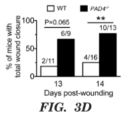
[0044] Figures 3a to 3e indicate that PAD4 deficiency facilitates wound
repair in
normoglycemic mice. Fig. 3a is a representative Western blot of wounds from WT
(+/+) and
PAD4-/- (-I-) mice. H3Cit was absent in the wounds from PAD4 mice. mice. Ly6G
levels in
wounds were similar in both genotypes (See also Fig 12a, Fig 12b). Fig. 3b is
a panel of
photographs of wounds of WT and PAD4 mice. mice. Wounds of PAD4 healed healed
faster and both
healed without apparent signs of infection. Scale, 5 mm. Fig. 3c is a graph
indicating
changes in wound area compared to day 0. Wound area reduced faster in PAD4
mice
mice
starting day 1 post wounding. *P<0.05, **P<0.01, ***P<0.001 versus WT,
Student's t test, n
= 9-16. Fig. 3d is a graph indicating significantly more PAD4 mice mice had
wounds completely
closed by day 14. **P<0.01, two-tailed Fisher's exact test. Fig. 3e is a graph
of re-
epithelialization determined from H&E staining on 3-day wounds from WT and
PAD4 mice
mice
(data not shown), re-epithelialization occurred faster in PAD4 mice. mice.
***P<0.001, Student's
t test, n = 6-9. Images of H&E staining and confocal microscopy of 3-day
wounds from WT
and PAD4 mice. mice. H&E revealed the presence of extracellular DNA in the
scab of WT mice,
while neutrophils appeared intact (ring-shaped,) in PAD4 scabs scabs (data not
shown). Confocal
immunofluorescence images (lower panels) showed intact neutrophil morphology
and an
absence of H3Cit in the scabs of PAD4 mice mice compared to the NETs in the
scabs of WT
mice (data not shown)
[0045] Figures 4a to 4i are graphs and Western blots indicating that PAD4
deficiency
or DNase 1 treatment enhances wound healing in diabetic mice. WT and PAD4 mice
mice were
treated with vehicle or STZ. Wounding was performed 8 weeks after diabetic
induction. All
mice were provided with antibiotics (2.5% Sulfatrim) in the drinking water
immediately after
wounding. (Fig. 4a-Fig. 4h). Fig. 4a to Fig. 4c are graphs showing wound area
reduction.
Fig. 4d to Fig. 4f are graphs indicating percent mice with open wounds per
time (Fig. 4a-Fig.
4h). Data from all groups were obtained simultaneously in multiple experiments
but split into
three graphs (Fig. 4a-Fig. 4c and Fig. 4d-Fig. 41) to facilitate comparison.
*P<0.05,
**P<0.01, ***P<0.001 between groups on respective post-wounding day (Fig. 4a-
Fig. 4c,
Student's t test) or between curves (Fig. 4d-Fig. 4f, log-rank test), n = 6-9.
(Fig. 4a) Wound
healing was impaired in STZ-induced diabetic WT mice compared to normoglycemic
mice
(vehicle). (Fig. 4b)PAD4-/- mice had much faster wound repair than WT under
diabetic
conditions. (Fig. 4c) Diabetes did not impair wound repair in PAD4 mice. mice.
(Fig. 4d) STZ-
9
CA 02974369 2017-07-19
WO 2016/118476 PCT/US2016/013847
induced diabetic WT mice had delayed wound closure compared to normoglycemic
mice
(vehicle). (Fig. 4e) STZ-treated PAD4-/- mice achieved total wound closure
earlier than STZ-
treated WT mice. (Fig. 41) Wound closure was not significantly different (NS)
between
normoglycemic (vehicle) and diabetic (STZ)PAD4-/- mice. Fig. 4g is a
representative Western
blot and summarized data (normalized to mean of vehicle) showing higher H3Cit
levels in
wounds from STZ-induced diabetic mice one day post wounding. (Fig. 4h, Fig.
4i) DNase 1
(dornase alfa) treatment facilitated wound area reduction (upper panels) and
re-
epithelialization (lower panels) in both (Fig. 4h) diabetic and (Fig. 4i)
normoglycemic WT
mice. (Fig. 4h) DNase 1 treatment did not provide additional benefits in wound
healing in
diabetic PAD4-/- mice. (Fig. 4h) *P<0.05, ***P<0.001 and NS non-significant
using Kruskal-
Wallis test followed by Dunn's post test, #13<0.05, "P<0.01 using Mann-Whitney
test, n = 5-
9. (Fig. 4i) *P<0.05, Student's t-test, n = 9-10
[0046] Figures 5A to 5E are graphs showing that neutrophil and platelet
count is
increased in aging WT mice and so is neutrophil susceptibility to produce
NETs: Fig. 5A is
a graph of neutrophil counts in peripheral blood of young (8 weeks) vs. old
(24 months) WT
mice. n = 6-8. Fig. 5B is a graph of platelet counts in young (8 weeks) vs.
old (24 months)
WT mice. n = 6-8. Fig. 5C is a graph of quantification of the percent of H3Cit-
positive
neutrophils by thresholding analysis of immunostained cytospins of red blood
cell-depleted
blood cells. n = 6-8. Fig. 5D is a graph of quantification of Ly6G-positive
neutrophils in the
total leukocyte cytospin population. n = 6-8. In C and D, young mice were 6 -
8 weeks and
old mice were 15 - 20 months old. Fig. 5E is a graph of the percentage of NET-
forming
peripheral blood neutrophils after incubation with vehicle (unstimulated, US),
4 1.IM
ionomycin (iono), or 100 nM phorbol 12-myristate 13-acetate (PMA) for 3.5 h.
Neutrophils
from old (24 - 27 months) mice formed significantly more NETs under all
conditions than
neutrophils from young (2 - 5 months) mice. n = 5. *P <0.05, **P <0.01, ***P
<0.001.
[0047]=
Figures 6A to 6E are graphs and images indicating that PAD4 mice are
protected from age-related decline in systolic and diastolic heart function
compared to
WT mice: Fig. 6A is 4 graphs of left ventricular ejection fraction (LVEF) as a
measure of
systolic function and cardiac dimensions (IVS;d, LVPW;d and LVID;d) of WT and
PAD4-/- retired breeders (1217 months) were measured by transthoracic
echocardiography. WT retired breeders showed a significantly reduced LVEF
compared
to PAD4-/- retired breeders. Cardiac dimensions were not significantly
different between
WT and PAD4"- retired breeders. n = 7-11. Fig. 6B is 4 graphs, the same
echocardiographic
CA 02974369 2017-07-19
WO 2016/118476 PCT/US2016/013847
measurements of LVEF and cardiac dimensions were repeated in a group of young
(6 - 8
weeks) and old (14 - 18 months) WT and PAD4-/- mice that had been kept on
standard lab
diet. Measurements showed similar results as in the retired breeders with a
significant
difference between the LVEF of old WT and old PAD4-/- mice (left panel). LVEFs
of old
PAD4"- mice were comparable to young PAD4-/- mice. Old WT and PAD4"- had
similar
cardiac dimensions. n = 4-7. Fig. 6C are representative ultrasound M-mode
images of the
left ventricle showed better contractility in the PAD4-/- mice compared to the
old WT mice.
S, systole; D, diastole. Fig. 6D is a graph of ventricular diastolic
dysfunction was evaluated
in young WT and PAD4-/- (6 - 8 weeks) mice as well as old WT and PAD4-/- mice
(15 - 20
months). The flow pattern across the mitral valve was assessed using Pulsed
Wave Doppler
mode and ventricular filling pattern was calculated as the ratio between the E
and A wave.
Only the old WT mice showed evidence of impaired ventricular relaxation with
an average
E/A ratio below 1. n = 4-6. Fig. 6E is characteristic images of Pulsed Wave
Doppler
measurements of the E and A wave showed a normal E'A pattern (E>A) in the old
PAD4-/-
mice and a reversed pattern (E<A) in the old WT mice, leading to a ratio of
under 1. *P <
0.05, **13 < 0.01, ***P <0.001.
[0048] Figures 7A to 7D are graphs and images indicating that PAD4-
deficiency
reduced age-related cardiac fibrosis: Fig. 7A is a graph of interstitial
collagen, Cardiac
interstitial fibrosis was assessed by Sirius Red staining for collagen fibers
in sections of the
left ventricle of the heart of WT and PAD4-/- retired breeders (1217 months).
The percentage
of fibrotic area in the heart tissue was quantified by ImageJ shown in Fig.
7C, excluding
perivascular fibrosis. In PAD4-/- retired breeders, there was significantly
less interstitial
fibrosis than in WT retired breeders. n = 6. Fig. 7B is a graph showing
interstitial collagen.
The same analysis was performed for young (6-8 weeks) WT and young PAD4-/-
mice as
well as for old (14-18 months) WT and old PAD4-/- mice on standard diet.
Quantification of
Sirius red staining again showed less fibrosis in the old PAD4"- mice compared
to the old
WT mice. In old PAD4-/- mice, the percentage of interstitial collagen remained
comparable
to young PAD4-/- mice. n = 7-8. Fig. 7C are images, Sirius red staining of
cardiac tissue
showed more fibrotic strands in the myocardium of WT retired breeders compared
to the
PAD4-/- retired breeders. Composite images of the left ventricle were
generated using the
ImageJ MosaicJ software; representative mosaics are presented. Scale bar = 100
i.tm.
Arrowheads indicate stained collagen strands. Fig. 7D are images, the increase
in
myocardial interstitial collagen fibers in WT retired breeders compared to
PAD4-/- retired
11
CA 02974369 2017-07-19
WO 2016/118476 PCT/US2016/013847
breeders was more clearly visible at higher magnification in the Sirius red
staining (left) and
in Masson's trichrome stain for collagen (right, collagen fibers are blue (see
arrows)). Scale
bar = 100 i.tm. Arrowheads point to collagen fibers. *P < 0.05, **P < 0.01
[0049]
Figures 8A to 8C are graphs and images, Old PAD4 mice have significantly
less collagen staining in their lungs than old WT mice. Fig. 8A is a graph
showing the
percentage of collagen positive area in lung tissue of WT and PAD4-/- retired
breeders (12-17
months) was quantified using Masson's trichrome stain for collagen and
subsequently color
gating for blue fibers. Retired WT breeders had a significantly higher
percentage of collagen
in lung tissue than retired PAD4-/- breeders. n = 6-7. Fig. 8B is a graph of
interstitial
collagen/lung tissue %; the same analysis for collagen fibers within the lung
tissue was
performed for young (6-8 weeks) WT and PAD4-/- mice and old (14-18 months) WT
and
PAD4-/- mice. While collagen content increased from young mice to old mice in
both WT
and PAD4-/- mice, this increase was significantly higher in the old WT mice. n
= 4. Fig. 8C is
a panel of representative photographs of lung sections stained with Masson's
trichrome stain.
Scale bar = 20 p.m. *P <0.05, ** P <0.01, **** P <0.0001.
[0050] Figures 9a to 9f show graphs and images of the basic parameters of
STZ-
induced diabetes in WT and PAD4-1- mice. Mice were injected i.p. with vehicle
or STZ (50
mg/kg per day) for 5 consecutive days. Body weight and fed blood glucose were
examined
starting 1 week after completion of injections. Fig. 9a is a graph of weight
over time, STZ-
treated mice gained less weight compared to the vehicle control. Fig. 9 b is a
graph of
glucose over time. Diabetes was defined as fed blood glucose >300 mg/dL
(indicated by blue
dotted line). STZ-treated mice became diabetic the first week after treatment.
(Fig. 9a, Fig.
9b) ***P<0.001 at all time points starting week 1 between vehicle and STZ,
Student's t test,
n = 15 for Vehicle, n = 13 for STZ. Fig. 9c is an image that validates
diabetes induction.
Representative immunofluorescence images showing a marked reduction of insulin-
producing 0 cells and disrupted islet morphology in the pancreas of STZ-
treated mice. Fig.
9d, and Fig. 9e PAD4-/- mice attained body weight (Fig. 9d) and fed blood
glucose levels
(Fig. 9e) similar to WT after STZ injection. AB indicates the period of
antibiotic treatment
(after wounding), which did not affect fed blood glucose levels in any group
(Fig. 9e). (Fig.
9d,Fig. 9e) ***P<0.001 at all time points starting week 1 between WT vehicle
and WT STZ,
r<0.001 at all time points starting week 1 between PAD4-/- vehicle and PAD4-/-
STZ,
Student's t test, n = 7 for WT Vehicle, n = 9 for WT STZ, n = 5 for PAD4-/-
Vehicle, n = 6 for
12
CA 02974369 2017-07-19
WO 2016/118476 PCT/US2016/013847
PAD4 -/- STZ.Fig. 9f is a graph of percent mice induced to be diabetic. Chi-
square test
indicates no difference between WT and PAD4 in in diabetes inducibility using
STZ. P=1.00
[0051] Figures 10a to 10b are graphs of wound healing over time.
Antibiotics do not
abrogate the beneficial effect of PAD4 deficiency on wound healing. Under
antibiotic
treatment, PAD4 mice mice still fared better in terms of (Fig. 10a) wound area
reduction and
(Fig. 10b) days required for total wound closure. *P<0.05, **P<0.01 between
groups on the
same day or between curves, Student's t test, n = 7 for WT Vehicle, n = 5 for
PAD4
Vehicle.
[0052] Figure 11 shows a graph of percent NETS. High glucose (HG)
enhances PMA
(100 nM)-stimulated NET formation in neutrophils isolated from healthy
subjects compared
to neutrophils exposed to normal glucose (NG) medium or mannitol (M), osmotic
control.
**P<0.01, repeated measures ANOVA, n = 5 per condition.
[0053] Figure 12a and 12b are Western blots and quantitative graphs
indicating
H3Cit (Fig.12a) is absent while neutrophil recruitment (Fig. 12b) is
unaffected in wounds of
PAD4 mice. mice. Summarized Western blot data of Figure 3a +/+, WT; -/-, PAD4-
/- **P<0 .01
versus day 1 WT, #4413<0.001 versus WT on respective day, Student's t test, n
= 5-8 for WT, n
= 5-9 for PAD4.
100541 Figure 13 is a schematic of chemical reactions to obtain
peptidomimetic
PAD4 inhibitors useful in the instant invention, e.g. compounds 1-16. This
figure was
obtained from Trabocchi et al. I Enzyme Inhib. Med. Chem., early online 1-6
(2014):
DOI:10.3109/147563662014947976, in order to illustrate compounds 1-16
described therein.
Detailed Description
Definitions
[0055] For convenience, the meaning of certain terms and phrases used in
the
specification, examples, and appended claims, are provided below. If there is
an apparent
discrepancy between the usage of a term in the art and its definition provided
herein, the
definition provided within the specification shall prevail.
[0056] Definitions of common terms in cell biology and molecular biology
can be
found in "The Merck Manual of Diagnosis and Therapy", 18th Edition, published
by Merck
Research Laboratories, 2006 (ISBN 0-911910-18-2); Robert S. Porter et al.
(eds.), The
Encyclopedia of Molecular Biology, published by Blackwell Science Ltd., 1994
(ISBN 0-
632-02182-9); and Robert A. Meyers (ed.), Molecular Biology and Biotechnology:
a
13
CA 02974369 2017-07-19
WO 2016/118476 PCT/US2016/013847
Comprehensive Desk Reference, published by VCH Publishers, Inc., 1995 (ISBN 1-
56081-
569-8); The ELISA guidebook (Methods in molecular biology 149) by Crowther J.
R. (2000);
Fundamentals of RIA and Other Ligand Assays by Jeffrey Travis, 1979,
Scientific
Newsletters; Immunology by Werner Luttmann, published by Elsevier, 2006.
Definitions of
common terms in molecular biology are also be found in Benjamin Lewin, Genes
IX,
published by Jones & Bartlett Publishing, 2007 (ISBN-13: 9780763740634);
Kendrew et al.
(eds.)õ Molecular Biology and Biotechnology: a Comprehensive Desk Reference,
published
by VCH Publishers, Inc., 1995 (ISBN 1-56081-569-8) and Current Protocols in
Protein
Sciences 2009, Wiley Intersciences, Coligan et al., eds.
[0057] Unless otherwise stated, the present invention was performed using
standard
labratoty techniques found, for example in the Molecular Cloning: A Laboratory
Manual, 3rd
Ed., Sambrook and Russel, Cold Spring Harbor Laboratory Press, 2001; or e.g.
the latest
edition of Methods in Enzymology Series. Editor: John Abelson, Melvin Simon
,Anna Pyle,
Elsevier Science Publishing Inc. New York.
[0058] The terms "decrease" , "reduced", "reduction" , "decrease" or
"inhibit" are all
used herein generally to mean a decrease by a statistically significant
amount. However, for
avoidance of doubt, 'reduced", "reduction" or "decrease" or "inhibit" means a
decrease by
at least 10% as compared to a reference level, e.g. in in the absence of an
agent, for example
a decrease by at least about 20%, or at least about 30%, or at least about
40%, or at least
about 50%, or at least about 60%, or at least about 70%, or at least about
80%.
[0059] The terms "increased" ,"increase" or "enhance" or "activate" are
all used
herein to generally mean an increase by a statically significant amount; for
the avoidance of
any doubt, the terms "increased", "increase" or "enhance" or "activate" means
an increase of
at least 10% as compared to a reference levelõ e.g. in in the absence of an
agent, for example
an increase of at least about 20%, or at least about 30%, or at least about
40%, or at least
about 50%, or at least about 60%, or at least about 70%, or at least about
80%, or at least
about a 2-fold, or at least about a 3-fold, or at least about a 4-fold, or at
least about a 5-fold or
at least about a 10-fold increase, or any increase between 2-fold and 10-fold
or greater as
compared to a reference level.
[0060] The term "statistically significant" or "significantly" refers to
statistical
significance and generally means a two standard deviation (25D) below normal,
or lower,
concentration of the marker. The term refers to statistical evidence that
there is a difference.
14
CA 02974369 2017-07-19
WO 2016/118476 PCT/US2016/013847
It is defined as the probability of making a decision to reject the null
hypothesis when the null
hypothesis is actually true. The decision is often made using the p-value.
[0061] Other than in the operating examples, or where otherwise
indicated, all
numbers expressing quantities of ingredients or reaction conditions used
herein should be
understood as modified in all instances by the term "about." The term "about"
when used in
connection with percentages can mean 1%.
[0062] The singular terms "a," "an," and "the" include plural referents
unless context
clearly indicates otherwise. Similarly, the word "or" is intended to include
"and" unless the
context clearly indicates otherwise. Although methods and materials similar or
equivalent to
those described herein can be used in the practice or testing of this
disclosure, suitable
methods and materials are described below. The abbreviation, "e.g." is derived
from the Latin
exempli gratia, and is used herein to indicate a non-limiting example. Thus,
the abbreviation
"e.g." is synonymous with the term "for example."
[0063] All patents and other publications identified are expressly
incorporated herein
by reference for the purpose of describing and disclosing, for example, the
methodologies
described in such publications that might be used in connection with the
present invention.
These publications are provided solely for their disclosure prior to the
filing date of the
present application. Nothing in this regard should be construed as an
admission that the
inventors are not entitled to antedate such disclosure by virtue of prior
invention or for any
other reason. All statements as to the date or representation as to the
contents of these
documents is based on the information available to the applicants and does not
constitute any
admission as to the correctness of the dates or contents of these documents.
[0064] As used herein, the term "administer" refers to the placement of a
composition
into a subject by a method or route which results in at least partial
localization of the
composition at a desired site such that desired effect is produced. A compound
or
composition described herein can be administered by any appropriate route
known in the art
including, but not limited to, oral or parenteral routes, including
intravenous, intramuscular,
subcutaneous, transdermal, airway (aerosol), pulmonary, nasal, rectal, and
topical (including
buccal and sublingual) administration. In certain embodiments, the anti-NET
compound is
administered by local administration, e.g. local injection, or other method
allowing delivery
to a target site within an organ. As used herein, the term "local" means
localized to the organ
or wound, i.e. not systemic administration.
CA 02974369 2017-07-19
WO 2016/118476 PCT/US2016/013847
[0065] Some exemplary modes of administration include, but are not
limited to,
injection, infusion, instillation, inhalation, or ingestion. "Injection"
includes, without
limitation, intravenous, intramuscular, intraarterial, intrathecal,
intraventricular, intracapsular,
intraorbital, intracardiac, intradermal, intraperitoneal, transtracheal,
subcutaneous,
subcuticular, intraarticular, sub capsular, subarachnoid, intraspinal,
intracerebro spinal, and
intrasternal injection and infusion. In preferred embodiments, the
compositions are
administered by intravenous infusion or injection.
[0066] As used herein, the term "antibody" refers to immunoglobulin
molecules and
immunologically active portions of immunoglobulin molecules, i.e., molecules
that contain
an antigen binding site that immunospecifically bind an antigen. The terms
also refers to
antibodies comprised of two immunoglobulin heavy chains and two immunoglobulin
light
chains as well as a variety of forms besides antibodies; including, for
example, Fv, Fab, and
F(ab)'2 as well as bifunctional hybrid antibodies (e.g., Lanzavecchia et al.,
Eur. J. Immunol.
17, 105 (1987)) and single chains (e.g., Huston et al., Proc. Natl. Acad. Sci.
U.S.A., 85, 5879-
5883 (1988) and Bird et al., Science 242, 423-426 (1988), which are
incorporated herein by
reference). (See, generally, Hood et al., Immunology, Benjamin, N.Y., 2ND ed.
(1984),
Harlow and Lane, Antibodies. A Laboratory Manual, Cold Spring Harbor
Laboratory (1988)
and Hunkapiller and Hood, Nature, 323, 15-16 (1986), which are incorporated
herein by
reference).
[0067] As used herein in the context of expression, the terms "treat,"
"treatment,"
"treating" and the like, in the context of the present invention insofar as it
relates to any of the
conditions recited herein (e.g. fibrosis, Diabetes (e.g. NET driven
inflammation and delayed
wound healing in Diabetes)), mean to relieve, alleviate, ameliorate, inhibit,
slow down,
reverse, or stop the progression, aggravation, deterioration, progression,
anticipated
progression or severity of at least one symptom or complication associated
with such
condition (e.g. fibrosis, Diabetes (e.g. NET driven inflammation and delayed
wound healing
in Diabetes)). In one embodiment, the symptoms of a condition are alleviated
by at least 5%,
at least 10%, at least 20%, at least 30%, at least 40%, or at least 50%.
[0068] By "lower" in the context of a disease marker or symptom is meant
a
statistically significant decrease in such level. The decrease can be, for
example, at least
10%, at least 20%, at least 30%, at least 40% or more, and is preferably down
to a level
accepted as within the range of normal for an individual without such
disorder.
16
CA 02974369 2017-07-19
WO 2016/118476 PCT/US2016/013847
[0069] As used herein, the phrase "therapeutically effective amount" or
"effective
dose" refers to an amount that provides a therapeutic benefit in the
treatment, prevention, or
management of a condition caused by NETS (e.g. fibrosis or inhibition of wound
healing, or
treatment of diabetes), e.g. an amount that provides a statistically
significant decrease in at
least one symptom of the condition (e.g. collagen deposition or slow wound
healing, or
inflammation of diabetes). Determination of a therapeutically effective amount
is well within
the capability of those skilled in the art. Generally, a therapeutically
effective amount can
vary with the subject's history, age, condition, sex, as well as the severity
and type of the
medical condition in the subject, and administration of other pharmaceutically
active agents.
[0070] As used herein, the term "pharmaceutical composition" refers to
the active
agent in combination with a pharmaceutically acceptable carrier of chemicals
and compounds
commonly used in the pharmaceutical industry.
[0071] The phrase "pharmaceutically acceptable" is employed herein to
refer to those
compounds, materials, compositions, and/or dosage forms which are, within the
scope of
sound medical judgment, suitable for use in contact with the tissues of human
beings and
animals without excessive toxicity, irritation, allergic response, or other
problem or
complication, commensurate with a reasonable benefit/risk ratio.
[0072] The phrase "pharmaceutically acceptable carrier" as used herein
means a
pharmaceutically acceptable material, composition or vehicle, such as a liquid
or solid filler,
diluent, excipient, solvent or encapsulating material, involved in carrying or
transporting the
subject agents from one organ, or portion of the body, to another organ, or
portion of the
body. Each carrier must be "acceptable" in the sense of being compatible with
the other
ingredients of the formulation, for example the carrier does not decrease the
impact of the
agent on the treatment. In other words, a carrier is pharmaceutically inert.
[0073] As used herein, a "subject" means a human or animal. In one
embodiment, the
animal is a vertebrate such as a primate, rodent, domestic animal, avian
species, fish or game
animal. The terms, "patient", "individual" and "subject" are used
interchangeably herein.
[0074] Preferably, the subject is a mammal. The mammal can be a human or
non-
human primate. Mammals other than humans can be advantageously used as
subjects that
represent animal models of fibrosis, wound healing or diabetic conditions. In
addition, the
methods described herein can be used to treat domesticated animals and/or
pets.
[0075] The subject can be one who has been previously diagnosed with an
organ
fibrosis, or diabetes, or a subject identified as having one or more
complications related to an
17
CA 02974369 2017-07-19
WO 2016/118476 PCT/US2016/013847
organ fibrosis or diabetes, and optionally, but need not have already
undergone treatment for
the condition, or the one or more complications related to the condition.
[0076] A subject can also be one who is not suffering from the condition,
e.g. fibrosis.
or diabetes. For example, a subject can be one who exhibits one or more risk
factors for
fibrosis or diabetes; e.g. having a family history if the disease or being of
older age, e.g. a
subject over 30 years of age, or over 40 years of age, or over 50 years of
age. Accordingly,
methods for preventing the formation of organ fibrosis are also provided, the
methods
comprise treating the subject determined to be at risk for fibrosis, with an
anti-NET
compound. In certain embodiments, the patient at risk of fibrosis is at least
40 years of age,
at least 50 years of age, at least 60 years of age, or at least 70 years of
age. In certain
embodiments, the patient at risk of fibrosis is a patient that is to be
exposed to radiation, e.g. a
patient of any age.
NETosis
[0077] Embodiments of the technology described herein are based, in part,
on the
discovery that NETosis in a subject slows the wound healing process and that
NETosis is
linked with collagen deposition in organ fibrosis. It has also been determined
herein that
increased NETosis is present in Diabetes.
[0078] As used herein, the term "NET" refers to extracellular complexes
of
nucleosomes and proteins, e.g. proteins having anti-microbial activity. The
nucleosomes may
be derived from neutrophils, mast cells, eosinophils, monocytes, or
leukocytes. "NETosis"
refers to the formation of NETS through a unique form of cell death that is
characterized by
the release of decondensed chromatin and granular contents to the
extracellular space.
[0079] Herein, we have determined that NETosis is elevated in wounds and
in
subjects that have diabetes. We have further determined that NETosis is
prominent in aging
and have found a connection between the prevalence of NETosis and organ
fibrosis. In
particular, we have determined that peptidylarginine deiminase 4 (PAD4), a key
enzyme
needed for the formation of NETS, promotes age related organ fibrosis. Thus,
methods for
treating wounds, diabetes and fibrosis are provided. The methods comprise
administrating a
therapeutically effective amount of at least one anti-NET compound (e.g. a PAD
4 inhibitor;
a DNase, a histone-degrading enzyme; an inhibitor of chromatin decondensation;
an antibody
against a component of a NET; a protease inhibitor, or an elastase inhibitor,
or protease
inhibitor) to a subject in need of treatment.
Anti-NET Compounds
18
CA 02974369 2017-07-19
WO 2016/118476 PCT/US2016/013847
[0080] Some embodiments are directed to methods for the treatment or
prevention of
organ fibrosis, or NET associated complications in diabetes (e.g. increased
inflammation and
delayed wound healing), in a patient with anti-NET compound. Other embodiments
are
directed to methods for facilitating wound healing in a subject comprising
administering an
anti-NET compound. In certain embodiments the anti-NET compounds are delivered
directly
to the wound. As used herein, "anti-NET compounds" can include any compound
that
degrades or targets for degradation any component of a NET and/or that
prevents the
formation of NETs (e.g. PAD4 inhibitors). Also included are compounds that
otherwise
inhibit the activity of a NET component or impair the ability of a cell to
form a NET, e.g.
inhibition of PAD4, which is required for NET formation. An anti-NET compound
can be a
nucleic acid (DNA or RNA), small molecule, lipid, carbohydrate, protein,
peptide, antibody,
or antibody fragment. In some embodiments, an anti-NET compound is an enzyme,
e.g. an
enzyme which cleaves and/or degrades, e.g. a nucleic acid, protein,
polypeptide, or
carbohydrate.
[0081] As used herein, the term "small molecule" refers to a chemical
agent which
can include, but is not limited to, a peptide, a peptidomimetic, an amino
acid, an amino acid
analog, a polynucleotide, a polynucleotide analog, an aptamer, a nucleotide, a
nucleotide
analog, an organic or inorganic compound (i.e., including heteroorganic and
organometallic
compounds) having a molecular weight less than about 10,000 grams per mole,
organic or
inorganic compounds having a molecular weight less than about 5,000 grams per
mole,
organic or inorganic compounds having a molecular weight less than about 1,000
grams per
mole, organic or inorganic compounds having a molecular weight less than about
500 grams
per mole, and salts, esters, and other pharmaceutically acceptable forms of
such compounds.
[0082] In certain embodiments an anti-NET compound is selected from the
group
consisting of; DNase, heparin, an antibody (i.e. an antibody to histones or to
a particular
histone), a histone degrading enzyme (i.e. mast cell proteinase 1 (Gene
ID:1215)), plasmin
(Gene ID: 5340), cathepsin D (Gene ID:1509) or activated protein C (Gene
ID:5624)) or an
inhibitor of chromatin decondensation (i.e.staurosporine, HDAC inhibitors
(i.e. M344),
PAD4 inhibitors, protease inhibitors, or elastase inhibitors (i.e. Geling)).
[0083] In one embodiment, the anti-NET compound is not heparin. In one
embodiment, the anti-NET compound is not DNase. In some embodiments, the anti-
NET
compound is selected from the group consisting of; a histone-degrading enzyme;
an inhibitor
19
CA 02974369 2017-07-19
WO 2016/118476
PCT/US2016/013847
of chromatin decondensation; an antibody against a component of a NET; a
protease
inhibitor, an elastase inhibitor; or a PAD4 inhibitor.
[0084] Anti-NET compounds can be produced recombinantly using methods
well
known to those of skill in the art (See Sambrook et al., Molecular Cloning: A
Laboratory
Manual (2 ed.), Cold Spring Harbor Laboratory Press, Cold Spring Harbor, N.Y.,
USA
(1989)). Alternatively, anti-NET compounds are available commercially e.g.
Pulmozyme
(Genentech; San Francisco, California), DNase (#D5319 Sigma-Aldrich; St.
Louis,
M0)(#90083 Thermo Scientific; Rockford, IL), RNAse (#R4642 Sigma-Aldrich; St.
Louis,
MO), Heparin (Celsus; Cincinatti, OH), anti-histone antibodies (ab1791,
ab8580, ab8898,
ab6002, ab1790, ab9053, ab10158, ab71594, ab4269 Abcam; Cambridge, MA), mast
cell
proteinase 1 (5146-SE-010 R&D Systems; Minneapolis, MN), thrombin (HCT-0020
Haematologic Technologies; Essex Junction, VT), plasmin (HCPM-0140
Haematologic
Technologies; Essex Junction, VT), cathepsin D (1014-AS-010 R&D Systems;
Minneapolis,
MN), activated protein C (AEZ004B Aniara; Mason, OH), staurosporine (S4400
Sigma-
Aldrich; St. Louis, MO), M344 (M5820 Sigma-Aldrich; St. Louis, MO) or Gelin
(G0528
Sigma-Aldrich; St. Louis, MO).
[0085] In certain embodiments, the anti-NET compound is a monoclonal
antibody
(See, generally, Hood et al., Immunology, Benjamin, N.Y., 2ND ed. (1984),
Harlow and
Lane, Antibodies. A Laboratory Manual, Cold Spring Harbor Laboratory (1988)
and
Hunkapiller and Hood, Nature, 323, 15-16 (1986), which are incorporated herein
by
reference).
[0086] In some embodiments, the anti-NET agent is a PAD4 inhibitor. As
used
herein, "PAD4" refers to peptidylarginine deiminase 4, an enzyme that converts
protein
arginine residues to citrulline through a deimination reaction (e.g. SEQ ID
NO: 01 (mRNA)
and SEQ ID NO: 02 (protein)).
[0087] In certain embodiments, the anti-NET agent is a general PAD
inhibitor, i.e. is
an inhibitor that inhibits more than one type of PAD enzyme, e.g. PAD1, and/or
PAD2,
and/or PAD3 or, and/or PAD4. See e.g. Wang et al., Anticancer peptidylarginine
deiminase
(PAD) inhibitors regulate the autophagy flux and the mammalian target of
rapamycin
complex 1 activity J Blot Chem. 2012 Jul 27;287(31):25941-53; e.g. YW3-56. See
also PCT
Pulication WO/2014/188193 entitiled `peptidylarginine deiminases (pad)
inhibitors."
[0088] PAD4 is distinguished from other PAD family enzymes by having a
nuclear
localization signal and thus being able to enter the nucleus and citrullinate
histones. As
CA 02974369 2017-07-19
WO 2016/118476 PCT/US2016/013847
described herein, a loss of PAD4 activity results in decreased NET formation
and decreased
DVT in mice. A PAD4 inhibitor can decrease the expression or activity of PAD4.
[0089] Inhibition of PAD4 can be monitored by measuring PAD4 activity. A
non-
limiting example of an assay of PAD4 activity is as follows: a candidate
inhibitor, in a
reaction buffer comprising 100 mM HEPES (pH 7.6), 50 mM NaC1, and 0.5 mM
tris(2-
carboxyethyl)phosphine (TCEP) can be preincubated with PAD4 (0.2 M) (in the
presence or
absence of 10 mM CaC12) at 37 C for 15 min prior to the addition of the
substrate, N-a-
benzoyl-L-arginine ethyl ester (BAEE) (10 mM final concentration) (and 10 mM
CaC12 if
CaC12 was absent in the pre-incubation) to initiate the reaction.After 15 min
the reactions can
be quenched by flash freezing in liquid nitrogen. For color development, 200
tL of freshly
prepared COLDER solution (2.25 M H3PO4, 4.5 M H2SO4, 1.5 mM NH4Fe(SO4), 20 mM
diacetyl monoxime, and 1.5 mM thiosemicarbazide) can be added to each of the
quenched
reactions, vortexed to ensure complete mixing, and then incubated at 95 C for
30 minutes.
The absorbance at 540 nm can then measured and compared to a citrulline
standard curve to
determine the concentration of citrulline produced during the course of the
reactions (PAD4
deiminates the BAEE substrate). IC50 values can be determined by fitting the
concentration-
response data to Eq. (1)
[0090] Fractional activity of PAD4 = 1/(1+([candidate inhibitor]/1C50))
(Eq. 1)
[0091] The concentration of an inhibitor that corresponds to the midpoint
(fractional
activity = 0.5) can be referred to as the IC50. Kits for measuring PAD4
activity are also
commercially available, e.g. Cat No. 7000560, Cayman Chemical; Ann Arbor, MI.
[0092] Any inhibitors of PAD4 can be used in the methods described
herein. For
example, in some embodiments, a PAD4 inhibitor can be a small molecule
inhibitor. Small
molecule inhibitors of PAD4 are known in the art (see, for example, Luo et al.
Biochemistry
2006; U.S. Patent 7,964.636; and U.S. Patent Publications 2007/0276040 and
2011/0142868;
each of which is incorporated by reference herein in its entirety) and
include, by way of non-
limiting example, Cl-amidine and F-amidine. In some embodiments, the PAD4
inhibitor can
be specific for PAD4. In some embodiments, the PAD4 inhibitor can be a PAD
family
inhibitor. PAD4 inhibitors are commercially available, e.g. Cl-amidine
(Catalog number
10599, CAS 913723-61-2, Cayman Chemical; Ann Arbor, MI) and F-amidine (Catalog
number 10610; Cayman Chemica; Ann Arbor, MI).
[0093] As used herein, "Cl-aminidine" refers to a compound having the
structure of
formula I:
21
CA 02974369 2017-07-19
WO 2016/118476 PCT/US2016/013847
NH2
CINH
0=
LH n
HH N 2
NThr
0
n=1, 2, 3
Formula I
[0094] As used herein, "Fl-amidine" refers to a compound having the
structure of
formula II:
NH2
NH
0=
LN n
NH2
0
n=1, 2, 3
Formula II
[0095] In some embodiments, the PAD4 inhibitor can be an antibody, a
polypeptide
comprising a fragment of an antibody, or a nucleic acid. Antibodies, and
methods of making
them are described above herein.
[0096] . In certain embodiments, the inhibitors are selective PAD4
inhibitors that are
reversible, e.g. including but not limited to GSK484 and GSK199 (Nat. Chem.
Biology, in
Press).
[0097] In certain embodiments, the PAD4 inhibitor is a tetrazole analog,
e.g. as
described in Subramanian et al., Design, synthesis and biological evaluation
of tetrazole
analogs of Cl-amidine as protein arginine deiminase inhibitors J. Med. Chem.,
DOT:
10.1021/jm501636x Publication Date (Web): January 5,2015.
[0098] In one embodiment the tetrazole analog is biphenyl tetrazole tert-
butyl Cl-
amidine (BTT-Cl-amidine) that exhibits enhanced cell killing in a PAD4
expressing cells also
blocks the formation of neutrophil extracellular traps (Subramanian et al.,
Supra).
[0099] In certain embodiments, the PAD4 inhibitor is a peptidomimetic
compound,
e.g. including but not limited to 1,2,3-triazole peptidomimetic based
derivatives incorporating
beta-phenylalanine and guanidine scaffolds, e.g. as described in Trabocchi et
al.
22
CA 02974369 2017-07-19
WO 2016/118476 PCT/US2016/013847
Peptidomimetics as protein arginine deiminase 4 (PAD4) inhibitors, I Enzyme
Inhib. Med.
Chem., early online 1-6 (2014): DOI:10.3109/147563662014947976. See also
Figure 13 that
illustrates chemistry for 16 peptidomimetic PAD4 inhibitors as described in
Trabocchi et al.
Supra, e.g. 1,2,3-triazole peptidomimetic based derivatives.
[00100] In certain embodiments, the anti-NET compound is an inhibitor of
NET
release from cells, e.g. Cl-amidine blocks NET release from NZM neutrophils in
vitro, other
inhibitors of NET release are known to those of skill in the art.
[00101] In certain embodiments, the PAD4 inhibitor is BB-Cl-amidine
(Knight et al.
Peptidylarginine deiminase inhibition disrupts NET formation and protects
against kidney,
skin and vascular disease in lupus-prone MRL/lpr mice Ann Rheum Dis
doi:10.1136/annrheumdis-2014-205365, online August 2014).
[00102] In certain embodiments, the PAD4 inhibitor is YW3-56, as described
in Wang
et al., (2012) 1 Biol. Chem 287(31):25941-53.
[00103] PAD4 inhibitors which comprise a nucleic acid can be RNAi agents
and/or
gene silencing agents. As used herein, "gene silencing" or "gene silenced" in
reference to an
activity of an RNAi molecule, for example a siRNA or miRNA refers to a
decrease in the
mRNA level in a cell for a target gene by at least about 5%, about 10%, about
20%, about
30%, about 40%, about 50%, about 60%, about 70%, about 80%, about 90%, about
95%,
about 99% or more of the mRNA level found in the cell without the presence of
the miRNA
or RNA interference molecule. In one preferred embodiment, the mRNA levels are
decreased by at least about 70%, about 80%, about 90%, about 95%, about 99% or
more.
[00104] As used herein, the term "RNAi" refers to any type of interfering
RNA,
including but are not limited to, siRNAi, shRNAi, endogenous microRNA and
artificial
microRNA. For instance, it includes sequences previously identified as siRNA,
regardless of
the mechanism of down-stream processing of the RNA (i.e. although siRNAs are
believed to
have a specific method of in vivo processing resulting in the cleavage of
mRNA, such
sequences can be incorporated into the vectors in the context of the flanking
sequences
described herein). The term "RNAi" and "RNA interfering" with respect to an
agent of the
invention, are used interchangeably herein.
[00105] As used herein an "siRNA" refers to a nucleic acid that forms a
double
stranded RNA, which double stranded RNA has the ability to reduce or inhibit
expression of
a gene or target gene when the siRNA is present or expressed in the same cell
as the target
gene, sEH. The double stranded RNA siRNA can be formed by the complementary
strands.
23
CA 02974369 2017-07-19
WO 2016/118476
PCT/US2016/013847
In one embodiment, a siRNA refers to a nucleic acid that can form a double
stranded siRNA.
The sequence of the siRNA can correspond to the full length target gene, or a
subsequence
thereof. Typically, the siRNA is at least about 15-50 nucleotides in length
(e.g., each
complementary sequence of the double stranded siRNA is about 15-50 nucleotides
in length,
and the double stranded siRNA is about 15-50 base pairs in length, preferably
about 19-30
base nucleotides, preferably about 20-25 nucleotides in length, e.g., 20, 21,
22, 23, 24, 25, 26,
27, 28, 29, or 30 nucleotides in length).
[00106] As used herein "shRNA" or "small hairpin RNA" (also called stem
loop) is a
type of siRNA. In one embodiment, these shRNAs are composed of a short, e.g.
about 19 to
about 25 nucleotide, antisense strand, followed by a nucleotide loop of about
5 to about 9
nucleotides, and the analogous sense strand. Alternatively, the sense strand
can precede the
nucleotide loop structure and the antisense strand can follow.
[00107] The terms "microRNA" or "miRNA" are used interchangeably herein
are
endogenous RNAs, some of which are known to regulate the expression of protein-
coding
genes at the posttranscriptional level. Endogenous microRNA are small RNAs
naturally
present in the genome which are capable of modulating the productive
utilization of mRNA.
The term artificial microRNA includes any type of RNA sequence, other than
endogenous
microRNA, which is capable of modulating the productive utilization of mRNA.
MicroRNA
sequences have been described in publications such as Lim, et al., Genes &
Development, 17,
p. 991-1008 (2003), Lim et al Science 299, 1540 (2003), Lee and Ambros
Science, 294, 862
(2001), Lau et al., Science 294, 858-861 (2001), Lagos-Quintana et al, Current
Biology, 12,
735-739 (2002), Lagos Quintana et al, Science 294, 853-857 (2001), and Lagos-
Quintana et
al, RNA, 9, 175-179 (2003), which are incorporated by reference. Multiple
microRNAs can
also be incorporated into a precursor molecule. Furthermore, miRNA-like stem-
loops can be
expressed in cells as a vehicle to deliver artificial miRNAs and short
interfering RNAs
(siRNAs) for the purpose of modulating the expression of endogenous genes
through the
miRNA and or RNAi pathways.
[00108] As used herein, "double stranded RNA" or "dsRNA" refers to RNA
molecules
that are comprised of two strands. Double-stranded molecules include those
comprised of a
single RNA molecule that doubles back on itself to form a two-stranded
structure. For
example, the stem loop structure of the progenitor molecules from which the
single-stranded
miRNA is derived, called the pre-miRNA (Bartel et al. 2004. Cell 116:281-297),
comprises a
dsRNA molecule.
24
CA 02974369 2017-07-19
WO 2016/118476
PCT/US2016/013847
[00109] As used herein, the term "complementary" or "complementary base
pair"
refers to A:T and G:C in DNA and A:U in RNA. Most DNA consists of sequences of
nucleotide only four nitrogenous bases: base or base adenine (A), thymine (T),
guanine (G),
and cytosine (C). Together these bases form the genetic alphabet, and long
ordered sequences
of them contain, in coded form, much of the information present in genes. Most
RNA also
consists of sequences of only four bases. However, in RNA, thymine is replaced
by uridine
(U).
[00110] As used herein, the term "nucleic acid" or "nucleic acid sequence"
refers to
any molecule, preferably a polymeric molecule, incorporating units of
ribonucleic acid,
deoxyribonucleic acid or an analog thereof. The nucleic acid can be either
single-stranded or
double-stranded. A single-stranded nucleic acid can be one strand nucleic acid
of a denatured
double- stranded DNA. Alternatively, it can be a single-stranded nucleic acid
not derived
from any double-stranded DNA. In one aspect, the template nucleic acid is DNA.
In another
aspect, the template is RNA. Suitable nucleic acid molecules are DNA,
including genomic
DNA, ribosomal DNA and cDNA. Other suitable nucleic acid molecules are RNA,
including
mRNA, rRNA and tRNA. The nucleic acid molecule can be naturally occurring, as
in
genomic DNA, or it may be synthetic, i.e., prepared based up human action, or
may be a
combination of the two. The nucleic acid molecule can also have certain
modification such as
2'-deoxy, 2'-deoxy-2'-fluoro, 2'-0-methyl, 2'-0-methoxyethyl (2'-0-M0E), 2'-0-
aminopropyl
(2'-0-AP), 2'-0-dimethylaminoethyl (2'-0-DMA0E), 2'-0-dimethylaminopropyl (2'-
0-
DMAP), 2'-0-dimethylaminoethyloxyethyl (2'-0-DMAEOE), or 2'-0--N-
methylacetamido
(2'-0-NMA), cholesterol addition, and phosphorothioate backbone as described
in US Patent
Application 20070213292; and certain ribonucleoside that are is linked between
the 2'-
oxygen and the 4'-carbon atoms with a methylene unit as described in US Pat
No. 6,268,490,
wherein both patent and patent application are incorporated hereby reference
in their entirety.
[00111] In some embodiments, a nucleic acid which is or which encodes a
PAD4
inhibitor further comprises a vector. The term "vector", as used herein,
refers to a nucleic
acid construct designed for delivery to a host cell or for transfer between
different host cells.
As used herein, a vector can be viral or non-viral. The term "vector"
encompasses any
genetic element that is capable of replication when associated with the proper
control
elements and that can transfer gene sequences to cells. A vector can include,
but is not
limited to, a cloning vector, an expression vector, a plasmid, phage,
transposon, cosmid,
chromosome, virus, virion, etc.
CA 02974369 2017-07-19
WO 2016/118476
PCT/US2016/013847
[00112] As used herein, the term "expression vector" refers to a vector
that directs
expression of an RNA or polypeptide from sequences linked to transcriptional
regulatory
sequences on the vector. The sequences expressed will often, but not
necessarily, be
heterologous to the cell. An expression vector may comprise additional
elements, for
example, the expression vector may have two replication systems, thus allowing
it to be
maintained in two organisms, for example in human cells for expression and in
a prokaryotic
host for cloning and amplification. As used herein, the term "viral vector"
refers to a nucleic
acid vector construct that includes at least one element of viral origin and
has the capacity to
be packaged into a viral vector particle. The viral vector can contain the
PAD4 inhibitor in
place of non-essential viral genes. The vector and/or particle may be utilized
for the purpose
of transferring any nucleic acids into cells either in vitro or in vivo.
Numerous forms of viral
vectors are known in the art. Vectors useful in the methods described herein
can include, but
are not limited to, plasmids, retroviral vectors, adenoviral vectors, adeno-
associated viral
vectors, and pox virus vectors.
[00113] The term "replication incompetent" when used in reference to a
viral vector
means the viral vector cannot further replicate and package its genomes. For
example, when
the cells of a subject are infected with replication incompetent recombinant
adeno-associated
virus (rAAV) virions, the heterologous (also known as transgene) gene is
expressed in the
patient's cells, but, the rAAV is replication defective (e.g., lacks accessory
genes that encode
essential proteins for packaging the virus) and viral particles cannot be
formed in the patient's
cells. The term "transduction" as used herein refers to the use of viral
particles or viruses to
introduce exogenous nucleic acids into a cell. The term "transfection" as used
herein in
reference to methods, such as chemical methods, to introduce exogenous nucleic
acids, such
as the nucleic acid sequences encoding an agent which decreases the activity
and/or level of
PAD4 as described herein, into a cell. As used herein, the term transfection
does not
encompass viral-based methods of introducing exogenous nucleic acids into a
cell. Methods
of transfection include physical treatments (electroporation, nanoparticles,
magnetofection),
and chemical-based transfection methods. Chemical-based transfection methods
include, but
are not limited to those that use cyclodextrin, polymers, liposomes,
nanoparticles, cationic
lipids or mixtures thereof (e.g., DOPA, Lipofectamine and UptiFectin), and
cationic
polymers, such as DEAE-dextran or polyethylenimine.
26
CA 02974369 2017-07-19
WO 2016/118476 PCT/US2016/013847
[00114] Methods of making RNAi agents which inhibit the expression and/or
activity
of PAD4 are well known in the art. Sequences complementary to the mRNA
encoding PAD4
(i.e. SEQ ID NO: 1) can be used to design RNAi agents as described above
herein.
[00115] The disruption of NETS can be monitored in vivo or in vitro. In
one
embodiment, the disruption of NETS is monitored by assesing the level of NET
release in
stored blood in the presence and absence of a test compound, e.g. by ELISA
and/or
determination of DNA concentration as described herein. In one embodiment, the
ability of a
test compound to disrupt NETS is monitored in vivo, e.g. by determining the
ability to
prevent platelet adhesion and aggregation.
Methods of Treatment
[00116] Described herein are methods of treating conditions such as
diabetes (e.g.
NET associated inflammation and delayed wound healing in diabetes), fibrosis,
and skin
wounds. As determined herein, these conditions are associated with an
increased NETosis,
and thus NETS can be targeted for treatment of theses disorders.
[00117] In one embodiment, a method of treating or preventing organ
fibrosis in a
subject is provided. The method comprises administering to a subject in need
of treatment, a
therapeutically effective amount of at least one anti-NET compound.
[00118] As used herein "Organ fibrosis" refers to fibrotic deposition that
can occur in
any organ. Fibrotic deposition (fibrosis) is a pathological condition
characterized by
excessive synthesis and accumulation of extracellular matrix proteins, loss of
tissue
homeostasis and organ failure.
[00119] As used herein "age-related organ fibrosis" refers to fibrosis
that occurs in
organs that has not been associated with any underlying disease, i.e. fibrosis
occurring as a
consequence of aging, e.g. idiopathic organ fibrosis, a non-limiting example;
idiopathic
pulmonary fibrosis (IPF).
[00120] As used herein, the term "preventing" as it relates to fibrosis
refers to
inhibition of intersiatial collagen deposition. In one embodiment, the
deposition of collagen
is decreased by at least 10%, at least 20%, at least 30%, at least 40%, or at
least 50%.
Deposition of collagen can be determined using methods well known to those of
skill in the
art, e.g. as described in Example 2, or by monitoring mRNA (See e.g. Casey et
al. (1996)
Biology of Reproduction, 55, 1253-1260). Collagen deposition can also be
monitored using
histology assays, e.g. in tissue samples. Collagen antibodies are commercially
available from
27
CA 02974369 2017-07-19
WO 2016/118476
PCT/US2016/013847
Rockland immunochemical corporation (Limerick, PA) e.g, COLLAGEN Type I
Antibody
600-401-103-0.5, or from Santa Cruz Biotechnology (Dallas, Texas).
[00121] As used herein, "treating" as it relates to organ fibrosis refers
to reducing at
least one measurable symptom of organ fibrosis. In one embodiment, the
meseauable
symptom is loss of organ function. Accodingly, in certain embodiments, the
symptoms of
fibrosis are dependent upon the organ affected, e.g. kidney, liver, heart,
lung. Those of skill
in the art are well versed in detecting proper oragan function. Non limiting
examples include
assessing blood to determine kidney glomerular filtration rate (kidney
function), or level of
liver enzymes (liver function), or determine levels of oxygen and CO2 in the
blood (lung
function); or performing e.g. echocardiograms, or EKG's of the heart, in the
case of e.g. age
related fibrosis of the heart. Techniques for measuring organ function are
standard in the art.
In certain embodiments, organ function is increased or improved by least 10%,
at least 20%,
at least 30%, at least 40%, or at least, 50% as compared to function prior to
treatment with
the anti-NET compound.
[00122] Methods for diagnosis of fibrosis are well known and include for
example
examination of tissue sections for collagen deposition, imaging studies, and
assessment of
organ function.
Diabetes
[00123] Also provided are methods for treating NET associated
complications in
diabetes (e.g inflammation and delayed wound healing). The methods comprise
administering a therapeutically effective amount of at least one anti-NET
compound. In some
embodiments, the subject has been diagnosed with Type 1, Type 1.5 or Type 2
diabetes, or
has been determined to have a pre-diabetic condition.
[00124] The terms "diabetes" and "diabetes mellitus" are used
interchangeably herein.
A "pre-diabetic condition" refers to a metabolic state that is intermediate
between normal
glucose homeostasis, metabolism, and states seen in frank Diabetes Mellitus.
Pre-diabetic
conditions include, without limitation, Metabolic Syndrome ("Syndrome X"),
Impaired
Glucose Tolerance (IGT), and Impaired Fasting Glycemia (IFG). IGT refers to
post-prandial
abnormalities of glucose regulation, while IFG refers to abnormalities that
are measured in a
fasting state. The World Health Organization defines values for IFG as a
fasting plasma
glucose concentration of 6.1 mmol/L (100 mg/dL) or greater (whole blood 5.6
mmol/L; 100
mg/dL), but less than 7.0 mmol/L (126 mg/dL)(whole blood 6.1 mmol/L; 110
mg/dL).
Metabolic Syndrome according to National Cholesterol Education Program (NCEP)
criteria
28
CA 02974369 2017-07-19
WO 2016/118476 PCT/US2016/013847
are defined as having at least three of the following: blood pressure 130/85
mm Hg or higher;
fasting plasma glucose 6.1 mmol/L or higher; waist circumference >102 cm (men)
or >88 cm
(women); triglycerides 1.7 mmol/L or higher; and HDL cholesterol <1.0 mmol/L
(men) or
1.3 mmol/L (women).
[00125] Type 1 diabetes is an autoimmune disease that results in
destruction of insulin-
producing beta cells of the pancreas. Lack of insulin causes an increase of
fasting blood
glucose (around 70-120 mg/dL in nondiabetic people) that begins to appear in
the urine above
the renal threshold (about 190-200 mg/di in most people). The World Health
Organization
defines the diagnostic value of fasting plasma glucose concentration to 7.0
mmo1/1 (126
mg/di) and above for Diabetes Mellitus (whole blood 6.1 mmo1/1 or 110 mg/di),
or 2-hour
glucose level of 11.1 mmol/L or higher (200 mg/dL or higher).
[00126] Type 1 diabetes can be diagnosed using a variety of diagnostic
tests that
include, but are not limited to, the following: (1) glycated hemoglobin (Al C)
test, (2) random
blood glucose test and/or (3) fasting blood glucose test.
[00127] The Glycated hemoglobin (Al C) test is a blood test that reflects
the average
blood glucose level of a subject over the preceding two to three months. The
test measures
the percentage of blood glucose attached to hemoglobin, which correlates with
blood glucose
levels (e.g., the higher the blood glucose levels, the more hemoglobin is
glycated). An Al C
level of 6.5 percent or higher on two separate tests is indicative of
diabetes. A result between
6 and 6.5 percent is considered prediabetic, which indicates a high risk of
developing
diabetes.
[00128] The Random Blood Glucose Test comprises obtaining a blood sample
at a
random time point from a subject suspected of having diabetes. Blood glucose
values can be
expressed in milligrams per deciliter (mg/dL) or millimoles per liter
(mmol/L). A random
blood glucose level of 200 mg/dL (11.1 mmol/L) or higher indicates the subject
likely has
diabetes, especially when coupled with any of the signs and symptoms of
diabetes, such as
frequent urination and extreme thirst.
[00129] For the fasting blood glucose test, a blood sample is obtained
after an
overnight fast. A fasting blood glucose level less than 100 mg/dL (5.6 mmol/L)
is considered
normal. A fasting blood glucose level from 100 to 125 mg/dL (5.6 to 6.9
mmol/L) is
considered prediabetic, while a level of 126 mg/dL (7 mmol/L) or higher on two
separate
tests is indicative of diabetes. Type 1 diabetes can be distinguished from
type 2 diabetes
using a C-peptide assay, which is a measure of endogenous insulin production.
The presence
29
CA 02974369 2017-07-19
WO 2016/118476 PCT/US2016/013847
of anti-islet antibodies (to Glutamic Acid Decarboxylase, Insulinoma
Associated Peptide-2 or
insulin), or lack of insulin resistance, determined by a glucose tolerance
test, is also indicative
of type 1, as many type 2 diabetics continue to produce insulin internally,
and all have some
degree of insulin resistance.
[00130] Testing for GAD 65 antibodies has been proposed as an improved
test for
differentiating between type 1 and type 2 diabetes as it appears that the
immune system is
involved in Type 1 diabetes etiology. Type 1.5 (also known as LADA Diabetes)
is performed
by determining the presence of anti-LADA antibodies.
[00131] Each of the diabetic conditions have overlapping symptoms.
Exemplary
symptoms of diabetes include, but are not limited to, excessive thirst
(polydipsia), frequent
urination (polyuria), extreme hunger (polyphagia), extreme fatigue, weight
loss,
hyperglycemia, low levels of insulin, high blood sugar (e.g., sugar levels
over 250 mg, over
300 mg), presence of ketones present in urine, fatigue, dry and/or itchy skin,
blurred vision,
slow healing cuts or sores, more infections than usual, numbness and tingling
in feet, diabetic
retinopathy, diabetic nephropathy, blindness, memory loss, renal failure,
cardiovascular
disease (including coronary artery disease, peripheral artery disease,
cerebrovascular disease,
atherosclerosis, and hypertension), neuropathy, autonomic dysfunction,
hyperglycemic
hyperosmolar coma, and combinations thereof.
[00132] A therapeutically effective amount of an anti-NET compound is the
amount of
a compound administered to a subject that is sufficient to produce a
statistically significant,
measurable change in a symptom of Type 1, Type 1.5 or Type 2 diabetes that has
NET
involvement, e.g. increased inflammation or delayed wound healing.
[00133] In certain embodiments, the symptom of diabetes that has NET
involvement is
ameliorated by at least 10%, at least 20%, at least 30%, at least 40%, or at
least 50%, as
compared to the symptom prior to treatment with the anti-NET compound.
[00134] In certain embodiments, the symptom of diabetes having NET
involvement is
delay of wound healing.
[00135] In certain embodiments, the symptom of diabetes having NET
involvement is
inflammation. Reduction in inflammation can be monitored by physical
examination, as well
as the reduction in the presence of inflammatory markers. Acute inflammatory
markers
known to the person skilled in the art include C-reactive protein (CRP),
fibrinogen, D-dimer,
serum amyloid A (SAA), pregnancy-associated polypeptide A (PAPP-A),
intercellular
adhesion molecules (e.g. ICAM-1, VCAM-1), IL-1-beta, IL-6, IL-8, IL-17 IL-
18/IL-18b;
CA 02974369 2017-07-19
WO 2016/118476 PCT/US2016/013847
TNF-alpha; myeloperoxidase (WO); TF; monocyte chemoattractant protein 1 (MCP-
1); P-
selectin; E-selectin; platelet activating factor acetyl hydrolase (PAF-AH);
von Willebrand
Factor (vWF). Preferred markers of acute inflammation for use in a method
described herein
are CRP, fibrinogen, D-dimer and SAA, of which CRP and D-dimer are more
preferably
used. D-Dimer is a marker of thrombolysis and its generation may be NET-
dependent.
[00136] In certain embodiments, a method for treatment of diabetes is
provided that
comprises the administration of a therapeutically effective amount of an agent
used to treat
diabetes and at least one anti-NET compound. In one embodiment, the agent used
to treat
diabetes is insulin. Other agents used to treat diabetes include, but are not
limited to,
Biguanides, Metformin (Glucophage), Metformin liquid ( Riomet), Metformin
extended
release (Glucophage XR, Fortamet, Glumetza), Sulfonylureas, Glimepiride
(Amaryl),
Glyburide (Diabeta, Micronase), Glipizide (Glucotrol, Glucotrol XL),
Micronized glyburide
(Glynase), Meglitinides, Repaglinide (Prandin), D-Phenylalanine Derivatives,
Nateglinide
(Starlix), Thiazolidinediones, Pioglitazone (TZDs), Pioglitazone, (Actos), DPP-
4 Inhibitor,
Sitagliptin (Januvia), Saxagliptin (Onglyza), Linagliptin ( Tradjenta), Alpha-
glucosidase,
Acarbose (Precose), Miglitol (Glyset), Bile Acid Sequestrants, Colesevelam
(Welchol),Pioglitazone & metformin) (Actoplus Met), Glyburide & metformin
(Glucovance),
Glipizide & metformin (Metaglip), Sitagliptin & metformin (Janumet),
Saxagliptin &
metformin (kombiglyze ), Repaglinide & metformin (Prandimet) and, Pioglitazone
&
glimepiride (Duetact).
Wound healing
[00137] Also provided are methods for treatments of wounds. In certain
embodiments
the patient that is administered an anti-net compound for the treatment of
wounds, has
previously been diagnosed with diabetes.
[00138] As used herein "wound healing" refers to the intricate process
where the skin
(or another organ-tissue) repairs itself after injury. The classic model of
wound healing is
divided into three or four sequential, yet overlapping, phases: (1)
hemostasis, when clot stops
bleeding, (2) inflammation, (3) proliferation and (4) remodeling . Upon injury
to the skin, a
set of complex biochemical events takes place in a closely orchestrated
cascade to repair the
damage (See e.g, Stadelmann, WK; Digenis, AG; Tobin, GR (1998). "Physiology
and healing
dynamics of chronic cutaneous wounds". American journal of surgery 176 (2A
Suppl): 26S-
38S). During the inflammation phase, bacteria and cell debris are phagocytosed
and removed
from the wound by white blood cells. Platelet-derived growth factors (stored
in the alpha
31
CA 02974369 2017-07-19
WO 2016/118476
PCT/US2016/013847
granules of the platelets) are released into the wound that cause the
migration and division of
cells during the proliferative phase. The proliferation phase is characterized
by angiogenesis,
collagen deposition, granulation tissue formation, epithelialization, and
wound contraction
(Midwood, K.S.; (2004). "Tissue repair and the dynamics of the extracellular
matrix". The
International Journal of Biochemistry & Cell Biology 36 (6): 1031-1037). New
blood
vessels are formed and fibroblasts grow and form a new, provisional
extracellular matrix
(ECM) by excreting collagen and fibronectin. Concurrently, re-
epithelialization of the
epidermis occurs, in which epithelial cells proliferate and 'crawl' atop the
wound bed,
providing cover for the new tissue.
[00139] The growth of tissue around the wound site is a result of the
migration of cells
and collagen deposition by these cells. The alignment of collagen describes
the degree of
scarring; basket-weave orientation of collagen is characteristic of normal
skin, whereas
aligned collagen fibers lead to significant scarring.
[00140] As used herein the term "facilitating wound healing" refers to an
acceleration
the process of normal wound healing and/or inhibiting the amount of formation
of scar tissue
that occurs from the wound healing process.
[00141] In certain embodiments, effective treatment can also be determined
by
measuring the diameter of the wound over time. In certain embodiments, the
diameter of the
wound is decreased by at least 10%, at least 20%, at least 30%, at least 40%,
or at least
50%/per unit time as compared to the diameter decrease per unit time usually
observed in
patients in the process of wound healing.
[00142] In certain embodiments, the formation of scar tissue is reduced by
at least
10%, at least 20%, at least 30%, at least 40%, or at least 50%, as compared to
an expected
healing process in the absence of the anti-NET compound.
[00143] In certain embodiments, the anti-NET compound is provided in a
pharmaceutically acceptable carrier that is time released, or that is
integrated in a skin graft,
or delivery device.
[00144] In certain embodiments, the treatment of a wound is assessed by
monitoring
dissolution of NETS in the wound.
[00145] Some embodiments relate to the use of at least one anti-NET
compound and
compositions containing at least one such anti-NET compound for the treatment
of diabetes,
treatment of fibrosis, or for facilitating wound healing. A composition
containing an anti-
32
CA 02974369 2017-07-19
WO 2016/118476 PCT/US2016/013847
NET compound is used to reduce the severity, duration, or number of symptoms
associated
with the condition to be treated.
[00146] In one embodiment, a single administration of an anti-NET compound
decreases the level of an indicator, symptom, or marker of fibrosis by at
least 10%, e.g., by at
least 20%, at least 30%, at least 50%, at least 75%, at least 75%, at least
90%, at least 95%, at
least 99% or more as compared to the level of the indicator, symptom, or maker
of a
cardiovascular condition prior to treatment with the anti-NET compound.
[00147] In certain embodiments, a single administration of an anti-NET
compound to a
patient decreases the deposition of interstiatial collagen in the patient's
organ by at least 10%,
e.g., by at least 20%, at least 30%, at least 50%, at least 75%, at least 75%,
at least 90%, at
least 95%, at least 99% or more as compared to the presence of collagen in the
absence of
treatment with the anti-NET compound.
[00148] In one embodiment, a single administration of an anti-NET compound
to a
group of patients facilitates wound healing by at least 10%, e.g., by at least
10%, by at least
20%, at least 30%, at least 50%, at least 75%, a at least 75%, at least 90%,
at least 95%, at
least 99% or more as compared to the rate of wound healing in a group of
patients not
administered the anti-NET compound.
[00149] The methods described herein relate to the use of at least one
anti-NET
compound or a pharmaceutical composition for treatment. In certain embodiments
the at
least one anti-NET compound is administered as a prophylactic, i.e. a patient
exhibiting
symptoms, markers, or indications of a condition described herein can be
treated with at least
one anti-NET compound in order to prevent or reverse the progression of the
condition or to
lessen the severity of future symptoms, markers, or indicators of the
condition.
[00150] In certain embodiments the methods provided herein involve the use
of at least
one anti-NET compound. In further embodiments, the method provided herein
involves the
use of two or more anti-NET compounds, non limiting examples -a PAD4 inhibitor
and a
DNase.
[00151] In certain embodiments, the effective dose of at least one anti-
NET compound
is administered to a patient repeatedly.
[00152] In certain embodiments, administering a single dose of an anti-NET
compound to a patient decreases the concentration of NETs at a target site
(e.g. organ or
wound) by least 10%, e.g., by at least 20%, at least 30%, at least 50%, at
least 75%, or more
as compared to the level of NETs prior to treatment with the anti-NET
compound.
33
CA 02974369 2017-07-19
WO 2016/118476 PCT/US2016/013847
[00153] In one embodiment, a single administration of an anti-NET compound
to a
patient decreases the level of an indicator, symptom, or marker of a condition
described
herein by at least 10%, e.g., by at least 20%, at least 30%, at least 50%, at
least 75%, at least
90% more as compared to the level of the indicator, symptom, or maker of the
condition prior
to treatment with the anti-NET compound.
[00154] In certain embodiments the composition comprising at least one
anti-NET
compound further comprises a pharmaceutically acceptable carrier. Non-limiting
examples
of antibiotics include, e.g., kanamycin, actinomycin D, doxorubicin,
bleomycin, and
mithramycin. Antibiotics are well known to those of skill in the art.
[00155] In some embodiments, the at least one anti-NET compound or a
pharmaceutical composition thereof, is administered with another
pharmaceutically active
agent, e.g. a pharmaceutically active agent for treating a patient with a
wound, fibrosis or
diabetes. The anti-NET compound can be administered in combination with other
pharmaceuticals and/or other therapeutic methods of treatment concurrently.
[00156] In some embodiments, the additional agent administered is an
antibiotic, e.g.
when the anti-NET compound is used for facilitating wound healing.
[00157] In some embodiments, the additional agent administered is an anti-
inflammatory agent, anti-A number of anti-inflammatory agents are known in the
art, non-
limiting examples of which are Alclofenac; Alclometasone Dipropionate;
Algestone
Acetonide; Alpha Amylase; Amcinafal; Amcinafide; Amfenac Sodium; Amiprilose
Hydrochloride; Anakinra; Anirolac; Anitrazafen; Apazone; Balsalazide Disodium;
Bendazac;
Benoxaprofen; Benzydamine Hydrochloride; Bromelains; Broperamole; Budesonide;
Carprofen; Cicloprofen; Cintazone; Cliprofen; Clobetasol Propionate;
Clobetasone Butyrate;
Clopirac; Cloticasone Propionate; Cormethasone Acetate; Cortodoxone;
Deflazacort;
Desonide; Desoximetasone; Dexamethasone Dipropionate; Diclofenac Potassium;
Diclofenac
Sodium; Diflorasone Diacetate; Diflumidone Sodium; Diflunisal; Difluprednate;
Diftalone,
Dimethyl Sulfoxide; Drocinonide; Endrysone; Enlimomab; Enolicam Sodium;
Epirizole;
Etodolac; Etofenamate; Felbinac; Fenamole; Fenbufen; Fenclofenac; Fenclorac;
Fendosal;
Fenpipalone; Fentiazac; Flazalone; Fluazacort; Flufenamic Acid; Flumizole;
Flunisolide
Acetate; Flunixin; Flunixin Meglumine; Fluocortin Butyl; Fluorometholone
Acetate;
Fluquazone; Flurbiprofen; Fluretofen; Fluticasone Propionate; Furaprofen;
Furobufen;
Halcinonide; Halobetasol Propionate; Halopredone Acetate; Ibufenac; Ibuprofen;
Ibuprofen
Aluminum; Ibuprofen Piconol; Ilonidap; Indomethacin; Indomethacin Sodium;
Indoprofen;
34
CA 02974369 2017-07-19
WO 2016/118476 PCT/US2016/013847
Indoxole; Intrazole; Isoflupredone Acetate; Isoxepac; Isoxicam; Ketoprofen;
Lofemizole
Hydrochloride; Lomoxicam; Loteprednol Etabonate; Meclofenamate Sodium;
Meclofenamic
Acid; Meclorisone Dibutyrate; Mefenamic Acid; Mesalamine; Meseclazone;
Methylprednisolone Suleptanate; Morniflumate; Nabumetone; Naproxen; Naproxen
Sodium;
Naproxol; Nimazone; Olsalazine Sodium; Orgotein; Orpanoxin; Oxaprozin;
Oxyphenbutazone; Paranyline Hydrochloride; Pentosan Polysulfate Sodium;
Phenbutazone
Sodium Glycerate; Pirfenidone; Piroxicam; Piroxicam Cinnamate; Piroxicam
Olamine;
Pirprofen; Prednazate; Prifelone; Prodolic Acid; Proquazone; Proxazole;
Proxazole Citrate;
Rimexolone; Romazarit; Salcolex; Salnacedin; Salsalate; Salycilates;
Sanguinarium Chloride;
Seclazone; Sermetacin; Sudoxicam; Sulindac; Suprofen; Talmetacin;
Talniflumate;
Talosalate; Tebufelone; Tenidap; Tenidap Sodium; Tenoxicam; Tesicam; Tesimide;
Tetrydamine; Tiopinac; Tixocortol Pivalate; Tolmetin; Tolmetin Sodium;
Triclonide;
Triflumidate; Zidometacin; Glucocorticoids; Zomepirac Sodium.
[00158] In some embodiments the additional agent administered is an anti-
fibrolytic
agent is administered. Additional anti-fibrinolytic agents include, for
example, Plasminogen,
prekallikrein, kininogens, Factors XII, XIIIa, plasminogen proactivator,
tissue plasminogen
activator[TPA], Streptokinase; Urokinase: Anisoylated Plasminogen-
Streptokinase Activator
Complex; Pro-Urokinase; (Pro-UK); rTPA (alteplase or activase; r denotes
recombinant),
rPro-UK; Abbokinase; Eminase; Sreptase Anagrelide Hydrochloride; Bivalirudin;
Dalteparin
Sodium; Danaparoid Sodium; Dazoxiben Hydrochloride; Efegatran Sulfate;
Enoxaparin
Sodium; Ifetroban; Ifetroban Sodium; Tinzaparin Sodium; tenecteplase,
retaplase;
Trifenagrel; Warfarin; Dextrans.
[00159] In some embodiments, the additional agent administered is agent to
treat
diabetes. Such agents include those agents known in the art for treatment of
diabetes and or
for having anti-hyperglycemic activities, for example, inhibitors of
dipeptidyl peptidase 4
(DPP-4) (e.g., Alogliptin, Linagliptin, Saxagliptin, Sitagliptin,
Vildagliptin, and Berberine),
biguanides (e.g., Metformin, Buformin and Phenformin), peroxisome proliferator-
activated
receptor (PPAR) modulators such as thiazolidinediones (TZDs) (e.g.,
Pioglitazone,
Rivoglitazone, Rosiglitazone and Troglitazone), dual PPAR agonists (e.g.,
Aleglitazar,
Muraglitazar and Tesaglitazar), sulfonylureas (e.g., Acetohexamide,
Carbutamide,
Chlorpropamide, Gliclazide, Tolbutamide, Tolazamide, Glibenclamide
(Glyburide),
Glipizide, Gliquidone, Glyclopyramide, and Glimepiride), meglitinides
("glinides") (e.g.,
Nateglinide, Repaglinide and Mitiglinide), glucagon-like peptide-1 (GLP-1) and
analogs
CA 02974369 2017-07-19
WO 2016/118476 PCT/US2016/013847
(e.g., Exendin-4, Exenatide, Liraglutide, Albiglutide), insulin and insulin
analogs (e.g.,
Insulin lispro, Insulin aspart, Insluin glulisine, Insulin glargine, Insulin
detemir, Exubera and
NPH insulin), alpha-glucosidase inhibitors (e.g., Acarbose, Miglitol and
Voglibose), amylin
analogs (e.g. Pramlintide), Sodium-dependent glucose cotransporter T2 (SGLT
T2) inhibitors
(e.g., Dapgliflozin, Remogliflozin and Sergliflozin) and others (e.g.
Benfluorex and
Tolrestat).
[00160] The anti-NET compound and the pharmaceutically active agent can be
administrated to the subject in the same pharmaceutical composition or in
different
pharmaceutical compositions (at the same time or at different times). When
administrated at
different times, an anti-NET compound and the pharmaceutically active agent
can be
administered within 5 minutes, 10 minutes, 20 minutes, 60 minutes, 2 hours, 3
hours, 4,
hours, 8 hours, 12 hours, 24 hours of administration of the other. When the
anti-NET
compound, and the pharmaceutically active agent are administered in different
pharmaceutical compositions, routes of administration can be different. For
example, the
anti-NET compound is administered by any appropriate route known in the art
including, but
not limited to oral or parenteral routes, including intravenous,
intramuscular, subcutaneous,
transdermal, airway (aerosol), pulmonary, nasal, rectal, and topical
(including buccal and
sublingual) administration, and pharmaceutically active agent is
administration by a different
route, e.g. a route commonly used in the art for administration of said
pharmaceutically active
agent. In a non-limiting example, an anti-NET compound can be administered
orally, while a
pharmaceutically active agent can be administrated subcutaneously.
[00161] Efficacy of treatment or prevention of disease can be assessed,
for example by
measuring a marker, indicator, or symptom of the condition, or any other
measurable
parameter appropriate. It is well within the ability of one skilled in the art
to monitor efficacy
of treatment or prevention by measuring any one of such parameters, or any
combination of
parameters.
[00162] A treatment is evident when there is a statistically significant
improvement in
one or more parameters of health, or by a failure to worsen or to develop
symptoms where
they would otherwise be anticipated. As an example, a favorable change of at
least 10% in a
measurable parameter of fibrosis, wound healing, or diabetes, and preferably
at least 20%,
30%, 40%, 50% or more can be indicative of effective treatment. Efficacy for a
given anti-
NET compound or formulation of that drug can also be judged using an
experimental animal
model for a condition described herein as known in the art. When using an
experimental
36
CA 02974369 2017-07-19
WO 2016/118476
PCT/US2016/013847
animal model, efficacy of treatment is evidenced when a statistically
significant increase in a
marker is observed.
[00163] The dosage ranges for the administration of an anti-NET compound
depend
upon the form of the compound, its potency, and the extent to which symptoms,
markers, or
indicators of a condition described herein are desired to be reduced, for
example the
percentage reduction desired for collagen deposition, inflammation, scar size.
The dosage
should not be so large as to cause adverse side effects, such as
hyperviscosity syndromes,
pulmonary edema, congestive heart failure, and the like. Generally, the dosage
will vary with
the age, condition, and sex of the patient and can be determined by one of
skill in the art. The
dosage can also be adjusted by the individual physician in the event of any
complication.
[00164] Patients can be administered a therapeutic amount of an anti-NET
compound,
such as 0.5 ng/kg, 1.0 ng/kg, 2.0 ng/kg, 2.5 ng/kg, 5 ng/kg, 10 ng/kg, 15
ng/kg, 20 ng/kg, 25
ng/kg, 30 ng/kg, 40 ng/kg or 50 ng/kg, 0.5 mg/kg, 1.0mg/kg, 2.0 mg/kg, 2.5
mg/kg, 5 mg/kg,
mg/kg, 15 mg/kg, 20 mg/kg, 25 mg/kg, 30 mg/kg, 40 mg/kg or 50 mg/kg. The anti-
NET
compound can be administered, for example, by intravenous infusion over a
period of time,
such as over a 5 minute, 10 minute, 15 minute, 20 minute, or 25 minute period.
The
administration is repeated, for example, on a regular basis, such as hourly
for 3 hours, 6
hours, 12 hours or longer or such as biweekly (i.e., every two weeks) for one
month, two
months, three months, four months or longer. After an initial treatment
regimen, the
treatments can be administered on a less frequent basis. For example, after
administration
biweekly for three months, administration can be repeated once per month, for
six months or
a year or longer. Administration of the anti-NET compound can reduce levels of
a marker or
symptom of a condition described herein, e.g., inflammation or collagen
deposition by at
least 10%, at least 15%, at least 20%, at least 25%, at least 30%, at least
40%, at least 50%, at
least 60%, at least 70%, at least 80 % or at least 90% or more.
[00165] Before administration of a full dose of the anti-NET compound,
patients can
be administered a smaller dose, such as a 5% infusion, and monitored for
adverse effects,
such as an allergic reaction.
[00166] In general, the efficacy of a given treatment can be monitored by
assessing the
disruption of NETs, as increased NETs have been associated with the conditions
described
herein. A reduction in NETs can be determined by tissue analysis and anti-Net
antibodies.However, a treatment is considered "effective treatment," as the
term is used
herein, if any one or all of the signs or symptoms of a condition described
herein are altered
37
CA 02974369 2017-07-19
WO 2016/118476 PCT/US2016/013847
in a beneficial manner, other clinically accepted symptoms are improved, or
even
ameliorated, e.g., by at least 10% following treatment with a compound as
described herein.
Efficacy can also be measured by a failure of an individual to worsen as
assessed by
hospitalization, or need for medical interventions (i.e., progression of the
disease is halted).
Another marker of the efficacy of treatment as described herein is survival.
Statistical
survival rates for specific conditions described herein are well established ¨
when an
individual or group of individuals treated according to the methods described
herein survives
beyond the expected time or at a greater than expected rate, the treatment can
be considered
effective.
Pharmaceutical Compositions
[00167] For administration to a subject, the compounds can be provided in
pharmaceutically acceptable compositions. These pharmaceutically acceptable
compositions
comprise a therapeutically-effective amount of at least one anti-NET compound
described
above, formulated together with one or more pharmaceutically acceptable
carriers (additives)
and/or diluents. As described in detail below, the pharmaceutical compositions
described
herein can be specially formulated for administration in solid or liquid form,
including those
adapted for the following: (1) oral administration, for example, drenches
(aqueous or non-
aqueous solutions or suspensions), lozenges, dragees, capsules, pills, tablets
(e.g., those
targeted for buccal, sublingual, and systemic absorption), boluses, powders,
granules, pastes
for application to the tongue; (2) parenteral administration, for example, by
subcutaneous,
intramuscular, intravenous or epidural injection as, for example, a sterile
solution or
suspension, or sustained-release formulation; (3) topical application, for
example, as a cream,
lotion, gel, ointment, or a controlled-release patch or spray applied to the
skin; (4)
intravaginally or intrarectally, for example, as a pessary, cream, suppository
or foam; (5)
sublingually; (6) ocularly; (7) transdermally; (8) transmucosally; or (9)
nasally. Additionally,
compounds can be implanted into a patient or injected using a drug delivery
system. Coated
delivery devices can also be useful. See, for example, Urquhart, et al., Ann.
Rev. Pharmacol.
Toxicol. 24: 199-236 (1984); Lewis, ed. "Controlled Release of Pesticides and
Pharmaceuticals" (Plenum Press, New York, 1981); U.S. Pat. No. 3,773,919; U.S.
Pat. No.
6,747,014; and U.S. Pat. No. 35 3,270,960.
[00168] As used here, the term "pharmaceutically acceptable" refers to
those
compounds, materials, compositions, and/or dosage forms which are, within the
scope of
sound medical judgment, suitable for use in contact with the tissues of human
beings and
38
CA 02974369 2017-07-19
WO 2016/118476 PCT/US2016/013847
animals without excessive toxicity, irritation, allergic response, or other
problem or
complication, commensurate with a reasonable benefit/risk ratio.
[00169] As used here, the term "pharmaceutically-acceptable carrier" means
a
pharmaceutically-acceptable material, composition or vehicle, such as a liquid
or solid filler,
diluent, excipient, manufacturing aid (e.g., lubricant, talc magnesium,
calcium or zinc
stearate, or steric acid), or solvent encapsulating material, involved in
carrying or transporting
the subject compound from one organ, or portion of the body, to another organ,
or portion of
the body. Each carrier must be "acceptable" in the sense of being compatible
with the other
ingredients of the formulation and not injurious to the patient. Some examples
of materials
which can serve as pharmaceutically-acceptable carriers include, but are not
limited to: (1)
sugars, such as lactose, glucose and sucrose; (2) starches, such as corn
starch and potato
starch; (3) cellulose, and its derivatives, such as sodium carboxymethyl
cellulose,
methylcellulose, ethyl cellulose, microcrystalline cellulose and cellulose
acetate; (4)
powdered tragacanth; (5) malt; (6) gelatin; (7) lubricating agents, such as
magnesium
stearate, sodium lauryl sulfate and talc; (8) excipients, such as cocoa butter
and suppository
waxes; (9) oils, such as peanut oil, cottonseed oil, safflower oil, sesame
oil, olive oil, corn oil
and soybean oil; (10) glycols, such as propylene glycol; (11) polyols, such as
glycerin,
sorbitol, mannitol and polyethylene glycol (PEG); (12) esters, such as ethyl
oleate and ethyl
laurate; (13) agar; (14) buffering agents, such as magnesium hydroxide and
aluminum
hydroxide; (15) alginic acid; (16) pyrogen-free water; (17) isotonic saline;
(18) Ringer's
solution; (19) ethyl alcohol; (20) pH buffered solutions; (21) polyesters,
polycarbonates
and/or polyanhydrides; (22) bulking agents, such as polypeptides and amino
acids (23) serum
component, such as serum albumin, HDL and LDL; (22) C2-C12 alchols, such as
ethanol; and
(23) other non-toxic compatible substances employed in pharmaceutical
formulations.
Wetting agents, binding agents, fillers, lubricants, coloring agents,
disintegrants, release
agents, coating agents, sweetening agents, flavoring agents, perfuming agents,
preservative,
water, salt solutions, alcohols, antioxidants, polyethylene glycols, gelatin,
lactose, amylose,
magnesium stearate, talc, silicic acid, viscous paraffin,
hydroxymethylcellulose,
polyvinylpyrrolidone and the like can also be present in the formulation. The
terms such as
"excipient", "carrier", "pharmaceutically acceptable carrier" or the like are
used
interchangeably herein.
[00170] Many organized surfactant structures have been studied and used
for the
formulation of drugs. These include monolayers, micelles, bilayers and
vesicles. Vesicles,
39
CA 02974369 2017-07-19
WO 2016/118476 PCT/US2016/013847
such as liposomes, have attracted great interest because of their specificity
and the duration of
action they offer from the standpoint of drug delivery. Liposomes are
unilamellar or
multilamellar vesicles which have a membrane formed from a lipophilic material
and an
aqueous interior. The aqueous portion contains the composition to be
delivered. Liposomes
can be cationic (Wang et al., Biochem. Biophys. Res. Commun., 1987, 147, 980-
985),
anionic (Zhou et al., Journal of Controlled Release, 1992, 19, 269-274), or
nonionic (Hu et al.
S.T.P.Pharma. Sci., 1994, 4, 6, 466). Liposomes can comprise a number of
different
phospholipids, lipids, glycolipids, and/or polymers which can impart specific
properties
useful in certain applications and which have been described in the art (Allen
et al., FEBS
Letters, 1987, 223, 42; Wu et al., Cancer Research, 1993, 53, 3765;
Papahadjopoulos et al.
Ann. N.Y. Acad. Sci., 1987, 507, 64; Gabizon et al. PNAS, 1988, 85, 6949;
Klibanov et al.
FEBS Lett., 1990, 268, 235; Sunamoto et al. Bull. Chem. Soc. Jpn., 1980, 53,
2778; Illum et
al. FEBS Lett., 1984, 167, 79; Blume et al. Biochimica et Biophysica Acta,
1990, 1029, 91;
US Patent Nos. 4,837,028; 5,543,152; 4,426,330; 4,534,899; 5,013,556;
5,356,633;
5,213,804; 5,225,212; 5,540,935; 5,556,948; 5,264,221; 5,665,710; European
Patents EP 0
445 131 Bl; EP 0 496 813 Bl; and European Patent Publications WO 88/04924; WO
97/13499; WO 90/04384; WO 91/05545; WO 94/20073; WO 96/10391; WO 96/40062; WO
97/0478).
[00171] The compositions described herein can be prepared and formulated
as
emulsions or microemulsions. Emulsions are typically heterogeneous systems of
one liquid
dispersed in another in the form of droplets usually exceeding 0.11Am in
diameter and have
been described in the art. microemulsion can be defined as a system of water,
oil and
amphiphile which is a single optically isotropic and thermodynamically stable
liquid solution
and can comprise surfactants and cosurfactants. Both of these drug delivery
means have been
described in the art (see e.g., Ansel's Pharmaceutical Dosage Forms and Drug
Delivery
Systems, Allen, LV., Popovich NG., and Ansel HC., 2004, Lippincott Williams &
Wilkins
(8th ed.), New York, NY; Idson, in Pharmaceutical Dosage Forms, Lieberman,
Rieger and
Banker (Eds.), 1988, Marcel Dekker, Inc., New York, N.Y., volume 1, p. 199;
Rosoff, in
Pharmaceutical Dosage Forms, Lieberman, Rieger and Banker (Eds.), 1988, Marcel
Dekker,
Inc., New York, N.Y., Volume 1, p. 245; Block in Pharmaceutical Dosage Forms,
Lieberman, Rieger and Banker (Eds.), 1988, Marcel Dekker, Inc., New York,
N.Y., volume
2, p. 199, 245, & 335; Higuchi et at., in Remington's Pharmaceutical Sciences,
Mack
Publishing Co., Easton, Pa., 1985, p. 301; Leung and Shah, in: Controlled
Release of Drugs:
CA 02974369 2017-07-19
WO 2016/118476 PCT/US2016/013847
Polymers and Aggregate Systems, Rosoff, M., Ed., 1989, VCH Publishers, New
York, pages
185-215; Schott, in Remington's Pharmaceutical Sciences, Mack Publishing Co.,
Easton, Pa.,
1985, p. 271; Constantinides et at., Pharmaceutical Research, 1994, 11, 1385-
1390; Ritschel,
Meth. Find. Exp. Clin. Pharmacol., 1993, 13, 205; Ho et at., J. Pharm. Sci.,
1996, 85, 138-
143; Lee et al., Critical Reviews in Therapeutic Drug Carrier Systems, 1991,
p. 92; U.S.
Patent Nos. 6,191,105; 7,063,860; 7,070,802; 7,157,099).
[00172] In one embodiment, the liposome or emulsion formulation comprises
a
surfactant. Surfactants find wide application in formulations such as
emulsions (including
microemulsions) and liposomes. The nature of the hydrophilic group (also known
as the
"head") provides the most useful means for categorizing the different
surfactants used in
formulations (Rieger, in Pharmaceutical Dosage Forms, Marcel Dekker, Inc., New
York,
N.Y., 1988, p. 285). Suitable surfactants include fatty acids and/or esters or
salts thereof, bile
acids and/or salts thereof In certain embodiments, the surfactant can be
anionic, cationic, or
nonionic. The use of surfactants in drug products, formulations and in
emulsions has been
reviewed (Rieger, in Pharmaceutical Dosage Forms, Marcel Dekker, Inc., New
York, N.Y.,
1988, p. 285).
[00173] In some embodiments, various penetration enhancers can be employed
to
effect the efficient delivery of anti-NET compounds across cell membranes.
Penetration
enhancers can be classified as belonging to one of five broad categories,
i.e., surfactants, fatty
acids, bile salts, chelating agents, and non-chelating non-surfactants all of
which have been
described elsewhere (see e.g., Malmsten, M. Surfactants and polymers in drug
delivery,
Informa Health Care, New York, NY, 2002; Lee et at., Critical Reviews in
Therapeutic Drug
Carrier Systems, 1991, p.92; Takahashi et al., J. Pharm. Pharmacol., 1988, 40,
252; Touitou,
E., et al. Enhancement in Drug Delivery, CRC Press, Danvers, MA, 2006;
Muranishi,
Critical Reviews in Therapeutic Drug Carrier Systems, 1990, 7, 1-33; El Hariri
et at., J.
Pharm. Pharmacol., 1992, 44, 651-654; Brunton, Chapter 38 in: Goodman &
Gilman's The
Pharmacological Basis of Therapeutics, 9th Ed., Hardman et at. Eds., McGraw-
Hill, New
York, 1996, pp. 934-935; Swinyard, Chapter 39 In: Remington's Pharmaceutical
Sciences,
18th Ed., Gennaro, ed., Mack Publishing Co., Easton, Pa., 1990, pages 782-783;
Yamamoto
et al., J. Pharm. Exp. Ther., 1992, 263, 25; Yamashita et al., J. Pharm. Sci.,
1990, 79, 579-
583; Jarrett, J. Chromatogr., 1993, 618, 315-339; Katdare, A. et al.,
Excipient development
for pharmaceutical, biotechnology, and drug delivery, CRC Press, Danvers, MA,
2006; Buur
et al., J. Control Rel., 1990, 14, 43-51)
41
CA 02974369 2017-07-19
WO 2016/118476 PCT/US2016/013847
[00174] Oral formulations and their preparation are described in detail in
U.S. Patent
6,887,906, US Publication. No. 20030027780, and U.S. Patent No. 6,747,014,
each of which
is incorporated herein by reference. Compositions and formulations for
parenteral,
intraparenchymal (into the brain), intrathecal, intraventricular or
intrahepatic administration
can include sterile aqueous solutions which can also contain buffers, diluents
and other
suitable additives such as, but not limited to, penetration enhancers, carrier
compounds and
other pharmaceutically acceptable carriers or excipients. Aqueous suspensions
can further
contain substances which increase the viscosity of the suspension including,
for example,
sodium carboxymethylcellulose, sorbitol and/or dextran. The suspension can
also contain
stabilizers.
[00175] A composition comprising at least one anti-NET compound can be
administered directly to the airways of a subject in the form of an aerosol or
by nebulization.
For use as aerosols, an anti-NET compound in solution or suspension may be
packaged in a
pressurized aerosol container together with suitable propellants, for example,
hydrocarbon
propellants like propane, butane, or isobutane with conventional adjuvants. An
anti-NET
compound can also be administered in a non-pressurized form such as in a
nebulizer or
atomizer.
[00176] An anti-NET compound can also be administered directly to the
airways in the
form of a dry powder. For use as a dry powder, an anti-NET compound can be
administered
by use of an inhaler. Exemplary inhalers include metered dose inhalers and dry
powdered
inhalers.
[00177] Aerosols for the delivery to the respiratory tract are known in
the art. See for
example, Adjei, A. and Garren, J. Pharm. Res., /: 565-569 (1990); Zanen, P.
and Lamm, J.-
W. J. Int. I Pharm., 114: 111-115 (1995); Gonda, I. "Aerosols for delivery of
therapeutic an
diagnostic agents to the respiratory tract," in Critical Reviews in
Therapeutic Drug Carrier
Systems, 6:273-313 (1990); Anderson et al., Am. Rev. Respir. Dis., 140: 1317-
1324 (1989))
and have potential for the systemic delivery of peptides and proteins as well
(Patton and
Platz, Advanced Drug Delivery Reviews, 8:179-196 (1992)); Timsina et. al.,
Int. I Pharm.,
101: 1-13 (1995); and Tansey, I. P., Spray Technol. Market, 4:26-29 (1994);
French, D. L.,
Edwards, D. A. and Niven, R. W., Aerosol Sci., 27: 769-783 (1996); Visser, J.,
Powder
Technology 58: 1-10 (1989)); Rudt, S. and R. H. Muller, J. Controlled Release,
22: 263-272
(1992); Tabata, Y, and Y. Ikada, Biomed. Mater. Res., 22: 837-858 (1988);
Wall, D. A., Drug
Delivery, 2: 10 1-20 1995); Patton, J. and Platz, R., Adv. Drug Del. Rev., 8:
179-196 (1992);
42
CA 02974369 2017-07-19
WO 2016/118476
PCT/US2016/013847
Bryon, P., Adv. Drug. Del. Rev., 5: 107-132 (1990); Patton, J. S., et al.,
Controlled Release,
28: 15 79-85 (1994); Damms, B. and Bains, W., Nature Biotechnology (1996);
Niven, R. W.,
et al., Pharm. Res., 12(9); 1343-1349 (1995); and Kobayashi, S., et al.,
Pharm. Res., 13(1):
80-83 (1996), contents of all of which are herein incorporated by reference in
their entirety.
[00178] The compositions can also be delivered by injection, e.g. locally
to fibrotic
tissue and organs. In certain embodiments, the compositions are delivered
using a device, or
bandage, used in the process of treatment of a wound.
[00179] The compositions described herein can additionally contain other
adjunct
components conventionally found in pharmaceutical compositions, at their art-
established
usage levels. Thus, for example, the compositions can contain additional,
compatible,
pharmaceutically-active materials such as, for example, antipruritics,
astringents, local
anesthetics or anti-inflammatory agents, or can contain additional materials
useful in
physically formulating various dosage forms of the compositions described
herein, such as
dyes, flavoring agents, preservatives, antioxidants, opacifiers, thickening
agents and
stabilizers. However, such materials, when added, should not unduly interfere
with the
biological activities of the components of the compositions described herein.
The
formulations can be sterilized and, if desired, mixed with auxiliary agents,
e.g., lubricants,
preservatives, stabilizers, wetting agents, emulsifiers, salts for influencing
osmotic pressure,
buffers, colorings, flavorings and/or aromatic substances and the like which
do not
deleteriously interact with the anti-NET compound(s) of the formulation.
[00180] As used herein, the phrase "subject in need of treatment" refers
to a subject
who is diagnosed with or identified as suffering from, having or at risk for
developing the
condition to be treated, e.g. fibrosis, diabetes or wounds.
[00181] Toxicity and therapeutic efficacy can be determined by standard
pharmaceutical procedures in cell cultures or experimental animals, e.g., for
determining the
LD50 (the dose lethal to 50% of the population) and the ED50 (the dose
therapeutically
effective in 50% of the population). The dose ratio between toxic and
therapeutic effects is
the therapeutic index and it can be expressed as the ratio LD50/ED50.
Compositions that
exhibit large therapeutic indices, are preferred. Murine genetics and surgical
techniques have
generated a number of mouse models for the study of fibrosis and diabetes or
mice impaired
in the ability to limit the concentration of NETs. Such models can be used for
in vivo testing
of anti-NET compounds, as well as for determining a therapeutically effective
dose. A
43
CA 02974369 2017-07-19
WO 2016/118476
PCT/US2016/013847
suitable mouse model is, for example, the DNse-/- mouse described herein or
the mouse
model of stroke described herein.
[00182] The data obtained from the cell culture assays and animal studies
can be used
in formulating a range of dosage for use in humans. The dosage of such
compounds lies
preferably within a range of circulating concentrations that include the ED50
with little or no
toxicity. The dosage can vary within this range depending upon the dosage form
employed
and the route of administration utilized.
[00183] The therapeutically effective dose can be estimated initially from
cell culture
assays. A dose can be formulated in animal models to achieve a circulating
plasma
concentration range that includes the IC50 (i.e., the concentration of the
therapeutic which
achieves a half-maximal inhibition of symptoms) as determined in cell culture.
Levels in
plasma can be measured, for example, by high performance liquid
chromatography. The
effects of any particular dosage can be monitored by a suitable bioassay.
[00184] The amount of an anti-NET compound which can be combined with a
carrier
material to produce a single dosage form will generally be that amount of the
compound
which produces a therapeutic effect. Generally out of one hundred percent,
this amount will
range from about 0.1% to 99% of compound, preferably from about 5% to about
70%, most
preferably from 10% to about 30%.
[00185] The dosage can be determined by a physician and adjusted, as
necessary, to
suit observed effects of the treatment. Generally, the compositions are
administered so that
the anti-NET compound is given at a dose from 1 pg/kg to 150 mg/kg, 1 pg/kg to
100 mg/kg,
1 pg/kg to 50 mg/kg, 1 pg/kg to 20 mg/kg, 1 pg/kg to 10 mg/kg, 111g/kg to
lmg/kg, 100
pg/kg to 100 mg/kg, 100 pg/kg to 50 mg/kg, 100 pg/kg to 20 mg/kg, 100 pg/kg to
10 mg/kg,
100 g/kg to lmg/kg, 1 mg/kg to 100 mg/kg, 1 mg/kg to 50 mg/kg, 1 mg/kg to 20
mg/kg, 1
mg/kg to 10 mg/kg, 10 mg/kg to 100 mg/kg, 10 mg/kg to 50 mg/kg, or 10 mg/kg to
20
mg/kg.It is to be understood that ranges given here include all intermediate
ranges, for
example, the range 1 mg/kg to 10 mg/kg includes lmg/kg to 2 mg/kg, lmg/kg to 3
mg/kg,
lmg/kg to 4 mg/kg, lmg/kg to 5 mg/kg, lmg/kg to 6 mg/kg, lmg/kg to 7 mg/kg,
lmg/kg to 8
mg/kg, lmg/kg to 9 mg/kg, 2mg/kg to 10mg/kg, 3mg/kg to 10mg/kg, 4mg/kg to
10mg/kg,
5mg/kg to 10mg/kg, 6mg/kg to 10mg/kg, 7mg/kg to 10mg/kg,8mg/kg to 10mg/kg,
9mg/kg to
10mg/kg etc... It is to be further undertood that the ranges intermediate to
the given above
are also within the scope of the methods and compositions described herein,
for example, in
44
CA 02974369 2017-07-19
WO 2016/118476
PCT/US2016/013847
the range lmg/kg to 10 mg/kg, dose ranges such as 2mg/kg to 8 mg/kg, 3mg/kg to
7 mg/kg,
4mg/kg to 6mg/kg etc.
[00186] With respect to duration and frequency of treatment, it is typical
for skilled
clinicians to monitor subjects in order to determine when the treatment is
providing
therapeutic benefit, and to determine whether to increase or decrease dosage,
increase or
decrease administration frequency, discontinue treatment, resume treatment or
make other
alteration to treatment regimen. The dosing schedule can vary from once a week
to daily
depending on a number of clinical factors, such as the subject's sensitivity
to the anti-NET
compound. The desired dose can be administered at one time or divided into
subdoses, e.g.,
2-4 subdoses and administered over a period of time, e.g., at appropriate
intervals through the
day or other appropriate schedule. Such sub-doses can be administered as unit
dosage forms.
In some embodiments, administration is chronic, e.g., one or more doses daily
over a period
of weeks or months. Examples of dosing schedules are administration daily,
twice daily,
three times daily or four or more times daily over a period of 1 week, 2
weeks, 3 weeks, 4
weeks, 1 month, 2 months, 3 months, 4 months, 5 months, or 6 months or more.
The desired
dose can be administered using continuous infusion or delivery through a
controlled release
formulation. In that case, the anti-NET compound contained in each sub-dose
must be
correspondingly smaller in order to achieve the total daily dosage. The dosage
unit can also
be compounded for delivery over several days, e.g., using a conventional
sustained release
formulation which provides sustained release of the anti-NET compound over a
several day
period. Sustained release formulations are well known in the art and are
particularly useful
for delivery of agents at a particular site, such as could be used with the
agents described
herein. In this embodiment, the dosage unit contains a corresponding multiple
of the daily
dose.
[00187] The skilled artisan will appreciate that certain factors can
influence the dosage
and timing required to effectively treat a subject, including but not limited
to the severity of
the disease or disorder, previous treatments, the general health and/or age of
the subject, and
other diseases present. Moreover, treatment of a subject with a
therapeutically effective
amount of a composition can include a single treatment or a series of
treatments. Estimates
of effective dosages and in vivo half-lives for the anti-NET compounds
described herein can
be made using conventional methodologies or on the basis of in vivo testing
using an
appropriate animal model, as described elsewhere herein.
A method of assessing efficacy of anti-NET treatments
CA 02974369 2017-07-19
WO 2016/118476 PCT/US2016/013847
[00188] As described herein, the inventors have found that increased
levels of NETs
are associated with impaired wound healing, diabetes and fibrosis, and have
provided
methods of treating or preventing these disorders by administering one or more
anti-NET
compounds. Accordingly, some embodiments are generally related to assays and
methods for
assessing the efficacy of the administration of one of more anti-NET
compounds. In certain
embodiments, the assays and methods are directed to determination of the level
of NETs in a
biological sample of a subject.
[00189] The methods and assays described herein include determining the
level of
NETs in samples obtained from a patient before and after treatment with one or
more anti-
NET compounds, wherein a reduction in the level of NETs following the
treatment with the
anti-NET compound is indicative of efficacy.
[00190] The sample obtained from a patient can include, but is not limited
to, blood or
blood products. Blood products in the context of samples obtained from a
patient can
include, but are not limited to, any component of a patient's blood (e.g.
plasma) and/or blood
or a component thereof that has been treated or processed (e.g. with an anti-
coagulant or
preservative).
[00191] In certain embodiments, the sample obtained from the patient prior
to
treatment with one or more anti-NET compounds can be obtained at any time
prior to
administration of the anti-NET compound, for example, about 1 minute prior to
treatment,
about 10 minutes prior to treatment, about 1 hour prior to treatment, about 1
day prior to
treatment, about 1 week prior to treatment, about 2 weeks prior to treatment,
about 1 month
prior to treatment, or earlier. In certain embodiments, the sample obtained
from the patient
after treatment with one or more anti-NET compounds can be obtained at any
time after
administration of the anti-NET compound, for example, about 10 minutes after
treatment,
about 1 hour after treatment, about 1 day after treatment, about 1 week after
treatment, about
2 weeks after treatment, or later.
[00192] In certain embodiments, the level of NETs is determined using
labeled DNA
detection reagents (i.e. Hoechst 33258 or SytoxGreen), immunodetection of
citullinated
histones, detection of nucelosomes and/or components thereof (i.e. Cell death
detection kit,
Roche), or electrophoresis of plasma DNA.
[00193] Some embodiemnts of the present invention may be defined in any of
the
following numbered paragraphs:
46
CA 02974369 2017-07-19
WO 2016/118476 PCT/US2016/013847
Paragraph 1. A method of treating or preventing organ fibrosis in a subject,
the method
comprising:
administering to a subject in need of treatment, a therapeutically effective
amount of at least
one anti-NET compound.
Paragraph 2. The method of paragraph 1, wherein the at least one anti-NET
compound is
selected from the group consisting of:
DNase; a hi stone-degrading enzyme; an inhibitor of chromatin decondensation;
an antibody
against a component of a NET; a protease inhibitor, an elastase inhibitor; and
a PAD4
inhibitor.
Paragraph 3. The method of any of paragraphs 1 to 2, wherein the PAD4
inhibitor is
selected from the group consisting of:
Cl-amidine and F-amidine.
Paragraph 4. The method of any of paragraphs 1 to 3, wherein said
therapeutically effective
amount of anti-NET compound is administered prophylactically.
Paragraph 5. The method of any of paragraphs 1 to 4, wherein the subjects age
is selected
from the group consisting of: over 40 years of age, over 30 years of age, over
50 years of age,
over 60 years of age, and over 70 years of age.
Paragraph 6. The method of any of paragraphs 1 to 5, wherein the subject is
diagnosed with
a disease selected from the group consisting of: heart disease, lung disease,
kidney disease,
liver disease, and diabetes.
Paragraph 7. The method of any of paragraphs 1 to 6, wherein said
therapeutically effective
amount of anti-NET compound is given repeatedly.
Paragraph 8. The method any of paragraphs 1 to 7, wherein the subject is
diagnosed as
having age-related organ fibrosis.
Paragraph 9. The method of any of paragraphs 1 to 8, wherein the subject is
diagnosed with
an organ fibrosis selected from the group consisting of; heart fibrosis, lung
fibrosis, liver
fibrosis, kidney fibrosis, skin fibrosis, soft tissue fibrosis, and intestine
fibrosis.
Paragraph 10. The method of any of paragraphs 1 to 9, wherein the
administration is local
administration to one or more target sites in an organ having fibrosis.
Paragraph 11. The method of any of paragraphs 1 to 10, wherein the subject
does not have
cystic fibrosis.
Paragraph 12. A method for facilitating wound healing comprising administering
a
therapeutically effective amount of at least one anti-NET compound.
47
CA 02974369 2017-07-19
WO 2016/118476 PCT/US2016/013847
Paragraph 13. The method of claim 12, wherein the anti-NET compound is
selected from the
group consisting of:
DNase; a hi stone-degrading enzyme; an inhibitor of chromatin decondensation;
an antibody
against a component of a NET; a protease inhibitor, an elastase inhibitor; and
a PAD4
inhibitor.
Paragraph 14. The method of any of paragraphs 12 to 13, wherein a DNAse and an
additional anti-NET compound selected from the group consisting of; a histone-
degrading
enzyme; an inhibitor of chromatin decondensation; a NET release inhibitor; an
antibody
against a component of a NET; a protease inhibitor, an elastase inhibitor; and
a PAD4
inhibitor, are administered.
Paragraph 15. The method of any of paragraphs 12 to 13, wherein the anti-NET
compound is
not a DNase.
Paragraph 16. The method of any of paragraphs 12 to 15, wherein the PAD4
inhibitor is
selected from the group consisting of:
Cl-amidine and F-amidine.
Paragraph 17. The method of any of paragraphs 12 to 16, wherein said
therapeutically
effective amount of anti-NET compound is administered prophylactically.
Paragraph 18. The method of any of paragraphs 12 to 17, wherein said
therapeutically
effective amount of anti-NET compound is given repeatedly.
Paragraph 19. The method of any of paragraphs 12 to 18, wherein the subject is
diagnosed as
having diabetes.
Paragraph 20. A method for treating NET associated inflammation and
complications in
diabetes comprising administering a therapeutically effective amount of at
least one anti-NET
compound.
Paragraph 21. The method of claim 20, wherein the anti-NET compound is
selected from the
group consisting of:
DNase; a histone-degrading enzyme; an inhibitor of chromatin decondensation; a
NET
release inhibitor; an antibody against a component of a NET; a protease
inhibitor, an elastase
inhibitor; and a PAD4 inhibitor.
Paragraph 22. The method of any of paragraphs 20 to 21, wherein the anti-NET
compound is
not a DNase.
Paragraph 23. The method of any of paragraphs 20 to 22, wherein the anti-NET
compound is
not an elastase inhibitor.
48
CA 02974369 2017-07-19
WO 2016/118476 PCT/US2016/013847
Paragraph 24. The method any of paragraphs 20 to 23, wherein the PAD4
inhibitor is
selected from the group consisting of:
Cl-amidine and F-amidine.
Paragraph 25. The method of any of paragraphs 20 to 24, wherein said
therapeutically
effective amount of anti-NET compound is administered prophylactically.
Paragraph 26. The method of any of paragraphs 20 to 25, wherein said
therapeutically
effective amount of anti-NET compound is given repeatedly.
Paragraph 27. The method of any of paragraphs 20 to 26, wherein the subject is
diagnosed as
having diabetes type 1.
Paragraph 28. The method of any of paragraphs 20 to 27, wherein the subject is
diagnosed as
having diabetes type II.
Paragraph 29. The method of any of paragraphs 20 to 28, wherein inflammation
is reduced
by at least 10%, at least 20%, at least 30%, or at least 50% as compared to
inflammation prior
to treatment.
EXAMPLES
[00194] EXAMPLE 1: NETS impair wound healing, especially in Diabetes
[00195] METHODS
[00196] Animals. All animal procedures were reviewed and approved by the
Institutional Animal Care and Use Committee of Boston Children's Hospital.
CD18-/- mice
and PAD4-/- mice were on a C57BL/6J background and were routinely crossed to
WT mice
from the Jackson Laboratory (Bar Harbor, ME). Age- and gender-matched control
mice
included the WT littermates of the two strains and C57BL/6J purchased from the
Jackson
Laboratory. Nine-week old male diabetic db/db mice and the normoglycemic
control m+/db
mice were purchased from Jackson Laboratory. All mice were fed standard lab
diet and
maintained under standard laboratory conditions free of specific pathogens.
Sample size was
chosen based on previous experience with the animal strains and animal models.
Genotypes
of animals were open to investigators.
[00197] Human blood cell samples. The study was approved by the
Institutional
Review Board of Boston Children's Hospital and Joslin Diabetes Center, and
conformed to
the principles outlined in the Declaration of Helsinki. Blood samples were
obtained after
written informed consent was obtained. Diabetic patients were recruited only
if they were
below 70 years old, not on steroid or other immunosuppressive medications, not
presenting
49
CA 02974369 2017-07-19
WO 2016/118476 PCT/US2016/013847
any signs of active infection (fever, high leukocyte count and diagnosis of
infection), no
diagnosis of cancer in the past 5 years and no overt heart failure.
[00198] Induction of diabetic murine model. Mice were induced to be
diabetic using
multiple low dose injections of streptozotocin (STZ). Six to 8-week old male
C57BL/6 or
PAD4-/- mice were randomized into treatment groups of either vehicle or STZ
according to
their blood glucose levels and body weight at baseline. Mice were fasted for 5
hours and then
injected with vehicle or STZ (i.p., 50 mg/kg per day, pH 4, dissolved in 0.1 M
sodium citrate
buffer) for 5 consecutive days. Fed blood glucose level was measured starting
1 week
afterwards. Mice with fed blood glucose level above 300 mg/dL were considered
diabetic and
used for further experiments. Pancreatic islets were stained for insulin using
a rabbit
polyclonal anti-insulin antibody (1:500, Cell Signaling, Cat. no. 4590).
[00199] Measurement of basal H3Cit on mouse cytospins. Murine whole blood
was
collected via the retro-orbital venous plexus. Red blood cells were lysed
using ACK lysing
buffer. After centrifugation, cells were resuspended in 7.5% BSA/PBS and spun
at 1600 rpm
for 4 minutes onto slides and instantly fixed with 4% PFA at 4 C overnight and
then stained
using rabbit polyclonal anti-H3Cit (1:1,000, abcam, Cat. no. ab5103) and rat
monoclonal
anti-mouse Ly6G (1:500, BD Pharmingen, Cat. no. 551459). H3Cit+ neutrophils
were
determined by thresholding analysis using ImageJ software (NIH).
[00200] Mouse neutrophil isolation and NETosis assay. Peripheral blood
neutrophils
were isolated with Percoll (GE Healthcare) gradients as described39. Purity of
cells was >90%
as determined by Wright-Giemsa staining. Neutrophils were resuspended in HBSS
(with
calcium, magnesium and 5.5 mM glucose) for experiments involving high glucose;
otherwise
they were resuspended in HEPES-buffered RPMI medium. Neutrophils were plated
at 50,000
cells/well in 96-well glass-bottomed plates and stimulated with Klebsiella
pneumoniae LPS
(Sigma) at indicated concentrations for 2.5 hours. For high glucose
experiments, neutrophils
were isolated from normoglycemic mice and pre-incubated for 1 h in media with
normal (5.5
mM) or high (22 mM) glucose concentration. Twenty-two mM corresponds to 396
mg/dL,
which is similar to the fed blood glucose level in STZ-induced mice 8 weeks
post-induction
(376.3 26.9 mg/dL). Mannitol (16.5 mM in medium with 5.5 mM glucose) was
employed
as an osmotic control. LPS (in respective medium) was added and neutrophils
were further
incubated for 2.5 h. Cells were then fixed in 2% PFA, permeabilized, blocked,
stained with
anti-H3Cit (1:1,000, abcam, Cat. no. ab5103), Alexa Fluor 488-conjugated anti-
rabbit
secondary antibody (1:1,500, Invitrogen) and Hoechst 33342 (1:10,000,
Invitrogen).
CA 02974369 2017-07-19
WO 2016/118476
PCT/US2016/013847
Percentages of H3Cithigh cells and NETs were determined from 5-6 non-
overlapping fields
per well and the average was taken from duplicates or triplicates for each
condition in every
experiment. Exposure time for H3Cit and DNA were identical for all treatments
within the
same experiment. Spread NETs were counted in a single channel for DNA. Images
of this
channel were exported in black-and-white for better contrast for
quantification.
[00201] Human neutrophil isolation and NETosis assay. Blood was drawn from
healthy subjects or diabetic patients into EDTA-coated tubes. Neutrophils were
isolated using
Histopaque -1119 (Sigma) and Percoll Plus (GE Healthcare) gradients as
described19, a
method that cause minimal activation of neutrophils during isolation. Purity
of cells was
>95% as determined by Wright-Giemsa staining. For experiments involving high
glucose,
neutrophils were resuspended in glucose-free HEPES-buffered RPMI supplemented
with
glucose at 5.5 mM (normal), 22 mM (high) or 5.5 mM plus 16.5 mM mannitol
(osmotic
control) and 2% heat-inactivated fetal bovine serum. Neutrophils were plated
at 10,000
cells/well in 96-well Cellbind plates (Corning). After incubation in
respective media for 1
h, cells were stimulated with ionomycin (4 1..1M) or PMA (100 nM) for 2.5
hours. For
experiments that did not involve high glucose, cells were resuspended in HEPES-
buffered
RPMI medium (11 mM glucose) supplemented with 2% heat-inactivated fetal bovine
serum,
plated at 10,000 cells/well and incubated with ionomycin (4 1..1M) for 2.5 h.
Cells were then
instantly fixed in 2% PFA with Hoechst 33342 (1:10,000) for NET
quantification. Percentage
of NETs was determined from 6 non-overlapping fields per well and the average
was taken
from triplicates for each condition in every experiment. Analysis was
performed by an
experimenter blinded to treatment conditions.
[00202] Wounding and macroscopic healing assessment. Full-thickness
excisional
wounds were made on the dorsal skin under aseptic conditions as described22.
Mice were
anesthetized with ketamine and xylazine (100 mg/kg and 10 mg/kg, respectively,
i.p.). Hair
was removed and the skin was cleaned with 70% ethanol and betadine. A fold of
the dorsal
skin was then picked up along the midline, placed over dental wax and punched
through with
a 4-mm disposable sterile biopsy punch (Miltex) such that 2 wounds were
generated in one
punch. The procedure was repeated, thus 4 wounds were made per mouse. The mice
were
housed individually after wounding. In experiments involving diabetic mice,
all mice were
provided ad libitum with antibiotics (2.5% Sulfatrim) in drinking water.
Wounds were
digitally photographed using a Sony Camcorder and total wound areas were
calculated using
51
CA 02974369 2017-07-19
WO 2016/118476 PCT/US2016/013847
ImageJ software. Wound area was expressed as a percentage compared to the area
on day 0
when the wounds were made.
[00203] Western blot analysis. Levels of H3Cit and Ly6G of mouse wounds
and PAD4
expression in human neutrophils were quantified by Western blot. After
collection of mouse
wounds or isolation of human neutrophils, the samples were snap frozen and
homogenized in
RIPA buffer supplemented with protease inhibitor cocktails (Sigma) on ice.
After
centrifugation at 20,000 g for 20 min at 4 C, the protein content of the
supernatant was
determined by bicinchoninic acid protein assay and an equal amount of protein
per sample
was resolved on gradient gels (4-20%, Lonza) and electrobloted on PVDF
membranes, which
were then incubated with primary antibodies (rabbit polyclonal anti-H3Cit,
1:1,000, abcam,
Cat. no. ab5103; rabbit polyclonal anti-H3, 1:6,000, abcam, Cat. no. ab1791;
rat monoclonal
anti-mouse Ly6G, 1:500, BD Pharmingen, Cat. no. 551459; mouse monoclonal anti-
human
PAD4, 1:2,000, abcam, Cat. no. ab128086) at 4 C overnight and subsequently
with
appropriate HRP-conjugated secondary antibodies for 2 h at room temperature.
The blots
were developed with enhanced chemiluminescence substrate. Equal loading was
confirmed
by probing for GAPDH (1:40,000, Ambion; Cat. no. AM4300). Blots were
quantified using
ImageJ software.
[00204] Immunofluorescence widefield and confocal microscopy. Localization
of
H3Cit and neutrophils in the wounds were examined by immunofluorescence
microscopy.
Wounds were dissected, cut in half and instantly embedded in OCT. The tissue
was
cryosectioned into 10 [tm and 20 [tm sections for wide-field and confocal
immunofluorescence microscopy, respectively. The sections were post-fixed in
zinc fixative
(100 mM Tris-HC1, 37 mM zinc chloride, 23 mM zinc acetate, 3.2 mM calcium
acetate),
permeabilized and incubated with primary antibodies against H3Cit (1:1,000,
abcam, Cat. no.
ab5103) and Ly6G (1:500, BD Pharmingen, Cat. no. 551459) at 4 C overnight and
then
Alexa Fluor-conjugated secondary antibodies (1:1,500, Invitrogen) for 2 hours
at room
temperature. Hoechst 33342 (1:10,000) was used to stain for DNA. Images were
acquired
with Zeiss Axiovision software using an Axiovert 200 wide-field fluorescence
microscope
(Zeiss) coupled to an Axiocam MRm monochromatic CCD camera (Zeiss) or with
Olympus
Fluoview software using the Olympus IX 81 confocal microscope.
[00205] Histological examination. Neutrophil recruitment and re-
epithelialization were
examined in H&E-stained sections. Wounds were cut in half, fixed overnight in
zinc fixative
and embedded in paraffin. The tissue was sectioned at 10 [tm and stained with
H&E. Images
52
CA 02974369 2017-07-19
WO 2016/118476
PCT/US2016/013847
were acquired with the Zeiss Axiovision software using an Axioplan light
microscope
coupled to a color Zeiss HRc camera.
[00206] Neutrophil depletion. Neutrophils of 10-week old WT mice were
depleted one
day before wounding by i.v. injection of a specific anti-neutrophil antibody
(ultra-low
endotoxin and azide free rat anti-Ly6G, 1A8 clone, Biolegend, Cat. no. 127632)
at a dose of
1.tg/g mouse. Control mice were injected with rat IgG. The mice were re-dosed
at 2.5 1.tg/g
mouse 2 days after the first injection. Levels of circulating neutrophils were
evaluated by
flow cytometry (BD FACSCanto II) using a FITC-conjugated rat monoclonal anti-
mouse
neutrophil antibody (1:300, anti-7/4, abcam, Cat. no. ab53453) and analyzed
using FlowJo
software. About 80% of circulating neutrophils were depleted throughout the 3-
day wound
healing period.
[00207] DNase 1 treatment. Normoglycemic and diabetic WT mice, randomized
by
blood glucose levels before assigning to treatments, were injected with 101.tg
i.v. and 501.tg
i.p. DNase 1 (dornase alfa, Genentech) 30 min before wounding and then 501.tg
i.p. every 12
hours until wound collection on day 3. Control mice were injected with vehicle
(8.77 mg/mL
sodium chloride and 0.15 mg/mL calcium chloride)40.
[00208] Statistical analysis Data are presented as mean s.e.m. of at
least two
independent experiments, and were analyzed using Mann-Whitney test, two-tailed
Student's
t-test (unpaired), Kruskal-Willis test followed by Dunn's post test, or
repeated measures
ANOVA with Bonferroni's post test, where appropriate. Percentage of mice with
total wound
closure and rate of diabetes induction between WT and PAD4-/- were analyzed
with two-
tailed Fisher's exact test of contingency tables. Percentage of mice with open
wounds was
analyzed with the log-rank test after constructing the Kaplan-Meier curves.
All analyses were
performed using GraphPad Prism software (Version 5.0). Results were considered
significant
when P<0.05.
[00209] NETs were originally recognized as a host defense mechanism in
which
neutrophils release their nuclear and granular contents to contain and kill
pathogens'.
Bacterial endotoxins, such as lipopolysaccharides (LPS), stimulate the release
of NETs' that
form extensive webs of DNA coated with cytotoxic histones and microbicidal
proteases1'2. A
prerequisite for NETosis is modification of arginine residues of histones to
citrulline by
PAD4, which changes the charge of the histones, leading to massive chromatin
decondensation3'4. Recently it became evident that NETs also form during
sterile
53
CA 02974369 2017-07-19
WO 2016/118476
PCT/US2016/013847
inflammation5. NETs are a key scaffold in pathologic thrombi and fuel
cardiovascular,
inflammatory and thrombotic diseases in mice and humans5'6.
[00210] Under diabetic conditions, neutrophils produce more superoxide'
and
cytokines8. Tumor necrosis factor-a, which primes neutrophils for NETosis9'1 ,
is increased
in diabetic patients". The diabetic microenvironment is thus pro-NETotic. To
test whether
diabetes predisposes neutrophils to NETosis, we isolated neutrophils from the
fresh whole
blood obtained from both type 1 and type 2 diabetic patients whose glycated
hemoglobin
(HbAlc) was >6.5%, indicating mild prolonged hyperglycemia (Fig. la).
Neutrophils from
these patients were indeed more susceptible to NETosis when stimulated with
the calcium
ionophore, ionomycin (Fig. lb). PAD4 is a calcium-dependent enzyme12 that is
key in
mediating NETosis13. Western blotting revealed a 4-fold upregulation of PAD4
protein
expression in the neutrophils from diabetic patients (Fig. lc), which may
explain their
higher susceptibility to NET formation. Our present findings are complemented
by a recent
report showing that circulating NET-related biomarkers, nucleosomes, cell-free
double-
strand DNA and neutrophil elastase, are increased in type 2 diabetic patients'
serum, and
that nucleosomes positively correlate with the patients' HbAl c levels".
[00211] Because hyperglycemia is common to both type 1 and type 2
diabetes, as
indicated by the significantly higher HbAl c in the diabetic cohort compared
to the healthy
controls (Fig. la, [Table 1]), we hypothesized that high glucose may
contribute to neutrophil
priming. We therefore isolated neutrophils from healthy donors and pre-
incubated them in
media with normal or high glucose concentrations prior to stimulation with
ionomycin or
phorbol 12-myristate 13-acetate (PMA) which triggers production of reactive
oxygen species
(ROS). Both ionomycin and PMA stimulated more of the high glucose-exposed
neutrophils
to produce NETs compared to pre-incubation with normal glucose or equal
concentrations of
the non-metabolizable sugar alcohol, mannitol (Fig. ld, and data not shown).
Thus, the
increased susceptibility of diabetic neutrophils to NETosis is at least in
part due to elevations
in blood glucose. Our observations differ from earlier reports15'16 which
suggested that high
glucose/diabetes does not affect or impairs NETosis. This difference is likely
due to the pre-
activation of human neutrophils during isolation with dextran sedimentation, a
method that
can induce ROS production and NET formation17'18 prior to culture, which could
result in the
loss of the primed neutrophil population during the preparatory process in the
previous
studies. Using Histopaque/Percoll gradients for human neutrophil isolation'',
we found a
clear priming effect by diabetes or high glucose on NETosis.
54
CA 02974369 2017-07-19
WO 2016/118476 PCT/US2016/013847
[00212] Table 1 Parameters of healthy subjects and diabetic patients
Healthy Subjects Diabetic Patients
Age (years) 36 6 40 6
Leukocyte count (K/ L) 6.00 0.70 6.58 1.07
Platelet count (K/ L) 299.20 20.00 276.20 12.20
HbAlc (%) 5.62 0.08 8.35 0.61 **
Glucose (mg/dL) 88.50 3.13 134.20 21.67 (a)
Cholesterol (mg/dL) 178.30 16.28 179.50 16.07
Triglycerides (mg/dL) 102.30 26.24 259.20 97.35
HDL (mg/dL) 72.50 14.31 54.67 11.79
LDL (mg/dL) 86.17 9.01 97.83 16.00
**P<0.01, (a) P = 0.0542 versus healthy subjects
[00213] We then examined the susceptibility to NETosis in diabetic mouse
models,
which are amenable to experimentation needed to study the role of PAD4 and
impact of
NETs on diabetic wound healing. Immunostaining of fresh blood cells from
streptozotocin
(STZ)-induced diabetic mice (a model of type 1 diabetes) (data not shown)
revealed a -4
fold increase in neutrophils positive for citrullinated histone H3 (H3Cit), a
biomarker of
NETosis, compared to normoglycemic mice (data not shown). About 4.5 fold more
isolated
neutrophils from diabetic mice were H3Cithigh (Fig. le) and -2% produced NETs
after
incubation in vitro without stimulation, while <0.2% NETs were seen in the
normoglycemic
controls (Fig. 10. LPS further stimulated more neutrophils from the STZ-
induced diabetic
mice to be H3Cithigh (Fig le, Fig 1g) and form NETs (Fig. if, Fig. 1g)
compared to vehicle-
treated normoglycemic mice. Thus, similar to humans, diabetes has inflammatory
or
metabolic components that predispose mouse neutrophils to NETosis. Although
there is no
specific anti-mouse PAD4 antibody to evaluate whether PAD4 protein expression
is
increased by diabetes, neutrophil priming could be also attributable to an
increased PAD4
activity as indicated by elevated histone H3 citrullination4 (Fig. le, data
not shown). Similar
NETosis assays were performed with neutrophils from genetically modified db/db
mice (data
not shown), a type 2 diabetic model. These neutrophils were also predisposed
to
hypercitrullinate histone H3 and form NETs (Fig. lh, Fig. li) when compared to
the
CA 02974369 2017-07-19
WO 2016/118476 PCT/US2016/013847
neutrophils from normoglycemic control m+/db mice, indicating enhanced NETosis
is a
common phenomenon in murine diabetes regardless of the type or etiology as we
observed in
the human condition. LPS again stimulated more of the high glucose-exposed
neutrophils
from normoglycemic WT mice to histone hypercitrullination (Fig. 1j) and NET
production
(Fig. lk), indicating a possible priming role of high glucose. Thus the mouse
models of
diabetes represent well the human condition in respect to susceptibility to
NETosis and
induction of PAD4 activity.
[00214] Depletion of neutrophils in mice was previously shown to
accelerate re-
epithelialization of uninfected diabetic wounds20. Because NETs can be
injurious to tissues21,
we asked whether NETs form in wounds and impact healing. We examined
excisional
wounds22 from normoglycemic WT mice. H&E staining confirmed that recruitment
of
leukocytes, mainly neutrophils, overlaps with the keratinocyte proliferation
stage that leads to
re-epithelialization (data not shown). Therefore, neutrophils or NETs could
interfere with
healing. Analysis of wound proteins by Western blotting showed a progressively
increasing
level of H3Cit that peaked from 3 to 7 days after wounding (Fig. 2a).
Immunofluorescence
images of 3-day wounds showed that hypercitrullinated neutrophils were present
in the
wound bed immediately beneath the scab (data not shown). Confocal microscopy
substantiated the presence of NETs in skin wounds. Externalized DNA
colocalized with
H3Cit in areas associated with intense staining of the neutrophil membrane
marker, Ly6G
(data not shown). Of note, H3Cit and neutrophils were absent in the surface
layers of
unwounded skin (data not shown). Skin expresses PAD isoforms 1-323 which could
citrullinate extracellular proteins in the scab. To verify the cellular source
of H3Cit, we
subjected CD18 (132 integrin)-deficient (CD/8-/-) mice, which are defective in
leukocyte
recruitment, to wounding. In these mice, both H3Cit and Ly6G were undetectable
by Western
blotting in 3-day wounds (Fig. 2b, left panels), a time when H3Cit was maximal
in the WT
wounds (Fig. 2a), indicating that H3Cit is of leukocyte origin. H&E staining
and
immunofluorescence microscopy showed that the few CD/8-/- neutrophils present
in these
wounds were H3Cit+ and produced NETs (data not shown). Indeed, CD/8-/-
neutrophils
produced NETs efficiently in vitro (data not shown), showing that 132
integrins were not
required for NETosis. Wounds from WT mice with depleted neutrophils also
showed
markedly reduced H3Cit (Fig. 2b, right panels). Thus, our data indicate that
neutrophils are
the source of the H3Cit present in the wounds.
56
CA 02974369 2017-07-19
WO 2016/118476 PCT/US2016/013847
[00215] To establish the role of NETs in wound healing, we compared wounds
of WT
to PAD4 mice. mice. Prominent extracellular DNA structures observed by H&E
were absent in
PAD4 scabs scabs (data not shown), as were the H3Cit and extracellular
chromatin patterns seen
in WT mice by confocal microscopy (data not shown). In contrast to the robust
H3Cit
signals in WT wounds, no H3Cit was detected in wounds from PAD4 mice mice
despite normal
neutrophil recruitment (Fig. 3a and data not shown). Unlike neutrophil
recruitment-
defective P-/E-selectin double mutants that have opportunistic infections24
and impaired
wound healing22, wounds in PAD4 mice mice did not show overt signs of
infection (Fig. 3b) and
healed faster than WT (Fig. 3b, Fig. 3c). This is likely because other
neutrophil functions
such as phagocytosis13, degranulation and RO S production (our unpublished
observations)
are intact in PAD4 neutrophils neutrophils so that these neutrophils are fully
capable of performing
other host defense mechanisms. About 80% of PAD4 mice mice had all wounds
healed on day 14
compared to only 25% of WT controls (Fig. 3d). The beneficial effect of PAD4
deficiency on
wound healing was observed very early after injury (Fig. 3c), indicating that
NETs might
impair the onset of initial healing processes such as keratinocyte migration.
In line with this
hypothesis, re-epithelialization progressed 3-fold faster in PAD4 mice mice
compared to WT
(Fig. 3e, and data not shown). Immunofluorescence staining of Ki67 (a
proliferation
marker) and TUNEL (indicator of apoptosis) was not different between 3-day
wounds from
WT and PAD4-/-mice (data not shown). It is thus likely that migration per se
is affected,
perhaps due to a modification of matrix proteins induced by NETs. Although WT
and PAD4
neutrophils also express PAD2 and PAD3 13, our data demonstrate that PAD4, the
only
nuclear PAD, is essential for the histone H3 citrullination and NETosis in
skin wounds.
Coudane et al.25 reported that PAD4 is the main PAD isoform detected in scabs
of wounds
from WT mice, and that PAD2 is unnecessary for citrullination of scab proteins
as observed
in PAD2-deficient mice, further strengthening the unique deimination role of
PAD4 in the
wounds.
[00216] We next examined whether NETs interfere with diabetic wound
healing. Type
1 diabetes was induced in WT and PAD4 mice mice by STZ and 8 weeks later these
mice were
subjected to wounding. Changes in body weight, fed blood glucose and diabetes
induction
rate were similar between the two genotypes (Fig. 9d-f]). As expected,
diabetic WT mice
healed more slowly than normoglycemic controls (Fig. 4a). All normoglycemic WT
mice
healed by day 16, while -20% of diabetic mice still had open wounds on day 19
(Fig. 4d).
Diabetic PAD4-/-mice healed >35% faster than diabetic WT mice on day 7 (Fig.
4b) and had
57
CA 02974369 2017-07-19
WO 2016/118476 PCT/US2016/013847
all wounds closed by day 15 (Fig. 4e). Notably, diabetes did not impair wound
healing in
PAD4-/- mice (Fig. 4c,f), which underscores NETs as the major determinant
delaying healing
in the diabetic mice. Higher H3Cit levels were detected in wounds of STZ-
induced diabetic
mice compared to the normoglycemic WT mice 1 day post wounding (Fig. 4g). The
enhanced NETosis in vivo recapitulates our in vitro observations (Fig. le-g),
further
supporting the role of NETs in the delay in diabetic wound repair.
Antibiotics, provided to
mimic the medical regimen of diabetic patients with chronic wounds, did not
abolish the
beneficial effect of PAD4 deficiency ([Supplementary Fig. 10]).
[00217] Enhanced wound healing in PAD4-/- mice suggests that NETs may be a
redundant host defense mechanism that compromises wound repair. NETs and hi
stones
directly induce epithelial and endothelial cell death21, and cause
cytotoxicity in vitro and in
vivo via calcium influx26. High neutrophil elastase concentration, a component
of NETs1'2, can
cause degradation of the wound matrix and delay healing27. Such a cytotoxic
environment
produced by NETs may explain the slower keratinocyte repopulation in the wound
beds of
WT mice. Because PAD4 is not expressed in the skin23, its negative effect on
wound healing is
most likely due to infiltrating neutrophils. In fact, using NETs to defend
against microbes may
not be very effective during wound healing as Staphylococcus species, which
are very
abundant in diabetic wounds28, degrade NETs to escape trapping29, and the NET
degradation
products can affect the proper healing process30'31. Thus, the non-selective
cytotoxicity of
NETs and/or their degradation products resulting from bacterial infection may
profoundly
delay wound healing.
[00218] Farrera and Fadeel reported that pre-digestion of NETs with DNase
1
accelerated their clearance by macrophages in vitro 32. Facilitated clearance
of NETs in wounds
may reduce their toxicity and diminish wound matrix degradation that is
essential for the
directional migration of keratinocytesB. We thus tested whether systemic DNase
1 treatment
could accelerate wound healing in diabetic mice that were maintained on
antibiotics. Without
DNase 1 treatment, diabetic PAD4-/- mice healed better in terms of both a
greater reduction in
wound area (Fig. 4h, upper panel) and more re-epithelialization (Fig. 4h,
lower panel)
compared to the diabetic WT mice as examined on day 3 post wounding.
Administration of
DNase 1 promoted wound area reduction by >20% and enhanced re-
epithelialization by
>75% in diabetic WT mice, an extent similar to that of DNase 1-treated
normoglycemic WT
mice (Fig. 4h). Interestingly, DNase 1 treatment did not provide further
benefits in healing
the wounds of diabetic PAD4-/- mice (Fig. 4h). These data indicate that NETs
are the major
58
CA 02974369 2017-07-19
WO 2016/118476 PCT/US2016/013847
source of extracellular DNA that hinders wound healing. Such beneficial
effects of DNase 1
were not confined to diabetic wounds. Three days post wounding, wound areas in
normoglycemic mice treated with DNase 1 were smaller than in those treated
with vehicle
(Fig. 41, upper panel). Re-epithelialization was also enhanced by ¨54% in the
DNase 1-
treated group (Fig. 4i, lower panel), while neutrophil recruitment was not
affected (data not
shown). Our current findings corroborate positive results from pilot clinical
trials with
activated protein C (APC), which cleaves and reduces the cytotoxicity of
histones34 and
facilitates healing of chronic wounds35 and diabetic ulcers36. Topical
treatment with an
ointment containing fibrinolysin and DNase (Elase) is used clinically for
wound debridement.
In addition to removing necrotic tissue, our findings suggest that the DNase
component may
also cleave NETs to reduce cytotoxicity and enhance wound recovery.
[00219] In summary, our data demonstrate that diabetes activates
neutrophils to
overproduce PAD4 and NETs and identify NETs as a key factor delaying wound
healing.
PAD4 inhibition and cleavage of NETs by DNase 1 could be novel therapeutic
approaches to
wound resolution, not only in diabetes, but also to wounds resulting from
aseptic procedures
such as surgeries of normoglycemic patients. We further validate the
importance of PAD4 in
human disease, and report the upregulation of PAD4 in diabetic patients, thus
providing new
rationale to develop specific PAD4 inhibitors. Because PAD4 and NET formation
contribute
to inflammatory and thrombotic diseases5'6 that are prominent in
diabetics37'38, anti-NET
therapy could have additional benefits. The increased NETosis in diabetes
suggests that
NETs may fuel these disorders and inhibiting NETosis or cleavage of NETs may
lessen them.
[00220] Example 1 References
1. Brinkmann, V., et al. Neutrophil extracellular traps kill bacteria. Science
303, 1532-
1535 (2004)
2. Urban, C.F., et al. Neutrophil extracellular traps contain calprotectin, a
cytosolic
protein complex involved in host defense against Candida albicans. PLoS Pathog
5, e1000639 (2009).
3. Wang, Y., et al. Histone hypercitrullination mediates chromatin
decondensation and
neutrophil extracellular trap formation. J Cell Biol 184, 205-213 (2009).
4. Wang, Y., et al. Human PAD4 regulates histone arginine methylation levels
via
demethylimination. Science 306, 279-283 (2004).
5. Yipp, B.G. & Kubes, P. NETosis: how vital is it? Blood 122, 2784-2794
(2013).
59
CA 02974369 2017-07-19
WO 2016/118476
PCT/US2016/013847
6. Martinod, K. & Wagner, D.D. Thrombosis: tangled up in NETs. Blood 123, 2768-
2776 (2014).
7. Karima, M., et al. Enhanced superoxide release and elevated protein kinase
C activity
in neutrophils from diabetic patients: association with periodontitis. J
Leukoc Biol
78, 862-870 (2005).
8. Hanses, F., Park, S., Rich, J. & Lee, J.C. Reduced neutrophil apoptosis in
diabetic
mice during staphylococcal infection leads to prolonged TNF-? production and
reduced neutrophil clearance. PLoS One 6, e23633 (2011).
9. Khandpur, R., et al. NETs are a source of citrullinated autoantigens and
stimulate
inflammatory responses in rheumatoid arthritis. Sci Transl Med 5, 178ra140
(2013).
10. Thomas, G.M., et al. Extracellular DNA traps are associated with the
pathogenesis of
TRALI in humans and mice. Blood 119, 6335-6343 (2012).
11. Alexandraki, K.I., et al. Cytokine secretion in long-standing diabetes
mellitus type 1
and 2: associations with low-grade systemic inflammation. J Clin Immunol 28,
314-321 (2008).
12. Luo, Y., et al. Inhibitors and inactivators of protein arginine deiminase
4: functional
and structural characterization. Biochemistry 45, 11727-11736 (2006).
13. Li, P., et al. PAD4 is essential for antibacterial innate immunity
mediated by
neutrophil extracellular traps. J Exp Med 207, 1853-1862 (2010).
14. Menegazzo, L., et al. NETosis is induced by high glucose and associated
with type 2
diabetes. Acta Diabetol (2014). [epub ahead of print]
15. Joshi, M.B., et al. High glucose modulates IL-6 mediated immune
homeostasis
through impeding neutrophil extracellular trap formation. FEB S Lett 587, 2241-
2246 (2013).
16. Riyapa, D., et al. Neutrophil extracellular traps exhibit antibacterial
activity against
burkholderia pseudomallei and are influenced by bacterial and host factors.
Infect
Immun 80, 3921-3929 (2012).
17. Fuchs, T.A., et al. Novel cell death program leads to neutrophil
extracellular traps. J
Cell Biol 176, 231-241 (2007).
18. Rebecchi, TM., Ferreira Novo, N., Julian, Y. & Campa, A. Oxidative
metabolism and
release of myeloperoxidase from polymorphonuclear leukocytes obtained from
CA 02974369 2017-07-19
WO 2016/118476 PCT/US2016/013847
blood sedimentation in a Ficoll-Hypaque gradient. Cell Biochem Funct 18, 127-
132 (2000).
19. Brinkmann, V., Laube, B., Abu Abed, U., Goosmann, C. & Zychlinsky, A.
Neutrophil
extracellular traps: how to generate and visualize them. J Vis Exp (2010).
20. Dovi, J.V., He, L.K. & DiPietro, L.A. Accelerated wound closure in
neutrophil-
depleted mice. J Leukoc Biol 73, 448-455 (2003).
21. Saffarzadeh, M., et al. Neutrophil extracellular traps directly induce
epithelial and
endothelial cell death: a predominant role of histones. PLoS One 7, e32366
(2012).
22. Subramaniam, M., et al. Role of endothelial selectins in wound repair. Am
J Pathol
150, 1701-1709 (1997).
23. Nachat, R., et al. Peptidylarginine deiminase isoforms 1-3 are expressed
in the
epidermis and involved in the deimination of K1 and filaggrin. J Invest
Dermatol
124, 384-393 (2005).
24. Frenette, P.S., Mayadas, T.N., Rayburn, H., Hynes, R.O. & Wagner, D.D.
Susceptibility to infection and altered hematopoiesis in mice deficient in
both P-
and E-selectins. Cell 84, 563-574 (1996).
25. Coudane, F., et al. Deimination and expression of peptidylarginine
deiminases during
cutaneous wound healing in mice. Eur J Dermatol 21, 376-384 (2011).
26. Abrams, ST., et al. Circulating histones are mediators of trauma-
associated lung
injury. Am J Respir Crit Care Med 187, 160-169 (2013).
27. Herrick, S., et al. Up-regulation of elastase in acute wounds of healthy
aged humans
and chronic venous leg ulcers are associated with matrix degradation. Lab
Invest
77, 281288 (1997).
28. Grice, E.A., et al. Longitudinal shift in diabetic wound microbiota
correlates with
prolonged skin defense response. Proc Natl Acad Sci U S A 107, 14799-14804
(2010).
29. Berends, E.T., et al. Nuclease expression by Staphylococcus aureus
facilitates escape
from neutrophil extracellular traps. J Innate Immun 2, 576-586 (2010).
30. Thammavongsa, V., Missiakas, D.M. & Schneewind, 0. Staphylococcus aureus
degrades neutrophil extracellular traps to promote immune cell death. Science
342, 863866 (2013).
61
CA 02974369 2017-07-19
WO 2016/118476
PCT/US2016/013847
31. Ishida, Y., Gao, J.L. & Murphy, P.M. Chemokine receptor CX3CR1 mediates
skin
wound healing by promoting macrophage and fibroblast accumulation and
function. J Immunol 180, 569-579 (2008).
32. Farrera, C. & Fadeel, B. Macrophage clearance of neutrophil extracellular
traps is a
silent process. J Immunol 191, 2647-2656 (2013).
33. Pilcher, B.K., et al. The activity of collagenase-1 is required for
keratinocyte
migration on a type I collagen matrix. J Cell Biol 137, 1445-1457 (1997).
34. Xu, J., et al. Extracellular histones are major mediators of death in
sepsis. Nat Med
15, 1318-1321 (2009).
35. Whitmont, K., et al. Treatment of chronic leg ulcers with topical
activated protein C.
Arch Dermatol 144, 1479-1483 (2008).
36. Whitmont, K., et al. Treatment of chronic diabetic lower leg ulcers with
activated
protein C: a randomised placebo-controlled, double-blind pilot clinical trial.
Int
Wound J (2013).
37. Laakso, M. & Kuusisto, J. Insulin resistance and hyperglycaemia in
cardiovascular
disease development. Nat Rev Endocrinol 10, 293-302 (2014).
38. Morel, 0., Jesel, L., Abbas, M. & Morel, N. Prothrombotic changes in
diabetes
mellitus. Semin Thromb Hemost 39, 477-488 (2013).
39. Demers, M., et al. Cancers predispose neutrophils to release extracellular
DNA traps
that contribute to cancer-associated thrombosis. Proc Natl Acad Sci U S A 109,
13076-13081 (2012).
40. Brill, A., et al. Neutrophil extracellular traps promote deep vein
thrombosis in mice. J
Thromb Haemost 10, 136-144 (2012).
[00221] EXAMPLE 2: PAD4 promotes fibrosis,
[00222] METHODS
[00223] Animals. Twenty-four to 27-month-old C57BL/6 mice for in vitro
NETosis
studies were obtained from the Aged Rodent Colony of the National Institute on
Aging of
the National Institutes of Health, maintained at Charles River Laboratories.
Young mice (8-
16 weeks old) for these experiments were obtained from the same colony.
[00224] PAD4-/- and corresponding wild-type (WT) mice were on a C57BL/6J
background. Retired breeders had been kept on LabDiet PicoLab Mouse Diet 20,
which is
fortified with a higher fat content for growth and reproduction (21.635%
calories provided
by fat), from 6-10 weeks of age until the time of sacrifice. Non-breeders and
all young mice
62
CA 02974369 2017-07-19
WO 2016/118476
PCT/US2016/013847
were kept on a standard laboratory diet (LabDiet Prolab IsoPro RMH 3000,
14.276%
calories provided by fat) throughout their life. Young animals were 6-8 weeks,
retired
breeders were 12-17 months old and old mice that had been kept on standard lab
diet were
14-18 months old. Old mice for the diastolic measurements were 18 months old
for the
PAD4-/- mice and between 15 and 20 months old for the old WT mice, while young
mice in
this experiment were 8 weeks old.
[00225] All groups were age and sex matched, and were fed ad libitum with
free
access to water. All experimental procedures were reviewed and approved by the
Institutional Animal Care and Use Committee of Boston Children's Hospital
(protocol no.
14-03-2631R).
[00226] Analysis of peripheral blood and cytospin. Blood was collected
from anesthetized mice via the retroorbital sinus into EDTA-coated capillary
tubes and was
analyzed by a Hemavet 950FS (Drew Scientific) for complete blood counts.
[00227] Twenty-five microliters of whole blood was incubated in ACK
(ammonium
chloride potassium) lysis buffer for 10 min on ice, then cytocentrifuged using
a Statspin
Cytofuge 2. Samples were immediately fixed in 4% paraformaldehyde for 2 h at
room
temperature and then immunostained for H3Cit and Ly6G as previously described
(16).
Images were acquired of cells from 10-15 fields of view at 200x magnification
using a
Zeiss Axiovert inverted epifluorescence microscope and Zeiss Axiovision
software.
Thresholding analysis was performed using ImageJ software to calculate the
population of
H3Cit-positive neutrophils in each sample.
[00228] Peripheral blood neutrophil isolation and NET induction.
Peripheral blood
neutrophils were isolated as described (16) and stimulated with calcium
ionophore (4 ilM) or
PMA (100 nM) for 3.5 h. Cells were fixed with 2% (vol/vol) paraformaldehyde,
and DNA
was stained with Hoechst 33342 (Invitrogen) for visualization of NETs using an
epifluorescent Axiovert microscope (Zeiss). NETs were counted from five
distinct fields of
view in triplicate wells and expressed as percentage of NET-forming cells per
total number
of cells in the field.
[00229] Plasma collection. Blood was collected from the retroorbital
plexus of
anesthetized mice (using 3.5 % isoflurane) into sodium citrate anticoagulant
(10% vol/vol).
Whole blood was centrifuged at 6000 rpm for 5 min, plasma was collected and
again
centrifuged at 13200 rpm for 5 min to remove any remaining cellular
components. Plasma
samples were immediately stored at -80 C until analysis.
63
CA 02974369 2017-07-19
WO 2016/118476
PCT/US2016/013847
[00230] Echocardiography. Cardiac function and heart dimensions were
measured as
described (11). The M-mode was used to evaluate left ventricular (LV) internal
dimension
(LVID), LV interventricular septum (LVIS), and LV posterior wall thickness
(LVPW) at end
diastole and end systole. Echocardiograms were stored digitally and ejection
fraction
(LVEF; percentage) was calculated using Vevostrain software (VisualSonics).
Flow pattern
across the mitral valve was measured in the 4-chamber view using the Pulsed
Wave (PW)
Doppler mode to determine evidence of impaired ventricular relaxation.
Ventricular filling
pattern is expressed as the ratio between the E and the A wave (E/A).
[00231] Blood pressure measurements. Systolic blood pressure was measured
using a
IITC 12M22931 non-invasive blood pressure system (IITC Life Science). Mice
were trained
twice several days before the measurements to accustom them to measurement
conditions.
For measurements, the mice were placed into restrainers and allowed to settle
down for 10
min. Systolic blood pressure was determined by the tail cuff in a chamber at
34 C. Blood
pressure was measured 5 times, and the mean of the obtained values is
presented (Table 2).
Table 2
WT PAD4-/- P-value
Total
leukocytes 7.902 1.033,n=9 9.068 1.123,n=8
0.4174
(x103/ 1)
Neutrophils
1.762 0.276, n=9 2.285 0.373, n=8 0.1662
(x103/ 1)
Platelets
1062 43.67, n=9 1002 38.42, n=8 0.5240
(x106/ 1)
Weights (g) 39.70 1.283, n=10 36.43 1.525, n=7
0.0951
Blood pressure 93.38 1.590, n=9 95.48 2.953, n=10
0.6464
(mm Hg)
[00232] Tissue
preparation and analysis. Anesthetized mice were sacrificed by
cervical dislocation, lungs and hearts removed and preserved in 10% neutral
buffered
formalin solution for at least 24 h. Organs were embedded in paraffin,
sectioned and
rehydrated. To assess collagen content in heart tissue, Sirius red staining
solution was
prepared with 0.5 g Direct Red 80 (Sigma) powder in 500 ml of saturated
aqueous solution
of picric acid (Sigma). Sirius red stains collagen I, II and III by reacting,
via its sulphonic
64
CA 02974369 2017-07-19
WO 2016/118476
PCT/US2016/013847
acid groups, with basic groups of the collagen molecule (32). Slides were
stained for 60 min,
washed twice in acidified water (5 % v/v acetic acid), dehydrated and mounted
using a
resinous mounting medium. At least 5 photographs of left ventricular heart
tissue were taken
at 250x magnification in brightfield microscopy in a blinded manner. The
content of red
fibers (collagen) per section was determined using ImageJ software, and
perivascular fibrosis
was excluded from the calculation. A subset of slides was stained with
Weigert's
hematoxylin before Sirius red staining and was used for the generation of
representative
pictures of heart tissue. For quantification, slides without nuclear staining
were used to avoid
interference of hematoxylin with the quantification algorithm. Mosaics of
representative
areas of the left ventricle were generated using the MosaicJ plugin of ImageJ
(33). For
trichrome staining of lung tissue the Masson trichrome stain kit (Sigma) was
used according
to the manufacturer's protocol. Nuclei were not stained with hematoxylin to
avoid
interference with the quantification of collagen content. For quantification,
at least 6
photographs of lung parenchyma were taken by brightfield microscopy by an
investigator
blinded to the identity of the samples. The area of blue fibers (collagen) per
lung tissue
(excluding empty alveolar spaces) was calculated using ImageJ software.
Staining of heart
tissue by trichrome stain was carried out in parallel. As these latter slides
were not used for
quantification, nuclei were stained using Weigert's hematoxylin.
[00233] Statistical analysis. Data are presented as means SEM. For
statistical tests,
a two-tailed Student's t-test or Mann-Whitney U-test was used when two groups
were
compared. For comparison of more than two groups, the one-way ANOVA with
Bonferroni's post-test was applied. Correlation analysis was performed between
the level of
heart fibrosis and EF using GraphPad Prism 6.0d software. All P values below
0.05 were
considered significant.
[00234] Both fibrosis and inflammation are closely associated with aging
(18). The
complex mechanisms involved in cellular deterioration with aging include the
accumulation
of DNA damage, mitochondrial dysfunction, increased susceptibility to
apoptosis, telomere
length shortening, epigenetic changes as well as oxidative stress (19, 20). It
is known that
elderly people experience significant changes in the function of their immune
system,
including a decline in the adaptive immune system, which creates an imbalance
between
adaptive and innate immune responses (21). Generally, aging leads to a more
pro-
inflammatory environment (22), with higher numbers of neutrophils and an
increase in ROS
production (5, 20) coupled with an increased susceptibility to pathogens and a
higher
CA 02974369 2017-07-19
WO 2016/118476
PCT/US2016/013847
incidence of inflammatory diseases (21), such as neurodegenerative disorders,
rheumatoid
arthritis, osteoporosis, diabetes, cardiovascular disease as well as
thrombosis (23).
Intriguingly, many of these illnesses have been reported to involve NETs.
[00235] Both the heart and the lung appear to be susceptible to age-
related fibrosis. In
cardiac aging, fibrotic remodeling may lead to diastolic dysfunction due to
increased
ventricular stiffness and, possibly, systolic heart failure (26), which is the
most common
cause for hospitalization for patients older than 65 years (27). In addition,
cardiac injury by
coronary artery disease or perimyocarditis can add to the fibrotic changes of
the aging heart,
making it even more important to understand the mechanisms underlying this
process.
[00236] Fibrotic lung diseases are characterized by enhanced collagen
deposition in the
airways, including the alveolar walls, and subsequent disturbance of pulmonary
gas-
exchange. Excessive fibrotic tissue remodeling is a predominant feature of
many chronic lung
diseases. Fibrotic lung diseases affect a large part of the older population
(28-30) and include
chronic obstructive pulmonary disease (COPD), fibrotic reactions after acute
or chronic lung
infections, inhalation of pulmonary irritants, autoimmune or allergic diseases
and idiopathic
pulmonary fibrosis (IPF) an aggressive form of lung fibrosis with no proven
treatment
option. Importantly, both COPD and IPF are again clearly associated with aging
(30, 31).
Given the need for a better understanding of the complex mechanisms linking
inflammation
to fibrosis and aging, the goal of this study was to determine if there is
interplay between
PAD4/NETs, fibrosis and aging.
[00237] Results
[00238] Neutrophil susceptibility to form NETs increases with mouse age
[00239] There are many changes that occur in the aging immune system,
including an
increase in hematopoietic stem cells of the myeloid lineage versus cells of
the lymphoid
lineage (34). To further study the effect of aging in mice, we examined blood
and neutrophils
from young (8-16 weeks) and old (24-27 months) mice obtained from the NIH' s
NIA
C57BL/6 Aged Rodent Colony. We were able to confirm that in these mice,
neutrophil
counts were elevated with age (Figure 5A), along with platelet counts (Figure
5B). Using
citrullinated histone H3 (H3Cit) as a biomarker of PAD4 activity and
neutrophil priming for
NETosis, we examined basal levels of circulating H3Cit+ cells and found that a
greater
percentage of neutrophils were primed toward NETosis in the old mice (Figure
5C). We also
saw that a higher percentage of circulating leukocytes were neutrophils in the
old mice using
the neutrophil-specific marker Ly6G (Figure 5D). To evaluate whether
neutrophils from the
66
CA 02974369 2017-07-19
WO 2016/118476 PCT/US2016/013847
older animals had a greater tendency to release NETs, we isolated peripheral
blood
neutrophils and stimulated them with calcium ionophore or PMA. We found that
after
incubation, both with or without stimulation neutrophils from older mice had a
greater
propensity to form NETs as quantified by microscopy (Figure 5E). Taken
together, these
observations indicate that in aging mice, NET formation is likely to be
exacerbated. We
hypothesized that increased NETosis, and the deleterious effects of NET
formation, may lead
to organ fibrosis. To study this, we focused on spontaneous organ fibrosis
that occurs with
natural aging in mice.
[00240] PAD4-/- mice are protected from age-related decline in heart
function
[00241] In C57BL/6 mice, NETosis is dependent on the histone modifying
enzyme
PAD4. Using PAD4-/- mice or DNase 1 infusion in WT mice, our group has
previously shown
that extracellular DNA/NETs have deleterious effects on heart function in the
setting of acute
myocardial injury (11). Therefore, we hypothesized that increased NETosis in
old WT mice
might constitute a chronic insult to the myocardium, resulting in a decline of
heart function,
and that reduction of NETosis in PAD4-/- mice might have protective effects.
We performed
echocardiography on WT and PAD4-/- retired breeders (12-17 months old), using
age- and
sex- matched groups of males and females. The mice were housed in the same
animal room
and had received an enriched "reproduction diet" throughout their life. Blood
cell counts, body
weights and blood pressure were not significantly different between the two
genotypes (Table
2). We evaluated the left ventricular ejection fraction (LVEF) of these
animals (Figure 6A)
and found that WT retired breeders showed a decline in their LVEF to 50.7%,
consistent with
literature on heart function in aging WT mice (35). Surprisingly, however, old
PAD4-/- retired
breeders retained a significantly better heart function with an average LVEF
of 61.2%,
comparable to the LVEF of young mice (Figure 6B, first panel). No differences
were seen
between male and female mice. End-diastolic dimensions of the heart such as
the diameter of
the interventricular septum (IVS;d), the left ventricular posterior wall
(LVPW;d) and left
ventricular inner diameter (LVID;d) were assessed in both groups of mice
(Figure 2A) to
check for possible significant dimensional differences such as severe
ventricular dilation or
wall hypertrophy that could underlie the observed changes in heart function.
None of the
measured structural parameters yielded significant differences between the two
genotypes,
suggesting that myocardial contractility and thus heart function itself is
compromised in old
WT but not PADe- breeders.
67
CA 02974369 2017-07-19
WO 2016/118476 PCT/US2016/013847
[00242] To exclude the possible effects of the reproduction diet received
by the retired
breeders, we repeated all echocardiographic measurements on groups of WT and
PADe- mice
that were allowed to age on standard lab diet (Figure 6B, 6C). Old WT and
PAD4"- mice
were 14-18 months old, age- and sex-matched and housed in the same animal
room. In
addition, LVEF was measured in young gender-matched mice (6-8 weeks) on
standard diet.
Again, the old WT mice showed a decline in LVEF (Figure 6B) compared to the
young WT
mice, with similar LVEF values to those observed in the retired WT breeders.
This indicates
that the reduction in heart function in old mice was independent of the
dietary factors in our
study. In this second group, the old PAD4"- mice again had a significantly
higher mean LVEF
that was comparable to the means seen in young PADe- or WT mice (Figure 6B,
first panel),
corroborating that PADe- mice are protected from an age-dependent decline in
systolic heart
function. Measurement of structural parameters again showed similar heart
dimensions for
old WT and PAD4"- mice on standard diet with no significant differences for
IVS;d, LVPW;d
and LVID;d (Figure 6B, 6C).
[00243] In contrast to the notable decline in LVEF seen in mice with old
age, in
humans, age-associated decline of heart function is mostly associated with
diastolic
dysfunction (36, 37). For that reason, we evaluated signs of diastolic
dysfunction in a set of
old WT (14-20 months) and old PAD4"- (18 months) mice on standard diet and
compared to
young WT and PADe- (2 months) mice. Specifically, the mitral inflow pattern
was measured
by echocardiography (Figure 6D and 6E) and the ratio between the E wave
(representing the
early, passive filling of the ventricle during diastole) and the A wave
(representing the active
filling of the ventricle by atrial contraction) was calculated. Representative
images are shown
in Figure 6E. Generally, an E'A ratio of less than 1 is considered a sign of
impaired
ventricular relaxation and, hence, diastolic dysfunction, which can be caused
by increased
stiffness of the heart. In the old WT non-breeder mice, the average E'A ratio
was 0.83,
corroborating our previous observation of heart dysfunction in these mice
(Figure 6D and
6E left panel). In contrast, none of the PADe- old non-breeders showed signs
of diastolic
dysfunction: the average E'A ratio was 1.44 and significantly higher compared
to old WT
mice (Figure 6D and 6E, right panel). Unlike the old WT mice, old PAD4"- did
not have a
significant decline in E'A ratio compared to young PAD4"- mice. Thus, only in
the PAD4"-
mice was the heart function preserved in old age
[00244] PAD4-/- mice have significantly less interstitial myocardial
fibrosis than
WT mice
68
CA 02974369 2017-07-19
WO 2016/118476 PCT/US2016/013847
[00245] As old WT mice had clearly reduced heart function compared to old
PAD4'''
mice without significant changes in heart dimensions, we aimed to determine if
there were
tissue changes in the myocardium. As organ fibrosis is a form of tissue
remodeling often
associated with old age and chronic inflammation, it seemed possible that the
functional
changes were due to an increase in myocardial fibrosis with age. We therefore
harvested the
hearts of WT and PAD4''' retired breeders (n = 6) and assessed interstitial
heart fibrosis by
Sirius red stain, which is used to identify and quantify collagen in cardiac
tissue (32, 38, 39)
(Figure 7A, 7C and 7D left panels). Perivascular staining was excluded from
this analysis.
Interestingly, WT retired breeders showed significantly more interstitial
fibrosis than the age-
matched 12-17 months PAD4i- mice (Figure 7A). In contrast, Sirius red-positive
collagen
fibers in the PAD4''' breeder hearts were mainly located around vessels with
little interstitial
fibrosis (Figure 7C). We performed the same analysis in the old WT and PAD4"-
non-
breeders and found a similar difference between the WT and the PADe- mice
(Figure 7B).
Additionally, the hearts of young WT and PAD4''' mice were assessed to
determine whether a
fibrosis difference between the genotypes was already present at an early age.
At 6-8 weeks
WT and PAD4''' mice had comparably low interstitial heart fibrosis (Figure
7B), indicating
that the observed difference between WT and PAD4''' mice was indeed an age-
related
phenomenon. Remarkably, in the old PAD4''' non-breeders, the amount of
fibrotic tissue
remained similar to that of young PAD4'or WT mice, indicating that those old
mice were
protected from age-related myocardial interstitial fibrosis. Increased
fibrosis in old WT
compared to old PAD4'/' myocardium could also be observed qualitatively by
Masson's
trichrome staining (Figure 7D, right panels), another type of staining
commonly used to
visualize collagen and fibrotic tissue changes (40, 41).
[00246] In spite of the significant visible difference in interstitial
fibrosis between old
WT and PAD4-/- mice, the determined percentage of interstitial fibrotic area
appeared low.
Therefore, we wondered whether the difference in fibrotic tissue within the
heart could explain
the difference in functionality in the two groups. Correlation analysis of
level of heart fibrosis
and LVEF of all mice was performed and indeed showed a significant (P < 0.03)
negative
correlation, with a correlation coefficient (r) of -0.44. Although this result
does not exclude
additional factors in the development of heart dysfunction, it is highly
probable that fibrosis
determines tissue properties such as stiffness (26, 42) and thus organ
function in these mice.
[00247] Age-related interstitial pulmonary fibrosis is reduced in PAD4-/-
mice
compared to WT mice
69
CA 02974369 2017-07-19
WO 2016/118476
PCT/US2016/013847
[00248] After the surprising finding that PAD4'/' mice were protected from
myocardial
interstitial fibrosis in old age, we sought to extend our study to a second
organ system that is
highly susceptible to age-/inflammatory disease-related fibrosis. We assessed
pulmonary
interstitial fibrosis for both WT and PAD4'/' genotypes in the retired
breeders and also in the
old non-breeders and in young mice. The lungs of retired PADe breeders also
had
significantly less organ fibrosis as assessed by Masson's trichrome stain,
compared to WT
retired breeders (Figure 8A)In the old mice on standard lab diet, the
difference was also
highly significant (Figure 8B, 8C). Compared to young mice, both WT and
PAD4'/' old mice
showed an age-related increase in collagen deposition in the lung (Figure 8A)
However, in
the WT mice, this increase was more pronounced.
[00249] Thus, our data show that aging is associated with an increase of
interstitial
fibrosis in different organs as determined by two different histochemical
stains for collagen.
PAD4'/' mice are, to a great degree, protected from this age-associated
fibrosis.
[00250] DISCUSSION
[00251] Understanding the mechanisms leading to age-related organ
dysfunction is
essential for providing adequate care for our rapidly aging population. Thus,
the goal of the
present study was to investigate the interplay between aging, PAD4NETs and
organ
function.
[00252] In spite of their proposed protective role in infectious diseases
(6), NETs and
their components are cytotoxic, pro-inflammatory and pro-thrombotic (10, 12,
43). Elevated
levels of NETs are associated with a number of non-infectious diseases such as
autoimmune
disease (44-46), arteriosclerosis (47), cancer (16), DVT (14, 48) and
myocardial
infarction(11), all of which present a growing challenge to the health care
system as the
incidence of these diseases increases dramatically with age. Excessive NET
formation is not
only a side-product of these diseases, but NETs themselves can also negatively
impact organ
function as we have recently shown in an acute model of MUR (11). Mice with a
defect in
forming NETs because they lack the enzyme PAD4 maintain a significantly better
heart
function after acute MUR (11) and are also protected from venous thrombosis
(13) that too
may be triggered by hypoxia (49).
[00253] While these diseases in which NETs are implicated have a higher
incidence in
old age, neutrophil function and the predisposition to form NETs itself may be
altered in the
aging individual. It is known that the balance between innate immunity and
adaptive
immunity shifts towards innate immunity with a decrease in lymphocytes
accompanied by
CA 02974369 2017-07-19
WO 2016/118476 PCT/US2016/013847
either an increase or no change in neutrophil counts in older people (50, 51).
An increase in
neutrophil counts and neutrophil percent of total leukocytes was found in the
old mice in our
study compared to young mice. In humans, the expansion of the neutrophil
population is
accompanied by an increase in ROS production by the neutrophils (5).
Interestingly, ROS
have been shown to be inducers of NETosis (25, 52), thus providing a possible
link between
old age and NETosis. In addition, aging humans are known to have elevated
platelet counts,
as was also shown in our animal model. Interestingly, activation of platelets
through TLR4
and their subsequent interactions with neutrophils have been proposed to
stimulate NETosis
(53). In our study, the propensity of neutrophils from older mice for PAD4-
mediated histone
citrullination and NET formation was significantly elevated compared to young
mice after
exposure to PMA, a ROS-dependent inducer of NETosis, and ionomycin, a ROS-
independent
stimulator that directly induces calcium influx into the cells and activates
PAD4 (25). Even
without stimulation, neutrophils from old mice had higher baseline values of
H3Cit and
produced more NETs after isolation. To our knowledge this is the first study
evaluating NET
formation in peripheral blood neutrophils from aging mice. One previous
publication showed
that neutrophils that had extravasated into the peritoneum had a reduced
propensity to form
NETs in older mice (54), but this recruited peritoneal neutrophil population
is likely
modified/activated by the transmigration and less likely to form NETs (55).
[00254] To assess whether organ function in old age was affected by the
excessive
ability of the old mice to form cytotoxic NETs, we measured heart function in
old mice that
either could form NETs (WT) or were defective in NETosis (PAD4-/-), both in
retired breeders
and in mice that had received standard lab diet throughout their life. We
chose this organ
system as our group has previously shown in an acute model of MUR that the
PAD4-/- mice
are protected from a decline in heart function compared to WT mice. Aging can
cause
myocardial damage via excessive ROS production by mitochondria-rich
cardiomyocytes (56),
and extracellular ROS augment neutrophil-endothelial interactions (57). We
were thus
interested whether in old age, which is accompanied by an elevated activation
of NETosis, a
long-term release of NETs would also lead to differences in heart function.
Interestingly, we
observed a significant difference between both the systolic and the diastolic
functional
measurements in old WT versus old PAD4-/- mice. While WT mice had an age-
related,
expected decline of LVEF with values very similar to those previously reported
in the
literature (35), LVEF in the old PAD4-/- mice remained comparable to that of
young mice,
both for systolic (LVEF) as well as diastolic (E/A ratio;) parameters.
Therefore, the aging
71
CA 02974369 2017-07-19
WO 2016/118476
PCT/US2016/013847
mouse heart could be undergoing chronic injury due to NET formation over time,
negatively
affecting heart function even in the absence of a specific event such as
myocardial infarction.
[00255] Age-related structural remodeling of the human heart and decline
of heart
function is associated with cardiomyocyte hypertrophy and interstitial
fibrosis. In young,
healthy hearts, myocytes and myocardial bundles are surrounded by thin layers
of connective
tissue, the endomysium and perimysium, respectively. In contrast, with age,
extracellular
matrix proteins accumulate in the interstitium and result in endomysial and
perimysial
fibrosis (42). We used Sirius red staining to identify collagen in the
myocardium and to
assess interstitial fibrosis in the old and young WT mice. We found an
increase in interstitial
fibrosis in the old WT mice. However, such an age-related increase was absent
in old PAD4-/-
mice. While collagen and other extracellular matrix (ECM) components play an
important
role in maintaining tissue integrity and provide "healthy signaling," it is
likely that excessive
ECM accumulation reduces ventricular compliance and impairs cardiac function,
both
diastolic and systolic (42), as we have seen in the old WT mice. That an
increase in
interstitial fibrosis is a relevant factor in the age-related functional
decline of the WT
hearts was further corroborated by an inverse correlation between the extent
of interstitial
fibrosis and the LVEF of mice.
[00256] Another organ highly susceptible to fibrosis is the lung (28, 30).
Here,
however, the age-related fibrosis we observed was only in part dependent on
PAD4
expression. Respiration exposes the airways of the lung to the outside world
and injury
leading to fibrosis could be contributed by minor infections resulting in
injurious cytokine
production and/or inhalation of particulate matter. However, even in the old
lungs PAD4-
deficiency significantly reduced fibrosis, which might lead to improved lung
performance.
[00257] The reduction of interstitial collagen in the PAD4-/- mice and the
protection
from heart malfunction is striking and brings up the question as to why PAD4-/-
mice would
be protected from fibrosis in old age. As mentioned above, histones, the main
protein
component of NETs (58), have been shown to have cytotoxic effects on
endothelium and
epithelium (10, 59). Chronic elevation of these components in tissue might
therefore lead to
perpetual injury and the formation of excess ECM. Furthermore, neutrophil
elastase, a
protease which is released along with NETs, has been shown to directly
contribute to lung
fibrosis in an animal model of bleomycin-induced lung injury (60). A recently
published
study proposed a direct link between NETs and fibrosis (61). In this study,
NETs promoted
differentiation of lung fibroblasts in culture into a myofibroblast phenotype
which in turn
72
CA 02974369 2017-07-19
WO 2016/118476
PCT/US2016/013847
demonstrated increased connective tissue growth factor expression, collagen
production and
proliferation/migration. It is therefore reasonable to hypothesize that in
vivo, NETs may
similarly modify cellular behavior, thus promoting fibrosis.
[00258] Our results suggest that old age per se can be seen as a "NET-
inducing state"
with NET-dependent consequences to the organism. However, examining the organ
function
of old mice or aging humans, the damage to any organ system is likely the sum
of insults
over a lifetime. The NET-inducing events that might contribute to organ
dysfunction include
hypoxia, mechanical injury and various types of infections. As humans are much
more
exposed to such stressors than mice living in a protected specific pathogen-
free environment,
one would expect the unfavorable effects of life to be of even more
consequence in humans
than in laboratory mice.
[00259] Our study on aging in mice indicates that limiting PAD4 activity
and excessive
NET production in known NET-inducing diseases, especially in old age, would be
beneficial.
NET-targeted therapeutics could involve digesting NETs with DNases, inhibiting
their
formation with agents such as PAD4 inhibitors, or neutralizing their toxic
components such
as histones or elastase (10, 62). These approaches will have positive long-
term effects on
organ function and perhaps even longevity of individuals.
[00260] Example 2 References
1. Mayadas TN, Cullere X, & Lowell CA (2014) The Multifaceted Functions of
Neutrophils. Annu Rev Pathol 9:181-218.
2. Clark RA (1999) Activation of the Neutrophil Respiratory Burst Oxidase.
J Infect
Dis 179 Suppl 2:S309-317.
3. Siwik DA, Pagano PJ, & Colucci WS (2001) Oxidative Stress Regulates
Collagen
Synthesis and Matrix Metalloproteinase Activity in Cardiac Fibroblasts. Am J
Physiol Cell Physiol 280(1):C53-60.
4. Sirker A, Zhang M, Murdoch C, & Shah AM (2007) Involvement of Nadph
Oxidases in Cardiac Remodelling and Heart Failure. Am J Nephrol 27(6):649-660.
5. Ogawa K, Suzuki K, Okutsu M, Yamazaki K, & Shinkai S (2008) The
Association of
Elevated Reactive Oxygen Species Levels from Neutrophils with Low-Grade
Inflammation in the Elderly. Immun Ageing 5:13.
6. Brinkmann V, et at. (2004) Neutrophil Extracellular Traps Kill Bacteria.
Science
303(5663):1532-1535.
73
CA 02974369 2017-07-19
WO 2016/118476
PCT/US2016/013847
7. Wang Y, et at. (2009) Hi stone Hypercitrullination Mediates Chromatin
Decondensation and Neutrophil Extracellular Trap Formation. J Cell Blot
184(2):205-213.
8. De Meyer SF, Suidan GL, Fuchs TA, Monestier M, & Wagner DD (2012)
Extracellular Chromatin Is an Important Mediator of Ischemic Stroke in Mice.
Arterioscl Thromb Vasc Blot 32(8):1884-1891.
9. Kolaczkowska E & Kubes P (2013) Neutrophil Recruitment and Function in
Health and Inflammation. Nat Rev Immunol 13(3):159-175.
10. Xu J, et at. (2009) Extracellular Histones Are Major Mediators of Death
in Sepsis.
Nat Med 15(11)1318-1321.
11. Savchenko AS, et at. (2014) Vwf-Mediated Leukocyte Recruitment with
Chromatin Decondensation by Pad4 Increases Myocardial Ischemia/Reperfusion
Injury in Mice. Blood 123(1):141-148.
12. Brill A, et at. (2012) Neutrophil Extracellular Traps Promote Deep Vein
Thrombosis in Mice. J Thromb Haemost 10(1):136-144.
13. Martinod K, et at. (2013) Neutrophil Histone Modification by
Peptidylarginine
Deiminase 4 Is Critical for Deep Vein Thrombosis in Mice. Proc Natl Acad Sci
US
A 110(21):8674-8679.
14. Fuchs TA, et at. (2010) Extracellular DNA Traps Promote Thrombosis.
Proc
Natl Acad Sci USA 107(36):15880-15885.
15. Thomas GM, et al. (2012) Extracellular DNA traps are associated with
the
pathogenesis of TRALI in humans and mice. Blood 119(26):6335-6343.
16. Demers M, et at. (2012) Cancers Predispose Neutrophils to Release
Extracellular DNA Traps That Contribute to Cancer-Associated Thrombosis.
Proc Natl Acad Sci USA 109(32):13076-13081.
17. Cools-Lartigue J, et at. (2013) Neutrophil Extracellular Traps
Sequester
Circulating Tumor Cells and Promote Metastasis. J Clin Invest.
19. Varagic J, Susic D, & Frohlich E (2001) Heart, Aging, and Hypertension.
Curr
Opin Cardiol 16(6):336-341.
20. Kapetanaki MG, Mora AL, & Rojas M (2013) Influence of Age on Wound
Healing
and Fibrosis. J Pathol 229(2):310-322.
21. Sohal RS & Weindruch R (1996) Oxidative Stress, Caloric Restriction,
and Aging.
Science 273(5271):59-63.
74
CA 02974369 2017-07-19
WO 2016/118476
PCT/US2016/013847
22. Aw D, Silva AB, & Palmer DB (2007) Immunosenescence: Emerging
Challenges for an Ageing Population. Immunology 120(4):435-446.
23. Meyer KC, Rosenthal NS, Soergel P, & Peterson K (1998) Neutrophils and Low-
Grade Inflammation in the Seemingly Normal Aging Human Lung. Mech
Ageing Dev 104(2):169-181.
24. Tabas I & Glass CK (2013) Anti-Inflammatory Therapy in Chronic Disease:
Challenges and Opportunities. Science 339(6116):166-172.
25. Akong-Moore K, Chow OA, von Kockritz-Blickwede M, & Nizet V (2012)
Influences of Chloride and Hypochlorite on Neutrophil Extracellular Trap
Formation. PloS One 7(8):e42984.
26. Parker H, Dragunow M, Hampton MB, Kettle AJ, & Winterbourn CC (2012)
Requirements for Nadph Oxidase and Myeloperoxidase in Neutrophil Extracellular
Trap Formation Differ Depending on the Stimulus. J Leukoc Biol 92(4):841-849.
27. Wei JY (1992) Age and the Cardiovascular System. N Engl J Med
327(24):1735-1739.
28. DeFrances CJ, Cullen KA, & Kozak LJ (2007) National Hospital Discharge
Survey:
2005 Annual Summary with Detailed Diagnosis and Procedure Data. Vital Health
Stat /3 (165):1-209.
29. Navaratnam V, et al. (2011) The Rising Incidence of Idiopathic
Pulmonary Fibrosis in
the U.K. Thorax 66(6):462-467.
30. Afonso AS, Verhamme KM, Sturkenboom MC, & Brusselle GG (2011) Copd in
the General Population: Prevalence, Incidence and Survival. Respir Med
105(12):1872-1884.
31. van Durme YM, et al. (2009) Prevalence, Incidence, and Lifetime Risk
for the
Development of Copd in the Elderly: The Rotterdam Study. Chest 135(2):368-377.
32. Wolters PJ, Collard HR, & Jones KD (2014) Pathogenesis of Idiopathic
Pulmonary Fibrosis. Annu Rev Pathol 9:157-179.
33. Junqueira LC, Bignolas G, & Brentani RR (1979) Picrosirius Staining
Plus
Polarization Microscopy, a Specific Method for Collagen Detection in Tissue
Sections. Histochem J 11(4):447-455.
34. Thevenaz P & Unser M (2007) User-Friendly Semiautomated Assembly of
Accurate
Image Mosaics in Microscopy. Microsc Res Tech 70(2):135-146.
CA 02974369 2017-07-19
WO 2016/118476
PCT/US2016/013847
35. Beerman I, et al. (2010) Functionally Distinct Hematopoietic Stem Cells
Modulate
Hematopoietic Lineage Potential During Aging by a Mechanism of Clonal
Expansion. Proc Natl Acad Sci USA 107(12):5465-5470.
36. Yang B, Larson DF, & Watson R (1999) Age-Related Left Ventricular
Function
in the Mouse: Analysis Based on in Vivo Pressure-Volume Relationships. Am
J Physiol 277(5 Pt 2):H1906-1913.
36. Loffredo FS, Nikolova AP, Pancoast JR, & Lee RT (2014) Heart Failure
with
Preserved Ejection Fraction: Molecular Pathways of the Aging Myocardium.
Circ Res 115(1):97-107.
37. Pugh KG & Wei JY (2001) Clinical Implications of Physiological Changes
in the
Aging Heart. Drugs Aging 18(4):263-276.
38. Namba T, et al. (1997) Regulation of Fibrillar Collagen Gene Expression
and
Protein Accumulation in Volume-Overloaded Cardiac Hypertrophy. Circulation
95(10):2448-2454.
39. Ammarguellat F, Larouche I, & Schiffrin EL (2001) Myocardial Fibrosis
in Doca-
Salt Hypertensive Rats : Effect of Endothelin Eta Receptor Antagonism.
Circulation 103 (2):319-324.
40. Savchenko AS, et at. (2014) Neutrophil Extracellular Traps Form
Predominantly During the Organizing Stage of Human Venous
Thromboembolism Development. J Thromb Haemost.
41. Bancroft J, Gamble, M (2008) Connective Tissue and Stains. Theory and
Practice of
Histological Techniques, ed Bancroft J (Churchill-Livingston Elsevier,
London), pp
135 - 160.
42. Frangogiannis ABaNG (Aging and Cardiac Fibrosis. Aging and Disease.
43. Demers M & Wagner DD (2014) Netosis: A New Factor in Tumor Progression
and
Cancer-Associated Thrombosis. Semin Thromb Hemost 40(3):277-283.
44. Hakkim A, et at. (2010) Impairment of Neutrophil Extracellular Trap
Degradation Is
Associated with Lupus Nephritis. Proc Natl Acad Sci USA 107(21):9813-9818.
45. Kessenbrock K, et at. (2009) Netting Neutrophils in Autoimmune Small-
Vessel
Vasculitis. Nat Med 15(6):623-625.
46. Dwivedi N, et al. (2012) Felty's Syndrome Autoantibodies Bind to
Deiminated Histones and Neutrophil Extracellular Chromatin Traps.
Arthritis Rheum 64(4):982-992.
76
CA 02974369 2017-07-19
WO 2016/118476
PCT/US2016/013847
47. Borissoff JI, et al. (2013) Elevated Levels of Circulating DNA and
Chromatin Are
Independently Associated with Severe Coronary Atherosclerosis and a
Prothrombotic State. Arterioscler Thromb Vasc Blot 33(8):2032-2040.
48. Diaz JA FT, Jackson TO, Stabler CC, Kremer Hovinga JA, Lammle B, Henke
PK,
Myers DD Jr, Wagner DD, Wakefield TW and the Michigan Research Venous
Group (2013) Plasma DNA Is Elevated in Patients with Deep Vein Thrombosis. J
Vasc Surg: Venous and Lym Dis 1(4):341-348.
49. Brill A, Suidan GL, & Wagner DD (2013) Hypoxia, Such as Encountered at
High
Altitude, Promotes Deep Vein Thrombosis in Mice. J Thromb Haemost 11(9):1773-
1775.
50. Cakman I, Rohwer J, Schutz RM, Kirchner H, & Rink L (1996)
Dysregulation
between Thl and Th2 T Cell Subpopulations in the Elderly. Mech Ageing Dev
87(3): 197-209.
52. Schroder AK & Rink L (2003) Neutrophil Immunity of the Elderly. Mech
Ageing
Dev 124(4):419-425.
53. Li P, et at. (2010) Pad4 Is Essential for Antibacterial Innate Immunity
Mediated by
Neutrophil Extracellular Traps. J Exp Med 207(9):1853-1862.
54. Clark SR, et at. (2007) Platelet T1r4 Activates Neutrophil
Extracellular Traps to
Ensnare Bacteria in Septic Blood. Nat Med 13(4):463-469.
55. Tseng CW, et at. (2012) Innate Immune Dysfunctions in Aged Mice
Facilitate the
Systemic Dissemination of Methicillin-Resistant S. Aureus. PLoS One
7(7):e41454.
56. Papayannopoulos V, Metzler KD, Hakkim A, & Zychlinsky A (2010)
Neutrophil Elastase and Myeloperoxidase Regulate the Formation of
Neutrophil Extracellular Traps. J Cell Blot 191(3):677-691.
57. Dai DF & Rabinovitch PS (2009) Cardiac Aging in Mice and Humans: The
Role of
Mitochondrial Oxidative Stress. Trends Cardiovasc Med 19(7):213-220.
58. Patel KD, Zimmerman GA, Prescott SM, McEver RP, & McIntyre TM (1991)
Oxygen Radicals Induce Human Endothelial Cells to Express Gmp-140 and
Bind Neutrophils. J Cell Blot 112(4):749-759.
59. Urban CF, et at. (2009) Neutrophil Extracellular Traps Contain
Calprotectin,
a Cytosolic Protein Complex Involved in Host Defense against Candida
Albicans. PLoS Path 5(10):e1000639.
77
CA 02974369 2017-07-19
WO 2016/118476
PCT/US2016/013847
60. Saffarzadeh M, et at. (2012) Neutrophil Extracellular Traps Directly
Induce
Epithelial and Endothelial Cell Death: A Predominant Role of Histones. PLoS
One
7(2):e32366.
61. Chua F, et at. (2007) Mice Lacking Neutrophil Elastase Are Resistant to
Bleomycin-Induced Pulmonary Fibrosis. Am J Pathol 170(1):65-74.
62. Chrysanthopoulou A, et at. (2014) Neutrophil Extracellular Traps
Promote
Differentiation and Function of Fibroblasts. J Pathol 233(3):294-307.
63. Martinod K & Wagner DD (2014) Thrombosis: Tangled up in NETs. Blood
123(18):2768-2776.
[00261] Sequence Listing
[00262] SEQ ID NO: 1 PAD4 mRNA NCBI Ref Seq:
NM 012387
1 acagccagag ggacgagcta gcccgacgat ggcccagggg acattgatcc gtgtgacccc
61 agagcagccc acccatgccg tgtgtgtgct gggcaccttg actcagcttg acatctgcag
121 ctotgoccct gaggactgca cgtccttcag catcaacgcc tccccagggg tggtcgtgga
181 tattgcccac ggccctccag ccaagaagaa atccacaggt tcctccacat ggcccctgga
241 ccctggggta gaggtgaccc tgacgatgaa agtggccagt ggtagcacag gcgaccagaa
301 ggttcagatt tcatactacg gacccaagac tccaccagtc aaagctctac tctacctcac
361 cggggtggaa atctccttgt gcgcagacat cacccgcacc ggcaaagtga agccaaccag
421 agctgtgaaa gatcagagga cctggacctg gggcccttgt ggacagggtg ccatcctgct
481 ggtgaactgt gacagagaca atctcgaatc ttctgccatg gactgcgagg atgatgaagt
541 gcttgacagc gaagacctgc aggacatgtc gctgatgacc ctgagcacga agacccccaa
601 ggacttcttc acaaaccata cactggtgct ccacgtggcc aggtctgaga tggacaaagt
661 gagggtgttt caggccacac ggggcaaact gtcctccaag tgcagcgtag tcttgggtcc
721 caagtggccc tctcactacc tgatggtccc cggtggaaag cacaacatgg acttctacgt
781 ggaggccctc gctttcccgg acaccgactt cccggggctc attaccctca ccatctccct
841 gctggacacg tccaacctgg agctccccga ggctgtggtg ttccaagaca gcgtggtctt
901 ccgcgtggcg ccctggatca tgacccccaa cacccagccc ccgcaggagg tgtacgcgtg
961 cagtattttt gaaaatgagg acttcctgaa gtcagtgact actctggcca tgaaagccaa
1021 gtgcaagctg accatctgcc ctgaggagga gaacatggat gaccagtgga tgcaggatga
1081 aatggagatc ggctacatcc aagccccaca caaaacgctg cccgtggtct tcgactctcc
1141 aaggaacaga ggcctgaagg agtttcccat caaacgcgtg atgggtccag attttggcta
1201 tgtaactcga gggccccaaa cagggggtat cagtggactg gactcctttg ggaacctgga
1261 agtgagcccc ccagtcacag tcaggggcaa ggaatacccg ctgggcagga ttctcttcgg
1321 ggacagctgt tatcccagca atgacagccg gcagatgcac caggccctgc aggacttcct
1381 cagtgcccag caggtgcagg cccctgtgaa gctctattct gactggctgt ccgtgggcca
1441 cgtggacgag ttcctgagct ttgtgccagc acccgacagg aagggcttcc ggctgctcct
1501 ggccagcccc aggtcctgct acaaactgtt ccaggagcag cagaatgagg gccacgggga
1561 ggccctgctg ttcgaaggga tcaagaaaaa aaaacagcag aaaataaaga acattctgtc
1621 aaacaagaca ttgagagaac ataattcatt tgtggagaga tgcatcgact ggaaccgcga
1681 gctgctgaag cgggagctgg gcctggccga gagtgacatc attgacatcc cgcagctctt
1741 caagctcaaa gagttctcta aggcggaagc ttttttcccc aacatggtga acatgctggt
1801 gctagggaag cacctgggca tccccaagcc cttcgggccc gtcatcaacg gccgctgctg
1861 cctggaggag aaggtgtgtt ccctgctgga gccactgggc ctccagtgca ccttcatcaa
1921 cgacttcttc acctaccaca tcaggcatgg ggaggtgcac tgcggcacca acgtgcgcag
1981 aaagcccttc tccttcaagt ggtggaacat ggtgccctga gcccatcttc cctggcgtcc
78
CA 02974369 2017-07-19
WO 2016/118476 PCT/US2016/013847
2041 tctccctcct ggccagatgt cgctgggtcc tctgcagtgt ggcaagcaag agctcttgtg
2101 aatattgtgg ctccctgggg gcggccagcc ctcccagcag tggcttgctt tcttctcctg
2161 tgatgtccca gtttcccact ctgaagatcc caacatggtc ctagcactgc acactcagtt
2221 ctgctctaag aagctgcaat aaagtttttt taagtcactt tgtac
[00263] SEQ ID NO: 2 PAD4 amino acid sequence NCBI Ref Seq:
N-PjY.;6519
1 maqgtlirvt peqpthavcv lgtltqldic ssapedctsf sinaspgvvv diahgppakk
61 kstgsstwpl dpgvevtltm kvasgstgdg kvqisyygpk tppvkallyl tgveislcad
121 itrtgkvkpt ravkdqrtwt wgpcgqgail lvncdrdnle ssamdcedde vldsedlqdm
181 slmtlstktp kdfftnhtiv lhvarsemdk vrvfqatrgk lsskcsvvlg pkwpshylmv
241 pggkhnmdfy vealafpdtd fpglitltis lldtsnlelp eavvfqdsvv frvapwimtp
301 ntqppqevya csifenedfl ksvttlamka kcklticpee enmddqwmqd emeigyiqap
361 hktlpvvfds prnrglkefp ikrvmgpdfg yvtrgpqtgg isgldsfgnl evsppvtvrg
421 keyplgrilf gdscypsnds rqmhgalqdf lsaqqvgapv klysdwlsvg hvdeflsfvp
481 apdrkgfrll lasprscykl fgeggneghg eallfegikk kkqqkiknil snktlrehns
541 fvercidwnr ellkrelgla esdiidipql fklkefskae affpnmvnml vlgkhlgipk
601 pfgpvingrc cleekvcsll eplglqctfi ndfftyhirh gevhcgtnvr rkpfsfkwwn
661 mvp
79