Note: Descriptions are shown in the official language in which they were submitted.
CA 02974837 2017-07-25
WO 2016/123703 PCT/CA2016/050089
ACETABULUM RIM DIGITIZER
DEVICE AND METHOD
FIELD OF THE APPLICATION
[0001] The present application relates to computer-assisted surgery using
inertial
sensors and more particularly to tools for determining a pelvic tilt for
subsequent acetabular
cup positioning procedures in hip surgery.
BACKGROUND OF THE ART
[0002] In hip arthroplasty, the acetabulum is reamed to subsequently
receive therein
an acetabular cup. The acetabular cup is an implant that is received in the
reamed
acetabulum and serves as a receptacle for a femoral head or femoral head
implant.
Accordingly, tools such as a reamer and a cup impactor are used in the
procedure. One of
the challenges in such procedures is to provide an adequate orientation to the
acetabular
cup. Indeed, an inaccurate orientation may result in a loss of movements,
improper gait,
and/or premature wear of implant components. For example, the acetabular cup
is typically
positioned in the reamed acetabulum by way of an impactor. The impactor has a
stem at an
end of which is the acetabular cup. The stem is handled by an operator that
impacts the free
end so as to drive the acetabular cup into the acetabulum. It is however
important that the
operator hold the stem of the impactor in a precise three-dimensional
orientation relative to
the pelvis so as to ensure the adequate orientation of the acetabular cup, in
terms of
inclination and anteversion.
[0003] For this purpose, computer-assisted surgery has been developed in
order to
help the operator in positioning and orienting the impactor to a desired
orientation, notably
by enabling the determination of the pelvic tilt, acetabular plane or like
orientation data of
the pelvis. Among the various tracking technologies used in computer-assisted
surgery,
optical navigation, C-arm validation and manual reference guides have been
used. The
optical navigation requires the use of a navigation system, which adds
operative time. It also
requires pinning a reference on the patient, which adds to the invasiveness of
the
procedure. It is also bound to line-of-sight constraints which hamper the
normal surgical
flow. C-arm validation requires the use of bulky equipment and the validation
is not cost-
effective. Moreover, it does not provide a quantitative assessment of the cup
positioning
- 1 -
CA 02974837 2017-07-25
WO 2016/123703 PCT/CA2016/050089
once done, and is generally used post-operatively as opposed to intra-
operatively. Finally,
manual jigs, such as an A-frame, do not account for the position of the
patient on the
operative table. Accordingly, inertial sensors are used for their cost-
effectiveness and the
valuable information they provide.
SUMMARY OF THE APPLICATION
[0004] It is therefore an aim of the present invention to provide an
acetabulum rim
digitizer that addresses issues associated with the prior art.
[0005] Therefore, in accordance with a first embodiment of the present
disclosure,
there is provided a computer-assisted surgery (CAS) system for tracking an
orientation of a
pelvis comprising: at least one instrument, the instrument having an
acetabulum abutment
end adapted to be received in an acetabulum, a rim abutment adapted to be
abutted against
a rim of the acetabulum, and an indicator representative of a physical
orientation of the
instrument; at least one inertial sensor unit connected to the at least one
instrument, the
inertial sensor unit producing readings representative of its orientation; a
computer-assisted
surgery processor unit operating a surgical assistance procedure and
comprising a
coordinate system module for setting a pelvic coordinate system from readings
of the at
least one inertial sensor unit when the at least one instrument has the
acetabulum abutment
end received in the acetabulum, the coordinate system module setting the
pelvic coordinate
system by obtaining a plurality of orientation values from the at least one
inertial sensor unit
when the rim abutment is abutted against locations of the rim, one of said
orientation values
having the indicator aligned with a reference landmark, the coordinate system
module
defining an acetabular plane representative of the pelvic coordinate system
from the
plurality of orientation values; and a tracking module for tracking an
orientation of the at
least one inertial sensor unit relative to the pelvic coordinate system during
movements
thereof using the readings from the inertial sensor unit, and an interface for
outputting
orientation data as a function of the pelvic coordinate system.
[0006] Further in accordance with the first embodiment, the at least one
instrument has a
pin guide thereon adapted to position a pin in the acetabulum in a desired
location relative to
the pelvic coordinate system.
- 2 -
CA 02974837 2017-07-25
WO 2016/123703 PCT/CA2016/050089
[0007] Still further in accordance with the first embodiment, the indicator is
a light source
emitting a light beam on the reference landmark.
[0008] Still further in accordance with the first embodiment, a first of the
orientation values
obtained has the indicator aligned with a reference landmark.
[0009] Still further in accordance with the first embodiment, said first of
the orientation
values is programmed from preoperative imaging as being representative of a
patient
orientation.
[0010] Still further in accordance with the first embodiment, the tracking
module tracks at
least one tool supporting one of the inertial sensor unit relative to the
pelvic coordinate
system.
[0011] Still further in accordance with the first embodiment, the tracking
module calculates
at least one of an anteversion and an inclination of the at least one tool
relative to the pelvis.
[0012] In accordance with a second embodiment of the present disclosure, there
is provided
a computer-assisted surgery (CAS) system for tracking an orientation of a
pelvis comprising:
at least one instrument, the instrument having an acetabulum abutment end
adapted to be
abutted against a rim of the acetabulum in a planned complementary abutment;
at least one
inertial sensor unit connected to the at least one instrument, the inertial
sensor unit
producing readings representative of its orientation; a computer-assisted
surgery processor
unit operating a surgical assistance procedure and comprising a coordinate
system module
for setting a pelvic coordinate system from readings of the at least one
inertial sensor unit
when the at least one instrument has the acetabulum abutment end abutted
against a rim of
the acetabulum in a planned complementary manner, the coordinate system module
setting
the pelvic coordinate system by defining an acetabular plane representative of
the pelvic
coordinate system based on the planned complementary abutment; and a tracking
module
for tracking an orientation of the at least one inertial sensor unit relative
to the pelvic
coordinate system during movements thereof using the readings from the
inertial sensor
unit, and an interface for outputting orientation data as a function of the
pelvic coordinate
system.
- 3 -
CA 02974837 2017-07-25
WO 2016/123703 PCT/CA2016/050089
[0013] Further in accordance with the second embodiment, the at least one
instrument has
a pin guide thereon adapted to position a pin in the acetabulum in a desired
location relative
to the pelvic coordinate system.
[0014] Still further in accordance with the second embodiment, the tracking
module tracks
at least one tool supporting one of the inertial sensor unit relative to the
pelvic coordinate
system.
[0015] Still further in accordance with the second embodiment, the tracking
module
calculates at least one of an anteversion and an inclination of the at least
one tool relative to
the pelvis.
[0016] Still further in accordance with the second embodiment, the acetabulum
abutment
end is a tripod having three abutment tabs adapted to be abutted in the
planned
complementary abutment.
[0017] Still further in accordance with the second embodiment, the acetabulum
abutment
end is patient-specifically fabricated based on preoperative imaging of the
patient.
[0018] Still further in accordance with the second embodiment, the acetabulum
abutment
end has adjustable prongs connected to a remainder of the instrument by a
lockable joints,
for the acetabulum abutment end to be arranged for the planned complementary
abutment
based on preoperative imaging of the patient.
[0019] Still further in accordance with the second embodiment, each said prong
has a
translational DOF joint and a rotational DOF joint.
[0020] In accordance with a third embodiment of the present disclosure, there
is provided a
method for tracking an orientation of a pelvis in computer-assisted hip
surgery comprising:
obtaining an instrument having an inertial sensor unit, an acetabulum abutment
end adapted
to contact a rim of an acetabulum, and a rotation indicator; initializing an
orientation of the
instrument with the acetabulum abutment end against the rim of the acetabulum
and with
the rotation indicator aligned with a pelvic landmark; recording the
orientation for at least the
initial orientation; defining an acetabular plane representative of a pelvic
coordinate system
from the orientation; and producing orientation data relative to the pelvic
coordinate system
using inertial sensor units.
- 4 -
CA 02974837 2017-07-25
WO 2016/123703 PCT/CA2016/050089
[0021] Further in accordance with the third embodiment, producing orientation
data
comprises producing anteversion and/or inclination angles of a tool with an
inertial sensor
unit relative to the pelvis.
[0022] Still further in accordance with the third embodiment, recording the
orientation
comprises recording a plurality of orientation values each associated with a
different contact
location between the rim and the acetabulum abutment end.
[0023] Still further in accordance with the third embodiment, guiding an
installation of a pin
whose orientation is known in the pelvic coordinate system.
[0024] Still further in accordance with the third embodiment, initializing an
orientation of the
instrument with the acetabulum abutment end against the rim of the acetabulum
and with
the rotation indicator aligned with a pelvic landmark is based on preoperative
imaging
representative of a patient orientation.
BRIEF DESCRIPTION OF THE DRAWINGS
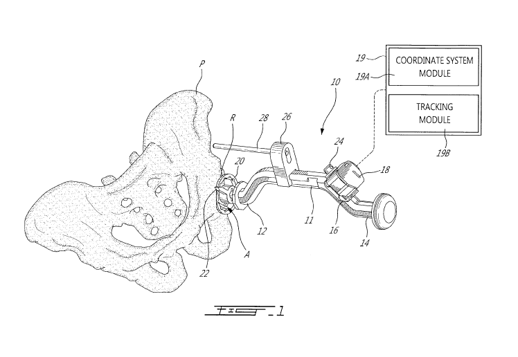
[0025] Fig. 1 is a perspective view of an acetabulum rim digitizer device
in
accordance with the present disclosure, relative to a pelvis;
[0026] Fig. 2 is an enlarged perspective view of a tooling end of the
acetabulum rim
digitizer device of Fig. 1;
[0027] Fig. 3 is an enlarged elevation view of a tooling end of an
acetabulum rim
digitizer device with planar surface in accordance with another embodiment of
the present
disclosure;
[0028] Fig. 4 is a perspective view of the acetabulum rim digitizer
device of Fig. 3;
and
[0029] Fig. 5 is a perspective view of an acetabulum rim digitizer device
with prongs
in accordance with another embodiment of the present disclosure.
- 5 -
CA 02974837 2017-07-25
WO 2016/123703 PCT/CA2016/050089
DESCRIPTION OF THE EXEMPLARY EMBODIMENTS
[0030] Referring to the drawings and more particularly to Figs. 1 and 2,
an
acetabulum rim digitizer device or instrument is generally shown at 10,
relative to a pelvis P
having an acetabulum A, the acetabulum having a rim R. The device 10 and
method related
to the device 10 may be used to determine pelvic orientation data in various
forms (e.g.,
pelvic tilt, anteversion/inclination of acetabulum, etc). The device 10 may
also be used to
accurately navigate instruments used in hip arthroplasty or like procedures,
including bone
model and cadaver testing, such as an acetabular reamer, a cup impactor, an
impactor
guiding pin, using inertial sensors.
[0031] The device 10 has an elongated body 11 having a tooling end 12 and
a
handle end 14. Although illustrated as having an axially offset portion, the
body of the
device 10 may also be fully straight or have any other appropriate shape.
[0032] The device 10 has a receptacle 16 for releasably receiving therein
an inertial
sensor unit 18, in a known manner. Alternatively, the inertial sensor unit 18
may be integral
or embedded into the elongated body 11. The inertial sensor unit 18 may have a
gyroscope
set to track the orientation of the device 10, by integrating the angular
velocity data recorded
by the sensor through a registration process. The inertial sensor unit 18 may
also comprise
an accelerometer set used to calibrate an initial position of the device 10,
and to correct
gyroscope drift when stable positions are recorded. Other types of inertial
sensors may be
provided in the inertial sensor unit 18 to complement the data or as
alternatives to the
accelerometer and/or gyroscope, such as inclinometers, magnetometers, among
other
possible inertial sensors.
[0033] The inertial sensor unit 18 uses its inertial sensor readings to
continually
calculate the orientation and velocity of a body without the need for an
external reference,
i.e., no signal transmission from outside of the sensor assembly is necessary,
the inertial
sensor unit 18 is self-contained. This process is commonly known as dead
reckoning and is
documented and forms part of the common general knowledge. An initial
orientation and
velocity must be provided to the inertial sensor unit 18, i.e., the X-Y-Z
coordinate system of
Fig. 1, after which the orientation is tracked by integrating the angular
rates of gyroscope
readings at each time step. With an accurate estimate of the orientation of
the inertial
sensor unit 18 with respect to the World frame of reference, gravitational
effects can be
- 6 -
CA 02974837 2017-07-25
WO 2016/123703 PCT/CA2016/050089
removed and inertial forces acting on the accelerometers can be integrated to
track changes
in velocity and position. Since the inertial sensor unit 18 has no need for an
external
reference, it may be immune to environmental factors such as magnetic fields
and operate
under a wide range of conditions.
[0034] The inertial sensor unit 18 is part of a computer-assisted hip
surgery system
for navigating instruments, used to implement the method 10, as will be
detailed below. The
system comprises a computer-assisted surgery (CAS) processing unit 19, that
may be a
stand-alone computer connected to the inertial sensor unit 18, for instance by
wireless
communication. It is however pointed out that the CAS processing unit may be
partially or
entirely integrated into the inertial sensor unit 18, also known as pod. The
inertial sensor
unit 18, when incorporating the CAS processing unit, may thus be equipped with
user
interfaces to provide the navigation data, whether it be in the form of LED
displays, screens,
numerical displays, etc. The computer-assisted surgery (CAS) processing unit
19 may have
a coordinate system module 19A and a tracking module 19B, described in further
detail
hereinafter, and part of a surgical assistance procedure programmed into the
CAS
processing unit 19.
[0035] A hemispherical base 20 is secured to the tooling end 12. The base
20 may
be releasably connected to the body 11 (e.g., by screwing engagement) to
enable the
selection of a base 20 of appropriate dimension, based on the acetabulum being
operated
on. The geometry of the base 20 may be known as quasi-hemispherical, frusto-
spherical,
etc. Indeed, as the base 20 is seated into the acetabulum during registration,
it is expected
that the base 20 is well seated in the acetabulum and does not shift position
during the
registration process. For this purpose, pressure sensor(s) may be provided on
or near the
surface of the base 20. The pressure sensor(s) provides signals that can be
monitored to
determine whether the base 20 is adequately applied against the surface of the
acetabulum.
[0036] The device 10 may additional comprise a tab 22, which is spaced
apart from
the base 20 and is designed to be seated on the acetabulum rim for each
acquired points,
as observed in Figs. 1 and 2. The device 10 may further have a rotation
indicator 24, used
to define a fixed rotation axis, not parallel to the rim plane normal, to
build a full coordinate
system for the acetabulum. In the illustrated embodiment, the rotation
indicator 24 is a light
source emitting a visible light beam, although other rotation indicators may
be used such as
- 7 -
CA 02974837 2017-07-25
WO 2016/123703 PCT/CA2016/050089
a mechanical arm, a laser, a marking on the instrument, or any other visual
indicator. A pin
guide 26 may also be provided as projecting laterally from the elongated body
11, featuring
a slot for guiding the insertion of a pin 28 in the pelvis, following the
registration. The
rotation indicator 24 is in a known physical orientation in the coordinate
system of the inertial
sensor unit 18.
[0037] The CAS processing unit is programmed with geometric data relating
the
body 11 (e.g., its axes) to the orientation of the components thereon, such as
the base 20,
the tab 22 and the rotation indicator 24. This geometric data, obtained pre-
operatively, is
used by the CAS processing unit (shown as 18) to perform the method and
sequence
described below.
[0038] Still referring to Fig. 1, the acetabulum rim digitizer device 10
may be used
intra-operatively with the following intraoperative method:
1. Either prior to or following reaming of the acetabulum A, the base 20 of
the device 10 is seated into the acetabulum A. The base 20 has been selected
and
installed to have a diameter complementary to that of the acetabulum A.
2. The rotation indicator 24 is used to give a predetermined orientation to
the device 10. Depending on the embodiment, this rotation indicator 24 may be
oriented to
point, mark, touch a pre-operatively identifiable landmark. For example, in
the case of the
pelvis, the identifiable landmark may be lateral anterior-superior iliac spine
(ASIS), the 12
o'clock feature of the acetabulum rim, the acetabulum notch, among other
features.
3. Registration may be initiated, through the user interface of the CAS
processing unit (e.g., button on the inertial sensor unit 18 is turned on).
4. Without unseating the base 20, for example as confirmed from the
pressure sensor(s) in the base 20 or by having the operator applying suitable
pressure on
the device 10, the device 10 is manually rotated to position the tab 22 onto a
different
segment of the acetabulum rim R (Fig. 2).
5. Either through a user request or through a stability criterion, the
inertial
sensor unit 18 records the current orientation of the digitizer device 10 and
provides
feedback to the user, for confirmation.
- 8 -
CA 02974837 2017-07-25
WO 2016/123703 PCT/CA2016/050089
6. The steps 4 ¨ 5 are repeated until a sufficient number of acetabulum
rim positions are recorded by the inertial sensor unit 18, for instance as
indicated by the
inertial sensor unit 18 or based on a predetermined number of measurements
required.
7. The CAS processing unit (e.g., incorporating the inertial sensor unit
18)
then records and provides data related to the acetabulum orientation or pelvic
tilt, in any
appropriate form (i.e., the pelvic coordinate system).
[0039] To perform the method described above, the CAS processing unit
must be
programmed in the following sequence:
1. The CAS processing unit sets the initial orientation of the acetabulum
rim digitizer device 10 when the user initiates the initial recording. This
initial position is
recorded by assuming arbitrary yaw, roll and pitch are provided by the
accelerometer set in
the inertial sensor unit 18. From this initial position, and knowing the
orientation of the
rotation indicator 24 relative to the rim digitizer device 10, the rotation
axis may be defined
as:
r 0 tat?. onA xzsin W orld
2. After initialization of the registration, the gyroscope set in the
inertial
sensor unit 18 is used to track the orientation of the acetabulum rim
digitizer device 10. The
orientation of the device 10 is recorded at the various points of contact
between the tab 22
and the acetabulum rim R. The inclination data (roll & pitch) provided by the
accelerometer
set in the inertial sensor unit 18 may be used to correct drift in the
gyroscope data (for
instance, using Kalman or Complementary filters). The collection of
orientation data at
various points provides the orientation of the rim digitizer device 10 in the
World coordinate
system:
rtmD is ttz grinW orld
3. At the various points of contact, with the stable orientation the
position
of the tab 22 may thus be calculated based on the orientation of the rim
digitizer device 10
obtained. Each of these positions is recorded in a coordinate system
maintained by the CAS
processing unit, and is representative of a point on the acetabulum rim R.
According to an
embodiment, the origin of the coordinate system is located at the center of
the
- 9 -
CA 02974837 2017-07-25
WO 2016/123703 PCT/CA2016/050089
hemispherical base 20. As such the position of each point on the rim can be
identified as
follow:
?trig otnthiW or = rtmDtgthzgrinW rid * tabini? tar-2.D tjtttzgrC Enitgr
4. When a sufficient number of points has been recorded, the rim points
registered can be used to define an acetabular rim plane. According to an
embodiment, a
plane is fitted through the rim points using an appropriate method such as
Least Squares
Fitting. This acetabular rim plane is therefore known:
rimPlaneArorrnalinWorld
5. The acetabular rim plane is used to build an acetabulum coordinate
system, as follows:
rnYAxs = rtmPlangN ormalinW or Id
rtn7.2Axs = rtrra Axts * rataftortAxtsinW orbs?
rtmFAxts = rtn-22.4xts * rtn-2.X.Axs
acgta &LE /trrnhiW or hs? = [nm..X.Axs rtffarAxts nn-22,4xs]
6. Using pre-operative planning data (CT-Scan, two-dimensional X-Rays,
3-D modeling, etc...), the pelvis coordinate system is created. Any standard
definition may
be used, for example the Lewinnek pelvic coordinate system. The pre-operative
planning
data may be referenced to the acetabulum coordinate system, using the same
landmarks
and rotation features as used during the registration method. Through data
inferred from the
pre-operative planning, the relationship between the acetabulum coordinate
system and the
pelvis coordinate system may be established. By inputting this relationship
into the
navigation system, the following relationship is obtained:
pelvisInWorld = acetabiThiminWorld* pelvisinAcetabiihm
7. The gravity axis of the World coordinate system may also be used to
determine the pelvic tilt from the computed pelvisInWorld coordinate system.
[0040] In the embodiment described above, the acetabular rim plane
acquired with
landmarks may be matched with a plane defined in pre-operative planning.
Alternatively, or
-10-
CA 02974837 2017-07-25
WO 2016/123703 PCT/CA2016/050089
additionally, the CAS processing unit may instead match the rim landmarks with
a surface
defined in pre-operative planning. This surface can be a 3D surface
representing the
acetabulum rim contour. The CAS processing unit can calculate using Least
Squares Fitting
the transformation on the acquired rim points which positions the points
closest to the pre-
planning contour of the acetabulum rim R.
[0041] The surface can also be a set of 2D contours, acquired using X-
Rays
images, combined with respective projective camera calibrations. In one
embodiment,
camera calibration could be performed as per F. CHERIET et al, Int. J. Patt.
Recogn. Artif.
Intel!. 13, 761 (1999). DOI: 10.1142/S0218001499000434 TOWARDS THE SELF-
CALIBRATION OF A MULTIVIEW RADIOGRAPHIC IMAGING SYSTEM FOR THE 3D
RECONSTRUCTION OF THE HUMAN SPINE AND RIB CAGE. The CAS processing unit
could compute by Least Squares Fitting the transformation on the acquired rim
points for
which a retro-projection of the points onto the X-Ray, as defined by the
projective camera
model, is closest to the defined 2D contour.
[0042] In another embodiment, an ultrasound device may be fixed to the
device 10,
for ultrasound readings to be obtained when the device 10 is seated into the
acetabulum A.
The ultrasound readings may be used to create the rim surface, and thus
replaces the tab
22 of the device 10, alleviating the need for physical contact with the
acetabulum rim R. As
the base 20 is seated into the acetabulum A and the ultrasound device is held
still relative to
the acetabulum, it is possible to rebuild the acetabulum rim surface
accurately in space
when combining the ultrasound data with the orientation data provided from the
inertial
sensor unit 18. This information can be used to match the registered rim
contour with the
pre-operative planned contour.
[0043] Using the device 10, the pin 28 may be positioned to a desired
orientation,
using the orientation data. For example, the pin 28 may be driven into the
pelvis so as to
serve as an impactor guide. The longitudinal axis of the pin 28 could thus be
driven to an
orientation parallel to a normal of the acetabulum rim plane. In an
embodiment, the
navigation of the device 10 for pin placement is done by providing anteversion
and
inclination values to the user.
[0044] Referring to Figs. 3 and 4, in yet another embodiment, a planar
surface 30, or
multiple coplanar features (three fixed tabs in Fig. 4), may be used as an
alternative to the
-11 -
CA 02974837 2017-07-25
WO 2016/123703 PCT/CA2016/050089
tab 22 at the tooling end 12 of the elongated body 11. The base 20 may or may
not be
present, although the base 20 may provide some manipulation stability to aid
in applying the
planar surface 30 to the acetabular rim R. The device 10 of Figs. 3 and 4
could be used to
acquire, in a single step, the planar surface as well as the rotation
landmark. The device 10
has a configuration that is planned to be in a unique complementary engagement
with the
rim of the acetabulum, for instance based on pre-operative imaging for
instance by having
patient specific contact surfaces being negatives of patient tissue for unique
complementary
engagement. With the embodiment of Figs. 3 and 4, steps 4-6 of the method
described
above would not be necessary, provided suitable pre-planning is performed.
Similarly, steps
2-4 of the sequence performed by the CAS processing unit are no longer
required. The
inertial sensor unit 18 may not need a gyroscope set for the embodiment of
Figs. 3 and 4.
[0045] Referring to Fig. 5, in yet another embodiment, three adjustable
prongs 50
may be used as alternatives to the planar surface 30, effectively forming a
tripod. When
used, the user is requested to position all of the prongs onto known landmarks
(e.g., unique
complementary engagement, based on pre-operative imaging). The prongs 50 have
abutment ends 51, displaceable axially (e.g., along a longitudinal axis of the
body 11) and in
rotation (e.g., about an axis parallel to the longitudinal axis of the body
11). Hence, the
prongs 50 are each provided with a housing 52 enabling lockable translational
DOF and
rotational DOF.
[0046] The known landmarks against which the ends 51 of the prongs 50 are
to be
abutted are either defined by identifiable anatomical landmarks, or by for
instance,
constraining the rotation of the instrument by using a stopper in the
acetabulum notch.
[0047] For the embodiment of Fig. 5, the pre-operative planning is be
used to define
the unique adjustments to the tripod to extend to the prongs 50, and to
identify the unique
position of the device 10 when positioned into the acetabulum A with the
predetermined
abutment between the prongs 50 and the pelvis P.
[0048] As the position of the device 10 of Fig. 5 is unique with regards
to the pelvis
anatomy, a single reading of an inclinometer in the inertial sensor unit 18
would be sufficient
to record the current pelvis tilt.
- 12 -
CA 02974837 2017-07-25
WO 2016/123703 PCT/CA2016/050089
[0049] A method used in combination with the device 10 of Fig. 5 would be
as
follows:
1. During the pre-operative planning, identify three landmarks to be used.
2. A coordinate system is created from these three landmarks, the
relationship between this coordinate system and pelvis coordinate system would
also be
known from pre-operative planning.
3. From the data computed from the pre-operative planning, the required
adjustments on the tripod would be performed to set the position and/or
orientation of each
prong 50.
4. The device 10 is then positioned in the manner shown in Fig. 5 onto the
pre-identified landmarks, either visually or by using a mechanical feature to
constrain
rotation.
5. When stable, the orientation data for the device 10 is recorded using
the inertial sensor unit 18, and this data is used to calculate the pelvis
tilt by using the
known relationship between the device 10 and the pelvis P.
pgiv[sInW or W = frEpodinW arid pglinsharpod
[0050] The device 10 of any of the preceding figures is therefore used to
provide a
means for intra-operatively evaluating the tilt of the pelvis and obtain
acetabular orientation
data, whether the surgery is performed in supine or lateral decubitus. The
data provided by
the CAS processing unit may be used, for instance, to reposition the pelvis
onto the table, to
guide the user in aligning a non-navigated instrument with a desired cup
alignment or be
used as an input for navigation of surgical instruments during total hip
arthroplasty
procedure. Although cross-products of axes are discussed above, vectors
representative of
a direction of the axes may be used for the cross-products.
[0051] As shown in Fig. 1, the CAS processor unit 19 may have a coordinate
system
module 19A and a tracking module 19B. Based on the embodiment the CAS
processor unit
19 supports, the modules 19A and 19B may have different functions. For
example, for the
embodiment of Figs. 1 and 2, the coordinate system module 19A sets a pelvic
coordinate
-13-
CA 02974837 2017-07-25
WO 2016/123703 PCT/CA2016/050089
system from readings of the inertial sensor unit 18 when the at least one
instrument 10 has
the acetabulum abutment end received in the acetabulum. The coordinate system
module
19A sets the pelvic coordinate system by obtaining a plurality of orientation
values from the
inertial sensor unit 18 when the rim abutment tab 22 is abutted against
locations of the rim.
One of the orientation values has the indicator 24 aligned with a reference
landmark. Thus,
the coordinate system module 19A defines an acetabular plane representative of
the pelvic
coordinate system from the plurality of orientation values. The tracking
module 19B then
tracks an orientation of inertial sensor units relative to the pelvic
coordinate system during
movements thereof using the readings from the inertial sensor units.
[0052] As another example, for the embodiment of Figs. 3-5, the
coordinate system
module 19A sets a pelvic coordinate system from readings of the inertial
sensor unit 18
when the instrument 10 has the acetabulum abutment end abutted against a rim
of the
acetabulum in the planned complementary manner. The coordinate system module
19A
sets the pelvic coordinate system by defining an acetabular plane
representative of the
pelvic coordinate system based on the planned complementary abutment. The
tracking
module 19B then tracks an orientation of inertial sensor units relative to the
pelvic
coordinate system during movements thereof using the readings from the
inertial sensor
units.
- 14-