Note: Descriptions are shown in the official language in which they were submitted.
LEG LENGTH CALCULATION IN COMPUTER-ASSISTED SURGERY
CROSS-REFERENCE TO RELATED APPLICATION
[0001] The present application claims the priority of United States
Provisional
Patent Application No. 62/110,861, filed on February 2, 2015.
TECHNICAL FIELD
[0002] The present disclosure relates to a system and method used in
Computer-
Assisted Surgery (CAS) to provide leg length discrepancy and offset
measurements,
for instance in hip surgery.
BACKGROUND OF THE ART
[0003] In orthopedic surgery, for instance hip replacement, leg
length discrepancy
is a change of leg length along the longitudinal axis of the patient, between
a
preoperative length and an intra-operative or post-operative length. Also in
hip
replacement, offset is the measurement of the translational shift of the leg
along a
medio-lateral axis of the patient, at the hip joint. Both these parameters are
relevant
during hip surgery, including total hip replacement, acetabular cup
implanting,
femoral implanting (e.g., head and neck implant, resurfacing). Hence, there is
a
need for systems and methods for determining leg length discrepancy and offset
that is minimally invasive yet precise and accurate.
SUMMARY
[0004] It is aim of the present disclosure to provide novel systems
and methods
for determining leg length discrepancy and offset to assess orthopedic hip
surgery.
[0005] Therefore, in accordance with a first embodiment of the
present
disclosure, there is provided a computer-assisted surgery system for
outputting at
least one of a leg length discrepancy and an offset between a preoperative leg
condition and a post-implant rejointing leg condition comprising: at least one
instrument; at least one inertial sensor unit connected to the at least one
instrument,
the inertial sensor unit producing readings representative of its orientation;
a
computer-assisted surgery processor unit operating a surgical assistance
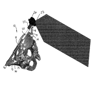
procedure
and comprising a coordinate system module for setting a pelvic coordinate
system
1
Date Recue/Date Received 2022-09-13
CA 02974850 2017-07-25
WO 2016/123704
PCT/CA2016/050090
from readings of the at least one inertial sensor unit when the at least one
instrument is in a given orientation relative to the pelvis, a tracking module
for
tracking an orientation of the at least one instrument relative to the pelvic
coordinate
system during movements thereof using the readings from the inertial sensor
unit on
the instrument, and a geometrical relation data module for recording
preoperatively
a media-lateral orientation of the at least one instrument representative of a
media-
lateral axis of the legs relative to the pelvic coordinate system and a
distance
between the legs along the medio-lateral axis, for recording after implant
rejointing
the media-lateral orientation and said distance, and for calculating at least
one of a
leg length discrepancy and an offset, based on said distances and said medic-
lateral orientations; an interface for outputting at least the leg length
discrepancy or
the offset between the preoperative leg condition and the post-implant
rejointing leg
condition.
[0006] Further in accordance with the first embodiment, the at least one
instrument is a caliper having a body with a translational joint for
expanding/contracting, and legs configured for abutment with pelvic landmarks.
[0007] Still further in accordance with the first embodiment, the at
least one
instrument supports a light source emitting a light beam that is perpendicular
relative
to a direction of the translational joint.
[0008] Still further in accordance with the first embodiment, the light
source is
displaceable along the body, the light beam being a leg alignment marker when
the
caliper is abutted against the pelvic landmarks.
[0009] Still further in accordance with the first embodiment, the given
orientation
has a direction of the translational joint parallel to a medic-lateral axis of
the pelvis.
[0010] Still further in accordance with the first embodiment, an ankle
clamp has
ankle interfaces configured to remain fixed to the ankles, with linkages
interconnecting the ankle interfaces.
[0011] Still further in accordance with the first embodiment, a scale in
the
linkages measures the distance.
2
CA 02974850 2017-07-25
WO 2016/123704
PCT/CA2016/050090
[0012] Still further in accordance with the first embodiment, the
linkages include
at least a translational joint in a direction generally aligned with a medic-
lateral axis
between the legs.
[0013] Still further in accordance with the first embodiment, indicators
are
provided for receiving ends of the caliper for recording the medic-lateral
orientation
with the caliper abutted against the ankle clamp.
[0014] Still further in accordance with the first embodiment, the at
least one
instrument is an acetabular-implant impactor, and wherein the impactor
supports a
light source emitting a light beam having a known orientation relative to a
longitudinal axis of the impactor.
[0015] Still further in accordance with the first embodiment, the given
orientation
has the light beam illuminating the medic-lateral axis of the pelvis, with a
shaft of the
impactor lying in a plane of the light beam.
[0016] Still further in accordance with the first embodiment, an ankle
clamp has
ankle interfaces configured to remain fixed to the ankles, with linkages
interconnecting the ankle interfaces, the ankle clamp further comprising
indicators
for being illuminated by the light beam for recording the medic-lateral
orientation.
[0017] Still further in accordance with the first embodiment, a scale is
in the
linkages to measure the distance.
[0018] In accordance with a second embodiment of the present disclosure,
there
is provided a computer-assisted surgery system for outputting at least one of
a leg
length discrepancy and an offset between a preoperative leg condition and a
post-
implant rejointing leg condition comprising: at least one instrument; at least
one
inertial sensor unit connected to the at least one instrument, the inertial
sensor unit
producing readings representative of its orientation; a computer-assisted
surgery
processor unit operating a surgical assistance procedure and comprising a
coordinate system module for setting a pelvic coordinate system from readings
of
the at least one inertial sensor unit when the at least one instrument is in a
given
orientation relative to the pelvis, a tracking module for tracking an
orientation of the
at least one instrument relative to the pelvic coordinate system during
movements
thereof using the readings from the inertial sensor unit on the instrument,
and a
geometrical relation data module for recording preoperatively a landmark
orientation
3
CA 02974850 2017-07-25
WO 2016/123704
PCT/CA2016/050090
relative to the pelvic coordinate system and a distance when the at least one
instrument has a first end abutted to a pelvic landmark and a second end
abutted to
a leg landmark, for recording after implant rejointing the landmark
orientation and
said distance, and for calculating at least one of a leg length discrepancy
and an
offset, based on said distances and said landmark orientations; an interface
for
outputting at least the leg length discrepancy or the offset between the
preoperative
leg condition and the post-implant rejointing leg condition.
[0019] Further in accordance with the second embodiment, the at least
one
instrument is a caliper having a body with a translational joint for
expanding/contracting, and legs configured for contacting the pelvic landmark
and
the leg landmark.
[0020] Still further in accordance with the second embodiment, the
caliper
supports a light source emitting a light beam that is perpendicular relative
to a
direction of the translational joint.
[0021] Still further in accordance with the second embodiment, the given
orientation has the light beam illuminating the medio-lateral axis of the
pelvis.
[0022] Still further in accordance with the second embodiment, a scale
is on the
translational joint to obtain said distances.
[0023] Still further in accordance with the second embodiment, the at
least one
instrument includes a mechanical gauge having body with a translational joint
for
expanding/contracting, and bores configured for being connected to pins
constituting
the pelvic landmark and the leg landmark.
[0024] Still further in accordance with the second embodiment, a scale
is on the
translational joint to obtain said distances.
[0025] Still further in accordance with the second embodiment, the at
least one
instrument includes an acetabular-implant impactor supporting the inertial
sensor
unit, and wherein the impactor supports a light source emitting a light beam
having a
known orientation relative to a longitudinal axis of the impactor.
4
CA 02974850 2017-07-25
WO 2016/123704
PCT/CA2016/050090
[0026] Still further in accordance with the second embodiment, the given
orientation has the light beam illuminating the medio-lateral axis of the
pelvis, with a
shaft of the impactor lying in a plane of the light beam.
[0027] Still further in accordance with the second embodiment, the
landmark
orientation has the light beam illuminating a longitudinal axis of the
mechanical
gauge, with a shaft of the impactor lying in a plane of the light beam.
[0028] In accordance with the third embodiment of the present
disclosure, there
is provided a method for repeating a leg alignment between a preoperative leg
condition and a post-implant rejointing leg condition, comprising: pre-
operatively,
with the patient in supine decubitus, orienting a light source using landmarks
on the
pelvis to produce a light beam aligned with a transverse plane of the pelvis,
positioning at least one of the legs of the patient in alignment with the
light beam,
and setting landmarks on the legs of the patient, distally from the pelvis;
post post-
implant rejointing, with the patient in supine decubitus, repeating the
orienting and
the positioning, and noting a movement of the ladmarks.
[0029] Still further in accordance with the third embodiment, setting
landmarks on
the legs of the patient comprises projecting a light beam from a landmark on a
first
of the legs onto a scale on a landmark on a second of the legs.
[0030] Still further in accordance with the first embodiment, noting a
movement of
the landmarks comprises at least noting a displacement of the light beam on
the
scale.
[0031] Still further in accordance with the first embodiment, wherein
noting a
movement of the landmarks comprises at least noting a variation of distance
between the landmarks.
DESCRIPTION OF THE DRAWINGS
[0032] Fig. 1 is a perspective view of a caliper instrument on a pelvis
during a leg
positioning technique;
[0033] Fig. 2 is a perspective view of the caliper instrument on a
mechanical
ankle clamp;
CA 02974850 2017-07-25
WO 2016/123704
PCT/CA2016/050090
[0034] Fig. 3 is a perspective view of an impactor using in leg length
and offset
measurement relative to a pelvis;
[0035] Fig. 4 is a perspective view of an impactor using in leg length
and offset
measurement relative to the mechanical ankle clamp;
[0036] Fig. 5 is a perspective view of a pinned mechanical gauge;
[0037] Fig. 6 is an enlarged perspective view of the pinned mechanical
gauge;
[0038] Fig. 7 is a perspective view of a pinned mechanical gauge and
impactor;
[0039] Fig. 8 is a perspective view of the pinned mechanical gauge and
impactor;
[0040] Fig. 9 is a perspective view of the pinned mechanical gauge and
caliper
instrument;
[0041] Fig. 10 is an enlarged view of the scale on the caliper
instrument;
[0042] Fig. 11 is a perspective view of the mechanical ankle clamp with
light
source;
[0043] Fig. 12 is an enlarged view of a scale on the mechanical ankle
clamp; and
[0044] Fig. 13 is a block diagram showing a computer-assisted surgery
system
operating with instruments to calculate leg length discrepancy and offset, in
accordance with the present disclosure.
DETAILED DESCRIPTION
[0045] In the proposed disclosure, the leg length discrepancy and offset
measurements are resolved using basic trigonometry. Leg length discrepancy
and/or offset are measured to quantify the post-operative gait of the patient,
to
diagnose a patient condition, to assist in a physiotherapy treatment, or even
to
perform corrective actions intra-operatively, among numerous other
possibilities.
The measurements may be performed on a patient during hip replacement surgery,
or can be performed on a bone model or cadaver. In general, the distance
measurements are obtained based on the readings from mechanical instruments.
The use of inertial sensors may assist in giving precision and accuracy to the
afore-
mentioned measurements. For example, as shown in Fig. 1, a caliper instrument
10
6
may be used. The caliper instrument 10 is described in US Patent Application
Publication No. 2014/0031829 and uses inertial sensor technology.
[0046] As
shown in Fig. 1, the caliper instrument 10 may be used as part of a
bone digitizer in a bone digitizing system, to create a frame of reference for
subsequent navigation of tools relative to bones in surgery, for instance
based on
the determination of the medio-lateral axis of the pelvis. The instrument 10
is
referred to as a caliper, as it features a pair of legs 12 movable relative to
one
another, e.g., in a telescopic manner. The
expression "caliper" is used
nonrestrictively. Any other appropriate expression may be used to describe the
instrument 10, such as medio-lateral digitizer.
[0047] In the
illustrated embodiment, the legs 12 of Fig. 1 each comprise a
translational joint 13 so as to be expandable or contractible along the Y
axis. For
instance, the translational joints 13 may be any of sliding joint, telescopic
joint,
prismatic joint, indexing joint, etc. As an alternative, a single one of the
legs may
have a joint. It is also considered to use rotational joints as an alternative
to
translational joints 13, with an axis of the rotational joint being normal to
a plane of
the caliper instrument 10. A locking mechanism is typically provided, to lock
the
translational joints 13 and, therefore, set the legs 12 in a selected length.
The free
end of each leg 12 has an abutment end 14, for which any appropriate shape is
considered, such as flat contact surfaces, discs, various concavities or
convexities,
pointy ends, etc., as a function of the type of bone or bodily part the
caliper
instrument 10 will be contacting. The flat ends 14 of Fig. 1 are well suited
to be
used with a pelvis, with the ends 14 contacting the anterior superior iliac
spines
(ASIS) on opposite sides of the pelvis, in pelvic surgery, with the patient in
supine
decubitus. Alternatively, the caliper instrument 10 could be used for the
posterior
superior iliac spine as well, among other possibilities.
[0048] Still
referring to Fig. 1, the legs 12 are interconnected by an elongated
body 20 of the caliper instrument 10. The elongated body 20 features a
translational
joint 21 such that the elongated body 20 is expandable or contractible along
the X
axis. The translational joint 21 may be any appropriate joint, such as
translational
joints, telescopic joint, prismatic joints and/or indexing joints. It is also
considered to
use rotational joints as an alternative to the translational joint 21.
7
Date Recue/Date Received 2022-09-13
CA 02974850 2017-07-25
WO 2016/123704
PCT/CA2016/050090
[0049] A locking mechanism may be provided, thereby allowing the user to
set
the length of the elongated body 20. An inertial sensor support or receptacle
23 is
defined on the elongated body 20. The inertial sensor support 23 is, for
instance,
made with a specific geometry in order to precisely and accurately accommodate
an
inertial sensor unit in a predetermined complementary connection, simplifying
an
initialization between an inertial sensor unit 26 (Fig. 2) and caliper
instrument 10.
For instance, the inertial sensor unit has a preset orientation that is
aligned with a
dimension of the caliper instrument 10. In other words, the mechanical
constraints
in the attachment of inertial sensor unit in the support 23 are such that the
three
axes of the inertial sensor unit are aligned with the X, Y and Z axis of the
caliper
instrument 10. Therefore, the caliper instrument illustrated in Fig. 1 may
expand and
contract along both the X axis and the Y axis. A light source 24 is also
provided on
the caliper instrument 10. The light source 24 is of the type producing a
planar
beam, such that a projection of the planar beam on a surface produced a line.
The
light source 24 may be on a carriage 25 so as to be displaceable in
translation along
the elongated body 20. Alternatively, it is considered to configure the
carriage 25 to
be snap-fitted to the elongated body 20, so as to allow its installation at
any position
along the elongated body 20.
[0050] The inertial sensor unit 26 used with the caliper instrument 10
may have
any appropriate type of inertial sensor, to provide 3-axis orientation
tracking. For
instance, the inertial sensor unit may have sets of accelerometers and/or
gyroscopes, etc. The inertial sensor unit may be known as a sourceless sensor
unit,
as a micro-electromechanical sensor unit, etc. As mentioned above, the
inertial
sensor unit is matingly received in the inertial sensor support 23 in a
predetermined
complementary connection, such that the initializing of the inertial sensor
unit will
have the inertial sensor unit specifically oriented relative to the X-Y-Z
coordinate
system illustrated in Fig. 1 (with the Z axis being the cross-product of the X
and Y
axes).
[0051] The inertial sensor unit 26 uses inertial sensor readings to
continually
calculate the orientation and velocity of a body without the need for an
external
reference, i.e., no signal transmission from outside of the sensor assembly is
necessary, the inertial sensor unit 26 is self-contained. This process is
commonly
known as dead-reckoning and forms part of the common general knowledge. An
initial orientation and velocity must be provided to the inertial sensor unit
26, i.e., the
8
CA 02974850 2017-07-25
WO 2016/123704
PCT/CA2016/050090
X-Y-Z coordinate system of Fig. 1, after which the orientation is tracked by
integrating the angular rates of gyroscope readings at each time step. With an
accurate estimate of the orientation of the inertial sensor unit 26 with
respect to the
Earth frame of reference, gravitational effects can be removed and inertial
forces
acting on the accelerometers can be integrated to track changes in velocity
and
position. Since the inertial sensor unit 26 has no need for an external
reference, it
may be immune to environmental factors such as magnetic fields and operate
under
a wide range of conditions.
[0052] Referring to Fig. 2, a mechanical clamp 30 is illustrated. The
mechanical
clamp 30 has ankle hoops 31 or like ankle attachments or interfaces, separated
by a
lockable translation joint 32. Hence, a distance between the ankle hoops 31
may be
adjusted. The distance between the two ankle hoops 31 can be read from a scale
on
the joint 32. The ankle hoops 31 are illustrated as being inverted U-shaped
structures. According to an embodiment, the hoops 31 each abut against the
pair of
ankle malleoli, such that the interconnection between the hoop 31 and
respective
ankle is stable and reproducible. For this purpose, the hoop 31 may have
cavities
31A to accommodate the malleoli. Other configurations are considered,
including
different shapes for the hoops 31, with straps, other joint sets, etc.
[0053] The mechanical clamp 30 may have visual indicators 33 to receive
therein
the ends 14 of the caliper instrument 10 in the manner shown in Fig. 2, to use
the
scale of the caliper instrument 10, and also ensure precise and reproducible
alignment between caliper instrument 10 and mechanical clamp 30, such that the
interconnection between the caliper instrument 10 and the mechanical clamp 30
is
reproducible from a pre-operative to a post-operative interaction. The visual
indicators 33 may identify the center of two malleoli on both ankles, when the
mechanical clamp 30 is used. Moreover, the ankle hoops 31 may translate
longitudinally with respect to one another (i.e., along the leg), by way of
lockable
translational joint 34. Other types of joints (i.e., linkages) may also be
used to allow
relative movement between the ankle hoops 31 and the lockable translation
joint 32.
For example, the lockable translation joint 32 may have hinges at its ends, by
which
it would be connected to the ankle hoops 31. Accordingly, the ankle interfaces
31
may remain in a fixed relation with the ankles, while the various joints
described
herein allow relative movement between the ankles. The visual indicators 33
are
9
positioned such that any relative movement between a pre-operative condition
and a
post implant rejointing condition can be quantified as described below.
[0054] Referring to Figs. 5, 6, 7 and 8, a mechanical gauge in
accordance with
the present disclosure is shown at 40, and is another of the instruments that
may be
used to implement the method of the present disclosure. The mechanical gauge
40
is of the type using a pair of pins 41, though pin holes 42 located at opposed
ends of
the mechanical gauge 40. A scale 43 is provided on a lockable translational
joint 44
of the gauge 40. Accordingly, the mechanical gauge 40 can be used to measure
distances. In an embodiment, the mechanical gauge 40 is biased to a zero
reading
on the scale 43.
[0055] Referring to Figs. 3, 4, 7 and 8, an impactor is shown at 50.
The impactor
50 is of the type used in impacting an acetabular cup implant in the
acetabulum, for
instance as described in in PCT International Publication No. WO 2014/197988.
The impactor 50 may be used as one of the instruments to measure the leg
length
discrepancy and the offset, for the simple reason that may already be used for
the
implant procedure. The impactor 50 has the light source 51 allowing its
alignment,
and an inertial sensor unit 52 similar to the unit 26, containing a gyroscope
for dead-
reckoning.
[0056] Referring to Fig. 13, a system for navigating the instruments
described
above in computer-assisted hip surgery is generally shown at 100, and is of
the type
used to implement the method detailed below. In an embodiment, the system 100
is
used for assisting the user in performing hip surgery, but also has the
modules to
perform the leg length discrepancy and offset calculations described herein.
The
system 100 comprises a computer-assisted surgery (CAS) processing unit 102.
The
CAS processing unit 2 may be integrated into one or more inertial sensor units
26
and 52, also known as pods that are mounted to the various instruments of the
system 100, or as a module of a computer or portable device, among other
possibilities.
[0057] The inertial sensor units 26 and 52 incorporate the processing
unit 102
and may thus be equipped with a user interface(s) 103 to provide the
navigation
data, whether it be in the form of LED displays, screens, numerical displays,
etc.
Alternatively, the inertial sensor unit 26 and 52 may be connected to a stand-
alone
processing device B that would include a screen or like monitor, to provide
additional
Date Recue/Date Received 2022-09-13
CA 02974850 2017-07-25
WO 2016/123704
PCT/CA2016/050090
display capacity and surface. By way of example, the processing device B is a
wireless portable device such as a tablet in a wired or wireless communication
with
the inertial sensor unit 26/52.
[0058] The inertial sensor unit 26/52 may be known as micro-electro-
mechanical
sensors (MEMS) and may include one or more of inertial sensors, such as
accelerometers, gyroscopes, magnetometers, among other possible inertial
sensors.
The inertial sensors are sourceless sensors automatically providing data
influenced
by natural phenomena, such as gravity. The inertial sensor unit A also have a
body,
typically defined by a casing, giving the inertial sensor unit A, by which the
inertial
sensor unit A may be secured to the instruments.
[0059] The processing unit 102 comprises different modules to perform
the
navigation. A surgical flow module 102A may be used in conjunction with the
user
interface 103 or a processing device B to guide the operator through the steps
leading to the navigation. This may entail providing a step-by-step guidance
to the
operator, and prompting the operator to perform actions, for instance pressing
on a
"record" interface that is part of the interface 103 or entering data as
measured from
the scales of the caliper instrument 10 or mechanical gauge 40, for the system
100
to record instant orientations and position data. While this occurs throughout
the
surgical procedure, the prompting and interactions between the system 100 and
the
user will not be described in a remainder of the description, as they will
implicitly
occur. It is contemplated to have the surgical flow module 102A present in the
processing device B, with concurrent action between the inertial sensor unit A
and
the processing device B to guide the operator during the measuring procedures
detailed below, and with a communication with the operator to record the
progress of
the procedure.
[0060] A tracking module 102B may also be part of the processing unit
102. The
tracking module 102B receives readings from the inertial sensors 26/52, and
converts these readings to useful information, i.e., the navigation data. As
described
above, the navigation data may be orientation data relating an instrument to
the
pelvis. The tracking module 102B may perform dead-reckoning to track the
inertial
sensors 26/52, as described below.
[0061] The coordinate system module 1020 creates the coordinate system.
The
coordinate system is the virtual frame by which the orientation of the
instruments
and tools is related to the orientation of the bone. For example, the
coordinate
11
CA 02974850 2017-07-25
WO 2016/123704
PCT/CA2016/050090
system module 102C sets a pelvic coordinate system from readings of the
inertial
sensor 26/52 when instruments are in a given orientation relative to the
pelvis.
[0062] In order to output the record orientations at discrete desired
orientations
and calculate offset and leg length discrepancy, via the user interface 103 or
processing device B, the processing unit 102 may be preprogrammed with
geometrical relation data module 102D. The geometrical relation data module
102D
is therefore used to record orientations of the various instruments supporting
the
inertial sensors 26/52, and uses these orientations along with distances to
calculate
the leg length discrepancy and/or the offset.
[0063] The inertial sensor units 26/52 are designed such that they are
connected
in single possible orientation to the instruments and tools, such that the
orientation
of the inertial sensor units 26/52 is known relative to the instruments and
tools to
which it is connected when turned on. By way of the connector 5, the inertial
sensor
units A may be portable and detachable units, used with one device/instrument,
and
then transferred to another device/instrument, preserving in the process
orientation
data of the global coordinate system, using dead-reckoning.
[0064] The geometrical relation data module 102D is programmed for
specific
use with the devices and instruments described herein. Accordingly, when an
inertial sensor unit is mounted to one of the devices and instruments, the
relation
between the device/instrument and a coordinate system of the inertial sensor
unit is
known (in contrast to a global coordinate system) by the geometrical relation
data
module 102D. For example, the relation may be between an axis or a 3D
coordinate
system of the device/instrument and the coordinate system of the inertial
sensor unit
A.
[0065] The navigation of instruments is intended to mean tracking at
least some
of the degrees of freedom of orientation in real-time or quasi-real time, such
that the
operator is provided with navigation data calculated by computer assistance.
The
inertial sensors A used in the following method may be interrelated in the
global
coordinate system (hereinafter, coordinate system), provided appropriate steps
are
taken to record or calibrate the orientation of the inertial sensors A in the
coordinate
system. The coordinate system serves as a reference to quantify the relative
orientation of the different items of the surgery, i.e., the instruments and
devices
relative to the pelvis.
12
CA 02974850 2017-07-25
WO 2016/123704
PCT/CA2016/050090
[0066] The present application contemplates different techniques to
provide the
leg length and offset measurements. In general, the techniques each comprise
two
procedures, i.e., leg positioning, and taking the leg length and/or offset
measurements. The following paragraphs set out different techniques to measure
leg length discrepancy and offset, between a pre-operative condition, and a
post-
operative condition, using some of the instruments described above. For
clarity, the
expression post-operative is used herein as representative of a part of the
procedure
after positioning of the implant on the bone, when the leg can be rejointed,
i.e. post-
implant rejointing. However, post-operative includes intra-operative, in that
the
measurements may be taken before the end of the procedure, to allow corrective
measures to be taken, for example. Hence, throughout the text, the use of the
expression "post-operative" includes intra-operative interventions. The
techniques
that do not use the mechanical gauge 40 are non-invasive, in that they may be
used
over the skin, or in that they do not require patient tissue alterations other
than the
ones required for surgery.
[0067] Procedure of leg positioning
[0068] The purpose of this procedure is to position or reposition the
leg along the
longitudinal axis of the patient (a.k.a., cranial-caudal axis), in a
reproducible manner.
If the leg is laid flat on the table, this leg positioning may enable
alignment of the leg
with the frontal plate of the patient. In order to measure offset and leg
length
discrepancy precisely and accurately, the leg positioning must be replicated
between measurements. The impact on the measurements of the leg length
discrepancy introduced by misalignment of the leg is minimized by the use of
this
procedure. The procedure is performed as follows:
1. The patient is placed in supine decubitus.
2. Referring to Fig. 1, the caliper instrument 10 is placed on two pelvic
landmarks,
after being telescopically arranged to have a suitable length. For example,
the
caliper instrument 10 is placed on the two anterior-superior iliac spines, in
the
manner shown in Fig. 1. An assumption is made that the caliper instrument 10
is aligned with the media-lateral axis of the patient. A light beam is shone
from
the light source 24 that is attached to the caliper instrument 10. The light
source
24 is connected to the frame of the caliper instrument 10 such that the light
beam is projected distally and perpendicular to the frame of the caliper
13
CA 02974850 2017-07-25
WO 2016/123704
PCT/CA2016/050090
instrument 10, and therefore parallel to the longitudinal axis of the patient,
a.k.a., the cranial-caudal axis, in direction Z of Fig. 1. The user is
required to
align the first leg with the projected light beam, by manually displacing it.
3. Different approaches are considered for the alignment. For instance, as the
light beam produces a line, the user may align the light beam line with leg
landmarks. For example, a center of the knee cap and a center of the ankle to
be shone by the light beam line. Temporary pen or ink markings may be made
on the knee and/or ankle to indicate the landmarks used for alignment.
4. The light source 24 is then slid along the caliper instrument 10, using
carriage
25. The light beam is therefore translated laterally. As a result, the second
leg
can be aligned in the same way, by manually displacing it, as guided by the
selected landmarks. Since the light beam is perpendicular to the caliper
instrument 10 ¨ and hence also perpendicular to the medio-lateral axis of the
patient -, the light beam indicates the projection of the sagittal plane on
the
patient. As an alternative to assuming this, the table plane can be assessed
by
a pod to determine if the table plane is leveled. Once aligned using the light
source 24 in the manner described above, the assumption is made that the legs
are physically aligned with respect to the longitudinal axis. Moreover, as the
patient is in supine decubitus, it can be assumed that the legs are within the
frontal plane. As a result, the leg is along the longitudinal axis (i.e. the
intersection of both sagittal and frontal planes). Based on these assumptions,
the leg length discrepancy can be measured along the longitudinal axis. The
offset can be measured along the medio-lateral axis. This is achieved by
comparing data obtained from the instruments described above, between pre-
operative measurements, and intra-operatively and/or post-operative
measurements.
[0069] Procedure: leg length discrepancy and/or offset measurements
[0070] Numerous techniques are possible for this procedure, as described
below
with reference to the figures.
[0071] Technique 1: the instruments required are the caliper instrument
10, or
alternatively the impactor 50, with light source 24 and dead-reckoning of the
inertial
sensor unit 26 or 52, to measure leg length discrepancy.
14
1. Referring to Fig. 2, with the patient's legs positioned using the leg
positioning
procedure mentioned above, the mechanical clamp 30 is rigidly attached to the
ankles of the patient;
2. The first medio-lateral axis, i.e., that of the pelvis, is acquired by
using the
caliper instrument 10 in the manner described above, or the impactor 50. The
impactor 50 may be navigated to determine the medio-lateral axis, for instance
as described in PCT International Publication No. WO 2014/197988. For
example, the light beam of the light source is in a known relation relative to
a
shaft of the impactor 50;
3. After acquiring the pelvic medio-lateral axis, the caliper instrument 10 is
moved
to the ankles to acquire a second medio-lateral axis, near the feet. For
example,
the second medio-lateral axis, i.e., the leg medio-lateral axis, may be
defined by
the line connecting the two centers of both ankles (as in Fig. 2), thus making
use of the visual indicators of the mechanical clamp 30 to physically provide
these landmarks. In the arrangement of Fig. 2, the caliper instrument 10 is in
a
position to record the medio-lateral axis at the ankle;
4. In the acquisition of the medio-lateral axes, the inertial sensor unit
26 attached
to the caliper instrument 10 (or impactor in alternative embodiment) contains
a
gyroscope. The gyroscope will provide data that is then used by a CAS
processing unit to perform dead-reckoning and hence acquire the relative
orientation between the two medio-lateral axes, unman, i.e., at the hip (Fig.
1)
and at the ankles (Fig. 2);
5. During or upon finishing the hip surgery, with the operated leg rejointed,
the
angle
between two medio-lateral axes is obtained by repeating steps 1-
4. The same leg positioning technique is used prior to taking the measurements
to ensure the legs are positioned in the same way as preoperatively, i.e.,
parallel to the sagittal plane;
6. Based on the known distance between the two ankles (D) as obtained from the
scale on the joint 32 (e.g., scale 21) and the angular difference in a, the
leg
length discrepancy can be resolved as: D - .
nOrtn). A
positive value would mean a longer leg post-operatively, whereas a negative
value would mean a shorter leg post-operatively. Figs. 3 and 4 illustrate
technique 1 using the impactor alternative. It should be noted that D may vary
between a pre-operative measurement and a post-operative measurement,
whereby the first D in the solution is D measured post-operatively and the
Date Recue/Date Received 2022-09-13
CA 02974850 2017-07-25
WO 2016/123704
PCT/CA2016/050090
second D in the solution is D measured pre-operatively. It is also
contemplated
to fix the D, whereby step 5 would not require repositioning the leg as in
step 1.
[0072]
Technique 2: caliper instrument 10 is used for this technique, to measure
the offset.
1. The patient's legs are positioned using the leg positioning technique
described
above, sliding the light source 24 on the caliper instrument 10 to align the
projected light beam on both legs, as in Fig. 1;
2. The readings from the inertial sensor unit 26 on the caliper instrument 10
are
recorded (oitmelm);
3. Upon finishing the hip surgery, steps 1-2 are repeated to acquire the
readings
(am:0d from the inertial sensor unit on the caliper instrument 10;
4. The offset can be resolved as: Oveaum
igyortni, positive value indicates an
increase in the offset and negative value indicates a decrease in the offset.
[0073]
Technique 3: this technique uses the mechanical measuring gauge 40 and
dead-reckoning.
1. The patient's legs are positioned using the leg positioning technique.
2. Prior to cutting the femoral neck and preparation of the acetabulum, as
shown in
Figs. 5 and 6,a first pin 41 is fixed on the ASIS and another pin 41 is fixed
on
the greater trochanter area; both pins 41 are on the operated side. The pins
41
respectively constitute a pelvis landmark and a leg landmark;
3. The mechanical gauge 40 is fixed to the two pins 41, and the distance M
between the two pins 41 is known from the scale 42 of the gauge 40;
4. The impactor 50 as shown in Fig. 7 is used and firstly aligned with the
medio-
lateral axis (using the light source 51 thereof to project a light beam on the
two
ASIS); then, the impactor 50 is aligned using the light source 51 with the
long
axis of the mechanical gauge 40, as in Fig. 8, showing a landmark orientation.
The inertial sensor unit 52 containing a gyroscope to perform dead-reckoning
to
acquire the angle (#1,1v21,õ note j4 Vg 'IT
/2) between the medic-lateral axis
and the long axis of the mechanical gauge 40;
5. The gauge 40 is removed, while the pins 41 are kept on the femur and
pelvis, at
which point the user may proceed with the femoral procedure;
16
CA 02974850 2017-07-25
WO 2016/123704
PCT/CA2016/050090
6. Upon finishing placing the femoral implant and/or the acetabular
component and
rejointing the leg (intra operatively or post operatively), the distance of
the
gauge 40 is adjusted, and the gauge 40 is reattached to the two pins 41. The
angle (ginutcp., note g my ffiZ) are acquired between the medio-lateral
axis and the long axis of the gauge 40 by repeating step 4-5; The same leg
positioning procedure is used beforehand to make sure the legs are positioned
in the same way as preoperatively;
7. The leg length discrepancy can be
resolved as:
girskia,p.p.a.") ¨bi =gint4rivid ; The offset can be resolved as:
LYI =047-sarlit) ¨ =acricApiraid = It should be noted that M may vary
between a pre-operative measurement and a post-operative measurement,
whereby the first M in the solution is M measured post-operatively and the
second M in the solution is M measured pre-operatively.
[0074]
Technique 4: this technique involves the caliper instrument 10 for a direct
measurement of leg length discrepancy (proximal)
1. The patient's legs are positioned using the leg positioning procedure
described
above;
2. The ends 14 of the caliper instrument 10 are placed on the ASIS of the
operated
side and on a marked reference on the skin on the femur (e.g., a landmark on
the skin), as shown in Figs. 9 and 10, respectively the pelvic landmark and
the
leg landmark. The light source 24 is displaced to project its beam on the
opposite ASIS, when selecting the marked reference;
3. The initial distance measurement is recorded on the caliper instrument 10
(lifprevid;
4. Upon finishing the surgery, the distance measurement is obtained using the
caliper instrument 10 (N9.119), after repeating the leg positioning procedure,
and by repeating steps 2 and 3;
5. The leg length discrepancy can be resolved as: pfin,auv. ¨ Nitiptev.
[0075]
Technique 5: Direct measurement of leg length discrepancy (distal), using
the caliper instrument 10, the mechanical clamp 30 and using one of the light
sources 24 or 51.
17
CA 02974850 2017-07-25
WO 2016/123704
PCT/CA2016/050090
1. The patient's legs are positioned using the leg positioning procedure
described
above;
2. The mechanical clamp 30 is placed on both ankles, in the manner shown in
Fig. 2;
3. The light source 51 is connected to the mechanical clamp 30, and is
projected
on a scale 70 on the operated leg to record the initial leg length (ncuev,d,
as
shown in Fig. 11. Surgery may be initiated, however, the translational joint
34 is
unlocked to allow the translation of the ankle hoops 31 relative to one
another;
4. Upon finishing the surgery, the distance measurement is obtained using the
mechanical clamp 30 (X59atc.ti) by repeating the steps 1-3; and
5. The leg length discrepancy is resolved as: zwamci, ¨
18