Note: Descriptions are shown in the official language in which they were submitted.
- 1 -
ULTRASONIC SURGICAL INSTRUMENT
This application is a divisional of Canadian Patent Application No. 2,582,520,
filed on
October 7, 2005.
Field of the Invention
The present invention relates, in general, to ultrasonic surgical instruments
and, more
particularly, to an ultrasonic surgical clamp coagulator apparatus
particularly configured to
provide increased tissue transaction forces.
Background of the Invention
Ultrasonic surgical instruments are finding increasingly widespread
applications in surgical
procedures by virtue of the unique performance characteristics of such
instruments.
Depending upon specific instrument configurations and operational parameters,
ultrasonic
surgical instruments can provide substantially simultaneous cutting of tissue
and
homeostasis by coagulation, desirably minimizing patient trauma. The cutting
action is
typically effected by an end-effector at the distal end of the instrument,
which transmits
ultrasonic energy to tissue brought into contact with the end-effector.
Ultrasonic instruments
of this nature can be configured for open surgical use, laparoscopic or
endoscopic surgical
procedures including robotic-assisted procedures.
Ultrasonic surgical instruments have been developed that include a clamp
mechanism to
press tissue against the blade of the end-effector in order to couple
ultrasonic energy to the
tissue of a patient. Such an arrangement (sometimes referred to as a clamp
coagulator
shears or an ultrasonic transector) is disclosed in U.S. Pat. Nos. 5,322,055;
5,873,873 and
6,325,811. The surgeon activates the clamp arm to press the clamp pad against
the blade by
squeezing on the handgrip or handle.
Some current ultrasonic shears devices, however, have the tendency to create
tissue tags.
Tissue tags are the tissue that remains clamped in the jaw that is not
transected after the
majority of the tissue in the jaw has been transected and falls away. Tissue
tags may result
from insufficient end-effector proximal loading and/or lower proximal blade
activity. Surgeons
may mitigate tissue tags either through the addition of vertical tension (i.e.
putting tension on
CA 2974924 2017-07-31
- 2 -
the tissue using the blade) or rearward traction on the device in order to
move the
untransected tissue to a more active portion of the blade to complete the cut.
Some current ultrasonic shears devices utilize tissue pads that close in
parallel with the
surface of the blade. This presents certain problems in terms of the pressure
profile exerted
on the tissue. As tissue is compressed between the jaw and the blade, the
proximal portion
of the blade deflects under load more than the proximal portion of the clamp
arm moves in
applying the load against the blade. This deflection is in part created by the
portion of the
blade distal to the most distal node of the device. It is also partly created
by the deflection of
the transmission rod proximal to the most distal node. Additionally, the fact
that blade
amplitude decreases moving proximal of the tip of the blade makes the
situation worse since
the amount of energy transferred to the tissue, even if the pressure was
constant, is reduced.
Current tissue pad designs utilize PTFE material to contact the tissue and
blade. Although
these designs have been adequate, they tend to suffer from longevity issues
since the pads
tend to deteriorate over long surgical procedures. Additionally, newer designs
of clamp
coagulator shears increase blade amplitude and/or the loading of the pad
against the tissue
and blade and overwhelm the pad material, resulting in less than required
tissue pad life. The
pad material limits the amount of force that may be applied against the tissue
and blade,
which in turn limits the tissue thickness or vessel size that some current
clamp coagulator
shears may effectively cut and coagulate.
Some current designs of clamp coagulator shears utilize an inner tube within
an outer tube
concept to drive the clamp arm open and close. During surgical procedures the
clamp arm
may be subjected to axial clamp forces exceeding 2.5 pounds and/or torsional
abuse loads
and may cause the clamp arm to disengage from the inner tube or completely
from the
shears.
Some current designs of clamp coagulator shears utilize a constant force
spring mechanism
that prevents the application of too much force to the clamp arm and blade.
Although the
mechanism provides relatively constant force to the system, the spring imparts
some slope to
the force curve. In applications where the clamp force is low, the slope is
not significant. In
applications with high clamp forces, however, the difference in force
attributable to the slope
over the possible range of spring compressions becomes very significant and
may exceed
CA 2974924 2017-07-31
- 3
the maximum force allowable in the blade, in the tube assemblies or in other
components of
the system. The high slope could allow the maximum force to be exceeded under
abuse
modes or through normal manufacturing tolerance variations. If this occurs the
blade may
bend, the actuation mechanism may fail or undesirable tissue effects may occur
(i.e. fast
cutting, but minimal tissue coagulation). This situation is aggravated by the
fact that the jaw
(the clamp arm and pad) of the device can meet sufficient resistance to engage
the force
limiting mechanism when the jaw almost contacts the blade (when transecting
thin tissue or
at the end of the transaction or clamping solid objects such as other devices)
or when the jaw
is still open (when transecting thick tissue).
Some current designs of clamp coagulator shears utilize force-limiting springs
to ensure that
clamp forces are within a specified range. It is also necessary for the force-
limiting spring
design to allow the surgeon to "feather" (apply less than the maximum force
and slowly
increase to the maximum force). In these mechanisms, therefore, the jaws close
until a
predetermined force is met and then the additional stroke drives the mechanism
into the
force limiting range. In some cases, though, the surgeon may, unknowingly,
fail to apply the
full force of the jaw against the tissue resulting in incomplete tissue cuts
or insufficient
coagulation. Alternatively, the surgeon may unknowingly release full force of
the jaw against
the tissue during a transaction that results in incomplete tissue cuts or
insufficient
coagulation.
Some current designs of clamp coagulator shears utilize a foot pedal to
energize the surgical
instrument. The surgeon operates the foot pedal while simultaneously applying
pressure to
the handle to press tissue between the jaw and blade to activate a generator
that provides
energy that is transmitted to the cutting blade for cutting and coagulating
tissue. Key
drawbacks with this type of instrument activation include the loss of focus on
the surgical
field while the surgeon searches for the foot pedal, the foot pedal getting in
the way of the
surgeon's movement during a procedure and surgeon leg fatigue during long
cases.
Some current designs of clamp coagulator shears have eliminated the foot pedal
and
provided hand activation on a stationary trigger. This may be cumbersome,
especially for
surgeons with large hands.
CA 2974924 2017-07-31
- 4 -
Some current designs of clamp coagulator utilize handles that are either of a
pistol or
scissors grips design. The scissor grip designs may have one thumb or finger
grip that is
immovable and fixed to the housing and one movable thumb or finger grip. This
type of grip
may not be entirely familiar to surgeons who use other open-type surgical
instruments, such
as hemostats, where both thumb and finger grips move in opposition to one
another.
It would be desirable to provide an ultrasonic surgical instrument that
overcomes some of the
deficiencies of current instruments. The ultrasonic surgical instrument
described herein
overcomes those deficiencies.
Brief Summary of the Invention
An ultrasonic clamp coagulator assembly embodying the principles of the
present invention is
configured to permit selective cutting, coagulation and clamping of tissue
during surgical
procedures. An elongated portion of the instrument can be configured for
endoscopic
applications and has an outside diameter of less than 6mm. The construction
includes a
clamping mechanism, including a clamp arm pivotally mounted at the distal
portion of the
instrument, which is specifically configured to create a desired level of
tissue clamping
forces, exceeding 4 pounds when the trigger is fully closed, notwithstanding
the relatively
small cross-section of the elongated portion.
The clamping mechanism also includes a pad design and pad material that
enables the
higher tissue clamping forces.
The clamp coagulator device also includes a force-limiting mechanism that
effectively
smooths out abusive tissue forces.
The clamp coagulator device also features hand activation configured in such a
way to
provide an ergonomically pleasing grip and operation for the surgeon. Hand
switches are be
placed in the range of the natural swing of the surgeon's thumb, whether
gripping surgical
instrument right-handed or left handed.
In a further aspect, there is provided an ultrasonic clamp coagulator
apparatus comprising:
an ultrasonic waveguide having a proximal end and a distal end;
CA 2974924 2017-07-31
- 5
an ultrasonically actuated blade attached to the distal end of the waveguide;
a first tissue pad having a first tissue engaging surface;
a second tissue pad having a second tissue engaging surface; and
a clamp member defining a distal portion for receiving the first tissue pad
and
a proximal portion for receiving the second tissue pad and pivotable with
respect to
said blade and having an open position in which at least a portion of the
clamp
member is spaced from the blade and a closed position in which the clamp
member
is adjacent to the blade for clamping tissue between the first and second
tissue
pads and the blade.
In a further aspect, there is provided a method of assembling an ultrasonic
clamp coagulator
apparatus comprising the steps of:
providing:
an ultrasonic waveguide having a proximal end and a distal end;
an ultrasonically actuated blade attached to the distal end of the waveguide;
a first tissue pad having a first tissue engaging surface and a second
engaging surface having a first flange;
a second tissue pad having a second tissue engaging surface and a
second engaging surface having a second flange; and
a clamp member pivotable with respect to said blade and having an open
position in which at least a portion of the clamp member is spaced from the
blade
and a closed position in which the clamp member is adjacent to the blade for
clamping tissue between the first and second tissue pads and the blade, the
clamp
member comprising a slot for slidably receiving the first and second flanges;
and
slidably engaging the first flange within the slot and slidably engaging the
second
flange within the slot.
In a further aspect, there is provided a method of assembling an ultrasonic
clamp coagulator
apparatus comprising the steps of:
providing:
an ultrasonic waveguide having a proximal end and a distal end;
an ultrasonically actuated blade attached to the distal end of the waveguide;
CA 2974924 2017-07-31
- 6 -
,
a first tissue pad having a first tissue engaging surface and a second
engaging surface having a first flange;
a second tissue pad having a second tissue engaging surface and a
second engaging surface having a second flange; and
a clamp member pivotable with respect to said blade and having an open
position in which at least a portion of the clamp member is spaced from the
blade
and a closed position in which the clamp member is adjacent to the blade for
clamping tissue between the first and second tissue pads and the blade, the
clamp
member comprising a first and second slot for slidably receiving the first and
second
flanges, respectively; and
engaging at least one of the first and second tissue pads within the slot.
In a further aspect, there is provided a method of assembling an ultrasonic
clamp coagulator
apparatus comprising the steps of:
providing:
an ultrasonic waveguide having a proximal end and a distal end;
an ultrasonically actuated blade attached to the distal end of the waveguide;
a first tissue pad having a first tissue engaging surface;
a second tissue pad having a second tissue engaging surface; and
a clamp member defining a distal portion for receiving the first tissue pad
and
a proximal portion for receiving the second tissue pad and pivotable with
respect to
said blade and having an open position in which at least a portion of the
clamp
member is spaced from the blade and a closed position in which the clamp
member
is adjacent to the blade for clamping tissue between the first and second
tissue
pads and the blade; and
attaching at least one of the first and second tissue pads to the clamp
member.
In a further aspect, there is provided a method of assembling an ultrasonic
clamp coagulator
apparatus comprising the steps of:
providing:
an ultrasonic waveguide having a proximal end and a distal end;
an ultrasonically actuated blade attached to the distal end of the waveguide;
a first tissue pad having a first tissue engaging surface;
CA 2974924 2017-07-31
- 7
a second tissue pad having a second tissue engaging
surface; and
a clamp member defining a distal portion for receiving the first tissue pad
and
having the second tissue pad engaged at a proximal portion of the clamp member
wherein the clamp member is pivotable with respect to said blade member; and
attaching the first tissue pad to the distal portion of the clamp member.
In a further aspect, there is provided an ultrasonic clamp coagulator
apparatus comprising:
a housing comprising an actuator;
an outer tube having a proximal end joined to the housing, and a distal end;
an actuator element reciprocably positioned within the outer tube and
operatively connected to the actuator;
an ultrasonic waveguide having a proximal end and a distal end defining a
longitudinal axis and further positioned within the outer tube;
an ultrasonically actuated blade attached to the distal end of the waveguide;
a first tissue pad and a second tissue pad; and
a clamp member connected to the distal end of the outer tube and having a
clamp arm having a distal end and a proximal end, a first slot positioned at
the
clamp arm distal end that defines a first cross-sectional shape in a direction
perpendicular to the longitudinal axis, and a second slot positioned at the
clamp
arm proximal end that defines a second cross-sectional shape in a direction
perpendicular to the longitudinal axis, and the first slot configured for
engaging the
first tissue pad and the second slot configured for engaging the second tissue
pad
and wherein the first cross-sectional shape is different than the second cross-
sectional shape.
In a further aspect, there is provided an ultrasonic clamp coagulator
apparatus comprising:
a housing;
an outer tube having a proximal end joined to the housing, and a distal end;
an ultrasonic waveguide having a proximal end and a distal end defining a
longitudinal axis and further positioned within the outer tube;
an ultrasonically actuated blade attached to the distal end of the waveguide;
a first tissue pad and a second tissue pad; and
CA 2974924 2017-07-31
- 8
a clamp member connected to the distal end of the outer tube and having a
clamp arm having a distal end and a proximal end, a first slot positioned at
the
clamp arm distal end that defines a first cross-sectional shape in a direction
perpendicular to the longitudinal axis, and a second slot positioned at the
clamp
arm proximal end that defines a second cross-sectional shape in a direction
perpendicular to the longitudinal axis, the first slot configured for engaging
the first
tissue pad and the second slot configured for engaging the second tissue pad,
and
wherein the first cross-sectional shape is different than the second cross-
sectional
shape.
In a further aspect, there is provided an ultrasonic clamp coagulator
apparatus comprising:
a housing;
an outer tube having a proximal end joined to the housing, and a distal end;
an ultrasonic waveguide having a proximal end and a distal end defining a
longitudinal axis and further positioned within the outer tube;
an ultrasonically actuated blade attached to the distal end of the waveguide;
a clamp member connected to the distal end of the outer tube and having a
clamp
arm having a distal end a first slot positioned at the clamp arm distal end
that
defines a first cross-sectional shape in a direction perpendicular to the
longitudinal
axis, and a second slot positioned at the clamp arm proximal end that defines
a
second cross-sectional shape in a direction perpendicular to the longitudinal
axis,
wherein the first cross-sectional shape is different than the second cross-
sectional
shape.
In a further aspect, there is provided an ultrasonic surgical instrument
comprising:
a housing for accepting a transducer wherein the housing defines a first
housing
surface and a second housing surface;
a first switch positioned on the first housing surface and electrically
connected to a
generator for providing an electrical signal to the generator;
a second switch positioned on the first housing surface;
a first partition positioned between the first switch and the second switch,
wherein
the first partition defines a first partition surface that is higher than the
first housing
surface; and
CA 2974924 2017-07-31
- 9
a third switch positioned on the second housing surface and electrically
connected to the generator for providing an electrical signal to the
generator, a
fourth switch positioned on the second housing surface, and a second partition
positioned between the third switch and fourth switch, wherein the second
partition
defines a second partition surface that is higher than the second housing
surface.
In a further aspect, there is provided an ultrasonic surgical instrument
comprising:
a housing for accepting a transducer wherein the housing defines a first
housing surface and a second housing surface, and the transducer defines a
longitudinal axis and is configured for electrical connection to a generator;
a first and second switch positioned on the first housing surface for
providing
an electrical signal to the generator for controlling a first and second level
of
ultrasonic energy delivered by the transducer, a first partition positioned
between
the first and second switch, wherein the first partition defines a partition
surface that
is higher than the first housing surface; and
a third and fourth switch positioned on the second housing surface and for
providing an electrical signal to the generator for controlling the first and
second
level of ultrasonic energy delivered by the transducer, a second partition
positioned
between the third and fourth switch, wherein the second partition defines a
partition
surface that is higher than the second housing surface.
In a further aspect, there is provided an ultrasonic surgical instrument
comprising:
a housing for accepting a transducer wherein the housing defines a first
housing surface and a second housing surface;
a first switch positioned on the first housing surface and electrically
connected to a generator for providing an electrical signal to the generator;
a second switch positioned on the first housing surface;
a first partition positioned between the first switch and the second switch,
wherein the first partition defines a first partition surface that is higher
than the first
housing surface;
a second partition adjacent to at least one of the first and second switches,
wherein the second partition defines a second partition surface that is higher
than
the first housing surface;
CA 2974924 2017-07-31
- 10 -
a third switch positioned on the second housing surface and electrically
connected to the generator for providing an electrical signal to the
generator;
a fourth switch positioned on the second housing surface;
a third partition positioned between the third switch and fourth switch,
wherein
the third partition defines a third partition surface that is higher than the
second
housing surface; and
a fourth partition adjacent to at least one of the third and fourth switches,
wherein the fourth partition defines a fourth partition surface that is higher
than the
second housing surface.
In a further aspect, there is provided a tissue pad for use in an ultrasonic
clamp coagulator,
comprising:
a) a first tissue pad portion, the first tissue pad portion having a tissue
engaging surface and first and second ends defining a first axis; and
b) a second tissue pad portion, the second tissue pad portion made from a
composition having a greater resistance to heat than the first tissue pad
portion, the
second tissue pad portion having a tissue engaging surface and first and
second
ends defining a second axis; and
wherein the first and second tissue pad portions are arranged so that the
first
and second axes are collinear and the tissue engaging surface of the first
tissue
pad portion is coplanar with the tissue engaging surface of the second tissue
pad
portion.
In a further aspect, there is provided a method of mounting a tissue pad onto
a clamp arm of
an ultrasonic clamp coagulator, the clamp arm having a proximal portion and a
distal portion, and the tissue pad comprising: i) a first tissue pad portion,
the first
tissue pad portion having first and second ends and a tissue engaging surface,
and
ii) a second tissue pad portion, the second tissue pad portion made from a
composition having a greater resistance to heat than the first tissue pad
portion, the
second tissue pad portion having first and second ends and a tissue engaging
surface, and the mounting method comprising the step of:
a) inserting the first tissue pad portion into the distal portion of the clamp
arm,
the first tissue pad portion being oriented during the insertion so that its
second end
CA 2974924 2017-07-31
- 11 -
faces toward the second tissue pad portion, which is positioned at the
proximal
portion of the clamp arm.
In a further aspect, there is provide a method of mounting a tissue pad onto a
clamp arm of
an ultrasonic clamp coagulator, the clamp arm having a proximal portion and a
distal portion, and having a slot formed therein, and the tissue pad
comprising: i) a
first tissue pad portion, the first tissue pad portion having first and second
ends and
a tissue engaging surface, and a flange formed on a surface opposite the
tissue
engaging surface and ii) a second tissue pad portion, the second tissue pad
portion
made from a composition having a greater resistance to heat than the first
tissue
pad portion, the second tissue pad portion having first and second ends and a
tissue engaging surface, and the mounting method comprising the step of:
a) inserting the first tissue pad portion into the distal portion of the arm
by
inserting the flange formed on the first tissue pad portion into the slot, the
first tissue
pad portion being oriented during the insertion so that its second end faces
toward
the proximal portion of the arm; and
b) inserting the second tissue pad portion into the proximal portion of the
arm.
In a further aspect, there is provided a method of mounting a tissue pad onto
a clamp arm of
an ultrasonic clamp coagulator, the tissue pad comprising a first tissue pad
portion,
the first tissue pad portion made from a composition comprising PTFE, the
first
tissue pad portion having first and second ends; and a second tissue pad
portion,
the second tissue pad portion made from a composition comprising a polyimide,
the
second tissue pad portion having first and second ends and a smooth tissue
engaging surface disposed therebetween, and the
method comprising the step of:
a) inserting one of the first and second tissue pad portions into the
arm of the ultrasonic clamp coagulator by orienting it relative to the other
of
the tissue pad portions.
In a further aspect, there is provided a tissue pad for use in an ultrasonic
clamp coagulator,
comprising:
CA 2974924 2017-07-31
- 12 -
a) a first tissue pad portion, the first tissue pad portion made from a
composition comprising PTFE, the first tissue pad portion having first and
second
ends defining a first axis and a rough tissue engaging surface disposed
therebetween, the second end forming an end surface oriented at an acute angle
with respect to the rough tissue engaging surface; and
b) a second tissue pad portion, the second tissue pad portion made from a
composition comprising a polyimide, the second tissue pad portion having first
and
second ends defining a second axis and a smooth tissue engaging surface
disposed therebetween and the first end forming an end surface oriented at an
obtuse angle with respect to the smooth tissue engaging surface, wherein the
rough
tissue engaging surface of the first tissue pad portion is substantially
coplanar with
the smooth tissue engaging surface of the second tissue pad portion and the
first
and second tissue pad portions are arranged so that the first and second axes
are
collinear [and the first end forming an end surface oriented at an obtuse
angle with
respect to the smooth tissue engaging surface].
In a further aspect, there is provided a method of mounting a tissue pad onto
a clamp arm of
an ultrasonic clamp coagulator, the tissue pad comprising a first tissue pad
portion,
the first tissue pad portion made from a composition comprising PTFE, the
first
tissue pad portion having first and second ends and a rough tissue engaging
surface disposed therebetween, the second end forming an end surface oriented
at
an acute angle with respect to the rough tissue engaging surface; and a second
tissue pad portion, the second tissue pad portion made from a composition
comprising a polyimide, the second tissue pad portion having first and second
ends
and a smooth tissue engaging surface disposed therebetween, the first end
forming
an end surface oriented at an obtuse angle with respect to the smooth tissue
engaging surface and the method comprising the step of:
a) inserting one of the first and second tissue pad portions into the arm of
the
ultrasonic clamp coagulator by orienting it relative to the other of the
tissue pad
portions so that the acute angle end surface of the first tissue pad portion
and the
obtuse angle end surface of the second tissue pad are adjacent one another.
CA 2974924 2017-07-31
- 13
Brief Description of the Figures
The novel features of the invention are set forth with particularity in the
appended claims.
The invention itself, however, both as to organization and methods of
operation, may best be
understood by reference to the following description, taken in conjunction
with the
accompanying drawings in which:
FIG. 1 is a perspective view illustrating an embodiment of an ultrasonic
surgical instrument in
accordance with the present invention;
FIG. 2 is a perspective assembly view of an embodiment of an ultrasonic
surgical instrument
in accordance with the present invention;
FIG. 3a is a perspective assembly view of the clamp arm and tissue pads;
FIG. 3b is an elevation section view of the clamp arm and "T" groove;
FIG. 3c is an elevation section view of the clamp arm and dovetail groove;
FIG. 3d is a perspective view of the tissue pads aligned and staked within the
clamp arm;
FIG. 3e is an elevation view of the clamp arm illustrating the tapered
profile;
FIG. 3f is a top plan view of the clamp arm;
FIG. 4a is a perspective assembly view of the blade, clamp arm, tissue pads
and actuator
tube with the clamp arm in the closed position;
FIG. 4b is a perspective assembly view of the blade, clamp arm, tissue pads
and actuator
tube with the clamp arm in the open position;
FIG. 4c is a schematic of a clamp arm in accordance with the present invention
illustrating
force calculations;
FIG. 5 is a cutaway elevation view of the housing portion of an ultrasonic
surgical instrument
in accordance with an embodiment of the present invention illustrating force-
limiting springs
CA 2974924 2017-07-31
- 14
and clamp closure detent mechanism and partial cutaway elevation view of the
transmission
rod and end effector;
FIG. 6a is an exploded view of the housing illustrating the thumb actuation
buttons and
switch assembly and linkage of the finger grip clamp actuator;
FIG 6b is an exploded view of the housing with the switch assembly removed for
clarity;
FIG. 7 is a perspective assembly view of the switch assembly and electrical
ring contactors;
FIG. 8a is a perspective assembly view of the switch assembly and electrical
ring contactors;
FIG. 8b is a perspective view of the proximal end of the transducer
illustrating conductor
rings;
FIG. 8c is an electrical schematic of the pushbutton circuit;
FIG. 9 is a perspective view of an ultrasonic surgical instrument with a cut
away view of the
housing and connected to a transducer;
FIG. 10 is a perspective view of an ultrasonic surgical instrument with the
trigger extended
distally and the clamp arm in the open position;
FIG. 11 is a perspective view of an ultrasonic surgical instrument with the
trigger retracted
proximally and the clamp arm in the closed position;
FIG. 12 is an elevation view of a left-handed grip of an embodiment of an
ultrasonic surgical
instrument in accordance with the present invention;
FIG. 13 is an elevation view of a left-handed grip of an ultrasonic surgical
instrument in
accordance with an embodiment of the present invention with the index finger
accessing the
rotation wheel;
FIG. 14 is an elevation view of a left-handed grip of an ultrasonic surgical
instrument in
accordance with the present invention with the thumb accessing a first
activation button;
CA 2974924 2017-07-31
- 15
FIG. 15 is an elevation view of a left-handed grip of an ultrasonic surgical
instrument in
accordance with the present invention with the thumb accessing a second
activation button;
FIG. 16a-c are force curves illustrating various forces as a function of the
trigger position and
tissue conditions;
FIG. 17 is an elevation view of the surgical instrument with graphical
illustrations of the
surgeon finger placement;
FIG. 18 is a perspective assembly view of a second embodiment of an ultrasonic
surgical
instrument in accordance with the present invention;
FIGURE 19 is an exploded view of a handpiece connector;
FIGURES 20a-b are exploded views of a large slip ring and a small slip ring,
respectively;
FIGURE 21 is an exploded view of the flex circuit apparatus;
FIGURE 22 is an electrical schematic of the flex circuit of Figure 21;
FIGURE 23 is an elevation view of a surgical instrument in accordance with one
aspect of
the invention; and
FIGURE 24 is a perspective view of a surgical instrument in an alternate
aspect of the
invention.
Detailed Description of the Invention
Before explaining the present invention in detail, it should be noted that the
invention is not
limited in its application or use to the details of construction and
arrangement of parts
illustrated in the accompanying drawings and description. The illustrative
embodiments of the
invention may be implemented or incorporated in other embodiments, variations
and
modifications, and may be practiced or carried out in various ways. Further,
unless otherwise
indicated, the terms and expressions employed herein have been chosen for the
purpose of
describing the illustrative embodiments of the present invention for the
convenience of the
CA 2974924 2017-07-31
- 16
reader and are not for the
purpose of limiting the invention.
Further, it is understood that any one or more of the following-described
embodiments,
expressions of embodiments, examples, etc. can be combined with any one or
more of the
other following-described embodiments, expressions of embodiments, examples,
etc.
The present invention is particularly directed to an improved ultrasonic
surgical clamp
coagulator apparatus which is configured for effecting tissue cutting,
coagulation, and/or
clamping during surgical procedures. The present apparatus can be readily
configured for
use in open surgical procedures, as well as laparoscopic or endoscopic
procedures and
robot-assisted surgical procedures. Versatile use is facilitated by selective
use of ultrasonic
energy. When ultrasonic components of the apparatus are inactive, tissue can
be readily
gripped and manipulated, as desired, without tissue cutting or damage. When
the ultrasonic
components are activated, the apparatus permits tissue to be gripped for
coupling with the
ultrasonic energy to effect tissue coagulation, with application of increased
pressure
efficiently effecting tissue cutting and coagulation. If desired, ultrasonic
energy can be
applied to tissue without use of the clamping mechanism of the apparatus by
appropriate
manipulation of the ultrasonic blade.
As will become apparent from the following description, the present clamp
coagulator
apparatus is particularly configured for disposable use by virtue of its
straightforward
construction. As such, it is contemplated that the apparatus be used in
association with an
ultrasonic generator unit of a surgical system, whereby ultrasonic energy from
the generator
unit provides the desired ultrasonic actuation for the present clamp
coagulator apparatus. It
will be appreciated that a clamp coagulator apparatus embodying the principles
of the
present invention can be configured for non-disposable or multiple use, and
non-detachably
integrated with an associated ultrasonic generator unit. However, detachable
connection of
the present clamp coagulator apparatus with an associated ultrasonic generator
unit is
presently preferred for single-patient use of the apparatus.
The present invention will be described in combination with an ultrasonic
instrument as
described herein. Such description is exemplary only, and is not intended to
limit the scope
and applications of the invention. For example, the invention is useful in
combination with a
CA 2974924 2017-07-31
- 17 -
multitude of ultrasonic instruments including those described in, for example,
U.S. Pat. Nos.
5,938,633; 5,935,144; 5,944,737; 5,322,055, 5,630,420; and 5,449,370.
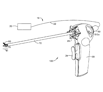
With reference to FIGS. 1-3, an embodiment of a surgical system 19, including
an ultrasonic
surgical instrument 100 in accordance with the present invention is
illustrated. The surgical
system 19 includes an ultrasonic generator 30 connected to an ultrasonic
transducer 50 via
cable 22, and an ultrasonic surgical instrument 100. It will be noted that, in
some
applications, the ultrasonic transducer 50 is referred to as a "hand piece
assembly" because
the surgical instrument of the surgical system 19 is configured such that a
surgeon may
grasp and manipulate the ultrasonic transducer 50 during various procedures
and
operations. A suitable generator is the GEN 300 sold by Ethicon Endo-Surgery,
Inc. of
Cincinnati, Ohio.
The ultrasonic surgical instrument 100 includes a multi-piece handle assembly
68 adapted to
isolate the operator from the vibrations of the acoustic assembly contained
within transducer
50. The handle assembly 68 can be shaped to be held by a user in a
conventional manner,
but it is contemplated that the present ultrasonic surgical instrument 100
principally be
grasped and manipulated by a trigger-like arrangement provided by a handle
assembly of the
instrument, as will be described. While multi-piece handle assembly 68 is
illustrated, the
handle assembly 68 may comprise a single or unitary component. The proximal
end of the
ultrasonic surgical instrument 100 receives and is fitted to the distal end of
the ultrasonic
transducer 50 by insertion of the transducer into the handle assembly 68. The
ultrasonic
surgical instrument 100 may be attached to and removed from the ultrasonic
transducer 50
as a unit. The ultrasonic surgical instrument 100 may include a handle
assembly 68,
comprising mating housing portion 69, housing portion 70, and a transmission
assembly 71.
When the present instrument is configured for endoscopic use, the construction
can be
dimensioned such that transmission assembly 71 has an outside diameter of
approximately
5.5 mm. The elongated transmission assembly 71 of the ultrasonic surgical
instrument 100
extends orthogonally from the instrument handle assembly 68. The transmission
assembly
71 can be selectively rotated with respect to the handle assembly 68 as
further described
below. The handle assembly 68 may be constructed from a durable plastic, such
as
polycarbonate or a liquid crystal polymer. It is also contemplated that the
handle assembly 68
may alternatively be made from a variety of materials including other
plastics, ceramics or
metals.
CA 2974924 2017-07-31
- 18 -
The transmission assembly 71 may include an outer tubular member or outer
sheath 72, an
inner tubular actuating member 76, a waveguide 80 and end- effector 81 (blade
79, clamp
arm 56 and one or more clamp pads 58). As will be described, the outer sheath
72, the
actuating member 76, and the waveguide or transmission rod 80 may be joined
together for
rotation as a unit (together with ultrasonic transducer 50) relative to handle
assembly 68. The
waveguide 80, which is adapted to transmit ultrasonic energy from transducer
50 to blade 79
may be flexible, semi-flexible or rigid. The waveguide 80 may also be
configured to amplify
the mechanical vibrations transmitted through the waveguide 80 to the blade 79
as is well
known in the art. The waveguide 80 may further have features to control the
gain of the
longitudinal vibration along the waveguide 80 and features to tune the
waveguide 80 to the
resonant frequency of the system. In particular, waveguide 80 may have any
suitable cross-
sectional dimension. For example, the waveguide 80 may have a substantially
uniform cross-
section or the waveguide 80 may be tapered at various sections or may be
tapered along its
entire length. In one expression of the current embodiment, the waveguide
diameter is about
0.113 inches nominal to minimize the amount of deflection at the blade 79 so
that gapping in
the proximal portion of the end effector 81 is minimized.
Ultrasonic waveguide 80 may further include at least one radial hole or
aperture 66 extending
there through, substantially perpendicular to the longitudinal axis of the
waveguide 80. The
aperture 66, which may be positioned at a node, is configured to receive a
connector pin 27
which connects the waveguide 80, to the tubular actuating member 76, and the
tubular outer
sheath 72, a rotation knob 29 together for conjoint rotation, including the
end effector 81 ,
relative to instrument handle assembly 68.
In one embodiment of the present invention, the ultrasonic waveguide 80 may
have a
plurality of grooves or notches (not shown) formed in its outer circumference.
The grooves
may be located at nodes of the waveguide 80 to act as alignment indicators for
the
installation of a damping sheath 62 and stabilizing silicone rings or
compliant supports during
manufacturing. A seal 67 may be provided at the distal-most node, nearest the
end-effector
81 , to abate passage of tissue, blood, and other material in the region
between the
waveguide 80 and actuating member 76.
The blade 79 may be integral with the waveguide 80 and formed as a single
unit. In an alternate expression of the current embodiment, blade 79 may be
connected by a
CA 2974924 2017-07-31
- 19 -
threaded connection, a welded joint, or other coupling mechanisms. The distal
end of the
blade 79 is disposed near an anti-node in order to tune the acoustic assembly
to a preferred
resonant frequency fo when the acoustic assembly is not loaded by tissue. When
ultrasonic
transducer 50 is energized, the distal end of blade 79 is configured to move
longitudinally in
the range of, for example, approximately 10 to 500 microns peak-to-peak, and
preferably in
the range of about 20 to about 200 microns at a predetermined vibrational
frequency fo of, for
example, 55,500 Hz.
In accordance with the illustrated embodiment, blade 79 is curved along with
the associated
clamp arm 56. This is illustrative only, and blade 79 and a corresponding
clamp arm 56 may
be of any shape as is known to the skilled artisan.
Ultrasonic transducer 50, and an ultrasonic waveguide 80 together provide an
acoustic
assembly of the present surgical system 19, with the acoustic assembly
providing ultrasonic
energy for surgical procedures when powered by generator 30. The acoustic
assembly of
surgical instrument 100 generally includes a first acoustic portion and a
second acoustic
portion. In the present embodiment, the first acoustic portion comprises the
ultrasonically
active portions of ultrasonic transducer 50, and the second acoustic portion
comprises the
ultrasonically active portions of transmission assembly 71. Further, in the
present
embodiment, the distal end of the first acoustic portion is operatively
coupled to the proximal
end of the second acoustic portion by, for example, a threaded connection.
With particular reference to FIGS. 2, and 9-11 , reciprocal movement of
actuating member 76 drives the clamp arm open and closed. A force-limiting
mechanism 91
is operatively connected to actuating member 76 and comprises a tube collar
cap 98 that
secures distal washer 97, distal wave spring 96, proximal washer 95 and
proximal wave
spring 94 onto collar cap 93. Collar 93 includes axially extending lugs 92 in
engagement with
suitable openings 75 in the proximal portion of tubular actuating member 76. A
circumferential groove 74 on the actuating member 76 receives on 0-ring 73 for
engagement
with the inside surface of outer sheath 72.
Rotation of the actuating member 76 together with tubular outer sheath 72 and
inner
waveguide 80 is provided by a connector pin 27 extending through these
components and
rotation knob 29. Tubular actuating member 76 includes an elongated slot 31
through which
CA 2974924 2017-07-31
- 20 -
the connector pin 27 extends to accommodate reciprocal movement of the
actuating member
76 relative to the outer sheath 72 and inner waveguide 80.
The force limiting mechanism 91 provides a portion of the clamp drive
mechanism of the
instrument 100, which affects pivotal movement of the clamp arm 56 by
reciprocation of
actuating member 76. The clamp drive mechanism further includes a drive yoke
33 which is
operatively connected with an operating trigger 34 of the instrument, with the
operating
trigger 34 thus interconnected with the reciprocable actuating member 76 via
drive yoke 33
and force limiting mechanism 91. Trigger 34 is rotatably connected to drive
yoke 33 via pins
35 and 36 and link 37 and rotatably connected to drive yoke 33 and housing 68
via post 38.
Movement of trigger 34 toward handgrip 68 translates actuating member 76
proximally,
thereby pivoting clamp arm 56 toward blade 79. The trigger-like action
provided by trigger 34
and cooperating handgrip 68 facilitates convenient and efficient manipulation
and positioning
of the instrument, and operation of the clamping mechanism at the distal
portion of the
instrument whereby tissue is efficiently urged against the blade 79. Movement
of trigger 34
away from handgrip 68 translates actuating member 76 distally, thereby
pivoting clamp arm
56 away from blade 79.
With particular reference to FIGS. 1-4, therein is illustrated one embodiment
of clamp
member 60 for use with the present ultrasonic surgical instrument 100 and
which is
configured for cooperative action with blade 79. The clamp member 60 in
combination with
blade 79 is commonly referred to as the end effector 81 , and the clamp member
60 is also
commonly referred to as the jaw. The clamp member 60 includes a pivotally
movable clamp
arm 56, which is connected to the distal end of outer sheath 72 and actuation
member 76, in
combination with a tissue engaging pad or clamp pad 58. In one expression of
the
embodiment, clamp pad 58 is formed from TEFLON trademark name of E. I. Du
Pont de
Nemours and Company, a low coefficient of friction polymer material, or any
other suitable
low-friction material. Clamp pad 58 mounts on the clamp arm 56 for cooperation
with blade
79, with pivotal movement of the clamp arm 56 positioning the clamp pad in
substantially
parallel relationship to, and in contact with, blade 79, thereby defining a
tissue treatment
region. By this construction, tissue is grasped between clamp pad 58 and blade
79. As
illustrated, clamp pad 58 may be provided with non- smooth surface, such as a
saw tooth-like
configuration to enhance the gripping of tissue in cooperation with the blade
79. The saw
CA 2974924 2017-07-31
- 21 -
tooth-like configuration, or teeth, provide traction against the movement of
the blade. The
teeth also provide counter traction to the blade and clamping movement. As
would be
appreciated by one skilled in the art, the saw tooth-like configuration is
just one example of
many tissue engaging surfaces to prevent movement of the tissue relative to
the movement
of the blade 79. Other illustrative examples include bumps, criss-cross
patterns, tread
patterns, a bead or sand blasted surface, etc.
With particular reference to Fig. 3a, a first expression of the current
embodiment includes a
clamp pad 58 having a proximal portion 58b that is smoother than a distal
portion 58a, such
that proximal portion 58b may be devoid of saw-tooth-like teeth or other
tissue engaging
surfaces contemplated. Utilizing a smooth proximal portion 58b on clamp pad 58
allows
tissue in the proximal region to move distally, following the vibratory motion
of the blade, to
the more active region of the blade 79 to prevent tissue tagging. This concept
takes
advantage of the inherent motion profile of blade 79. Due to sinusoidal
motion, the greatest
displacement or amplitude of motion is located at the most distal portion of
blade 79, while
the proximal portion of the tissue treatment region is on the order of 50% of
the distal tip
amplitude. During operation, the tissue in the proximal region of end effector
(area of portion
58b) will desiccate and thin, and the distal portion of end effector 81 will
transect tissue in
that distal region, thereby allowing the desiccated and thin tissue within the
proximal region
to slide distally into the more active region of end effector 81 to complete
the tissue
transaction.
In a second expression of the current embodiment, clamp pad 58 consists of one
single pad
having a smooth proximal end 58b and a distal portion 58a that comprises a saw
tooth-like
configuration. In a third expression of the current embodiment, clamp pad 58
may consist of
two separate components, distal portion 58a' that comprises saw tooth-like
teeth and
proximal portion 58b' that is smoother relative to distal portion 58a'. The
advantage of two
separate components 58a' and 58b' is that each pad may be constructed from
different
materials. For example, having a two-piece tissue pad allows the use of a very
lubricious
material at the distal end that is not particularly resistant to high
temperatures compared to a
very high temperature material at the proximal end that is not particularly
lubricious because
the proximal end is an area of lower amplitude. Such a configuration matches
the tissue pad
materials to the amplitude of the blade 79.
CA 2974924 2017-07-31
- 22 -
In a fourth expression of the current embodiment of the present invention,
clamp pad 58a' is
formed from TEFLON or any other suitable low-friction material. Clamp pad
58b' is formed
from a base material and at least one filler material, which is a different
material from the
base material. The surface of proximal clamp pad 58b' may be smoother than
distal clamp
pad 58a', or proximal clamp pad 58b' may also have a similar type saw-tooth
configuration.
Several benefits and advantages are obtained from one or more of the
expressions of the
invention. Having a tissue pad with a base material and at- least-one filler
material allows the
base material and the at-least-one filler material to be chosen with a
different hardness,
stiffness, lubricity, dynamic coefficient of friction, heat transfer
coefficient, abradability, heat
deflection temperature, glass transition temperature and/or melt temperature
to improve the
wearability of the tissue pad, which is important when high clamping forces
are employed
because tissue pads wear faster at higher clamping forces than at lower
clamping forces.
Applicants found, in one experiment, that a 15% graphite-filled
polytetrafluoroethylene tissue
pad showed substantially the same wear with a 7 pound clamping force as a 100%
polytetrafluoroethylene tissue pad showed with a 1.5 pound clamping force.
Having a flexible
clamping arm and/or a flexible tissue pad should also improve the wearability
of the tissue
pad due to the ability of the flexible member to more evenly distribute the
load across the
entire surface of the tissue pad. Further benefits and expressions of this
embodiment are
disclosed in United States provisional patent application, serial number
60/548,301 , filed on
February 27, 2004.
In a fifth expression of the current embodiment, a tissue pad with a base
material and at least
two filler materials allows the base material and the at- least-two filler
materials to be chosen
with a different hardness, stiffness, lubricity, dynamic coefficient of
friction, heat transfer
coefficient, abradability, heat deflection temperature, and/or melt
temperature to improve the
wearability of the tissue pad, which is important when high clamping forces
are employed
because tissue pads wear faster at higher clamping forces than at lower
clamping forces.
Applicants found, in one experiment, that a 15% graphite-filled, 30% PTFE-
filled polyimide
tissue pad showed substantially the same or better wear with a 4.5 pound
clamping force as
a 100% polytetrafluoroethylene tissue pad showed with a 1.5 pound clamping
force. The
advantage of a 15% graphite-filled, 30% PTFE-filled polyimide tissue pad is
increased heat
resistance, which improves the overall wear resistance of the tissue pad. This
polyimide-
composite clamp pad has a useful heat resistance up about 800 F to about 1200
F, as
CA 2974924 2017-07-31
- 23 -
compared to a useful heat resistance up to about 660 F of a PTFE clamp pad.
Alternatively,
Other materials are also useful for a portion of the tissue pad (that is
element 58b'), such as
ceramics, metals, glasses and graphite.
Referring to FIGS. 3a-e, one expression of clamp arm 56 has different shaped
slots for
accepting two or more tissue pads. This configuration prevents mis- loading of
the tissue
pads and assures that the appropriate pad is loaded at the correct location
within clamp arm
56. For example clamp arm 56 may comprise a distal T-shaped slot 53a for
accepting a T-
shaped flange 53b' of distal clamp pad 58a' and a proximal wedged-shaped or
dove tailed-
shaped slot 55a for accepting a wedge-shaped flange 55b' of proximal clamp pad
58b'. Tab
stop 51 engages the proximal end of proximal clamp pad 58b' to secure the
clamp pads onto
clamp arm 56. As would be appreciated by those skilled in the art, flanges
53b' and 55b' and
corresponding slots 53a and 55a may have alternate shapes and sizes to secure
the clamp
pads to the clamp arm. The illustrated flange configurations shown are
exemplary only and
accommodate the particular clamp pad material of one embodiment, but the
particular size
and shape of the flange may vary, including, but not limited to, flanges of
the same size and
shape. For unitary tissue pads, the flange may be of one configuration.
Further, other tab
stops are possible and may include any of the multiple methods of mechanically
attaching
the clamp pads to the clamp arm, such as rivets, glue, press fit or any other
fastening means
well know to the artisan.
In a second expression of the current embodiment, clamp pads 58a and 58b are
cut on a
bias so the interface between the two pads creates an overlap to minimize
gapping (Figs. 4a,
4b). For example, a 45 degree biased cut does allow some gapping to occur, but
the amount
of gap seen by the tissue is
minimized.
In a third expression of the current embodiment, clamp arm 56 increases in its
height
dimension from the distal end to the proximal end (D1 < D2). Preferably, D2 is
from about
105% to about 120% greater than D1 and more preferably, D2 is from about 108%
to about
113% greater than D1, and most preferably, D2 is about 110% greater than D1.
Slot 153
accepts the flanges from one clamp pad 58 or two clamp pads 58a and 58b.
Tapered clamp
arm 56 allows for the use of use flat pads and increases the pressure in the
proximal portion
of end effector 81 as well as the interference with blade 79. When clamp arm
56 deflects at a
CA 2974924 2017-07-31
- 24 -
greater rate than the blade 79, pressure still exists at the tissue pad and
blade interface and
no gap is created. Additionally, the increased pressure helps to offset the
decreased blade
amplitude at the proximal end of blade 79 and provides a relatively constant
pressure
between the clamp pad 58 and blade 79.
A first expression for a method for inserting clamp pads includes a) inserting
first and second
clamp pads having a first-shaped flange into a clamp arm 56 having a slot that
accepts the
first-shaped flange; and b) engaging a pad stop to secure the clamp pads
within the clamp
arm. In a second expression of this method one clamp pad may be fabricated
from a
polymeric material such as TEFLON, and the second clamp pad may be fabricated
from a
base material and at least one filler material, which is a different material
from the base
material and that clamp arm is fabricated from metal, such as stainless steel,
or titanium. The
tissue surfaces of the clamp pads may be smooth or have tissue gripping
features, such as a
saw-tooth configuration.
A third expression for a method for inserting clamp pads includes a) inserting
a first clamp
pad having a first-shaped flange into a clamp arm having a slot that accepts
the first-shaped
flange; b) inserting a second clamp pad having a second-shaped flange into the
clamp arm
having a slot that accepts the second-shaped flange; and c) engaging a pad
stop to secure
the clamp pads within the clamp arm. In a fourth expression of this method one
clamp pad
may be fabricated from a polymeric material such as TEFLON, and the second
clamp pad
may be fabricated from a base material and at least one filler material, which
is a different
material from the base material and that clamp arm is fabricated from metal,
such as
stainless steel, or titanium. The tissue surfaces of the clamp pads may be
smooth or have
tissue-gripping features, such as a saw-tooth configuration.
A first expression of a method for replacing clamp pads 58 would include the
steps of: a)
disengaging a pad stop; b) removing a first clamp pad from the clamp arm; c)
removing a
second clamp pad from the clamp arm; d) inserting third and fourth clamp pads
into the
clamp arm; and e) engaging a pad stop to secure the third and fourth clamp
pads within the
clamp arm. In a second expression of this method one of the third and fourth
clamp pads
may be fabricated from a polymeric material such as TEFLON, and the other
clamp pad may
be fabricated from a base material and at least one filler material, which is
a different material
from the base material and that clamp arm is fabricated from metal, such as
stainless steel,
CA 2974924 2017-07-31
- 25 -
or titanium. The tissue surfaces of the clamp pads may be smooth or have
tissue gripping
features, such as a saw-tooth configuration.
Referring now to FIG. 4, pivotal movement of the clamp member 60 with respect
to blade 79
is affected by the provision of a pair of pivot points on the clamp arm 56
that interface with
the outer tube 72 and inner tube 76 respectively. The outer tube 72 is
grounded to handle 68
through rotation knob 29. Clamp arm 56 is pivotally connected to outer tube 72
via
corresponding through holes 52a and 52b on clamp arm 56 and 52c and 52d on
outer tube
72. A securing pin or rivet 57 slides through holes 52a-d to secure clamp arm
56 to outer
tube 72. In one embodiment pin 57 is laser welded to clamp arm 56 so that pin
57 is fixed to
clamp arm 56 and rotates relative to outer sheath 72.
Inner tube 76 translates along the longitudinal axis of outer tube 72 and is
grounded to the
handle 68 through rotation knob 29. Pivot studs 54a, b (54a not shown) on
clamp arm 56
engage pivot holes 54c,d (54d not shown) at the distal end of inner tube 76.
The pivotal
connection of clamp arm 56 to the inner and outer tubes 76, 72 provide more
robustness to
the end effector 81 and minimize failure modes due to excessive axial or
torsional abuse
loads. Further, the embodiment increases the effectiveness of the end effector
81 to provide
clamp forces in excess of 1.5 lbs. Reciprocal movement of the actuating member
76, relative
to the outer sheath 72 and the waveguide 80, thereby affects pivotal movement
of the clamp
arm 56 relative to the end- blade 79.
FIG. 4c illustrates a force diagram and the relationship between the actuation
force FA
(provided by actuation member 76) and transection force FT (measured at the
midpoint of the
optimal tissue treatment area).
FT = FA (X2 X1) Equation [1]
Where FA equals the spring preload of proximal spring 94 (less frictional
losses), which, in
one embodiment, is about 12.5 pounds, and FT equals about 4.5 pounds as shown
in FIG.
16c. FIG. 16c provides a graphical illustration of FT and FA as a function of
trigger 34
movement as well as input forces at trigger 34.
FT is measured in the region of the clamp arm/blade interface where optimal
tissue treatment
occurs as defined by tissue marks 61a and 61 b. Tissue marks 61a, b are etched
or raised
CA 2974924 2017-07-31
- 26 -
on clamp arm 56 to provide a visible mark to the surgeon so the surgeon has a
clear
indication of the optimal tissue treatment area. Tissue marks 61a, b are about
7mm apart in
distance, and more preferably 5mm apart in distance.
Rotation of the transmission assembly 71 of ultrasonic surgical instrument 100
may be
affected together with relative rotational movement of ultrasonic transducer
50 with respect to
instrument handle assembly 68. In order to join the transmission assembly 71
to the
ultrasonic transducer 50 in ultrasonic- transmitting relationship, the
proximal portion of the
outer sheath 72 may be provided with a pair of wrench flats 46. The wrench
flats 46 allow
torque to be applied by a suitable torque wrench or the like to thereby permit
the waveguide
80 to be joined to the ultrasonic transducer 50. The ultrasonic transducer 50,
as well as the
transmission assembly 71 , is thus rotatable, as a unit, by suitable
manipulation of rotation
knob 29, relative to handle assembly 68 of the instrument. The interior of
handle assembly
68 is dimensioned to accommodate such relative rotation of the ultrasonic
transducer 50. A
spring 28 is loaded against rotation knob 29 and an inner housing surface 65.
Spring 28
provides a compression or force against rotation knob 29 to inhibit
inadvertent rotation of end
effector 81.
Referring now to FIGS. 2, 5, 6 and 16, force limiting mechanism 91 provides a
first and
second compression spring, distal spring 96 and proximal spring 94. Distal
spring 96 is
operationally coupled to yoke 33, which in turn is driven by trigger 34.
Proximal spring 94 is
in operational relationship with distal spring 96. Distal spring 96 generates
the end effector
load and proximal spring 94 maintains the consistency of the end effector
load. As a result,
the end effector load is more tightly controlled and component abuse load
conditions are
reduced. Washers 97 and 95 are a safe guard against distal spring 96 being
fully
compressed (FIG. 5), thereby preventing the spring material to yield and
render spring 96
useless in subsequent clamp arm closures. As would be appreciated by one
skilled in the art,
the application of a dual spring force limiting system has applicability in
other energy-based
surgical devices (such as RF, microwave and laser) that encounter clamping
forces, as well
as mechanical devices, such as, clip appliers, graspers and staplers.
In one expression of the current embodiment, distal spring 96 has a spring
constant greater
than 100 pounds per inch and preferably greater than 125 pounds per inch and
most
preferably about 135 pounds per inch. It is not required that distal spring 96
be preloaded,
CA 2974924 2017-07-31
- 27 -
,
but may be preloaded at less than 10 pounds, and preferably less than 5
pounds, and most
preferably at about 1 pound. Proximal spring 94 has a spring constant greater
than 25
pounds per inch and preferably greater than 50 pounds per inch and most
preferably about
70 pounds per inch. Proximal spring 94 is preloaded to a force necessary to
achieve the
desired transection force as noted in Equation 1 , above, and is a function of
the mechanical
advantage of the clamp arm 56 coupling means and frictional losses in the
device. In a
second expression of the current embodiment, proximal spring 94 is preloaded
at about 12.5
pounds.
Referring now to FIG. 16a, curve 82 illustrates actuation member 76 force and
curve 83
represents trigger 34 force as a function of the angular rotation of trigger
34 (on the x-axis, -
18.0 is the clamp arm 56 fully open and 0.0 is the clamp arm fully closed and
against blade
79) under no tissue or minimal tissue load operation. Point 82a represents the
point at which
yoke 33 begins to deflect or compress distal spring 96 and the actuation
member 76 force
increases as trigger 34 is depressed further until the force reaches the
preload value of
proximal spring 94 at inflection point 82b, and the slope of the force curve
decreases.
In FIG. 16b, curve 84 illustrates actuation member 76 force and curve 85
represents trigger
34 force as a function of the angular rotation of trigger 34 under abusive
tissue load
operation, whereby tissue completely fills the end effector in the open
position. Point 84a
represents the point at which yoke 33 begins to deflect or compress distal
spring 96 and the
actuation member 76 force increases as trigger 34 is depressed until the force
reaches the
preload value of proximal spring 94 at inflection point 84b, at which point
the slope of the
force curve decreases.
Referring now to FIGS. 2 and 5, surgical instrument 100 further provides for a
means for
indicating to the surgeon that the trigger has reached full travel and the
clamp arm 56 is
applying the correct coaptation force to the tissue. This is useful during
protracted surgical
operations or tissue transection activities when the surgeon's grip may relax,
just a bit,
without the surgeon's knowledge, and the pressure delivered to the tissue from
the clamp
arm 56 may be unknowingly decreased.
In one expression of the current embodiment, a detent spring 110 is supported
within a
detent support 112 located within housing portion 69. A detent tab 114 on
trigger 34 engages
CA 2974924 2017-07-31
- 28 -
and snaps back detent spring 110 when trigger 34 is fully closed or actuation
member 76 has
reached it most proximal travel. Detent spring 110 is generally planar and
made of a flexible
plastic that adequately deflects when it engages tab 114 thereby providing an
audible and/or
tactile signal to the surgeon that there is full end effector 81 closure.
Advantageously, tab
114 strikes and deflects detent spring 110 when trigger 34 is rotated from the
full closure
position and in the opposite direction thereby providing an audible and/or
tactile signal to the
surgeon that full closure of end effector 81 no longer exists. As would be
appreciated by the
skilled artisan, the indicating means may be either tactile, audible or visual
or a combination.
Various types of indicators may be used including dome switches, solid stops,
cantilever
springs or any number of mechanical or electrical switches known to those
skilled in the art.
Further various means may be used to provide feedback to the surgeon,
including, but not
limited to, lights, buzzers, and vibratory elements.
Referring now to FIGS. 1 , 2 and 6-8 housing 68 includes a proximal end, a
distal end, and a
cavity 59 extending longitudinally therein. Cavity 59 is configured to accept
a switch
assembly 300 and the transducer assembly 50, which interfaces with housing 68
via switch
assembly 300.
Transducer 50 includes a first conductive ring 400 and a second conductive
ring 410 which
are securely disposed within the transducer body 50. In one expression of the
current
embodiment, first conductive ring 400 comprises a ring member, which is
disposed between
the transducer 50 and the horn 130. Preferably the first conductive ring 400
is formed
adjacent to or as part of the flange member 160 within the cavity 162 and is
electrically
isolated from other electrical components. The first conductive ring 400 is
anchored to and
extends upwardly from a non-conductive platform or the like (not shown) which
is formed
within the transducer body 50. The first conductive ring 400 is electrically
connected to the
cable 22 (FIG. 1 ) by means of one or more electrical wires (not shown), which
extend along
the length of the transducer body 50 to the first conductive ring 400.
The second conductive ring 410 of the transducer 50 similarly comprises a ring
member that
is disposed between the transducer body 150 and the horn 130. The second
conductive ring
410 is disposed between the first conductive ring 400 and the horn 130 and
therefore the first
and second conductive rings 400, 410 are concentric members. The second
conductive ring
410 is likewise electrically isolated from the first conductive ring 400 and
other electrical
CA 2974924 2017-07-31
- 29 -
components contained within the transducer 50. Similar to the first conductive
ring 400, the
second conductive ring 410 preferably is anchored to and extends upwardly from
the non-
conductive platform. It will be understood that the first and second
conductive rings 400, 410
are sufficiently spaced from one another so that they are electrically
isolated from each other.
This may be accomplished by using one or more spacers 413 disposed between the
first and
second conductive rings 400, 410 or between the rings 400, 410 and other
members within
the transducer 50. The second conductive ring 410 is also electrically
connected to the cable
22 (FIG. 1 ) by means of one more electrical wires (not shown), which extend
along the
length of the transducer 50 to the second conductive ring 410. The second
conductive ring
410 is thus provided to partially define a second electrical pathway from the
cable 22 to the
switch mechanism 300. A suitable ultrasonic transducer 50 is Model No. HP054,
sold by
Ethicon Endo-Surgery, Inc. of Cincinnati, Ohio.
In one expression of the current embodiment, the distal end of transducer 50
threadedly
attaches to the proximal end of transmission rod 80. The distal end of
transducer 50 also
interfaces with switch assembly 300 to provide the surgeon with finger-
activated controls on
surgical instrument 100.
Switch assembly 300 comprises a pushbutton assembly 310, a flex circuit
assembly 330, a
switch housing 350, a first spring slip ring conductor 360 and a second spring
slip ring
conductor 370. Switch housing 350 is generally cylindrical and is supported
within handle
assembly 68 by way of corresponding supporting mounts on switch assembly 350
and
housing portions 69 and 70. Housing 350 defines a first cavity 353, a mounting
boss 352 and
a second cavity 351. Cavity 353 is sized to accept the proximal end of
transducer 50,
whereby horn 130 passes through cavity 351 to interface with transmission rod
80. Mounting
boss 352 accepts slip ring conductors 360 and 370, which in turn electrically
engage ring
contacts 400 and 410, respectively. An alignment pin 354 and snap-fit pin 355
align with
corresponding apertures of the flex circuit assembly 330 and pushbutton
assembly 310 to
secure all components together as discussed below.
With particular reference now to FIG. 8a, slip ring conductors 360 and 370 are
generally
open-ended 0-shaped springs that slip onto mounting boss 352. Each spring slip-
ring
comprises two pressure point contacts (361 a-b and 371 a-b) that contact the
respective ring
conductor 400 and 410 of transducer 50. The spring tension of the slip rings
360 and 370
CA 2974924 2017-07-31
- 30 -
cause positive contact between contacts 361 a-b, 371 a-b and conductors 400
and 410. It is
evident that the slip-ring construction allows electrical contact to be made
even as transducer
50 may be rotated by the surgeon during use of the instrument. Posts 364 and
374 of the
respective slip rings electrically connect to the respective conductor within
flex circuit 330 to
complete the electrical circuit as shown in Fig. 8c.
A flex circuit 330 provides for the electro-mechanical interface between
pushbuttons 311a, b,
312a, b and the generator 30 via transducer 50. Flex circuit comprises four
dome switches
332a, b and 334a, b that are mechanically actuated by depressing pushbuttons
311a, b or
312a, b, respectively of corresponding pushbutton assembly 310. Dome switches
332 and
334 are electrical contact switches, that when depressed provide an electrical
signal to
generator 30 as shown by the electrical wiring schematic of Fig. 8c. Flex
circuit 330 also
comprises two diodes within a diode package 336, also illustrated in Fig. 8c.
Flex circuit 330
provides conductors, 335 and 337 as is known to those in the art, that connect
to slip ring
conductors 360 and 370 via electrical tabs 364 and 374, respectively, which in
turn provide
electrical contact to ring conductors 400 and 410, which in turn are connected
to conductors
in cable 22 that connect to generator 30. Tabs 364 and 374 are soldered to
conductors 335
and 337.
Flex circuit 330 generally wraps around switch housing 350 so that dome
switches 334a, b
and 332a, b interface with the corresponding backing surfaces 356a, b and
358a, b on switch
housing 350. Backing surfaces provide a firm support for the dome switches
during
operation, discussed below. Dome switches 334a, b and 332a, b may be fixedly
attached to
backing surfaces 356a, b and 358a, b by any convenient method, such as, an
adhesive. Flex
circuit is secured to switch housing 350 via alignment pin 354 and snap-fit
pin 355 on switch
assembly 350 and corresponding alignment hole 338 and snap-fit hole 339 on
flex circuit
330.
Layered on top of flex circuit is pushbutton assembly 310, which has a
corresponding saddle-
shape as flex circuit 330, and generally wraps around switch housing 350.
Pushbutton
assembly 310 comprises four pushbuttons, distal pushbuttons 312a, b and
proximal
pushbuttons 311a, b which have corresponding pressure studs 315a, b and 314a,
b. The
pushbuttons are connected to cantilever elements 313a, b and 316a, b, which
provide a
spring-back action after the pushbuttons are depressed. As is readily
apparent, by
CA 2974924 2017-07-31
- 31 -
,
depressing pushbuttons 311 and 312 the corresponding pressure studs 314 and
315
depress against corresponding dome switches 334 and 332 to activate the
circuit illustrated
in FIG. 8c. Switches 312a and b are in parallel so that a surgeon may operate
the
pushbuttons using either a left hand or a right hand. Likewise, switches 311a
and b are in
parallel so that a surgeon may operate the pushbuttons using either a left
hand or a right
hand. When the surgeon depresses either switch 312a or 312b, the generator
will respond
with a certain energy level, such as a maximum ("max") power setting; when the
surgeon
depresses either switch 311a or 311 b, the generator will respond with a
certain energy level,
such as a minimum ("min") power setting, which conforms to accepted industry
practice for
pushbutton location and the corresponding power setting.
Alternatively, the pushbuttons may be molded into the switch housing 350 or
into the handle
assembly 68 to reduce the number of components and increase the reliability of
the overall
device. The pushbuttons may be attached through small cantilever sections,
which allow for
sturdy attachment of the pushbutton to the other components, while at the same
time
allowing for a low force to activate the pushbuttons.
Referring now to FIGS. 12-15, one expression of the current embodiment allows
switches
311a, b and 312a, b configured in such a way to provide an ergonomically
pleasing grip and
operation for the surgeon. Switches may be placed in the range of the natural
swing of the
surgeon's thumb, whether gripping surgical instrument 100 right-handed or left
handed. In a
second expression of the current embodiment, the switches are placed on
housing 68 to
prevent inadvertent button activation on the side of the instrument opposite
the thumb while
the surgeon depresses trigger 34 or rotates rotation knob 29. In a third
expression of the
current embodiment a series of partitions, such as ridges and/or depressions
or "peaks and
valleys" that are integrated onto the housing 68. In one example the housing
defines a first
surface and the series of partitions define at least one second surface such
that the second
surface is higher than the housing surface. The partition may also define a
third surface that
is lower than the housing surface. As can be seen in FIGS. 1 , 2 switches
312a, b are
surrounded by an upper ridge 320 and a lower ridge 324. Ridges 320 and 324 may
be
discrete physical features, both separated from each other, or ridges 320 and
324 may be
continuous in nature without departing from the scope of the invention.
Further, the ridges
320 and 324 may continue across the entire upper portion of housing 68, as
shown in FIGS.
12-15, or ridges 320 and 324 may be more discrete as shown in FIGS. 1 and 2.
This
CA 2974924 2017-07-31
- 32 -
construction and situation of switches 312a, b prevent the risk of inadvertent
button activation
even if a finger crosses over the button due to the fact that the ridges cause
the finger to
pass above the plane of the button. The ridges also provide tactile feedback
to the surgeon
as to the location of the pushbuttons and whether the button represents min or
max power
activation. As is readily evident, switches 312a, b are surrounded by ridges
320 and 324 and
pushbuttons 311a,b are situated above and proximal of ridge 320. Such tactile
feedback is
essential to the surgeon, so the surgeon may continuously assess the surgical
site, but
confidently understand which pushbuttons are being activated. In a further
expression of the
current embodiment, switch 312a, b are nestled within a depression 322 and
further
surrounded by ridges 320 and 324.
Referring to FIG. 12, a surgeon's left hand is accessing instrument 100. The
fore finger and
middle finger are poised to activate trigger 34, and the ring finger and
pinkie grasp hand grip
39. The thumb is conveniently positioned to sweep upward to activate
pushbutton 312a or
311a. Ridges 320 and 324 extend across the upper portion of housing 69.
In FIG. 13, the opposite side of instrument 100 shown in FIG. 12 is
illustrated showing
pushbuttons 311 b and 312b. Here the surgeon's forefinger is accessing
rotation knob 29 to
rotate end effector 81. As can be seen, pushbutton 312b is subject to
inadvertent activation
by the forefinger. However, ridge 324 causes the forefinger to elevate above
the plane of
pushbutton 312b thereby reducing the risk of inadvertent activation.
In FIG. 14, the surgeon has depressed trigger 34 to close clamp arm 56 against
blade 79,
and the left thumb has easily accessed pushbutton 312b to activate max power.
In FIG. 15, the surgeon has depressed trigger 34 to close clamp arm 56 against
blade 79,
and the left thumb has easily accessed pushbutton 311 b to activate min power.
Referring to FIG. 17, an expression of surgical instrument 100 is shown
graphically
illustrating a surgeon's finger placement on instrument 100. Instrumental in
the activation of
the instrument 100 is the placement of the forefinger 382 and middle finger
384 on trigger 34.
(Using the forefinger and middle finger to activate trigger 34 is exemplary
only. Surgeons
with smaller hands may opt to activate trigger 34 with the middle finger and
ring finger,
thereby making the forefinger available to rotate knob 29 or even use the ring
finger and
pinkie to active trigger 34.) Trigger 34 comprises a base element 45, which
comprises the
CA 2974924 2017-07-31
- 33 -
detent tab 114 and linkage with yoke 33, discussed below. Attached to base
element 45 is a
generally T-shaped finger interface 43 , which in conjunction with base
element 45 define two
generally U-shaped openings, a forefinger groove 42 and a middle finger groove
44. The
most distal surface portion of T-shaped finger interface 43 defines an
actuating surface 41
that also accepts placement of fingers 382 and 384. Grooves 42 and 44 are
sized to accept
different sized fingers, a common variable as is evident depending upon the
sex and size of
the surgeon. In a first expression of the current embodiment, the size of
grooves 42 and 44
are based on anthropic data for 5th percentile females through to 95th
percentile males for
finger size. In a second expression of the current embodiment, grooves 42 and
44 are
tapered, whereby the dimension of each groove opening is larger than the
dimension of base
of each groove 42 and 44. This configuration advantageously allows fingers of
varying size to
nestle snuggly within each groove and minimize the clearance between the
finger and walls
of the grooves.
Referring now also to FIGS. 10 and 11 , the clamp arm 56 is fully open
relative to the blade
79 when trigger 34 is in its most distal position (FIG. 10). Fingers 382 and
384 may be placed
within respective grooves 42 and 44 or alternatively on surface 41 to actuate
trigger 34
through its arcuate travel designated by arrow 47. When trigger reaches its
full proximal
travel (when detent tab 114 engages detent spring 110), the clamp arm 56 is in
its fully
closed position relative to the blade 79 (FIG. 11 ). In order to reverse the
trigger along its
travel 47, fingers 382 and 384 engage grooves 42 and 44 and push trigger 34
distally to
open the end effector. The clamp arm 56 is not biased open so the surgeon
cannot control
the opening of clamp arm 56 via surface 41.
Referring now to Fig. 18, elements having similar reference numerals as shown
in Fig. 2
have the similar function as already discussed. Particular attention is
directed to an alternate
handle assembly 168 for actuating the end effector 81. The handle assembly 168
includes
two pivoting handle portions 420 and 422 coupled to a right shroud 169 and a
left shroud
170.
The right shroud 169 is adapted to snap fit on the left shroud 170 via a
plurality of inwardly facing prongs formed on the left shroud 170 to form
housing 171. When
the left shroud 170 is attached to the right shroud 169, a cavity is formed
therebetween to
accommodate various components that form the handle assembly 168 as further
discussed
CA 2974924 2017-07-31
- 34 -
below. Apertures 172 and 174 are also formed to accommodate thumb ring or
handle portion
420 and finger ring or handle portion 422, which are located exterior of the
left and right
shrouds to the actuating linkage contained within the left and right shrouds.
Aperture 173 is
also formed at the proximal end of shrouds to accommodate transducer 50 (See
figure 8b).
Handle assembly 168 includes a U-shaped yoke 424 slidably attachable within
housings 169
and 170 via slots 421a and 421 b and pins 423a and 423b, respectively. The
distal end of
handle 420 at hole 402 attaches to right shroud 169 and yoke via pin 423a, and
the proximal
end of handle 420 attaches to yoke 424 via link 428 attached to hole 404 via
pin 426 and
hole 410 via pin 430. The distal end of handle 422 at hole 406 attaches to
right shroud 169
and yoke via pin 423b, and the proximal end of handle 422 attaches to yoke 424
via link 432
attached to hole 408 via pin 434 and hole 412 via pin 430. In practice as the
handles 420 and
422 are moved away from housing 171 (for example, the surgeon's thumb
cooperates with
handle 420, and the surgeon's forefinger and middle finger cooperate with
handle 422), end
effector 81 moves away from blade 79 to form an open jaw (the open position),
and as
handles 420 and 422 are moved toward housing 171 , end effector 81 rotates
toward blade
79 to capture tissue (the closed position).
In one expression of the current embodiment, a detent spring 482 is supported
within
housing portion 171. A detent cam 480 rotates on yoke 168 and engages and
snaps back
detent spring 482 when handles 420 and 422 are in the fully closed position.
Detent spring
482 is generally made of a flexible plastic that adequately deflects when it
engages cam 480
thereby providing an audible signal to the surgeon that there is full end
effector 81 closure.
Advantageously, 480 strikes and deflects detent spring 482 when handles 420
and 422 are
rotated from the full closure position and in the opposite direction thereby
providing an
audible signal to the surgeon that full closure of end effector 81 no longer
exists.
Referring also now to Fig. 24, a second expression of the current embodiment
is shown
having an actuator post 433 attaches to handle 422 and engages a dome switch
435
covered by silicon rubber located on housing assembly 171. When handle 422 is
fully closed,
post 433 presses against the silicone which in turn transfers the force to the
dome switch
435, allowing the switch to provide an audible and tactile feedback to the
surgeon. In one
embodiment post 433 is a cylinder having a diameter of 0.170 inches with a
0.070 inch slot in
the middle. A preferred duronneter for the silicon rubber material is 20 Shore
A.
CA 2974924 2017-07-31
- 35 -
Referring also now to Fig. 23, also enclosed within housing 171 are connector
450, slip rings
452, 454, flex circuit 456 and rocker switch 462. Rocker switch 462 rotatably
attaches to right
shroud 169 via aperture 469 and switches 462 and 464 are positioned exterior
housing 171
for access by the surgeonu. Switches 462 and 464 are mechanically connected
via a rocker
arm 466 comprising a pivot post 468 which interfaces with aperture 469. In
this configuration,
switches 462 and 464 cannot be simultaneously depressed, which, if were the
case, would
provide an error message from generator 30. A flex circuit 456 provides for
the electro-
mechanical interface between switches 464 and 466 and the generator 30 via the
transducer
50 (see Fig. 8b). Referring to Fig. 21 , flex circuit 456 includes, at the
distal end, two dome
switches 500 and 502 that are mechanically actuated by depressing
corresponding switches
464 and 466, respectively. Dome switches 500 and 502 are electrical contact
switches, that
when depressed provide an electrical signal to generator 30 as shown by the
electrical wiring
schematic of Fig. 22. Flex circuit 456 also comprises two diodes within a
diode package 504,
also illustrated in Fig. 22. Flex circuit 456 provides conductors, as is known
to those in the
art, that connect to slip ring conductors 452 and 454 via connector 450, which
in turn provide
electrical contact to ring conductors 400 and 410 (Fig. 8b), which in turn are
connected to
conductors in cable 32 that connect to generator 30.
With particular reference now to Figs. 19 and 20a-b, slip ring conductors 452
and 454 are
generally open-ended 0-shaped springs that slip onto mounting surfaces 453 and
455 of
connector 450, respectively. Each spring slip-ring comprises two pressure
point contacts
(510a-b and 522a-b) that contact the respective ring conductor 400 and 410 of
handpiece 50.
The spring tension of the slip rings 452 and 454 cause positive contact
between contacts
510a-b, 522a-b and conductors 400 and 410. It is evident that the slip-ring
construction
allows electrical contact to be made even as hand piece 50 may be rotated by
the surgeon
during use of the instrument. Posts 512 and 524 of the respective slip rings
electrically
connect to the respective conductor within flex circuit 456 to complete the
electrical circuit as
shown in Fig. 22.
Referring again to Fig. 18, rotation coupler 130 rotatably engages the distal
end of right and left shrouds 169 and 170. Rotation knob 129 couples to
rotational coupler
130, whereby two spring tabs 175 and 175a (not shown) provide an outward
tension or force
against the inner surface of rotation knob 129 to inhibit inadvertent rotation
of end effector
81.
CA 2974924 2017-07-31
- 36
In an alternate expression of the invention, handles 420 and 422 have a soft-
touch molded
thermo plastic elastomer liner 550 on the inner surface of
handles 420 and 422. Plastic liner 550 provides comfort to the surgeon and
prevents finger and hand fatigue. Plastic liner 550 also provides an enhance
gripping surface
between the handles and the surgeon's thumb and fingers as opposed to the
smooth plastic
surface interface of the prior art. This is particularly advantageous for
accepting multiple digit
sizes of male and female surgeons and still providing a comfortable and
positive gripping
surface. Plastic liner 550 may be smooth or have contours molded onto the
surface of liner
550, such as ribs, as illustrated in Figs. 23 and 24. Other contours may be
bumps, and peaks
and valleys. Various other shapes and interfaces are within the scope of this
invention as
would be obvious to one skilled in the art. Plastic liner 550 is also useful
on the interface
between the surgeon's finger and trigger 34 (Fig. 12).
In one expression of the current embodiment, the soft-touch liner 550 has a
durometer (hardness) rating from about 35 Shore A to about 75 Shore A, and
more
particularly from about 50 Shore A to about 60 Shore A. Such appropriate
materials are
available from LNP of Exton, PA (stock no. 8211-55 6100 GYO-826-3) and
Advanced
Elastomer Systems of Akron, OH (stock no. 8211-556100).
The soft-touch material may also be useful to help the surgeon identify a
particular feature of the instrument while the surgeon is focused on the
operation at hand. For example, a "soft touch" having one contour interface
may be placed on the "max" button, and a "soft touch" having a second
contour interface may be place on the "min" button so the surgeon may easily
recognize the
presence of either button without having to lose focus of the surgical site.
"Soft touch" may
also be implemented on knobs 29 and 129 with contours to identify various
rotation
positions of end effector 81.
While the present invention has been illustrated by description of several
embodiments, it is
not the intention of the applicant to restrict or limit. Numerous variations,
changes, and
substitutions will occur to those skilled in the art. Moreover, the structure
of each element
associated with the present invention can be alternatively described as a
means for providing
the function performed by the element.
CA 2974924 2017-07-31