Note: Descriptions are shown in the official language in which they were submitted.
CA 02974925 2017-07-25
WO 2016/128975
PCT/IL2016/050156
METHODS OF PREVENTING SECONDARY INFECTIONS
FIELD AND BACKGROUND OF THE INVENTION
The present invention, in some embodiments thereof, relates to a method of
preventing secondary infections in subjects infected with a pathogen using
agents that
down-regulate extracellular matrix remodeling.
Viral pandemics, such as influenza have caused millions of deaths worldwide.
An extreme example is the 1918 pandemic which spread to six continents and
infected
¨500 million people reaching death toll of 50 million. Investigation of
clinical cases and
.. autopsy samples indicated that more than 95% of case fatalities were
complicated by
secondary bacterial infections, most commonly Streptococcus pneumoniae (S.
pneumoniae). Immune cells recruited to the site of infection are critical for
influenza
clearance. However, growing evidence shows that infiltrating immune cells can
also
generate excessive inflammatory responses resulting in collateral tissue
damage and
disruption of the blood-air-barrier.
Tissue tolerance to pathogens is an important evolutionary trade-off,
balancing
the host immune response to pathogens while maintaining tissue function.
However,
tolerance capacity differs between various organs; lungs have a relatively low
tissue
tolerance capacity, and are more vulnerable to tissue damage. Accordingly, it
has been
argued that during respiratory viral infections uncontrolled host-derived
immune
responses, rather than viral titers, may be the leading cause of death. These
responses
are primarily associated with inflammatory monocytes, granulocytes,
macrophages and
dendritic cells. Accordingly, influenza-infected lungs are diffusely
hemorrhagic,
potentially linking the host response with tissue destruction. Tissue
breaching may
prime secondary bacterial invasion coupled with tissue disruption and, in
extreme cases,
may result in death. The interaction between influenza and secondary bacterial
infections has long been studied, yet the molecular mechanisms by which
influenza
infection primes the tissue to secondary infections are not fully understood.
One of the host's tolerance components is the integrity of respiratory
epithelial
barriers anchored to the extracellular matrix (ECM). The ECM scaffold is
produced by
the cells in the tissue and is composed of two layers: I) the interstitial
matrix, a three-
dimensional gel of polysaccharides and fibrous proteins, and II) the basement
CA 02974925 2017-07-25
WO 2016/128975
PCT/IL2016/050156
2
membrane, a mesh-like sheet formed at the base of epithelial tissues. ECM
turnover is
regulated by multiple proteolytic enzymes including matrix metalloproteinases
(MMPs)
that are responsible for the irreversible cleavage of a plethora of ECM
molecules under
normal and pathological conditions. Dysregulated proteolytic activity is often
associated
.. with inflammation, cancer, and infectious diseases. Accordingly, studies in
pathological
conditions have shown that dysregulated proteolysis of ECM molecules and
related
protein fibers have significant effects on tissue function. Specifically, MMPs
were
shown to play critical roles in lung organogenesis and many MMPs are involved
in the
acute and chronic phases of lung inflammatory diseases (Greenlee et al., 2007,
Physiological reviews 87, 69-98). Several substrates of MMPs have been
identified
during lung development, including ECM scaffold proteins, cell adhesion
molecules,
growth factors, cytokines, and chemokines (Greenlee et al.. 2007,
Physiological reviews
87, 69-98).
Membrane type-I matrix metalloproteinase (MT1-MMP/MMP-14), a membrane
tethered collagenase, is a key regulator in development and homeostasis of the
lung as
well as mediating wound healing, airway remodeling, and cell trafficking.
Accordingly,
it is expressed by multiple cell populations in the respiratory tract,
including fibroblasts,
endothelial cells and macrophages (Greenlee et al., 2007, Physiological
reviews 87, 69-
98). The functions of macrophage-derived proteases during inflammation are
typically
associated with tissue invasion or degradative events. In macrophages MT1-MMP
serves not only as a protease acting on the ECM, but also regulates macrophage
immune
response. Recruited monocytes and macrophages up-regulate a broad spectrum of
ECM
remodelers including various MMPs. Depending on the conditions, macrophages
express a spectrum of MMPs and their inhibitors: these have been associated
with both
physiological and pathological lung remodeling events. MMP-9 (gelatinase B)
was
shown to be beneficial for recovery from influenza infection by promoting
migration of
neutrophils to the infection site (Bradley et al., 2012, PLoS pathogens 8,
e1002641).
Despite these important findings, a systematic analysis of ECM proteolytic
pathways
during respiratory infections, including the trade-off between ECM integrity
and
immune protection, has never been completed.
Background art includes Cheung et al., Cardiovasc Pathol. 2006 Mar-
Apr;15(2):63-74, Elkington et al., 2005 British Society for Immunology,
Clinical and
CA 02974925 2017-07-25
WO 2016/128975
PCT/IL2016/050156
3
Experimental Immunology, 142:12-20; Devy et al., Biochemistry Research
International, Volume 2011, Article ID 191670, doi:10.1155/2011/191670;
Renckens et
al.. J Immunol 2006; 176:3735-3741; Vanlaere et al., Clinical Microbiology
Reviews,
Apr. 2009,Vol 22, p. 224-239 and Udi et al., 2015, Structure 23, 1-12, January
6,
2015.
SUMMARY OF THE INVENTION
According to an aspect of some embodiments of the present invention there is
provided a method of treating or preventing a disease associated with a
secondary
infection in a subject infected with a pathogen comprising administering to
the subject a
therapeutically effective amount of an anti-pathogenic agent directed towards
the
pathogen and a therapeutically effective amount of an agent which down-
regulates at
least one extracellular matrix-associated polypeptide, thereby treating or
preventing the
disease associated with a secondary infection in the subject.
According to an aspect of some embodiments of the present invention there is
provided a method of treating a subject infected with a pathogen comprising
administering to the subject a therapeutically effective amount of an anti-
pathogenic
agent directed towards the pathogen and a therapeutically effective amount of
an agent
which down-regulates at least one extracellular matrix-associated polypeptide,
thereby
treating the subject.
According to an aspect of some embodiments of the present invention there is
provided an article of manufacture comprising an anti-pathogenic agent and an
agent
which down-regulates at least one extracellular matrix-associated polypeptide.
According to an aspect of some embodiments of the present invention there is
provided a pharmaceutical composition comprising an anti-pathogenic agent as a
first
active agent, an agent which down-regulates at least one extracellular matrix-
associated
polypeptide as a second active agent and a pharmaceutically acceptable
carrier.
According to an aspect of some embodiments of the present invention there is
provided a method of treating influenza in a subject in need thereof
comprising
administering to the subject a therapeutically effective amount of an agent
which down-
regulates an extracellular matrix-associated polypeptide, thereby treating the
influenza.
CA 02974925 2017-07-25
WO 2016/128975
PCT/IL2016/050156
4
According to some embodiments of the invention, the extracellular matrix-
associated polypeptide is set forth in Table 1.
According to some embodiments of the invention, the secondary infection is a
bacterial infection, a viral infection or a fungal infection.
According to some embodiments of the invention, the secondary infection is a
blood infection.
According to some embodiments of the invention, the disease is sepsis.
According to some embodiments of the invention, the administering comprises
co-administering.
According to some embodiments of the invention, the pathogen is selected from
the group consisting of a virus, a bacteria and a fungus.
According to some embodiments of the invention, the at least one polypeptide
is
a matrix metalloproteinase (MMP).
According to some embodiments of the invention, the matrix metalloproteinase
is selected from the group consisting of membrane type 1-matrix
metalloproteinase 1
(MT1-MMP1), MMP-9. MMP-8 and MMP-3.
According to some embodiments of the invention, the at least one polypeptide
is
membrane type 1-matrix metalloproteinase 1 (MT1-MMP1).
According to some embodiments of the invention, the infection is a respiratory
infection.
According to some embodiments of the invention, the pathogen is a virus.
According to some embodiments of the invention, the virus is a respiratory
virus.
According to some embodiments of the invention, the respiratory virus is
influenza.
According to some embodiments of the invention, the anti-pathogenic agent is a
neuraminidase inhibitor (NAT).
According to some embodiments of the invention, the neuraminidase inhibitor is
selected from the group consisting of Laninamivir, Oseltamivir, Peramivir and
Zanamivir.
According to some embodiments of the invention, the neuraminidase inhibitor is
Oseltamivir.
CA 02974925 2017-07-25
WO 2016/128975
PCT/IL2016/050156
According to some embodiments of the invention, the secondary infection is a
bacterial infection.
According to some embodiments of the invention, the bacterial infection is S.
pneumoniae.
5 According to some embodiments of the invention, the agent which down-
regulates the at least one polypeptide is an antibody.
According to some embodiments of the invention, the agent which down-
regulates the at least one polypeptide is a polynucleotide agent.
According to some embodiments of the invention, the extracellular matrix-
associated polypeptide is set forth in Table 1.
According to some embodiments of the invention, the at least one polypeptide
is
a matrix metalloproteinase (MMP).
According to some embodiments of the invention, the matrix metalloproteinase
is selected from the group consisting of membrane type 1-matrix
metalloproteinase 1
(MT 1 -MMP 1 ), MMP-9. MMP- 8 and MMP-3.
According to some embodiments of the invention, the at least one polypeptide
is
membrane type 1-matrix metalloproteinase 1 (MT 1-MMP1).
According to some embodiments of the invention, the anti-pathogenic agent is
an
antiviral agent.
According to some embodiments of the invention, the anti-viral agent is a
neuraminidase inhibitor (NAT).
According to some embodiments of the invention, the neuraminidase inhibitor is
selected from the group consisting of Laninamivir, Oseltamivir, Peramivir and
Zan amivir.
According to some embodiments of the invention, the neuraminidase inhibitor is
0 seltamivir.
According to some embodiments of the invention, the extracellular matrix-
associated polypeptide is set forth in Table 1.
According to some embodiments of the invention, the extracellular matrix
associated polypeptide is MT 1 -MMP 1 .
Unless otherwise defined, all technical and/or scientific terms used herein
have
the same meaning as commonly understood by one of ordinary skill in the art to
which
CA 02974925 2017-07-25
WO 2016/128975
PCT/IL2016/050156
6
the invention pertains. Although methods and materials similar or equivalent
to those
described herein can be used in the practice or testing of embodiments of the
invention,
exemplary methods and/or materials are described below. In case of conflict,
the patent
specification, including definitions, will control. In addition, the
materials, methods, and
examples are illustrative only and are not intended to be necessarily
limiting.
BRIEF DESCRIPTION OF THE SEVERAL VIEWS OF THE DRAWINGS
Some embodiments of the invention are herein described, by way of example
only, with reference to the accompanying drawings. With specific reference now
to the
drawings in detail, it is stressed that the particulars shown are by way of
example and for
purposes of illustrative discussion of embodiments of the invention. In this
regard, the
description taken with the drawings makes apparent to those skilled in the art
how
embodiments of the invention may be practiced.
In the drawings:
FIGs. 1A-D. Global analysis of extra cellular matrix gene circuits during
influenza viral infection (A) K-means clustering (k = 20) of 3530
differentially
expressed genes (Experimental Procedures) in lungs following influenza
infections at 10
time points (n=4 for each time point). Dynamic range is scaled between -2 to 2
fold
changes and color coded. 13.5 % (479) of the elevated genes are annotated as
involved
in ECM remodeling. Functional annotation was done using (cbl-
gorilladotcsdattechniondotacdatil) clusters are annotated accordingly and
colored. (B)
Shown are a subset of gene ontologies (GO) enriched (p<10-4) in infected
lungs. (C)
Submatrix of gene expression dynamics following influenza infection of ECM
remodeling genes. (D) Bar graph showing fold changes relative to TO using qPCR
measurement of MT1-MMP expression following influenza infection. Each sample
was
run in triplicates from 4 mice (2 biological repeats). Error bars represent
standard
deviation (SD) of the average number. The target genes were normalized to the
endogenous reference gene GAPDH and relative to a non-infected control sample
using
AA CT normalization method.
FIGs. 2A-C. MT1-MMP expression is mostly induced in myeloid cells following
influenza infection. (A) FACS analysis from lung of influenza infected mice 74
hours
post viral infection (Experimental procedures; n=25) compared to non-infected
controls
CA 02974925 2017-07-25
WO 2016/128975
PCT/IL2016/050156
7
(n=25). Gated are MT1-MMP expressing cells stained using anti-MT1-MMP antibody
as well as CD45, CD1 lb and Epcam (Experimental procedures). (B) Histogram
plots
showing MT1-MMP, Epcam and CD1 lb mean fluorescence intensity before (grey)
and
74 hours post influenza viral infection (red). (C) Bar graph showing qPCR
measurement
of MT1-MMP expression in sorted cell populations. Error bars represent SD of
the
average number. T-test ** p<0.001.
FIGs. 3A-I. Influenza infection induces changes in ECM morphology (A)
Global mass spectrometry analysis of cell free ECM scaffolds (see experimental
procedures). Quantitative protein abundance is presented by relative
measurement (with
reference to control uninfected tissue) using gray scale color code ranging
from -1 to 1
white to black. Proteins depleted from the ECM post infection are annotated
and
colored white. Heatmap showing significant changes in protein quantification
(p<0.01,
t-test) in de-cellularized infected lung tissue as compared to non-infected
control.
Samples were analyzed in duplicates for 2 time points post infection (74. 122
hours post
infection) with lethal dose of influenza infection using Mascot software (B)
Representative scanning electron microscope imaging of infected versus control
lungs.
Arrows and arrow-heads point to orientation changes in collagen fibrils with D-
banding
patterns as quantified in sub figure (C) Directionality of fibers on the
boundaries of
alveoli are analyzed using Fiji package (Experimental procedures). (D-H)
Representative immuno-staining images of ECM components during infection taken
from (n=20) animals and screened in multiple tissue sections and slides imaged
under
the same exposure conditions. (E-I) Quantification of immunostaining using
imageJ
package (Experimental procedures), Error bars represent SD of the average
number.
FIGs. 4A-K. Blocking MT1-MMP activity protects lung ECM components. (A)
Cartoon showing experimental setup for influenza infection with various
treatments.
Mice were infected with sub-lethal dose of PR8 influenza strain (Experimental
Procedures) (B-E) AirSEM imaging of alveolar and bronchial compartments 74
hours
post infection using fixed tissue sections from whole lung, cut 300jtm thick
and stained
for AirSEM (Experimental Procedures). Alveolar wall thickness and bronchial
cell
numbers were measured at different areas in multiple sections. Scale of main
image-
501.im. inset scale- 20.m. Bar graph quantifies wall thickness and cell
numbers in
alveoli and bronchi, respectively. Error bars represent SD of the average
number; field
8
of view (FOY). (F-G) Representative second harmonic generation (SHG) images
originating from an unstained 50p.rn thick lung tissue sections. The detected
SHG signal
representing collagen is shown in red after reproduction of the z-stack using
ImarisTM
software package version 7.7.1. Bar graph is showing collagen volumes analyzed
and
quantified using ImarisTM package and tested for significance using t-test.
Error bars
represent SD of the average number (H-I) Lung immuno-staining for laminin. Bar
graph
shows laminin intensity analyzed using ImageJ package. (J-K) Collagen type I
in situ
zymography in lung tissue using fluorogenic substrate to detect collagenolytic
activity
in lung section with high sensitivity. Green signal and arrows point to active
collagenase localization among bronchial epithelial lining cells or
infiltrating immune
cells. Scale- 50p.m; Error bars represent SD of the average number; field of
view
(FOV).
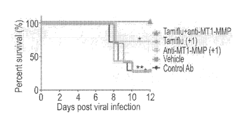
FIGs. 5A-H. Combining anti-viral treatment with ECM protection supports
survival and prevents systemic bacterial sepsis. (A, D) Cartoon showing
experimental
setup for influenza and S. pneumoniae co-infections in preventative and
therapeutic
modes. Mice were infected with sub-lethal doses of influenza followed by
infection
with Strep. pneumoniae (Experimental procedures). Treatment groups included:
TamifluTm, anti-MT1-MMP Fab, or the combinations of both. Administration was
done
using preventive mode, one day before infection (A-C) or as therapeutic mode
one-day
post infection (D-E). Vehicle-treated mice served as controls (Data are
combined from
three independent experiments with 7-10 mice in each group). (B, E) Survival
curves
(Kaplan-Meier) of co-infected mice receiving different treatments a day before
(-1) or a
day after (+1) the infections. Data is collected from 3 independent
experiments of 5
mice in each group. * P<0.01; **p< 0.001 using Log-rank (Mantel-Cox) test. (C,
F)
Relative weight loss of co-infected mice at several time points post viral
infection. Error
bars represent SD from the mean. (G-H) S. pneumonia bacterial loads from
spleen
lysates of infected mice 6 days post viral infection (Experimental
procedures).
FIGs. 6A-D. Gene and protein expression levels of ECM modulators during the
course of influenza infection. (A) Bar graph showing qPCR measurements of ECM
representative genes during different time points post infection in whole lung
tissue
(fold change relative to expression levels in TO) infection with lethal dose
of influenza
infection (experimental procedures). Each sample was run in triplicates from 4
mice (2
Date Recue/Date Received 2021-07-12
CA 02974925 2017-07-25
WO 2016/128975
PCT/IL2016/050156
9
biological repeats). Error bars represent standard deviation (SD) (B) Western
blot
analysis of several representative proteases during different time points of
influenza
infection using a reducing SDS-PAGE gel. (n=10). (C) Quantification of western
blot
results using ImageJ software. Average relative density of the protein of
interest is
relative to GAPDH internal control (D) Mean values of body weight (blue; left
y axis)
and viral titers (red; right y axis) during the course of influenza infection.
Error bars
represent standard deviations of body weight, calculated on 2-4 animals in
each time
point from 3 independent experiments. Viral burdens of whole lung homogenates
in the
lungs of mice using qPCR for genome copies of Matrix protein 2 (M2) followed
by
conversion into viral particle numbers using a calibration curve.
FIGs. 7A-D. Immune cells express active MT1-MMP during infection. (A)
Immunostaining and bar graph quantification of MT1-MMP and F4/80 marker co-
localization in infected lungs (74 hours PI) versus healthy controls. Arrows
point to
MT1-MMP stained cells. Representative images from multiple sections. (B) Bar
graph
quantifying panel A. Error bar represent SD, *P < 0. 01, t-test. (C) Collagen
type I in
situ zymography combined with CD45 staining. Arrows point to cells expressing
either
marker at both control and infected sections (74 hours PI). Scale bar-5Ortm
(D) Bar
graph quantifying panel C. Error bar represent SD, *P < 0. 01, t-test.
FIG. 8. Global expression analysis of MT1-MMP expressing cells. (A) K-means
clustering (k = 6) of 2169 differentially expressed genes in CD4510s and
45'g
populations of cells sorted 74 hours post infection (n>5 mice included in each
group-
infected and non-infected control). Mice were infected with lethal dose of
4x10³ PFU
of PR8 influenza (experimental procedures).
FIGs. 9A-D. Lung destructive phenotypes demonstrated using AirSEM imaging
of whole lung or de-cellularized tissue. (A) Imaging of whole lung tissue.
Arrows point
to boundaries of alveolar openings with cells (non-infected control) or
depleted of cells
(infected) (B) Imaging of ECM scaffolds (after de-cellularization) of infected
lungs
compared to healthy controls. Arrows point to alveolar duct boundaries
containing thick
organized collagen bundles (non-infected control) or distorted fibrils
(infected). (C)
Lung cell counts in control and infected lungs scanning multiple lung
sections, n=5. (D)
Directionality imaging analysis was done by Fiji package. Graphs were plotted
using
I0
GraphPad PrismTM 6. The relative frequency of fiber spatial orientation was
measured
using the "Directionality" plugin analysis tool in Fiji package version 6.1.1.
FIGs. 10A-C. Calibration of single viral infection. (A) Weight loss of mice
subjected to single viral infection at different dosages. Data set was
analyzed from 5
mice at each time point. Error bar represent SD and analyzed using t-test. (B)
Survival
of mice exposed to single viral infection at different dosages. Error bar
represent SD
and analyzed using t-test ** P < 0.001. (C) Viral burdens of whole lung
homogenates in
the lungs 4 days post infection using standard PFU assay. Samples were run
with 2
biological repeats 3 animals at each time point. X-axis represents the viral
amounts used
for the infection.
FIGs. 11A-F. MT1-MMP inhibition does not interfere with immune cell
recruitment or cytokine induction. (A) FACS analysis of whole lung tissue
subjected to
influenza infection 74 hours post infection and treated either with anti-MT1-
MMP
inhibitor antibody or non-relevant GST control Ab. Mice were infected at sub-
lethal
influenza dose (experimental procedures). Data is gated on MT1-MMP expressing
cells
stained with MT1-MMP antibody as well as CD45, Ly6G, Ly6C, CD1 lb, NK46, TCR(3
(Experimental procedures). Experiments were done twice using 3 mice per group.
(B)
Representative sections of lung tissue stained for macrophages using F4/80
marker
taken at 74 hours post infection. Scale bars = 50urn. FOV indicates the entire
field of
view at magnification of x20. (C) Quantification of figure B using multiple
tissue
sections from at least 3 mice per group. Error bar represent SD, ***P <
0.0001, t-test.
(D) Infiltrating immune cells in BALF from mice subjected to single viral
infection and
taken at 24, 48, 72, 122 hours PI. Mice were infected with sub-lethal dose of
influenza.
Samples were run with 2 biological repeats. Tested significant over non-
infected mice
using t-test * P < 0.01. (E-F) TNF-u, and IL-1(3 levels in BALT of mice
subjected to
single viral infection and taken at 24, 48, 72, 96, 122 hours PI. Samples were
run with 2
biological repeats. Error bar represent SD and analyzed using t-test * P <
0.01.
FIGs. 12A-F. Viral loads in the lung following Anti-MT1-MMP Ab treatment
74 Hours PI (A) Representative lung tissue sections stained for influenza
virus using
TamifluTm, control Ab and anti-MT1-MMP Ab. Mice were infected sub-lethal dose
of
influenza (experimental procedures). (B-C) Bar graph quantification of
influenza virus
24 and 48 hours PI. Error bar represent SD, **P < 0.001, t-test. FOV indicates
the entire
Date Recue/Date Received 2021-07-12
II
field of view at magnification of x20. Number of infected cells was normalized
to DAPI
using ImageJ. (D) PFU values of whole lung tissue 4 days and 7 days post viral
infection (experimental procedures). Samples were run in triplicates of 2
biological
repeats. Error bar represent SD. LEM -1; Tami-1; Tami+LEM-1 designate the
different
treatments, single or combined agents, given one day before the infection (Day-
1).
LEM+1; Tami+1; Tami+LEM+1 designate the different treatments, single or
combined
agents, given one day after the infection (Day+1). LEM refers to anti-MT1-MMP
Ab
(LEM2/15), GST refers to non-relevant Ab. (E) Viral burdens in the lungs 24,
48 and 96
hours post infection. Whole lung homogenates were used for PFU assay, testing
2
animals at each time point and running 2 biological replicates. Error bar
represent SD, *
P < 0.01 using 2-way ANOVA. (F) CFU values in the lungs of co-infected mice 2
days
post bacterial infection. Error bars represent SD from the mean. Data are
combined
from two independent experiments with five mice in each group.
FIGs. 13A-D. ECM destruction is not perturbed by low viral titers. (A) AirSEM
images of lungs 74 hours post infection from either TamifluTm-treated, vehicle-
treated
or control mice. (B) Alveolar wall thickness measured using ImageJ
(Experimental
procedures), each mark represents a mean of measurements from a section-based
region
for an individual animal. (C) Bar graph represents viral counts in vehicle-
treated and
TamifluTm-treated mice lungs using qPCR. Each column represents the mean of 3
mice.
Bars indicate mean and SD from the average. X-axis represents hours post viral
infection. (D) MT1-MMP expression levels in mice lungs infected with 90 PFU of
PR8
influenza strain undergoing different treatments. Error bar represent SD and
analyzed
using t-test ** P < 0.001.
FIGs. 14A-B. Combined anti-viral and tissue protection therapy maintains lung
structural features. (A) AirSEM imaging of lung sections representing changes
in lung
bronchi and alveoli during infection under several treatment modalities. Scale
bar -
20p,m. (B) Bar graph quantifying cell numbers in the different treatments
taken from
multiple sections.
FIG. 15. MT1-MMP expression in human respiratory epithelial cells upon
influenza infection. Log2 relative expression levels of MT1-MMP correlating
with the
infection course (hours post infection) of human bronchial epithelial cells
infected with
Date Recue/Date Received 2021-07-12
CA 02974925 2017-07-25
WO 2016/128975
PCT/IL2016/050156
12
H1N1 strain A/PR/8/34 (PR8). Error bars represent SD. Data analyzed from
(Shapira
SD, 2009).
DESCRIPTION OF SPECIFIC EMBODIMENTS OF THE INVENTION
The present invention, in some embodiments thereof, relates to a method of
preventing secondary infections in subjects infected with a pathogen using
agents that
down-regulate extracellular matrix remodeling.
Before explaining at least one embodiment of the invention in detail, it is to
be
understood that the invention is not necessarily limited in its application to
the details set
forth in the following description or exemplified by the Examples. The
invention is
capable of other embodiments or of being practiced or carried out in various
ways.
Infectious disease treatments have conventionally focused on pathogen
elimination, either by administering antimicrobial drugs or by stimulating
host immune
responses using vaccination. The present inventors performed global genomics
and
proteomics analyses of an influenza mouse model and revealed an unexpected
plethora
of extracellular matrix (ECM)-related genes and proteins responsible for
dysregulated
ECM remodeling events during the course of infection (Figures 1A-D and 6A-D).
MT1-
MMP was the main collagenase leading to destruction of ECM scaffolds of
alveoli and
bronchi of infected mouse lungs. Electron microscopy of intact lungs, global
mass
spectrometry, two-photon and immune staining, and tissue zymography, revealed
a
multifaceted destruction of basement membrane components (Figures 3A-I and 9A-
D).
This unprecedented damage to lungs contributed to loss of blood-air barrier
and resulted
in systemic spread of secondary bacterial infection through leakage from lungs
to
internal organs causing sepsis and mortality. These devastating phenotypes and
resulting deadly outcome were reversed by blocking the activity of MT1-MMP
(Figures
4A-K), thus offering a new mode of therapeutic intervention through tissue
support. As
shown in Figures 5A-H, combining anti-viral treatment with ECM protection
supports
survival and prevents systemic bacterial sepsis.
The present inventors suggest this novel treatment opportunity for infection,
designed to support tissue morphology and homeostasis while mitigating
inappropriate
host responses and collateral tissue damage.
CA 02974925 2017-07-25
WO 2016/128975
PCT/IL2016/050156
13
Thus, according to a first aspect of the present invention there is provided a
method of treating a subject infected with a pathogen comprising administering
to the
subject a therapeutically effective amount of an anti-pathogenic agent
directed towards
the pathogen and a therapeutically effective amount of an agent which down-
regulates at
least one extracellular matrix-associated polypeptide, herein below, thereby
treating the
subject.
As used herein the term "method" refers to manners, means, techniques and
procedures for accomplishing a given task including, but not limited to, those
manners,
means, techniques and procedures either known to, or readily developed from
known
manners, means, techniques and procedures by practitioners of the chemical,
pharmacological, biological, biochemical and medical arts.
As used herein, the term "treating" includes abrogating, substantially
inhibiting,
slowing or reversing the progression of a condition, substantially
ameliorating clinical
or aesthetical symptoms of a condition or substantially preventing the
appearance of
clinical or aesthetical symptoms of a condition.
As used herein, the term "subject" refers to a mammalian subject ¨ for example
a
human subject.
The subjects who are treated have pathogens which cause an infection.
As used herein, the term "pathogen" refers to a microbe or microorganism such
as a virus, bacterium, prion or fungus that causes a disease (e.g. a
respiratory disease).
According to a particular embodiment, the pathogen is a human pathogen.
Exemplary pathogenic viruses may belong to the following families:
Adenoviridae, Picornaviridae, Herpesviridae, Hepadnaviridae, Flaviviridae,
Retroviridae, Orthomyxoviridae, Paramyxoviridae, Papovaviridae, Polyomavirus,
Rhabdoviridae, Togaviridae. Particular pathogenic viruses contemplated by the
present
invention are those that cause smallpox, influenza, mumps, measles,
chickenpox, ebola,
or rubella.
According to a particular embodiment, the virus is one which brings about a
respiratory infection (e.g. an upper respiratory tract infection and/or a
lower respiratory
tract infection).
CA 02974925 2017-07-25
WO 2016/128975
PCT/IL2016/050156
14
Thus, according to a particular embodiment, the pathogenic virus is an
influenza
virus (e.g. influenza virus A - (e.g. H1N1, H2N2, H3N2, H5N1, H7N7, H1N2,
H9N2,
H7N2, H7N3, H1ON7 and H7N9), influenza virus B or influenza virus C).
In another embodiment, the pathogenic virus is a parainfluenza virus (hPIV)
including the human parainfluenza virus type 1 (hPIV-1) (causes croup); the
human
parainfluenza virus type 2 (hPIV-2) (causes croup and other upper and lower
respiratory
tract illnesses), the human parainfluenza virus type 3 (hPIV-3) (associated
with
bronchiolitis and pneumonia) and the human parainfluenza virus type 4 (hPIV-
4).
In yet another embodiment, the pathogenic virus is a respiratory syncytial
virus
(RSV).
Exemplary pathogenic bacteria include Mycobacterium tuberculosis which
causes tuberculosis, Streptococcus and Pseudomonas which cause pneumonia, and
Shigella, Campylobacter and Salmonella which cause foodbome illnesses. Other
exemplary pathogenic bacteria contemplated by the present invention are those
that
cause infections such as tetanus, typhoid fever, diphtheria, syphilis and
Hansen's disease.
According to one embodiment, the pathogen causes an acute infection in the
subject.
According to another embodiment, the pathogen causes a chronic infection in
the
subject.
The term "anti-pathogenic agent" refers to an antimicrobial agent and
includes,
but is not limited to antiviral agents, antibacterial agents, antiviral
agents, anti-prion
agents.
1. antiviral agents
Antiviral agents which can be used for combination therapy according to
aspects
of the present invention include CRX4 and CCR5 receptor inhibitors such as
amantadine
and rimantadine and pleconaril. Further antiviral agents that can be used in
the
combination therapy of this aspect of the present invention include agents
which
interfere with viral processes that synthesize virus components after a virus
invades a
cell. Representative agents include nucleotide and nucleoside analogues that
look like
the building blocks of RNA or DNA, but deactivate the enzymes that synthesize
the
RNA or DNA once the analogue is incorporated. Acyclovir is a nucleoside
analogue,
and is effective against herpes virus infections. Zidovudine (AZT), 3TC, FTC,
and other
15
nucleoside reverse transcriptase inhibitors (NRTI), as well as non-nucleoside
reverse
transcriptase inhibitors (NNRTI), can also be used. Integrase inhibitors can
also be used.
Other antiviral agents include antisense oligonucleotides and ribozymes
(directed against
viral RNA or DNA at selected sites).
Some viruses, such as HIV, include protease enzymes, which cleave viral
protein
chains apart so they can be assembled into their final configuration. Protease
inhibitors
are another type of antiviral agent that can be used in the combination
therapy described
herein.
The final stage in the life cycle of a virus is the release of completed
viruses from
to the host
cell. Some active agents, such as zanamivir (RelenzaTM) and oseltamivir
(Tamiflu') treat influenza by preventing the release of viral particles by
blocking a
molecule named neuraminidase that is found on the surface of flu viruses.
Still other antiviral agents function by stimulating the patient's immune
system.
Interferons, including pegylated interferons, are representative compounds of
this class.
Interferon alpha is used, for example, to treat hepatitis B and C. Various
antibodies,
including monoclonal antibodies, can also be used to target viruses.
Anti-bacterial agents:
The antibacterial agent which can be used for combination therapy according to
aspects of the present invention may be bactericidal or bacteriostatic.
In one embodiment, the antibacterial agent is an antibiotic.
As used herein, the term "antibiotic agent" refers to a group of chemical
substances, isolated from natural sources or derived from antibiotic agents
isolated from
natural sources, having a capacity to inhibit growth of, or to destroy
bacteria. Examples
of antibiotic agents include, but are not limited to; Amikacin; Amoxicillin;
Ampicillin;
Azithromycin; Azlocillin; Aztieonam; Aztreonam; Carbenicillin; Cefaclor;
Cefepime;
Cefetamet; Cefinetazole; Cefixime; Cefonicid; Cefoperazone; Cefotaxime;
Cefotetan;
Cefoxitin; Cefpodoxime; Cefprozil; Cefsulodin; Ceftazidime; Ceftizoxime;
Cefiliaxone;
Cefuroxime; Cephalexin; Cephalothin; Cethromycin; Chloramphenicol; Cinoxacin;
Ciprofloxacin; Clarithromycin; Clindamycin; Cloxacillin; Co-amoxiclavuanate;
Dalbavancin; Daptomycin; Dicloxacillin; Doxycycline; Enoxacin; Erythromycin
estolate; Erythromycin ethyl succinate; Erythromycin glucoheptonate;
Erythromycin
lactobionate; Erythromycin stearate; Erythromycin; Fidaxomicin; Fleroxacin;
Date Recue/Date Received 2021-07-12
CA 02974925 2017-07-25
WO 2016/128975
PCT/IL2016/050156
16
Gentamicin; Imipenem; Kanamycin; Lomefloxacin; Loracarbef; Methicillin;
Metronidazole; Mezlocillin; Minocycline; Mupirocin; Nafcillin; Nalidixic acid;
Netilmicin; Nitrofurantoin; Norfloxacin; Ofloxacin; Oxacillin; Penicillin G;
Piperacillin; Retapamulin; Rifaxamin, Rifampin; Roxithromycin; Streptomycin;
Sulfamethoxazole; Teicoplanin; Tetracycline; Ticarcillin; Tigecycline;
Tobramycin;
Trimethoprim; Vancomycin; combinations of Piperacillin and Tazobactam; and
their
various salts, acids, bases, and other derivatives. Anti-bacterial antibiotic
agents include,
but are not limited to, aminoglycosides, carbacephems, carbapenems,
cephalosporins,
cephamycins, fluoroquinolones, glycopeptides, lincosamides, macrolides,
monobactams, penicillins, quinolones, sulfonamides, and tetracyclines.
Antibacterial agents also include antibacterial peptides. Examples include but
arc not limited to abaccin; andropin; apidaccins; bombinin; brcvinins; buforin
II;
C AP 18 ; cecropins; ceratotoxin; defen sins ; dermaseptin; dermcidin;
drosomycin;
esculentins; indolicidin; LL37; magainin; maximum H5; melittin; moricin;
prophenin;
protegrin; and or tachyplesins.
Anti-fungal agents:
The term "anti-fungal agent" refers to an agent or chemical that interferes
with
fungal infection through blocking spore germination, adhesion to substrates,
or
interfering with any metabolic process or step that is required for growth and
development of the fungus or its spores.
Anti-Protozoal agent:
The term "anti-protozoal" as used herein refers to any chemical or agent that
interferes with the parasitic or other life cycle features of a broad range of
eukaryotic
microbes and invertebrate worms. The agent or chemical might block protein
synthesis,
essential lipid production, respiratory processes or other metabolic events or
growth
control steps.
As mentioned herein above, the present invention contemplates administering
both an agent directed against the pathogen (as detailed herein above) and an
agent
which down-regulates at least one extracellular matrix-associated polypeptide.
The term one extracellular matrix-associated polypeptide refers to a
polypeptide
that reduces the formation or enhances the degradation of the extracellular
matrix or is
comprised in the extracellular matrix.
CA 02974925 2017-07-25
WO 2016/128975
PCT/IL2016/050156
17
According to a particular embodiment, the extracellular matrix-associated
polypeptide is a fibrous protein such as collagen, elastin, fibronectin, and
laminin.
According to another embodiment, the extracellular matrix-associated
polypeptide is a protease such as a matrix metalloproteinase, an enzyme
belonging to the
class A Disintegrin And Metalloproteinase with Thrombospondin Motifs (ADAMTS)
including ADAMTS1-17 and those belonging to the lysyl oxidase family such as
Lysyl
oxidase homolog 2 (LOXL).
In one embodiment, the extracellular matrix-associated polypeptide is set
forth in
Table 2B of the Examples section herein below.
Preferably, the extracellular matrix-associated polypeptide is set forth in
Table 1,
herein below. Exemplary cDNA sequences of each of the genes are provided
therein.
Table l
Symbol Gene (Human) SEQ iD Gene (mouse)
TIMP1 NM_003254.2 1 NM 001044384
ADAMTS4 NM 005099.4 2 NM 172845
TNC NM 002160.3 3 NM 011607
VCAN NM 001126336.2 4 NM 001134475
THBS1 NM_003246.3 5 NM 011580
PLAU NM_001145031.1 6 NM_008873
HAS 1 NM 001297436.1 7 NM_008215
SERPINA3 NM_001085.4 8 NM_001033335
(3F),
NM_009253
(3M),
NM_009251
(3G)
NM_009252
(3N)
SERPINE1 NM_000602 9 NM 008871
MMP3 NM_002422.3 10 NM 010809
ADAMTS 15 NM 139055.2 11 NM 001024139
PRSS22 NM 022119.3 12 NM 133731
ITGA5 NM_002205.2 13 NM 010577
LGMN NM_005606.6 14 NM 011175
MMP14 NM_004995.3 15 NM 008608
GZMB NM_004131.4 16 NM_013542
MMP9 NM 004994.2 17 NM 013599
LCN2 NM_005564.3 18 NM 008491
MMP8 NM_001304441.1 19 NM_008611
CA 02974925 2017-07-25
WO 2016/128975
PCT/IL2016/050156
18
LOXL3 NM 001289164.1 20 NM 013586
AIF1 NM 001623.3 21 NM 019467
LOXL2 NM 002318.2 22 NM 033325
TIMP3 NM_000362.4 23 NM 011595
LOXL1 NM_005576.3 24 NM_010729
ADAM8 NM 001109.4 25 NM 007403
SERPING1 NM 000062.2 26 NM 009776
SERINC3 NM_006811.2 27 NM_012032
Downregulation of ECM-associated polypeptides can be effected on the
genomic and/or the transcript level using a variety of molecules which
interfere with
transcription and/or translation [e.g., RNA silencing agents (e.g., antisense,
siRNA,
shRNA, micro-RNA), Ribozyme and DNAzymel, or on the protein level using e.g.,
antagonists, enzymes that cleave the polypeptide and the like.
Following is a list of agents capable of downregulating expression level
and/or
activity of ECM-associated polypeptides.
One example, of an agent capable of ECM-associated polypeptides is an
antibody or antibody fragment capable of specifically binding thereto and down-
regulating activity thereof.
Preferably, the antibody binds with a Ki of less than 1000 nm, more preferably
less than 100 nm and even more preferably less than 10 nm to its target
polypeptide.
Preferably, the antibody specifically binds at least one epitope of the
polypeptide. As used herein, the term "epitope" refers to any antigenic
determinant on
an antigen to which the paratope of an antibody binds.
Epitopic determinants usually consist of chemically active surface groupings
of
molecules such as amino acids or carbohydrate side chains and usually have
specific
three dimensional structural characteristics, as well as specific charge
characteristics.
According to one embodiment, the epitopic determinant is on the surface of the
polypeptide.
According to another embodiment, when the polypeptide is a matrix
metalloproteinase (MMP) such as MT1-MMP1, MMP-9, MMP-8 and MMP-3 the
antibody binds to (and may optionally be generated by immunizing with) a
hapten
compound, [2-(2-minoethylcarbomoy1)-ethoxymethyl] -tris-[2-(N-(3-imidazol-1-yl-
propyl))-ethoxymethyl]methane. This hapten molecule closely mimics the local
WO 2016/128975
PCT/IL2016/050156
19
structure and conformation of the reactive zinc site inMMPs (see WO
2008/102359).
In one embodiment, the antibody is capable of specifically binding to the
active
form of the antibody and not to the proenzyme form.
Preferably, the antibody is specific to the particular matrix
metalloproteinase
(MMP) and binds with at least 5 times higher affinity to that particular MMP
than a non
relevant MMP.
According to a specific embodiment, the polypeptide is MT1-MMP1, also known
as MMP-14.
Examples of antibodies that bind and down-regulate MMP-14 include those
produced by the LEM-2/15 hybridoma cells as detailed in Udi et al.. Structure
23, 1-12,
January 6,2015.
According to another embodiment, the antibody targets a surface epitope of
MMP-14. Thus, for example the antibody may bind to the VB loop of MMP-14 (for
example residues 160-173 and/or residues 218-233 of 1\4MP-14). In another
embodiment, the antibody is one which causes a conformational swiveling motion
of the
V-B loop of MMP-14.
An exemplary amino acid sequence of the VH of a MMP-14 downregulating
antibody is presented in SEQ ID NO: 54. An exemplary amino acid sequence of
the VL
of a MMP-14 downregulating antibody is presented in SEQ ID NO: 55.
In yet another embodiment, the antibody is such that it down-regulates the
collagenase activity of MMP-14, but does not affect the activation of pro-MMP-
2.
Additional antibodies which down-regulate MMP-14 are disclosed in US patent
No. 8,501,181 and Devy et al., Biochemistry Research International, Volume
2011,
Article ID 191670, 11 pages, doi:10.1155/2011/191670.
The term "antibody" as used in this invention includes intact molecules as
well
as functional fragments thereof, such as Fab, F(ab')2, and Fv that are capable
of binding
to macrophages. These functional antibody fragments are defined as follows:
(1) Fab,
the fragment which contains a monovalent antigen-binding fragment of an
antibody
molecule, can be produced by digestion of whole antibody with the enzyme
papain to
yield an intact light chain and a portion of one heavy chain; (2) Fab', the
fragment of an
antibody molecule that can be obtained by treating whole antibody with pepsin.
Date Recue/Date Received 2021-01-27
WO 2016/128975
PCT/IL2016/050156
followed by reduction, to yield an intact light chain and a portion of the
heavy chain;
two Fab' fragments are obtained per antibody molecule; (3) (Fab')2, the
fragment of the
antibody that can be obtained by treating whole antibody with the enzyme
pepsin
without subsequent reduction; F(ab')2 is a dimer of two Fab' fragments held
together by
5 two disulfide bonds; (4) Fv, defined as a genetically engineered fragment
containing the
variable region of the light chain and the variable region of the heavy chain
expressed as
two chains; and (5) Single chain antibody ("SCA"), a genetically engineered
molecule
containing the variable region of the light chain and the variable region of
the heavy
chain, linked by a suitable polypeptide linker as a genetically fused single
chain
10 molecule.
Methods of producing polyclonal and monoclonal antibodies as well as
fragments thereof are well known in the art (See for example, Harlow and Lane,
Antibodies: A Laboratory Manual, Cold Spring Harbor Laboratory, New York,
1988).
15 Antibody
fragments according to some embodiments of the invention can be
prepared by proteolytic hydrolysis of the antibody or by expression in E. coli
or
mammalian cells (e.g. Chinese hamster ovary cell culture or other protein
expression
systems) of DNA encoding the fragment. Antibody fragments can be obtained by
pepsin
or papain digestion of whole antibodies by conventional methods. For example.
20 antibody fragments can be produced by enzymatic cleavage of antibodies
with pepsin to
provide a 5S fragment denoted F(abl)2. This fragment can be further cleaved
using a
thiol reducing agent, and optionally a blocking group for the sulfhydryl
groups resulting
from cleavage of disulfide linkages, to produce 3.5S Fab' monovalent
fragments.
Alternatively, an enzymatic cleavage using pepsin produces two monovalent Fab'
fragments and an Fe fragment directly. These methods are described, for
example, by
Goldenberg. U.S. Pat. Nos. 4,036,945 and 4.331,647, and references contained
therein.
See also Porter, R.
R. [Biochem. J. 73: 119-126 (1959)]. Other methods of cleaving antibodies,
such as
separation of heavy chains to form monovalent light-heavy chain fragments,
further
cleavage of fragments, or other enzymatic, chemical, or genetic techniques may
also be
used, so long as the fragments bind to the antigen that is recognized by the
intact
antibody.
Date Recue/Date Received 2021-01-27
WO 2016/128975
PCT/IL2016/050156
21
Fv fragments comprise an association of VH and VL chains. This association
may be noncovalent, as described in Inbar et al. [Proc. Nat'l Acad. Sci. USA
69:2659-62
(19720]. Alternatively, the variable chains can be linked by an intermolecular
disulfide
bond or cross-linked by chemicals such as glutaraldehyde. Preferably, the Fv
fragments
comprise VH and VL chains connected by a peptide linker. These single-chain
antigen
binding proteins (sFv) are prepared by constructing a structural gene
comprising DNA
sequences encoding the VH and VL domains connected by an oligonucleotide. The
structural gene is inserted into an expression vector, which is subsequently
introduced
into a host cell such as E. coli. The recombinant host cells synthesize a
single
polypeptide chain with a linker peptide bridging the two V domains. Methods
for
producing sFvs arc described, for example, by [Whitlow and Filpula, Methods 2:
97-
105 (1991); Bird et al., Science 242:423-426 (1988); Pack et al.,
Bio/Technology
11:1271-77 (1993); and U.S. Pat. No. 4,946,778].
Another form of an antibody fragment is a peptide coding for a single
complementarity-determining region (CDR). CDR peptides ("minimal recognition
units") can be obtained by constructing genes encoding the CDR of an antibody
of
interest. Such genes are prepared, for example, by using the polymerase chain
reaction
to synthesize the variable region from RNA of antibody-producing cells. See,
for
example, Larrick and Fry Methods, 2: 106-10 (1991)].
Humanized forms of non-human (e.g., murine) antibodies are chimeric
molecules of immunoglobulins, immunoglobulin chains or fragments thereof (such
as
Fv, Fab, Fab'. F(ab')2 or other antigen-binding subsequences of
antibodies) which
contain minimal sequence derived from non-human immunoglobulin. Humanized
antibodies include human immunoglobulins (recipient antibody) in which
residues form
a complementary determining region (CDR) of the recipient are replaced by
residues
from a CDR of a non-human species (donor antibody) such as mouse, rat or
rabbit
having the desired specificity, affinity and capacity. In some instances, Fv
framework
residues of the human immunoglobulin are replaced by corresponding non-human
residues. Humanized antibodies may also comprise residues which are found
neither in
the recipient antibody nor in the imported CDR or framework sequences. In
general, the
humanized antibody will comprise substantially all of at least one, and
typically two,
Date Recue/Date Received 2021-01-27
CA 02974925 2017-07-25
WO 2016/128975
PCT/IL2016/050156
22
variable domains, in which all or substantially all of the CDR regions
correspond to
those of a non-human immunoglobulin and all or substantially all of the FR
regions are
those of a human immunoglobulin consensus sequence. The humanized antibody
optimally also will comprise at least a portion of an immunoglobulin constant
region
(Fc), typically that of a human immunoglobulin [Jones et al., Nature, 321:522-
525
(1986); Riechmann et al., Nature, 332:323-329 (1988); and Presta, Curr. Op.
Struct.
Biol., 2:593-596 (1992)].
Methods for humanizing non-human antibodies are well known in the art.
Generally, a humanized antibody has one or more amino acid residues introduced
into it
from a source which is non-human. These non-human amino acid residues are
often
referred to as import residues, which are typically taken from an import
variable
domain. Humanization can be essentially performed following the method of
Winter
and co-workers [Jones et al., Nature, 321:522-525 (1986); Riechmann et al.,
Nature
332:323-327 (1988); Verhoeyen et al., Science, 239:1534-1536 (1988)], by
substituting
rodent CDRs or CDR sequences for the corresponding sequences of a human
antibody.
Accordingly, such humanized antibodies are chimeric antibodies (U.S. Pat. No.
4,816,567), wherein substantially less than an intact human variable domain
has been
substituted by the corresponding sequence from a non-human species. In
practice,
humanized antibodies are typically human antibodies in which some CDR residues
and
possibly some FR residues are substituted by residues from analogous sites in
rodent
antibodies.
Human antibodies can also be produced using various techniques known in the
art, including phage display libraries [Hoogenboom and Winter, J. Mol. Biol.,
227:381
(1991); Marks et al., J. Mol. Biol., 222:581 (1991)]. The techniques of Cole
et al. and
Boerner et al. are also available for the preparation of human monoclonal
antibodies
(Cole et al., Monoclonal Antibodies and Cancer Therapy, Alan R. Liss, p. 77
(1985) and
Boerner et al., J. Immunol., 147(1):86-95 (1991)]. Similarly, human antibodies
can be
made by introduction of human immunoglobulin loci into transgenic animals,
e.g., mice
in which the endogenous immunoglobulin genes have been partially or completely
inactivated. Upon challenge, human antibody production is observed, which
closely
resembles that seen in humans in all respects, including gene rearrangement,
assembly,
and antibody repertoire. This approach is described, for example, in U.S. Pat.
Nos.
CA 02974925 2017-07-25
WO 2016/128975
PCT/IL2016/050156
23
5,545,807; 5,545.806; 5,569,825; 5,625,126; 5,633.425; 5,661,016, and in the
following
scientific publications: Marks et al., Bio/Technology 10,: 779-783 (1992);
Lonberg et
al.. Nature 368: 856-859 (1994); Morrison, Nature 368 812-13 (1994); Fishwild
et al.,
Nature Biotechnology 14, 845-51 (1996); Neuberger, Nature Biotechnology 14:
826
(1996); and Lonberg and Huszar, Intern. Rev. Immunol. 13, 65-93 (1995).
Down-regulation of ECM-associated polypeptides can be also achieved by RNA
silencing. As used herein, the phrase "RNA silencing" refers to a group of
regulatory
mechanisms [e.g. RNA interference (RNAi), transcriptional gene silencing
(TGS), post-
transcriptional gene silencing (PTGS), quelling, co-suppression, and
translational
repression] mediated by RNA molecules which result in the inhibition or
"silencing" of
the expression of a corresponding protein-coding gene. RNA silencing has been
observed in many types of organisms, including plants, animals, and fungi.
As used herein, the term "RNA silencing agent" refers to an RNA which is
capable of specifically inhibiting or "silencing" the expression of a target
gene. In
certain embodiments, the RNA silencing agent is capable of preventing complete
processing (e.g, the full translation and/or expression) of an mRNA molecule
through a
post-transcriptional silencing mechanism. RNA silencing agents include
noncoding
RNA molecules, for example RNA duplexes comprising paired strands, as well as
precursor RNAs from which such small non-coding RNAs can be generated.
Exemplary
RNA silencing agents include dsRNAs such as siRNAs, miRNAs and shRNAs. In one
embodiment, the RNA silencing agent is capable of inducing RNA interference.
In
another embodiment, the RNA silencing agent is capable of mediating
translational
repression.
According to an embodiment of the invention, the RNA silencing agent is
specific to the target RNA (e.g., MMP-14) and does not cross inhibit or
silence a gene
or a splice variant which exhibits 99% or less global homology to the target
gene, e.g.,
less than 98%, 97%, 96%, 95%, 94%, 93%, 92%, 91%, 90%, 89%, 88%, 87%, 86%,
85%, 84%, 83%, 82%. 81% global homology to the target gene.
RNA interference refers to the process of sequence-specific post-
transcriptional
gene silencing in animals mediated by short interfering RNAs (siRNAs). The
corresponding process in plants is commonly referred to as post-
transcriptional gene
silencing or RNA silencing and is also referred to as quelling in fungi. The
process of
CA 02974925 2017-07-25
WO 2016/128975
PCT/IL2016/050156
24
post-transcriptional gene silencing is thought to be an evolutionarily-
conserved cellular
defense mechanism used to prevent the expression of foreign genes and is
commonly
shared by diverse flora and phyla. Such protection from foreign gene
expression may
have evolved in response to the production of double-stranded RNAs (dsRNAs)
derived
from viral infection or from the random integration of transposon elements
into a host
genome via a cellular response that specifically destroys homologous single-
stranded
RNA or viral genomic RNA.
The presence of long dsRNAs in cells stimulates the activity of a ribonuclease
III enzyme referred to as dicer. Dicer is involved in the processing of the
dsRNA into
short pieces of dsRNA known as short interfering RNAs (siRNAs). Short
interfering
RNAs derived from dicer activity are typically about 21 to about 23
nucleotides in
length and comprise about 19 base pair duplexes. The RNAi response also
features an
endonuclease complex, commonly referred to as an RNA-induced silencing complex
(RISC), which mediates cleavage of single-stranded RNA having sequence
complementary to the antisense strand of the siRNA duplex. Cleavage of the
target
RNA takes place in the middle of the region complementary to the anti sense
strand of
the siRNA duplex.
Accordingly, some embodiments of the invention contemplate use of dsRNA to
down-regulate protein expression from mRNA.
According to one embodiment, the dsRNA is greater than 30 bp. The use of
long dsRNAs (i.e. dsRNA greater than 30 bp) has been very limited owing to the
belief
that these longer regions of double stranded RNA will result in the induction
of the
interferon and PKR response. However, the use of long dsRNAs can provide
numerous
advantages in that the cell can select the optimal silencing sequence
alleviating the need
to test numerous siRNAs; long dsRNAs will allow for silencing libraries to
have less
complexity than would be necessary for siRNAs; and, perhaps most importantly,
long
dsRNA could prevent viral escape mutations when used as therapeutics.
Various studies demonstrate that long dsRNAs can be used to silence gene
expression without inducing the stress response or causing significant off-
target effects -
see for example IStrat et al., Nucleic Acids Research, 2006, Vol. 34, No. 13
3803-3810;
Bhargava A et al. Brain Res. Protoc. 2004;13:115 125; Diallo M., et al.,
CA 02974925 2017-07-25
WO 2016/128975
PCT/IL2016/050156
Oligonucleotides. 2003;13:381-392; Paddison P.J., et al., Proc. Natl Acad.
Sci. USA.
2002;99:1443-1448; Tran N., et al., FEBS Lett. 2004;573:127-134].
In particular, the invention according to some embodiments thereof
contemplates introduction of long dsRNA (over 30 base transcripts) for gene
silencing
5 in cells where the interferon pathway is not activated (e.g. embryonic
cells and oocytes)
see for example Billy et al., PNAS 2001, Vol 98, pages 14428-14433. and Diallo
et al,
Oligonucleotides, October 1, 2003, 13(5): 381-392.
doi:10.1089/154545703322617069.
The invention according to some embodiments thereof also contemplates
introduction of long dsRNA specifically designed not to induce the interferon
and PKR
10 pathways for down-regulating gene expression. For example, Shinagwa and
Ishii
[Genes & Dev. 17 (11): 1340-1345, 2003] have developed a vector, named pDECAP,
to
express long double-strand RNA from an RNA polymerase II (Pol II) promoter.
Because the transcripts from pDECAP lack both the 5'-cap structure and the 3'-
poly(A)
tail that facilitate ds-RNA export to the cytoplasm, long ds-RNA from pDECAP
does
15 .. not induce the interferon response.
Another method of evading the interferon and PKR pathways in mammalian
systems is by introduction of small inhibitory RNAs (siRNAs) either via
transfection or
endogenous expression.
The term "siRNA" refers to small inhibitory RNA duplexes (generally between
20 18-30 basepairs) that induce the RNA interference (RNAi) pathway.
Typically, siRNAs
are chemically synthesized as 21mers with a central 19 bp duplex region and
symmetric
2-base 3'-overhangs on the termini, although it has been recently described
that
chemically synthesized RNA duplexes of 25-30 base length can have as much as a
100-
fold increase in potency compared with 21mers at the same location. The
observed
25 .. increased potency obtained using longer RNAs in triggering RNAi is
theorized to result
from providing Dicer with a substrate (27mer) instead of a product (21mer) and
that this
improves the rate or efficiency of entry of the siRNA duplex into RISC.
It has been found that position of the 3'-overhang influences potency of an
siRNA and asymmetric duplexes having a 3'-overhang on the antisense strand are
generally more potent than those with the 3'-overhang on the sense strand
(Rose et al.,
2005). This can be attributed to asymmetrical strand loading into RISC, as the
opposite
efficacy patterns are observed when targeting the antisense transcript.
WO 2016/128975
PCT/IL2016/050156
26
The strands of a double-stranded interfering RNA (e.g., an siRNA) may be
connected to form a hairpin or stem-loop structure (e.g., an shRNA). Thus, as
mentioned the RNA silencing agent of some embodiments of the invention may
also be
a short hairpin RNA (shRNA).
The term "shRNA", as used herein, refers to an RNA agent having a stem-loop
structure, comprising a first and second region of complementary sequence, the
degree
of complementarity and orientation of the regions being sufficient such that
base pairing
occurs between the regions, the first and second regions being joined by a
loop region,
the loop resulting from a lack of base pairing between nucleotides (or
nucleotide
analogs) within the loop region. The number of nucleotides in the loop is a
number
between and including 3 to 23, or 5 to 15, or 7 to 13, or 4 to 9, or 9 to 11.
Some of the
nucleotides in the loop can be involved in base-pair interactions with other
nucleotides
in the loop. It will be recognized by one of skill in the art that the
resulting single chain
oligonucleotide forms a stem-loop or hairpin structure comprising a double-
stranded
region capable of interacting with the RNAi machinery.
It will be appreciated that the RNA silencing agent of some embodiments of the
invention need not be limited to those molecules containing only RNA, but
further
encompasses chemically-modified nucleotides and non-nucleotides.
In some embodiments, the RNA silencing agent provided herein can be
functionally associated with a cell-penetrating peptide." As used herein, a
"cell-
penetrating peptide" is a peptide that comprises a short (about 12-30
residues) amino
acid sequence or functional motif that confers the energy-independent (i.e.,
non-
endocytotic) translocation properties associated with transport of the
membrane-
permeable complex across the plasma and/or nuclear membranes of a cell. The
cell-
penetrating peptide used in the membrane-permeable complex of some embodiments
of
the invention preferably comprises at least one non-functional cysteine
residue, which is
either free or derivatized to form a disulfide link with a double-stranded
ribonucleic acid
that has been modified for such linkage. Representative amino acid motifs
conferring
such properties are listed in U.S. Pat. No. 6,348,185.
The cell-penetrating peptides of some embodiments of
the invention preferably include, but are not limited to, penetratin,
transportan, pIsl,
TAT(48-60), pVEC, MTS, and MAP.
Date Recue/Date Received 2021-01-27
CA 02974925 2017-07-25
WO 2016/128975
PCT/IL2016/050156
27
mRNAs to be targeted using RNA silencing agents include, but are not limited
to, those whose expression is correlated with an undesired phenotypic trait.
Exemplary
mRNAs that may be targeted are those that encode truncated proteins i.e.
comprise
deletions. Accordingly the RNA silencing agent of some embodiments of the
invention
may be targeted to a bridging region on either side of the deletion.
Introduction of such
RNA silencing agents into a cell would cause a down-regulation of the mutated
protein
while leaving the non-mutated protein unaffected.
According to another embodiment the RNA silencing agent may be a miRNA or
miRNA mimic.
The term "microRNA", "miRNA", and "miR" are synonymous and refer to a
collection of non-coding single-stranded RNA molecules of about 19-28
nucleotides in
length, which regulate gene expression. miRNAs are found in a wide range of
organisms (viruses.fwdarw.humans) and have been shown to play a role in
development, homeostasis, and disease etiology.
The term "microRNA mimic" refers to synthetic non-coding RNAs that are
capable of entering the RNAi pathway and regulating gene expression. miRNA
mimics
imitate the function of endogenous microRNAs (miRNAs) and can be designed as
mature, double stranded molecules or mimic precursors (e.g., or pre-miRNAs).
miRNA
mimics can be comprised of modified or unmodified RNA, DNA, RNA-DNA hybrids,
or alternative nucleic acid chemistries (e.g., LNAs or 2'-0,4'-C-ethylene-
bridged nucleic
acids (ENA)). For mature, double stranded miRNA mimics, the length of the
duplex
region can vary between 13-33, 18-24 or 21-23 nucleotides. The miRNA may also
comprise a total of at least 5, 6, 7, 8, 9, 10, 11, 12, 13, 14, 15, 16, 17,
18, 19, 20, 21, 22,
23, 24, 25, 26, 27, 28, 29, 30, 31, 32, 33, 34, 35, 36, 37, 38. 39 or 40
nucleotides. The
sequence of the miRNA may be the first 13-33 nucleotides of the pre-miRNA. The
sequence of the miRNA may also be the last 13-33 nucleotides of the pre-miRNA.
Another agent capable of downregulating ECM-associated polypeptides is a
DNAzyme molecule capable of specifically cleaving an mRNA transcript or DNA
sequence of the polypeptide.
Downregulation ECM-associated polypeptides can also be effected by using an
antisense polynucleotide capable of specifically hybridizing with an mRNA
transcript
encoding same.
CA 02974925 2017-07-25
WO 2016/128975
PCT/IL2016/050156
28
Another agent capable of downregulating ECM-associated polypeptides is a
ribozyme molecule capable of specifically cleaving an mRNA transcript encoding
same.
Another agent capable of downregulating ECM-associated polypeptides would
be any molecule which binds to and/or cleaves the polypeptide. Such molecules
can be
antagonists, or inhibitory peptide.
For example, Zarrabi et al (J Biol Chem. 2011 Sep 23; 286(38): 33167-33177)
discloses peptides that inhibit MMP-14.
It will be appreciated that a non-functional analogue of at least a catalytic
or
binding portion of any of the disclosed polypeptides can be also used as an
agent which
down-regulates ECM-associated polypeptides.
Another agent which can be used along with some embodiments of the invention
to down-regulate the ECM-associated polypeptides is a molecule which prevents
activation or substrate binding thereto.
Additional exemplary inhibitors of matrix metalloproteinases include the
hydroxamate inhibitors, small peptide analogs of fibrillar collagens, which
specifically
interact in a bidentate manner via the hydroxyl and carbonyl oxygens of the
hydroxamic
group with the zinc ion in the catalytic site [Grams et al., (1995), Biochem.
34: 14012-
14020; Bode et al., (1994), EMBO J., 13: 1263-1269].
Hydroxamate-based MMP inhibitors are usually composed of either a carbon
back-bone (WO 95/29892, WO 97/24117, WO 97/49679 and EP 0780386), a peptidyl
back-bone (WO 90/05719, WO 93/20047, WO 95/09841 and WO 96/06074) or a
peptidomimetic back-bone [Schwartz et al., Progr. Med. Chem., 29: 271-
334(1992);
Rasmussen et al.. Pharfnacol. Ther., 75: 69-75 (1997); Denis et al.. Invest.
New Drugs,
15: 175-185 (1997)]. Alternatively, they contain a sulfonamido sulfonyl group
which is
bonded on one side to a phenyl ring and a sulfonamido nitrogen which is bonded
to an
hydroxamate group via a chain of one to four carbon atoms (EP 0757984 Al).
Other peptide-based MMP inhibitors are thiol amides which exhibit collagenase
inhibition activity (U.S. Pat. No. 4,595,700), N-carboxyalkyl derivatives
containing a
biphenylethylglycine which inhibit MMP-3, MMP-2 and collagenase (Durette, et
al.,
WO-9529689), lactam derivatives which inhibit MMPs, TNF-alpha and aggrecanase
(see U.S. Pat. No. 6,495,699) and Tricyclic sulfonamide compounds (see U.S.
Pat. No.
6,492,422).
CA 02974925 2017-07-25
WO 2016/128975
PCT/IL2016/050156
29
Other MMP inhibitors are the chemically modified nonmicrobial tetracyclines
(CMTs) that were shown to block expression of several MMPs in vitro. (Axisa et
al.,
2002. Stroke 33: 2858-2864).
Recently, a mechanism-based MMP inhibitor, SB-3CT, was designed according
to the X-ray crystallographic information of the MMP active site (Brown et
al., 2000).
X-ray absorption studies revealed that binding of this molecule to the
catalytic zinc
reconstructs the conformational environment around the active site metal ion
back to
that of the pro-enzyme [Kleifeld etal., 2001, J Biol. Chem. 276: 17125-311.
In the context of a combination therapy, combination therapy compounds may be
administered by the same route of administration (e.g. intrapulmonary, oral,
enteral, etc.)
that the described compounds are administered. In the alternative, the agents
for use in
combination therapy with the herein described agents may be administered by a
different
route of administration.
The agent which down-regulates ECM-associated polypeptides can be
administered immediately prior to (or after) the anti-pathogenic agent, on the
same day
as, one day before (or after), one week before (or after), one month before
(or after), or
two months before (or after) the anti-pathogenic agent, and the like.
The agents which down-regulate ECM-associated polypeptides and the anti-
pathogenic agent can be administered concomitantly, that is, where the
administering for
each of these agents can occur at time intervals that partially or fully
overlap each other.
The agents described herein can be administered during time intervals that do
not
overlap each other. For example, the first agent can be administered within
the time
frame of t=0 to 1 hours, while the second agent can be administered within the
time
frame of t=1 to 2 hours. Also, the first agent can be administered within the
time frame
of t=0 to 1 hours, while the second agent can be administered somewhere within
the
time frame of t=2-3 hours, t=3-4 hours, t=4-5 hours, t=5-6 hours, t=6-7 hours,
t=7-8
hours, t=8-9 hours, t=9-10 hours, and the like. Moreover, the second agent can
be
administered somewhere in the time frame of t=minus 2-3 hours, t=minus 3-4
hours,
t=minus 4-5 hours, t=5-6 minus hours, t=minus 6-7 hours, t=minus 7-8 hours,
t=minus
8-9 hours, t=minus 9-10 hours.
The agents of the present invention are typically provided in combined amounts
to treat the infection and/or to reduce symptoms or disease associated with a
secondary
CA 02974925 2017-07-25
WO 2016/128975
PCT/IL2016/050156
infection. This amount will evidently depend upon the particular agent
selected for use,
the nature and number of the other treatment modality, the condition(s) to be
treated,
prevented and/or palliated, the species, age, sex, weight, health and
prognosis of the
subject, the mode of administration, effectiveness of targeting, residence
time, mode of
5 clearance, type and severity of side effects of the agents and upon many
other factors
which will be evident to those of skill in the art.
The present inventors have shown that administration of an antibody which
binds to and down-regulates MMP-14 prevents complications of a secondary
infection.
More specifically, the present inventors showed that administration of an MMP-
14
10 antibody together with an antiviral agent reduced the symptoms in
animals infected with
the influenza virus (as the primary infection) and S. pneumoniae (as the
secondary
infection).
Thus, the present inventors propose that administration of agents which
specifically down-regulate ECM-associated polypeptides and an antipathogenic
agent
15 may prevent (or reduce the symptoms of) a secondary infection.
Thus, according to another aspect of the present invention there is provided a
method of treating or preventing a disease associated with a secondary
infection in a
subject infected with a pathogen comprising administering to the subject a
therapeutically effective amount of an anti-pathogenic agent directed towards
the
20 pathogen and a therapeutically effective amount of an agent which down-
regulates an
ECM-associated polypeptide, thereby treating or preventing the disease
associated with
the secondary infection in the subject.
As used herein, the phrase "secondary infection- refers to an infection that
occurs during or after treatment of another pre-existing infection. It may
result from the
25 treatment itself or from changes in the immune system.
The term "preventing" refers to inhibiting or arresting the development of the
secondary infection and/or causing the prevention, reduction, remission, or
regression of
symptoms of the secondary infection.
According to a particular embodiment, the combination therapy proposed by the
30 present invention reduces the complications or treats a disease (e.g.
sepsis) associated
with the secondary infection.
CA 02974925 2017-07-25
WO 2016/128975
PCT/IL2016/050156
31
The secondary infection may be a bacterial infection, a viral infection or a
fungal
infection.
The primary and the secondary infections are typically infections of the same
organ (e.g. lungs and/or respiratory tract).
In one embodiment, the primary infection is viral infection (e.g. influenza)
and
the secondary infection is a bacterial infection (e.g. S. pneunioniae).
In another embodiment, the primary infection is a bacterial infection and the
secondary infection is a viral infection.
In yet another embodiment, the primary infection is viral infection and the
secondary infection is a fungal infection.
In yet another embodiment, the primary infection is a bacterial infection and
the
secondary infection is a fungal infection.
According to yet another aspect of the present invention there is provided a
method of treating influenza in a subject in need thereof comprising
administering to the
subject a therapeutically effective amount of an agent which down-regulates at
least one
ECM-associated polypeptide, thereby treating the influenza.
ECM-associated polypeptides have been described herein above. According to a
particular embodiment, the ECM-associated polypeptide is set forth in Table 1
¨ for
example MT 1 -MMP 1 .
According to a particular embodiment, treatment of influenza is effected by
administering an antibody which down-regulates an amount of MT1-MMP1, such as
those described herein above.
In order to prevent the collapse of the ECM, preferably, the agent is provided
no
more than 5 days after the start of symptoms of the influenza virus, no more
than 4 days
after the start of symptoms of the influenza virus, no more than 3 days after
the start of
symptoms of the influenza virus, no more than 2 days after the start of
symptoms of the
influenza virus, and even no more than 1 day after the start of symptoms of
the influenza
virus.
In any of the method and uses described herein, the agents can be used per se
or
in a pharmaceutical composition which further comprises a pharmaceutically (or
cosmetically) acceptable carrier.
CA 02974925 2017-07-25
WO 2016/128975
PCT/IL2016/050156
32
In one embodiment, the agents are co-formulated in the same pharmaceutical
composition.
In another embodiment, the agents are formulated in separate pharmaceutical
compositions. The separate pharmaceutical compositions may be comprised in a
single
article of manufacture, e.g. a kit.
As used herein a "pharmaceutical composition" refers to a preparation of one
or
more of the active ingredients described herein with other chemical components
such as
physiologically suitable carriers and excipients. The purpose of a
pharmaceutical
composition is to facilitate administration of a compound to an organism.
Herein the term "active ingredient" refers to any of the agents described
herein.
It will be appreciated that the pharmaceutical compositions may comprise
additional
active agents known to be useful in treating a particular disease.
Hereinafter, the phrases "physiologically acceptable carrier" and
"pharmaceutically acceptable carrier" which may be interchangeably used refer
to a
carrier or a diluent that does not cause significant irritation to an organism
and does not
abrogate the biological activity and properties of the administered compound.
An
adjuvant is included under these phrases.
Herein the term "excipient" refers to an inert substance added to a
pharmaceutical composition to further facilitate administration of an active
ingredient.
Examples, without limitation, of excipients include calcium carbonate, calcium
phosphate, various sugars and types of starch, cellulose derivatives, gelatin,
vegetable
oils and polyethylene glycols.
Pharmaceutical compositions of the present invention may be manufactured by
processes well known in the art, e.g., by means of conventional mixing.
dissolving,
granulating, dragee-making, levigating, emulsifying, encapsulating, entrapping
or
lyophilizing processes.
Pharmaceutical compositions for use in accordance with the present invention
thus may be formulated in conventional manner using one or more
physiologically
acceptable carriers comprising excipients and auxiliaries, which facilitate
processing of
the active ingredients into preparations which, can be used pharmaceutically.
Proper
formulation is dependent upon the route of administration chosen.
CA 02974925 2017-07-25
WO 2016/128975
PCT/IL2016/050156
33
For injection, the active ingredients of the pharmaceutical composition may be
formulated in aqueous solutions, preferably in physiologically compatible
buffers such
as Hank's solution, Ringer's solution, or physiological salt buffer.
Suitable routes of administration may, for example, include oral, rectal,
transmucosal, especially transnasal, intestinal or parenteral delivery,
including
intramuscular, subcutaneous and intramedullary injections as well as
intrathecal, direct
intraventricular, intracardiac, e.g., into the right or left ventricular
cavity, into the
common coronary artery, intravenous, intraperitoneal, intranasal, or
intraocular
injections.
According to a particular embodiment, the route of administration is via
topical
delivery.
Alternately, one may administer the pharmaceutical composition in a local
rather
than systemic manner, for example, via injection of the pharmaceutical
composition
directly into a tissue region of a patient.
Pharmaceutical compositions of the present invention may be manufactured by
processes well known in the art, e.g., by means of conventional mixing,
dissolving,
granulating, dragee-making, levigating, emulsifying, encapsulating, entrapping
or
lyophilizing processes.
Pharmaceutical compositions for use in accordance with the present invention
thus may be formulated in conventional manner using one or more
physiologically
acceptable carriers comprising excipients and auxiliaries, which facilitate
processing of
the active ingredients into preparations which, can be used pharmaceutically.
Proper
formulation is dependent upon the route of administration chosen.
For injection, the active ingredients of the pharmaceutical composition may be
formulated in aqueous solutions, preferably in physiologically compatible
buffers such
as Hank's solution, Ringer's solution, or physiological salt buffer. For
transmucosal
administration, penetrants appropriate to the barrier to be permeated are used
in the
formulation. Such penetrants are generally known in the art.
For oral administration, the pharmaceutical composition can be formulated
readily by combining the active compounds with pharmaceutically acceptable
carriers
well known in the art. Such carriers enable the pharmaceutical composition to
be
formulated as tablets, pills, dragees, capsules, liquids, gels, syrups,
slurries, suspensions.
CA 02974925 2017-07-25
WO 2016/128975
PCT/IL2016/050156
34
and the like, for oral ingestion by a patient. Pharmacological preparations
for oral use
can be made using a solid excipient, optionally grinding the resulting
mixture, and
processing the mixture of granules, after adding suitable auxiliaries if
desired, to obtain
tablets or dragee cores. Suitable excipients are, in particular, fillers such
as sugars,
including lactose, sucrose, mannitol, or sorbitol; cellulose preparations such
as, for
example, maize starch, wheat starch, rice starch, potato starch, gelatin, gum
tragacanth,
methyl cellulose, hydroxypropylmethyl-cellulose, sodium carbomethylcellulose;
and/or
physiologically acceptable polymers such as polyvinylpyrrolidone (PVP). If
desired,
disintegrating agents may be added, such as cross-linked polyvinyl
pyrrolidone, agar, or
alginic acid or a salt thereof such as sodium alginate.
Dragee cores are provided with suitable coatings. For this purpose,
concentrated
sugar solutions may be used which may optionally contain gum Arabic, talc,
polyvinyl
pyrrolidone, carbopol gel, polyethylene glycol, titanium dioxide, lacquer
solutions and
suitable organic solvents or solvent mixtures. Dyestuffs or pigments may be
added to
the tablets or dragee coatings for identification or to characterize different
combinations
of active compound doses.
Pharmaceutical compositions which can be used orally, include push-fit
capsules
made of gelatin as well as soft, sealed capsules made of gelatin and a
plasticizer, such as
glycerol or sorbitol. The push-fit capsules may contain the active ingredients
in
admixture with filler such as lactose, binders such as starches, lubricants
such as talc or
magnesium stearate and, optionally, stabilizers. In soft capsules, the active
ingredients
may be dissolved or suspended in suitable liquids, such as fatty oils, liquid
paraffin, or
liquid polyethylene glycols. In addition, stabilizers may be added. All
formulations for
oral administration should be in dosages suitable for the chosen route of
administration.
For buccal administration, the compositions may take the form of tablets or
lozenges formulated in conventional manner.
For administration by nasal inhalation, the active ingredients for use
according
to the present invention are conveniently delivered in the form of an aerosol
spray
presentation from a pressurized pack or a nebulizer with the use of a suitable
propellant,
e.g., dichlorodifluoromethane, trichlorofluoromethane, dichloro-
tetrafluoroethane or
carbon dioxide. In the case of a pressurized aerosol, the dosage unit may be
determined
by providing a valve to deliver a metered amount. Capsules and cartridges of,
e.g.,
CA 02974925 2017-07-25
WO 2016/128975
PCT/IL2016/050156
gelatin for use in a dispenser may be formulated containing a powder mix of
the
compound and a suitable powder base such as lactose or starch.
The pharmaceutical composition described herein may be formulated for
parenteral administration, e.g., by bolus injection or continuous infusion.
Formulations
5 for injection may be presented in unit dosage form, e.g., in ampoules or
in multidose
containers with optionally, an added preservative. The compositions may be
suspensions, solutions or emulsions in oily or aqueous vehicles, and may
contain
formulatory agents such as suspending, stabilizing and/or dispersing agents.
Pharmaceutical compositions for parenteral administration include aqueous
10 solutions of the active preparation in water-soluble form. Additionally,
suspensions of
the active ingredients may be prepared as appropriate oily or water based
injection
suspensions. Suitable lipophilic solvents or vehicles include fatty oils such
as sesame
oil, or synthetic fatty acids esters such as ethyl oleate, triglycerides or
liposomes.
Aqueous injection suspensions may contain substances, which increase the
viscosity of
15 the suspension, such as sodium carboxymethyl cellulose, sorbitol or
dextran.
Optionally, the suspension may also contain suitable stabilizers or agents
which
increase the solubility of the active ingredients to allow for the preparation
of highly
concentrated solutions.
Alternatively, the active ingredient may be in powder form for constitution
with
20 a suitable vehicle, e.g., sterile, pyrogen-free water based solution,
before use.
The pharmaceutical composition of the present invention may also be
formulated in rectal compositions such as suppositories or retention enemas,
using, e.g.,
conventional suppository bases such as cocoa butter or other glycerides.
Pharmaceutical compositions suitable for use in context of the present
invention
25 include compositions wherein the active ingredients are contained in an
amount
effective to achieve the intended purpose. More specifically, a
therapeutically effective
amount means an amount of active ingredients (e.g. the compounds described
herein)
effective to prevent, alleviate or ameliorate symptoms of a disorder (e.g.,
fibrotic or
inflammatory disease) or prolong the survival of the subject being treated.
30
Determination of a therapeutically effective amount is well within the
capability
of those skilled in the art, especially in light of the detailed disclosure
provided herein.
CA 02974925 2017-07-25
WO 2016/128975
PCT/IL2016/050156
36
For any preparation used in the methods of the invention, the therapeutically
effective amount or dose can be estimated from animal models to achieve a
desired
concentration or titer. Such information can be used to more accurately
determine
useful doses in humans.
Toxicity and therapeutic efficacy of the active ingredients described herein
can
be determined by standard pharmaceutical procedures in experimental animals.
The
data obtained from these animal studies can be used in formulating a range of
dosage
for use in human. The dosage may vary depending upon the dosage form employed
and
the route of administration utilized. The exact formulation, route of
administration and
dosage can be chosen by the individual physician in view of the patient's
condition. (See
e.g., Fingl. et al., 1975, in "The Pharmacological Basis of Therapeutics", Ch.
1 p.1).
Dosage amount and interval may be adjusted individually to provide cell
numbers sufficient to induce normoglycemia (minimal effective concentration,
MEC).
The MEC will vary for each preparation, but can be estimated from in vitro
data.
Dosages necessary to achieve the MEC will depend on individual characteristics
and
route of administration. Detection assays can be used to determine plasma
concentrations.
The amount of a composition to be administered will, of course, be dependent
on the subject being treated, the severity of the affliction, the manner of
administration.
the judgment of the prescribing physician, etc.
Compositions of the present invention may, if desired, be presented in a pack
or
dispenser device, such as an FDA approved kit, which may contain one or more
unit
dosage forms containing the active ingredient. The pack may, for example,
comprise
metal or plastic foil, such as a blister pack. The pack or dispenser device
may be
accompanied by instructions for administration. The pack or dispenser may also
be
accommodated by a notice associated with the container in a form prescribed by
a
governmental agency regulating the manufacture, use or sale of
pharmaceuticals, which
notice is reflective of approval by the agency of the form of the compositions
or human
or veterinary administration. Such notice, for example, may be of labeling
approved by
the U.S. Food and Drug Administration for prescription drugs or of an approved
product
insert. Compositions comprising a preparation of the invention formulated in a
compatible pharmaceutical carrier may also be prepared, placed in an
appropriate
CA 02974925 2017-07-25
WO 2016/128975
PCT/IL2016/050156
37
container, and labeled for treatment of an indicated condition, as if further
detailed
above.
It is expected that during the life of a patent maturing from this application
many
relevant antiviral/antibacterial agents will be developed and the scope of the
term
antiviral/antibacterial is intended to include all such new technologies a
priori.
As used herein the term "about" refers to 10 %
The terms "comprises", "comprising", "includes", "including", "having" and
their conjugates mean "including but not limited to".
The term "consisting of" means "including and limited to".
The term "consisting essentially of" means that the composition, method or
structure may include additional ingredients, steps and/or parts, but only if
the
additional ingredients, steps and/or parts do not materially alter the basic
and novel
characteristics of the claimed composition, method or structure.
As used herein, the singular form "a", "an" and "the" include plural
references
unless the context clearly dictates otherwise. For example, the term "a
compound" or
"at least one compound" may include a plurality of compounds, including
mixtures
thereof.
Throughout this application, various embodiments of this invention may be
presented in a range format. It should be understood that the description in
range format
is merely for convenience and brevity and should not be construed as an
inflexible
limitation on the scope of the invention. Accordingly, the description of a
range should
be considered to have specifically disclosed all the possible subranges as
well as
individual numerical values within that range. For example, description of a
range such
as from 1 to 6 should be considered to have specifically disclosed subranges
such as
from 1 to 3, from 1 to 4, from 1 to 5, from 2 to 4, from 2 to 6, from 3 to 6
etc., as well
as individual numbers within that range, for example, 1, 2, 3, 4, 5, and 6.
This applies
regardless of the breadth of the range.
Whenever a numerical range is indicated herein, it is meant to include any
cited
numeral (fractional or integral) within the indicated range. The phrases
"ranging/ranges
between" a first indicate number and a second indicate number and
"ranging/ranges
from" a first indicate number "to" a second indicate number are used herein
CA 02974925 2017-07-25
WO 2016/128975
PCT/IL2016/050156
38
interchangeably and are meant to include the first and second indicated
numbers and all
the fractional and integral numerals therebetween.
It is appreciated that certain features of the invention, which are, for
clarity,
described in the context of separate embodiments, may also be provided in
combination
in a single embodiment. Conversely, various features of the invention, which
are, for
brevity, described in the context of a single embodiment, may also be provided
separately or in any suitable subcombination or as suitable in any other
described
embodiment of the invention. Certain features described in the context of
various
embodiments are not to be considered essential features of those embodiments,
unless
the embodiment is inoperative without those elements.
Various embodiments and aspects of the present invention as delineated
hereinabove and as claimed in the claims section below find experimental
support in the
following examples.
EXAMPLES
Reference is now made to the following examples, which together with the above
descriptions illustrate some embodiments of the invention in a non limiting
fashion.
Generally, the nomenclature used herein and the laboratory procedures utilized
in the present invention include molecular, biochemical, microbiological and
recombinant DNA techniques. Such techniques are thoroughly explained in the
literature. See, for example, "Molecular Cloning: A laboratory Manual"
Sambrook et
al., (1989); "Current Protocols in Molecular Biology" Volumes I-III Ausubel,
R. M., ed.
(1994); Ausubel et al., "Current Protocols in Molecular Biology", John Wiley
and Sons,
Baltimore, Maryland (1989); Perbal, "A Practical Guide to Molecular Cloning",
John
Wiley & Sons, New York (1988); Watson et al.. "Recombinant DNA", Scientific
American Books, New York; Birren et al. (eds) "Genome Analysis: A Laboratory
Manual Series", Vols. 1-4, Cold Spring Harbor Laboratory Press, New York
(1998);
methodologies as set forth in U.S. Pat. Nos. 4,666,828; 4,683,202; 4.801,531;
5,192,659
and 5,272,057; "Cell Biology: A Laboratory Handbook", Volumes I-III Cellis, J.
E., ed.
(1994); "Culture of Animal Cells - A Manual of Basic Technique" by Freshney,
Wiley-
Liss. N. Y. (1994), Third Edition; "Current Protocols in Immunology" Volumes I-
III
Coligan J. E., ed. (1994); Stites et al. (eds), "Basic and Clinical
Immunology" (8th
WO 2016/128975
PCT/IL2016/050156
39
Edition), Appleton & Lange, Norwalk, CT (1994); Mishell and Shiigi (eds),
"Selected
Methods in Cellular Immunology", W. H. Freeman and Co., New York (1980);
available immunoassays are extensively described in the patent and scientific
literature,
see, for example, U.S. Pat. Nos. 3,791,932; 3.839,153; 3,850,752; 3,850,578;
3,853,987; 3,867,517; 3,879.262; 3,901,654; 3,935,074; 3,984,533; 3,996,345;
4,034,074; 4,098,876; 4,879,219; 5,011,771 and 5,281,521; "Oligonucleotide
Synthesis" Gait, M. J., ed. (1984); "Nucleic Acid Hybridization" Hames, B. D.,
and
Higgins S. J., eds. (1985); "Transcription and Translation" Hames, B. D., and
Higgins
S. J., eds. (1984); "Animal Cell Culture" Freshney, R. I., ed. (1986);
"Immobilized
Cells and Enzymes" IRL Press, (1986); "A Practical Guide to Molecular Cloning"
Perbal, B., (1984) and "Methods in Enzymology" Vol. 1-317, Academic Press;
"PCR
Protocols: A Guide To Methods And Applications", Academic Press, San Diego, CA
(1990); Marshak et al., "Strategies for Protein Purification and
Characterization - A
Laboratory Course Manual" CSHL Press (1996).
Other general references are provided throughout
this document. The procedures therein are believed to be well known in the art
and are
provided for the convenience of the reader.
MATERIALS AND METHODS
Influenza Virus and Streptococcus pneumoniae Bacterial Agent: Mouse-
adapted PR8 virus, influenza A/Puerto Rico/8/34 (A/PR/ 8/34, H1N1) was
persistently
grown in hen egg amnion and influenza effective titers were quantified as
previously
described (Achdout et al.. 2003). Streptococcus pneurnoniae (S. pneumoniae)
D39 type
2 encapsulated strain was grown in Todd-Hewitt broth (Difco Laboratories) For
isolation and infection of mice, bacteria were grown overnight on tryptic soy
agar
(Hylab Laboratories) supplemented with 3% (vol/vol) sheep erythrocyte at 37 C
and
were then harvested by centrifugation at4000 g for 20 mm to pellet the
bacteria and
dilute it to the desired concentration.
Infection Procedures: Female C57BL/6J mice (4-5 weeks of age) were
anesthetized with ketamine-xylazine and were intra-nasally inoculated with 50
Ill of
diluted virus. The same stock was used for all the experiments containing
influenza
Date Recue/Date Received 2021-01-27
40
A/Puerto Rico/8/34 (A/PR/ 8/34, H1N1) strain, 9X10⁷ PFU/ml, HA 1:1,024. To
study
pathogenesis of Influenza infection, C57BL/6 mice were intra-nasally infected
with
4x10³ PFU of influenza PR8 virus equivalent to lethal dose. Mice were
sacrificed on 3,
7, 11, 26, 32, 49, 74, 98, 122, 148 hours post infection and the lungs were
harvested and
homogenized for RNA isolation. To study the effectivity of anti-MT1-MMP
inhibitor,
both in the single viral infection model and in the double infection model
combining S.
pneumoniae, a sub-lethal dose of 800 PFU was used, which was diluted
accordingly and
administered along the same route. S. pneumoniae was grown on tryptic soy agar
(Hylab Laboratories) supplemented with 3% (vol/vol) sheep erythrocytes. The
bacterium was diluted in sterile PBS and administered intra-nasally 4 days
post viral
infection at a dose of 30 CFU, in a volume of 50u1. The mice were anesthetized
and
held in an upright position while inoculated. Mice were weighted and monitored
at least
daily for illness and mortality. All animal procedures were performed
according to
IACUC guidelines and were approved by the committee of the Weizmann Institute
of
Science.
Treatment of Animals: Mice were treated in the single infection experiments as
well as in the co-infection experiments with 3 mg/kg of LEM 2/15 Fab fragment
at a
total volume of 100p1 per injection, given intra-peritoneally every day. GST-
Fab,
designated in text as control Fab, served as non-relevant control and was
given the same
dose as the LEM 2/15 treated group. PBS used as a vehicle control.
LEM 2/15 (Anti-MT1-MMP Ab) Purification: Hybridoma cells of LEM-2/15
were grown in DCCM (serum-free medium designed for hybridoma cell growth and
monoclonal antibody production, purchased from Biological Industries). Cells
were
precipitated by centrifugation at 193 g, and the supernatant was collected.
The
supernatant was dialyzed against 20 mM phosphate buffer (pH 8). A 1 ml
HiTrapTm
protein A high-performance column was equilibrated with 100 mM phosphate
buffer
(pH 8), and the supernatant was loaded at 1 ml/min. The antibody was eluted
with 100
mM citrate buffer (pH 6) and dialyzed against 50 mM Tris-HC1 (pH 7.5) and 150
mM
NaCl.
Antibody Digestion with Papain: Papain was activated in 0.5 M Tris-HC1 (pH
8), 10 mM EDTA, and 5mM dithiothreitol for 15 min at 370C. Active papain was
added
to a solution of intact LEM-2/15 at a ratio of 1:1,000, and the digestion
process was
Date Recue/Date Received 2021-07-12
41
carried out for 3 h at 370C. The digestion reaction was terminated with the
addition of
20mM iodoacetamide in the dark at room temperature for 30 min. The Fab
fragment
was isolated from the Fc by a protein A column, and the Fab fragment was
collected
from the flow through and dialyzed against 50 mM Tris-HC1 (pH 7.5) and 150 mM
NaCl. The purity of the Fab fragment was estimated by 12% SDS-PAGE gel. Pure
Fab
fragment was filtered to assure sterility and kept at -80 C conditions until
use.
Glutathione S-transferases (GST)-Fab Fragment: Fab fragment from the
whole GST antibody were produced as described in the upper section (Antibody
Digestion with Papain).
to
Quantification of Viral and Bacterial Loads: Viral titers in the lungs were
determined by titration of organ homogenate on MDCK cells and plaque forming
units
(PFUs) were quantified as described in (Okuda et al.,2001). Strep. pneumoniae
levels
were determined by plating titrated amounts of organ homogenate on tryptic soy
agar
plates supplemented with 3% sheep erythrocytes (Hylab Laboratories). Organs
were
homogenized using the GentleMACSTm lml of appropriate buffer for PFUs or 10m1
of
sterile water for Strep. pneumoniae for CFUs. Viral burdens were also
quantified using
qPCR, as described before for the detection of virus in patients (Hindiyeh et
al., 2005).
S. pneumoniae identification was done using qPCR as previously described
(Ogunniyi
et al., 2002). Serial dilutions of Influenza A (A/PR/8/34) virus titrated on
Madin-Darby
Canine Kidney (MDCK) cells were used as standards to determine the quantity of
the
influenza virus by quantitative real-time PCR (qRT-PCR) and convert the qPCR
results
into viral load numbers.
RNA Isolation: Lungs were removed and immediately transferred into RNA
Latter solution (Invitrogen). For RNA isolation, the lung was cut into small
pieces in the
presence of QIAzolTM, homogenized using SPEX CertiPrepTM homogenizer, and
total
RNA was extracted with a miRNeasy Mini KitTM (Qiagen0). RNA integrity was
determined (Tapestationrm, Agilent Technologies) and concentration measured
with a
Qubit Fluorometric Quantitation device (LifeTechnologies).
Preparation of RNA Sequencing Libraries: For RNA-seq a derivation of
MARS-seq technique was used as described in (Jaitin et al., 2014). In brief,
total RNA
was fragmented into fragments having an average size of 300 nucleotides by
chemical
heat (95 C) treatment for 4:30 min (NEBNextIm Magnesium RNA Fragmentation
Date Recue/Date Received 2021-07-12
42
Module). The 3' polyadenylated fragments were enriched by selection on poly dT
beads
(DynabeadsTM Invitrogen). Strand-specific cDNA was synthesized using a poly T-
VN
oligo (18 T) and Affinity Script RT" enzyme (Agilent). Double-strand DNA was
obtained using Second strand synthesis kit (NEB). DNA ends were repaired using
T4
polynucleotide kinase and T4 polymerase (NEB-Next). After the addition of an
adenine
base residue to the 5' end using Klenow enzyme (NEB-Next), a barcode Illumina
compatible adaptor (IDT) was ligated to each fragment. The washed DNA fragment
was
amplified by PCR (12 cycles) using specific primers (IDT) to the ligated
adaptors. The
quality of each library was analyzed by TapeStation' (Agilent).
Pre-Processing of RNA -Seq Data: RNA-seq was performed as described in
Lavin et al., 2014. In brief, all reads, both from whole lung (Figure 1) and
cell
populations (Figure 8) were aligned to the mouse reference genome (NCBI 37,
MM9)
using the TopHat aligner. Normalized expression table was created using ESAT
garberlabdotumassmeddotedu/software/esat/ based on the negative binomial
distribution
and a local regression model. Data manipulation-Discard genes from table that
have
values > 0 only once; Calculate 75 percentile of data result is 33 (set noise
to 32); Find
max of raw and discard if max <32; 1og2 values as x+32; Average replicates;
keep rows
where max - min > 0.8 (notice not 2 fold but 1.75); K-Means in matlab for 20
clusters;
manually ordered the clusters for visual purpose picture was done in GeneE.
qPCR: Total RNA was reverse transcribed to cDNA using high capacity cDNA
reverse transcription kit (Applied Biosystems). RT-PCR was performed with
LightCycler0 480 SYBR green I master mix (Roche) in triplicate, using GAPDH
and
13-actin for normalization. Primer list is provided in Table 2A, herein below.
Table 2A
Gene Direction sequence SEQ ID
MT1-MMP Forward 5 -AGCAC TGGGTGTTTGAC G-3 28
MT1-MMP Reverse 5 -GTC TTC C CATTGGGCATC-3 29
MMP-9 Forward 5 -CAGAC GTGGGTC GATTC C-3 30
MMP-9 Reverse 5 -TCATC GATCATGTC TC GC -3 31
MMP-8 Forward 5 -GCAGC GCTTCTTCAGC TT-3 32
MMP-8 Reverse 5 -GTGTGTGTC CAC TTGGGA-3 33
MMP-2 Forward 5 -AC GATGATGACC GGAAGT-3 34
MMP-2 Reverse 5 -GTGTAGATC GGGGCCATC-3 35
TIMPl-var2 Forward 5 -GCAGTGATTTC CC C GC CA-3 36
TIMPl-var2 Reverse 5 -GGGGGC CATCATGGTATC-3 37
Date Recue/Date Received 2021-07-12
43
MMP-3 Forward 5 -AAGGAGGCAGCAGAGAAC-3 38
MMP-3 Reverse 5 -GCAC TGTCATGCAATGGG-3 39
LGMN Forward 5 -GCC TACCAGATCATCCAC -3 40
LGMN Reverse 5 -ACATC TGTGCC GTTAGGT-3 41
GZMB Forward 5- ACAACACTCTTGACGCTG-3 42
GZMB Reverse 5 -C GAGAGTGGGGCTTGAC T-3 43
LCN2 Forward 5 -ACAAC CAGTTC GC CATGG-3 44
LCN2 Reverse 5 -AAGC GGGGTGAAAC GTTCC -3 45
PR8 MATRIX 46
A INF A-CDC Forward 5-GACCRATCCTGTCACTGAC-3
PR8 MATRIX 47
A INF A-CDC Reverse 5-TGCAGTCCTCGCTCACTGGGCACG-3
16S rRNA Forward 5 -GGTGAGTAAC GC GTAGGTAA-3 48
16S Rrna Reverse 5 -AC GATC C GAAAAC CTTCTTC-3 49
TIMP-2 Forward TCTAGGAGTCCCAGTCAGCC 50
TIMP-2 Reverse CAACAAGGACTGCCAAGCAC 51
GAPDH Forward GCCCTTGAGCTAGGACTGGA 52
GAPDH Reverse TACGGCCAAATCCGTTCACA 53
In Gel Proteolysis and Mass Spectrometry Analysis: Lung samples were de-
cellularized using 0.5% EDTA supplemented with 2% triton, shaking for 24
hours.
Samples were then dehydrated using a Speedvac and weighted. Samples were then
subjected to in-solution digestion using activated MMP-13, 500nM in TNC buffer
in
(50 mM Tris-HC1, 150 mM NaCl, 5 mM MgCl2, 5 mM CaCl2, pH 7.4) a volume of
120p1, for 24 hours shaking at 30 C. The volume and concentration of activated
MMP-
13 were adjusted according to the weight of each sample, and each sample was
run in
duplicates. Protein extract was loaded on SDS-PAGE for a short run. The
proteins in the
to gel were reduced with 2.8 mM DTT (60 C for 30 min), modified with 8.8 mM
iodoacetamide in 100mM ammonium bicarbonate (in the dark, room temperature for
30
min) and digested in 10% acetonitrile and 10mM ammonium bicarbonate with
modified
trypsin (Promega) at a 1:10 enzyme-to-substrate ratio, overnight at 37 C. An
additional
second trypsinization was done for 4 hours. The resulting tryptic peptides
were resolved
by reverse-phase chromatography on 0.075 X 200-mm fused silica capillaries
(J&W)
packed with Reprosil0 reversed phase material (Dr Maisch GmbH, Germany). The
peptides were eluted with linear 95 minutes gradients of 7 to 40% and 8
minutes at 95%
acetonitrile with 0.1% formic acid in water at flow rates of 0.25 ul/min. Mass
spectrometry was performed by an ion-trap mass spectrometer (OrbitrapTM XP,
Date Recue/Date Received 2021-07-12
44
Thermo) in a positive mode using repetitively full MS scan followed by
collision
induces dissociation (CID) of the 7 most dominant ion selected from the first
MS scan.
The mass spectrometry data was analyzed using the MaxQuantTM 1.3Ø5
software searching against the mouse section of the Uniprot database with mass
tolerance of 20 ppm for the precursor masses. Peptide- and protein-level false
discovery
rates (FDRs) were filtered to 1% using the target-decoy strategy. Protein
table were
filtered to eliminate the identifications from the reverse database, and
common
contaminants and single peptide identifications. The data was quantified by
label free
analysis using the same software, based on extracted ion currents (XICs) of
peptides
enabling quantitation from each LC/MS run for each peptide identified in any
of
experiments. To search for ECM related proteins, annotations were determined
using
the GORILLA Bioinformatics Resources.
Western Blot: Lung samples were homogenized using gentleMACS' (Miltenyi
Biotec) according to manufacturer instructions using 500p1 rippa buffer
containing
protease inhibitor cocktail (Roche). Protein levels were then measured using
BCA kit
(Pierce Biotechnology) and run in duplicates on SDS-PAGE gel using a mini-
electrophoresis apparatus (Bio-Rad Laboratories, Inc.). The resolved
polypeptides were
transferred onto a nitrocellulose membrane in Tris-glycine buffer containing
25%
methanol. The membranes were blocked with 5% dried milk, and then incubated
with
goat anti-MMP-8 (SantaCruz), rabbit anti-MMP-9 (Abeam) or rabbit anti-MT1MMP
(Abeam) antibodies. Rabbit anti-GAPDH (SantaCruz) was included in each
procedure
to avoid inter-assay variations. Nitrocellulose membranes were incubated with
goat
anti-rabbit HRP conjugated antibody (Abeam), or bovine anti-goat HRP (Sigma).
Membranes were developed using EZ-ECL chemiluminescence detection kit
(Biological industries). A molecular mass protein standard (PageRulerTM
Prestained
Protein ladder, Fermentas) was included in each assay.
Lung Preparation for Imaging: Lungs were inflated by using PBS for in situ
zymography or 4% PFA for other imaging purposes. This was done by exposing the
trachea, inserting a cannula 22G, 0.8x25mm, (Cathy IV cannula, HMD Healthcare
LTD) and injecting 5m1 fluid. The cannula was ligated to the trachea to avoid
spillage.
Mouse lungs were harvested at different time points post infection, embedded
in OCT
and frozen in -80 C until analyzed.
Date Recue/Date Received 2021-07-12
45
Two-Photon Microscopy and Second Harmonics Generation: Before imaging,
lungs were cut 300 p.m and immediately visualized using a two-photon
microscope in
the in-vivo imaging unit in the Weizmann institute (2PM:Zeiss LSM 510 META
NLO;
equipped with a broadband Mai Tai-HP-femtosecond single box tunable Ti-
sapphire
oscillator, with automated broadband wavelength tuning 700-1,020 nm from
Spectraphysics, for two-photon excitation). For collagen second harmonic
imaging a
wavelength of 800 nm was used (detection at 400nm).
AirSEM and SEM Imaging of Intact Lung Tissues and Lung ECM Scaffolds:
Fixed lungs were sectioned into 300um sections. Sections were washed three
times in a
in large volume of PBS to remove OCT remnants, followed by three DDW
washes. For
tissue imaging, the slices were gently placed on a SuperFrost Plus glass
slides and
stained as previously described. Briefly, sections were washed with DDW and
stained
with 0.1% ruthenium red (EM grade, Sigma-Aldrich) in a 0.1 M sodium cacodylate
buffer pH 7.4 (analytical standard, Sigma-Aldrich) for 15 min. The sections
were then
thoroughly washed with DDW and stained with a 2% uranyl acetate solution for
10 min.
The samples were then washed with DDW and allowed to dry in the air at room
temperature for 5-7 min before airSEMTm. ECM scaffolds were first de-
cellularized
using 0.5% EDTA supplemented with 2% triton for 24 hours. Staining was done as
previously described. For conventional scanning electron microscopy (SEM)
samples
were further dehydrated through an ethanol series increasing in concentration
to 100%
ethanol, were dried in a critical point dryer and coated by Au/Palladium
according to
standard sample preparation procedure for SEM imaging with an Ultra 55 Feg
Zeiss
SEM operating at 2 kV.
Immunohistochemistry: Immunohistochemistry was performed using standard
techniques on 10p.m cryo-sections. Sections were fixed with 4% PFA, blocked
with 3%
BSA, incubated overnight with primary rabbit anti-laminin (Sigma), or rabbit
anti-
lumican (abeam), or rabbit anti-collagen IV or rat antibody for F4/80 and CD45
cell
surface protein (abeam). LEM2/15 was conjugated to Alexa Fluor 555 Protein by
Labeling Kit (Molecular Probes), according to manufacturer's instructions.
Sections
were then washed with PBS, incubated with a goat anti-rabbit HRP conjugated
(Jackson). Fluorescein or Cy3 conjugated anti-HRP kit was used (Perkin Ehlmer)
Date Recue/Date Received 2021-07-12
46
respectively, followed by DAPI staining (Sigma) and mounting with immune-mount
(Thermo Scientific). Samples were imaged using Nikon 80i eclipse microscope.
Collagen Type I in Situ Zymography: Non-fixed lungs samples were cut into
lOpm sections, gently washed to remove OCT. After washing a 1 mg/ml DQ
collagen
type I (Molecular Probes) was diluted to 40 mg/ml with developing buffer (50
mM Tris
(pH 7.5), 100 mM NaCl, 5 mM CaCl2). Samples were incubated for 4 hours at 37
C.
the reaction was stopped with 4% paraformaldehyde and followed by the desired
immune-staining, mounted with immune-mount (Thermo Scientific) and imaged
using
Nikon 80i eclipse microscope.
Relative Frequency of Fiber Orientation Analysis: Imaging analysis was done
by Fiji package, Directionality analysis. Graphs were plotted using GraphPad
PrismTm
6. The relative frequency of fiber spatial orientation was measured using the
"Directionality" plugin analysis tool in Fiji package version 6.1.1.
Flow Cytometry: Lungs from infected and control uninfected C57BL/6J mice
were immersed in cold PBS, cut into small pieces in 5 ml DMEM containing 10%
bovine fetal serum (FACS buffer). Cell suspensions were grinded using 1-ml
syringe
cup on a 70- lm cell strainers (BD Falcon). Cells were washed with ice-cold
PBS.
Remaining red blood cells were lysed using ammonium chloride solution (Sigma).
Cells
were harvested and immersed 1 ml FACS buffer [PBS+2% FCS, 1 mM EDTA]. Lung
cells were stained with antibodies against multiple surface antigens: PE-
conjugated
LEM 2/15 (anti-mouse MT1-MMP ab), PerCP/cy5.5-conjugated anti-mouse CD45
(clone ¨F11) or Pacific blue- anti-mouse CD45, APC-Cy7-conjugated EPCAM, APC-
conjugated anti-CD11b, PerCP/cy5.5- anti-mouse Ly6C, FITC- anti-mouse Ly6G
(clone 1A8), FITC-anti-mouse NKp46, FITC-anti-mouse-TCR-P. Flow cytometry was
performed using FACSAriaTM III Flow Cytometer (BD biosciences), and data was
analyzed using FlowjoTM V 10Ø8 software. Sorted cells from non- infected
control and
infected mice treated with PBS were further subjected to RNA extraction, as
previously
mentioned in RNA extraction section, and were sequenced using RNA-Seq
profiling
and qPCR.
Date Recue/Date Received 2021-07-12
CA 02974925 2017-07-25
WO 2016/128975
PCT/IL2016/050156
47
EXAMPLE 1
Extra Cellular Matrix Genes are Induced During Influenza Infection
In order to systematically describe the effect of influenza inflection on host
ECM circuits. genome-wide RNA-seq was used to measure the temporal
transcriptional
response of whole lung tissue during a seven-day course of influenza (Altboum
et al.,
2014). Gene expression was measured at ten time points following infection.
C57BL/6
mice were infected by intranasal inoculation of mouse-adapted PR8 influenza A
H1N1
virus using either lethal or sub-lethal dosages. PR8 infection is widely used
as a
influenza infection model (Morens et al., 2008; Tate et al., 2011;
Taubenberger and
Morens, 2006; Watanabe et al., 2013) consisting of rigorous alveolar spread,
acute
pulmonary hemorrhage and intensive host responses. Along with the disease
progression, the symptoms and loss of body weight arc initiated 24-48 hours
post
infection with an increase in viral load in the lungs (Figure 6D). As
expected, influenza
infection resulted in induction of genes that are involved in inflammation
chemotaxis
(CXCL1 , CXCL10. Tub, IlIr,), defense against viral infection (ISG15, IFNB1,
IRF7,
IFITI and IFIT3) and various chemokines (CCL2, CCL3, CCL4, CXCL2) as well as
down-regulation of genes related to lung homeostasis (secretoglobins and
relevant
transcription factors (e.g. NKx2.1)) (Figure 1A). Furthermore, many genes
which were
down-regulated following infection belong to oxygen-reduction processes,
surfactant
homeostasis (SFTPA1, SFTPC), cell-cell adhesion molecules such as integrins,
cadherin, claudin (CLDN18. ITGB2, CDH5) and lipid metabolism (APOE, APOC1,
AP0A1BP). Additional genes that were statistically significantly up-regulated
are set
forth in Table 2B herein below.
Table 2B
Symbols Genes (mouse)
1190002H23RIK NM_025427
1500012F01R1K NM_001081005
2010001M09RIK NM_027222
AA467197 NM 001004174
Actal NM_009606
Actb NM_007393
Adam15 NM_001037722
CA 02974925 2017-07-25
WO 2016/128975
PCT/IL2016/050156
48
Adam8 NM 007403
Adamts15 NM_001024139
Adamts4 NM_172845
Aifl NM_019467
Alpl NM_007431
Angpt14 NM 020581
Apod NM 007470
Apo16 NM 028010
Apo19a NM 001162883
Apo19b NM 173743
Arrb2 NM_145429
Atf3 NM_007498
AW112010 NM_001177351
B2m NM_009735
B4galt1 NM_022305
Bakl NM_007523
B atf2 NM_028967
Bc13 NM_033601
Bdkrbl NM 007539
Bgn NM_007542
B st2 NM_198095
Clqa NM_007572
Clqb NM_009777
Clqc NM_007574
C1qtnf6 NM_028331
C3ar 1 NM_009779
Casp4 NM_007609
Cc12 NM 011333
Cc120 NM 001159738
Cc14 NM_013652
Cc15 NM_013653
Cc17 NM_013654
Cd274 NM 021893
Cd3001f NM 001169153
Cd3d NM_013487
Cd72 NM_001110322
Cd8a NM 009857
Cd8b1 NM_009858
Cdca3 NM_013538
Cdknla NM_007669
Cebpd NM_007679
Cfb NM 001142706
Cidea NM_007702
CA 02974925 2017-07-25
WO 2016/128975
PCT/IL2016/050156
49
Ckap4 NM 175451
Ckm NM_007710
C1dn4 NM_009903
Cmk1r1 NM_008153
Cmpk2 NM_020557
Collal NM_007742
Colla2 NM_007743
Col3a1 NM_009930
Cox7a1 NM_009944
Cpxml NM 019696
Csrnpl NM 153287
Ctgf NM 010217
Ctps NM_016748
Ctss NM_021281
Ctsz NM_022325
Cxcll NM 008176
Cxcl10 NM 021274
Cxcl12 NM_021704
Cxcl13 NM 018866
Cxcl16 NM_023158
Cxc12 NM_009140
Cxc15 NM_009141
Cxcl9 NM_008599
Cyp4f18 NM_024444
Cyr61 NM_010516
Daxx NM_001199733
Dbp NM_016974
Ddit4 NM 029083
Ddx58 NM_172689
Dhx58 NM_030150
Dntt NM_009345
Dtx31 NM_001013371
Ecml NM 007899
Edeml NM_138677
Eif2ak2 NM_011163
Eln NM_007925
Epha2 NM_010139
Epstil NM_029495
F3 NM_010171
Fam26f NM_175449
Fbnl NM_007993
Fcerlg NM_010185
Fcgrl NM_010186
CA 02974925 2017-07-25
WO 2016/128975
PCT/IL2016/050156
Fcgr4 NM 144559
Fgfrl NM 001079908
Fkbp5 NM 010220
Flnb NM_134080
Fnl NM_010233
Fscn1 NM_007984
Fst NM_008046
Fxyd5 NM 001111073
Gadd45g NM_011817
Gbp10 NM 001039646
Gbp2 NM_010260
Gbp3 NM_018734
Gbp4 NM_008620
Gbp5 NM_153564
Gbp6 NM_194336
Gbp9 NM_172777
Glycaml NM_008134
Gm12250 NM_001135115
Gm13889 NM 001145034
Gm14446 NM_001101605
Gm4841 NM 001034859
Gm4951 NM_001033767
Gpd 1 NM_010271
Gpx3 NM_008161
Gm NM_008175
Gvinl NM 001039160
Gzmb NM_013542
H2-Q7 NM 010394
H2-Q9 NM 001201460
H2-T10 NM_010395
H2-T22 NM_010397
H2-T23 NM_010398
H2-T9 NM 010399
Hasl NM_008215
Hcls 1 NM_008225
He1z2 NM_183162
Hmgal NM_001166537
Hmgal NM_001166477
Hspa8 NM_031165
1830012016Rik NM_001005858
Ier5 NM_010500
Ifi203 NM 001045481
Ifi204 NM_008329
CA 02974925 2017-07-25
WO 2016/128975
PCT/IL2016/050156
51
Ifi205 NM 172648
Ifi2712a NM 029803
Ifi35 NM_027320
1fi44 NM_133871
Ifi47 NM_008330
Ifihl NM_027835
Ifit 1 NM_008331
Ifit2 NM_008332
Ifit3 NM_010501
Ifitm3 NM 025378
Igtp NM_018738
Iigpl NM 001146275
II 1 Ora NM_008348
1118bp NM_010531
Illb NM_008361
Elm NM_001039701
1121r NM_021887
Irfl NM_001159396
Irf5 NM 012057
Irf7 NM_016850
Irf8 NM_008320
Irgl NM_008392
Irgml NM_008326
Irgm2 NM_019440
Isg15 NM_015783
Isg20 NM_020583
Itga5 NM_010577
Junb NM 008416
Kcnn4 NM 001163510
Krt13 NM_010662
Krt4 NM_008475
Laptm5 NM_010686
Lars2 NM 153168
Lek NM_001162433
Lcn2 NM_008491
Lgal s3bp NM_011150
Lga1s9 NM_001159301
Lgmn NM_011175
Li1rb4 NM_013532
Lox NM_010728
Loxll NM_010729
Lox12 NM 033325
Lox13 NM_013586
CA 02974925 2017-07-25
WO 2016/128975
PCT/IL2016/050156
52
Ly6a NM 010738
Ly6c1 NM 010741
Ly6c2 NM_001099217
Ly6i NM_020498
Lyvel NM_053247
Mmp14 NM_008608
Mmp3 NM_010809
Mmp8 NM_008611
Mnda NM 001033450
Mndal NM 001170853
Mpegl NM_010821
Ms4a4b NM_021718
Ms4a4c NM_029499
Ms4a6b NM_027209
Ms4a6c NM_028595
Ms4a6d NM_026835
Mtl NM_013602
Mt2 NM_008630
Mxl NM 010846
Mx2 NR_003508
Mxdl NM_010751
Myhl NM_030679
Myh8 NM_177369
Mylpf NM_016754
Nampt NM_021524
Nfkbia NM_010907
N1rc5 NM 001033207
Nppa NM_008725
Nt5c3 NM_026004
Oas la NM_145211
Oaslg NM_011852
0as2 NM_145227
Oasll NM 145209
0as12 NM_011854
Ogfr NM_031373
Parp12 NM_172893
Parp14 NM_001039530
Parp9 NM_030253
Pen l NM_001159367
Pfkfb3 NM_001177757
Phfl 1 b NM_001164327
Phflld NIN4_199015
Pirb NM_011095
CA 02974925 2017-07-25
WO 2016/128975
PCT/IL2016/050156
53
P1a2g7 NM 013737
P1ac8 NM_139198
Plat NM_008872
Plan NM_008873
Pld4 NM_178911
Pnp NM_013632
Pnp2 NM 001123371
Pou2af1 NM_011136
Ppal NM_026438
Prmtl NM 019830
Prss22 NM_133731
Psmb10 NM_013640
Psmb8 NM_010724
Psme2 NM_011190
Pstpipl NM_011193
Ptafr NM_001081211
NM_175168
Ptx3 NM_008987
Pycard NM_023258
Pyhinl NM_175026
Qsoxl NM 001024945
Relb NM_009046
Retnla NM_020509
Rhox8 NM 001004193
Rpsa NM_011029
Rsad2 NM_021384
Rtp4 NM_023386
S100a14 NM_001163526
S100a4 NM_011311
S100a6 NM_011313
S100a8 NM_013650
S100a9 NM_009114
Saal NM_009117
Saa3 NM_011315
Samd91 NM_010156
Samhdl NM_00 l 139520
Sbno2 NM_183426
Sdc3 NM_011520
Sell NM_001164059
Sema7a NM_011352
Scrinc3 NM_012032
Serpina3f NM_001033335
5erpina3g NM_009251
CA 02974925 2017-07-25
WO 2016/128975
PCT/IL2016/050156
54
Serpina3m NM 009253
Serpina3n NM 009252
Serpine 1 .. NM_008871
Serpingl NM_009776
Sfn NM_018754
Sh3pxd2b NM_177364
Slc15a3 NM_023044
S1c25a37 NM_026331
S1c7a5 NM_011404
Slfnl NM 011407
S1fn2 NM_011408
S1fn4 NM_011410
S1fn5 NM _1 83201
Slfn8 NM_001167743
S1fn9 NM_172796
Snx32 NM 001024560
Socsl NM_009896
Socs3 NM_007707
Sparcl 1 NM_010097
Sphkl NM_011451
Sppl NM_009263
Sprr 1 a NM_009264
Statl NM_009283
Stat2 NM 019963
Tapl NM_013683
Tap2 NM_011530
Tapbp NM 001025313
Tcf7 NM_009331
Tgfbi NM_009369
Tgm2 NM_009373
Tgtpl NM_011579
Tgtp2 NM_001145164
Thbs 1 NM 011580
Themis2 NM_001033308
Thrsp NM_009381
Thyl NM_009382
Timpl NM_001044384
Tinagll NM 001168333
Tnc NM_011607
Tnfaip2 NM_009396
Tnfrsfl2a NM_013749
Tnni2 NM 009405
Tor3 a NM_023141
CA 02974925 2017-07-25
WO 2016/128975
PCT/IL2016/050156
Tpm2 NM 009416
Trafdl NM_172275
Trexl NM_011637
Tribl NM_144549
Trim25 NM_009546
Trim30a NM_009099
Tubalc NM_009448
Tubb5 NM_011655
Tubb6 NM_026473
Ubc NM 019639
Ucpl NM_009463
Usp18 NM_011909
Vcan NM_001134475
Wars NM_001164488
Xafl NM_001037713
Xdh NM_011723
Zbpl NM_021394
Znfxl NM_001033196
Of special notice was a large group of genes involved in ECM remodeling
including macromolecule metabolism and protease synthesis (Figure 1A).
Remarkably,
this group of genes was highly over-represented throughout infection,
exhibiting a wide
5 panel of pathways involved in multiple ECM remodeling events (Figure 1A-
C). An
enrichment of functional categories relating to extra cellular modulators
involved in
proteolysis, collagen remodeling and catabolism, fibrinolysis, wound healing,
homeostasis and cell migration (p< le) was uncovered, peaking 74 hours post
infection (Figure 1B). All together, 479 out of 3530 differentially expressed
genes
10 (13.6%) arc related to ECM remodeling. These included serine proteases,
lysyl
oxidases, cathepsins, disintegrins (ADAMs), metalloproteinases (MMPs) and
their
natural inhibitors (TIMPs), which serve as ECM modifiers that determine the
turnover
of different ECM components. Within this group of genes, robust induction of
MT1-
MMP at both the RNA (400 fold change; Figure 1D) and protein levels 48 hours
post
15 infection (Figure 6A-B) was found. Using quantitative real time PCR, the
temporal
changes in MT1-MMP expression was corroborated as well as other representative
genes belonging to the MMP family (MMP-3, 8 and 9) and modulating the ECM
(Figure 6A, B, C).
CA 02974925 2017-07-25
WO 2016/128975
PCT/IL2016/050156
56
EXAMPLE 2
11177-MMP Expression is Induced in Myeloid Cells Post Influenza Infection
In order to identify the cell population acting as the source of MT1-MMP
during
infection course by flow cytometry was performed. Of the population of cells
expressing MT1-MMP in the non-infected lung, the overwhelming majority was non-
hematopoietic cell population (CD45-, 89.5%), while only 10.5% were of
hematopoietic
origin (CD45+) (Figure 2A). Post infection, the CD45+ MT1-MMP expressing
population increased four-fold (40.9%). Specifically, the CD11b+' MT1-MMP+
portion
of the immune cells increased from 32.9% to 64.9%, while the non-
hematopoietic
(CD451 MT1-MMP-expressing cells decreased by two-fold (Figure 2A). Histogram
plots (Figure 2B) further show that the overall increase in MT1-MMP expression
post
infection (Figure 2B) can be associated with increased expression of MT1-MMP
in
CD11b+ cells (Figure 2B), rather than lung epithelial cells, in which MT1-MMP
was
reduced post infection (Figure 2B). MT1-MMP expression in CD45+ versus CD45-
sorted populations before and during infection was further validated at the
RNA level
using qPCR (Figure 2C). Immunostaining for MTl -MMP as well as F4/80 markers
in
influenza-infected lungs confirm our observation that MT1-MMP expressing cells
largely co-localize with F4/80 positive cells at 74 hours post infection.
Since
macrophages are both CD11b and F4/80+ immune cells, these findings suggest
that
macrophages are a significant source of MT1-MMP following infection (Figure 7A
arrow heads. 7B). In order to monitor the collagenase activity of influenza-
infected
lungs, in situ zymography was used. It was found that following infection,
collagenolytic activity is mostly associated with CD45+ cells, as well as CD45-
cells
lining the bronchi of infected lungs (Figure 7C arrows, D).
In order to further characterize the MT1-MMP-expressing populations before
and after (74 hours) infection, RNA-seq analysis was performed on sorted MT1-
MMP-
expressing CD45+ and CD45- subpopulations. In total, 2169 genes were found to
be
differentially expressed in cells post-infection as compared to un-infected
cells in both
CD45+ and CD45- populations (Figure 8). Consistent with the analysis from
whole lung
RNA-seq, an increase in activation of inflammatory signaling pathways and
cytokine
production in both immune (CD45+) and stromal (CD45-) cells expressing MT1-MMP
following infection was observed (Figure 8). The immune cells, in particular,
exhibited
CA 02974925 2017-07-25
WO 2016/128975
PCT/IL2016/050156
57
significant up-regulation of cytokines (CCL2, CCL3, CCL4, CXCL2, IL1b) and
anti-
viral response genes (e.g. SLFN4, IFIT1 and IFIT2), while the stromal cells
exhibited
down-regulation of genes associated with lung homeostatic functions such as
surfactant
production (e.g. SFTPB, SFTPC). The immune population showed increase in
expression of monocyte/macrophage/DC markers (e.g. CD lib) and decrease of
multiple
B cell markers (e.g. CD19, CD37, CD79). Thus, MT1-MMP expression post-
infection
is associated with activated immune cells from the myeloid compartment (Figure
8).
EXAMPLE 3
Influenza Infection Induces Destruction of ECM Morphology and Composition
MT1-MMP plays a major role in cancer-associated invasion processes through
degradation of fibrillar collagen, laminin and other ECM components. To
evaluate the
functional role of MT1-MMP in influenza infection, mass spectrometry analysis
(Figure
3A) as well as scanning electron microscopy (SEM) imaging of lung tissues
devoid of
its cellular compartment (de-cellularized) before and after infection was
performed
(Figures 9A-B). SEM analysis of influenza-infected lungs showed massive
distortion of
ECM morphology (Figure 3B-C) as well as rearrangement of collagen fibers,
specifically in the alveolar walls. At 74 hours post-infection, collagen
bundles on the
boundaries of alveolar sacs displayed unraveled fiber ends and dispersed
orientation
angles (Figure 3B). This was further confirmed by measuring the orientation of
the
fibrils composing the alveolar walls (Figure 3C). In addition, the alveolar
space and
septa were distorted in the infected lungs (Figure 3B). To validate these
results and
further analyze the integrity of the whole tissue ¨ including the cells and
ECM in their
native environment ¨ during infection, a novel form of electron microscopy
imaging,
AirSEM (Solomonov et al., 2014) was used. AirSEM enables visualization of
native
hydrated tissues in ambient conditions, thus avoiding potential artifacts
associated with
sample preparation for SEM. Imaging of virally-infected lung tissues exhibited
tissue
destruction characterized by both alveolar and bronchial cell depletion as
well as
distortion of alveolar sacs and ducts followed by alveolar wall thinning
(Figure 9A-B).
.. Finally, AirSEM imaging of fresh lung ECM scaffolds (de-cellularized
tissues) showed
similar alveolar collagen degradation and distortion patterns as observed in
conventional SEM analysis (Figure 9A-B).
CA 02974925 2017-07-25
WO 2016/128975
PCT/IL2016/050156
58
EXAMPLE 4
Global Proteomics Analysis Identifies Degradation of ECM Scaffolds During
Influenza Infection
In order to globally examine the proteolytic implications on ECM remodeling
during influenza infection, a tandem mass spectrometry (LC-MS-MS) approach was
used (Figure 3A, Table 3, herein below). Analysis of the de-cellularized lung
tissue 74
and 120 hours post infection compared with non-infected tissue showed
compositional
changes that could be connected to structural changes observed in the
architecture of
collagen fibrils lining the alveolar wall (Figure 3B-C). Specifically, the
proteomic data
analysis of the influenza-infected lungs identified modifications in the
molecular
composition of the ECM, including gradual depletion of collagen and subtypes
of
laminin molecules (Figure 3A). In addition, multiple basement-membrane-
associated
components as well as basal-cell-adhesion molecules (e.g. Nidogen, Decorin,
Collagen
types IV, XII, XIV and Fibrillin; Figure 3A) were depleted from infected lung
tissue
indicating massive transformation of ECM integrity and molecular composition.
The
loss of representative ECM molecules (Collagen IV, Decorin and Lumican) was
confirmed by staining the ECM scaffolds of infected and non-infected lungs
(Figure
3D-I). Importantly, several of the components depleted during infection, such
as type I
collagen, laminin nidogen, lumican, mimecan, fibrillin and decorin, are known
MT1-
MMP-substrates (Koziol et al.. 2012; McQuibban et al., 2000; Noel et al.,
2012;
Overall, 2002; Shimizu-Hirota et al., 2012; Stegemann et al., 2013). Since
multiple
MT1-MMP substrates are degraded during the course of influenza infection, it
was
hypothesized that lung ECM proteolysis and host mortality can be protected by
specifically blocking the collagenase activity of MT1-MMP.
Table 3
Norman Normal Normal
zed ized ized
Protein annotation control T72 T120
ADP/ATP translocasc 1 OS=Mus musculus GN=S1c25a4 6.15357 5.9772
PE=1 SV=4 - [ADT1_MOUSE1 5947 76302 0
ADP/ATP translocase 2 OS=Mus musculus GN=S1c25a5 6.22319 5.8876
PE=1 SV=3 - [ADT2_MOUSE] 8168 84499 0
Advanced glycosylation end product-specific receptor
OS=Mus musculus GN=Ager PE=2 SV=1 - 6.47372
1C5H3H4_MOUSE1 5601 0 0
CA 02974925 2017-07-25
WO 2016/128975
PCT/IL2016/050156
59
Agrin OS=Mus musculus GN=Agrn PE=4 SV=1 - 6.30817
6.5930 6.51159
[M0QWP1_MOUSE] 5158 93331
9322
Alpha-amylase 1 OS=Mus musculus GN=Amyl PE=1 SV=2 - 5.6598
7.46071
[AMYLMOUSE] 0 6227 1241
Annexin Al OS=Mus musculus GN=Anxal PE=1 SV=2 - 5.8247
[ANXA 1 _MO USE[ 0 02042 0
Annexin A2 (Fragment) OS=Mus musculus GN=Anxa2 PE=2 5.67946
5.9576 6.19540
SV=1 - [B0V2N8_MOUSE] 5046 16046 5289
Aquaporin-5 OS=Mus musculus GN=Aqp5 PE=2 SV=1 - 6.16271 5.9619
[AQP5_MOUSE] 6299 71735 0
Basal cell adhesion molecule OS=Mus musculus GN=Bcam 6.22791 5.9999
PE=2 SV=1 - [BCAM_MOUSE1 2096 52109 0
Basement membrane-specific heparan sulfate proteoglycan
core protein OS=Mus musculus GN=Hspg2 PE=2 SV=1 - 7.41537
7.3222 7.23077
[E9PZ16_MOUSE] 7503 87706
0907
Beta-actin-like protein 2 OS=Mus musculus GN=Actb12 PE=1 6.38613
6.7857 7.05208
SV=1 - [ACTBL_MOUSE] 8757 14287 4571
Beta-globin OS=Mus musculus GN=Hbb-b1 PE=2 SV=1 - 6.44700
6.9642 6.93812
[A8DUK4_MOUSE] 7246 98502
6292
Carboxylesterasc 1D OS=Mus musculus GN=Ccsld PE=1 5.52786 5.6346
SV=1 - [CES1D_MOUSE] 9285 53635 0
Caveolin (Fragment) OS=Mus musculus GN=Cavl PE=2 6.49595 6.2124
SV=2 - [D3Z148_MOUSE] 5194 14678 0
Chitinase-3-like protein 4 OS=Mus musculus GN=Chi314 5.78250
PE=1 SV=2 - [CH3L4_MOUSE] 122 0 0
Collagen alpha-1(I) chain OS=Mus musculus GN=Collal 7.07580
7.0284 6.75321
PE=1 SV=4 - [C01A1_MOUSEI 5627 73097 0524
Collagen alpha-1(III) chain OS=Mus musculus GN=Col3a1 6.93420
7.0603 7.23163
PE=2 SV=4 - [CO3 A l_MOUSE] 7535 66511 175
Collagen alpha-HIV) chain OS=Mus musculus GN=Col4a1 7.69502
7.6945 7.53629
PE=2 SV=4 - [C04A1_MOUSE] 0525 67589 0851
Collagen alpha-1(VI) chain OS=Mus musculus GN=Col6a1 6.86809
6.5512 6.15880
PE=2 SV=1 - [C06A1_MOUSE] 465 79889 609
Collagen alpha-1(XII) chain OS=Mus musculus GN=Coll2a1 5.71461
PE=4 SV=1 - [J3KMS9_MOUSE[ 1383 0 0
Collagen alpha-1(XIV) chain OS=Mus musculus GN=Coll4a1 6.12501
PE=2 SV=1 - [B7ZNH7_MOUSE] 6434 0 0
Collagen alpha-2(I) chain OS=Mus musculus GN=Col1a2 6.54328
6.8139 7.02359
PE=2 SV=2 - [C01A2_MOUSE] 0973 95104 1569
Collagen alpha-2(1V) chain OS=Mus musculus GN=Col4a2 7.55082
7.5309 7.33268
PE=2 SV=4 - [C04A2_MOUSEI 7668 63433 6213
Collagen alpha-2(VI) chain OS=Mus musculus GN=Col6a2 6.94736
6.7924 6.63568
PE=2 SV=3 - [CO6A2_MOUSE] 9979 65562 8357
Collagen alpha-3(1V) chain OS=Mus musculus GN=Col4a3 7.21593
7.6501 7.41634
PE=1 SV=2 - [C04A3_MOUSE] 568 27224 2179
Collagenase 3 OS=Mus musculus GN=Mmp13 PE=1 SV=1 - 7.72568
7.1590 7.28630
IMMP13_MOUSE1 074 2446
4725
Decorin OS=Mus musculus GN=Dcn PE=2 SV=1 - 6.22520
[PGS2_MOUSE] 272 0 0
Desmoglein-l-alpha OS=Mus musculus GN=Dsgl a PE=2 5.54874
5.8527 6.62085
SV=2 - [DSG1A_MOUSE] 7085 24683 048
CA 02974925 2017-07-25
WO 2016/128975
PCT/IL2016/050156
Desmoplakin OS=Mus musculus GN=Dsp PE=2 SV=1 - 6.16230
6.3829 6.99294
[DESP_MOUSE] 4582 26331
3381
Dimethylaniline monooxygenase [N-oxide-forming] 2
OS=Mus musculus GN=Fmo2 PE=1 SV=3 - 5.71466 5.3119
IFM02_MOUSE1 1158 17766 0
Elongation factor 1-alpha 1 OS=Mus musculus GN=Eefl al 5.59895 5.7641
PE=1 SV=3 - [EF1Al_MOUSE] 5869 82366 0
EMILIN-1 OS=Mus musculus GN=Emilinl PE=1 SV=1 - 6.39283
[EMILl_MOUSE] 0 0 6508
Fibrillin-1 OS=Mus musculus GN=Fbnl PE=4 SV=1 - 5.87776 5.7047
[A2AQ53_MOUSE] 4759 77353 0
Fibrinogen beta chain OS=Mus musculus GN=Fgb PE=2 5.85849 6.6872
SV=1 - [FIBB_MOUSE] 0424 89698 0
Fibrinogen gamma chain OS=Mus musculus GN=Fgg PE=2 5.83914
7.0408 6.62371
SV=1 - [FIBG_MOUSE] 522 81] 19 4799
Fibrinogen, alpha polypeptide OS=Mus musculus GN=Fga 6.05632
7.2614 6.90940
PE=2 SV=1 - [Q99K47_MOUSE] 5883 85562 505
Fibronectin OS=Mus musculus GN=Fnl PE=1 SV=4 - 6.81868
6.8807 7.35353
[FINC_MOUSE] 8119 40949
9538
Filamin, alpha (Fragment) OS=Mus musculus GN=Flna PE=4 7.00850
6.6284 6.82798
SV=1 - [B7FAV1_MOUSE] 5708 36178 4047
Gelsolin OS=Mus musculus GN=Gsn PE=1 SV=3 - 5.72935 5.7633
[GELS_MOUSE] 6743 26472 0
Haptoglobin OS=Mus musculus GN=Hp PE=1 SV=1 - 6.1097
[HPT_MOUSE] 0 91413 0
Hemoglobin subunit alpha OS=Mus musculus GN=Hba PE=1 6.1079
6.39890
SV=2 - [HBA_MOUSEJ 0 0139 4972
Hemopexin OS=Mus musculus GN=Hpx PE=1 SV=2 - 6.0102
[HEMO_MOUSE] 0 80907 0
Histone H2A OS=Mus musculus GN=Hist1h2a1PE=2 SV=1 - 7.39213
6.9170 7.01366
[F8WIX8_MOUSE] 8835 80096
0548
Histone H2A type 1-H OS=Mus musculus GN=Hist1h2ah 7.39213
6.9268 6.85700
PE=1 SV=3 - [H2A1H_MOUSE] 8835 79953 2872
Histone H2B type 1-F/J/L OS=Mus musculus GN=Hist1h2bf 7.49139
7.1245 7.00540
PE=1 SV=2 - [H2B1F_MOUSE] 0907 77253 3699
Histone H3 (Fragment) OS=Mus musculus GN=H3f3a PE=2 7.10148
6.9597 7.04992
SV=1 - [E0CZ27_MOUSE] 4514 92034 2513
Histone H4 OS=Mus musculus GN=Hist1h4a PE=1 SV=2 - 7.67612
7.5807 7.45816
[H4_MOUSE] 5661 81002
7442
Ig gamma-1 chain C region secreted form OS=Mus musculus 5.96145
GN=Ighgl PE=1 S V =1 - [IGHGl_MOU SE] 5553 0 0
Ig kappa chain V-II region 26-10 OS=Mus musculus PE=1 6.41901 5.8368
SV=1 - [KV2A7_MOUSE] 4568 03643 0
Ig mu chain C region secreted form OS=Mus musculus 6.20128
6.4343 6.52631
GN=Igh-6 PE=1 SV=2 - [IGHM_MOUSE] 6175 77902 4192
Indolethylamine N-methyltransferase OS=Mus musculus 5.63786
GN=Inmt PE=1 SV=1 - [INMT_MOUSE1 4703 0 0
Junction plakoglobin OS=Mus musculus GN=Jup PE=1 SV=3 6.26479
6.4654 7.01760
- [PLAK_MOUSE1 1243 10008 1329
Lactotransferrin OS=Mus musculus GN=Ltf PE=2 SV=4 - 5.7626
[TRFL_MOUSE] 0 88986 0
CA 02974925 2017-07-25
WO 2016/128975
PCT/1L2016/050156
61
Laminin subunit alpha-2 OS=Mus musculus GN=Lania2 PE=2 5.93776
SV=1 - [E8VQ43_MOUSE] 6596 0 0
Laminin subunit alpha-3 OS=Mus musculus GN=Lama3 PE=4 6.67499 6.2605
SV=1 - [E9PUR4_MOUSE] 0286 28414 0
Laminin subunit alpha-4 OS=Mus musculus GN=Lama4 PE=1 6.51138 6.0999
SV=2 - VLAMA4_MOUSEI 5176 45813 0
Laminin subunit alpha-5 OS=Mus musculus GN=Lama5 PE=1 6.60731
6.5470 6.32705
SV=4 - [LAMA5_MOUSE] 5399 91642 3775
Laminin subunit beta-1 OS=Mus musculus GN=Lambl PE=1 5.95133
SV=3 - [LAMB l_MOUSE] 5808 0 0
Laminin subunit beta-2 OS=Mus musculus GN=Lamb2 PE=2 6.61025 6.2639
SV=2 - [LAMB2_MOUSE] 6604 76054 0
Laminin subunit beta-3 OS=Mus musculus GN=Lamb3 PE=2 6.68721 6.3794
SV=2 - [LAMB 3_MOUSE] 5625 47773 0
Laminin subunit gamma-1 OS=Mus musculus GN=Lanicl 6.70043
6.2094 6.04884
PE=2 SV=1 - [E8VQJ3_MOUSE] 806 42052 3786
Laminin subunit gamma-2 OS=Mus musculus GN=Lamc2 6.21890 6.1420
PE=4 SV=1 - [G5E874_MOUSE] 5096 10192 0
Lumican OS=Mus musculus GN=Lum PE=1 SV=2 - 6.18972
[LUM_MOUSE] 3414 0 0
Lysozyme C-2 OS=Mus musculus GN=Lyz2 PE=1 SV=2 - 5.83391 6.3067
[LYZ2_MOUSE] 544 2877 0
MCG1050941 OS=Mus musculus GN=Gm5414 PE=2 SV=1 - 6.89961
7.1358 7.36932
[Q6IEZ8_MOUSE] 9386 1107 4207
MCG16555 OS=Mus musculus GN=Vdac3-ps1 PE=4 SV=1 - 5.51622
1.13QPE8_MOUSE1 4232 0 0
Microfibril-associated glycoprotein 4 OS=Mus musculus 6.34290 6.4346
GN=Mfap4 PE=1 SV=1 - [MFAP4_MOUSE] 4739 3342 0
Mimecan OS=Mus musculus GN=Ogn PE=2 SV=1 - 5.7010
[MIME_MOUSE] 0 58917 0
Myelin proteolipid protein OS=Mus musculus GN=P1p1 PE=1 6.98709
6.4326 6.58636
SV=2 - [MYPR_MOUSE] 8666 34236 9313
Myeloid bactenecin (F1) OS=Mus musculus GN=Ngp PE=2 5.44115 6.1421
SV=1 - [008692_MOUSE] 199 01018 0
Myeloperoxidase OS=Mus musculus GN=Mpo PE=2 SV=2 - 6.3827
[PERM_MOUSE] 0 61391 0
Myosin-10 OS=Mus musculus GN=Myh10 PE=1 SV=2 - 6.10724
[MYH1O_MOUSE] 4043 0 0
Myosin-11 OS=Mus musculus GN=Myhl 1 PE=4 SV=1 - 6.54913
6.0236 6.48685
[E9QPE7_MOUSE] 8497 21662
2262
Myosin-9 OS=Mus musculus GN=Myh9 PE=1 SV=4 - 6.53407
5.8757 6.34610
[MYH9_MOUSE] 7311 75823
934
Neurofilament heavy polypeptide OS=Mus musculus 6.13890
6.9786 6.97512
GN=Nefh PE=1 SV=3 - [NEFI_MOUSE] 2554 4523 0413
Nidogen-1 OS=Mus musculus GN=Nidl PE=1 SV=2 - 7.12837
7.1132 6.73053
[NID l_MOUSE] 0461 90014 3502
Nidogcn-2 OS=Mus musculus GN=Nid2 PE=1 SV=2 - 6.24140 5.9835
[NID2_MOUSE] 8448 93362 0
Peptidyl-prolyl cis-trans isomerase B OS=Mus musculus 6.01287
GN=Ppib PE=2 SV=2 - [PPIB_MOUSE] 4469 0 0
Periostin OS=Mus musculus GN=Postn PE=1 SV=2 - 6.72572
6.2516 6.20505
[POSTN_MOUSE] 5241 65549
634
CA 02974925 2017-07-25
WO 2016/128975
PCT/IL2016/050156
62
Peroxiredoxin-1 (Fragment) OS=Mus musculus GN=Prdx1 5.98103
5.7530 6.14260
PE=2 SV=1 - [131AXW5_MOUSE] 7111 60397 1977
Phosphate carrier protein, mitochondrial OS=Mus musculus 5.91683
GN=S1c25a3 PE=1 SV=1 - [MPCP_MOUSE] 531 0 0
Platelet glycoprotein 4 OS=Mus musculus GN=Cd36 PE=1 6.16384 5.9502
SV=2 - [CD36_MOUSEJ 265 72926 0
Polyubiquitin-C (Fragment) OS=Mus musculus GN=Ubc 6.96891 6.8488
6.75618
PE=2 SV=1 - [E9Q5F6_MOUSE] 1307 9605 1153
Prelamin-A/C OS=Mus musculus GN=Lmna PE=1 SV=2 - 5.74909
[LMNA_MOUSE] 2722 0 0
Protein 4732456N10Rik OS=Mus musculus 7.25265
7.3998 7.72608
GN=4732456N10Rik PE=3 SV=1 - [E9Q1ZO_MOUSE] 58 52941 2657
Protein Col4a5 (Fragment) OS=Mus musculus GN=Co14a5 7.14155
7.1129 7.24588
PE=4 SV=1 - [F7CK55_MOUSE] 8716 9109 5041
Protein Col4a6 OS=Mus musculus GN=Col4a6 PE=2 SV=1 - 6.66766 7.0414
[B1AVK5_MOUSE] 3539 05713 0
Protein Col6a3 OS=Mus musculus GN=Col6a3 PE=4 SV=1 - 7.18277
6.8878 6.65193
[J3QQ16_MOUSE] 2976
60833 8048
Protein Krt78 OS=Mus musculus GN=Krt78 PE=2 SV=1 - 7.99164
8.1841 8.64001
[E9Q0FO_MOUSE] 9161
26545 7022
Protein-glutamine gamma-glutamyltransferase 2 OS=Mus 6.60662
6.3496 6.82860
musculus GN=Tgm2 PE=1 SV=4 - [TGM2_MOUSE] 3968 0286 7255
Serotransferrin OS=Mus musculus GN=Tf PE=1 SV=1 - 5.56990 5.9831
[TRFE_MOUSE] 3604 04216 0
Serum albumin OS=Mus musculus GN=Alb PE=1 SV=3 - 6.76985
7.2117 6.55564
IALBU_MOUSE] 6217
12096 8421
Spectrin alpha chain, non-erythrocytic 1 OS=Mus musculus 6.27396
GN=Sptanl PE=2 SV=1 - [A3KGU5_MOUSE] 2184 0 0
Spectrin beta chain, non-erythrocytic 1 OS=Mus musculus 5.99351
GN=Sptbni PE=1 SV=2 - [SPTB2_MOUSE] 5702 0 0
7.12532 6.1124 6.73051
Tenascin GRCm38.p3 [GCF_000001635.23] 211 90357 2501
Titin OS=Mus musculus GN=Ttn PE=2 SV=1 - 5.74329 6.2951
[E9Q8K5_MOUSE] 1257 32427 0
Tubulin alpha-1C chain OS=Mus musculus GN=Tubal c PE=1 6.33172
5.9827 6.39914
SV=1 - [TBA1C_MOUSE] 549 44883
0301
Tubulointerstitial nephritis antigen-like OS=Mus musculus 5.83145
GN=Tinagll PE=2 SV=1 - [H3B,J97_MOUSE] 8484 0 0
Voltage-dependent anion-selective channel protein 1 OS=Mus 5.66683 5.4053
musculus GN=Vdacl PE=1 SV=3 - [VDACLMOUSE] 3354 15445 .. 0
von Willebrand factor OS=Mus musculus GN=Vwf PE=1 5.75776
SV=2 - WWF_MOUSE] 8857 0 0
EXAMPLE 5
Inhibition of MT1-MMP Protects from Tissue Destruction without Modulating the
Immune Response
MT1-MMP knockout mice suffer from multiple abnormalities and die within 3-
5 weeks after birth (Holmbeck et al., 1999); hence. the role of MT1-MMP was
CA 02974925 2017-07-25
WO 2016/128975
PCT/IL2016/050156
63
evaluated using a selective allosteric inhibitory antibody of MT1-MMP (LEM
2/15)
effective at nano-molar concentrations (Udi et al., 2015). Importantly, this
antibody has
been shown to selectively interact with MT1-MMP expressed on the cell surface
and
inhibit its collagenase activity without significantly interfering with proMMP-
2
activation and enzyme dimerization on the cell surface (Udi et al., 2015). In
order to
evaluate the role of MT1-MMP during influenza infection, mice were infected
with sub-
lethal doses of influenza virus and treated with either vehicle (PBS), control
antibody or
anti-MT1-MMP antibody during the course of the disease (26, 49, 74 hours;
Figures
4A-K). Representative images are shown from tissue sections at 74 hours post
infection
in Figures 4A-K. AirSEM imaging of hydrated de-cellularized tissues
demonstrated that
blocking 1\411-MMP collagenolytic activity protected tissue integrity (both
alveolar and
bronchi structures), ECM morphology, and collagen structure as well as the
molecular
composition of ECM scaffolds (Figure 4B-E). 3D analysis of two-photon
microscopy in
second harmonic generation showed that collagen type I fibers were more
abundant and
maintained a continuous alveolar sac boundaries when infected mice were
treated with
an anti-MT1-MMP inhibitor (Figure 4F-G). Furthermore, blocking MT1-MMP
proteolytic activity also protected laminin, a major basement-membrane
constituent,
from degradation in the infected lungs (Figure 4H-J). Finally, functional in
situ
zymography indicated significant attenuation of proteolysis following
treatment (Figure
4J-K), suggesting that MT1-MMP is a major driver of tissue destruction in the
infection
setting.
In order to better understand whether MT1-MMP is involved in immune
modulation, the abundance of macrophages, neutrophils, lymphocytes and natural
killer
(NK) cells was analyzed at 74 hours post-infection using either anti-MT1-MMP
inhibitor or a control antibody. The results show that there is enhanced
recruitment of
both macrophages and neutrophils to infected lungs with no detectable
differences
between anti-MT1-MMP and control-treated animals (Figure 11A). In addition, in
lung
tissue sections stained for F4/80 marker, similar macrophage infiltration was
observed
upon anti-MT1-MMP treatment (Figure 11B-C). Additionally, broncho-alveolar
lavage
fluid (BALF) was collected from both anti-MT1-MMP treated as well as control
treated
mice in order to evaluate cytokine level of major immune-modulating cytokines
(IL-1(3
and TNF-a), which have been shown to play a major role in the infection
process
64
(Aldridge et al., 2009; Glaccum et al., 1997; Goldbach-Mansky and Kastner,
2009).
Both IL-1f3 and TNF-a were strongly induced in influenza-infected mice
regardless of
MTI-MMP activity (Figure 11D-F), suggesting the immune response was
unaffected.
To evaluate whether MTI-MMP activity modulates viral loads during influenza
infection, the plaque forming unit (PFU) assay was carried out (Figure 12A)
and also
the viral RNA was quantified using PFU assay (Figure 12B). In addition, lung
tissue
sections were stained for viral abundance at both 24 and 74 hours post
infection (Figure
12D-F). Overall, these analyses showed minimal effect of MTI-MMP inhibition on
viral burden which was limited to the early phases of infection (24 hours); at
later
.. stages, no effect was discernible. These results demonstrate that MTI-MMP
is not
significantly involved in immune modulation or regulation of viral loads;
thus, the
major MT I-MMP influence on influenza infection is the massive ECM fibrillary
protein
degradation and tissue damage stemming from its collagenase activity.
EXAMPLE 6
Tissue Damage Results from Host Proteolytic Activity Rather than Viral
Cytopathology
The present inventors then investigated whether the destructive phenotypes in
the lung
tissue are a direct consequence of viral cytopathology or rather a result of a
host-
associated immune response driving the dysregulated ECM proteolysis. The
conventional influenza treatment, Oseltamivir phosphate (TamifluTm), a
selective
inhibitor of influenza A and B viral neuraminidase was analyzed (Figure 13A-
D). Virus
titers from whole-lung homogenates of vehicle-treated and TamifluTm-treated
mice were
quantified using qPCR (Figure 13C) and compared to topography of lung
structural
features visualized in lung tissue sections using AirSEM (Figure 9A-B).
TamifluTm
dramatically reduces the viral burden (10-100 fold), meaning the tissue is
exposed to
low but persistent viral presence (Figure 13C). In spite of the lower viral
titers, the same
destructive lung tissue and ECM phenotypes were observed, including multifocal
alveolar wall thinning and a substantial loss of alveolar cells, in both
vehicle-treated and
TamifluTm-treated mice (Figure 13A, B). Such irreversible destruction may be
the main
cause for loss of barrier integrity, and thus provides a window of opportunity
for
bacterial invasion. These results prompted the present inventors to test
whether
Date Recue/Date Received 2021-07-12
65
protecting ECM integrity via blocking MT1-MMP proteolysis can improve the
lethal
outcome of influenza-bacteria co-infections.
EXAMPLE 7
Blocking MT1-MMP in Infected Mice Promotes Tissue Maintenance and Prevents
Sepsis
As noted above, the viral infection of influenza results in overwhelming
damage
to the ECM molecular composition and lung structure even with current first-
line
influenza medication targeting the virus. To further support the physiological
consequences of influenza-induced ECM proteolysis and to mimic the frequent
conditions that endanger influenza-infected hospitalized patients, an
established
protocol of influenza secondary bacterial infection using S. pneumoniae was
used
(McCullers and Herman, 2001; McCullers, 2003; McCullers, 2004). 10 groups of
mice
(10 mice per group) were infected twice within a range of 96 hours combining
PR8
influenza virus followed by S. pneumoniae, with both at sub-lethal doses
(Figure 5), and
the different groups were treated with either vehicle, irrelevant control
antibody,
TamifluTm, anti-MT1-MMP, or a combined therapy of both agents. To mimic
potential
treatment modes, mice were treated in two different protocols; prophylactic
(before the
infection) or therapeutic (post infection) (Figure 5A-B). The group of mice
that was
treated with TamifluTm as a preventive agent (TamifluTm-1) exihibited the same
survival
rates and clinical scores as the group that received anti-MT1-MMP (Figure 5B-
C),
suggesting that anti-viral or ECM protection are equally effective in
preventing co-
infection induced mortality when administered as a preventive treatment.
In line with reports (Jefferson et al., 2014) demonstrating that TamifluTm is
not
effective in reducing hospitalozation duration or influenza symptoms when
administered post-infection, co-infected mice treated with TamifluTm (+1
group) did not
exhibit an improved response when compared with the vehicle group (20%
survival)
(Figure 5E-F). In these same therapeutic settings (Figure 5D), treatment with
the anti-
MT1-MMP inhibitor was significantly better, indicating a pronounced theraputic
effect
(70% survival). Importantly, combined application of TamifluTm and anti-MT1-
MMP
treatment resulted in 100% survival rates when used preventatively or
therapeutically
(Figure 5B-E). This was further supported by AirSEM images demonstrating the
destructive phenotype of alveolar and bronchial structures of the TamifluTm (-
1) group,
Date Recue/Date Received 2021-07-12
66
which suggest that even as a prophylactic measure, TamifluTm is ineffective in
preventing collateral damage in the lung ECM (Figure 14). To extend these
observations
of ECM fibrillar protein degradation, destruction of basement membrane
constituents
and disruption of the air-blood barrier in influenza infection, the present
inventors
further tested for bacterial dissemination into the blood stream (sepsis) and
infections in
distant organs of co-infected mice. It was found that vehicle-treated mice
developed
bacteremia and show dissemination of S. pneumoniae into the spleen 2 days post
bacterial infection, while mice receiving anti-MT1-MMP inhibitor did not
develop
systemic bacterial dissemination and maintained a local and confined lung
infection
to (Figure 5G-H).
DISCUSSION
The present examples suggest that therapeutic strategies to fight influenza
infections should be aimed not only at the viral infectivity but also with the
purpose of
increasing tissue tolerance. This is especially true when taking into
consideration that
secondary bacterial infections involving S. pneumoniae, Staphylococcus aureus,
and
Haemophilus influenzae among others that are the main risk factors for reduced
survival
among high-risk populations (McCullers and Herman, 2001; McCullers, 2014;
Morens
et al., 2008). Co-infections, in many cases, involve severe lung inflammation
driven by
robust responses of the immune system to the viral insult. These responses
dramatically
impair tissue homeostasis, including disruption of the respiratory epithelium,
ECM and
basement membrane elements. The host response to viral infections includes
tissue
damage associated with oxidative stress (Avissar et al., 1996; Bozinovski et
al., 2012;
Campbell et al., 1987; Suliman et al., 2001; Yatmaz et al., 2013). On the one
hand,
MMP activity is required for the normal immune response to infection. On the
other
hand, host-derived MMPs may also cause infection-related immunopathology. This
paradox results from the delicate balance between normal MMP function and
destructive MMP-related host tissue damage (Elkington et at., 2005). Since
MMPs play
a crucial role in irreversible remodeling of the ECM, these robust proteases
are tightly
controlled and regulated (Gaffney J, 2015; Lopez-Otin and Matrisian, 2007;
Turk,
2006). Moreover, maintaining tissue homeostasis during infection can be
challenging
when immune cells expressing active proteases are recruited towards
respiratory
pathogens.
Date Recue/Date Received 2021-07-12
CA 02974925 2017-07-25
WO 2016/128975
PCT/IL2016/050156
67
Using genome-wide transcriptional profiling of influenza infected lungs, an
extraordinarily large number of genes engaged in extracellular matrix turnover
and
protein catabolism were observed at various time points during the infection
course.
Previous studies have assessed the transcriptional signatures during influenza
infection
by comparing in vitro host responses to different influenza viral strains
using human
respiratory bronchial and epithelial cell lines as well as in vivo experiments
in mice and
ferrets (Bortz et al., 2011; Brandes M, 2013; Chevrier et al., 2011; Elkington
et al.,
2005; Hartmann et al., 2015; Josset et al., 2014; Kash et al., 2004; Leon et
al., 2013;
Ljungberg et al., 2012; Peng et al., 2014; Shapira et al., 2009). To evaluate
the
relevance of MT1-MMP to human infections, data from primary human bronchial
epithelial cells infected in vitro with a Hi Ni influenza strain A/PR/8/34 was
analyzed.
While human macrophages would be a better model, this data show that MT1-MMP
is
up-regulated also in a primary human cell model (Figure 15) (Shapira et al.,
2009)
suggesting the relevance of our findings to the human ECM remodeling response
to
.. influenza infection.
Expanding on these studies, the present inventors focused a systematic
analysis
on ECM remodeling and, specifically, the activity of MT1-MMP during influenza
infection. Noticeably, it was found that MT1-MMP is expressed almost entirely
by
stromal cells in the healthy lung during homeostasis. In contrast, following
infection,
MT1-MMP expression is primarily observed in the immune compartment and was
accompanied by increased collagenolytic activity. Analysis of MT1-MMP-
expressing
cell populations showed a robust relationship with cytokine, chemokines and
anti-viral
response genes confirming that MT1-MMP is an inherent circuit of the host anti-
viral
response program. It was also found that multiple known substrates of MT1-MMP
.. (Koziol Al, 2012; Stegemann et al., 2013), including fibrillary and
basement membrane
collagens (colIV, colXII, colXIV) as well as proteoglycans, are irreversibly
cleaved and
lost from the lungs of influenza-infected mice. These compositional changes
were
accompanied by ECM scaffold degradation and depletion of epithelial cells,
thus
contributing to a destructive phenotype that included loss of alveolar space,
thinning of
.. the alveolar wall and distortion of airway structures. Together, it has
been shown that
influenza infections induce expression and activity of MT1-MMP, which
contributes to
CA 02974925 2017-07-25
WO 2016/128975
PCT/IL2016/050156
68
uncontrolled degradation of the structural ECM components that are required
for
maintaining the lung integrity and function.
Elevated MT1-MMP expression levels have been described in the context of
cancer cell metastasis implicating MT1-MMP as an invasive marker associated
with
advanced stages and poor prognosis (Zarrabi et al., 2011). This property was
attributed
to the collagenolytic activity of MT1-MMP, degrading the pen-cellular
environment
and endothelial barriers to make a path for metastatic cells. However, the
role of MT1-
MMP in influenza infection has not been previously reported. Accordingly,
elevated
expression levels of tissue inhibitors of metalloproteinases (TIMPs), such as
TIMP-1
and TIMP-3, the endogenous inhibitors of most metalloproteinases were noted.
Nevertheless, expression levels of TIMP-2 the endogenous inhibitor of MT1-MMP
were not significantly changed. Other ECM enzymes have been shown to play a
role in
influenza infection as well (Bradley et al., 2012). A significant increase in
MMP-8 at
the protein level was also noted (Figures 6A-D).
Previous studies have sought to disentangle viral cytopathic effects from
inflammatory collateral damage during influenza infection (Boon et al., 2011;
Kash et
al., 2004; Kobasa et al.. 2007; Tate et al., 2009). It has now been shown that
even in low
viral titers, lung ECM destruction is significant. This suggests that the main
damage to
the ECM during infection is a parallel pathway driven by proteolytic events
associated
with host response irrespective of direct viral loads (Jamieson et al., 2013;
Medzhitov et
al., 2012; Schneider and Ayres, 2008). Under these conditions, it has now been
shown
that ECM damage can be almost completely rescued by selectively modulating MT1-
MMP proteolytic activity. Using an anti-MT1-MMP inhibitory Fab fragment, it
was
possible to maintain tissue structure and improve the outcome of influenza
infections
irrespective of viral replication. It is noteworthy that this MT1-MMP antibody
is highly
selective in targeting the collagenase activity and does not interfere with
enzyme
dimerization and maturation of pro-MMP-2 required for tissue homeostasis (Udi
et al..
2015). The present study shows that MT1-MMP is not critically involved in
immune
cell recruitment or IL-10 and TNF-ci production. This is in line with previous
studies
showing that macrophage-derived MT1-MMP regulated subjacent cellular
proteolysis
rather than directly being involved in migration or cell trafficking through
host tissues
(Shimizu-Hirota et al., 2012).
69
In order to mimic the natural disease progression, co-infection settings of
influenza and S. pneumoniae were used to show that targeting the virus with
TamifluTm
alone is ineffective in controlling ECM damage and does not predict successful
management of the disease following bacterial infection. Importantly,
inhibition of
MT1-MMP activity, which protected tissue architecture and composition without
significantly affecting the viral loads, exhibited improved disease management
when
administrated as either a prophylactic or therapeutic agent. In agreement,
mice treated
with TamifluTm developed sepsis due to dissemination of bacteria from the
lungs to the
systemic circulation, while those treated with the MT1-MMP inhibitory antibody
to exhibited reduced spread of bacteria through blood-air disruption. This
further suggests
that the maintenance of tissue homeostasis is a parallel process that, at
least in our
influenza model, is as important therapeutically as controlling the viral
load.
Importantly, the combination of the two treatments achieved complete survival
rates
both in the prophylactic and therapeutic modes. This further supports the
present
findings that the combination of the two strategies, targeting viral
replication as well as
maintaining host barrier homeostasis and preventing tissue destruction,
greatly increases
the survival outcome.
Date Recue/Date Received 2021-07-12
CA 02974925 2017-07-25
WO 2016/128975
PCT/IL2016/050156
REFERENCES
Achdout, H., Arnon, T.I., Markel, G., Gonen-Gross, T., Katz, G., Lieberman,
N., Gazit,
R., Joseph, A., Kedar, E., and Mandelboim, 0. (2003). Enhanced recognition of
human
NK receptors after influenza virus infection. J Immunol 171, 915-923.
Aldridge, J.R., Jr., Moseley, C.E., Boltz, D.A., Negovetich, N.J., Reynolds,
C., Franks,
J., Brown, S.A., Doherty, P.C., Webster, R.G., and Thomas, P.G. (2009).
TNF/iNOS-
producing dendritic cells are the necessary evil of lethal influenza virus
infection. Proc
Natl Acad Sci U S A 106, 5306-5311.
Allen, IC., Scull. M.A., Moore, C.B., Ho11, E.K., McElvania-TeKippe, E.,
Taxman,
D.J., Guthrie, E.H., Pickles. R.J., and Ting. J.P. (2009). The NLRP3
inflammasome
mediates in vivo innate immunity to influenza A virus through recognition of
viral
RNA. Immunity 30, 556-565.
Altboum, Z., Steuerman, Y., David, E., Barnett-Itzhaki, Z., Valadarsky, L.,
Keren-
Shaul, H., Meningher, T., Mendelson, E., Mandelboim, M., Gat-Viks, I., et al.
(2014).
Digital cell quantification identifies global immune cell dynamics during
influenza
infection. Molecular systems biology /0, 720.
Avissar, N., Finkelstein, J.N., Horowitz, S., Willey, J.C., Coy, E., Frampton,
M.W.,
Watkins, R.H., Khullar, P., Xu, Y.L.. and Cohen. H.J. (1996). Extracellular
glutathione
peroxidase in human lung epithelial lining fluid and in lung cells. The
American journal
of physiology 270, L173-182.
Boon, A.C., Finkelstein, D., Zheng, M., Liao, G., Allard. J., Klumpp, K.,
Webster, R.,
Peltz, G., and Webby, R.J. (2011). H5N1 influenza virus pathogenesis in
genetically
diverse mice is mediated at the level of viral load. mBio 2.
Bortz, E., Westera, L., Maamary, J., Steel, J., Albrecht. R.A., Manicassamy,
B., Chase,
G., Martinez-Sobrido, L., Schwemmle, M., and Garcia-Sastre, A. (2011). Host-
and
strain-specific regulation of influenza virus polymerase activity by
interacting cellular
proteins. mBio 2.
Bozinovski, S., Scow, H.J., Crack, P.J., Anderson, G.P., and Vlahos, R.
(2012).
Glutathione peroxidase-1 primes pro-inflammatory cytokine production after LPS
challenge in vivo. PloS one 7, e33172.
CA 02974925 2017-07-25
WO 2016/128975
PCT/IL2016/050156
71
Bradley, L.M., Douglass, M.F., Chatterjee, D., Akira, S., and Baaten, B.J.
(2012).
Matrix metalloprotease 9 mediates neutrophil migration into the airways in
response to
influenza virus-induced toll-like receptor signaling. PLoS pathogens 8,
e1002641.
Brandes M, K.F., Kuchen S, Germain RN. (2013). A systems analysis identifies a
feedforward inflammatory circuit leading to lethal influenza infection. . Cell
3, 197-212.
Brandes, M., Klauschen, F., Kuchen, S., and Germain, R.N. (2013). A systems
analysis
identifies a feedforward inflammatory circuit leading to lethal influenza
infection. Cell
154, 197-212.
Campbell, E.J., Senior, R.M., and Welgus, H.G. (1987). Extracellular matrix
injury
during lung inflammation. Chest 92, 161-167.
Chevrier, N., Mertins, P., Artyomov, M.N., Shalek, A.K., Iannacone, M.,
Ciaccio, M.F.,
Gat-Viks, I., Tonti, E., DeGrace, M.M., Clauscr, K.R., et al. (2011).
Systematic
discovery of TLR signaling components delineates viral-sensing circuits. Cell
/47, 853-
867.
Elkington, P.T., O'Kane, C.M., and Friedland, J.S. (2005). The paradox of
matrix
metalloproteinases in infectious disease. Clinical and experimental immunology
142,
12-20.
Gaffney J, S.I., Zehorai E, Sagi 1(2015). Multilevel regulation of matrix
metalloproteinases in tissue homeostasis indicates their molecular specificity
in vivo
(Honoken, New Jersey: John Wiley & Sons, Inc).
Genis, L., Galvez, B.G., Gonzalo, P., and Arroyo, A.G. (2006). MT1-MMP:
Universal
or particular player in angiogenesis? Cancer Metast Rev 25, 77-86.
Glaccum, M.B., Stocking, K.L., Charrier, K., Smith, J.L., Willis, C.R.,
Maliszewski, C.,
Livingston, D.J., Peschon, J.J., and Morrissey, P.J. (1997). Phenotypic and
functional
characterization of mice that lack the type I receptor for IL-1. J Immunol
159, 3364-
3371.
Goldbach-Mansky, R., and Kastner, D.L. (2009). Autoinflammation: the prominent
role
of IL-1 in monogenic autoinflammatory diseases and implications for common
illnesses. The Journal of allergy and clinical immunology 124, 1141-1149; quiz
1150-
1141.
Greenlee, K.J., Werb, Z., and Kheradmand, F. (2007). Matrix metalloproteinases
in
lung: multiple, multifarious, and multifaceted. Physiological reviews 87, 69-
98.
CA 02974925 2017-07-25
WO 2016/128975
PCT/IL2016/050156
72
Hartmann, B.M., Thakar, J., Albrecht, R.A., Avey, S., Zaslaysky, E.,
Marjanovic, N.,
Chikina, M., Fribourg, M., Hayot, F., Schmolke, M., et al. (2015). Human
Dendritic
Cell Response Signatures Distinguish 1918, Pandemic, and Seasonal H1N1
Influenza
Viruses. Journal of virology 89, 10190-10205.
Hindiyeh, M., Levy, V., Azar, R., Varsano, N., Regev, L., Shalev, Y.,
Grossman, Z.,
and Mendelson, E. (2005). Evaluation of a multiplex real-time reverse
transcriptase
PCR assay for detection and differentiation of influenza viruses A and B
during the
2001-2002 influenza season in Israel. Journal of clinical microbiology 43, 589-
595.
Holmbeck, K., Bianco, P., Caterina, J., Yamada, S.. Kromer, M., Kuznetsov,
S.A.,
Mankani, M., Robey, P.O., Poole, A.R., Pidoux, I., et al. (1999). MT1-MMP-
deficient
mice develop dwarfism, osteopenia, arthritis, and connective tissue disease
due to
inadequate collagen turnover. Cell 99, 81-92.
Jaitin, D.A., Kenigsberg, E., Keren-Shaul, H., Elefant, N., Paul, F.,
Zaretsky, I.,
Mildner, A., Cohen, N., Jung, S., Tanay, A., et al. (2014). Massively parallel
single-cell
RNA-seq for marker-free decomposition of tissues into cell types. Science 343,
776-
779.
Jamieson, A.M., Pasman, L., Yu, S., Gamradt, P., Homer, R.J., Decker. T.. and
MedzhitoN, R. (2013). Role of tissue protection in lethal respiratory viral-
bacterial
coinfection. Science 340. 1230-1234.
Jefferson, T., Jones, M., Doshi, P., Spencer, E.A., Onakpoya, I., and
Heneghan, C.J.
(2014). Oseltamivir for influenza in adults and children: systematic review of
clinical
study reports and summary of regulatory comments. BMJ 348. g2545.
Josset, L., Zeng, H., Kelly, S.M., Tumpey, T.M., and Katze, M.G. (2014).
Transcriptomic characterization of the novel avian-origin influenza A (H7N9)
virus:
specific host response and responses intermediate between avian (H5N1 and
H7N7) and
human (H3N2) viruses and implications for treatment options. mBio 5, e01102-
01113.
Kash, J.C., Basler, C.F., Garcia-Sastre, A., Carter, V., Billharz, R., Swayne,
D.E.,
Przygodzki, R.M., Taubenberger, J.K., Katze, M.G., and Tumpey, T.M. (2004).
Global
host immune response: pathogenesis and transcriptional profiling of type A
influenza
viruses expressing the hemagglutinin and neuraminidase genes from the 1918
pandemic
virus. J Virol 78, 9499-9511.
CA 02974925 2017-07-25
WO 2016/128975
PCT/IL2016/050156
73
Kobasa, D., Jones, S.M., Shinya, K., Kash, J.C., Copps, J., Ebihara, H.,
Hatta, Y., Kim,
J.H., Halfmann, P., Hatta, M., et al. (2007). Aberrant innate immune response
in lethal
infection of macaques with the 1918 influenza virus. Nature 445, 319-323.
Koziol Al, M.-A.M., Clemente C, Gonzalo P, Arroyo AG. (2012). Site-specific
cellular
functions of MT1-MMP. Eur J Cell Biol 9/, 889-895.
Koziol, A., Martin-Alonso, M., Clemente, C., Gonzalo, P., and Arroyo, A.G.
(2012).
Site-specific cellular functions of MT1-MMP. European journal of cell biology
91, 889-
895.
Lavin, Y., Winter, D., Blecher-Gonen, R., David, E., Keren-Shaul, H., Merad,
M., Jung,
S., and Amit. I. (2014). Tissue-resident macrophage enhancer landscapes are
shaped by
the local microenvironment. Cell 159, 1312-1326.
Leon, A.J., Banner, D., Xu, L.L., Ran, L.S., Peng, Z.Y., Yi, K., Chen, C., Xu,
F.P.,
Huang, J.R., Zhao, Z., et al. (2013). Sequencing, Annotation, and
Characterization of
the Influenza Ferret Infectome. Journal of virology 87, 1957-1966.
Lin, K.L., Suzuki, Y., Nakano, H., Ramsburg, E., and Gunn, M.D. (2008). CCR2+
monocyte-derived dendritic cells and exudate macrophages produce influenza-
induced
pulmonary immune pathology and mortality. J Immunol 180, 2562-2572.
Ljungberg, K., McBrayer, A., Camp, J.V., Chu, Y.K., Tapp, R., Noah, D.L.,
Grimes, S.,
Proctor, M.L., Liljestrom, P., Jonsson, C.B., et al. (2012). Host Gene
Expression
Signatures Discriminate between Ferrets Infected with Genetically Similar H1N1
Strains. PloS one 7.
Lopez-Otin, C., and Matrisian, L.M. (2007). Emerging roles of proteases in
tumour
suppression. Nature reviews Cancer 7, 800-808.
Lopez-Otin, C., and Overall. C.M. (2002). Protease degradomics: a new
challenge for
proteomics. Nature reviews Molecular cell biology 3. 509-519.
Lu, P., Takai, K., Weaver, V.M., and Werb, Z. (2011). Extracellular matrix
degradation
and remodeling in development and disease. Cold Spring Harb Perspect Biol 3.
McCullers, D.L., and Herman, J.P. (2001). Adrenocorticosteroid receptor
blockade and
excitotoxic challenge regulate adrenocorticosteroid receptor mRNA levels in
hippocampus. Journal of neuroscience research 64, 277-283.
McCullers, J., Bartmess, KC (2003). Role of neuraminidase in lethal synergism
between
influenza virus and Streptococcus pneumoniae. J Infect Dis 187, 1000-1009.
CA 02974925 2017-07-25
WO 2016/128975
PCT/IL2016/050156
74
McCullers, J.A. (2004). Effect of antiviral treatment on the outcome of
secondary
bacterial pneumonia after influenza. The Journal of infectious diseases 190,
519-526.
McCullers, J.A. (2006). Insights into the interaction between influenza virus
and
pneumococcus. Clinical microbiology reviews 19, 571-582.
McCullers, J.A. (2014). The co-pathogenesis of influenza viruses with bacteria
in the
lung. Nature reviews Microbiology /2, 252-262.
McCullers, J.A., and Rehg, J.E. (2002). Lethal synergism between influenza
virus and
Streptococcus pneumoniae: characterization of a mouse model and the role of
platelet-
activating factor receptor. The Journal of infectious diseases 186, 341-350.
McQuibban, G.A., Gong, J.H., Tam, E.M., McCulloch, C.A., Clark-Lewis, I., and
Overall, C.M. (2000). Inflammation dampened by gelatinase A cleavage of
monocyte
chemoattractant protein-3. Science 289, 1202-1206.
Medzhitov, R., Schneider, D.S., and Soares, M.P. (2012). Disease tolerance as
a defense
strategy. Science 335, 936-941.
Morens, D.M., Taubenberger, J.K., and Fauci, A.S. (2008). Predominant role of
bacterial pneumonia as a cause of death in pandemic influenza: implications
for
pandemic influenza preparedness. The Journal of infectious diseases 198, 962-
970.
Morrison, C.J., Butler, G.S., Rodriguez, D., and Overall, C.M. (2009). Matrix
metalloproteinase proteomics: substrates, targets, and therapy. ClUT Opin Cell
Biol 21,
645-653.
Newby, A.C. (2008). Metalloproteinase expression in monocytes and macrophages
and
its relationship to atherosclerotic plaque instability. Arteriosclerosis,
thrombosis, and
vascular biology 28, 2108-2114.
Noel, A., Gutierrez-Fernandez, A., Sounni, N.E., Behrendt, N., Maquoi, E.,
Lund, I.K.,
Cal, S., Hoyer-Hansen, G., and Lopez-Otin, C. (2012). New and paradoxical
roles of
matrix metalloproteinases in the tumor microenvironment. Frontiers in
pharmacology 3,
140.
Ogunniyi, A.D., Giammarinaro, P., and Paton, J.C. (2002). The genes encoding
virulence-associated proteins and the capsule of Streptococcus pneumoniae are
up-
regulated and differentially expressed in vivo. Microbiology 148, 2045-2053.
CA 02974925 2017-07-25
WO 2016/128975
PCT/IL2016/050156
Overall, C.M. (2002). Molecular determinants of metalloproteinase substrate
specificity: matrix metalloproteinase substrate binding domains, modules, and
exosites.
Mol Biotechnol 22, 51-86.
Parks, W.C., and Shapiro, S.D. (2001). Matrix metalloproteinases in lung
biology.
Respiratory research 2, 10-19.
Peltola, V.T., and McCullers, J.A. (2004). Respiratory viruses predisposing to
bacterial
infections: role of neuraminidase. The Pediatric infectious disease journal
23. S87-97.
Peng. X., Alfoldi, J., Gori, K., Eisfeld, A.J., Tyler, S.R., Tisoncik-Go, J.,
Brawand, D.,
Law, G.L., Skunca, N., Hatta, M., et al. (2014). The draft genome sequence of
the ferret
(Mustela putorius furo) facilitates study of human respiratory disease. Nat
Biotechnol
32, 1250-U1114.
Read, A.F., Graham, A.L., and Raberg, L. (2008). Animal Defenses against
Infectious
Agents: Is Damage Control More Important Than Pathogen Control? Plos Biol 6,
2638-
2641.
Schneider, D.S., and Ayres, J.S. (2008). Two ways to survive infection: what
resistance
and tolerance can teach us about treating infectious diseases. Nature reviews
Immunology 8, 889-895.
Sela-Passwell, N., Kikkeri, R., Dym, 0., Rozenberg, H., Margalit, R., Arad-
Yellin, R.,
Eisenstein, M., Brenner, 0., Shoham, T., Danon, T., et al. (2012). Antibodies
targeting
the catalytic zinc complex of activated matrix metalloproteinases show
therapeutic
potential. Nature medicine 18. 143-147.
Shapira, S.D., Gat-Viks, I., Shum, B.O., Dricot, A., de Grace, M.M., Wu, L.,
Gupta,
P.B., Hao, T., Silver, S.J., Root, D.E., et al. (2009). A physical and
regulatory map of
host-influenza interactions reveals pathways in H1N1 infection. Cell 139, 1255-
1267.
Shimizu-Hirota, R., Xiong, W., Baxter, B.T., Kunkel, S.L., Maillard, I., Chen,
X.W.,
Sabeh, F., Liu, R., Li, X.Y., and Weiss, S.J. (2012). MT1-MMP regulates the
PI3K
delta.Mi-2/NuRD-dependent control of macrophage immune function (vol 26. pg
395,
2012). Gene Dev 26, 1122-1122.
Soares, M.P., Gozzelino, R., and Weis, S. (2014). Tissue damage control in
disease
tolerance. Trends in immunology 35, 483-494.
CA 02974925 2017-07-25
WO 2016/128975
PCT/IL2016/050156
76
Solomonov, I., Talmi-Frank, D., Milstein, Y., Addadi, S., Aloshin, A., and
Sagi, I.
(2014). Introduction of correlative light and airSEM microscopy imaging for
tissue
research under ambient conditions. Scientific reports 4, 5987.
Stegemann, C., Didangelos, A., Barallobre-Barreiro, J., Langley, S.R., Mandal,
K.,
Jahangiri, M., and Mayr, M. (2013). Proteomic identification of matrix
metalloproteinase substrates in the human vasculature. Circulation
Cardiovascular
genetics 6. 106-117.
Suliman, H.B., Ryan, L.K., Bishop, L., and Folz, R.J. (2001). Prevention of
influenza-
induced lung injury in mice overexpressing extracellular superoxide dismutase.
American journal of physiology Lung cellular and molecular physiology 280, L69-
78.
Tate, M.D., Brooks, A.G., and Reading, P.C. (2011). Specific sites of N-linked
glycosylation on the hemagglutinin of H1N1 subtype influenza A virus determine
sensitivity to inhibitors of the innate immune system and virulence in mice. J
Immunol
187, 1884-1894.
Tate, M.D., Deng, Y.M., Jones, J.E., Anderson, G.P., Brooks, A.G., and
Reading, P.C.
(2009). Neutrophils ameliorate lung injury and the development of severe
disease
during influenza infection. J Imrnunol 183, 7441-7450.
Taubenberger, J.K., and Morens, D.M. (2006). 1918 Influenza: the mother of all
pandemics. Emerging infectious diseases 12, 15-22.
Teijaro, J.R., Walsh, K.B., Rice, S., Rosen, H., and Oldstone, M.B. (2014).
Mapping the
innate signaling cascade essential for cytokine storm during influenza virus
infection.
Proceedings of the National Academy of Sciences of the United States of
America 111,
3799-3804.
Thomas, P.G., Dash, P., Aldridge, JR., Jr., Ellebedy, A.H., Reynolds, C.,
Funk, A.J.,
Martin, W.J., Lamkanfi, M., Webby, R.J., Boyd, K.L., et al. (2009). The
intracellular
sensor NLRP3 mediates key innate and healing responses to influenza A virus
via the
regulation of caspase-1. Immunity 30, 566-575.
Tumpey, TM., Garcia-Sastre, A., Taubenberger, J.K., Palese, P., Swayne, D.E..
Pantin-
Jackwood, M.J., Schultz-Cherry, S., Solorzano, A., Van Rooijen, N., Katz,
J.M., et al.
(2005). Pathogenicity of influenza viruses with genes from the 1918 pandemic
virus:
functional roles of alveolar macrophages and neutrophils in limiting virus
replication
and mortality in mice. Journal of virology 79, 14933-14944.
WO 2016/128975 PCT/IL2016/050156
77
Turk, B. (2006). Targeting proteases: successes, failures and future
prospects. Nature
reviews Drug discovery 5, 785-799.
Udi, Y., Grossman, M., Solomonov, I., Dym, 0., Rozenberg, H., Moreno, V.,
Cuniasse,
P., Dive, V.. Arroyo, A.G.. and Sagi, I. (2015). Inhibition mechanism of
membrane
metalloprotease by an exosite-swiveling conformational antibody. Structure 23,
104-
115.
Watanabe, T., Kiso, M., Fukuyama, S., Nakajima, N., Imai, M., Yamada, S..
Murakami,
S., Yamayoshi, S., Iwatsuki-Horimoto, K., Sakoda, Y., et al. (2013).
Characterization of
H7N9 influenza A viruses isolated from humans. Nature 501, 551-555.
White, M.R., Doss, M., Boland, P., Tecle, T., and Hartshorn, K.L. (2008).
Innate
immunity to influenza virus: implications for future therapy. Expert review of
clinical
immunology 4, 497-514.
Yatmaz, S., Scow, H.J., Gualano, R.C., Wong, Z.X., Stambas, J., Selemidis, S.,
Crack,
P.J., Bozinov ski, S., Anderson, G.P., and Vlahos, R. (2013). Glutathione
peroxidase-1
reduces influenza A virus-induced lung inflammation. American journal of
respiratory
cell and molecular biology 48, 17-26.
Zarrabi, K., Dufour, A., Li, J., Kuscu, C., Pulkoski-Gross, A., Zhi, J., Hu,
Y., Sampson,
N.S., Zucker, S., and Cao, J. (2011). Inhibition of matrix metalloproteinase
14 (MMP-
14)-mediated cancer cell migration. The Journal of biological chemistry 286,
33167-
33177.
Although the invention has been described in conjunction with specific
embodiments thereof, it is evident that many alternatives, modifications and
variations
will be apparent to those skilled in the art. Accordingly, it is intended to
embrace all
such alternatives, modifications and variations that fall within the spirit
and broad scope
of the appended claims.
Citation or identification of any reference in this application shall not be
construed
as an admission that such reference is available as prior art to the present
invention. To the
extent that section headings are used, they should not be construed as
necessarily limiting.
Date Recue/Date Received 2021-01-27