Note: Descriptions are shown in the official language in which they were submitted.
CA 02975000 2017-07-26
WO 2016/119701 PCT/CN2016/072347
COMPOUNDS FOR ENHANCING PPARy EXPRESSION AND NUCLEAR
TRANSLOCATION AND THERAPEUTIC USE THEREOF
Field of the Invention
[0001] The invention provides a method of enhancing the expression and
nuclear
translocation of PPARy and related therapeutic use.
Background of the Invention
[0002] Among the nuclear receptor families, peroxisome proliferator
activated receptors
(PPARs) have been attracting attentions over the past decade. PPARs are
nuclear transcription
factors activated by their ligand and act as crucial regulatory factors in the
metabolic syndrome
(Guan, Y. J. Am. Soc. Nephrol, 2004, 15, 2801-2815). Therefore, PPARs play an
important role
in the genesis, development and control of diseases such as insulin
resistance, impaired glucose
tolerance, Type II diabetes, obesity, hyperlipidemia, hypertension,
angiocardiopathy,
artherosclerosis, etc.
[0003] PPARs are classified into three subtypes: PPARa, PPAR8 and PPARy,
which
regulate expression of the gene by binding to a specific DNA sequence of a
gene (Berger, J. et al.,
The Journal of Biological Chemistry, 1999, 274 (10), 6718-6725). PPARa is
mainly expressed
in the liver, heart, intestinal tract, kidney and macrophage, and, after being
activated, can
increase the metabolism of fatty acids, alleviate inflammatory response in
macrophages, and
reduce low density lipoprotein cholesterol; PPARy is expressed in the
adipocyte, placentoma and
other tissues, and, after being activated, can not only lower blood glucose
level and increase
insulin sensitivity, but also plays a key role in lipid metabolism, cytokine
antagonization, anti-
inflammation, immune-regulation, blood pressure regulation, etc. (Kasuga, J.
et al., Bioorg. Med.
Chem. 2007, 15, 5177-5190; US 8822519 B2).
[0004] PPARy containing three isoforms, yl, y2 and 73, are transcribed from
the same gene
through alternative splicing and display different tissue specificity. PPARy 1
and 73 are identical
in length; however, 72 contains an additional N-terminal region of 28 amino
acids. All PPARy
isoforms can be activated by the anti-diabetic agents thiazolidinediones
(TZDs). TZDs act by
targeting PPARys in nucleus and thus improve insulin resistance, majorly in
adipose tissue, and
act in liver and skeletal muscle in a minor way where PPARy has lower
expression. PPARy is
1
CA 02975000 2017-07-26
WO 2016/119701 PCT/CN2016/072347
essential for adipocyte differentiation because PPARy could modulate
expression of genes in
adipose tissue. Ligand-induced activation causes enhancement in lipid
metabolism, lipid uptake
and insulin action, and attenuation in lipolysis and free fatty acid (FFA)
release. Therefore,
circulating FFAs are decreased and lipid levels in adipose tissue are
increased. It has been
proposed that PPARy agonist improves hyperglycemia, which responds to highly
FFA-induced
insulin resistance by redistributing the lipids away from liver and muscle.
Fatty acids that drain
from visceral adipose tissue into subcutaneous fat causes reduction in glucose
production of liver
and improvement of glucose homeostasis. PPARy ligand is also associated with
regulation of
adipokine synthesis in adipose tissue, such as tumor necrosis factor-a (TNF-
a), interleukin-6 (IL-
6), resistin, leptin, adiponectin, that affects insulin action. TNF-a, IL-6,
and resistin could lead
to insulin resistance, whereas leptin and adiponectin may improve insulin
sensitivity. In addition,
PPARy reduces the expression and thus accumulation of pro-inflammatory
cytokines, such as
TNF-a, and chemokines of macrophage in the adipose tissue of obese and insulin-
resistant
rodents in which insulin signal transduction is inhibited. Thus, PPARy
agonists contribute to
anti-inflammation.
Consequently, PPARy activation would lead to insulin sensitivity
improvement in liver and skeletal muscle, and hyperglycemia mitigation (Peng,
Yi-Hui,
Structure based drug design of peroxisome proliferator-activated receptor
(PPAR) agonists,
Doctoral Dissertation, National Tsing Hua University, 2010).
[0005]
Many diseases are associated with the regulation of PPARy. PPARy agonists have
been used in the treatment of diabetes mellitus and other diseases that
feature insulin resistance.
However, in addition to diabetes mellitus, researchers found that PPARy
involves multiple
regulatory mechanisms and is a potential target for the treatment of various
diseases including:
atherosclerosis, dyslipidemia, obesity and syndrome X, cardiovascular
diseases, inflammation
and neurology diseases, Alzheimer's disease, multiple sclerosis, Parkinson's
disease, ischemic
stroke, spinal cord injury, psoriatic arthritis, and chronic obstructive
pulmonary disease. PPARy
is also reported to be associated with eye diseases, viral infections, renal
diseases, polycystic
ovarian diseases, inflammatory bowel diseases, asthma, diseases of the bone,
aging and longevity,
drug metabolism, wound healing, acne, and mitochondrial dysfunction diseases
(Kumar A,
Hasamnis A. A clinical update on peroxisome proliferator-activated receptors.
Syst Rev Pharm
2010; 1:175-81).
2
CA 02975000 2017-07-26
WO 2016/119701 PCT/CN2016/072347
[0006] It is reported that PPARs have an inflammation regulatory effect.
Particularly, it can
regulate inflammatory response after tissue injury. According to the research
on stroke,
ischemia/reperfusion (I/R) injury represents a challenging pathophysiological
condition with
serious clinical implications, in a broad range of conditions such as organ
transplantation,
compartment syndrome, myocardial infarction, stroke, and hemorrhagic,
traumatic, or septic
shock. Tissue ischemia together with subsequent reperfusion has been shown to
trigger a whole
cascade of inflammatory events that, if not counteracted in the early stages,
result in cell necrosis
with irreversible tissue damage in affected organs. Research efforts in recent
years have
provided increasing evidence that PPARs represent major regulators of this
inflammatory
response; PPAR activation could be shown to restrict inflammation and exert
multiple beneficial
effects against ischemia/reperfusion injury. Consequently, pharmacological
agents targeting
PPARs have been suggested as potential therapeutics for the treatment of I/R
(Neher MD,
Weckbach S, Huber-Lang MS, Stahel PF. New insights into the role of peroxisome
proliferator-
activated receptors in regulating the inflammatory response after tissue
injury. PPAR Res. 2012;
2012:728461).
[0007] Similarly to its role in traumatic central nervous system (CNS)
injuries, a strong
relationship between PPAR tissue expression and I/R injury can be
demonstrated. In kidney I/R,
PPARy expression is strongly increased in endothelial cells, interstitial
cells, and collecting ducts
of the kidney peaking from 1.5 to 5 hours after reperfusion. Similar up-
regulation of PPARy was
detected in the cortical pen-infarct area after focal cerebral ischemia in
rats. Interestingly, Lee
and colleagues (C. H. Lee, 0. K. Park, K. Y. Yoo et al., "The role of
peroxisome proliferator-
activated receptor y, and effects of its agonist, rosiglitazone, on transient
cerebral ischemic
damage," Journal of the Neurological Sciences, vol. 300, no. 1-2, pp. 120-129,
2011) have
recently found in a model of transient cerebral ischemia that PPARy-
immunoreactivity in the
hippocampus was colocalized with microglial cells indicating a high functional
state of microglia
in the ischemic brain.
[0008] In recent years, animal studies of I/R injury in various organs have
revealed a crucial
role of PPARs in reducing or even preventing tissue injury and organ
dysfunction after ischemia
and reperfusion. Consequently, a wide variety of natural and synthetic PPAR
agonists have been
tested in experimental I/R models and been shown to significantly improve the
outcome of I/R
injury. The mechanisms of tissue protection by PPAR ligands have been thought
to be
3
CA 02975000 2017-07-26
WO 2016/119701 PCT/CN2016/072347
multifactorial, since these agonists can interact with variable parameters of
the IR-induced
inflammatory cascade and inhibit multiple targets on the way to injury
progression. The
proposed mechanisms of action include: (i) reduced expression of adhesion
molecules like
ICAM-1 and p-selectin on endothelial cells, (ii) decreased vascular
permeability with suppressed
edema formation, (iii) inhibited release of pro-inflammatory mediators like
cytokines and
chemokines, (iv) reduced activation of inflammatory cells like neutrophils,
(v) decreased
formation of reactive oxygen species, (vi) suppressed cell apoptosis and
necrosis, and (vii)
inhibited platelet aggregation and thrombus formation. Similarly to CNS
injuries, the majority
of these anti-inflammatory effects are initiated by PPAR-induced suppression
of transcription
factors (mainly NF-i(B) and subsequent inhibition of pro-inflammatory gene
transcription. In
addition to the mentioned general effects of PPAR activation on the
inflammatory response in
I/R, numerous tissue-specific impacts of PPAR agonists in different organ
systems have been
described.
[0009] Renal ischemia is a major cause of acute renal failure that is
complicated by the fact
that subsequent reperfusion of hypoxic tissue may cause additional injury.
Agonists to all three
PPAR isoforms, PPARa, PPARP/8, and PPARy, significantly reduce tissue damage
in mice
subjected to kidney ischemia and reperfusion. This reno-protection is
reflected in attenuation of
cortical and medullary necrosis, reduction of histological signs of renal
damage, and finally in
strongly increased renal function with lowered serum creatinine and urea
nitrogen levels. The
mechanisms underlying these beneficial properties may consist of induction of
fatty acid 0-
oxidation enzymes by PPARs in kidney tissue; transgenic mice expressing PPARa
in the
proximal tubule have been shown to exert increased fatty acid oxidation and be
protected from
I/R-induced kidney failure.
[0010] I/R injury of the lung still occurs in 20% of patients after lung
transplantation and
remains the main cause of death during the first month after transplantation
(R. C. King, 0. A. R.
Binns, F. Rodriguez et al., "Reperfusion injury significantly impacts clinical
outcome after
pulmonary transplantation," Annals of Thoracic Surgery, vol. 69, no. 6, pp.
1681-1685, 2000).
Application of the synthetic PPARy ligand pioglitazone or the natural PPARy
agonist 15-deoxy-
6,12, 14-prostaglandin J2 (15d-PGJ2) before ischemia could attenuate lung I/R
injury in rats. A
recent study by Okada et al. (M. Okada, S. F. Yan, and D. J. Pinsky,
"Peroxisome proliferator-
activated receptor-7 (PPAR-7) activation suppresses ischemic induction of Egr-
1 and its
4
CA 02975000 2017-07-26
WO 2016/119701 PCT/CN2016/072347
inflammatory gene targets," FASEB Journal, vol. 16, no. 14, pp. 1861-1868,
2002) indicated
that PPARy activation suppresses activation of the zinc finger transcription
factor early growth
response gene-1 (Egr-1), which has a crucial role in the inflammatory response
in ischemic
vessels. Thus, as a consequence of PPARy activation, the induction of Egr-1
target genes such
as interleukin-10 is prevented, IR-associated leukostasis is decreased, and
overall survival is
improved.
[0011] Intestinal and gastric I/R injuries are serious clinical conditions
resulting from
abdominal aneurism, acute mesenteric ischemia, small bowel transplantation, or
shock. In rodent
models of intestinal I/R, all three isotypes of PPAR agonists showed profound
anti-inflammatory,
anti-oxidative and anti-apoptotic effects that were associated with a
decreased I/R-induced
mortality rate. Similarly, pioglitazone and rosiglitazone suppressed gastric-
mucosal erosion and
damage in gastric I/R rats. Additionally, beneficial effects of early enteral
nutrition after gut I/R
could be linked to PPAR induction. The nutrition component glutamine has been
reported to
exert gut protection by activation of PPARy.
[0012] Ischemic cerebrovascular disease represents the third leading cause
of death and is
one of the major causes of neurological dysfunction and disability. Various
studies have
suggested that PPAR agonists may prevent or decrease the severity of both
focal and global
ischemia. In humans, stroke incidence was reduced when men with coronary heart
disease and
low HDL and LDL cholesterol values were treated with the fibrate and PPARa
agonist
gemfibrozil. Application of PPARa, PPAR0/8, and PPARy ligands in transient
ischemic brain
injury of rodents resulted in significantly attenuated neuronal damage and
reduced infarction
volume, increased cerebral blood flow, and improved neurological outcome
parameters. This
neuroprotection was observed when animals were treated preventively before
ischemia, at the
time of cerebral infarction, or shortly after with a time window of efficacy
of two hours after
ischemia. In contrast to transient ischemia, PPARy activation failed to
decrease infarction
volume when blood flow was interrupted permanently without subsequent
reperfusion. These
findings support evidence that the neuroprotective role of PPARy is specific
to events occurring
during reperfusion.
[0013] Overall, various studies provide evidence that ligands to PPARs
cause a substantial
reduction of I/R injury in diverse organs by interfering with multiple targets
of the I/R-induced
inflammatory cascade.
CA 02975000 2017-07-26
WO 2016/119701 PCT/CN2016/072347
[0014] PPARy-based PPAR agonists have the properties of anti-inflammation,
anti-oxidation
and anti-MMF's and can provide protection to cells. Hence, in addition to
acute ischemic
diseases, such PPARy agonists are particularly suitable for the treatment of
acute organ injuries,
including acute lung injury, acute renal injury, acute liver injury, acute
myocarditis, acute
myocardial infarction, acute gastro-intestinal injury and acute peritonitis.
In addition, due to the
capability of treating multiple organ injuries, PPARy agonists can treat
diseases such as sepsis
and cardiorenal syndrome that clinically cause multiple organ injuries. By
modification of their
dosage form and carriers to prolong the half-life and control release
properties of the drugs,
PPARy agonists can be used in the treatment of chronic inflammatory diseases.
[0015] Clinically available PPARy agonists include agents for reducing
blood glucose, such
as TZDs and agents for lowering blood lipids, such as statins.
[0016] TZDs are effective oral hypoglycemic agents (see Fig. 1). The
pharmacological
action has been proven to be activating PPARy. However, TZDs have many side
effects. For
example, troglitazone causes hepatitis and has been taken off the shelf since
2000; France and
Germany have suspended the use of pioglitazone because reports indicate that
this drug may
increase the risk of bladder cancer; and use of rosiglitazone has been
suspended in the United
Kingdom and the European Union since 2010 due to increased risk of
cardiovascular diseases.
[0017] PPARy agonists currently under experimentation include netoglitazone
and
rivoglitazone. Early developed but not yet approved drugs include ciglitazone.
These drugs still
have the main chemical skeleton of thiazolidinedione, and thus may still
increase the risk of liver
inflammation, bladder cancer and cardiovascular diseases.
[0018] Statins are pharmaceutical agents for lowering blood lipid levels
(see Fig. 2). The
pharmacological action of statins is mainly the inhibition of HMG-CoA. Statins
are
characterized in the chemically modified structure based on the main backbone
of the target
enzyme substrate HMG-CoA. Statins include lovastatin, pravastatin,
simvastatin, fluvastatin,
rosuvastatin, atorvastatin, and pitavastatin. It is reported that statins can
activate PPAR.
[0019] Other PPAR dual (alpha and gamma) agonistic drugs include
tesaglitazar, ragaglitazar,
naveglitazar and muraglitazar (see Fig. 3). They are used for treating type 2
diabetes mellitus
and dyslipidemia. However, there are toxicity concerns for these drugs
identified by the US
FDA.
6
CA 02975000 2017-07-26
WO 2016/119701 PCT/CN2016/072347
[0020] In view of the above, currently available drugs that modulate PPAR
activity or
expression are mainly those having thiazolidinedione main backbone or HMG-CoA
analogues.
The choice of clinically safe and effective PPARy agonists is limited.
Currently available
PPARy agonists still incur potential risk of side effects. There is a need to
provide novel PPARy
modulators that can be used in the treatment of PPARy-related disorders or
conditions.
[0021] US 3,031,450 discloses substituted pyrimido[5,4-d]pyrimidine
compounds having the
following formula:
R4
C N
\
N7 C 2C¨Ri
R3¨i6.
5/
3N
\47"
R2
wherein two, three or all four of the substitutents R1 through R4 are basic
moieties
selected from the group consisting of amino, lower alkylamino, dialkylamino
wherein the alkyl
moieties have from 1 to 12 carbon atoms, mono-(hydroxy-lower al kyl)amino, di-
(hydroxy-lower
alkyl)-amino, (hydroxy-lower alkyl)-alkylamino wherein the alkyl moiety has
from 1 to 12
carbon atoms, (lower alkoxy-lower alkyl)-amino, lower alkenyl-amino,
cyclohexyl-amino,
halophenyl-amino, nitrophenyl-amino, (lower alkoxy-phenyl) amino, [(di-lower
alkyl-amino)-
phenyl]-amino, benzylamino, semicarbazidyl, hydrazinyl, guanidyl,
ethyleneimino, piperidyl,
lower alkyl-piperidyl, lower alkoxy-piperidyl, hydroxy-piperidyl, pyrrolidyl,
lower alkyl-
pyrrolidyl, lower alkoxy-pyrrolidyl, hydroxy-pyrrolidyl, morpholyl, lower
alkyl-morpholyl,
lower alkoxy-morpholyl, hydroxy-morpholyl, tetrahydropyridyl, lower alkyl-
tetrahydropyridyl,
lower alkoxy-tetrahydropyridyl, hydroxy tetrahydropyridyl, hexamethyleneimino,
lower alkyl-
hexamethyleneimino, lower alkoxy-hexamethyleneimino, hydroxy-
hexamethyleneimino,
tetrahydroquinolyl, lower alkyl-tetrahydroquinolyl, lower alkoxy-
tetrahydroquinolyl, hydroxy-
tetrahydroquinolyl, piperazyl, lower alkylpiperazyl, lower alkoxy-piperazyl,
hydroxy-piperazyl
and N'-lower alkylpiperazyl, and the remaining substituents R1 to R4 are
selected from the group
consisting of hydrogen, halogen, hydroxyl, mercapto, lower alkyl, phenyl,
lower alkoxy, di-
lower-alkyl-amino-lower alkoxy and lower alkyl-thio, phenyl-thio, benzyl-thio,
lower alkoxy-
7
CA 02975000 2017-07-26
WO 2016/119701 PCT/CN2016/072347
lower alkoxy, their non-toxic alkali metal salts and their non-toxic acid
addition salts. Among
these compounds, dipuridamole is of particular interest.
[0022] Dipyridamole is a substituted pyrimido[5,4-d]pyrimidine compound
having structural
formula (I):
0H
N ()H
IN
N N
(I)
()H
[0023] It is known that dipyridamole can inhibit phosphodiesterase (PDE)
and the uptake of
adenosine. However, animal studies reveal that a high dose is not
therapeutically acceptable.
Previous research found that high dose dipyridamole will cause accumulation of
adenosine in
renal tissues and consequently increase the side effects of vasoconstriction
and reduction of renal
blood flow, particularly in small mammals such as cats and dogs (Arend LJ,
Thompson CI, and
Spielman WS. Dipyridamole decreases glomerular filtration in the sodium-
depleted dog.
Evidence for mediation by intrarenal adenosine. Circ Res 56: 242-251, 1985;
Thompson CI,
Sparks HV, and Spielman WS. Renal handling and production of plasma and
urinary adenosine.
Am J Physiol Renal Fluid Electrolyte Physiol 248: F545¨F551, 1985; Arend LJ,
Bakris GL,
Burnett JC Jr Megerian C, and Spielman WS. Role for intrarenal adenosine in
the renal
hemodynamic response to contrast media. J Lab Clin Med 110: 406-411, 1987;
Katholi RE,
Taylor GJ, McCann WP, Woods WT Jr, Womack KA, McCoy CD, Katholi CR, Moses HW,
Mishkel GJ, and Lucore CL. Nephrotoxicity from contrast media: attenuation
with theophylline.
Radiology 195: 17-22, 1995). In addition, Lin et al. (Lin JJ, Churchill PC,
and Bidani AK. The
effect of dipyridamole on the initiation phase of postischemic acute renal
failure in rats. Can J
Physiol Pharmacol 65: 1491-1495, 1987) discloses that administering
dipyridamole will
exacerbate the condition of mice suffering from postischemic acute renal
failure. Hence,
dipyridamole is considered unsuitable for use in small mammals. Intravenous
injection of
dipyridamole to induce vasodilation as used in cardiography in nuclear
medicine serves as an
example that administrating dipyridamole in a large amount during a short
period will cause
8
CA 02975000 2017-07-26
WO 2016/119701 PCT/CN2016/072347
hypotension. Therefore, a method of reducing the theraeputic dose of
dipyridamole in clincial
treatment is needed.
Summary of the Invention
[0024] It is found in the present invention that certain substituted
pyrimido[5,4-
d]pyrimidine compounds such as dipyridamole are capable of enhancing the
expression and
nuclear translocation of PPARy. Therefore, the present invention provides a
novel type of
PPARy modulators having the pyrimido[5,4-d]pyrimidine main structure and a
method of
preventing or treating PPARy-related disorders or conditions, such as insulin
resistance, impaired
glucose tolerance, Type II diabetes, obesity, hyperlipidemia, hypertension,
angiocardiopathy,
atherosclerosis, dyslipidemia, obesity and syndrome X, cardiovascular
diseases, inflammation
and neurology diseases, Alzheimer's disease, multiple sclerosis, Parkinson's
disease, ischemic
stroke, spinal cord injury, psoriatic arthritis, chronic obstructive pulmonary
disease, eye diseases,
viral infections, polycystic ovarian disease, inflammatory bowel disease,
asthma, diseases of the
bone, aging and longevity, drug metabolism, wound healing, acne, mitochondrial
dysfunction
diseases, ischemic stroke, renal diseases, liver diseases, lung diseases,
cardiovascular diseases,
autoimmune disorders, and systemic inflammatory response syndrome (sepsis),
using such
PPARy modulators. The invention also relates to a method of increasing the
expression and
nuclear translocation of PPARy.
[0025] In an embodiment, the invention relates to a method of preventing
or treating
PPARy-related disorders or conditions, comprising administering to a subject
in need thereof a
therapeutically effective amount of a PPARy modulator, preferably a compound
of formula I:
R1
N
NY" II R4
R2 N N
R3
(0
wherein each of R1, R2, R3 and R4 is independently selected from the group
consisting of
heterocyclyl and di(hydroxyalkyl)amino,
or a pharmaceutically acceptable salt thereof.
9
CA 02975000 2017-07-26
WO 2016/119701 PCT/CN2016/072347
[0026] In another embodiment, the present invention provides a method of
preventing or
treating PPARy-related diseases, comprising administering to a subject in need
thereof a
pharmaceutical composition comprising a therapeutically effective amount of a
compound of
formula I or a pharmaceutically acceptable salt thereof encapsulated in a
pharmaceutically
acceptable cell-penetrating drug delivery system.
[0027] The present invention also relates to use of a compound of formula
I or a
pharmaceutically acceptable salt thereof in the manufacture of a medicament
for preventing or
treating PPARy-related disorders or conditions. In a preferred embodiment, the
medicament
comprises a compound of formula I or a pharmaceutically acceptable salt
thereof encapsulated in
a pharmaceutically acceptable cell-penetrating drug delivery system.
[0028] The present invention further relates to a pharmaceutical
composition for
preventing or treating PPARy-related diseases, comprising a therapeutically
effective amount of
a compound of formula I or a pharmaceutically acceptable salt thereof
encapsulated in a
pharmaceutically acceptable cell-penetrating drug delivery system. In a
preferred embodiment,
the compound is dipyridamole and the cell-penetrating drug delivery system is
a liposome.
[0029] The present invention is described in detail in the following
sections. Other
characterizations, purposes and advantages of the present invention can be
easily found in the
detailed descriptions and claims of the invention.
Brief Description of the Drawings
[0030] Figure 1 shows the chemical structures of the thiazolidinedione
(TZD) drugs.
[0031] Figure 2 shows the chemical structures of the statin drugs.
[0032] Figure 3 shows the chemical structures of the PPAR dual agonist
(alpha and gamma)
drugs.
[0033] Figures 4a and 4b are schemes showing the action of dipyridamole
when delivered in
free form or in a cell-penetrating drug delivery system.
[0034] Figure 5 shows the data of the diameters of the liposomes.
[0035] Figure 6 shows survival rate of mice with LPS-induced sepsis after
treatment.
[0036] Figures 7a and 7b show the expression of PPARy in HEK293 cells
treated with the
dipyridamole (free form) and dipyridamole liposome.
CA 02975000 2017-07-26
WO 2016/119701 PCT/CN2016/072347
[0037]
Figure 8 shows viability of 11EK293 cells treated with dipyridamole (free
form) and
dipyridamole liposome.
[0038]
Figure 9 shows the liver markers (AST, ALT) and kidney markers (BUN,
Creatinine)
after treatment.
[0039] Figure 10 shows the HE stain of liver tissues after treatment.
[0040] Figure 11 shows the HE stain of lung tissues after treatment.
[0041]
Figures 12a and 12b show the Western blot analysis of PPARy expression in
kidney
and liver, respectively, after treatment.
[0042]
Figures 13a and 13b show the effect of dipyridamole and dipyridamole liposome
on
blood pressure.
Detailed Description of the Invention
[0043]
Unless otherwise defined herein, scientific and technical terms used in
connection
with the present invention shall have the meaning commonly understood by those
of ordinary
skill in the art. The meaning and scope of the terms should be clear; however,
in the event of any
latent ambiguity, definitions provided herein take precedence over any
dictionary or extrinsic
definition.
[0044] As
utilized in accordance with the present disclosure, the following terms,
unless
otherwise indicated, shall be understood to have the following meanings.
[0045] The
term "PPARy modulators" as used herein refers to the agents that can
modulate the expression or nuclear translocation of PPARy.
[0046] The
term "alkyl" as used herein refers to a saturated straight-chain or branched
hydrocarbon group having 1 to 6 carbon atoms, especially 1 to 4 carbon groups,
for example
methyl, ethyl, propyl, 1-methylethyl, butyl, 1-methylpropyl, 2-methylpropyl,
1,1-dimethylethyl,
pentyl, 1-methylbutyl, 2-methylbutyl, 3-methylbutyl, 2,2-dimethylpropyl, 1-
ethylpropyl, hexyl,
1,1-dimethylpropyl, 1,2-dimethylpropyl, 1-methylpentyl, 2-methylpentyl, 3-
methylpentyl, 4-
methylpentyl, 1,1-dimethylbutyl, 1,2-dimethylbutyl, 1,3-dimethylbutyl, 2,2-
dimethylbutyl, 2,3-
dimethylbutyl, 3,3-dimethylbutyl, 1-ethylbutyl, 2-ethylbutyl, 1,1,2-
trimethylpropyl, 1,2,2-
trimethylpropyl, 1-ethyl-l-methylpropyl, 1-ethy1-2-methylpropyl. Ci-C4-alkyl
means for
example methyl, ethyl, propyl, 1-methylethyl, butyl, 1-methylpropyl, 2-
methylpropyl or 1,1-
dimethylethyl.
11
CA 02975000 2017-07-26
WO 2016/119701 PCT/CN2016/072347
[0047] The term "heterocycly1" as used herein refers to a monocyclic
radical having 5 to
8 ring members, wherein in each case 1, 2, 3 or 4 of these ring members are
heteroatoms selected,
independently from each other, from the group consisting of oxygen, nitrogen
and sulfur.
[0048] The term "preventing" or "prevention" as used herein refers to
delaying the onset
of the symptoms of a susceptible subject or reducing the occurrence of a
disease.
[0049] The term "treating" or "treatment" as used herein denotes reducing
and/or
improving the symptoms of a susceptible subject or increasing the survival
rate of the subject
with certain lethal disorders or conditions.
[0050] The term "PPARy-related disorders or conditions" as used herein
denotes the
disorders or conditions wherein the modulation of PPARy is beneficial. For
example, such
disorders or conditions include insulin resistance, impaired glucose
tolerance, Type II diabetes,
obesity, hyperlipidemia, hypertension, angiocardiopathy, atherosclerosis,
dyslipidemia, obesity
and syndrome X, cardiovascular diseases, inflammation and neurology diseases,
Alzheimer's
disease, multiple sclerosis, Parkinson's disease, ischemic stroke, spinal cord
injury, psoriatic
arthritis, chronic obstructive pulmonary disease, eye diseases, viral
infections, polycystic ovarian
disease, inflammatory bowel disease, asthma, diseases of the bone, aging and
longevity, drug
metabolism, wound healing, acne, mitochondrial dysfunction diseases, ischemic
stroke, renal
diseases, liver diseases, lung diseases, cardiovascular diseases, autoimmune
disorders, systemic
inflammatory response syndrome (sepsis), and the like.
[0051] The term "subject" as used herein denotes animals, especially
mammals. In one
preferred embodiment, the term "subject" denotes humans. In another preferred
embodiment, the
term "subject" denotes companion animals, such as cats and dogs.
[0052] The term "therapeutically effective amount" as used herein refers
to the amount of
an active ingredient used alone or in combination with other
treatments/medicaments for treating
PPARy-related disorders or conditions that show therapeutic efficacy.
[0053] The term "carrier," "pharmaceutically acceptable carrier," "cell-
penetrating drug
delivery system" or " pharmaceutically acceptable cell-penetrating drug
delivery system" refers
to particles that can encapsulate active pharmaceutical ingredients. Examples
of cell-penetrating
drug delivery systems suitable for the present invention include niosomes,
polymersomes,
nanoparticles, liposomes, nano suspended particles, solid lipid nanoparticles,
magnetic nano-
carriers, micelles, macromolecular conjugates, particulate drug carriers, and
the like.
12
CA 02975000 2017-07-26
WO 2016/119701 PCT/CN2016/072347
[0054] Unless otherwise required by context, singular terms shall include
the plural and
plural terms shall include the singular.
[0055] The inventors of the invention surprisingly found that compounds
having a
pyrimido[5,4-d]pyrimidine structure can enhance the expression and nuclear
translocation of
PPARy, and thus may serve as novel types of PPARy modulators. In a preferred
embodiment,
the pyrimido[5,4-d]pyrimidine compound is dipyridamole.
[0056] The present invention thus provides a method of preventing or
treating PPARy-
related disorders or conditions, comprising administering to a subject in need
thereof a
therapeutically effective amount of a compound of formula (I):
R1
NNr R4
!T
R2
)N iI'l
R3
(I)
wherein each of R1, R2, R3 and R4 is independently selected from the group
consisting of heterocyclyl and di(hydroxyalkyl)amino,
or a pharmaceutically acceptable salt thereof.
[0057] In an embodiment, R1 and R3 are heterocyclyl, preferably piperidyl,
and R2 and R4 are
di(hydroxyalkyl)amino, preferably N,N-di(hydroxyethyl)amino.
[0058] In a preferred embodiment, the compound is dipyridamole.
[0059] In another embodiment, the compound is encapsulated in a cell-
penetrating drug
delivery system, such as a niosome, a polymersome, a nanoparticle, a liposome,
a nano
suspended particle, a solid lipid nanoparticle, a magnetic nano-carrier, a
micelle, a
macromolecular conjugate or a particulate drug carrier.
[0060] In a preferred embodiment, the cell-penetrating drug delivery system
is a liposome.
In another embodiment, the liposome has a diameter in the range of about 100-
300 nm,
preferably about 150-280 nm, more preferably about 180-270 nm.
[0061] It is known in the art that when dipyridamole is administered in
free form, it binds to
the receptors on cell membrane and activates signaling pathways that cause
unfavorable side
effects. The inventors found that dipyridamole can promote PPARy expression
and nuclear
translocation and thus activate the downstream signaling pathway. Activation
of the PPARy
13
CA 02975000 2017-07-26
WO 2016/119701 PCT/CN2016/072347
signaling pathway can facilitate the treatment of many diseases known to be
associated with
PPARy inactivation.
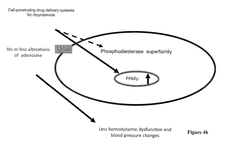
[0062] Figure 4a shows that if dipyridamole is administered in free form,
it mainly acts
outside of cells and will promote the accumulation of adenosine, which will
lead to
hemodynamic dysfunction and blood pressure changes. Figure 4b shows that
dipyridamole
inside the cell can activate the PPARy signaling pathway, which can inhibit
renal loss caused by
LPS. In such case, the unfavorable action of dipyridamole outside the cell can
be avoided. In
the Examples below, the present invention demonstrates that by delivering
dipyridamole directly
into cells, the binding to the receptors on the cell membrane can be avoided
so as to reduce side
effects such as oxidative stress and vasoconstriction caused by the
accumulation of adenosine.
[0063] The way nanoparticle drug carriers enter cells is different from
that of conventional
drugs. Conventional drugs enter cells by diffusion, which is dose-dependent.
That is, the higher
the drug concentration in the blood, the higher the drug concentration in the
cells, and the drugs
can only enter cytoplasm. Nanoparticle drug delivery systems are absorbed by
cells through
endocytosis and are lysosomotropic after entering cells. At the initial stage
after injection, the
concentration of the nanoparticle drug delivery systems increases in a time-
dependent manner.
[0064] Endocytosis is a process to incorporate extracellular materials into
cells. This process
can be categorized into three types, i.e., phagocytosis, pinocytosis, and
receptor-mediated
endocytosis. Phagocytosis only occurs in specialized cells. These cells
proliferate and aggregate
upon stimulation by extracellular materials and engulf them into lysosomes in
the cells for
degradation. This process occurs in macrophages and neutrophils of the immune
system.
Pinocytosis is a process that internalizes extracellular fluid and molecules
within it through the
invagination of the cell membrane to form a pocket, which then pinches off
into the cell to form
a vesicle. The vesicle then travels into the cytosol and fuses with other
vesicles such as
endosomes and lysosomes.
[0065] Depending on the structure of the carriers, pinocytosis can be
categorized into two
types, fluid phase pinocytosis and adsorptive pinocytosis. If the carrier does
not have a
functional group that interacts with the cells, the cells will engulf the drug
carrier by fluid phase
pinocytosis. This process is slow and dependent on the carrier concentration
around the cell
membrane. Adsorptive pinocytosis occurs when the carrier has a hydrophobic
group or is
positively charged. Such carrier will be physically adsorbed by the cell
membrane and increase
14
CA 02975000 2017-07-26
WO 2016/119701 PCT/CN2016/072347
the engulfing ability of the cells. The above two types of endocytosis are non-
specific processes
and are not suitable for delivery of drugs to their targets. Targeting can
only be achieved in
certain cancer tissues through enhanced permeability and retention (EPR).
[0066] Receptor-mediated endocytosis is a process by which cells absorb
molecules
(endocytosis) by the inward budding of plasma membrane vesicles containing
proteins with
receptor sites specific to the molecules being absorbed. After the drug
carrier binds to the
receptor on the cell, an intrinsic signal will trigger the cell membrane to
form a coated pit. The
surface area of a coated pit amounts to 1 to 2% of the cell membrane. The
coated pit will detach
from the cell membrane and enter into the cell to form coated vesicles in the
cell, and
subsequently form endosomes and move inside the cell in saltatory motion. An
endosome is a
complicated structure comprising microtubules and vesicles. The vesicles can
fuse with Golgi.
Due to the proton pump (ATPase), endosomes usually become acidic. The
endosomes will then
fuse with lysosomes to form secondary lysosomes.
[0067] The cell membrane is a barrier to be overcome for efficient delivery
of therapeutics
into a target site in mitochondria, cytoplasm or nucleus. The hydrophobic
phospholipids are
major components of the cell membrane that obstruct the transportation of
therapeutics. Thus,
various delivery systems, such as liposomes, nanoparticles and viral vectors,
have been
developed to transfer small molecules, peptides, proteins, and
oligonucleotides across the
membrane. Such manner of drug delivery is herein referred-to as cell-
penetrating drug delivery
systems.
[0068] A number of cell-penetrating drug delivery systems (liposomes, cell
penetrating
peptides, cationic polymer conjugates, and polymeric nanoparticles) have been
explored for
intracellular delivery of therapeutics. They need to be adapted to cross a
series of membrane
barriers in order to reach the site of drug action in the cells. During this
process, a significant
portion of the drug molecules will be lost at each successive barrier. These
barriers include
cellular association and internalization of the drug-carriers by endocytosis;
intracellular
trafficking and release of drug or drug-carrier into the cytoplasm;
cytoplasmic translocation of
drug or drug-carrier to nucleus or any other cellular organelle; and the
nuclear/organellar uptake.
Cells contain several intracellular organelles with specific functions.
Intracellular targeting of
therapeutics to these specific organelles is expected not only to
significantly enhance the
therapeutic efficacy but also reduce non-specific effects and hence toxicity.
Therefore, there is
CA 02975000 2017-07-26
WO 2016/119701 PCT/CN2016/072347
significant interest in achieving intracellular target-specific delivery of
therapeutics using
different cell-penetrating drug delivery systems.
[0069] The cell-penetrating drug delivery systems that facilitate the
endocytosis of drugs
include nano-sized polymeric carriers and liposomes. Depending on the
properties of the drugs
and preparation processes, nano-sized drug carriers can be categorized into
nanoparticles,
nanoliposomes, nano suspended particles, solid lipid nanoparticles, magnetic
nano-carriers, and
the like.
[0070] In addition to the above-mentioned cell-penetrating drug delivery
systems, cell-
penetrating peptides (CPP), biodegradable nanoparticles, and viral vectors may
also be used as
delivery systems for enhancing the penetration of drugs into cells.
[0071] The cellular internalization of RGD peptides is primarily mediated
by the clathrin,
caveolae and macropinocytosis endocytic pathways at the plasma membrane. As
one of the
primary effectors of endocytic transport at the plasma membrane, clathrin-
mediated endocytosis
is involved in the transport of large extracellular particles into the cell
through the receptor-
dependent endocytosis of ligands. An alternative route for peptide
internalization is through
caveolae-mediated endocytosis. Internalization through this pathway is
facilitated by lipid rafts
in the cell membrane; these rafts contain caveolin-1 proteins that form
endosomes, which are
then transported throughout the cell. In contrast, macropinocytosis involves
the fluid-phase
endocytosis of small extracellular particles into the cell. It has been
demonstrated that the aVr33
integrin can be internalized through both the clathrin and caveolae-dependent
endocytic
pathways as part of the regulation of integrin turnover. Therefore, RGD
peptides are ideal for
cell penetrating drug delivery system (Cam A, Sivaguru M, Gonzalez de Mejia E.
Endocytic
mechanism of internalization of dietary peptide lunasin into macrophages in
inflammatory
condition associated with cardiovascular disease. PLoS One. 2013 Sep
5;8(9):e72115).
[0072] As the cell membrane constitutes a major barrier for intracellular
delivery of large-
sized hydrophilic proteins, peptides and oligonucleotides, cell penetrating
peptides (CPPs) have
been explored to overcome this barrier. These CPPs can ferry molecules or
colloidal drug
delivery systems that are tagged to them across the cell membrane, into the
cytoplasm and to the
nucleus. The characteristics of CPPs are attributed to the presence of a
stretch of 9-16 cationic
amino acid residues; the most commonly studied CPPs include HIV-1
transactivating
transcriptional activator (TAT) peptide, HSV VP-22 (Herpes Simplex virus type-
1 transcription
16
CA 02975000 2017-07-26
WO 2016/119701 PCT/CN2016/072347
factor) peptide and penetratin. Several theories have been proposed to
determine the exact
mechanism by which these CPPs enter the cells. For example, TAT penetration
through cell
membrane has been shown to be independent of receptors and transporters, and
has been
suggested to enter the cell by forming an inverted micelle by destabilizing
the phospholipid
bilayer. The main benefit of TAT coupling is that, along with efficient
delivery of molecules,
biological activity of the coupled molecule is preserved, and the size of the
molecule being
transported is also not a rate-limiting factor.
[0073] TAT has been suggested not only to enhance intracellular delivery,
but also nuclear
delivery, and hence has been investigated for nucleic acid delivery. TAT
peptide conjugated to
antisense oligonucleotide has been shown to deliver oligonucleotides to the
nucleus. After being
internalized, TAT peptide has also been found to co-localize inside the Golgi
body along with
BODIPY-ceramide, which is a marker for Golgi body. Therefore, it is quite
possible that there is
direct trafficking from the early endosome to the Golgi body without entering
the late endosome.
A secretory pathway could be present where the peptide enters the cytosol from
the endoplasmic
reticulum. Gene therapy has demonstrated a significant potential in the
treatment of genetic,
acquired and neurodegenerative disorders. Amongst non-viral gene delivery
methods, various
drug delivery systems and polymers are being investigated such as liposomes,
cationic lipid-
DNA, polymer complexes. To overcome relatively inefficient cellular uptake of
non-viral gene
expression vectors, TAT peptide conjugation to vectors has been explored.
[0074] Kleeman et al. have demonstrated gene expression in alveolar basal
epithelial cells
with polyethylenimine (PEI) covalently coupled to TAT through a polyethylene
glycol (PEG)
spacer which demonstrated higher transfection efficiencies in vivo in mice
lung following
intratracheal administration than unconjugated PEG complex. In a similar study
by Rudolph et
al., solid lipid particles conjugated to dimeric HIV-1 TAT demonstrated
enhanced gene delivery
to the lungs.
[0075] CPPs typically have an amino acid composition that either contains a
high relative
abundance of positively charged amino acids such as lysine or arginine or has
sequences that
contain an alternating pattern of polar/charged amino acids and non-polar,
hydrophobic amino
acids. These two types of structures are referred to as polycationic or
amphipathic, respectively.
A third class of CPPs comprises the hydrophobic peptides, containing only
apolar residues, with
low net charge or hydrophobic amino acid groups that are crucial for cellular
uptake. Among the
17
CA 02975000 2017-07-26
WO 2016/119701 PCT/CN2016/072347
cell-penetrating peptides, the arginine-rich cell-penetrating peptides have
been the most widely
studied. Examples include the TAT peptide from the HIV transactivator protein
TAT, Penetratin,
a 16 amino acid domain from the Antennapedia protein of Drosophila, a flock
house virus (FHV)
coat peptide (sequence 35-49), and oligoarginines.
[0076] Biodegradable nanoparticle-mediated intracellular delivery is a
dynamic process;
involving endocytosis, exocytosis, and sorting into different intracellular
compartments. It
appears that the NP surface and its interaction with cell surface controls the
uptake and
intracellular trafficking of biodegradable nanoparticles, and hence that of
the encapsulated
therapeutic agents.
[0077] Viral vectors are tools commonly used by molecular biologists to
deliver genetic
materials into cells. This process can be performed inside a living organism
(in vivo) or in cell
culture (in vitro). Hence, viral vectors are applicable options for use in
cell-penetrating drug
delivery systems.
[0078] Cell-penetrating peptides and biodegradable nanoparticles can be
used not only to
modify drugs but also be conjugated to carries to enhance the transmembrane
effects.
[0079] Dipyridamole is an equilibrative nucleoside transporter (ENT)
inhibitor. Nucleoside
transporters (NTs) play an essential role in the transport of nucleosides
across cellular
membranes. Dipyridamole blocks the equilibrative nucleoside transporter (ENT),
which
facilitates the transmembranous diffusion of adenosine. Dipyridamole will
increase the
extracellular endogenous adenosine concentration, mainly in situations of
increased extracellular
formation of adenosine, such as occurs during hypoxia or inflammation.
However, the
extracellular endogenous adenosine concentration induced by dipyridamole
causes vasodilatation,
which contributes to the metabolic control of organ perfusion. Dipyridamole
stress myocardial
imaging is a successful, widely used technique for diagnosing and evaluating
coronary artery
disease. Coronary vasodilation with IV dipyridamole is associated with
significant reductions in
blood flow to collateral-dependent myocardium consistent with coronary steal
in patients with
CAD. In addition, there have been further studies that discovered
vasoconstrictor and
vasodilator effects of dipyridamole in many organs, including kidney, lung,
pancreas, brain and
so on.
[0080] Dipyridamole not only causes vasoconstriction in some organs but can
also lead to
low blood pressure and subsequent side effects such as vertigo and
palpitations due to dilation of
18
CA 02975000 2017-07-26
WO 2016/119701 PCT/CN2016/072347
blood vessels of the heart. The effect of reducing blood pressure makes
dipyridamole unsuitable
for the treatment of patients who are physiologically unstable, such as those
having, but not
limited to, sepsis, ischemic stroke, hemorrhagic stroke, acute lung injury,
acute liver injury,
myocardium infarct, and cardiorenal syndrome. Furthermore, the blood-flow
restricting effect of
dipyridamole limits its application in the treatment of diseases involving
organs rich with blood
vessels.
[0081] Since the pharmacological action of dipyridamole is mainly on cell
membranes, a
delivery system designed for membrane penetration that avoids binding with
equilibrative
nucleoside transporter on the membrane while enhancing the intracellular
signal transduction and
PPARy regulation can prevent the effect of tissue hypoperfusion due to
increased cardiovascular
dilation and local blood flow restriction. The limitation in clinical
applications of dipyridamole
in acute and severe patients due to the decrease of blood pressure can thus be
lifted.
[0082] Dipyridamole is also a non-selective phosphodiesterase inhibitor.
Increase of
intracellular drug delivery will enhance the inhibition of dipyridamole on
intracellular PDE.
Members of the PDE family have unique cell- and tissue-specific distribution.
Dipyridamole
may be used as anti-inflammatory, anti-oxidant, anti-fibrosis, and smooth
muscle relaxing agents
for treating diseases associated with PDE regulation depending on the
distribution profile of PDE
on cell membranes or in cytoplasm in different tissues.
[0083] The unique cell- and tissue-specific distribution of PDEs are shown
in the below table
(see US 2012/0065165):
PDE
Isoenzyme Substrate Tissue Expression
1 Ca2+/calmodulin- Heart, brain, lung, smooth muscle
stimulated
2 cGMP-stimulated Adrenal gland, heart, lung, liver,
platelets
3 cGMP-inhibited Heart, lung, liver, platelets, adipose
tissue, inflammatory cells
4 cAMP-selective Sertoli cells, kidney, brain, liver, lung,
inflammatory cells
cGMP-specific Lung, platelets, vascular smooth
muscle, heart
6 cGMP-specific Photoreceptor
19
CA 02975000 2017-07-26
WO 2016/119701 PCT/CN2016/072347
7 cAMP-specific, Skeletal muscle, heart, kidney, brain,
high affinity pancreas, T lymphocytes
8 cAMP-selective Testes, eye, liver, skeletal muscle,
heart, kidney, ovary, brain, T
lymphocytes
9 cGMP-specific Kidney, liver, lung, brain, possibly
heart
cGNO-sensitive, Testes, brain
cAMP-selective
11 cGMP-sensitive, Skeletal muscle, prostate, kidney, liver,
dual specficity pituitary and salivary glands, testes
[0084]
Increase in the capability of dipyridamole to penetrate the membrane can
facilitate the
inhibition of PDE3, PDE5 and PDE8 in specific tissues and confer dipyridamole
therapeutic
efficacy in diseases associated with PDE3, PDE5 and PDE8. In such case,
dipyridamole may be
used for treating lower urinary tract dysfunction and erectile dysfunction,
like other PDE5
inhibitors. Furthermore, since dipyridamole is a non-selective PDE inhibitor,
it may be used for
the treatment of PDE associated diseases when delivered by a transmembrane
drug delivery
system.
[0085] In
an embodiment, the compound of formula (I) of the invention is encapsulated
in a cell-penetrating drug delivery system for delivery into the cell. In a
preferred embodiment,
the cell-penetrating drug delivery system is a niosome, a polymersome, a
nanoparticle, a
liposome, a nano suspended particle, a solid lipid nanoparticle, a magnetic
nano-carrier, a
micelle, a macromolecular conjugate or a particulate drug carrier. Preferably,
the cell-
penetrating drug delivery system is a liposome. The liposome suitable for the
present invention
has a diameter in the range of about 100-300 nm, preferably about 150-280 nm,
more preferably
about 180-270 nm.
[0086] In
another embodiment of the invention, the cell-penetrating drug delivery
systems may be niosomes, polymersomes, or polymers that have a diameter of
less than 1 um.
Modifications can be made based on surface electric potential,
hydrophilicity/hydrophobicity,
size, morphology, shape and/or surface curvature.
[0087] The
liposome formulation of the invention may comprise vesicles of various
nature (e.g., unilamellar or multilamellar), composition, size, and
characteristics, enclosing an
CA 02975000 2017-07-26
WO 2016/119701 PCT/CN2016/072347
aqueous medium of diverse composition, pH and osmotic strength. In a preferred
embodiment,
the main constituents of the liposome lipid layer membrane are selected from
the group
consisting of natural or synthetic phospholipids such as those listed below:
- 1,2-Dilauroyl-sn-Glycero-3-Phosphocholine (DLPC)
- 1,2-Dimyristoyl-sn-Glycero-3-Phosphocholine (DMPC)
- 1,2-Dipalmitoyl-sn-Glycero-3-Phosphocholine (DPPC)
- 1,2-Distearoyl-sn-Glycero-3-Phosphocholine (DSPC)
- 1,2-Dioleoyl-sn-Glycero-3-Phosphocholine (DOPC)
- 1,2-Dimyristoyl-sn-Glycero-3-Phosphoelhanolamine (DMPE)
- 1,2-Dipalmitoyl-sn-Glycero-3-Phosphoelhanolamine (DPPE)
- 1,2-Distearoyl-sn-Glycero-3-Phosphoelhanolamine (DSPE)
- 1,2-Dioleoyl-sn-Glycero-3-Phosphoelhanolamine (DOPE)
- 1-Myristoy1-2-Palmitoyl-sn-Glycero-3-Phosphocholine (MPPC)
- 1-Palmitoy1-2-Myristoyl-sn-Glycero-3-Phosphocholine (PMPC)
- 1-Stearoy1-2-Palmitoyl-sn-Glycero-3-Phosphocholine (SPPC)
- 1-Palmitoy1-2-Stearoyl-sn-Glycero-3-Phosphocholine (PSPC)
- 1,2-Dimyristoyl-sn-Glycero-3-[Phospho-rac-(1-glycerol)] (DMPG)
- 1,2-Dipalmitoyl-sn-Glycero-3-[Phospho-rac-(1-glycerol)] (DPPG)
- 1,2-Di stearoyl-s/i-Glycero-3-[Pho spho-rac-(1-glycerol)] (DSPG)
- 1,2-Dioleoyl-sn-Glycero-3-[Phospho-rac-(1-glycerol)] (DOPG)
- 1,2-Dimyristoyl-sn-Glycero-3-Phosphate (DMPA)
- 1,2-Dipalmitoyl-sn-Glycero-3-Phosphate (DPPA)
- 1,2-Dipalmitoyl-sn-Glycero-3-[Phospho-L-Serine] (DPP S)
-phosphatidylserine (PS), and
- Natural L-a-phosphatidylcholine (from chicken egg, EPC, or from soy, SPC
and HSPC).
[0088] Preferred phospholipids are long saturated phospholipids, e.g.
those having alkyl
chains of more than 12, preferably more than 14, more preferably more than 16,
most preferably
more than 18 carbon atoms.
[0089] Preferred liposome compositions for use according to the invention
are preferably
those in which the liposomes are uni- and/or multilamellar, and comprise:
21
CA 02975000 2017-07-26
WO 2016/119701 PCT/CN2016/072347
(i) 1 to 100, preferably 40 to 70 mol% physiologically acceptable
phospholipids, preferably
selected from the group consisting of DLPC, DMPC, DPPC, DSPC, DOPC, DMPE,
DPPE,
DSPE, DOPE, MPPC, PMPC, SPPC, PSPC, DMPG, DPPG, DSPG, DOPG, DMPA, DPPA,
DPPS, PS,EPC, SPC and HSPC.
(ii) 1 to 100, preferably 40 to 70 mol% sphingolipids, preferably
sphingomyelin;
(iii) 1 to 100, preferably 40 to 70 mol% surfactants, preferably featuring
hydrophobic alkyl
ethers (e.g. Brij), alkyl esters, polysorbates, sorbitan esters, and/or alkyl
amides;
(iv) 5 to 100, preferably 50 to 100 mol% amphiphilic polymers and/or co-
polymers, preferably
block copolymers comprising at least one block of a hydrophilic polymer or
copolymer such as
polyethylene glycol, and at least one block of a hydrophobic polymer or
copolymer such as
poly(lactide), poly(caprolactone), poly(butylene oxide), poly(styrene oxide),
poly(styrene),
poly(ethylethylene), or polydimethylsiloxanes,
(v) 0 to 60 mol%, preferably 20 to 50 mol% toxin retention-enhancing
compounds, preferably
sterol derivatives, preferably cholesterol, or
(vi) 0 to 30 mol%, preferably 1 to 5 mol% steric stabilizers, preferably
PEGylated compounds,
preferably PEGylated lipids, more preferably DSPE-PEG.
[0090] In a preferred embodiment, liposome-like vesicles are made from
polymers and
comprise no lipids, for which reason they are formally not considered
liposomes but are called
polymersomes. However, for the purpose of the present invention, polymersomes
are meant to
be encompassed by the term liposome as used for defining the invention and the
claims.
[0091] Similarly, liposome-like vesicles made from synthetic surfactants
and comprising
no lipids are called niosomes. However, for the purpose of the present
invention, niosomes are
meant to be encompassed by the term liposome as used for defining the
invention and the claims.
[0092] In an embodiment of the invention, polymerization of different high
molecular
polymers can be used, which comprise those in tri-block copolymer form such as
ABA and BAB,
and those in block copolymer form such as PLLA-PEG, PLGA-PEG, PLA-PEG, PLLA-
mPEG,
PLGA-mPEG and PLA-mPEG. Various shapes such as asterisk and L form can be
designed,
including block copolymers of PEG-(PLGA)8, PEG-(PLLA)8 and PEG-(PDLA)8 Star.
PEGylated modification can be used to modify any vehicle such as polymeric
vehicle and
liposome to achieve the effect of reducing the binding rate of plasma proteins
(see Park, J. et al.,
(2009) "PEGylated PLGA nanoparticles for the improved delivery of doxorubicin.
22
CA 02975000 2017-07-26
WO 2016/119701 PCT/CN2016/072347
Nanomedicine." 5(4):410-418.; ',tick, M. et al., (1998) "Plasma protein
adsorption on
biodegradable microspheres consisting of poly(D,L-lactide-co-glycolide),
poly(L-lactide) or
ABA triblock copolymers containing poly(oxyethylene). Influence of production
method and
polymer composition." J. Control Release. 55(2-3):107-20.; and Sempf, K. et
al, (2013)
"Adsorption of plasma proteins on uncoated PLGA nanoparticles." Eur. J. Pharm.
Biopharm.
85(1):53-60).
[0093] The animal dose should not be extrapolated to a human equivalent
dose (HED) by
a simple conversion based on body weight. The Food and Drug Administration has
suggested
that the extrapolation of animal dose to human dose is correctly performed
only through
normalization to BSA, often represented in mg/m2. The human dose equivalent
can be more
appropriately calculated using the formula: HED (mg/kg) = Animal dose (mg/kg)
multiplied by
Animal Km/Human Km. To convert the dose used in a mouse to a dose based on
surface area for
humans, multiply 22.4 mg/kg (Baur's mouse dose) by the Km factor (3) for a
mouse and then
divide by the Km factor (37) for a human (see below Table).
Values based on data from FDA Draft Guidelines
Species Weight (kg)BSA (m2) Km factor
Human
Adult 60 1.6 37
Child 20 0.8 25
Baboon 12 0.6 20
Dog 10 0.5 20
Cat 2.5 0.2 12.5
Monkey 3 0.24 12
Rabbit 1.8 0.15 12
Guinea pig0.4 0.05 8
Rat 0.15 0.025 6
Hamster 0.08 0.02 5
Mouse 0.02 0.007 3
[0094] To convert a dose expressed in mg/kg to dose in mg/m2, multiply by
Km value.
According to the present invention, the effective dose of liposome-
dipyridamole in mice is 10
mg/kg-100 mg/kg, in hamsters 6-60 mg/kg, in rats 5-50 mg/kg, in guinea pigs
3.75-37.5 mg/kg,
in rabbits 2.5-25 mg/kg, in monkeys 2.5-25 mg/kg, in dogs 1.5-15 mg/kg, in
cats 2.4-24 mg/kg,
in baboons 1.5-15 mg/kg, in children 1.2-12 mg/kg, and in adults 0.81-8.1
mg/kg. Taking into
consideration the differences in drug sensitivity among species, the broadest
dose range without
23
CA 02975000 2017-07-26
WO 2016/119701 PCT/CN2016/072347
limiting the species is 0.4-160 mg/kg, preferably 0.6-120 mg/kg, more
preferably 0.8 mg/kg-100
mg/kg.
[0095] Having now generally described the invention, the same may be more
readily
understood through reference to the following examples, which provide
exemplary protocols for
the production of the pharmaceutical composition of the invention and its use
in the enhancement
of the treatment of acute stroke. The examples are offered for illustrative
purposes only, and are
not intended to limit the scope of the present invention in any way. Efforts
have been made to
ensure accuracy with respect to numbers used (e.g., amounts, temperatures,
etc.), but some
experimental error and deviation should, of course, be allowed for.
Examples
Example 1: Preparation of dipyridamole liposome
[0096] Liposomes were prepared with positive and neutral charge containing
phospholipid and cholesterol, in which the mole percent of cholesterol was 5%
to 75%, either
with or without PEG2000- DSPE at 5 mol% to phospholipids. Small unilamellar
vesicles were
prepared. The dried lipid films were hydrated with ammonium sulfate and
sequentially extruded
through a series of polycarbonate membrane filters. Dipyridamole was
encapsulated into the
liposomes via a transmembrane pH gradient or dehydration-rehydration, and the
diameters of the
extruded liposomes were in the range of 100-350 nm. The diameter of the
liposome-
dipyridamole was about 169 to 276 nm as shown in Figure 5.
Example 2: HEK293 cells treated with the dipyridamole liposome
Example 2.1: Expression of PPARy in cells treated with the dipyridamole
liposome
[0097] The cell line used in the assay was human embryonic kidney cells,
HEK293. The
cells were treated with the agents shown in Table 1 below.
Table 1: Experimental Design
Groups 1 2 3 4 5 6
LPS 6h+ LPS 6h+ LPS 6h+ LPS 6h+
Dipyridamole Dipyridamole Dipyridamole Dipyridamole
10Ong/mL
Treatments None LPS 10 ug/mL 100 ug/mL 10 ug/mL 100 ug/mL
(Free form) (Free form) (Liposome) (Liposome)
2h 2h 2h 2h
24
CA 02975000 2017-07-26
WO 2016/119701 PCT/CN2016/072347
[0098] The cells were collected at Oh, 3h and 12h after treatment. The
collected cells
were washed with 1504, Buffer A (10mM Hepes p11=7.9, 1.5mM MgC12, 10mM KC1,
1.0mM
DTT, 0.1 % Triton-X 100), and centrifuged at 3000g for 10 minutes at 4 C.
Supernatant
containing cytosolic proteins was collected, and the pellets were re-suspended
with 504 Buffer
B (20mM Hepes pfl = 7.9, 1.5mM MgC12, 0.42M NaC1, 1.0mM DTT, 1.0M PMSF, 0.2mM
EDTA) and incubated on ice for 30 minutes followed by centrifuging at 12000g
for 10 minutes at
4 C. Supernatant containing nucleic proteins was then collected, and the
expression of P65
protein, which is indicative of the activation of PPARy, was analyzed using
Western blotting.
The method is as follows:
[0099] Protein concentration was measured using Bradford assay. 6X sample
buffer (0.8
mM Tris-HC1, 10 mM EDTA, 10% SDS, 60% glycerol, 0.6 M P-mercaptoethano, 0.06%
bromophenol blue, pH 6.8) was added into 50 ug of nuclear proteins and an
equal volume of
lysis buffer was added into the samples. After being heated at 95 C for 10
minutes to denature
the proteins, the samples were immediately cooled on ice.
[00100] The samples were then separated by 10% SDS-PAGE electrophoresis
(100 V) and
transferred from the SDS-PAGE gels to PVDF membranes by wet blotting. The PVDF
membranes were then treated with 5% skimmed milk at room temperature for 60
minutes to
block non-specific binding. The membranes were incubated with primary antibody
overnight at
4 C and washed three times with PBST. The membranes were incubated with
secondary
antibody at room temperature for 60 minutes and washed three times with PBST.
The
membranes were then washed one more time with PBS and incubated with an
enhanced
chemiluminescence (ECL) substrate for detection. Photos of the images were
taken using
automated chemiluminescence and fluorescence imaging system (UVP Biospectrum).
The
expression of PPARy in the testing groups relative to the control group is
shown in Figure 7a (12
hour) and Figure 7b (0 and 3 hour).
[00101] The primary antibodies used in this experiment is rabbit anti-human
PPARy
antibody (1:1000) (catalog no.: 07-466), MILLIPORE; and rabbit anti-human
Lamin A/C
(1:1000) (catalog no.: GTX62457), GeneTex. The secondary antibody used in this
experiment is
mouse anti-rabbit HRP (1:3000) (ab6721), sigma.
Example 2.2: Viability of cells treated with the dipyridamole liposome
CA 02975000 2017-07-26
WO 2016/119701 PCT/CN2016/072347
[00102] The number of viable cells was evaluated 24 hours after initial
treatment using the
Cell Counting Kit-8 (Dojindo Laboratories, Japan) following the manufacturer's
instructions,
and the optical absorbance at wavelength 450 nm was measured for the
supernatant of each well
using the plate reader Multiskan EX (Thermo Fisher Scientific Inc., Waltham,
MA). The data
are shown in Figure 8.
Example 3: Survival rate analysis in mice with LPS-induced sepsis
[00103] Male C57B1/6J mice, 8-12 weeks of age, were used in this study.
They were
reared in an air-conditioned environment with 6 am to 18 pm light cycle and
fed standard rodent
chow ad libitum. LPS (Escherichia coli 0111:B4) (SigmaAldrich, Milwaukee, WI,
USA) was
freshly dissolved in sterile pyogen-free water each time when applied. First,
mice were injected
intraperitoneally with LPS (16 mg/kg) and followed for 72 hours to observe the
survival rate.
The dose of LPS was determined by preliminary experiments that demonstrated
longer survival
than 24 hours in half of the animals injected. Dipyridamole was administered 1
hour after the
LPS treatment.
Example 4: Detection of biomarkers in plasma
4.1: Liver markers (AST, ALT) and kidney markers (BUN, Creatinine)
[00104] Blood samples for biochemical measurements were collected from each
animal
before and at 24 hours into the experiment. Samples were separated by
centrifugation, and the
serum was stored at -80 C until analysis. Serum total cholesterol was measured
using Merck
assay kits (Darmstadt, Germany). Serum blood urea nitrogen (BUN), creatinine,
alanine
aminotransferase (ALT), and aspartate aminotransferase (AST) were also
measured using a
SPOTCHEMTM automatic dry chemistry system (SP-4410; Arkray, Shanghai, Japan).
The data
are shown in Figure 9.
[00105] Figure 9 shows that LPS treatment induces liver and kidney injuries
which
significantly increase aspartate aminotransferase (AST), alanine
aminotransferase (ALT),
creatinine, and BUN levels in the blood. Treatments with low doses of
dipyridamole or liposome
dipyridamole attenuate LPS-induced increases of AST, ALT, creatinine, and BUN
levels in serum.
This indicates that dipyridamole has the therapeutic efficacy of treating
acute or chronic liver and
kidney inflammation as well as sepsis. In addition, since high dose of
dipyridamole leads to
changes in blood pressure and influences physiological conditions and survival
rate, the dose-
dependent efficacy of lipo some dipyridamole may imply a broader range of
applicable doses.
26
CA 02975000 2017-07-26
WO 2016/119701 PCT/CN2016/072347
Example 5: Measurement of tissue injuries
5.1:IHC
[00106] Paraffin-embedded sections (3 um) were prepared from livers and
lungs that were
fixed in 10% phosphate-buffered formalin. Periodic acid-Schiff (PAS) stain was
used for the
analysis of morphology with light microscopy (Nikon E800; Melville, NY) by a
blinded observer.
For each mouse, at least 10 high-power fields were examined. The HE stain of
liver and lung
tissues are shown in Figures 10 and 11, respectively.
[00107] Excessive inflammation and tissue damage induced by accumulated
macrophages
and neutrophils were observed in HE-stained liver and lung sections 72 hours
after LPS
treatment. Post-treatment with dipyridamole or liposome dipyridamole
attenuated tissue damage
and inflammation. Compared to free form dipyridamole, liposome dipyridamole
demonstrates
better therapeutic efficacy in histology.
5.2: Activation state of PPARy in kidney and liver
[00108] Mouse tissues were homogenized in 10 mM Tris¨HC1 (pH 7.5), 1 mM
EDTA, 250
mM sucrose, 10 mM 2-mercaptoethanol (Nacarai tesque, Inc.), protease inhibitor
(cOmplete,
Mini, Roche Diagnostics) and phosphatase inhibitor (PhosSTOP, Roche
Diagnostics). Six up-
and-down strokes were used in a Braun Potter S homogenizer running at 1000
rpm. The
homogenate was centrifuged (800g), and the pellet was discarded. The
supernatant was
centrifuged again at 12,000g for 10 mM, and the resulting supernatant was
collected. After the
samples were collected, protein concentration was measured by Bradford assay.
6X sample
buffer (0.8 mM Tris-HC1, 10 mM EDTA, 10% SDS, 60% glycerol, 0.6 M P-
mercaptoethano,
0.06% bromophenol blue, pH 6.8) was added into 50 ug of whole cell proteins
and an equal
volume of lysis buffer was added into the samples. After being heated at 95 C
for 10 minutes to
denature the proteins, the samples were immediately cooled on ice.
[00109] The samples were then separated by 10% SDS-PAGE electrophoresis
(100 V) and
transferred from the SDS-PAGE gels to PVDF membranes by wet blotting. The PVDF
membranes were then treated with 5% skimmed milk at room temperature for 60
minutes to
block non-specific binding. The membranes were incubated with primary antibody
overnight at
4 C and washed three times with PBST. The membranes were incubated with
secondary
antibody (anti-rabbit IgG, sigma) at room temperature for 60 minutes and
washed three times
with PBST. The membranes were then washed one more time with PBS and incubated
with an
27
CA 02975000 2017-07-26
WO 2016/119701 PCT/CN2016/072347
enhanced chemiluminescence (ECL) substrate for detection. Photos of the images
were taken
using automated chemiluminescence and fluorescence imaging system (UVP
Biospectrum).
Antibody used: t-PPARy (1:1000; abcam ab191407) and r3-actin (1:1000; GeneTex
GTX109639).
The data of t-PPARy expression in kidney and liver are shown in Figures 12a
and 12b,
respectively.
[00110] From the experimental results, it is clearly learned that no matter
in kidney or liver
tissue, dipyridamole or liposome dipyridamole has the activity to induce PPARy
expression in a
dose dependent manner. Due to enhanced drug penetration into the cells by
liposome through
phagocytosis and fusion, the expression of PPARy is greatly increased.
Example 6: The effect of the dipyridamole liposome on blood pressure
[00111] Dipyridamole has a blood pressure-lowering effect. In the treatment
of various
acute and critical conditions, lowering the blood pressure may influence the
prognosis of disease.
Hence, detecting the effect of the dipyridamole liposome of the invention on
blood pressure can
permit evaluation of maximum dose feasible for clinical use.
[00112] Blood pressure of the mice was measured using a non-invasive blood
pressure
device after intravenous administration of the following agents: (1) saline,
(2) LPS, (3) LPS prior
to dipyridamole (free form), 10 mg/kg, (4) LPS prior to dipyridamole (free
form), 100 mg/kg, (5)
LPS prior to dipyridamole liposome, 10 mg/kg, and (6) LPS prior to
dipyridamole liposome, 100
mg/kg. The results are shown in Figure 13a.
[00113] Additional test results with different doses (no LPS prior to drug
treatment) are
shown in Figure 13b.
The above experimental data demonstrate that by increasing the ability of
dipyridamole to enter
the cell, the pharmacological mechanism of dipyridamole will be changed,
leading to the
increased activity of dipyridamole on PPARy expression and the reduction of
activity of
dipyridamole on cell membrane, thereby reducing the stimulation of drug on
blood vessel and
the consequent severe interference on blood flow. By increasing the activity
of dipyridamole on
PPARy expression, dipyridamole demonstrates potential in the treatment of
multiple diseases.
By the action of multiple mechanisms, the anti-inflammatory and anti-apoptosis
activities of
dipyridamole are increased via PPARy pathway. Dipyridamole may be used for
treating acute
and severe diseases and small mammals without interfering with blood pressure.
28