Note: Descriptions are shown in the official language in which they were submitted.
CA 02975557 2017-07-31
WO 2016/160653
PCT/US2016/024421
DEVICES AND METHODS FOR ULTRASOUND IMAGING
CROSS-REFERENCE TO RELATED APPLICATION(S)
[0001] This patent application claims the benefit of U.S. Nonprovisional
Patent
Application No. 15/073,749, filed March 18, 2016, and to U.S. Provisional
Patent
Application No. 62/140,564, filed March 31, 2015, the entireties of each of
which are
incorporated herein by reference.
TECHNICAL FIELD
[0002] Embodiments of the present disclosure relate generally to medical
devices
and related methods. More specifically, the present disclosure relates to
devices useful in
ultrasound-guided medical procedures, such as biopsy and diagnostic
procedures.
BACKGROUND
[0003] Medical procedures are often complicated by a limited field of view of
the
treatment area, and/or by temporal delay between capturing an image of the
treatment area
and performing the procedure. Sonography is one example of medical imaging,
wherein
sound waves (ultrasound) are sent through the body to collect information on
internal
tissues and structures based on the time, speed, and frequency at which the
sound waves are
reflected. In endoscopic ultrasound (EUS), an ultrasound probe is introduced
into the body
for internal imaging. The tip of the probe makes contact with tissue directly
or via a fluid-
filled balloon to emit and detect returning sound waves for display as an
image. However,
the ultrasound probe usually must be exchanged for other instruments to biopsy
tissue or
perform other procedures at a region of interest identified via the ultrasound
image. While
some devices include a needle adjacent to an ultrasound transducer, e.g., for
endobronchial
ultrasound transbronchial needle aspiration (EBUS-TBNA), the field of view is
limited and
does not allow for wide-angle imaging. In addition, these devices do not allow
for
exchange of the needle or use of other instruments during imaging.
- 1 -
CA 02975557 2017-07-31
WO 2016/160653
PCT/US2016/024421
SUMMARY OF THE DISCLOSURE
[0004] The present disclosure includes a medical device comprising a shaft,
the
shaft including a working channel extending from a proximal end of the shaft
to a distal end
of the shaft, and an ultrasound sensor disposed radially outward of a distal
portion of the
working channel such that the ultrasound sensor at least partially surrounds
the working
channel. The working channel of the medical device may be configured to
receive an
instrument and maintain an orientation of the instrument with respect to the
shaft.
Additionally or alternatively, the working channel may have a non-circular
cross-sectional
area to prevent rotation of the instrument within the working channel. In some
aspects, the
medical device may comprise the instrument. The instrument may be translatable
from the
proximal end of the shaft to the distal end of the shaft. In addition or
alternatively, the
instrument may comprise a flexible material, and/or may have a preset curved
configuration, such that a distal end of the instrument has a substantially
straight
configuration within the working channel and the curved configuration outside
the working
channel. In some aspects of the present disclosure, the shaft of the medical
device may
include an area having an echogenic signature distinct from a remainder of the
shaft. In
examples wherein the medical device comprises an instrument having a preset
curved
configuration, the distal end of the instrument may be radially aligned with
the area while in
the curved configuration.
[0005] The ultrasound sensor of the medical device may include a plurality of
sensors or a single ultrasound sensor disposed about the working channel to at
least partially
surround the working channel. For examples of medical devices comprising a
plurality of
ultrasound sensors, the ultrasound sensors may be spaced at regular intervals
about a
perimeter of the shaft. In some aspects, the ultrasound sensor (e.g., single
ultrasound sensor
or plurality of ultrasound sensors) may completely surround the working
channel. The
- 2 -
CA 02975557 2017-07-31
WO 2016/160653
PCT/US2016/024421
ultrasound sensor (e.g., single ultrasound sensor or plurality of ultrasound
sensors) may be
configured to image a single field of view greater than about 90 degrees,
greater than about
180 degrees, and/or may be configured to image a single field of view of about
360 degrees.
In some aspects of the present disclosure, the working channel of the medical
device may be
a first working channel, wherein the medical device may further comprise a
second working
channel radially inward of the ultrasound sensor.
[0006] In some aspects of the present disclosure, the medical device may
comprise a
controller coupled to the proximal end of the shaft and a plurality of control
members for
deflecting the distal end of the shaft in at least two different planes. The
plurality of control
members may extend from the controller to a portion of the shaft proximal to
the ultrasound
sensor.
[0007] The present disclosure also includes a medical device comprising a
shaft, the
shaft including a working channel extending from a proximal end of the shaft
to a distal end
of the shaft; an ultrasound sensor radially outward of a distal portion of the
working
channel; and a controller coupled to the proximal end of the shaft for
deflecting the distal
end of the shaft in at least two different planes. In some aspects, the
medical device may
further comprise a plurality of control members extending from the controller
to a portion of
the shaft proximal to the ultrasound sensor. Additionally or alternatively,
the ultrasound
sensor may completely surround the working channel. In some examples, the
ultrasound
sensor may be configured to image a single field of view greater than about 90
degrees,
greater than about 180 degrees, and/or may be configured to image a single
field of view of
about 360 degrees.
[0008] Medical devices according to the present disclosure may be useful in
treating
patients and/or conducting medical procedures. For example, the present
disclosure
includes a method of treating a patient comprising inserting a shaft of a
medical device into
- 3 -
CA 02975557 2017-07-31
WO 2016/160653
PCT/US2016/024421
a passageway of the patient, the shaft including a working channel extending
from a
proximal end of the shaft to a distal end of the shaft, and an ultrasound
sensor disposed
radially outward of a distal portion of the working channel such that the
ultrasound sensor at
least partially surrounds the working channel; the method further comprising
generating at
least one image with the ultrasound sensor. In some aspects, the method may
further
comprise inserting an instrument into the working channel, and manipulating
the instrument
to conduct a medical procedure on the passageway while generating the image.
BRIEF DESCRIPTION OF THE FIGURES
[0009] The accompanying drawings, which are incorporated in and constitute a
part
of this specification, illustrate various exemplary embodiments and together
with the
description, serve to explain the principles of the disclosed embodiments.
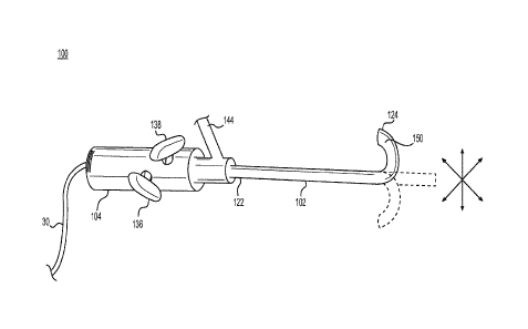
[0010] Fig. 1 shows an exemplary medical device, in accordance with one or
more
embodiments of the present disclosure.
[0011] Fig. 2A shows a distal portion of a medical device, and Figs. 2B and 2C
show cross-sectional views of Fig. 2A.
[0012] Figs. 3A and 3B show cross-sections of medical devices, in accordance
with
embodiments of the present disclosure.
[0013] Figs. 4A and 4B show exemplary instruments, in accordance with one or
more embodiments of the present disclosure.
[0014] Fig. 5A shows bronchial passageways of a patient, and Fig. 5B
illustrates a
medical procedure within a bronchial passageway, in accordance with one or
more
embodiments of the present disclosure.
- 4 -
CA 02975557 2017-07-31
WO 2016/160653
PCT/US2016/024421
DETAILED DESCRIPTION
[0015] Embodiments of the present disclosure include medical devices useful in
obtaining ultrasound images within the body, e.g., via one or more ultrasound
sensors, and
methods of performing medical procedures using such devices.
[0016] The term "ultrasound sensor" as used herein includes devices configured
to
transmit and/or receive ultrasound 20 kHz) and other frequency sound waves for
producing an image. For example, ultrasound sensors suitable for the present
disclosure
include transceivers and transducers capable of both transmitting and
receiving ultrasound.
By measuring the time between sending ultrasound signals and receiving the
echoes of
those signals reflected by various features in the body, the distance to those
features may be
determined, e.g., to obtain images. The images may provide information
regarding tissue
structure (e.g., density, shape, contour, etc.), the presence or absence of
tumors, lesions, or
other abnormalities, the size and location of any such abnormalities, and/or
blood flow or
other fluid flow characteristics. The images may be two-dimensional or three-
dimensional.
[0017] Ultrasound imaging may have advantages over other types of imaging. For
example, ultrasound can provide real-time images, avoiding a delay between
capturing an
image of an area of interest and performing a medical procedure on that area.
Further,
because ultrasound sensors operate via sound waves, e.g., rather than
electromagnetic
radiation, they are typically less harmful to the patient.
[0018] Fig. 1 shows an exemplary medical device 100 according to some
embodiments of the present disclosure. The medical device 100 may comprise a
controller
104 and a shaft 102 extending from a proximal end 122 to a distal end 124. The
controller
104 may have any suitable shape, including cylindrical and ergonomic shapes
for easy or
comfortable gripping by one or both hands. The shaft 102 may include one or
more
ultrasound sensors 150. The ultrasound sensor(s) 150 may be at or proximate to
the distal
- 5 -
CA 02975557 2017-07-31
WO 2016/160653
PCT/US2016/024421
end 124 of the shaft 102. The controller 104 may include an electronic cable
30, e.g., for
providing power to the ultrasound sensor(s) 150 and/or for communication
between the
ultrasound sensor(s) 150 and a processor or graphical interface.
[0019] In some embodiments, the medical device 100 may be steerable, e.g., to
allow an operator to navigate the shaft 102 through tortuous anatomy and/or
towards a site
of interest. Any suitable steering mechanism may be used. For example, the
steering
mechanism may comprise a plurality of steering wires coupling the controller
104 to the
shaft 102, e.g., to transmit user input from the controller 104 to the shaft
102 to articulate or
deflect the shaft 102 along one or more planes.
[0020] As shown in Fig. 1, for example, the controller 104 may include one or
more
actuators, e.g., first and second actuators 136, 138, each coupled to at least
one control
member 282 (e.g., mechanical or electronic steering wire) that extends along
the shaft 102
(see Fig. 2A). In some embodiments, the first actuator 136 may control
deflection of the
distal end 124 of the shaft 102 in one plane (e.g., xy plane), and the second
actuator 138
may control deflection of the distal end 124 in a different plane (e.g., yz
plane). For
example, the first actuator 136 may be coupled to a first pair of control
members 282 such
that rotational and/or translational motion of the first actuator 136 may
deflect the distal end
124 of the shaft 102 in one plane. Similarly, the second actuator 138 may be
coupled to a
second pair of control members 282, such that rotational and/or longitudinal
motion of the
second actuator 138, independent of the first actuator 136, may deflect the
distal end 124 in
a different plane. Concerted movement of the first and second actuators 136,
138 may
achieve deflection in a plurality of other planes, e.g., providing for 360
degree manipulation
of the shaft 102. In some embodiments, each actuator 136, 138 may be coupled
to only one
control member 282 and/or the controller 104 may include only one actuator.
- 6 -
CA 02975557 2017-07-31
WO 2016/160653
PCT/US2016/024421
[0021] Other steering mechanisms suitable for manipulating the shaft 102 may
be
used, including, but not limited to, other types of mechanical mechanisms and
electrical
mechanisms. For example, the controller 104 may be in electrical communication
with
various portions of the shaft 102 (e.g., via electronic control members 282),
such that user
input at the actuators 136, 138 may be converted to electrical signals to
control deflection of
the distal end 124 of the shaft 102. In some embodiments, the medical device
100 may not
include a steering mechanism. For example, the medical device 100 need not be
steerable
according to some aspects of the present disclosure.
[0022] The shaft 102 may include one or more working channels 230 and/or one
or
more auxiliary channels 280. An example is illustrated in Figs. 2A-2C, wherein
Fig. 2B
shows a cross-sectional view of a distal portion of the shaft 102 of Fig. 2A,
and Fig. 2C
shows a cross-sectional view of a proximal portion of the shaft 102 of Fig.
2A. In some
embodiments, for example, the shaft 102 may include one working channel 230
and four
auxiliary channels 280 as shown. While Figs. 2A-2C show the shaft 102
including one
working channel 230 and four auxiliary channels 280, the shaft 102 may include
more than
one working channel (e.g., two, three, or more working channels). Further, the
shaft 102
may include fewer or more than four auxiliary channels 280 (e.g., two, three,
five, or six or
more auxiliary channels 280), or may not include any auxiliary channels 280.
[0023] The working channel 230 may receive one or more instruments inserted
into
the working channel 230 for performing a medical procedure. Suitable
instruments may
include, but are not limited to, needle devices, forceps, scalpels, snares,
biopsy brushes,
optical devices, and imaging devices (e.g., in addition to the ultrasound
sensor(s)). The
working channel 230 may extend from the proximal end 122 of the shaft to the
distal end
124, and may be in communication with a proximal inlet for insertion of the
instruments.
For example, the medical device 100 may include a side port 144 (see Fig. 1)
in
- 7 -
CA 02975557 2017-07-31
WO 2016/160653
PCT/US2016/024421
communication with the working channel 230 for the insertion of one or more
instruments
into the working channel 230.
[0024] In some embodiments, the working channel 230 may be configured to
maintain one or more instruments in a specific orientation with respect to the
shaft 102. For
example, the working channel 230 may have a cross-sectional shape and/or one
or more
surface features complementary to the instrument to "key" the instrument to
the working
channel and limit relative rotation between the instrument and the working
channel. The
working channel 230 may have a non-circular cross-sectional shape as shown in
Figs. 2B
and 2C, e.g., having a tapered or narrowed portion 231. An instrument having a
complementary cross-sectional shape may maintain its orientation as it passes
through the
working channel 230. The distal end of the instrument therefore may have a
unique radial
location upon exiting the distal end 124 of the shaft 102. By aligning the
shaft 102 with a
target site (e.g., positioning the shaft 102 such that the narrowed portion
231 of the working
channel 230 points toward the target site), the instrument may have the proper
orientation
for performing a medical procedure at the target site.
[0025] In some embodiments, the shaft 102 may include an area 211 having a
unique echogenic pattern or signature to assist in directing the instrument(s)
towards the
target site. Any suitable material, combination of materials, surface
features, and/or texture
may be used to produce a unique echogenic signature to be identified in an
ultrasound
image. For example, the area 211 may include grooves, divots, lattice marks,
stepped
portions, projections, ridges, and/or other distinguishing surface features or
textures.
Further, for example, the area 211 may comprise one or more materials having a
different
density than other portions of the shaft 102, such that the area 211 may be
identified in an
ultrasound image.
- 8 -
CA 02975557 2017-07-31
WO 2016/160653
PCT/US2016/024421
[0026] The area 211 may be integrated into the shaft 102 (e.g., integrated
into the
ultrasound sensor 150 or other distal portion of the shaft 102) in order to
have a fixed
position with respect to the working channel 230. Upon locating a target site
in the body
via ultrasound imaging, the area 211 (also visible via ultrasound) may be
aligned with the
target site to likewise align the working channel 230 (and, for example,
narrowed portion
231) with the target site. Instruments inserted into the working channel 230
and
maintaining a specific orientation as they pass through the working channel
230 therefore
may have the proper orientation for performing a medical procedure at the
target site.
[0027] The auxiliary channels 280 may accommodate control members 282 for
deflecting the shaft 102, as mentioned above, and/or for connecting the
ultrasound sensor
150 to a power source or for electronic communication. In some embodiments,
the
auxiliary channels 280 and/or control members 282 may terminate proximal to
the
ultrasound sensor 150, as shown in Fig. 2A. Each auxiliary channel 280 may
house one or
more control members 282. Further referring to Figs. 2A and 2C, for example,
two of the
auxiliary channels 280 may accommodate a pair of control members 282 coupled
to the first
actuator 136, and the remaining two auxiliary channels may accommodate a
separate pair of
control members 282 coupled to the second actuator 138 (only one control
member 282 is
shown in Fig. 2A for clarity). One of the auxiliary channels 280 also may
accommodate an
electronic control member 282 to couple the ultrasound sensor 150 to a power
source, a
processor for generating images, and/or a graphical interface for displaying
images.
Ultrasound Sensors
[0028] The ultrasound sensor(s) may be in wired or wireless communication with
a
processor for analyzing the ultrasound signals to produce an image. In some
embodiments,
for example, the ultrasound sensor(s) may be configured to communicate with a
processor
such as a computer via an electronic cable, as mentioned above. In some
embodiments, the
- 9 -
CA 02975557 2017-07-31
WO 2016/160653
PCT/US2016/024421
medical device may include a processor. Referring to Fig. 1, for example, the
medical
device 100 may include a processor in the controller 104, the shaft 102, or
the ultrasound
sensor 150. Further, the processor may be in wired or wireless communication
with a
suitable graphical interface to display the images generated via the
ultrasound sensor(s).
[0029] In some embodiments, the ultrasound sensor(s) may completely surround
the
working channel. The ultrasound sensor may include a single sensor partially
or completely
surrounding the working channel, or a plurality of sensors disposed around the
working
channel such that the plurality of sensors partially or completely surround
the working
channel.
[0030] The ultrasound sensor(s) may be fixed with respect to the working
channel
(e.g., incorporated into the wall of the shaft or otherwise immovable relative
to the working
channel), while instruments may be translatable through the working channel
and relative to
the ultrasound sensor(s). In some embodiments, the ultrasound sensor(s) may at
least
partially surround the working channel(s) of the medical device, e.g., to
allow a user to view
sites of interest in the body while independently and simultaneously
manipulating
instruments passed through the working channel(s). The ultrasound sensor(s)
may be
configured to image a single field of view greater than about 90 degrees,
greater than about
180 degrees, greater than about 270 degrees, or a single field of view of
about 360 degrees
(panoramic view) about the shaft. Thus, for example, the ultrasound sensor(s)
may provide
a relatively wide field of view in a single image (e.g., the entire image
captured
simultaneously), rather than patching together images captured in sequence
with a more
narrow field of view. By including a relatively wide field of view in a single
image, the
ultrasound sensor(s) may help to guide the user in performing a medical
procedure.
[0031] Figs. 2A-2B and Fig. 3A illustrate examples of medical devices
comprising a
single ultrasound sensor. For example, Figs. 2A-2B show a single ultrasound
sensor 150
- 10 -
CA 02975557 2017-07-31
WO 2016/160653
PCT/US2016/024421
that includes a lumen therethrough to define or otherwise accommodate the
working
channel 230, such that the ultrasound sensor 150 completely surrounds the
working channel
230. In some embodiments, the shaft may include a wall separating at least
part of the
lumen of the ultrasound sensor from one or more working channels. Fig. 3A
illustrates a
cross-sectional view of a shaft 302a (which may include any of the features of
shaft 102
discussed above) comprising a single ultrasound sensor 350a disposed radially
outward of,
and completely surrounding, two working channels 330a, 332a. As shown, a wall
portion
362a separates the ultrasound sensor 350a from the working channels 330a,
332a. At least
one of the working channels (e.g., working channel 330a) may be configured to
maintain
instruments in a particular orientation upon exiting the distal end of the
shaft 302a, as
discussed above. In some embodiments, both working channels 330a, 332a may be
configured to maintain the orientation of instruments as they are passed
through the
respective working channels. The shaft may include only one working channel
(e.g., 330a),
or include more than two working channels, such as three or more working
channels.
[0032] While Figs. 2A-2C and Fig. 3A illustrate examples of devices comprising
a
single ultrasound sensor, additional embodiments are encompassed within the
present
disclosure. For example, a single ultrasound sensor need not completely
surround the
working channel(s). In some embodiments, the ultrasound sensor may form an arc
that only
partially surrounds the working channel(s).
[0033] In some embodiments, the medical device may comprise a plurality of
ultrasound sensors, e.g., two, three, four, five, or six or more sensors. The
ultrasound
sensors may be configured to produce individual images (e.g., arc-shaped
images), and/or
may be combined to generate a single field of view. In some embodiments, for
example,
the plurality of ultrasound sensors may provide for a 360 degree view. The
individual
- 11 -
CA 02975557 2017-07-31
WO 2016/160653
PCT/US2016/024421
images may be captured simultaneously and/or may be combined simultaneously
into a
single image.
[0034] Fig. 3B illustrates a cross-sectional view of a shaft 302b (which may
include
any of the features of elongate bodies 102 or 302a discussed above) comprising
four
ultrasound sensors 350b disposed radially outward of the working channel 330b.
The
sensors 350b may be disposed within a wall portion 362b of the shaft 302b. In
some
embodiments, the plurality of sensors 350b may be regularly spaced (e.g.,
symmetrically
spaced) about the working channel 330b. The wall portion 362b may separate
each
ultrasound sensor 350b from the working channel 330b and/or from adjacent
ultrasound
sensors 350b.
Instruments
[0035] As mentioned above, the instruments used for performing a medical
procedure according to the present disclosure may have a shape complementary
to the shape
of the working channel. With respect to the shaft 102 shown in Figs. 2A-2C,
for example,
the instruments to be inserted into the working channel 230 may include a
shaft that has a
complementary non-circular cross-sectional shape, such that once inserted into
the working
channel 230, the instruments cannot rotate relative to the working channel 230
and maintain
their radial orientation.
[0036] The instruments may have a preset or predetermined shape, such that the
distal end of the instruments curve or bend radially outward upon exiting the
working
channel of the medical device. For example, the instruments may have a preset
curved
configuration wherein the distal end of the instrument bends back proximally.
In some
embodiments, the instrument may comprise a flexible material, e.g., a shape-
memory
material such as Nitinol, that allows the instrument to have a straight
configuration while
housed in the working channel, and a curved configuration outside the working
channel.
- 12 -
CA 02975557 2017-07-31
WO 2016/160653
PCT/US2016/024421
When the instrument exits the working channel to adopt the preset curved
configuration, the
distal end of the instrument may come within the field of view of the
ultrasound sensor(s).
[0037] Figs. 4A and 4B show instruments according to some embodiments of the
present disclosure, wherein Fig. 4A shows a biopsy brush 710, and Fig. 4B
shows a needle.
Other types of instruments are encompassed by the present disclosure, as
mentioned above.
Each instrument 710, 720 is shown extending through the working channel 430 of
an
exemplary shaft 402 (which may include any of the features of shafts 102,
302a, or 302b
discussed above) comprising a working channel 430 and an ultrasound sensor
radially
outward of the working channel 430. The working channel 430 has a narrowed
portion 431,
similar to the shape of working channel 230 shown in Figs. 2A-2C. Further, the
shaft 402
includes an area 411 with a unique echogenic signature, similar to area 211 of
shaft 102
shown in Figs. 2A-2C, wherein the area 411 is radially aligned with the
narrowed portion
431 of the working channel 430.
[0038] Referring to Fig. 4A, the brush 710 may have a body 714 with a narrowed
portion 715 complementary to the narrowed portion of the working channel 430,
such that,
once inserted into the working channel 430, the brush 710 cannot rotate about
an axis of
working channel 430 to change its orientation. The distal end 712 of the brush
710 may be
preshaped into a curved configuration, such that the distal end 712 bends back
proximally,
aligned with the narrowed portion 715. The brush 710 may comprise a flexible
material
that allows the distal end 712 to adopt a linear configuration for insertion
into the working
channel 430 until exiting the working channel 430 as shown. While in the
curved
configuration, the distal end 712 of the brush 410 may point towards the
echogenic area 411
of the shaft 402. The curvature may allow the distal end 712 of the brush 710
to bend back
and into the field of view of the ultrasound sensor 450. The distal end 712 of
the brush may
- 13 -
CA 02975557 2017-07-31
WO 2016/160653
PCT/US2016/024421
extend radially outward and include bristles for collecting tissue samples
from within a
patient's body, e.g., from a tissue surface adjacent to the shaft 402.
[0039] Fig. 4B shows a needle 720 extending through the working channel 430,
wherein the needle has a pointed distal end 722 for sampling tissue. The body
724 of the
needle 720 also may have a shape complementary to the working channel 430,
e.g., in order
to "key" the needle 720 to the working channel 430 to maintain its orientation
relative to the
shaft 402. Similar to the brush 710 of Fig. 4A, the needle 720 may have a
preset curved
configuration, such that upon exiting the working channel 430, the distal end
722 of the
needle 720 may point towards the echogenic area 411 of the shaft 402, and may
bend back
within view of the ultrasonic sensor 450.
[0040] The medical devices and instruments disclosed herein may be used to
image
and/or conduct medical procedures on any suitable passageway, channel,
structure, or
surface within the body, including, but not limited to, features of the
respiratory system, the
gastrointestinal system, and/or the cardiovascular system. In some
embodiments, for
example, the medical device may be used in endobronchial ultrasound (EBUS)
procedures
to view various features of the respiratory system. In this procedure, an
endoscopic
ultrasound probe is introduced into the trachea and advanced into the bronchus
and
bronchial passageways for analysis, e.g., to locate and/or identify
abnormalities such as
lesions or enlarged lymph nodes, which may be located beyond the inner
bronchial wall.
EBUS may be used to image tracheobronchial lymph nodes to screen for lung
cancer, for
example, wherein ultrasound allows visualization of diseased or otherwise
abnormal tissues
outside of the bronchial airways.
[0041] Figs. 5A and 5B illustrate an exemplary EBUS procedure using the
devices
and instruments of the present disclosure. Fig. 5A shows the bronchus of a
patient,
including various bronchial passageways 502. The shaft 802 of a medical device
(which
- 14 -
CA 02975557 2017-07-31
WO 2016/160653
PCT/US2016/024421
may include any of the features of shafts 102, 302a, 302b, and/or 402, or
medical device
100 discussed above) may be inserted into the bronchus 500 and advanced into a
bronchial
passageway 502 as shown in Fig. 5B. The shaft 802 may include an ultrasound
sensor 850
(which may include any of the features of ultrasound sensors 150, 350a, 350b,
and/or 450
discussed above), such that the ultrasound sensor 850 partially or completely
contacts the
walls of the bronchial passageway 502 to facilitate imaging. The ultrasound
sensor 850
may be used to generate images of the passageway 502, and/or of anatomical
features
deeper in the anatomy (beyond the walls of passageway 502) in real time as the
shaft 802 is
moved along the passageway 502. The shaft 802 may include an area 811 with a
specific
echogenic signature visible in the images.
[0042] Upon locating a site of interest, e.g., lesion 565, along the surface
of the
passageway 502 or even deeper than the surface of passageway 502, the shaft
802 may be
positioned (e.g., translated and/or rotated) such that the lesion 565 is
radially aligned with
the area 811 on the shaft 802. An instrument 820 (which may include any of the
features of
instruments 710 and/or 720 discussed above) such as a biopsy needle may be
inserted into
the shaft 802 via a working channel of the shaft 802, wherein upon exiting the
working
channel, the distal end of the instrument 820 may bend back proximally to come
within the
field of view of the ultrasound sensor 850. The distal end of the instrument
820 also may be
aligned with the lesion 565 to collect a tissue sample for analysis. Upon
collecting the
sample, the instrument 820 may be withdrawn into the working channel (e.g., by
bending
the distal end of the instrument 820 into a linear configuration for passage
through the
working channel), and withdrawn from the patient's body.
[0043] Other embodiments of the present disclosure will be apparent to those
skilled
in the art from consideration of the specification and practice of the
embodiments disclosed
herein. While certain features of the present disclosure are discussed within
the context of
- 15 -
CA 02975557 2017-07-31
WO 2016/160653
PCT/US2016/024421
exemplary procedures (e.g., EBUS and biopsy procedures), the devices,
instruments, and
methods are not so limited and may be used in other areas of the body, and for
other
medical procedures according to the general principles disclosed. It is
intended that the
specification and examples be considered as exemplary only, with a true scope
and spirit of
the present disclosure being indicated by the following claims.
- 16 -