Note: Descriptions are shown in the official language in which they were submitted.
CA 02976347 2017-08-10
WO 2016/153549
PCT/US2015/051518
DETECTION OF BRAIN INJURY
CROSS-REFERENCE TO RELATED APPLICATIONS
This application claims the benefit of the filing date of U.S. Non-Provisional
Patent
Application Ser. No. 14/669,454, filed March 26, 2015, the disclosure of which
is
incorporated by reference in its entirety.
REFERENCE TO SEQUENCE LISTING
This application includes as part of the originally filed subject matter a
Sequence
Listing electronically submitted via EFS-Web as a single text file named
"UM014002SL.txt".
The Sequence Listing text file was created on Mar. 25, 2015 and is 122 kb in
size. The
contents of the Sequence Listing are hereby incorporated by reference.
BACKGROUND OF THE INVENTION
The acute and chronic molecular effects of mild TBI (mTBI) have not been well
studied or characterized. Over the past several decades it has become
increasingly clear that
repetitive mTBI is capable of altering the biochemical activity of the brain
in ways that
cannot be detected by current methodologies. Highlighting this issue is the
definitive link
between repeated mTBI and the development of chronic traumatic encephalopathy
(CTE) in
athletes and soldiers. The immediate issue facing an individual that has
suffered a mTBI is
determining when it is safe to return to high risk activities after a
concussive injury without
risking permanent brain damage that occurs at a cellular level. Unfortunately,
no non-
invasive diagnostic methods or tools currently exist to evaluate TBI-caused
neuronal damage
or CTE progression.
MicroRNAs ("miRNAs") are endogenous, non-coding small RNAs approximately 22
base pair in length. MiRNAs are highly conserved across species, accounting
for 1-2% of the
genes in eukaryotic genomes while potentially regulating 30% of all annotated
human genes.
Mature miRNAs bind sequence-specific sites in the 3'- untranslated region (3'-
UTR) of their
target mRNAs and inhibit protein synthesis by repressing translation or
regulating mRNA
degradation. Some single miRNA have been predicted to regulate several hundred-
target
mRNAs. MiRNAs are important epigenetic regulators of biological processes and
many are
expressed specifically in an organ, cell or cellular compartment. The
discovery that
circulating miRNAs are altered in pathological conditions has spawned the
development of
miRNAs as potential biomarkers of neurodegenerative diseases. The release of
miRNAs
1
CA 02976347 2017-08-10
WO 2016/153549
PCT/US2015/051518
whether passive, associated with Argonaut2 (ago2) or mediated by active
secretion via
exosomes or microvesicles is believed to dramatically effect protein
expression throughout
the central nervous system. In the case of CTE, the definitive diagnosis of
the disease is made
post-mortem by the identification of neuronal death in specific areas of the
brain e.g.,
cerebral hemispheres, thalamus and medial temporal lobe. However profound loss
of neurons
and brain atrophy are late-occurring events in the pathogenesis of the disease
and are
preceded by metabolic changes such as hyperphosphorylation of tau and
deposition of
neurofibrillary tangles presumably leading to synaptic dysfunction and loss,
neurite retraction
and axonal degeneration. Such damage has been demonstrated to release stable
miRNA into
the systemic circulation.
SUMMARY OF THE INVENTION
The present invention features methods and kits useful for the minimally
invasive
detection of brain injury. In a first aspect, the invention provides a method
of detecting a
brain injury in a patient, such as a human, by contacting a biological sample
derived from the
patient with at least one miR-specific oligodeoxynucleotide probe having at
least 70 %
complementarity to a sequence selected from SEQ ID NOs. 1-69, determining the
expression
level of at least one microRNA represented by SEQ ID NOs. 1-69 by quantifying
at least one
such miR-specific oligodeoxynucleotide probe, and comparing the expression
level with a
control expression level derived from a healthy subject, wherein a 1.2 fold or
greater
difference between the patient and control microRNA expression levels
indicates that the
patient has suffered a brain injury. In one embodiment, the method further
provides for the
treatment of the patient with a therapeutically-effective amount of an
antioxidant, such as
alpha-tocopherol, ascorbate, coenzyme Q, alpha-lipoic acid, curcumin,
glutathione, uric acid,
a carotene, superoxide dismutase, a catalase, a peroxiredoxin, a thioredoxin,
tirilazad
mesylate, or NXY-059, if brain injury is detected. In another embodiment, the
biological
sample is blood, cerebral spinal fluid, brain tissue. In a further embodiment,
the biological
sample is blood plasma or serum. The method can be used to detect brain
injuries such as
traumatic brain injury and chronic traumatic encephalopathy. The method can be
performed
using polymerase chain reaction (PCR), in situ hybridization, Northern blot,
or gene chip
analysis using, e.g., DNA oligonucleotide probes. In one embodiment, the
biological sample
is derived before the patient has suffered a brain injury. In another
embodiment, the method
is repeated on biological samples derived from the patient over a period of
time to allow for
measurement of brain injury progression or healing.
2
CA 02976347 2017-08-10
WO 2016/153549
PCT/US2015/051518
In a second aspect, the invention provides a minimally-invasive method of
detecting a
brain injury in a patient, such as a human, by contacting a blood, plasma, or
serum sample
derived from the patient with at least one miR-specific oligodeoxynucleotide
probe having at
least 70 % complementarity to a sequence selected from SEQ ID NOs. 1-69,
determining the
expression level of at least one microRNA represented by SEQ ID NOs. 1-69 by
quantifying
at least one such miR-specific oligodeoxynucleotide probe, and comparing the
expression
level with a control expression level derived from a healthy subject, wherein
a 1.2 fold or
greater difference between the patient and control microRNA expression levels
indicates that
the patient has suffered a brain injury. In one embodiment, the method further
provides for
the treatment of the patient with a therapeutically-effective amount of an
antioxidant.
In a third aspect, the invention provides a kit detecting a brain injury that
includes (a)
one or more miR-specific oligonucleotide probes having at least 70%
complementarity to a
sequence selected from SEQ ID NOs. 1-69, (b) one or more control samples, and
(c)
instructions indicating the use of the probes and control samples for
detecting a brain injury.
Unless defined otherwise, all technical and scientific terms used herein have
the
meaning commonly understood by a person skilled in the art to which this
invention belongs.
The following references provide one of skill with a general definition of
many of the terms
used in this invention: Singleton et al., Dictionary of Microbiology and
Molecular Biology
(2nd ed. 1994); The Cambridge Dictionary of Science and Technology (Walker
ed., 1988);
The Glossary of Genetics, 5th Ed., R. Rieger et al. (eds.), Springer Verlag
(1991); and Hale &
Marham, The Harper Collins Dictionary of Biology (1991). As used herein, the
following
terms have the meanings ascribed to them unless specified otherwise.
As used herein, the singular form "a," "an," and "the" include plural
references unless
the context clearly dictates otherwise. For example, the term "a cell"
includes a plurality of
cells, including mixtures thereof The term "a nucleic acid molecule" includes
a plurality of
nucleic acid molecules.
As used herei, the terms below have the meanings indicated.
The term "bond" refers to a covalent linkage between two atoms, or two
moieties
when the atoms joined by the bond are considered to be part of larger
substructure. A bond
may be single, double, or triple unless otherwise specified. A dashed line
between two atoms
in a drawing of a molecule indicates that an additional bond may be present or
absent at that
position.
An "expression profile" or "hybridization profile" of a particular sample is
essentially
a fingerprint of the state of the sample; while two states may have any
particular gene
3
CA 02976347 2017-08-10
WO 2016/153549
PCT/US2015/051518
similarly expressed, the evaluation of a number of genes simultaneously allows
the
generation of a gene expression profile that is unique to the state of the
cell. That is, normal
tissue may be distinguished from abnormal (e.g., diseased or injured) tissue,
and within
abnormal tissue, different prognosis states (for example, good or poor long
term survival
prospects) may be determined. By comparing expression profiles of tissue
(e.g., blood, tissue
biopsy or necropsy sample, or cerebral spinal fluid) in different states,
information regarding
which genes are important (including both up- and down-regulation of genes) in
each of these
states is obtained. The identification of sequences that are differentially
expressed in tissue,
as well as differential expression resulting in different prognostic outcomes,
allows the use of
this information in a number of ways. For example, a particular treatment
regime may be
evaluated (e.g., to determine whether a therapeutic drug acts to improve the
long-term
prognosis in a particular patient). Similarly, diagnosis may be done or
confirmed by
comparing patient samples with known expression profiles. Furthermore, these
gene
expression profiles (or individual genes) allow screening of drug candidates
that alter or
normalize tissue expression profiles to impart a clinical benefit.
The term "imaging agent" as used herein refers to any moiety useful for the
detection,
tracing, or visualization of a compound when coupled thereto. Imaging agents
include, e.g.,
an enzyme, a fluorescent label (e.g., fluorescein), a luminescent label, a
bioluminescent label,
a magnetic label, a metallic particle (e.g., a gold particle), a nanoparticle,
an antibody or
fragment thereof (e.g., a Fab, Fab', or F(ab')2 molecule), and biotin. An
imaging agent can be
coupled to a compound by, for example, a covalent bond, ionic bond, van der
Waals
interaction or a hydrophobic bond. An imaging agent can be a radiolabel
coupled to or a
radioisotope incorporated into the chemical structure of a compound used
according to the
invention. Methods of detecting such imaging agents include, but are not
limited to, positron
emission tomography (PET), X-ray computed tomography (CT) and magnetic
resonance
imaging (MRI).
As used herein interchangeably, a "miR gene product," "microRNA," "miR," or
"miRNA" refers to the unprocessed (e.g., precursor) or processed (e.g.,
mature) RNA
transcript from a miR gene. As the miR gene products are not translated into
protein, the term
"miR gene products" does not include proteins. The unprocessed miR gene
transcript is also
called a "miR precursor" or "miR prec" and typically comprises an RNA
transcript of about
70-100 nucleotides in length. The miR precursor can be processed by digestion
with an
RNAse (for example, Dicer, Argonaut, or RNAse III (e.g., E. coli RNAse III))
into an active
4
CA 02976347 2017-08-10
WO 2016/153549
PCT/US2015/051518
19-25 nucleotide RNA molecule. This active 19-25 nucleotide RNA molecule is
also called
the "processed" miR gene transcript or "mature" miRNA.
The term "neurodegenerative disorder" as used herein, refers to any disease,
disorder,
condition, or symptom characterized by the structural or functional loss of
neurons.
Neurodegenerative disorders include, e.g., Alzheimer's disease, Parkinson's
disease,
Huntington's Disease, Lewy Body dementia, and amyotrophic lateral sclerosis
(ALS).
As used herein, "probe oligonucleotide" or "probe oligodeoxynucleotide" refers
to an
oligonucleotide that is capable of hybridizing to a target oligonucleotide. By
"miR-specific
oligonucleotide probe" or "probe oligonucleotide specific for a miR" is meant
a probe
oligonucleotide that has a sequence selected to hybridize to a specific miR
gene product, or to
a reverse transcript of the specific miR gene product.
"Target oligonucleotide" or "target oligodeoxynucleotide" refers to a molecule
to be
detected (e.g., via hybridization).
As used herein, "sample" refers to any biological matter derived from a
subject (e.g.,
a human). Samples include, but are not limited to, blood, PBMC, plasma,
platelets, serum,
cerebral spinal fluid (CSF), saliva, cells, tissues, and organs. In certain
embodiments of the
invention, preferred samples include blood plasma, CSF, and brain tissue.
The phrase "therapeutically effective" is intended to qualify the amount of
active
ingredients used in the treatment of a disease or disorder. This amount will
achieve the goal
of reducing or eliminating the disease or disorder.
The term "therapeutically acceptable" refers to those compounds (or salts,
esters,
prodrugs, tautomers, zwitterionic forms, etc. thereof) which are suitable for
use in contact
with the tissues of patients without undue toxicity, irritation, and allergic
response, are
commensurate with a reasonable benefit/risk ratio, and are effective for their
intended use.
As used herein, reference to "treatment" of a patient is intended to include
prophylaxis. The term "patient" means mammals and non-mammals. Mammals means
any
member of the mammalian class including, but not limited to, humans; non-human
primates
such as chimpanzees and other apes and monkey species; farm animals such as
cattle, horses,
sheep, goats, and swine; domestic animals such as rabbits, dogs, and cats;
laboratory animals
including rodents, such as rats, mice, and guinea pigs; and the like. Examples
of non-
mammals include, but are not limited to, birds, and the like. The term
"patient" does not
denote a particular age or sex.
The term "prodrug" refers to a compound that is made more active in vivo.
Certain
compounds may also exist as prodrugs, as described in Hydrolysis in Drug and
Prodrug
5
CA 02976347 2017-08-10
WO 2016/153549
PCT/US2015/051518
Metabolism: Chemistry, Biochemistry, and Enzymology, Testa, Bernard and Wiley-
VHCA,
Zurich, Switzerland 2003. Prodrugs of the compounds are structurally modified
forms of the
compound that readily undergo chemical changes under physiological conditions
to provide
the compound. Additionally, prodrugs can be converted to the compound by
chemical or
biochemical methods in an ex vivo environment. For example, prodrugs can be
slowly
converted to a compound when placed in a transdermal patch reservoir with a
suitable
enzyme or chemical reagent. Prodrugs are often useful because, in some
situations, they may
be easier to administer than the compound, or parent drug. They may, for
instance, be bio-
available by oral administration whereas the parent drug is not. The prodrug
may also have
improved solubility in pharmaceutical compositions over the parent drug. A
wide variety of
prodrug derivatives are known in the art, such as those that rely on
hydrolytic cleavage or
oxidative activation of the prodrug. An example, without limitation, of a
prodrug is a
compound that is administered as an ester (the "prodrug"), but then is
metabolically
hydrolyzed to the carboxylic acid, the active entity. Additional examples
include peptidyl
derivatives of a compound.
Compounds can exist as therapeutically acceptable salts. Suitable salts
include those
fointed with both organic and inorganic acids. Such acid addition salts will
normally be
pharmaceutically acceptable. However, salts of non-pharmaceutically acceptable
salts 'May be
of utility in the preparation and purification of the compound in question.
Basic addition salts
may also be formed and be pharmaceutically acceptable. For a more complete
discussion of
the preparation and selection of salts, refer to Stahl, P. Heinrich,
Pharmaceutical Salts:
Properties, .Selection, and Use, WileyNCHA, Zurich, Switzerland (2002).
The term "therapeutically acceptable salt" as used herein, represents salts or
zwitterionic forms of a compound which are water or oil-soluble or dispersible
and
therapeutically acceptable as defined herein. The salts can be prepared during
the final
isolation and purification of the compounds or separately by reacting the
appropriate
compound in the form of the free base with a suitable acid. Representative
acid addition salts
include acetate, adipate, alginate, L-ascorbate, aspartate, benzoate,
benzenesulfonate
(besylate), bisulfate, butyrate, camphorate, camphorsulfonate, citrate,
digluconate, formate,
fumarate, gentisate, glutarate, glycerophosphate, glycolate, hemisulfate,
heptanoate,
hexanoate, hippurate, hydrochloride, hydrobromide, hydroiodide, 2-
hydroxyethansulfonate
(isethionate), lactate, maleate, malonate, DL-mandelate, mesitylenesulfonate,
methanesulfonate, naphthylenesulfonate, nicotinate, 2-naphthalenesulfonate,
oxalate,
pamoate, pectinate, persulfate, 3-phenylproprionate, phosphonate, picrate,
pivalate,
6
CA 02976347 2017-08-10
WO 2016/153549
PCT/US2015/051518
propionate, pyroglutamate, succinate, sulfonate, tartrate, L-tartrate,
trichloroacetate,
trifluoroacetate, phosphate, glutamate, bicarbonate, para-toluenesulfonate (p-
tosylate), and
undecanoate. Also, basic groups in the compounds can be quaternized with
methyl, ethyl,
propyl, and butyl chlorides, bromides, and iodides; dimethyl, diethyl,
dibutyl, and diamyl
sulfates; decyl, lauryl, myristyl, and steryl chlorides, bromides, and
iodides; and benzyl and
phenethyl bromides. Examples of acids which can be employed to form
therapeutically
acceptable addition salts include inorganic acids such as hydrochloric,
hydrobromic, sulfuric,
and phosphoric, and organic acids such as oxalic, maleic, succinic, and
citric. Salts can also
be formed by coordination of the compounds with an alkali metal or alkaline
earth ion.
BRIEF DESCRIPTION OF THE DRAWINGS
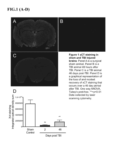
FIG. 1 shows System xCT staining in sham and TBI-injured brains. Panel A is a
surgical sham animal. Panel B is a TBI animal 48 hours after TBI. Panel C is a
TBI animal 46
days post-TBI. Panel D is a graphical representation of the loss of and modest
recovery of
xCT straining that occurs over a 46 day period after TBI. One way ANOVA,
Tukey's post-
hoc. **=p<0.01. Data collected by laser scanning cytometry.
FIG. 2 panels E and F show neurological severity scores and foot faults,
respectively,
from injured and un-injured animals at 48 hours, 2 weeks, and 46 days post-
TBI. In both
assessments there was a significant improvement from 48 hours to 46 days post-
TBI that
corresponds with the return of xCT to the neuromotor cortex. However,
neurological scoring
remained significantly lower than shams that had normal levels of xCT
expression. n=12
animals per group; unpaired two-tailed t-test. ***=p<0.001.
FIG. 3 shows System xCT (red) and GFAP (green) staining in the cortex of rats
and
human patients. Panel A is a surgical sham rat. Panel B is a TBI rat 46 days
after injury.
Panel C is a human control patient. Panel D is a stage IV CTE patient. Data
collected at 60x
using an Olympus FV1000 confocal microscope. Human tissue was kindly provided
by the
Center for the Study of Traumatic Encephalopathy (Boston University).
FIG. 4 is a heat map displaying fold changes in 84 oxidative stress genes
comparing
TBI to Sham. Total RNA was isolated from FFPE 7 nIVI slices from 4 sham
control rats or 4
TBI rats and pooled for cDNA synthesis and preamp with universal oxidative
stress array
RT2PCR primers. Oxidative Stress Array plates were run on a Bio-Rad iQ5
iCycler. Data for
control and TBI was normalized with Rplpl. Boundary was set for 2-fold
changes.
FIG. 5 is a scatter plot displaying fold changes in predicted xCT targeting
miRNA.
Total RNA was isolated from FFPE 7 nIVI slices from 4 sham control rats or 4
TBI rats and
7
CA 02976347 2017-08-10
WO 2016/153549
PCT/US2015/051518
pooled for cDNA synthesis and preamp with universal oxidative stress array
RT2PCR
primers. Rat miFinder Array plates were run on a Bio-Rad iQ5 iCycler. Data for
control and
TBI was normalized with SNORD61 and SNORD95. Boundary was set for 2-fold
changes.
FIG. 6 is a chart of miRNA probes used in PCR Array CMIHS02277. * = predicated
to target xCT (SLC7A11).
FIG. 7 is a chart showing the fold change, 95% confidence interval, and p
values
between miRNA expression profiles on the PCR Array CMIHS02277 between human
peripheral blood plasma obtained from control subjects and those having
suffered acute TBI
(within 24-72 hours).
FIG. 8 is a chart showing the fold change, 95% confidence interval, and p
values
between miRNA expression profiles on the PCR Array CMIHS02277 between human
peripheral blood plasma obtained from control subjects and football players.
FIG. 9 is a chart showing the fold change, 95% confidence interval, and p
values
between miRNA expression profiles on the PCR Array CMIH502277 between human
peripheral blood plasma obtained from football players and those having
suffered acute TBI
(within 24-72 hours).
FIG. 10 is a chart showing the fold change, 95% confidence interval, and p
values
between miRNA expression profiles on the PCR Array CMIH502277 between human
peripheral blood plasma obtained from control subjects and soccer players.
FIG. 11 is a chart showing the fold change, 95% confidence interval, and p
values
between miRNA expression profiles on the PCR Array CMIH502277 between human
peripheral blood plasma obtained from control subjects and those with chronic
TBI.
FIG. 12 is a chart showing the fold change, 95% confidence interval, and p
values
between miRNA expression profiles on the PCR Array CMIH502277 between human
peripheral blood plasma obtained from subjects that have suffered acute TBI
(within 24-72
hours) and those with chronic TBI.
DETAILED DESCRIPTION OF THE INVENTION
The invention features non-invasive methods of detecting, diagnosing, and
tracking
traumatic brain injury (TBI) or chronic traumatic encephalopathy (CTE), and
related
conditions, by evaluating the expression of one or more microRNAs ("miRNAs")
in a sample
(e.g., brain tissue, blood sample, or cerebral spinal fluid sample) derived
from a subject (e.g.,
a human) considered to have suffered from, or is at risk of suffering from, a
TBI or other
neurological defect. The methods of the invention can be used to diagnose,
predict, and
8
6
g anunppoo2napo2nanponpp 61 noannunnpopoapnopo22nnn 96-?111u
ZS onnooanopop2opoapop2on 81 222nann2noonnnapn22pn q96I-?IItu
1Ç oonnnoapnoonoo22opnpno LI nnapnpann22p22pn22p2n 3L-1-31
Og nnppannpannaponpnno 91
anappnanooppopuppnpnn 1 7 L-?Illu
ç1 p2o222p2ann222noapppp OZ -?1Itu
617 E0000EMPEoannapoan 17I nn2p2noappoannnpan2o ZZ-?1Itu
817 ppoanpanppopanopono 1
n222n22on2no2nnponnpOPE clI8I-21Itu
L17 nonnnooanoanoop2opnpno Z I
nnapnpoann22pnapn22p2p PL-PI
917 22nonnoonoanappn000anop II
apnapo2napnonpo2n2appn P8I-?1Itu
Ç17 panoppnan2nopnapopn OI noananoanapoponpnnapo I0I-?Iltu
1717 npo222ppppnnanppo2napo 6
oanon2nopno2n2nnpoponn PO 1 -Wm.
17 nnnpnp2n2n2n2n2pnnnppo 8
poannappnopnnpopo2nnpn Z -?1Itu
Z17 apop22222noo22popn22no L
anapoopanno0OPPOoonon 0 gi-?1Im
It nopananapnpnapopn 9
2ppn2nopnpnponponpnp22 1717I -Wm.
Ç nnapnpannapnapn22an JL-1-0l
017 000nnooanopnooppopnpno 17
nn22n2n2nn22pnapn22p2n c1L-PI
6 onnnon2nopnonppopnpno
nnapnpann22pnapn22p2n EL-PI
8 ano222naonapoopoppo Z
anananopaponpnnoapn IZ-?1Itu
L aanpnnnopnoonnanapan 1
nopnopoappapnapppnpo Z17I-?1Itu
vAr ar *ay ar auruN
aauanbas ,E suayIns .1/ aauanbas ,s suayIns Ti
5,7s 5,7s v Nantu
I am",
ppow oaptupp ipuomou X.Iolpioqpi E UT igi aouapadxa will snoi2aruou OI
.y -LIT polpin2ammop JO polpin2oidn oq oi, punoj (Cionnoodsoi `snoi2artdou .y
pup suaichis
74) soloods yTul TsH z pup I saw", =oaptupp ipuomou 2uTmouoj polpin2oi
XifpnualojjTp
OM SOTOOdS Vt\alTIU UT:4100 ITT kionooslp alp soopiqt.uo uonuonuT Tuosoid oqi
'TWA OTJEUMEJT E 2unuouodxo
JO )15TI TE SE JO SPIT MIT loofqns E JO Etuspid pool(' otp uT oaptupp
ipuomou igi qnm g
pomolloo yt\aptu Jo uonoolop alp JOJ MOUE UOTJUOAUT OUT Jo spotpow alp
`uonuonuT alp Jo
Tuounpoqino ouo ui louuosiod XIBIJITUI JO TUOUIOOJOJUO Am' Xq JO `siuoppop
Joqpi JO Jpinonion
`(Iipcppoj pup 2upcoq "2.a) spods onnop `siuoppop 2umpj "2.a 'uT poouopodxo
osoip sp tions
'swan onpulnp.0 opzIninul JO oi2uT5 Xq posnpo oaptupp ipuomou Jo uol55oi2oid
otp Jonuoul
8ISISOSIOZS9/IDd
617SESI/9I0Z OM
OT-80-LTOZ LVE9L6Z0 VD
OI
17II mo222pEppnanEpo2n2po 8 L
nopno2annEoponnnnono2 PO I-?1Itu
11 nnunan2aan2EnnnEpo2 LL poanappnoEnnEopo2nnpn dc-zItu
Z;11 E22222noo22popn22no 9L
2apoopanno0OPPOoonon 0 CI-?1Itu
1 1 1 nopananampapopn CL
appanopnEnponponpnp22 1717I-?1Itu
011 oonnooannEnompopnEno 17L napnpannapapaaan JL-1-al
601 000nnooanopnooppopnpno L nnnaann22En2En22p2n c1L-PI
801 oonnnoanopnompopnEno a napnpann22papaaan PL-PI
LOI onano222naoapo2poppo IL anananoaponEnno2En IZ-?IItu
901 EnnEnnnopnoonnaapan OL nopnopo2ppapappEnpo Z17I-?Iltu
vAr ar *ay ar
auruN
5,7s aauanbas ,E sna0andou w 5,7s
aauanbas ,s sna0andou w V NI111111
Z alqul
69 anoppEpoanpnonEppo2an 9 Popnopoannampo2nnnap P6I-?1Itu
89 annnopapopnopoaapon C
nop2oonopopaanonappp P817I -Wm.
L9 2opoannopnaannonnE1oo 17 no22pna2poormapponn P9Z-?IItu
99 22npoopo2ppon2oonn22nE
anpooppnonanno2ann 1 -P8Iz-lltu
C9 onaappppoponampo2nn Z
no2poananaannnnnpon CI-?1Itu
1
2Enopoop22pnnanEpan2 P 0Z-?1Itu
179 nnnunan2aan2EnnnEpo 0 poanappnoEnnEopo2nnpn Z-?1Itu
9 anon22onoannopo2nnpo 6Z annEpo222nnop2E22322E CZ-?IItu
Z9 oonnnE222poo2nnpoponp 8Z
nnnp222nE2222noonn2222 P "Z-?1Itu.
a
2pn2o2opnpapEnnoannunn L1-?Iltu
19 o2poannanE22on2ponnno 9Z
2paanop2onoonpopppan PO -?1Itu
09 ano2poopponn0000n22nnn CZ
ttPPPOopp22nEEEEn22no2p P ff 1-WW
6C appaooppnapno2pppnp 17Z anpano2Enonpna2nnnon 6-?IItu
8C anno2ponpppan222oopon d-
6 -?1Itu
L g 2panooanoppappoppo223 Z
222nananponnapn22En P96 I -Wm.
9C
Enoppooannoapnonn22n ZZ nopoponopapn22nEpo22nnn Z8I-?1Itu
CC ano2232pop2an2o2n2no IZ
anopopo2oop000an00002E 0IZ-?1Itu
17C apon000022panon22ono2E OZ
nnnop2p2o2p2E2E32222pan Z17-?1Itu
vAr ar =onr ar auruN
aauanbas ,E suayIns .1/ aauanbas ,s suayIns Ti
5,7s 5,7s v Nantu
8ISISOSIOZS9/IDd
617SESI/9I0Z OM
OT-80-LTOZ LVE9L6Z0 VD
II
0171 anopnBpo2nEnonnm2n2n S01
nopo2nannpo2nnnn2on P6 I -?1Itu
61 annnonapoponpo2n2pon 1701
22E onoponnEnn2nonn2BE2 817I-?1Itu
8 1 opo2nnonnn22nnonnnnoo 01
no22Ena2poormEn2pponn P9Z-?IItu
L 1 opo2nBon2oonn22npopEE zOI
n2nBoopnnonanno2n2nn 1 -P8jz-llin
91 onan2nBEEopon2nmo2nn 101
no2po2nanan2nnnnmon2 gi-?1Itu
ç1 2Enopoop22EnnanBEE2n2 001
opponn2ponunnonn22n2p P CIZ-ITIU
ff 1 nnnnan2n2n2n2pnnnBpo2 66
po2nn2EnnoEnnpopo2nnpn Z-?1Itu
1 anon22onon2nnopo2nrmo 86
o2nnnBo222opop2p22322E SZ-?1Ilu
ai oonnnE222poo2nnpoponp L6
nnna22m2222noonn2222 P 'Z-IIIII
1 1 2nn2o2opmanBnno2nnnnn 96
Enna2n222nnonnEn222op L 1 -?Illu
0I o2po2nnana2on2ponnno g6
2BE22nop2onoonpoppEn2n PO -?1Itu
6ZI 2no2poopponn0000n22nnn 176
ttPPPOopp22nEEEEn22no2E P ff I -Wm.
8Z 1 n2BEE2ooppnapno2nBEnp 6
annano2nnonnnn22nnnon P6-?IItu
6 -?1Itu
LZI anoo2nonBapponBo22on Z6
222nann2nBonnann22pn P96 I -Wm.
2 a I-?1Itu
16
oopoponopann22mpo22nnn
9ZI ano2232Boan2n2o2n2no 06 2nopopo2pop0002nopoo2p OIZ-?1Itu
n Z17-?1Itu
CZ 1 68
n2pon000022anon22ono2p nnnop2p2o2aapo2222an
17ZI nnnEpoo2n2po2n2nBormpo 88
no2nnnnnpopo2pnopo22nnn 96-?111u
ZI ponnoo2nopop2opo2pop2on L8 222nann2noonnann22pn c1961-?1Itu
ZZ1 oonnno2nnoonoo22onnEno 98 nannBann22E22nn22an 3L-131
IZI nnnnnnnmann2opo2Enno g8
2n2Enno2nooppoEnnumnp 1 7 L-?Illu
OZ 1 E23222aann222no2nBEE 178
oonnonn22000nnononnoo2 OZ-?1Itu
611 oo OOPTIPPP on2nnapon2n 8
2nn2ano2ppoannnEn2n2o ZZ-?1Itu
811 Epo2nBannBopanopono Z8
n222n22on2no2nrmonnE OPP q I 8 I -IITIU
L11 nonnnoo2no2noop2onnEno 18
nn2mwo2nn22En2nn22ap PL-13"
911 nonnoono2n2pnn0002nop 08
2Enapo2n2Enonpo2n22BEn P8I-?1Itu
gii panoEnnan2nopn2popn 6L
o2nano2n2poponEnn2pon P ICI 1 -11111.
vAr ar =onr ar
auruN
5,7s aauanbas ,E sna0a1dou w 5,7s
aauanbas ,s sn30a4dou w V NI111111
8ISISOSIOZS9/IDd 617SESI/9I0Z OM
OT-80-LTOZ LVE9L6Z0 VD
CA 02976347 2017-08-10
WO 2016/153549
PCT/US2015/051518
Methods of miRNA Expression Profiling
The expression level of at least one miRNA species can be measured in a
biological
sample (e.g., an organ, tissue, or cell sample, such as brain tissue, blood
sample, or cerebral
spinal fluid (CSF)) obtained from a patient (e.g., a human). For example, a
tissue sample
(e.g., brain tissue, blood, or CSF) can be removed from a patient suspected of
suffering from
or at risk of suffering a brain injury (e.g., TBI or CTE) by conventional
biopsy techniques. In
another embodiment, a blood or CSF sample can be removed from the patient
(e.g., a
human), and cells (e.g., white blood cells) or serum can be isolated for RNA
extraction by
standard techniques. In order to determine baseline miRNA expression profiles,
a blood,
CSF, or tissue sample is preferably obtained from the patient prior to
initiation of any activity
that carries a heightened risk of TBI, including but not limited to impact
sports (e.g., boxing,
American football, rugby, hockey, baseball, and soccer), military or law
enforcement service,
medical conditions that leave subjects susceptible to falls (e.g., blindness,
advanced age), or
any other that places the subject at increased risk of suffering TBI (e.g.,
race car driving,
skydiving, and victims of assault). Baseline blood or tissue samples are also
ideally obtained
prior to radiotherapy, chemotherapy or other therapeutic treatment in order to
gauge miRNA
expression profile changes during the course of treatment. A corresponding
control tissue or
blood sample can be obtained from unaffected tissues of the patient, from a
normal human
individual or population of normal individuals, or from cultured cells
corresponding to the
majority of cells in the patient's sample. The control tissue or blood sample
is then processed
along with the sample from the patient, so that the miRNA expression profile
derived from
the patient's sample can be compared to a corresponding miRNA expression
profile derived
from a sample taken from a control subject or group. A reference miRNA
expression profile
standard for the biological sample can also be used as a control.
An alteration (e.g., an increase or decrease) in the level of one or more of
the miRNAs
identified herein (e.g., SEQ ID NOS:1-140) in the sample obtained from a
patient (e.g., a
human), relative to the level of corresponding miRNAs in a control sample, is
indicative of
the presence of brain injury (e.g., TBI) in the patient. In one embodiment,
the expression
level of at least one miRNA in the test sample is greater than the expression
level of a
corresponding miRNA in the control sample (i.e., expression of the miRNA is
"up-
regulated"). As used herein, expression of a miRNA is "up-regulated" when the
amount of
miRNA in a fluid, cell, or tissue sample from a patient is greater than the
amount of the same
miRNA in a control fluid, cell, or tissue sample. In another embodiment, the
expression level
of the at least one miRNA in the test sample is less than the expression level
of the
12
CA 02976347 2017-08-10
WO 2016/153549
PCT/US2015/051518
corresponding miRNA in the control sample (i.e., expression of the miRNA is
"down-
regulated"). As used herein, expression of a miRNA is "down-regulated" when
the amount of
miRNA produced in a fluid, cell, or tissue sample from a patient is less than
the amount
produced in a fluid, control cell, or tissue sample. A patient miRNA
expression profile is
considered to indicate the presence of a brain injury if the up or down-
regulation is 1.2, 1.3,
1.4, 1.5, 1.6, 1.7, 1.8, 1.9, or 2.0 fold or greater relative to the control
expression profile. The
relative miRNA expression in the control and normal samples can be determined
with respect
to one or more miRNA expression standards. The standards can comprise, for
example, a
zero miRNA gene expression level, the miRNA expression profiles of
standardized cell lines,
the miRNA expression profiles in unaffected tissues of the patient (e.g., a
human), or the
average level of miRNA expression previously obtained for a population of
normal controls
(e.g., human controls).
The level of a miRNA expression in a sample can be measured using any
technique
that is suitable for detecting RNA expression levels in a biological sample.
Suitable
techniques (e.g., Northern blot analysis, RT-PCR, in situ hybridization) for
determining RNA
expression levels in a biological sample (e.g., cells, tissues) are well known
to those of skill
in the art. In a particular embodiment, the level of at least one miRNA
species is detected
using Northern blot analysis. For example, total cellular RNA can be purified
from cells by
homogenization in the presence of nucleic acid extraction buffer, followed by
centrifugation.
Nucleic acids are precipitated, and DNA is removed by treatment with DNase and
precipitation. The RNA molecules are then separated by gel electrophoresis on
agarose gels
according to standard techniques, and transferred to nitrocellulose filters.
The RNA is then
immobilized on the filters by heating. Detection and quantification of
specific RNA is
accomplished using appropriately labeled DNA or RNA probes complementary to
the RNA
in question. See, e.g., Molecular Cloning: A Laboratory Manual, J. Sambrook et
al., eds., 2nd
edition, Cold Spring Harbor Laboratory Press, 1989, Chapter 7, the entire
disclosure of which
is incorporated by reference.
Suitable probes for Northern blot hybridization of a given miRNA can be
produced
from the nucleic acid sequences of the miRNA sequences described and listed
herein and
include, but are not limited to, probes having at least about 70%, 75%, 80%,
85%, 90%, 95%,
98%, 99% or complete complementarity to a miRNA of interest. Methods for
preparation of
labeled DNA and RNA probes, and the conditions for hybridization thereof to
target
nucleotide sequences, are described in Molecular Cloning: A Laboratory Manual,
J.
Sambrook et al., eds., 2nd edition, Cold Spring Harbor Laboratory Press, 1989,
Chapters 10
13
CA 02976347 2017-08-10
WO 2016/153549
PCT/US2015/051518
and 11, the disclosures of which are incorporated herein by reference. For
example, the
nucleic acid probe can be labeled with, e.g., a radionuclide, such as 3H, 32p,
33P5 14C5 or 35S; a
heavy metal; a ligand capable of functioning as a specific binding pair member
for a labeled
ligand (e.g., biotin, avidin or an antibody); a fluorescent molecule; a
chemiluminescent
molecule; an enzyme or the like.
Probes can be labeled to high specific activity by either the nick translation
method of
Rigby et al. (1977), J. Mol. Biol. 113:237-251 or by the random priming method
of Fienberg
et al. (1983), Anal. Biochem. 132:6-13, the entire disclosures of which are
incorporated
herein by reference. The latter is the method of choice for synthesizing 32P-
labeled probes of
high specific activity from single-stranded DNA or from RNA templates. For
example, by
replacing preexisting nucleotides with highly radioactive nucleotides
according to the nick
translation method, it is possible to prepare 32P-labeled nucleic acid probes
with a specific
activity well in excess of 108 cpm/microgram. Autoradiographic detection of
hybridization
can then be performed by exposing hybridized filters to photographic film.
Densitometric
scanning of the photographic films exposed by the hybridized filters provides
an accurate
measurement of miR gene transcript levels. Using another approach, miR gene
transcript
levels can be quantified by computerized imaging systems, such as the
Molecular Dynamics
400-B 2D Phosphorimager available from Amersham Biosciences, Piscataway, N.J.
Where radionuclide labeling of DNA or RNA probes is not practical, the random-
primer method can be used to incorporate an analogue, for example, the dTTP
analogue 5-
(N-(N-biotinyl-epsilon-aminocaproy1)-3-aminoallyl)deoxyuridine triphosphate,
into the probe
molecule. The biotinylated probe oligonucleotide can be detected by reaction
with biotin-
binding proteins, such as avidin, streptavidin, and antibodies (e.g., anti-
biotin antibodies)
coupled to fluorescent dyes or enzymes that produce color reactions.
In addition to Northern and other RNA hybridization techniques, determining
the
levels of RNA transcripts can be accomplished using the technique of in situ
hybridization.
This technique requires fewer cells than the Northern blotting technique, and
involves
depositing whole cells onto a microscope cover slip and probing the nucleic
acid content of
the cell with a solution containing radioactive or otherwise labeled nucleic
acid (e.g., cDNA
or RNA) probes. This technique is particularly well suited for analyzing
tissue biopsy
samples from subjects. The practice of the in situ hybridization technique is
described in
more detail in U.S. Pat. No. 5,427,916, the entire disclosure of which is
incorporated herein
by reference. Suitable probes for in situ hybridization of a given miRNA can
be produced
14
CA 02976347 2017-08-10
WO 2016/153549
PCT/US2015/051518
from the nucleic acid sequences having at least about 70%, 75%, 80%, 85%, 90%,
95%,
98%, 99% or complete complementarity to a miRNA of interest, as described
above.
The relative number of miRNA gene transcripts in cells can also be determined
by
reverse transcription of miRNA gene transcripts, followed by amplification of
the reverse-
transcribed transcripts by polymerase chain reaction (RT-PCR). The levels of
miRNA gene
transcripts can be quantified in comparison with an internal standard, for
example, the level
of mRNA from a "housekeeping" gene present in the same sample. A suitable
"housekeeping" gene for use as an internal standard includes, e.g., myosin or
glyceraldehyde-
3-phosphate dehydrogenase (G3PDH). Methods for performing quantitative and
semi-
quantitative RT-PCR, and variations thereof, are well known to those of skill
in the art.
In some instances, it may be desirable to simultaneously determine the
expression
level of a plurality of different miRNA species in a sample (e.g., brain
tissue, blood, or
cerebral spinal fluid (CSF)). In other instances, it may be desirable to
determine the
expression level of the transcripts of all known miRNA species correlated with
a brain injury.
Assessing brain injury-specific expression levels for hundreds of miRNA
species is time
consuming and requires a large amount of total RNA (e.g., at least 20
micrograms for each
Northern blot) and autoradiographic techniques that require radioactive
isotopes.
To overcome these limitations, an oligolibrary, in microchip format (i.e., a
microarray), may be constructed containing a set of oligonucleotide (e.g.,
oligodeoxynucleotides) probes that are specific for a set of miRNA species.
Using such a
microarray, the expression level of multiple microRNAs in a biological sample
(e.g., brain
tissue, blood, or cerebral spinal fluid (CSF)) can be determined by reverse
transcribing the
RNAs to generate a set of target oligodeoxynucleotides, and hybridizing them
to probe the
oligonucleotides on the microarray to generate a hybridization, or expression,
profile. The
hybridization profile of the test sample can then be compared to that of a
control sample to
determine which microRNAs have an altered expression level consistent with a
suspected
disease, condition, or disorder, such as traumatic brain injury.
Accordingly, the invention provides methods of diagnosing whether a subject
has, or
is at increased risk of suffering from a TBI comprising reverse transcribing
RNA from a test
sample (e.g., brain tissue, blood, or cerebral spinal fluid (CSF)) obtained
from the subject
(e.g., a human) to provide a set of target oligodeoxynucleotides, hybridizing
the target
oligodeoxynucleotides to a microarray comprising miRNA-specific probe
oligonucleotides to
provide a hybridization profile for the test sample, and comparing the test
sample
hybridization profile to a hybridization profile generated from a control
sample or reference
CA 02976347 2017-08-10
WO 2016/153549
PCT/US2015/051518
standard, wherein an alteration in the signal of at least one miRNA is
indicative of the subject
either having, or being at risk for developing, TBI. In one embodiment, the
microarray
comprises miRNA-specific probe oligonucleotides for a substantial portion of
all known
human miRNAs. In one embodiment, the microarray comprises miRNA-specific probe
oligonucleotides for one or more miRNAs selected from the group consisting of
miR-142,
miR-21, let-7a, let-7b, let-7f, miR-144, miR-150, miR-32, miR-13 Oa, miR-101a,
miR-18a,
let-7d, miR-181b, miR-223, miR-320, miR-374, let-7e, miR-196b, miR-96, miR-
423, miR-
210, miR-182, miR-196a, miR-39, miR-9a, miR-133a, miR-30a, miR-137, miR-23a,
miR-25,
miR-32, miR-203 a, miR-153, miR-218-1, miR-26 a, miR-148 a, and miR-19 a .
The microarray can be prepared from gene-specific oligonucleotide probes
generated
from known miRNA sequences. The array may contain two different
oligonucleotide probes
for each miRNA, one containing the active, mature sequence and the other being
specific for
the precursor of the miRNA. The array may also contain controls, such as one
or more mouse
sequences differing from human orthologs by only a few bases, which can serve
as controls
for hybridization stringency conditions. tRNAs or other RNAs (e.g., rRNAs,
mRNAs) from
both species may also be printed on the microchip, providing an internal,
relatively stable,
positive control for specific hybridization. One or more appropriate controls
for non-specific
hybridization may also be included on the microchip. For this purpose,
sequences are selected
based upon the absence of any homology with any known miRNAs.
The microarray may be fabricated using techniques known in the art. For
example,
probe oligonucleotides of an appropriate length, e.g., 40 nucleotides, are 5'-
amine modified at
position C6 and printed using commercially available microarray systems, e.g.,
the
GeneMachine OmniGridTM 100 Microarrayer and Amersham CodeLinkTM activated
slides.
Labeled cDNA oligomer corresponding to the target RNAs is prepared by reverse
transcribing the target RNA with labeled primer. Following first strand
synthesis, the
RNA/DNA hybrids are denatured to degrade the RNA templates. The labeled target
cDNAs
thus prepared are then hybridized to the microarray chip under hybridizing
conditions, e.g.,
6x SSPE/30% formamide at 25 C for 18 hours, followed by washing in 0.75x TNT
(Tris
HC1/NaC1/Tween 20) at 37 C for 40 minutes. At positions on the array where
the
immobilized probe DNA recognizes a complementary target cDNA in the sample,
hybridization occurs. The labeled target cDNA marks the exact position on the
array where
binding occurs, allowing automatic detection and quantification. The output
consists of a list
of hybridization events, indicating the relative abundance of specific cDNA
sequences, and
therefore the relative abundance of the corresponding complementary miRNAs, in
the patient
16
CA 02976347 2017-08-10
WO 2016/153549
PCT/US2015/051518
sample. Image intensities of each spot on the array are proportional to the
abundance of the
corresponding miRNA in the patient sample.
The use of the array has several advantages for miRNA expression detection.
First,
the global expression of several hundred genes can be identified in the same
sample at one
time point. Second, through careful design of the oligonucleotide probes,
expression of both
mature and precursor molecules can be identified. Third, in comparison with
Northern blot
analysis, the chip requires a small amount of RNA, and provides reproducible
results using
2.5 micrograms of total RNA. The relatively limited number of miRNAs (a few
hundred per
species) allows the construction of a common microarray for several species,
with distinct
oligonucleotide probes for each. Such a tool would allow for analysis of trans-
species
expression for each known miRNA under various conditions.
In addition to use for quantitative expression level assays of specific miRNA,
a
microchip containing miRNA-specific probe oligonucleotides corresponding to a
substantial
portion of the miRNome, preferably the entire miRNome, may be employed to
carry out
miRNA gene expression profiling, for analysis of miRNA expression patterns.
Distinct
miRNA signatures can be associated with established disease markers, or
directly with a
disease state.
According to the expression profiling methods described herein, total RNA from
a
sample (e.g., brain tissue, blood, or cerebral spinal fluid (CSF)) from a
subject (e.g., a human)
suspected of suffering or at risk of suffering a TBI is quantitatively reverse
transcribed to
provide a set of labeled target oligodeoxynucleotides complementary to the RNA
in the
sample. The target oligodeoxynucleotides are then hybridized to a microarray
comprising
miRNA-specific probe oligonucleotides to provide a hybridization profile for
the sample. The
result is a hybridization profile for the sample representing the expression
pattern of miRNA
in the sample. The hybridization profile comprises the signal from the binding
of the target
oligodeoxynucleotides from the sample to the miRNA-specific probe
oligonucleotides in the
microarray. The profile may be recorded as the presence or absence of binding
(signal vs.
zero signal). More preferably, the profile recorded includes the intensity of
the signal from
each hybridization. The profile is compared to the hybridization profile
derived from a
normal, i.e., non-TBI, control sample. An alteration in the signal is
indicative of the presence
of, or propensity to develop, TBI in the subject.
Other techniques for measuring miRNA gene expression are also within the skill
in
the art, and include various techniques for measuring rates of RNA
transcription and
degradation.
17
CA 02976347 2017-08-10
WO 2016/153549
PCT/US2015/051518
Treatment of Brain Injury
As described herein, brain injury is associated with marked loss of System x,-
antiporter expression in brain tissues and an overall loss in antioxidant
capacity in these
tissues. Weak antioxidant mechanisms allow for accumulation of reactive oxygen
species
(ROS) that chemically damage surround cells and tissues.
Upon making a clinical determination that a patient (e.g., a human) has
suffered a
brain injury, a clinician may determine that administration of an antioxidant
or antioxidant
therapy course is appropriate. Examples of antioxidants include, but are not
limited to, alpha-
tocopherol, ascorbate, coenzyme Q, alpha-lipoic acid, curcumin, glutathione,
uric acid,
carotenes (e.g., retinol, beta-carotene), superoxide dismutase, catalases,
peroxiredoxins,
thioredoxins, tirilazad mesylate, and NXY-059. In one embodiment, the patient
is
administered a therapeutically-effective amount of one or more antioxidants in
order to slow
the progression of brain injury.
Methods of Diagnostic Imaging
The present invention provides for the diagnosis and medical evaluation of
patients
(e.g., a human) suffering from, or at risk of suffering from TBI, CTE, or
related conditions.
For example, an imaging agent specific for System x,- can also be used, alone
or in
combination with other agents and compounds, in medical imaging applications
to diagnose
or follow the progression of diseases, disorders, conditions or symptoms
related to TBI or
CTE in a patient (e.g., a human). For example, radiologists and other medical
clinicians are
skilled in the use of radiographic imaging devices, such as positron emission
tomography
(PET) scanners, and methods of imaging tracer compounds, such as the
radionuclides. (e.g.,
Saha, Basics of PET Imaging: Physics, Chemistry, and Regulations, Springer
(2010) ISBN
978-1-4419-0804-9, hereby incorporated by reference).
The methods of the present invention are also useful for the medical imaging
and
diagnosis of humans and animals, e.g., domesticated animal, companion animals
(e.g., dogs
and cats), exotic animals, farm animals (e.g., ungulates, including horses,
cows, sheep, goats,
and pigs), and animals used in scientific research (e.g., rodents and non-
human primates).
Compound Administration and Formulation
Basic addition salts can be prepared during the final isolation and
purification of the
compounds by reaction of a carboxy group with a suitable base such as the
hydroxide,
18
CA 02976347 2017-08-10
WO 2016/153549
PCT/US2015/051518
carbonate, or bicarbonate of a metal cation or with ammonia or an organic
primary,
secondary, or tertiary amine. The cations of therapeutically acceptable salts
include lithium,
sodium, potassium, calcium, magnesium, and aluminum, as well as nontoxic
quaternary
amine cations such as ammonium, tetramethylammonium, tetraethylammonium,
methylamine, dimethylamine, trimethylamine, triethylamine, diethylamine,
ethylamine,
tributylamine, pyridine, N,N-dimethylaniline, N-methylpiperidine, N-
methylmorpholine,
dicyclohexylamine, procaine, dibenzylamine, N,N-dibenzylphenethylamine, 1-
ephenamine,
and N,N'-dibenzylethylenediamine. Other representative organic amines useful
for the
formation of base addition salts include ethylenediamine, ethanolamine,
diethanolamine,
pip eridine , and pip erazine .
A salt of a compound can be made by reacting the appropriate compound in the
form
of the free base with the appropriate acid. A compound can be prepared in a
form of
pharmaceutically acceptable salts that will be prepared from nontoxic
inorganic or organic
bases including but not limited to aluminum, ammonium, calcium, copper,
ferric, ferrous,
lithium, magnesium, manganic salts, manganous, potassium, sodium, zinc, and
the like. Salts
derived from pharmaceutically acceptable organic non-toxic bases include salts
of primary,
secondary, and tertiary amines, substituted amines including naturally-
occurring substituted
amines, cyclic amines, and basic ion exchange resins, such as arginine,
betaine, caffeine,
choline, ethylamine, 2-diethylaminoethano, 1,2-dimethylaminoethanol,
ethanolamine,
ethylenediamine, N-ethyl-morpholine, N-ethylpiperidine, glucamine,
glucosamine, histidine,
hydroxylamine, isopropylamine, lysine, methylglucamine, morpholine,
piperazine,
piperidine, polyamine resins, procaine, purines, theobromine, triethylamine,
trimethylamine,
trishydroxylmethyl amino methane, tripropyl amine, and tromethamine.
If the compounds are basic, salts could be prepared in a form of
pharmaceutically
acceptable salts that will be prepared from nontoxic inorganic or organic
acids including but
not limited to hydrochloric, hydrobromic, phosphoric, sulfuric, tartaric,
citric, acetic, fumaric,
alkylsulphonic, naphthalenesulphonic, para-toluenesulphonic, camphoric acids,
benzenesulfonic, benzoic, camphorsulfonic, citric, ethanesulfonic, gluconic,
glutamic,
isethonic, lactic, maleic, malic, mandelic, methanesulfonic, mucic, nitric,
pamoic,
pantothenic, phosphoric, and succinic.
While it may be possible for the compounds to be administered as the raw
chemical, it
is also possible to present them as a pharmaceutical formulation. Accordingly,
the present
invention provides a pharmaceutical formulation comprising a compound or a
pharmaceutically acceptable salt, ester, prodrug or solvate thereof, together
with one or more
19
CA 02976347 2017-08-10
WO 2016/153549
PCT/US2015/051518
pharmaceutically acceptable carriers thereof and optionally one or more other
therapeutic
ingredients. The carrier(s) must be "acceptable" in the sense of being
compatible with the
other ingredients of the formulation and not deleterious to the recipient
thereof. Proper
formulation is dependent upon the route of administration chosen. Any of the
well-known
techniques, carriers, and excipients may be used as suitable and as understood
in the art; e.g.,
in Remington's Pharmaceutical Sciences. The pharmaceutical compositions of the
present
invention may be manufactured in a manner that is itself known, e.g., by means
of
conventional mixing, dissolving, granulating, dragee-making, levigating,
emulsifying,
encapsulating, entrapping or compression processes.
The formulations include those suitable for oral, parenteral (including
subcutaneous,
intradermal, intramuscular, intravenous, intraarticular, and intramedullary),
intraperitoneal,
transmucosal, transdermal, rectal and topical (including dermal, buccal,
sublingual and
intraocular) administration although the most suitable route may depend upon
for example
the condition and disorder of the recipient. When used in the diagnostic
imaging methods of
the invention, compounds can be administered to the patient (e.g., a human) by
intravenous
injection. The formulations may conveniently be presented in unit dosage form
and may be
prepared by any of the methods well known in the art of pharmacy. All methods
include the
step of bringing into association a compound of the present invention or a
pharmaceutically
acceptable salt, ester, prodrug or solvate thereof ("active ingredient") with
the carrier which
constitutes one or more accessory ingredients. In general, the formulations
are prepared by
uniformly and intimately bringing into association the active ingredient with
liquid carriers or
finely divided solid carriers or both and then, if necessary, shaping the
product into the
desired formulation.
Formulations suitable for oral administration may be presented as discrete
units such
as capsules, cachets or tablets each containing a predetermined amount of the
active
ingredient; as a powder or granules; as a solution or a suspension in an
aqueous liquid or a
non-aqueous liquid; or as an oil-in-water liquid emulsion or a water-in-oil
liquid emulsion.
The active ingredient may also be presented as a bolus, electuary or paste.
Pharmaceutical preparations which can be used orally include tablets, push-fit
capsules made of gelatin, as well as soft, sealed capsules made of gelatin and
a plasticizer,
such as glycerol or sorbitol. Tablets may be made by compression or molding,
optionally
with one or more accessory ingredients. Compressed tablets may be prepared by
compressing
in a suitable machine the active ingredient in a free-flowing form such as a
powder or
granules, optionally mixed with binders, inert diluents, or lubricating,
surface active or
CA 02976347 2017-08-10
WO 2016/153549
PCT/US2015/051518
dispersing agents. Molded tablets may be made by molding in a suitable machine
a mixture
of the powdered compound moistened with an inert liquid diluent. The tablets
may optionally
be coated or scored and may be formulated so as to provide slow or controlled
release of the
active ingredient therein. All formulations for oral administration should be
in dosages
suitable for such administration. The push-fit capsules can contain the active
ingredients in
admixture with filler such as lactose, binders such as starches, and/or
lubricants such as talc
or magnesium stearate and, optionally, stabilizers. In soft capsules, the
active compounds
may be dissolved or suspended in suitable liquids, such as fatty oils, liquid
paraffin, or liquid
polyethylene glycols. In addition, stabilizers may be added. Dragee cores are
provided with
suitable coatings. For this purpose, concentrated sugar solutions may be used,
which may
optionally contain gum arabic, talc, polyvinyl pyrrolidone, carbopol gel,
polyethylene glycol,
and/or titanium dioxide, lacquer solutions, and suitable organic solvents or
solvent mixtures.
Dyestuffs or pigments may be added to the tablets or dragee coatings for
identification or to
characterize different combinations of active compound doses.
Compounds may be formulated for parenteral administration by injection, e.g.,
by
bolus injection or continuous infusion. Formulations for injection may be
presented in unit
dosage form, e.g., in ampoules or in multi-dose containers, with an added
preservative.
Compositions may take such forms as suspensions, solutions or emulsions in
oily or aqueous
vehicles, and may contain formulatory agents such as suspending, stabilizing
and/or
dispersing agents. The formulations may be presented in unit-dose or multi-
dose containers,
for example sealed ampoules and vials, and may be stored in powder form or in
a freeze-
dried (lyophilized) condition requiring only the addition of the sterile
liquid carrier, for
example, saline or sterile pyrogen-free water, immediately prior to use.
Extemporaneous
injection solutions and suspensions may be prepared from sterile powders,
granules and
tablets of the kind previously described.
Formulations for parenteral administration include aqueous and non-aqueous
(oily)
sterile injection solutions of the active compounds which may contain
antioxidants, buffers,
bacteriostats and solutes which render the formulation isotonic with the blood
of the intended
recipient; and aqueous and non-aqueous sterile suspensions which may include
suspending
agents and thickening agents. Suitable lipophilic solvents or vehicles include
fatty oils such
as sesame oil, or synthetic fatty acid esters, such as ethyl oleate or
triglycerides, or liposomes.
Aqueous injection suspensions may contain substances which increase the
viscosity of the
suspension, such as sodium carboxymethyl cellulose, sorbitol, or dextran.
Optionally, the
21
CA 02976347 2017-08-10
WO 2016/153549
PCT/US2015/051518
suspension may also contain suitable stabilizers or agents which increase the
solubility of the
compounds to allow for the preparation of highly concentrated solutions.
In addition to the formulations described previously, a compound may also be
formulated as a depot preparation. Such long acting formulations may be
administered by
implantation (for example subcutaneously or intramuscularly) or by
intramuscular injection.
Thus, for example, a compound may be formulated with suitable polymeric or
hydrophobic
materials (for example, as an emulsion in an acceptable oil) or ion exchange
resins, or as
sparingly soluble derivatives, for example, as a sparingly soluble salt.
For buccal or sublingual administration, a compound may take the form of
tablets,
lozenges, pastilles, or gels formulated in conventional manner. Such
compositions may
comprise the active ingredient in a flavored basis such as sucrose and acacia
or tragacanth.
A compound may also be formulated in rectal compositions such as suppositories
or
retention enemas, e.g., containing conventional suppository bases such as
cocoa butter,
polyethylene glycol, or other glycerides.
A compound may be administered topically, that is by non-systemic
administration.
This includes the application of a compound externally to the epidermis or the
buccal cavity
and the instillation of such a compound into the ear, eye and nose, such that
the compound
does not significantly enter the blood stream. In contrast, systemic
administration refers to
oral, intravenous, intraperitoneal and intramuscular administration.
Formulations suitable for topical administration include solid, liquid or semi-
liquid
preparations suitable for penetration through the skin to the site of
inflammation such as gels,
liniments, lotions, creams, ointments or pastes, and drops suitable for
administration to the
eye, ear or nose. The active ingredient may comprise, for topical
administration, from
0.001% to 10% w/w, for instance from 1% to 2% by weight of the formulation. It
may
however comprise as much as 10% w/w but preferably will comprise less than 5%
w/w, more
preferably from 0.1% to 1% w/w of the formulation.
Via the topical route, a pharmaceutical composition may be in the form of
liquid or
semi liquid such as ointments, or in the form of solid such as powders. It may
also be in the
form of suspensions such as polymeric microspheres, or polymer patches and
hydrogels
allowing a controlled release. This topical composition may be in anhydrous
form, in aqueous
form or in the form of an emulsion. The compounds are used topically at a
concentration
generally of between 0.001 % and 10% by weight and preferably between 0.01%
and 1% by
weight, relative to the total weight of the composition.
22
CA 02976347 2017-08-10
WO 2016/153549
PCT/US2015/051518
For administration by inhalation, a compound can be conveniently delivered
from an
insufflator, nebulizer pressurized packs or other convenient means of
delivering an aerosol
spray. Pressurized packs may comprise a suitable propellant such as
dichlorodifluoromethane, trichlorofluoromethane, dichlorotetrafluoroethane,
carbon dioxide
or other suitable gas. In the case of a pressurized aerosol, the dosage unit
may be determined
by providing a valve to deliver a metered amount. Alternatively, for
administration by
inhalation or insufflation, a compound may take the form of a dry powder
composition, for
example a powder mix of the compound and a suitable powder base such as
lactose or starch.
The powder composition may be presented in unit dosage form, in for example,
capsules,
cartridges, gelatin or blister packs from which the powder may be administered
with the aid
of an inhalator or insufflator.
Preferred unit dosage formulations are those containing an effective dose, as
herein
below recited, or an appropriate fraction thereof, of the active ingredient.
It should be understood that in addition to the ingredients particularly
mentioned
above, formulations described herein may include other agents conventional in
the art having
regard to the type of formulation in question, for example those suitable for
oral
administration may include flavoring agents.
A compound may be administered orally or via injection at a dose of from 0.1
to 500
mg/kg per day. The dose range for adult humans is generally from 5 mg to 2
g/day. Tablets or
other forms of presentation provided in discrete units may conveniently
contain an amount of
compound which is effective at such dosage or as a multiple of the same, for
instance, units
containing 5 mg to 500 mg, usually around 10 mg to 200 mg.
Compounds can be administered at a daily dose of about 0.001 mg/kg to 100
mg/kg
of body weight, in 1 to 3 dosage intakes. Further, compounds can be used
systemically, at a
concentration generally of between 0.001 % and 10% by weight and preferably
between 0.01
% and 1 % by weight, relative to the weight of the composition.
The amount of active ingredient that may be combined with the carrier
materials to
produce a single dosage form will vary depending upon the host treated and the
particular
mode of administration.
A compound can be administered in various modes, e.g. orally, topically, or by
injection. The precise amount of compound administered to a patient will be
the
responsibility of the attendant physician. The specific dose level for any
particular patient
will depend upon a variety of factors including the activity of the specific
compound
employed, the age, body weight, general health, sex, diets, time of
administration, route of
23
CA 02976347 2017-08-10
WO 2016/153549
PCT/US2015/051518
administration, rate of excretion, drug combination, the precise disorder
being treated, and
the severity of the indication or condition being treated. Also, the route of
administration may
vary depending on the condition and its severity.
In certain instances, it may be appropriate to administer at least one
compound
described herein (or a pharmaceutically acceptable salt, ester, or prodrug
thereof) in
combination with another therapeutic or diagnostic agent. By way of example
only, if one of
the side effects experienced by a patient upon receiving one of the compounds
described
herein is hypertension, then it may be appropriate to administer an anti-
hypertensive agent in
combination with the initial therapeutic agent. Or, by way of example only,
the therapeutic
effectiveness of one of the compounds described herein may be enhanced by
administration
of an adjuvant (i.e., by itself the adjuvant may only have minimal therapeutic
benefit, but in
combination with another therapeutic agent, the overall therapeutic benefit to
the patient is
enhanced). Or, by way of example only, the benefit of experienced by a patient
may be
increased by administering one of the compounds described herein with another
therapeutic
agent (which also includes a therapeutic regimen) that also has therapeutic
benefit. By way of
example only, in a treatment for pain involving administration of one of the
compounds
described herein, increased therapeutic benefit may result by also providing
the patient with
another therapeutic agent for pain. In any case, regardless of the disease,
disorder or
condition being treated, the overall benefit experienced by the patient may
simply be additive
of the two therapeutic agents or the patient may experience a synergistic
benefit.
Specific, non-limiting examples of possible combination therapies include use
of a
compound together with inert or active compounds, or other drugs including
wetting agents,
flavor enhancers, preserving agents, stabilizers, humidity regulators, pH
regulators, osmotic
pressure modifiers, emulsifiers, UV-A and UV-B screening agents, antioxidants,
depigmenting agents such as hydroquinone or kojic acid, emollients,
moisturizers, for
instance glycerol, PEG 400, or urea, antiseborrhoeic or antiacne agents, such
as S-
carboxymethylcysteine, S-benzylcysteamine, salts thereof or derivatives
thereof, or benzoyl
peroxide, antibiotics, for instance erythromycin and tetracyclines,
chemotherapeutic agent,
for example, paclitaxel, antifungal agents such as ketoconazole, agents for
promoting
regrowth of the hair, for example, minoxidil (2,4-diamino-6-
piperidinopyrimidine 3-oxide),
non-steroidal anti-inflammatory agents, carotenoids, and especially p-
carotene, antipsoriatic
agents such as anthralin and its derivatives, eicosa-5,8,11,14-tetraynoic acid
and eicosa-
5,8,11-triynoic acid, and esters and amides thereof, retinoids, e.g., RAR or
RXR receptor
ligands, which may be natural or synthetic, corticosteroids or oestrogens,
alpha-hydroxy
24
CA 02976347 2017-08-10
WO 2016/153549
PCT/US2015/051518
acids and a-keto acids or derivatives thereof, such as lactic acid, malic
acid, citric acid, and
also the salts, amides or esters thereof, or p-hydroxy acids or derivatives
thereof, such as
salicylic acid and the salts, amides or esters thereof, ion-channel blockers
such as potassium-
channel blockers, or alternatively, more particularly for the pharmaceutical
compositions, in
combination with medicaments known to interfere with the immune system,
anticonvulsant
agents include, and are not limited to, topiramate, analogs of topiramate,
carbamazepine,
valproic acid, lamotrigine, gabapentin, phenytoin and the like and mixtures or
pharmaceutically acceptable salts thereof A person skilled in the art will
take care to select
the other compound(s) to be added to these compositions such that the
advantageous
properties intrinsically associated with the compounds are not, or are not
substantially,
adversely affected by the envisaged addition.
In any case, the multiple therapeutic or diagnostic agents may be administered
in any
order or even simultaneously. If simultaneously, the multiple therapeutic or
diagnostic agents
may be provided in a single, unified form, or in multiple forms (by way of
example only,
either as a single pill or as two separate pills). One of the therapeutic or
diagnostic agents
may be given in multiple doses, or both may be given as multiple doses. If not
simultaneous,
the timing between the multiple doses may be any duration of time ranging from
a few
minutes to four weeks.
Thus, in another aspect, methods for diagnosing or treating diseases,
disorders,
conditions, or symptoms in a subject (e.g., a human or animal) in need of such
treatment are
presented herein, the methods comprising the step of administering to the
subject an amount
of a compound effective to reduce or prevent the disease, disorder, condition,
or symptom, in
combination with at least one additional agent for the treatment of said
disorder that is known
in the art.
Examples
It is understood that the foregoing examples are merely illustrative of the
present
invention. Certain modifications of the articles and/or methods employed may
be made and
still achieve the objectives of the invention. Such modifications are
contemplated as within
the scope of the claimed invention.
Example 1. Gene Array Analysis Before and After TBI in Rodent Model.
System xc- is a cystine/glutamate antiporter comprised of two distinct
subunits xCT
and 4F2hc (SLC3A2) and a member of the heteromeric amino acid transporter
(HAT) family.
CA 02976347 2017-08-10
WO 2016/153549
PCT/US2015/051518
Under physiological conditions, System x,- mediates the exchange of
extracellular L-cystine
and intracellular L-glutamate across the plasma membrane. In the CNS, the
influx of L-
cystine represents the critical rate limiting step in the biosynthesis of
glutathione (GSH) while
the concurrent efflux of L-glutamate serve as a non-vesicular route of
excitatory
neurotransmitter release to initiate excitatory amino acid (EAA) signalling.
GSH serves as the
key cellular antioxidant responsible for scavenging reactive oxygen species
(ROS) that
develop as a result of physiological cellular metabolism. Thus a global loss
of System x,-
activity would result in decreased intracellular glutathione levels, leaving
the CNS vunerable
to oxidative stress due to an increase in cellular ROS. While it is likely
that other antioxidant
systems such as SOD1, 50D2, and catalase would initially metabolize ROS, as an
individual
ages these compenstatory enzymes lose scavenging efficiency resulting in a
prolonged
elevation in ROS. With glutathione missing and supporting antioxidant systems
operating
with less efficiency, ROS accumulation could result in unsurmountable
oxidative stress
leading to neuropathology associated with the gradual process of
neurodegenerative events
leading to CTE.
In animal studies we have found that a single TBI produced a rapid, global,
long-term
loss of a key transporter protein subunit, xCT (SLC7A11; FIG. 1 - 3). Over
time (46 days
post-TBI), the levels of xCT gradually returned but never reached pre-TBI
levels suggesting
the injury induced a long-term loss of xCT. xCT is the catalytic subunit of
System xc-, a
ubiquitous antiporter responsible for the biosynthesis of gluthathione (GSH)
in the brain.
GSH is the primary cellular anti-oxidant that scavenges damaging reactive
oxygen species
(ROS) that develop as a result of normal metabolism or neuronal injury.
Example 2: miRNA Expression Profiles of Oxidative Stress and Antioxidant
Defense Genes
To follow up this study, we performed a gene array analysis to determine if
oxidative
stress genes up-regulate to compensate for the loss of GSH (FIG. 4). From this
study we
found exactly the opposite occurred; TBI resulted in a down-regulation of key
anti-oxidant
defense genes leaving neurons critically susceptible to ROS damage. In an
effort to elucidate
the mechanism of how the xCT subunit and anti-oxidant defense genes were down-
regulated
by TBI we performed a microRNA (miRNA) expression profile study on rat (R.
norvegus)
cortical brain tissue following neuronal injury in a lateral fluid percussion
TBI model (FIG.
5). In light of our prior findings, we chose miRNAs that specifically interact
with anti-
oxidant defense genes and xCT. A specific cluster of miRNAs that were
significantly (2-13
fold) up or down regulated as a result of TBI was discovered (Table 3).
Further studies
26
CA 02976347 2017-08-10
WO 2016/153549
PCT/US2015/051518
indicate all of these highly conserved miRNAs are novel to TBI research, and
can be detected
in the plasma.
Table 3
miRNA Name Fold Up-Regulation
rno-miR-142-3p 2.0894
rno-miR-21-5p 1.7652
rno-let-7a-5p 1.7782
rno-let-7b-5p 1.6374
rno-let-7f-5p 3.9548
rno-miR-144-3p 20.49
rno-miR-150-5p 8.8712
rno-miR-32-5p 1.9359
rno-miR-130a-3p 1.5581
rno-miR-101a-3p 1.8134
rno-miR-18a-5p 1.8539
rno-let-7d-5p 1.8442
rno-miR-181b-5p 1.5577
rno-miR-223-3p 3.3426
rno-miR-320-3p 1.6849
rno-miR-374-5p 1.8993
rno-let-7e-5p 1.9764
rno-miR-196b-5p 8.9494
rno-miR-96-5p 1.6113
rno-miR-423-3p 2.8863
rno-miR-210-3p 1.7867
rno-miR-182 2.7308
rno-miR-196a-5p 12.2571
cel-miR-39-3p 6.3604
cel-miR-39-3p 6.9183
miRNA Name Fold Down-Regulation
rno-miR-9a-5p -220228.6761
Example 3: miRNA Expression Profiles of Human Peripheral Blood Plasma
Approximately 3 mL of blood was collected from each subject and processed as
described below. Total RNA was isolated and prepared from 200 1AL plasma
according to the
miRNeasy Serum/Plasma Kit (50) protocol according to the manufacturer's
instructions
(Qiagen). cDNA was prepared using the isolated total RNA. Real-time PCR was
performed
using the Qiagen miScript SYBR Green PCR kit and custom miScript miRNA PCR
Array on
a Bio-Rasd iQ5 cycler. Data analysis was performed using the Qiagen Data
Analysis Center.
The following plasma samples were obtained:
27
CA 02976347 2017-08-10
WO 2016/153549 PCT/US2015/051518
Sample Source Description n=
Control No TBI, non-athlete 14
Football Players No TBI with 3 months 49
Soccer Players No TBI with 3 months 19
Acute TBI 24-72 post-TBI 4
Chronic TBI 3 months post-TBI 6
All plasma samples were obtained by voluntary donation according to the
Institutional
Review Board protocols of The University of Montana. Samples were obtained at
random
(control group), from student athletes participating in football or soccer
sports, and from
subjects known to have suffered acute (within 72 hours) TBI or chronic
(greater than 72
hours) TBI.
The data presented shows the fold change when comparing sample, the 95% CI and
p
values. Initial analysis of the data show that there are strong trends towards
increases in some
miRNA levels following Acute TBI with miR-142-3p, miR-150-5p, and miR-196b-5p
showing significance in the screening between Control and Acute TBI.
Significant
differences were also found in let-7f-5p, miR-150-5p, and miR-196b-5p between
Control and
Soccer Players. Much of the data is trending toward significance (i.e., p<
0.05). The data also
suggest there may be a strong influence of gender on the changes in miRNA
levels (Football
v Soccer Players). Interestingly the analysis of the chronic TBI group
suggests that the levels
of miRNA rebound and drop potentially as a compensatory measure and this
finding suggests
we may able to further use this panel to distinguish between acute and chronic
TBI using this
panel.
28
CA 02976347 2017-08-10
WO 2016/153549
PCT/US2015/051518
All Embodiments
All publications and patent applications cited in this specification are
herein
incorporated by reference as if each individual publication or patent
application were
specifically and individually indicated to be incorporated by reference.
Although the
foregoing invention has been described in some detail by way of illustration
and example for
purposes of clarity of understanding, it will be readily apparent to those of
ordinary skill in
the art in light of the teachings of this invention that certain changes and
modifications may
be made thereto without departing from the spirit or scope of the appended
claims.
Other embodiments are within the claims.
What is claimed is:
29