Note: Descriptions are shown in the official language in which they were submitted.
REAL-TIME DETECTION OF INFLUENZA VIRUS
[0001]
BACKGROUND OF THE INVENTION
[0002] Influenza ("flu") is an infectious disease capable of inflicting upon a
wide variety of hosts, including birds
and mammals. Flu is caused by an RNA virus of the orthomyxoviridae family
(that generally comprises the type A,
B, and C influenza viruses). Avian flu is caused by a virus,of this family
adapted to birds, thus it is also named bird
flu, avian influenza, or bird influenza. A current pandemic threat stems from
an unprecedented outbreak of the
H5N1 strain of the influenza A virus in Asia and Europe. This strain has an
ability to mutate and adapt itself to a
wide range of hosts, including birds and humans. The Homeland Security Council
issued the "National Strategy for
Pandemic Influenza" ("The Strategy") in November of 2005 in response to the
current pandemic threat A critical
part of that initiative focuses on the rapid identification of Avian Flu in
patients and birds. The strategy seeks to
improve the surveillance and detection of the Avian Flu.
[0003] As of November 2005, the virus causing the Avian Flu pandemic threat
was known to have infected 121
people in four countries, resulting in 62 deaths over the past two years.
Those infected with H5N1 had, in almost all
cases, extensive physical contact with infected birds. Although the virus has
not yet shown an ability to transmit
efficiently between humans, as is seen with the annual human influenza virus
epidemic, it raises a serious concern
that it will acquire this capability through genetic mutation or exchange of
genetic material with a human influenza
virus.
[0004] Influenza causes approximately 36,000 deaths and more than 200,000
hospitalizations each year in the U.S.
alone, and costs the U.S. over $10 billion annually. In addition, the last
three pandemics, in 1918, 1957, and 1968,
killed approximately 40 million, 2 million, and 1 million people worldwide,
respectively.
100051 There remains a pressing need for devices and methods that can
accurately and rapidly detect the presence
of Avian Flu to provide an early warning of a pandemic in order to contain the
spread of the disease. An ideal
system would (1) allow for retrieval, transmission, and analysis of data from
such devices; and (2) provide a real-
time warning system to health and government officials. The present invention
satisfies this need and provides
related advantages.
SUMMARY OF INVENTION
(0006] The present invention provides a system for detecting an analyte
indicative of an influenza viral infection in
a bodily fluid from a subject. The system typically comprises a) a fluidic
device, said fluidic device comprising a
sample collection unit and an assay assembly, wherein said sample collection
unit allows a sample of bodily fluid
suspected to contain said analyte to react with reactants contained within
said assay assembly to yield a detectable
signal indicative of the presence of said analyte; b) a reader assembly
comprising a detection assembly for detecting
said detectable signal; and c) a communication assembly for transmitting said
detected signal to said external device.
The system is capable of detecting an influenza type A, B, and/or C viral
infection. In general, the analyte may
comprise a surface glycoprotein of an influenza virus, which can be
bemagglutinin (e.g., HI, 112,113, H4, H5, H6,
-1-
CA 2977365 2017-08-24
H7, 118, 119, 1410, H11, 1412, H13, 1114, HIS, and H16) and/or neuraminidase
(e.g., Ni, N2, N3, N4, and N5). The
bodily fluid can be dravvn from a subject selected from the group consisting
of human, poultry and wild birds.
100071 The present invention also provides a system for detecting a plurality
of analytes, at least two of which are
indicative of an influenza viral infection in a bodily fluid from a subject.
The system typically comprises a) a fluidic
device, said fluidic device comprising a sample collection unit and an assay
assembly, wherein said sample
collection unit allows a sample of bodily fluid suspected to contain said
plurality of analytes to react with reactants
contained within said assay assembly to yield one or more detectable signals
indicative of the presence of said at
least two analytes; b) a reader assembly comprising a detection assembly for
detecting said one or more detectable
signals; and c) a communication assembly for transmitting said detected signal
to said external device.
[0008] The present invention further provides a method of using the subject
systems. In one aspect, the present
invention provides a method for detecting an analyte indicative of an
influenza infection in a bodily fluid of a
subject. The method involves the steps of a) providing a subject system; b)
allowing a sample of bodily fluid to
react with the reactants contained within said assay assembly to yield a
detectable signal indicative of the presence
of said analyte; and c) detecting said detectable signal. In another aspect,
the method comprises the steps of a)
providing a fluidic device comprising at least one sample collection unit, an
immunoassay assembly containing
immunoassay reagents, a plurality of channels in fluid communication with said
sample collection unit and/or said
immunoassay assembly; b) actuating said fluidic device and directing said
immunoassay reagents within said fluidic
device; c) allowing a sample of bodily fluid suspected to contain said analyte
to react with said immunoassay
reagents contained within said assay immunoassay assembly to yield a
detectable signal indicative of the presence of
said analyte indicative of an influenza viral infection in said sample; and d)
detecting said detectable signal
generated from said analyte collected in said sample of bodily fluid. Where
desired, the sample of bodily fluid used
for such detection is less than about 500 microliters. A variety of influenza
viral infections can be detected. They
include but are not limited to influenza type A, B, and C viral infection.
100091 The present invention further provides a method of detecting a
plurality of analytes, at least two of which
are indicative of an influenza viral infection in a bodily fluid from a
subject. The method comprise the steps of a)
providing a fluidic device comprising at least one sample collection unit, an
immunoassay assembly containing
immunoassay reagents, a plurality of channels in fluid communication with said
sample collection unit and/or said
immunoassay assembly; b) actuating said fluidic device and directing said
immunoassay reagents within said fluidic
device; c) allowing a sample of bodily fluid suspected to contain said
plurality of analytes to react with said
immunoassay reagents contained within said assay immunoassay assembly to yield
one or more detectable signals
indicative of the presence of said at least two analytes in said sample; and
d) detecting said one or more detectable
signals generated from said plurality of analytes collected in said sample of
bodily fluid.
[0010] Also provided in the present invention is a fluidic device for
detecting a type of influenza viral infection.
The fluidic devices comprise a cartridge comprising a plurality of reactants,
at least two of which are reactive with
different analytes present in a bodily fluid from a subject, wherein said
different analytes are indicative of the type
of influenza infection. In one aspect, each of the at least two reactants
binds to a different surface glycoprotein of an
influenza virus. The different surface glycoprotein may be a member selected
from the group consisting of
hemagglutinin and neuraminidase. Any two of the following surface
glycoproteins can be the target analytes of the
at least two reactants: hemagglutinin 1, hemagglutinin. 2, hemagglutinin 3,
hemagglutinin 4, hemagglutinin 5,
hemagglutinin 6, hemagglutinin. 7, heniagglutinin 8, hemagglutinin 9,
hernagglutinin 10, hemagglutinin 11,
hemagglutinin 12, hemagglutinin 13, hemagglutinin 14, hemagglutinin 15,
hemagglutinin 16, neuraminidase 1,
neuraminidase 2, neuraminidase 3, neuraminidase 4, and neuraminidase 5. In a
preferred embodiment, one of the at
2.
CA 2977365 2017-08-24
least two reactants binds to hemagglutinin 5 and the other binds to
neuraminidase 1. Where desired, the cartridge
may further comprise a sample collection unit and an assay assembly. In some
aspects, the assay assembly is an
immunoassay assembly comprising immunoreactants.
100111
BRIEF DESCRIPTION OF THE DRAWINGS
100121 A better
understanding of the features and advantages of the present invention will be
obtained by reference to the following
detailed description that sets forth illustrative embodiments, in which the
principles of the invention are utilized, and
the accompanying drawings of which:
100131 Figure 1 is one embodiment showing multiple components of the present
system.
10014) Figure 2 shows different layers of an exemplary fluidic device prior to
assembly.
100151 Figure 3 and 4 illustrate the fluidic network within an exemplary
fluidic device.
10016) Figure 5 shows a top, side, and bottom view of exemplary reagent
chambers of the present invention.
t00171 Figure 6 illustrates an exemplary side view of a reagent chamber in
fluidic communication with a fluidic
device.
[00151 Figure 7 illustrates exemplary reagent chambers being filled with
reagents.
100191 Figures Sand 9 illustrate a side view of an exemplary fluidic device is
combination with actuating elements
of the reader assembly.
100201 Figure 10 compares a two-step assay with a competitive binding assay.
100211 Figure 11 shows an exemplary two-step chemiluminescence enzyme
itnmunoassay.
100221 Figure 12 shows the increased sensitivity of the two-step
cheralluminesccnce enzyme immunoassay.
100231 Figure 13 shows the ability of TOSCA to assay less than ideal samples
and maintain desired sensitivity.
100241 Figure 14 shows an exemplary ELISA.
100251 Figure 15 shows an exemplary ELISA for a virus.
DETAILED DESCRIPTION OF THE INVENTION
100261 One aspect of the present invention is a system for detecting an
analyte indicative of an influenza viral
infection present in a sample of bodily fluid. The analyte may be indicative
of an influenza type A, type B, and/or
type C viral infection. The analyte may comprise at least one surface
glyeoprotein of an influenza virus. Exemplary
surface glycoproteins are, without limitation, hemagglutinins and
neununinidases. Hemnagglutinin surface proteins
include, but are not limited to, HI, 112, 113, 114, 115, 116, H7,118, 119,
1110, HI 1, H12,1113, 1114,1115, and H16.
Non-limiting neuraminidase surface proteins include N1,142, N3, N4, and N5.
The analyte may also comprise an
antibody to a surface glycoprotein of an influenza virus that is generated by
the infected host.
100271 Another aspect of the present invention is a system for detecting a
plurality of analytes, at least two of
which are indicative of an influenza viral infection present in a sample of
bodily fluid. Similarly, the analytes may
be indicative of an influenza type A, type D, and/or type C viral infection.
The analytes may comprise a plurality of
surface glycoproteins of an influenza virus. In some embodiments, the
plurality of surface glycoproteins comprises
a hemagglutinin and a neuraminidase. The hemagglutinin may be selected from
the group consisting of HI, 112,113,
-3-
CA 2977365 2 0 1 8 ¨1 2 ¨ 05
H4, H5, H6, H7, H8, H9, H10, HI 1 , H12, H13, H14, H15, and H16, and the
neuraminidase may be selected from
the group consisting of N1, N2, N3, N4, and N5. In preferred embodiments the
hemagglutinin is H5 and the
neuraminidase is Ni. The analytes may also be a plurality of antibodies
specific for surface glycoproteins of an
influenza virus. The system is capable of detecting and/or quantifying the
analytes of particular interest.
10028I One further aspect of the present invention is system for detecting a
plurality of analytes incorporated into a
single entity such as a viral particle or cell or cell fragment In this aspect
the plurality of analytes are preferably a
combination of analytes, at least two of which are indicative of an influenza
viral infection in a sample of bodily
fluid. The analytes may be indicative of an influenza type A, type B, or type
C viral infection. The plurality of
analytes may comprise a combination of surface glycoproteins of an influenza
virus. In some embodiments the
plurality of analytes may be a combination of surface glycoproteins comprising
a combination of a hemagglutinin
and a neuraminidase. The hemagglutinin may be selected from the group
consisting of HI, H2, H3, H4, HS, H6,
H7, 118,119, 1110, H 11, 1112, H13, H14, HIS, and H16, and the neuraminidase
may be selected from the group
consisting of N1, N2, N3, N4, and N5. In preferred embodiments the combination
of analytes is associated with a
virulent strain of influenza such as the 115N1 combination. This aspect of the
invention is specific for detecting the
combination of the plurality of analytes. It can distinguish between infection
with a virulent strain such as a
combination of 115N1 and a putative-infection with a different combination of
analytes. One variation of this aspect
of the invention is to utilize one or more reactants reactive with one or more
viral antigens (e.g., anti-viral surface
glycoprotein antibody) to capture the viral particles at a reaction site, and
then apply another set of reactant (either
one or multiple reactants) to specifically detect for the bound viral
particles. One exemplary set up will utilize anti-
H2 antibodies as the capturing antibodies, and anti-N5 antibodies, preferably
enzyme-labeled anti-N5 antibodies as
the detecting reagent.
[0029i In some embodiments the system detects a plurality of human antibodies
to viral antigens such as
antibodies to surface glycoproteins of an influenza virus. These human
antibodies can be circulating in the infected
subjects.
[00301 In some embodiments the analyte of interest may be a complex of an
analyte indicative of an influenza
viral infection in a sample of bodily fluid and a human antibody to the
analyte. The analyte may be any analyte
indicative of an influenza viral infection described herein, but is preferably
the H5 hemagglutinin, the Ni
neuraminidase, or the H5N1 complex of the H5 and NI surface glycoproteins.
[00311 A further aspect of the present invention is a system for detecting a
plurality of analytes, wherein at least
one analyte is indicative of an influenza viral infection in a sample of
bodily fluid, and wherein at least one analyte
is a biomarker in the sample of bodily fluid indicative of the stress imposed
on the human body by the viral
infection. The at least one analyte indicative of an influenza viral infection
may be any analyte indicative of an
influenza viral infection described herein. Exemplary biomarkers indicative of
the stress imposed on the human
body by the viral infection include, without limitation, CRP, TNFa,
interleukins and the like.
[00321 The subject system typically comprises a fluidic device having one or
more of the following components: a
sample collection unit, an assay assembly, a reader assembly, and a
communication assembly. The sample collection
unit typically allows a sample of bodily fluid collected from a subject to
react with reactants contained within the
assay assembly for generating a signal indicative of the presence of the
analyte of interest. The reader assembly
detects the signal, which is then transmitted via the communication assembly
to an external device for further
.. processing.
[0033] Any bodily fluids suspected to contain an analyte of interest can be
used in conjunction with the subject
system or devices. Commonly employed bodily fluids include but are not limited
to blood, plasma, blood serum,
4
CA 2977365 2017-08-24
saliva, urine, gastric and digestive fluid, tears, stool, semen, vaginal
fluid, interstitial fluids and cerebrospinal fluid.
In a preferred embodiment, the bodily fluids are used directly for detecting
the analytes present therein with the
subject fluidic device without further processing. Where desired, however, the
bodily fluids can be pre-treated
before performing the analysis with the subject fluidic devices. The choice of
pre-treatments will depend on the type
.. of bodily fluid used and/or the nature of the analyte under investigation.
For instance, where the analyte is present at
low level in a sample of bodily fluid, the sample can be concentrated via any
conventional means to enrich the
analyte. Methods of concentrating an analyte include but are not limited to
drying, evaporation, centrifugation,
sedimentation, precipitation, and amplification. Where the analyte is a
nucleic acid, it can be extracted using various
lytic enzymes or chemical solutions according to the procedures set forth in
Sambrook et al. ("Molecular Cloning: A
Laboratory Manual"), or using nucleic acid binding resins following the
accompanying instructions provided by
manufactures. Where the analyte is a molecule present on or within a cell,
extraction can be performed using lysing
agents including but not limited to denaturing detergent such as SDS or non-
denaturing detergent such as Thesit ,
sodiurn deoxycholate, TritoeX-100, and TWEEN 20. In some embodiments sample
pretreatment is accomplished
automatically within the fluidic device.
[0034] The volume of bodily fluid to be used with a fluidic device of the
present invention is generally less than
about 500 microliters, typically between about 1 to 100 microliters. Where
desired, a sample of 1 to 50 microliters
or 1 to 10 microliters can be used for detecting an analyte using the subject
fluidic device.
[0035] A benefit of the current invention is that only a very small volume of
blood is required to detect an analyte
of interest in animals. In some embodiments between about 1 microliter and
about 50 microliters are drawn. In
preferred embodiment between about 1 microliter and 10 microliters are drawn.
In preferred embodiments about 5
microliters of blood are drawn from the subject.
[0036] A bodily fluid may be drawn from a subject and brought into the fluidic
device in a variety of ways,
including but not limited to, lancing, injection, Or pipetting. In one
embodiment, a lancet punctures the skin and
draws the sample into the fluidic device using, for example, gravity,
capillary action, aspiration, or vacuum force.
The lancet may be part of the fluidic device, or part of a reader assembly, or
as a stand alone component. Where
needed, the lancet may be activated by a variety of mechanical, electrical,
electromechanical, or any other known
activation mechanism or any combination of such methods. In another embodiment
where no active mechanism is
required, a subject can simply provide a bodily fluid to the fluidic device,
as for example, could occur with a saliva
sample. The collected fluid can be placed in the sarnple collection unit
within the fluidic device. In yet another
embodiment, the fluidic device comprises at least one microneedle which
punctures the skin. The microneedle can
be used with a fluidic device alone, or can puncture the skin after the
fluidic device is inserted into a reader
assembly.
[003'7] In some embodiments a microneedle is about the size of a human bait
and has an integrated microreservoir
or cuvette. The microneedle may painlessly penetrate the skin of a subject and
draw a small blood sample. More
preferably, the microneedle collects about 0.01 to about 1 microliter,
preferably about 0.05 to about 0.5 microliters
and more preferably about 0.1 to about 0.3 microliters of capillary blood. In
some embodiments a microneedle may
be constructed out of silicon and is about 10 to about 200 microns in
diameter, preferably about 50 to about 150
microns in diameter, and most preferably about 100 microns in diameter, making
their application to the skin
virtually painless. To ensure that a capillary is actually struck by a needle,
a plurality of microneedles may be used
for sample collection. Such microneedles may be of the type marketed by Pehlan
(Palo Alto, Calif) and/or
Kumetrix (Union City, Calif.). U.S. Patent. No. 6,503,231 discloses
microneedles which may be used with the
present invention.
5
CA 2977365 2017-08-24
[0038] Microfabrication processes that may be used in making the microneedles
disclosed herein include without
limitation lithography; etching techniques such as wet chemical, dry, and
photoresist removal; thermal oxidation of
silicon; electroplating and electroless plating; diffusion processes such as
boron, phosphorus, arsenic, and antimony
diffusion; ion implantation; film deposition such as evaporation (filament,
electron beam, flash, and shadowing and
step coverage), sputtering, chemical vapor deposition (CVD), epitaxy (vapor
phase, liquid phase, and molecular
beam), electroplating, screen printing, and lamination. See generally Jaeger,
Introduction to Microelectronic
Fabrication (Addison-Wesley Publishing Co., Reading Mass. 1988); Runyan, et
al., Semiconductor Integrated
Circuit Processing Technology (Addison-Wesley Publishing Co., Reading Mass.
1990); Proceedings of the IEEE
Micro Electro Mechanical Systems Conference 1987-1998; Rai-Choudhury, ed.,
Handbook of Microlithography,
.. Micromachining & Microfabrication (SPIE Optical Engineering Press,
Bellingham, Wash. 1997). Alternatively,
microneedles may be molded in silicon wafers and then plated using
conventional wire cutting techniques with
nickel, gold, titanium or various other biocompatible metals. In some
embodiments microneedles can be fashioned
from biopolymers. In some embodiments microneedles may be fabricated and
employed for the claimed devices
according to the methods of Mukerjee et al., Sensors and Actuators A:
Physical, Volume 114, Issues 2-3, 1 Sep.
2004, Pages 267-275,
100391 In preferred embodiments a microneedle is only used once and then
discarded. In some embodiments a
mechanical actuator can insert and withdraw the microneedle from the subject,
discard the used needle, and reload a
new microneedle. The mechanical technologies developed and manufactured in
very high volumes for very small
disk drives have a similar set of motion and low cost requirements. In
preferred embodiments the actuator is a
MEMS (micro machined electromechanical system) device fabricated using
semiconductor-like batch processes.
Such actuators include without limitation nickel titanium alloy, neumatic, or
piezo electric devices. In some
embodiments the microneedles are about 1 micron to about 10 microns in
thickness, preferably about 2 microns to
about 6 microns in thickness, and most preferably about 4 microns in
thickness. In some embodiments the
microneedles are about 10 microns to about 100 microns in height, preferably
about 30 microns to about 60 microns
.. in height, and most preferably about 40 microns in height.
100401 Figure 1 illustrates an exemplary system of the present invention. As
illustrated, a fluidic device provides a
bodily fluid from a subject and can be inserted into a reader assembly. The
fluidic device may take a variety of
configurations and in some embodiments the fluidic device may be in the form
of a cartridge. An identifier (ID)
detector may detect an identifier on the fluidic device. The identifier
detector communicates with a communication
assembly via a controller which transmits the identifier to an external
device. Where desired, the external device
sends a protocol stored on the external device to the communication assembly
based on the identifier. The protocol
to be run on the fluidic device may comprise instructions to the controller of
the reader assembly to perform the
protocol on the fluidic device, including but not limited to a particular
assay to be run and a detection method to be
performed. Once the assay is performed on the fluidic device, a signal
indicative of an analyte indicative of an
influenza viral infection in the bodily fluid sample is generated and detected
by a detection assembly. The detected
signal may then be communicated to the communications assembly, where it can
be transmitted to the external
device for processing, including without limitation, calculation of the
analyte concentration in the sample or
determination of the presence of the analyte.
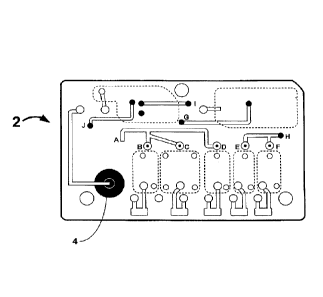
100411 Figure 2 illustrates exemplary layers of a fluidic device according to
the present invention prior to assembly
of the fluidic device which is disclosed in more detail below. Figures 3 and 4
show a top and bottom view,
respectively, of an exemplary fluidic device after the device has been
assembled. The different layers are designed
and assembled to form a three dimensional fluidic channel network. A sample
collection unit 4 provides a sample of
6
CA 2977365 2017-08-24
bodily fluid from a subject. As will be explained in further detail below a
reader assembly comprises actuating
elements (not shown) can actuate the fluidic device to start and direct the
flow of a bodily fluid sample and assay
reagents in the fluidic device. In some embodiments actuating elements first
cause the flow of sample in the fluidic
device 2 from sample collection unit 4 to reaction sites 6, move the sample
upward in the fluidic device from point
G' to point G, and then to waste chamber 8. The actuating elements then
initiate the flow of reagents from reagent
chambers 10 to point B', point C', and point D', then upward to points B, C,
and D, respectively, then to point A,
down to point A', and then to waste chamber 8 in the same manner as the
sample.
[0042] A sample collection unit 4 in a fluidic device 2 may provide a bodily
fluid sample from a subject by any of
the methods described above. If necessary, the sample may first be processed
by diluting the bodily fluid in a
dilution chamber, and or may be filtered by separating the plasma from the red
blood cells in a filtration chamber. In
some embodiments the sample collection unit, diluting chamber, and filtration
chamber may be the same
component, and in some embodiments they may be different components, or any
two may be the same component
and the other may be a separate component. In some embodiments there may be
more than one sample collection
unit in the fluidic device.
[0043] In some embodiments it may be desirable to detect the presence of
analytes on a cell or viral surface, within
a cell or viral membrane, or inside a cell. The difficulty of detecting such
analytes is that cells and other formed
elements are particulate and components of cells do not readily interact with
traditional assay chemistries which are
designed to operate on analytes in solution. Cell-surface analytes react
slowly and inefficiently with surface bound
probes, and analytes inside the cell may not react at all with bound probes.
To allow the detection of such analytes,
in some embodiments the fluidic device may include a lysing assembly to lyse
cells present in the bodily fluid
sample. The lysing assembly may be incorporated with the sample collection
unit, a dilution chamber, and/or a
filtration chamber. In some embodiments the sample collection unit, dilution
chamber, and lysing component are
within the same element in the fluidic device. In some embodiments the lysing
component may be incorporated with
an assay reagent described below.
.. [0044] Where desired, lysing agents may be impregnated and then dried into
porous mats, glass fiber mats,
sintered flits or particles such as Porex, paper, or other similar material.
Lysing agents may be dried onto flat
surfaces. Lysing agents may also be dissolved in liquid diluents or other
liquid reagents. In preferred embodiments
porous materials are used to store the lysing agents because they can store a
lysing agent in dry form likely to be
very stable. They also facilitate the mixing of the bodily fluid sample with
the lysing agent by providing a tortuous
path for the sample as it moves through the porous material In preferred
embodiments such porous materials have a
disc shape with a diameter greater than its thickness. In some embodiments
lysing agents may be dried onto porous
materials using lyophilization, passive evaporation, exposure to warm dry
flowing gas, or other known methods.
[0045] A variety of lysing agents are available in the art and are suitable
for use in connection with the subject
fluidic device. Preferred lysing agents are non-denaturing, such as non-
denaturing detergents. Non-limiting
examples of non-denaturing detergents include Thesit , sodium deoxycholate,
TritoreX-100, and TWEEN*20. The
agents are preferably non-volatile in embodiments where the agents are
impregnated into a solid porous materials. In
some embodiments lysing agents are mixed together. Other materials may be
mixed with the lysing agents to modify
the lytic effects. Such exemplary materials may be, without limitation,
buffers, salts, and proteins. In preferred
embodiments lysing agents will be used in amounts that are in excess of the
minimum amount required to lyse cells.
In some embodiments lysing agents will be used that can lyse both white and
red cells.
100461 One of the advantages of the present invention is that any reagents
necessary to perform an assay on a
fluidic device according to the present invention are preferably on-board, or
housed within the fluidic device before,
7
CA 2977365 2017-08-24
during, and after the assay. In this way the only inlet or outlet from the
fluidic device is preferably the bodily fluid
sample initially provided by the fluidic device. This design also helps create
an easily disposable fluidic device
where all fluids or liquids remain in the device. The on-board design also
prevents leakage from the fluidic device
into the reader assembly which should remain free from contamination from the
fluidic device.
[00471 In a preferred embodiment there is at least one reagent chamber, In
some embodiments there may be two,
three, four, five, six, or more, or any number of reagent chambers as are
necessary to fulfill the purposes of the
invention. A reagent chamber is preferably in fluid communication with at
least one reaction site, and when the
fluidic device is actuated as described herein, reagents contained in said
reagent chambers are released into the
fluidic channels within the fluidic device.
[0048] Reagents according to the present invention include without limitation
wash buffers, enzyme substrates,
dilution buffers, conjugates, enzyme-labeled conjugates, sample diluents, wash
solutions, sample pre-treatment
reagents including additives such as detergents, polymers, chelating agents,
albumin-binding reagents, enzyme
inhibitors, enzymes, anticoagulants, red-cell agglutinating agents,
antibodies, or other materials necessary to run an
assay on a fluidic device. An enzyme conjugate can be either a polyclonal
antibody or monoclonal antibody labeled
with an enzyme that can yield a detectable signal upon reaction with an
appropriate substrate. Non-limiting
examples of such enzymes are alkaline phosphatase and horseradish peroxidase.
In some embodiments the reagents
comprise immunoassay reagents.
10049] In some embodiments a reagent chamber contains approximately about 50 1
to about lml of fluid. In some
embodiments the chamber may contain about 100111 of fluid. The volume of
liquid in a reagent chamber may vary
depending on the type of assay being run or the sample of bodily fluid
provided. In some embodiments the reagents
are initially stored dry and liquefied (e.g., dissolved or melted) upon
initiation of the assay being run on the fluidic
device.
100501 Figures 5 and 6 illustrate an exemplary embodiment of a sealed reagent
chamber. Figure 5 shows a top,
side, and bottom view of a reagent clamber. A top layer 11 contains a
plurality of blisters or pouches 13. A bottom
layer 15 has a bottom surface that is bonded to the fluidic device base 17 as
shown in Figure 6. The bottom layer 15
has a plurality of fluidic channels 19 dispersed through the entire surface,
where each channel traverses the bottom
layer 15. The fluid in the reagent chamber is contained within the chamber by
pressure burstable seal 21 between the
fluidic channel 19 and the chamber 13. The burstable seal 21 is designed such
that at a pm-determined pressure the
seal bursts allowing the fluid in the chamber 13 to flow out into a fluidic
channel 19.
.. 100511 Figure 7 shows an exemplary process of filling the reagent chambers
13 with, for example, reagents.
Reagent chambers 13 may be filled with fluid using a fill channel and a vacuum
draw channel. The process of filling
the reagents involves first removing all the air from the chamber. This is
done by drawing a vacuum through the
vacuum draw channel. Once the vacuum is drawn, a permanent seal is placed
between the fill channel and the
vacuum draw channel. Next, required reagents are dispensed into the chamber
through the fill channel. Then, a
permanent seal is placed between the chamber and the fill channel. This
ensures that when the chamber is
compressed, the fluid can flow in only one direction, towards the burstable
seal. If the compression imparts a
pressure larger than the burst pressure of seal, the seal bursts and the fluid
flows into the fluidic channel.
100521 Figures 8 and 9 illustrate an embodiment of a fluidic device in
operation with actuating elements as
described herein. Fluidic device 2 contains a reagent chamber 10 and a layer
of burstable foil 12 enclosing the
reagent chamber. Above the burstable foil 12 is a portion of the rnicrofluidic
circuit 14. A tough, but elastomeric top
cover 16 acts as the top layer of the fluidic device 2. The reader assembly
includes a valve actuation plate 18.
Securely attached to the plate 18 is a non-coring needle 20 such that when the
plate is lowered, the sharp edge of the
8
CA 2977365 2017-08-24
needle contacts the elastomeric cover 16. The top cover could also be made of
flexible silicone material that would
act as a moisture impermeable seal. This embodiment also provides a solution
to liquid evaporation and leakage
from a fluidic device by isolating any liquid reagents in the fluidic device
from any dry reagents until the assay is
initiated.
100531 In preferred embodiments the reagent chamber and sample collection unit
are fluidly connected to reaction
sites where bound probes can detect an analyte of interest in the bodily fluid
sample using the assay. A reaction site
could then provide a signal indicative of the presence of the analyte of
interest, which can then be detected by a
detection device described in detail herein below.
[0054] In some embodiments the reactions sites are flat but they may take on a
variety of alternative surface
configurations. The reaction site preferably forms a rigid support on which a
reactant can be immobilized. The
reaction site surface is also chosen to provide appropriate light-absorbing
characteristics. For instance, the reaction
site may be functionalized glass, Si, Ge, GaAs, GaP, Si02, SilsI4, modified
silicon, or any one of a wide variety of
gels or polymers such as (poly)tetrafluoroethylene,
(poly)vinylidenedifluoride, polystyrene, polycarbonate,
polypropylene, or combinations thereof. Other appropriate materials may be
used in accordance with the present
invention.
100551 A reactant immobilized at a reaction site can be anything useful for
detecting an analyte of interest in a
sample of bodily fluid. For instance, such reactants include without
limitation, antibodies, cell membrane receptors,
monoclonal antibodies and antisera reactive with a specific analyte indicative
of an influenza viral infection. Various
commercially available reactants such as a host of polyclonal and monoclonal
antibodies specifically developed for
specific analytes can be used.
[0056] A preferred class of reactants are antibodies. As used herein, an
"antibody" (interchangeably used in plural
form) is an imtmmogjobulin molecule capable of specific binding to a target,
such as an analyte in a bodily fluid,
through at least one antigen recognition site, located in the variable region
of the immunoglobulin molecule. As
used herein, the term encompasses not only intact antibodies, but also
fragments thereof (such as Fab, Fab', F(ab12,
Fv, single chain (ScFv), mutants thereof, fusion proteins, humanized
antibodies, and any other modified
configuration of the immunoglobulin molecule that comprises an antigen
recognition site of the required specificity.
10057] The subject methods and apparatus can utilize antibody reactants that
are commercially available or
generated de novo. Laboratory methods for producing polyclonal antibodies and
monoclonal antibodies, are known
in the art. For example, see Harlow and I Ane, Antibodies: A Laboratory
Manual, Cold Spring Harbor Laboratory,
New York (1988) teed Sambrook et al. (1989). Briefly, monoclonal antibodies
useful for the present invention can
be biologically produced by introducing an antigen of an influenza virus into
an animal, e.g., mouse or rat. The
antibody producing cells in the animal are isolated and fused with myeloina
cells or heteromyeloma cells to produce
hybrid cells or hybridomas.
100581 Particular isotypes of a monoclonal *antibody can be prepared either
directly by selecting from the initial
fusion, or prepared secondarily, from a parental hybridoma secreting a
monoclonal antibody of different isotype by
using the sib selection technique to isolate class switch variants using the
procedure described in Steplewski et al.
(1985) Proc. Natl. Acad. Sci. 82:8653 or Spira et al. (1984)]. Immunol Methods
74:307.
10059] The antibody reactants can be linked (i.e., conjugated) to a suitable
detectable label depending on the
particular assay reaction.
10060] In some embodiments a reactant detects an analyte indicative of an
influenza type A, type B, or type C viral
infection. The analyte may comprise at least one surface glycoprotein of an
influenza virus. Exemplary surface
glycoproteins are, without limitation, a hemagglutinin and a neuraminidase.
Hemagglutinin surface proteins include
9
CA 2977365 2017-08-24
HI, H2, H3, H4, HS, 146, H7, H8, H9, HIO, HI I, H12, H13, HI4, HI 5, and H16.
Neuraminidase surface proteins
include N1, N2, N3, N4, and N5.
00611 In some embodiments the reactants detect a plurality of analytes, at
least two of which are indicative of an
influenza viral infection in a sample of bodily fluid. The analytes may be
indicative of an influenza type A, type 13,
or type C viral infection. The analytes may comprise a plurality of surface
glycoproteins of an influenza virus. In
some embodiments the plurality of surface glycoproteins comprises a
hemagglutinin and a neuraminidase. The
hemagglutinin may be selected from the group consisting of H1, 112, H3,
114,113, H6, H7, HS, H9, HIO, Hi 1, H12,
1113, H14, 1115, and 1116, and the neuraminidase may be selected from the
group consisting of Ni, N2, N3, N4, and
N5. In preferred embodiments the hemagglutinin is 115 and the neuraminidase is
Ni.
100621 One skilled in the art will appreciate that there are many ways of
immobilizing various reactants onto a
support where reaction can take place. The immobilization may be covalent or
noncovalent, via a linker moiety, or
tethering them to an immobilized moiety. These methods are well known in the
field of solid phase synthesis and
micro-arrays (Beier et al., Nucleic Acids Res. 27:1970-1-977 (1999). Non-
limiting exemplary binding moieties for
attaching either nucleic acids or proteinaceous molecules such as antibodies
to a solid support include streptavidin or
avidin/biotin linkages, carbamate linkages, ester linkages, amide, thiolester,
(N)-functionalized thiourea,
functionalized maleinaide, amino, disulfide, amide, hydrazone linkages, and
among others. In addition, a silyl
moiety can be attached to a nucleic acid directly to a substrate such as glass
using methods known in the art.
100631 In some embodiments there are more than one reaction sites which can
allow for detection of multiple
analytes of interest from the same sample of bodily fluid, In some embodiments
there are 2, 3,4, 5, 6, or more
reaction sites, or any other number of reaction sites as may be necessary to
carry out the intent of the invention.
[00641 In embodiments with multiple reaction sites on a fluidic device, each
reaction site may be immobilized with
a reactant different from a reactant on a different reaction site. In a
fluidic device with, for example, three reaction
sites, there may be three different probes, each bound to a different reaction
site to bind to three different analytes of
interest in the sample. In some embodiments there may be different reactants
bound to a single reaction site if, for
example, a CCD with multiple detection areas were used as the detection
device, such that multiple different
analytes could be detected in a single reaction site. The capability to use
multiple reaction sites in addition to
multiple different probes on each reaction site enables the high-throughput
characteristics of the present invention.
10065] In preferred embodiments of the invention the fluidic device includes
at least one waste chamber to trap or
capture all liquids after they have been used in the assay. In preferred
embodiments, there is more than one waste
chamber, at least one of which is to be used with a calibration assembly
described herein below. On-board waste
chambers also allow the device to be easily disposable. The waste chamber is
preferably in fluidic communication
with at least one reaction site.
10066] At least one of these channels will typically have small cross
sectional dimensions. In some embodiments
the dimensions are from about .01 mm to about 5 nun, preferably from about .03
mm to about 3 mm, and more
preferably from about .05 nun to about 2 mm. Fluidic channels in the fluidic
device may be created by, for example
without limitation, precision injection molding, laser etching, or any other
technique known in the art to carry out
the intent of the invention.
100671 To ensure that a given assay response (e.g. a photon count) produced at
a reaction site correlates with an
accurate concentration of an analyte of interest in a sample, it is preferably
advantageous to calibrate the fluidic
device before detecting the response (e.g., detecting photons). Calibrating a
fluidic device at the point of
manufacturing for example may be insufficient to ensure an accurate analyte
concentration is determined because a
fluidic device may be shipped prior to use and may undergo changes in
temperature, for example, so that a
I0
CA 2977365 2017-08-24
calibration performed at manufacturing does not take into effect any
subsequent changes to the structure of the
fluidic device or reagents contained therein. In a preferred embodiment of the
present invention, a fluidic device has
a calibration assembly that mimics the assay assembly in components and design
except that a sample is not
introduced into the calibration assembly. Referring to Figures 3 and 4, a
calibration assembly occupies about half of
the fluidic device 2 and includes reagent chambers 32, reactions sites 34, a
waste chamber 36, and fluidic channels
38. Similar to the assay assembly, the number of reagent chambers and reaction
sites may vary depending on the
assay being run on the fluidic device and the number of analytes being
detected.
[0068] Where desired, a sensor for assessing the reliability of an assay for
an analyte in a bodily fluid with the use
of the subject fluidic device can be provided together with the fluidic
device, the reader and/or within the packaging
of the subject system. The sensor is capable of detecting a change in
operation parameters under which the subject
system normally operates. The operation parameters include but are not limited
to temperature, humidity, and
pressure, which may affect the performance of the present system.
[00691 A fluidic device and reader assembly may, after manufacturing, be
shipped to the end user, together or
individually. As a reader assembly is repeatedly used with multiple fluidic
devices, it may be necessary to have
sensors on both the fluidic device and reader assembly to detect such changes
during shipping, for example. During
shipping, pressure or temperature changes can impact the performance of a
number of components of the present
system, and as such a sensor located on either the fluidic device or reader
assembly can relay these changes to, for
example, the external device so that adjustments can be made during
calibration or during data processing on the
external device. For example, if the pressure or temperature of a fluidic
device reached a certain level during
shipping, a sensor located on the fluidic device could detect this change had
occurred and convey this information, to
the reader assembly when it is inserted into the reader assembly by the user.
There may be an additional detection
device in the reader assembly to perform this, or such a device may be
incorporated into another system component.
In some embodiments this information may be wirelessly transmitted to either
the reader assembly or the external
device. Likewise, a sensor in the reader assembly can detect similar changes.
In some embodiments, it may be
desirable to have a sensor in the shipping packaging as well, either instead
of in the system components or in
addition thereto.
[0070] Manufacturing of the fluidic channels may generally be carried out by
any number of microfabrication
techniques that are well known in the art. For example, lithographic
techniques are optionally employed in
fabricating, for example, glass, quartz or silicon substrates, using methods
well known in the semiconductor
manufacturing industries such as photolithographic etching, plasma etching or
wet chemical etching. Alternatively,
micromacbining methods such as laser drilling, micromilling and the like are
optionally employed. Similarly, for
polymeric substrates, well known manufacturing techniques may also be used.
These techniques include injection
molding or stamp molding methods where large numbers of substrates are
optionally produced using, for example,
rolling stamps to produce large sheets of microscale substrates or polymer
rnicrocasting techniques where the
substrate is polymerized within a micromachined mold.
[0071] In some embodiments at least one of the different layers of the fluidic
device may be constructed of
polymeric substrates. Non limiting examples of polymeric materials include
polystyrene, polycarbonate,
polypropylene, polydimethysiloxanes (PDMS), polyurethane, polyvinylchloride
(PVC), and polysulfone.
[0072] The fluidic device may be manufactured by stamping, thermal bonding,
adhesives or, in the case of certain
substrates, for example, glass, or semi-rigid and non-rigid polymeric
substrates, a natural adhesion between the two
components. In some embodiments the fluidic device is manufactured by
ultrasonic or acoustic welding.
11
CA 2977365 2017-08-24
[0073] Figure 2 shows one embodiment of the invention in which fluidic device
2 is comprised of 7 layers.
Features as shown are, for example, cut in the polymeric substrate such that
when the layers are properly positioned
when assembly will form a fluidic network. In some embodiments more or fewer
layers may be used to construct a
fluidic device to carry out the purpose of the invention.
[0074] One objective of the present invention is to prevent fluid inside a
fluidic device from contacting the
components of a reader assembly which may need to remain dry and or
uncontaminated, and also to prevent
contamination to a detection device within the reader assembly. A leak in the
fluidic device could result in liquids,
for example reagents or waste, escaping from the fluidic device and
contaminating the reader. In other embodiments
a liquid absorbing material, such as polymeric materials found in diapers,
could be placed within a portion of the
fluidic channel or waste chamber to absorb the waste liquid. A non-limiting
example of such a polymer is sodium
polyacrylate. Such polymers can absorb fluids hundreds of times their weight.
Hence, only minute quantities of such
polymeric materials may be required to accomplish the goal of absorbing leaked
fluids. In some embodiments a
waste chamber is filled with a superabsorbent material. In some embodiments
leaked liquid may be converted into a
gel or other solid or semi-solid form.
[0075] Another objective of the present system is to provide a fluidic device
that can run a variety of assays on a
fluidic device. A protocol dependent on the identity of the fluidic device may
be transferred from an external device
where it can be stored to a reader assembly to enable the reader assembly to
carry out the specific protocol on the
fluidic device. In preferred embodiments, the fluidic device has an identifier
(ID) that is detected or read by an
identifier detector described herein. The identifier can then be communicated
to a communication assembly, where it
can then be transferred or transmitted to an external device.
[0076] In some embodiments the identifier may be a bar code identifier with a
series of black and white lines,
which can be read by an identifier detector such as a bar code reader, which
are well known. Other identifiers could
be a series of alphanumerical values, colors, raised bumps, or any other
identifier which can be located on a fluidic
device and be detected or read by an identifier detector. In some embodiments
the identifier may comprise a storage
OT memory device and can transmit information to an identification detector.
In some embodiments both techniques
may be used.
[0077] Once a bodily fluid sample is provided to a fluidic device, it is
inserted in a reader assembly. In some
embodiments the fluidic device is partially inserted manually, and then a
mechanical switch in the reader assembly
automatically properly positions the fluidic device inside the reader
assembly. Any other mechanism known in the
art for inserting a disk or cartridge into a device may be used as well. In
some embodiments only manual insertion
may be required.
100781 In some embodiments the reader assembly comprises an identifier
detector for detecting or reading an
identifier on the fluidic device, a controller for automatically controlling
the detection assembly and also mechanical
components of the reader assembly, for example, pumps and/or valves for
controlling or directing fluid through the
fluidic device, a detection device for detecting a signal created by an assay
run on the fluidic device, and a
communication assembly for communicating with an external device.
100791 An identifier detector detects an identifier on the fluidic device
which is communicated to a communication
assembly. In some embodiments the identifier detector can be a bar code
scanner-like device, reading a bar code on
a fluidic device. The identifier detector may also be an LED that emits light
which can interact with an identifier
which reflects light and is measured by the identifier detector to determine
the identity of a fluidic device.
[0080] In preferred embodiments the reader assembly houses a controller which
controls a pump and a series of
valves to control and direct the flow of liquid within the fluidic device. In
some embodiments the reader assembly
12
CA 2977365 2017-08-24
may comprises multiple pumps. The sample and reagents are preferably pulled
through the fluidic channels by a
vacuum force created by sequentially opening and closing at least one valve
while activating a pump within the
reader assembly. Methods of using at least one valve and at least one pump to
create a vacuum force are well
known. While a negative pulling force may be used, a positive pushing force
may also be generated by at least one
pump and valve according to the present invention. In other embodiments
movement of fluid on the fluidic device
may be by electro-osmotic, capillary, piezoelectric, or microactuator action.
[00811 Figures 8 and 9 illustrate an exemplary sequence to initiate the flow
of a reagent within the fluidic device.
An actuation plate 18 in the reader assembly comprises a non-coring needle or
pin 20 which when lowered flexes
the top cover 16, as it is preferably made of strong, flexible elastomeric
material. However, the easily rupturable foil
12 then ruptures due to the stress induced by the flexing of top cover 16.
Valves located downstream to the reagent
chamber puncture different areas of foil in the fluidic device and can then
work in tandem with a pump within the
reader assembly to create a vacuum force to pull the reagent out of the
reagent chamber 6 into a fluidic channel and
then direct the flow of the reagent to a reaction site. At least one valve is
preferably fluidically connected to a pump
housed within the reader assembly. The non-coring needle or pin 20 is removed
from the fluidic device when the
device is removed from the reader assembly. One of the advantages of this
embodiment is that no on-chip pump is
required, which, at least, decreases the size and cost of the fluidic device,
and allows the device to be disposable.
[00821 A reaction assembly preferably houses a detection assembly for
detecting a signal produced by at least one
assay on the fluidic device. Figure 1 illustrates an exemplary position of a
detection device of the present inventiOn
in relation to the fluidic device which is below the fluidic device. The
detection assembly may be above the fluidic
device or at a different orientation in relation to the fluidic device based
on, for example, the type of assay being
performed and the detection mechanism being employed.
[0083] In preferred embodiments an optical detector is used as the detection
device. Non-limiting examples
include a photodiode, photomultiplier tube (PMT), photon counting detector, or
charge-coupled device (CCD). In
some embodiments a pin diode may be used. In some embodiments a pin diode can
be coupled to an amplifier to
create a detection device with a sensitivity comparable to a PMT. Some assays
may generate luminescence as
described herein. In some embodiments chemiluminescence is detected. In some
embodiments a detection assembly
could include a plurality of fiber optic cables connected as a bundle to a CCD
detector or to a PMT array. The fiber
optic bundle could be constructed of discrete fibers or of many small fibers
fused together to form a solid bundle.
Such solid bundles are commercially available and easily interfaced to CCD
detectors.
[0084] In some embodiments, the detection system may comprise non-optical
detectors or sensors for detecting a
particular parameter of a subject. Such sensors may include temperature,
conductivity, potentiometric, and
arnperometric, for compounds that are oxidized or reduced, for example, 02,
H202, and 12, or oxidizable/reducible
organic compounds.
(0085] A communication assembly is preferably housed within the reader
assembly and is capable of transmitting
and receiving information wirelessly from an external device. Such wireless
communication may be bluetooth or
RTM technology. Various communication methods can be utilized, such as a dial-
up wired connection with a
modem, a direct link such as a T1, ISDN, or cable line. In preferred
embodiments a wireless connection is
established using exemplary wireless networks such as cellular, satellite, or
pager networks, GPRS, or a local data
transport system such as Ethernet or token ring over a local area network In
some embodiments the information is
encrypted before it is transmitted over a wireless network. In some
embodiments the communication assembly may
contain a wireless infrared communication component for sending and receiving
information.
13
CA 2977365 2017-08-24
[0086] In some embodiments the communication assembly can have a memory or
storage device, for example
localized RAM, in which the information collected can be stored. A storage
device may be required if information
can not be transmitted at a given time due to, for example, a temporary
inability to wirelessly connect to a network.
The information can be associated with the fluidic device identifier in the
storage device. In some embodiments the
communication assembly can retry sending the stored information after a
certain amount of time. In some
embodiments the memory device can store the information for a period of ten
days before it is erased.
[0087] In preferred embodiments an external device communicates with the
communication assembly within the
reader's assembly. An external device can wirelessly communicate with a reader
assembly, but can also
communicate with a third party, including without limitation a patient,
medical personnel, clinicians, laboratory
personnel, or others in the health care industry.
[0088] In some embodiments the external device can be a computer system,
server, or other electronic device
capable of storing information or processing information. In some embodiments
the external device includes one or
more computer systems, servers, or other electronic devices capable of storing
information or processing
information. In some embodiments an external device may include a database of
subject information, for example
but not limited to, medical records or subject history, clinical trial
records, or preclinical trial records. In preferred
embodiments, an external device stores protocols to be run on a fluidic device
which can be transmitted to the
communication assembly of a reader assembly when it has received an identifier
indicating which fluidic device has
been inserted in the reader assembly. In some embodiments a protocol can be
dependent on a fluidic device
identifier. In some embodiments the external device stores more than one
protocol for each fluidic device. In other
embodiments subject information on the external device includes more than one
protocol. In preferred embodiments
the external server stores mathematical algorithms to process a photon count
sent from a communication assembly
and in some embodiments to calculate the analyte concentration in a bodily
fluid sample.
10089] In some embodiments the external device can include one or more servers
as are known in the art and
commercially available. Such servers can provide load balancing, task
management, and backup capacity in the
event of failure of one or more of the servers or other components of the
external device, to improve the availability
of the server. A server can also be implemented on a distributed network of
storage and processor units, as known in
the art, wherein the data processing according to the present invention reside
on workstations such as computers,
thereby eliminating the need for a server.
[0090] A server can includes a database and system processes. A database can
reside within the server, or it can
reside on another server system that is accessible to the server. As the
information in a database may contains
sensitive information, a security system can be implemented that prevents
unauthorized users from gaining access to
the database.
10091) One advantage of the present invention is that information can be
transmitted from the external device back
to not only the reader assembly, but to other parties or other external
devices, for example without limitation, a PDA
or cell phone. Such communication can be accomplished via a wireless network
as disclosed herein. In some
= embodiments a calculated analyte concentration or other subject
information can be sent to, for example but not
limited to, medical personal or the subject.
Methods of Use
[0092) The subject apparatus and systems provide an effective means for real-
time detection of analytes indicative
of an influenza viral infection present in a bodily fluid from a subject.
14
CA 2977365 2017-08-24
[0093] One aspect of the present invention is a method of detecting an analyte
indicative of an influenza viral
infection. The analyte may comprise at least one surface glycoprotein of an
influenza virus. Exemplary surface
glycoproteins are, without limitation, a hemagglutinin and a neuraminidase.
Hemagglutinin surface proteins include
H1, 112, H3, 114, H5, H6, H7, 118,119, H10, 1111, 1112, 1113,1114, HIS, and
H16. Neuraminidase surface proteins
include NI, N2, N3, N4, and N5. The analyte may also comprise an antibody to a
surface glycoprotein of an
influenza virus.
[0094] One aspect of the present invention is a method for detecting a
plurality of analytes, at least two of which
are indicative of an influenza viral infection in a sample of bodily fluid.
The analytes may be indicative of an
influenza type A, type B, or type C viral infection. The analytes may comprise
a plurality of surface glycoproteins
of an influenza virus. In some embodiments the plurality of surface
glycoproteins comprises a hemagglutinin and a
neuraminidase. The hemagglutinin may be selected from the group consisting of
H1, 112, 113, H4, 115, 116, 117, 118,
H9, H10, HI 1, H12, H13, H14, H15, and 1116, and the neuraminidase may be
selected from the group consisting of
N1, N2, N3, N4, and N5. In preferred embodiments the method detects for both
hemagglutinin H5 and
neuraminidase NI. In one embodiment the method provides for detection of H5
and Ni in the same viral particle(s)
(see Figure 15).
100951 One further aspect of the present invention is a method for detecting a
plurality of analytes incorporated
into a single entity such as a viral particle or cell or cell fragment. hi
this aspect the plurality of analytes are
preferably a combination or complex of analytes, at least two of which are
indicative of an influenza viral infection
in a sample of bodily fluid. The analytes may be indicative of an influenza
type A, type B, or type C viral infection.
The plurality of analytes may comprise a combination or complex of surface
glycoproteins of an influenza virus. In
some embodiments the plurality of analytes may be a combination of surface
glycoproteins comprising a
combination of a hemagglutinin and a neuraminidase. The hemagglutinin may be
selected from the group consisting
of H1, 112, 113, H4, H5, 116, H7, H8, H9, H10, 1111,1112, H13, H14, H15, and
H16, and the neuraminidase may be
selected from the group consisting of NI, N2, N3, N4, and N5. In preferred
embodiments the combination of
analytes is associated with a virulent strain of influenza such as the H5NI
combination. This aspect of the invention
is specific for detecting the combination of the plurality of analytes. It can
distinguish between infection with a
virulent strain such as a combination of H5N1 and a putative-infection with a
different combination of analytes.
[0096] In some embodiments the method detects a plurality of human antibodies
to viral antigens such as
antibodies to surface glycoproteins of an influenza virus.
[0097] In some embodiments the analyte of interest may be a complex of an
analyte indicative of an influenza
viral infection in a sample of bodily fluid and a human antibody to the
analyte. The analyte may be any analyte
indicative of an influenza viral infection described herein, but is preferably
the H5 hemagglutinin, the Ni
neuraminidase, or the H5N1 complex of the H5 and Ni surface glycoproteins.
[0098] A further aspect of the present invention is a method for detecting a
plurality of analytes, wherein at least
one analyte is indicative of an influenza viral infection in a sample of
bodily fluid, and wherein at least one analyte
is a biomarker in the sample of bodily fluid indicative of the stress imposed
on the human body by the viral infection
or an indicator of the body's response to the infection. The at least one
analyte indicative of an influenza viral
infection may be any analyte indicative of an influenza viral infection
described herein. Exemplary biomarkers
indicative of the stress imposed on the human body by the viral infection
include, without limitation, CRP,
TNFa, interleulcins and the like. Exemplary biornarkers indicative of the
body's defensive reaction to the virus
include antibodies to the virus, particularly of the IgM isotype.
CA 2977365 2017-08-24
[0099] The subject apparatus and systems have a spectrum of utility in, for
example, disease diagnosis and disease
detection.
[00100] Accordingly, in one embodiment, the present invention provides a
method of detecting an analyte
indicative of an influenza viral infection in a bodily fluid from a subject
comprises providing a fluidic device
comprising at least one sample collection unit, an immunoassay assembly
containing immunoassay reagents, a
plurality of channels in fluid communication with said sample collection unit
and/or said immunoassay assembly;
actuating said fluidic device and directing said immunoassay reagents within
said fluidic device; allowing a sample
of bodily fluid suspected to contain said analyte to react with said
immunoassay reagents contained within said assay
immunoassay assembly to yield a detectable signal indicative of the presence
of said analyte in said bodily fluid; and
detecting said detectable signal generated from said analyte initially
collected in said sample of bodily fluid.
Preferably, a sample of bodily fluid of less than about 500 ul is used for one
or more of these applications.
[001011 As used herein, the term "subject" and "patient" is used
interchangeably, which refers to an animal,
preferably an avian (bird) or a mammalian species (for example, human). The
term avian as used herein includes
poultry_ Mammals include, but are not limited to, marines, simians, humans,
farm animals, sport animals, and pets.
[001021 As used herein, in some aspects the terms "reagents" and "reactants"
are used interchangeably.
[001031 In some embodiments a sample of bodily fluid can first be provided to
the fluidic device by any of the
methods described herein. The fluidic device can then be inserted into the
reader assembly. An identification
detector housed within the reader assembly can detect an identifier of the
fluidic device and communicate the
identifier to a communication assembly, which is preferably housed within the
reader assembly. The communication
assembly then transmits the identifier to an external device which transmits a
protocol to nm on the fluidic device
based on the identifier to the communication assembly. A controller preferably
housed within the reader assembly
controls actuating elements including at least one pump and one valve which
interact with the fluidic device to
control and direct fluid movement within the device. In some embodiments the
first step of the assay is a wash cycle
where all the surfaces within the fluidic device are wetted using a wash
buffer. The fluidic device is then calibrated
.. using a calibration assembly by running the same reagents as will be used
in the assay through the calibration
reaction sites, and then a luminescence signal from the reactions sites is
detected by the detection means, and the
signal is used in calibrating the fluidic device. The sample containing the
analyte is introduced into the fluidic
channel. The sample may be diluted and further separated into plasma or other
desired component at a. filter. The
separated sample now flows through the reaction sites and analytes present
therein will bind to reactants bound
thereon. The plasma of sample fluid is then flushed out of the reaction wells
into a waste chamber. Depending on
the assay being run, appropriate reagents are directed through the reaction
sites to carry out the assay. All the wash
buffers and other reagents used in the various steps, including the
calibration step, are collected in wash tanks The
signal produced in the reaction sites is then detected by any of the methods
described herein.
1001041 A variety of assays may be performed on a fluidic device according to
the present invention to detect an
analyte of interest in a sample. A wide diversity of labels is available in
the art that can be employed for conducting
the subject assays. In some embodiments labels are detectable by
spectroscopic, photochemical, biochemical,
immunochemical, or chemical means. For example, useful nucleic acid labels
include 32P, 35S, fluorescent dyes,
electron-dense reagents, enzymes, biotin, digoxigenin, or haptens and proteins
for which antisera or monoclonal
antibodies are available. A wide variety of labels suitable for labeling
biological components are known and are
reported extensively in both the scientific and patent literature, and are
generally applicable to the present invention
for the labeling of biological components. Suitable labels include
radionucleides, enzymes, substrates, cofactors,
inhibitors, fluorescent moieties, chemiluminescent moieties, bioluminescent
labels, calorimetric labels, or magnetic
16
CA 2977365 2017-08-24
particles. Labeling agents optionally include, for example, monoclonal
antibodies, polyclonal antibodies, proteins, or
other polymers such as affinity matrices, carbohydrates or lipids. Detection
proceeds by any of a variety of known
methods, including spectrophototnetric or optical tracking of radioactive or
fluorescent markers, or other methods
which track a molecule based upon size, charge or affinity. A detectable
moiety can be of any material having a
detectable physical or chemical property. Such detectable labels have been
well-developed in the field of gel
electrophoresis, column chromatograpy, solid substrates, spectroscopic
techniques, and the like, and in general,
labels useful in such methods can be applied to the present invention. Thus, a
label includes without limitation any
composition detectable by spectroscopic, photochemical, biochemical,
irrnnunochemical, electrical, optical, thermal
or chemical means.
1001051 In some embodiments the label is coupled directly or indirectly to a
molecule to be detected such as a
product, substrate, or enzyme, according to methods well known in the art. As
indicated above, a wide variety of
labels are used, with the choice of label depending on the sensitivity
required, ease of conjugation of the compound,
stability requirements, available instrumentation, and disposal provisions.
Non-radioactive labels are often attached
by indirect means. Generally, a ligand molecule is covalently bound to a
polymer. The ligand then binds to an anti-
ligand molecule which is either inherently detectable or covalently bound to a
signal system, such as a detectable
enzyme, a fluorescent compound, or a chetniluminescent compound. A number of
ligands and anti-ligands can be
used. Where a ligand has a natural anti-ligand, for example, biotin,
thyroxine, and cortisol, it can be used in
conjunction with labeled, anti-ligands. Alternatively, any haptenic or
antigenic compound can be used in
combination with an antibody.
[001061 In some embodiments the label can also be conjugated directly to
signal generating compounds, for
example, by conjugation with an enzyme or fluorophore. Enzymes of interest as
labels will primarily be hydrolases,
particularly phosphatases, esterases and glycosidases, or oxidoreductases,
particularly peroxidases. Fluorescent
compounds include fluorescein and its derivatives, rhodamine and its
derivatives, dansyl, and umbelliferone.
Cherniluminescent compounds include luciferin, and 2,3-
dihydrophthalazinediones, such as himinol and dioxetanes
[001071 Methods of detecting labels are well known to those of skill in the
art. Thus, for example, where the label is
a radioactive label, means for detection include a scintillation counter or
photographic film as in autoradiography.
Where the label is a fluorescent label, it may be detected by exciting the
fluorochrome with the appropriate
wavelength of light and detecting the resulting fluorescence by, for example,
microscopy, visual inspection, via
photographic film, by the use of electronic detectors such as digital cameras,
charge coupled devices (CCDs) or
photomultipliers and phototubes, or other detection device. Similarly,
enzymatic labels are detected by providing
appropriate substrates for the enzyme and detecting the resulting reaction
product. Finally, simple colorimetric
labels are often detected simply by observing the color associated with the
label. For example, conjugated gold often
appears pink, while various conjugated beads appear the color of the bead.
[001081 In some embodiments the detectable signal may be provided by
luminescence sources. "Luminescence" is
the term commonly used to refer to the emission of light from a substance for
any reason other than a rise in its
temperature. In general, atoms or molecules emit photons of electromagnetic
energy (e.g., light) when they move
from an "excited state" to a lower energy state (usually the ground state).
There are many causes of excitation. If
exciting cause is a photon, the luminescence process is referred to as
"photoluminescence". If the exciting cause is
an electron, the luminescence process is referred to as "electroluminescence".
More specifically,
electrolurninescence results from the direct injection and removal of
electrons to form an electron-hole pair, and
subsequent recombination of the electron-hole pair to emit a photon.
Luminescence which results from a chemical
reaction is usually referred to as "chemiluminescence". Luminescence produced
by a living organism is usually
17
CA 2977365 2017-08-24
referred to as "bioluminescence". If photoluminescence is the result of a spin-
allowed transition (e.g., a single-
singlet transition, triplet-triplet transition), the photoluminescence process
is usually referred to as "fluorescence".
Typically, fluorescence emissions do not persist after the exciting cause is
removed as a result of short-lived excited
states which may rapidly relax through such spin-allowed transitions. If
photoluminescence is the result of a
.. spin-forbidden transition (e.g., a triplet-singlet transition), the
photoluminescence process is usually referred to as
"phosphorescence". Typically, phosphorescence emissions persist long after the
exciting cause is removed as a
result of long-lived excited states which may relax only through such spin-
forbidden transitions. A "luminescent
label" may have any one of the above-described properties.
[00109] Suitable chemiluminescent sources include a compound which becomes
electronically excited by a
.. chemical reaction and may then emit light which serves as the detectible
signal or donates energy to a fluorescent
acceptor. A diverse number of families of compounds have been found to provide
chemiluminescence under a
variety or conditions. One family of compounds is 2,3-dihydro-1,4-
phthalazinedione. A frequently used compound
is huninol, which is a 5-amino compound. Other members of the family include
the 5-amino-6,7,8-trimethoxy- and
the dimethylamino[caThenz analog. These compounds can be made to luminesce
with alkaline hydrogen peroxide or
calcium hypochlorite and base. Another family of compounds is the 2,4,5-
triphenylimidazoles, with lophine as the
corrunon name for the parent product. Cherniluminescent analogs include para-
dimethylamino and -methoxy
substituents. Chemiluminescence may also be obtained with oxalates, usually
oxalyl active esters, for example, p-
nitrophenyl and a peroxide such as hydrogen peroxide, under basic conditions.
Other useful chemiluminescent
compounds that are also known include N-alkyl acridinum esters and dioxetanes.
Alternatively, luciferins may be
used in conjunction with luciferase or lucigenins to provide bioluminescence.
1001101 In some embodiments immunoassays are run on the fluidic device. While
competitive binding assays,
which are well known in the art, may be run in some embodiments, in certain
embodiments a two-step method is
used which eliminates the need to mix a conjugate and a sample before exposing
the mixture to an antibody, which
may be desirable when very small volumes of sample and conjugate are used, as
in the fluidic device of the present
invention. A two-step assay has additional advantages over the competitive
binding assays when used with a fluidic
device as described herein. It combines the ease of use and high sensitivity
of a sandwich (competitive binding)
immunoassay with the ability to assay small molecules.
1001111 In an exemplary two-step assay shown in Figure 10, the sample
containing analyte ("Ag") first flows over a
reaction site containing antibodies ("Ab"). The antibodies bind the analyte
present in the sample. After the sample
passes over the surface, a solution with analyte conjugated to a marker
("labeled Ag") at a high concentration is
passed over the surface. The conjugate saturates any of the antibodies that
have not yet bound the analyte. Before
equilibrium is reached and any displacement of pre-bound unlabelled analyte
occurs, the high-concentration
conjugate solution is washed oft The amount of conjugate bound to the surface
is then measured by the appropriate
technique, and the detected conjugate is inversely proportional to the amount
of analyte present in the sample.
.. [00112J An exemplary measuring technique for a two-step assay is a
chemiluminescence enzyme immunoassay as
shown in Figure 11. As is known in the field, the marker can be a commercially
available marker such as dioxitane-
phosphate, which is not luminescent but becomes luminescent after hydrolysis
by, for example, alkaline
phosphatase. An enzyme such as alkaline phosphatase is also passed over the
substrate to cause the marker to
luminesce. In some embodiments the substrate solution is supplemented with
enhancing agents such as, without
.. limitation, fluorescein in mixed micelles, soluble polymers, or PVC which
create a much brighter signal than the
lurninophore alone. Moreover, an alkaline phosphatase conjugate with a higher
turnover number than that used in
the commercial assay is employed. This allows signal generation to proceed
much more rapidly and a higher overall
]8
CA 2977365 2017-08-24
signal is achieved. The increased sensitivity of the two-step chemiluminescent
enzyme immunoassay (TOSCA) is
illustrated in Figure 12. Figure 12 shows that for analytes in the picomolar
concentration, TOSCA is able to provide
a more robust signal (higher sensitivity) than a competitive binding assay.
Use of a two-step binding assay thus
contributes to higher sensitivity capabilities of the present invention.
1001131 Additionally, TOSCA is less sensitive to matrix effects than other
methodologies. This allows one to work
with samples that have not been extensively pre-processed using standard
laboratory techniques such as, for
example, solid phase extraction and chromatography. The ability of TOSCA to
assay less than ideal samples and
maintain desired sensitivity is illustrated in Figure 13. Compared to
competitive binding assay, for all sample
preparations (and dilutions), TOSCA has better sensitivity than competitive
binding.
[001141 One useful immtmoassay that can be run on the fluidic device is ELISA
(Enzyme-Linked hnmunoSorbent
Assay). Performing an ELISA generally involves at least one antibody capable
of binding an antigen of interest
(i.e., an analyte that is indicative of influenza viral infection). A sample
containing or suspected to contain the
antigen of interest is immobilized on a support (e.g., a microtiter plate, a
well or other support having a surface for
immobilization) either non-specifically (e.g., via adsorption to the surface)
or specifically (e.g., via capture by
another antibody specific to the same antigen, in a "sandwich" ELISA). After
the antigen is immobilized the
detection antibody is added, forming a complex with the antigen. The detection
antibody can be conjugated to an
enzyme, or can itself be detected by a secondary antibody which is in turn
conjugated to an enzyme. Upon addition
of a substrate for the conjugated enzyme, a detectable signal is generated
which indicates the presence and/or
quantity of the antigen in the sample. The choice of substrates will depend on
the enzyme conjugated. Suitable
substrates include fluorogenic and chromogenic substrates. One of skill in the
art would be knowledgeable as to the
parameters that can be modified to increase the signal detected as well as
other variations of ELISAs known in the
art.
[001151 Figure 14 illustrates a typical ELISA. As shown, a solid phase capture
surface can include an attached first
antibody to which diluted plasma (sample) can be added. Analyte if present in
the sample can bind to the first
antibody and become immobilized. An enzyme reagent can be added that includes,
for example, an antibody
coupled or conjugated to an enzyme (e.g., alkaline phosphatase). If the
antibody portion of the enzyme reagent can
bind the analyte, then the enzyme reagent also becomes immobilized at the
capture surface. Addition of a substrate
for the enzyme can result in a product producing an effect, for example, light
that can be measured and plotted as
shown. In this manner the amount of analyte present in a sample can be
measured.
[001161 Figure 15 illustrates an exemplary ELISA for use with the fluidic
device of the invention. As shown, a
solid phase capture surface of the device can include a first antibody, "solid
phase antibody 1", that is surface
immobilized and specific for a test antigen (e.g., antibody specific for a
neuraminidase on a virus). If the test
antigen is present in a test sample (e.g., blood) exposed to the solid phase
antibody 1 then the test antigen can
become immobilized (captured) at the capture surface. Subsequently provided is
a second antibody that is specific
for a second test antigen and includes a conjugated detectable compound, shown
as "enzyme labeled antibody 2"
(e.g., enzyme labeled antibody specific for a hemagglutinin on a virus), that
can be added after the test sample (e.g.,
blood). Binding and subsequent detection of the second conjugated antibody at
the capture surface can indicate the
presence of the first and second test antigens in the test sample. In use, the
first and second test antigens can include
any of' the neurazninidase or hem.agglutanin types described herein.
1001171 Although different first and second antigens (and antibodies) are used
in the illustrated example, it is
envisioned that a single type of test antigen could be detected using two
forms of the same antibody (i.e., an
immobilized solid phase form for antigen capture and an enzyme labeled form
for detection).
19
CA 2977365 2017-08-24
[00118] The term "analytes" according to the present invention includes
without limitation drugs, prodrugs,
pharmaceutical agents, drug metabolites, biomarkers such as expressed proteins
and cell markers, antibodies,
antigens, viruses, serum proteins, cholesterol, polysaccharides, nucleic
acids, biological analytes, biomarker, gene,
protein, or hormone, or any combination thereof. At a molecular level, the
analytes can be polypeptide glycoprotein,
polysaccharide, lipid, nucleic acid, and a combination thereof.
[00119] Of interest are biomarkers are associated with a particular disease or
with a specific disease stage. Such
analytes include but are not limited to those associated with autoimmune
diseases.
[00120] Also of interest are analytes that are indicative of a microorganism_
Exemplary microorganisms include
but are not limited to bacterium, virus, fungus and protozoa.
[00121] The analyte may be indicative of an influenza type A, type B, or type
C viral infection. The analyte may
comprise at least one surface glycoprotein of an influenza virus. Exemplary
surface glycoproteins are, without
limitation, a hemagglutinin and a neuraminidase. Hemagglutinin surface
proteins include H1, 112, H3, 114, H5, H6,
117, H8, H9, H10, H11, H12, H13, H14, HIS, and H16. Neuraminidase surface
proteins include Ni, N2, N3, N4,
and N5.
[00122] One aspect of the present invention is a system for detecting a
plurality of analytes, at least two of which
are indicative of an influenza viral infection in a sample of bodily fluid.
The analytes may be indicative of an
influenza type A, type B, or type C viral infection. The analytes may comprise
a plurality of surface glycoproteins
of an influenza virus. In some embodiments the plurality of surface
glycoproteins comprises a hemagglutinin and a
neuraminidase. The hemagglutinin may be selected from the group consisting of
HI, 112, 113, H4, 115, H6, H7, H8,
H9, 1110, 1111, H12, H13, H14, H15, and 1116, and the neuraminidase may be
selected from the group consisting of
N1, N2, N3, N4, and N5. In preferred emborlimPnts the hemagglutinin is 115 and
the neuraminidase is Ni. The
system is capable of detecting and/or quantifying the analytes of particular
interest.
[00123] By detecting the presence of the viral antigens or antibodies to the
antigens, for example, the fluidic device
can detect the presence of a type of influenza virus in the sample of bodily
fluid from the subject.
[00124] Analytes that can be detected by the subject method also include blood-
born pathogens selected from a
non-limiting group that consists of Staphylococcus epidermidis, Escherichia
coil, methicillin-resistant
Staphylococcus aureus (MSRA), Staphylococcus aureus, Staphylococcus hominis,
Enterococcus faecalis,
Pseudomonas aeruginosa, Staphylococcus capitis, Staphylococcus warn en,
Klebsiella pneumoniae, Haemophilus
influnzae, Staphylococcus simulans, Streptococcus pneumoniae and Candida
albicans.
[00125] Analytes that can be detected by the subject method also encompass a
variety of sexually transmitted
diseases selected from the following: gonorrhea (Neisseria gorrhoeae),
syphilis (Treponena pallidum), Chlamydia
(Chlamydia trachomatis), nongonococcal urethritis (Ureaplasm urealyticum),
yeast infection (Candida albicans),
chancroid (Haemophilus ducreyi), trichomoiaiasis (Trichomonas vaginalis),
genital herpes (HSV type I & II), HIV I,
11W II and hepatitis A, B, C, G, as well as hepatitis caused by UV.
[00126] It is envisioned that in some embodiments the present invention
provides for monitoring an infection by a
pathogen either directly through detection of the pathogen or indirectly, for
example, by detection of a analyte
associated with a pathogen (e.g., a viral antigen) or even by detection of an
antibody to a component or product
associated with a pathogen (e.g., an antibody to a viral antigen). It is also
envisioned that a pathogen can be
indirectly detected through an immune-related response to the pathogen.
Detection of the pathogen can be
performed on a test sample from a subject that is asymptomatic or symptomatic
for the pathogen. Detection of the
pathogen can be performed on a test sample from a subject before, during or
after infection with the pathogen. As
CA 2977365 2017-08-24
such, it is envisioned that an early stage infection (e.g., in some cases an
asymptomatic infection), or any later phase
of infection can be monitored for the pathogen of interest.
[00127] A wide range of pathogen concentrations in a sample from a subject can
be detected either directly or
indirectly as discussed above using the invention. The amount of pathogen
present in a test sample can be expressed
in any of a number of ways well known in the art_ By way of non-limiting
examples, the number of pathogens can
be expressed as viral burden (e.g., where the infection is a viral infection),
infectious units (1U), and/or infectious
units per million cells or milliliter (IUPM). In one example, it is envisioned
that pathogens can be detected in a test
sample at a concentration of from 100 IU per ml of sample up to 1 x 1091U per
ml of sample using the invention In
another example pathogens can be detected from 100 1U per ml of sample up to
1000 IU per ml of sample using the
invention. In yet another example pathogens can be detected from 1,000 IU per
ml of sample up to 1 x 10⁶ IU per
ml of sample using the invention.
1001281 In a separate embodiment, the present invention provides a method of
monitoring more than one
pharmacological parameter usefiil for assessing efficacy and/or toxicity of an
anti-influenza therapeutic agent. The
method comprises subjecting a sample of bodily fluid from a subject
administered with the anti-influenza
therapeutic agent to a fluidic device for monitoring said more than one
pharmacological parameter, said fluidic
device comprising at least one sample collection unit, and an assay assembly
comprising reaction reagents; actuating
said fluidic device and directing said immunoassay reagents within said
fluidic device; allowing said sample of
bodily fluid to react with immunoassay reagents to yield detectable signals
indicative of the values of the more than
one pharmacological parameter from said sample; and detecting said detectable
signal generated from said sample of
bodily fluid. Where desired, the method further involves repeating the steps
at a time interval prompted by a
wireless signal communicated to the subject.
[00129] For the purposes of this invention, a "therapeutic agent" is intended
to include any substances that have
therapeutic utility and/or potential. Such substances include but are not
limited to biological or chemical compounds
such as a simple or complex organic or inorganic molecules, peptides, proteins
(e.g. antibodies) or a polynucleotides
(e.g. anti-sense). A vast array of compounds can be synthesized, for example
polymers, such as polypeptides and
polynucleotides, and synthetic organic compounds based on various core
structures, and these are also included in
the term "therapeutic agent". In addition, various natural sources can provide
compounds for screening, such as
plant or animal extracts, and the like. It should be understood, although not
always explicitly stated that the agent is
used alone or in combination with another agent, having the same or different
biological activity as the agents
identified by the inventive screen. The agents and methods also are intended
to be combined with other therapies.
[001301 Pharmacodynamic (PD) parameters according to the present invention
include without limitation physical
parameters such as temperature, heart rate/pulse, blood pressure, and
respiratory rate, and biomarkers such as
proteins, cells, and cell markers. Biomarkers could be indicative of disease
or could be a result of the action of a
drug. Pharmacokinetic (PK) parameters according to the present invention
include without limitation drug and drug
metabolite concentration. Identifying and quantifying the PK parameters in
real time from a sample volume is
extremely desirable for proper safety and efficacy of drugs. If the drug and
metabolite concentrations are outside a
desired range and/or unexpected metabolites are generated due to an unexpected
reaction to the drug, immediate
action may be necessary to ensure the safety of the subject. Similarly, if any
of the pharmacodynamic (PD)
parameters fall outside the desired range during a treatment regime, immediate
action may have to be taken as well
[00131] In preferred embodiments physical parameter data is stored in or
compared to store profiles of physical
parameter data in a bioinformatics system which may be on an external device
incorporating phannacogenornic and
pharmacolcinetic data into its models for the determination of toxicity and
dosing. Not only does this generate data
21
CA 2977365 2017-08-24
for clinical trials years prior to current processes but also enables the
elimination of current disparities between
apparent efficacy and actual toxicity of drugs through real-time continuous
monitoring. During the go/no go
decision process in clinical studies, large scale comparative population
studies can be conducted with the data stored
on the database. This compilation of data and real-time monitoring allows more
subjects to enter clinical trials in a
safe fashion earlier than currently allowed. In another embodiment biomarkers
discovered in human tissue studies
can be targeted by the device for improved accuracy in determining drug
pathways and efficacy in cancer studies.
[00132] In another embodiment, the present invention provides a method of
detecting at least two distinct analytes
indicative of an influenza viral infection of different concentrations in a
bodily fluid from a subject comprises
providing a fluidic device comprising a sample collection unit, an assay
assembly, and a plurality of channels in
fluid communication with said sample collection unit and/or said assay
assembly; allowing a sample of bodily fluid
to react with a plurality of reactants contained in said assay assembly to
yield signals indicative of the concentrations
of said at least two analytes; and detecting said signals that are indicative
of the presence or absence of the at least
two distinct analytes, wherein said signals are detectable over a range of 3
orders of magnitude.
[00133] Currently, a need exists for the detecting more than one analyte
indicative of an influenza viral infection
where the analytes are present in widely varying concentration range, for
example, one analyte is in the pg/ml
concentration and another is in the ng/ml concentration Cherniluminescence-
ELISA has the ability to simultaneously
assay analytes that are present in the same sample in a wide concentration
range. Another advantage for being able
to detect concentrations of different analytes present in a wide concentration
range is the ability to relate the ratios of
the concentration of these analytes to safety and efficacy of multiple drugs
administered to a subject For example,
unexpected drug-drug interactions can be a common cause of adverse drug
reactions. A real-time, concurrent
measurement technique for measuring different analytes would help avoid the
potentially disastrous consequence of
adverse drug-drug interactions.
[00134] Being able to monitoring the rate of change of an analyte
concentration or PD or PK over a period of time
in a single subject, or performing trend analysis on the concentration, PD, or
PK, whether they are concentrations of
drugs or their metabolites, can help prevent potentially dangerous situations.
For example, if glucose were the
analyte of interest, the concentration of glucose in a sample at a given time
as well as the rate of change of the
glucose concentration over a given period of time could be highly useful in
predicting and avoiding, for example,
hypoglycemic events. Such trend analysis has widespread beneficial
implications in drug dosing regimen. When
multiple drugs and their metabolites are concerned, the ability to spot a
trend and take proactive measures is often
desirable.
[001351 Accordingly, the present invention provides a method of performing a
trend analysis on the concentration
of an analyte indicative of an influenza viral infection in a subject. The
method comprise a) providing a fluidic
device comprising at least one sample collection unit, an immunoassay assembly
containing immunoassay reagents,
a plurality of channels in fluid communication with said sample collection
unit and/or said immunoassay assembly;
b) actuating said fluidic device and directing said immunoassay reagents
within said fluidic device; c) allowing a
sample of bodily fluid to react with said immunoassay reagents contained
within said assay immunoassay assembly
to yield a detectable signal indicative of the presence of said analyte in
said sample; d) detecting said detectable
signal generated from said analyte collected in said sample of bodily fluid;
and e) repeating steps a) through d) for a
single subject over a period of time to detect concentrations of said analyte,
thereby performing said trend analysis.
[00136] In some embodiments, a method of detecting an analyte indicative of an
influenza viral infection in a
bodily fluid from a subject using an assay transmitted from an external device
is provided. The method comprises
providing a fluidic device comprising at least one sample collection unit and
an immunoassay assembly containing
22
CA 2 97 7 3 65 2 0 17 - 0 8 - 2 4
immunoassay reagents, detecting said fluidic device and wirelessly
transmitting an immunoassay protocol to said
device; allowing a sample of bodily fluid to react with immunoassay reagents
to yield a detectable signal indicative
of the presence of said analyte using said transmitted immunoassay protocol;
and detecting said detectable signal.
[00137] Communication between a reader assembly and an external storage device
allows for a reader assembly of
the present invention to download a fluidic device-specific protocol to run on
the fluidic device based on the identity
of the fluidic device. This allows a reader assembly to be used
interchangeably with any appropriate fluidic device
described herein. In addition, the external device can store a plurality of
protocols associated with a given fluidic
device, and depending on, for example, a subject's treatment regime or plan,
different protocols can be
communicated from the external device to the reader assembly to be run on the
fluidic device to detect a variety of
analytes indicative of an influenza viral infection. The external device can
also store a plurality of protocols
associated not only with a fluidic device, but also with a particular subject
or subjects, such that a protocol can be
associated with a subject as well as with a fluidic device.
[00138] The present invention allows for automatic quantification of a
pharmacological parameter of a subject as
well as automatic comparison of the parameter with, for example, the subject's
medical records which may include a
history of the monitored parameter, or medical records of another group of
subjects. Coupling real-time analyte
monitoring with an external device which can store data as well as perform any
type of data processing or algorithm,
for example, provides a device that can assist with typical subject care which
can include, for example, comparing
current subject data with past subject data. The present invention therefore
creates a business method which
effectively performs at least part of the monitoring of a subject that is
currently performed by medical personnel.
1001391 One of the significant advantages of the envisioned network is
illustrated in Figure 20. As all the
information is securely channeled through the intemet, this allows the
simultaneous sharing of the information with
various interested parties, while satisfying the appropriate clinical,
regulatory and business needs.
[00140] In some embodiments, the present invention provides a method of
transmitting a pharmacological
parameter of a subject via a handheld device comprises providing a fluidic
device comprising at least one sample
collection unit and an assay assembly; allowing a sample of bodily fluid to
react with reactants contained within said
assay assembly to yield a detectable signal indicative of the presence of said
analyte indicative of an influenza virus;
detecting said detectable signal; transmitting said signal to an external
device; processing said signal in said external
device; and transmitting said processed signal via a handheld device.
[00141] One advantage of the current invention is that assay results can be
substantially immediately communicated
to any third party that may benefit from obtaining the results. For example,
once the analyte concentration is
determined at the external device, it can be transmitted to a patient or
medical personnel who may need to take
further action. The communication step to a third party can be performed
wirelessly as described herein, and by
transmitting the data to a third party's hand held device, the third party can
be notified of the assay results virtually
anytime and anywhere. Thus, in a time-sensitive scenario, a patient may be
contacted immediately anywhere if
urgent medical action may be required.
1001421 In some embodiments a method of automatically selecting a protocol to
be run on a fluidic device
comprises providing a fluidic device comprising an identifier detector and an
identifier; detecting said identifier with
said identifier detector; transferring said identifier to an external device;
and selecting a protocol to be run on said
fluidic device from a plurality of protocols on said external device
associated with said identifier.
100143] By detecting each fluidic device based on an identifier associated
with the fluidic device after it is inserted
in the reader assembly, the system of the present invention allows for fluidic
device-specific protocols to be
downloaded from an external device and run on the fluidic device. In some
embodiments the external device can
23
CA 2977365 2017-08-24
store a plurality of protocols associated with the fluidic device or
associated with a particular subject or group of
subjects. For example, when the identifier is transmitted to the external
device, software on the external device can
obtain the identifier. Once obtained, software on the external device, such as
a database, can use the identifier to
identify protocols stored in the database associated with the identifier. If
only one protocol is associated with the
identifier, for example, the database can select the protocol and software on
the external device can then transmit the
protocol to the communication assembly on the reader assembly. The ability to
use protocols specifically associated
with a fluidic device allows for any appropriate fluidic device to be used
with a single reader assembly, and thus
virtually any analyte of interest can be detected with a single reader
assembly.
(00144] In some embodiments multiple protocols may be associated with a single
identifier. For example, if it is
beneficial to detect from the same subject an analyte once a week, and another
analyte twice a week, protocols on
the external device associated with the identifier can also each be associated
with a different day of the week, so that
when the identifier is detected, the software on the external device can
select a specific protocol that is associated
with the day of the week.
(00145] In some embodiments a subject may be provided with a plurality of
fluidic devices to use to detect a variety
of analytes. A subject may, for example, use different fluidic devices on
different days of the week In some
embodiments the software on the external device associating the identifier
with a protocol may include a process to
compare the current day with the day the fluidic device is to be used based on
a clinical trial for example. If for
example, the two days of the week are not identical, the external device can
wirelessly send notification to the
subject using any of the methods described herein or known in the art to
notify them that an incorrect fluidic device
is in the reader assembly and also of the correct fluidic device to use that
day. This example is only illustrative and
can easily be extended to, for example, notifying a subject that a fluidic
device is not being used at the correct time
of day.
[00146] In some embodiments, the present invention provides a method of
obtaining pharmacological data useful
for assessing efficacy and/or toxicity of an anti-influenza pharmaceutical
agent from a test animal. The method
involves the steps of a) providing a fluidic device comprising at least one
sample collection unit, an assay assembly;
and a plurality of channels in fluid communication with said sample collection
unit and/or said assay assembly; b)
allowing a sample of biological fluid of less than about 50 ul to react with
reactants contained within said assay
assembly to yield a detectable signal generated from an analyte indicative of
an influenza viral infection initially
collected in said sample that is indicative of a pharmacological parameter;
and c) detecting said detectable signal;
and d) repeating the reaction and detection steps with a second sample of
biological fluid from the same test animal.
In a related embodiment, the present invention provides a method comprising a)
providing a fluidic device
comprising at least one sample collection unit, an assay assembly; and a
plurality of channels in fluid
communication with said sample collection unit and/or said assay assembly; b)
allowing a sample of biological fluid
to react with reactants contained within said assay assembly to yield a
detectable signal generated from an analyte
initially collected in said sample that is indicative of a pharmacological
parameter; and c) detecting said detectable
signal; and d) repeating the reaction and detection steps with a second sample
of biological fluid from the same test
animal, wherein the animal is not subjected to anesthesia.
1001471 When using laboratory animals in preclinical testing of an anti-
influenza pharmaceutical agent, it is often
necessary to kill the test subject to extract enough blood to perform an assay
to detect an analyte of interest. This has
both financial and ethical implications, and as such it may be advantageous to
be able to draw an amount of blood
from a test animal such that the animal does not need to be killed. In
addition, this can also allow the same test
animal to be tested with multiple pharmaceutical agents at different times,
thus allowing for a more effective
24
CA 2977365 2017-08-24
preclinical trial. On average, the total blood volume in a mouse, for example,
is 6-8 mL of blood per 100 gram of
body weight. A benefit of the current invention is that only a very small
volume of blood is required to perform
preclinical trials on mice or other small laboratory animals. In some
embodiment between about 1 microliter and
about SO microliters are drawn. In preferred embodiment between about I
microliter and 10 microliters are drawn.
In preferred embodiments about 5 microliters of blood are drawn.
1001481 A further advantage of keeping the test animal alive is evident in a
preclinical time course study. When
multiple mice, for example, are used to monitor the levels of an analyte in a
test subject's bodily fluid over time, the
added variable of using multiple subjects is introduced into the trial. When,
however, a single test animal can be
used as its own control over a course of time, a more accurate and beneficial
preclinical trial can be performed.
1001491 While preferred embodiments of the present invention have been shown
and described herein, it will be
obvious to those skilled in the art that such embodiments are provided by way
of example only. Numerous
variations, changes, and substitutions will now occur to those skilled in the
art..
It should be understood that various alternatives to the embodiments of the
invention described herein may be
erraaloyed in practicing the invention.
=
-25-
CA 2 97 73 65 2 017 ¨0 8 ¨2 4