Note: Descriptions are shown in the official language in which they were submitted.
CA 02977606 2017-08-23
CA Application
Blakes Ref: 10082/00003
MODIFIED IMMUNOCYTE, METHOD FOR PRODUCING MODIFIED
IMMUNOCYTE AND UTILIZATION THEREOF
DESCRIPTION
Technical Field
[0001]
The present invention relates to a new tool capable of appropriately inducing
the
activation of immunity in the immune response in the body.
Background Art
[0002]
Based on the results of previous studies that clarify the classification of
immunocytes,
the process of maturation, the function of cells, and the like, various
immunocytes are used for
immunotherapy. Immunotherapy is a therapeutic method of disease, which is not
dependent on
chemical compounds, by artificially stimulating induction of innate immunity,
acquired
immunity, or a combination thereof. Therefore, it is expected as a therapeutic
method capable
of alleviating a physical burden on a patient, which induces a function
inherently provided in the
body.
[0003]
In innate immunity, immunocytes involved in innate immunity instantaneously
responses foreign substances in the body by pattern recognition, therefore,
innate immunity is
expected to be effective even for the cases that are not covered by antigen-
specific immunocyte
therapy. Accordingly, the therapy for improving innate immunity has an
advantage capable of
being used not only for monotherapy but also for combination therapy for the
purpose of
supplementing the antigen-specific immunocyte therapy.
1
23196461.1
CA 02977606 2017-08-23
CA Application
Blakes Ref: 10082/00003
[0004]
As the immunocytes involved in innate immunity, natural killer cells (NK
cells), y8 T
cells, and natural killer T cells (NKT cells) are known. In the lymphocytes in
the body, the
proportion of these immunocytes involved in innate immunity is generally low.
Therefore, the
immunotherapy for enhancing the innate immunity actually adopts the method in
which
lymphocytes are collected from a subject to be treated, the intended
immunocytes are cultured,
the number of the cells is increased, and then the cells are returned to the
body of the subject to
be treated. In a conventional method, however, there were some cases that the
intended
immunocytes did not proliferate and were not activated as desired, and these
depends on the state
of the lymphocytes collected from a subject to be treated..
[0005]
In view of such problems, the present inventors are involved in the
establishment of a
method for utilizing the cells in which cells are amplified by passing through
initialized cells
such as iPS cells from patient-derived NKT cells and then the cells are
ralifferentiated to NKT
cells (Patent Literatures 1 and 2).
Citation List
Patent Literature
[0006]
Patent Literature 1: WO 2008/038579 (published on April 3, 2008)
Patent Literature 2: WO 2010/027094 (published on March 11, 2010)
2
23196461.1
CA 02977606 2017-08-23
CA Application
Blakes Ref: 10082/00003
SUMMARY OF INVENTION
Technical Problem
[0007]
As a result of investigating various approaches to enhance innate immunity in
the body,
the present inventors concluded that it is required to prepare novel
immunocyte having an
excellent functional capability to activate innate immunity easily and
effectively and to improve
the cell proliferation, rather than proliferating and activating the cells
themselves involved in
innate immunity.
[0008]
In view of the above investigation, an object of the present invention is to
provide
functional immunocytes capable of activating innate immunity and a method for
producing the
immunocytes, as a new tool capable of appropriately inducing the activation of
immunity in the
immune response in the body.
Solution to Problem
[0009]
The present inventors have found that a modified immunocyte having an improved
productivity of Th1 cytokines (particularly interferon-y) through the
activation by the receipt of
the stimulation from a CD1d ligand can be prepared by expressing an invariant
T-cell receptor of
a NKT cell on a surface of a particular T cell. Further, the modified
immunocyte not only has
improved the productivity of Th1 cytokines through the stimulation of a CD1d
ligand, but also
has showed an improvement in the cell proliferation ability. No report has
been made at all on
what conditions should be satisfied in order to make the above specific
immunocyte to be in a
state of triggering appropriate immune induction as described above. As a
result of intensive
studies based on these findings, the present inventors have completed the
present invention.
3
23196461A
CA 02977606 2017-08-23
CA Application
Blakes Ref: 10082/00003
That is, the present invention includes the following features in order to
solve the above
problems.
[0010]
(1) A modified immunocyte, expressing: an exogenous invariant T-cell receptor
a chain;
and an exogenous T-cell receptor f3 chain forming a dimer with the T-cell
receptor a chain, on a
surface of the modified immunocyte; and
(2) a modified immunocyte, including: a polynucleotide encoding an invariant T-
cell
receptor a chain; and a polynucleotide encoding a T-cell receptor 3 chain
forming a din-ter with
the T-cell receptor a chain.
Advantageous Effects of Invention
[0011]
According to the present invention, a new tool capable of appropriately
inducing the
activation of immunity in the immune response in the body can be provided.
BRIEF DESCRIPTION OF DRAWINGS
[0012]
Fig. 1 is a diagram showing that modified immunocytes were able to be prepared
by
using T cell lines.
Fig. 2 is a diagram in which modified immunocytes prepared by using activated
T cells
derived from peripheral blood mononuclear cells (PBMCs) and the cytokine
productivity thereof
were confirmed.
Fig. 3 is a diagram showing that the modified immunocytes in Fig. 2 can mature
dendritic cells.
4
23196461 1
CA 02977606 2017-08-23
CA Application
Blakes Ref: 10082/00003
Fig. 4 is a diagram in which NKT cells and yo T cells in peripheral blood of a
healthy
subject were confirmed before and after the activation by each ligand.
Fig. 5 is a diagram in which modified immunocytes prepared by using yo T cells
derived
from PBMCs were confirmed.
Fig. 6 is a diagram in which proliferation activation ability and cytokine
productivity of
the modified immunocytes in Fig. 5 were confirmed.
Fig. 7 is a diagram showing the results of the evaluation of the anti-tumor
effects by
modified immunocytes based on the tumor sizes in model animals, which had been
measured in
12 to 24 days after the inoculation of tumor cells.
Fig. 8 is a diagram showing the results of the confirmation of cell
proliferation and cell
population by flow cytometry after the lapse of a predetermined number of days
from the culture
of peripheral blood mononuclear cells (PBMCs).
DESCRIPTION OF EMBODIMENTS
[0013]
[Modified immunocyte according to the present invention]
A first aspect of the present invention is to provide a modified immunocyte.
The
modified immunocyte (1) expresses an exogenous invariant T-cell receptor a
chain, and an
exogenous T-cell receptor p chain forming a dimer with the T-cell receptor a
chain on a surface
of the modified immunocyte; or (2) contains a polynucleotide encoding an
invariant T-cell
receptor a chain, and a polynucleotide encoding a T-cell receptor p chain
forming a dimer with
the T-cell receptor a chain.
[0014]
The modified immunocyte effectively produces Thl-type cytokines (in more
detail,
23196461.1
CA 02977606 2017-08-23
CA Application
Blakes Ref: 10082/00003
induces the production of interferon-y so as to show high yield) by the
stimulation of a CD 1d
ligand while expressing an invariant T-cell receptor a chain and a T-cell
receptor 13 chain on a
surface of the modified immunocyte. According to the present invention, as
proved in
Examples, even a cell line can be used as a material, therefore, the same
advantage as that of
using the desired number of the functional immunocytes that enhance innate
immunity can be
practically received. As proved in Examples, the modified immunocyte produces
interferon-y,
and when being co-cultured with a dendritic cell (DC), the modified immunocyte
strongly
induces the production of IL-12 (IL-12p70) almost without inducing the
production of IL-10,
which is the immunosuppressive, by DC. Therefore, the modified immunocytes are
suitable for
use in immunotherapy for improving innate immunity based on the induction of
direct and
indirect cytokine production.
[0015]
When being used in the present specification, the term "T cell" means a T cell
on which
TCR of a NKT cell (hereinafter referred to as NKT-TCR) is not originally
surface-expressed.
That is, the a and p chains of NKT-TCR in the modified immunocyte are not
endogenous but are
exogenous. Therefore, when being used in the present specification, the "T
cell" can be read as
a "T cell other than the NKT cell". From the above, in a case of referring to
a NKT cell in the
present specification, "NK" or "natural killer" is necessarily added before
the "T cell" to describe
this.
[0016]
When being used in the present specification, the "NKT cell" is a CD Id-
restricted T cell.
That is, in more detailed definition, the NKT cell is a cell in which the
diversity to the ligand of
TCR is limited, and such a NKT cell is also referred to as an invariant NKT
cell (iNKT cell).
[0017]
6
23196461.1
CA 02977606 2017-08-23
CAApplication
Blakes Ref: 10082/00003
An exogenous T-cell receptor (hereinafter referred to as TCR) a chain
expressed on a
surface of a modified immunocyte is an a, chain (for example, human Va24 and
mouse Va14)
specific to an iNKT cell. An exogenous TCR 13 chain expressed on a surface of
a modified
immunocyte is a 13 chain (for example, human VI311, and mouse VI38.2, V[37 and
VI32) specific
to an iNKT cell, which forms a dimer with the TCR a chain as described above.
Further, in the
modified immunocyte, in a case of using a T cell derived from human, it is
preferred that the
TCR cc chain is Va24, and the TCR 13 chain is vim.
[0018]
In one embodiment of the 1-cell receptor to be used in the present invention,
for
example, human Va24 is encoded by the polynucleotide represented by the
nucleic acid
sequence deposited under GenBank Accession No. DQ341448, and human V1311 is
encoded by
the polynucleotide represented by the nucleic acid sequence deposited under
GenBank Accession
No. DQ341459. In addition, as shown in Example 1, the nucleic acid sequence
information of
the polynucleotide encoding the invariant T-cell receptor a chain to be used
in the present
invention can be determined, for example, by subcloning the NKT cell lines
established from
healthy volunteers. In the similar way, the nucleic acid sequence information
of Va14, V138.2,
VI37, VI32, and the like can be determined from the NKT cell lines derived
from mice. Based
on the nucleic acid sequence information, the polynucleotide of the TCR a
chain or TCR 13 chain
of the present invention can be prepared.
[0019]
Suitably, the polynucleotide encoding the TCR a chain to be used in the
present
invention is preferably a polynucleotide showing high homology with SEQ ID NO:
1, and the
polynucleotide encoding the TCR P chain is preferably a polynucleotide showing
high homology
with SEQ ID NO: 2. Herein, the "high homology" means 90% or more of homology,
preferably
7
23196461.1
CA 02977606 2017-08-23
CA Application
Blakes Ref: 10082/00003
95% or more of homology, and more preferably 98% or more of homology.
[0020]
The cell that can be used as a material for preparing the modified immunocyte
is a T cell,
and preferably a CD3 positive T cell (y8 T cell, af3 T cell expressing variant
TCR,
mucosa-associated invariant T (MATT) cell, and the like) (in the present
specification, with the
intention of CD3 positive T cell, also referred to as a Cd3 positive cell).
The CD3 positive T
cell is not particularly limited, but may be an established T cell line, a T
cell collected from an
individual, or the like. Specific examples of the CD3 positive T cell can
include an activated T
cell, a 76 T cell, and/or a MALT cell. The activated T cell, y6 T cell, and/or
MATT cell may be
(1) activated after being collected in an inactive state, or (2) activated at
the time point when the
cell is collected. Herein, in the case of (1), for example, an established
inactive T cell line or an
inactive T cell collected from an individual can be activated by stimulation
in vitro. In the case
of (2), for example, the cell is derived from the peripheral blood collected
from an individual,
and activated at the time of the collection. As confirmed in Example 3
described later, in the
immunotherapy or immune induction in which a cell collected from an individual
as a
preparation material is returned to the individual as a modified immunocyte, a
CD3 positive T
cell derived from the peripheral blood of the individual is most preferred.
[0021]
In one embodiment, the modified immunocyte is stored (preferably
cryopreserved) or
used while keeping the state at the time of being prepared. In another
embodiment, the
modified immunocyte is activated after the preparation, and then stored
(preferably
cryopreserved) or used. In this embodiment, the modified immunocyte is
activated by a CD1d
ligand or a proliferation activator of a y8 T cell. By the activation, the
modified immunocyte
shows the improvement in the cell proliferation ability together with the
effective production of
8
23196461.1
CA 02977606 2017-08-23
CA Application
Blakes Ref: 10082/00003
interferon-y (Examples 1 to 3), and shows the functional maturation of a
dendritic cell (DC)
(Example 2).
[0022]
The CD1d ligand means a glycolipid recognized by NKT-TCR in a state of being
bound
to CD1d. Examples of the glycolipid include a-GalCer (a-galactosylceramide), a-
C-GalCer
(a-C-galactosylceramide), iGB3 (isoglobotrihexosylceramide), GD3 (ganglioside
3),
GSL-1(a-linked glucuronic acid), and GSL-1'SA (galacturonic acid). Among them,
a-GalCer
or a-C-GalCer is preferred.
[0023]
The proliferation activator of a y6 T cell is a known agent that proliferates
and activates
y6 T cells. Examples of the known agent include aminobisphosphonate,
zoledronic acid,
pamidronate disodium, (E)-4-hydroxy-3-methyl-2-butenyl diphosphate, and a heat
shock protein.
[0024]
In one embodiment, the activation of the modified immunocyte according to the
present
invention can be performed by bringing the CD1d-expressing cell in a state of
being pulsed
(loaded) with a CD1d ligand into contact with the modified immunocyte in a
reaction system in
vitro. In another embodiment, the activation may be performed by administering
the modified
immunocyte and the CD id ligand-pulsed CD1d expressing cell to a subject to be
administered.
By the administration to a subject to be administered, the modified immunocyte
and the CD1d
ligand-pulsed CD1d expressing cell are brought into contact with each other in
the body of the
subject, and the modified immunocyte can be activated in the similar manner as
in the reaction
system in vitro. In this case, the CD1d-expressing cell and the modified
immunocyte are
administered simultaneously or sequentially to the subject to be administered.
Here, in a case
where the CD1d-expressing cell and the modified immunocyte are administered
sequentially to
9
23196461.1
CA 02977606 2017-08-23
CA Application
Blakes Ref: 10082/00003
the subject to be administered, the order of the administration of the two
cells to the subject to be
administered is not particularly limited.
[0025]
The CD1d-expressing cell in a state of being pulsed (loaded) with a CD1d
ligand can be
obtained by binding the CD1d ligand to the CD1d on the cell surface through co-
culturing of any
CD1d-expressing cell with a CD1d ligand. The CD1d-expressing cell can be a
tumor cell, a
dendritic cell normally present in healthy subjects, or any cell expressing
CD1d from a
polynucleotide encoding CD1d artificially introduced (including established
cell lines) (for
example, see WO 2007/097370, WO 2010/061930, WO 2013/018778, and the like).
[0026]
In one embodiment, the TCR a and TCR 13 chains are expressed via a vector
introduced
into the modified immunocyte. In this embodiment, in a case where the vector
is the mRNA
itself, the TCR a and TCR 13 chains are translated directly from the mRNAs. In
this
embodiment, the modified immunocyte maintains the expression of the TCR a and
TCR 13
chains over at least around 48 hours after the introduction of mRNAs.
Therefore, it is preferred
that the modified immunocyte in this embodiment is used within at least around
48 hours after
the preparation, after being activated by the above-described CD1d ligand
and/or a proliferation
activator of a 76 T cell (except in a case where the modified immunocyte is
stored (preferably
cryopreserved)). This is because the modified immunocyte in this embodiment
returns to the
original state of the body as the degradation of mRNAs, and does not express
the TCR a and
TCR f3 chains. That is, the immunotherapy and the immune induction in the
body, in which the
modified immunocyte in this embodiment is used, do not fall under gene
therapy.
[0027]
In one embodiment, the TCR a and TCR P chains are expressed from the DNA
23196461.1
CA 02977606 2017-08-23
CA Application
Blakes Ref: 10082/00003
maintained in the modified immunocyte. The modified immunocyte in this
embodiment is
activated at an appropriate time point before use. Therefore, in this
embodiment, the modified
immunocyte can be stably and easily proliferated to the required number of
cells by culturing.
[0028]
The modified immunocyte in one aspect of the present invention is suitable for
a cell for
immunotherapy, an immunity inducer described later, and various other
applications.
[0029]
As described above, the modified immunocyte in one aspect of the present
invention is
suitable as a cell for immunotherapy. Examples of the disease that can be
treated by the
modified immunocyte include, but are not limited to, cancers, infections, and
allergic diseases.
Further, the modified immunocyte can be used in combination with other cells
for
immunotherapy. In particular, the modified immunocyte can be used together
with the cell to
be functional and to be activated as a result of the above-described
production of interferon-y,
functional maturation of DCs, and the like. Herein, treatment has been
described as an example,
but the modified immunocyte in one aspect of the present invention is
effective for preventing
the above-described diseases by inducing the immunity in an individual.
[0030]
[Immunity inducer according to the present invention]
A second aspect of the present invention is to provide an immunity inducer
containing
the modified immunocyte. This modified immunocyte can induce an immune
response in an
individual according to the production of interferon-y, the maturation of DCs,
and the like.
Although the modified immunocyte can exhibit immune inducibility by
activation, activation of
the modified immunocyte can be implemented inside or outside of the body.
[0031]
11
23/96461.1
CA 02977606 2017-08-23
CA Application
Blakes Ref: 10082/00003
Therefore, in one embodiment, the immunity inducer further contains an
activator for
the modified immunocyte. The activator is specifically described as the CD1d
ligand and the
proliferation activator of a 76 T cell as described above. In another
embodiment, the immunity
inducer is made into a kit by combining with the activator.
[0032]
Further, in another embodiment, the immunity inducer is made into a kit by
combining
with a CD1d ligand-pulsed CD1d-expressing cell. In this embodiment, the CD1d
ligand-pulsed
CD1d-expressing cell can be administered at the same time as or before and
after the
administration of the immunity inducer.
[0033]
[Method for producing modified immunocyte according to the present invention]
A third aspect of the present invention is to provide a method for producing
the
above-described modified immunocyte. The method includes introducing a
polynucleotide
encoding an invariant T-cell receptor a chain, and a polynucleotide encoding a
T-cell receptor 13
chain forming a dimer with the T-cell receptor a chain, into a CD3 positive
cell.
[0034]
In one embodiment, the coding region in each of the two polynucleotides is
formed by
RNA. That is, a preferable example of the polynucleotide is mRNA. The main
advantage of
introducing mRNA into a cell is that, as described above, the method of
administering the
prepared modified immunocyte does not fall under gene therapy.
[0035]
In another embodiment, the coding region in each of the two polynucleotides
can be
formed by DNA capable of persistently transforming cells. Accordingly, as an
example of the
polynucleotide, a known vector or the like in which a polynucleotide encoding
a T-cell receptor
12
23196461.1
CA 02977606 2017-08-23
CA Application
Blakes Ref: 10082/00003
a chain and a polynucleotide encoding a T-cell receptor p chain are contained
can be mentioned.
[0036]
In one embodiment, the CD3 positive cell is derived from peripheral blood. The
peripheral blood is preferably obtained from a subject to whom the modified
immunocyte is to
be administered. In an embodiment in which the CD3 positive cell is a 76 T
cell, the CD3
positive cell can be proliferated by the proliferation activator of a 76 T
cell before the
introduction of the two polynucleotides. As described in Examples below, the
y6 T cell
proliferates by a treatment using a proliferation activator. Therefore, the
initial existing number
of the 76 T cells in peripheral blood can be secured at least in a sufficient
number of 76 T cells.
[0037]
[Method for activating modified immunocyte according to the present invention]
A fourth aspect of the present invention is to provide a method for activating
a modified
immunocyte. The method includes co-culturing a modified immunocyte with a CD1d
ligand or
a proliferation activator of a 76 T cell. Details of the modified immunocyte,
the CD1d ligand,
and the proliferation activator of a 76 T cell are all as described in the
previous items.
[0038]
In a preferred embodiment, the CD1d ligand is bound to CD1d. In some
embodiments,
a CD1d ligand is bound to the CD1d expressed on a surface of a dendritic cell.
In a specific
embodiment, the dendritic cell is a human dendritic cell into which a
polynucleotide encoding a
disease-specific antigen has been introduced. This is because the antigenic
peptide can further
induce the acquired immunity in the body of a subject. In another embodiment,
a CD1d ligand
is bound to immobilized CD1d. In this case, the modified immunocyte can be
used for
administration to a subject without the isolation of the activated modified
immunocyte.
[0039]
13
23196461.1
CA 02977606 2017-08-23
CA Application
Blakes Ref: 10082/00003
(Summary)
To summarize the above, the present invention includes the following features
in order
to solve the above problems.
[0040]
(1) A modified immunocyte, expressing an exogenous invariant T-cell receptor a
chain,
and an exogenous T-cell receptor 13 chain forming a dimer with the T-cell
receptor a chain, on a
surface of the modified immunocyte;
(2) A modified immunocyte, including a polynucleotide encoding an invariant T-
cell
receptor a chain, and a polynucleotide encoding a T-cell receptor p chain
forming a dimer with
the T-cell receptor a chain;
(3) The modified immunocyte described in the above (1) or (2), in which the
invariant
T-cell receptor a chain is Va24, and the T-cell receptor 13 chain is VI311;
(4) The modified immunocyte described in any one of the above (1) to (3), in
which (i)
a variant T-cell receptor a chain and a T-cell receptor 13 chain, or (ii) a T-
cell receptor y chain and
a T-cell receptor 6 chain are further expressed on a surface of the modified
immunocyte;
(5) The modified immunocyte described in the above (4), in which a cell being
a
material for the modified immunocyte is a yo T cell derived from peripheral
blood;
(6) The modified immunocyte described in any one of the above (1) to (5),
activated by
a CD1d ligand and/or a proliferation activator of a 76 T cell;
(7) An immunity inducer, containing the modified immunocyte described in any
one of
the above (1) to (6);
(8) The immunity inducer described in the above (7), further containing a CD1d
ligand
and/or a proliferation activator of a 76 T cell;
(9) A method for producing a modified immunocyte, including introducing a
14
23196461.1
CA 02977606 2017-08-23
CA Application
Blakes Ref: 10082/00003
polynucleotide encoding an invariant T-cell receptor a chain, and a
polynucleotide encoding a
T-cell receptor f3 chain forming a dimer with the T-cell receptor a chain,
into a CD3 positive cell;
(10) The method described in the above (9), in which a coding region in each
of the two
polynucleotides is formed by RNA;
(11) The method described in the above (10), in which a material for the
modified
immunocyte is collected from peripheral blood or a sample obtained by
culturing the peripheral
blood;
(12)A method for activating a modified immunocyte, including co-culturing a
modified
immunocyte produced by the method described in any one of the above (9) to
(11) with a CD1d
ligand and/or a proliferation activator of a y5 T cell;
(13) The method described in the above (12), in which the CD1d ligand is bound
to
CD1d; and
(14) A method for inducing immunity of a subject, including administering the
modified
immunocyte described in any one of the above (1) to (6) or the immunity
inducer described in
the above (7) or (8) to the subject.
Examples
[0041]
[Materials and Methods]
Materials and methods used in each of the Examples described later are as
follows.
[0042]
(Reagents)
Human and canine recombinant GM-CSF and IL-4 were purchased from R&D systems
(Minneapolis, MN). IL-2 was purchased from Shionogi & Co., LTD (Osaka, Japan).
23196461.1
CA 2,977,606
CPST Ref: 10082/00003
oc-GalCer was synthesized by Dr. Yasuyuki Ishii in RIKEN. oc-GalCer and
vehicle (0.4%
dimethylsulfoxide (DMSO)) were diluted in phosphate-buffered saline (PBS).
Zoledronic acid
(ZOL) was purchased from Novartis Pharmaceuticals Ltd. The following
monoclonal antibodies
(mAbs) were purchased, respectively: anti-human CD3, anti-human CD11 c (B-
1y6), anti-human
CD40, anti-human CD86 (2311), and an associated receptor of a a chain and a 13
chain of human
invariant NKT cells (6B11) from BD (San Diego, CA); anti-human Vot24 (C15),
VI311 (C21), y9
from Beckman Coulter; anti-human CD3 (UCHT1) from e-Bioscience; and anti-human
CD1d-
tetramer from MBL. A FACSCalibur (trademark) instrument and CELLQuest
(trademark)
software (BD Biosciences) or FlowJo (Tree Star, San Carlos, CA) software were
used for
analysis.
[00431
(Cell lines)
A Jurkat cell line was obtained from BRC, RIKEN. A HEK293 cell line was
purchased
from the American Type Culture Collection (Rockville, MD). In order to
introduce human CD1d
into HEK293 cells, pCMV6-XLA4/hCD1d (OriGene Technologies Inc., Rockville, MD)
and a
pCAG-puromycin resistance gene (provided by Dr. Keigo Nishida in RCAI, RIKEN)
were co-
transfected into HEK293 cells, and the resultant cells were selected by
puromycin. After one
week, MX-hCD1d-transfected HEK293 cells were subsequently sorted based on the
expression
of hCD1d by FACS Aria Sorter.
[0044]
(Isolation of human PBMC)
Human PBMCs were obtained from buffy coats derived from healthy blood donors,
and
isolated by density gradient centrifugation of Ficoll-HypaqueTM (Amersham
Pharmacia Biotech,
Uppsala, Sweden). In a case of PBMCs and in some cases, CD14+ monocytes
purified by
16
CPST Doc: 406068.1
Date Recue/Date Received 2022-02-25
CA 02977606 2017-08-23
CA Application
Blakes Ref: 10082/00003
magnetic beads (Miltenyi Biotec Inc.) separation were washed three times with
PBS, and the
resultant CD14+ monocytes were stored using a serum-free cryopreservation
medium Cellbanker
2 (JUJI Field Inc., Tokyo, Japan) in liquid nitrogen until use. All of the
tests were approved by
the RIKEN institutional review board.
[0045]
(Generation of human dendritic cells (DCs))
CD14+ cells isolated by using magnetic beads (Miltenyi Biotec Inc.) were used
for the
generation of immature DCs (imDCs). Monocytes were cultured for 3 days in the
presence of
GM-CSF (100 ng/mL) and IL-4 (25 ng/mL) to generate imDCs.
[0046]
(In vitro generation of iNKT cell lines and Vy9V62 T cell lines)
In order to prepare NKT cell lines, PBMCs were pulsed using a-GalCer (100
ng/mL) in
the presence of 100 U/mL IL-2. After 10 to 14 days, human iNKT cells were
stained using
FITC-labeled anti-VamAb, and selected using anti-FITC magnetic beads (Miltenyi
Biotec Inc.).
Human iNKT cells were maintained in the presence of 100 U/mL IL-2, 5 ng/mL IL-
7, and 10
ng/mL IL-15.
[0047]
In order to prepare Vy9V62 T cell lines, PBMCs were cultured in the presence
of ZOL
(100 ytmol/L) and 300 U/mL IL-2. After 10 to 14 days, y6 T cells were stained
using
FITC-labeled anti-y9mAb, and selected using anti-FITC magnetic beads (Miltenyi
Biotec Inc.).
Human Vy9V62 T cells were maintained in the presence of 300 U/mL IL-2.
[0048]
(In vitro transcription (IVT) of RNAs)
EGFP (enhanced green fluorescent protein) in a pSP64 Poly (A) vector was
excised
17
23196461.1
CA 02977606 2017-08-23
CA Application
Blakes Ref: 10082/00003
with HindIII and I3amHI, and re-cloned into a pGEM-4Z vector (Promega,
Madison, WI). The
ovalbumin (OVA) plasmid used for this test has been previously described. The
expression
plasmid for MART-1 (pcDNA3 (+)-MART-1) was isolated. For the IVT, these
plasmids were
linearized by restriction enzyme digestion (BamHI for EGFP and OVA, and Nod
for MART-1),
purified by a QIAquick PCR Purification Kit (QIAGEN GmbH, Hilden, Germany),
and used as a
template. The RNAs were generated under the control of a T7 promoter sequence
on the vector
by using a mMESSAGE mMACHINE T7 Ultra Kit (Ambion, Austin, TX). The template
DNAs were digested with DNase I based on the kit. IVT RNAs were then purified
by an
RNeasy Mini/Midi Kit (QIAGEN, Valencia, CA), and eluted in water. RNA
integrity was
verified by agarose gel electrophoresis under denaturing conditions, and the
concentration was
determined by a spectrophotometer.
[0049]
(Preparation of TCR-transduced PBLs)
RNA electroporation of T cells was performed as reported so far. In brief,
peripheral
blood leukocytes (PBLs) at 106 cells/mL were stimulated in vitro with 50 ng/mL
anti-CD3 mAb
OKT3 (Janssen pharmaceutical, Inc., Tokyo, Japan) and 300 IU/mL IL-2 in 10%
FCS-containing
RPMI. Two or three days later, T cells were washed once with OptiMEM, and
suspended in
OptiMEM at a concentration of 5x10⁶/100 4. 10 jig of each RNA was transferred
to a 4-mm
cuvette, 100 1.tL of cell suspension was added into the cuvette, and the
resultant mixture was
pulsed in a BTX. The pulse conditions were square-wave pulse, 500 V, and 5 m
second.
Immediately after the electroporation, the cells were transferred to a fresh
CM with 300 IU/mL
IL-2, and incubated at 37 C.
[0050]
(Cytokine production assay)
18
23196461.1
CA 02977606 2017-08-23
CA Application
Blokes Ref: 10082/00003
After the electroporation of NKT-TCR mRNA, Va24+1/311+ cells and Vcc24-V1311-
cells were sorted by FACS Aria, and used as responder cells. For a stimulator,
CD1d 293 was
pulsed for 24 hours with or without 500 ng/mL et-GalCer. In some experiments,
CD1d 293 was
treated for 24 hours with 10 p.mol/L ZOL, and used as a stimulator. lx i05
responder cells were
co-cultured for 24 hours with lx104 stimulator cells. The culture supernatant
was harvested,
and interferon--y production was measured by IFN-y ELISA (BD).
[0051] =
(DC maturation)
NKT-TCR mRNA-electroporated T cells were sorted, and then co-cultured with
autologous immature DCs (1:1) for 24 hours in the presence or absence of 100
ng/mL ot-GalCer.
As a positive control, 100 ng/mL LPS were used. After 24 hours, DCs were
analyzed for CD40
and CD86 by flow cytometry, and IL10 and IL12 p70 production in the culture
supernatant was
measured by ELISA (BD).
[0052]
(Cytotoxicity Assay)
The cytotoxic activity of 76 T cells or NKT-TCR-electroporated y6 T cells were
analyzed by using a LDH assay kit according to instructions of the
manufacturer (Takara Bio
Company). As target cells, CD1d 293 was treated for 24 hours with or without
500 ng/mL
oc-GalCer or 10 iumol/L ZOL. 1x10⁴ target cells were co-cultured with 10x105
effector cells for
12 hours in 1% FCS/RPMI. The culture supernatant was incubated with a freshly
prepared
Reaction Mixture containing tetrazolium salts, and the absorbance was measured
at 490 nm.
The data are mean standard deviation of triplicate wells based on three
independent
experiments. After subtracting the background control value, the cytotoxicity
value (%) was
calculated as follows.
19
23196461.1
CA 02977606 2017-08-23
CA Application
Blakes Ref: 10082/00003
Cytotoxicity (%) {(effector: target cell mixture - effector cell control) -
spontaneous target cell
control} / (maximum target cell control - spontaneous target cell control) x
100
[0053]
(Statistical analysis)
Differences in the in vitro data were analyzed using a Mann-Whitney U test. P
< 0.05
was considered statistically significant.
[0054]
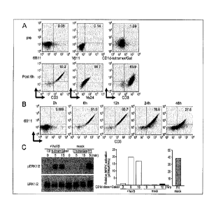
[Example 1: Preparation of modified immunocytes using Jurkat cell lines]
Vet and -vp chains of NKT cell TCR derived from the NKT cell lines that had
been
established from healthy volunteers were initially subcloned. The mRNAs were
generated from
the coding regions of TCR a and TCR p chains in the DNA (TCR a chain: SEQ ID
NO: 1, and
TCR 13 chain: SEQ ID NO: 2), respectively by an in vitro transcription
approach. After both of
the TCR chains were transfected into Jurkat T cells by electroporation, the
expression of
NKT-TCR was determined by cytometry using a combination of anti-Va24 and V1311
Ab, or a
combination of CD3 and anti-6B11 or anti-CD1d/Gal-tetramer (Fig. 1A).
[0055]
As reported, an expression of both of the Va24 and V1311 was evaluated by anti-
6B11
mAb. The expression level of NKT-TCR was up-regulated during 6 to 12 hours,
and decreased
48 hours later (Fig. 1B). Particularly, the expression of the Vo24 and V1311
was detected on the
Jurkat cells exceeding 90% of the whole Jurkat cells after 6 hours. After
that, the downstream
of the TCR signal was assessed after the cells were cultured together with a
solid phase of
a-GalCer-binding CD1d antibody. Mitogen-activated protein kinase (MAPK) was
phosphorylated in 10 minutes after the stimulation (Fig. 1C), and it is
indicated that TCR
signaling was clearly augmented in the Va24 and V1311 TCR mRNA-transfected
Jurkat cells.
231964,1.1
CA 02977606 2017-08-23
CA Application
Blakes Ref: 10082/00003
[0056]
As described above, by introducing the mRNAs of the TCR a and TCR p chains, a
modified immunocyte transiently expressing functional NKT-TCR on a Jurkat cell
was able to be
produced.
[0057]
[Example 2: Preparation of modified immunocytes using activated T cells
derived from
peripheral blood mononuclear cells (PBMCs) of healthy subjects]
The mRNAs of Va24 and V1311 TCR chains were transfected into an activated
primary
T cell that had been generated for 3 days by anti-CD3 Ab and IL-2. The
expression of TCR
chain (Va24) and 13 chain (V1311) on the mRNA-transfected T cell was assessed
by anti-6B11
mAb. The 6B11+ cells were 60 to 70% of the cells derived from CD3+ T cells,
and it is
indicated that both of the chains were apparently expressed on the surfaces of
the cells (Figs. 2A
and 2B).
[0058]
The TCR signaling was analyzed in Va24+VI311+ transfected (hereinafter
referred to as
"NKT-TCR") cells and Va241/1311- non-transfected (hereinafter referred to as
"NKT-TCR"")
cells after the stimulation with a solid phase of a-GalCer-binding CD1d
antibody. The
NKT-TCR4 cells, but not the NKT-TCR" cells showed the activation of MAP kinase
signal (right
column of Fig. 2C).
[0059]
Furthermore, the cytokine production was analyzed by co-culturing the NKT-TCR
cells
together with the cells loaded with a-GalCer (CD1d-HEK293 cells/Gal). The NKT-
TCR cells
produced more interferon-7 but not IL-4 in a a-GalCer dependent manner.
However, both of
the NKT-TCR- cells and the activated T cells without transfection did not
produce any
21
23196461.1
CA 02977606 2017-08-23
CA Application
Slakes Ref: 10082/00003
interferon-7 (Fig. 2D). Therefore, the Va24 TCR and v311 TCR mRNA-transfected
activated
T cells (NKT-TCR + cells) were functional to produce interferon-y, and it is
indicated that these
Th1 type-skewed 6B11+ cells can mimic Th1 type NKT cells for 48 hours.
[0060]
Subsequently, the adjuvant effect of NKT-TCR + cells was confirmed. It has
been
reported that NKT cells induce the mattiration of DCs in both of the phenotype
and function in
vivo and in vitro. It was assessed whether or not the NKT-TCR + T cells can
mature DCs. The
maturation markers and cytokine productions were evaluated after the NKT-TCR +
T cells and
autologous monocyte-derived mature DCs were cultured.
[0061]
Up-regulation of costimulatory molecules on DCs by NKT-TCR + T cells was
observed
similar to the up-regulation by LPS stimulation (Fig. 3A). Further, in the DCs
matured by
NKT-TCR + T cells, IL-12p'70 production was antigen-specifically remarkably
observed, but
IL-10 production was hardly observed (Fig. 3B). The L-12p70 acts in a
direction of stimulating
the immunity, and the IL-10 acts conversely in a direction of suppressing the
immunity.
Therefore, the maturation of DCs by NKT-TCR + T cells is much more favorable
than the LPS
stimulation for the immune induction.
[0062]
[Example 3: Preparation of modified immunocytes using yo T cells derived from
peripheral blood mononuclear cells (PBMCs) of healthy subjects]
The y8 T cell is well-known as one of the innate lymphocytes. Among the y6 T
cells,
79 type of 76 T cells can be proliferated by zoledronic acid (ZA)-loaded cells
in which some
endogenous 76 T cell ligands were up-regulated on antigen-presenting cells
(APCs). As shown
in Fig. 4, even those who recognize NKT cells with only extremely low
frequency in the
22
23196461.1
CA 02977606 2017-08-23
CA Application
Makes Ref: 10082/00003
peripheral blood have the appropriate number of yo T cells. In addition, yo T
cells have the
potential to proliferate in an amount much larger than the NKT cells (Figs. 4A
and 4B).
[0063]
Apparently, y8 T cells usually do not express NKT-TCR, however these can
express
NKT-TCR together with y93 TCR after the transfection by electroporation (Fig.
5). When these
NKT-TCR + cells were co-cultured with ZA-loaded CD1d-HEK293 cells, y8 T cells
produced
interferon-y, and these were confirmed to be y8 T cells.
[0064]
In order to investigate the difference in the function due to the expression
of NKT-TCR
in the y5 T cell, (1) the 78 T cells cultured with the stimulation of ZA, and
(2) the NKT-TCR + y5
T cells co-cultured with the CD1d-HEK293 cell/Gal were compared to each other.
The results
are shown in Fig.6. When the number of cells was counted in 72 hours after the
stimulation or
co-culture, there was a remarkable difference in the cell proliferation (left
panel of Fig. 6). In
the similar manner, when the yield of interferon-y in 48 hours after the
stimulation or co-culture
was measured by ELISA, a remarkable increase in the yield in NKT-TCR + y3 T
cells was
observed (right panel of Fig. 6).
[0065]
As described above, by expressing the NKT-TCR, and applying the stimulation of
=
a-GalCer, it was indicated that the cell proliferation ability of the 78 T
cells and the yield of the
interferon-y were improved.
[0066]
From the above, it was found that by modifying the y8 T cells that are present
in a
relatively large amount in peripheral blood, and further can be proliferated
in an amount
sufficient for clinical use, the availability of the y3 T cells can be
significantly improved.
23
23196461.1
CA 02977606 2017-08-23
CA Application
Blakes Ref: 10082/00003
Further, based on the individual difference, the yo T cells either are not
activated by ZA alone, or
cause the case Where the stimulation is insufficient, but the modified 76 T
cells newly bring an
option to use the a-GalCer. Accordingly, the modified y6 T cells substantially
reduce the
number of the individuals who cannot use the y6 T cells or have low
effectiveness in using the y6
T cells, and thus can provide an opportunity for the treatment to more
individuals.
[0067]
In addition, in common in each of the Examples described above, what has been
introduced into each cell is mRNAs of the a and j3 chains of the NKT cell TCR.
As is
extremely well proved in Examples 1 and 2, the a and p chains in a cell
surface decreases with
the lapse of time. However, it is not that the number of cells is decreased.
That is, in the
NKT-TCR+ cells, by the decomposition of the introduced mRNAs, the expression
level of the a
and f3 chains of the NKT cell TCR is gradually decreased, and it is eventually
only returned to
the state before the introduction of mRNAs. Therefore, the application of the
cells obtained in
these Examples to immunotherapy does not fall under the gene therapy. It is
apparent that the
cells of these Examples, in which the exogenous factors to be introduced do
not remain, exhibit
only extremely low side effects that are beyond comparison with the
conventional gene therapy.
In a case where the cells of these Examples are applied in the immunocytc
therapy in which
autologous cells are used, it can be regarded that there are substantially no
side effects.
[0068]
Further, the regulations for performing gene therapy are not applied,
therefore, there is
almost no restriction on the place to handle the cells of these Examples.
Since the nature of the
cells returns to the state in the body with the lapse of time, the instruments
and
biologically-derived materials used for preparing the cells can be disposed by
a disposal method
equivalent to that for the instruments and the like used usually in medical
facilities.
24
23196461.1
CA 02977606 2017-08-23
CA Application
Blakes Ref: 10082/00003
[0069]
[Example 4: Confirmation of in vivo anti-tumor effect of modified immunocytes]
In order to investigate the in vivo anti-tumor effect by immunocytes in which
NKT-TCR
had newly expressed, verification was performed by using a yo T cell in which
NKT-TCR had
expressed (NKT-TCR + T cell of Example 3: hereinafter referred to as a
modified
immunocyte).
[0070]
Immunodeficient mice to which 2x10⁶ K562 cells had been subcutaneously
inoculated
were prepared as model animals to evaluate the anti-tumor effect by the
modified immunocytes.
The modified immunocytes were prepared in accordance with the same procedures
as those in
Example 3. The following two kinds of treated products were administered to
the tumor
inoculation sites in the model animals in 7 days after the inoculation
(respectively n = 2).
(1) 100 vtl of medium in which 2x10⁶ modified immunocytes are suspended
("y6+NKT TCR" in
Fig. 7: dark line)
(2) 100 1.11 of medium alone ("non-treated" in Fig. 7: pale line)
[0071]
The results of evaluating the anti-tumor effects by the modified immunocytes
based on
the tumor sizes in the model animals, which had been measured in 12 to 24 days
after the
inoculation, are shown in Fig. 7. As shown in Fig. 7, all of the model animals
to which
modified immunocytes had been administered showed no increase in the tumor
size in 12 to 24
days after the inoculation. On the other hand, all of the model animals to
which the modified
immunocytes had not been administered showed increase in the tumor size with
the lapse of time
(in particular, after the 17th day of the inoculation). From the above, it was
revealed that the
modified immunocytes (NKT-TCR+ yE= T cells) exhibit extremely excellent anti-
tumor activity in
23196461 1
CA 02977606 2017-08-23
CA Application
Blakes Ref: 10082/00003
vivo.
[0072]
[Comparative Example: Efficacy of NK cells introduced with NKT-TCR]
Even in a case where NKT-TCR was introduced into a NK cell, in order to
investigate
whether or not the same effect as that of the modified immunocyte in Example 3
is shown, the
peripheral blood mononuclear cells (PBMCs) of healthy subjects, which had been
collected as
described above, were cultured in a medium containing 1000 U/ml IL2. After
confirming the
proliferation of the cells, the mRNAs for the expression of NKT-TCR were
introduced into the
cells, and the cell population in which NKT-TCRs were surface-expressed was
confirmed. The
results of the confirmation of cell proliferation and cell population by flow
cytometry after the
lapse of a predetermined number of days from the culture of peripheral blood
mononuclear cells
(PBMCs) are shown in Fig. 8.
[0073]
As shown on the left side of the arrow in Fig. 8, the proliferation of cells
at each time
point of 3 days and 7 days after the start of the culture was confirmed by
flow cytometry.
Further, at the time point after 7 days, the proliferation of cells was
further analyzed by using
other fluorescent-labeled antibodies, and CD3+CD56- cell population (11.0%),
CD3-CD56+ cell
population (72.1%), CD16+CD56- cell population (76.4%), and CD16-CD56+ cell
population
(17.4%) were confirmed to be present. NKT-TCR mRNAs (Va24 RNA and V1311 mRNA)
were electroporated at the time point of 8 days after the start of the
culture, and after 6 hours,
Va24+V1311+ cells were confirmed by flow cytometry. As shown on the right side
of the arrow
in Fig. 8, the percentage of the cells reacting with anti-6B11 mAb was 41.0%,
and the percentage
of the Va24+V[311+ cells was 71.5% in CD3+ cells, but on the contrary, the
percentage of the
cells reacting with anti-6B11 mAb was 2.31%, and the percentage of the
Va24+V[311+ cells was
26
2319646L1
CA 02977606 2017-08-23
CA Application
Blakcs Ref: 10082/00003
8.5% in the CD56+ cells containing NK cells.
[0074]
As described above, as compared with the CD3+ cells, in the NK cells,
expression itself
of the NKT-TCR was suppressed. Therefore, it was revealed that the cells that
exhibit the same
effects as those of the modified immunocytes based on CD3+ cells as prepared
in Example 3
were able to be obtained only extremely inefficiently when the NK cells were
used as the
material. In the experiments described above, extremely unexpected results
that betray usual
expectation for those skilled in the art, which is the expectation that cells
showing the function as
in NKT cells will be obtained when the mRNAs of NKT-TCR were introduced into
the NK cells,
were shown. Therefore, as shown in Example 3, it has revealed that CD3+ cells
are extremely
suitable for the preparation of the modified immunocytes for immunotherapy.
[0075]
The present invention is not limited to each of the above-described
embodiments and
Examples, and various modifications can be made within the scope indicated in
the claims, and
embodiments obtained by appropriately combining the technical means disclosed
in different
embodiments, respectively are also included in the technical scope of the
present invention.
Further, by combining the technical means disclosed in each embodiment and
each Example,
respectively, new technical features can be formed.
Industrial Applicability
[0076]
The present invention can be used for immunocyte therapy. In particular, the
present
invention can be used as an immunity inducer that activates the effector cells
directly exhibiting
cytotoxic immunity and other immunocytes.
27
23196461.1